Embed Size (px)
Citation preview

HUMAN NEUROSCIENCECLINICAL CASE STUDY
published: 21 October 2014doi: 10.3389/fnhum.2014.00861
Epidural electrocorticography for monitoring of arousal inlocked-in stateSuzanne Martens1,2,3,4, Michael Bensch5, Sebastian Halder6,7, Jeremy Hill3, Femke Nijboer8, AnderRamos-Murguialday6,9, Bernhard Schoelkopf3, Niels Birbaumer6,10 and Alireza Gharabaghi1,2*1 Division of Functional and Restorative Neurosurgery and Division of Translational Neurosurgery, Department of Neurosurgery, Eberhard Karls University
Tuebingen, Tuebingen, Germany2 Neuroprosthetics Research Group, Werner Reichardt Center for Integrative Neuroscience, Eberhard Karls University Tuebingen, Tuebingen, Germany3 Department of Empirical Inference, Max Planck Institute for Intelligent Systems, Tuebingen, Germany4 Department of Medical Physics, University Medical Center Utrecht, Utrecht University, Utrecht, Netherlands5 Department of Computer Engineering, Wilhelm-Schickard Institute for Computer Science, Eberhard Karls University Tuebingen, Tuebingen, Germany6 Institute of Medical Psychology and Behavioral Neurobiology, Eberhard Karls University Tuebingen, Tuebingen, Germany7 Institute of Psychology, University of Wuerzburg, Wuerzburg, Germany8 Research Group Human Media Interaction, Department of Electrical Engineering, Mathematics and Computer Science, University of Twente, Enschede,
Netherlands9 Health and Quality of life Unit, Fatronik-Tecnalia, San Sebastian, Spain10 Istituto di Ricovero e Cura a Carattere Scientifico, IRCCS Ospedale San Camillo, Venezia, Italy
Edited by:Marta Olivetti, Sapienza Universityof Rome, Italy
Reviewed by:Maurizio Mattia, Istituto Superioredi Sanità, ItalyPierre Megevand, Hofstra NorthShore-LIJ School of Medicine, USA
*Correspondence:Alireza Gharabaghi, Division ofFunctional and RestorativeNeurosurgery and Division ofTranslational Neurosurgery,Department of Neurosurgery,Eberhard Karls UniversityTuebingen, Otfried-Mueller-Str. 45,72076 Tuebingen, Germanye-mail: [email protected]
Electroencephalography (EEG) often fails to assess both the level (i.e., arousal) andthe content (i.e., awareness) of pathologically altered consciousness in patients withoutmotor responsiveness. This might be related to a decline of awareness, to episodesof low arousal and disturbed sleep patterns, and/or to distorting and attenuating effectsof the skull and intermediate tissue on the recorded brain signals. Novel approachesare required to overcome these limitations. We introduced epidural electrocorticography(ECoG) for monitoring of cortical physiology in a late-stage amytrophic lateral sclerosispatient in completely locked-in state (CLIS). Despite long-term application for a period ofsix months, no implant-related complications occurred. Recordings from the left frontalcortex were sufficient to identify three arousal states. Spectral analysis of the intrinsicoscillatory activity enabled us to extract state-dependent dominant frequencies at <4, ∼7and ∼20 Hz, representing sleep-like periods, and phases of low and elevated arousal,respectively. In the absence of other biomarkers, ECoG proved to be a reliable toolfor monitoring circadian rhythmicity, i.e., avoiding interference with the patient whenhe was sleeping and exploiting time windows of responsiveness. Moreover, the effectsof interventions addressing the patient’s arousal, e.g., amantadine medication, couldbe evaluated objectively on the basis of physiological markers, even in the absence ofbehavioral parameters. Epidural ECoG constitutes a feasible trade-off between surgicalrisk and quality of recorded brain signals to gain information on the patient’s present levelof arousal. This approach enables us to optimize the timing of interactions and medicalinterventions, all of which should take place when the patient is in a phase of high arousal.Furthermore, avoiding low-responsiveness periods will facilitate measures to implementalternative communication pathways involving brain-computer interfaces (BCI).
Keywords: electrocorticography, epidural recording, locked-in state, coma, consciousness, brain-computerinterface, neuroprosthetic devices
INTRODUCTIONAssessing both the level (i.e., arousal) and the content (i.e.,awareness) of pathologically altered consciousness in clinicalenvironments is limited to evaluating patients’ motor responsive-ness (Laureys et al., 2009). Neurodegenerative diseases or injuriesto the central nerve system may paralyze the affected patients tosuch a degree that they lose any remaining ability to communicateby volitional muscle control, thereby impeding the assessment ofthe different dimensions of consciousness (Laureys et al., 2004).
In the case of amyotrophic lateral sclerosis (ALS), this dis-connection from the environment progresses slowly. In thelate stage of the disease, in which the patients are no longerable to move their body or to speak, this condition spans atransition from the locked-in state (LIS), with very limitedremnants of voluntary movements such as muscle twitchesor eye movements, to the completely locked-in state (CLIS),with the loss of all motor control (Kübler and Birbaumer,2008).
Frontiers in Human Neuroscience www.frontiersin.org October 2014 | Volume 8 | Article 861 | 1
Martens et al. Epidural ECoG monitoring of arousal
This is paralleled by a decline of other physiological measuresfor communication, including sphincter and facial electromyog-raphy as well as oculography (Murguialday et al., 2011). Similarly,body signals mediated by the parasympathetic and sympatheticnervous system show significant abnormalities (Pinelli et al.,1995) such as decreased heart rate variation (Pisano et al., 1995),alterations of the excretory function of the salivary glands (Giesset al., 2000), disturbances of the gastrointestinal tract (Toepferet al., 1997, 1999), and alterations of the skin responses (Masuret al., 1995).
In addition, when examining CLIS patients with electroen-cephalography (EEG), Kotchoubey et al. (2003) reported a largevariability of event-related responses (ERP) including a com-plete loss of ERP. Even when provided with EEG-based brain-computer interfaces (BCIs), ALS patients are unable to retaincommunication once they enter CLIS. This loss of the ability tocommunicate might be related to the disease itself and may reflectan irreversible decline of awareness, in which case it would beunavoidable (Kübler and Birbaumer, 2008). On the other hand,such a lack of communication could also be due to methodologi-cal and technical problems that could be overcome by alternativeBCI approaches (Bensch et al., 2014). Recently, a metabolic BCIbased on near-infrared spectroscopy has been introduced as apromising tool for communication in CLIS (Gallegos-Ayala et al.,2014).
However, locked-in patients are known to suffer from dis-turbed sleep patterns with increased fragmentation during thecourse of the disease (Ferguson et al., 1995; Soekadar et al.,2013). These fluctuations might inherently limit the successof novel BCI approaches in the affected patients. Detectingepisodes of low arousal may therefore be essential in opti-mizing the timing of communication attempts towards phasesof higher arousal. At the same time, distorting and attenuat-ing effects of the skull and intermediate tissue on the neuralsignals inherent to the classical EEG approach might be sur-mounted by signal detection closer to the brain (Buzsáki et al.,2012; Bensch et al., 2014). Recently, we have introduced epidu-ral ECoG as a tool to assess attention and cognitive functionin LIS (Bensch et al., 2014). However, this technique appliedevent-related brain-potentials at specific time points to trackthe long-term transition from the locked-in to the completelylocked in-state, thereby not allowing a close-meshed monitor-ing of the patient’s current state of arousal (Bensch et al.,2014).
Thus, a novel methodology for continuous monitoring is stillrequired to gain information on the patient’s present level ofarousal and to overcome the limitations of current approaches soas to interact with patients without motor responsiveness.
METHODSCLINICAL CASEThe patient described in this manuscript was a well-informed40-year-old male, late-stage ALS patient, who had been diag-nosed with this disease 10 years before hospitalization and whoalready had a 7-year history of artificial ventilation. Being fullyaware of the long-term consequences of ALS with respect tothe loss of all ability to communicate in later stages of this
progressive disease, he was determined to retain this ability foras long as possible. Although he was already in a late stage ofthis disease when he contacted us, he was still able to com-municate reliably via muscle twitches. During the period ofseveral months in which we had the opportunity to examinehim on a daily basis, his status deteriorated continuously, i.e.,a transition from LIS to CLIS took place. While he was ini-tially able to gain some control of an EEG-based BCI, his BCIperformance dropped to random over the course of severalweeks.
Before entering this stage of his disease, the patient and hislegal representative had given informed consent to implantationof an ECoG grid, both on first contact before hospitalizationand during his stay in hospital before surgery. The purpose ofthis implantation was to monitor the patient’s arousal in theabsence of other biomarkers and to develop alternative commu-nication pathways for him with ECoG-based BCI technology. Theresults of the BCI communication study do not constitute partof the present report. This study was conducted in accordancewith the Declaration of Helsinki and with the guidelines of thelocal ethics committee of the University of Tuebingen (MedicalFaculty).
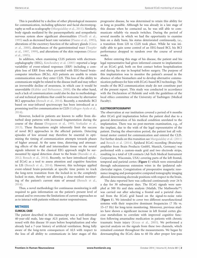
ELECTROCORTICOGRAPHYThe observation at our institution covered a period of 6 monthsafter ECoG grid implantation before the patient died due to ageneral deterioration of his medical condition unrelated to theimplantation. There was no post-mortem examination, e.g., ofthe implant, due to the wish of the legal representative of thepatient. During the observation period, the patient lost all voli-tional motor control for communication and entered the CLIS.For further details on this transition see Murguialday et al. (2011)and Bensch et al. (2014). Epidural ECoG recording (BrainAmpamplifier from Brain Products GmbH, Munich, Germany) wasperformed with a custom-made grid and two electrode strips,resulting in a total of 128 contacts (Ad-Tech Medical InstrumentCorporation, Wisconsin, USA) covering parts of the left frontal,temporal and parietal cortex (Figure 1) which were externalizedthrough subcutaneous extension wires in the ipsilateral sub-clavicular region. Coregistration of preoperative magnetic reso-nance imaging and postoperative computed tomography imagingallowed determining electrode positions with respect to the brain.
The data reported here was collected continuously over 24 ha day for 30 subsequent days. The ECoG signals were sam-pled at 500 Hz and data analysis (Matlab, The Mathworks™)was carried out after selecting a frontal and a parietal chan-nel from the ECoG grid based on the signal-to-noise ratio(Figure 1). We intended to cover two different neurofunctionalsystems with their respective dominant frequencies (7 Hz vs.15–17 Hz) for long-term monitoring. Moreover, previous stud-ies have shown a significant increase in left frontal cortex glu-cose metabolism to correlate with improved cognitive func-tion following amantadine medication in patients with chronictraumatic brain injury (Kraus et al., 2005). We performed aspectral analysis on the signals from these two channels, whichremained constant throughout the measurements. We began bydownsampling the ECoG data to 60 Hz after proper low-pass
Frontiers in Human Neuroscience www.frontiersin.org October 2014 | Volume 8 | Article 861 | 2
Martens et al. Epidural ECoG monitoring of arousal
FIGURE 1 | Lateral projections of implanted ECoG recording with a gridand two electrode strips resulting in a total of 128 contacts that covera large part of the left hemisphere. Reference (r), ground (g) andrecording channels on the frontal (f) and parietal (p) cortex for monitoringare indicated.
filtering to reduce the data set size. We then divided the datainto 30-s epochs, estimated an autoregressive (AR) model ofthe order of 7 in each epoch, and derived the power spectrumfrom the AR coefficients in each epoch as described in Nielsen(1992). We extracted the dominant frequency over time as thefrequency within the 2–30 Hz band with maximum spectralpower per epoch and per channel. We then classified each 30-sepoch as a slow-wave epoch, if the dominant frequency in bothchannels was lower than 4 Hz and as a non-slow-wave epochotherwise. We derived a slow-wave on/off curve by treatingeach consecutive epoch as a time sample and by assigning avalue of 1 to slow-wave epochs and a value of 0 to non-slow-wave epochs. We then calculated the autocorrelation functionof this slow-wave on/off curve to detect periodic components.We also calculated an auto-correlation function on the simulta-neously recorded temperature and the heartbeat rate to detectrhythmicity.
RESULTSCLINICAL CHARACTERISTICSEpidural ECoG was feasible and safe for monitoring corticalphysiology in a late-stage ALS patient in CLIS. Despite long-term application for a period of 6 months, no implant-relatedcomplications arose; in particular, there was no infection at thesite of externalization of the connection wires through the skin.Although a multi-channel device was implanted to maximizesignal recording for BCI application, two recording channels(one frontal and one parietal, see Figure 1) were sufficient forlongitudinal monitoring of arousal, indicating that less extendedimplants are feasible for this purpose in future cases.
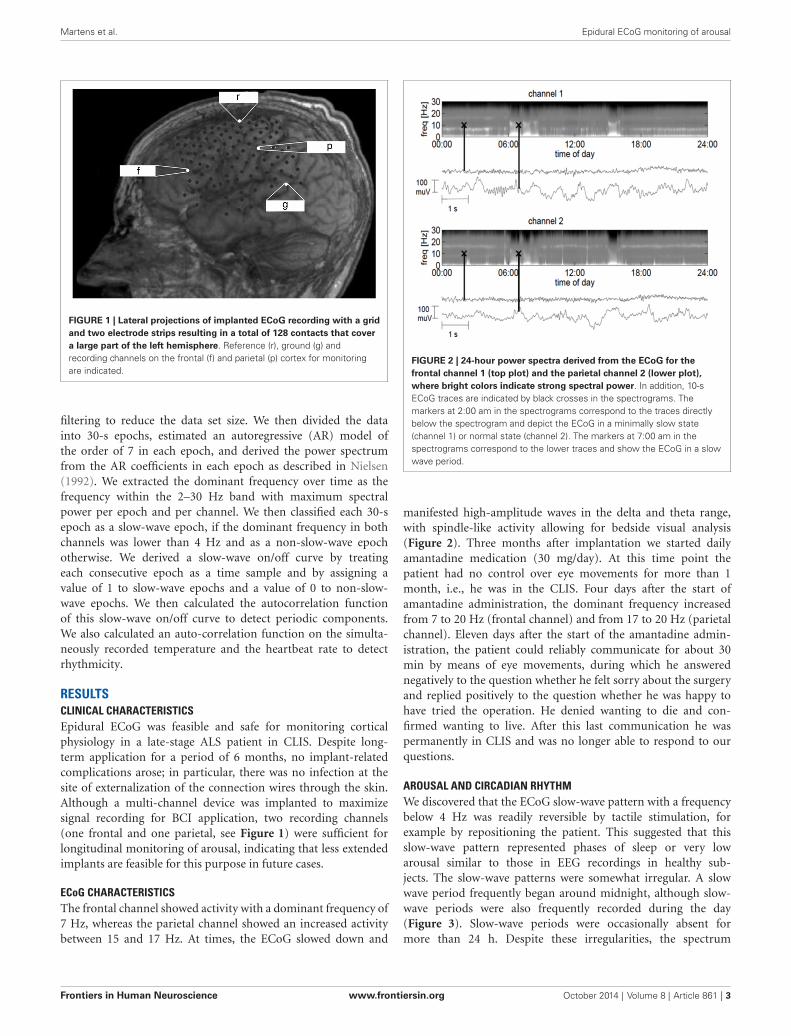
ECoG CHARACTERISTICSThe frontal channel showed activity with a dominant frequency of7 Hz, whereas the parietal channel showed an increased activitybetween 15 and 17 Hz. At times, the ECoG slowed down and
FIGURE 2 | 24-hour power spectra derived from the ECoG for thefrontal channel 1 (top plot) and the parietal channel 2 (lower plot),where bright colors indicate strong spectral power. In addition, 10-sECoG traces are indicated by black crosses in the spectrograms. Themarkers at 2:00 am in the spectrograms correspond to the traces directlybelow the spectrogram and depict the ECoG in a minimally slow state(channel 1) or normal state (channel 2). The markers at 7:00 am in thespectrograms correspond to the lower traces and show the ECoG in a slowwave period.
manifested high-amplitude waves in the delta and theta range,with spindle-like activity allowing for bedside visual analysis(Figure 2). Three months after implantation we started dailyamantadine medication (30 mg/day). At this time point thepatient had no control over eye movements for more than 1month, i.e., he was in the CLIS. Four days after the start ofamantadine administration, the dominant frequency increasedfrom 7 to 20 Hz (frontal channel) and from 17 to 20 Hz (parietalchannel). Eleven days after the start of the amantadine admin-istration, the patient could reliably communicate for about 30min by means of eye movements, during which he answerednegatively to the question whether he felt sorry about the surgeryand replied positively to the question whether he was happy tohave tried the operation. He denied wanting to die and con-firmed wanting to live. After this last communication he waspermanently in CLIS and was no longer able to respond to ourquestions.
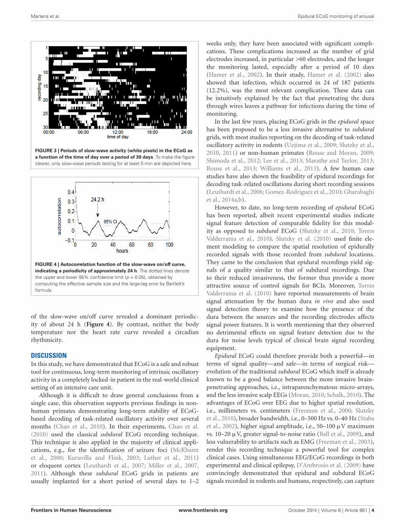
AROUSAL AND CIRCADIAN RHYTHMWe discovered that the ECoG slow-wave pattern with a frequencybelow 4 Hz was readily reversible by tactile stimulation, forexample by repositioning the patient. This suggested that thisslow-wave pattern represented phases of sleep or very lowarousal similar to those in EEG recordings in healthy sub-jects. The slow-wave patterns were somewhat irregular. A slowwave period frequently began around midnight, although slow-wave periods were also frequently recorded during the day(Figure 3). Slow-wave periods were occasionally absent formore than 24 h. Despite these irregularities, the spectrum
Frontiers in Human Neuroscience www.frontiersin.org October 2014 | Volume 8 | Article 861 | 3
Martens et al. Epidural ECoG monitoring of arousal
FIGURE 3 | Periods of slow-wave activity (white pixels) in the ECoG asa function of the time of day over a period of 30 days. To make the figureclearer, only slow-wave periods lasting for at least 5 min are depicted here.
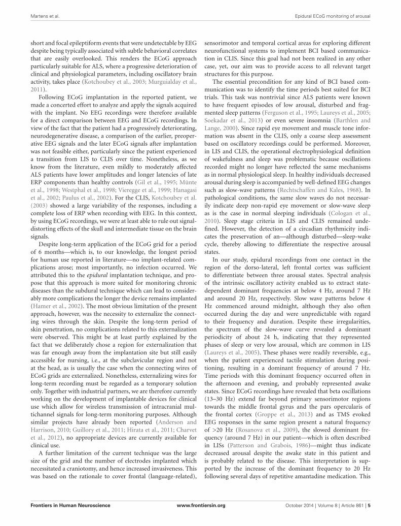
FIGURE 4 | Autocorrelation function of the slow-wave on/off curve,indicating a periodicity of approximately 24 h. The dotted lines denotethe upper and lower 95% confidence limit (p = 0.05), obtained bycomputing the effective sample size and the large-lag error by Bartlett’sformula.
of the slow-wave on/off curve revealed a dominant periodic-ity of about 24 h (Figure 4). By contrast, neither the bodytemperature nor the heart rate curve revealed a circadianrhythmicity.
DISCUSSIONIn this study, we have demonstrated that ECoG is a safe and robusttool for continuous, long-term monitoring of intrinsic oscillatoryactivity in a completely locked-in patient in the real-world clinicalsetting of an intensive care unit.
Although it is difficult to draw general conclusions from asingle case, this observation supports previous findings in non-human primates demonstrating long-term stability of ECoG-based decoding of task-related oscillatory activity over severalmonths (Chao et al., 2010). In their experiments, Chao et al.(2010) used the classical subdural ECoG recording technique.This technique is also applied in the majority of clinical appli-cations, e.g., for the identification of seizure foci (McKhannet al., 2000; Kuruvilla and Flink, 2003; Luther et al., 2011)or eloquent cortex (Leuthardt et al., 2007; Miller et al., 2007,2011). Although these subdural ECoG grids in patients areusually implanted for a short period of several days to 1–2
weeks only, they have been associated with significant compli-cations. These complications increased as the number of gridelectrodes increased, in particular >60 electrodes, and the longerthe monitoring lasted, especially after a period of 10 days(Hamer et al., 2002). In their study, Hamer et al. (2002) alsoshowed that infection, which occurred in 24 of 187 patients(12.2%), was the most relevant complication. These data canbe intuitively explained by the fact that penetrating the durathrough wires leaves a pathway for infections during the time ofmonitoring.
In the last few years, placing ECoG grids in the epidural spacehas been proposed to be a less invasive alternative to subduralgrids, with most studies reporting on the decoding of task-relatedoscillatory activity in rodents (Uejima et al., 2009; Slutzky et al.,2010, 2011) or non-human primates (Rouse and Moran, 2009;Shimoda et al., 2012; Lee et al., 2013; Marathe and Taylor, 2013;Rouse et al., 2013; Williams et al., 2013). A few human casestudies have also shown the feasibility of epidural recordings fordecoding task-related oscillations during short recording sessions(Leuthardt et al., 2006; Gomez-Rodriguez et al., 2010; Gharabaghiet al., 2014a,b).
However, to date, no long-term recording of epidural ECoGhas been reported, albeit recent experimental studies indicatesignal feature detection of comparable fidelity for this modal-ity as opposed to subdural ECoG (Slutzky et al., 2010; TorresValderrama et al., 2010). Slutzky et al. (2010) used finite ele-ment modeling to compare the spatial resolution of epidurallyrecorded signals with those recorded from subdural locations.They came to the conclusion that epidural recordings yield sig-nals of a quality similar to that of subdural recordings. Dueto their reduced invasiveness, the former thus provide a moreattractive source of control signals for BCIs. Moreover, TorresValderrama et al. (2010) have reported measurements of brainsignal attenuation by the human dura in vivo and also usedsignal detection theory to examine how the presence of thedura between the sources and the recording electrodes affectssignal power features. It is worth mentioning that they observedno detrimental effects on signal feature detection due to thedura for noise levels typical of clinical brain signal recordingequipment.
Epidural ECoG could therefore provide both a powerful—interms of signal quality—and safe—in terms of surgical risk—evolution of the traditional subdural ECoG which itself is alreadyknown to be a good balance between the more invasive brain-penetrating approaches, i.e., intraparenchymatous micro-arrays,and the less invasive scalp EEGs (Moran, 2010; Schalk, 2010). Theadvantages of ECoG over EEG due to higher spatial resolution,i.e., millimeters vs. centimeters (Freeman et al., 2000; Slutzkyet al., 2010), broader bandwidth, i.e., 0–500 Hz vs. 0–40 Hz (Stabaet al., 2002), higher signal amplitude, i.e., 50–100 µV maximumvs. 10–20 µV, greater signal-to-noise ratio (Ball et al., 2009), andless vulnerability to artifacts such as EMG (Freeman et al., 2003),render this recording technique a powerful tool for complexclinical cases. Using simultaneous EEG/ECoG recordings in bothexperimental and clinical epilepsy, D’Ambrosio et al. (2009) haveconvincingly demonstrated that epidural and subdural ECoGsignals recorded in rodents and humans, respectively, can capture
Frontiers in Human Neuroscience www.frontiersin.org October 2014 | Volume 8 | Article 861 | 4
Martens et al. Epidural ECoG monitoring of arousal
short and focal epileptiform events that were undetectable by EEGdespite being typically associated with subtle behavioral correlatesthat are easily overlooked. This renders the ECoG approachparticularly suitable for ALS, where a progressive deterioration ofclinical and physiological parameters, including oscillatory brainactivity, takes place (Kotchoubey et al., 2003; Murguialday et al.,2011).
Following ECoG implantation in the reported patient, wemade a concerted effort to analyze and apply the signals acquiredwith the implant. No EEG recordings were therefore availablefor a direct comparison between EEG and ECoG recordings. Inview of the fact that the patient had a progressively deteriorating,neurodegenerative disease, a comparison of the earlier, preoper-ative EEG signals and the later ECoG signals after implantationwas not feasible either, particularly since the patient experienceda transition from LIS to CLIS over time. Nonetheless, as weknow from the literature, even mildly to moderately affectedALS patients have lower amplitudes and longer latencies of lateERP components than healthy controls (Gil et al., 1995; Münteet al., 1998; Westphal et al., 1998; Vieregge et al., 1999; Hanagasiet al., 2002; Paulus et al., 2002). For the CLIS, Kotchoubey et al.(2003) showed a large variability of the responses, including acomplete loss of ERP when recording with EEG. In this context,by using ECoG recordings, we were at least able to rule out signal-distorting effects of the skull and intermediate tissue on the brainsignals.
Despite long-term application of the ECoG grid for a periodof 6 months—which is, to our knowledge, the longest periodfor human use reported in literature—no implant-related com-plications arose; most importantly, no infection occurred. Weattributed this to the epidural implantation technique, and pro-pose that this approach is more suited for monitoring chronicdiseases than the subdural technique which can lead to consider-ably more complications the longer the device remains implanted(Hamer et al., 2002). The most obvious limitation of the presentapproach, however, was the necessity to externalize the connect-ing wires through the skin. Despite the long-term period ofskin penetration, no complications related to this externalizationwere observed. This might be at least partly explained by thefact that we deliberately chose a region for externalization thatwas far enough away from the implantation site but still easilyaccessible for nursing, i.e., at the subclavicular region and notat the head, as is usually the case when the connecting wires ofECoG grids are externalized. Nonetheless, externalizing wires forlong-term recording must be regarded as a temporary solutiononly. Together with industrial partners, we are therefore currentlyworking on the development of implantable devices for clinicaluse which allow for wireless transmission of intracranial mul-tichannel signals for long-term monitoring purposes. Althoughsimilar projects have already been reported (Anderson andHarrison, 2010; Guillory et al., 2011; Hirata et al., 2011; Charvetet al., 2012), no appropriate devices are currently available forclinical use.
A further limitation of the current technique was the largesize of the grid and the number of electrodes implanted whichnecessitated a craniotomy, and hence increased invasiveness. Thiswas based on the rationale to cover frontal (language-related),
sensorimotor and temporal cortical areas for exploring differentneurofunctional systems to implement BCI based communica-tion in CLIS. Since this goal had not been realized in any othercase, yet, our aim was to provide access to all relevant targetstructures for this purpose.
The essential precondition for any kind of BCI based com-munication was to identify the time periods best suited for BCItrials. This task was nontrivial since ALS patients were knownto have frequent episodes of low arousal, disturbed and frag-mented sleep patterns (Ferguson et al., 1995; Laureys et al., 2005;Soekadar et al., 2013) or even severe insomnia (Barthlen andLange, 2000). Since rapid eye movement and muscle tone infor-mation was absent in the CLIS, only a coarse sleep assessmentbased on oscillatory recordings could be performed. Moreover,in LIS and CLIS, the operational electrophysiological definitionof wakefulness and sleep was problematic because oscillationsrecorded might no longer have reflected the same mechanismsas in normal physiological sleep. In healthy individuals decreasedarousal during sleep is accompanied by well-defined EEG changessuch as slow-wave patterns (Rechtschaffen and Kales, 1968). Inpathological conditions, the same slow waves do not necessar-ily indicate deep non-rapid eye movement or slow-wave sleepas is the case in normal sleeping individuals (Cologan et al.,2010). Sleep stage criteria in LIS and CLIS remained unde-fined. However, the detection of a circadian rhythmicity indi-cates the preservation of an—although disturbed—sleep-wakecycle, thereby allowing to differentiate the respective arousalstates.
In our study, epidural recordings from one contact in theregion of the dorso-lateral, left frontal cortex was sufficientto differentiate between three arousal states. Spectral analysisof the intrinsic oscillatory activity enabled us to extract state-dependent dominant frequencies at below 4 Hz, around 7 Hzand around 20 Hz, respectively. Slow wave patterns below 4Hz commenced around midnight, although they also oftenoccurred during the day and were unpredictable with regardto their frequency and duration. Despite these irregularities,the spectrum of the slow-wave curve revealed a dominantperiodicity of about 24 h, indicating that they representedphases of sleep or very low arousal, which are common in LIS(Laureys et al., 2005). These phases were readily reversible, e.g.,when the patient experienced tactile stimulation during posi-tioning, resulting in a dominant frequency of around 7 Hz.Time periods with this dominant frequency occurred often inthe afternoon and evening, and probably represented awakestates. Since ECoG recordings have revealed that beta oscillations(13–30 Hz) extend far beyond primary sensorimotor regionstowards the middle frontal gyrus and the pars opercularis ofthe frontal cortex (Groppe et al., 2013) and as TMS evokedEEG responses in the same region present a natural frequencyof >20 Hz (Rosanova et al., 2009), the slowed dominant fre-quency (around 7 Hz) in our patient—which is often describedin LISs (Patterson and Grabois, 1986)—might thus indicatedecreased arousal despite the awake state in this patient andis probably related to the disease. This interpretation is sup-ported by the increase of the dominant frequency to 20 Hzfollowing several days of repetitive amantadine medication. This
Frontiers in Human Neuroscience www.frontiersin.org October 2014 | Volume 8 | Article 861 | 5
Martens et al. Epidural ECoG monitoring of arousal
increase towards the natural frequency of the frontal cortexin our patient indicates increased arousal and is in line withearlier results in studies on the influence of amantadine inpatients with chronic traumatic brain injury (Kraus et al., 2005).They showed a significant increase in left frontal cortex glucosemetabolism correlated with improved cognitive function fol-lowing amantadine medication. Similarly, in our patient theincrease of the intrinsic oscillatory pattern was paralleled bya new episode of reliable communication by means of eyemovements.
In the absence of other biomarkers, epidural ECoG provedto be a reliable tool for monitoring circadian rhythmicity inCLIS, i.e., avoiding interference with the patient when he wassleeping and exploiting time windows of responsiveness. Thisenabled nursing staff and therapists to adopt interventions inaccordance with the patient’s individual arousal (i.e., schedulethem in the afternoons and evenings). Moreover, we demon-strated that sufficient arousal monitoring could be acquired fromone frontal electrode contact, indicating that future implantsmay require only a relatively small burr hole, thus reducingsurgical risks in this vulnerable patient group even further.This methodology might also present a useful tool for monitor-ing the arousal in other conditions with pathologically alteredconsciousness whenever the patients’ motor responsiveness iscompromised.
CONCLUSIONSWe implemented a novel technique for continuous, long-term monitoring of arousal in a chronic disease withoutfurther physiological or behavioral parameters. The epiduralECoG approach presented here constitutes a feasible trade-offbetween surgical risk and quality of brain signals. The infor-mation provided is essential for both the social and the med-ical environment in dealing with expectations and planninginterventions that aim to implement alternative communica-tion pathways such as BCIs in conditions of missing motorresponsiveness.
ACKNOWLEDGMENTSAlireza Gharabaghi was supported by grants from the GermanResearch Council [DFG GH 94/2-1, DFG EC 307], and from theFederal Ministry for Education and Research [BFNT 01GQ0761,BMBF 16SV3783, BMBF 03160064B, BMBF V4UKF014].
REFERENCESAnderson, G. S., and Harrison, R. R. (2010). “Wireless integrated circuit
for the acquisition of electrocorticogram signals,” in Circuits and Systems(ISCAS), Proceedings of 2010 IEEE International Symposium (Paris, France),2952–2955.
Ball, T., Kern, M., Mutschler, I., Aertsen, A., and Schulze-Bonhage, A.(2009). Signal quality of simultaneously recorded invasive and non-invasive EEG. Neuroimage 46, 708–716. doi: 10.1016/j.neuroimage.2009.02.028
Barthlen, G. M., and Lange, D. J. (2000). Unexpectedly severe sleep and respiratorypathology in patients with amyotrophic lateral sclerosis. Eur. J. Neurol. 7, 299–302. doi: 10.1046/j.1468-1331.2000.00044.x
Bensch, M., Martens, S., Halder, S., Hill, J., Nijboer, F., Ramos, A., et al. (2014).Assessing attention and cognitive function in completely locked-in state withevent-related brain potentials and epidural electrocorticography. J. Neural Eng.11:026006. doi: 10.1088/1741-2560/11/2/026006
Buzsáki, G., Anastassiou, C. A., and Koch, C. (2012). The origin of extracellularfields and currents—EEG, ECoG, LFP and spikes. Nat. Rev. Neurosci. 13, 407–420. doi: 10.1038/nrn3241
Chao, Z. C., Nagasaka, Y., and Fujii, N. (2010). Long-term asynchronous decodingof arm motion using electrocorticographic signals in monkeys. Front. Neuroeng.3:3. doi: 10.3389/fneng.2010.00003
Charvet, G., Foerster, M., Chatalic, G., Michea, A., Porcherot, J., Bonnet, S.,et al. (2012). A wireless 64-channel ECoG recording electronic for implantablemonitoring and BCI applications: WIMAGINE. Conf. Proc. IEEE Eng. Med. Biol.Soc. 2012, 783–786. doi: 10.1109/EMBC.2012.6346048
Cologan, V., Schabus, M., Ledoux, D., Moonen, G., Maquet, P., and Laureys, S.(2010). Sleep in disorders of consciousness. Sleep Med. Rev. 14, 97–105. doi: 10.1016/j.smrv.2009.04.003
D’Ambrosio, R., Hakimian, S., Stewart, T., Verley, D. R., Fender, J. S.,Eastman, C. L., et al. (2009). Functional definition of seizure provides newinsight into post-traumatic epileptogenesis. Brain 132, 2805–2821. doi: 10.1093/brain/awp217
Ferguson, K. A., Strong, M. J., Ahmad, D., and George, C. F. (1995). Sleep andbreathing in amyotrophic lateral sclerosis. Sleep 18, 514.
Freeman, W. J., Holmes, M. D., Burke, B. C., and Vanhatalo, S. (2003). Spatialspectra of scalp EEG and EMG from awake humans. Clin. Neurophysiol. 114,1053–1068. doi: 10.1016/s1388-2457(03)00045-2
Freeman, W. J., Rogers, L. J., Holmes, M. D., and Silbergeld, D. L. (2000).Spatial spectral analysis of human electrocorticograms including the alpha andgamma bands. J. Neurosci. Methods 95, 111–121. doi: 10.1016/s0165-0270(99)00160-0
Gallegos-Ayala, G., Furdea, A., Takano, K., Ruf, C. A., Flor, H., and Birbaumer,N. (2014). Brain communication in a completely locked-in patient using bed-side near-infrared spectroscopy. Neurology 82, 1930–1932. doi: 10.1212/WNL.0000000000000449
Gharabaghi, A., Naros, G., Walter, A., Grimm, F., Schuermeyer, M., Roth, A.,et al. (2014a). From assistance towards restoration with epidural brain-computer interfacing. Restor. Neurol. Neurosci. 32, 517–525. doi: 10.3233/RNN-140387
Gharabaghi, A., Naros, G., Walter, A., Roth, A., Bogdan, M., Rosenstiel, W., et al.(2014b). Epidural electrocorticography of phantom hand movement follow-ing long-term upper-limb amputation. Front. Hum. Neurosci. 8:285. doi: 10.3389/fnhum.2014.00285
Giess, R., Naumann, M., Werner, E., Riemann, R., Beck, M., Puls, I., et al. (2000).Injections of botulinum toxin A into the salivary glands improve sialorrhoeain amyotrophic lateral sclerosis. J. Neurol. Neurosurg. Psychiatry 69, 121–123.doi: 10.1136/jnnp.69.1.121
Gil, R., Neau, J. P., Dary-Auriol, M., Agbo, C., Tantot, A. M., and Ingrand, P. (1995).Event-related auditory evoked potentials and amyotrophic lateral sclerosis. Arch.Neurol. 52, 890–896. doi: 10.1001/archneur.1995.00540330068017
Gomez-Rodriguez, M., Grosse-Wentrup, M., Peters, J., Naros, G., Hill, J., Scholkopf,B., et al. (2010). “Epidural ECoG online decoding of arm movement intention inhemiparesis,” in Brain Decoding: Pattern Recognition Challenges in Neuroimaging(WBD), 2010 First Workshop (Istanbul), 36–39.
Groppe, D. M., Bickel, S., Keller, C. J., Jain, S. K., Hwang, S. T., Harden, C., et al.(2013). Dominant frequencies of resting human brain activity as measured bythe electrocorticogram. Neuroimage 79, 223–233. doi: 10.1016/j.neuroimage.2013.04.044
Guillory, K. S., Askin, R. E., Smith, C. F., Mcdonnall, D., Hiatt, S., and Wilder,A. M. (2011). “Wireless electrocortigraph (ECoG) recording system,” in NeuralEngineering (NER), 2011 5th International IEEE/EMBS Conference (Cancun),196–197.
Hamer, H. M., Morris, H. H., Mascha, E. J., Karafa, M. T., Bingaman, W. E., Bej,M. D., et al. (2002). Complications of invasive video-EEG monitoring withsubdural grid electrodes. Neurology 58, 97–103. doi: 10.1212/wnl.58.1.97
Hanagasi, H. A., Gurvit, I. H., Ermutlu, N., Kaptanoglu, G., Karamursel, S.,Idrisoglu, H. A., et al. (2002). Cognitive impairment in amyotrophic lateralsclerosis: evidence from neuropsychological investigation and event-relatedpotentials. Brain Res. Cogn. Brain Res. 14, 234–244. doi: 10.1016/s0926-6410(02)00110-6
Hirata, M., Matsushita, K., Suzuki, T., Yoshida, T., Morris, S., Yanagisawa, T.,et al. (2011). A Fully-Implantable wireless system for human brain-machineinterfaces using brain surface electrodes: W-HERBS. IEICE Trans. Commun. 94,2448–2453. doi: 10.1587/transcom.e94.b.2448
Frontiers in Human Neuroscience www.frontiersin.org October 2014 | Volume 8 | Article 861 | 6
Martens et al. Epidural ECoG monitoring of arousal
Kotchoubey, B., Lang, S., Winter, S., and Birbaumer, N. (2003). Cognitive process-ing in completely paralyzed patients with amyotrophic lateral sclerosis. Eur. J.Neurol. 10, 551–558. doi: 10.1046/j.1468-1331.2003.00647.x
Kraus, M. F., Smith, G. S., Butters, M., Donnell, A. J., Dixon, E., Yilong, C.,et al. (2005). Effects of the dopaminergic agent and NMDA receptor antago-nist amantadine on cognitive function, cerebral glucose metabolism and D2receptor availability in chronic traumatic brain injury: a study using positronemission tomography (PET). Brain Inj. 19, 471–479. doi: 10.1080/02699050400025059
Kübler, A., and Birbaumer, N. (2008). Brain-computer interfaces and communi-cation in paralysis: extinction of goal directed thinking in completely paral-ysed patients? Clin. Neurophysiol. 119, 2658–2666. doi: 10.1016/j.clinph.2008.06.019
Kuruvilla, A., and Flink, R. (2003). Intraoperative electrocorticography in epilepsysurgery: useful or not? Seizure 12, 577–584. doi: 10.1016/s1059-1311(03)00095-5
Laureys, S., Boly, M., Moonen, G., and Maquet, P. (2009). “Coma,” in Encyclopediaof Neuroscience, ed L. Squire (Oxford: Academic Press), 1133–1142.
Laureys, S., Owen, A. M., and Schiff, N. D. (2004). Brain function in coma,vegetative state and related disorders. Lancet Neurol. 3, 537–546. doi: 10.1016/s1474-4422(04)00852-x
Laureys, S., Pellas, F., Van Eeckhout, P., Ghorbel, S., Schnakers, C., Perrin, F.,et al. (2005). The locked-in syndrome: what is it like to be conscious butparalyzed and voiceless? Prog. Brain Res. 150, 495–511. doi: 10.1016/s0079-6123(05)50034-7
Lee, J., Choi, H., Kim, T., Lee, H., Kim, I. Y., Jang, D. P., et al. (2013). “Theeffectiveness of epidural ECoG on brain computer interface in primate,” inBrain-Computer Interface (BCI), 2013 International Winter Workshop (Gangwo),107–108.
Leuthardt, E. C., Miller, K., Anderson, N. R., Schalk, G., Dowling, J., Miller, J., et al.(2007). Electrocorticographic frequency alteration mapping: a clinical techniquefor mapping the motor cortex. Neurosurgery 60, 260–270; discussion 270–271.doi: 10.1227/01.neu.0000255413.70807.6e
Leuthardt, E. C., Miller, K. J., Schalk, G., Rao, R. P., and Ojemann, J. G. (2006).Electrocorticography-based brain computer interface—the Seattle experience.IEEE Trans. Neural Syst. Rehabil. Eng. 14, 194–198. doi: 10.1109/tnsre.2006.875536
Luther, N., Rubens, E., Sethi, N., Kandula, P., Labar, D. R., Harden, C., et al. (2011).The value of intraoperative electrocorticography in surgical decision making fortemporal lobe epilepsy with normal MRI. Epilepsia 52, 941–948. doi: 10.1111/j.1528-1167.2011.03061.x
Marathe, A. R., and Taylor, D. M. (2013). Decoding continuous limb movementsfrom high-density epidural electrode arrays using custom spatial filters. J. NeuralEng. 10:036015. doi: 10.1088/1741-2560/10/3/036015
Masur, H., Schulte-Oversohl, U., Papke, K., Oberwittler, C., and Vollmer, J. (1995).Sympathetic skin response in patients with amyotrophic lateral sclerosis. Funct.Neurol. 10, 131–135.
McKhann, G. M. 2nd, Schoenfeld-Mcneill, J., Born, D. E., Haglund, M. M., andOjemann, G. A. (2000). Intraoperative hippocampal electrocorticography topredict the extent of hippocampal resection in temporal lobe epilepsy surgery.J. Neurosurg. 93, 44–52. doi: 10.3171/jns.2000.93.1.0044
Miller, K. J., Abel, T. J., Hebb, A. O., and Ojemann, J. G. (2011). Rapid onlinelanguage mapping with electrocorticography. J. Neurosurg. Pediatr. 7, 482–490.doi: 10.3171/2011.2.PEDS1156
Miller, K. J., Dennijs, M., Shenoy, P., Miller, J. W., Rao, R. P., andOjemann, J. G. (2007). Real-time functional brain mapping using elec-trocorticography. Neuroimage 37, 504–507. doi: 10.1016/j.neuroimage.2007.05.029
Moran, D. (2010). Evolution of brain-computer interface: action potentials, localfield potentials and electrocorticograms. Curr. Opin. Neurobiol. 20, 741–745.doi: 10.1016/j.conb.2010.09.010
Münte, T. F., Tröger, M. C., Nusser, I., Wieringa, B. M., Johannes, S., Matzke,M., et al. (1998). Alteration of early components of the visual evokedpotential in amyotrophic lateral sclerosis. J. Neurol. 245, 206–210. doi: 10.1007/s004150050206
Murguialday, A. R., Hill, J., Bensch, M., Martens, S., Halder, S., Nijboer, F., et al.(2011). Transition from the locked in to the completely locked-in state: aphysiological analysis. Clin. Neurophysiol. 122, 925–933. doi: 10.1016/j.clinph.2010.08.019
Nielsen, K. D. (1992). Computer Assisted Sleep Analysis. Aalborg: Aalborg University.Patterson, J. R., and Grabois, M. (1986). Locked-in syndrome: a review of 139 cases.
Stroke 17, 758–764. doi: 10.1161/01.str.17.4.758Paulus, K. S., Magnano, I., Piras, M. R., Solinas, M. A., Solinas, G., Sau, G. F.,
et al. (2002). Visual and auditory event-related potentials in sporadic amy-otrophic lateral sclerosis. Clin. Neurophysiol. 113, 853–861. doi: 10.1016/s1388-2457(02)00082-2
Pinelli, P., Pisano, F., and Miscio, G. (1995). The possible role of a sec-ondary pathogenetic factor in amyotrophic lateral sclerosis. Adv. Neurol. 68,29–40.
Pisano, F., Miscio, G., Mazzuero, G., Lanfranchi, P., Colombo, R., and Pinelli, P.(1995). Decreased heart rate variability in amyotrophic lateral sclerosis. MuscleNerve 18, 1225–1231. doi: 10.1002/mus.880181103
Rechtschaffen, A., and Kales, A. (1968). A Manual of Standardized Terminology,Techniques and Scoring System for Sleep Stages of Human Subjects 1968. Bethesda,MD: U.S. Dept. of Health, Education and Welfare.
Rosanova, M., Casali, A., Bellina, V., Resta, F., Mariotti, M., and Massimini, M.(2009). Natural frequencies of human corticothalamic circuits. J. Neurosci. 29,7679–7685. doi: 10.1523/JNEUROSCI.0445-09.2009
Rouse, A. G., and Moran, D. W. (2009). Neural adaptation of epidural electro-corticographic (EECoG) signals during closed-loop brain computer interface(BCI) tasks. Conf. Proc. IEEE Eng. Med. Biol. Soc. 2009, 5514–5517. doi: 10.1109/IEMBS.2009.5333180
Rouse, A. G., Williams, J. J., Wheeler, J. J., and Moran, D. W. (2013).Cortical adaptation to a chronic micro-electrocorticographic brain com-puter interface. J. Neurosci. 33, 1326–1330. doi: 10.1523/JNEUROSCI.0271-12.2013
Schalk, G. (2010). Can Electrocorticography (ECoG) support robust and power-ful brain-computer interfaces? Front. Neuroeng. 3:9. doi: 10.3389/fneng.2010.00009
Shimoda, K., Nagasaka, Y., Chao, Z. C., and Fujii, N. (2012). Decoding continuousthree-dimensional hand trajectories from epidural electrocorticographic signalsin Japanese macaques. J. Neural Eng. 9:036015. doi: 10.1088/1741-2560/9/3/036015
Slutzky, M. W., Jordan, L. R., Krieg, T., Chen, M., Mogul, D. J., and Miller,L. E. (2010). Optimal spacing of surface electrode arrays for brain-machineinterface applications. J. Neural Eng. 7:26004. doi: 10.1088/1741-2560/7/2/026004
Slutzky, M. W., Jordan, L. R., Lindberg, E. W., Lindsay, K. E., and Miller,L. E. (2011). Decoding the rat forelimb movement direction from epiduraland intracortical field potentials. J. Neural Eng. 8:036013. doi: 10.1088/1741-2560/8/3/036013
Soekadar, S. R., Born, J., Birbaumer, N., Bensch, M., Halder, S., Murguialday, A. R.,et al. (2013). Fragmentation of slow wave sleep after onset of complete locked-instate. J. Clin. Sleep Med. 9, 951–953. doi: 10.5664/jcsm.3002
Staba, R. J., Wilson, C. L., Bragin, A., Fried, I., and Engel, J. Jr. (2002).Quantitative analysis of high-frequency oscillations (80–500 Hz) recorded inhuman epileptic hippocampus and entorhinal cortex. J. Neurophysiol. 88, 1743–1752.
Toepfer, M., Folwaczny, C., Lochmüller, H., Schroeder, M., Riepl, R. L., Pongratz,D., et al. (1999). Noninvasive (13)C-octanoic acid breath test shows delayedgastric emptying in patients with amyotrophic lateral sclerosis. Digestion 60,567–571. doi: 10.1159/000007708
Toepfer, M., Schroeder, M., Klauser, A., Lochmüller, H., Hirschmann, M.,Riepl, R. L., et al. (1997). Delayed colonic transit times in amyotrophiclateral sclerosis assessed with radio-opaque markers. Eur. J. Med. Res. 2,473–476.
Torres Valderrama, A., Oostenveld, R., Vansteensel, M. J., Huiskamp, G. M., andRamsey, N. F. (2010). Gain of the human dura in vivo and its effects on invasivebrain signal feature detection. J. Neurosci. Methods 187, 270–279. doi: 10.1016/j.jneumeth.2010.01.019
Uejima, T., Kita, K., Fujii, T., Kato, R., Takita, M., and Yokoi, H. (2009). Motionclassification using epidural electrodes for low-invasive brain-machine interface.Conf. Proc. IEEE Eng. Med. Biol. Soc. 2009, 6469–6472. doi: 10.1109/IEMBS.2009.5333547
Vieregge, P., Wauschkuhn, B., Heberlein, I., Hagenah, J., and Verleger, R. (1999).Selective attention is impaired in amyotrophic lateral sclerosis—a study of event-related EEG potentials. Brain Res. Cogn. Brain Res. 8, 27–35. doi: 10.1016/s0926-6410(99)00004-x
Frontiers in Human Neuroscience www.frontiersin.org October 2014 | Volume 8 | Article 861 | 7
Martens et al. Epidural ECoG monitoring of arousal
Westphal, K. P., Heinemann, H. A., Grözinger, B., Kotchoubey, B. J., Diekmann,V., Becker, W., et al. (1998). Bereitschaftspotential in amyotrophic lat-eral sclerosis (ALS): lower amplitudes in patients with hyperreflexia (spas-ticity). Acta Neurol. Scand. 98, 15–21. doi: 10.1111/j.1600-0404.1998.tb07372.x
Williams, J. J., Rouse, A. G., Thongpang, S., Williams, J. C., and Moran, D. W.(2013). Differentiating closed-loop cortical intention from rest: building anasynchronous electrocorticographic BCI. J. Neural Eng. 10:046001. doi: 10.1088/1741-2560/10/4/046001
Conflict of Interest Statement: The authors declare that the research was conductedin the absence of any commercial or financial relationships that could be construedas a potential conflict of interest.
Received: 30 July 2014; accepted: 06 October 2014; published online: 21 October 2014.Citation: Martens S, Bensch M, Halder S, Hill J, Nijboer F, Ramos-Murguialday A,Schoelkopf B, Birbaumer N and Gharabaghi A (2014) Epidural electrocor-ticography for monitoring of arousal in locked-in state. Front. Hum. Neurosci.8:861. doi: 10.3389/fnhum.2014.00861This article was submitted to the journal Frontiers in Human Neuroscience.Copyright © 2014 Martens, Bensch, Halder, Hill, Nijboer, Ramos-Murguialday,Schoelkopf, Birbaumer and Gharabaghi. This is an open-access article distributedunder the terms of the Creative Commons Attribution License (CC BY). The use,distribution and reproduction in other forums is permitted, provided the originalauthor(s) or licensor are credited and that the original publication in this journalis cited, in accordance with accepted academic practice. No use, distribution orreproduction is permitted which does not comply with these terms.
Frontiers in Human Neuroscience www.frontiersin.org October 2014 | Volume 8 | Article 861 | 8


