Embed Size (px)
Citation preview
Ultrasound in Med. & Biol., Vol. 35, No. 1, pp. 136–143, 2009Copyright © 2008 World Federation for Ultrasound in Medicine & Biology
Printed in the USA. All rights reserved0301-5629/09/$–see front matter
doi:10.1016/j.ultrasmedbio.2008.07.011
● Original Contribution
ENHANCED CAVEOLAE-MEDIATED ENDOCYTOSIS BY DIAGNOSTICULTRASOUND IN VITRO
VINCENZO LIONETTI,*† ANTONIO FITTIPALDI,‡ SILVIA AGOSTINI,‡ MAURO GIACCA, ‡§
FABIO A. RECCHIA*¶ and EUGENIO PICANO†
*Sector of Medicine, Scuola Superiore Sant’Anna, Pisa; †Institute of Clinical Physiology-CNR, Pisa; ‡MolecularBiology Laboratory, Scuola Normale Superiore,Pisa; §Molecular Medicine Laboratory, International Centre for
Genetic Engineering and Biotechnology, Trieste, Italy; and ¶Department of Physiology, New York Medical College,Valhalla, NY, USA
(Received 26 February 2008; revised 26 May 2008; in final form 16 July 2008)
Abtract—The modulation of cellular endothelial permeability is a desirable goal for targeted delivery of labelsand therapeutic macromolecules; the underlying mechanisms, however, remains poorly understood. Here, wehypothesize that a higher endothelial permeability may result as an outcome of selective enhancement of caveolarendocytosis by ultrasound (US), in the frequency and intensity range of current clinical diagnostic use. To assessthe role of free radicals in this phenomenon, we exposed confluent human endothelial cells to pulsed diagnosticUS for 30 min, with a mechanical index (MI) of 0.5 and 1.2, using a 1.6-MHz cardiac US scan, and endothelialcells not exposed to US were used as control. Here we show that pulsed diagnostic US with a MI of 1.2 (highmechanical index ultrasound [HMIUS]) were able to selectively enhance endothelial caveolar internalization ofrecombinant glutathione-S-transferase (GST)-Tat11-EGFP fusion protein (26 � 1 vs. 11.6 � 1 A.U, p < 0.001 vs.control), without disruption of plasma membrane integrity. Moreover, pulsed diagnostic US with a MI of 0.5 (lowmechanical index ultrasound) did not increase caveolar endocytosis compared with control (11.4 � 1.2 vs. 11.6� 1). Free-radical generation inhibitors, such as catalase and superoxide dismutase, reduced the HMIUS-inducedcaveolar internalization by a 49.29% factor; finally, HMIUS-induced caveolar endocytosis was found to beassociated with a significant increase in the phosphorylation of tyr-14-caveolin1, ser1177-eNOS and Thr202/Tyr204-ERK½ compared with control. These findings show how HMIUS irradiation of human endothelial cellscause a selective enhancement of caveolar-dependent permeability, partially mediated by free radicals genera-tion, inducing a marked increase of phosphorylation of caveolar-related proteins. Thus, the use of diagnostic UScould potentially be used as an adjuvant to drive caveolar traffic of extracellular peptides by using a higher levelof US energy. (E-mail: [email protected]) © 2008 World Federation for Ultrasound in Medicine &Biology.
Key Words: Caveolae, Endothelial cells, Endocytosis, Ultrasound, Tat11, Free radicals.
INTRODUCTION
Human endothelial cells use several endocytic pathwaysto internalize a large variety of molecules, the mostprominent pathways being clathrin-mediated endocytosisand caveolar internalization/endocytosis (Le Roy et al.2005). In vascular endothelial cells, caveolae are a high-ly-expressed subset of lipid raft domains (Predescu and
This work was supported in part by a research fund from theCompagnia di San Paolo, Turin, Italy. F.A. Recchia is an EstablishedInvestigator of the American Heart Association. We are very grateful toManuella Walker, who revised the English version of the manuscript.
Address correspondence to: Vincenzo Lionetti, M.D., Ph.D.,
Sector of Medicine, Scuola Superiore Sant’Anna, Piazza Martiri dellaLibertà, 33, 56122 Pisa, Italy.E-mail: [email protected]136
Palade 1993) exploiting multiple functions ranging fromserving as endocytic carriers (Pelkmans et al. 2004) toorganizing and regulating signalling cascades (Minshallet al. 2003). Furthermore, caveolae transcytosis plays animportant role in the continuous exchange of moleculesacross the endothelium (Predescu et al. 2007); caveolin-1is the major caveolar protein required for the integrity ofsuch caveolae functions (Liu et al. 2002).
Specific physical or chemical stimuli, such asmechanical forces or free radicals, can affect endo-cytic pathways (Langer 1998) and membrane perme-ability and if not properly modulated, can limit ther-apeutic strategies based on targeted delivery of pep-tides, DNA and other biotherapeutic compounds
across cell membranes. The issues regarding plasma
Ultrasound and caveolar traffic in HUVEC ● V. LIONETTI et al. 137
membrane permeability to drugs, peptides and DNA inmammalian cells have been addressed through the useof diagnostic ultrasound (US) (Mukherjee et al.2000).Despite these proofs of principle that diagnostic UScan enhance some therapies by modulating drug inter-nalization in cells, the progress on this topic has beenlimited by an insufficient understanding of US mech-anisms of action (Andreassi et al. 2007). Most likely,during US exposure several biological effects are me-diated by intracellular reactive oxygen species gener-ation (Basta et al. 2003), which subsequent formationto US treatment is strongly dependent on thresholdacoustic pressure at specific frequencies (Riesz andKondo 1992). Therefore, we wanted to explore andassess the role of US, in the frequency and intensityrange of current clinical diagnostic use, in modulatingcellular functions of key biological relevance, such asvesicular endocytosis in endothelial cells.
So far, no study has been carried out to separatechemical effects (i.e., free radicals generation) from me-chanical effects on membrane permeability induced byUS in endothelial cells.
In our study, we hypothesized that US irradiation inpulsed mode, in the frequency and intensity range ofcurrent clinical diagnostic use, could potentially be usedfor regulation of the endothelial permeability throughenhancement of caveolae-dependent endocytic pathwaysat different US energy intensity levels.
One of the major aims of the present study was toevaluate the in vitro effects of transient US exposure (30min) at different MI on a caveolae-dependent endocyticpathway, in a monolayer of endothelial cells treated withinhibitors of free radical generation. To achieve this goal,antioxidant catalase and superoxide dismutase (SOD)were used during low mechanical index ultrasound(LMIUS) and/or high mechanical index ultrasound(HMIUS) in the presence of specific probes for cellularpermeability.
Although the levels of protein internalization incells treated with LMIUS were comparable with thoseobserved in untreated cells, HMIUS significantly in-creased cellular permeability through a caveolae-de-pendent pathway. The use of free radicals (ROS) gen-eration inhibitors on cells treated with HMIUS re-duced the caveolae-dependent internalization of ourprotein probe of a 49.29% factor; nonetheless, thelevels of protein delivery remained significantlyhigher than those detected in cells not treated withHMIUS, thus proving that HMIUS effectively stimu-lates caveolae-mediated endocytosis in endothelialcells, with a mechanism only partially related to free
radicals generation.MATERIALS AND METHODS
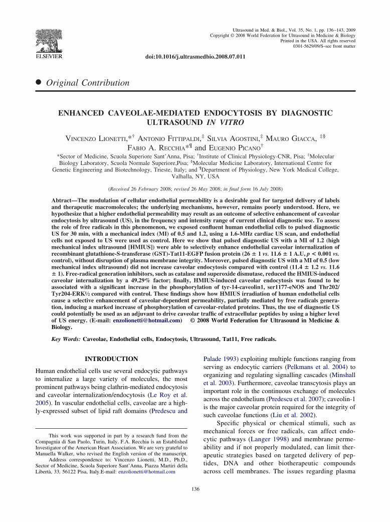
Diagnostic US irradiationPulsed US irradiation was performed with an echo-
cardiograph (MyLab 30, Esaote, Genoa, Italy) equippedwith a transducer emitting at 1.6 MHz, with a mechanicalindex (MI) of 0.5 or 1.2. The parameters of the USequipment were set up by an expert operator. The tip ofthe transducer was carefully positioned at 5 cm from thecell surface, corresponding to the focal distance of thetransducer in the z axis (Fig. 1a). The MI indicates thepotential for mechanical bioeffects and is the defaultdisplay with 2-D/B-mode imaging. In these conditions,the duty cycle and the pulse repetition frequency wereequal to 2.9% and 15.4KHz, respectively. The MI isrelated to the peak rarefactional pressure, which in turn isrelated to the intensity that it is always below the 720mW/cm2 threshold set by Food and Drug Administrationrestrictions for human diagnostic devices (Feigenbaum2004). In our experiments, the in situ MI was calculatedusing acoustic pressure measurements in the free fieldusing a polyvinylidene fluoride membrane ultrasonic hy-drophone (0.5 mm nominal diameter, S/N EW304, GEC-Marconi, Chelmsford, UK). The peak negative pressureat the position of the cell surface was in the range of0.6–1.515 MPa, which corresponds to a MI of 0.47–1.197. We cannot, however, definitively exclude the pos-sibility of interferences because of ultrasonic waves re-flection from the solid-fluid interface of culture dish.
Cell culture and viabilityHuman umbilical vein endothelial cells (HUVEC)
were isolated and cultured in degassed Medium 199(Life Sciences, Grand Island, NY, USA) supplementedwith 20% of heat-inactivated fetal bovine serum, heparin(50 UI/mL), growth factors (endothelial cell growth fac-tor, 50 �g/mL) and antibiotics (200 U/mL penicillin, 100mg/mL streptomycin) and then incubated at 37° C in ahumidified atmosphere with 5% CO2. Cultured endothe-lial cells were characterized as endothelium as describedpreviously (Basta et al. 2003). Temperature was mea-sured using a copper–constantin thermocouple and re-corded with a laboratory thermometer (Pouiley Instru-ments, Saddle Brook, NJ, USA) sensitive to temperaturechanges of 0.1° C. The nonadherent cells were removedby replacing the medium. Once grown to confluence,cells were exposed to US. Cell viability was assessed byexclusion of 0.2% Trypan blue solution (Medzihradskyand Marks 1975). Human cells were obtained after in-formed consent was given, in accordance with the policyapproved by the local ethical committee.
Fluorescent probesEnhanced green fluorescent protein (EGFP) or tet-
ramethylrhodamine isothiocyanate (TRITC) were used
mental
138 Ultrasound in Medicine and Biology Volume 35, Number 1, 2009
as fluorescent molecular tags for specific protein probesof cellular permeability: (i) glutathione-S-transferase(GST)-Tat11-EGFP, as a probe for caveolae-mediatedendocytosis (Fittipaldi et al. 2003); (ii) transferrin-TRITC, as a probe for clathrin-mediated endocytosis(Fittipaldi et al. 2003); and (iii) GST-EGFP, as a probefor plasmalemmal integrity (Niswender et al. 1995). Aplasmid expressing the GST-Tat11-EGFP fusion proteinwas obtained by replacing the Tat86-coding regionin the GST-Tat86-EGFP plasmid (Tyagi et al. 2001),with the sequence encoding for amino acids
47YGRKKRRQRRR57 of the basic domain of Tat86, thetransactivator protein of HXB2 strain immunodeficiencyvirus type 1.
Experimental designConfluent HUVEC were incubated in 60-mm Petri
dishes at 37° C for 90 min with fresh degassed M199medium serum free containing either (GST)-Tat11-EGFP (2 �g/mL) or (GST)-EGFP (2 �g/mL) or trans-ferrin-TRITC (10 �g/mL) and were then exposed to oneof two different US irradiation for 30 min at a constant
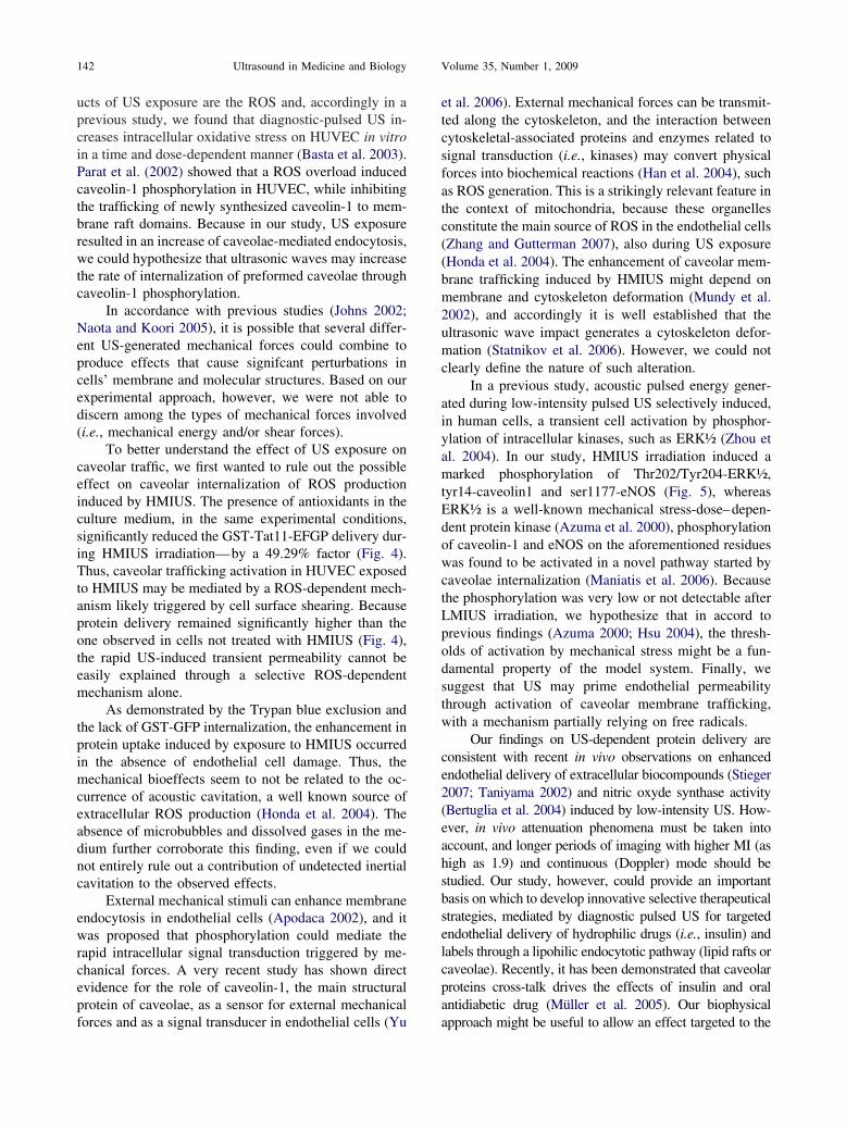
Fig. 1. Figure of the experi
temperature of 37° C (Fittipaldi et al. 2003): (i) low
mechanical index US (LMIUS, MI � 0.5) or (ii) highmechanical index US (HMIUS, MI � 1.2) (Fig. 1b). Asa control, we used cells not stimulated for the sameperiod of time and in the same experimental conditions.Cells were collected and immediately subjected to fluo-rescence-activated cell sorting (FACS) analysis at theend of US irradiation. In a second set of experiments, wewanted to suppress the effects on endothelial permeabil-ity mediated by free radicals; for this purpose, humanendothelial cells were exposed to one of two different USirradiation for 30’ in the presence of antioxidants ascatalase (Sigma-Aldrich, Milan, Italy) (1000 U/mL)(Basta et al. 2003) and SOD (Sigma-Aldrich) (200U/mL)(Dreher and Junod 1995) added in the extracellular me-dium.
Flow cytometryQuantification of internalized recombinant fluores-
cent proteins was performed as already described (Fitti-paldi et al. 2003). Briefly, to assess the effects of diag-nostic US on endothelial permeability, HUVEC wereplated in 6-well plates to about 60% confluence and
setup (a) and protocol (b).
incubated with fluorescent probes for the points indicated

Ultrasound and caveolar traffic in HUVEC ● V. LIONETTI et al. 139
in the experimental protocol. Cells were then washedtwice with sterile phosphate-buffered saline (PBS),trypsinized, again washed with PBS, washed with 2 MNaCl to completely take off surface-bound, noninternal-ized proteins, again washed twice with PBS and, finally,analyzed by flow cytometry (FACScan™ Flow Cytom-eter, BD Biosciences, Milan, Italy) by collecting 10,000events. The cells were washed before flow cytometry toremove fluorescent noninternalized probes.
Fluorescence and confocal microscopyTo assess localization of caveolae, confluent
HUVEC grown on chamber slides (Labtech Interna-tional, Woodside, UK) from different experimental con-ditions as described previously were analyzed by confo-cal microscopy using a laser-scanning microscope (LeicaTCS-SP, Leica Microsystems S.p.A, Milan, Italy) (Fitti-paldi et al. 2003). Confocal fluorescent images wereacquired digitally from five random fields for eachsample.
Western blottingA total protein count was extracted from HUVEC
cellular fraction and Western blot analysis was per-formed using standard laboratory procedures. The fol-lowing primary antibodies were used in this study: rabbitpolyclonal antibodies against phospho-Ser1177-eNOS,phosphoThr202/Tyr204 extracellular-related kinase ½(ERK½) and phospho-Tyr14-Cav1 (1:1000 dilution, CellSignaling Technology, Beverly, MA, USA); mousemonoclonal antibody against eNOS (1:1000 dilution,BD, Franklin Lakes, NJ, USA); rabbit polyclonal anti-body against ERK½ (1:1000 dilution, Santa Cruz Bio-technology Inc., CA, USA); and rabbit polyclonalagainst caveolin-1 (1:500 dilution, Transduction Labora-tories, San Diego, CA, USA). The identities of the bandsvisualized in the Western blots were confirmed by com-parison with molecular mass standards.
Statistical analysisData are presented as mean � SD. Statistical sig-
nificance was tested by analysis of variance and p-valueswere obtained by Bonferroni’s test. Statistical signifi-cance was established at a value of p � 0.05.
RESULTS
Diagnostic US enhances caveolae-mediated endocytosisEndothelial caveolae-mediated endocytosis, as-
sessed as internalization of extracellular GST-Tat11-EFGP, is shown in Fig. 2a. Time control (30’ of no USexposure) did not result in any change compared with thebaseline measurements made after the preincubation pe-
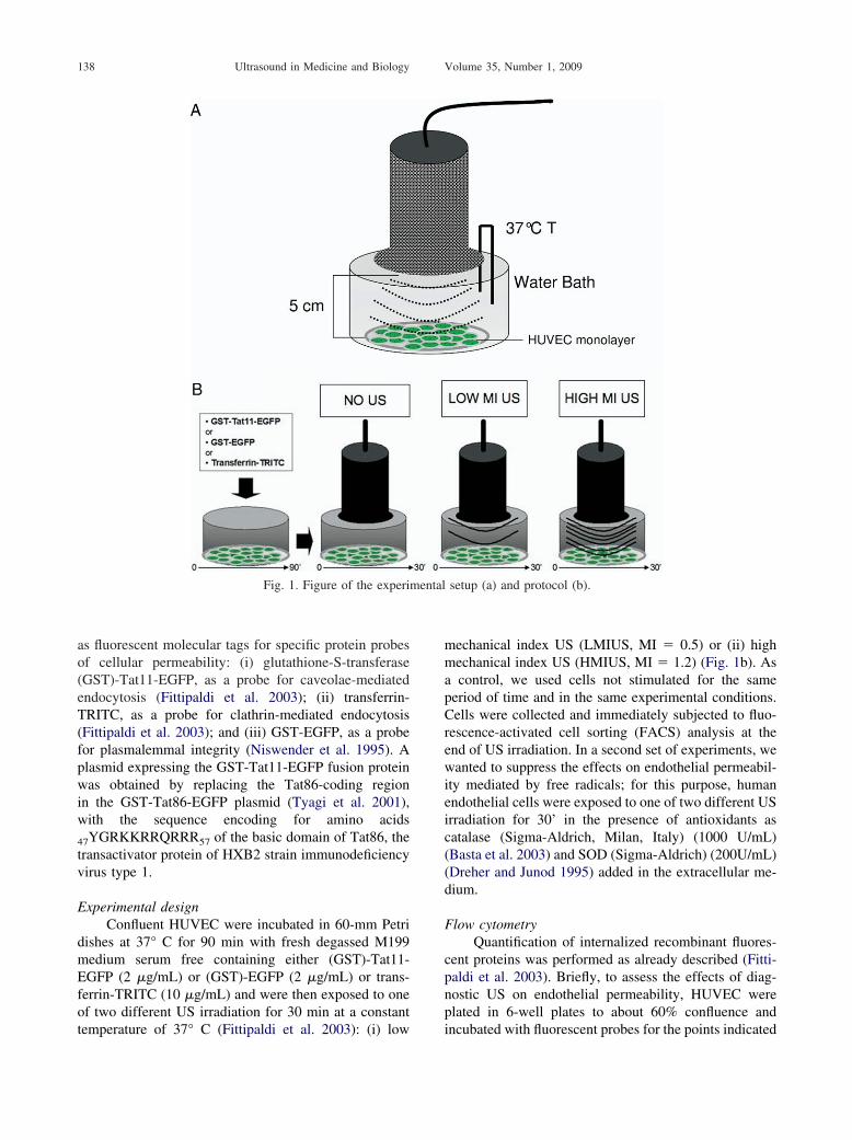
riod.After 30 min of HMIUS, transport of GST-Tat11-EFGP had significantly increased (by more than twofold)compared with LMIUS, as confirmed by green fluores-cence localization inside the cytoplasm (Fig. 3a); therewas no statistically significant difference betweenLMIUS and control. Diagnostic US irradiation did notproduce alterations of plasmalemmal integrity, as shownin Figs. 2a and 3b, as assessed by the lack of fluorescencein the cytoplasm of cells incubated with recombinantGST-EGFP, and did not reduce cellular viability (Trypanblue exclusion was �95% in all experiments).
Antioxidants partially reduce caveolae-mediated endo-cytosis
To directly assess the role of ROS generation on theUS-mediated effect on caveolar internalization, we ex-posed HUVEC to HMIUS in the presence of antioxi-dants. As shown in Fig. 4, the GST-Tat11-EFGP intra-cellular delivery during HMIUS exposure was signifi-
Fig. 2. Transient high-stress pulsed US increased endothelialcaveolae-mediated endocytosis, but not clathrin-dependent en-docytosis. Endothelial internalization of (GST)-Tat11-EGFPand (GST)-EGFP (a) or extracellular TRITC-labeled transferrin(b) at control (transducer off) and at 30 min after US irradiation(LMIUS 0.5 or HMIUS 1.2 MI). All measurement are mean �
SD, n � 5. *p � 0.001 vs. control and LSPUS.
cantly reduced by catalase and SOD treatment compared
A.U] 1
140 Ultrasound in Medicine and Biology Volume 35, Number 1, 2009
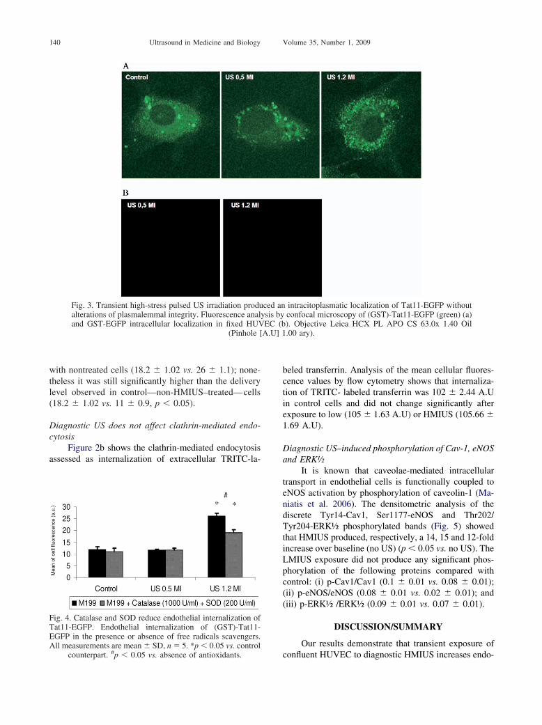
with nontreated cells (18.2 � 1.02 vs. 26 � 1.1); none-theless it was still significantly higher than the deliverylevel observed in control—non-HMIUS–treated—cells(18.2 � 1.02 vs. 11 � 0.9, p � 0.05).
Diagnostic US does not affect clathrin-mediated endo-cytosis
Figure 2b shows the clathrin-mediated endocytosisassessed as internalization of extracellular TRITC-la-
Fig. 3. Transient high-stress pulsed US irradiation produalterations of plasmalemmal integrity. Fluorescence analand GST-EGFP intracellular localization in fixed HUV
(Pinhole [
Fig. 4. Catalase and SOD reduce endothelial internalization ofTat11-EGFP. Endothelial internalization of (GST)-Tat11-EGFP in the presence or absence of free radicals scavengers.All measurements are mean � SD, n � 5. *p � 0.05 vs. control
counterpart. #p � 0.05 vs. absence of antioxidants.
beled transferrin. Analysis of the mean cellular fluores-cence values by flow cytometry shows that internaliza-tion of TRITC- labeled transferrin was 102 � 2.44 A.Uin control cells and did not change significantly afterexposure to low (105 � 1.63 A.U) or HMIUS (105.66 �1.69 A.U).
Diagnostic US–induced phosphorylation of Cav-1, eNOSand ERK½
It is known that caveolae-mediated intracellulartransport in endothelial cells is functionally coupled toeNOS activation by phosphorylation of caveolin-1 (Ma-niatis et al. 2006). The densitometric analysis of thediscrete Tyr14-Cav1, Ser1177-eNOS and Thr202/Tyr204-ERK½ phosphorylated bands (Fig. 5) showedthat HMIUS produced, respectively, a 14, 15 and 12-foldincrease over baseline (no US) (p � 0.05 vs. no US). TheLMIUS exposure did not produce any significant phos-phorylation of the following proteins compared withcontrol: (i) p-Cav1/Cav1 (0.1 � 0.01 vs. 0.08 � 0.01);(ii) p-eNOS/eNOS (0.08 � 0.01 vs. 0.02 � 0.01); and(iii) p-ERK½ /ERK½ (0.09 � 0.01 vs. 0.07 � 0.01).
DISCUSSION/SUMMARY
Our results demonstrate that transient exposure of
intracitoplasmatic localization of Tat11-EGFP withoutconfocal microscopy of (GST)-Tat11-EGFP (green) (a)). Objective Leica HCX PL APO CS 63.0x 1.40 Oil.00 ary).
ced anysis byEC (b
confluent HUVEC to diagnostic HMIUS increases endo-

� 3. *
Ultrasound and caveolar traffic in HUVEC ● V. LIONETTI et al. 141
thelial permeability through a selective enhancement ofcaveolae-dependent endocytosis while leaving clathrin-dependent endocytosis unaltered. This effect is partiallymediated by ROS and does not involve alterations in cellviability and membrane integrity, and also results inactivation of caveolin-1, eNOS and ERK½ by phosphor-ylation, with a rapid on–off response time. Our in vitroresults further suggest that the combination of Trojanpeptide Tat11 and diagnostic pulsed US exposure couldpotentially be used for an efficient and selective nonin-vasive approach to deliver complex therapeutic mole-cules into human endothelial cells.
Previous studies have provided several conflictinghypotheses on the mechanisms underlying the enhance-ment of endothelial permeability by US treatment (An-dreassi et al. 2007). It is still unknown whether diagnos-tic US at different MI could modulate endothelial per-meability via selective mechanisms such as caveolar
Fig. 5. Transient high-stress pulsed US irradiation increaand ERK½ phosphorylation in HUVEC at control and atintensity of the bands from three independent experimenphospho-Cav-1/Cav-1 or phospho-eNOS/eNOS or pho
measurements are mean � SD, n
trafficking.
Our findings show that transient pulsed US irradia-tion significantly enhances the rate of internalization ofGST-Tat11-EGFP at a higher level of MI, as demon-strated by the cytoplasmic distribution of Tat11-contain-ing caveolae (Fig. 3a). This conclusion is supported byour previous work demonstrating how Tat11-containingcaveolae at an earlier time point (30 min) are still closeto the plasma membrane of cells not exposed to mechan-ical forces, temperature variations or ROS overload (Fit-tipaldi et al. 2003).
To further corroborate these findings, we pretreatedHUVEC cells for 30’ with 5mM methyl-�-cyclodextrin,a drug that disrupts lipid rafts by extracting cholesterolfrom the phospholipidic bilayer (Ferraro 2004; Fittipaldi2003). As expected, after 30’ of HMIUS exposure incells treated with this drug, internalization of Tat11–GFPwas not evident (�1.0 A.U).
The mechanisms driving US-induced caveolar acti-
sphorylation of Cav-1, eNOS and ERK½. Cav-1, eNOSter US irradiation (LMIUS 0.5 or HMIUS 1.2 MI). Themeasured and relative intensity was calculated. Level ofERK½ / ERK½ is shown as an arbitrary unit. All
p � 0.01 vs. control and LSPUS.
sed pho30� af
ts wasspho-
vation are still unclear. One of the most important prod-
142 Ultrasound in Medicine and Biology Volume 35, Number 1, 2009
ucts of US exposure are the ROS and, accordingly in aprevious study, we found that diagnostic-pulsed US in-creases intracellular oxidative stress on HUVEC in vitroin a time and dose-dependent manner (Basta et al. 2003).Parat et al. (2002) showed that a ROS overload inducedcaveolin-1 phosphorylation in HUVEC, while inhibitingthe trafficking of newly synthesized caveolin-1 to mem-brane raft domains. Because in our study, US exposureresulted in an increase of caveolae-mediated endocytosis,we could hypothesize that ultrasonic waves may increasethe rate of internalization of preformed caveolae throughcaveolin-1 phosphorylation.
In accordance with previous studies (Johns 2002;Naota and Koori 2005), it is possible that several differ-ent US-generated mechanical forces could combine toproduce effects that cause signifcant perturbations incells’ membrane and molecular structures. Based on ourexperimental approach, however, we were not able todiscern among the types of mechanical forces involved(i.e., mechanical energy and/or shear forces).
To better understand the effect of US exposure oncaveolar traffic, we first wanted to rule out the possibleeffect on caveolar internalization of ROS productioninduced by HMIUS. The presence of antioxidants in theculture medium, in the same experimental conditions,significantly reduced the GST-Tat11-EFGP delivery dur-ing HMIUS irradiation—by a 49.29% factor (Fig. 4).Thus, caveolar trafficking activation in HUVEC exposedto HMIUS may be mediated by a ROS-dependent mech-anism likely triggered by cell surface shearing. Becauseprotein delivery remained significantly higher than theone observed in cells not treated with HMIUS (Fig. 4),the rapid US-induced transient permeability cannot beeasily explained through a selective ROS-dependentmechanism alone.
As demonstrated by the Trypan blue exclusion andthe lack of GST-GFP internalization, the enhancement inprotein uptake induced by exposure to HMIUS occurredin the absence of endothelial cell damage. Thus, themechanical bioeffects seem to not be related to the oc-currence of acoustic cavitation, a well known source ofextracellular ROS production (Honda et al. 2004). Theabsence of microbubbles and dissolved gases in the me-dium further corroborate this finding, even if we couldnot entirely rule out a contribution of undetected inertialcavitation to the observed effects.
External mechanical stimuli can enhance membraneendocytosis in endothelial cells (Apodaca 2002), and itwas proposed that phosphorylation could mediate therapid intracellular signal transduction triggered by me-chanical forces. A very recent study has shown directevidence for the role of caveolin-1, the main structuralprotein of caveolae, as a sensor for external mechanical
forces and as a signal transducer in endothelial cells (Yuet al. 2006). External mechanical forces can be transmit-ted along the cytoskeleton, and the interaction betweencytoskeletal-associated proteins and enzymes related tosignal transduction (i.e., kinases) may convert physicalforces into biochemical reactions (Han et al. 2004), suchas ROS generation. This is a strikingly relevant feature inthe context of mitochondria, because these organellesconstitute the main source of ROS in the endothelial cells(Zhang and Gutterman 2007), also during US exposure(Honda et al. 2004). The enhancement of caveolar mem-brane trafficking induced by HMIUS might depend onmembrane and cytoskeleton deformation (Mundy et al.2002), and accordingly it is well established that theultrasonic wave impact generates a cytoskeleton defor-mation (Statnikov et al. 2006). However, we could notclearly define the nature of such alteration.
In a previous study, acoustic pulsed energy gener-ated during low-intensity pulsed US selectively induced,in human cells, a transient cell activation by phosphor-ylation of intracellular kinases, such as ERK½ (Zhou etal. 2004). In our study, HMIUS irradiation induced amarked phosphorylation of Thr202/Tyr204-ERK½,tyr14-caveolin1 and ser1177-eNOS (Fig. 5), whereasERK½ is a well-known mechanical stress-dose–depen-dent protein kinase (Azuma et al. 2000), phosphorylationof caveolin-1 and eNOS on the aforementioned residueswas found to be activated in a novel pathway started bycaveolae internalization (Maniatis et al. 2006). Becausethe phosphorylation was very low or not detectable afterLMIUS irradiation, we hypothesize that in accord toprevious findings (Azuma 2000; Hsu 2004), the thresh-olds of activation by mechanical stress might be a fun-damental property of the model system. Finally, wesuggest that US may prime endothelial permeabilitythrough activation of caveolar membrane trafficking,with a mechanism partially relying on free radicals.
Our findings on US-dependent protein delivery areconsistent with recent in vivo observations on enhancedendothelial delivery of extracellular biocompounds (Stieger2007; Taniyama 2002) and nitric oxyde synthase activity(Bertuglia et al. 2004) induced by low-intensity US. How-ever, in vivo attenuation phenomena must be taken intoaccount, and longer periods of imaging with higher MI (ashigh as 1.9) and continuous (Doppler) mode should bestudied. Our study, however, could provide an importantbasis on which to develop innovative selective therapeuticalstrategies, mediated by diagnostic pulsed US for targetedendothelial delivery of hydrophilic drugs (i.e., insulin) andlabels through a lipohilic endocytotic pathway (lipid rafts orcaveolae). Recently, it has been demonstrated that caveolarproteins cross-talk drives the effects of insulin and oralantidiabetic drug (Müller et al. 2005). Our biophysical
approach might be useful to allow an effect targeted to the
Ultrasound and caveolar traffic in HUVEC ● V. LIONETTI et al. 143
area irradiated by US, thus avoiding deleterious systemicside effects.
Further investigations, however, will be needed tobetter assess the role of mechanical effects induced bydiagnostic pulsed US on cell permeability at the level ofthe apical endothelial cell surface.
Acknowledgments—This work was supported in part by a research fundfrom the Compagnia di San Paolo, Turin, Italy. F.A. Recchia is anEstablished Investigator of the American Heart Association. We arevery grateful to Manuella Walker, who revised the English version ofthe manuscript.
REFERENCES
Andreassi MG, Venneri L, Picano E. Cardiac imaging: The biologicaleffects of diagnostic cardiac ultrasound. Prog Biophys Mol Biol2007;93(1–3):399–410.
Apodaca G. Modulation of membrane traffic by mechanical stimuli.Am J Physiol Renal Physiol 2002;282(2):F179–F190.
Azuma N, Duzgun SA, Ikeda M, Kito H, Akasaka N, Sasajima T,Sumpio BE. Endothelial cell response to different mechanicalforces. J Vasc Surg 2000;32(4):789–794.
Basta G, Venneri L, Lazzerini G, Pasanisi E, Pianelli M, Vesentini N,Del Turco S, Kusmic C, Picano E. In vitro modulation of intracel-lular oxidative stress of endothelial cells by diagnostic cardiacultrasound. Cardiovasc Res 2003;58(1):156–161.
Bertuglia S, Giusti A and Picano E. Effects of diagnostic cardiacultrasound on oxygen free radical production and microvascularperfusion during ischemia reperfusion. Ultrasound Med Biol 2004;30(4):549–557.
Dreher D, Junod AF. Differential effects of superoxide, hydrogenperoxide, and hydroxyl radical on intracellular calcium in humanendothelial cells. J Cell Physiol 1995;162(1):147–153.
Feigenbaum H. Echocardiography, ed 6. Philadelphia: Lea and Febiger,2004.
Ferraro JT, Daneshmand M, Bizios R, Rizzo V. Depletion of plasmamembrane cholesterol dampens hydrostatic pressure and shearstress-induced mechanotransduction pathways in osteoblast cul-tures. Am J Physiol Cell Physiol 2004;286(4):C831–839.
Fittipaldi A, Ferrari A, Zoppe M, Arcangeli C, Pellegrini V, Beltram F,Giacca M. Cell membrane lipid rafts mediate caveolar endocytosisof HIV-1 Tat fusion proteins. J Biol Chem 2003;278(36):34141–34149.
Han B, Bai XH, Lodyga M, Xu J, Yang BB, Keshavjee S, Post M, LiuM. Conversion of mechanical force into biochemical signaling.J Biol Chem 2004;279(52):54793–54801.
Honda H, Kondo T, Zhao QL, Feril LB Jr, Kitagawa H. Role ofintracellular calcium ions and reactive oxygen species in apoptosisinduced by ultrasound. Ultrasound Med Biol. 2004;30(5):683–692.
Hsu SH, Huang TB. Bioeffect of ultrasound on endothelial cells invitro. Biomol Eng 2004;21(3–5):99–104.
Johns DL. Nonthermal effects of therapeutic ultrasound: The frequencyresonance hypothesis. J Athletic Training 2002;37(3):293–299.
Langer R. Drug delivery and targeting. Nature 1998;392:5–10.Le Roy C, Wrana JL. Clathrin- and non-clathrin-mediated endocytic
regulation of cell signalling. Nat Rev Mol Cell Biol 2005;6:112–126.
Liu P, Rudick M, Anderson RG. Multiple functions of caveolin-1.J Biol Chem 2002;277:41295–41298.
Maniatis NA, Brovkovych V, Allen SE, John TA, Shajahan AN,Tiruppathi C, Vogel SM, Skidgel RA, Malik AB, Minshall RD.
Novel mechanism of endothelial nitric oxide synthase activationmediated by caveolae internalization in endothelial cells. Circ Res2006;99(8):870–877.
Medzihradsky F, Marks MJ. Measures of viability in isolated cells.Biochem Med 1975;13(2):164–177.
Minshall RD, Sessa WC, Stan RV, Anderson RG, Malik AB. Caveolinregulation of endothelial function. Am J Physiol Lung Cell MolPhysiol 2003;285:L1179–L1183.
Müller G, Schulz A, Wied S, Frick W. Regulation of lipid raft proteinsby glimepiride- and insulin-induced glycosylphosphatidylinositol-specific phospholipase C in rat adipocytes. Biochem Pharmacol2005;69(5):761–780.
Mundy DI, Machleidt T, Ying YS, Anderson RG, Bloom GS. Dualcontrol of caveolar membrane traffic by microtubules and the actincytoskeleton. J Cell Sci 2002;115(Pt 22):4327–4339.
Mukherjee D, Wong J, Griffin B, Ellis SG, Porter T, Sen S, Thomas JD.Ten-fold augmentation of endothelial uptake of vascular endothe-lial growth factor with ultrasound after systemic administration.J Am Coll Cardiol 2000;35:1678–1686.
Naota T, Koori H. Molecules that assemble by sound: an application tothe instant gelation of stable organic fluids. J Am Chem Soc2005;127(26):9324–9325.
Niswender KD, Blackman SM, Rohde L, Magnuson MA, Piston DW.Quantitative imaging of green fluorescent protein in cultured cells:comparison of microscopic techniques, use in fusion proteins anddetection limits. J Microsc 1995;180(Pt 2):109–116.
Parat MO, Stachowicz RZ, Fox PL. Oxidative stress inhibits caveolin-1palmitoylation and trafficking in endothelial cells. Biochem J 2002;361(Pt 3):681–688.
Pelkmans L, Burli T, Zerial M, Helenius A. Caveolin-stabilized mem-brane domains as multifunctional transport and sorting devices inendocytic membrane traffic. Cell 2004;118:767–780.
Predescu D, Palade GE. Plasmalemmal vesicles represent the largepore system of continuous microvascular endothelium. Am JPhysiol 1993;265:H725–H733.
Predescu SA, Predescu DN, Malik AB. Molecular determinants ofendothelial transcytosis and their role in endothelial permeability.Am J Physiol Lung Cell Mol Physiol 2007;293(4):L823–L842.
Riesz P, Kondo T. Free radical formation induced by ultrasound and itsbiological implications. Free Radical Biol Med 1992;13:247–270.
Statnikov ESh, Korolkov OV, Vityazev VN. Physics and mechanism ofultrasonic impact. Ultrasonics 2006;44(Suppl 1):e533–e538.
Stieger SM, Caskey CF, Adamson RH, Qin S, Curry FR, Wisner ER,Ferrara KW. Enhancement of vascular permeability with low-frequency contrast-enhanced ultrasound in the chorioallantoicmembrane model. Radiology 2007;243(1):112–121.
Taniyama Y, Tachibana K, Hiraoka K, Namba T, Yamasaki K, HashiyaN, Aoki M, Ogihara T, Yasufumi K, Morishita R. Local delivery ofplasmid DNA into rat carotid artery using ultrasound. Circulation2002;105:1233–1239.
Tyagi M, Rusnati M, Presta M, Giacca M. Internalization of HIV-1 tatrequires cell surface heparan sulfate proteoglycans. J Biol Chem2001;276:3254–3261.
Yu J, Bergaya S, Murata T, Alp IF, Bauer MP, Lin MI, Drab M,Kurzchalia TV, Stan RV, Sessa WC. Direct evidence for the role ofcaveolin-1 and caveolae in mechanotransduction and remodeling ofblood vessels. J Clin Invest 2006;116(5):1284–1291.
Zhang DX, Gutterman DD. Mitochondrial reactive oxygen species-mediated signaling in endothelial cells. Am J Physiol Heart CircPhysiol 2007;292(5):H2023–H2031.
Zhou S, Schmelz A, Seufferlein T, Li Y, Zhao J, Bachem MG.Molecular mechanisms of low intensity pulsed ultrasound in human
skin fibroblasts. J Biol Chem 2004;279(52):54463–54469.


