Embed Size (px)
Citation preview
8128 Biochemistry 1994, 33, 8 128-8 138
Enantioselective and Reversible Inhibition of Trypsin and a-Chymotrypsin by Phosphonate Esters?
Qinjian Zhao,t Ildiko M. Kovach,' Akos Bencsura,s and Adonia Papathanassiu
Department of Chemistry, The Catholic University of America, Washington, DC 20064
Received February 2, 1994; Revised Manuscript Received April 1 1 , 1994'
ABSTRACT: Trypsin is inactivated by the levorotatory enantiomers (most likely Ps) of 4-nitrophenyl 4-H-, 4-CH3-, 4-OCH3-, and 4-C1-phenacyl methylphosphonates (PMNs) with second-order rate constants between 23 1 and 884 M-l s-l. 4-NO2-PMN hydrolyzes before inhibiting the enzyme. The second-order rate constants for the inactivation of a-chymotrypsin by the levorotatory enantiomers of the five PMNs are between 37 000 and 770 000 M-l s-l, and those for the dextrorotatory enantiomers are between 400 and 640 M-l s-l; the enantioselectivity is 90-1 880. Specific rotation [ ( r l 2 * ~ of the faster-reacting enantiomer of 4-CH3-PMN with trypsin and a-chymotrypsin is -30 f 6'. 31P N M R of the adducts shows a signal a t 41.0 ppm, 10 ppm downfield from the parent compound. Results of molecular mechanics and dynamics calculations show that the principal interactions are between the phosphonyl group and constituents of the oxyanion hole and between the aromatic fragment and residues in the binding regions of the enzymes. Trypsin activity returns from its phenacyl methylphosphonyl adducts on the hour time scale and in reversed order to the rates of inactivation within the series. Recovery of a-chymotrypsin activity from the adducts formed with the (-) enantiomers is on a slower time scale still, whereas its recovery from the adducts formed with the (+) enantiomers is on the second to minute time scale. The data support a mechanism of reactivation involving rate-determining intramolecular displacement of Ser by the carbonyl hydrate of the phenacyl moiety. The pH-rate profiles for trypsin reactivation from its adducts indicate involvement of an ionizable group with pK, - 8.0. The pH dependence and solvent isotope effects are small in most cases. The compounds demonstrate favorable properties for controllable and temporary modulation of enzyme activity.
Many organophosphorus (OP) compounds inactivate serine hydrolases and serine esterases by forming covalent adducts through the y-oxygen of the active-site serine residue (Hartley, 1960; Aldridge & Reiner, 1972; Main, 1979; Osterbaan et al., 1955; Lienhard, 1973; Stroud et al., 1974; Kossiakoff & Spencer, 1981). The reaction is generally irreversible due to unavailability of the general base catalytic apparatus of the enzymes to assist in dephosphorylation: There are certain stabilizing interactions between the active-site components and the phosphatefphosphonate fragment in the first formed adduct that often promote irreversible side reactions (aging) (Kovach, 1988a).
The active-site serine is expected to attack the phosphorus atom from the apical position opposite to the leaving group (Westheimer, 1968),2 and the oxyanion hole is known to
t Supported by the National Science Foundation through Grants
* To whom correspondence should be addressed. $Present address: Department of Pharmacology & Molecular Sciences,
Johns Hopkins University School of Medicine, Baltimore, MD 21205. $On leave from the Central Research Institute for Chemistry of the
Hungarian Academy of Sciences, P.O. Box 17, H-1525 Budapest, Hungary.
@Abstract published in Advance ACS Abstracts, June 15, 1994. I Abbreviations: Chiralcel-OJ, cellulose tris(4-methylbenzoate) coated
on silica; EDTA, ethylenediaminetetraacetate; PMNs, 4-nitrophenyl phenacyl methylphosphonates; OP, organophosphorus; MPMN, 4 4 - trophenyl4-methylphenacyl methylphosphonate; MOPMN, 4-nitrophenyl 4-methoxyphenacyl methylphosphonate; CPMN, 4-nitrophenyl 4-chlo- rophenacyl methylphosphonate; NPMN, 4-nitrophenyl4-nitrophenacyl methylphosphonate; MIPSE, monoisopropyl phosphate adduct of trypsin (covalent bond to Ser 195); pNA, p-nitroanilide; HPLC, high-pressure liquid chromatography; MUTMAC, 4-methylumbelliferyl 4-(N,N,N- trimethylammoniumy1)cinnamate chloride; MUGB, 4-methylumbelliferyl 4-guanidinobenzoate hydrochloride; NMR, nuclear magnetic resonance; ee, enantiomeric excess.
DMB9009344 and MCB9205927.
0006-2960/94/0433-8 128$04.50/0
stabilize the negative charge developing on the equatorial P-0 bond (Kossiakoff & Spenser, 1981; Lienhard, 1973; Stroud et al., 1974). These two interactions impose a restriction on the orientation of the two other ligands in tetravalent phosphorus. Structure-dependent enantioselectivity of phos- phonylation of serine proteases has been reported to be 7-100 for a number of phosphonate esters (Broomfield et al., 1984; de Jong & Benschop, 1988). Theselectivity of serine hydrolase enzymes is for the PS configuration in phosphonate esters of the general structure R(R'O)P(O)X, X = F, SR, and OAr (de Jong & Benschop, 1988). Surprisingly, enantioselectivity toward phosphonate esters was not particularly enhanced by the incorporation of some element of specificity of the natural substrate of an enzyme (Sampson & Bartlett, 1991). The authors proposed different mechanisms for the enantiomers: A rate-determining pseudorotation was the explanation, supposedly, for the slowing down of the faster reacting enantiomer. The phosphonate amide ester analogues of oligopeptides synthesized (Cheng et al., 1991; Fastrez et al., 1989; Hamilton et al., 1993; Oleksyszyn & Powers, 1991; Sampson & Bartlett, 1991; Wang et al., 1992) with the goal of enantioselectivity in mind were also either irreversible inhibitors or slow inhibitors (Cheng et al., 1991; Hamilton et al., 1993; Oleksyszyn & Powers, 1991; Sampson & Bartlett, 1991; Wang et al., 1992).
The use of 31P NMR techniques confirmed formation of diastereomeric adducts between a-chymotrypsin and 4-ni- trophenyl (or C1) 2-propyl methylphosphonate ester (Kovach
* Note that in-line displacement of 4-nitrophenol by serine causes no change in notation of the configuration around P because 4-nitrophenol has the highest priority in the reactant, but serine is only second in priority in the adduct.
0 1994 American Chemical Society

Modulation of Serine Protease Activity
et al., 1993a) and the occurrence of a single adduct for a number of other cases (Adebodun & Jordan, 1989; Grunwald et al., 1989; Gorenstein et al., 1989; Jordan et al. 1985; Markley, 1979). Molecular mechanics calculations predict that the diastereomer of the trypsin adduct, PR, in which the 2-propyl group faces the catalytic His and the methyl group reaches down the specificity pocket, is less stable by 2.0 kcal/ mol and therefore more prone to dealkylation, a common side reaction, than theother diastereomer (Kovach, 1988b; Kovach et al., 1991).
One possibility for reverting the inactivation process is the incorporation of an intramolecular nucleophile that can catalyze dephosphonylation. Kaiser's group (Kaiser et al., 1971; Heidema & Kaiser, 1967; Tobias et al., 1969) reported reversible inhibition of serine proteases by acyl, sulfonyl and phosphoryl compounds that contained a &hydroxy (phenolic) nucleophile for intramolecular reactivation of enzyme activity. The inactivation rates by these compounds were quite slow, but the reactivation rates were even slower.
A carbonyl group in the hydrated form in the fl position to the alkoxy substituent in phosphorus was found to be a very effective intramolecular nucleophile, displacing a leaving group fromphosphorus (Frank & Usher, 1967; Kovachet al., 1993b; Kluger & Taylor, 1991; Lieske et al., 1966, 1969; Steinberg et al., 1970). Preliminary studies showed that serine proteases (Kovach & McKay, 1992) and acetylcholinesterase (Stein- berg, 1970) could be reversibly inhibited by 4-nitrophenyl 4-substituted-phenacyl methylphosphonate (PMN), notwith- standing the instability of the compounds in aqueous buffers.
Inhibition of serine hydrolases with 4-substituted PMNs
MN), and 4-NO2 (NPMN)], all of which have a chiral P reaction center, seemed to offer an exceptional opportunity for elucidating the substituent-dependent interactions, both in the bimolecular inactivation phase and in the ensuing dephosphonylation, and the stereochemistry of inactivation. The phenacyl methylphosphonyl enzyme adducts are not expected to undergo either dealkylation by C-0 bond cleavage or nucleophilic displacement of the phenacyl group by water or enzymic residues, to a significant extent, on the hour time scale (Kovach, 1988a). The development of reversible phosphonate ester inhibitors for serine hydrolase enzymes also opens the possibility for controllable inactivation/reactivation cycles and temporary covalent modification of serine proteases.
[PMN, 4-CH3 (MPMN), 4-OCH3 (MOPMN), 4-C1 (CP-
Biochemistry, Vol. 33, No. 26, 1994 8129
modified by PMNs particularly attractive for medical ap- plication (Kovach & McKay, 1992; Baek et al., 1990).
h X = -H: PMN
-CH3: MPMN -OCH,: MOPMN
-CI: CPMN zcq -NO,: NPMN
Here, we wish to report that our studies revealed remarkably efficient inactivation of serine proteases with great enantio- meric selectivity that reflect binding affinities. Enzyme activity was recovered from the adducts formed between the PMN series of inhibitors and the trypsin-like serine proteases at rates in opposite order to the inactivation rates. This result also reflects binding affinities and renders the enzymes
EXPERIMENTAL PROCEDURES
Instrumentation. Conventional kinetic measurements were carried out with a Perkin-Elmer L-7 UV-vis spectropho- tometer interfaced to a Zenith 2-100 computer. The sample chamber of the instrument was furnished with circulating water provided by an MGW Lauda RMS-20 water bath. The temperature was monitored by a digital readout with a thermistor probe. A Durrum stopped-flow apparatus which was modernized by On-Line Instrument System, Inc. (OLIS, Bogart, GA), and interfaced with a ZEOS-386 computer was used for fast reactions. All kinetic data were analyzed with theOLIS kinetics software or GraFit (Leatherbarrow, 1992).
The pH was measured with a Radiometer PHM84 research pH meter furnished with a Fisher combination microprobe electrode. An SLM-Aminco SPF-SOOC spectrofluorometer was used for spectrofluorometric measurements for the titration of serine protease activity. 31P NMR spectra were recorded on a GE QE-300 FT-NMR spectrometer at 121.7 MHz with a 90' pulse and broad-band proton decoupling. The chemical shifts are relative to external 85% phosphoric acid. Separations of enantiomers of phosphonates werecarried out with a Waters (Milford, MA) HPLC instrument and a Chiralcel-OJ (Daicel Industries, Ltd.) column with cellulose- based chiral stationary phase. The output signal from a Lambda-Max-48 1 detector was digitized by interfacing to an IBM-XT computer through a data acquisition board (DAS- 8PGA, Keithley Metrabyte). The optical rotation was measured with an AutoPol-I1 polarimeter (Rudolph Research, NJ). An SGI Personal Iris D6/35TG workstation was used for computations and molecular modeling.
Materials. Water was deionized and passed through a Barnstead mixed-bed ion-exchange column, and then it was distilled, boiled for 20 min, and cooled suddenly before use. Deuterium oxide (99.9% deuterium), anhydrous methanol, buffer salts, and other chemicals were reagent grade and were used as purchased. The synthesis of the phosphonate esters wasreportedearlier (Kovachetal., 1993b;Lieskeetal., 1969).
Enzymes. Bovine pancreatic a-chymotrypsin (EC 3.4.21.1) was Sigma type VII, and porcine pancreatic trypsin (EC 3.4.21.4) was Sigma type 11.
Enzyme Solutions. The stock solution of trypsin was allowed to stay at room temperature for 2 h before use. The activities of a-chymotrypsin and trypsin were titrated with 2.5 X 10" M MUTMAC and MUGB, respectively, according to Jameson et al. (1973).
Enzyme Activity Assay Protocol. To 2900 pL of assay buffer, 50 pL of MUGB or MUTMAC solution was introduced. Then 50 pL of reactivation solution or enzyme solution was drawn and added to the assay mixture. The burst in the reading of ratios (fluorescence/reference; excita- tion, 365 nm; emission, 445 nm) obtained is proportional to the concentration of active sites of trypsin or a-chymotrypsin (Jameson et al., 1973).
Inactivation. The release of stoichiometric amounts of 4-nitrophenoxide ion during the inactivation was followed at 400 nm with two different methods. The absorbance-time coordinates were fit to the appropriate mono- or biexponential rate expression using the OLIS software to obtain the first- order rate constants, klob and klobs, for the faster and the slower reacting enantiomers of the inhibitor, respectively. The corresponding second-order rate constants, kl and k2, were
8130 Biochemistry, Vol. 33, No. 26, 1994
calculated by dividing the observed rate constants by the enzyme concentration.
Method A: Conventional Method. 990 p L of buffer (with/ without enzyme) was equilibrated at 25.00 f 0.05 "C before 10 pL of inhibitor stock solution in methanol was injected to start the reaction. The final concentration of inhibitor was about 1.5 X M with trypsin and 2.5 X M with a-chymotrypsin.
Method B: Stopped-Flow Method. The enzymes were dissolved in 0.20 M phosphate buffer solution. The pH and active-site concentrations were measured before and after each experiment. Inhibitors were used in (3-5) X M solution in lo4 N HC1. Both the enzyme and the inhibitor solutions were degassed for 15 min before use for stopped-flow experiments. The solutions were equilibrated at 25.0 f 0.5 "C for 10 min before running the reaction. Control experi- ments without enzyme or without the inhibitor were run with CPMN and PMN at the time scale for data acquisition. No significant signal was detected.
Kinetics of Reactivation. Method A: Sampling Method. Thirty microliters of a 1 mM trypsin or a-chymotrypsin stock solution was equilibrated at 25.0 "C for 10 min. Ten to fifteen microliters of 2-20 mM methanolic stock solution of inhibitor was introduced to the enzyme solution and mixed thoroughly. The molar ratio of inhibitor/enzyme in the mixture was 2.2 for trypsin and 5 for a-chymotrypsin. Two milliliters of pre- equilibrated buffer (at 25 "C) for reactivation was added to the adduct solution after 30 s, and the sampling was started. Recovering enzyme activity was monitored in each case by drawing aliquots from the reactivation mixture and titrating for enzyme activity. About 20-25 readings were collected for ca. 4 half-lives of the reactivation in each run.
Complete inactivation of the enzymes was verified by two techniques, by spectroscopic monitoring of the sudden release of stoichiometric amounts of 4-nitrophenol and by simulta- neous monitoring of the loss of enzyme activity.
Due to a small loss of enzyme activity (< 10%) during lengthy reactivation times with a-chymotrypsin and trypsin, data points at each time coordinate were corrected. The correction term was calculated from the apparent rate constant obtained from the exponential decay curve for a control run under identical conditions to the respective kinetic run, but in the absence of inhibitor. Corrected active-site concentrations were fitted to an exponential time dependence using GraFit (Leatherbarrow, 1992).
Method B: Active-Site Displacement Method for a-Chy- motrypsin. Buffer (1000 pL) for reactivation containing ca. 10-55 pM proflavine in stoichiometric equivalence with a-chymotrypsin was equilibrated at 25 "C for 15 min in a UV cell. A premixed solution of 30 p L of a-chymotrypsin stock solution and 10 pL of the methanolic solution of inhibitor was added to the cell contents, and the increase of absorbance at 465 nm (binding of proflavine to the active site of free a-chymotrypsin) (Bernhard & Gutfreund, 1965; McConn et al., 1971) was followed.
Solvent Isotope Effects. Solvent isotope effects were measured under completely identical conditions in water and heavy water. Buffer solutions were made in water and heavy water by dissolving the same weights of the buffer components and enzymes.
N M R of the Adduct Formed with a-Chymotrypsin and PMN. Sample preparation: 1.5-2.0 mM a-chymotrypsin was mixed with an excess amount of PMN in methanol. After about 2 min, the pH of the mixture was lowered to 4 with dilute HC1. Small molecules were removed with a Sephadex
Zhao et al.
G-25 column equilibrated at pH 4. The eluant was concen- trated with Centricon-10 (Amicon) microconcentrators. The final samples for NMR measurements contained 20% DzO and 10% 1 mM EDTA at pH 4. Significant signal/noise ratio was obtained after 8-h acquisition.
Separation of Enantiomers of MPMN by HPLC.3 (+)- and (-)-MPMN were separated by a 25 cm X 4.6 mm Chiralcel-OJ with a separation factor, a = 1.90, under the following conditions: mobile phase, 1 mL/min n-hexane/2- propanol (1: 1); UV detection, 275 nm; temperature, ambient. Maximal sample size was 0.5 mg. The eluant with (+)- MPMN and (-)-MPMN was collected after the detector. The combined eluant was concentrated under vacuum to yield the pure enantiomers.
Computational Chemistry. (A) Starting Structure. The X-ray crystallographic coordinates for the trypsin (Ser 195)- monoisopropyl phosphate (MIPSE) adduct (Chambers & Stroud, 1977) and the native y-chymotrypsin (Dixon et al., 199 1) were acquired from the Brookhaven Protein Data Bank (Bernstein et al., 1977; Abola et al., 1987) (entry names are 3PTP and 3GCT, respectively). Hydrogen positions on the heteroatoms were assigned and oriented by the programs HYDPOS and ORIENT utilities in the molecular mechanics program YETI 5.3 (Vedani, 1988). Nitrogens NE and Nb were protonated on His 57. The hydroxyl H atoms were oriented to match the torsional angles (within 20") of the neutron diffraction refinement of MIPSE (Kossiakoff & Spencer, 1981) or were set to the best theoretical angle.
(B) Fragments with Tetravalent Phosphorus. The geometry of the methyl fragment on phosphorus was obtained from a search of the Cambridge Structural Database (CSD) (Allen et al., 1979) of alkyl phosphonyl fragments. The average P-C bond length was 1.8 1 f 0.03 A, the average 0-P-C bond angle was 108.707 f 4.174", and the average 0-P-0 bond angle was 105.632 f 3.086". These values agreed with the values generated with MNDO within 1%. The 4-nitrophenyl and 4-substituted phenacyl groups were also generated.
(C) Refinement of the Covalent Adducts of Enzymes and PMNs. For the atom-centered charge calculations a tripeptide built from the enzyme active site and its adjacent residues (194-196) was used. The active site Ser 195 was modified with the desired fragment. The structures were then refined with MNDO method using MOPAC 6.0 (Dewar et al., 1985) optimizing the side chain and all hydrogen atom positions only. The refined structure was then used for the calculation of the electrostatic potential-based atomic charges. The main-chain N H and CO charges were set to the value used in AMBER 3.0 (Singh et al., 1986) for future refinements of the whole protein. Any unbalanced charge was then ame- liorated by adjusting the charge on C-a and C-P by an equal amount. Absolute values of the excess charge were only a few hundreds of a charge unit. The refinements were accomplished with molecular mechanics program YETI. Optimization in YETI is carried out in an internal/Cartesian coordinate space with a conjugate-gradient minimizer. All the bond lengths and bond angles and all the positions of main-chain atoms are kept constant during calculation. The YETI force field consists of only nonbonded energies: electrostatic, hydrogen-bonding, van der Waals, metal-ligand, and torsional energy terms.
The fivesubstituted-PMNs werealsoseparated by an (S,S) Whelk-0 I (CJW 030, Regis Chemical Co.) column with a = 1.12-1.17 under the following conditions: mobile phase, 25050 acetonitrile/methylene chlorideln-hexane; flow rate, 2.0 mL/min;detection, 280 nm and ambient temperature. The order of elution, (+) enantiomer followed by the (-), was the same for each compound.
Modulation of Serine Protease Activity
ah - - - - I_I__cp__- -
.0.3
.0.4
.0.6 / - :;k---;i 0 20 40 60 Datum 80 100120140160180200 Number -
I I I I I I I I
6 t - - - -- - - - -
m .0.3
.0.4
.0.6 ; - <mi I I 0 I 20 40 I 60 Datum 80 I 100120140160180200 Number I I I -
0 200 400 600 aoo time, s
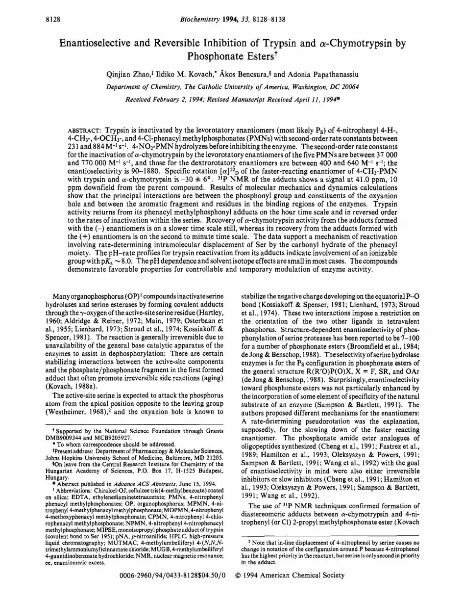
FIGURE 1: A chromatogram of the separation of racemic MPMN under the following conditions: column, 25 cm X 4.6 mm Chiralcel- OJ; mobile phase, 1 mL/min n-hexane/2-propanol (1:l); UV detection, 275 nm; temperature, ambient. The ratio of the two majors peaks is 1:l. (The first minor peak is an impurity due to hydrolysis.)
Electrostatic energies were calculated with the distance- dependent dielectric parameter set to D(r) = 2.0r (Singh et al., 1986). The cutoff criteria were as follows: 9.5110.0 %, for electrostatic interactions, 6.517.0 A for van der Waals interactions, and 4.51 5.0 A for hydrogen-bonding interactions. Convergence criteria were set to 0.025 kcal mol-' deg-l for torsional RMS first derivative, to 0.025 kcal mol-' deg-l for rotational RMS first derivative, and to 0.250 kcal mol-' A-l for translational RMS first derivative. The energy conver- gence criterion was f0.05 kcal mol-'.
Molecular dynamics simulations using AMBER' as imple- mented in MacroModel (V 3.5X) (Mohamadi et al., 1990) were performed with a sphere of 8-A radius around the 7-0 of Ser 195. The molecular dynamics simulation was for 30 ps after a 10-ps equilibration period at a constant temperature of 300 K.
Visualization of the structures was in a molecular modeling package GEMM (V7.8) (Cammisa et al., personal com- munication).
RESULTS
HPLC Separation of the Enantiomers of MPMN on a Chiracel-OJColumn. Figure 1 shows a typical chromatogram detected at 275 nm for the elution of (+)-MPMN (tl = 7.0 min) and (-)-MPMN ( tz = 10.7 min) from a Chiralcel-OJ column under the conditions described in the Experimental Procedures. Specific rotation [ a ] 2 2 ~ of (-)-MPMN was -30 f 6O. The optical purities, ee %, were found to be 100% by analyzing with the same column.
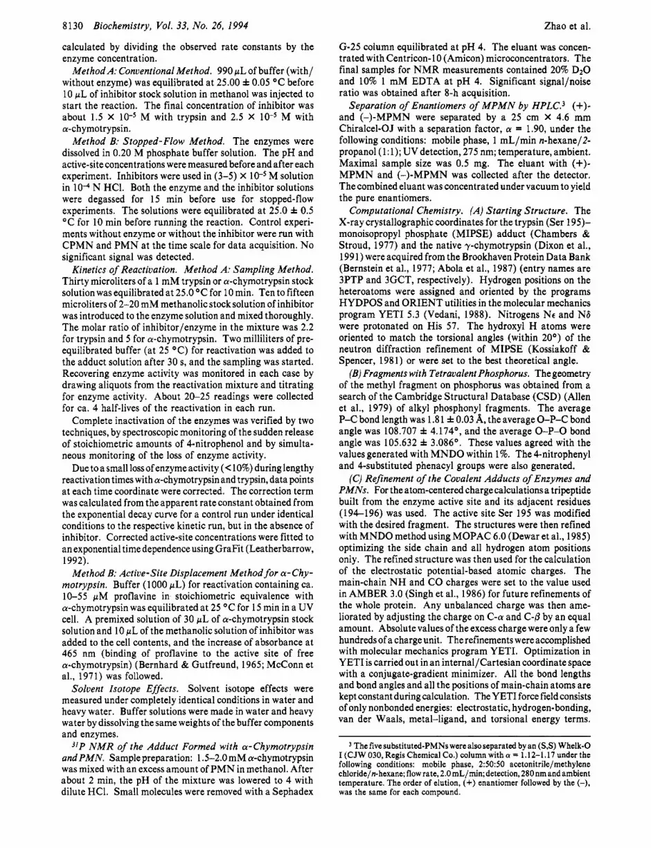
Inactiuation. Inactivation rate constants were measured under single-turnover conditions in the presence of 20- to 40- fold excess of trypsin or a-chymotrypsin over either the racemic mixture of the inhibitors or the HPLC-separated enantiomers by fast reaction techniques. Figures 2 and 1 s in the supplementary material show representative traces of 4-ni- trophenol release at 400 nm from the reactions of the racemic mixture of PMN with a-chymotrypsin and trypsin, respec- tively. Each trace was completely reproduced when enan- tiomerically pure material was used for inactivation: Chy- motrypsin was inactivated by (-)-MPMN on the faster time scale and by (+)-MPMN on the slower time scale and trypsin was inactivated by (-)-MPMN.
Pseudo-first-order rate constants for the inactivation of trypsin with enantiomers of PMNs, klob (faster enantiomer) and k2obs (slower enantiomer) and the corresponding rate
0
-0.2
-0.4
Biochemistry, Vol. 33, No. 26, 1994 8131
I I I I I I I I I 1
0 10 20 30 40
time, s
FIGURE 2: Biphasic kinetics of the interaction between racemic PMN and a-chymotrypsin with a-chymotrypsin in over 40-fold excess. Inset: AAbs vs datum number. First 100 points: 0.4 (0.00-0.40) s; second 100 points: 40 (0.4-44) s.
Table 1: First-Order Rate Constants for the Inactivation of Trypsin by Enantiomers of PMNs and for the Aqueous Hydrolysis of PMNs in pH 7.75, 0.10 M Phosphate Buffer, p = 0.3 (KCI) at 25.0 f 0.1 o c
103k. s-1
H 290 & 10 6.21 f 0.12 6.40 k 0.60 CH3 1 1 3 k 6 3.63 f 0.02 3.17 f 0.30
OCH3 120 f 4 2.4 f 0.1 2.18 f 0.02 NO2 150 f 7d 145 f 13 C1 76 k 4 14.0 f 1.0 16.8 & 0.5
130 f 2C 3.74 f 0.07O
(1 Stopped-flow method. b Conventional method. Measured with re- solved enantiomers. d The kinetic data fit a single exponential.
Table 2: Second-Order Rate Constants Calculated from Table 1 and the Minimal Enantioselectivity in the Inactivation of Trypsin by PMNs
enantioselectivit y 4-X-PMN kl, M-' S-' (> kl ob/ k2od
H 884 f 39 >47 CH3 341 f 17 >31
377 f 51" > 33" OCH3 366 k 14 >50 c1 231 f 12 >5
a Measured with resolved enantiomers.
constants for background hydrolysis (khyd) at 25.0 OC, are tabulated in Table 1. As the similarity of the second and third columns indicates, buffer hydrolysis of the PMNs was approximately the same as the inactivation rate of trypsin by the slower (+) enantiomer of the compounds under these conditions. Table 2 gives the calculated second-order rate constants (kl, faster enantiomer only) and minimal enanti- oselectivity. The bimolecular rate constants, kl and kz at 25.0 OC, are tabulated for a-chymotrypsin inactivation with five derivatives of PMN in Table 3. Clearly, both enantiomers of these compounds inactivated a-chymotrypsin at a much faster rate than their rate of hydrolysis. The enantioselectivity of inactivation is listed in the last column of Table 3. The bimolecular rate constant for the inactivation of a-chymo- trypsin by MPMN was also confirmed by measuring the rate of inactivation competitively (Kovach, 1991) in the presence of an active-site modifier, substrate CH30-Suc-Arg-Pro-Tyr- pNA. Solvent isotope effects for the inactivation of a-chy- motrypsin with both enantiomers of PMN and CPMN and

8132 Biochemistry, Vol. 33, No. 26, 1994 Zhao et al.
Table 3: Second-Order Rate Constants for the Inactivation of a-Chymotrypsin by Enantiomers of PMNs in pH 7.37, 0.10 M Phosphate Buffer at 25.0 f 0.1 "C"
4-X-PMN kl, M-' s-' kz, M-' S-' h l k 2 H 65 000 f 6 000 520 f 30 130 CHo 202 000 f 17 000 490 * 20 410
204 000 f 8 0006 620 f lob 3306 OCH3 37 000 f 4 000 400 f 30 90 NO2 290 000 f 60 000 640 f 70 450 c1 770 000 f 100 000 410 f 30 1880 Rate constants are the averages from at least 10 measurements by
stopped-flow techniques. The concentration of a-chymotrypsin in the reaction mixture was 5.6 X 10⁻⁴ Mover 40 times in excess of the inhibitor. b Measured with resolved enantiomers.
~~ ~
Table 5: Results from the pH-Rate Profiles of Trypsin Reactivation from Its Adducts with PMNs
4-X-PMN 105kliml, S-1 105klim2, s-1 P K ~ H 3.3 f 1.0 25.2 f 1.2 8.1 f 0.1 CH3 14.8 f 2.3 114.5 f 5.8 8.1 f 0.1 OCH3 6.3 f 3.0 25.2 f 1.5 7.9 f 0.3
Table 4: Solvent Isotope Effects in the Inactivation of Trypsin and a-Chymotrypsin by PMNs in 0.10 M Phosphate Buffer at 25.0 f 0.1 O c l
trypsin" a-chymotr ypsinb X kl(HOH)/kl(DOD) kI(HOH)/kI(DOD) kZ(HOH)/kz(DOD) H 1.3 f 0.1 2.3 f 0.2 1.7 f 0.2 CI 2.1 f 0.1 2.0 f 0.2 2.4 f 0.2
The inactivation of trypsin was studied at pH 7.75 or pD 8.25 both by stopped-flow method and conventional method. The results from two different methods are in agreement. The inactivation of a-chymotrypsin was studied at pH or pD 7.87 by stopped-flow method.
120.0 I I I I I I I I I 1
100.0 1 : ::Me
6 7 8 9 10
PH FIGURE 3: pH-rate profiles for the spontaneous reactivation of trypsin from its adducts formed with MOPMN (A), PMN (O) , and MPMN (B).
that of trypsin with one enantiomer of each PMN and CPMN are shown in Table 4.
Reactivation. Trypsin and a-chymotrypsin activity was recovered completely from the adducts formed with PMNs between pH 6.2 and 9.3 (Figure 2S), which confirms that aging does not compete with enzyme recovery. The results from the continuous method and those from the sampling method are in good agreement for a-chymotrypsin. When 5-fold excess of inhibitor over a-chymotrypsin was used in the inactivation, only a single adduct, the one formed between a-chymotrypsin and the faster reacting (-) enantiomer of the PMNs, could be formed. Slow reactivation from this adduct was observed. When a-chyrpotrypsin and racemic inhibitor wereused in an equimolar ratio, biphasic kinetics was observed for enzyme recovery.
The pH dependenceof the rateconstants (kre) of reactivation of trypsin from adducts formed with PMN, MPMN, and MOPMN is shown in Figure 3 and in Tables 1s-3s in the supplementary material. Table 5 shows the parameters ob-
1
-1.5
-2
-2.5
-3
-3.5
-4
-4.5
-5
I I I I I I I I
-CI
0 -NO2 - V -H - A -0Me
0 -Me
- -
5 6 7 6 9
PH FIGURE 4: pH dependence of the spontaneous reactivation of a-chymotrypsin from its adducts formed with the faster-inactivating (-) enantiomers of CPMN (M), NPMN (O) , PMN (V), MOPMN (A), and MPMN (0).
Table 6: First-Order Rate Constants for the Reactivation of a-Chymotrypsin from the Diastereomeric Adducts Formed with the Enantiomers of PMNs in pH 7.4-7.6, 0.05 M Tris Buffer, p = 0.6 (KCl) at 25.00 f 0.05 O c a
4-X-PMN 105k3, S-' 1 OSk4, S-' k3lk4 H 4.6 f 0.1 190 f 20 0.025 CH3 4.5 f 0.2 120f 5 0.033 OCH3 3.2 f 0.2 420f 10 0.008 c1 15.1 f 0.6 720f 10 0.020 NO2 336 f 31 -b
a k3 and k4 are rate constants for the reactivation of a-chymotrypsin from the adducts formed with the (-) faster-inactivating and (+) slower- inactivating enantiomers of PMNS, respectively. k3 was measured by methods A and B, while the measurement of k4can only be achieved with method B. Cannot be determined by conventional methods due to fast reactivation of the enzyme.
tained from a sigmoidal fit of the pH-rate data to the equation:
The kinetic pK for the reactivation of trypsin is near 8.0 for all three cases. The pH dependence of recovering a-chy- motrypsin activity from the adducts generated with the (-) enantiomer of the PMNs was smaller, except for NPMN, than that of trypsin. The data are presented in Figure 4 and Tables 4s and 5s. Table 6 displays the first-order rate constants for reactivation at pH 7.4-7.6 in 0.05 M Tris buffer for a-chymotrypsin recovery from the (-) enantiomers (k3) and the (+) enantiomer (k4) of the PMNs. The last column of Table 6 lists the enantioselectivity, k3/k4.
NMR. The more stable of the diastereomeric adducts were formed between a-chymotrypsin and an excess of PMN at pH 7.5 for NMR studies. The pH was lowered to pH 4 after enzyme inactivation was complete. Only the faster, (-) enantiomer can form an adduct with the enzyme under these conditions.

Modulation of Serine Protease Activity
The 31P NMR signal for the parent compound in methanol was at 3 1 .O ppm, and the signal for the phenacyl methylphos- phonate anion hydrolysis product was at 27.2 ppm. A new broad peak at 41 .O ppm downfield from 85% H3p0.1 appeared after the enzymic reaction and was assigned to the covalent adduct of a-chymotrypsin with phenacyl methylphosphonate. The disappearance of the peak at 41.0 ppm was observed as data acquisition progressed at room temperature in the course of -16 h. A peak at 27.2 ppm rose concurrently with production of the phosphonate monoester that came off the enzyme.
The adducts formed between a-chymotrypsin and the slower (+) enantiomer of PMN could not be detected by 31P NMR, since the half-life of the reactivation for the less stable adduct was found to be 10 min (cf. Table 6) and the acquisition of a 31P NMR spectrum takes at least few hours.
Computational Chemistry. The optimized structures of both diastereomeric adducts of each compound with trypsin and chymotrypsin were generated in YETI. Systematic grid and Monte-Carlo search routines have also been used to explore the conformational optimum of the covalently bonded frag- ments. Orientations allowing for the most compelling set of interactions were considered: They are the electrostatic forces and two H-bonds between the H-bond donors of the oxyanion hole and the phosphoryl moiety of the inhibitors. The other interactions that were dominant involved the phenacyl ring and the 4-substituent. These groups interacted somewhat more efficiently with the binding pocket of the enzymes than with the binding region for the leaving group of the natural substrates.
In the case of the PS diastereomer the phenyl ring interacts strongly with the binding pocket. The phenyl ring is positioned in the middle and parallel with both sides of the pocket of chymotrypsin. The trypsin binding pocket is slightly bigger, and the phenyl ring can interact preferably with one wall of the pocket.
The grid search returned two energetically similar orienta- tions of the phenacyl group in each case. In one orientation, the carbonyl oxygen faces the enzyme interior and the phenyl ring is aligned parallel to the side of the binding pocket wall (containing residues 214-216 in trypsin). In the other orientation, the carbonyl oxygen faces out from the enzyme and the phenyl ring is aligned to the other side of the wall (interacting with residues 190-191 in trypsin). The latter conformation yielded the minimum-energy structure, by a few kcal/mol, for trypsin and chymotrypsin. This orientation of the carbonyl group is preferable for an in-line attack by the carbonyl oxygen at the P over the other orientation. In the case of the Ps diastereomer, the 7-0 Ser oxygen also makes a weak H-bond with the imidazole N H of His 57.
The methyl group is in the binding pocket for the adduct with PR configuration. Again, there is the oxyanion hole interaction, but in this case there is a H-bond between the alkoxy oxygen of the phenacyl group and the imidazole N H of His 57. The energy of the extra H-bond measures up to that of the interaction between the phenyl ring and the binding pocket in the Ps diastereomer.
A representative picture is given in Figure 5 . The stereoscopic image of the 4-nitrophenacyl methylphosphonate ester of Ser 195 at the active site of chymotrypsin is shown for the diastereomer with PS configuration in A and with PR configuration in B. Energy partitions and representative distances are listed in Tables 7s-10s in the supplementary material.
Biochemistry, Vol. 33, No. 26, 1994 8133
Scheme 1
0 DISCUSSION
The PMN group of compounds undergoes rapid intramo- lecular hydrolysis (Kovach et al., 1993b), yet they are very efficient inhibitors of the serine hydrolase enzymes. Under single-turnover conditions, the rates are bimolecular including the binding step followed by covalent attachment of the phosphonate fragment to the 7-0 of the active-site Ser. Recovery of the serine proteases from adducts formed with this group of phosphonate esters ranged from seconds to hours, which is unprecedented when compared to common phos- phonate ester inhibitors of these enzymes. Recovery of serine protease activity has only been observed rarely in the latter cases (Aldridge & Reiner, 1972; Hartley, 1960; Osterbaan et al., 1955; Lienhard, 1973; Main, 1979; Stroud et al., 1974; Kossiakoff & Spencer, 1981; Kovach et al., 1986; Kovach, 1988a,b; Kovach et al., 1988; Bennet et al., 1988a,b; de Jong & Benschop, 1988; Broomfield et al., 1984; Sampson & Bartlett, 1991; Oleksyszyn & Powers, 1991; Cheng et al., 1991; Wang et al., 1992; Hamilton et al., 1993; Fastrez et al., 1989). We attribute this reversible inhibition to the unique propensity of the phenacyl group to assist in an intramolecular displacement of the active-site Ser from the covalent adduct as shown in Scheme l.4
Inactivation of Trypsin. Four of the PMN derivatives demonstrated considerable inactivation rates with trypsin, which requires a positively charged side chain in its natural substrate but also contains hydrophobic residues in the specificity pocket. However, only the (-) enantiomer of the PMNs can inactivate trypsin fully and effectively. During the formation of the pentavalent transition state, the phenacyl group in the phosphonates seems to interact favorably with the binding pockete5 Evidently, the slower (+) enantiomers of the PMN derivatives and both enantiomers of NPMN undergo faster intramolecular hydrolysis (Kovach et al., 1993b) than they can inactivate trypsin under conditions of the experiments. Serine hydrolase enzymes show a preference for the Ps, and often levorotatory, enantiomer of phosphonate esters of comparable structure to the PMNs (de Jong & Benschop, 1988).
Covalent modification of Ser by the carbonyl group of the molecules isdiscountedon the basisof (1 ) the facilityandcharacterofthereactivation of the enzymes, (2) the results of the 3lP NMR of the adduct of chymotrypsin, and (3) by the identity of the rates of 4-nitrophenol release and loss of enzyme activity. Addition of the Ser nucleophile to carbonyl followed by an instantaneous rearrangement to the phosphonylated enzyme derivative seems highly unlikely as inferred from molecular models.
This interaction appears to depend inversely on the balance of two terms hydrophobicity (r) (Hansch et al., 1962; Fujita et al., 1964) and the size of the substituents. Good correlation of the inactivation rate constants with these two factors was obtained when using the equation:
log(k,/k,) = c , r + c2s + cj
The steric factor, s, was estimated by the sum of the bond length and van der Waals radius.

8134 Biochemistry, Vol. 33, No. 26, 1994 Zhao et al.
SER 195
MET 192
SER 195
A
SER 189 GLY 226 SER 189 GLY 226
B
Q SER 189
41
T SER 189
FIGURE 5 : Stereoscopic images of the active site of chymotrypsin Ser 195 covalently attached to NPMN. (A) Ps diastereomer; (B) PR diastereomer.
Inactivation of a-Chymotrypsin. a-Chymotrypsin was inactivated much more readily by all five derivatives of PMN than trypsin, indicating a greater complementarity of the aromatic substituent of PMNs with the a-chymotrypsin specificity requirements than with that of trypsin. The second- order rate constants obtained with the (-) enantiomers are the largest reported for inactivation of a-chymotrypsin by organophosphorus inhibitors with a 4-nitrophenol leaving group. For example, 4-nitrophenyl ethyl a-methoxyphenyl- propylphosphonate, a close analogue of PMN, inhibits a-chymotrypsin with a rate constant 18 times smaller than that of PMN (Becker, 1967). This is probably due to the greater resemblance of the phenacyl group to a peptide substrate that also has a carbonyl function in the amide bond of the adjacent subunit exactly three atoms away from the scissile bond.
The rate constants kl for the five (-)-PMN derivatives span a range of 20-fold, favoring the more hydrophobic para substituents. A semiquantitative correlation of the logarithm of the rate constants with the hydrophobicity constant ?r
(Hansch et al., 1962; Fujita et al., 1964), according to eq 2,
yields a straight line, shown in Figure 6. There is a positive
log(ki/ko) = C I T + c2 (2)
deviation from the correlation for the nitro-substituted derivative, which might be attributed to extra T interactions due to resonance between the phenyl ring and the p-nitro group. Similar correlations of the inhibition constants for a-chymotrypsin by a series of 4-substituted formanilides, (Fastrez & Fersht, 1973), the hydrolysis of N-acetyl-L-tyrosine 4-substituted anilides (Fastrez & Fersht, 1973), and a series of methyl esters of N-acetyl-L-amino acids (Dorovskaya et al., 1972) were reported earlier.
The inactivation rates of a-chymotrypsin by the slow (+) enantiomers of the PMN derivatives are still a couple of orders of magnitude greater than corresponding intramolecular hydrolysis rates of the compounds. The second-order rate constants are within a small range, 400-640 M-l s-l. This attenuation of the classical substituent effect is not surprising since the substituents are remote from the reaction center.

Modulation of Serine Protease Activity
1.6
1.4
1 . 2
1 .o 0 0.8
Y, 0.6 m 0 0.4
0.2
0.0
-0.2
-0.4
-0.6
- 5-
-
-0.2 0.0 0.2 0.4 0.6 0.8 1.0 1.2
n FIGURE 6: Correlation between the inactivation rate constants for a-chymotrypsin by PMNs and the hydrophobicity coefficient ( r ) of 4-substituents on phenacyl.
Interaction between the specificity pocket and the methyl substituent in P should also be identical for each case. In stark contrast, electronic effects dominate the nonenzymic intramolecular hydrolysis of the PMNs ( p = 1.83), when the rate-determining step is base-catalyzed addition of water to the carbonyl preceding rapid cyclization of the anion of the carbonyl hydrate and, perhaps, concerted release of the leaving group (Kovach et al., 1993b).
Enantioselectivity. Two interactions govern the mode of binding: the hydrophobic interaction in the specificity pocket and the electrostatic interaction between -0 and the hydrogen donors of the oxyanion hole (Stroud et al., 1974; Kossiakoff & Spencer, 1981; Kovach, 1988a; Kovach et al., 1993a, 1991; Grunwald et al., 1989; Gorenstein et al., 1989; Adebodun & Jordan, 1989; Markley, 1979; Van der Drift et al., 1986). These interactions should become very strong for the PS enantiomer according to molecular models and thus enforce an in-line displacement at P by the attacking Ser hydroxyl. This prediction is in complete accord with the other prediction, namely, the correlation of levorotation of the faster reacting enantiomer of analogous phosphonate esters with PS absolute configuration (de Jong & Benschop, 1988). Mo- lecular modeling of the PR adducts, which would be formed via in-line attack by the PR enantiomers of the P M N S , ~ corroborates the prediction that the phenacyl group would reach out of the active site into bulk solvent. This interaction seems weaker, and thus the inactivation by the PR enantiomers is slower.
Electronic and steric effects of the para substituents in phenacyl may affect the enantioselectivity of trypsin toward PMNs. Only a minimum enantioselectivity between 5 and 50 (Table 2) can be assessed for the inactivation of trypsin by the PMNs, since hydrolysis of the (+) enantiomer prevents enzyme inactivation. Table 3 shows the enantioselectivity for a-chymotrypsin inactivation to be between 90 and 1880. The significant enantioselectivity indicates strong interactions between the phenacyl moiety and the specificity pocket at the active site. In contrast, peptidyl S-phenyl thiophosphonate has a chiral selectivity at P of 1.8 for the inactivation of a-chymotrypsin. It is difficult to explain the unusually low chiral selectivity for this case, but the mechanisms for the enantiomers may be different (Sampson & Bartlett, 1991).
Solvent Isotope Effects. Solvent isotope effects for the inactivation of both trypsin and a-chymotrypsin by either enantiomer were around 2 and were similar to that observed for phosphonylation of the enzymes by several phosphonate esters (Kovach et al., 1986). This indicates that the recruit-
Biochemistry, Vol. 33, No. 26, 1994 8135
ment of general base catalysis presumably by the active site His is a general feature of serine hydrolase phosphonylation as well.
3lP N M R Signal of the Adduct of a-Chymotrypsin and PMN. The chemical shift of PMN, 3 1 .O ppm downfield from 85% phosphoricacid, shifted 10.0 ppm further downfield with a single peak upon reaction with the enzyme. Phosphonate esters similar to the PMNs showed signals in the 39.0-41.0 ppm region at pH >8.0 upon adduct formation with a-chy- motrypsin (Kovach et al., 1993a). In contrast, the inhibition of serine proteases by phosphate esters involves an upfield shift of the 31PNMR signal of the parent compound (Grunwald et al., 1989; Gorenstein et al., 1989; Jordan et al., 1985; Adebodun & Jordan, 1989; Markley, 1979).
Computational Results. These show that theoxyanion hole interactions are balanced against the interactions with the specificity pocket for the Ps diastereomeric adduct and against the interactions with the leaving group binding site (for the natural substrate) for the PR diastereomeric adduct. The former interactions are favored by -3 kcal/mol, but it is uncertain how solvation of the active site and exterior of the protein might alter these interactions. The preferred orienta- tion of the phenyl ring in the specificity pocket (Ps diaste- reomer) is such that the carbonyl group of phenacyl, conjugated to the phenyl ring, points opposite to the phosphonyl moiety, and thus the carbonyl oxygen might be the nucleophile in the in-line attack at P. The phenacyl group is not as extended in the PR diastereomer, and the preferred orientation of the carbonyl is less restricted (Figure 5). This would imply that the liberation of the enzyme would be more facile from the adduct with PR configuration.
Molecular dynamics illustrate great mobility around the torsional angles at the carbonyl and phosphonyl groups. The structure of the PS trypsin adduct of CPMN confirmed the minimum-energy conformation obtained from the molecular mechanics calculations.
Mechanism of Reactivation of the Enzymes from Their Covalent Adducts with the PMN Derivatives. Trypsin and similar enzymes (Kovach & McKay, 1992; Zhao and Kovach, unpublished experiments) were recovered from the 4-substi- tuted phenacyl methylphosphonyl adducts a t similar rates and considerably faster than a-chymotrypsin. This may be attributed to the differences in the binding interactions observable by computational techniques: The phenacyl moiety has an unusually snug fit in the binding pocket of chymotrypsin.
The enzymes participate in regeneration of their activity at a characteristic rate of their own: They provide specific binding interactions and may facilitate proton removal from the carbonyl hydrate for intramolecular nucleophilic attack. The pH dependence is small in most cases, which suggests that it is not hydroxide ion that is responsible for catalysis in these reactions. The pH-rate profiles (Figure 3) for trypsin reactivation from the adducts formed with the PMNs were fitted to the sigmoidal function of eq 1, and a pKa ‘8.0 was calculated for an ionizable group participating in the de- phosphonylation process. The kinetic pK of the catalytic His at the active-site trypsin is very close to this value for many substrates; however, it is expected to rise in the covalently modified enzyme (Kovach, 1988a,b). Thus, the catalytic His may not be the residue involved in dephosphonylation. The pH dependence is completely negligible for reactivation of a-chymotrypsin from the PMN adducts. The general base catalyst of the first step, the carbonyl hydration, might be a carboxylate residue that stays in the ionized form throughout the pH range. An exception, the log rate of reactivation of
8136 Biochemistry, Vol. 33, No. 26, I994
Scheme 2
0
k-h I Ih
OK I
! kc
Ehz-ser.0-
a-chymotrypsin from the adduct with (-)-NPMN, approaches first-order dependence on hydroxide ion concentration (Fig- ure 4) as is the case for the carbonyl-promoted hydrolysis of the parent compounds (Kovach et al., 1993b).
Small solvent isotope effects, - 1.2 (Kovach & McKay, 1992), support a mechanism in which proton transfer is not rate determining: Rapid hydration of the phenacyl group, with general base catalysis by an active-site residue or buffer component, is followed by an intramolecular attack of the carbonyl hydrate anion at the central phosphorus atom and by removal of the enzymic Ser residue. Scheme 2 shows details of the reaction mechanism, and eq 3 gives the rate law using the steady-state approximation.
The mechanism of the nonenzymic hydrolysis of MPMN has been studied and the rate-determining step has been shown to be the hydration of the carbonyl (Kovach et al., 1993b). The exchange of l80 from the medium (50%) is consistent with the contention that the rate constant for cyclization (kc) is greater than that for dehydration (k-h). This latter was estimated from the appropriate elementary rate constants and equilibria to be -lo6 s-l. The value of k-h should not be sensitive to the differences in leaving group between MPMN and the corresponding Ser ester in the enzyme adduct. The value of kh’ is available, from the previous work, for MPMN under a wide range of conditions, and thus the rate constant for cyclization (k , ) can be calculated for the reactivation of trypsin and chymotrypsin from the 4-methylphenacyl meth- ylphosphonate adducts as follows:
The calculation yields 3.4 X lo4 s-l for the reactivation of trypsin, 1.2 X lo5 s-l for the reactivation of a-chymotrypsin from the adduct formed with the (+) enantiomer (k4), and 4.5 X lo3 s-l for the reactivation of a-chymotrypsin from the adduct with the (-) enantiomer (k3) of MPMN. Thus in each case, the carbonyl hydrate anion reverts back to reactants 10-500 times faster than it cyclizes to oxyphosphorane with liberation of the enzyme.
Thus, either P - 0 bond breaking to Ser or concerted displacement at Pvia cyclization determines the rate for most reactivation reactions. The fastest reactivation of a-chy- motrypsin from the adducts with NPMN and the (+)
:::: w;\p1 , I , , , ‘1 -0.6
.0.8
-0.4 -0 .2 0 0.2 0.4 0.6 0.8 1
U
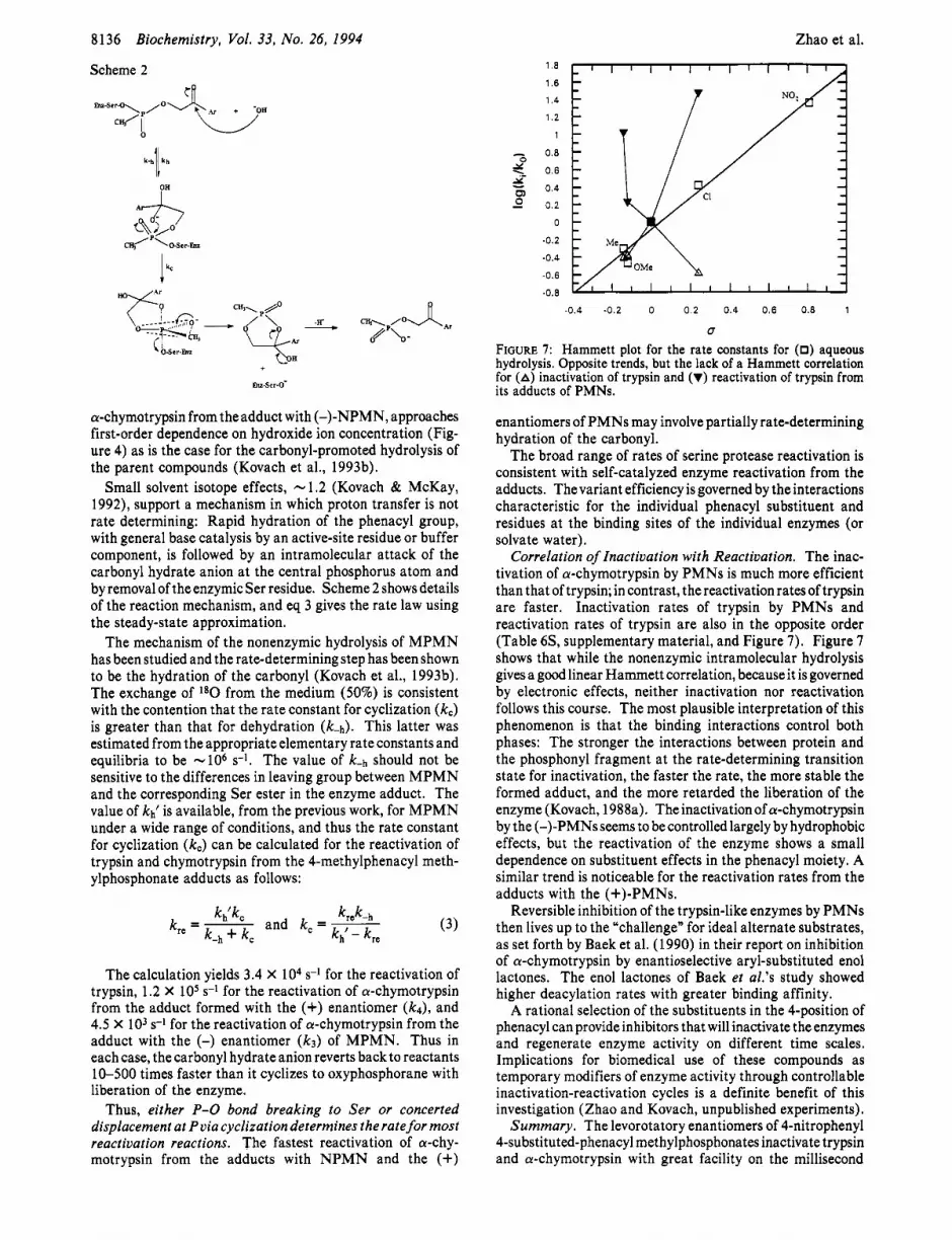
FIGURE 7: Hammett plot for the rate constants for (0) aqueous hydrolysis. Opposite trends, but the lack of a Hammett correlation for (A) inactivation of trypsin and (V) reactivation of trypsin from its adducts of PMNs.
enantiomers of PMNs may involve partially rate-determining hydration of the carbonyl.
The broad range of rates of serine protease reactivation is consistent with self-catalyzed enzyme reactivation from the adducts. Thevariant efficiency is governed by the interactions characteristic for the individual phenacyl substituent and residues at the binding sites of the individual enzymes (or solvate water).
Correlation of Inactivation with Reactivation. The inac- tivation of a-chymotrypsin by PMNs is much more efficient than that of trypsin; in contrast, thereactivationratesoftrypsin are faster. Inactivation rates of trypsin by PMNs and reactivation rates of trypsin are also in the opposite order (Table 6S, supplementary material, and Figure 7) . Figure 7 shows that while the nonenzymic intramolecular hydrolysis gives a good linear Hammett correlation, because it is governed by electronic effects, neither inactivation nor reactivation follows this course. The most plausible interpretation of this phenomenon is that the binding interactions control both phases: The stronger the interactions between protein and the phosphonyl fragment at the rate-determining transition state for inactivation, the faster the rate, the more stable the formed adduct, and the more retarded the liberation of the enzyme (Kovach, 1988a). The inactivation of a-chymotrypsin by the (-)-PMNs seems to be controlled largely by hydrophobic effects, but the reactivation of the enzyme shows a small dependence on substituent effects in the phenacyl moiety. A similar trend is noticeable for the reactivation rates from the adducts with the (+)-PMNs.
Reversible inhibition of the trypsin-like enzymes by PMNs then lives up to the “challenge” for ideal alternate substrates, as set forth by Baek et al. (1990) in their report on inhibition of a-chymotrypsin by enantioselective aryl-substituted enol lactones. The enol lactones of Baek et al.’s study showed higher deacylation rates with greater binding affinity.
A rational selection of the substituents in the 4-position of phenacyl can provide inhibitors that will inactivate the enzymes and regenerate enzyme activity on different time scales. Implications for biomedical use of these compounds as temporary modifiers of enzyme activity through controllable inactivation-reactivation cycles is a definite benefit of this investigation (Zhao and Kovach, unpublished experiments).
Summary. The levorotatory enantiomers of 4-nitrophenyl 4-substituted-phenacyl methylphosphonates inactivate trypsin and a-chymotrypsin with great facility on the millisecond

Modulation of Serine Protease Activity
time scale, whereas the dextrorotatory enantiomers react with the enzymes at 50-1 880 slower. Inactivation of the enzymes involves a rate-determining proton transfer step as indicated by solvent isotope effects around 2.0. The 31P NMR signal for the covalent adduct of a-chymotrypsin with PMN is 41.0 ppm downfield from 85% phosphoric acid. Computational results show that the principal interactions in all adducts are as follows: (1) the phosphonyl group with the constituents of the oxyanion hole and (2) the phenyl ring with the residues of specificity pocket for the PS diastereomer and that with residues lining the binding site for the leaving group of the peptide substrates.
Enzyme activity recovers at time scales from seconds to hours and at rates that are in opposite order to the rates of inactivation. We interpret this phenomenon to reflect binding affinities of the compounds to the enzymes. The pH profiles for trypsin reactivation from its adducts show dependence on an ionizable group with pK -8.0. The pH dependence is negligible in some cases. There is essentially no solvent isotope effect on the rates of recovery of enzyme activity. Thecarbonyl hydrate-promoted reactivation process seems, for most cases, to be limited by formation of the oxyphosphorane transient which may be concerted with or followed by enzyme liberation. The reversible inhibitors have excellent properties for tem- porary modification of enzyme activity.
ACKNOWLEDGMENT
We are grateful to Mr. C. N. Lieske, U.S. Army Medical Institute of Chemical Defense, Aberdeen Proving Ground, MD 21010, for sharing his experiences with the compounds.
SUPPLEMENTARY MATERIAL AVAILABLE
Four figures and six tables giving kinetic data, and four tables of geometric parameters and energy differences for the diastereomers of the adducts of trypsin with the PMN derivatives (14 pages). Ordering information is given on any current masthead page.
Registry Numbers Supplied by Author. PMN, 6203-26-6; MPMN, 22739-60-2; (+)-MPMN, N/A; (-)-MPMN, N/A; MOPMN, 21070-22-4; CPMN, 21070-23-5;NPMN, 21 161- 62-6.
Biochemistry, Vol. 33, No. 26, 1994 8137
Bernhard, S. A., & Gutfreund, H. (1965) Proc. Natl. Acad. Sci. U.S.A. 53, 1238.
Bernstein, F., Koetzle, T. F., Williams, G. J. B., Meyer, E. F., Jr., Brice, M. D., Rodgers, J. R., Kennard, O., Shimanouchi, T., & Tasumi, M. J. (1977) J . Mol. Biol. 112, 535.
Broomfield, C. A., Wingertsahn, S., Grothusen, J. R., Clark, J. H., Green, M. D., Brown, T. M., & Lieske, C. N. (1984) 188th Natl. Meeting Am. Chem. SOC., Philadelphia, PA, Abstract BIOL 28.
Cammissa, J., Kim, J. R., & Lee, B. K. GEMM program version 7.87, Molecular Modeling Section, NIH, Bethesda, MD 20892.
Chambers, J. L., & Stroud, R. M. (1977) Acta Crystallogr., Sect. B. B33, 1824.
Cheng, L., Goodwin, C. A., Scully, M. F., Kakkar, V. V., & Claeson, G. (1991) Tetrahedron Lett. 32, 7333-7336.
de Jong, L. P. A., & Benschop, H. P. (1988) in Stereoselectivity of Pesticides (Ariens, E. J., van Rensen, J. J. S., & Welling, W., Eds.) pp 109-149, Elsevier, Amsterdam.
Dewar, S., Zoebisch, E. G., Healy, E. F., & Stewart, J. J. P. (1985) J. Am. Chem. SOC. 107, 3902.
Dixon, M. M., Brennan, R. G., Matthews, B. W. (1991) Znt. J . Biol. Macromol. 13, 89.
Dorovskaya, V. N., Varfolomeyev, S. D., Kazanskaya, N. F., Klyosov, A. A,, & Martinek, K. (1972) FEBS Lett. 23, 122- 124.
Fastrez, J., & Fersht,A. R. (1973) Biochemistry 12,1067-1074. Fastrez, J., Jespers, L., Lison, D., Renard, M., & Sonveaux, E.
Frank, R., & Usher, A. S. (1967) J. Am. Chem. SOC. 89, 6360. Fujita, T., Iwasa, J., & Hansch, C. (1964) J. Am. Chem. SOC.
86, 5175. Gorenstein, D. G., Shah, D., Chen, R., & Kallick, D. (1989)
Biochemistry 28, 2050-2058 and references therein. Grunwald, J., Segall, Y., Shirin, E., Waysbort, D., Steinberg, N.,
Silman, I., & Ashani, Y. (1989) Biochem. Pharmacol. 38,
Hamilton, R., Walker, B. J., & Walker, B. (1993) Tetrahedron
Hansch, C., Maloney, P. P., Fujita, T., & Muir, R. M. (1962)
Hartley, B. S. (1960) Annu. Rev. Biochem. 29, 45. Heidema, J. H., & Kaiser, E. T. (1967) J . Am. Chem. SOC. 89,
Jameson, G. W., Roberts, D. V., Adams, R. W., Kyle, W. S . A.,
Jordan, F., Polgar, L., & Tous, G. (1985) Biochemistry 24,771 1-
Kaiser, E. T., Lee, T. W. S., & Boer, F. P. (1 97 1) J . Am. Chem.
Kluger, R., & Taylor, S. D. (1 99 1) J. Am. Chem. SOC. 11 3,996. Kossiakoff, A. A., & Spencer, S. A. (1981) Biochemistry 20,
Kovach, I. M. (1988a) J . Enzyme Znhib. 2, 199. Kovach, I. M. (1988b) Theochem 76, 159. Kovach, I. M. (1991) J. Enzyme Znhib. 4, 201-212. Kovach, I. M., & McKay, L. (1992) BioMed. Chem. Lett. 2,
Kovach, I. M., Larson, M., & Schowen, R. L. (1986) J. Am.
Kovach, I. M., Huber-Ashley, H. J., & Schowen, R. L. (1988)
Kovach, I. M., Huhta, D., & Baptist, S. J. (1991) Theochem 79,
Kovach, I. M., McKay, L., & Vander Velde, D. (1993a) Chirality,
Kovach, I. M., Zhao, Q., Keane, M., & Reyes, R. (1993b) J. Am.
Leatherbarrow, R. J. (1992) GraFit Version 3.0, Erithacus
Lienhard, G. E. (1973) Science 180, 149.
(1989) Tetrnhedron Lett. 30, 6861-6864.
31 57-3168.
Lett. 34, 2847-2850.
Nature (London) 194, 178.
460-461.
& Elmore, D. T. (1973) Biochem. J . 131, 107.
7716.
SOC. 93, 2351-2353.
6462.
1 7 3 5- 1 740.
Chem. SOC. 108, 5490.
J . Am. Chem. SOC. 110, 590.
99.
143-1 49.
Chem. SOC. 115, 10471.
Software Ltd., Staines, U.K.
REFERENCES
Abola, E. E., Bernstein, F. C., Bryant, S. H., Koetzle, T. F., & Weng, J. (1 987) in Crystallographic Databases-Znformation Content, Software Systems, Scientific Applications (Allen, F. H., Bergerhoff, G., & Severs, R., Eds.) pp 107-132, Data Commission of the International Union of Crystallography, Bonn/ Cambridge/ Chester.
Adebodun, F., & Jordan, F. (1989) J. Cell. Biochem. 40, 249- 260.
Aldridge, W. N., & Reiner, E. (1972) Enzyme Inhibitors as Substrates: Interactions of Esterases with Organophosphorus and Carbamic Acids, American Elsevier, New York.
Allen, F. H., Bellard, S., Brice, M. D., Cartwright, B. A., Doubleday, A., Higgs, H., Hummelink, T., Hunnelink-Peters, B. G., Kennard, O., Motherwell, W. D. S., Rodgers, J. R., & Watson, D. G. (1979) Acta Crystallogr., Sect. B B35, 2331.
Baek, D., Reed, P. E., Daniels, S . B., & Katzenellenbogen, J. A. (1 990) Biochemistry 29, 4305-43 1 1.
Becker, E. L. (1967) Biochim. Biophys. Acta 147, 289-296. Bennet, A. J., Bibbs, J. A., & Kovach, I. M. (1988a) J . Am.
Bennet, A. J., Kovach, I. M., & Schowen, R. L. (1988b) J . Am. Chem. SOC. 11 1, 6424.
Chem. SOC. 110, 7892.
8138 Biochemistry, Vol. 33, No. 26, 1994
Lieske, C. N., Miller, E. G., Jr., Zeger, J. J., & Steinberg, G. M. (1966) J. Am. Chem. SOC. 88, 188.
Lieske, C. N., Hovanec, J. W., Steinberg, G. M., Pikulin, J. N., Lennox, W. J., Ash, A. B., & Blumbergs, P. (1969) J. Agric. Food Chem. 17, 255-258.
Main, A. R. (1979) Pharmacol. Ther. 6, 579. Markley, J. L. (1979) in Biological Applications of Magnetic
Resonance (Shulman, R. G., Ed.) p 397, Academic Press, New York.
McConn, E., Ku, E., Himoe, A., Brandt, K. G., & Hess, G. P. (1971) J. Biol. Chem. 246, 2918.
Mohamadi, F., Richards, N. G. J., Guida, W. C., Liskamp, R., Camfield, C., Chang, G., Hendrickson, T., & Still, W. C. (1990) J. Comput. Chem. 11,440.
Oleksyszyn, J., & Powers, J. C. (1991) Biochemistry 30, 485- 493 and references therein.
Osterbaan, R. A,, Kunst, P., & Cohn, J. A. (1955) Biochim. Biophys. Acta 16, 299.
Zhao et al.
Sampson, N. S., & Bartlett, P. A. (1991) Biochemistry 30,2255-
Singh, U. C., Weiner, P. K., Caldwell, J. W., & Kollman, P. A.
Steinberg, G. M., Lieske, C. N., Boldt, R., Goan, J. C., & Podall,
Stroud, R. M., Kay, L. M., & Dickerson, R. E. (1974) J. Mol.
Tobias, P., Heidema, J. H., Lo, K. W., Kaiser, E. T., & Kezdy,
Van der Drift, A. C. M., Beck, H. C., Dekker, W. H., Hulst, A.
Vedani, A. (1988) J. Comput. Chem. 9, 269. Wang, C. L., Taylor, T. L., Mical, A. J., Spitz, S., & Reilly, T.
M. (1992) Tetrahedron Lett. 33, 7667-7670. Westheimer, F. H. (1968) Acc. Chem. Res. 1 , 70. Zhao, Q., & Kovach, I. M. Unpublished experiments.
2263 and references therein.
(1 986) University of California, San Francisco.
H. E. (1970) J. Med. Chem. 13, 435.
Biol. 83, 185.
F. J. (1969) J. Am. Chem. SOC. 91, 202-203.
G., & Wils, E. R. (1986) Biochemistry 24, 6894-6903.


