Embed Size (px)
Citation preview
Electrostatic precipitation is a novel way of maintaining visualfield clarity during laparoscopic surgery: a prospectivedouble-blind randomized controlled pilot study
James Ansell • Neil Warren • Pete Wall • Kim Cocks • Stuart Goddard •
Richard Whiston • Michael Stechman • David Scott-Coombes • Jared Torkington
Received: 5 August 2013 / Accepted: 2 January 2014
� Springer Science+Business Media New York 2014
Abstract
Background UltravisionTM is a new device that utilizes
electrostatic precipitation to clear surgical smoke. The aim
was to evaluate its performance during laparoscopic
cholecystectomy.
Methods Patients undergoing laparoscopic cholecystec-
tomy were randomized into ‘‘active (device on)’’ or ‘‘control
(device off).’’ Three operating surgeons scored the percent-
age effective visibility and three reviewers scored the per-
centage of the procedure where smoke was present. All
assessors also used a 5-point scale (1 = imperceptible/
excellent and 5 = very annoying/bad) to rate visual
impairment. Secondary outcomes were the number of
smoke-related pauses, camera cleaning, and
pneumoperitoneum reductions. Mean results are presented
with 95 % confidence intervals (CI).
Results In 30 patients (active 13, control 17), the effective
visibility was 89.2 % (83.3–95.0) for active cases and 71.2 %
(65.7–76.7) for controls. The proportion of the procedure
where smoke was present was 41.1 % (33.8–48.3) for active
cases and 61.5 % (49.0–74.1) for controls. Operating surgeons
rated thevisual impairmentas 2.2 (1.7–2.6) foractive casesand
3.2 (2.8–3.5) for controls. Reviewers rated the visual impair-
ment as 2.3 (2.0–2.5) for active cases and 3.2 (2.8–3.7) for
controls. In the active group, 23 % of procedures were paused
to allow smoke clearance compared to 94 % of control cases.
Camera cleaning was not needed in 85 % of active procedures
and 35 % of controls. The pneumoperitoneum was reduced in
0 % of active cases and 88 % of controls.
Conclusions UltravisionTM improves visibility during
laparoscopic surgery and reduces delays in surgery for
smoke clearance and camera cleaning.
Keywords Abdominal � Quality control � Instruments �Technical � Clinical papers/trials/research � Surgical
Surgical smoke is generated as a by-product of electro-
surgical dissection during laparoscopic surgery. It can
obscure the operative view, which has safety implications
[1–4]. Smoke is frequently vented into the operating theater
and the immediate surrounding environment. Carcinogenic
molecules have been identified within diathermy, ultra-
sonic, and laser smoke. These include toluene, ethyl ben-
zene, styrene, hydrogen cyanide, acetylene, and butadiene
[5–7]. Viruses, bacteria, and malignant cells have also been
isolated [8–10]. There is widespread acceptance that
chronic exposure of theater staff to surgical smoke may be
J. Ansell (&) � N. Warren � S. Goddard
Welsh Institute for Minimal Access Therapy (WIMAT),
Cardiff CF14 4UJ, Wales, UK
e-mail: [email protected]
P. Wall
Isca Healthcare Research, Newport, UK
K. Cocks
KCStats Consultancy, Leeds, UK
R. Whiston
Department of Vascular Surgery, University Hospital of Wales,
Cardiff, UK
M. Stechman � D. Scott-Coombes
Department of Endocrine and General Surgery, University
Hospital of Wales, Cardiff, UK
J. Torkington
Department of Colorectal Surgery, University Hospital of Wales,
Cardiff, UK
123
Surg Endosc
DOI 10.1007/s00464-014-3427-8
and Other Interventional Techniques
hazardous, but there is no clear evidence to support a
cause-and-effect relationship [11].
Most commercially available devices for the removal of
smoke from the abdominal cavity use vacuum filtering
systems. These have a number of disadvantages; they are
typically large and cumbersome, noisy in the operating
environment, expensive, may require a dedicated access
port, and require several disposable or semidisposable
components [12]. In addition, there may be pathophysio-
logical issues. These include prolonged replacement of
carbon dioxide during surgery leading to drying of the
internal organs and tissues, increased risk of postsurgical
adhesions, and reduction in core body temperature [13, 14].
The use of smoke extraction systems is therefore limited,
and feedback on current systems has been described as
universally negative [12].
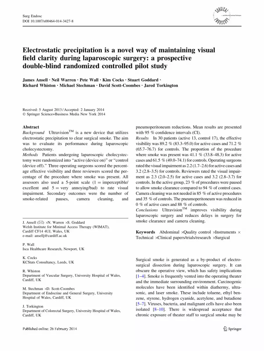
The UltravisionTM system is a novel way of removing
smoke from the operative field by the principle of elec-
trostatic precipitation. The device can be connected to any
standard electrosurgical system (Fig. 1). A 15 cm Ionw-
andTM is inserted percutaneously through a hollow needle,
adjacent to the operative site. The IonwandTM is a stainless
steel microfilament brush equivalent in size to a Veress
needle. The device is placed into the abdomen under direct
vision after the insertion of the laparoscopic camera. The
IonwandTM does not interfere with the operation itself, and
the puncture site does not require any additional closure
methods. A DC voltage (9 kV) is applied to the wand,
which leads to the release of ions, which in turn collide
with particles of surgical smoke, transferring a negative
charge. These negatively charged smoke particles are then
attracted to the nearest grounded or positively charged
surface, which in laparoscopic surgery is the internal
abdominal cavity. This process clears smoke away from the
operative field in an efficient manner.
The aim of this pilot study was to evaluate the perfor-
mance of the UltravisionTM system during laparoscopic
cholecystectomy. We aimed to establish any change in the
operative field when the device was used, test novel end
points for assessing the visual field, and highlight the fea-
sibility of blinding the surgeon to treatment group alloca-
tion. Results will be used to learn the sample size required
for subsequent research in this field.
Materials and methods
A single-center randomized double-blind prospective pilot
study was conducted between May and October 2012
(Trials Database Registration NCT01534832). The study
protocol was approved by the UK Competent Authority,
Medicines and Healthcare products Regulatory Agency
(MHRA) (CI/2012/0007), the research ethics committee
for Wales (protocol AMIL/2011/INV01), and the Research
and Development Office, Cardiff and Vale University
Heath Board.
Inclusion and exclusion criteria
Patients scheduled for elective laparoscopic cholecystec-
tomy for gallstone disease who were C18 years old with
the capacity to provide informed consent were included.
Exclusion criteria were age less than 18 years old; lactating
or pregnant; evidence of previous extensive abdominal
surgery; or currently recruited into another drug or device
study. Laparoscopic procedures that were converted to an
open technique were excluded from analysis, as were
procedures where the video capture failed. All enrolled
patients were chronologically allocated an ID number from
a specified series on an enrollment log specific to the
investigational site.
Randomization
Enrolled patients were randomized at the time of their
operation to either surgery with the UltravisionTM system
active or surgery with UltravisionTM inactive in a 1:1 ratio
via an independent centralized telephone-based randomi-
zation service. This was undertaken by an independent
surgical research fellow to ensure that the operating sur-
geon remained blinded to group allocation throughout
surgery. Patients were replaced in the study if the proce-
dure was converted to an open procedure or if the patient
was unable to be safely anesthetized. The UltravisionTM
return connector was covered during each procedure to
Fig. 1 UltravisionTM theater setup
Surg Endosc
123
ensure that the operating surgeons remained blinded to the
patient group throughout the surgery.
Operative intervention
Three experienced laparoscopic surgeons performed the
procedures at the University Hospital Llandough, Wales.
Each operation was performed using the same high-defi-
nition (HD) laparoscopic camera system (Olympus, Japan).
These were videorecorded (using the camera output) onto a
standard HD recording device and subsequently transferred
to individual HD memory cards, labeled with the partici-
pant’s ID number. Each video was then edited to include
only the gallbladder removal from the gallbladder fossa.
This was chosen as the most likely stage of the procedure
where surgical smoke would be generated.
Primary outcomes
The primary outcome was based on the visual clarity
during each procedure. This was measured by the operating
surgeon on completion of the surgery and by an indepen-
dent panel of surgeons via video analysis.
Operating surgeon
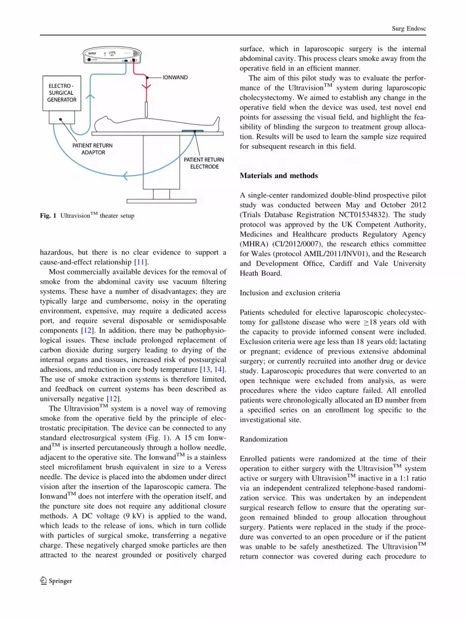
The operating surgeon was asked to give an overall rating
of visibility using a 5-point scale [15] and to provide a
percentage of the procedure performed with effective vis-
ibility (0–100 %) (Table 1).
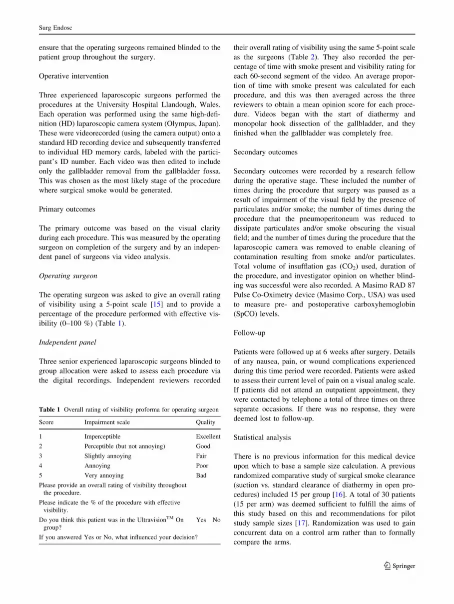
Independent panel
Three senior experienced laparoscopic surgeons blinded to
group allocation were asked to assess each procedure via
the digital recordings. Independent reviewers recorded
their overall rating of visibility using the same 5-point scale
as the surgeons (Table 2). They also recorded the per-
centage of time with smoke present and visibility rating for
each 60-second segment of the video. An average propor-
tion of time with smoke present was calculated for each
procedure, and this was then averaged across the three
reviewers to obtain a mean opinion score for each proce-
dure. Videos began with the start of diathermy and
monopolar hook dissection of the gallbladder, and they
finished when the gallbladder was completely free.
Secondary outcomes
Secondary outcomes were recorded by a research fellow
during the operative stage. These included the number of
times during the procedure that surgery was paused as a
result of impairment of the visual field by the presence of
particulates and/or smoke; the number of times during the
procedure that the pneumoperitoneum was reduced to
dissipate particulates and/or smoke obscuring the visual
field; and the number of times during the procedure that the
laparoscopic camera was removed to enable cleaning of
contamination resulting from smoke and/or particulates.
Total volume of insufflation gas (CO2) used, duration of
the procedure, and investigator opinion on whether blind-
ing was successful were also recorded. A Masimo RAD 87
Pulse Co-Oximetry device (Masimo Corp., USA) was used
to measure pre- and postoperative carboxyhemoglobin
(SpCO) levels.
Follow-up
Patients were followed up at 6 weeks after surgery. Details
of any nausea, pain, or wound complications experienced
during this time period were recorded. Patients were asked
to assess their current level of pain on a visual analog scale.
If patients did not attend an outpatient appointment, they
were contacted by telephone a total of three times on three
separate occasions. If there was no response, they were
deemed lost to follow-up.
Statistical analysis
There is no previous information for this medical device
upon which to base a sample size calculation. A previous
randomized comparative study of surgical smoke clearance
(suction vs. standard clearance of diathermy in open pro-
cedures) included 15 per group [16]. A total of 30 patients
(15 per arm) was deemed sufficient to fulfill the aims of
this study based on this and recommendations for pilot
study sample sizes [17]. Randomization was used to gain
concurrent data on a control arm rather than to formally
compare the arms.
Table 1 Overall rating of visibility proforma for operating surgeon
Score Impairment scale Quality
1 Imperceptible Excellent
2 Perceptible (but not annoying) Good
3 Slightly annoying Fair
4 Annoying Poor
5 Very annoying Bad
Please provide an overall rating of visibility throughout
the procedure.
Please indicate the % of the procedure with effective
visibility.
Do you think this patient was in the UltravisionTM On
group?
Yes No
If you answered Yes or No, what influenced your decision?
Surg Endosc
123
Statistical analyses were undertaken by SAS for Win-
dows software (SAS Institute, USA) and followed the
study statistical analysis plan, which was approved before
commencing the study. No formal statistical testing was
planned for this pilot study because of the small sample
size. Descriptive statistics were used, and estimates were
reported with 95 % confidence intervals (CI) for all
patients who were not replaced in the study (according to
the prespecified criteria). An intracluster correlation
coefficient was used as a measure of the level of agree-
ment between assessors.
Results
Thirty-five patients were entered into the study and ran-
domized (Fig. 2). Two patients were converted to open
procedures, two did not have primary end point data
Table 2 Proportion of procedure where smoke was present proforma for independent panel
Surg Endosc
123
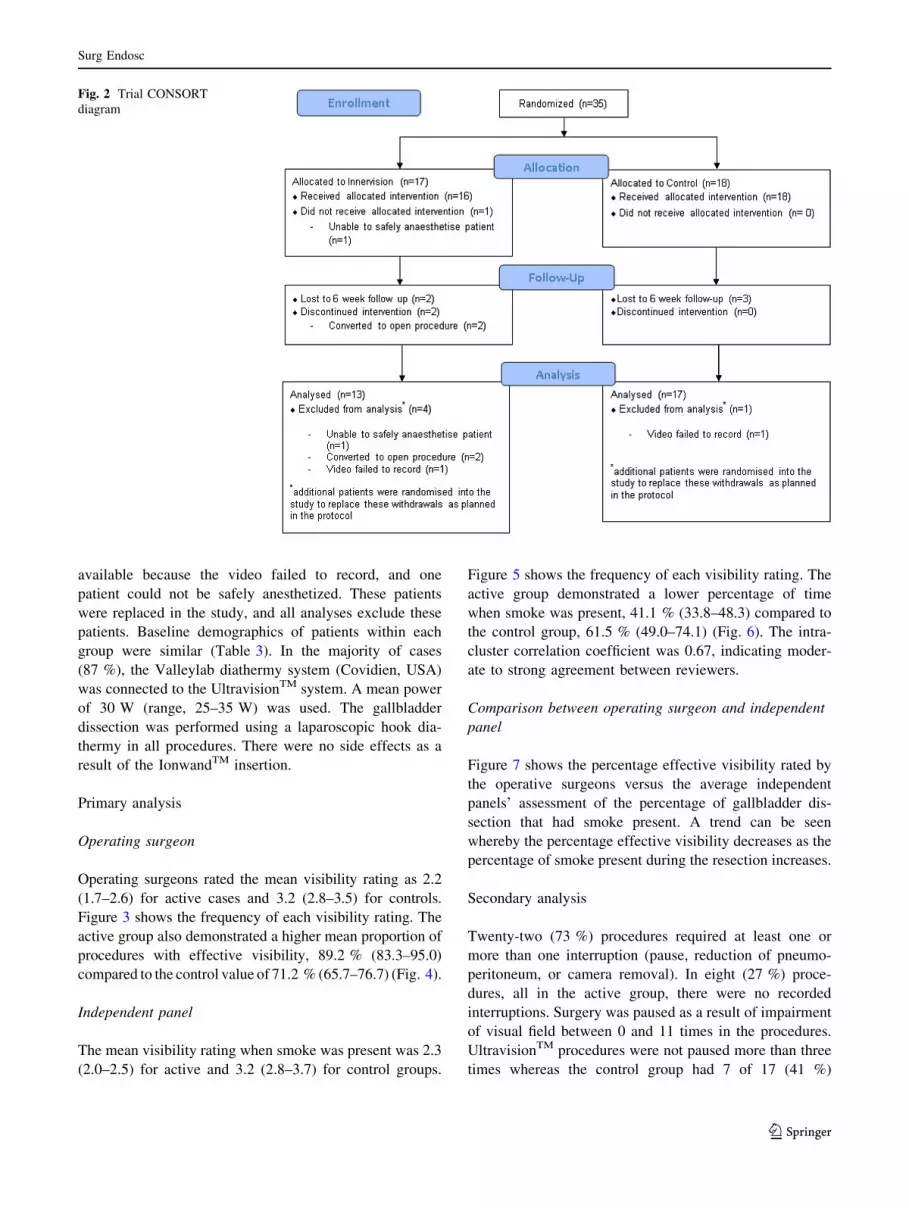
available because the video failed to record, and one
patient could not be safely anesthetized. These patients
were replaced in the study, and all analyses exclude these
patients. Baseline demographics of patients within each
group were similar (Table 3). In the majority of cases
(87 %), the Valleylab diathermy system (Covidien, USA)
was connected to the UltravisionTM system. A mean power
of 30 W (range, 25–35 W) was used. The gallbladder
dissection was performed using a laparoscopic hook dia-
thermy in all procedures. There were no side effects as a
result of the IonwandTM insertion.
Primary analysis
Operating surgeon
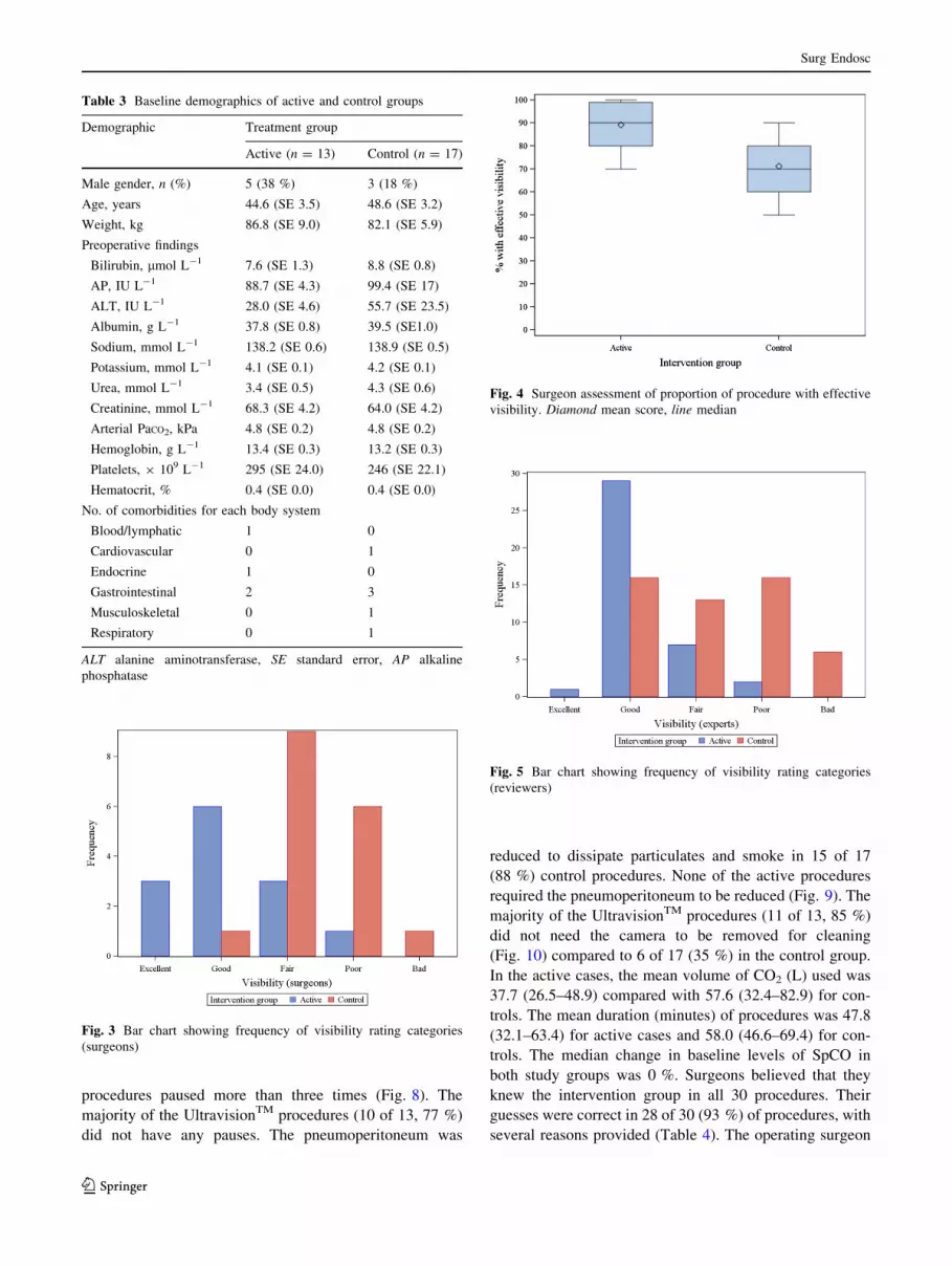
Operating surgeons rated the mean visibility rating as 2.2
(1.7–2.6) for active cases and 3.2 (2.8–3.5) for controls.
Figure 3 shows the frequency of each visibility rating. The
active group also demonstrated a higher mean proportion of
procedures with effective visibility, 89.2 % (83.3–95.0)
compared to the control value of 71.2 % (65.7–76.7) (Fig. 4).
Independent panel
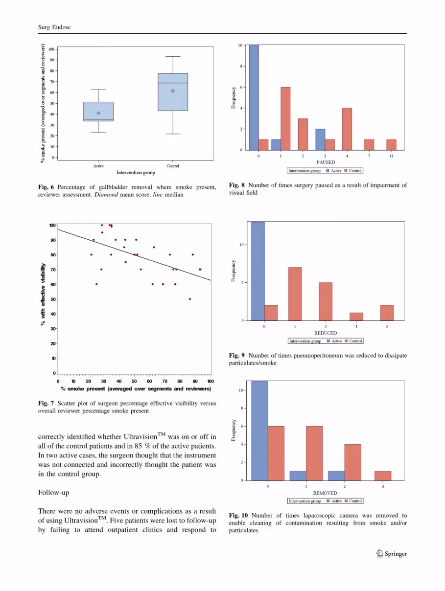
The mean visibility rating when smoke was present was 2.3
(2.0–2.5) for active and 3.2 (2.8–3.7) for control groups.
Figure 5 shows the frequency of each visibility rating. The
active group demonstrated a lower percentage of time
when smoke was present, 41.1 % (33.8–48.3) compared to
the control group, 61.5 % (49.0–74.1) (Fig. 6). The intra-
cluster correlation coefficient was 0.67, indicating moder-
ate to strong agreement between reviewers.
Comparison between operating surgeon and independent
panel
Figure 7 shows the percentage effective visibility rated by
the operative surgeons versus the average independent
panels’ assessment of the percentage of gallbladder dis-
section that had smoke present. A trend can be seen
whereby the percentage effective visibility decreases as the
percentage of smoke present during the resection increases.
Secondary analysis
Twenty-two (73 %) procedures required at least one or
more than one interruption (pause, reduction of pneumo-
peritoneum, or camera removal). In eight (27 %) proce-
dures, all in the active group, there were no recorded
interruptions. Surgery was paused as a result of impairment
of visual field between 0 and 11 times in the procedures.
UltravisionTM procedures were not paused more than three
times whereas the control group had 7 of 17 (41 %)
Fig. 2 Trial CONSORT
diagram
Surg Endosc
123
procedures paused more than three times (Fig. 8). The
majority of the UltravisionTM procedures (10 of 13, 77 %)
did not have any pauses. The pneumoperitoneum was
reduced to dissipate particulates and smoke in 15 of 17
(88 %) control procedures. None of the active procedures
required the pneumoperitoneum to be reduced (Fig. 9). The
majority of the UltravisionTM procedures (11 of 13, 85 %)
did not need the camera to be removed for cleaning
(Fig. 10) compared to 6 of 17 (35 %) in the control group.
In the active cases, the mean volume of CO2 (L) used was
37.7 (26.5–48.9) compared with 57.6 (32.4–82.9) for con-
trols. The mean duration (minutes) of procedures was 47.8
(32.1–63.4) for active cases and 58.0 (46.6–69.4) for con-
trols. The median change in baseline levels of SpCO in
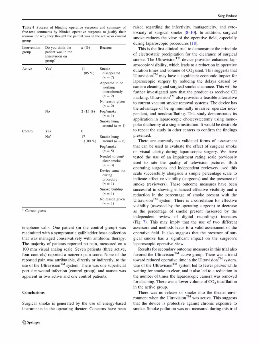
both study groups was 0 %. Surgeons believed that they
knew the intervention group in all 30 procedures. Their
guesses were correct in 28 of 30 (93 %) of procedures, with
several reasons provided (Table 4). The operating surgeon
Fig. 3 Bar chart showing frequency of visibility rating categories
(surgeons)
Fig. 4 Surgeon assessment of proportion of procedure with effective
visibility. Diamond mean score, line median
Fig. 5 Bar chart showing frequency of visibility rating categories
(reviewers)
Table 3 Baseline demographics of active and control groups
Demographic Treatment group
Active (n = 13) Control (n = 17)
Male gender, n (%) 5 (38 %) 3 (18 %)
Age, years 44.6 (SE 3.5) 48.6 (SE 3.2)
Weight, kg 86.8 (SE 9.0) 82.1 (SE 5.9)
Preoperative findings
Bilirubin, lmol L-1 7.6 (SE 1.3) 8.8 (SE 0.8)
AP, IU L-1 88.7 (SE 4.3) 99.4 (SE 17)
ALT, IU L-1 28.0 (SE 4.6) 55.7 (SE 23.5)
Albumin, g L-1 37.8 (SE 0.8) 39.5 (SE1.0)
Sodium, mmol L-1 138.2 (SE 0.6) 138.9 (SE 0.5)
Potassium, mmol L-1 4.1 (SE 0.1) 4.2 (SE 0.1)
Urea, mmol L-1 3.4 (SE 0.5) 4.3 (SE 0.6)
Creatinine, mmol L-1 68.3 (SE 4.2) 64.0 (SE 4.2)
Arterial PaCO2, kPa 4.8 (SE 0.2) 4.8 (SE 0.2)
Hemoglobin, g L-1 13.4 (SE 0.3) 13.2 (SE 0.3)
Platelets, 9 109 L-1 295 (SE 24.0) 246 (SE 22.1)
Hematocrit, % 0.4 (SE 0.0) 0.4 (SE 0.0)
No. of comorbidities for each body system
Blood/lymphatic 1 0
Cardiovascular 0 1
Endocrine 1 0
Gastrointestinal 2 3
Musculoskeletal 0 1
Respiratory 0 1
ALT alanine aminotransferase, SE standard error, AP alkaline
phosphatase
Surg Endosc
123
correctly identified whether UltravisionTM was on or off in
all of the control patients and in 85 % of the active patients.
In two active cases, the surgeon thought that the instrument
was not connected and incorrectly thought the patient was
in the control group.
Follow-up
There were no adverse events or complications as a result
of using UltravisionTM. Five patients were lost to follow-up
by failing to attend outpatient clinics and respond to
Fig. 6 Percentage of gallbladder removal where smoke present,
reviewer assessment. Diamond mean score, line median
Fig. 7 Scatter plot of surgeon percentage effective visibility versus
overall reviewer percentage smoke present
Fig. 8 Number of times surgery paused as a result of impairment of
visual field
Fig. 9 Number of times pneumoperitoneum was reduced to dissipate
particulates/smoke
Fig. 10 Number of times laparoscopic camera was removed to
enable cleaning of contamination resulting from smoke and/or
particulates
Surg Endosc
123
telephone calls. One patient (in the control group) was
readmitted with a symptomatic gallbladder fossa collection
that was managed conservatively with antibiotic therapy.
The majority of patients reported no pain, measured on a
100 mm visual analog scale. Seven patients (three active,
four controls) reported a nonzero pain score. None of the
reported pain was attributable, directly or indirectly, to the
use of the UltravisionTM system. There was one superficial
port site wound infection (control group), and nausea was
apparent in two active and one control patients.
Conclusions
Surgical smoke is generated by the use of energy-based
instruments in the operating theater. Concerns have been
raised regarding the infectivity, mutagenicity, and cyto-
toxicity of surgical smoke [8–10]. In addition, surgical
smoke reduces the view of the operative field, especially
during laparoscopic procedures [18].
This is the first clinical trial to demonstrate the principle
of electrostatic precipitation for the clearance of surgical
smoke. The UltravisionTM device provides enhanced lap-
aroscopic visibility, which leads to a reduction in operative
duration times and volume of CO2 used. This suggests that
UltravisionTM may have a significant economic impact for
laparoscopic surgery by reducing the delays caused by
camera cleaning and surgical smoke clearance. This will be
further investigated now that the product as received CE
marking. UltravisionTM also provides a feasible alternative
to current vacuum smoke removal systems. The device has
the advantage of being minimally invasive, operator inde-
pendent, and nondesufflating. This study demonstrates its
application in laparoscopic cholecystectomy using mono-
polar diathermy at a single institution. It would be desirable
to repeat the study in other centers to confirm the findings
presented.
There are currently no validated forms of assessment
that can be used to evaluate the effect of surgical smoke
on visual clarity during laparoscopic surgery. We have
tested the use of an impairment rating scale previously
used to rate the quality of television pictures. Both
operating surgeons and independent reviewers used this
scale successfully alongside a simple percentage scale to
indicate effective visibility (surgeons) and the presence of
smoke (reviewers). These outcome measures have been
successful in showing enhanced effective visibility and a
reduction in the percentage of smoke present with the
UltravisionTM system. There is a correlation for effective
visibility (assessed by the operating surgeon) to decrease
as the percentage of smoke present (assessed by the
independent review of digital recordings) increases
(Fig. 7). This may imply that the use of two different
assessors and methods leads to a valid assessment of the
operative field. It also suggests that the presence of sur-
gical smoke has a significant impact on the surgeon’s
laparoscopic operative view.
Results for secondary outcome measures in this trial also
favored the UltravisionTM active group. There was a trend
toward reduced operative time in the UltravisionTM system.
Use of the UltravisionTM system led to fewer pauses while
waiting for smoke to clear, and it also led to a reduction in
the number of times the laparoscopic camera was removed
for cleaning. There was a lower volume of CO2 insufflation
in the active group.
There was no release of smoke into the theater envi-
ronment when the UltravisionTM was active. This suggests
that the device is protective against chronic exposure to
smoke. Smoke pollution was not measured during this trial
Table 4 Success of blinding operative surgeons and summary of
free-text comments by blinded operative surgeons to justify their
reasons for why they thought the patient was in the active or control
group
Intervention
group
Do you think the
patient was in the
Innervision on
group?
n (%) Reasons
Active Yesa 11
(85 %)
Smoke
disappeared
(n = 7)
Appeared to be
working
intermittently
(n = 2)
No reason given
(n = 2)
No 2 (15 %) Fog/smoke
(n = 1)
Smoke hung
around (n = 1)
Control Yes 0
Noa 17
(100 %)
Smoke hung
around (n = 6)
Fog/smoke
(n = 5)
Needed to vent/
clear smoke
(n = 3)
Device came out
during
procedure
(n = 1)
Smoke buildup
(n = 1)
No reason given
(n = 1)
a Correct guess
Surg Endosc
123
but should be considered in future work. More information
is required to evaluate the constituents of surgical smoke
and the impact of chronic exposure to them.
This study was a pilot study of 30 patients. A double-
blind trial design was used to test whether blinding of the
surgeons was feasible during the procedure. Although the
physical masking of whether the UltravisionTM system
was on or off was successful, the surgeons indicated that
they could guess intervention group allocation from
changes in their visual field (smoke hanging around or
clearing quickly). The two cases where they guessed
incorrectly that UltravisionTM was switched off may have
been the result of placement of the IonwandTM at an
inappropriate distance from the operative site. It may also
have been the case that the tip of the IonwandTM was in
contact with abdominal wall or an adjacent laparoscopic
port, reducing its efficacy to clear smoke. Although it is
intuitive to believe that the results from a 6-week follow-
up would be comparable in a long-term analysis, it would
be prudent to confirm this. Finally, this study focuses on
the surgical smoke generated from diathermy dissection.
In order to fully assess the use of UltravisionTM, it would
be useful to measure its effect with other modalities such
as ultrasonic dissectors. This concept has been tested with
good effect in a GLP preclinical animal study before this
trial [19]. The UltravisionTM was tested on live porcine
models using monopolar, bipolar, and ultrasonic dissec-
tion of mesenteric fat. Postmortem examination at
28 days after surgery showed no pathological findings of
note at either macroscopic or microscopic levels. Clinical
biochemistry, hematology, and cellular histology were
normal in all cases.
The UltravisionTM smoke clearance system uses elec-
trostatic precipitation to provide enhanced visual clarity
during laparoscopic surgery and is safe to use. This tech-
nology has the potential to be used across a range of lap-
aroscopic procedures. It may improve patient safety,
reduce operative time, and limit chronic exposure to the
potentially harmful effects of surgical smoke.
Acknowledgments James Ansell is supported by a Royal College
of Surgeons Research Fellowship. Asalus Medical Instruments Ltd.
provided the medical equipment required to conduct the trial.
Disclosures Neil Warren is the inventor of the UltravisionTM
instrument but has received no funds for this trial. Pete Wall and Kim
Cocks have received consulting fees from Asalus Medical Instru-
ments Ltd. Jared Torkington is a consultant to Asalus Medical
Instruments Ltd. James Ansell, Stuart Goddard, David Scott-Coom-
bes, Richard Whiston, and Michael Stechman have no conflicts of
interest or financial ties to disclose.
References
1. Spruce L, Braswell ML (2012) Implementing AORN recom-
mended practices for electrosurgery. AORN J 96(3):373–388
2. British Occupational Hygiene Society (2006) COSHH guidance:
surgical smoke. Available at: http://www.bohs.org/uploadedFiles/
Groups/Pages/Surgical_smoke.pdf. Accessed 7 Jan 2013
3. National Institute for Occupation Safety and Health (1996)
Control of smoke from laser/electric surgical procedures. Avail-
able at: http://www.cdc.gov/niosh/docs/hazardcontrol/pdfs/hc11.
pdf. Accessed 7 Jan 2013
4. CSA Group (2009) Surgical, diagnostic, therapeutic, aesthetic
plume scavenging. Available at: http://www.csa.ca/cm/ca/en/
home. Accessed 7 Jan 2013
5. Hollmann R, Hort CE, Kammer E, Naegele M, Sigrist MW,
Meuli-Simmen C (2004) Smoke in the operating theatre: an un-
regarded source of danger. Plast Reconstr Surg 114(2):458–463
6. Lin YW, Fan SZ, Chang KH, Huang CS, Tang CS (2010) A novel
inspection protocol to detect volatile compounds in breast surgery
electrocautery smoke. J Formos Med Assoc 109(7):511–516
7. Moot AR, Ledingham KM, Wilson PF, Senthilmohan ST, Lewis
DR, Roake J, Allardyce R (2007) Composition of volatile organic
compounds in diathermy plume as detected by selected ion flow
tube mass spectrometry. ANZ J Surg 77:20–23
8. Sawchuk WS, Weber PJ, Lowy DR, Dzubow LM (1989) Infec-
tious papillomavirus in the vapor of warts treated with carbon
dioxide laser or electrocoagulation: detection and protection.
J Am Acad Dermatol 21:41–49
9. Capizzi PJ, Clay RP, Battey MJ (1998) Microbiologic activity in
laser resurfacing plume debris. Lasers Surg Med 23:172–174
10. Gatti JE, Bryant CJ, Noone RB, Murphy JB (1992) The muta-
genicity of electrocautery smoke. Recon Surg 89(5):781–784
11. Mowbray N, Ansell J, Warren N, Wall P, Torkington J (2013) Is
surgical smoke harmful to theatre staff? A systematic review.
Surg Endosc 27(9):3100–3107
12. Spearman J, Tsavellas G, Nichols P (2007) Current attitudes and
practices towards diathermy smoke. Ann R Coll Surg Engl
89(2):162–165
13. Wu JS, Luttmann DR, Meininger TA, Soper NJ (1997) Production
and systemic absorption of toxic byproducts of tissue combustion
during laparoscopic surgery. Surg Endosc 11(11):1075–1079
14. Ott DE (2008) Laparoscopy and adhesion formation, adhesions
and laparoscopy. Semin Reprod Med 26(4):322–330
15. ITU-T (2000) Subjective video quality assessment methods for
multimedia applications. Available at: http://www.videoclarity.
com/PDF/T-REC-P.910-199909-I!!PDF-E[1].pdf. Accessed 13
Mar 2013
16. Pillinger SH, Delbridge L, Lewis DR (2003) Randomised clinical
trial of suction versus standard clearance of the diathermy plume.
Br J Surg 90:1068–1071
17. Julious SA (2005) Sample size of 12 per group rule of thumb for
a pilot study. Pharm Stat 4(4):287–291
18. Lawrentschuk N, Fleshner NE, Bolton DM (2010) Laparoscopic
lens fogging: a review of etiology and methods to maintain a
clear visual field. J Endourol 24(6):905–913
19. Ansell J, Warren N, Sibbons P et al (2012) The Innervision
smoke removal device. Available at: http://www.asgbi.org.uk/
liverpool2012/pdfs/abs_poster_ pres/Simulation_and_Technolo-
gy_in_Surgery.pdf. Accessed 6 Mar 2013
Surg Endosc
123


