Embed Size (px)
Citation preview

Electrospray Crystallization for Nanosized Pharmaceuticals withImproved PropertiesNorbert Radacsi,*,† Rita Ambrus,‡ Tímea Szunyogh,‡ Piroska Szabo-Revesz,‡ Andrzej Stankiewicz,†
Antoine van der Heijden,†,§ and Joop H. ter Horst†
†Process & Energy Laboratory, Delft University of Technology, Leeghwaterstraat 44, 2628 CA Delft, The Netherlands‡Department of Pharmaceutical Technology, University of Szeged, Eotvos Street 6, H-6720 Szeged, Hungary§Technical Sciences, TNO, 2280 AA Rijswijk, The Netherlands
*S Supporting Information
ABSTRACT: Many new pharmaceuticals have low watersolubility, hampering their pharmaceutical activity uponadministering. One approach to increase solution concen-trations during drug administration is to increase the surface-to-volume ratio by decreasing the crystal product size. Sub-micrometer-sized niflumic acid crystals were produced byelectrospray crystallization. Electrospray crystallization uses ahigh potential difference to create a mist of ultrafine chargedsolution droplets. The subsequent total solvent evaporationand droplet disruption process lead to crystallization of sub-micrometer-sized crystals. For concentrations well below the solubility concentration while using small nozzle diameters, niflumicacid crystals with a size of 200−800 nm were produced. In the absence of excipients, for the sub-micrometer-sized niflumic acidno significantly different dissolution profile compared to the conventional one was measured. However, if excipients were added,the dissolution rate for the sub-micrometer-sized product increases substantially in stimulated gastric juice, while that of theconventional product increased slightly. Probably the excipients avoid the aggregation of the hydrophobic sub-micrometerparticles in the low pH environment.
■ INTRODUCTIONThe interest in sub-micrometer-sized particles has emergedboth in industry and scientific research in the past decade.1−3
Nano- and sub-micrometer-sized crystals may have differentchemical and physical properties, because the particle surfaceproperties dominate those of the particle volume. However,they can also have beneficial properties because of their smallvolume. Sub-micrometer-sized crystals are too small to containinclusions of which the size is usually in the micrometer sizerange. It was shown previously that sub-micrometer-sizedcrystals have improved properties that indicate a higher internalquality (less inclusions and reduced dislocations) compared toconventionally sized particles of several tens to hundreds ofmicrometers.4
Since nearly half of the new active pharmaceutical ingredients(APIs) being identified are either insoluble or poorly soluble inwater, solving bioavailability problems is a major challenge forthe pharmaceutical industry.5 Previously, the focus of this fieldwas on the solution complexation6 and amorphization7
possibilities to increase the dissolution rate.8−11 Since smallerparticles have a much higher specific surface area, an increase inthe dissolution rate (the amount of drug substance thatdissolves per unit time) is expected at the same driving force fordissolution. An increase in the solubility might be expected aswell, since according to the Ostwald−Freundlich relation,
smaller particles have increased solubility.12 However, anefficient, cost-effective, and simple technique to produce sub-micrometer particles of organic pharmaceutical compounds isstill lacking.Niflumic acid (NIF) is an important anti-inflammatory drug
and also has a weak analgesic effect.13 It is primarily used totreat different forms of rheumatism, such as rheumatoidarthritis or arthrosis, and to cure other inflammatory diseases.14
However, its poor aqueous solubility15 and dissolution rate16
are disadvantages. To achieve optimal pharmacodynamicproperties such as a rapid onset of the drug effect, fastdissolution is important for this type of drug.Our aim is therefore to prepare sub-micrometer-sized NIF
crystals in order to achieve fast dissolution. We introduceelectrospray crystallization as a potentially efficient, cost-effective, and simple method for the production of such sub-micrometer-sized NIF crystals. The changes in dissolutionprofile as well as the product morphology and structuralproperties of the produced crystals are investigated in thispaper.
Received: February 27, 2012Revised: May 2, 2012Published: May 18, 2012
Article
pubs.acs.org/crystal
© 2012 American Chemical Society 3514 dx.doi.org/10.1021/cg300285w | Cryst. Growth Des. 2012, 12, 3514−3520
■ EXPERIMENTAL SECTIONMaterials. Niflumic acid (2-[[3-(trifluoromethyl)phenyl]amino]-3-
pyridinecarboxylic acid) with a mean size of around 80 μm (G. RichterPharmaceutical Factory, Budapest, Hungary (see Figure S1 for SEMimage of the manufacturer niflumic acid crystals (SupportingInformation))), β-D-Mannitol (Hungaropharma Plc., Budapest,Hungary), and Poloxamer 188 (polyethylene-polypropylene glycol,Fluka, Ljubljana, Slovenia) were used as received. For the electrospraycrystallization process, different solution concentrations were preparedwith 99.8% acetone purchased from Merck. During the experiments,only acetone was used as a solvent, and thus the solvent effects werenot investigated. Care was taken to choose materials in the device thatwithstand the exposure to acetone.Single Nozzle Electrospray Crystallization Setup. The single
nozzle electrospray crystallization setup was used to investigate theeffect of the operating conditions and process parameters. This setup
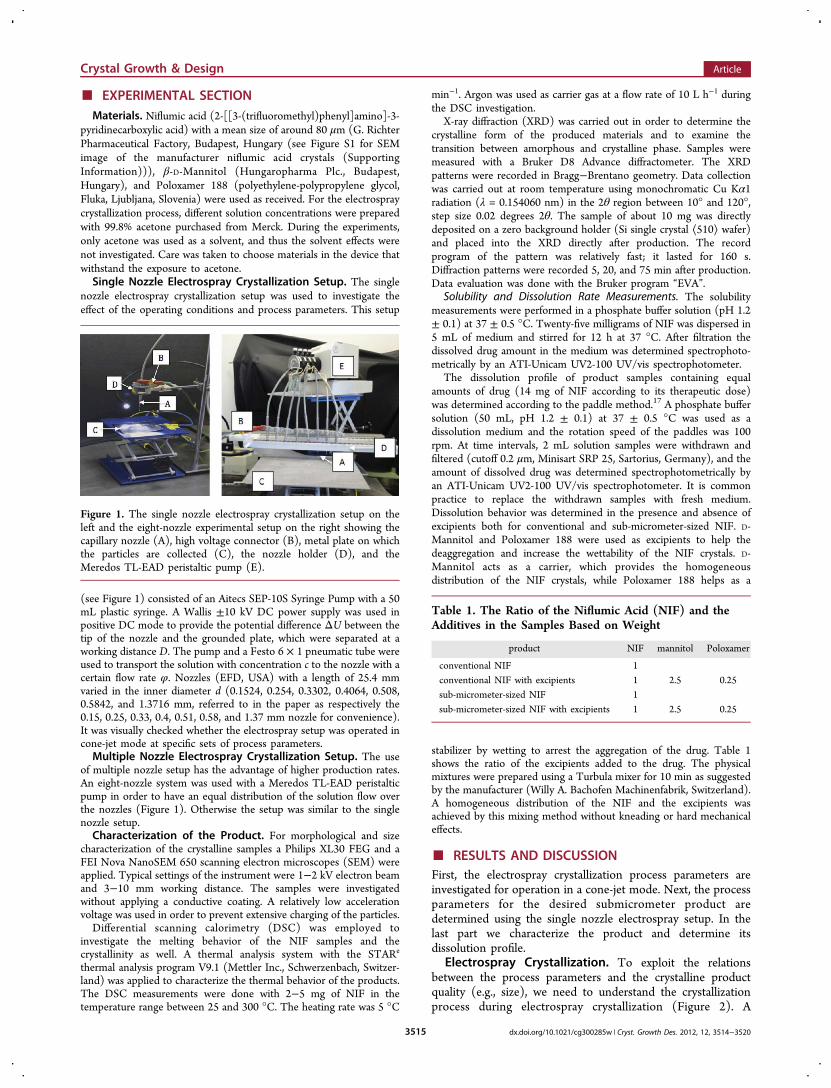
(see Figure 1) consisted of an Aitecs SEP-10S Syringe Pump with a 50mL plastic syringe. A Wallis ±10 kV DC power supply was used inpositive DC mode to provide the potential difference ΔU between thetip of the nozzle and the grounded plate, which were separated at aworking distance D. The pump and a Festo 6 × 1 pneumatic tube wereused to transport the solution with concentration c to the nozzle with acertain flow rate φ. Nozzles (EFD, USA) with a length of 25.4 mmvaried in the inner diameter d (0.1524, 0.254, 0.3302, 0.4064, 0.508,0.5842, and 1.3716 mm, referred to in the paper as respectively the0.15, 0.25, 0.33, 0.4, 0.51, 0.58, and 1.37 mm nozzle for convenience).It was visually checked whether the electrospray setup was operated incone-jet mode at specific sets of process parameters.Multiple Nozzle Electrospray Crystallization Setup. The use
of multiple nozzle setup has the advantage of higher production rates.An eight-nozzle system was used with a Meredos TL-EAD peristalticpump in order to have an equal distribution of the solution flow overthe nozzles (Figure 1). Otherwise the setup was similar to the singlenozzle setup.Characterization of the Product. For morphological and size
characterization of the crystalline samples a Philips XL30 FEG and aFEI Nova NanoSEM 650 scanning electron microscopes (SEM) wereapplied. Typical settings of the instrument were 1−2 kV electron beamand 3−10 mm working distance. The samples were investigatedwithout applying a conductive coating. A relatively low accelerationvoltage was used in order to prevent extensive charging of the particles.Differential scanning calorimetry (DSC) was employed to
investigate the melting behavior of the NIF samples and thecrystallinity as well. A thermal analysis system with the STARe
thermal analysis program V9.1 (Mettler Inc., Schwerzenbach, Switzer-land) was applied to characterize the thermal behavior of the products.The DSC measurements were done with 2−5 mg of NIF in thetemperature range between 25 and 300 °C. The heating rate was 5 °C
min−1. Argon was used as carrier gas at a flow rate of 10 L h−1 duringthe DSC investigation.
X-ray diffraction (XRD) was carried out in order to determine thecrystalline form of the produced materials and to examine thetransition between amorphous and crystalline phase. Samples weremeasured with a Bruker D8 Advance diffractometer. The XRDpatterns were recorded in Bragg−Brentano geometry. Data collectionwas carried out at room temperature using monochromatic Cu Kα1radiation (λ = 0.154060 nm) in the 2θ region between 10° and 120°,step size 0.02 degrees 2θ. The sample of about 10 mg was directlydeposited on a zero background holder (Si single crystal ⟨510⟩ wafer)and placed into the XRD directly after production. The recordprogram of the pattern was relatively fast; it lasted for 160 s.Diffraction patterns were recorded 5, 20, and 75 min after production.Data evaluation was done with the Bruker program “EVA”.
Solubility and Dissolution Rate Measurements. The solubilitymeasurements were performed in a phosphate buffer solution (pH 1.2± 0.1) at 37 ± 0.5 °C. Twenty-five milligrams of NIF was dispersed in5 mL of medium and stirred for 12 h at 37 °C. After filtration thedissolved drug amount in the medium was determined spectrophoto-metrically by an ATI-Unicam UV2-100 UV/vis spectrophotometer.
The dissolution profile of product samples containing equalamounts of drug (14 mg of NIF according to its therapeutic dose)was determined according to the paddle method.17 A phosphate buffersolution (50 mL, pH 1.2 ± 0.1) at 37 ± 0.5 °C was used as adissolution medium and the rotation speed of the paddles was 100rpm. At time intervals, 2 mL solution samples were withdrawn andfiltered (cutoff 0.2 μm, Minisart SRP 25, Sartorius, Germany), and theamount of dissolved drug was determined spectrophotometrically byan ATI-Unicam UV2-100 UV/vis spectrophotometer. It is commonpractice to replace the withdrawn samples with fresh medium.Dissolution behavior was determined in the presence and absence ofexcipients both for conventional and sub-micrometer-sized NIF. D-Mannitol and Poloxamer 188 were used as excipients to help thedeaggregation and increase the wettability of the NIF crystals. D-Mannitol acts as a carrier, which provides the homogeneousdistribution of the NIF crystals, while Poloxamer 188 helps as a
stabilizer by wetting to arrest the aggregation of the drug. Table 1shows the ratio of the excipients added to the drug. The physicalmixtures were prepared using a Turbula mixer for 10 min as suggestedby the manufacturer (Willy A. Bachofen Machinenfabrik, Switzerland).A homogeneous distribution of the NIF and the excipients wasachieved by this mixing method without kneading or hard mechanicaleffects.
■ RESULTS AND DISCUSSIONFirst, the electrospray crystallization process parameters areinvestigated for operation in a cone-jet mode. Next, the processparameters for the desired submicrometer product aredetermined using the single nozzle electrospray setup. In thelast part we characterize the product and determine itsdissolution profile.
Electrospray Crystallization. To exploit the relationsbetween the process parameters and the crystalline productquality (e.g., size), we need to understand the crystallizationprocess during electrospray crystallization (Figure 2). A
Figure 1. The single nozzle electrospray crystallization setup on theleft and the eight-nozzle experimental setup on the right showing thecapillary nozzle (A), high voltage connector (B), metal plate on whichthe particles are collected (C), the nozzle holder (D), and theMeredos TL-EAD peristaltic pump (E).
Table 1. The Ratio of the Niflumic Acid (NIF) and theAdditives in the Samples Based on Weight
product NIF mannitol Poloxamer
conventional NIF 1conventional NIF with excipients 1 2.5 0.25sub-micrometer-sized NIF 1sub-micrometer-sized NIF with excipients 1 2.5 0.25
Crystal Growth & Design Article
dx.doi.org/10.1021/cg300285w | Cryst. Growth Des. 2012, 12, 3514−35203515
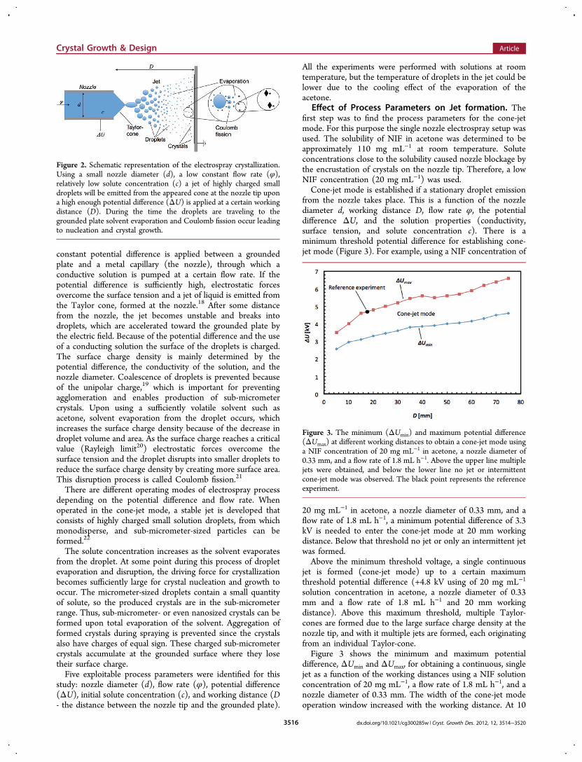
constant potential difference is applied between a groundedplate and a metal capillary (the nozzle), through which aconductive solution is pumped at a certain flow rate. If thepotential difference is sufficiently high, electrostatic forcesovercome the surface tension and a jet of liquid is emitted fromthe Taylor cone, formed at the nozzle.18 After some distancefrom the nozzle, the jet becomes unstable and breaks intodroplets, which are accelerated toward the grounded plate bythe electric field. Because of the potential difference and the useof a conducting solution the surface of the droplets is charged.The surface charge density is mainly determined by thepotential difference, the conductivity of the solution, and thenozzle diameter. Coalescence of droplets is prevented becauseof the unipolar charge,19 which is important for preventingagglomeration and enables production of sub-micrometercrystals. Upon using a sufficiently volatile solvent such asacetone, solvent evaporation from the droplet occurs, whichincreases the surface charge density because of the decrease indroplet volume and area. As the surface charge reaches a criticalvalue (Rayleigh limit20) electrostatic forces overcome thesurface tension and the droplet disrupts into smaller droplets toreduce the surface charge density by creating more surface area.This disruption process is called Coulomb fission.21
There are different operating modes of electrospray processdepending on the potential difference and flow rate. Whenoperated in the cone-jet mode, a stable jet is developed thatconsists of highly charged small solution droplets, from whichmonodisperse, and sub-micrometer-sized particles can beformed.22
The solute concentration increases as the solvent evaporatesfrom the droplet. At some point during this process of dropletevaporation and disruption, the driving force for crystallizationbecomes sufficiently large for crystal nucleation and growth tooccur. The micrometer-sized droplets contain a small quantityof solute, so the produced crystals are in the sub-micrometerrange. Thus, sub-micrometer- or even nanosized crystals can beformed upon total evaporation of the solvent. Aggregation offormed crystals during spraying is prevented since the crystalsalso have charges of equal sign. These charged sub-micrometercrystals accumulate at the grounded surface where they losetheir surface charge.Five exploitable process parameters were identified for this
study: nozzle diameter (d), flow rate (φ), potential difference(ΔU), initial solute concentration (c), and working distance (D- the distance between the nozzle tip and the grounded plate).
All the experiments were performed with solutions at roomtemperature, but the temperature of droplets in the jet could belower due to the cooling effect of the evaporation of theacetone.
Effect of Process Parameters on Jet formation. Thefirst step was to find the process parameters for the cone-jetmode. For this purpose the single nozzle electrospray setup wasused. The solubility of NIF in acetone was determined to beapproximately 110 mg mL−1 at room temperature. Soluteconcentrations close to the solubility caused nozzle blockage bythe encrustation of crystals on the nozzle tip. Therefore, a lowNIF concentration (20 mg mL−1) was used.Cone-jet mode is established if a stationary droplet emission
from the nozzle takes place. This is a function of the nozzlediameter d, working distance D, flow rate φ, the potentialdifference ΔU, and the solution properties (conductivity,surface tension, and solute concentration c). There is aminimum threshold potential difference for establishing cone-jet mode (Figure 3). For example, using a NIF concentration of
20 mg mL−1 in acetone, a nozzle diameter of 0.33 mm, and aflow rate of 1.8 mL h−1, a minimum potential difference of 3.3kV is needed to enter the cone-jet mode at 20 mm workingdistance. Below that threshold no jet or only an intermittent jetwas formed.Above the minimum threshold voltage, a single continuous
jet is formed (cone-jet mode) up to a certain maximumthreshold potential difference (+4.8 kV using of 20 mg mL−1
solution concentration in acetone, a nozzle diameter of 0.33mm and a flow rate of 1.8 mL h−1 and 20 mm workingdistance). Above this maximum threshold, multiple Taylor-cones are formed due to the large surface charge density at thenozzle tip, and with it multiple jets are formed, each originatingfrom an individual Taylor-cone.Figure 3 shows the minimum and maximum potential
difference, ΔUmin and ΔUmax, for obtaining a continuous, singlejet as a function of the working distances using a NIF solutionconcentration of 20 mg mL−1, a flow rate of 1.8 mL h−1, and anozzle diameter of 0.33 mm. The width of the cone-jet modeoperation window increased with the working distance. At 10
Figure 2. Schematic representation of the electrospray crystallization.Using a small nozzle diameter (d), a low constant flow rate (φ),relatively low solute concentration (c) a jet of highly charged smalldroplets will be emitted from the appeared cone at the nozzle tip upona high enough potential difference (ΔU) is applied at a certain workingdistance (D). During the time the droplets are traveling to thegrounded plate solvent evaporation and Coulomb fission occur leadingto nucleation and crystal growth.
Figure 3. The minimum (ΔUmin) and maximum potential difference(ΔUmax) at different working distances to obtain a cone-jet mode usinga NIF concentration of 20 mg mL−1 in acetone, a nozzle diameter of0.33 mm, and a flow rate of 1.8 mL h−1. Above the upper line multiplejets were obtained, and below the lower line no jet or intermittentcone-jet mode was observed. The black point represents the referenceexperiment.
Crystal Growth & Design Article
dx.doi.org/10.1021/cg300285w | Cryst. Growth Des. 2012, 12, 3514−35203516
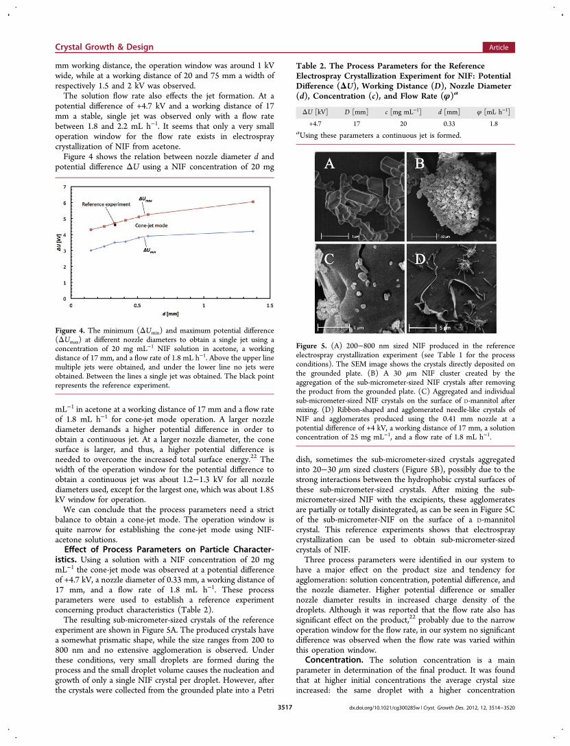
mm working distance, the operation window was around 1 kVwide, while at a working distance of 20 and 75 mm a width ofrespectively 1.5 and 2 kV was observed.The solution flow rate also effects the jet formation. At a
potential difference of +4.7 kV and a working distance of 17mm a stable, single jet was observed only with a flow ratebetween 1.8 and 2.2 mL h−1. It seems that only a very smalloperation window for the flow rate exists in electrospraycrystallization of NIF from acetone.Figure 4 shows the relation between nozzle diameter d and
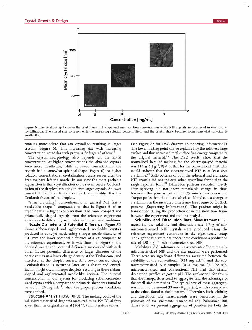
potential difference ΔU using a NIF concentration of 20 mg
mL−1 in acetone at a working distance of 17 mm and a flow rateof 1.8 mL h−1 for cone-jet mode operation. A larger nozzlediameter demands a higher potential difference in order toobtain a continuous jet. At a larger nozzle diameter, the conesurface is larger, and thus, a higher potential difference isneeded to overcome the increased total surface energy.22 Thewidth of the operation window for the potential difference toobtain a continuous jet was about 1.2−1.3 kV for all nozzlediameters used, except for the largest one, which was about 1.85kV window for operation.We can conclude that the process parameters need a strict
balance to obtain a cone-jet mode. The operation window isquite narrow for establishing the cone-jet mode using NIF-acetone solutions.Effect of Process Parameters on Particle Character-
istics. Using a solution with a NIF concentration of 20 mgmL−1 the cone-jet mode was observed at a potential differenceof +4.7 kV, a nozzle diameter of 0.33 mm, a working distance of17 mm, and a flow rate of 1.8 mL h−1. These processparameters were used to establish a reference experimentconcerning product characteristics (Table 2).The resulting sub-micrometer-sized crystals of the reference
experiment are shown in Figure 5A. The produced crystals havea somewhat prismatic shape, while the size ranges from 200 to800 nm and no extensive agglomeration is observed. Underthese conditions, very small droplets are formed during theprocess and the small droplet volume causes the nucleation andgrowth of only a single NIF crystal per droplet. However, afterthe crystals were collected from the grounded plate into a Petri
dish, sometimes the sub-micrometer-sized crystals aggregatedinto 20−30 μm sized clusters (Figure 5B), possibly due to thestrong interactions between the hydrophobic crystal surfaces ofthese sub-micrometer-sized crystals. After mixing the sub-micrometer-sized NIF with the excipients, these agglomeratesare partially or totally disintegrated, as can be seen in Figure 5Cof the sub-micrometer-NIF on the surface of a D-mannitolcrystal. This reference experiments shows that electrospraycrystallization can be used to obtain sub-micrometer-sizedcrystals of NIF.Three process parameters were identified in our system to
have a major effect on the product size and tendency foragglomeration: solution concentration, potential difference, andthe nozzle diameter. Higher potential difference or smallernozzle diameter results in increased charge density of thedroplets. Although it was reported that the flow rate also hassignificant effect on the product,22 probably due to the narrowoperation window for the flow rate, in our system no significantdifference was observed when the flow rate was varied withinthis operation window.
Concentration. The solution concentration is a mainparameter in determination of the final product. It was foundthat at higher initial concentrations the average crystal sizeincreased: the same droplet with a higher concentration
Figure 4. The minimum (ΔUmin) and maximum potential difference(ΔUmax) at different nozzle diameters to obtain a single jet using aconcentration of 20 mg mL−1 NIF solution in acetone, a workingdistance of 17 mm, and a flow rate of 1.8 mL h−1. Above the upper linemultiple jets were obtained, and under the lower line no jets wereobtained. Between the lines a single jet was obtained. The black pointrepresents the reference experiment.
Table 2. The Process Parameters for the ReferenceElectrospray Crystallization Experiment for NIF: PotentialDifference (ΔU), Working Distance (D), Nozzle Diameter(d), Concentration (c), and Flow Rate (φ)a
ΔU [kV] D [mm] c [mg mL−1] d [mm] φ [mL h−1]
+4.7 17 20 0.33 1.8aUsing these parameters a continuous jet is formed.
Figure 5. (A) 200−800 nm sized NIF produced in the referenceelectrospray crystallization experiment (see Table 1 for the processconditions). The SEM image shows the crystals directly deposited onthe grounded plate. (B) A 30 μm NIF cluster created by theaggregation of the sub-micrometer-sized NIF crystals after removingthe product from the grounded plate. (C) Aggregated and individualsub-micrometer-sized NIF crystals on the surface of D-mannitol aftermixing. (D) Ribbon-shaped and agglomerated needle-like crystals ofNIF and agglomerates produced using the 0.41 mm nozzle at apotential difference of +4 kV, a working distance of 17 mm, a solutionconcentration of 25 mg mL−1, and a flow rate of 1.8 mL h−1.
Crystal Growth & Design Article
dx.doi.org/10.1021/cg300285w | Cryst. Growth Des. 2012, 12, 3514−35203517
contains more solute that can crystallize, resulting in largercrystals (Figure 6). This increasing size with increasingconcentration coincides with previous findings of others.23
The crystal morphology also depends on the initialconcentration. At higher concentrations the obtained crystalswere more needle-like, while at lower concentrations thecrystals had a somewhat spherical shape (Figure 6). At highersolution concentrations, crystallization occurs earlier after thedroplets have left the nozzle. In our view the most probableexplanation is that crystallization occurs even before Coulombfission of the droplets, resulting in even larger crystals. At lowerconcentrations, crystallization occurs later, possibly after theCoulomb fission of the droplets.When crystallized conventionally, in general NIF has a
needle-like shape,24 comparable to that in Figure 6 of anexperiment at a higher concentration. The more compact andprismatically shaped crystals from the reference experimentindicate quite different growth behavior under these conditions.Nozzle Diameter and Potential Difference. Figure 5D
shows ribbon-shaped and agglomerated needle-like crystalsproduced in cone-jet mode using a larger nozzle diameter of0.41 mm and lower potential difference of 4 kV compared tothe reference experiment. As it was shown in Figure 4, thenozzle diameter and potential difference are coupled with eachother. Lower potential difference or larger diameter of thenozzle results in a lower charge density at the Taylor-cone, andtherefore, at the droplet surface. At a lower surface chargeCoulomb-fission is occurring later or is absent and crystal-lization might occur in larger droplets, resulting in these ribbon-shaped and agglomerated needle-like crystals. The optimalconcentration in our system for producing sub-micrometer-sized crystals with a compact and prismatic shape was found tobe around 20 mg mL−1, when the proper process conditionswere applied.Structure Analysis (DSC, XRD). The melting point of the
sub-micrometer-sized drug was measured to be 199 °C, slightlylower than the original material (204 °C) and literature values13
(see Figure S2 for DSC diagram (Supporting Information)).The lower melting point can be explained by the relatively largesurface and thus increased total surface free energy compared tothe original material.25 The DSC results show that thenormalized heat of melting for the electrosprayed materialwas 114 ± 6 J g−1, 85% of that for the conventional NIF. Thiswould indicate that the electrosprayed NIF is at least 85%crystalline.26 XRD patterns of both the spherical and elongatedNIF crystals did not indicate other crystalline forms than thesingle reported form.24 Diffraction patterns recorded directlyafter spraying did not show remarkable change in time;however, the powder pattern at 75 min shows more andsharper peaks than the others, which could indicate a change incrystallinity in the measured time frame (see Figure S3 for XRDpatterns (Supporting Information)). The product might betransformed during the production or in the short time framebetween the experiment and the first analysis.
Solubility and Dissolution Rate Measurements. Formeasuring the solubility and dissolution rate 1.7 g of sub-micrometer-sized NIF crystals were produced using thereference experiment conditions in the eight-nozzle setup.The eight nozzle setup has under these conditions a productionrate of 150 mg h−1 sub-micrometer-sized NIF.Solubility and dissolution rate measurements of both the sub-
micrometer-sized NIF and the raw material were performed.There were no significant differences measured between thesolubility of the conventional (0.23 mg mL−1) and the sub-micrometer-sized NIF samples (0.21 mg mL−1). The sub-micrometer-sized and conventional NIF had also similardissolution profiles at gastric pH. The explanation for this isthat the nanoparticles tend to aggregate, and the advantage ofthe small size diminishes. The typical size of these aggregateswas found to be around 30 μm (Figure 5B), which correspondsto the values found in the literature.27 Therefore, both solubilityand dissolution rate measurements were performed in thepresence of the excipients D-mannitol and Poloxamer 188.These additives prevent aggregation of powders for both the
Figure 6. The relationship between the crystal size and shape and used solution concentration when NIF crystals are produced in electrospraycrystallization. The crystal size increases with the increasing solution concentration, and the crystal shape becomes from somewhat spherical toneedle-like.
Crystal Growth & Design Article
dx.doi.org/10.1021/cg300285w | Cryst. Growth Des. 2012, 12, 3514−35203518
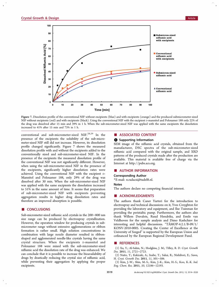
conventional and sub-micrometer-sized NIF.28,29 In thepresence of the excipients the solubility of the sub-micro-meter-sized NIF still did not increase. However, its dissolutionprofile changed significantly. Figure 7 shows the measureddissolution profile with and without the excipients added to theconventionally sized and sub-micrometer-sized NIF. In thepresence of the excipients the measured dissolution profile ofthe conventional NIF was not significantly different. However,when using the sub-micrometer-sized NIF in the presence ofthe excipients, significantly higher dissolution rates wereachieved. Using the conventional NIF with the excipient D-Mannitol and Poloxamer 188, only 29% of the drug wasdissolved after 30 min. When the sub-micrometer-sized NIFwas applied with the same excipients the dissolution increasedto 51% in the same amount of time. It seems that preparationof sub-micrometer-sized NIF with excipients preventingaggregation results in higher drug dissolution rates andtherefore an improved absorption is possible.
■ CONCLUSIONS
Sub-micrometer-sized niflumic acid crystals in the 200−800 nmsize range can be produced by electrospray crystallization.However, the operation window for producing crystals in sub-micrometer range without extensive agglomerations or ribbonformation is rather small. High solution concentrations incombination with large nozzle diameter resulted in ribbon-shaped and agglomerated needle-like crystals having the samecrystal structure. When the excipients D-mannitol andPoloxamer 188 were mixed with the sub-micrometer-sizedniflumic acid the dissolution rate of the drug was enhanced. Wecan conclude that it is possible to increase the bioavailability ofdrugs by drastically reducing the crystal size of niflumic acid,while preventing their aggregation by applying the properexcipients.
■ ASSOCIATED CONTENT*S Supporting InformationSEM image of the niflumic acid crystals, obtained from themanufacturer, DSC spectra of the sub-micrometer-sizedniflumic acid compared with the original sample, and XRDpatterns of the produced crystals made after the production areavailable. This material is available free of charge via theInternet at http://pubs.acs.org.
■ AUTHOR INFORMATIONCorresponding Author*E-mail: [email protected].
NotesThe authors declare no competing financial interest.
■ ACKNOWLEDGMENTSThe authors thank Caner Yurteri for the introduction toelectrospray and technical discussions on it, Yves Creyghton forproviding the laboratory and equipment, and Ilse Tuinman forproviding the peristaltic pump. Furthermore, the authors alsothank Willem Duvalois, Ruud Hendrikx, and Emile vanVeldhoven for the sample analysis and Dimo Kashchiev forinteresting and helpful discussions. “TAMOP-4.2.1/B-09/1/KONV-2010-0005. Creating the Center of Excellence at theUniversity of Szeged” is supported by the European Union andcofinanced by the European Regional Development Fund.
■ REFERENCES(1) Xu, Y.; Al-Salim, N.; Hodgkiss, J. M.; Tilley, R. D. Cryst. GrowthDes. 2011, 11, 2721−2723.(2) Naito, T.; Kakizaki, A.; Inabe, T.; Sakai, R.; Nishibori, E.; Sawa,H. Cryst. Growth Des. 2011, 11, 501−506.(3) Kim, J.-W.; Shin, M.-S.; Kim, J.-K.; Kim, H.-S.; Koo, K.-K. Ind.Eng. Chem. Res. 2011, 50, 12186−12193.
Figure 7. Dissolution profile of the conventional NIF without excipients (blue) and with excipients (orange) and the produced submicrometer-sizedNIF without excipients (red) and with excipients (black). Using the conventional NIF with the excipient D-mannitol and Poloxamer 188 only 22% ofthe drug was dissolved after 15 min and 39% in 1 h. When the sub-micrometer-sized NIF was applied with the same excipients the dissolutionincreased to 41% after 15 min and 73% in 1 h.
Crystal Growth & Design Article
dx.doi.org/10.1021/cg300285w | Cryst. Growth Des. 2012, 12, 3514−35203519
(4) Radacsi, N.; Stankiewicz, A. I.; Creyghton, Y. L. M.; van derHeijden, A. E. D. M.; ter Horst, J. H. Chem. Eng. Technol. 2011, 34,624−630.(5) Saharan, V. A.; Kukkar, V; Kataria, M.; Gera, M; Choudhury, P.M. Int. J. Health Res. 2009, 2, 107−124.(6) Kata, M.; Ambrus., R.; Aigner, Z. J. Incl. Phenom 2002, 44, 123−126.(7) Ambrus, R.; Aigner, Z.; Soica, C.; Peev, C.; Szabo-Revesz, P. Rev.Chim. 2007, 58, 206−209.(8) Kesisoglou, F.; Panmai, S.; Wu, Y. Adv. Drug Delivery Rev 2007,59, 631−644.(9) Leuner, C.; Dressmann, J. Eur. J. Pharm. Biopharm. 2000, 50, 47−60.(10) Rabinow, B. E. Nat. Rev. Drug Discov. 2004, 3, 785−796.(11) Patravale, V. B.; Date, A. A.; Kulkarni, R. M. J. Pharm.Pharmacol. 2004, 56, 827−840.(12) Muller, R. H.; Peters, K. Int. J. Pharm. 1998, 160, 229−237.(13) The Merck Index, 11th ed.; Budavari, S.; O’Neil, M. J., Smith, A.,Heckelman, P. E., Eds.; Merck & Co.: Rahway, NJ, 1989; Monograph6444.(14) Martindale: The Extra Pharmacopoeia, 31st ed.; Reynolds, J. E.F., Ed.; The Royal Pharmaceutical Society: London, 1996.(15) Takacs-Novak, K.; Avdeef, A.; Box, K. J.; Podanyi, B.; Szasz, Gy.J. Pharm. Biomed. Anal. 1994, 12, 1369−1377.(16) Ambrus, R.; Aigner, Z.; Dehelean, C.; Szabo-Revesz, P. Rev.Chim. 2007, 58, 60−64.(17) European Pharmacopoeia, 3rd ed.; Council of Europe:Strasbourg, 1996; pp 128−129.(18) Taylor, G. I. Proc. R. Soc. London A 1964, 280, 383.(19) Yurteri, C. U.; Hartman, R. P. A.; Marijnissen, J. C. M. Kona2010, 28, 91−115.(20) Rayleigh, L. Philos. Mag. 1882, 14, 184−186.(21) Suzuki, K.; Matsumoto, H.; Minagawa, M.; Kimura, M.;Tanioka, A. Polym. J. 2007, 39 (11), 1128−1134.(22) Hartman, R. P. A. Electrohydrodynamic Atomization in the Cone-Jet Mode. Ph.D. dissertation, Delft University of Technology: Delft,1998.(23) Scholten, E.; Dhamankar, H.; Bromberg, L.; Rutledge, G. C.;Hatton, T. A. Langmuir 2011, 27, 6683−6688.(24) Krishna Murthy, H. M.; Vijayan, M. Acta Crystallogr. 1979, B35,262−263.(25) Yang, G. C.; Nie, F. D.; Huang, H. J. Energ. Mater. 2007, 25,35−47.(26) Wagner, M. Thermal Analysis in Practice; Mettler-Toledo AG:Schwerzenbach, 2009; Chapter 7.6: DSC Evaluations, pp 90−132.(27) van Ommen, J. R.; Yurteri, C. U.; Ellis, N.; Kelder, E. M.Particuology 2010, 8, 572−577.(28) P. Kocbek, P.; Baumgartner, S.; Kristl, J. Int. J. Pharm. 2006, 312,179−186.(29) Hecq, J.; Deleers, M.; Fanara, D.; Vranckx, H.; Amighi, K. Int. J.Pharm. 2005, 299, 167−177.
Crystal Growth & Design Article
dx.doi.org/10.1021/cg300285w | Cryst. Growth Des. 2012, 12, 3514−35203520