Embed Size (px)
Citation preview

Electrochemical Regulation of Budding Yeast PolarityArmin Haupt1., Alexis Campetelli1., Daria Bonazzi1., Matthieu Piel2, Fred Chang3*, Nicolas Minc1*
1 Institut Jacques Monod, UMR7592 CNRS, Paris, France, 2 Institut Curie, UMR 144 CNRS/IC, Paris, France, 3 Department of Microbiology and Immunology, Columbia
University College of Physicians and Surgeons, New York, New York, United States of America
Abstract
Cells are naturally surrounded by organized electrical signals in the form of local ion fluxes, membrane potential, andelectric fields (EFs) at their surface. Although the contribution of electrochemical elements to cell polarity and migration isbeginning to be appreciated, underlying mechanisms are not known. Here we show that an exogenous EF can orient cellpolarization in budding yeast (Saccharomyces cerevisiae) cells, directing the growth of mating projections towards sites ofhyperpolarized membrane potential, while directing bud emergence in the opposite direction, towards sites of depolarizedpotential. Using an optogenetic approach, we demonstrate that a local change in membrane potential triggered by light issufficient to direct cell polarization. Screens for mutants with altered EF responses identify genes involved in transducingelectrochemical signals to the polarity machinery. Membrane potential, which is regulated by the potassium transporterTrk1p, is required for polarity orientation during mating and EF response. Membrane potential may regulate membranecharges through negatively charged phosphatidylserines (PSs), which act to position the Cdc42p-based polarity machinery.These studies thus define an electrochemical pathway that directs the orientation of cell polarization.
Citation: Haupt A, Campetelli A, Bonazzi D, Piel M, Chang F, et al. (2014) Electrochemical Regulation of Budding Yeast Polarity. PLoS Biol 12(12): e1002029. doi:10.1371/journal.pbio.1002029
Academic Editor: Mark D. Rose, Princeton University, United States of America
Received April 10, 2014; Accepted November 12, 2014; Published December 30, 2014
Copyright: � 2014 Haupt et al. This is an open-access article distributed under the terms of the Creative Commons Attribution License, which permitsunrestricted use, distribution, and reproduction in any medium, provided the original author and source are credited.
Data Availability: The authors confirm that all data underlying the findings are fully available without restriction. All relevant data are within the paper and itsSupporting Information files.
Funding: This work was supported by funds from the National Institutes of Health (http://www.nih.gov/, GM056836) to FC and the CNRS, ANR (http://www.agence-nationale-recherche.fr/, grant 10PDOC00301), FRM (http://www.frm.org/, grant AJE20130426890), the FP7 CIG and ITN ‘‘FungiBrain’’ (http://ec.europa.eu/research/mariecurieactions/) and the ‘‘Mairie de Paris emergence’’ program (http://www.paris.fr/, LS100805) to NM. The funders had no role in study design, datacollection and analysis, decision to publish, or preparation of the manuscript.
Competing Interests: The authors have declared that no competing interests exist.
Abbreviations: EF, electric field; LatA, latrunculin A; PS, phosphatidylserine; TMP, transmembrane potential; WT, wild-type.
* Email: [email protected] (FC); [email protected] (NM)
. These authors contributed equally to this work.
Introduction
Cell polarization arises from the asymmetric accumulation of
cellular components near a region of the plasma membrane.
Although the roles of polarity proteins such as small GTPases and
cytoskeletal elements have been studied extensively [1], much less
is known about the possible contribution of electrochemical
elements. Recent studies identifying certain ion transporters in
regulating processes such as cell migration and polarized cell
growth indicate potential roles of local pH, ion fluxes, and
membrane potentials at the plasma membrane [2–8]. How these
elements interface with established modules of polarity networks
remains to be defined.
The importance of electricity in cell polarization is illustrated by
the ability of electric fields (EFs) to direct cell polarization. It has
been appreciated for decades that most cells—ranging from
bacteria, fungi, and amoebas to animal cells—are electrotactic,
and robustly orient polarity, migration, or division to applied
exogenous EFs [9–14]. EFs of similar intensities as those used in
these experiments naturally surround cells in tissues, and even
individual cells such as fungal cells [10,15,16]. The physiological
relevance of endogenous EFs has been demonstrated in fungal
infection [17], immune cell response [18], wound healing,
regeneration, and development [6,10,19,20]. These findings have
led to the proposal that in addition to responding to chemical and
mechanical signals, cells may also be responding to endogenous
electrotactic signals to guide cell polarization [20]. The response of
cells to exogenous EFs provides a powerful tool to study
electrochemical elements in cell polarization.
The molecular mechanisms of cell polarity are currently best
understood in the budding yeast, Saccharomyces cerevisiae.
Polarized cell growth in these cells is tightly controlled by intrinsic
and extrinsic spatial cues. Haploid budding yeast cells display an
axial budding pattern, in which new buds form adjacent to
previous bud sites, while diploid cells exhibit a bipolar pattern, in
which buds emerge at sites of previous division or growth [21,22].
During mating, cells of opposite mating type polarize towards each
other in response to gradients of secreted pheromones; exogenous
application of the pheromone a-factor causes cells to grow a
mating projection, forming a pear-shaped ‘‘shmoo.’’ The core
polarity machinery required for both bud and shmoo formation is
organized around the small GTPase Cdc42p, which coordinates
actin assembly and exocytosis [23–25]. Bud site selection is
specified by a Ras-like protein Rsr1p and its regulators [23].
During mating, these spatial cues used to direct budding are
turned off, so that cells can polarize towards the mating partner.
This reorientation of polarity involves Far1p and its interactions
with the receptor-coupled Gb protein and Cdc42 GEF [25–27].
As demonstrated by mutants affected in the regulation of only
shmoos or only budding [23,28,29], there are specific molecular
PLOS Biology | www.plosbiology.org 1 December 2014 | Volume 12 | Issue 12 | e1002029
differences in the mechanisms governing budding and shmoo
polarity. In general, still little is appreciated about electrochemical
aspects of cell polarization in this cell type.
Here, we show that cell polarity can be directed by exogenous
EFs in budding yeast. Although EFs have been shown to direct
polarized growth in Schizosaccharomyces pombe [13] and Candidaalbicans [30,31], there have been no reports to date in S.cerevisiae. We find that although EFs do not appear to affect wild-
type (WT) budding cells, they do have robust effects on cells in the
presence of pheromone and on mutants defective in bud site
selection. We find a potassium channel and membrane lipid
charges as components mediating EF responses. We further show,
using a light-activated rhodopsin, that local membrane potential
itself is capable of directing polarization. Our results demonstrate
the importance of electrochemical signaling in cell polarity and
begin to define mechanistically how they contribute to polarized
cell growth.
Results
Electrotactic Responses of Budding Yeast PolarityWe tested whether exogenous EFs can influence cell polariza-
tion in budding yeast. Yeast cells were grown in the presence of
EFs in microfluidic channels, which allow for defined EF lines and
heat control [13]. Haploid WT cells were mostly resistant to EF
effects and budded at their normal axial position (Figure 1A and
1B). The bud site selection mutant rsr1D forms buds in random
directions, in the absence of EF. In the EF, however, almost all
new buds emerged at the cathode-facing side of the rsr1D cells
after 1 h of exposure to an EF of 50 V/cm (Figure 1A and 1B;
Movie S1). Cells did not exhibit any major signs of stress, cell
death, or stress pathway activation [32], but grew with slightly
reduced growth rates and prolonged cell cycle length as controls
(Figure S1). Cathodal bud orientation displayed dose dependence
on EF intensity and duration of application (Figure S2A and S2C).
Diploid WT cells also polarized towards the cathode significantly
more than WT haploids; this may reflect a less stringent regulation
of budding pattern in diploids (Figure 1B). Thus, the EF was not
able to efficiently override the normal spatial cues involved in axial
budding, but could direct bud site polarization if these cues were
absent or weak.
The application of EFs also directed the site of shmoo tip
formation but, surprisingly, in the opposite direction. In the
presence of uniform concentrations of a-factor and an EF,
budding yeast cells showed a strong polarization towards the
anode (Figures 1C, 1D, S2B, S2D, and S2E; Movie S2). Changing
the EF direction induced the formation of a second shmoo tip
towards the new anode (Figure 1E; Movie S3). To rule out
possible effects of adding mating factor exogenously, we also noted
similar effects in mating pairs of cells. The EF disrupted mating
and caused cells to polarize towards the anode of the EF instead of
towards each other (Figures 1G and S2F). Cells that were induced
to shmoo without external pheromones, by overexpressing Ste4p,
the b subunit of the G protein involved in pheromone response,
also polarized toward the anode [33] (Figure 1F and 1G; Movies
S4 and S5). Thus, although bud and shmoo formation use many of
the same components of the polarity machinery [21,22], there is a
striking difference in directionality (cathodal versus anodal) for
how budding and shmooing yeasts respond to EFs.
EF Response Involves the Cdc42p-Based PolarityMachinery
We next tested whether cell polarization in response to EF
requires the same polarity machinery normally used in budding or
shmooing. The highly conserved small GTPase Cdc42p was
required to polarize buds and shmoos in the absence or presence
of the EF, as assessed with the loss-of-function mutant allele cdc42-118 (Figures 2A, 2B, and S3A) [34]. In addition, mutants
specifically defective in establishing polarity during mating but
not budding, such as bem1-s1 (a point mutant in the scaffold
protein Bem1p [29]) and the formin null mutant bni1D [28],
showed similar polarization defects in the absence or presence of
the EF (Figures 2B, S3B, and S3E). Imaging GFP-Cdc42 [35] and
the associated components Cdc24-GFP [36] (a GEF for Cdc42p)
and Bem1-GFP [37] revealed that polarity caps assembled and
oriented to the EF prior to bud or shmoo emergence (Figure 2C–
2E). Bem1-GFP cap assembly was dependent on Cdc42p in the
presence or absence of EF (Figure S3C and S3D). Actin also
appeared to similarly mediate cell polarization in both instances.
In budding cells, actin was dispensable for EF-induced Bem1-GFP
cap cathodal orientation, although actin depolymerization ap-
peared to accelerate polar cap accumulation at the cathodal side.
In shmooing cells, actin inhibition caused rapid disappearance of
the cap in the presence or absence of the EF [27] (Figures 2D, 2F,
and S3F). Together, these data show that the EF acts in
reorienting polarized cell growth through the normal polarity
machinery, including Cdc42p and its regulators.
To investigate how EF directs mating projections, we tested the
role of Far1p and Cdc24p. Mutant far1-s and cdc24-m cells have a
specific orientation defect in response to a-factor, as they are not
able to orient appropriately towards gradients of a-factor, and
polarize instead using bud site selection cues [25–27]. In saturating
concentrations of a-factor, we found that both of these mutants
polarized towards the cathode of the EF (the opposite direction as
WT cells) (Figure 2G). This reversal was also observed at non-
saturating concentrations of pheromones (Figure S3G). As rsr1Dmutants in the absence of a-factor bud towards the cathode, this
suggests that far1-s and cdc24-m mutants may use machinery that
orients buds to direct shmoo projections to the cathode.
Author Summary
The ability of cells to orient towards spatial cues is criticalfor processes such as migration, wound healing, anddevelopment. Although the role of electrochemical signalsis well characterized in processes such as neuronalsignaling, their function in cell polarity is much lessunderstood or appreciated. Application of exogenouselectric fields can direct cell polarization in many celltypes, and electric fields of similar magnitude surroundcells and tissues naturally. However, the significance andmechanism of these responses remain poorly understood.Here, we introduce budding yeast (Saccharomyces cerevi-siae) as a powerful model system to study electrochemicalregulation of cell polarity. We show that application ofelectric fields causes budding yeast to polarize in particulardirections. We begin to identify key proteins involved inthis response, which implicate an electrochemical pathwayinvolving membrane potential, membrane charge, and anion channel, which ultimately regulate the central polarityfactor Cdc42p. These key proteins are not only needed forresponse to electric fields, but also contribute to cellpolarity more generally. To test whether a change inmembrane potential is sufficient to control cell polariza-tion, we introduce a light-sensitive ion channel into yeastand show that we can now control the site of polarizationsimply by using a focused laser beam. Thus, our studyshows that electrochemical regulation is an integralcomponent of cell polarity pathways.
Electrochemical Control of Yeast Polarity
PLOS Biology | www.plosbiology.org 2 December 2014 | Volume 12 | Issue 12 | e1002029

Electrochemical Control of Yeast Polarity
PLOS Biology | www.plosbiology.org 3 December 2014 | Volume 12 | Issue 12 | e1002029

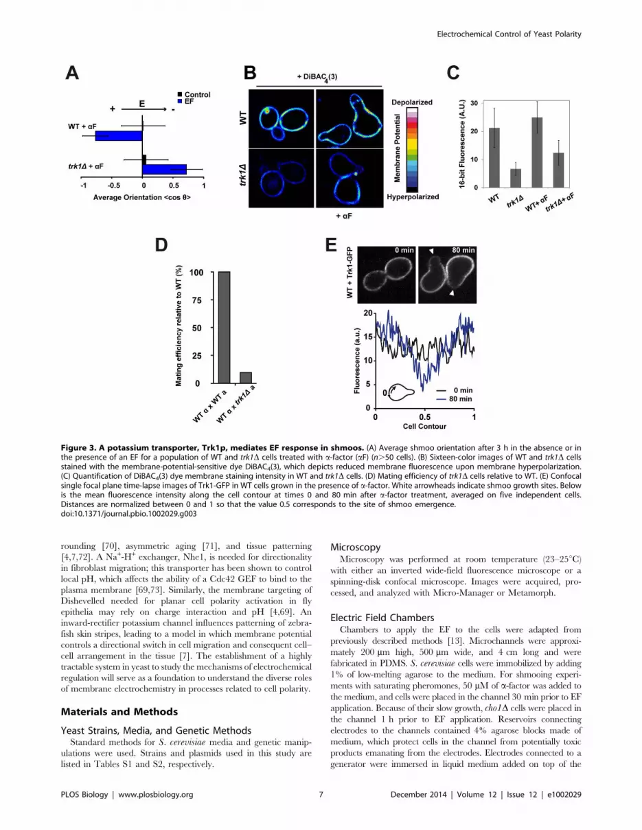
EF Response Involves theMembrane-Potential-Regulating PotassiumTransporter Trk1p
EFs are thought to affect cellular processes at or outside the
plasma membrane, but not in the cell interior. They have been
postulated to generate subcellular asymmetries in transmem-
brane potentials (TMPs) [13,38,39], and/or displace charged
membrane proteins at the cell surface [40,41]. To test whether
membrane transporters mediate EF responses, we screened a set
of well-characterized mutants and inhibitors affecting transport
at the membrane. We found that calcium, sodium, and proton
transport systems are not critical for EF sensing for bud or
shmoo reorientation (Figure S4). We found, however, that a
potassium transporter mutant trk1D was defective in the anodal
orientation of mating projection, but not in budding orientation;
these cells oriented shmoos to the cathode, in a similar manner
as far1-s and cdc24-m mutants (Figure 3A; Movie S6). Trk1p is
a high-affinity inward potassium transporter that displays
conserved features in bacteria, plants, and fungi. In yeast,
Trk1p is a major TMP regulator [42,43], and trk1D cells exhibit
hyperpolarized resting potential (Figure 3B and 3C) [42]. A
trk2D mutant, in the secondary K+-import system (Trk2p), did
not display any orientation defect in the EF, however [44].
Similarly to far1-s and cdc24-m mutants, trk1D mutants formed
shmoos with normal morphology and timing, but were defective
in mating (efficiencies of ,10% of WT; Figure 3D), and
displayed significant defects in polarizing in the correct direction
in mating pairs (Figure S5B). In contrast, trk1D had no defects
in bud emergence and haploid axial patterns (Figure S5A). We
found that Trk1-GFP was located throughout the plasma
membrane, but was reduced in emergent growing buds and
shmoo tips, in a pattern similar to that of other membrane
transporters [45,46]. In shmooing cells, measurements of
fluorescence intensity showed a stable back-to-front gradient,
with a concentration ratio of about 3-fold (Figures 3E and S5C).
In the presence of the EF, we observed a similar depletion of
Trk1-GFP at the shmoo tip growing towards the anode, without
noticeable change in protein distribution prior to tip growth
(Figure S5D and S5E). Together, these data suggest that a
natural gradient of Trk1p leading to local differences in
potassium import may contribute to polarity regulation for
shmoo tip orientation and EF response.
To shed more light on why cells may polarize in these
different directions, we performed computational simulations
and analytical calculations of the local EF strengths and electric
potentials along the membrane of S. cerevisiae cells (Figure S6).
This showed that sites of bud and shmoo emergence correspond
to the minimum and maximum local EF potentials, and to sites
of depolarized and hyperpolarized TMPs, respectively. This
analysis thus led to the prediction that if EF-induced polarity is
sensitive to TMPs, shmoos should emerge at sites of hyperpo-
larized TMP, while buds should emerge at sites of depolarized
TMP.
Asymmetries in Membrane Potential Can Direct PolarityTo directly test the nature of the electrochemical signaling
orienting polarity, we developed an optogenetic approach to
locally modulate TMPs and/or ion fluxes [47]. Microbial opsins
are light-gated transmembrane channels or pumps that have
been used to modulate TMPs for neuron activation or silencing
[48], as well as in other cell types such as yeast [49,50]. We
expressed different opsins tagged with GFP, and found that
Halorhodopsin-GFP (NpHR) displayed the most robust expres-
sion and plasma membrane targeting, although there was some
low level accumulation of Halorhodopsin-GFP in internal
membranes, as often seen in other cell types [51] (Figure
S7A). Halorhodopsin is a reversible inward chloride pump that
causes rapid hyperpolarization of the TMP upon activation with
green/yellow light [48]. We confirmed that Halorhodopsin
could drive membrane hyperpolarization upon light activation
in budding yeast, by measuring changes in global membrane
potential in single cells following laser exposure, using the
sensitive dye DiBAC4(3) (Figure S7B and S7C). We implement-
ed a photoactivation assay to locally hyperpolarize mating and
budding yeast cells at specific sites on the plasma membrane
[52]. Cells were illuminated on a small square-shaped region at
the cell surface with a yellow laser for 20 min, and subsequently
filmed for 2 h to compute polarized growth orientation
(Figure 4A and 4B). Laser exposure did not cause the cells to
die or halt growth, but we did note a reduction in growth rate of
,10%–15% in cells exposed to the laser compared to non-
exposed controls in the same field. Accordingly, measurement of
stress pathway activation revealed a minor stress response that
remained negligible compared to typical osmotic stress respons-
es (Figure S8A–S8C).
Strikingly, many cells expressing Halorhodopsin subsequently
grew mating projections towards the site of the laser illumination
(Figure 4C and 4D). This effect on orientation caused by light was
similar to the one caused by 20 min of EF exposure (Figure S2D).
Control cells that either did not express Halorhodopsin or
expressed an unrelated GFP-tagged transmembrane protein,
Hxt3-GFP, with similar localization [53] polarized in directions
independent of the laser, showing that this effect was opsin-
dependent and not due to cellular damage from the laser itself [54]
(Figures 4D and S8D). Similar treatments in budding cells did not
orient bud site emergence however (Figure 4D). These data
suggest that the direction of mating projections can be controlled
by local hyperpolarization of membrane potentials.
Figure 1. Budding versus shmooing yeast cells polarize in opposite directions in an electric field. (A) Phase contrast time lapse of WTand rsr1D budding yeast cells growing under an EF of 50 V/cm. White arrowheads point at sites of bud emergence. On the right are radial histogramsof polarized growth direction (indicated as the final angle of bud emergence with the EF, h) for WT and rsr1D cells in the presence or in the absenceof an EF. (B) Average bud orientation, computed as ,cosh. after 3 h of growth in the absence or in the presence of an EF, for a population ofhaploids and diploids of the indicated genotype. A positive average orientation represents an orientation to the cathode (negative electrode of theEF), whereas a negative orientation stands for an orientation to the anode. (C) Phase contrast time lapse of WT and rsr1D budding yeast cells growingmating projections (‘‘shmoos’’) in the presence of a-factor (aF) under an EF of 50 V/cm. White arrowheads point at sites of shmoo emergence. On theright are radial histograms of polarized growth direction. (D) Average shmoo orientation after 3 h of mating tip growth in the absence or in thepresence of an EF for a population of WT and rsr1D cells treated with a-factor. (E) Time-lapse images of shmoo reorientation in a WT cell afterinverting the EF direction. A second shmoo is formed at the new anodal side after reversing the EF. Blue arrows indicate sites of shmoo emergence.(F) Time lapse of WT cells overexpressing Ste4p (Ste4-OE) in the absence or in the presence of an EF of 50 V/cm. White arrowheads point at sites ofpolarized growth. (G) Average shmoo orientation after 3 h in the absence or in the presence of an EF for a population of WT cells treated with a-factor, WT mating pairs, and WT cells overexpressing Ste4p. n.50 cells for all conditions. Error bars represent standard deviations. Scale bars: 2 mm.doi:10.1371/journal.pbio.1002029.g001
Electrochemical Control of Yeast Polarity
PLOS Biology | www.plosbiology.org 4 December 2014 | Volume 12 | Issue 12 | e1002029

Electrochemical Control of Yeast Polarity
PLOS Biology | www.plosbiology.org 5 December 2014 | Volume 12 | Issue 12 | e1002029

Membrane Potential May Influence Lipid-MediatedMembrane Surface Charge to Steer Cdc42p Polarity Caps
We next asked how local changes in membrane potential
influence the Cdc42-based polarity machinery. Although membrane
potential could impact proton transport and local pH [13] or the
transport of other ions, our candidate screen did not reveal any
obvious role for proton or other ion transport systems other than
Trk1p (Figure S4C and S4D). Another way by which membrane
potential may affect polarity is through membrane electrostatics by
affecting charged lipid flipping [55–57]. PS is a negatively charged
lipid that acts as an electrostatic platform at the inner leaflet to
regulate membrane binding of proteins including Cdc42p [58]. In
budding yeast, PS concentrates at sites of shmoo and bud emergence
[58]. A PS synthesis mutant cho1D has defects in Cdc42p
recruitment, shmoo polarity, and mating [58]. We found that this
mutant also exhibited an abnormal EF response in that it oriented
mating projections to the cathode of the EF, much like trk1D, far1-s,and cdc24-m mutants (Figure 5A). Conversely, mutants in a lipid
flippase complex, dnf1-2D or lem3D, which may have increased PS
and negative surface charges [59], showed significant increased
anodal shmoo orientation in the EF. PS and membrane charges
affected EF response only in shmoos, not in buds (Figure 5B). Next,
we imaged PS localization using a GFP-Lact-C2 probe [58,60]. In
shmooing cells, PS rapidly accumulated and persisted at the anodal
side, long before shmoo appearance. In budding cells, PS also initially
accumulated at the anodal side, but then reverted to the cathodal side
immediately prior to bud emergence, often leaving a secondary patch
at the anodal side (Figure 5C and 5D). Thus, asymmetries in
membrane potential may bias the localization of Cdc42p and other
polarity factors through effects on PS and membrane charge.
Discussion
These are the first studies, to our knowledge, showing the input
of membrane electrochemistry in the regulation of cell polarity in
S. cerevisiae. We find that EFs direct the site of bud formation and
mating projections in different directions. Our optogenetic
experiments further show that in shmooing cells, local hyperpo-
larization of membrane potential is actually sufficient for polarity
reorientation (Figure 6). The mating defects of trk1D and cho1Dmutants [58], for instance, demonstrate that this pathway
contributes to cell polarization even in the absence of EFs. Our
results suggest a model in which the asymmetric segregation of
Trk1p and possibly other transporters produces positive charges at
the back of the cell and negative charges on PS lipids at the front
of the cell, which promotes the polarized distribution of Cdc42. In
the absence of EFs, the asymmetry of Trk1p localization may arise
from initial polarization of membrane insertion. These electro-
chemical pathways may thus represent a positive feedback loop
that stabilizes the axis of Cdc42-based polarity for chemotropism.
A surprising finding of this study is the different behavior of
budding versus shmooing cells. Although these polarization
systems share downstream polarity regulators, we found clear
differences in the requirement for upstream electrochemical
elements. rsr1D cells bud towards the cathode, while the same
strain shmoos towards the anode. Mutations in Far1, Cdc24,
Trk1, and Cho1 all cause cells to shmoo in an abnormal direction
in response to EF and have mating defects in the absence of EF,
but have little or no effect on bud site selection [27,58]. The
cathodal orientation of budding cells in EFs suggests that buds
originate at sites of depolarized TMP. However, genetic and
optogenetic analysis demonstrate that they may not be dependent
on gradients of membrane potential or PS levels, suggesting that
regulation of bud site selection is determined by a distinct
mechanism. Although it is not yet known what elements act
upstream of Cdc42p to drive cathodal growth, a plausible
hypothesis is that EFs may localize some charged membrane
proteins by direct electrophoresis, as suggested in other systems
[13,40]. The EF thus causes a tug-of-war between two competitive
pathways that steer polarity in different directions, with the anodal
one being dominant in response to mating factors.
The observed responses of cells to exogenously applied EFs lead
to a question of whether EFs normally contribute to polarity
regulation. Tissues and even individual polarized cells are
surrounded by EFs, which may arise from asymmetries in ion
transport [61–65]. We speculate that fungi may respond to their
own EFs, possibly during mating, in the context of fungal
communities such as biofilms, and in their natural environment
to guide them during invasion of host tissues, for instance. It would
be interesting to examine the role of genes such as Trk1 on various
fungal behaviors.
Mechanisms of electrochemical regulation of cell polarity are
likely conserved. Our data are consistent with recent findings
implicating a similar set of actors in fission yeast, neutrophils,
keratocytes, and slime molds [9,10,12,13,40]. Fission yeasts
respond to EFs by orienting their growth axis perpendicular to
the EF, producing bent morphologies. Cdc42, formins, and the
Pma1 proton pump at the plasma membrane are identified as
critical elements. Pma1 affects cell polarity and actin assembly
even in the absence of exogenous EFs, indicating a role for
membrane potential and intracellular pH in regulating normal tip
growth [13]. Migrating neutrophils, keratocytes, and slime molds
orient migration to exogenous EFs, possibly through effects on
membrane potential [10,66]. These responses involve the phos-
phorylation and charge additions on phosphatidylinositol lipids—
mediated by PI3-kinase—that recruit and activate Rho GTPases
for polarized migration.
Recent studies on plasma membrane pumps and channels are
beginning to reveal the pivotal role of membrane potential, pH,
and/or local ion transport in cell migration [67–69], mitotic
Figure 2. EF response involves Cdc42p polarization. (A) Percentage of new bud formation after 2 h in the absence or in the presence of an EFfor a population of WT, rsr1D, and cdc42-118 rsr1D (at restrictive temperature, 36uC). (B) Percentage of shmoo formation after 2 h in the absence or inthe presence of an EF for a population of WT, cdc42-118 (at restrictive temperature), bem1-s1, and bni1D cells treated with a-factor (aF). (C) Confocalsingle plane time-lapse images of GFP-Cdc42 and Cdc24-GFP expressed in rsr1D cells grown under an EF, in the absence or in the presence of a-factor. White arrowheads indicate the successive positions of the protein polar caps. (D) Confocal single plane time-lapse images of Bem1-GFP incontrol and LatA-treated rsr1D cells grown in the absence and in the presence of an EF. White arrowheads indicate the successive positions of Bem1-GFP polar caps. (E) Confocal single plane time-lapse images of Bem1-GFP in control and LatA-treated rsr1D cells grown with or without an EF in thepresence of a-factor. Note that LatA treatment induces rapid dispersion of the Bem1-GFP signal at the cap, with or without EF. White arrowheadsindicate the successive positions of Bem1-GFP polar caps. (F) Temporal evolution of the average orientation of Bem1-GFP caps with respect to theapplied EF in a population of rsr1D cells, treated with and without LatA or a-factor (top) (n = 13 cells for budding [blue], n = 9 cells for budding + LatA[green], n = 4 cells for shmooing [red]). Half-time (t1/2) corresponding to the mean orientation of Bem1-GFP polar caps to the cathode or anode of theEF is shown at the bottom. (G) Average shmoo orientation after 3 h in the absence or in the presence of an EF for a population of WT, rsr1D, cdc24-m,far1-s, and rsr1D far1-s cells treated with a-factor. n.50 cells for each condition. Error bars represent standard deviations. Scale bars: 2 mm.doi:10.1371/journal.pbio.1002029.g002
Electrochemical Control of Yeast Polarity
PLOS Biology | www.plosbiology.org 6 December 2014 | Volume 12 | Issue 12 | e1002029
rounding [70], asymmetric aging [71], and tissue patterning
[4,7,72]. A Na+-H+ exchanger, Nhe1, is needed for directionality
in fibroblast migration; this transporter has been shown to control
local pH, which affects the ability of a Cdc42 GEF to bind to the
plasma membrane [69,73]. Similarly, the membrane targeting of
Dishevelled needed for planar cell polarity activation in fly
epithelia may rely on charge interaction and pH [4,69]. An
inward-rectifier potassium channel influences patterning of zebra-
fish skin stripes, leading to a model in which membrane potential
controls a directional switch in cell migration and consequent cell–
cell arrangement in the tissue [7]. The establishment of a highly
tractable system in yeast to study the mechanisms of electrochemical
regulation will serve as a foundation to understand the diverse roles
of membrane electrochemistry in processes related to cell polarity.
Materials and Methods
Yeast Strains, Media, and Genetic MethodsStandard methods for S. cerevisiae media and genetic manip-
ulations were used. Strains and plasmids used in this study are
listed in Tables S1 and S2, respectively.
MicroscopyMicroscopy was performed at room temperature (23–25uC)
with either an inverted wide-field fluorescence microscope or a
spinning-disk confocal microscope. Images were acquired, pro-
cessed, and analyzed with Micro-Manager or Metamorph.
Electric Field ChambersChambers to apply the EF to the cells were adapted from
previously described methods [13]. Microchannels were approxi-
mately 200 mm high, 500 mm wide, and 4 cm long and were
fabricated in PDMS. S. cerevisiae cells were immobilized by adding
1% of low-melting agarose to the medium. For shmooing experi-
ments with saturating pheromones, 50 mM of a-factor was added to
the medium, and cells were placed in the channel 30 min prior to EF
application. Because of their slow growth, cho1D cells were placed in
the channel 1 h prior to EF application. Reservoirs connecting
electrodes to the channels contained 4% agarose blocks made of
medium, which protect cells in the channel from potentially toxic
products emanating from the electrodes. Electrodes connected to a
generator were immersed in liquid medium added on top of the
Figure 3. A potassium transporter, Trk1p, mediates EF response in shmoos. (A) Average shmoo orientation after 3 h in the absence or inthe presence of an EF for a population of WT and trk1D cells treated with a-factor (aF) (n.50 cells). (B) Sixteen-color images of WT and trk1D cellsstained with the membrane-potential-sensitive dye DiBAC4(3), which depicts reduced membrane fluorescence upon membrane hyperpolarization.(C) Quantification of DiBAC4(3) dye membrane staining intensity in WT and trk1D cells. (D) Mating efficiency of trk1D cells relative to WT. (E) Confocalsingle focal plane time-lapse images of Trk1-GFP in WT cells grown in the presence of a-factor. White arrowheads indicate shmoo growth sites. Belowis the mean fluorescence intensity along the cell contour at times 0 and 80 min after a-factor treatment, averaged on five independent cells.Distances are normalized between 0 and 1 so that the value 0.5 corresponds to the site of shmoo emergence.doi:10.1371/journal.pbio.1002029.g003
Electrochemical Control of Yeast Polarity
PLOS Biology | www.plosbiology.org 7 December 2014 | Volume 12 | Issue 12 | e1002029

reservoirs. In these conditions, growth rate and cell cycle periods were
almost unaffected, and no significant stress was induced (Figure S1).
OptogeneticsThe optogenetic assay used a 535-nm laser, with a power of
,5 mW, interfaced with an iLas system (Roper Scientific)
mounted on a confocal spinning disk and a 636 objective. This
allowed irradiation of multiple regions of interest (of 20620 px2) in
a given field of view. Cells were placed on a 2% agar pad
containing 20 mM of all-trans retinal and 50 mM of a-factor for
shmooing experiments. Cells were put on the pad 30 min prior to
laser excitation. The laser was turned on for a continuous period of
20 min, and the cells were subsequently filmed for 2 h to monitor
polarized growth. Laser exposure did not induce major changes in
growth rate or stress levels (Figure S8A–S8C).
Pharmacological InhibitorsAll inhibitors were prepared at the indicated concentration and
applied before EF application. Latrunculin A (LatA) (Sigma) was
used at a final concentration of 100 mM from a 1006 stock in
DMSO. The calcium ionophore A23187 was used at a final
concentration of 10 mM. The calcium chelating agent EGTA was
used at a final concentration of 2 mM.
Quantitative Mating AssaysEfficiency of mating in trk1D mutants was assayed by quantitative
counting of mating diploids. WT Mat a (AC 131), WT Mat a (AC
129), and trk1D Mat a (AC 31) cells were grown to mid-log phase in
YPD medium and concentrated to 10 OD/ml; WT Mat a cells were
incubated at a 10:1 ratio with target WT or trk1D Mat a cells, and
collected into a soft pellet by centrifugation. After 4 h of mating at
30 uC, cells were suspended in liquid YPD, and serial dilutions were
plated on medium selective for diploids. Mating efficiency was
compared between WT and trk1D by counting the number of
diploid colonies obtained at different dilutions. About 500 colonies
were counted for each condition, and the assay was repeated twice.
In addition to this assay, we also counted the number of genuine
zygotes by microscopy after 4 h of mating. To this aim, WT Mat
Figure 4. An optogenetic assay shows that asymmetries in membrane potential can direct polarity. (A) Optogenetic assay to generateasymmetries in membrane potential and assess for effect on polarity. Schematic representation of the experimental setup: a yellow laser (l= 535 nm)is used to photoactivate Halorhodopsin (Halo) in selected regions of rsr1D cells. h is the final angle of shmoo or bud emergence with respect to thedirection of the photoactivated region. (B) rsr1D (left) and Halorhodopsin-GFP-expressing rsr1D (right) cells in the presence of a-factor (aF) and retinalare continuously photoactivated from time 0 to 20 min at the indicated yellow region. After 2 h, shmoos grow and polarity orientation can bequantified with respect to the photoactivated region. White arrowheads indicate sites of shmoo formation. (C) Quantification of optogeneticexperiments: radial histogram of polarized growth orientation with respect to photoactivation angle in rsr1D and rsr1D + Halorhodopsin-GFP cellstreated with a-factor. (D) Average orientation of polarized growth in budding and shmooing cells after 2 h of growth following local photoactivationfor a population of rsr1D, rsr1D Hxt3-GFP, and rsr1D + Halorhodopsin-GFP cells (n.70 cells gathered from four independent datasets for allconditions and n = 166 cells gathered from seven independent experiments for rsr1D + Halorhodopsin-GFP + a-factor). **Student’s t test, p,0.05.Error bars represent standard deviations.doi:10.1371/journal.pbio.1002029.g004
Electrochemical Control of Yeast Polarity
PLOS Biology | www.plosbiology.org 8 December 2014 | Volume 12 | Issue 12 | e1002029

a, WT Mat a, and trk1D Mat a cells were grown in YPD liquid
medium to mid-log phase and concentrated to 10 OD/ml. WT
Mat a cells were stained with calcofluor for 5 min, subsequently
rinsed with YPD, and incubated at a 10:1 ratio with target WT or
trk1D Mat a cells. A 10-ml drop of each mixture was then spotted
onto a YPD plate and incubated for 4 h at 30 uC. The mixtures
were then imaged on a microscope, and mating efficiency was
computed as the ratio of genuine zygotes to the total number of
Mat a cells in the field of view. About 500 cells were counted for
each condition, and the assay was repeated twice. This assay
yielded a mating efficiency in the trk1D of about 30% of the WT.
Budding PatternsTo test the role of Trk1p in axial budding, we generated a trk1D
strain in the W303 background with a WT copy of the BUD4 gene
(AC 134). Cells were then grown to mid-log phase in YPD
medium and stained with calcofluor for 5 min to mark bud scars.
Axially budding cells were counted when more than three scars
were clustered at one site on the surface.
Chemotropism Efficiency in ZygotesTo compare the efficiency of chemotropism in WT versus trk1D
cells, we used a previously described assay that takes advantage of
the fact that WT cells grow towards their mating partners
irrespective of previous bud site selection, while mutants with
defective mating polarity (like far1-s or cdc24-m) use bud site
selection cues to grow shmoos [74]. WT Mat a, WT Mat a, and
trk1D Mat a cells were grown in YPD liquid medium to mid-log
phase and concentrated to 10 OD/ml. WT Mat a and trk1D Mat
a cells were stained with calcofluor for 5 min, subsequently rinsed
with YPD, and incubated at a 1:1 ratio with target WT Mat acells. A 10-ml drop of each mixture was then spotted onto a YPD
plate and incubated for 4 h at 30uC. The mixtures were then
imaged to assess the position of the bud scar relative to the fusion
site in each newly formed zygote. Only zygotes with a single
fluorescent bud scar were counted. Zygotes were scored as
proximal if the bud scar was in the one-third of the cell adjacent
to the fusion site, medial for the middle one-third, and distal for
the one-third away from the fusion site (Figure S5B).
Membrane Potential MeasurementTo measure global membrane potential in single budding yeast
cells, we used the membrane potential dye DiBAC4(3) (Invitrogen),
which absorbs in blue light and depicts increased membrane
fluorescence upon membrane depolarization, with a sensitivity of
nearly 1% per millivolt [7]. Cells were incubated with a
concentration of 50 mM dye for 30 min, and images were taken
on a confocal spinning disk. Relative membrane potential values
were then quantified as membrane signal subtracted from
background signal. To assess membrane hyperpolarization by
Halorhodopsin, cells were immobilized at the bottom of a
microfluidic chamber, between a dialysis membrane and the
coverslip [75]. Cells expressing Halorhodopsin-GFP were
bleached by long-time exposure with a blue laser. Medium was
Figure 5. Membrane hyperpolarization orients polarity through local phosphatidylserine accumulation. (A) Average shmoo orientationin the absence or presence of an EF for a population of WT, cho1D, dnf1-2D, and lem3D cells treated with a-factor (aF) (n.50 cells). (B) Average budorientation after 3 h in the absence and in the presence of an EF for a population of rsr1D, rsr1D cho1D, rsr1D dnf1-2D, and rsr1D lem3D cells (n.50cells). (C) Sixteen-color epifluorescence time lapses of shmooing and budding cells polarizing in EFs and expressing GFP-Lact-C2 probe (a marker forPS). White arrowheads point at sites of PS accumulation. (D) Quantification of PS localization in EFs. The ratio of anodal versus cathodal signal iscomputed by measuring the total amount at the membrane on both facing sides of the cell. Left: ratio evolution for the depicted sequences in (C).The black arrows indicate the moment when shmoo tip or bud was first visible. Right: average ratio of anodal versus cathodal PS signal for shmooingand budding cells. **Student’s t test, p,0.001. Error bars represent standard deviations.doi:10.1371/journal.pbio.1002029.g005
Electrochemical Control of Yeast Polarity
PLOS Biology | www.plosbiology.org 9 December 2014 | Volume 12 | Issue 12 | e1002029

subsequently exchanged with YPD containing 50 mM DiBAC4(3)
dye, and the dye was left to stain the cells for 30 min. Dye staining
intensity at the membrane was then measured in the same pre-
bleached cells at two consecutive time points spaced by 3 min (I0
and I1) to compute dye photo-bleaching. These cells were then
exposed to the yellow laser for 3 min, and the final dye staining
was computed again (I2). The specific loss of dye staining
associated with Halorhodopsin effects on membrane potential
was then computed asI2
I1� I0
I1{1, which is expected to be negative
for membrane hyperpolarization and positive for membrane
depolarization.
Computer Simulations of EF Effects on Yeast CellsComputer simulations were performed using the Matlab Partial
Differential Equation Toolbox (MathWorks). The cell surfaces as
well as the channel sides were considered as perfect insulators,
while the cell interior and the surrounding medium as conductors.
Analytical Calculations of EF Effects on Yeast CellsThe electric potential, W, created by the applied EF, ~EE, was
analytically computed by solving the Laplace equation: DW = 0,
with the boundary condition at the insulating membrane
~++W� �
:~nn� �
~0, with ~nn the vector normal to the membrane, and
the limit condition at infinity ~++W� �
inf~~EE. S. cerevisiae cells were
represented by a sphere, leading to the classical results [76] for the
potential at the membrane Wm and the field at the membrane ~EEm:
Wm~{3
2ER cos h and ~EEm~{
3
2E sin h:~eeh, with R the radius of
the sphere and h the angle with the field.
Supporting Information
Figure S1 EF effects on cell physiology. (A) Effect of EFs on
the timing of bud (dose-dependent, left) and shmoo emergence
(100 V/cm, right). (B) Effect of EF (100 V/cm for 1 h) on stress
levels of cells in presence of 50 mM a-factor as measured by Hog1-
GFP nuclear accumulation. Osmotic stress (0.5 M NaCl for
5 min) is used as a positive control for stress. (C) Quantification of
Hog1-GFP nuclear to cytoplasmic levels. n.25 cells for each
condition. Error bars represent standard deviations.
(TIF)
Figure S2 Polarity orientation to EFs displays dosedependence on EF strength and duration of application.(A) Evolution of the average orientation of bud site emergence
angles of rsr1D cells after 2 h under different EF strengths. (B)
Evolution of the average orientation of shmoo tip growth angles of
WT cells in the presence of a-factor after 2 h under different EF
strengths. (C) Evolution of the average orientation of bud site
emergence angles of rsr1D cells under an EF of 50 V/cm as a
function of the duration of EF application. Orientation was
measured 2 h after start of EF application. (D) Evolution of the
average orientation of shmoo tip growth angles of WT cells in the
presence of a-factor under an EF of 50 V/cm as a function of the
duration of EF application. Orientation was measured 2 h after
start of EF application. (E) Shmoo orientation of WT cells in EFs is
Figure 6. Influence of electrochemical asymmetries on polarity. (A) During normal cell polarization, electrochemical layers segregate to thefront and the back of the cell and may influence polarization processes, for instance during mating. (B) In an EF, the anode-facing side hashyperpolarized membrane potential, which drives anodal growth of the shmoos, in a Trk1-, Cho1-, and Far1-dependent manner. The secondarydefault orientation mode appears to be the cathodal orientation, which drives bud emergence and shmoo growth in trk1D, cho1D, and far1-smutants by a yet unknown mechanism. (C) Optogenetic experiments directly suggest that local hyperpolarization of cell membrane potential candrive shmoo polarized growth but not bud site emergence. aF, a-factor.doi:10.1371/journal.pbio.1002029.g006
Electrochemical Control of Yeast Polarity
PLOS Biology | www.plosbiology.org 10 December 2014 | Volume 12 | Issue 12 | e1002029

independent of pheromone concentration. (F) Images of adjacent
cells of opposite mating type in the absence or presence of EFs (left
panel). Mat a cells were stained with calcofluor prior to the
experiment. Note that control mating pairs polarize towards each
other to form a zygote, while cells in EFs grow shmoo tips to the
anode, and fail to mate. Right bar graph: percentage of mating
cells after 3 h in no EF or an EF of 50 V/cm. n.50 cells for each
condition. Error bars represent standard deviations. Scale bars:
2 mm.
(TIF)
Figure S3 EF orients polarized growth through canon-ical downstream polarity effectors. (A and B) Time lapses of
indicated mutants grown in the absence or presence of exogenous
EFs. Note that mutants that fail to polarize grow in a near
isotropic manner, with no bud or shmoo tip emergence. (C and D)
Time lapses of Bem1-GFP localization in the indicated mutants
grown in the absence or presence of exogenous EFs at the
restrictive temperature, 36uC. Note that Bem1-GFP fails to
polarize in cdc42-118rsr1D cells independent of EF presence.
White arrowheads indicate the successive positions of Bem1-GFP
polar caps. Cells were cultured at 36uC for 1 h prior to EF assay.
(E) Average orientation of bud site emergence angles in the
indicated mutants (n.50 for each condition). (F) Control for the
effect of LatA in the microfluidic set-up used for EF applications.
Abp1 is a marker for actin patches that becomes diffuse when actin
is fully depolymerized. (G) EF-dependent shmoo orientation of
WT and far1-s cells is independent of pheromone concentration.
Error bars represent standard deviations. Scale bars: 5 mm.
(TIF)
Figure S4 Candidate screen for ion transport systemsinvolved in EF response in buds versus shmoos. (A and B)
Strain background (W303 versus S2888c) does not impact EF
orientation of buds or shmoos. (C) Average orientation of bud site
emergence angles in the indicated mutants and drugs in an rsr1Dbackground. (D) Average orientation of shmoo tip growth angles
in the indicated mutants and drugs in the presence of a-factor in a
WT background (n.50 for each condition). Drug concentrations
are indicated in Materials and Methods. Error bars represent
standard deviations.
(TIF)
Figure S5 Effects of Trk1 on budding patterns andchemotropism during mating and dynamic localizationof Trk1-GFP during bud and shmoo emergence. (A) Axial
budding pattern in trk1D cells expressing a WT BUD4 (strain AC
134). (B) Position of zygote fusion sites compared to previous bud
scars in WT and trk1D cells. (C) Trk1-GFP signal is reduced at
shmoo tips. Optical sections spaced by 200 nm were used for
maximum intensity projections (left, ‘‘MAX’’). The 19 individual
sections are shown on the right. Three representative individual
cells are depicted. (D) Changes of localization of Trk1-GFP during
bud emergence in presence (in rsr1D background) or absence (WT
background) of an EF. (E) Changes of localization of Trk1-GFP in
shmooing cells exposed to an EF. Note the disappearance of Trk1-
GFP at the shmoo tip growing to the anode.
(TIF)
Figure S6 Computational simulation of EF effects onmembrane potential predicts a hyperpolarization at theanode-facing side and a depolarization at the cathode-facing side. (A) Computational simulation of the EF-induced
electric potential (W) landscape around a S. cerevisiae cell created by
an EF of 50 V/cm. The cytoplasm is set at an arbitrary homoge-
nous reference potential. The lines represent the equipotentials.
(B) Predicted local changes in extra-transmembrane potential
created by the EF.
(TIF)
Figure S7 Optimization of optogenetic control of mem-brane potential in S. cerevisiae. (A) Images of WT cells
expressing different opsins (Archaerhodopsin, Channelrhodopsin,
and Halorhodopsin—from left to right) tagged with GFP, after
18 h of induction at 25uC. (B) Assay to monitor Halorhodopsin
light-induced membrane depolarization in single cells using
DiBAC4(3). Cells expressing Halorhodopsin-GFP are placed in a
microfluidic flow chamber (see Materials and Methods), and the
GFP signal is first bleached with 15 stacks of 5-s exposure with a
blue laser. Cells are subsequently rinsed with DiBAC4(3) dye and
left to stain for 30 min. Effects of dye photo-bleaching are
accounted for by taking single slices spaced apart by 3 min, and
measuring membrane intensity subtracted from background
before and after the 3-min interval (I0 and I1). Hyperpolarization
induced by yellow light activation of Halorhodopsin is then
assessed by exposing cells to a yellow laser for 3 min, and
measuring fluorescence in the green channel (I2). Specific loss of
fluorescence associated with membrane hyperpolarization is
computed asI2
I1� I0
I1{1, which accounts for dye photo-bleaching,
and is expected to be positive upon membrane depolarization and
negative upon membrane hyperpolarization. (C) Halorhodopsin
activation triggers hyperpolarization of rsr1D cells. Fluorescence
changes are computed as described in (B). p-Value is 0.079 as
calculated by Student’s t test. Error bars represent standard
deviations, and n$32 cells were analyzed.
(TIF)
Figure S8 Effects of optogenetic assays on cell physiol-ogy. (A) Effect of locally restricted yellow light exposure for
20 min on the growth rate of shmoos. (B) Effect of local yellow
light exposure (yellow boxes) for 20 min on stress levels of cells in
presence of 50 mM a-factor as measured by Hog1-GFP nuclear
accumulation. Osmotic stress (0.5 M NaCl for 5 min) is used as a
positive control for stress. (C) Quantification of Hog1-GFP nuclear
to cytoplasmic levels. (D) Subcellular localization of Hxt3-GFP
expressed under its endogenous promoter in rsr1D cells. n.25
cells for each condition. Error bars represent standard deviations.
(TIF)
Table S1 Strains used in this study.
(XLSX)
Table S2 Plasmids used in this study.
(XLSX)
Movie S1 Haploid rsr1D S. cerevisiae cells buddingtoward the cathode of the EF. Elapsed time = 180 min. Time
is in hours: minutes.
(AVI)
Movie S2 Haploid MAT a WT S. cerevisiae cells shmootoward the anode of the EF in the presence of a-factor.Elapsed time = 160 min. Time is in hours: minutes.
(AVI)
Movie S3 Inducing two sites of polarization by switch-ing the direction of the EF. Cell in a-factor and EF. After
140 min, the EF was reversed. Elapsed time = 290 min. Time is in
hours: minutes.
(AVI)
Movie S4 WT cells overexpressing Ste4p fail to stabilizepolarity at a single place and grow successive shmoos all
Electrochemical Control of Yeast Polarity
PLOS Biology | www.plosbiology.org 11 December 2014 | Volume 12 | Issue 12 | e1002029
around the surface. Elapsed time = 240 min. Time is in hours:
minutes.
(AVI)
Movie S5 WT cells overexpressing Ste4p and grown inthe EF stabilize shmoo growth towards the anode.Elapsed time = 190 min. Time is in minutes.
Movie S6 Merged movie depicting haploid MAT a WTcells growing shmoos toward the anode of the EF andsubsequently haploid MAT a trk1D cells growing shmoostoward the cathode. Time is in minutes.
(AVI)
Data S1 Excel spreadsheet containing, in separatesheets, the underlying numerical data and statisticalanalysis for Figures 1B, 1D, 1G, 2A, 2B, 2F, 2G, 3A, 3C,3D, 3E, 4D, 5A, 5B, 5D, S1A, S1C, S2A, S2B, S2C, S2D,
S2E, S2F, S3E, S3G, S4A, S4B, S4C, S4D, S5A, S5B, S7C,S8A, and S8C.(XLSX)
Acknowledgments
The authors acknowledge members of the Minc and Chang laboratory for
discussions and technical assistance. We thank the Kominsky, Arkowitz,
Pringle, Kaknosen, Cunningham, Sychrova, Li, Grinstein, Carman,
Stillman, Jackson, Leon, Boyden, Han, and Whiteway labs for sharing
plasmids, strains, and protocols.
Author Contributions
The author(s) have made the following declarations about their
contributions: Conceived and designed the experiments: AH AC FC
NM. Performed the experiments: AH AC DB NM. Analyzed the data: AH
AC DB NM. Contributed reagents/materials/analysis tools: AH AC DB
MP FC NM. Wrote the paper: AH AC DB FC NM.
References
1. Drubin DG, Nelson WJ (1996) Origins of cell polarity. Cell 84: 335–344.
2. Campetelli A, Bonazzi D, Minc N (2012) Electrochemical regulation of cell
polarity and the cytoskeleton. Cytoskeleton (Hoboken) 69: 601–612.
3. Schonichen A, Webb BA, Jacobson MP, Barber DL (2013) Consideringprotonation as a posttranslational modification regulating protein structure and
function. Annu Rev Biophys 42: 289–314.
4. Simons M, Gault WJ, Gotthardt D, Rohatgi R, Klein TJ, et al. (2009)Electrochemical cues regulate assembly of the Frizzled/Dishevelled complex at
the plasma membrane during planar epithelial polarization. Nat Cell Biol 11:286–294.
5. Magalhaes MA, Larson DR, Mader CC, Bravo-Cordero JJ, Gil-Henn H, et al.
(2011) Cortactin phosphorylation regulates cell invasion through a pH-dependent pathway. J Cell Biol 195: 903–920.
6. Levin M (2009) Bioelectric mechanisms in regeneration: unique aspects andfuture perspectives. Semin Cell Dev Biol 20: 543–556.
7. Inaba M, Yamanaka H, Kondo S (2012) Pigment pattern formation by contact-
dependent depolarization. Science 335: 677.
8. Chang F, Minc N (2014) Electrochemical control of cell and tissue polarity.
Annu Rev Cell Dev Biol 30: 317–336.
9. Sun Y, Do H, Gao J, Zhao R, Zhao M, et al. (2013) Keratocyte fragments andcells utilize competing pathways to move in opposite directions in an electric
field. Curr Biol 23: 569–574.
10. Zhao M, Song B, Pu J, Wada T, Reid B, et al. (2006) Electrical signals controlwound healing through phosphatidylinositol-3-OH kinase-gamma and PTEN.
Nature 442: 457–460.
11. Song B, Zhao M, Forrester JV, McCaig CD (2002) Electrical cues regulate the
orientation and frequency of cell division and the rate of wound healing in vivo.
Proc Natl Acad Sci U S A 99: 13577–13582.
12. Sato MJ, Kuwayama H, van Egmond WN, Takayama AL, Takagi H, et al.
(2009) Switching direction in electric-signal-induced cell migration by cyclicguanosine monophosphate and phosphatidylinositol signaling. Proc Natl Acad
Sci U S A 106: 6667–6672.
13. Minc N, Chang F (2010) Electrical control of cell polarization in the fission yeastSchizosaccharomyces pombe. Curr Biol 20: 710–716.
14. Bonazzi D, Minc N (2014) Dissecting the molecular mechanisms of electrotactic
effects. Adv Wound Care (New Rochelle) 3: 139–148.
15. Jaffe LF, Stern CD (1979) Strong electrical currents leave the primitive streak of
chick embryos. Science 206: 569–571.
16. Kropf DL, Caldwell JH, Gow NA, Harold FM (1984) Transcellular ion currentsin the water mold Achlya. Amino acid proton symport as a mechanism of
current entry. J Cell Biol 99: 486–496.
17. van West P, Morris BM, Reid B, Appiah AA, Osborne MC, et al. (2002)
Oomycete plant pathogens use electric fields to target roots. Mol Plant Microbe
Interact 15: 790–798.
18. Lin F, Baldessari F, Gyenge CC, Sato T, Chambers RD, et al. (2008)
Lymphocyte electrotaxis in vitro and in vivo. J Immunol 181: 2465–2471.
19. Hotary KB, Robinson KR (1992) Evidence of a role for endogenous electricalfields in chick embryo development. Development 114: 985–996.
20. Zhao M (2009) Electrical fields in wound healing—an overriding signal thatdirects cell migration. Semin Cell Dev Biol 20: 674–682.
21. Drubin DG (1991) Development of cell polarity in budding yeast. Cell 65: 1093–
1096.
22. Chang F, Peter M (2003) Yeasts make their mark. Nat Cell Biol 5: 294–299.
23. Chant J, Herskowitz I (1991) Genetic control of bud site selection in yeast by a
set of gene products that constitute a morphogenetic pathway. Cell 65: 1203–1212.
24. Martin SG, Arkowitz RA (2014) Cell polarization in budding and fission yeasts.
FEMS Microbiol Rev 38: 228–253.
25. Nern A, Arkowitz RA (1998) A GTP-exchange factor required for cell
orientation. Nature 391: 195–198.
26. Butty AC, Pryciak PM, Huang LS, Herskowitz I, Peter M (1998) The role of
Far1p in linking the heterotrimeric G protein to polarity establishment proteins
during yeast mating. Science 282: 1511–1516.
27. Nern A, Arkowitz RA (1999) A Cdc24p-Far1p-Gbetagamma protein complex
required for yeast orientation during mating. J Cell Biol 144: 1187–1202.
28. Evangelista M, Blundell K, Longtine MS, Chow CJ, Adames N, et al. (1997)
Bni1p, a yeast formin linking cdc42p and the actin cytoskeleton during polarized
morphogenesis. Science 276: 118–122.
29. Chenevert J, Corrado K, Bender A, Pringle J, Herskowitz I (1992) A yeast gene
(BEM1) necessary for cell polarization whose product contains two SH3
domains. Nature 356: 77–79.
30. Brand A, Shanks S, Duncan VM, Yang M, Mackenzie K, et al. (2007) Hyphal
orientation of Candida albicans is regulated by a calcium-dependent
mechanism. Curr Biol 17: 347–352.
31. Brand AC, Morrison E, Milne S, Gonia S, Gale CA, et al. (2014) Cdc42 GTPase
dynamics control directional growth responses. Proc Natl Acad Sci U S A 111:
811–816.
32. Ferrigno P, Posas F, Koepp D, Saito H, Silver PA (1998) Regulated nucleo/
cytoplasmic exchange of HOG1 MAPK requires the importin beta homologs
NMD5 and XPO1. EMBO J 17: 5606–5614.
33. Whiteway M, Hougan L, Thomas DY (1990) Overexpression of the STE4 gene
leads to mating response in haploid Saccharomyces cerevisiae. Mol Cell Biol 10:
217–222.
34. Kozminski KG, Chen AJ, Rodal AA, Drubin DG (2000) Functions and
functional domains of the GTPase Cdc42p. Mol Biol Cell 11: 339–354.
35. Wedlich-Soldner R, Altschuler S, Wu L, Li R (2003) Spontaneous cell
polarization through actomyosin-based delivery of the Cdc42 GTPase. Science
299: 1231–1235.
36. Nern A, Arkowitz RA (2000) Nucleocytoplasmic shuttling of the Cdc42p
exchange factor Cdc24p. J Cell Biol 148: 1115–1122.
37. Butty AC, Perrinjaquet N, Petit A, Jaquenoud M, Segall JE, et al. (2002) A
positive feedback loop stabilizes the guanine-nucleotide exchange factor Cdc24
at sites of polarization. EMBO J 21: 1565–1576.
38. Gross D, Loew LM, Webb WW (1986) Optical imaging of cell membrane
potential changes induced by applied electric fields. Biophys J 50: 339–348.
39. Kralj JM, Hochbaum DR, Douglass AD, Cohen AE (2011) Electrical spiking in
Escherichia coli probed with a fluorescent voltage-indicating protein. Science
333: 345–348.
40. Allen GM, Mogilner A, Theriot JA (2013) Electrophoresis of cellular membrane
components creates the directional cue guiding keratocyte galvanotaxis. Curr
Biol 23: 560–568.
41. Jaffe LF (1977) Electrophoresis along cell membranes. Nature 265: 600–602.
42. Madrid R, Gomez MJ, Ramos J, Rodriguez-Navarro A (1998) Ectopic
potassium uptake in trk1 trk2 mutants of Saccharomyces cerevisiae correlates
with a highly hyperpolarized membrane potential. J Biol Chem 273: 14838–
14844.
43. Gaber RF, Styles CA, Fink GR (1988) TRK1 encodes a plasma membrane
protein required for high-affinity potassium transport in Saccharomyces
cerevisiae. Mol Cell Biol 8: 2848–2859.
44. Calero F, Gomez N, Arino J, Ramos J (2000) Trk1 and Trk2 define the major
K(+) transport system in fission yeast. J Bacteriol 182: 394–399.
45. Perez-Valle J, Jenkins H, Merchan S, Montiel V, Ramos J, et al. (2007) Key role
for intracellular K+ and protein kinases Sat4/Hal4 and Hal5 in the plasma
membrane stabilization of yeast nutrient transporters. Mol Cell Biol 27: 5725–
5736.
Electrochemical Control of Yeast Polarity
PLOS Biology | www.plosbiology.org 12 December 2014 | Volume 12 | Issue 12 | e1002029
46. Eldakak A, Rancati G, Rubinstein B, Paul P, Conaway V, et al. (2010)
Asymmetrically inherited multidrug resistance transporters are recessivedeterminants in cellular replicative ageing. Nat Cell Biol 12: 799–805.
47. Adams DS, Tseng AS, Levin M (2013) Light-activation of the Archaerhodopsin
H(+)-pump reverses age-dependent loss of vertebrate regeneration: sparkingsystem-level controls in vivo. Biol Open 2: 306–313.
48. Zhang F, Vierock J, Yizhar O, Fenno LE, Tsunoda S, et al. (2011) Themicrobial opsin family of optogenetic tools. Cell 147: 1446–1457.
49. Hildebrandt V, Fendler K, Heberle J, Hoffmann A, Bamberg E, et al. (1993)
Bacteriorhodopsin expressed in Schizosaccharomyces pombe pumps protonsthrough the plasma membrane. Proc Natl Acad Sci U S A 90: 3578–3582.
50. Lang-Hinrichs C, Queck I, Buldt G, Stahl U, Hildebrandt V (1994) Thearchaebacterial membrane protein bacterio-opsin is expressed and N-terminally
processed in the yeast Saccharomyces cerevisiae. Mol Gen Genet 244: 183–188.51. Arrenberg AB, Del Bene F, Baier H (2009) Optical control of zebrafish behavior
with halorhodopsin. Proc Natl Acad Sci U S A 106: 17968–17973.
52. Strickland D, Lin Y, Wagner E, Hope CM, Zayner J, et al. (2012) TULIPs:tunable, light-controlled interacting protein tags for cell biology. Nat Methods 9:
379–384.53. Ko CH, Liang H, Gaber RF (1993) Roles of multiple glucose transporters in
Saccharomyces cerevisiae. Mol Cell Biol 13: 638–648.
54. Kono K, Saeki Y, Yoshida S, Tanaka K, Pellman D (2012) Proteasomaldegradation resolves competition between cell polarization and cellular wound
healing. Cell 150: 151–164.55. McNamee MG, McConnell HM (1973) Transmembrane potentials and
phospholipid flip-flop in excitable membrane vesicles. Biochemistry 12: 2951–2958.56. McLaughlin S, Harary H (1974) Phospholipid flip-flop and the distribution of
surface charges in excitable membranes. Biophys J 14: 200–208.
57. Hall JE (1981) Voltage-dependent lipid flip-flop induced by alamethicin.Biophys J 33: 373–381.
58. Fairn GD, Hermansson M, Somerharju P, Grinstein S (2011) Phosphatidylserineis polarized and required for proper Cdc42 localization and for development of
cell polarity. Nat Cell Biol 13: 1424–1430.
59. Das A, Slaughter BD, Unruh JR, Bradford WD, Alexander R, et al. (2012)Flippase-mediated phospholipid asymmetry promotes fast Cdc42 recycling in
dynamic maintenance of cell polarity. Nat Cell Biol 14: 304–310.60. Yeung T, Gilbert GE, Shi J, Silvius J, Kapus A, et al. (2008) Membrane
phosphatidylserine regulates surface charge and protein localization. Science319: 210–213.
61. Nuccitelli R, Jaffe LF (1976) The ionic components of the current pulses
generated by developing fucoid eggs. Dev Biol 49: 518–531.62. Robinson KR, Jaffe LF (1975) Polarizing fucoid eggs drive a calcium current
through themselves. Science 187: 70–72.
63. Peng HB, Jaffe LF (1976) Polarization of fucoid eggs by steady electrical fields.Dev Biol 53: 277–284.
64. Weisenseel MH, Nuccitelli R, Jaffe LF (1975) Large electrical currents traversegrowing pollen tubes. J Cell Biol 66: 556–567.
65. Nuccitelli R, Poo MM, Jaffe LF (1977) Relations between ameboid movement
and membrane-controlled electrical currents. J Gen Physiol 69: 743–763.66. Gao RC, Zhang XD, Sun YH, Kamimura Y, Mogilner A, et al. (2011) Different
roles of membrane potentials in electrotaxis and chemotaxis of dictyosteliumcells. Eukaryot Cell 10: 1251–1256.
67. Choi CH, Webb BA, Chimenti MS, Jacobson MP, Barber DL (2013) pH sensingby FAK-His58 regulates focal adhesion remodeling. J Cell Biol 202: 849–859.
68. Frantz C, Barreiro G, Dominguez L, Chen X, Eddy R, et al. (2008) Cofilin is a
pH sensor for actin free barbed end formation: role of phosphoinositide binding.J Cell Biol 183: 865–879.
69. Frantz C, Karydis A, Nalbant P, Hahn KM, Barber DL (2007) Positive feedbackbetween Cdc42 activity and H+ efflux by the Na-H exchanger NHE1 for
polarity of migrating cells. J Cell Biol 179: 403–410.
70. Stewart MP, Helenius J, Toyoda Y, Ramanathan SP, Muller DJ, et al. (2011)Hydrostatic pressure and the actomyosin cortex drive mitotic cell rounding.
Nature 469: 226–230.71. Hughes AL, Gottschling DE (2012) An early age increase in vacuolar pH limits
mitochondrial function and lifespan in yeast. Nature 492: 261–265.72. Pai VP, Aw S, Shomrat T, Lemire JM, Levin M (2012) Transmembrane voltage
potential controls embryonic eye patterning in Xenopus laevis. Development
139: 313–323.73. Denker SP, Barber DL (2002) Cell migration requires both ion translocation and
cytoskeletal anchoring by the Na-H exchanger NHE1. J Cell Biol 159: 1087–1096.
74. Follette PJ, Arkowitz RA (2009) Chemotropism during yeast mating. Methods
Mol Biol 571: 99–110.75. Charvin G, Cross FR, Siggia ED (2008) A microfluidic device for temporally
controlled gene expression and long-term fluorescent imaging in unperturbeddividing yeast cells. PLoS ONE 3: e1468.
76. Cole KS (1969) Membranes, ions and impulses. Berkeley: University ofCalifornia Press.
Electrochemical Control of Yeast Polarity
PLOS Biology | www.plosbiology.org 13 December 2014 | Volume 12 | Issue 12 | e1002029


