Embed Size (px)
Citation preview

Journal of the American College of Cardiology Vol. 58, No. 8, 2011© 2011 by the American College of Cardiology Foundation ISSN 0735-1097/$36.00
Heart Rhythm Disorders
Electrocardiographic Comparison ofVentricular Arrhythmias in Patients WithArrhythmogenic Right Ventricular Cardiomyopathyand Right Ventricular Outflow Tract TachycardiaKurt S. Hoffmayer, MD,* Orlando N. Machado, MD,* Gregory M. Marcus, MD, MAS,*Yanfei Yang, MD,* Colleen J. Johnson, MD,* Simon Ermakov, BS,* Eric Vittinghoff, PHD,†Ulhas Pandurangi, MD,‡ Hugh Calkins, MD,§ David Cannom, MD,� Kathleen C. Gear, RN, BSN,¶Crystal Tichnell, MGC,§ Young Park, RN,� Wojciech Zareba, MD, PHD,# Frank I. Marcus, MD,¶Melvin M. Scheinman, MD*
San Francisco and Los Angeles, California; Chennai, Tamil Nadu, India; Baltimore, Maryland;
Published by Elsevier Inc. doi:10.1016/j.jacc.2011.05.017
Tucson, Arizona; and Rochester, New York
JACC JOURNAL CME
This article has been selected as the month’s JACC Journal CME activity.
Accreditation and Designation StatementThe American College of Cardiology Foundation (ACCF) is accred-ited by the Accreditation Council for Continuing Medical Education(ACCME) to provide continuing medical education for physicians.
The ACCF designates this Journal-based CME activity for a maxi-mum of 1 AMA PRA Category 1 Credit(s). Physicians should only claimcredit commensurate with the extent of their participation in the activity.
Method of Participation and Receipt of CME CertificateTo obtain credit for JACC CME, you must:1. Be an ACC member or JACC subscriber.2. Carefully read the CME-designated article available online and
in this issue of the journal.3. Answer the post-test questions. At least two out of the three questions
provided must be answered correctly to obtain CME credit.4. Complete a brief evaluation.5. Claim your CME credit and receive your certificate electronically by
following the instructions given at the conclusion of the activity.
CME Objective for This Article: At the conclusion of this activity,
Marcus has received speaker fees from Biotronik, Medtronic, and St. Jude Medical, andresearch support from St. Jude Medical and Astellas. Dr. Calkins has received research
characteristics of ventricular arrhythmias distinguish patients witharrhythmogenic right ventricular cardiomyopathy from those withright ventricular outflow tract ventricular tachycardia.
CME Editor Disclosure: JACC CME Editor Ajit Raisinghani,MD, FACC, reports that he has no financial relationships orinterests to disclose.
Author Disclosures: Dr. Marcus has received speaker fees fromBiotronik, Medtronic, and St. Jude Medical, and research supportfrom St. Jude Medical and Astellas. Dr. Calkins has receivedresearch support from Medtronic, Boston Scientific, and St. JudeMedical; and has served as a consultant to Medtronic. Dr. Cannomis the Chair of the Physicians Corporate Advisory Group forMedtronic and has received speaker fees from Medtronic, BostonScientific, and Sanofi-Aventis. Dr. Scheinman has receivedspeaker fees from St. Jude Medical, Boston Scientific, Medtronic,and Biotronik. All other authors have reported that they have norelationships relevant to the contents of this paper to disclose.
Medium of Participation: Print (article only); online (article andquiz)
CME Term of Approval:Issue date: August 16, 2011
the learner should be able to evaluate whether electrocardiographic Expiration date: August 15, 2012
support from Medtronic, Boston Scientific, and St. Jude Medical; and has served as aconsultant to Medtronic. Dr. Cannom is the Chair of the Physicians Corporate AdvisoryGroup for Medtronic and has received speaker fees from Medtronic, Boston Scientific,and Sanofi-Aventis. Dr. Scheinman has received speaker fees from St. Jude Medical,Boston Scientific, Medtronic, and Biotronik. All other authors have reported that theyhave no relationships relevant to the contents of this paper to disclose.
From the *Division of Cardiology, Electrophysiology Section, UCSF Medical Center,San Francisco California; †Department of Epidemiology and Biostatistics, UCSFMedical Center, San Francisco California; ‡Madras Medical Mission, Chennai,Tamil Nadu, India; §John Hopkins University, Baltimore, Maryland; �Good Samar-itan Hospital, Los Angeles, California; ¶University of Arizona, Sarver Heart Center,Tucson, Arizona; and the #University of Rochester, Rochester, New York. Dr.
Manuscript received March 9, 2011; revised manuscript received May 9, 2011,accepted May 13, 2011.
832 Hoffmayer et al. JACC Vol. 58, No. 8, 2011Electrocardiographic Comparison of ARVD/C and RVOT Arrhythmias August 16, 2011:831–8
Electrocardiographic Comparison of Ventricular Arrhythmias inPatients With Arrhythmogenic Right Ventricular Cardiomyopathyand Right Ventricular Outflow Tract Tachycardia
Objectives The purpose of this study was to evaluate whether electrocardiographic characteristics of ventricular arrhythmiasdistinguish patients with arrhythmogenic right ventricular dysplasia/cardiomyopathy (ARVD/C) from those withright ventricular outflow tract tachycardia (RVOT-VT).
Background Ventricular arrhythmias in RVOT-VT and ARVD/C-VT patients can share a left bundle branch block/inferior axismorphology.
Methods We compared the electrocardiographic morphology of ventricular tachycardia or premature ventricular contrac-tions with left bundle branch block/inferior axis pattern in 16 ARVD/C patients with that in 42 RVOT-VT patients.
Results ARVD/C patients had a significantly longer mean QRS duration in lead I (150 � 31 ms vs. 123 � 34 ms,p � 0.006), more often exhibited a precordial transition in lead V6 (3 of 17 [18%] vs. 0 of 42 [0%] with RVOT-VT,p � 0.005), and more often had at least 1 lead with notching (11 of 17 [65%] vs. 9 of 42 [21%], p � 0.001).The most sensitive characteristics for the detection of ARVD/C were a QRS duration in lead I of �120 ms (88%sensitivity, 91% negative predictive value). QRS transition at V6 was most specific at 100% (100% positive pre-dictive value, 77% negative predictive value). The presence of notching on any QRS complex had 79% sensitivityand 65% specificity of (55% positive predictive value, 85% negative predictive value). In multivariate analysis,QRS duration in lead I of �120 ms (odds ratio [OR]: 20.4, p � 0.034), earliest onset QRS in lead V1 (OR:17.0, p � 0.022), QRS notching (OR: 7.7, p � 0.018), and a transition of V5 or later (OR: 7.0, p � 0.030)each predicted the presence of ARVD/C.
Conclusions Several electrocardiographic criteria can help distinguish right ventricular outflow tract arrhythmias originatingfrom ARVD/C compared with RVOT-VT patients. (J Am Coll Cardiol 2011;58:831–8) © 2011 by the AmericanCollege of Cardiology Foundation
Arrhythmogenic right ventricular dysplasia/cardiomyopathy(ARVD/C) is an inherited disease characterized by a progressivereplacement of myocytes of the right ventricle with fibrous andfatty tissue (1,2). The clinical spectrum is diverse, and ventric-ular tachycardia (VT) and sudden cardiac death are hallmarksof the disease (1–4).
In contrast, right ventricular outflow tract tachycardia(RVOT-VT) is the most common cause of idiopathic VTand is a relatively benign condition. Treatment with abla-tion is frequently recommended, with a success rate �90%(5,6). VTs from this region also share a left bundle branchblock (LBBB) QRS morphology/inferior axis pattern, anddifferentiation between these 2 distinct disease states isparamount. The presence of T-wave inversion in V1 to V3 innormal sinus rhythm may aid the diagnosis of ARVD/C, butrecent data showed that these changes may be present in only32% of ARVD/C patients as well as 1% to 3% of normal youngpatients (7–10). Similarly, other popular noninvasive studiesshow a 50% to 60% false-negative rate (echocardiography),and a 70% false-positive rate (magnetic resonance imaging[MRI]) for the diagnosis of ARVD/C (10,11). It is there-fore important for the clinician to distinguish ventriculararrhythmias of an LBBB QRS morphology/inferior axisRVOT ventricular arrhythmia from those that occur due toARVD/C.
The purpose of our study was to determine whether surface
electrocardiographic characteristics could distinguish patientswith RVOT-VT from patients with ARVD/C-VT with anLBBB and an inferior QRS axis pattern by analysis ofelectrocardiographic characteristics.
Methods
Patients. Electrocardiograms (ECGs) from patients withARVD/C were selected from 2 cohorts: 13 patients fromthe North American ARVD Registry with confirmedARVD/C by the core laboratory and 3 patients from theMadras Medical Mission, Chennai, Tamil Nadu, India. Allpatients had confirmed ARVD/C based on the revised taskforce criteria (10,12). Only patients presenting with aventricular arrhythmia characterized by an LBBB patternand inferior axis were included. Electrocardiographic trac-ings were excluded if they were of poor quality, if theydisplayed fewer than 10 leads, or if VT was too rapid(preceding T-wave interfering with subsequent QRS) toallow precise measurements of the QRS onset. BecauseARVD/C patients may have several morphologies of VT orpremature ventricular contractions (PVCs), we analyzedeach morphology as a distinct data entry. One of theARVD/C patients had 2 distinct morphologies. Thus, therewere 17 different data entries for ARVD/C patients.
The RVOT-VT cohort consisted of 42 consecutivepatients with RVOT-VT successfully treated with abla-tion. All patients with RVOT-VT had structurally nor-
mal hearts as assessed by physical examination and
mmmC
R
AwE
1FRtRw(ltih4tficssgtAsgm
IR
pp
ss
so0
833JACC Vol. 58, No. 8, 2011 Hoffmayer et al.August 16, 2011:831–8 Electrocardiographic Comparison of ARVD/C and RVOT Arrhythmias
echocardiography (and/or MRI). Patients with a diagno-sis of RVOT-VT were included if they had at least one12-lead electrocardiographic tracing with spontaneousPVCs or VT.Electrocardiographic analysis. We used ECGs that wererecorded with at least 10 simultaneous leads. The ampli-tudes were 1 mV/cm. For digital analysis, hard copies of theECGs were scanned on a flat bed scanner at a resolution of600 dpi and processed in Adobe Photoshop CS version 8(Adobe Systems Incorporated, San Jose, California) bychanging the size to 200 pixels/cm, rendering the ECGs to500 pixels/s if they were originally recorded at 25 mm/s.Two investigators made measurements blinded to the diag-nosis. The time difference from the lead with earliest QRSonset to the onsets for each lead was measured. If more than1 lead shared a simultaneous onset with the lead determinedto be earliest, they were all determined to be of earliestonset. We also measured earliest onset of QRS to initialpeak/nadir of each lead and QRS duration for each lead(Fig. 1). We measured QRS notching (a deflection of �0.5mV on the QRS that did not cross baseline) (Fig. 2) and theprecordial R-to-S wave transition, defined as the precordiallead where R-wave amplitude was equal to or greater thanthe S-wave amplitude.Data analysis. Continuous variables are expressed as mean �SD if normally distributed and median and interquartilerange if not normally distributed. The Wilcoxon rank sumtest was used to compare continuous variables. Categoricalvariables were compared using the chi-square test statistic.We assessed interobserver agreement for binary outcomesusing the kappa statistic and for continuous outcomes usingintraclass correlation. Standard definitions were used for test-ing characteristics including sensitivity, specificity, positivepredictive value, and negative predictive value (13). Multivari-able logistic regression was used to identify a subset ofelectrocardiographic predictors best able to discriminateARVD/C ventricular arrhythmias from RVOT. For electro-cardiographic predictors that examined the same measurementin all 12 leads, a Bonferroni correction was performed toaddress multiple hypothesis testing (a p value cutoff of �0.004was therefore considered statistically significant for these uni-variate analyses). For individual electrocardiographic character-istics with a priori evidence suggesting utility as a predictor(lead I QRS duration �120 ms [14]), a 2-tailed p value �0.05was considered statistically significant in univariate analysis.Beginning with 6 predictors with p � 0.05 in single-predictor
odels, we screened models with 2-, 3-, and 4-predictorodels using 5-fold cross-validation of the C-statistic, aeasure of discrimination. STATA version 10.0 (StataCorp,ollege Station, Texas) was used for all statistical analyses.
esults
total of 59 ECGs demonstrating ventricular arrhythmiasere analyzed. Seventeen were from the ARVD/C cohort.
leven of the 17 ECGs (65%) had revealed VT, and 6 of the o7 ECGs (35%) showed PVCs.orty-two ECGs were fromVOT-VT patients, and 7 of
he 42 ECGs (17%) of theVOT-VT group showed VT,hereas the other 35 ECGs
83%) showed PVCs. The base-ine noninvasive imaging charac-eristics and presence of T-wavenversions of the ARVD/C co-ort are shown in Table 1. Onlyof the 16 patients (25%) had
ransthoracic echocardiographicndings consistent with ARVD/Compared with 12 of 16 MRIcans, computed tomographycans, or right ventricular angio-rams (75%) showing right ven-ricular changes consistent withRVD/C. The diagnosis of the other patients included a
trong family history (such as autopsy confirmation) to-ether with abnormalities on the ECG and/or positive geneutations associated with ARVD/C.The mean and SD duration of the QRS complex in leadwas greater in ARVD/C patients than in those withVOT-VT (150 � 31 ms vs. 123 � 34 ms, p � 0.006) (Fig. 3).
We did not find any significant difference betweenARVD/C and RVOT-VT ECGs in QRS durations inother leads or the earliest onset of QRS to peak/nadirmeasurements.
Notching in at least 1 lead was more often observed inARVD/C ECGs compared with RVOT-VT ECGs (11of 17 [65%] vs. 9 of 42 [21%], p � 0.001). We also founda significant preponderance of notching during the up-stroke of the QRS complex in ARVD/C ECGs comparedwith RVOT-VT ECGs (8 of 17 [47%] vs. 5 of 42 [12%],p � 0.003). The presence of multiple notches acrossseveral leads was seen more often in the ARVD/C ECGscompared with RVOT-VT ECGs (6 of 17 [35%] vs. 5 of42 [12%], p � 0.04).
Precordial transition at V6 or later was observed in 3 of 17atients (18%) with ARVD/C and in none of the 42atients with RVOT-VT (p � 0.005). If V5 was used for
transition, 8 of 17 of the ARVD/C cohort (47%) had atransition at V5 or later compared with only 4 of 42 patientswith RVOT-VT (9.5%) (p � 0.001).
The most sensitive test was duration of QRS in lead I of�120 ms (88%). The QRS transition at V6 was the mostpecific (100%). The individual test characteristics arehown in Table 2.
The intraobserver agreement between the 2 ECG readershowed an intraclass coefficient with regard to the QRSnset to V6 measurement of 0.60 (95% confidence interval:.43 to 0.77) and a kappa value of 0.74 for the assessment
Abbreviationsand Acronyms
ARVD/C � arrhythmogenicright ventriculardysplasia/cardiomyopathy
ECG � electrocardiogram
LBBB � left bundle branchblock
MRI � magnetic resonanceimaging
OR � odds ratio
PVC � prematureventricular contraction
RVOT-VT � right ventricularoutflow tract tachycardia
VT � ventriculartachycardia
f QRS notching.

Q(
834 Hoffmayer et al. JACC Vol. 58, No. 8, 2011Electrocardiographic Comparison of ARVD/C and RVOT Arrhythmias August 16, 2011:831–8
Using these electrocardiographic test characteristics,we used univariate and multivariate logistic regression todetermine which variables were independent predictorsof ARVD/C. Because transition at V6 was had specificityof 100% for ARVD/C, this variable fell out of the model.For multivariate analysis, duration of QRS in lead I �120ms (odds ratio [OR]: 20.4, p � 0.034), earliest onset
RS on lead V1 (OR: 17.0, p � 0.022), QRS notchingOR: 7.7, p � 0.018), and transition V5 or later (OR: 7.0,
p � 0.030) were all statistically significant. The specificORs, 95% confidence intervals, and p values are shown inTable 3. A 5-fold cross-validation of the C-statistic, a
Figure 1 Examples of Measurements
Simultaneous recordings of a 12-lead electrocardiogram in a patient with rightventricular outflow tract ventricular tachycardia demonstrating an example ofour measurements. The initial onset was in lead V2 (arrow), and it was usedas a baseline (long vertical line) for our measurement of initial onset to localonset, where local onsets are marked as small vertical lines in the other leadsin the left column. The duration was 66 ms in lead I, 13 ms in lead II, and 13ms in lead III. In the middle column, we measured the initial onset to peak/nadir in each lead. This duration was 97 ms in aVR and 94 ms in aVL. In theright column, we show examples of measurements of QRS duration. QRS dura-tion in lead I measured 58 and 129 ms in V4.
measure of discrimination, was 0.49 to 0.64 for the
single-predictor models and 0.85 for the multivariatemodel.
Using electrocardiographic criteria to distinguish thesite of origin of VT/PVCs previously published by Dixitet al. (15) as well as the observed successful site ofablation for the RVOT-VT group, we showed thatARVD/C patients are more likely to have a free wall(nonseptal) location (70.6% vs. 16.7%, p � 0.001) (Table 4)and the location of the free-wall site was mostly anterior(52.9% vs. 9.5%, p � 0.001). RVOT-VT patients weremore likely to have septal location (83.3% vs. 29.4%, p �0.001).
Discussion
It is important to distinguish ventricular arrhythmias of anLBBB QRS morphology/inferior axis ARVD/C ventricular
Figure 2 Examples of Notching
Example of multiple notching (arrows) during ventricular tachycardiain a patient with arrhythmogenic right ventricular dysplasia/cardiomyopathy.

tdw
ade by
835JACC Vol. 58, No. 8, 2011 Hoffmayer et al.August 16, 2011:831–8 Electrocardiographic Comparison of ARVD/C and RVOT Arrhythmias
arrhythmia from those that occur due to RVOT-VT toassist in differentiating these 2 conditions. To our knowl-edge, this report consists of the largest cohort of patientswith ARVD/C with ventricular arrhythmias consisting of
ARVD/C Baseline Noninvasive CharacteristicsTable 1 ARVD/C Baseline Noninvasive Characteristics
Patient # Echocardiogram (Transthoracic)
1 Biatrial enlargement Right atr
2 Within normal limits Markedwith adivert
3 Mild biatrial enlargement Within n
4 Within normal limits Within n
5 Severe right ventricular dysfunction, left ventricular dysfunction Moderat
6 Within normal limits Distal sewith d
7 Within normal limits Right ve
8 Within normal limits Within n
9 Within normal limits Focal thihypokwith m
10 Moderate right ventricular dilation with moderateright ventricular dysfunction
Right verightwith f
11 Severe left atrial enlargement No fattyapica
12 Within normal limits Severe r
13 Within normal limits Within n
14 Within normal limits Not avai
15 Dilated right ventricular, moderate dysfunction Not avai
16 Biventricular dysfunction, right ventricular enlargement Not avai
*Patient underwent computed tomography, not magnetic resonance imaging. †Patients 14 throughcriteria (12). All other patients (1 to 13) were from the ARVD registry with confirmed diagnosis m
ARVD/C � arrhythmogenic right ventricular dysplasia/cardiomyopathy.
Figure 3 Duration of QRS in Lead I in ARVD/CVersus RVOT-VT
Boxplot showing the duration of lead I QRS in right ventricular outflow tracttachycardia (RVOT-VT) versus arrhythmogenic right ventricular dysplasia/cardio-myopathy (ARVD/C). The mean duration of the QRS complex in lead I wasgreater in ARVD/C patients than those with RVOT-VT (150 � 31 ms vs. 123 �
34 ms, p � 0.006). The middle line represents the median (50th percentile),each box boundary represents the 25th to 75th percentiles (interquartilerange).
an LBBB with an inferior axis morphology. In this study,we found that certain features of the QRS complex duringventricular arrhythmias could assist in distinguishingARVD/C from idiopathic RVOT-VT. This is based onseveral findings, such as a precordial transition at V5 or later,he presence of notching on the QRS complex, and a QRSuration in lead I of �120 ms. The most specific findingsere late precordial transition (100% specificity at V6 and
90% specificity at V5 or later), and the most sensitive wasduration of QRS in lead I of �120 ms, followed by thepresence of any notching on the QRS complex (Fig. 4).
The differences observed in the QRS complexes in ourARVD/C study population compared with our RVOT-VTpopulation may be due to 2 factors. The most important isthe replacement of normal right ventricular myocardialtissue with fibrous and fatty tissue, which may delaycell-to-cell conduction and facilitate the development ofre-entrant ventricular arrhythmias (16). We hypothesizethat this results in a greater delay from earliest onset to localonset of the QRS complex, greater duration of the QRScomplex, and irregularities of conduction manifest as notch-ing of the QRS complex. Another factor may be related toa greater frequency of the site of origin of the rightventricular free wall for patients with ARVD/C sites moreremote from the normal His-Purkinje conduction tissuethan in those patients with RVOT-VT. These factors alsoexplain the late precordial transition and possibly notching
Magnetic Resonance Imaging T-Wave Inversions
argement None
ulations of the right ventricle, irregular contour of the free wallf myocardial thinning; small focal pouch or mildly aneurysmalof the anterior right ventricular wall.
V1–V4
limits V1–V3
limits V1–V5
ion of right ventricle and right atrium None
fibrosis and thinning, no fatty tissue, enlarged right ventriclesia
V1–V4
r wall thinning and trabeculation, no fatty infiltration None
limits V1–V4
of the free wall of the right ventricle with associated; dilation of the right atrium and right ventricular outflow tractlation of the right ventricle.
V1–V6, II, III, aVF
r dilation with areas of wall thinning; findings compatible withular aneurysms; transmural right ventricular fat in free wallinning; hypokinesia and akinesia of the right ventricle.
V1–V5
tes, right ventricular apex dyskinesia and right ventricularrysm
V1–V4
ntricular dilation, wall thinning, and hypokinesis with dysplasia None
limits* (computed tomography) V1–V3, III, aVF
V1–V4
V1–V4
V1–V6
d right ventricular angiograms performed that were consistent with ARVD/C by modified task forcethe core laboratory.
ial enl
trabecreas oiculum
ormal
ormal
e dilat
ptumyskine
ntricula
ormal
nninginesiaild di
ntriculaventricocal th
infiltral aneu
ight ve
ormal
lable†
lable†
lable†
16 ha
of the QRS. A number of studies have found that VT

T
y.
836 Hoffmayer et al. JACC Vol. 58, No. 8, 2011Electrocardiographic Comparison of ARVD/C and RVOT Arrhythmias August 16, 2011:831–8
origins in patients with RVOT-VT are predominantly inthe septum (6,15). Therefore, differences between electro-cardiographic characteristics of ventricular arrhythmias inpatients with RVOT-VT versus ARVD/C are partiallyexplained by differences in the site of VT origin.
When we compared the site of VT origin with othernoninvasive data, we found important discrepancies be-tween the site of VT origin compared with the location ofstructural right ventricular abnormalities found by echocar-diography or MRI (Table 1). Conceivably, current nonin-vasive methodologies are not sufficiently sensitive to detectthe small areas of myocardial scarring associated with orresponsible for VT foci in the thin-walled right ventricle. Atthe time that these MRI studies were performed, gadolin-ium enhancement was not in routine use. Conceivably,detection of scarring using this methodology would betterexplain the discrepancies between site of VT origin and theactual changes found on noninvasive imaging.
In the study by Ainsworth et al. (12), the electrocardio-graphic tracings of VT from 20 ARVD/C patients werecompared with those from 24 patients with RVOT-VT.They described several criteria of QRS duration and axisthat were helpful in distinguishing the 2 groups. Only 9patients in that study had QRS complexes with an LBBBwith an inferior axis. For this specific group, increased QRSduration in lead I helped differentiate patients withARVD/C from those with RVOT-VT. In addition, QRSduration was longer in the inferior leads and in V1 to V3.
wo patients were found with a precordial transition at V6or later, but it is not specified whether these patients wereamong the 9 patients with an LBBB and an inferior axis. In
Test Characteristics for Identifying ARVD/CTable 2 Test Characteristics for Identifying
Test AlgorithmSpecifi
%
Duration of QRS in lead I of �120 ms 4
Notching on QRS complex 7
Notching on upstroke of QRS 8
Multiple notching of the QRS across several leads 8
Earliest onset of QRS on lead V1 9
Transition at V6 or later 10
Transition at V5 or later 9
ARVD/C � arrhythmogenic right ventricular dysplasia/cardiomyopath
Univariate and Multivariate AnalysisTable 3 Univariate and Multivariate Analysis
Test Algorithm Univariate OR 95% C
Duration of QRS in lead I of �120 ms 6.82 1.38–33
Notching on QRS complex 6.72 1.95–23
Notching on upstroke of QRS 6.58 1.73–24
Multiple notching of the QRS across several leads 4.04 1.03–15
Earliest onset of QRS on lead V1 5.18 1.24–21
Transition at V5 or later† 8.44 2.08–34
*Using backward deletion of the variables with the highest p value, these variables were eliminate
significance for each is lost after multivariate adjustment. The C-statistic for the multivariate model wasCI � confidence interval; OR � odds ratio.
our study, we were able to confirm that the duration of theQRS in lead I was helpful in distinguishing between the 2groups. Other discriminators were QRS notching andprecordial transition at V5 or later.
Multiple criteria are required to diagnose ARVD/C.These include characteristic electrocardiographic as well asmorphological criteria (10,12,17). Our observations suggestthat electrocardiographic parameters may help guide theclinician to the proper diagnosis. In addition, it is clear thatthe initial echocardiographic evaluation (even when usingthe recent revised suggestive criteria [18]) was very limited insupporting the diagnosis of ARVD/C. The echocardiogramwas normal or equivocal in 12 of 16 patients (75%). Of interest,the finding that T-wave inversions V1 to V3 of the 12-leadECG in sinus rhythm was found in 12 of 16 patients (75%),suggesting that both the electrocardiographic findings de-scribed for VT/PVCs as well as the surface ECG can be usedfor useful noninvasive screening.Study limitations. The sample size is relatively smallbecause this is a rare disease and we studied a subset ofpatients with LBBB/inferior axis. To increase the samplesize for a more robust analysis, we included patients withboth VT and PVCs. Ainsworth et al. (14) analyzed a subsetof patients with PVCs and found identical findings com-pared with those with VT. They concluded that measuringPVC duration might represent a practical method of differ-entiating the 2 entities and warrants future study. Becauseour sample size is relatively small, there are limitations toour multivariate analysis for electrocardiographic predictorsof ARVD/C, and the model is prone to overfitting. We also
/C
Sensitivity,%
PositivePredictive Value, %
NegativePredictive Value, %
88 41 91
65 55 85
47 62 80
35 55 77
35 60 77
18 100 77
47 67 81
p Value C-Statistic Multivariate OR 95% CI p Value
0.018 0.63 20.38 1.25–33.32 0.034
0.003 0.64 6.67 1.38–32.10 0.018
0.006 0.59 * * *
0.045 0.49 � � �
0.024 0.54 17.0 1.50–193.30 0.022
0.03 0.59 7.0 1.21–40.64 0.030
entially. The 3 notching variables are likely in part colinear and may be the reason that statistical
ARVD
city,
8
9
8
8
0
0
0
I
.59
.18
.97
.80
.70
.35
d sequ
0.85. †V6 transition has 100% specificity and accordingly cannot be used in logistic regression.
o
fme
837JACC Vol. 58, No. 8, 2011 Hoffmayer et al.August 16, 2011:831–8 Electrocardiographic Comparison of ARVD/C and RVOT Arrhythmias
recognize that the 3 covariates related to QRS notching areat least in part colinear and that may be the main reason thatstatistical significance for each is lost after multivariableadjustment. We realize that having 4 covariates violates the
VT/PVC Anatomic Localizationby ElectrocardiographyTable 4 VT/PVC Anatomic Localizationby Electrocardiography
LocationARVD/C(n � 17)
RVOT-VT(n � 42) p Value
Free wall (total) 12 (70.6) 7 (16.7) �0.001
Anterior 9 (52.9) 4 (9.5) �0.001
Posterior 2 (11.8) 2 (4.8) 0.333
Inflow tract/His bundle region 1 (5.9) 1 (2.4) 0.501
Septal (total) 5 (29.4) 35 (83.3) �0.001
Anterior septal 4 (23.5) 16 (38.1) 0.128
Mid septal 0 9 (21.4) 0.038
Posterior septal 1 (5.9) 10 (23.8) 0.109
Values are n (%). Data from Dixit et al. (15).ARVD/C � arrhythmogenic right ventricular dysplasia/cardiomyopathy; PVC � premature
ventricular contraction; RVOT-VT � right ventricular outflow tract ventricular tachycardia; VT �
ventricular tachycardia.
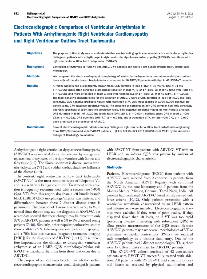
Figure 4 Characteristic Features
Twelve-lead electrocardiograms from patients with right ventricular outflow tract taopathy (ARVD/C) (D to H) showing characteristic features. (A) RVOT-VT from an anlead I (78 ms). (B) RVOT-VT originating superior to His bundle region showing precRVOT-VT from a posterior-septal location showing precordial transition at V3 and ning late precordial transition V5, wide QRS duration in lead I (124 ms), and earliesV6 and wide QRS duration in lead I (126 ms). (F) ARVD/C-VT shows very late preclate precordial transition V5, wide QRS duration in lead I (160 ms), and notching o(128 ms) and notching of the QRS (II, III, aVF, V to V ).
4 6rule of 1 covariate per 10 outcomes, although a recent papersuggests that this criterion may be too stringent (19);however, we acknowledge that our ratio is quite small.However, ARVD/C is a rare disease, and enrolling thenecessary number to comply with this rule is not feasible. Inaddition, the C-statistic, a measure of discrimination, forthe 4-variable multivariate model is 0.85 compared with0.49 to 0.64 for the univariate models, signifying an im-proved prediction of the multivariate model. Although ourfindings are statistically significant, many of the 95% con-fidence intervals are wide. However, even in the setting ofthese wide confidence intervals and even if the lowest value isused, the odds of predicting ARVD/C are still clinically useful.For example, the odds of having ARVD/C are at least 38%greater if the duration of QRS in lead I is �120 ms (lower limitf 95% confidence interval OR: 1.38, p � 0.034).
The methodology used in our report may be challengingor routine application for some clinicians, but the measure-ents can be done on any software system that has image
diting software with an appropriate selection tool. In
dia (RVOT-VT) (A to C) and arrhythmogenic right ventricular dysplasia/cardiomy--septal location showing precordial transition at V2 and narrow QRS duration intransition at V4, positive R-wave in aVL, and narrow QRS in lead I (86 ms). (C)RS duration in lead I (118 ms). (D) ARVD/C ventricular tachycardia (VT) show-
t QRS in V1 (vertical line). (E) ARVD/C-VT shows very late precordial transitiontransition V6 and wide QRS duration in lead I (150 ms). (G) ARVD/C-VT showsRS (II, III, aVF, V4 to V6). (H) ARVD/C-VT shows wide QRS duration in lead I
chycarteriorordialarrow Qt onseordialf the Q

slttAdo
838 Hoffmayer et al. JACC Vol. 58, No. 8, 2011Electrocardiographic Comparison of ARVD/C and RVOT Arrhythmias August 16, 2011:831–8
addition, some of the newer commercial electrocardiographsinclude the options to line up all lead tracings and includedigital calipers for measurements (Philips Tracemaster MD,Andover, Massachusetts). These measurements are readilyavailable with standard equipment used in the electrophys-iology laboratory. Even in the absence of these tools, othercriteria such as notching and precordial transition can bereadily appreciated.
Conclusions
Several electrocardiographic criteria can help distinguishventricular arrhythmia originating from ARVD/C fromRVOT-VT. Precordial transition at lead V6 was exclusivelyeen in ARVD/C patients. QRS duration of �120 ms inead I was sensitive for the diagnosis of ARVD/C, whereashe presence of notching in the QRS and precordialransition at lead V6 are specific for the diagnosis ofRVD/C. Multivariate logistic regression revealed that theuration of QRS in lead I of �120 ms, earliest onset QRSn lead V1, notching, and transition V5 or later all signifi-
cantly increased the odds of ARVD/C. A combination ofthese factors helps in differentiating ARVD/C fromRVOT-VT.
Reprint requests and correspondence: Dr. Melvin M. Schein-man, University of California, San Francisco, 500 ParnassusAvenue, MUE 434, San Francisco, California 94143-1354. E-mail:[email protected].
REFERENCES
1. Marcus FI, Fontaine G. Arrhythmogenic right ventricular dysplasia/cardiomyopathy: a review. Pacing Clin Electrophysiol 1995;18:1298–314.
2. Marcus FI, Fontaine GH, Guiraudon G, et al. Right ventriculardysplasia: a report of 24 adult cases. Circulation 1982;65:384–98.
3. Basso C, Corrado D, Marcus FI, Nava A, Thiene G. Arrhythmogenicright ventricular cardiomyopathy. Lancet 2009;373:1289–300.
4. Dalal D, Jain R, Tandri H, et al. Long-term efficacy of catheterablation of ventricular tachycardia in patients with arrhythmogenicright ventricular dysplasia/cardiomyopathy. J Am Coll Cardiol 2007;50:432–40.
5. Scheinman MM, Huang S. The 1998 NASPE prospective catheter
6. Joshi S, Wilber DJ. Ablation of idiopathic right ventricular outflowtract tachycardia: current perspectives. J Cardiovasc Electrophysiol2005;16 Suppl 1:S52–8.
7. Kazmierczak J, De Sutter J, Tavernier R, Cuvelier C, Dimmer C,Jordaens L. Electrocardiographic and morphometric features in pa-tients with ventricular tachycardia of right ventricular origin. Heart1998;79:388–93.
8. Marcus FI. Prevalence of T-wave inversion beyond V1 in youngnormal individuals and usefulness for the diagnosis of arrhythmogenicright ventricular cardiomyopathy/dysplasia. Am J Cardiol 2005;95:1070–1.
9. Morin DP, Mauer AC, Gear K, et al. Usefulness of precordial T-waveinversion to distinguish arrhythmogenic right ventricular cardiomyop-athy from idiopathic ventricular tachycardia arising from the rightventricular outflow tract. Am J Cardiol 2010;105:1821–4.
10. Marcus FI, Zareba W, Calkins H, et al. Arrhythmogenic rightventricular cardiomyopathy/dysplasia clinical presentation and diag-nostic evaluation: results from the North American MultidisciplinaryStudy. Heart Rhythm 2009;6:984–92.
11. Bomma C, Rutberg J, Tandri H, et al. Misdiagnosis of arrhythmo-genic right ventricular dysplasia/cardiomyopathy. J Cardiovasc Elec-trophysiol 2004;15:300–6.
12. Marcus FI, McKenna WJ, Sherrill D, et al. Diagnosis of arrhythmo-genic right ventricular cardiomyopathy/dysplasia: proposed modifica-tion of the task force criteria. Circulation 2010;121:1533–41.
13. Newman TB, Kohn MA. Evidence-Based Diagnosis. Cambridge,United Kingdom: Cambridge University Press, 2009.
14. Ainsworth CD, Skanes AC, Klein GJ, Gula LJ, Yee R, Krahn AD.Differentiating arrhythmogenic right ventricular cardiomyopathyfrom right ventricular outflow tract ventricular tachycardia usingmultilead QRS duration and axis. Heart Rhythm 2006;3:416 –23.
15. Dixit S, Gerstenfeld EP, Callans DJ, Marchlinski FE. Electrocardio-graphic patterns of superior right ventricular outflow tract tachycardias:distinguishing septal and free-wall sites of origin. J Cardiovasc Elec-trophysiol 2003;14:1–7.
16. Thiene G, Nava A, Corrado D, Rossi L, Pennelli N. Right ventricularcardiomyopathy and sudden death in young people. N Engl J Med1988;318:129–33.
17. Cox MG, van der Smagt JJ, Wilde AA, et al. New ECG criteria inarrhythmogenic right ventricular dysplasia/cardiomyopathy. Circ Ar-rhythm Electrophysiol 2009;2:524–30.
18. Yoerger DM, Marcus F, Sherrill D, et al. Echocardiographicfindings in patients meeting task force criteria for arrhythmogenicright ventricular dysplasia: new insights from the multidisciplinarystudy of right ventricular dysplasia. J Am Coll Cardiol 2005;45:860 –5.
19. Vittinghoff E, McCulloch CE. Relaxing the rule of ten events pervariable in logistic and Cox regression. Am J Epidemiol 2007;165:710–8.
Key Words: arrhythmogenic right ventricular cardiomyopathy y
ablation registry. Pacing Clin Electrophysiol 2000;23:1020–8. electrocardiography y right ventricular outflow tract.Go to http://cme.jaccjournals.orgto take the CME quiz for this article.


