Embed Size (px)
Citation preview

PRECLINICAL STUDIES
Effects of the combination of TRC105 and bevacizumabon endothelial cell biology
Yingmiao Liu & Hongyu Tian & Gerard C. Blobe &
Charles P. Theuer & Herbert I. Hurwitz &
Andrew B. Nixon
Received: 18 November 2013 /Accepted: 12 June 2014 /Published online: 5 July 2014# The Author(s) 2014. This article is published with open access at Springerlink.com
Summary Endoglin, or CD105, is a cell membrane glyco-protein that is overexpressed on proliferating endothelial cells(EC), including those found in malignancies and choroidalneovascularization. Endoglin mediates the transition fromquiescent endothelium, characterized by the relatively domi-nant state of Smad 2/3 phosphorylation, to active angiogenesisby preferentially phosphorylating Smad 1/5/8. The monoclo-nal antibody TRC105 binds endoglin with high avidity and iscurrently being tested in phase 1b and phase 2 clinical trials. Inthis report, we evaluated the effects of TRC105 on primaryhuman umbilical vascular endothelial cells (HUVEC) as asingle agent and in combination with bevacizumab. As singleagents, both TRC105 and bevacizumab efficiently blockedHUVEC tube formation, and the combination of both agentsachieved even greater levels of inhibition. We further assessedthe effects of each drug on various aspects of HUVEC func-tion. While bevacizumab was observed to inhibit HUVECviability in nutrient-limited medium, TRC105 had little effecton HUVEC viability, either alone or in combination withbevacizumab. Additionally, both drugs inhibited HUVECmigration and induced apoptosis. At the molecular level,TRC105 treatment of HUVEC lead to decreased Smad 1/5/8phosphorylation in response to BMP-9, a primary ligand forendoglin. Together, these results indicate that TRC105 acts asan effective anti-angiogenic agent alone and in combinationwith bevacizumab.
Keywords TRC105 . Endoglin . Bevacizumab .
Angiogenesis . Endothelial cells
Introduction
Anti-angiogenic therapies have emerged as prominent ap-proaches for cancer treatment over the past decade [1, 2].Tumor progression is heavily dependent on angiogenesis forprimary tumor growth and metastasis. Anti-angiogenic agentsinhibit an organism’s potential to develop new blood vesselsand prevent tumor growth by blocking access to oxygen andnutrients. Bevacizumab (Avastin™), the first approved anti-angiogenic drug, binds vascular endothelial growth factor(VEGF) and was approved by Food and DrugAdministration for the treatment of metastatic colorectal can-cer in 2004 [3]. Bevacizumab is currently approved for mul-tiple cancer indications, based on the prolongation of patientsurvival, clinically confirming the value of anti-angiogenictherapeutics which target the VEGF pathway [4].
Despite the widespread use of anti-angiogenic agents, theclinical benefit is limited and transient [5]. Such therapiesappear to benefit a subset of cancer patients; and those whorespond ultimately progress. This phenomenon is not surpris-ing given that angiogenesis is regulated through a complexinterplay of multiple pathways. When VEGF-mediated sig-naling is blocked by bevacizumab, other angiogenic pathwaysare activated, resulting in drug resistance. Therefore, combin-ing drugs that target different angiogenic pathways may be amore effective strategy. Currently, more than forty anti-angiogenic drugs are being tested in clinical trials [6].
Endoglin is a homodimeric transmembrane glycoproteinhighly expressed on proliferating endothelial cells [7, 8]. As aco-receptor for TGF-β and for bone morphogenic protein(BMP), endoglin associates with ALK1, an endothelial cell-specific type-I receptor, to promote downstream Smad 1/5/8
Y. Liu :H. Tian :G. C. Blobe :H. I. Hurwitz :A. B. Nixon (*)Department of Medicine, Duke University Medical Center,Box 2631, Durham, NC 27710, USAe-mail: [email protected]
C. P. TheuerTracon Pharmaceuticals, Inc., San Diego, CA 92121, USA
Invest New Drugs (2014) 32:851–859DOI 10.1007/s10637-014-0129-y

phosphorylation and endothelial cell proliferation, primarilyin response to BMP [9]. Recent data by Nolan-Stevaux et al.strongly supports that endoglin-dependent BMP signaling isthe critical pathway for Smad 1/5/8 activation in primaryHUVEC cells [10]. In contrast, in the absence of endoglin,another type-I receptor, ALK5, promotes downstream Smad2/3 phosphorylation that maintains a state of EC quiescence.This balance between Smad 1/5/8 and Smad 2/3 phosphory-lation regulates EC homeostasis [11]. When Smad 1/5/8 sig-naling predominates, EC undergo proliferation, migration,and promote angiogenesis; when Smad 2/3 signaling predom-inates, EC remain quiescent. Consistent with its angiogenicrole, endoglin is markedly upregulated on the endothelium ofmalignancies [8]. Dense staining of endoglin has been ob-served in the angiogenic blood vessels of more than 10 typesof tumor tissues and correlated with poor prognosis [12, 13],suggesting its potential as a target for clinical intervention[14].
TRC105 is a monoclonal antibody that binds endoglin withhigh avidity and is currently being evaluated in phase 1b andphase 2 clinical trials [15]. TRC105 exhibited promisingsafety and activity in the first-in-human, phase 1 trial [16].The phase 1, dose escalation study determined the recom-mended dose for phase 2 to be 10 mg/kg weekly, or15 mg/kg every two weeks. Both doses resulted in highcirculating TRC105 levels in patients plasma, with peak con-centrations ranging from 200 to 600 μg/ml [16].
Due to the fact that TRC105 targets an essential angiogenicpathway distinct from the VEGF pathway targeted bybevacizumab, the combination of both drugs may providegreater activity. In this study, we tested the effects ofTRC105 and bevacizumab as single agents, as well as incombination, on EC tube formation, viability, migration, andapoptosis. Further, we assessed the effects of TRC105 onpatterns of Smad phosphorylation in HUVEC cells.
Materials and methods
Cell culture
Low passage HUVEC cells were purchased from Clonetics/Lonza (Walkersville, MD). HUVEC were cultured in eitherregular medium containing EBM-2 basic medium supple-mented with EGM-2 MV single aliquots; or nutrient-limited medium containing EBM-2 basic medium sup-plemented with 0.5 % FBS and 30 ng/ml VEGF (Lonza,Walkersville, MD). All cells were maintained in a37 °C, 5 % CO2 incubator. TRC105 (5 mg/ml) wasprovided by Tracon Pharmaceuticals, Inc. (San Diego,CA). Bevacizumab (25 mg/ml) was from Genentech Inc.(San Francisco, CA).
HUVEC tube formation
HUVECwere pre-treated with 100μg/ml TRC105, 100 ng/mlbevacizumab, or both drugs for 8 h in regular medium. HumanIgG (Jackson Immuno Research, West Grove, PA) was usedas an isotype control. The cells were harvested and maintainedin drug containing medium, and 1.5×10⁴ HUVEC were inoc-ulated onto pre-polymerized ECMatrix gel (In vitro angiogen-esis assay kit, Chemicon, Temecula, CA). After 16 h incuba-tion, cells were visualized using the Axiobserver in the DukeLight Microscopy Core facility (LMCF). Closed polygonswere counted, and total tube length measured with theMetaMorph software (MDS Analytical Technologies,Sunnyvale, CA).
HUVEC viability (MTS assay)
HUVEC were inoculated at 5,000 cell/well onto a 96-wellplate. After overnight incubation, cells were treated with eitherregular or limited medium containing either TRC105,bevacizumab, the combination of both drugs, or IgG controlfor 72 h with daily medium change. At the termination of theassay, 20 μl MTS tetrazolium compound (CellTiter 96Aqueous One Solution Cell Proliferation Assay, Promega,Madison, WI) was added to each well, absorbance at490 nm was recorded 4 h later following the manufacturer’sprotocol.
HUVEC migration
HUVEC (8×10⁵/well) were inoculated onto a 6-well plate.After a confluent monolayer had formed overnight, a scratchwas introduced with a sterile 200 μl tip. Cell debris wasremoved by washing with PBS, fresh regular medium con-taining either IgG isotype control (100 μg/ml), TRC105(100 μg/ml), bevacizumab (100 ng/ml), or the combinationof TRC105 (100 μg/ml) and bevacizumab (100 ng/ml) wassupplied. Scratch filling was monitored using a live cell sta-tion in Duke LMCF over a period of 16 h. Percentage ofscratch filling was calculated as (Distance between gap edgesat time point 0 - distance at time point X)/distance at timepoint 0 *100 %, using MetaMorph software.
HUVEC apoptosis
HUVEC (1×10⁵/well) were inoculated onto a 6-well plate inwhich a gelatin-coated glass slide had been placed at thebottom. HUVEC were maintained in regular or limited medi-um with TRC105, bevacizumab, or the combination of bothfor 72 h. Fresh medium with drugs were applied daily. Cellswere then fixed in methanol: acetic acid (3:1) for 5 min at4 °C, washed three times with PBS, and stained with Hoechst33,342 (5 μg/ml, Calbiochem, La Jolla, CA) for 10 min at
852 Invest New Drugs (2014) 32:851–859

room temperature. Then cells were washed three more times,the slides removed from plate wells, and mounted onto a glasscarrier with Vectashield mounting medium for fluorescence(Vector Laboratories, Inc., Burlingame, CA). Apoptotic nucleiwere visualized under a fluorescence microscope in DukeLMCF. Ten representative fields were imaged, cells counted,and the ratio of apoptotic nuclei vs. total nuclei was calculatedwith MetaMorph software.
Smad signaling in HUVEC
HUVEC (5×10⁵/well) were inoculated onto a 6-well plate andincubated overnight. Cells were serum starved in EBM-2,0.1 % BSA, and 10 mM Hepes for 4 h. Cells were pretreatedwith TRC105 or isotype control for 1 h, and stimulated with0.2 ng/ml BMP-9, or 0.25 ng/ml TGF-β1 (R&D systems,Minneapolis, MN) for 1 h. Then cells were put on ice imme-diately, cell lysate harvested in lysis buffer: 20 mM Hepes,2 mM MgCl2, 1 mM EDTA and EGTA, 150 mM NaCl, 1 %Triton X-100, 0.1 % SDS, and protease inhibitors. Aftercentrifugation at 16,000×g for 10 min, cell debris and nucleiwere removed, and cell lysates were snap frozen before storedat −80 °C freezer.
Western immunoblots
Cell lysates (10 μg) were separated on a 4–20 % SDS-PAGEgel. Blots were incubated with rabbit-anti-phos-Smad1/5/8,anti-Smad 1, anti-Phos-Smad2, or anti-Smad 2/3 (CellSignaling, Danvers, MA), as well as mouse anti-β-actin forloading control, overnight at 4 °C. LI-COR specific goat anti-rabbit, or goat anti-mouse IgG secondary antibody (1:5,000)was added and incubated for 1 h at room temperature.Immunoblots were analyzed using the Odyssey imaging sys-tem (LI-COR Biotechnology, Lincoln, NE).
Results
TRC105 and bevacizumab inhibit HUVEC tube formation
To evaluate the anti-angiogenic function of TRC105, HUVECtube formation was first tested. Since TRC105 is a chimericantibody cloned into an IgG1 backbone, human IgG moleculewas used as an isotype control. As shown in Fig. 1a, over-night incubation of HUVEC with IgG control (100 μg/ml) ona matrigel leads to the development of extensive tubularnetworks, which can be quantified by counting the numberof closed polygons and total length of capillary tubes.TRC105 (100 μg/ml) and bevacizumab (100 ng/ml) treatedcells showed impaired tube formation, exhibited by less densetube network and some disrupted regions. Strikingly, when
cells were treated with both agents, only partially formedtubes and fewer closed polygons could be detected. After datanormalization, polygon formation was inhibited by 40–50 %using either drug alone and by more than 80 % using thecombination of both drugs. The combination of drugs issignificantly more effective in inhibiting polygon formationthan either drug used alone (p≤0.05; Fig. 1b). The total lengthof tubes, an earlier biologic step reflecting mainly cell migra-tion and alignment, was less affected, exhibiting 5–10 %
a
b
IgG (100µµµµg/ml) TRC (100µµµµg/ml)
BEV (100ng/ml) TRC + BEV
* *
*
0
0.2
0.4
0.6
0.8
1
1.2
Control TRC BEV TRC+ BEV
No
rmal
ized
Tu
be
Fo
rmat
ion
Number of polygons
Length of tubes
Fig. 1 TRC105 and bevacizumab inhibit HUVEC tubular network for-mation in in vitro angiogenesis assays. a. HUVEC cells growing onmatrigel overnight in the presence of IgG control (100 μg/ml) formedextensive tube networks. TRC105 (100 μg/ml), bevacizumab(100 ng/ml), or the combination of both drugs was added and the effectson polygon formation and tube length were visualized. b. Normalized bargraph showing the fold inhibition on polygon formation and tube length.Each column represents the mean±SD of three independent experiments.*p<0.05 compared to IgG control
Invest New Drugs (2014) 32:851–859 853

inhibition from each drug alone, and more than 30 % inhibi-tion using the combination of drugs. Overall, these resultsindicate that both TRC105 and bevacizumab inhibit EC tubeformation, and that the combination of drugs achieved morerobust inhibition than either drug alone.
Effect of TRC105 and bevacizumab on HUVEC viability
Since EC tube formation is a comprehensive assay reflectingmultiple cellular processes such as proliferation, migration/alignment, and apoptosis, we further examined each function-al aspect individually. To evaluate cell viability, HUVECweretreated with increasing concentrations of drug or isotype con-trol for 72 h, followed by the addition of the MTS tetrazoliumcompound, which undergoes a color change when bioreducedby metabolically active cells. While little to no effect of IgGwas observed at the ng/ml range (data not shown), IgG at theμg/ml range inhibited HUVEC viability. At this dose range,TRC105 (μg/ml) elicited comparable inhibition to IgG (μg/ml) alone, suggesting the inhibition observed in response toTRC105 was an IgG non-specific effect. In regular medium,bevacizumab showed no appreciable inhibition on HUVECviability when tested at concentrations of 100 ng/ml to1 μg/ml (Fig. 2a). Higher concentrations of bevacizumab(100 μg/ml to 1 mg/ml) were also tested, but again, the effectswere negligible (data not shown). When both drugs werecombined, the inhibitory effect on HUVEC viability wasessentially the same as IgG alone.
The lack of a bevacizumab effect on HUVEC viability inserum-containing medium was not surprising. The high abun-dance of growth factors present in the serum-containing me-dium, including FGF, EGF, VEGF, and IGF, all can serve aspotential compensatory factors upon VEGF blockage. To
isolate the effect of TRC105 and bevacizumab on VEGF-specific cell viability, we optimized a nutrient-limited mediumthat excluded all other growth factors (i.e., FGF, EGF, andIGF) except VEGF. Limited medium was formulated contain-ing 0.5 % FBS and 30 ng/ml VEGF. In this setting,bevacizumab produced a significant drug-specific dose-dependent inhibition on HUVEC viability (p<0.01 vs. IgGcontrol, Fig. 2b), while TRC105-mediated inhibition at μg/mlconcentrations was less robust and non-specific. A closercomparison among bevacizumab alone, bevacizumab+IgG,bevacizumab+TRC105 treated cells revealed little enhance-ment when TRC105 was added to bevacizumab. Therefore,the combination of both drugs did not lead to greater inhibi-tion of HUVEC viability as compared to bevacizumab alone.
TRC105 and bevacizumab inhibit HUVEC migration
The in vitro scratch assay was used to measure cell motility[17]. HUVEC cells were allowed to grow to confluence, thena gap was introduced into the cell monolayer and the processof cell migration was monitored over a period of 16 h. Inregular medium, untreated cells refilled the gap within 10–11 h. As shown in Fig. 3, incubation of HUVEC in thepresence of 100 μg/ml TRC105 significantly delayed endo-thelial cell migration (p<0.05). Treatment with 100 ng/mlbevacizumab achieved similar effects (p<0.01). Treatmentwith both drugs inhibited migration more than observed witheither TRC105 or bevacizumab alone (p≤0.01) (Fig. 3). IgGisotype control at 100μg/ml showed no inhibition onHUVECmigration (data not shown). When scratch filling was assessedin limited medium, HUVEC cells did not migrate well andnever reached confluence in the absence of drugs, even after24 h post scratch.
Drug Concentration
No
rmal
ized
Ab
sorb
ance
at
490
nm
a b
0.7
0.8
0.9
1.0
1.1
0 200 400 600 800 1000
no drug
BEV-ng/ml
0.4
0.5
0.6
0.7
0.8
0.9
1.0
1.1
0 200 400 600 800 1000
** **
**
**
**
Fig. 2 Effect of TRC105 andbevacizumab on HUVECviability. Subconfluent HUVECwere subjected to TRC105 (100–1,000 μg/ml), bevacizumab(100–1,000 ng/ml), both drugs, orIgG (100–1,000 μg/ml) treatmentfor 3 days in regular medium (a)and limited medium (b). Cellviability was measured by MTStetrazolium assay, and absorbanceat 490 nm was reported. Datarepresent the mean±SD of threeindependent experiments. **p<0.01 compared to IgG control
854 Invest New Drugs (2014) 32:851–859

TRC105 and bevacizumab induce HUVEC apoptosis
To test the apoptotic effects of TRC105 and bevacizumab,HUVEC were first treated with each drug individually inregular or limited medium for 72 h, and then subjected toHoechst staining (Fig. 4). Condensed, fragmented nucleirepresenting apoptotic features were counted, and the ratioof apoptotic nuclei vs. total nuclei was plotted. Camptothecin(4 μg/ml) was used as a positive control and induced morethan a 30-fold increase in the apoptotic cell ratio after over-night incubation (data not shown). IgG isotype control wasindistinguishable from the no drug control (data not shown)[18]. In regular medium, TRC105 exhibited a small, yetsignificant, dose-dependent induction of apoptosis, witha 3-fold increase at 100 μg/ml, and a 6-fold increase at1,000 μg/ml, versus no-drug control (Fig. 4a).Bevacizumab induced apoptosis to similar levels as seenwith TRC105 (p<0.01 vs. control). Additive effectswere not observed when the drugs were combined, asthe induction of apoptosis was similar compared toeither single agent.
In limited medium, the basal apoptosis rate was higher thanthat in regular medium (5 % vs. 1.2 %). TRC105 elicited a 2-fold induction of apoptosis at 1,000 μg/ml. Bevacizumab wasa more potent inducer of apoptosis under these conditions,achieving a 3.8 fold increase at 1,000 ng/ml (p<0.001 vs.control). As was seen with regular medium, the combinationof drugs in limited medium did not further increase apoptosis(Fig. 4b).
TRC105 differentially modulates Smad 1/5/8 and Smad 2/3signaling pathways in response to BMP-9 and TGF-β1
Multiple factors have been identified as endoglin ligands,including TGF-β1 and TGF-β3 [7], as well as BMP-9 and
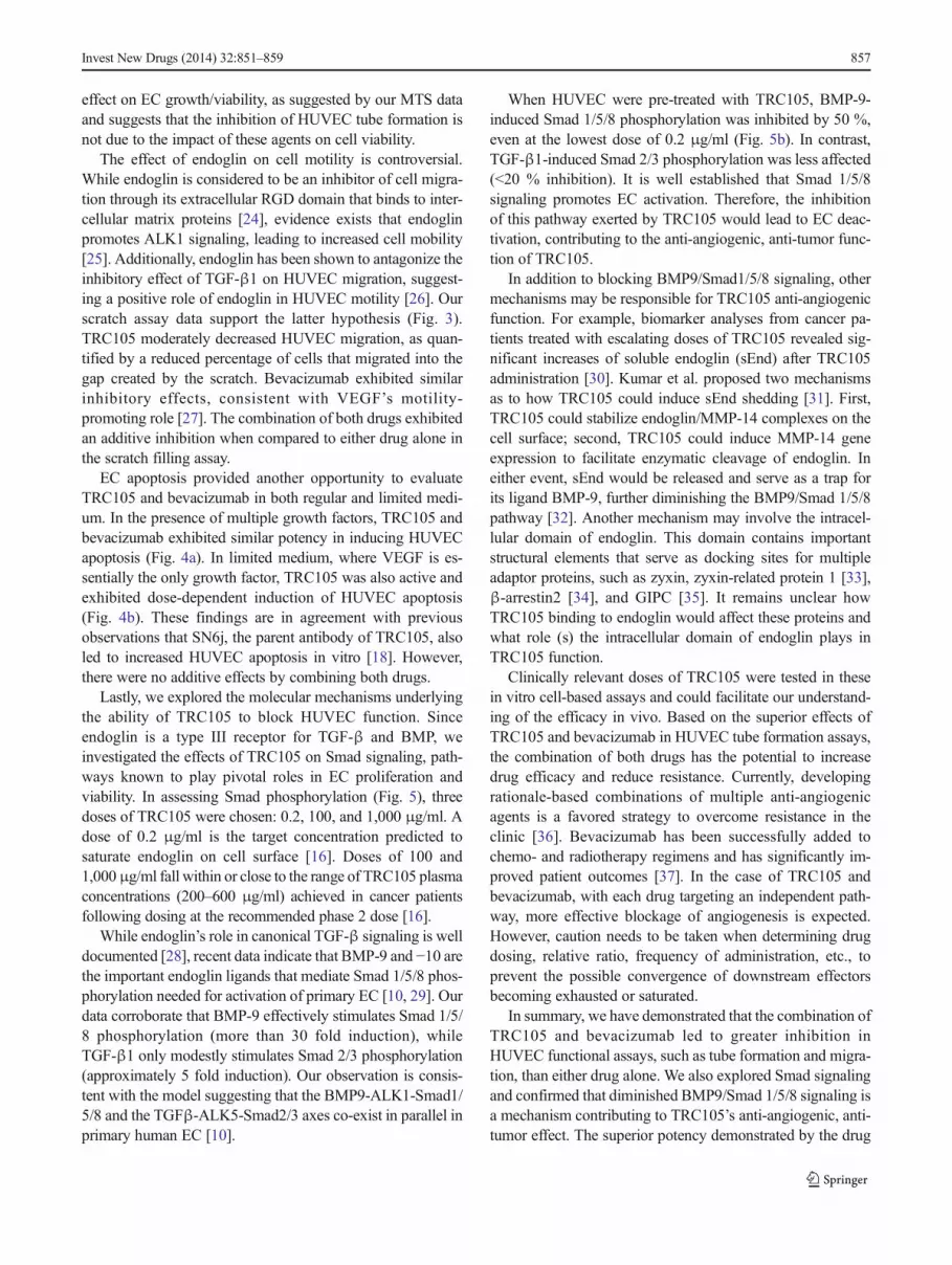
BMP-10 [19]. To investigate these signaling modalities,serum-starved HUVEC cells were exposed to either BMP-9or TGF-β1. As shown in Fig. 5, BMP-9 strongly acti-vated Smad 1/5/8 phosphorylation (~30 fold), but hadlittle effect on Smad 2. In contrast, TGF-β1 exposuremainly activated Smad 2 phosphorylation (~5 fold), butnot Smad 1/5/8 signaling.
To explore the underlying molecular mechanisms contrib-uting to the effects of TRC105 on HUVEC function, thephosphorylation status of Smad 1/5/8 and Smad 2 was inves-tigated. When HUVEC were pre-incubated with increasingamounts of TRC105, BMP-9-induced Smad 1/5/8 phosphor-ylation was inhibited across all doses of TRC105 tested(Fig. 5, panel b), including a dose of 0.2 μg/mL, which isthe TRC105 concentration expected, based on binding aviditystudies, to saturate endoglin binding sites on HUVECs. Incontrast, TRC105 treatment only marginally modulatedTGF-β1 induced Smad 2 phosphorylation (panel C). Bothisotype control IgG and bevacizumab exhibited negligible
*
* **
**
50
60
70
80
90
100
110
Control TRC BEV TRC+ BEV
% S
crat
ch f
illin
g
Fig. 3 TRC105 and bevacizumab inhibit HUVEC migration. HUVECconfluent monolayers were scratched to create a gap, exposed to TRC105(100 μg/ml), bevacizumab (100 ng/ml), or the combination of TRC105(100 μg/ml) and bevacizumab (100 ng/ml), and monitored for cell mi-gration for 16 h. Percentage of scratch filling at 10 h post scratch wasshown. Each column represents the mean±SD of three independentexperiments. * p<0.05; ** p<0.01 compared to no drug control
a
b
0
4
8
12
control
TTRC-1
000
g/m
l
BEV-1
00ng/m
l
BEV-1
000ng/m
l
TRC+BEV
% o
f Ap
op
toti
c N
ucl
ei
**
**
** **
**
0
10
20
30
Control
TTRC-1
000
g/m
l
BEV-1
00ng/m
l
BEV-1
000ng/m
l
TRC+BEV
% o
f Ap
op
toti
c N
ucl
ei
***
***
***
***
***
Fig. 4 TRC105 and bevacizumab induce HUVEC apoptosis. Apoptosiswas evaluated by Hoechst staining after HUVEC were treated withTRC105 (100 or 1,000 μg/ml), bevacizumab (100 or 1,000 ng/ml), orthe combination of drugs at the lower doses (100 μg/ml TRC105 and100 ng/ml bevacizumab) for 3 days. a. Regular medium. b. Limitedmedium. Each column represents the mean±SD of three independentexperiments. ** p<0.01, *** p<0.001 compared to no drug control
Invest New Drugs (2014) 32:851–859 855

inhibition of either Smad 1/5/8 or Smad 2 phosphorylation(data not shown).
Discussion
In this study, we evaluated the anti-angiogenic drug TRC105,and its combination with bevacizumab, in a series of HUVECfunctional assays. Initially, individual drugs were tested over abroad range of dosing levels under each assay condition. Inevery assay system, doses that induced moderate effects (typ-ically around 25–30 % inhibition) were chosen for theseanalyses in order to detect potential additive or synergisticeffects with both drugs. For most assays, the dose of TRC105was empirically determined to be 100 μg/ml, as lower dosesexhibited little effect on HUVEC function. This dose is clin-ically relevant as pharmacokinetic analyses revealed that theserum concentration of TRC105 following dosing in ad-vanced cancer patients at the recommended phase 2 doseranged from 200 to 600 μg/ml [16]. For bevacizumab, thedose tested in most assays was empirically determined to be100 ng/ml.
Despite the fact that TRC105 and bevacizumab are bothmonoclonal antibodies that block angiogenesis, the targets ofeach drug are different and utilize distinct mechanisms.Endoglin is an integral cell membrane receptor located onproliferating endothelial cells [20], whereas VEGF is a solubleangiogenic factor that is primarily released by tumor cells andtumor-associated stromal cells [21, 22]. Therefore, combiningTRC105 and bevacizumab has the potential to block comple-mentary pathways leading to improved efficacy.
We observed that individually, both TRC105 andbevacizumab blocked HUVEC tube formation, and the mostrobust inhibition was achieved when both drugs were com-bined (Fig. 1). The tube formation assay is a powerful tool tomonitor ECs during vascular network formation, representingan end-point evaluation of the complicated interplay amongmany processes, including proliferation, differentiation, mi-gration, apoptosis, etc. To further interrogate these processes,we next investigated specific EC functional biology in moredefined assay systems.
When given at 100–1,000 μg/ml levels, TRC105 and itsisotype control (IgG) similarly inhibited HUVEC viability, inboth regular and nutrient-limited medium (Fig. 2). In contrast,although bevacizumab elicited little effect on HUVEC viabil-ity in regular medium, it showed dose-dependent, drug-specific inhibition in limited medium (Fig. 2b). WhileTRC105 had little effect on HUVEC viability, several obser-vations are noteworthy. First, contrary to bevacizumab,TRC105 elicited no inhibitory effect on HUVEC growth innutrient-limited medium. Second, given that the steady stateplasma concentration of TRC105 in patients is inμg/ml range,the potential non-specific effect of IgG on EC growth shouldbe considered. Third, Anderberg et al., reported that geneticknockdown of endoglin sensitizes tumors to VEGF inhibition[23], advocating for the potential benefit of co-administrationof these drugs. The underlying mechanism is unlikely to be an
a
TRC - - 0.2 100 1000 - 0.2 100 1000
BMP-9 (0.2 ng/ml) TGF- 1 (0.25 ng/ml)
Phos- Smad1/5/8
Smad1
Phos- Smad2
Smad2/3
-actin
b
BMP-9 - + + + + - - - -
TGF- 1 - - - - - + + + +
TRC105 - - 0.2 100 1000 - 0.2 100 1000
0
10
20
30
(Ph
os-
Sm
ad 1
/5/8
) / S
mad
1
c
BMP-9 - + + + + - - - -
TGF- 1 - - - - - + + + +
TRC105 - - 0.2 100 1000 - 0.2 100 1000
0
2
4
6
8
Ph
os-
Sm
ad2/
(S
mad
2/3
)
Fig. 5 TRC105 diminished BMP-9 induced Smad 1/5/8 signaling inHUVEC cells. a. Western blotting showing the levels of phosphorylatedSmad 1/5/8, total Smad 1, phos-Smad 2, total Smad 2/3 in response to thestimulation of BMP-9 or TGF-β1. Cells were pre-incubated with variousdoses of TRC105 for 1 h prior to stimulation. b. Effect on Smad 1/5/8signaling as revealed by the ratio of phos-Smad 1/5/8 vs. total Smad 1. c.Effect on Smad 2 signaling as revealed by the ratio of phos-Smad 2 vs.total Smad 2/3. 0.2, 100, 1,000 depict TRC105 doses of 0.2, 100, and1,000 μg/ml, respectively
856 Invest New Drugs (2014) 32:851–859
effect on EC growth/viability, as suggested by our MTS dataand suggests that the inhibition of HUVEC tube formation isnot due to the impact of these agents on cell viability.
The effect of endoglin on cell motility is controversial.While endoglin is considered to be an inhibitor of cell migra-tion through its extracellular RGD domain that binds to inter-cellular matrix proteins [24], evidence exists that endoglinpromotes ALK1 signaling, leading to increased cell mobility[25]. Additionally, endoglin has been shown to antagonize theinhibitory effect of TGF-β1 on HUVEC migration, suggest-ing a positive role of endoglin in HUVEC motility [26]. Ourscratch assay data support the latter hypothesis (Fig. 3).TRC105 moderately decreased HUVEC migration, as quan-tified by a reduced percentage of cells that migrated into thegap created by the scratch. Bevacizumab exhibited similarinhibitory effects, consistent with VEGF’s motility-promoting role [27]. The combination of both drugs exhibitedan additive inhibition when compared to either drug alone inthe scratch filling assay.
EC apoptosis provided another opportunity to evaluateTRC105 and bevacizumab in both regular and limited medi-um. In the presence of multiple growth factors, TRC105 andbevacizumab exhibited similar potency in inducing HUVECapoptosis (Fig. 4a). In limited medium, where VEGF is es-sentially the only growth factor, TRC105 was also active andexhibited dose-dependent induction of HUVEC apoptosis(Fig. 4b). These findings are in agreement with previousobservations that SN6j, the parent antibody of TRC105, alsoled to increased HUVEC apoptosis in vitro [18]. However,there were no additive effects by combining both drugs.
Lastly, we explored the molecular mechanisms underlyingthe ability of TRC105 to block HUVEC function. Sinceendoglin is a type III receptor for TGF-β and BMP, weinvestigated the effects of TRC105 on Smad signaling, path-ways known to play pivotal roles in EC proliferation andviability. In assessing Smad phosphorylation (Fig. 5), threedoses of TRC105 were chosen: 0.2, 100, and 1,000 μg/ml. Adose of 0.2 μg/ml is the target concentration predicted tosaturate endoglin on cell surface [16]. Doses of 100 and1,000μg/ml fall within or close to the range of TRC105 plasmaconcentrations (200–600 μg/ml) achieved in cancer patientsfollowing dosing at the recommended phase 2 dose [16].
While endoglin’s role in canonical TGF-β signaling is welldocumented [28], recent data indicate that BMP-9 and −10 arethe important endoglin ligands that mediate Smad 1/5/8 phos-phorylation needed for activation of primary EC [10, 29]. Ourdata corroborate that BMP-9 effectively stimulates Smad 1/5/8 phosphorylation (more than 30 fold induction), whileTGF-β1 only modestly stimulates Smad 2/3 phosphorylation(approximately 5 fold induction). Our observation is consis-tent with the model suggesting that the BMP9-ALK1-Smad1/5/8 and the TGFβ-ALK5-Smad2/3 axes co-exist in parallel inprimary human EC [10].
When HUVEC were pre-treated with TRC105, BMP-9-induced Smad 1/5/8 phosphorylation was inhibited by 50 %,even at the lowest dose of 0.2 μg/ml (Fig. 5b). In contrast,TGF-β1-induced Smad 2/3 phosphorylation was less affected(<20 % inhibition). It is well established that Smad 1/5/8signaling promotes EC activation. Therefore, the inhibitionof this pathway exerted by TRC105 would lead to EC deac-tivation, contributing to the anti-angiogenic, anti-tumor func-tion of TRC105.
In addition to blocking BMP9/Smad1/5/8 signaling, othermechanisms may be responsible for TRC105 anti-angiogenicfunction. For example, biomarker analyses from cancer pa-tients treated with escalating doses of TRC105 revealed sig-nificant increases of soluble endoglin (sEnd) after TRC105administration [30]. Kumar et al. proposed two mechanismsas to how TRC105 could induce sEnd shedding [31]. First,TRC105 could stabilize endoglin/MMP-14 complexes on thecell surface; second, TRC105 could induce MMP-14 geneexpression to facilitate enzymatic cleavage of endoglin. Ineither event, sEnd would be released and serve as a trap forits ligand BMP-9, further diminishing the BMP9/Smad 1/5/8pathway [32]. Another mechanism may involve the intracel-lular domain of endoglin. This domain contains importantstructural elements that serve as docking sites for multipleadaptor proteins, such as zyxin, zyxin-related protein 1 [33],β-arrestin2 [34], and GIPC [35]. It remains unclear howTRC105 binding to endoglin would affect these proteins andwhat role (s) the intracellular domain of endoglin plays inTRC105 function.
Clinically relevant doses of TRC105 were tested in thesein vitro cell-based assays and could facilitate our understand-ing of the efficacy in vivo. Based on the superior effects ofTRC105 and bevacizumab in HUVEC tube formation assays,the combination of both drugs has the potential to increasedrug efficacy and reduce resistance. Currently, developingrationale-based combinations of multiple anti-angiogenicagents is a favored strategy to overcome resistance in theclinic [36]. Bevacizumab has been successfully added tochemo- and radiotherapy regimens and has significantly im-proved patient outcomes [37]. In the case of TRC105 andbevacizumab, with each drug targeting an independent path-way, more effective blockage of angiogenesis is expected.However, caution needs to be taken when determining drugdosing, relative ratio, frequency of administration, etc., toprevent the possible convergence of downstream effectorsbecoming exhausted or saturated.
In summary, we have demonstrated that the combination ofTRC105 and bevacizumab led to greater inhibition inHUVEC functional assays, such as tube formation and migra-tion, than either drug alone. We also explored Smad signalingand confirmed that diminished BMP9/Smad 1/5/8 signaling isa mechanism contributing to TRC105’s anti-angiogenic, anti-tumor effect. The superior potency demonstrated by the drug
Invest New Drugs (2014) 32:851–859 857

combination advocates their co-administration in vivo as atherapeutic strategy.
Acknowledgments We would like to thank the staff from Light Mi-croscopy Core Facility in Duke University Medical Center. This workwas supported by a grant from Tracon Pharmaceuticals, Inc.
Conflict of interest Y Liu and H Tian have no conflicts to disclose. GCBlobe is a consultant/advisory board member and has received honorar-ium from Genentech and Roche. CP Theuer is the president and CEO ofTracon Pharmaceuticals, Inc. HI Hurwitz has received research fundingfrom F Hoffman-La Roche, Amgen, Pfizer, Tracon Pharmaceuticals,Genentech, Sanofi, Morphotek and GSK. HI Hurwitz is a consultant/advisory board member for GlaxoSmithKline, Novartis, Genentech,Roche, Sanofi, Regeneron, BMS, Bayer, and Tracon. AB Nixon hasreceived research funding from Tracon Pharmaceuticals, F Hoffman-LaRoche, Amgen, Pfizer and is a consultant/advisory board member forGlaxoSmithKline and Novartis.
Open Access This article is distributed under the terms of the CreativeCommons Attribution License which permits any use, distribution, andreproduction in any medium, provided the original author(s) and thesource are credited.
References
1. Carmeliet P, Jain RK (2000) Angiogenesis in cancer and otherdiseases. Nature 407(6801):249–257
2. Folkman J (2007) Angiogenesis: an organizing principle for drugdiscovery? Nat Rev 6(4):273–286
3. Hurwitz H, Fehrenbacher L, NovotnyW, Cartwright T, Hainsworth J,HeimW, Berlin J, Baron A, Griffing S, Holmgren E, Ferrara N, FyfeG, Rogers B, Ross R, Kabbinavar F (2004) Bevacizumab plusirinotecan, fluorouracil, and leucovorin for metastatic colorec-tal cancer. N Engl J Med 350(23):2335–2342
4. Van Meter ME, Kim ES (2010) Bevacizumab: current updates intreatment. Curr Opin Oncol 22(6):586–591. doi:10.1097/CCO.0b013e32833edc0c
5. Jayson GC, Hicklin DJ, Ellis LM (2012) Antiangiogenic therapy-evolving view based on clinical trial results. Nat Rev Clin Oncol 9(5):297–303. doi:10.1038/nrclinonc.2012.8
6. Samant RS, Shevde LA (2011) Recent advances in anti-angiogenictherapy of cancer. Oncotarget 2(3):122–134
7. Cheifetz S, Bellon T, Cales C, Vera S, Bernabeu C, Massague J,Letarte M (1992) Endoglin is a component of the transforminggrowth factor-beta receptor system in human endothelial cells. JBiol Chem 267(27):19027–19030
8. Burrows FJ, Derbyshire EJ, Tazzari PL, Amlot P, Gazdar AF, KingSW, Letarte M, Vitetta ES, Thorpe PE (1995) Up-regulation ofendoglin on vascular endothelial cells in human solid tumors:implications for diagnosis and therapy. Clin Cancer Res 1(12):1623–1634
9. Lebrin F, Goumans MJ, Jonker L, Carvalho RL, Valdimarsdottir G,Thorikay M, Mummery C, Arthur HM, ten Dijke P (2004) Endoglinpromotes endothelial cell proliferation and TGF-beta/ALK1 signaltransduction. EMBO J 23(20):4018–4028
10. Nolan-Stevaux O, Zhong W, Culp S, Shaffer K, Hoover J,Wickramasinghe D, Ruefli-Brasse A (2012) Endoglin requirementfor BMP9 signaling in endothelial cells reveals new mechanism ofaction for selective anti-endoglin antibodies. PLoS ONE 7(12):e50920. doi:10.1371/journal.pone.0050920
11. Goumans MJ, Valdimarsdottir G, Itoh S, Rosendahl A, SiderasP, ten Dijke P (2002) Balancing the activation state of theendothelium via two distinct TGF-beta type I receptors.EMBO J 21(7):1743–1753
12. Dallas NA, Samuel S, Xia L, Fan F, Gray MJ, Lim SJ, Ellis LM(2008) Endoglin (CD105): a marker of tumor vasculature and poten-tial target for therapy. Clin Cancer Res 14(7):1931–1937
13. SeonBK, Haruta Y,Matsuno F, Haba A, Takahashi N, She X, HaradaN, Uneda S, Tsujie M, Tsujie T, Toi H, Tsai H (2010) Receptor-targeted anticancer therapy. Immunol Res 46(1–3):189–191. doi:10.1007/s12026-009-8131-8
14. Fonsatti E, Nicolay HJ, Altomonte M, Covre A, Maio M (2010)Targeting cancer vasculature via endoglin/CD105: a novel antibody-based diagnostic and therapeutic strategy in solid tumours.Cardiovasc Res 86(1):12–19. doi:10.1093/cvr/cvp332
15. SeonBK, Haba A,Matsuno F, Takahashi N, TsujieM, She X, HaradaN, Uneda S, Tsujie T, Toi H, Tsai H, Haruta Y (2011) Endoglin-targeted cancer therapy. Curr Drug Deliv 8(1):135–143
16. Rosen LS, Hurwitz HI, Wong MK, Goldman J, Mendelson DS, FiggWD, Spencer S, Adams BJ, Alvarez D, Seon BK, Theuer CP, LeighBR, Gordon MS (2012) A phase I first-in-human study of TRC105(Anti-Endoglin Antibody) in patients with advanced cancer. ClinCancer Res Off J Am Assoc Cancer Res 18(17):4820–4829. doi:10.1158/1078-0432.CCR-12-0098
17. Liang CC, Park AY, Guan JL (2007) In vitro scratch assay: a conve-nient and inexpensive method for analysis of cell migration in vitro.Nat Protoc 2(2):329–333
18. Tsujie M, Tsujie T, Toi H, Uneda S, Shiozaki K, Tsai H, Seon BK(2008) Anti-tumor activity of an anti-endoglin monoclonal antibodyis enhanced in immunocompetent mice. Int J Cancer 122(10):2266–2273. doi:10.1002/ijc.23314
19. Scharpfenecker M, van Dinther M, Liu Z, van Bezooijen RL, ZhaoQ, Pukac L, Lowik CW, ten Dijke P (2007) BMP-9 signals via ALK1and inhibits bFGF-induced endothelial cell proliferation and VEGF-stimulated angiogenesis. J Cell Sci 120(Pt 6):964–972. doi:10.1242/jcs.002949
20. Haruta Y, Seon BK (1986) Distinct human leukemia-associated cellsurface glycoprotein GP160 defined by monoclonal antibody SN6.Proc Natl Acad Sci U S A 83(20):7898–7902
21. Senger DR, Van de Water L, Brown LF, Nagy JA, Yeo KT, Yeo TK,Berse B, Jackman RW, Dvorak AM, Dvorak HF (1993) Vascularpermeability factor (VPF, VEGF) in tumor biology. CancerMetastasis Rev 12(3–4):303–324
22. Ferrara N, Gerber HP, LeCouter J (2003) The biology of VEGF andits receptors. Nat Med 9(6):669–676. doi:10.1038/nm0603-669
23. Anderberg C, Cunha SI, Zhai Z, Cortez E, Pardali E, Johnson JR,Franco M, Paez-Ribes M, Cordiner R, Fuxe J, Johansson BR,Goumans MJ, Casanovas O, ten Dijke P, Arthur HM, Pietras K(2013) Deficiency for endoglin in tumor vasculature weakens theendothelial barrier to metastatic dissemination. J Exp Med 210(3):563–579. doi:10.1084/jem.20120662
24. Conley BA, Koleva R, Smith JD, Kacer D, Zhang D, Bernabeu C,Vary CP (2004) Endoglin controls cell migration and composition offocal adhesions: function of the cytosolic domain. J Biol Chem279(26):27440–27449
25. Li C, Hampson IN, Hampson L, Kumar P, Bernabeu C, Kumar S(2000) CD105 antagonizes the inhibitory signaling of transforminggrowth factor beta1 on human vascular endothelial cells. FASEB J14(1):55–64
26. Goumans MJ, Valdimarsdottir G, Itoh S, Lebrin F, Larsson J,Mummery C, Karlsson S, ten Dijke P (2003) Activin receptor-likekinase (ALK) 1 is an antagonistic mediator of lateral TGFbeta/ALK5signaling. Mol Cell 12(4):817–828
27. Urbich C, Aicher A, Heeschen C, Dernbach E, HofmannWK, ZeiherAM, Dimmeler S (2005) Soluble factors released by endothelialprogenitor cells promote migration of endothelial cells and cardiac
858 Invest New Drugs (2014) 32:851–859

resident progenitor cells. J Mol Cell Cardiol 39(5):733–742. doi:10.1016/j.yjmcc.2005.07.003
28. Blanco FJ, Santibanez JF, Guerrero-Esteo M, Langa C, Vary CP,Bernabeu C (2005) Interaction and functional interplay betweenendoglin and ALK-1, two components of the endothelialtransforming growth factor-beta receptor complex. J Cell Physiol204(2):574–584. doi:10.1002/jcp.20311
29. Alt A, Miguel-Romero L, Donderis J, Aristorena M, Blanco FJ,Round A, Rubio V, Bernabeu C, Marina A (2012) Structural andfunctional insights into endoglin ligand recognition and binding.PLoS ONE 7(2):e29948. doi:10.1371/journal.pone.0029948
30. Liu Y, Starr MD, Brady JC, Dellinger A, Pang H, Adams B, Theuer CP,LeeNY,HurwitzHI,NixonAB (2014)Modulation of circulating proteinbiomarkers following TRC105 (anti-endoglin antibody) treatment inpatients with advanced cancer. Cancer Med. doi:10.1002/cam4.207
31. Kumar S, Pan CC, Bloodworth JC, Nixon A, Theuer C, Hoyt DG,Lee NY (2013) Antibody-directed coupling of endoglin and MMP-14 is a key mechanism for endoglin shedding and deregulation ofTGF-beta signaling. Oncogene. doi:10.1038/onc.2013.386
32. Castonguay R, Werner ED, Matthews RG, Presman E, Mulivor AW,Solban N, Sako D, Pearsall RS, Underwood KW, Seehra J, Kumar R,Grinberg AV (2011) Soluble endoglin specifically binds bone
morphogenetic proteins 9 and 10 via its orphan domain, inhibitsblood vessel formation, and suppresses tumor growth. J Biol Chem286(34):30034–30046. doi:10.1074/jbc.M111.260133
33. Sanz-Rodriguez F, Guerrero-Esteo M, Botella LM, Banville D,Vary CP, Bernabeu C (2004) Endoglin regulates cytoskeletalorganization through binding to ZRP-1, a member of the Limfamily of proteins. J Biol Chem 279(31):32858–32868. doi:10.1074/jbc.M400843200
34. Lee NY, Blobe GC (2007) The interaction of endoglin with beta-arrestin2 regulates transforming growth factor-beta-mediated ERKactivation and migration in endothelial cells. J Biol Chem 282(29):21507–21517. doi:10.1074/jbc.M700176200
35. Lee NY, Ray B, How T, Blobe GC (2008) Endoglin promotestransforming growth factor beta-mediated Smad 1/5/8 signaling andinhibits endothelial cell migration through its association with GIPC.J Biol Chem 283(47):32527–32533. doi:10.1074/jbc.M803059200
36. Shojaei F (2012) Anti-angiogenesis therapy in cancer: current chal-lenges and future perspectives. Cancer Lett 320(2):130–137. doi:10.1016/j.canlet.2012.03.008
37. Abdollahi A, Folkman J (2010) Evading tumor evasion: currentconcepts and perspectives of anti-angiogenic cancer therapy. DrugResist Updat 13(1–2):16–28. doi:10.1016/j.drup.2009.12.001
Invest New Drugs (2014) 32:851–859 859


