Embed Size (px)
Citation preview

Ann. N.Y. Acad. Sci. ISSN 0077-8923
ANNALS OF THE NEW YORK ACADEMY OF SCIENCESIssue: Foods for Health in the 21st Century
Effect of walnut (Juglans regia) polyphenolic compoundson ovalbumin-specific IgE induction in female BALB/c mice
Sarah S. Comstock,1 Laurel J. Gershwin,2 and Suzanne S. Teuber1
1Department of Internal Medicine, Division of Rheumatology, Allergy, and Clinical Immunology, University of California, Davis,School of Medicine, Davis, California, USA. 2Department of Pathology, Microbiology, and Immunology, University of California,Davis, School of Veterinary Medicine, Davis, California, USA
Address for correspondence: Suzanne S. Teuber, M.D., Division of Rheumatology, Allergy, and Clinical Immunology, Universityof California, Davis, School of Medicine, GBSF Suite 6510, 451 Health Sciences Drive, Davis, CA [email protected]
English walnuts are implicated in severe, IgE-mediated food allergy in humans. We sought to determine if polyphenoliccompounds extracted from the edible nut could promote IgE production to a coadministered allergen. BALB/c micewere sensitized to ovalbumin (OVA) with or without alum (AL) or polyphenolic-enriched extract via intraperitonealinjection. Serum was analyzed for total IgE and OVA-specific IgE, IgG1, and IgG2a/2b. Coadministration of walnutpolyphenolic-enriched extract with antigen and AL increased serum concentrations of antigen-specific IgE and IgG1.When AL was excluded from the injections, polyphenolic extract tended to enhance OVA-specific IgE and IgG1
over levels induced by OVA alone, but the increase did not reach significance. Serum IgG2a/2b levels were similarbetween mice receiving OVA/AL and OVA/AL with polyphenolics. Thus, walnut polyphenolic extract enhanced theTh2-skewing effect of an aluminum hydroxide adjuvant. This indicates that walnut polyphenolic compounds mayplay a role in allergic sensitization of genetically predisposed individuals.
Keywords: IgE; food allergy; food matrix; polyphenol; walnut; Juglans; mouse; antibodies; allergy; sensitization
Introduction
Tree nuts are one of eight foods responsible for about90% of allergic reactions to foods in the UnitedStates.1 Unlike cow’s milk and egg allergy, whereinthe majority of children tolerate the food after aperiod of avoidance, tree nut allergy appears to belifelong, though it appears about 9% of patients willbecome tolerant over time.2
Seed storage proteins are the major tree nut aller-gens. In walnut these are 2S albumins, legumin-group proteins (11S globulins), and vicilin-likeproteins (7S globulins).3–7 However, unlike shrimp-or fish-allergic individuals, who tend to have IgEagainst a single protein, individuals with walnut al-lergy generate IgE against multiple seed storage pro-teins. This suggests that factors in the food or gutmicroenvironment which colocalize with the pro-teins contribute to Th2 skewing and result in IgE tomultiple, concurrently presented food antigens.8,9
These factors could be lipids,10,11 lectins,12,13 car-bohydrates,14 trypsin inhibitors, or polyphenoliccompounds.15 In fact, others have shown that theadjuvant present at the time of sensitization cancause normally Th1-skewed mouse strains to pro-duce a Th2 profile and vice versa.16,17 More recently,it has been shown that components of the food ma-trix are vital to allergic sensitization.10,18 Herein, welook at the Th2-skewing potential of an English wal-nut polyphenolic-enriched extract using a murinemodel of Th2 sensitization.
Polyphenolic compounds have not yet beenlinked to food allergy. However, plant-phenoliccompounds from sources other than food havebeen linked with enhanced IgE production.15,19
Additionally, polyphenolic compounds have im-munomodulatory effects.20 Finally, phenolic com-pounds are found complexed with proteins, evenallergens,21 and complexed proteins are more likely
doi: 10.1111/j.1749-6632.2009.05274.x58 Ann. N.Y. Acad. Sci. 1190 (2010) 58–69 c© 2010 New York Academy of Sciences.

Comstock et al. Walnut polyphenolics tend to enhance IgE
to be internalized by antigen presenting cells.9,22
Thus, it is plausible that the polyphenolic contentof a food could influence its allergenicity. We un-dertook studies in BALB/c mice to determine ifadministration of English walnut (Juglans regia)polyphenolic compounds with an antigenic pep-tide, ovalbumin (OVA), could change the subse-quent serum antibody concentrations and in vitrocytokine production by spleen cells isolated from themice.
Materials and methods
AnimalsSix- to 11-week-old, female BALB/c mice (TheJackson Laboratory, Bar Harbor, ME, USA) werehoused four per cage and given food (Conven-tional Lab Chow, Purina, St. Louis, MO, USA; orBasal Diet 5755, Test Diet, Richmond, IN, USA)and water ad libitum. Animals were maintained asrequired under guidelines from the National Insti-tutes of Health. The University of California, Davis,Committee on Animal Use and Care approved theprotocol.
Polyphenolic preparations and assay for totalphenolicsWalnuts were extracted as previously described.23
The extract likely contained chemical compoundsin addition to polyphenolics but was extensively ex-tracted with methanol followed by hexanes to re-move lipids. The extract contains ellagic acid (EA),valoneic acid, pedunculagin, other ellagitannins, aswell as a quercetin pentoside.23 Following a proce-dure, colorimetry of total phenolics with phospho-molybdicphosphotungstic acid reagents, publishedpreviously,24 the concentration of phenolic com-pounds in polyphenolic solutions was determined.Concentrations are expressed as gallic acid equiva-lents (GAE).
ImmunizationsMice received 10 �g of OVA (chicken egg, grade5; Sigma-Aldrich, St. Louis, MO, USA) in 2 mg ofaluminum hydroxide gel (AL) i.p. Walnut polyphe-nolics were added at 5�M, 5 mM, or 50 mM GAE(5 �M/OVA/AL, 5 mM/OVA/AL, etc.). Total in-jected volume was 200 �L. Negative controls re-ceived phosphate buffered saline (PBS) with AL.Positive controls received OVA with AL (OVA/AL).
Some treatment groups received OVA alone or var-ious concentrations of polyphenolics in combi-nation with OVA but no AL (OVA, 5 �M/OVA,5 mM/OVA). Mice were sensitized on Day 0, boostedon day 21, and sacrificed on day 30 or day 180.Blood was taken on days 0, 30, 45, 60, 75, 90,and 180 or only on day 30. Sera were stored at−70◦C.
Measurement of serum ovalbumin-specific IgENinety-six well plates (CoStar, Cambridge, MA,USA) were coated with rat antimouse IgE mAb(Southern Biotechnologies, Birmingham, AL, USA)at 1 �g/mL in carbonate/bicarbonate buffer (pH9.6) by overnight incubation at 4◦C. Plates werewashed with PBS/0.05% Tween-20 (Bio-Rad, Her-cules, CA, USA). After a 2-h incubation with 10%bovine serum albumin (BSA) (Sigma-Aldrich) atroom temperature (RT), plates were washed, anddiluted samples and standards added. Plates were in-cubated for 2 h at RT and washed. Biotinylated OVAwas added. OVA, of the same type used for the im-munizations, was linked to Sulfo-NHS-LC-Biotinusing the EZ-Link Sulfo-NHS-LC-Biotin method byPierce (Rockford, IL, USA). Plates were incubatedfor 1 h at RT, washed and streptavidin-HRP (S-HRP) (R&D Systems, Minneapolis, MN, USA) wasadded. Plates were incubated for 1 h and washedbefore detection with stabilized hydrogen peroxidemixed with stabilized tetramethylbenzidine (TMB)(R&D Systems). After 30 min, 2 N sulfuric acidwas added to stop the reaction. Plates were read at450 nm on a Wallac Victor21420 Multilabel Counter(Perkin Elmer, Wellesley, MA, USA). OVA-specificIgE is expressed in arbitrary units and calculatedfrom the linear portion of a standard curve gener-ated from serial dilutions of a pool of mouse serumhigh in OVA-specific IgE. All sera were examined induplicate.
Measurement of serum OVA-specific IgG1
and IgG2a/2b
Microplates (CoStar) were coated with OVA(200 �g/mL (IgG1) or 300 �g/mL (IgG2a/2b)) byovernight incubation at 4◦C. Plates were washedwith PBS/0.05% Tween-20. After a 2-h incubationwith 10% BSA at RT, plates were washed, and dilutedsera samples and standards were added. Plates wereincubated for 2 h at RT and washed. Biotinylatedrabbit antimouse IgG1 (Zymed, S. San Francisco,
Ann. N.Y. Acad. Sci. 1190 (2010) 58–69 c© 2010 New York Academy of Sciences. 59

Walnut polyphenolics tend to enhance IgE Comstock et al.
CA, USA) or biotinylated rat antimouse IgG2a/2b
(BD PharMingen, Franklin Lakes, NJ, USA) wasadded. Plates were incubated for 1 h at RT, washed,and S-HRP was added. Plates were incubated for1 h at RT and washed before detection using TMB.After 25 min (IgG1) or 15 min (IgG2a/2b) of devel-opment, 2 N sulfuric acid was added to stop thereaction. Plates were read at 450 nm on the Wal-lac. OVA-specific IgG1 and IgG2a/2b are expressedin arbitrary units and calculated from the linearportion of a standard curve generated from se-rial dilutions of a pool of mouse serum that washigh in OVA-specific IgG. All sera were examined induplicate.
Measurement of total serum IgEOptEIA Mouse IgE ELISA sets (BD Biosciences, SanDiego, CA, USA) and the OptEIA Reagent Set B wereused according to the manufacturer’s instructions.All sera were examined in duplicate. Plates wereread on a PowerWave Select X (Bio-Tek Instruments,Winooski, VT, USA).
Splenocyte isolation and cultureSpleens were removed on day 30 and placed in petridishes containing RPMI 1640 (Gibco, Carlsbad, CA,USA) with 5% fetal bovine serum (FBS) (Gibco)(RPMI-W). Splenocytes were isolated. Cells werewashed in RPMI-W, treated to lyse red blood cells,and washed. Cell viability was determined by Try-pan blue (Gibco) exclusion. Cells were cultured inRPMI 1640 supplemented with 10% FBS, 2 mML-glutamine (Gibco), and 45 �g/mL gentamycin(Gibco). For each mouse, three wells of 2.5 × 10⁶
splenocytes were plated in 48 well plates (CoStar).Each well included 100 �g OVA/mL. Cells were keptin 5.2% CO2 at 37◦C for 72 h. Plates were spun at1000 rpm for 5 min and supernatants were trans-ferred to sterile tubes. Supernatants were stored at−70◦C until analysis.
Measurement of cytokines from cell culturesupernatants and serumQuantikine M kits (R&D Systems) for IL-4, IL-13,and IFN-� were used to measure cytokines from cellculture supernatants. Alternatively, Luminex MouseTh1/Th2 Six-Plex kits (Invitrogen BioSource, Carls-bad, CA, USA) were used to measure cytokines (IL-2, IL-4, IL-5, IL-10, IL-12, and IFN-�) from cell cul-ture supernatants or mouse serum. Kits were used
as directed by the manufacturers. All samples wereanalyzed in duplicate.
Statistical analysisDifferences in antibody and cytokine levels were de-termined by one-way ANOVA followed by Fischer’sleast significant difference test. To determine the ef-fects of treatments over time, two-way ANOVA fol-lowed by Fischer’s least significant difference test wasdone. To test the correlation between antibody levelsand cytokine expression, Pearson’s correlation wasused. Systat 9 by SPSS Inc. (Chicago, IL, USA)was used for all statistical analyses. A P value<0.05 was considered significant.
Results
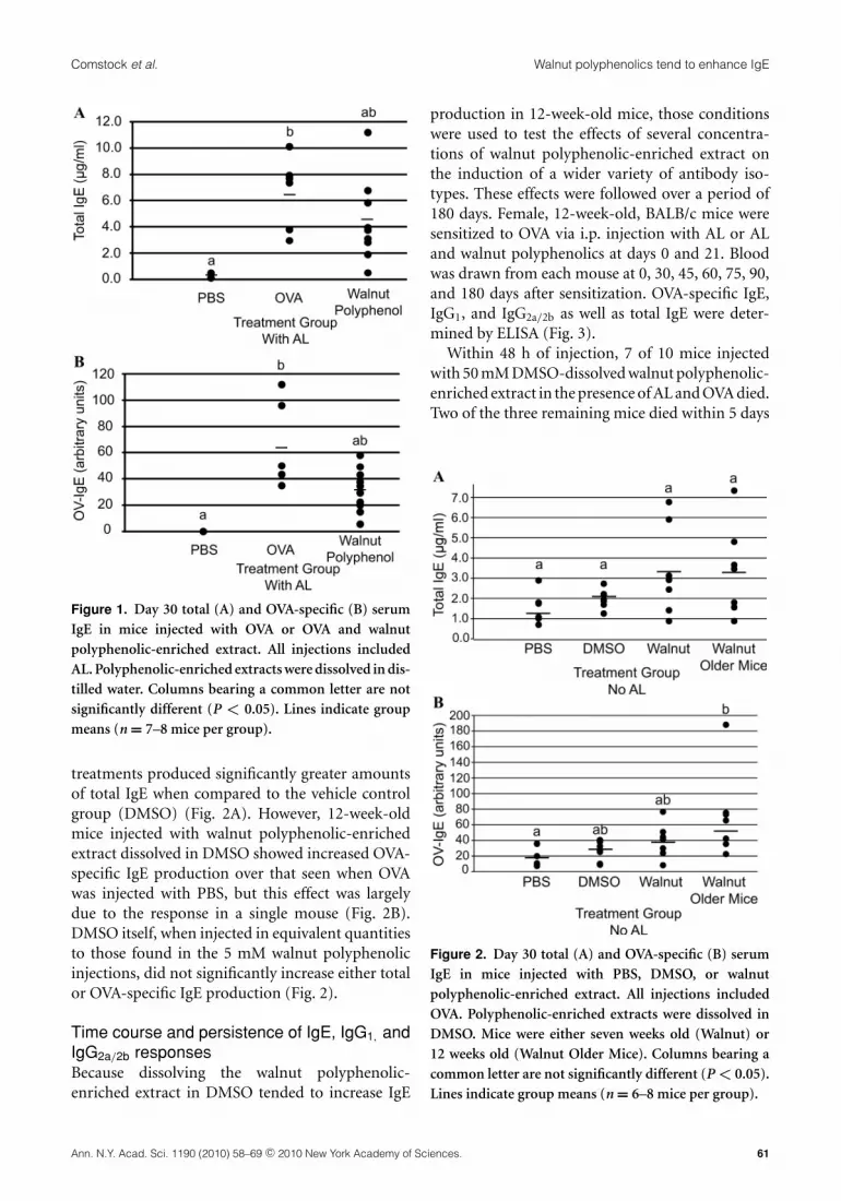
Walnut polyphenolic-enriched extractdissolved in water has no effect on IgEproductionMice were injected with 5 mM walnut polyphenolic-enriched extract in the presence of OVA and AL.Polyphenolic-enriched extracts were mixed withwater. Mice treated with walnut polyphenolic-enriched extract tended to produce less OVA-specific IgE as well as less total IgE than thoseinjected with OVA and AL (Fig. 1). However, nosignificant differences in IgE production were seenbetween the walnut-treated group and the positivecontrol.
Walnut polyphenolic-enriched extract tendsto increase OVA-specific IgE productionin the absence of adjuvantSince few significant differences were seen when thepolyphenolic-enriched extracts were diluted in wa-ter prior to injection, we next determined if an-other type of diluent might be more appropriate asmany of the walnut polyphenolic compounds areonly slightly soluble in water.25 In this experiment,animals were injected with OVA in the presence ofPBS, dimethyl sulfoxide (DMSO), or 5 mM walnutpolyphenolic-enriched extract dissolved in DMSO.No AL was administered in this experiment. Twogroups of walnut-treated mice were included. Onegroup contained mice that were 7 weeks of age andthe other contained mice that were 12 weeks of age.Although several mice in each of the walnut-treatedgroups produced high levels of total IgE, none of the
60 Ann. N.Y. Acad. Sci. 1190 (2010) 58–69 c© 2010 New York Academy of Sciences.
Comstock et al. Walnut polyphenolics tend to enhance IgE
Figure 1. Day 30 total (A) and OVA-specific (B) serumIgE in mice injected with OVA or OVA and walnutpolyphenolic-enriched extract. All injections includedAL. Polyphenolic-enriched extracts were dissolved in dis-tilled water. Columns bearing a common letter are notsignificantly different (P < 0.05). Lines indicate groupmeans (n = 7–8 mice per group).
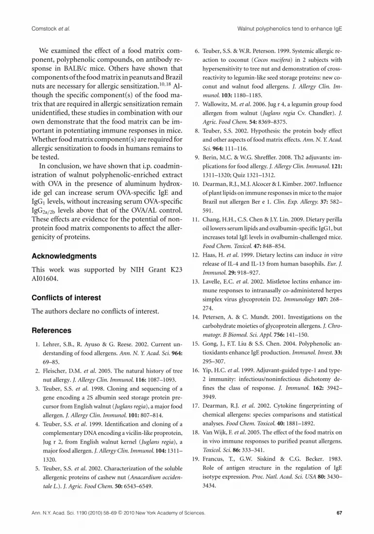
treatments produced significantly greater amountsof total IgE when compared to the vehicle controlgroup (DMSO) (Fig. 2A). However, 12-week-oldmice injected with walnut polyphenolic-enrichedextract dissolved in DMSO showed increased OVA-specific IgE production over that seen when OVAwas injected with PBS, but this effect was largelydue to the response in a single mouse (Fig. 2B).DMSO itself, when injected in equivalent quantitiesto those found in the 5 mM walnut polyphenolicinjections, did not significantly increase either totalor OVA-specific IgE production (Fig. 2).
Time course and persistence of IgE, IgG1, andIgG2a/2b responsesBecause dissolving the walnut polyphenolic-enriched extract in DMSO tended to increase IgE
production in 12-week-old mice, those conditionswere used to test the effects of several concentra-tions of walnut polyphenolic-enriched extract onthe induction of a wider variety of antibody iso-types. These effects were followed over a period of180 days. Female, 12-week-old, BALB/c mice weresensitized to OVA via i.p. injection with AL or ALand walnut polyphenolics at days 0 and 21. Bloodwas drawn from each mouse at 0, 30, 45, 60, 75, 90,and 180 days after sensitization. OVA-specific IgE,IgG1, and IgG2a/2b as well as total IgE were deter-mined by ELISA (Fig. 3).
Within 48 h of injection, 7 of 10 mice injectedwith 50 mM DMSO-dissolved walnut polyphenolic-enriched extract in the presence of AL and OVA died.Two of the three remaining mice died within 5 days
Figure 2. Day 30 total (A) and OVA-specific (B) serumIgE in mice injected with PBS, DMSO, or walnutpolyphenolic-enriched extract. All injections includedOVA. Polyphenolic-enriched extracts were dissolved inDMSO. Mice were either seven weeks old (Walnut) or12 weeks old (Walnut Older Mice). Columns bearing acommon letter are not significantly different (P < 0.05).Lines indicate group means (n = 6–8 mice per group).
Ann. N.Y. Acad. Sci. 1190 (2010) 58–69 c© 2010 New York Academy of Sciences. 61

Walnut polyphenolics tend to enhance IgE Comstock et al.
Figure 3. Serum antibody levels in 12-week-old miceinjected with PBS, OVA, OVA with 5 �M walnutpolyphenolic-enriched extract, or OVA with 5 mM wal-nut polyphenolic-enriched extract. All injections in-cluded AL. Polyphenolic extract was dissolved in DMSO.Blood samples were collected at days 0, 30, 45, 60, 75,90, and 180. Isotypes measured included IgE (A), IgG1
(B), and IgG2a/2b (C). Results are expressed as mean ±SEM (n = 10 mice per group). ∗∗P < 0.01 compared withPBS/AL treated mice, ∗P < 0.05 compared with OVA/ALtreated mice.
of injection. The remaining mouse was not boosted,and the treatment group was dropped from furtherstudies.
All OVA-specific antibody levels were negligibleon day 0 for each treatment group and remainednegligible in the PBS/AL control mice over time.Mice treated with either 5 �M or 5 mM wal-nut polyphenolics had significantly higher levelsof OVA-specific IgE and IgG1 at day 30 than theOVA/AL group (P < 0.05). Serum IgG2a/2b levelsalso tended to be higher in the mice treated with wal-nut polyphenolic extract (Fig. 3C). Treatment (P <
0.001) but not day (P = 0.182) affected serum OVA-specific IgE; however, there was a trend for peakingat day 30 in the polyphenolic-treated mice (Fig. 3A).OVA-specific IgG1 behaved similarly (treatment,P < 0.001; day, P = 0.874) (Fig. 3B). On day 30, totalIgE levels were elevated only in the 5 mM/OVA/ALgroup (P = 0.004). Total IgE did not differ amongthe groups past day 45. In all cases, treatment andday did not interact (P > 0.9). However, all OVA-specific antibody levels in the walnut polyphenolic-treated mice tended to be higher than those in theOVA/AL mice.
Walnut polyphenolics added to alum augmentthe day 30 OVA-specific IgE and IgG1
responseA larger number of 12-week-old BALB/c micewere sensitized to determine if the small increasein OVA-IgE demonstrated in Figure 2B was ob-served when walnut polyphenolic-enriched extractwas coadministered with the Th2 adjuvant AL.On day 30, mice were killed and serum wascollected. Total IgE was higher in mice receiv-ing 5 mM/OVA/AL (4.28 ± 1.1 �g/mL, P <
0.0001) than in mice receiving OVA/AL (2.09 ±0.53 �g/mL). Furthermore, serum OVA-specific IgEwas higher in mice injected with 5 �M/OVA/AL(3.14 ± 0.54, P = 0.032) or 5 mM/OVA/AL(3.62 ± 0.51, P = 0.003) than in mice injectedwith OVA/AL (1.97 ± 0.46) (Fig. 4A). This wasalso true for OVA-specific IgG1 where mice injectedwith 5 �M/OVA/AL (2.22 ± 0.26, P = 0.037) or 5mM/OVA/AL (2.31 ± 0.20, P = 0.016) had higherlevels than mice injected with OVA/AL (1.64 ±0.28) (Fig. 4B). On day 30, OVA-specific IgG2a/2b
levels did not differ for any treatment groups(P > 0.05), and all treatment groups expressed
62 Ann. N.Y. Acad. Sci. 1190 (2010) 58–69 c© 2010 New York Academy of Sciences.

Comstock et al. Walnut polyphenolics tend to enhance IgE
Figure 4. Day 30 serum antibody levels in 12-week-oldmice injected with PBS, OVA, and OVA with 5 �M walnutpolyphenolic extract or OVA with 5 mM walnut polyphe-nolic extract. All injections included AL. Polyphenolicextract was dissolved in DMSO. Isotypes measured in-cluded: IgE (A), IgG1 (B), and IgG2a/2b (C). Results areexpressed as mean ± SEM (n = 20 mice per group).Columns bearing a common letter are not significantlydifferent (P < 0.05).
higher IgG2a/2b levels than the negative controlgroup (P < 0.05) (Fig. 4C).
Walnut polyphenolics at 5 mM tend toincrease OVA-specific IgE, IgE, but notIgG2a/2b
To determine if polyphenolics dissolved in DMSOcould have an effect on IgE production in the ab-sence of other adjuvants, mice were injected asbefore; however, AL was excluded from some injec-tions. The addition of 5 mM walnut polyphenolic-enriched extract in the absence of AL tended toincrease total IgE production over that seen withadministration of OVA alone or with 5 �M polyphe-nolics (Fig. 5A). In addition, total IgE (P = 0.459)and OVA-specific serum IgE (P = 0.412) levels arethe same in mice treated with 5 mM/OVA as thosein mice treated with OVA/AL. As expected, micetreated with OVA alone have less OVA-specific IgEthan mice treated with OVA in the presence of AL(P = 0.025). Although mice treated with 5 mM/OVA(1.90 ± 0.028) had higher OVA-specific IgE levelsthan mice treated with 5 �M/OVA (0.49 ± 0.16, P =0.050), the direct comparison of OVA-specific IgElevels between mice treated with OVA alone (0.91 ±0.23) and those with 5 mM/OVA did not reach sig-nificance (P = 0.162).
For OVA-specific serum IgG1 (Fig. 5B), the micetreated with 5 mM/OVA expressed less serumOVA-specific antibody than those treated withOVA/AL (P = 0.028), and mice treated withOVA alone have less OVA-specific IgG1 than thosetreated with OVA/AL (P < 0.001). In the ab-sence of alum, IgG2a/2b antibody levels do notdiffer from those in the negative control (P >
0.05), but OVA/AL administration results in a sig-nificant increase in IgG2a/2b antibody (P < 0.05)(Fig. 5C).
In summary, addition of 5 mM walnut polyphe-nolics in the absence of alum tends to increaselevels of circulating OVA-specific IgE and IgG1,but not levels of IgG2a/2b. AL, in addition to in-creasing levels of circulating OVA-specific IgE andIgG1, also increases OVA-specific IgG2a/2b levels(Fig. 5).
Measurable cytokines in serum of miceOnce we established that walnut polyphenolics ad-ministered in DMSO with AL could increase day 30
Ann. N.Y. Acad. Sci. 1190 (2010) 58–69 c© 2010 New York Academy of Sciences. 63

Walnut polyphenolics tend to enhance IgE Comstock et al.
Figure 5. Day 30 serum antibody levels in 12-week-oldmice injected with PBS with AL, OVA alone, OVA withAL, OVA with 5 �M walnut polyphenolic extract, orOVA with 5 mM walnut polyphenolic extract. Polyphe-nolic extract was dissolved in DMSO. Isotypes measuredincluded: IgE (A), IgG1 (B), and IgG2a/2b (C). Resultsare expressed as mean ± SEM (n = 10 mice per group).Columns bearing a common letter are not significantlydifferent (P < 0.05).
OVA-specific IgE and IgG1, we examined changesin cytokine expression in those animals. The lev-els of six cytokines were measured in day 30 serumsamples. IFN-� , IL-10, IL-2, and IL-4 could not bedetected in serum samples from the mice. No differ-ences were seen in levels of IL-5 (6.13 ± 4.27 pg/mL)or IL-12 (58.79 ± 28.87 pg/mL) (values are average± SD for all mice combined).
Cytokine expression by splenocytesTo confirm mice expressed a Th2 profile in responseto OVA immunization, and to investigate whethersplenocytes from mice that had received polypheno-lics 9 days prior had a quantitatively higher degree ofTh2 cytokine secretion in response to OVA, spleno-cytes were harvested on day 30 and stimulated inculture. No polyphenolics were added to the cultureplates.
Levels of IL-4, IL-13, and IFN-� were measuredby ELISA (Table 1). IL-4 and IL-13 expression inthe treatment groups did not differ from that inthe OVA/AL positive control group (Table 1). IFN-� levels for all groups did not differ from that ofthe negative control (Table 1). When all measure-ments were combined, IL-4 levels were positivelycorrelated with serum IgE levels (r = 0.68).
Because few differences were detected in IL-4,IFN-� , and IL-13 levels in splenocyte cultures, a Lu-minex kit (Invitrogen BioSource) was used to mea-sure the expression of IL-2, IL-12, IL-5, and IL-10 insplenocyte cultures from mice injected without ALadjuvant. No differences were seen in levels of IL-2 or IL-10 production for any of the groups tested(Table 1). IL-12 production was lower in all miceimmunized with OVA than in the PBS control ani-mals (Table 1). Additionally, IL-5 levels were higherin all mice immunized with OVA than in the PBScontrol animals (Table 1).
Overall, the increase in IL-4, IL-13, and IL-5 lev-els in splenocyte cultures from OVA-immunizedanimals over those seen in cultures from nega-tive control animals are consistent with a Th2response. The decrease in IL-12 levels in spleno-cyte cultures from OVA-immunized animals com-pared to levels in splenocyte cultures from nega-tive control animals is also consistent with a Th2response.
64 Ann. N.Y. Acad. Sci. 1190 (2010) 58–69 c© 2010 New York Academy of Sciences.

Comstock et al. Walnut polyphenolics tend to enhance IgE
Table 1.Cytokine expression in splenocyte cultures stim-ulated with OVA for 72 h
Cytokine Treatment group Mean ± SD (pg/mL)
IL-4 PBS/AL 0a
OVA 22.88 ± 29.16b
OVA/AL 28.11 ± 27.46b
5 �M/OVA 13.77 ± 13.47ab
5 �M/OVA/AL 22.95 ± 20.30b
5 mM/OVA 30.12 ± 18.65b
5 mM/OVA/AL 32.55 ± 25.25b
IL-13 PBS/AL 43.44 ± 28.15a
OVA 1215.23 ± 600.89b
OVA/AL 1246.76 ± 459.54b
5 �M/OVA 830.11 ± 486.33b
5 �M/OVA/AL 650.35 ± 334.94ab
5 mM/OVA 967.39 ± 451.67b
5 mM/OVA/AL 832.23 ± 504.20b
IFN-� PBS/AL 8592.74 ± 7072.83a
OVA 11817.17 ± 5150.56a
OVA/AL 12002.34 ± 3623.52a
5 �M/OVA 8675.12 ± 4140.69a
5 �M/OVA/AL 9154.95 ± 4941.16a
5 mM/OVA 8843.75 ± 5864.06a
5 mM/OVA/AL 15365.54 ± 4295.11a
IL-2 PBS 129.06 ± 12.21a
OVA 135.82 ± 9.61a
5 �M/OVA 126.99 ± 19.71a
5 mM/OVA 124.46 ± 26.50a
IL-12 PBS 54.73 ± 15.01a
OVA 37.87 ± 10.02b
5 �M/OVA 39.35 ± 3.52b
5 mM/OVA 38.45 ± 11.31b
IL-5 PBS 10.93 ± 5.68a
OVA 538.74 ± 447.84b
5 �M/OVA 261.58 ± 191.33b
5 mM/OVA 255.55 ± 160.67b
IL-10 PBS 44.72 ± 11.80a
OVA 59.78 ± 16.40a
5 �M/OVA 46.46 ± 9.09a
5 mM/OVA 53.72 ± 21.79a
Note: Within a cytokine, measures bearing a commonletter are not different (P < 0.05).
Discussion
Results of these studies show that walnutpolyphenolic-enriched extract can enhance Th2 an-
tibody production (Fig. 4). The effects of addingwalnut polyphenolic-enriched extract at sensitiza-tion tended to persist over the course of 180 days(Fig. 3). However, enhanced Th2 antibody produc-tion was dependent upon the age of the animal,the solution in which the extract was adminis-tered and the presence of a Th2-skewing adju-vant (AL). Administration of walnut polyphenolic-enriched extract in the absence of a Th2-skewingadjuvant tended to increase Th2 antibody produc-tion over levels seen when OVA alone was admin-istered, but the differences did not reach signif-icance (Fig. 5). Such treatment did not increaseOVA-specific IgG2a/2b. However, administration ofAL increased levels of Th2 (IgE and IgG1) andTh1 (IgG2a/2b) antibodies. Administration of thepolyphenolic-enriched extract did not induce long-lasting effects on antigen-specific cytokine produc-tion by washed splenocytes, obtained 9 days after thelast polyphenolic exposure (Table 1). Polyphenolicadministration also did not result in any measur-able differences in circulating cytokine levels at day30. Finally, the effects on serum total IgE and OVA-specific IgE concentrations were significant only inthe short-term (day 30) (Fig. 3).
Plants synthesize polyphenolics in response to en-vironmental insults and also use them as signalingmolecules. In tree nuts, polyphenolic compoundsare concentrated in the “skin” or pellicle of the nutbut are also found in the kernel.26 EA, a hydrolysabletannin, is the major polyphenolic compound inthe pellicle of English walnuts; gallic acid, chloro-genic acid, and juglone, among others, are alsopresent.26–28 Other tree nuts also contain EA.29,30
The increased antigen-specific IgE and IgG1 produc-tion we saw is in agreement with that seen by otherswho sensitized mice in the presence of polyphenoliccompounds (including chlorogenic acid, a com-pound found in walnuts) from tobacco.15,19 Addi-tional studies have shown that polyphenolic com-pounds skew toward a Th2 immune response.29,31–36
Though the mechanism for the increased antigen-specific IgE in our animals is unknown, several pos-sibilities exist. The phenolic compounds may exerttheir effects through modulation of cytokine expres-sion by immune cells. In general, murine B cells thathave cross-linked CD40 molecules on their surfaces,are induced to switch to IgE production by the pres-ence of IL-4 or IL-13 in the microenvironment.37–42
Additionally, although IL-5 cannot increase IgE
Ann. N.Y. Acad. Sci. 1190 (2010) 58–69 c© 2010 New York Academy of Sciences. 65

Walnut polyphenolics tend to enhance IgE Comstock et al.
production alone, IL-5 has been shown to poten-tiate the IgE response in situations where IL-4 levelsare suboptimal.43 It has been shown that BSA con-jugated to a phenolic compound, rutin, induced anincrease in IgE receptor expression on B cells, a re-sponse that was abrogated by addition of anti-IL-4monoclonal antibody32 indicating that the pheno-lic conjugate-mediated changes by altering IL-4 ex-pression. Polyphenolic compounds have also beenshown to enhance Th2 cytokines while inhibitingTh1 cytokines.15,32 Our cytokine data do not in-dicate that IL-4 or IL-13 production was elevatedin the groups receiving walnut polyphenolic extract(Table 1). An increase in IL-5 production was ob-served (Table 1), but it was not significant over thatseen with injection of OVA or OVA/AL, so it is un-clear if the polyphenolics are increasing IL-5 andthereby potentiating IgE production. It is possiblethat the window to detect an increase in IL-4, IL-5, orIL-13 was missed, as it has been previously reportedthat cytokine production by splenocytes from trans-genic mice sensitized to OVA peaks 1 week after theinitial sensitization procedure.44 Herein, cytokineproduction by splenocytes was evaluated nearly 4weeks after initial sensitization of the mice. Addi-tionally, cytokine measurements were taken fromwashed splenocytes obtained 9 days after polyphe-nolic exposure. Thus, our experimental conditionsmay not have allowed us to determine the impactof walnut polyphenolic administration on cytokineproduction.
Alternatively, the mechanism by which the Th2antibody levels are increased in our mice may notbe changes in cytokine expression but rather in-creased lymphocyte sensitivity to cytokines. Thewalnut polyphenolic extract may have mediated theincrease in serum IgE and IgG1 in an interleukin-independent manner through direct effects on anti-gen uptake and presentation, gene expression of IgEor IgE receptors, IgE degradation pathways, or by in-hibiting interactions between IgE receptors and IgEmolecules. Future studies in which these parame-ters are measured must be conducted before con-clusions are drawn about the mechanisms by whichwalnut polyphenolic-enriched extract promotes theincrease in IgE and IgG1 antibody production.
In our experiments, mice were not evaluated forallergic reactions upon challenge with OVA. Micewere not challenged because it is unlikely therewould be a measurable difference between the re-
sponses of the OVA/AL treated mice and the micethat received OVA/AL in the presence of polyphe-nolics due to the concentrations of IgE present inthose mice.
Many routes of sensitization have been used inanimal models of food allergy. Our choice of i.p.sensitization does not mimic the route of sensitiza-tion in human food allergy. However, others havesuccessfully used the i.p. route of administration todetermine the inherent allergenicity of food pro-teins45 and have shown that i.p. administration isas effective and accurate as oral administration.46,47
They have shown a clear correlation between aller-genicity and antigen-specific IgE production usingi.p. sensitization.45,48 Additionally, an i.p. injectionof a bolus of chlorogenic acid increased IgE as ef-fectively as long-term oral exposure to chlorogenicacid.15 Herein, we wanted to determine the inherentTh2-skewing potential of a walnut polyphenolic-enriched extract. Thus, we began by testing i.p. ad-ministration of the substance in a previously estab-lished model of Th2 sensitization. Future researchwill be needed to determine if similar results are seenwhen antigen in the context of walnut polyphenolic-enriched extract is administered orally. However, onthe basis of previously published research,15,46 wepredict similar results will be seen by either route ofsensitization.
We chose to use a model antigen, OVA, in lieuof using a walnut allergen for sensitization. In thisway, we were able to separate the inherent sensitizingpotential of walnut polyphenolic-enriched extractfrom the sensitizing potential of walnut protein. Ad-ditionally, use of an established model antigen en-abled us to use fewer animals as no animal-intensivepreliminary studies to determine the optimal quan-tity of walnut allergen or to determine which walnutallergen would best sensitize the mice were required.Now that we have established the ability of walnutpolyphenolic-enriched extract to potentiate IgE re-sponses, future work should include one or more ofthe established walnut allergens in the sensitizationprocedure.
It is possible that the concentration of polyphe-nolics injected i.p. may be higher than plasma con-centrations achievable from the consumption offood with high polyphenolic content, but our inter-est lies in the gut microenvironment where 5 mMconcentrations of polyphenolic compounds arepossible.49–51
66 Ann. N.Y. Acad. Sci. 1190 (2010) 58–69 c© 2010 New York Academy of Sciences.
Comstock et al. Walnut polyphenolics tend to enhance IgE
We examined the effect of a food matrix com-ponent, polyphenolic compounds, on antibody re-sponse in BALB/c mice. Others have shown thatcomponents of the food matrix in peanuts and Brazilnuts are necessary for allergic sensitization.10,18 Al-though the specific component(s) of the food ma-trix that are required in allergic sensitization remainunidentified, these studies in combination with ourown demonstrate that the food matrix can be im-portant in potentiating immune responses in mice.Whether food matrix component(s) are required forallergic sensitization to foods in humans remains tobe tested.
In conclusion, we have shown that i.p. coadmin-istration of walnut polyphenolic-enriched extractwith OVA in the presence of aluminum hydrox-ide gel can increase serum OVA-specific IgE andIgG1 levels, without increasing serum OVA-specificIgG2a/2b levels above that of the OVA/AL control.These effects are evidence for the potential of non-protein food matrix components to affect the aller-genicity of proteins.
Acknowledgments
This work was supported by NIH Grant K23AI01604.
Conflicts of interest
The authors declare no conflicts of interest.
References
1. Lehrer, S.B., R. Ayuso & G. Reese. 2002. Current un-
derstanding of food allergens. Ann. N. Y. Acad. Sci. 964:
69–85.
2. Fleischer, D.M. et al. 2005. The natural history of tree
nut allergy. J. Allergy Clin. Immunol. 116: 1087–1093.
3. Teuber, S.S. et al. 1998. Cloning and sequencing of a
gene encoding a 2S albumin seed storage protein pre-
cursor from English walnut (Juglans regia), a major food
allergen. J. Allergy Clin. Immunol. 101: 807–814.
4. Teuber, S.S. et al. 1999. Identification and cloning of a
complementary DNA encoding a vicilin-like proprotein,
Jug r 2, from English walnut kernel (Juglans regia), a
major food allergen. J. Allergy Clin. Immunol. 104: 1311–
1320.
5. Teuber, S.S. et al. 2002. Characterization of the soluble
allergenic proteins of cashew nut (Anacardium occiden-
tale L.). J. Agric. Food Chem. 50: 6543–6549.
6. Teuber, S.S. & W.R. Peterson. 1999. Systemic allergic re-
action to coconut (Cocos nucifera) in 2 subjects with
hypersensitivity to tree nut and demonstration of cross-
reactivity to legumin-like seed storage proteins: new co-
conut and walnut food allergens. J. Allergy Clin. Im-
munol. 103: 1180–1185.
7. Wallowitz, M. et al. 2006. Jug r 4, a legumin group food
allergen from walnut (Juglans regia Cv. Chandler). J.
Agric. Food Chem. 54: 8369–8375.
8. Teuber, S.S. 2002. Hypothesis: the protein body effect
and other aspects of food matrix effects. Ann. N. Y. Acad.
Sci. 964: 111–116.
9. Berin, M.C. & W.G. Shreffler. 2008. Th2 adjuvants: im-
plications for food allergy. J. Allergy Clin. Immunol. 121:
1311–1320; Quiz 1321–1312.
10. Dearman, R.J., M.J. Alcocer & I. Kimber. 2007. Influence
of plant lipids on immune responses in mice to the major
Brazil nut allergen Ber e 1. Clin. Exp. Allergy. 37: 582–
591.
11. Chang, H.H., C.S. Chen & J.Y. Lin. 2009. Dietary perilla
oil lowers serum lipids and ovalbumin-specific IgG1, but
increases total IgE levels in ovalbumin-challenged mice.
Food Chem. Toxicol. 47: 848–854.
12. Haas, H. et al. 1999. Dietary lectins can induce in vitro
release of IL-4 and IL-13 from human basophils. Eur. J.
Immunol. 29: 918–927.
13. Lavelle, E.C. et al. 2002. Mistletoe lectins enhance im-
mune responses to intranasally co-administered herpes
simplex virus glycoprotein D2. Immunology 107: 268–
274.
14. Petersen, A. & C. Mundt. 2001. Investigations on the
carbohydrate moieties of glycoprotein allergens. J. Chro-
matogr. B Biomed. Sci. Appl. 756: 141–150.
15. Gong, J., F.T. Liu & S.S. Chen. 2004. Polyphenolic an-
tioxidants enhance IgE production. Immunol. Invest. 33:
295–307.
16. Yip, H.C. et al. 1999. Adjuvant-guided type-1 and type-
2 immunity: infectious/noninfectious dichotomy de-
fines the class of response. J. Immunol. 162: 3942–
3949.
17. Dearman, R.J. et al. 2002. Cytokine fingerprinting of
chemical allergens: species comparisons and statistical
analyses. Food Chem. Toxicol. 40: 1881–1892.
18. Van Wijk, F. et al. 2005. The effect of the food matrix on
in vivo immune responses to purified peanut allergens.
Toxicol. Sci. 86: 333–341.
19. Francus, T., G.W. Siskind & C.G. Becker. 1983.
Role of antigen structure in the regulation of IgE
isotype expression. Proc. Natl. Acad. Sci. USA 80: 3430–
3434.
Ann. N.Y. Acad. Sci. 1190 (2010) 58–69 c© 2010 New York Academy of Sciences. 67
Walnut polyphenolics tend to enhance IgE Comstock et al.
20. Scalbert, A. et al. 2005. Dietary polyphenols and the
prevention of diseases. Cr. Rev. Food. Sci. Nutr. 45: 287–
306.
21. Van Boxtel, E.L. et al. 2007. Peanut allergen Ara h 1
interacts with proanthocyanidins into higher molecular
weight complexes. J. Agric. Food Chem. 55: 8772–8778.
22. Morefield, G.L. et al. 2005. Role of aluminum-containing
adjuvants in antigen internalization by dendritic cells in
vitro. Vaccine. 23: 1588–1595.
23. Anderson, K.J. et al. 2001. Walnut polyphenolics inhibit
in vitro human plasma and LDL oxidation. J. Nutr. 131:
2837–2842.
24. Singleton, V.L. & J.A. Rossi, Jr. 1965. Colorimetry of total
phenolics with phosphomolybdic-phosphotungstic acid
reagents. Am. J. Enol. Vitic. 16: 144–158.
25. Bala, I. et al. 2006. Analytical methods for assay of ellagic
acid and its solubility studies. J. Pharmaceut. Biomed. 40:
206–210.
26. Colaric, M. et al. 2005. Phenolic acids, syringaldehyde,
and juglone in fruits of different cultivars of Juglans regia
L.J. Agric. Food Chem. 53: 6390–6396.
27. Jurd, L. 1956. Plant Polyphenols I. The polyphenolic
constituents of the pellicle of the walnut. J. Am. Chem.
Soc. 78: 3445–3448.
28. Jurd, L. 1957. Plant Polyphenols. III. The isolation of a
new ellagitannin from the pellicle of the walnut. J. Am.
Chem. Soc. 80: 2249–2252.
29. Senter, S.D., R.J. Horvat & W.R. Forbus. 1983. Compar-
ative GLC-MS analysis of phenolic acids of selected tree
nuts. J. Food Sci. 48: 798–799, 824.
30. Clifford, M.N. & A. Scalbert. 2000. Ellagitannins – na-
ture, occurrence and dietary burden. J. Sci. Food. Agr. 80:
1118–1125.
31. Francus, T. 1994. Plant polyphenolic-protein conjugates
activate murine spleen cells and bind to multiple cell
surface components. Proc. Soc. Exp. Biol. Med. 207: 117–
126.
32. Baum, C.G. et al. 1990. Cellular control of IgE induction
by a polyphenol-rich compound. Preferential activation
of Th2 cells. J. Immunol. 145: 779–784.
33. Francus, T. et al. 1988. Effects of tobacco glycoprotein
(TGP) on the immune system. II. TGP stimulates the
proliferation of human T cells and the differentiation of
human B cells into Ig secreting cells. J. Immunol. 140:
1823–1829.
34. Heo, Y., A. Saxon & O. Hankinson. 2001. Effect of diesel
exhaust particles and their components on the allergen-
specific IgE and IgG1 response in mice. Toxicology 159:
143–158.
35. Becker, C.G., R. Levi & J. Zavecz. 1979. Induction of
IgE antibodies to antigen isolated from tobacco leaves
and from cigarette smoke condensate. Am. J. Pathol. 96:
249–255.
36. Levi, R. et al. 1982. Cardiac and pulmonary anaphy-
laxis in guinea pigs and rabbits induced by glycoprotein
isolated from tobacco leaves and cigarette smoke con-
densate. Am. J. Pathol. 106: 318–325.
37. Jabara, H.H. et al. 1990. Induction of germ-line and
mature C epsilon transcripts in human B cells stimulated
with rIL-4 and EBV. J. Immunol. 145: 3468–3473.
38. Lebman, D. & R. Coffman. 1988. Interleukin 4 causes
isotype switching to IgE in T cell-stimulated clonal B cell
cultures. J. Exp. Med. 168: 853–862.
39. Gauchat, J. et al. 1990. Structure and expression of
germline epsilon transcripts in human B cells induced
by interleukin 4 to switch to IgE production. J. Exp. Med.
172: 463–473.
40. Punnonen, J. et al. 1993. Interleukin 13 induces inter-
leukin 4-independent IgG4 and IgE synthesis and CD23
expression by human B cells. Proc. Natl. Acad. Sci. USA
90: 3730–3734.
41. Shapira, S. et al. 1992. Molecular analysis of the induc-
tion of immunoglobulin E synthesis in human B cells by
interleukin 4 and engagement of CD40 antigen. J. Exp.
Med. 175: 289–292.
42. Mckenzie, A.N. et al. 1993. Interleukin 13, a T-cell-
derived cytokine that regulates human monocyte and
B-cell function. Proc. Natl. Acad. Sci. USA 90: 3735–
3739.
43. Pene, J. et al. 1988. Interleukin 5 enhances interleukin 4-
induced IgE production by normal human B cells. The
role of soluble CD23 antigen. Eur. J. Immunol. 18: 929–
935.
44. Shida, K. et al. 2000. Serum IgE response to orally in-
gested antigen: a novel IgE response model with allergen-
specific T-cell receptor transgenic mice. J. Allergy Clin.
Immunol. 105: 788–795.
45. Dearman, R.J. & I. Kimber. 2007. A mouse model for
food allergy using intraperitoneal sensitization. Methods
41: 91–98.
46. Dearman, R.J. et al. 2001. Characterization of antibody
responses induced in rodents by exposure to food pro-
teins: influence of route of exposure. Toxicology 167:
217–231.
47. Van Wijk, F. et al. 2004. Mixed antibody and T cell re-
sponses to peanut and the peanut allergens Ara h 1, Ara
h 2, Ara h 3 and Ara h 6 in an oral sensitization model.
Clin. Exp. Allergy. 34: 1422–1428.
68 Ann. N.Y. Acad. Sci. 1190 (2010) 58–69 c© 2010 New York Academy of Sciences.

Comstock et al. Walnut polyphenolics tend to enhance IgE
48. Dearman, R. et al. 2003. Evaluation of protein allergenic
potential in mice: dose-response analyses. Clin. Exp. Al-
lergy. 33: 1586–1594.
49. Erlund, I. et al. 2002. Plasma concentrations of the
flavonoids hesperetin, naringenin and quercetin in hu-
man subjects following their habitual diets, and diets
high or low in fruit and vegetables. Eur. J. Clin. Nutr. 56:
891–898.
50. Moon, J.H. et al. 2000. Accumulation of quercetin con-
jugates in blood plasma after the short-term ingestion
of onion by women. Am. J. Physiol. Regul. Integr. Comp.
Physiol. 279: R461–R467.
51. Cerda, B. et al. 2003. Evaluation of the bioavailability
and metabolism in the rat of punicalagin, an antioxidant
polyphenol from pomegranate juice. Eur. J. Nutr. 42:
18–28.
Ann. N.Y. Acad. Sci. 1190 (2010) 58–69 c© 2010 New York Academy of Sciences. 69


