Embed Size (px)
Citation preview

En
FMa
b
c
d
a
ARRAA
KPCNTTR
1
hrw22MwBEcba(ir
UT
h0
Carbohydrate Polymers 134 (2015) 664–672
Contents lists available at ScienceDirect
Carbohydrate Polymers
journa l homepage: www.e lsev ier .com/ locate /carbpol
ffect of the oxidation treatment on the production of celluloseanofiber suspensions from Posidonia oceanica: The rheological aspect
edia Bettaieb a,b,c, Oleksandr Nechyporchuk b,c,d, Ramzi Khiari a,b,c,∗,ohamed Farouk Mhenni a, Alain Dufresne b,c, Mohamed Naceur Belgacem b,c
University of Monastir, Faculty of Sciences, UR13ES63—Research Unity of Applied Chemistry & Environment, 5000 Monastir, TunisiaUniversité Grenoble Alpes, LGP2, F-38000 Grenoble, FranceCNRS, LGP2, F-38000 Grenoble, FranceUniversité Grenoble Alpes, LRP, F-38000 Grenoble, France
r t i c l e i n f o
rticle history:eceived 19 March 2015eceived in revised form 27 July 2015ccepted 28 July 2015vailable online 31 July 2015
a b s t r a c t
Different grades of cellulose nanofibrils (CNF) were prepared from Posidonia oceanica balls and leaves(POB and POL). Pretreatment using 2,2,6,6-tetramethylpiperidine-1-oxyl (TEMPO)-mediated oxidationwas performed to facilitate the fibrillation during ultrafine friction grinding process. The ensuing CNFbatches were compared in terms of morphology and degree of fibrillation. The rheological properties ofthe produced CNF suspensions were also analyzed for varying doses of sodium hypochlorite used duringthe TEMPO- mediated oxidation procedure. The stronger fibrous network structures were formed when
eywords:osidonia oceanicaellulose nanofibrils (CNF)anofibrillated cellulose (NFC)EMPO-oxidation
increasing the oxidant concentration, which was confirmed by the increase of the storage moduli value. P.oceanica balls were found to undergo stronger fibrillation and, consequently, to form stronger networks,compared to P. oceanica leaves, when using equivalent concentration of the oxidizing agent.
© 2015 Elsevier Ltd. All rights reserved.
EMheological behavior. Introduction
Nowadays there exists a strong need in development ofigh added-value products from renewable resources. Manyeports examine the use of annual plants or/and non-woodastes as new raw materials to produce fibers (Aguir & Mhenni,
006, 2007; Ben Douissa, Bergaoui, Mansouri, Khiari, & Mhenni,013; Ncibi, Altenor, Seffen, Brouers, & Gaspard, 2008; Khiari,henni, Belgacem, & Mauret, 2010), papers, fiberboards, and non-ovens for papermaking and packaging industries (Khiari, Mauret,
elgacem, & Mhenni, 2011; Mansouri et al., 2012; Youssef, Alil-Samahy, & Abdel Rehim, 2012), but also to meet pharma-eutical demands such as fabrication of compresses, dressings,andages, drugs, etc. (Hebeish, Abdelhady, & Youssef, 2013),nd prepare innovative materials, e.g., bio- or nanocomposites
Dufresne, 2012). These strategies are already applied in var-ous countries in order to valorize lignocellulosic wastes andecompose the broad use of cellulosic fibers (Antunes, Amaral, &∗ Corresponding author at: University of Monastir, Faculty of Sciences,R13ES63—Research Unity of Applied Chemistry & Environment, 5000 Monastir,unisia. Fax: +216 73 50 02 78.
E-mail address: khiari [email protected] (R. Khiari).
ttp://dx.doi.org/10.1016/j.carbpol.2015.07.091144-8617/© 2015 Elsevier Ltd. All rights reserved.
Belgacem, 2000; Abrantes, Amaral, Costa, & Duarte, 2007; Cordeiro,Belgacem, Torres, & Mourad, 2004; Dutt, Upadhyaya, Malik, &Tyagi, 2005; Dutt, Upadhyaya, Tyagi, Kumar, & Lal, 2008; Chia,Zakaria, Nguyen, & Abdullah, 2008; Rosli, Leh, Zainuddin, & Tanaka,2003; Hedjazi, Kordsahia, Patt, Latibrai, & Tschirner, 2008; Khiari,Mhenni, Belgacem, & Mauret, 2010; Khiari, Marrakchi, Belgacem,Mauret, & Mhenni, 2011). Over the last decade, the preparationand characterization of nanocellulose has gained much interest.Nanofibrillar cellulose which is also called cellulose microfibrilsand/or microfibrillar cellulose and/or more currently, cellulosenanofibrils (CNF), has been the subject of intense research activ-ities. In fact, it is a promising natural nano-structured polymer thatcould be potentially used by different industries, for instance inbiomedical applications (Siró & Plackett, 2008).
Since the 1980s, when Turbak, Snyder, and Sandberg (1983)and Herrick, Casebier, Hamilton, and Sandberg (1983) reportedthe preparation of nanofibrillar cellulose from wood fibers inwater suspension using high pressure homogenization, an expo-nential number of scientific works were performed on the isolationand characterization of CNF using high-pressure homogenizer
(Alila, Besbes, Vilar, Mutjé, & Boufi, 2013; Andresen, Johansson,Tanem, & Stenius, 2006; Andresen & Stenius, 2007; Bettaieb,Khiari, Dufresne, Mhenni, Putaux, & Boufi., 2015; Djafari Petroudy,Syverud, Chinga-Carrasco, Ghasemain, & Resalati, 2014; Erkisen,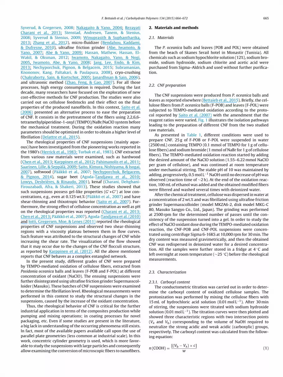
ate Po
SC22&YW22K(apdccp(ottpfi
otf(S2&LFscstoCaprSitar
bPcfltps
ippaIpwaa
F. Bettaieb et al. / Carbohydr
yverud, & Gregersen, 2008; Nakagaito & Yano, 2004; Rezayatiharani et al., 2013; Stenstad, Andresen, Tanem, & Stenius,008; Syverud & Stenius, 2009; Winuprasith & Suphantharika,013; Zhang et al., 2012), micro-fluidizer (Bendahou, Kaddami,
Dufresne, 2010), ultrafine friction grinder (Abe, Iwamoto, &ano, 2007; Abe & Yano, 2009; Hassan, Mathew, Hassan, El-akil, & Oksman, 2012; Iwamoto, Nakagaito, Yano, & Nogi,
005, Iwamoto, Abe, & Yano, 2008; Jang, Lee, Endo, & Kim,013; Nechyporchuk, Pignon, & Belgacem, 2015; Subramanian,nononov, Kang, Paltakari, & Paulapura, 2008), cryo-crushing
Chakraborty, Sain, & Kortschot, 2005; Janardhnan & Sain, 2006),nd ultrasonic method (Zhao, Feng, & Gao, 2007). For all thoserocesses, high energy consumption is required. During the lastecade, many researchers have focused on the exploration of newost-effective methods for CNF production. The studies were alsoarried out on cellulose feedstocks and their effect on the finalroperties of the produced nanofibrils. In this context, Saito et al.2006) presented an alternative process to ease the preparationf CNF. It consists in the pretreatment of the fibers using 2,2,6,6-etramethylpiperidine-1-oxyl (TEMPO)/NaBr/NaClO system beforehe mechanical treatment. During the oxidation reaction manyarameters should be optimized in order to obtain a higher level ofbrillation (Delattre et al., 2015).
The rheological properties of CNF suspensions (mainly aque-us) have been investigated from the pioneering works reported inhe 1980’s (Herrick et al., 1983; Turbak et al., 1983). CNF extractedrom various raw materials were examined, such as hardwoodChen et al., 2013; Karppinen et al., 2012; Pahimanolis et al., 2011;aarinen, Lille, & Seppälä, 2009; Saito, Kimura, Nishiyama, & Isogai,007), softwood (Pääkkö et al., 2007; Nechyporchuk, Belgacem,
Pignon, 2014), sugar beet (Agoda-Tandjawa et al., 2010;owys, Desbrières, & Rinaudo, 2001), kenaf (Charani, Dehghani-irouzabadi, Afra, & Shakeri, 2013). These studies showed thatuch suspensions possess gel-like properties (G′ » G′′) at low con-entrations, e.g., even at 0.125 wt.% (Pääkkö et al., 2007) and havehear-thinning and thixotropic behavior (Saito et al., 2007). Fur-hermore, the strong effect of cellulose concentration as well as pHn the rheological properties was reported (Charani et al., 2013;hen et al., 2013; Pääkkö et al., 2007). Agoda-Tandjawa et al. (2010)nd Iotti, Gregersen, Moe, & Lenes (2011) reported the rheologicalroperties of CNF suspensions and observed two shear-thinningegions with a viscosity plateau between them in flow curves.uch feature was attributed to the structural changes of CNF whilencreasing the shear rate. The visualization of the flow showedhat it may occur due to the changes of the CNF flocculi structure,s reported by Karppinen et al. (2012). All the above mentionedeports that CNF behaves as a complex entangled network.
In the present study, different grades of CNF were preparedy TEMPO-mediated oxidation of cellulose fibers, extracted fromosidonia oceanica balls and leaves (F-POB and F-POL) at differentoncentration of oxidant (NaClO). The ensuing suspensions wereurther disintegrated using ultrafine friction grinder Supermasscol-oider (Masuko). These batches of CNF suspensions were examinedo determine the fibrillation level. Rheological measurements wereerformed in this context to study the structural changes in theuspensions, caused by the increase of the oxidant concentration.
Thus, the rheological behavior of CNF is critical for the furtherndustrial application in terms of the composites production whileumping and mixing operations; in coating processes for novelackaging, etc. Even if some studies are present in the literature,
big lack in understanding of the occurring phenomena still exists.n fact, most of the available papers available call upon the use of
arallel plate geometries (less common at industrial scale). In thisork, concentric cylinder geometry is used, which is more favor-ble to study the suspensions with large particles and consequentlyllow examining the conversion of microscopic fibers to nanofibers.
lymers 134 (2015) 664–672 665
2. Materials and methods
2.1. Materials
The P. oceanica balls and leaves (POB and POL) were obtainedfrom the beach of Skanes Serail hotel in Monastir (Tunisia). Allchemicals such as sodium hypochlorite solution (12%), sodium bro-mide, sodium hydroxide, sodium chlorite and acetic acid werepurchased from Sigma–Aldrich and used without further purifica-tion.
2.2. CNF preparation
The CNF suspensions were produced from P. oceanica balls andleaves as reported elsewhere (Bettaieb et al., 2015). Briefly, the cel-lulose fibers from P. oceanica balls (F-POB) and leaves (F-POL) weresubjected to TEMPO-mediated oxidation according to the proto-col reported by Saito et al. (2007) with the amendment that thereagent ratios were varied. Fig. 1 illustrates the isolation pathwaysleading to the preparation of different CNF from the two studiedraw materials.
As presented in Table 1, different conditions were used toprepare CNF. 25 g of F-POB or F-POL were suspended in water(2500 mL) containing TEMPO (0.1 mmol of TEMPO for 1 g of cellu-lose fibers) and sodium bromide (1 mmol of NaBr for 1 g of cellulosefibers). The TEMPO-mediated oxidation reaction started by addingthe desired amount of the NaClO solution (1.55–6.22 mmol NaClOper gram of cellulose), and was continued at room temperatureunder mechanical stirring. The stable pH of 10 was maintained byadding, progressively, 0.5 mol L−1 NaOH until no decrease of pH wasobserved (reaction time of ∼2 h). At the end of the oxidation reac-tion, 100 mL of ethanol was added and the obtained modified fiberswere filtered and washed several times with deionized water.
After the chemical treatment, cellulose was dispersed in water ata concentration of 2 wt.% and was fibrillated using ultrafine frictiongrinder Supermasscolloider (model MKZA6-2, disk model MKG-C80, Masuko Sangyo Co., Ltd., Japan). The grinding was performedat 2500 rpm for the determined number of passes until the con-sistency of the suspension turned into a gel. In order to study theeffect of NaClO oxidant dose during the TEMPO-mediated oxidationreaction, the CNF-POB and CNF-POL suspensions were concen-trated using centrifuge Sigma 6-16KS at 10,000 rpm for 30 min. Thedry content was measured gravimetrically, and then the obtainedCNF was redispersed in deionized water for a desired concentra-tion. All the samples were further stored in a fridge at 4 ◦C andleft overnight at room temperature (∼25 ◦C) before the rheologicalmeasurements.
2.3. Characterization
2.3.1. Carboxyl contentThe conductometric titration was carried out in order to deter-
mine the carboxyl content of oxidized cellulose samples. Theprotonization was performed by mixing the cellulose fibers with15 mL of hydrochloric acid solution (0.01 mol L−1). After 30 minof stirring, the suspensions were titrated with sodium hydroxidesolution (0.01 mol L−1). The titration curves were then plotted andshowed three characteristic regions with two intersection points(Va and Vb) corresponding to the volume of NaOH required toneutralize the strong acidic and weak acidic (carboxylic) groups,respectively. The carboxyl content was calculated from the follow-
ing equation:n (COOH) = [(Vb − Va) × c]w
(1)
666 F. Bettaieb et al. / Carbohydrate Polymers 134 (2015) 664–672
from
ww
fe
2
sasrwTd
dt
2
m(f8c
TDy
Fig. 1. Schematic diagram of the preparation process of the NFC suspensions
here c is the concentration of the sodium hydroxide solution and is the oven-dry weight of cellulose.
The measurements were repeated at least in duplicate. The dif-erence between the obtained values was within an experimentalrror of 5%.
.3.2. Yield of fibrillationTo determine the fibrillation yield the produced CNF suspen-
ions were diluted to a solid content (%Sc) of 0.2% and centrifugedt 4000 rpm for 30 min to separate the nanofibrillated material (inupernatant fraction) from the non-fibrillated and/or partially fib-illated material (the sediment). After that the obtained fractionas dried at 90 ◦C in a halogen desiccator until a constant weight.
he yield of the nanofibrillated material or fibrillation yield wasetermined as follows:
Yield of fibrilation (%)
= 100 ×[
1 − weight of dried sedimentweight of diluted sample × %Sc
](2)
The measurements were repeated at least in duplicate and theifference between the obtained values was within an experimen-al error of 5%.
.3.3. Microscopy observationsThe morphology of the produced CNF was examined by trans-
ission electron microscopy (TEM), atomic force microscopy
AFM) and optical microscopy. TEM measurements were per-ormed using JEOL 200CX transmission electron microscope at0 kV. About 0.5 �L of the suspension was deposited onto a carbon-oated 300-mesh copper grid using a labnet micropipette. Waterable 1esignation, preparation conditions (NaClO concentration used for the TEMPO-mediated oield of the produced CNF from Posidonia oceanica leaves (POL) and balls (POB).
Sample NaClO concentrationa Carboxyl contentb
CNF-POL-20 1.55 250
CNF-POL-40 3.11 450
CNF-POL-60 4.66 550
CNF-POL-80 6.22 650
CNF-POB-20 1.55 350
CNF-POB-40 3.11 575
CNF-POB-60 4.66 600
CNF-POB-80 6.22 700
a (mmol NaClO per gram of cellulose).b (�mol g−1).
Posidonia oceanica balls and leaves using various concentrations of oxidant.
was allowed to evaporate. Additional drop of the CNF suspensionwas added onto their respective grids to increase the amount ofcellulose particles and the process was repeated. The nanofibrildimensions were evaluated using digital image analysis (Image J)as an average of 90 individual measurements. AFM measurementswere carried out in a tapping mode using Dimension Icon AtomicForce Microscope with an OTESPA cantilever. The samples werediluted to the concentration of 10−4 wt.% and a droplet of suspen-sion was placed on a mica disk and dried at 70 ◦C during 30 min.Nanoscope III software was used for the evaluation of the nanofib-rils diameter from the height profiles of AFM height images. At leastfour different areas of the sample were scanned and 200 heightswere measured for each sample. In order to detect the residualmicroscopic fiber fragments, the optical microscopy was carriedout using Carl Zeiss Axio Imager M1 optical microscope in trans-mission mode. The examined cellulose suspensions were diluted toa concentration of 0.2 wt.%, and a droplet was placed between theglass slide and a coverslip. The images were captured by AxioCamMRc 5 digital camera.
2.3.4. Rheological characterizationThe rheological behavior of the CNF suspensions was studied
using a stress- controlled rheometer DHR-3 (TA Instruments, USA)equipped with concentric cylinders geometry with a recessed bob(cup diameter of 36.2 mm; bob diameter of 28.8 mm, operatinggap of 4 mm). Since CNF suspensions tend to slip over the smoothgeometry surfaces (Nechyporchuk, Belgacem, Pignon, 2015), the
serrated geometry was used (roughness of ∼120 �m). The sampleswere kept overnight at room temperature (∼23 ◦C) before the mea-surements. After placing into the geometry the suspensions wereallowed to rest for 5 min in order to minimize the shear historyxidation reaction and grinding number of passes), carboxyl content, and fibrillation
Grinding number of passes Fibrillation yield (%)
25 4220 5810 70
6 7630 54
8 706 725 82
ate Po
ic
qrctar0ra
3
3
boTa
27bTa8ottoebpvN2
omliawrDeetwsfbwtla
Pfinai
F. Bettaieb et al. / Carbohydr
mposed by the loading. All the measurements were carried out atontrolled temperature of 23 ◦C on the CNF suspensions at 2 wt.%.
The oscillation strain sweeps from 0.01 to 10 % at the angular fre-uency of 5 rad s−1 were performed to detect the linear viscoelasticegions. Then the oscillation frequency sweeps in these linear vis-oelastic regions were performed in the range of 0.1–100 rad s−1
o measure the dynamic (storage and loss) moduli (G′ and G′′). Thepparent viscosity measurements were carried out at stepped shearate ramps (to eliminate the influence of thixotropy) in the range of.01–100 s−1, first by increasing and then by decreasing the shearate. All the measurements were performed in triplicate and anverage value was used for the data analysis.
. Results and discussion
.1. Morphological properties
Eight grades of CNF suspensions were prepared from P. oceanicaalls and leaves by TEMPO-mediated oxidation at different degreesf oxidation followed by grinding in Supermasscolloider (Masuko).he influence of NaClO concentration on the degree of oxidationnd fibrillation yield is shown in Table 1.
As seen from Table 1, the carboxyl content varied from50 �mol g−1 to 650 �mol g−1 for POL and from 350 �mol g−1 to00 �mol g−1 for POB. This difference in the oxidation reaction cane assigned, as expected, to the increase of NaClO dose during theEMPO-mediated oxidation reaction. The fibrillation yield followedlso the same trend as it increased from 42 to 76% and from 54 and2%, for CNF-POL and CNF-POB, respectively, despite the reductionf the number of passes through the grinder device. As expected,he highest carboxyl content and fibrillation yield were obtained forhe highest NaClO concentration used during the TEMPO-mediatedxidation reaction (CNF-POL-80 and CNF-POB-80 samples), what-ver the used raw material. Similar observations were reportedy several researchers (Dufresne, 2012; Saito & Isogai, 2004). Therimary hydroxyl groups of cellulose are thus and selectively con-erted to carboxylate groups via C6 aldehyde groups and onlyaClO and NaOH are consumed (Saito et al., 2006; Saito & Isogai,004; Shibata & Isogai, 2003).
From the aforementioned, it appears that TEMPO-mediatedxidation facilitates the defibrillation process and reduces theechanical forces necessary to obtain the equivalent fibrillation
evel. Consequently, a significant reduction of the number of passesn the grinder was required for the preparation of CNF-POL-80nd CNF-POB-80 samples when the carboxyl content of the pulpas in the range of 650–700 �mol g−1. Similar results were also
eported by others (Besbes, Rei Vilar, & Boufi, 2011, Benhamou,ufresne, Magnin, Mortha, & Kaddami, 2014; Dufresne, 2012). Theasier fibrillation of oxidized cellulose fibers results from variousffects such as carboxyl groups which generate negative chargeshat bring repulsive forces between microfbrils (Saito et al., 2006)ithin the cell wall contributing to loosen the microfibril cohe-
ion held by hydrogen bonding. Moreover, the oxidation treatmentavors the hydration and the swelling of the fibers. Thus the fibersecome more flexible and the crystalline zones more accessible,hich can be explained by an enhancement in the water reten-
ion value (WRV) of the fibers after oxidation. Finally, the oxidationoosens the primary S1 cell wall making the S2 layer more accessiblend more prone to fibrillation during the mechanical treatment.
Fig. 2 shows the morphological features of CNF-POL and CNF-OB. TEM and AFM observations (Fig. 2a–c) reveal the nanoscale
brils with wide distribution both in width and in length. Theanofibrils with diameter ranging from 5 to 21 nm for CNF-POBnd from 2 to 15 nm for CNF-POL were obtained. However, its complicated to measure the CNF length from TEM and AFMlymers 134 (2015) 664–672 667
images; therefore, it is difficult to determine the aspect ratio, whichcan affect the rheological properties (Ishii, Saito, & Isogai, 2011;Benhamou et al., 2014).
The optical microscopy images of the samples confirmed thatbetter fiber delamination occurred when the oxidation levelincreased. Some residual microscopic fibers and their fragmentswere detected at lower carboxyl content. However, they weremuch more disintegrated at higher oxidation conditions. Thesequalitative results are in agreement with the above quantitativecharacterization of the fibrillation yield.
To summarize the above, it should be noted that dependingon the oxidant dose during the TEMPO-mediated reaction of P.oceanica leaves and balls, different grades of CNF suspensions canbe obtained. At low oxidant dose, large non-fibrillated fibers arestill present in the suspension. When the oxidation level increases,the transition from microscopic fibers to CNF is acquired throughgrinding and, consequently, the gelification of the suspensions isobserved through an increase of the entanglements in the system.The above evolution of the suspension structure was analyzed fur-ther using rheometry.
3.2. Rheological properties
3.2.1. Oscillation measurementsThe viscoelastic properties of the CNF suspensions from P. ocean-
ica leaves prepared with different concentration of NaClO areillustrated in Fig. 3.
The linear region in Fig. 3a indicates the strain range for whichthe elastic structure of the suspension remains stable. After exceed-ing a critical strain, the structure is destroyed, which is representedby a non-linear behavior of the dynamic moduli. The further oscil-lation frequency experiments were performed at the oscillationstrain of 0.1% where there is no influence of strain on the measureddynamic moduli. Fig. 3b shows that all the suspensions exhibit gel-like properties with G′ � G′′. Increase of the oxidant concentrationresults in higher dynamic moduli denoting the stronger fibrous net-works of the suspensions, which match with the results shown inFig. 3a.
The formation of stronger networks can be explained by cellu-lose oxidation, which introduces a negative charge on the surfaceof cellulose nanofibrils and facilitates their separation. Thus, themicroscopic fibers undergo stronger delamination at higher oxi-dation level, as shown in Fig. 2d. Consequently, the specific surfacearea of cellulose increases, resulting in the creation of CNF entangle-ments and hence stronger fibrous network. This is also confirmedby higher fibrillation yield of more oxidized cellulose, as shown inTable 1. The increase of viscoelastic properties of TEMPO-oxidizedCNF with the increase of fibrillation level was reported previ-ously (Lasseuguette, Roux, & Nishiyama, 2008). On the contrary,Benhamou et al. (2014) observed an increase, followed by a pro-gressive decrease of the storage modulus values of CNF suspensionswhile increasing the oxidation level. The decrease of the suspensionstiffness was associated to the reduction of CNF aspect ratio andan enhancement of the repulsive forces between the nanofibrils.Similar behavior (increase with the following decrease of the stor-age modulus) was reported by Shogren, Peterson, Evans, & Kenar(2011) when progressively passing the CNF suspension throughhigh pressure homogenizer.
The above suggests that the control of the production processshould be performed to ensure the CNF isolation while preserv-ing their further deconstruction and the reduction of the aspect
ratio. Moreover, the increase of the oxidation level increases theelectrostatic repulsion between CNF, reducing the interparticleinteractions, which was also reported for the suspensions of cellu-lose nanocrystals (Li et al., 2015). This occurs due to the reduction
668 F. Bettaieb et al. / Carbohydrate Polymers 134 (2015) 664–672
Fig. 2. Microscopy observations of the produced CNF from Posidonia oceanica balls and leaves: TEM images (a) and (b), AFM height sensor images (c) and optical microscopyimages (d).
Fig. 3. Dynamic viscoelastic properties of the CNF suspensions produced from Posidonia oceanica leaves: (a, b) storage (�, �, � and �) and loss moduli (♦, �, © and �) and(c) loss angle: CNF-POL-20 (�, ♦), CNF-POL-40 (�, �), CNF-POL-60 (�, ©) and CNF-POL-80 (�, �).

F. Bettaieb et al. / Carbohydrate Polymers 134 (2015) 664–672 669
Fig. 4. Dynamic viscoelastic properties of the CNF suspensions produced from Posidonia oceanica balls: (a, b) storage (�, �, � and �) and loss moduli (♦, �, © and �) and (c)loss angle: CNF-POB-20 (�, ♦), CNF-POB-40 (�, �), CNF-POB-60 (�, ©) and CNF-POB-80 (�, �).
Fig. 5. Apparent viscosity of the CNF suspensions produced from Posidonia oceanica leaves: (a) NFC-POL-20, (b) CNF-POL-40, (c) CNF-POL-60, (d) CNF-POL-80 and (e) thecomparison of CNF- POL-20 (♦), CNF-POL-40 (�), CNF-POL-60 (©) and CNF-POL-80 (�). The direction of the arrow near the curve shows the increase (right) or decrease (left)of the stepped shear rate ramp.

670 F. Bettaieb et al. / Carbohydrate Polymers 134 (2015) 664–672
Fig. 6. Apparent viscosity of the CNF suspensions produced from Posidonia oceanica balls: (a) CNF-POB-20, (b) CNF-POB-40, (c) CNF-POB-60, (d) CNF-POB-80 and (e) thecomparison of CNF-POB-20 (♦), CNF-POB-40 (�), CNF-POB-60 (©) and CNF-POB-80 (�). The direction of the arrow near the curve shows the increase (right) or decrease (left)o
oB
eohtaeytbnsvtim
f the stepped shear rate ramp for the curve.
f the value of friction coefficient between the fibers (Bäckström,olivar, & Paltakari, 2012).
It is also worth noting that in the above works (Lasseuguettet al., 2008; Shogren et al., 2011; Benhamou et al., 2014) plate–plater cone–plate geometries were used. It can be assumed that theigher storage moduli of CNF suspensions oxidized at low reactionime, or those disintegrated less using mechanical treatment mayppear due to the low geometry gap. This occurs due to the pres-nce of non-disintegrated large fibers or entangled flocculi, whichield the higher level of elastic modulus. In our work the concen-ric cylinders geometry is used with much higher gap between theob and the cup. Thus, the measured properties correspond to theetwork properties of CNF suspensions, rather than to the effect ofingle flocculi, trapped in the gap between the rheometer tools. The
isual observations of the obtained suspensions in our work justifyhe increase of viscosity and gelification of the suspensions with anncrease of oxidant concentration. Thus, it represents the pass fromicroscopic fibers towards CNF, which is also confirmed by Fig. 2d.
Fig. 3c shows the evolution of the tangent of the loss angle(tan ı = G′′/G′) of the examined TEMPO-oxidized suspensions. Inthe whole frequency range the tangent of the loss angle is lessthan one, revealing the domination of the elastic behavior over theviscous one. It shows that the CNF network is capable to main-tain integrity more than to flow. In the low frequency range thisdomination enhances as the tangent of the loss angle decreaseswhen the oxidation level of the sample (CNF-POL-40, CNF-POL-60 and CNF-POL-80) increases. It shows that for CNF-POL-80 andCNF-POL-60 the deformation is more recoverable comparing toCNF-POL-40. However, at higher frequencies the tangent of the lossangle appears similar. CNF-POL-20 suspension falls out of this ten-dency, which is likely to occur due to lower oxidation level, hencelower fibrillation achieved, which was confirmed by microscopic
observation (see Fig. 2).Fig. 4 shows the viscoelastic behavior of CNF suspensionsprepared from P. oceanica balls. The dynamic moduli of these sus-pensions, comparing to that produced from P. oceanica leaves, is

ate Po
hacPiwofv
3
oiaoL2
oorstroaitit(blttwdsrn
Fehpi
otbdiiivattr
4
t
F. Bettaieb et al. / Carbohydr
igher, indicating that a stronger CNF structure are formed for similar concentration of NaClO. Consequently, a higher criti-al strain is observed for CNF-POB-20 comparing to that of CNF-OL-20, as illustrated in Fig. 4a. Apparently, the dynamic moduli
ncreases as the amount of oxidant grows, as seen from Fig. 3b,hich is similar to the behavior of CNF-POL samples. The tangent
f the loss angle values for CNF-POB samples are lower than thator CNF-POL denoting the higher effect of elastic behavior over theiscous one.
.2.2. Apparent viscosity measurementsThe flow properties of the CNF suspensions produced from P.
ceanica leaves are presented in Fig. 5. The shear thinning behav-or is observed for all samples and the viscosity decreases withn increase of the shear rate, similarly to what was reported forther types of CNF suspensions (Agoda-Tandjawa et al., 2010;asseuguette et al., 2008; Iotti et al., 2011; Nechyporchuk et al.,014).
Fig. 5a–d shows the flow behavior of the suspensions at differentxidation levels. All the curves, except in Fig. 6a, show a cross-overf the apparent viscosity when increasing and decreasing the shearate. Such behavior was already reported for TEMO-oxidized CNFuspensions (Lasseuguette et al., 2008). It is believed to occur dueo the breakdown of the CNF network structure at a specific shearate (here it is ∼1 s−1). As the experiment continues, some buildupf the structure (competing with the breakdown) may occur. Thus,fter reaching the maximal shear rate applied, the apparent viscos-ty is higher during the decrease of the shear rate than that duringhe increase. Moreover, the breakdown of the network structures also likely to occur at intermediate shear rates when decreasinghe shear rate. Such behavior was confirmed by Karppinen et al.2012) when using the visualization approach. They observed thereakdown of the uniform CNF network and formation of relatively
arge individualized flocculi at intermediate shear rates caused byhe shearing. The formation and breakdown of shear-induced struc-ures in CNF suspensions was also examined by Iotti et al. (2011),ho have studied a hysteresis loop in the shear rate–viscosity
ependence. It is also worth noting that at low shear rates the wall-lip phenomenon plays a significant role in NFC suspensions, aseported by Nechyporchuk, Belgacem, and Pignon (2015) which iseutralized at higher shear rates.
The viscosity of the different CNF suspensions is compared inig. 6e. By increasing the concentration of the oxidant, the appar-nt viscosity also increases. Obviously, it can be explained by theigher fibrillation level and creation of stronger network structure,romoted by the oxidation and, hence, the nanofibril individual-
zation.The flow properties of the CNF suspensions produced from P.
ceanica balls are shown in Fig. 6. While increasing and decreasinghe shear rate for these suspensions the trace and retrace differenceecomes less pronounced, as seen from Fig. 6a–d. Probably it occursue to the lower tendency of flocculi formation. Fig. 6e shows the
ncrease of the apparent viscosity as the oxidation level of the CNFncreases, similarly to that observed in Fig. 5e. The flow behav-or of CNF-POB-40 sample differs from the others as the apparentiscosity at high shear rates approaches the levels of CNF-POB-60nd CNF-POB-80. It may occur due to the shear-induced forma-ion of some specific structure, probably larger flocculi, comparedo CNF-POB-60 and CNF-POB-80, with a stronger response to theheometer geometry.
. Conclusion
The present paper was devoted to the preparation and charac-erization of cellulose nanofibrils from P. oceanica balls and leaves
lymers 134 (2015) 664–672 671
by chemical pretreatment, followed by ultrafine friction grindingin Supermasscolloider. The effect of NaOCl concentration duringthe TEMPO-mediated oxidation reaction was analyzed compar-ing the degree of oxidation, the number of passes through thegrinder and the yield of fibrillation. The rheological properties of theaqueous suspensions were also examined as a function of the oxi-dant concentration. The stronger fibrous network structures wereformed when increasing the oxidation level, as determined fromthe increase of the storage modulus values. The CNF-POB werefound to undergo stronger fibrillation and, consequently, to formstronger networks, compared to CNF-POL, when using equivalentconcentration of oxidizing agent.
Acknowledgements
The authors express their sincere gratitude to Dr. Jean-LucPutaux (CERMAV, Grenoble), Dr. Frédéric Pignon (Laboratory ofRheology and Processes, Grenoble) and Dr. Araceli Garcia (LGP2,France), for their support and availability as well as to the “PHC-UTIQUE CMCU” (project 13G1114), “Institut francais de cooperationen Tunisie- IFC Tunisie” and “CMIRA” for their financial support.
References
Abe, K., Iwamoto, S., & Yano, H. (2007). Obtaining cellulose nanofibers with auniform width of 15 nm from wood. Biomacromolecules, 8(10), 3276–3278.
Abe, K., & Yano, H. (2009). Comparison of the characteristics of cellulose microfibrilaggregates of wood, rice straw and potato tuber. Cellulose, 16(6), 1017–1023.
Abrantes, S., Amaral, M. E., Costa, A. P., & Duarte, A. P. (2007). Cynara cardunculus L.alkaline pulps: alternative fibres for paper and paperboard production.Bioresource Technology, 98, 2873–2878.
Agoda-Tandjawa, G., Durand, S., Berot, S., Blassel, C., Gaillard, C., Garnier, C., &Doublier, J.-L. (2010). Rheological characterization of microfibrillated cellulosesuspensions after freezing. Carbohydrate Polymers, 80(3), 677–686.
Aguir, C., & Mhenni, M. F. (2006). Experimental study on carboxymethylation ofcellulose extracted from Posidonia oceanica. Journal of Applied Polymer Science,98, 1808–1816.
Aguir, C., & Mhenni, M. F. (2007). Removal of basic blue 41 from aqueous solutionby carboxymethylated Posidonia oceanica. Journal of Applied Polymer Science,103, 1215–1225.
Alila, S., Besbes, I., Vilar, M. R., Mutjé, P., & Boufi, S. (2013). Non-woody plants asraw materials for production of microfibrillated cellulose (NFC): a comparativestudy. Industrial Crops and Products, 41(1), 250–259.
Andresen, M., Johansson, L. S., Tanem, B. S., & Stenius, P. (2006). Properties andcharacterization of hydrophobized nanofibrillar cellulose. Cellulose, 13,665–677.
Andresen, M., & Stenius, P. (2007). Water-in-oil emulsions stabilized byhydrophobized nanofibrillar cellulose. Journal of Dispersion Science andTechnology, 28, 837–844.
Antunes, A., Amaral, E., & Belgacem, M. N. (2000). Cynara cardunculus L.: chemicalcomposition and soda–anthraquinone cooking. Industrial Crops and Products,12, 85–91.
Bäckström, M., Bolivar, S., & Paltakari, J. (2012). Effect of ionic form on fibrillationand the development of the fibre network strength during the refining of thekraft pulps. O Papel: revista mensal de tecnologia em celulose e papel, 73(7),57–65.
Ben Douissa, N., Bergaoui, L., Mansouri, S., Khiari, R., & Mhenni, M. F. (2013).Macroscopic and microscopic studies of methylene blue sorption ontoextracted celluloses from Posidonia oceanica. Industrial Crops and Products, 45,106–113.
Bendahou, A., Kaddami, H., & Dufresne, A. (2010). Investigation on the effect ofcellulosic nanoparticles’ morphology on the properties of natural rubber basednanocomposites. European Polymer Journal, 46(4), 609–620.
Benhamou, K., Dufresne, A., Magnin, A., Mortha, G., & Kaddami, H. (2014). Controlof size and viscoelastic properties of nanofibrillated cellulose from palm treeby varying the TEMPO-mediated oxidation time. Carbohydrate Polymers, 99,74–83.
Bettaieb, F., Khiari, R., Dufresne, A., Mhenni, M. F., Putaux, J. L., & Boufi, S. (2015).Nanofibrillar cellulose from Posidonia oceanica: properties and morphologicalfeatures. Industrial Crops and Products, http://dx.doi.org/10.1016/j.indcrop.2014.12.060
Besbes, I., Rei Vilar, M., & Boufi, S. (2011). Nanofibrillated cellulose from alfa,
eucalyptus and pine fibres: preparation, characteristics and reinforcingpotential. Carbohydrate Polymers, 86, 1198–1206.Chakraborty, A., Sain, M., & Kortschot, M. (2005). Cellulose microfibrals: a novelmethod of preparation using high shear refining and cryocrushing.Holzforschung, 59, 102–107.
6 ate Po
C
C
C
C
D
D
D
D
D
E
H
H
H
H
I
I
I
I
J
J
K
K
K
K
L
L
applications. Carbohydrate Polymers, 89, 1027–1032.Zhang, J., Song, H., Lin, L., Zhuang, J., Pang, C., & Liu, S. (2012). Microfibrillated
cellulose from bamboo pulp and its properties. Biomass Bioenergy, 39, 78–83.
72 F. Bettaieb et al. / Carbohydr
harani, P. R., Dehghani-Firouzabadi, M., Afra, E., & Shakeri, A. (2013). Rheologicalcharacterization of high concentrated MFC gel from kenaf unbleached pulp.Cellulose, 20(2), 727–740.
hen, P., Yu, H., Liu, Y., Chen, W., Wang, X., & Ouyang, M. (2013). Concentrationeffects on the isolation and dynamic rheological behavior of cellulosenanofibers via ultrasonic processing. cellulose, 20(1), 149–157.
hia, C. H., Zakaria, S., Nguyen, K. L., & Abdullah, M. (2008). Utilisation ofunbleached kenaf fibres for the preparation of magnetic paper. Industrial Cropsand Products, 28(3), 333–339.
ordeiro, N., Belgacem, M. N., Torres, I. C., & Mourad, J. C. V. P. (2004). Chemicalcomposition and pulping of banana pseudo-stems. Industrial Crops andProducts, 19, 147–154.
elattre, C., Pierre, G., Gardarin, C., Traikia, M., Elboutachfaiti, R., Isogai, A., &Michaud, P. (2015). Antioxidant activities of a polyglucuronic acid sodium saltobtained from TEMPO-mediated oxidation of xanthan. Carbohydrate Polymers,116, 34–41.
jafari Petroudy, S. R., Syverud, K., Chinga-Carrasco, G., Ghasemain, A., & Resalati,H. (2014). Effects of bagasse microfibrillated cellulose and cationicpolyacrylamide on key properties of bagasse paper. Carbohydrate Polymers, 99,311–318.
ufresne, A. (2012). Nanocellulose from nature to high-performance tailoredmaterials. de Gruyter.
utt, D., Upadhyaya, J. S., Malik, R. S., & Tyagi, C. H. (2005). Studies on the pulp andpapermaking characteristics of some Indian non-woody fibrous raw materials.Cellulose Chemistry and Technology, 39(1–2), 115–128.
utt, D., Upadhyaya, J. S., Tyagi, C. H., Kumar, A., & Lal, M. (2008). Studies onIpomea carnea and Cannabis sativa as an alternative pulp blend for softwood:an optimization of kraft delignification process. Industrial Crops and Products,28, 128–136.
rkisen, O., Syverud, K., & Gregersen, O. (2008). The use of microfibrillatedcellulose produced from kraft pulp as strength enhancer in TMP paper. NordicPulp Paper Research Journal, 23, 299–304.
assan, M. L., Mathew, A. P., Hassan, E. A., El-Wakil, N. A., & Oksman, K. (2012).Nanofibers from bagasse and rice straw process optimization and properties.Wood Science and Technology, 46, 193–205.
ebeish, A. A., Abdelhady, M. M., & Youssef, A. M. (2013). TiO2 nanowire and TiO 2nanowire doped Ag-PVP nanocomposite for antimicrobial and self-cleaningcotton textile. Carbohydrate Polymers, 91, 549–559.
edjazi, S., Kordsahia, O., Patt, R., Latibrai, A. J., & Tschirner, U. (2008).Anthraquinone (AS/AQ) pulping of wheat straw and totally chlorine free (TCF)bleaching of pulps. Industrial Crops and Products, 62(2), 142–148.
errick, F. W., Casebier, R. L., Hamilton, J. K., & Sandberg, K. R. (1983). Nanofibrillarcellulose: morphology and accessibility. Journal of Applied Polymer Science, 37,797–813.
otti, M., Gregersen, Ø. W., Moe, S., & Lenes, M. (2011). Rheological studies ofmicrofibrillar cellulose water dispersions. Journal of Polymers and theEnvironment, 19(1), 137–145.
shii, D., Saito, T., & Isogai, A. (2011). Viscoelastic evaluation of average length ofcellulose nanofibers prepared by TEMPO-mediated oxidation.Biomacromolecules, 12(3), 548–550.
wamoto, S., Abe, K., & Yano, H. (2008). The effect of hemicelluloses on wood pulpnanofibrillation and nanofiber network characteristics. Biomacromolecules, 9,1022–1026.
wamoto, S., Nakagaito, A. N., Yano, H., & Nogi, M. (2005). Optically transparentcomposites reinforced with plant fibers-based nanofibers. Applied Physics A:Materials, 81, 1109–1112.
anardhnan, S., & Sain, M. (2006). Isolation of cellulose microfibrils—an enzymaticapproach. Bioresources, 1, 176–188.
ang, J. H., Lee, S. H., Endo, T., & Kim, N. H. (2013). Characteristics of microfibrillatedcellulosic fibers and paper sheets from Korean white pine. Wood Science andTechnology, 47, 925–937.
arppinen, A., Saarinen, T., Salmela, J., Laukkanen, A., Nuopponen, M., & Seppälä, J.(2012). Flocculation of microfibrillated cellulose in shear flow. Cellulose, 19(6),1807–1819.
hiari, R., Marrakchi, Z., Belgacem, M. N., Mauret, E., & Mhenni, M. F. (2011). Newlignocellulosic fibres-reinforced composite materials: a step forward in thevalorisation of the Posidonia oceanica balls. Composites Science and Technology,71, 1867–1872.
hiari, R., Mauret, E., Belgacem, M. N., & Mhenni, F. (2011). Tunisian date palmrachis used as an alternative source of fibres for papermaking applications.Bioresources, 6, 265–281.
hiari, R., Mhenni, M. F., Belgacem, M. N., & Mauret, E. (2010). Chemicalcomposition and pulping of date palm rachis and Posidonia oceanica—acomparison with other wood and non-wood fibre sources. BioresourceTechnology, 101, 775–780.
asseuguette, E., Roux, D., & Nishiyama, Y. (2008). Rheological properties ofmicrofibrillar suspension of TEMPO-oxidized pulp. Cellulose, 15(3), 425–433.
i, M. C., Wu, Q., Song, K., Lee, S., Qing, Y., & Wu, Y. (2015). Cellulose nanoparticles:structure–morphology–rheology relationships. ACS Sustainable Chemistry &Engineering, 3(5), 821–832.
lymers 134 (2015) 664–672
Lowys, M.-P., Desbrières, J., & Rinaudo, M. (2001). Rheological characterization ofcellulosic microfibril suspensions. Role of polymeric additives. FoodHydrocolloids, 15(1), 25–32.
Mansouri, S., Khiari, R., Bendouissa, N., Saadallah, S., Mhenni, F., & Mauret, E.(2012). Chemical composition and pulp characterization of Tunisian vinestems. Industrial Crops and Products, 36, 22–27.
Nakagaito, A. N., & Yano, H. (2004). The effect of morphological changes from pulpfiber towards nano-scale fibrillated cellulose on the mechanical properties ofhigh-strength plant fiber based composites. Applied Physics A: Materials Science& Processing, 78, 547–552.
Ncibi, M. C., Altenor, S., Seffen, M., Brouers, F., & Gaspard, S. (2008). Modelling singlecompound adsorption onto porous and non-porous sorbents using deformedWeibull exponential isotherm. Chemical Engineering Journal, 145(2), 196–292.
Nechyporchuk, O., Belgacem, M. N., & Pignon, F. (2014). Rheological properties ofmicro-/nanofibrillated cellulose suspensions: wall-slip and shear bandingphenomena. Carbohydrate Polymers, 112, 432–439.
Nechyporchuk, O., Pignon, F., & Belgacem, M. N. (2015). Morphological propertiesof nanofibrillated cellulose produced using wet grinding as an ultimatefibrillation process. Journal of Materials Science, 50(2), 531–541.
Nechyporchuk, O., Belgacem, M. N., & Pignon, F. (2015). Concentration effect ofTEMPO-oxidized nanofibrillated cellulose aqueous suspensions on the flowinstabilities and small-angle X-ray scattering structural characterization.Cellulose, 22(4), 2197–2210.
Pääkkö, M., Ankerfors, M., Kosonen, H., Nykänen, A., Ahola, S., Österberg, M.,Lindström, T., Laine, J. ‖, Larsson, P. T., Ikkala, O., & Lindström, T. (2007).Enzymatic hydrolysis combined with mechanical shearing and high-pressurehomogenization for nanoscale cellulose fibrils and strong gels.Biomacromolecules, 8(6), 1934–1941.
Pahimanolis, N., Hippi, U., Johansson, L.-S., Saarinen, T., Houbenov, N., Ruokolainen,J., & Seppälä, J. (2011). Surface functionalization of nanofibrillated celluloseusing click-chemistry approach in aqueous media. Cellulose, 18(5), 1201–1212.
Rezayati Charani, P., Dehghani-Firouzabadi, M., Afra, E., Blademo, Å., Naderi, A., &Lindström, T. (2013). Production of microfibrillated cellulose from unbleachedkraft pulp of kenaf and Scotch pine and its effect on the properties ofhardwood kraft microfibrillated cellulose paper. Cellulose, 20, 2559–2567.
Rosli, W., Leh, W. D., Zainuddin, C. P., & Tanaka, Z. (2003). Optimisation of sodapulping variables for preparation of dissolving pulps from oil palm fibre.Holzforschung, 57(1), 106–113.
Saarinen, T., Lille, M., & Seppälä, J. (2009). Technical aspects on rheologicalcharacterization of microfibrillar cellulose water suspensions. AnnualTransactions of the Nordic Rheology Society, 17, 121–130.
Saito, T., & Isogai, A. (2004). TEMPO-mediated oxidation of native cellulose. Theeffect of oxidation conditions on chemical and crystal structures of thewater-insoluble fractions. Biomacromolecules, 5, 1983–1989.
Saito, T., Kimura, S., Nishiyama, Y., & Isogai, A. (2007). Cellulose nanofibersprepared by TEMPO-mediated oxidation of native cellulose.Biomacromolecules, 8(8), 2485–2491.
Saito, T., Nishiyama, Y., Putaux, J. L., Vignon, M., & Isogai, A. (2006). Homogeneoussuspensions of individualized microfibrils from TEMPO-catalyzed oxidation ofnative cellulose. Biomacromolecules, 7, 1687–1691.
Shibata, I., & Isogai, A. (2003). Depolymerization of cellouronic acid duringTEMPO-mediated oxidation. Cellulose, 10, 151–158.
Shogren, R. L., Peterson, S. C., Evans, K. O., & Kenar, J. A. (2011). Preparation andcharacterization of cellulose gels from corn cobs. Carbohydrate Polymers, 86(3),1351–1357.
Siró, I., & Plackett, D. (2008). Characterization of nanofibrillar cellulose (NFC) filmsmade of different types of raw material. In Nordic Polymer Days. Sweden:Stockholm., 11–13 June.
Stenstad, P., Andresen, M., Tanem, B. S., & Stenius, P. (2008). Chemical surfacemodifications of nanofibrillar cellulose. Cellulose, 15, 35–45.
Subramanian, R., Knononov, A., Kang, T., Paltakari, J., & Paulapura, H. (2008).Natural cellulosic fibrils. Bioresources, 3, 192–203.
Syverud, K., & Stenius, P. (2009). Strench and barrier properties of NFC films.Cellulose, 16, 75–85.
Turbak, A. F., Snyder, F. W., & Sandberg, K. R. (1983). Nanofibrillar cellulose, a newcellulose product: Properties, uses, and commercial potential. Journal ofApplied Polymer Science, 37, 815–827.
Winuprasith, T., & Suphantharika, M. (2013). Microfibrillated cellulose frommangosteen (Garcinia Mangostana L.) rind: Preparation characterization, andevaluation as an emulsion stabilizer. Food hydrocolloids, 32, 383–394.
Youssef, M., Ali El-Samahy, M., & Abdel Rehim, M. H. (2012). Preparation ofconductive paper composites based on natural cellulosic fibers for packaging
Zhao, H. P., Feng, X. Q., & Gao, H. (2007). Ultrasonic technique for extractingnanofibres from nature materials. Applied Physics Letters, 90, 073–112.


