Embed Size (px)
Citation preview

African Journal of Biotechnology Vol. 11(45), pp. 10340-10349, 5 June, 2012 Available online at http://www.academicjournals.org/AJB DOI:10.5897/AJB12.368 ISSN 1684–5315 © 2012 Academic Journals
Full Length Research Paper
Effect of polyethylene glycol and mannitol on somatic embryogenesis of pigeonpea, Cajanus cajan (L.) Millsp.
Maluventhen Viji1, Pichamuthu Maheswari1, Thirupathi Karuppanapandian2 and Kumariah Manoharan1*
1Department of Plant Morphology and Algology, School of Biological Sciences, Madurai Kamaraj University, Madurai-
625 021, India. 2Department of Integrative Plant Science, School of Bioresource and Bioscience, College of Natural Science, Anseong-
Si-456 756, Republic of Korea.
Accepted 30 March, 2012
A protocol for high frequency somatic embryogenesis (SE) of pigeonpea, Cajanus cajan (L.) Millsp. was worked out. The age of the source seedlings for explants and type of explants were found to influence the callusing response. Concentration of phytohormones and growth factors were optimized for developing embryogenic callus consisting of globular cells with dense cytoplasm. Embryo axis or callus derived from such explants, when exposed to dehydration stress imposed by polyethylene glycol and osmotic stress created by D-mannitol were found to produce high frequency somatic embryos. Embryo axis from 3-day old sprouts or 28-day old callus derived from the embryo axis explants incubated for 4 h either in 4% polyethylene glycol (w/v) or 0.7 M mannitol were found optimal for SE. After exposure to stress incubation, explants or callus were cultured on semisolid Murashige and Skoog (MS) + 2,4-dichlorophenoxy acetic acid (2,4-D) (5 µM) + glutamine (0.03 mM) and the cultures formed only globular and heart shaped somatic embryos. The early stage somatic embryos subsequently developed into torpedo and cotyledonary somatic embryos in MS liquid medium supplemented with 2,4-D (2 µM), abscisic acid (3 µM) and glutamine (0.03 mM). The somatic embryos were converted into plantlets in hormone-free half-strength MS semisolid medium and subsequently established in garden soil. Key words: Abscisic acid, glutamine, osmotic stress, polyamines, water stress.
INTRODUCTION Pigeonpea is a large seeded protein rich grain legume cultivated in rain-fed areas of semi arid tropics and subtropics (Nene and Sheila VK 1990). Despite large area of cultivation, total productivity is low because of several biotic and abiotic stress factors affecting the plant
*Corresponding author. E-mail: [email protected]. Tel: +91-452-2459975. Fax: +91-452- 2459105
Abbreviations: 2,4-D, 2,4-Dichlorophenoxyacetic acid; ABA, abscisic acid; fr wt, fresh weight; kn, kinetin; MS, Murashige and Skoog’s medium; PEG, polyethylene glycol; SE, somatic embryogenesis; SFIM, stress factor incubation medium; WM, washing medium.
growth and development. Biotic factors that greatly affect the crop productivity include sterility mosaic disease (SMD), Fusarium wilt, Alternaria blight, Phytophthora stem-blight (Phytophthora f.sp. cajani), pod-fly (Melanagromyza obtuse) and Helicoverpa armigera (legume pod-borer) (Reddy et al., 1990). Abiotic stress-factors affecting the crop productivity include drought stress and photo- and thermo- sensitivity (Zavattieri, 2010). Agronomic traits such as tolerance to water logging and drought, grain quality involving higher protein content and amino acid composition with reference to essential amino acids of seed proteins, shortened vegetative phase and early maturity, compact and erect plant habit, identification and control of factors res-ponsible for excessive flower shedding and synchronous fruiting have been identified to be the desirable traits for

crop improvement. Consequently, there is a dire need to improve crop productivity, nutritive value and stress tolerance of this species.
However, conventional crop improvement is limited due to narrow genetic base and sexual incompatibility with wild relatives. Hence, genetic engineering methodologies need to be employed for augmenting the agronomic traits (Somers
et al., 2003). It is known that working out a
protocol for high frequency plantlet regeneration is a prerequisite for the application of genetic transformation methodologies (Graham
and Vance, 2003). Pigeonpea in
vitro is found to be recalcitrant, especially for high frequency plantlet regeneration (Baker and Wetzstein, 2004). Somatic embryogenesis (SE) is ideal for high frequency plantlet regeneration that could be used in genetic transformation studies. As compared to plantlet regeneration via organogenesis, the development of somatic embryo derived plantlets offer an attractive advantage for raising genetically homogeneous plantlets
without the formation of chimeras for transgenic studies (Graham and Vance, 2003).
Inductive conditions for SE in pigeonpea have been reported, indicating choice of explants and concentration of phytohormones and growth factors (Anbazhagan and Ganapathy, 1999; Mohan and Krishnamurthy, 2002; Krishna et al., 2011). Abiotic stress-factors such as polyethylene glycol (PEG), D-mannitol, sorbitol, NaCl, CdCl and Fe-EDTA were employed in embryogenic callus cultures in order to induce either somatic embryos or somatic embryo maturation in diverse plants such as carrot (Daucus carota) (Harada et al., 1990), alfalfa (Medicago sativa) (Taras et al., 2002), thale cress (Arabidopsis thaliana) (Ikeda-Iwai et al., 2003) and white spruce (Picea glauca) (Stasolla et al., 2003). In alfalfa, Fe-EDTA (1 mM) was employed to induce embryogenic callus from leaf protoplast-derived cells (Taras et al., 2002).
Even though several reports are available on the SE in legumes by employing various approaches, there has been no attempt to employ water- and osmotic-stress factors for the induction of SE in this group of plants. The present study is the first report of employing water (PEG)- and osmotic (mannitol)-stress for SE in pigeonpea. Results related to the development of a well defined protocol for high frequency SE by employing abiotic stress factors are presented. MATERIALS AND METHODS
Seeds of pigeonpea (Cajanus cajan (L.) Millsp. cv ICPL-81179) were obtained from Tamil Nadu Agriculture University, Coimbatore, India. Seeds were washed 3 times with distilled water, followed by treatment with 0.1% HgCl2 for 4 min and subsequently rinsed 5 times with sterile distilled water. Seeds were germinated aseptically on semisolid 0.7% agar-water medium. Seedlings, cultures and plantlets derived from somatic embryos were maintained at 25 ± 1°C under white fluorescent light (15 µmol m
-2 s
-1) with a light-dark
cycle of 16 h/8 h and RH of 80%.
Viji et al. 10341 Preparation of explants Seedlings were placed on Petri dishes lined with two layers of moist sterile Whatman No.1 paper for dissecting the explants. Embryo axis, cotyledonary node, epicotyl and shoot tip were excised and cultured. Leaves were excised into ca. 4 mm
2 pieces and cultured
with their abaxial surface in contact with the medium. Four explants per culture tube were inoculated. Culture responses related to amount of callus produced and frequency of somatic embryos were routinely determined on the 28
th day subsequent to inoculation.
Nutrient media, experimental solutions and conditions
Murashige and Skoog (1962) (MS) medium was used as the basal medium along with specified phytohormones and growth factors.
The pH of the media was adjusted to 5.6 with 0.1 M KOH before autoclaving at 15 psi for 15 min. The different nutrient media employed in the present study are listed in Table 1. The in vitro developed plantlets were grown in a mixture of sterilized vermiculite, sand and soil (2:1:1) and irrigated with Hoagland’s nutrient medium. The plantlets transferred to paper cups were
covered with polythene bags during hardening. When the plants had shown signs of acclimatization, the polythene covers were removed. Four week old hardened plants were found ideal for transfer to the field. Stress-factor incubation
Excised 3-day old embryo axis or 28-day old embryogenic callus
derived from the embryo axis were inoculated in hormone free stress factor (PEG/mannitol) incubation medium (SFIM) and incubated for specified durations as mentioned in Table 4 (Ikeda-Iwai
et al., 2003). Stress incubation was carried out in an orbital
shaker (New Brunswick, USA) at 100 rpm. After incubation in SFIM, the explants were washed 3 times in washing medium (WM) and cultured in somatic embryogenesis medium. Embryo axis explants suspended in the SFIM were washed thrice with the WM. Stress
factor incubated callus (ca.15 g) along with SFIM was transferred to tubes and centrifuged at 500× g for 3 min. Washing by centrifugation was repeated 3 times by using the WM. The supernatant was withdrawn by using Pasteur pipette. The pelleted callus tissue was cultured on the somatic embryogenesis medium with ca. 500 mg inoculum. The stress-factor incubated explants/ calluses were cultured on semisolid somatic embryogenesis medium and microscopic observations were carried out in 28-day old callus.
Suspension cultures
Suspension cultures were established only from 28-day old callus that was raised after incubation of callus in SFIM containing 4% PEG-6000 (w/v). For initiating suspension cultures ca. 500 mg fresh weight (fr wt) callus tissues were inoculated in 20 ml of medium in 150 ml Erlenmeyer flasks in various suspension culture media as indicated in Table 5. Suspension cultures were maintained on an orbital shaker at 100 rpm and routinely subcultured after 18 days (first subculture) and maintained by subsequent transfers either into the same formulation media or into abscisic acid (ABA) containing medium (second subculture) as indicated in Table 5.
Microscopic observations
The following calluses/suspension cells were evaluated micro-scopically for scoring the occurrence of somatic embryos at

10342 Afr. J. Biotechnol. Table 1. Different nutrient media employed for working out SE protocol in pigeonpea.
S/N Medium composition Experimental purpose and medium abbreviation
1 Semisolid MS + 2,4-D-5 µM + glutamine-0.03 mM (solidified with 0.7% agar)
Callusing from different explants/developing embryogenic callus /somatic embryogenesis
2 Semisolid MS + 2,4-D -1/2/3/4/5/6 µM + glutamine -0.03 mM Developing embryogenic callus
3 Semisolid MS + 2,4-D-5 µM + kn-5/10/15 µM Developing embryogenic callus
4 Semisolid MS + 2,4-D -5 µM + spermidine -5 mM Developing embryogenic callus
5 Liquid MS + 2,4-D - 5 µM + glutamine-0.03 mM Washing the stress- factor incubated explants/callus; washing medium (WM)
6 Hormone free MS liquid basal along with either PEG (2/4/6 % ;w/v) or mannitol (0.6/0.7/0.8 M)
Stress-factor incubation of explants/callus;
stress-factor incubation medium (SFIM)
7 Semisolid hormone free half-strength MS Plantlet regeneration
8 Liquid MS + 2,4-D -5/4/3/2/1 µM) + glutamine -0.03 mM Somatic embryo maturation
9 Liquid MS + 2,4-D -2 µM + ABA -1/2/3/4/5 µM + glutamine -0.03 mM Somatic embryo maturation
different stages by using phase contrast-fluorescent microscope (Nikon, Japan); a) twenty eight-day old calluses derived from embryo axis without subjecting the embryo axis to stress incubation (control); b) twenty eight day old calluses derived from stress-factor
incubated embryo axis; c) 28-day old calluses obtained subsequent to stress-factor incubated callus; d) 18-day old suspension cultured cells (first subculture; in various suspension culture media as indicated in Table 5) raised from 28-day old callus developed subsequent to polyethylene glycol (PEG) incubation and e) 36-day old suspension cultured cells (second subculture: in various suspension culture media as indicated in Table 5) raised from 28-day old callus developed subsequent to PEG incubation.
About 20 mg fr wt of callus or uniformly suspended cells from suspension cultures were employed for scoring embryogenesis frequency. Total number of somatic embryos per 50 mg dry weight of either callus or suspension cells was determined by working out the dry weight of different calluses and suspension cells. In a population of somatic embryos, different stages were scored and their percentage distribution presented.
Dry weight determination
Known amounts of callus or suspension cells were dried over night in a hot air oven at 110°C and the dry weight determined. For the determination of somatic embryos frequency per 50 mg dry weight, total callus from a culture tube was dried and the dry weight determined and expressed accordingly.
Data presentation
At least 120 explants were employed in each treatment. For callus samples, 30 tubes were inoculated with specified callus tissue either with or without PEG/mannitol incubation. For suspension cultures, 10 flasks were employed for each treatment. Each experiment was repeated thrice. A complete randomized design was used in all the experiments and analysis of variance and mean separations were carried out using Duncan’s multiple range test
(DMRT; Duncan, 1955). Significance was determined at 5% level.
Data presented are the mean of 3 replicates along with SD.
RESULTS AND DISCUSSION
Effect of seedling age and explant types on callus induction
A set of experiments was undertaken to optimize callus production as a function of seedling age and explant types (Table 2). The aim was to develop highly proliferative embryogenic callus which could subse-quently be induced to develop somatic embryos. In the present study, development of embryogenic callus was worked out in two stages involving: a) production of high amount of proliferative callus based on the screening of explants prepared from seedlings of different ages and b) production of embryogenic callus by screening different concentrations of phytohormones and supplementation of growth factors such as spermidine and glutamine. Results show that in contrast to explants prepared from the seedlings in the age group of 8 to 10 days, embryo axis prepared from 3-day old sprout was better for callus production based on the dry wt of callus. Among the explants prepared from the seedlings, shoot tip explants from 10-day old seedlings responded maximally in callus production. The order of callusing response among the different explants prepared from seedlings of different ages was as follows: 10-day old shoot tip > 9-day old cotyledonary node > 8-day old epicotyl > 10-day old leaf. Explants prepared from seedlings older than 10-day were found to accumulate phenolics in culture which inhibited callus production besides browning of the callus.
Several workers reported the in vitro response of seedling derived explants from different legume species such as cowpea
(Vigna unguiculata) (Ramakrishnan et
al., 2005), black gram (Vigna mungo) (Muruganantham et

Viji et al. 10343
Table 2. Effect of seedling age and explant types on callus induction in cultures of pigeonpea.
Seedling age (day)
Callus produced from explants per culture tube (mg dry wt)*
Embryo axis Cotyledonary node Epicotyl Shoot tip Leaf
3 165 ± 5.0 - - - -
8 - 42 ± 1.6b
59 ± 2.3a 37 ± 1.1
c 26 ± 0.8
d
9 - 76 ± 3.0a 29 ± 1.1
d 54 ± 1.6
b 42 ± 1.3
c
10 - 66 ± 2.0b 24 ± 1.2
d 82 ± 3.3
a 45 ± 2.2
c
*Determined on 28
th day subsequent to inoculation of explants; nutrient medium employed was MS + 2,4-D (5 µM) + glutamine (0.03 mM);
values in a row with the same letter are not significantly different according to Duncan’s multiple range test at 5% level.
Table 3. Effect of concentration of phytohormones and growth factors on the callusing response and nature of callus raised from 3 d old
embryo axis of pigeonpea.
Phytohormone/growth factor
concentration*
Callus produced per culture tube
(mg dry wt)** Nature of callus
1 µM 2,4-D 12 ± 0.4j
Compact, creamy, non-embryogenic
2 µM 2,4-D 28 ± 1.1h Compact, creamy, non-embryogenic
3 µM 2,4-D 43 ± 2.2g Compact, creamy, non-embryogenic
4 µM 2,4-D 66 ± 2.0e
Compact, creamy, non-embryogenic
5 µM 2,4-D 104 ± 3.2c
Friable, creamy, non-embryogenic
6 µM 2,4-D 81 ± 2.4
d Friable, creamy, non-embryogenic
2,4-D 5 µM + kn 5 µM 43 ± 1.0g
Compact, green, non-embryogenic
2,4-D 5 µM + kn 10 µM 54 ± 1.65f Compact, green, non-embryogenic, plantlet forming
2,4-D 5 µM + kn 15 µM 22 ± 0.9i
Compact, green, non-embryogenic
2,4-D 5 µM + spermidine 5 mM 116 ± 3.19b
Friable, yellowish green, embryogenic
2,4-D 5 µM + glutamine 0.03mM
169 ± 4.9a
Friable, yellowish green, embryogenic
*Phytohormones/growth factors were supplemented to MS medium; **determined on 28
th d subsequent to inoculation of explants. Values in a
column with the same letter are not significantly different according to Duncan’s multiple range test at 5% level
al., 2005) and chick pea (Cicer arietinum) (Shagufta et al., 2008). These studies focused on direct plantlet rege-neration, organogenesis via callus and somatic embryo formation. In general, the source and choice of explants is a critical factor that determines the success of most tissue culture experiments. It is known that there are two distinct phases in the development of embryogenic callus consisting of a) callus formation and b) acquisition of embryogenic potential. It is also known that the genotype of the species plays a critical role in SE in legume species (Karami et al., 2009). Low frequency formation of somatic embryos in pigeonpea from immature cotyledons has been reported together with obtention of poorly developed plantlets (Mohan and Krishnamurthy, 2002). Embryogenesis from haploid reproductive cells and production of somatic embryos up to globular stage has been reported employing anther culture technique in pigeonpea (Bajaj
et al., 1980).
Direct formation of somatic
embryos without any cell proliferation was observed from epidermal and sub-epidermal layers of cotyledon explants in pigeonpea by the supplementation of thidia-zuron (Mohan and Krishnamurthy, 2002).
Effect of phytohormones and growth factors on the induction of embryogenic callus Subsequent to the production of highly proliferative callus, a set of experiments was carried out to study the effect of varying concentrations of 2,4-dichloro-phenoxyacetic acid (2,4-D) and kinetin (kn) and supplementation of spermidine and glutamine in order to develop embryogenic callus (Table 3). Three-day old embryo axes were employed in these experiments. Friable callus that lacked organogenesis was found to be ideal for the induction of SE in groundnut (Arachis hypogaea) (Baker and Wetzstein, 2004). In the present study, supplementation of 2,4-D alone (in concentration superior to 2 µM) resulted in the production of higher amount of callus as compared to callus grown on medium supplemented with kinetin (kn) along with 2,4-D. When the concentration of kn was increased along with the supplementation of 2,4-D at 5 µM, there was a linear decrease in the amount of callus produced.
Also, kn supplementation did not favor the formation of embryogenic cells as was evaluated by microscopic
10344 Afr. J. Biotechnol.
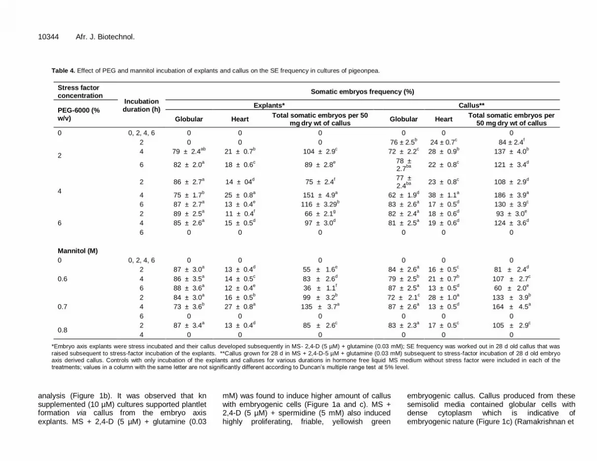
Table 4. Effect of PEG and mannitol incubation of explants and callus on the SE frequency in cultures of pigeonpea.
Stress factor concentration
Incubation duration (h)
Somatic embryos frequency (%)
PEG-6000 (% w/v)
Explants* Callus**
Globular Heart Total somatic embryos per 50
mg dry wt of callus
Globular Heart Total somatic embryos per
50 mg dry wt of callus
0 0, 2, 4, 6 0 0 0 0 0 0
2
2 0 0 0 76 ± 2.5b 24 ± 0.7
c 84 ± 2.4
f
4 79 ± 2.4ab
21 ± 0.7b 104 ± 2.9
c 72 ± 2.2
c 28 ± 0.9
b 137 ± 4.0
b
6 82 ± 2.0a 18 ± 0.6
c 89 ± 2.8
e
78 ± 2.7
ba
22 ± 0.8c 121 ± 3.4
d
4
2 86 ± 2.7a 14 ± 04
d 75 ± 2.4
f
77 ± 2.4
ba
23 ± 0.8c 108 ± 2.9
d
4 75 ± 1.7b 25 ± 0.8
a 151 ± 4.9
a 62 ± 1.9
d 38 ± 1.1
a 186 ± 3.9
a
6 87 ± 2.7a 13 ± 0.4
e 116 ± 3.29
b 83 ± 2.6
a 17 ± 0.5
d 130 ± 3.9
c
6
2 89 ± 2.5a 11 ± 0.4
f 66 ± 2.1
g 82 ± 2.4
a 18 ± 0.6
d 93 ± 3.0
e
4 85 ± 2.6a 15 ± 0.5
d 97 ± 3.0
d 81 ± 2.5
a 19 ± 0.6
d 124 ± 3.6
d
6 0 0 0 0 0 0
Mannitol (M)
0 0, 2, 4, 6 0 0 0 0 0 0
0.6
2 87 ± 3.0a 13 ± 0.4
d 55 ± 1.6
e 84 ± 2.6
a 16 ± 0.5
c 81 ± 2.4
d
4 86 ± 3.5a 14 ± 0.5
c 83 ± 2.6
d 79 ± 2.5
b 21 ± 0.7
b 107 ± 2.7
c
6 88 ± 3.6a 12 ± 0.4
e 36 ± 1.1
f 87 ± 2.5
a 13 ± 0.5
d 60 ± 2.0
e
0.7
2 84 ± 3.0a 16 ± 0.5
b 99 ± 3.2
b 72 ± 2.1
c 28 ± 1.0
a 133 ± 3.9
b
4 73 ± 3.6b 27 ± 0.8
a 135 ± 3.7
a 87 ± 2.6
a 13 ± 0.5
d 164 ± 4.5
a
6 0 0 0 0 0 0
0.8 2 87 ± 3.4
a 13 ± 0.4
d 85 ± 2.6
c 83 ± 2.3
a 17 ± 0.5
c 105 ± 2.9
c
4 0 0 0
0 0 0
*Embryo axis explants were stress incubated and their callus developed subsequently in MS- 2,4-D (5 µM) + glutamine (0.03 mM); SE frequency was worked out in 28 d old callus that was
raised subsequent to stress-factor incubation of the explants. **Callus grown for 28 d in MS + 2,4-D-5 µM + glutamine (0.03 mM) subsequent to stress-factor incubation of 28 d old embryo axis derived callus. Controls with only incubation of the explants and calluses for various durations in hormone free liquid MS medium without stress factor were included in each of the treatments; values in a column with the same letter are not significantly different according to Duncan’s multiple range test at 5% level.
analysis (Figure 1b). It was observed that kn supplemented (10 µM) cultures supported plantlet formation via callus from the embryo axis explants. MS + 2,4-D (5 µM) + glutamine (0.03
mM) was found to induce higher amount of callus with embryogenic cells (Figure 1a and c). MS + 2,4-D (5 µM) + spermidine (5 mM) also induced highly proliferating, friable, yellowish green
embryogenic callus. Callus produced from these semisolid media contained globular cells with dense cytoplasm which is indicative of embryogenic nature (Figure 1c) (Ramakrishnan et

Viji et al. 10345 Table 5. Effect of several concentrations of 2,4-D and ABA on the maturation of somatic embryos in 18 d and 36 d old suspension cultures of pigeonpea.
2,4-D (µM)
Somatic embryos at different stages (%) on
2,4-D + ABA (µM)
Somatic embryos at different stages (%) on 36 d (end of second subculture)** 18-day (end of first subculture)* 36-day (end of second subculture)*
Globular Heart Torpedo Total somatic embryos ***
Globular Heart Torpedo Total somatic embryos ***
Globular Heart Torpedo Cotyledonary Total somatic embryos ***
0 0 0 0 0 0 0 0 0 2,4-D 0 + ABA 0 0 0 0 0 0
5 79 ± 2.3a 21 ± 0.7c 0 194 ± 5.9e 78 ± 2.3a 22 ± 5.0c 0 192 ± 7.0e 2,4-D 2 + ABA 1 63 ± 2.6a 23 ± 1.2b 14 ± 0.5a 0 203 ± 6.0d
4 75 ± 2.3b 25 ± 0.9b 0 226 ± 6.3d 76 ± 2.4a 24 ± 1.1b 0 229 ± 8.7d 2,4-D 2 + ABA 2 51 ± 2.0b 25 ± 1.2b 15 ± 0.5a 9 ± 0.5c 216 ± 6.8c
3 73 ± 2.2b 27 ± 0.9a 0 267 ± 7.4b 75 ± 2.2a 25 ± 1.2b 7 ± 0.4 258 ± 8.7b 2,4-D 2+ ABA 3 26 ± 1.3d 10 ± 0.5d 16 ± 0.5a 48 ± 2.2a 294 ± 8.7a
2 67 ± 2.2c 25 ± 0.9b 8 ± 0.4 315 ± 9.5a 66 ± 2.1b 27 ± 1.1a 0 310 ± 9.3a 2,4-D 2 + ABA 4 47 ± 2.4c 20 ± 1c 15 ± 0.5a 18 ± 0.8b 242 ± 6.7b
1 7 8 ± 2.3a 22 ± 0.7c 0 243 ± 7.3c 76 ± 2.6a 24 ± 1.0b 0 242 ± 8.2c 2,4-D 2 + ABA 5 44 ± 2.1c 43 ± 2a 8 ± 0.3b 5 ± 0.3d 184 ± 5.7e
*Suspension cultures were initiated from 28 d old callus that was raised subsequent to PEG incubation of embryo axis derived callus and the cultures were subcultured after 18 days and were grown in
the same medium composition for the first and second subcultures. **Second subculture was initiated from cells grown in MS+2,4-D-2 µM + 0.03 mM glutamine during the first subculture and the cultures were grown in various concentrations of ABA supplemented medium during the second subculture. ***per 50 mg dry wt of cells. Values in a column with the same letter are not significantly different according to Duncan’s multiple range test at 5% level.
al., 2005). Spermidine has been shown to be an important growth factor which regulates plant growth and development and its supplementation in suspension cultures of ginseng (Panax ginseng) showed high frequency somatic embryo production (Kevers
et al., 2002). It was clear from
the results that 2,4-D along with either spermidine or glutamine was effective in the formation of embryogenic callus. Endogenous hormone level is considered as a crucial factor for SE from explants of pigeonpea (Mohan and Krishnamurthy, 2002). In hypocotyl cultures of carrot, the concentration of exogenously supplied auxin (10 mM) that is required for SE is usually known to be much higher than the endogenous auxin levels (Ribnicky et al., 1996). It has been shown in cultures of A. thaliana that explants with high endogenous level of auxin are more amenable for SE and these tissues did not even require supplementation of auxin for SE (Ikeda-Iwai
et al.,
2003). Relatively high concentration of 2,4-D (5 – 10 µM) has been shown to be an essential supplement for SE in cultures of soybean (Glycine max) (Finer and Nagasawa, 1988).
Supple-
mentation of 2,4-D was found to be critical for producing embryogenic callus and the subsequent formation of somatic embryos in cultures of black gram (Vigna mungo) and green gram (Vigna radiata) (Eapen and George, 1990) and moth bean (Vigna aconitifolia) (Kailash Choudhary et al., 2009). It has been shown that the formation of an embryogenic cell is related to hypermethylation of nuclear DNA due to 2,4-D (Namisivayam, 2007).
Furthermore, 2, 4-D which brings about changes in the physiology and gene expression of the embryogenic cells acts as a stress factor triggering embryogenic pattern of development in cultured cells. Genomic analysis employing cDNA microarray showed differential expression of ca. 495 genes in relation to SE due to 2,4-D supplementation in cultures of soybean (Glycine max) (Thibaud
et al., 2003). These genes have
been annotated to be of those involved in oxidative burst/detoxification, cell wall modify-cation, cell division and stress responses which were upregulated during the early stage of somatic embryos. Supplementation of high
concentration of kn (22.2 µM) has been shown to stimulate SE
in pigeonpea (Patel
et al., 1994). In
contrast, kn at relatively low concentrations (1.16 - 4.64 µM) has been shown to induce re-callusing with the formation of non-embryogenic cells in embryogenic cultures of pigeonpea which did not favor SE (Mohan and Krishnamurthy, 2002).
In vitro tissue browning due to accumulation of phenolic compounds has been considered to be a serious problem often associated with legume tissue cultures (Bhanumathi et al., 2005). Callus browning has been proposed to be due to hypersensitivity reaction resulting from the activation of phenol metabolizing enzymes such as phenylalanine ammonia lyase (PAL), pero-xidase and phenolases. Several reports showed that reduced form of nitrogen, particularly the amino acids such as glutamine, proline and alanine, could inhibit browning and improve cell proliferation and regeneration in specific geno-types of legumes (Kakkar et al., 2000). In black gram, high frequency formation of somatic embryos was observed in MS liquid medium supplemented with 2,4-D and glutamine

10346 Afr. J. Biotechnol.
Figure 1. Somatic embryogenesis of pigeonpea. a) 28-day old embryogenic callus culture grown on MS + 2,4-D (5 µM)
+ glutamine (0.03 mM); b) non-embryogenic cells in callus tissue grown on MS + 2,4-D (5 µM) + kn (5 µM); c) embryogenic cells in callus tissue grown on MS + 2,4-D (5 µM) + glutamine (0.03 mM); d) globular stage somatic embryos in callus tissue grown on MS + 2,4-D (5 µM) + glutamine (0.03 mM) subsequent to stress incubation in PEG (4%, w/v; 4 h) and after 28 d; e) heart stage somatic embryos in callus tissue grown on MS + 2,4-D (5 µM) + glutamine (0.03 mM) after to incubation in PEG (4%, w/v; 4 h) and after 28 days; f) torpedo stage somatic embryo from suspension culture grown in MS + 2,4-D (2 µM) + ABA (3 µM) + glutamine (0.03 mM) after 18 days; g) cotyledonary stage embryo showing xylogenesis after 36 days; h) mature cotyledonary stage embryo after 36 d; i) in vitro regenerated plantlet in
half-strength MS medium; j) hardened in vitro developed plantlet in garden soil; bar in a to g: 100 µM and in h: 500 µM. C, Cotyledon; R, root; X, xylem.
(Muruganantham et al., 2010). Comparably, in the present
study, browning was overcome by the supplementation of glutamine in the embryogenic callusing medium. Effect of stress incubation on SE Somatic cells are potentially embryogenic and an induction is required for conferring embryogenic com-petence in vitro (Stasolla et al., 2003). SE in cultures has been shown to have two distinct phases; viz. induction and maturation. Somatic embryo induction has been
shown to be a function of supplementation of 2,4-D along with abiotic stress-factor incubation of the cultures (Lincy et al., 2009). It has been proposed that auxin and abiotic stress-factors together play a central role in initiating signal transduction cascades resulting in reprogramming of gene expression associated with polarized callus growth leading to somatic embryos. Water- and osmotic-stress incubation in vitro was shown to mimic the conditions of the embryo sac of the zygotic embryos (Ikeda-Iwai
et al., 2003; Stasolla et al., 2003).
Whereas
osmotic stress-factor (mannitol and sorbitol) incubation was employed as an inductive condition for somatic
embryos in A. thaliana, in white spruce water-stress (PEG) incubation was shown to regulate the maturation and desiccation of the somatic embryos (Ikeda-Iwai
et al.,
2003; Stasolla et al., 2003). In white spruce, formation of ca. 500 somatic embryos per gram of fr wt
of callus has
been considered to be a high throughput somatic embryo production system (Stasolla et al., 2003). PEG-6000 at a low concentration of 50 mg/L was supplemented to embryogenic cultures of green gram for initiating plantlet regeneration subsequent to the maturation of somatic embryos (Siva Kumar et al., 2010).
In the present study, embryo axis or callus derived from embryo axis was stress incubated in shake flasks for specified durations (Table 4). It has been reported in A. thaliana that the incubation of shoot apical tip explants for duration up to 24 h in stress-factor (mannitol) containing semisolid medium resulted in SE (Ikeda-Iwai
et al., 2003).
However, our study showed that there was no embryo-genesis when the embryo axis or callus derived from it was stress incubated for 24 h on semisolid medium (results not shown). In A. thaliana, SE was observed when shoot apical tip explants were merely placed on dry filter paper for 20 min and subsequently incubated on semisolid nutrient medium
(Ikeda-Iwai
et al., 2003). The
controls in which shake flask incubation alone was carried out with embryo axis or callus derived from embryo axis without stress factor for 0, 2, 4 and 6 h showed no SE and all the samples lacked any of the somatic embryo stages. In the present study, stress incubation in either PEG or mannitol had significant effect on SE that varied in function of concentration and incubation duration of the stress factors (Table 4). Four percent PEG (w/v) incubation for 4 h showed high frequency of embryogenesis induction and also high frequency occurrence of heart shaped somatic embryos (Table 4).
Mannitol was also found to induce SE at an optimal concentration of 0.7 M and 4 h of incubation even though the frequency of response was low as compared to PEG. Embryo axis or callus derived from the embryo axis underwent senescence when incubated either in PEG (6%, w/v; 6 h) or mannitol (0.7 M; 6 h and 0.8 M; 4 h). Stress incubation with either PEG or mannitol resulted in the production of higher level of globular stage somatic embryos as compared to heart shaped somatic embryos (Figure 1d and e). There was no further maturation of the somatic embryos beyond heart shaped stage under these conditions. Prolonged maintenance of PEG/mannitol incubated callus on the semisolid SEM did not result in the maturation of somatic embryos (results not shown). As compared to stress incubated embryo axis, callus that was subjected to stress incubation responded at a higher level for somatic embryo production due to PEG/mannitol. Accordingly, subsequent experiments employing suspension cultures aiming to the maturation of somatic embryos were restricted to callus subjected to PEG incubation.
Viji et al. 10347 Maturation of somatic embryos in suspension cultures In the present study, formation of torpedo and cotyledonary stage somatic embryos was not observed in the MS semisolid media on which explants or callus were cultured after stress application. It is known that cells in the callus cultures experience cross inhibition in their SE potential due to their contact with the adjacent cell populations that does not possess embryogenesis potential (Karami et al., 2009). In a cell population, this potential is known to be a function of the relative position of the cells in the concentration gradient of embryo-genesis that exists in the callus tissue. In order to overcome the cross inhibition, cell suspension cultures are preferred where the cells are relatively free from cross inhibition. Hence, in the present study, maturation of the somatic embryos was worked out in MS liquid media. Results show the occurrence of two distinct phases in the behavior of cells in the suspension cultures. These phases consisted of the maintenance of embryogenesis potential and the maturation of somatic embryos in the cultures. Based on the observations of the present study, it was found essential to work out a suspension culture system where embryogenesis potential and somatic embryos maturation was obtained at an optimal level.
Previous reports show two distinct approaches in converting the early stage somatic embryos to maturation. Decrease in the concentration of 2,4-D from that employed for embryogenesis medium and supple-mentation of abscisic acid (ABA) along with 2,4-D in the maturation medium was shown to support the formation of cotyledonary stage somatic embryos (Varisai Mohamed
et al., 2004). It has been shown that pro-
embryogenic masses and mature stage somatic embryos were developed from leaf callus of pigeonpea in sus-pension cultures containing relatively low concentration of 2,4-D (Anbazhagan and Ganapathy, 1999). Accordingly, in the present study decrease in the concentration of 2,4-D and supplementation of ABA was attempted to work out the conditions for the maturation of the somatic embryos (Table 5; Figure 1f and i). Optimal conditions for the maturation was attempted by employing two subcultures of the suspension cultures each with a duration of 18-day either in the same culture medium or in a different medium in the second subculture as indicated in Table 5. Results show that decreasing the concentrations of 2, 4-D produced higher amount of total somatic embryos. 2,4-D at the optimal concentration of 2 µM resulted in the maturation to torpedo stage at the end of the second subculture.
Furthermore, transfer of cultures grown in 2 µM 2,4-D during the first subculture to medium containing several concentrations of ABA in the second subculture resulted in the maturation of somatic embryos to cotyledonary stage. ABA at 1µM did not support the formation of

10348 Afr. J. Biotechnol. cotyledonary stage somatic embryos and ABA at 3 µM was found to be optimal for the production of cotyle-donary stage somatic embryos (ca. 48%). ABA has been shown to be significantly increasing the formation of torpedo stage somatic embryos at high frequency and its maturation to cotyledonary stage in horsegram (Macrotyloma uniflorum) (Varisai Mohamed
et al., 2004).
Prolonged maintenance of cultures in the embryo maturation medium (MS + 2 µM 2,4-D + 3 µM ABA + 0.03 mM glutamine) and the related age of the cell suspension cultures were found to affect the frequency of matured somatic embryos, morphology of the cotyledonary stage somatic embryos and also their conversion into plantlets (results not shown). In the present study, cotyledonary stage somatic embryos developed into plantlets in hormone-free half strength MS semisolid medium at a germination frequency of 81% (Figure 1i). The in vitro developed plantlets were further grown in garden soil at a survival rate of 100% (Figure 1j). So far, a total of 60 plantlets were developed in garden soil. Conclusions
Based on the results of the present study, a protocol for high frequency SE and development of plantlets is presented for pigeonpea. The protocol essentially involved: a) establishment of embryogenic callus in MS + 2,4-D (5 µM) + glutamine (0.03 mM); b) PEG incubation (4% w/v for 4 h) of embryogenic callus for high frequency SE; c) establishment of suspension cultures for maintenance of high embryogenesis potential in MS + 2,4-D (2 µM) and glutamine (0.03 mM); d) conversion of torpedo stage somatic embryos to cotyledonary stage on MS + 2,4-D (2 µM) + ABA (3 µM) + glutamine (0.03 mM), and e) development of cotyledonary stage somatic embryos into plantlets and the subsequent growth and establishment of the plantlets in garden soil. Thus, there was stage-specific requirement for 2,4-D and ABA for the development of cotyledonary stage somatic embryos starting from the culture of embryo axis explants. In the present study, 2,4-D (5 µM) and glutamine (0.03 mM) induced the production of highly proliferative embryogenic callus from 3 d old embryo axis explants and the subsequent embryogenesis up to torpedo stage was worked out with 2 µM 2,4-D. Maturation of somatic embryos to cotyledonary stage was obtained with 2 µM 2,4-D and 3 µM ABA. REFERENCES
Anbazhagan VR, Ganapathy A (1999). Somatic embryogenesis in cell suspension cultures of pigeonpea (Cajanus cajan). Plant Cell Tissue
Org. Cult. 56: 179-184.
Bajaj YPS, Singh H, Gosal SS (1980). Haploid embryogenesis in anther cultures of pigeonpea (Cajanus cajan L.). Theor. Appl. Genet. 58:
157-159.
Baker CM, Wetzstein HY (2004). Leaflet development, induction time and medium influence on somatic embryogenesis in peanut (Arachis
hypogaea L.). Plant Cell Rep. 12: 925-929.
Bhanumathi P, Ganesan M, Jayabalan N (2005). A simple and improved protocol for direct and indirect somatic embryogenesis of peanut (Arachis hypogaea L.). J. Agric. Technol. 1: 327-344.
Duncan DB (1955). Multiple range and multiple F test. Biometrics 11: 42-54.
Eapen S, George L (1990). Ontogeny of somatic embryos of Vigna aconitifolia, Vigna mungo and Vigna radiata. Ann. Bot. 66: 219-226.
Finer JJ, Nagasawa A (1988). Development of an embryogenic suspension culture of soybean (Glycine max L.). Plant Cell Rep.7:
238-241. Graham PH, Vance CP (2003). Legumes: Importance and constraints to
greater use. Plant Physiol. 131: 872-877. Harada H, Kiyosue T, Kamada H, Kobayashi T (1990). Stress-induced
carrot somatic embryogenesis and its application to synthetic seeds.
In: The impact of Biotechnology in Agriculture, RS Sangwan and RS Sangwang Norreel (Eds), Kluwer Academic Publishers, The Neetherlands, pp. 129-157.
Ikeda-Iwai M, Umehara M, Satoh S, Kamada H (2003). Stress-induced somatic embryogenesis in vegetative tissues of Arabidopsis thaliana.
Plant J. 34: 107-114.
Kailash Choudhary M, Singh MS, Rathore NS (2009) Somatic embryogenesis and in vitro plant regeneration in moth bean [Vigna aconitifolia (Jacq.) Marechal]. a recalcitrant grain legume. Plant
Biotechnol. Rep. 3: 205-211. Karami O, Aghavaisi B, Pour AM (2009) Molecular aspects of somatic-
to-embryogenic transition in plants. J. Chem. Biol. 2: 177-190.
Kevers C, Gaspar T, Dommes J (2002). The beneficial role of different auxins and polyamines at successive stages of somatic embryo formation and development of Panax ginseng in vitro. Plant Cell
Tissue Org. Cult. 70: 181-188. Krishna G, Sairam Reddy PS, Ramteke WP, Rambabu P, Sohrab SS,
Rana D, Bhattacharya P (2011). In vitro regeneration through
organogenesis and somatic embryogenesis in pigeonpea (Cajanus cajan L.). Physiol. Mol. Biol. Plants. 26: 12-18.
Lincy AK, Remashree AB Sasikumar B (2009). Indirect and direct somatic embryogenesis from aerial stem explants of ginger (Zingiber officinale Rosc). Acta Bot. Croat. 1: 93-103.
Mohan ML, Krishnamurthy KV (2002). Somatic embryogenesis and plant regeneration in pigeonpea (Cajanus cajan L.). Biol. Plant. 45:
19-25. Murashige T, Skoog F (1962). Revised medium for rapid growth and
bioassays with tobacco tissue cultures. Physiol. Plant. 15: 473-497. Muruganantham M, Ganapathi A, Amutha S, Vengadesan G, Selvaraj N
(2005). Shoot regeneration from immature cotyledonary nodes in black gram (Vigna mungo (L.) Hepper). Indian J. Biotechnol. 4: 551-
557. Muruganantham MS, Amutha S, Ganapathi A (2010). Somatic embryo
productions by liquid shake culture of embryogenic calluses in Vigna
mungo L. In Vitro Cell. Dev. Biol. Plant. 46: 34-40.
Namisivayam P (2007) Acquisition of embryogenic competence during somatic embryogenesis. Plant Cell Tissue Org. Cult. 90: 36-40.
Nene YL, Sheila VK (1990). Pigeonpea geography and importance. In: The Pigeonpea, YL Nene, DH Susan and VK Sheila (Eds), CAB International, Wallingford, UK), pp.1-14.
Patel DB, Barve DM, Nagar N, Mehta AR (1994). Regeneration of pigeonpea, (Cajanus cajan L.) through somatic embryogenesis.
Indian J. Exp. Biol. 32: 581-583. Ramakrishnan K, Gnanam R, Sivakumar P, Manickam A (2005). In vitro
somatic embryogenesis from cell suspension cultures of cowpea (Vigna unguiculata L.). Plant Cell Rep. 24: 449-461.
Reddy MV, Sharma SB, Nene YL (1990) Pigeonpea: disease management. In The Pigeonpea, YL Nene, SD Hall and VK Sheila
(Eds), CAB, Wallingford, UK, pp. 303-347.
Ribnicky DM, Ilic N, Cohen JD, Cooke TJ (1996). The effects of exogenous auxins on endogenous indole-3-acetic acid metabolism, The implications for carrot somatic embryogenesis. Plant Physiol.
112: 549-558. Siva Kumar P, Gnanam R, Ramakrishnan R, Manickam A (2010).
Somatic embryogenesis and regeneration of Vigna radiata L. Biol.
Plant. 54: 245-251. Shagufta Z, Aamir A, Fayyaz AS, Javed I (2008) Somatic embryo-

genesis from immature cotyledons and leaf calli of chick pea (Cicer arietinum L.) Pak. J. Bot. 40: 523-531.
Stasolla C, Zyl LV, Egertsdotter U, Craig D, Liu W, Sederoff, RR (2003).
The effects of polyethylene glycol on gene expression of developing white spruce somatic embryos. Plant Physiol. 131: 49-60.
Somers DA, Samac DA, Olhoft PM (2003). Recent advances in legume
transformation. Plant Physiol. 131: 892-899. Taras PP, Prinsen E, Ayaydin F, Miskolczi P, Potters G, Asard H, Harry
A, Onckelen V, Dudits D, Feher A (2002). The role of auxin, pH and
stress in the activation of embryogenic cell division in leaf protoplast-derived cells of Alfalfa. Plant Physiol. 129: 1807-1819.
Thibaud NF, Shealy RT, Khanna A, Vodkin LO (2003). Clustering of
microarray data reveals transcript patterns associated with somatic embryogenesis in soybean (Glycine max L.). Plant Physiol. 132: 118-
136.
Viji et al. 10349 Varisai Mohamed S, Wang CS, Thiruvengadam M, Jayabalan N (2004).
In vitro plant regeneration via somatic embryogenesis through cell suspension cultures of horse gram (Macrotyloma uniflorum Lam.). In
Vitro Cell. Dev. Biol. Plant. 40: 284-289.
Zavattieri MA (2010) Induction of somatic embryogenesis as an example of stress-related plant reactions. Electr. J. Biotechnol. 13: 1-
9.


