Embed Size (px)
Citation preview

Effect of PITX2 knockdown on transcriptome of primary humantrabecular meshwork cell cultures
Seyed Hassan Paylakhi,1 Jian-Bing Fan,2 Mohadeseh Mehrabian,3 Majid Sadeghizadeh,1 Shahin Yazdani,4
Ali Katanforoush,5 Mozhgan Rezaei Kanavi,4,6 Mostafa Ronaghi,2 Elahe Elahi3,7,8
1Department of Genetics, Faculty of Biological Sciences, Tarbiat Modares University, Tehran, Iran; 2Illumina Inc, San Diego, CA;3Department of Biotechnology, University of Tehran, Tehran, Iran; 4Ophthalmic Research Center, Shahid Beheshti University ofMedical Sciences Tehran, Iran; 5Department of Computer Science, Faculty of Mathematics, Shahid Beheshti University G.C.,
6 Central Eye Bank of Iran, Tehran, Iran; 7Department of Biology, University College of Science, University of 8Center of Excellence in Biomathematics, School of Mathematics, Statistics and Computer Science, College
of Science, University of Tehran, Tehran, Iran
Purpose: To identify genes whose expressions in primary human trabecular meshwork (TM) cell cultures are affected bythe transcription factor pituitary homeobox 2 (PITX2) and to identify genes that may have roles in glaucoma. Knownglaucoma causing genes account for disease in a small fraction of patients, and we aimed at identification of other genesthat may have subtle and accumulative effects not easily identifiable by a genetic approach.Methods: Expression profiles derived using microarrays were compared between TM control cells and cells treated withPITX2 siRNAs using three protocols so as to minimize false positive and negative results. The first protocol was basedon the commonly used B statistic. The second and third protocols were based on fold change in expression. The secondprotocol used a threshold of at least 2 fold change in expression, whereas the third protocol used ranking in fold changewithout setting a threshold. The likelihood of a selected gene being a true positive was considered to correlate with thenumber of protocols by which it was selected. By considering all genes that were selected by at least one protocol, thelikelihood of false negatives was expected to decrease. Effects on a subset of selected genes were verified by real timePCR, western blots, and immunocytochemistry. Effects on ALDH1A1, were further pursued because its protein product,aldehyde dehydrogenase 1 family, member A1, has roles in oxidative stress and because oxidative stress is known to berelevant to the etiology of glaucoma.Results: The expression level of 41 genes was assessed by to be possibly affected by PITX2 knockdown. Twenty onegenes were down-regulated and twenty were upregulated. The expression of five genes was assessed to be altered by allthree analysis protocols. The five genes were DIRAS3 (DIRAS family, GTP-binding RAS-like 3), CXCL6 (chemokine(C-X-C motif) ligand 6), SAMD5 (sterile alpha motif domain containing 5), CBFB (core-binding factor, beta subunit),and MEIS2 (meis homeobox 2). Real time PCR experiments verified results on a subset of genes tested. Notably, theresults were also confirmed in two independent TMs. Effects on CXCL6 and ALDH1A1 were also confirmed by westernblots, and effects on ALDH1A1 were further shown by immunocytochemistry. Data consistent with PITX2 involvementin ALDH1A1 mediated response to oxidative stress were presented.Conclusions: Bioinformatics tools revealed that the genes identified affect functions and pathways relevant to glaucoma.Involvement of PITX2 in expression of some of the genes and in some of the pathways is being reported here for the firsttime. As many of the genes identified have not been studied vis-à-vis glaucoma, we feel they introduce new candidatesfor understanding this devastating disease.
Pituitary homeobox 2 (PITX2) is a homeoboxtranscription factor (TF) related to the paired class ofhomeodomain proteins [1]. It affects the development ofvarious ocular tissues. During murine embryonicdevelopment, PITX2 is expressed in neural crest andmesoderm precursors, both of which contribute to theperiocular mesenchyme [2]. Mice carrying targeted deletionsin PITX2 exhibit eye development defects that include
Correspondence to: Elahe Elahi, Professor, University College ofScience, University of Tehran, Tehran, Iran; Phone:00989122181251; FAX: 00982166405141; email:[email protected] or [email protected]
agenesis of the corneal epithelium and stroma, loss ofextraocular muscles, and abnormalities of the optic nerve[3-5]. Finally, mutations in PITX2 are cause of Axenfeld-Rieger syndrome (ARS) in a subset of patients [6,7]. ARS ischaracterized by defects in the anterior segment of the eye andsystemic malformations [6,7]. Notably, approximately 50%of ARS patients develop glaucoma, usually in adolescence orearly adulthood [8]. ARS patients harboring PITX2 mutationsare among those at risk of developing glaucoma [8].
The fact that mutations in PITX2 can cause the ARSphenotype and that the mutations may culminate in a non-congenital form of glaucoma may be signatures of PITX2functions in the mature TM [9]. Here, we report the effects of
Molecular Vision 2011; 17:1209-1221 <http://www.molvis.org/molvis/v17/a136>Received 10 February 2011 | Accepted 25 April 2011 | Published 5 May 2011
© 2011 Molecular Vision
1209
Tehran, Iran; Tehran, Tehran, Iran;

siRNA knockdown of PITX2 on global gene expression inprimary human TM cell cultures using high densitymicroarrays. Pathways and functions implicated for theaffected genes were derived using bioinformatics tools.CXCL6 (chemokine (C-X-C motif) ligand 6) which has rolesin immune response and ALDH1A1 (aldehyde dehydrogenase1 family, member A1) which has roles in oxidative stress wereamong the affected genes. The effects of the knockdown onALDH1A1 were further pursued [10,11].
METHODSThis research was performed in accordance with the HelsinkiDeclaration and with approval of the ethics board of theUniversity of Tehran. Eye globes were obtained from theCentral Eye Bank of Iran.
Preparation of primary TM cultures: Four primarycultures were developed from donors without history of eyedisease aged 25 (male; TM1), 30 (male; TM2), 65 (male;TM4), and 60 (female; TM5) years old at time of death [12].Cells were maintained as previously described [12]. Thenature of the cells was confirmed by demonstrating increasedexpression of myocilin mRNA and protein upondexamethasone treatment (data not shown) [12].
siRNA treatment and RNA extraction: Fourth to sixthpassage cells were used in all experiments. Each of the fourTM cultures was exposed to three siRNA treatments induplicate wells. Lipofectamine RNAiMAX (Invitrogen,Carlsbad, CA) was used for reverse transfection according tothe manufacturer’s instructions. We had previously shownthat this treatment has no detectable effect on cell growth (datanot shown). Approximately 8×10⁴ cells were added to wellscontaining siRNA to achieve a final siRNA concentration of75 nM. These siRNA duplices (Dharmacon Research,Lafayette, CO) were used: PITX2 siRNA-1 (J-017315–05),PITX2 siRNA-2 (J-017315–06), and scrambled siRNA-1(D-001810–01–20). All TM cultures had two independenttreatments with scrambled siRNA (i.e., two treatments, twowells for each) so as to enable testing of technical replicationof the array expression assessments.
Forty eight hours after exposure to siRNAs, cells wereharvested and cells of duplicate wells were combined andplaced in RNXTM-plus (Cinnagen, Tehran, Iran). Total RNAwas isolated according to the manufacturer’s instructions.RNA quality was assessed using density ratio of 28S to 18SrRNA bands. Half of the RNA was set aside for microarrayexperiments and the other half was used for real time PCR.
Assessment of knockdown by siRNAs: Candidate controlgenes for assessment of effects of PITX2 siRNAs wereACTB (actin, beta), β2M (beta-2 microglobulin), GAPDH(glyceraldehyde-3-phosphate dehydrogenase), and HPRT1(hypoxanthine phosphoribosyltransferase 1). cDNA synthesiswas done by standard procedures and real time PCR wasperformed on a Corbett 65H0 machine (Corbett Research,
Sidney, Australia) using the QuantiFast SYBR Green PCR Kit(QIAGEN, Germantown, MD). After selection of appropriatecontrol gene using GNorm program [13], PITX2 siRNAknockdown effects on PITX2 were determined by real timePCR. The real time PCR primers were obtained fromQIAGEN.
Microarray experiments and data analysis: Arrayexperiments were performed only on TM1 and TM2 culturesusing HumanRef-8 V2 Illumina Genome-Wide ExpressionBeadChips according to the manufacturer’s instructions(Illumina, San Diego, CA). Arrays were scanned with theIllumina BeadArray reader and images were analyzed usingBeadstudio VI software. The Beadstudio data werenormalized using a quantile based algorithm available in theBeadarray software [14]. Values for all genes were comparedbetween the two replicate control samples of TM1 andbetween the two replicate control samples of TM2. Similarly,effect of PITX2 siRNA treatments on three housekeepinggenes ACTB, β2M, and GAPDH on the different arrays weredetermined. Subsequently, genes the expressions of whichwere affected by siRNA treatments were assessed based onthree protocols as described below.
First, the Limma package was used [15]. For each of thefour assessments (two siRNAs, two TMs), genes were orderedbased on the value of their associated B statistic. Genes werethen selected on conditions that they had a B statistic value≥2 in at least two of the four assessments, that both siRNAswere represented in these assessments, and that the directionof change in expression was the same in the assessments.
In the second protocol, expression in presence of each oftwo siRNAs was compared separately to each of two controlsfor each TM, producing eight comparisons for the two TMs.Genes were selected on conditions that they showed ≥2 foldchange in the same direction in at least four of the eightcomparisons and that each siRNA was represented at leastonce among the selected comparisons.
In the third protocol, essentially the 40 top ranking genesaffected by siRNAs of PITX2 based on fold change inexpression were selected. Initially, fold changes in thepresence of siRNA1 in each TM were assessed by comparisonto average of controls for the respective TM, and forty topranking genes in each TM were identified (Group A genes:siRNA-1 top 40 TM1 list, siRNA-1 top 40 TM2 list).Subsequently, genes affected were sorted by adding rank ofgenes affected in the two TMs, and selecting the top 40 genes(Group A genes: siRNA-1 top 40 TM1/2 list). This processwas repeated for siRNA-2 (Group B genes: siRNA-2 top 40TM1 list, siRNA-2 top 40 TM2 list, siRNA-2 top 40 TM1/2list). Finally, the rankings of genes affected by both siRNAsin each TM were added, and then the rankings of both siRNAsin both TMs were added (Group C genes: siRNA-1/2 top 40TM1 list, siRNA-1/2 top 40 TM2 list, siRNA-1/2 top 40TM1/2 list). In this protocol, genes were ultimately selected
Molecular Vision 2011; 17:1209-1221 <http://www.molvis.org/molvis/v17/a136> © 2011 Molecular Vision
1210
that appeared at least once in each of the three Groups ofgenes. A computer program written to perform this selectiontask is available at Joint-rank.
Real time PCR: Real time PCR was performed on a subsetof microarray based selected genes on the two TMs that hadbeen used for microarray analysis and also on TM4 and TM5.cDNA synthesis and real time PCR were performed asdescribed above using QIAGEN primers for ACTB,ALDH1A1, CXCL6, DIRAS3 (DIRAS family, GTP-bindingRAS-like 3), DKK1 (Dickkopf-1), KCNJ2 (potassiuminwardly-rectifying channel, subfamily J, member 2),MEIS2 (meis homeobox 2), PITX2, and SAMD5 (sterile alphamotif domain containing 5). At least four replicate PCRs foreach gene in each TM were performed. Detailed informationon all primers is provided in Table 1. ACTB was used ascontrol gene in the real time PCR experiments except whereindicated otherwise. Fold change in expression of selectedgenes were obtained using software provided by the Corbettinstrument, used for the real time PCR experiments..
Western blot analysis: TM cultured cells were harvestedand lysed in Lameli lysis buffer. Protein (60–100 µg) waselectrophoresed on 10%–15% denaturing polyacrylamidegels and the resolved proteins were transferred ontonitrocellulose membranes. The ECL Advance Luminol basedchemiluminescence detection kit was used for detection ofspecific proteins after reaction with appropriate antibodiesaccording to the manufacturer’s instructions (GE Healthcare,Salt Lake City, UT). Specific antibodies used were goatpolyclonal antibodies (Santa Cruz Biotechnology, Inc., SantaCruz, CA) against human myocilin (C-15), Lamin B (C-20)and rabbit polyclonal antibodies (Abcam, Cambridge, MA)against ALDH1A1 (ab51028) and CXCL6 (ab9923). Laminserved as control protein. Secondary antibodies werehorseradish peroxidase conjugated anti-goat (SC-2768; SantaCruz Biotechnology) and anti-rabbit (Sigma-Aldrich, Poole,UK) antibodies. Western blot analysis for each protein wasperformed twice.
Immunofluorescence analysis on cultured cells andhistological sections: TM1 cells grown on coverslips in thepresence of scrambled siRNA or PITX2 siRNA-1 were fixedwith an acetone-methanol mix, blocked with BSA, incubatedwith the anti-ALDH1A1 antibody described above, andexposed to fluorescent conjugated goat anti-rabbit secondaryantibody (AF8035; Razi BioTech, Tehran, Iran). Nuclei werecounterstained with DAPI (Invitrogen). Cells were visualizedwith an Axioplan 2 fluorescence microscope (Carl Zeiss, Jena,Germany). Additionally, expression of both ALDH1A1 andCXCL6 were assessed in cryosections of a globe from a threeyear old cadaver.
Comparison of genes identified to genes reported inprevious glaucoma relevant array studies: Fifteen microarraygene expression studies in which effects of various glaucomarelevant conditions or treatments were studied, includingeffects of dexamethsone, pressure, TGFβ, and geneknockdown, have been reported [16-30]. PITX2 affectedgenes identified here were compared to genes identified in theprevious studies to identify possible commonalities.
In silico analysis of selected genes: Promoter regions ofselected genes were screened for PITX2 binding sites so as toidentify genes whose transcriptions are more likely to bedirectly affected by this TF. PITX2 binds the bicoid sequenceelement 5ʹ-TAATCC-3ʹ [31,32]. The region between 3,000 bpupstream and 200 bp downstream of transcription initiationsites of potential target genes was screened for presence of theelement [33,34]. Finally, genes identified by the microarrayanalysis were analyzed to identify potentially relevantfunctional pathways and functional categories or geneontology terms annotated by Kyoto Encyclopedia of Genesand Genomes (KEGG) [35] and Gene Ontology (GO) [36]using the Database for Annotation, Visualization, andIntegrated Discovery (DAVID) bioinformatics tool [37]. Acutoff p value of 0.05 was used for enriched KEGG pathwaysor GO functions.
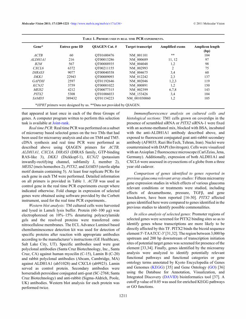
TABLE 1. PRIMERS USED IN REAL TIME PCR EXPERIMENTS.
Gene* Entrez gene ID QIAGEN Cat. # Target transcript Amplified exons Amplicon length(bp)
ACTB 60 QT01680476 NM_001101 ** 104ALDH1A1 216 QT00013286 NM_000689 11, 12 97
B2M 567 QT00088935 NM_004048 1,2 98CXCL6 6372 QT00211155 NM_002993 2 75DIRAS3 9077 QT00040558 NM_004675 3,4 60DKK1 22943 QT00009093 NM_012242 2,3 137
GAPDH 2597 QT01192646 NM_002046 1,2,3 119KCNJ2 3759 QT00001022 NM_000891 1,2 150MEIS2 4212 QT00077315 NM_002399 6,7,8 143PITX2 5308 QT01006033 NM_153426 3,4 104
SAMD5 389432 QT01154223 NM_001030060 1,2 105
*HPRT primers were designed by us. **Data not provided by QIAGEN.
Molecular Vision 2011; 17:1209-1221 <http://www.molvis.org/molvis/v17/a136> © 2011 Molecular Vision
1211

Hydrogen peroxide (H2O2) and lithium chloride (LiCl)treatments: H2O2 and LiCl treatments were used, respectively,as surrogates for analysis of effects of oxidative stress and Wntsignaling [38,39]. For assessment of these agents onexpression of PITX2 and ALDH1A1, TM1 cells were exposedto five treatments and each treatment had its own control. Thefirst treatment was exposure to PITX2 siRNA, and controltreatment was exposure to scrambled siRNA. In the second,cells were exposed to 600 mM H2O2 for 4 h, and in the thirdtreatment cells had the same exposure to H2O2 after havingbeen exposed to PITX2 siRNA-1. The fourth treatment wasexposure to 20 mM LiCl for 12 h. In the fifth treatment,H2O2 was added to cells that had already been exposed to LiClfor 12 h, and incubation was continued for an additional 4 h.After treatments, real time PCR was performed to assesslevels of PITX2 and ALDH1A1 expression. β2M was used ascontrol gene because H2O2 has been reported to affect actinexpression [39]. The effects of the H2O2 and LiCl treatmentson ALDH1A1 expression were also assessed by westernanalysis. Statistical analysis was done using RelativeExpression Software Tool (REST) software [40].
RESULTSGNorm identified ACTB and β2M as genes with best stabilityvalue (M=0.028). The amount of knockdown of PITX2mRNA by its siRNAs using ACTB as control in the four TMcultures (TM1, TM2, TM4, TM5) ranged from 76%–93% andthe average was 84% as assessed by real time PCR (Figure 1).The raw microarray data on TM1 and TM2 have beendeposited in the Gene Expression Omnibus (GEO) database,(accession GSE27275). The levels expression of most genesin the two independent TM1 culture control samples that hadbeen treated in identical manner with scrambled siRNA werevery similar and only one gene showed >│2.0│fold changein expression between the two replicates (GTF21; 2.1×).Similarly, only one gene showed >│2.0│fold change inexpression between the two independent scrambled siRNAtreated TM2 cells (TAF5L; 2.1×). These results are indicativeof acceptable experimental replication. Fold change inexpression of three housekeeping genes ACTB, β2M, andGAPDH due to PITX2 siRNA treatments was low for thesiRNAs in both TM1 and TM2, averaging 1.01 (Table 2).
Microarray data analysis: Protocols 1, 2, and 3identified, respectively, 26, 14, and 20 genes affected byPITX2 siRNA treatments, and some genes were commonbetween genes selected by different protocols. In total 41different genes were identified, and these will henceforth becalled “PITX2-filtered genes.” Nine genes were identified bytwo protocols and five by all three of the protocols. The genesidentified are listed in Table 3. The five genes identified byall the protocols were DIRAS3, CXCL6, SAMD5, CBFB (core-binding factor, beta subunit), and MEIS2. Known glaucomacausing genes, CYP1B1 (cytochrome P450, family 1,subfamily B, polypeptide 1), MYOC (myocilin), OPTN
(optineurin), and WDR36 (WD repeat domain 36) were notamong the genes affected by PITX2 [41]. LTBP2 (latenttransforming growth factor beta binding protein 2), a primaryglaucoma causing gene, was not probed on the arrays [42].
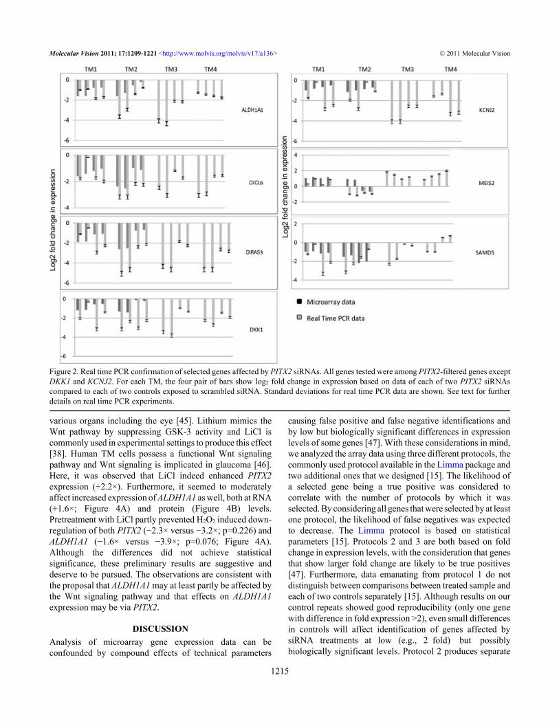
To confirm microarray based assessments, real time PCRwas performed on a subset of genes. Five genes affected byPITX2 siRNAs, ALDH1A1, CXCL6, DIRAS3, MEIS2,SAMD5, and two additional candidate genes, DKK1 andKCNJ2, were tested. The latter two were not among the genesidentified by the array analysis protocols; they were selectedfor assessment by real time PCR based on earlier reportssuggesting PITX2 may affect their expressions [17,43,44].Effects of PITX2 siRNAs as assessed by real time PCRcorroborated the array data of most genes tested in the senseof the genes being down-regulated or upregulated (Figure 2).The only exception was MEIS2 in TM2. Notably, the effectson the genes observed in TM1 and TM2, were also observedin TM4 and TM5 which originated from globes of olderindividuals. DKK1 and KCNJ2 that had not been selected bycriteria of the array analysis protocols were also shown by realtime PCR to be affected by PITX2 knockdown. Although thearray data on these two genes did not meet the protocolsselection criteria, the array data did in fact indicate somedown-regulation consistent with the real time PCR results(Figure 2).
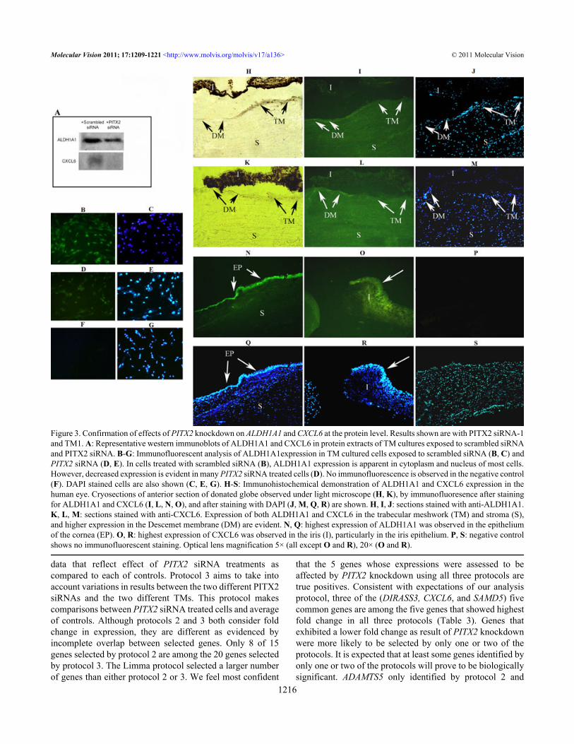
CXCL6 and ALDH1A1, were further analyzed by westernblotting, and the results evidenced that PITX2 siRNAsaffected decreased expression of CXCL6 and ALDH1A1 atthe protein level (Figure 3A). Furthermore, decreasedexpression of ALDH1A1 protein in PITX2 siRNA treatedcells was also shown by immunofluorescence analysis of thetreated cells (Figure 3B-G). Immunofluorescence on globesections showed expression of both genes in the trabecularmeshwork, and additionally in the stroma and Descemetmembrane (Figure 3H-S). Highest expressions of CXCL6and ALDH1A1 were observed, respectively in the irisepithelium and corneal epithelium.
Of the 41 PITX2-filtered genes identified by analysis ofarray data, 11 were also identified in one or more previouslyreported glaucoma related global gene expression studies(Table 4). Notably, DIRAS3 and CXCL6 that had shown thetwo highest fold changes in our experiments (3.9X and 3.3×,respectively; Table 3) and that had both been selected on thebasis of all three selection protocols, had also been repeatedlyreported in other studies [17-20]. ADAMTS5 that wasidentified by only one of our selection protocols waspreviously reported in three studies [17,21,22]. DKK1 andKCNJ2 which were shown to be affected by PITX2 siRNAsby real time PCR experiments were each previously reportedin one study [16,22].
In silico analysis of selected genes: The PITX2 bindingelement was identified within regions surrounding thetranscription initiation sites of some of the affected genes. Of
Molecular Vision 2011; 17:1209-1221 <http://www.molvis.org/molvis/v17/a136> © 2011 Molecular Vision
1212

the 41 PITX2 filtered genes, 18(43.9%) contained two or moreof the binding sequences (data not shown). For example,DKK1 and MEIS2 had, respectively, four and three bindingsites. Table 5 lists KEGG and GO terms that are enrichedamong the 41 PITX2 affected genes.
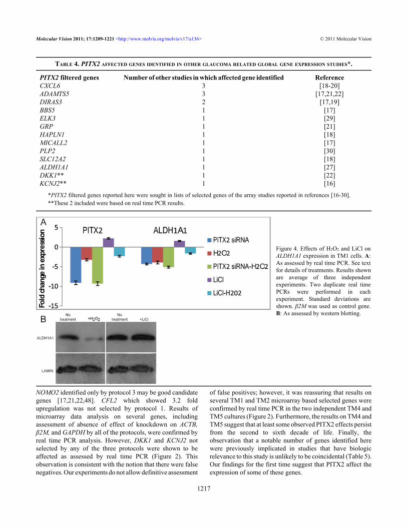
Oxidative stress and Wnt signaling: PITX2 siRNAdecreased PITX2 (−9.1×) and ALDH1A1 (−4.3×) expressionas already described above (Figure 4A). As ALDH1A1 isknown to be involved in the oxidative stress response of theeye, effect of H2O2 on PITX2 and ALDH1A1 expression was
tested. Under the condition of acute exposure to H2O2 usedhere, real time PCR showed that it decreased both PITX2(−3.2×) and ALDH1A1 (−3.9×) expression (Figure 4A). Effectof H2O2 on ALDH1A1 expression at the protein level was alsoevident (Figure 4B). Extent of down-regulation ofALDH1A1 upon simultaneous exposure to PITX2 siRNA andH2O2 (−5.1×) was comparable to effect of H2O2 alone(p=0.566), suggesting that H2O2 and PITX2 may affectALDH1A1 expression by a common pathway (Figure 4A). Itis known that Wnt signaling increases PITX2 expression in
Figure 1. PITX2 transcriptionknockdown by PITX2 siRNA-1 andPITX2 siRNA-2 as assessed by real timePCR.
TABLE 2. EFFECT OF PITX2 SIRNAS ON HOUSEKEEPING GENES ACTB, Β2M, AND GAPDH BASED ON MICROARRAY DATA.
Gene TM1* TM2* Average**ACTB 0.85 0.86 0.85B2M 1.13 1.23 1.18GAPDH 0.98 0.98 0.98
*Average effect of 2 siRNAs on each TM. **Average effect of 2 siRNAs on TM1 and TM2.
Molecular Vision 2011; 17:1209-1221 <http://www.molvis.org/molvis/v17/a136> © 2011 Molecular Vision
1213

TAB
LE 3
. MIC
RO
AR
RA
Y ID
ENTI
FIED
GEN
ES W
ITH
CH
AN
GED
EX
PRES
SIO
N D
UE
TO P
ITX2
SIR
NA
TR
EATM
ENTS
.
Gen
es id
entif
ied
base
d on
B st
atis
ticG
enes
iden
tifie
d ba
sed
on tw
o
fold
cha
nge
cut-
off
Gen
es id
entif
ied
base
d on
ran
k in
fo
ld c
hang
e w
ithou
t cut
-off
All
gene
s sel
ecte
d by
one
or
mor
e pr
otoc
ol
(Pro
toco
l 1)
(Pro
toco
l 3)
(pro
toco
l 1, 2
, +/o
r 3)
Gen
eB
val
ueP
valu
e
mR
NA
fold
c
hang
eG
ene
mR
NA
fold
chan
geG
ene
m
RN
A fo
ldch
ange
Gen
e
mR
NA
fold
chan
ge*
P 1
P 2
P 3
D
own
Up
D
own
Up
D
own
Up
D
own
Up
D
IRAS
35.
90.
0138
484.
2
DIR
AS3
3.3
D
IRAS
34.
2
DIR
AS3
3.9
X
XX
CXC
L64.
10.
0195
353.
1
CXC
L62.
9
CFL
2
3.5
CXC
L63.
3
XX
XSA
MD
54.
40.
0142
133
C
FL2
2.
8SA
MD
53
C
FL2
3.
2
XX
XYLT
14.
40.
0820
042.
9
KH
DRB
S32.
7
CXC
L63
SA
MD
52.
9
XX
XK
HD
RBS3
4.6
0.00
8829
2.8
SA
MD
52.
7
ALD
H1A
12.
5
XYLT
12.
9
X
AL
DH
1A1
3.6
0.03
0613
2.5
AD
AMTS
5
2.5
CBF
B2.
3
KH
DRB
S32.
8
XX
C
7orf
472.
70.
0481
482.
3
C7o
rf47
2.4
PL
P2
2.2
ALD
H1A
12.
5
X
XLO
C65
3602
4.2
0.04
0201
2.
3TM
EM65
2.
4TM
EM65
2.
1AD
AMTS
5
2.5
X
C
BFB
2.1
0.05
4419
2.3
C
BFB
2.4
AUH
2
PATZ
12.
4
X
C
7orf
634.
20.
0706
04
2.2
PATZ
12.
4
MEI
S2
2C
BFB
2.4
X
XX
PLP2
3.2
0.02
711
2.
2LO
C65
3602
2.
3BH
LHB3
2
C7o
rf47
2.3
XX
GRP
3.6
0.03
6988
2.1
BB
S5
2.2
PIP4
K2B
1.
9LO
C65
3602
2.
3
XX
PMS2
3.82
0.03
2
2.1
CTX
N1
2.
2FL
G1.
9
TMEM
65
2.3
X
XSM
C2
3.5
0.06
3532
2
MEI
S2
2.2
LIN
7A1.
8
PLP2
2.
2X
X
MEI
S23.
20.
0681
99
2
HAP
LN1
1.7
M
EIS2
2.
1X
XX
BHLH
B33.
30.
0248
362
GAL
NT1
1.7
C
TXN
1
2.2
X
G
BP2
30.
0390
74
2
QD
PR
1.6
C7o
rf63
2.
2X
IRS2
3.4
0.00
5579
2
PM
S2
1.5
BBS5
2.
2
X
LMN
B13.
50.
0063
32
NO
MO
2
1.5
GRP
2.1
X
SLC
12A2
2.9
0.04
161
1.9
MIC
ALL2
1.
4G
BP2
2
X
C
EBPG
2.5
0.06
1234
1.
9
BH
LHB3
2
X
XC
D4
2.9
0.03
9381
1.
8
LM
NB1
2
X
EL
K3
2.6
0.01
2323
1.8
IR
S22
X
FAM
70A
2.9
0.03
9441
1.8
SM
C2
2
X
G
BP5
4.3
0.03
2596
1.
8
AU
H
2
X
PTPR
R2.
40.
0137
09
1.8
PIP4
K2B
1.
9
X
C
EBPG
1.
9X
SL
C12
A21.
9
X
FLG
1.9
X
C
D4
1.
8X
EL
K3
1.8
X
LI
N7A
1.8
X
PT
PRR
1.
8X
FA
M70
A1.
8
X
GBP
5
1.8
X
PMS2
1.
8X
X
G
ALN
T11.
7
X
HAP
LN1
1.7
X
Q
DPR
1.
6
X
N
OM
O2
1.
5
X
M
ICAL
L2
1.4
X
*Ave
rage
fold
cha
nge
of p
roto
cols
.
Molecular Vision 2011; 17:1209-1221 <http://www.molvis.org/molvis/v17/a136> © 2011 Molecular Vision
1214
(Pro
toco
l 2)
various organs including the eye [45]. Lithium mimics theWnt pathway by suppressing GSK-3 activity and LiCl iscommonly used in experimental settings to produce this effect[38]. Human TM cells possess a functional Wnt signalingpathway and Wnt signaling is implicated in glaucoma [46].Here, it was observed that LiCl indeed enhanced PITX2expression (+2.2×). Furthermore, it seemed to moderatelyaffect increased expression of ALDH1A1 as well, both at RNA(+1.6×; Figure 4A) and protein (Figure 4B) levels.Pretreatment with LiCl partly prevented H2O2 induced down-regulation of both PITX2 (−2.3× versus −3.2×; p=0.226) andALDH1A1 (−1.6× versus −3.9×; p=0.076; Figure 4A).Although the differences did not achieve statisticalsignificance, these preliminary results are suggestive anddeserve to be pursued. The observations are consistent withthe proposal that ALDH1A1 may at least partly be affected bythe Wnt signaling pathway and that effects on ALDH1A1expression may be via PITX2.
DISCUSSIONAnalysis of microarray gene expression data can beconfounded by compound effects of technical parameters
causing false positive and false negative identifications andby low but biologically significant differences in expressionlevels of some genes [47]. With these considerations in mind,we analyzed the array data using three different protocols, thecommonly used protocol available in the Limma package andtwo additional ones that we designed [15]. The likelihood ofa selected gene being a true positive was considered tocorrelate with the number of protocols by which it wasselected. By considering all genes that were selected by at leastone protocol, the likelihood of false negatives was expectedto decrease. The Limma protocol is based on statisticalparameters [15]. Protocols 2 and 3 are both based on foldchange in expression levels, with the consideration that genesthat show larger fold change are likely to be true positives[47]. Furthermore, data emanating from protocol 1 do notdistinguish between comparisons between treated sample andeach of two controls separately [15]. Although results on ourcontrol repeats showed good reproducibility (only one genewith difference in fold expression >2), even small differencesin controls will affect identification of genes affected bysiRNA treatments at low (e.g., 2 fold) but possiblybiologically significant levels. Protocol 2 produces separate
Figure 2. Real time PCR confirmation of selected genes affected by PITX2 siRNAs. All genes tested were among PITX2-filtered genes exceptDKK1 and KCNJ2. For each TM, the four pair of bars show log2 fold change in expression based on data of each of two PITX2 siRNAscompared to each of two controls exposed to scrambled siRNA. Standard deviations for real time PCR data are shown. See text for furtherdetails on real time PCR experiments.
Molecular Vision 2011; 17:1209-1221 <http://www.molvis.org/molvis/v17/a136> © 2011 Molecular Vision
1215
data that reflect effect of PITX2 siRNA treatments ascompared to each of controls. Protocol 3 aims to take intoaccount variations in results between the two different PITX2siRNAs and the two different TMs. This protocol makescomparisons between PITX2 siRNA treated cells and averageof controls. Although protocols 2 and 3 both consider foldchange in expression, they are different as evidenced byincomplete overlap between selected genes. Only 8 of 15genes selected by protocol 2 are among the 20 genes selectedby protocol 3. The Limma protocol selected a larger numberof genes than either protocol 2 or 3. We feel most confident
that the 5 genes whose expressions were assessed to beaffected by PITX2 knockdown using all three protocols aretrue positives. Consistent with expectations of our analysisprotocol, three of the (DIRASS3, CXCL6, and SAMD5) fivecommon genes are among the five genes that showed highestfold change in all three protocols (Table 3). Genes thatexhibited a lower fold change as result of PITX2 knockdownwere more likely to be selected by only one or two of theprotocols. It is expected that at least some genes identified byonly one or two of the protocols will prove to be biologicallysignificant. ADAMTS5 only identified by protocol 2 and
Figure 3. Confirmation of effects of PITX2 knockdown on ALDH1A1 and CXCL6 at the protein level. Results shown are with PITX2 siRNA-1and TM1. A: Representative western immunoblots of ALDH1A1 and CXCL6 in protein extracts of TM cultures exposed to scrambled siRNAand PITX2 siRNA. B-G: Immunofluorescent analysis of ALDH1A1expression in TM cultured cells exposed to scrambled siRNA (B, C) andPITX2 siRNA (D, E). In cells treated with scrambled siRNA (B), ALDH1A1 expression is apparent in cytoplasm and nucleus of most cells.However, decreased expression is evident in many PITX2 siRNA treated cells (D). No immunofluorescence is observed in the negative control(F). DAPI stained cells are also shown (C, E, G). H-S: Immunohistochemical demonstration of ALDH1A1 and CXCL6 expression in thehuman eye. Cryosections of anterior section of donated globe observed under light microscope (H, K), by immunofluoresence after stainingfor ALDH1A1 and CXCL6 (I, L, N, O), and after staining with DAPI (J, M, Q, R) are shown. H, I, J: sections stained with anti-ALDH1A1.K, L, M: sections stained with anti-CXCL6. Expression of both ALDH1A1 and CXCL6 in the trabecular meshwork (TM) and stroma (S),and higher expression in the Descemet membrane (DM) are evident. N, Q: highest expression of ALDH1A1 was observed in the epitheliumof the cornea (EP). O, R: highest expression of CXCL6 was observed in the iris (I), particularly in the iris epithelium. P, S: negative controlshows no immunofluorescent staining. Optical lens magnification 5× (all except O and R), 20× (O and R).
Molecular Vision 2011; 17:1209-1221 <http://www.molvis.org/molvis/v17/a136> © 2011 Molecular Vision
1216
NOMO2 identified only by protocol 3 may be good candidategenes [17,21,22,48]. CFL2 which showed 3.2 foldupregulation was not selected by protocol 1. Results ofmicroarray data analysis on several genes, includingassessment of absence of effect of knockdown on ACTB,β2M, and GAPDH by all of the protocols, were confirmed byreal time PCR analysis. However, DKK1 and KCNJ2 notselected by any of the three protocols were shown to beaffected as assessed by real time PCR (Figure 2). Thisobservation is consistent with the notion that there were falsenegatives. Our experiments do not allow definitive assessment
of false positives; however, it was reassuring that results onseveral TM1 and TM2 microarray based selected genes wereconfirmed by real time PCR in the two independent TM4 andTM5 cultures (Figure 2). Furthermore, the results on TM4 andTM5 suggest that at least some observed PITX2 effects persistfrom the second to sixth decade of life. Finally, theobservation that a notable number of genes identified herewere previously implicated in studies that have biologicrelevance to this study is unlikely to be coincidental (Table 5).Our findings for the first time suggest that PITX2 affect theexpression of some of these genes.
TABLE 4. PITX2 AFFECTED GENES IDENTIFIED IN OTHER GLAUCOMA RELATED GLOBAL GENE EXPRESSION STUDIES*.
PITX2 filtered genes Number of other studies in which affected gene identified ReferenceCXCL6 3 [18-20]ADAMTS5 3 [17,21,22]DIRAS3 2 [17,19]BBS5 1 [17]ELK3 1 [29]GRP 1 [21]HAPLN1 1 [18]MICALL2 1 [17]PLP2 1 [30]SLC12A2 1 [18]ALDH1A1 1 [27]DKK1** 1 [22]KCNJ2** 1 [16]
*PITX2 filtered genes reported here were sought in lists of selected genes of the array studies reported in references [16-30]. **These 2 included were based on real time PCR results.
Figure 4. Effects of H2O2 and LiCl onALDH1A1 expression in TM1 cells. A:As assessed by real time PCR. See textfor details of treatments. Results shownare average of three independentexperiments. Two duplicate real timePCRs were performed in eachexperiment. Standard deviations areshown. β2M was used as control gene.B: As assessed by western blotting.
Molecular Vision 2011; 17:1209-1221 <http://www.molvis.org/molvis/v17/a136> © 2011 Molecular Vision
1217

Two sets of PITX2 related terms identified by thebioinformatics approach are notable, phagocytosis andimmune system related functions. Phagocytosis is recognizedas an important function of human TM cells, partly because itresults in clearing of substances that may hinder facile outflowof aqueous humor [49]. Immune related functions constituteeight of the 29 terms for enriched PITX2 related functionslisted in Table 4, and clinical and experimental studies suggestthat immune system functions are involved in glaucoma [50].Furthermore, immune related functions may be relevant tophagocytosis in the TM [51]. Two genes, CXCL6 andALDH1A1, were prioritized further analysis.
CXCL6 codes a pro-inflammatory cytokine that inducesdirected migration of monocytes and neutrophils [52]. It hasbeen identified to be modulated in three previous glaucomarelated global gene analyses [18-20]. Most recently, it wasreported that human TM cells secrete significant quantities ofCXCL6 [10]. The authors implicated cytokines in regulationof aqueous humor outflow, a function clearly relevant to theglaucoma phenotype [53,54]. Here for the first time wepresent evidence that PITX2 directly or indirectly affects theexpression of this gene. The earlier observations, our findingthat CXCL6 expression is affected by PITX2, and thatPITX2 mutations can cause glaucoma associated ARS suggest
that PITX2 may affect the ARS and glaucoma phenotypes viaan effect on the immune response mediated by CXCL6.
Down-regulation of ALDH1A1 and its expression in thehuman eye, most highly in the corneal epithelium, weredemonstrated (Figure 3). ALDH1A1 and ALDH3A1, alsoexpressed in the anterior segment of mammalian eyes, arealdehyde dehydrogenases that minimize the deleteriouseffects of oxidative damage caused largely by ultravioletradiation [55]. Highly reactive electrophilic products of lipidperoxidation, 4-hydroxy-2-nonenal (4-HNE) and malon-dialdehyde (MDA), are among the agents that promoteoxidative damage [56]. Whereas ALDH1A1 metabolizes both4-HNE and MDA, MDA is a poor substrate for the moreabundant ALDH3A1 [57]. Notably, mouse knockouts ofALDH1A1 exhibited lens opacity later than ALDH3A1 andALDH1A1 double knockouts [11]. Similarly, somephenotypic consequences in ALDH1A1 knockouts becameevident only at an embryonic stage when ALDH3A1was nolonger expressed [58]. Late onset glaucoma in some ARSpatients harboring PITX2 mutations may be due to existenceof proteins that complement ALDH1A1 activity.
Oxidative stress is an important component in theetiology of glaucoma [59]. Moreover, existence of oxidativestress protection mechanisms and physiologic consequencesof oxidative stress in the human TM cells have been reported
TABLE 5. FUNCTIONAL ANNOTATIONS ENRICHED FOR GENES AFFECTED BY PITX2 SIRNAS*.
Category GO ID and Term Number of genes p valueGOTERM_CC_ALL GO:0016363~nuclear matrix 2 0.0013123GOTERM_BP_ALL GO:0030098~lymphocyte differentiation 4 0.0013564GOTERM_CC_ALL GO:0034399~nuclear periphery 2 0.001421GOTERM_CC_ALL GO:0005789~endoplasmic reticulum membrane 3 0.0014526KEGG_PATHWAY hsa04666:Fc gamma R-mediated phagocytosis 2 0.0020273GOTERM_BP_ALL GO:0002520~immune system development 5 0.0026937GOTERM_BP_ALL GO:0002521~leukocyte differentiation 4 0.0026975GOTERM_MF_ALL GO:0019901~protein kinas binding 3 0.005062GOTERM_MF_ALL GO:0030246~carbohydrate binding 4 0.0055687GOTERM_MF_ALL GO:0019900~kinase binding 3 0.0071612GOTERM_BP_ALL GO:0046649~lymphocyte activation 4 0.0086461GOTERM_BP_ALL GO:0030097~hemopoiesis 4 0.0137107GOTERM_BP_ALL GO:0006955~immune response 6 0.0145876GOTERM_BP_ALL GO:0045321~leukocyte activation 4 0.0146618GOTERM_CC_ALL GO:0042175~nuclear envelope-endoplasmic reticulum network 3 0.0158339GOTERM_CC_ALL GO:0070160~occluding junction 2 0.0167652GOTERM_CC_ALL GO:0005923~tight junction 2 0.0167652GOTERM_BP_ALL GO:0048534~hemopoietic or lymphoid organ development 4 0.0177369GOTERM_CC_ALL GO:0043232~intracellularnon-membrane-bounded organelle 10 0.0186364GOTERM_CC_ALL GO:0043228~non-membrane-bounded organelle 10 0.0186364GOTERM_CC_ALL GO:0009898~internal side of plasma membrane 3 0.0186943GOTERM_CC_ALL GO:0044432~endoplasmic reticulum part 3 0.0215332GOTERM_CC_ALL GO:0005783~endoplasmic reticulum 5 0.0216708GOTERM_CC_ALL GO:0043296~apical junction complex 2 0.0220511GOTERM_CC_ALL GO:0016327~apico lateral plasma membrane 2 0.0226397GOTERM_BP_ALL GO:0001775~cell activation 4 0.0229807GOTERM_CC_ALL GO:0005730~nucleolus 4 0.0253255GOTERM_CC_ALL GO:0070013~intracellular organelle lumen 7 0.0281463GOTERM_BP_ALL GO:0010557~positive regulation of macromolecule biosynthetic process 5 0.0492617
*Enriched among 41 PITX2 genes identified by ≥1 array data analysis protocols.
Molecular Vision 2011; 17:1209-1221 <http://www.molvis.org/molvis/v17/a136> © 2011 Molecular Vision
1218

[60,61]. Treatment of TM cells with a relatively highconcentration of H2O2 caused decreased expression ofPITX2 and ALDH1A1, suggesting that ALDH1A1 is acomponent of the oxidative response in TM cells and thatoxidative stress effects on ALDH1A1 may be mediated byPITX2 (Figure 4). Although the data are preliminary, they areconsistent with the proposal that down-regulation of PITX2causes decreased ALDH1A1 expression, and this maycontribute to evolvement of the ARS phenotype, including itsglaucoma related features, via oxidative stress relatedpathways.
Some of the other genes affected by PITX2 are relevantto maintenance of homeostasis in TM cells. Expression ofDIRAS3 which is a Rho GTPase exhibited maximum down-regulation by PITX2 knockdown (Table 3). It has beenreported that ectopically expressed PITX2 in HeLa cellsprofoundly affected the cells’ morphology, migration, andproliferation, and that these effects were mediated throughRho GTPase signaling [62]. Furthermore, myocilin inducedloss of actin stress fibers, focal adhesion, and cell matrixcohesiveness in the TM was mediated by Rho GTPaseinhibition [63]. MEIS2, regulated by Pax6 and miRNA-204,has roles in vertebrate eye development [64,65]. We showhere that MEIS2 expression is also affected by PITX2,suggesting a complicated interaction between PITX2 andPAX6 in directing ocular development. PITX2 and MEIS2were shown to have identical spatial and temporal expressionpatterns in chicken embryonic facial prominences; cranio-facial anomalies are among the manifestations observed inARS patients [66].
In conclusion, the results presented suggest that theanalysis of the microarray data has led to identification ofgenes whose expressions are truly affected by PITX2. Weexpect that the approach used allowed identification ofimportant functions not easily identified by geneticapproaches because of their subtle effects and because ofexistence of compensatory mechanisms. The same approachcan be applied for FOXC1, which is also mutated in some ARSpatients [9]. Addressing the functions and expression patternsof identified genes may lead to better understanding ofbiochemical and physiologic pathways leading to glaucoma.
ACKNOWLEDGMENTSWe thank Dr. Bahman Zeinali for allowing access to Axioplan2 fluorescence microscope, and Kiomars Saliminejad, JavadRasouli, Paniz Rassouli, and Emad Heidary Arash fortechnical assistance. We acknowledge the OphthalmicResearch Center, Shahid Beheshti University of MedicalSciences and the Iran National Science Foundation forfunding this research.
REFERENCES1. Cox CJ, Espinoza HM, McWilliams B, Chappell K, Morton L,
Hjalt TA, Semina EV, Amendt BA. Differential regulation of
gene expression by PITX2 isoforms. J Biol Chem 2002;277:25001-10. [PMID: 11948188]
2. Gage PJ, Rhoades W, Prucka SK, Hjalt T. Fate maps of neuralcrest and mesoderm in the mammalian eye. InvestOphthalmol Vis Sci 2005; 46:4200-8. [PMID: 16249499]
3. Lu MF, Pressman C, Dyer R, Johnson RL, Martin JF. Functionof Rieger syndrome gene in left-right asymmetry andcraniofacial development. Nature 1999; 401:276-8. [PMID:10499585]
4. Gage PJ, Suh H, Camper SA. Dosage requirement of Pitx2 fordevelopment of multiple organs. Development 1999;126:4643-51. [PMID: 10498698]
5. Kitamura K, Miura H, Miyagawa-Tomita S, Yanazawa M,Katoh-Fukui Y, Suzuki R, Ohuchi H, Suehiro A, Motegi Y,Nakahara Y, Kondo S, Yokoyama M. Mouse Pitx2 deficiencyleads to anomalies of the ventral body wall, heart, extra- andperiocular mesoderm and right pulmonary isomerism.Development 1999; 126:5749-58. [PMID: 10572050]
6. Semina EV, Reiter R, Leysens NJ, Alward WL, Small KW,Datson NA, Siegel-Bartelt J, Bierke-Nelson D, Bitoun P,Zabel BU, Carey JC, Murray JC. Cloning and characterizationof a novel bicoid-related homeobox transcription factor gene,RIEG, involved in Rieger syndrome. Nat Genet 1996;14:392-9. [PMID: 8944018]
7. Alward WL. Axenfeld-Rieger syndrome in the age of moleculargenetics. Am J Ophthalmol 2000; 130:107-15. [PMID:11004268]
8. Strungaru MH, Dinu I, Walter MA. Genotype-phenotypecorrelations in Axenfeld-Rieger malformation and glaucomapatients with FOXC1 and PITX2 mutations. InvestOphthalmol Vis Sci 2007; 48:228-37. [PMID: 17197537]
9. Berry FB, Lines MA, Oas JM, Walter MA. Functionalinteractions between FOXC1 and PITX2 underlie thesensitivity to FOXC1 gene dose in Axenfeld-Riegersyndrome and anterior segment dysgenesis. Hum Mol Genet2006; 15:905-19. [PMID: 16449236]
10. Shifera AS, Trivedi S, Chau P, Bonnemaison LH, Iguchi R,Alvarado JA. Constitutive secretion of chemokines bycultured human trabecular meshwork cells. Exp Eye Res2010; 91:42-7. [PMID: 20403352]
11. Lassen N, Bateman JB, Estey T, Kuszak JR, Nees DW,Piatigorsky J, Duester G, Day BJ, Huang J, Hines LM,Vasiliou V. Multiple and additive functions of ALDH3A1 andALDH1A1: cataract phenotype and ocular oxidative damagein Aldh3a1 (−/−)/Aldh1a1 (−/−) knock-out mice. J Biol Chem2007; 282:25668-76. [PMID: 17567582]
12. Stamer DW, Roberts BC, Epstein DL, Allingham RR. Isolationof primary open-angle glaucomatous trabecular meshworkcells from whole eye tissue. Curr Eye Res 2000; 20:347-50.[PMID: 10855028]
13. Vandesompele J, De Preter K, Pattyn F, Poppe B, Van Roy N,De Paepe A, Speleman F. Accurate normalization of real-timequantitative RT-PCR data by geometric averaging of multipleinternal control genes. Genome Biol 2002; 3:H0034. [PMID:12184808]
14. Dunning MJ, Smith ML, Ritchie ME, Tavare S. Beadarray: Rclasses and methods for Illumina bead-based data.Bioinformatics 2007; 23:2183-4. [PMID: 17586828]
Molecular Vision 2011; 17:1209-1221 <http://www.molvis.org/molvis/v17/a136> © 2011 Molecular Vision
1219

15. Wettenhall JM, Smyth GK. limmaGUI: a graphical userinterface for linear modeling of microarray data.Bioinformatics 2004; 20:3705-6. [PMID: 15297296]
16. Berry FB, Skarie JM, Mirzayans F, Fortin Y, Hudson TJ,Raymond V, Link BA, Walter MA. FOXC1 is required forcell viability and resistance to oxidative stress in the eyethrough the transcriptional regulation of FOXO1A. Hum MolGenet 2008; 17:490-505. [PMID: 17993506]
17. Huang Y, Huang K, Boskovic G, Yue Huang Y, Huang K,Boskovic G, Dementieva Y, Denvir J, Primerano DA, ZhuGZ. Proteomic and genomic analysis of PITX2 interactingand regulating networks. FEBS Lett 2009; 583:638-42.[PMID: 19174163]
18. Lukas TJ, Miao H, Chen L, Riordan SM, Li W, Crabb AM, WiseA, Du P, Lin SM, Hernandez MR. Susceptibility to glaucoma:differential comparison of the astrocyte transcriptome fromglaucomatous African American and Caucasian Americandonors. Genome Biol 2008; 9:R111. [PMID: 18613964]
19. Comes N, Borras T. Individual molecular response to elevatedintraocular pressure in perfused postmortem human eyes.Physiol Genomics 2009; 38:205-25. [PMID: 19401404]
20. Liton PB, Luna C, Challa P, Epstein DL, Gonzalez P. Genome-wide expression profile of human trabecular meshworkcultured cells, nonglaucomatous and primary open angleglaucoma tissue. Mol Vis 2006; 12:774-90. [PMID:16862071]
21. Rozsa FW, Reed DM, Scott KM, Pawar H, Moroi SE, KijekTG, Krafchak CM, Othman MI, Vollrath D, Elner VM,Richards JE. Gene expression profile of human trabecularmeshwork cells in response to long-term dexamethasoneexposure. Mol Vis 2006; 12:125-41. [PMID: 16541013]
22. Luna C, Li G, Liton PB, Epstein DL, Gonzalez P. Alterationsin gene expression induced by cyclic mechanical stress intrabecular meshwork cells. Mol Vis 2009; 15:534-44. [PMID:19279691]
23. Ishibashi T, Takagi Y, Mori K, Naruse S, Nishino H, Yue BY,Kinoshita S. cDNA microarray analysis of gene expressionchanges induced by dexamethasone in cultured humantrabecular meshwork cells. Invest Ophthalmol Vis Sci 2002;43:3691-7. [PMID: 12454038]
24. Lo WR, Rowlette LL, Caballero M, Yang P, Hernandez MR,Borras T. Tissue differential microarray analysis ofdexamethasone induction reveals potential mechanisms ofsteroid glaucoma. Invest Ophthalmol Vis Sci 2003;44:473-85. [PMID: 12556371]
25. Leung YF, Tam PO, Lee WS, Lam DS, Yam HF, Fan BJ, ThamCC, Chua JK, Pang CP. The dual role of dexamethasone onanti-inflammation and outflow resistance demonstrated incultured human trabecular meshwork cells. Mol Vis 2003;9:425-39. [PMID: 12963864]
26. Yang P, Agapova O, Parker A, Shannon W, Pecen P, DuncanJ, Salvador-Silva M, Hernandez MR. DNA microarrayanalysis of gene expression in human optic nerve headastrocytes in response to hydrostatic pressure. PhysiolGenomics 2004; 17:157-69. [PMID: 14747662]
27. Zhao X, Ramsey KE, Stephan DA, Russell P. Gene and proteinexpression changes in human trabecular meshwork cellstreated with transforming growth factor-beta. InvestOphthalmol Vis Sci 2004; 45:4023-34. [PMID: 15505052]
28. Weisschuh N, Alavi MV, Bonin M, Wissinger B. Identificationof genes that are linked with optineurin expression using acombined RNAi–microarray approach. Exp Eye Res 2007;85:450-61. [PMID: 17663987]
29. Fan BJ, Wang DY, Tham CC, Lam DS, Pang CP. Geneexpression profiles of human trabecular meshwork cellsinduced by triamcinolone and dexamethasone. InvestOphthalmol Vis Sci 2008; 49:1886-97. [PMID: 18436822]
30. Fuchshofer R, Stephan DA, Russell P, Tamm ER. Geneexpression profiling of TGFbeta2- and/or BMP7-treatedtrabecular meshwork cells: Identification of Smad7 as acritical inhibitor of TGF-beta2 signaling. Exp Eye Res 2009;88:1020-32. [PMID: 19450457]
31. Amendt BA, Sutherland LB, Semina EV, Russo AF. Themolecular basis of Rieger syndrome. Analysis of Pitx2homeodomain protein activities. J Biol Chem 1998;273:20066-72. [PMID: 9685346]
32. Espinoza HM, Cox CJ, Semina EV, Amendt BA. A molecularbasis for differential developmental anomalies in Axenfeld-Rieger syndrome. Hum Mol Genet 2002; 11:743-53. [PMID:11929847]
33. Aerts S, Thijs G, Coessens B, Staes M, Moreau Y, De Moor B.Toucan: deciphering the cis-regulatory logic of coregulatedgenes. Nucleic Acids Res 2003; 31:1753-64. [PMID:12626717]
34. Aerts S, Van Loo P, Thijs G, Mayer H, de Martin R, Moreau Y,De Moor B. TOUCAN 2: the all-inclusive open sourceworkbench for regulatory sequence analysis. Nucleic AcidsRes 2005; 33:W393--6. [PMID: 15980497]
35. Kanehisa M, Goto S. KEGG: kyoto encyclopedia of genes andgenomes. Nucleic Acids Res 2000; 28:27-30. [PMID:10592173]
36. Ashburner M, Ball CA, Blake JA, Botstein D, Butler H, CherryJM, Davis AP, Dolinski K, Dwight SS, Eppig JT, Harris MA,Hill DP, Issel-Tarver L, Kasarskis A, Lewis S, Matese JC,Richardson JE, Ringwald M, Rubin GM, Sherlock G. Geneontology: tool for the unification of biology. The GeneOntology Consortium. Nat Genet 2000; 25:25-9. [PMID:10802651]
37. Sherman BT. Huang da W, Tan Q, Guo Y, Guo Y, Bour S, LiuD, Stephens R, Baseler MW, Lane HC, Lempicki RA.DAVID Knowledgebase: a gene-centered databaseintegrating heterogeneous gene annotation resources tofacilitate high-throughput gene functional analysis. BMCBioinformatics 2007; 8:426. [PMID: 17980028]
38. Hedgepeth CM, Conrad LJ, Zhang J, Huang HC, Lee VM, KleinPS. Activation of the Wnt signaling pathway: a molecularmechanism for lithium action. Dev Biol 1997; 185:82-91.[PMID: 9169052]
39. Shyam R, Shen X, Yue BY, Wentz-Hunter KK. Wnt geneexpression in human trabecular meshwork cells. Mol Vis2010; 16:122-9. [PMID: 20111673]
40. Pfaffl MW, Horgan GW, Dempfle L. Relative expressionsoftware tool (REST) for group-wise comparison andstatistical analysis of relative expression results in real-timePCR. Nucleic Acids Res 2002; 30:e36. [PMID: 11972351]
41. Wiggs JL. Genetic etiologies of glaucoma. Arch Ophthalmol2007; 125:30-7. [PMID: 17210849]
42. Narooie-Nejad M, Paylakhi SH, Shojaee S, Fazlali Z, RezaeiKanavi M, Nilforushan N, Yazdani S, Babrzadeh F, Suri F,
Molecular Vision 2011; 17:1209-1221 <http://www.molvis.org/molvis/v17/a136> © 2011 Molecular Vision
1220

Ronaghi M, Elahi E, Paisán-Ruiz C. Loss of functionmutations in the gene encoding latent transforming growthfactor beta binding protein 2, LTBP2, cause primarycongenital glaucoma. Hum Mol Genet 2009; 18:3969-77.[PMID: 19656777]
43. Gage PJ, Qian M, Wu D, Rosenberg KI. The canonical Wntsignaling antagonist DKK2 is an essential effector of PITX2function during normal eye development. Dev Biol 2008;317:310-24. [PMID: 18367164]
44. Damani SB, Topol EJ. Molecular genetics of atrial fibrillation.Genome Med 2009; 1:54. [PMID: 19490585]
45. Kioussi C, Briata P, Baek SH, Rose DW, Hamblet NS, HermanT, Ohgi KA, Lin C, Gleiberman A, Wang J, Brault V, Ruiz-Lozano P, Nguyen HD, Kemler R, Glass CK, Wynshaw-BorisA, Rosenfeld MG. Identification of a Wnt/Dvl/beta-Catenin-Pitx2 pathway mediating cell-type-specific proliferationduring development. Cell 2002; 111:673-85. [PMID:12464179]
46. Wang WH, McNatt LG, Pang IH, Millar JC, Hellberg PE,Hellberg MH, Steely HT, Rubin JS, Fingert JH, Sheffield VC,Stone EM, Clark AF. Increased expression of the WNTantagonist sFRP-1 in glaucoma elevates intraocular pressure.J Clin Invest 2008; 118:1056-64. [PMID: 18274669]
47. Master SR, Stoddard AJ, Bailey LC, Pan TC, Dugan KD,Chodosh LA. Genomic analysis of early murine mammarygland development using novel probe-level algorithms.Genome Biol 2005; 6:R20. [PMID: 15693949]
48. Jia S, Ren Z, Li X, Zheng Y, Zheng Y, Meng A. Samd2 andsamd3 are required for mesendoderm induction bytransforming growth factor-beta/nodal signals in zebrafish. JBiol Chem 2008; 283:2418-26. [PMID: 18025082]
49. Gasiorowski JZ, Russell P. Biological properties of trabecularmeshwork cells. Exp Eye Res 2009; 88:671-5. [PMID:18789927]
50. Tezel G, Wax MB. The immune system and glaucoma. CurrOpin Ophthalmol 2004; 15:80-4. [PMID: 15021215]
51. Buller C, Johnson DH, Tschumper RC. Human trabecularmeshwork phagocytosis. Observations in an organ culturesystem. Invest Ophthalmol Vis Sci 1990; 31:2156-63.[PMID: 2211012]
52. Yoshie O, Imai T, Nomiyama H. Chemokines in immunity. AdvImmunol 2001; 78:57-110. [PMID: 11432208]
53. Alvarado JA, Yeh RF, Franse-Carman L, Marcellino G,Brownstein MJ. Interactions between endothelia of thetrabecular meshwork and of Schlemm's canal: a new insightinto the regulation of aqueous outflow in the eye. Trans AmOphthalmol Soc 2005; 103:148-62. [PMID: 17057799]
54. Alvarado JA, Katz LJ, Trivedi S, Shifera AS. Monocytemodulation of aqueous outflow and recruitment to thetrabecular meshwork following selective lasertrabeculoplasty. Arch Ophthalmol 2010; 128:731-7. [PMID:20547951]
55. Piatigorsky J. Review: A case for corneal crystallins. J OculPharmacol Ther 2000; 16:173-80. [PMID: 10803428]
56. Truscott RJ. Age-related nuclear cataract: a lens transportproblem. Ophthalmic Res 2000; 32:185-94. [PMID:10971179]
57. Pappa A, Estey T, Manzer R, Brown D, Vasiliou V. Humanaldehyde dehydrogenase 3A1 (ALDH3A1): biochemicalcharacterization and immunohistochemical localization in thecornea. Biochem J 2003; 376:615-23. [PMID: 12943535]
58. Fan X, Molotkov A, Manabe S, Donmoyer CM, Deltour L,Foglio MH, Cuenca AE, Blaner WS, Lipton SA, Duester G.Targeted disruption of Aldh1a1 (Raldh1) provides evidencefor a complex mechanism of retinoic acid synthesis in thedeveloping retina. Mol Cell Biol 2003; 23:4637-48. [PMID:12808103]
59. Tezel G, Yang X, Luo C, Peng Y, Sun SL, Sun D. Mechanismsof immune system activation in glaucoma: oxidative stress-stimulated antigen presentation by the retina and optic nervehead glia. Invest Ophthalmol Vis Sci 2007; 48:705-14.[PMID: 17251469]
60. Caballero M, Liton PB, Epstein DL, Gonzalez P. Proteasomeinhibition by chronic oxidative stress in human trabecularmeshwork cells. Biochem Biophys Res Commun 2003;308:346-52. [PMID: 12901875]
61. Russell P, Johnson DH. Enzymes protective of oxidativedamage present in all decades of life in the trabecularmeshwork, as detected by two-dimensional gelelectrophoresis protein maps. J Glaucoma 1996; 5:317-24.[PMID: 8897231]
62. Wei Q, Adelstein RS. Pitx2a expression alters actin-myosincytoskeleton and migration of HeLa cells through RhoGTPase signaling. Mol Biol Cell 2002; 13:683-97. [PMID:11854422]
63. Shen X, Koga T, Park BC, SundarRaj N, Yue BY. Rho GTPaseand cAMP/protein kinase A signaling mediates myocilin-induced alterations in cultured human trabecular meshworkcells. J Biol Chem 2008; 283:603-12. [PMID: 17984096]
64. Zhang X, Friedman A, Heaney S, Purcell P, Maas RL. Meishomeoproteins directly regulate Pax6 during vertebrate lensmorphogenesis. Gens Dev 2002; 16:2097-107. [PMID:12183364]
65. Conte I, Carrella S, Avellino R, Karali M, Marco-Ferreres R,Bovolenta P, Banfi S. miR-204 is required for lens and retinaldevelopment via Meis2 targeting. Proc Natl Acad Sci USA2010; 107:15491-6. [PMID: 20713703]
66. Buchtová M, Kuo WP, Nimmagadda S, Benson SL, Geetha-Loganathan P, Logan C, Au-Yeung T, Chiang E, Fu K,Richman JM. Whole genome microarray analysis of chickenembryo facial prominences. Dev Dyn 2010; 239:574-91.[PMID: 19941351]
Molecular Vision 2011; 17:1209-1221 <http://www.molvis.org/molvis/v17/a136> © 2011 Molecular Vision
Articles are provided courtesy of Emory University and the Zhongshan Ophthalmic Center, Sun Yat-sen University, P.R. China.The print version of this article was created on 2 May 2011. This reflects all typographical corrections and errata to the articlethrough that date. Details of any changes may be found in the online version of the article.
1221


