Embed Size (px)
Citation preview
Dynamics of SNARE Assembly and Disassemblyduring Sperm Acrosomal ExocytosisGerardo A. De Blas, Carlos M. Roggero, Claudia N. Tomes
*[, Luis S. Mayorga
*[
Laboratorio de Biologıa Celular y Molecular, Instituto de Histologıa y Embriologıa, Consejo Nacional de Investigaciones Cientıficas y Tecnicas, Facultad de Ciencias Medicas,
Universidad Nacional de Cuyo, Mendoza, Argentina
The dynamics of SNARE assembly and disassembly during membrane recognition and fusion is a central issue inintracellular trafficking and regulated secretion. Exocytosis of sperm’s single vesicle—the acrosome—is asynchronized, all-or-nothing process that happens only once in the life of the cell and depends on activation ofboth the GTP-binding protein Rab3 and of neurotoxin-sensitive SNAREs. These characteristics make acrosomalexocytosis a unique mammalian model for the study of the different phases of the membrane fusion cascade. By usinga functional assay and immunofluorescence techniques in combination with neurotoxins and a photosensitive Ca2þ
chelator we show that, in unactivated sperm, SNAREs are locked in heterotrimeric cis complexes. Upon Ca2þ entry intothe cytoplasm, Rab3 is activated and triggers NSF/a-SNAP-dependent disassembly of cis SNARE complexes. MonomericSNAREs in the plasma membrane and the outer acrosomal membrane are then free to reassemble in loose transcomplexes that are resistant to NSF/a-SNAP and differentially sensitive to cleavage by two vesicle-associatedmembrane protein (VAMP)–specific neurotoxins. Ca2þ must be released from inside the acrosome to trigger the finalsteps of membrane fusion that require fully assembled trans SNARE complexes and synaptotagmin. Our resultsindicate that the unidirectional and sequential disassembly and assembly of SNARE complexes drive acrosomalexocytosis.
Citation: De Blas GA, Roggero CM, Tomes CN, Mayorga LS (2005) Dynamics of SNARE assembly and disassembly during sperm acrosomal exocytosis. PLoS Biol 3(10): e323.
Introduction
Regulated exocytosis is a sophisticated process thatrequires the specific attachment of secretory granules to theplasma membrane and the opening of fusion pores connect-ing the interior of the granule to the extracellular medium[1]. Several of the proteins involved have been identified andcharacterized by genetic approaches, reconstitution assays,and biochemical means. The current consensus paradigm formembrane fusion is based on results obtained from diversecellular systems, ranging from yeast to neurons. For instance,the roles assigned to small GTPases of the Rab family derivefrom studies carried out in endocytosis models [2], whereasthose of SNAREs come from neuroendocrine cell exocytosis[1]. At the core of this paradigm, Rabs promote the tether-ing—loose and reversible attachment—of the compartmentsthat will fuse [3]. Subsequently, the assembly of heterotri-meric trans SNARE complexes brings about the docking—tight and irreversible attachment—of the fusing membranes[4]. Docking is followed by the opening and expansion of thefusion pore. In regulated exocytosis, this final stage requiresan increase in cytoplasmic Ca2þ and the action of Ca2þ sensorproteins such as synaptotagmin [1]. After membrane fusion,SNAREs remain engaged in heterotrimeric cis complexes.Disassembly of the latter is achieved by the concerted actionof a-SNAP and NSF, and is required to prepare SNAREs forsubsequent rounds of fusion.
SNAREs are classified as R or Q based on the identity of ahighly conserved residue [5]. Q-SNAREs and R-SNAREscontribute three and one helixes, respectively, to ternarycomplexes. When Q- and R-SNAREs reside on the samemembrane, complexes are in a cis, fusion-incompetentconfiguration. In contrast, when Q- and R-SNAREs resideon opposite membranes, complexes are in a trans, fusion-competent configuration. In neurosecretory cells, exocytotic
SNARE complexes are composed of syntaxin1A and asynaptosome-associated protein of 25 kD (SNAP25), whichare two plasma membrane Q-SNAREs, and vesicle-associatedmembrane protein (VAMP) 2, which is a R-SNAREs found insecretory vesicles. These proteins are the target of botulinumand tetanus toxins, a set of highly specific zinc-dependentendoproteases [6]. In fact, the role of SNAREs in regulatedexocytosis was unequivocally established thanks to thestriking inhibitory effect of these neurotoxins on secretion[7]. Only when not assembled in tight complexes are SNAREssusceptible to cleavage [8], making these toxins excellent toolsfor the diagnosis of SNARE assembly status.The acrosome is a large membrane-limited granule that
overlies the nucleus of the mature spermatozoon [9]. Uponstimulation, sperm undergo exocytosis of this granule in asynchronized wave, with no recycling of components.Acrosomal exocytosis (AE) is an all-or-nothing event thatcomprises the opening of hundreds of fusion pores between
Received March 29, 2005; Accepted July 14, 2005; Published September 6, 2005DOI: 10.1371/journal.pbio.0030323
Copyright: � 2005 De Blas et al. This is an open-access article distributed under theterms of the Creative Commons Attribution License, which permits unrestricteduse, distribution, and reproduction in any medium, provided the original work isproperly cited.
Abbreviations: AE, acrosomal exocytosis; BoNT, botulinum neurotoxin; EA, E230A;NP-EGTA-AM, O-nitrophenyl EGTA–acetoxymethyl ester; FITC, fluorescein isothio-cyanate; PSA, Pisum sativum agglutinin; PVP, polyvinylpyrrolidone; SEM, standarderror of the mean; SLO, streptolysin-O; SNAP25, synaptosome-associated protein of25 kD; TeTx, tetanus toxin; TPEN, N,N,N9,N9-tetrakis (2-pyridymethyl) ethylenedi-amine; VAMP, vesicle-associated membrane protein
Academic Editor: Fred Hughson, Princeton University, United States of America
*To whom correspondence should be addressed. E-mail: [email protected] (CNT), [email protected] (LSM)
[These authors contributed equally to this work.
PLoS Biology | www.plosbiology.org October 2005 | Volume 3 | Issue 10 | e3231801
Open access, freely available online PLoS BIOLOGY
the outer acrosomal membrane and the plasmalemma. AEdepends on Rab3A, NSF/a-SNAP, and toxin-sensitive mem-bers of SNARE families [10–13]. It also requires an efflux ofCa2þ from inside the acrosome even in the presence of highcytosolic concentrations [14]. Concurrence of Rab- and toxin-sensitive, SNARE-dependent pathways is a hallmark of AEthat makes it a unique mammalian model to study thedifferent phases of the membrane fusion cascade. Thisfeature is not found in other systems. Most exocytotic modelseither do not have a well defined role for, or are negativelyregulated by, Rabs [1]. Likewise, systems in which Rabs arenecessary for fusion typically contain toxin-insensitiveSNARE isoforms.
By using a combination of neurotoxins and a photo-sensitive Ca2þ chelator, we show here that AE proceedsthrough a sequential set of events initiated when Rab3 isactivated and triggers NSF/a-SNAP-dependent disassembly ofcis SNARE complexes. SNAREs then reassociate in loose transcomplexes until an efflux of intra-acrosomal Ca2þ promotessynaptotagmin- and SNARE-dependent membrane fusion.
Results
Rab3A Is Required before, and SNAREs andSynaptotagmin VI after, Intra-Acrosomal Ca2þ Efflux
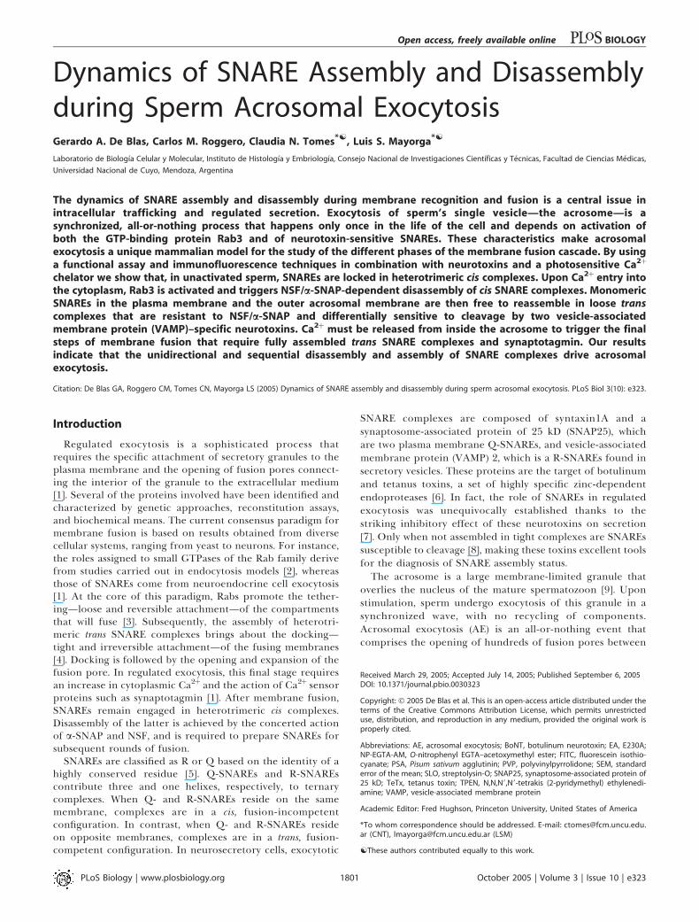
O-nitrophenyl EGTA–acetoxymethyl ester (NP-EGTA-AM )is a photolabile Ca2þ chelator that prevents inducer-triggeredAE in sperm permeabilized by streptolysin-O (SLO); NP-EGTA-AM does this by entering the cytosol, diffusing throughthe outer acrosomal membrane, and accumulating inside theacrosome [14]. UV photolysis of NP-EGTA-AM rapidlyreplenishes the acrosomal Ca2þ pool, resuming exocytosis(Figure 1). In combination with AE inhibitors, NP-EGTA-AMhelps to determine whether fusion-related factors arerequired before or after the intra-acrosomal Ca2þ-sensitivestep. Briefly, NP-EGTA-AM allows an AE inducer to preparethe fusion machinery up to the point when intra-acrosomalCa2þ is required. Inhibitors are then added and the tubesilluminated. Resistance to inhibitors—reflected in unaffectedexocytosis—implies that the targets of the fusion-relatedfactors are required upstream of intra-acrosomal Ca2þ efflux.Sensitivity to inhibitors—revealed by blocked exocytosis—means their targets are located after the intra-acrosomalCa2þ-sensitive step (see Figure 1). AE is always preventedwhen the inhibitors are added prior to the inducer andmaintained throughout the experiment. An anti-Rab3Aantibody inhibited exocytosis when added before challengingwith Ca2þ but not afterward. In contrast, antibodies againstsyntaxin1A, SNAP25, VAMP2, and synaptotagmin VI wereable to abrogate exocytosis even when added after theinducer (Figure 1). These results indicate that Rab3A isnecessary early in the fusion cascade, before Ca2þ is releasedfrom the acrosome, whereas SNAREs and synaptotagmin VIare required later, during or after the intra-acrosomal Ca2þ
efflux. Similar experiments support an early role for NSF/a-SNAP [13]. These observations are summarized in Table 1.
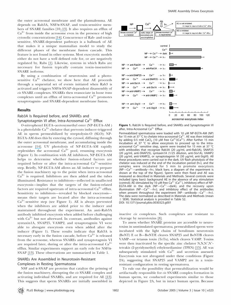
SNAREs Are Assembled in Neurotoxin-ResistantComplexes in Resting Spermatozoa
NSF and a-SNAP are proteins that catalyze the priming ofthe fusion machinery, disrupting the cis SNARE complex andactivating individual SNAREs, and are required for AE [13].This suggests that sperm SNAREs are initially assembled in
inactive cis complexes. Such complexes are resistant tocleavage by neurotoxins [8].To assess whether SNARE proteins are accessible to neuro-
toxins in unstimulated spermatozoa, permeabilized sperm wereincubated with the light chains of botulinum neurotoxin(BoNT) E or B—BoNT/E cleaves SNAP25 and BoNT/B cleavesVAMP—or tetanus toxin (TeTx), which cleaves VAMP. Toxinswere then inactivated by the specific zinc chelator N,N,N9,N9-tetrakis (2-pyridymethyl) ethylenediamine (TPEN) [15]. AE wassubsequently stimulated with Ca2þ and secretion assessed.Exocytosis was not abrogated under these conditions (Figure2A), suggesting that SNAP25 and VAMP2 are in a toxin-resistant configuration in resting spermatozoa.To rule out the possibility that permeabilization would be
artifactually responsible for cis SNARE complex formation inhuman sperm, we conducted experiments similar to thosedepicted in Figure 2A, but in intact human sperm. Because
Figure 1. Rab3A Is Required before, and SNAREs and Synaptotagmin VI
after, Intra-Acrosomal Ca2þ Efflux
Permeabilized spermatozoa were loaded with 10 lM NP-EGTA-AM (NP)for 15 min at 37 8C to chelate intra-acrosomal Ca2þ. AE was then initiatedby adding 0.5 mM CaCl2 (10 lM free Ca2þ)(Ca2þ). After further 15 minincubation at 37 8C to allow exocytosis to proceed up to the intra-acrosomal Ca2þ-sensitive step, sperm were treated for 15 min at 37 8Cwith antibodies that recognize Rab3A (20 lg/ml, anti-Rab3A), SNAP25(20 lg/ml, anti-SNAP25), syntaxin1A (1/25 dilution, anti-Stx1A), VAMP2(20 lg/ml, anti-VAMP2), or synaptotagmin VI (30 lg/ml, anti-StgVI). Allthese procedures were carried out in the dark. UV flash photolysis of thechelator was induced at the end of the incubation period (hm), and thesamples were incubated for 5 min to promote exocytosis(NP!Ca2þ!antibody!hm, black bars; a diagram of the experiment isshown at the top of the figure). Sperm were then fixed and AE wasmeasured as described in Materials and Methods. Several controls wereincluded (grey bars): background AE in the absence of any stimulation(control); AE stimulated by 10 lM free Ca2þ (Ca2þ), inhibitory effect of NP-EGTA-AM in the dark (NP!Ca2þ!dark), and the recovery uponillumination (NP!Ca2þ!hm); and inhibitory effect of the antibodieswhen present throughout the experiment (NP!antibody!Ca2þ!hm).The data were normalized as described in Materials and Methods (mean6 SEM). Statistical analysis is provided in Table S2.DOI: 10.1371/journal.pbio.0030323.g001
PLoS Biology | www.plosbiology.org October 2005 | Volume 3 | Issue 10 | e3231802
SNARE Assembly Drives Exocytosis
sperm were not permeabilized, holotoxins were used insteadof the isolated light chains. These clostridial neurotoxinsconsist of a heavy subunit, responsible for binding to plasmamembrane receptors (in particular gangliosides GT1b andGQ1b), and a light subunit that carries the proteolytic activity[6]. Heavy-chain-mediated binding is required for internal-ization of the light chain, which acts in the cytosol [6].Because both GT1b and GQ1b gangliosides have been isolatedfrom sperm preparations [16] we expected non-permeabi-lized sperm to be at least partially sensitive to holotoxins.Indeed, when intact (i.e., non-permeabilized) human spermwere exposed to BoNT/A (SNAP25-specific) and BoNT/F(VAMP-specific) before triggering exocytosis with the Ca2þ
ionophore A23187, an approximately 50% inhibition in AEwas observed (Figure S1). Toxin concentrations were 5-foldhigher than those required to inhibit exocytosis in permea-bilized sperm [12] to allow for the poor internalization of thecatalytic subunits. Once again, we found that the SNAREproteins were not accessible to neurotoxins in unstimulatedcells, since exocytosis was not attenuated when toxins wereinactivated by TPEN before challenging with A23187 (FigureS1), indicating that SNAP25 and VAMP were in a toxin-resistant configuration prior to initiating AE.
To show directly that TPEN blocks the proteolytic activityof neurotoxins, recombinant SNAP25 was incubated withBoNT/E in the absence or presence of increasing concen-trations of the zinc chelator. As shown in Figure 2B, BoNT/Ecleaved SNAP25. TPEN completely inhibited its activity atconcentrations of 1 lM and higher, in total agreement withthe functional data.
These results show that, in the absence of sperm stim-ulation, Q- and R-SNAREs are protected from neurotoxincleavage, not adopting a toxin-sensitive state during the 15-min incubation protocol. These results were similar inpermeabilized and intact sperm. Thus, we conclude thatSNAREs are engaged in a toxin-resistant configuration inresting sperm. An attractive possibility is that they are lockedin ternary cis complexes rather than undergoing cyclicassembly and disassembly like SNAREs in other systems. Ifthis is the case, addition of an excess of recombinant NSF/a-SNAP should force disassembly, rendering the SNAREs toxin-sensitive. We therefore incubated sperm with NSF/a-SNAP inthe presence of TeTx. The toxin was subsequently inactivatedby addition of TPEN, and AE was stimulated. Ca2þ failed toelicit exocytosis, indicating that NSF/a-SNAP rendered VAMPsensitive to toxin cleavage (Figure 2C). In conclusion, our dataindicate that, in resting sperm, SNAREs are in toxin-resistantcis complexes that can be disassembled by NSF/a-SNAP.
Rab3A Triggers the Disassembly of SNARE Complexesbefore Intra-Acrosomal Ca2þ EffluxIn our permeabilized sperm model, exocytosis is achieved
when an increase in the cytosolic Ca2þ concentrationpromotes the activation of Rab3A [10]. Alternatively, AEcan be initiated by adding recombinant Rab3A preloadedwith GTP-c-S [11]. Rab3A-triggered exocytosis is sensitive to
Figure 2. SNAREs Are Assembled in Neurotoxin-Resistant Complexes in
Resting Spermatozoa
(A) Permeabilized spermatozoa were treated at 37 8C for 15 min with 357nM BoNT/E, 100 nM BoNT/B, or 100 nM TeTx. Next, 2.5 lM TPEN (see [B])was added and AE was activated by adding 0.5 mM CaCl2 (10 lM freeCa2þ) and the incubation continued for an additional 15 min (black bars).Sperm were then fixed and AE was measured as described in Materialsand Methods. Several controls were included (grey bars): background AEin the absence of any stimulation (control); AE stimulated by 10 lM freeCa2þ (Ca2þ); TPEN effect on exocytosis (TPEN!Ca2þ); inhibitory effect ofthe neurotoxins on exocytosis (neurotoxin!Ca2þ); and block of neuro-toxin activity by TPEN (TPEN!neurotoxin!Ca2þ).(B) Recombinant SNAP25 (0.7 lg) was incubated for 15 min at 37 8C inthe presence of 0.6 lg of BoNT/E and increasing concentrations of TPEN.Samples were then resolved by SDS-PAGE and stained with Coomassieblue. Molecular weight standards are indicated on the left (in kilo-daltons). Densitometry and quantitation of the stained bands show100%, 7%, 92%, 98%, 100%, and 100% of intact SNAP25 in lanes 1–6(from left to right), respectively.(C) Treatment with TeTx was performed as described in (A), in thepresence of 310 nM NSF and 500 nM a-SNAP (NSF/aS) to promote SNAREcomplex dissociation (black bar). Incubation with NSF/a-SNAP in thepresence of TPEN-inactivated toxin did not affect exocytosis (grey bar).The data in (A) and (C) were normalized as described in Materials andMethods (mean 6 SEM). Statistical analysis is provided in Table S3.DOI: 10.1371/journal.pbio.0030323.g002
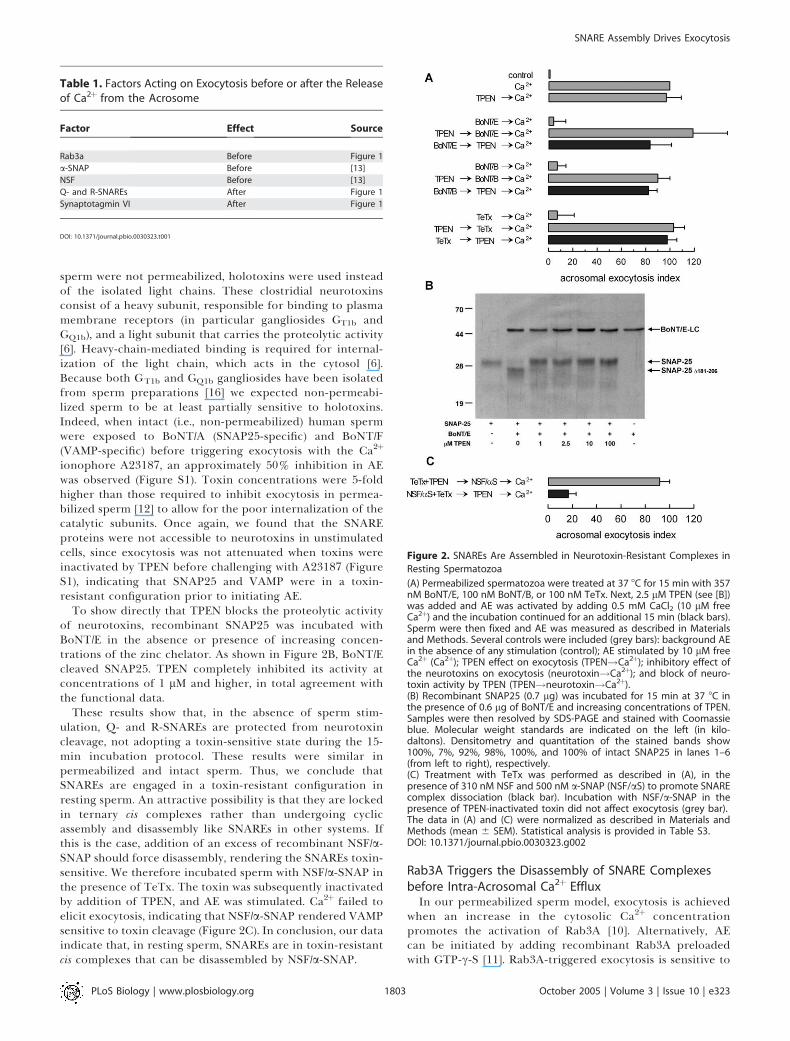
Table 1. Factors Acting on Exocytosis before or after the Releaseof Ca2þ from the Acrosome
Factor Effect Source
Rab3a Before Figure 1
a-SNAP Before [13]
NSF Before [13]
Q- and R-SNAREs After Figure 1
Synaptotagmin VI After Figure 1
DOI: 10.1371/journal.pbio.0030323.t001
PLoS Biology | www.plosbiology.org October 2005 | Volume 3 | Issue 10 | e3231803
SNARE Assembly Drives Exocytosis
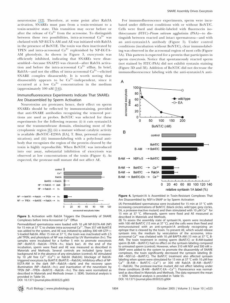
neurotoxins [12]. Therefore, at some point after Rab3Aactivation, SNAREs must pass from a toxin-resistant to atoxin-sensitive state. This transition may occur before orafter the release of Ca2þ from the acrosome. To distinguishbetween these two possibilities, intra-acrosomal Ca2þ waschelated with NP-EGTA-AM and AE was initiated with Rab3Ain the presence of BoNT/E. The toxin was then inactivated byTPEN and intra-acrosomal Ca2þ replenished by NP-EGTA-AM photolysis. As shown in Figure 3, exocytosis wasefficiently inhibited, indicating that SNAREs were disas-sembled—because SNAP25 was cleaved—after Rab3A activa-tion and before the intra-acrosomal Ca2þ efflux. In brief,Rab3A—and not the efflux of intra-acrosomal Ca2þ—elicitedSNARE complex disassembly. It is worth noting thatdisassembly appears to be Ca2þ-independent, since itoccurred at a low Ca2þ concentration in the medium(approximately 100 nM [14]).
Immunofluorescence Experiments Indicate That SNAREsAre Disassembled by Sperm Activation
Neurotoxins are proteases; hence, their effect on spermSNAREs should be reflected by immunostaining, providedthat anti-SNARE antibodies recognizing the removed por-tions are used as probes. BoNT/C was selected for theseexperiments for the following reasons: (i) it cuts syntaxin1Anear the transmembrane domain, eliminating most of thecytoplasmic region [6]; (ii) a mutant without catalytic activityis available (BoNT/C–E230A [EA], T. Binz, personal commu-nication); and (iii) immunolabeling with a polyclonal anti-body that recognizes the region of the protein cleaved by thetoxin is highly reproducible. When BoNT/C was introducedinto our assay, substantial inhibition of exocytosis wasobserved at low concentrations of the toxin (Figure 4). Asexpected, the protease-null mutant did not affect AE.
For immunofluorescence experiments, sperm were incu-bated under different conditions with or without BoNT/C.Cells were fixed and double-labeled with fluorescein iso-thiocyanate (FITC)–Pisum sativum agglutinin (PSA)—to dis-tinguish between reacted and intact spermatozoa—and withan anti-syntaxin1A antibody (Figure 5). Under controlconditions (incubation without BoNT/C), clear immunolabel-ing was observed in the acrosomal region of most cells (Figure5A). This pattern is expected for a protein that participates insperm exocytosis. Notice that spontaneously reacted sperm(not stained by FITC-PSA) did not exhibit syntaxin staining(Figure 5D and 5E). Addition of BoNT/C did not decrease theimmunofluorescence labeling with the anti-syntaxin1A anti-
Figure 3. Activation with Rab3A Triggers the Disassembly of SNARE
Complexes before Intra-Acrosomal Ca2þ Efflux
Permeabilized spermatozoa were loaded with 10 lM NP-EGTA-AM (NP)for 15 min at 37 8C to chelate intra-acrosomal Ca2þ. Then 357 nM BoNT/Ewas added to the system, and AE was initiated by adding 300 nM GTP-c-S-loaded Rab3A. After 15 min at 37 8C, the toxin was inactivated with 2.5lM TPEN, and photolysis of NP was induced by UV illumination (hm). Thesamples were incubated for a further 5 min to promote exocytosis(NP!BoNT/E!Rab3A!TPEN!hm, black bar). At the end of theincubation, sperm were fixed and AE was measured as described inMaterials and Methods. Several controls are included (grey bars):background AE in the absence of any stimulation (control); AE stimulatedby 10 lM free Ca2þ (Ca2þ) or Rab3A (Rab3A); blockage of Rab3A-triggered exocytosis by BoNT/E (BoNT/E!Rab3A); inhibitory effect of NP-EGTA-AM in the dark (NP!Rab3A!dark) and the recovery uponillumination (NP!Rab3A!hm); and inactivation of the neurotoxin byTPEN (NP!TPEN!BoNT/E!Rab3A!hm). The data were normalized asdescribed in Materials and Methods (mean 6 SEM). Statistical analysis isprovided in Table S4.DOI: 10.1371/journal.pbio.0030323.g003
Figure 4. Syntaxin1A Is Assembled in Toxin-Resistant Complexes That
Are Disassembled by NSF/a-SNAP or by Sperm Activation
(A) Permeabilized spermatozoa were incubated for 15 min at 37 8C withincreasing concentrations of BoNT/C (black circles, wild type; grey circles,EA, a protease-inactive mutant) and then stimulated with 10 lM Ca2þ for15 min at 37 8C. Afterwards, sperm were fixed and AE measured asdescribed in Materials and Methods.(B) To assess the assembly state of syntaxin1A, sperm were incubatedwith 100 nM BoNT/C (15 min at 37 8C), and the cells were then fixed andimmunostained with an anti-syntaxin1A antibody recognizing anepitope that is cleaved by the toxin. To prevent AE, which would releasesyntaxin into the medium by vesiculation of the acrosome, intra-acrosomal Ca2þwas chelated with 10 lM BAPTA-AM (15 min at 37 8C, B-AM). The toxin treatment in resting sperm (BoNT/C) or B-AM-loadedsperm (B-AM!BoNT/C) had no effect on the syntaxin labeling comparedto untreated sperm (control). However, when 310 nM NSF and 500 nM a-SNAP were added to the system to promote the disassembly of SNAREcomplexes, the toxin significantly decreased the syntaxin labeling (B-AM!NSF/aS!BoNT/C). The BoNT/C treatment also affected syntaxinlabeling when sperm were stimulated for 15 min at 37 8C with 10 lM freeCa2þ (B-AM! BoNT/C!Ca2þ) or 300 nM Rab3A (B-AM!BoNT/C!Rab3A). The protease-inactive mutant did not affect labeling underthese conditions (B-AM!BoNT/C-EA!Ca2þ). Fluorescence was normal-ized as described in Materials and Methods. The data represent the mean6 SEM. Statistical analysis is provided in Table S5.DOI: 10.1371/journal.pbio.0030323.g004
PLoS Biology | www.plosbiology.org October 2005 | Volume 3 | Issue 10 | e3231804
SNARE Assembly Drives Exocytosis
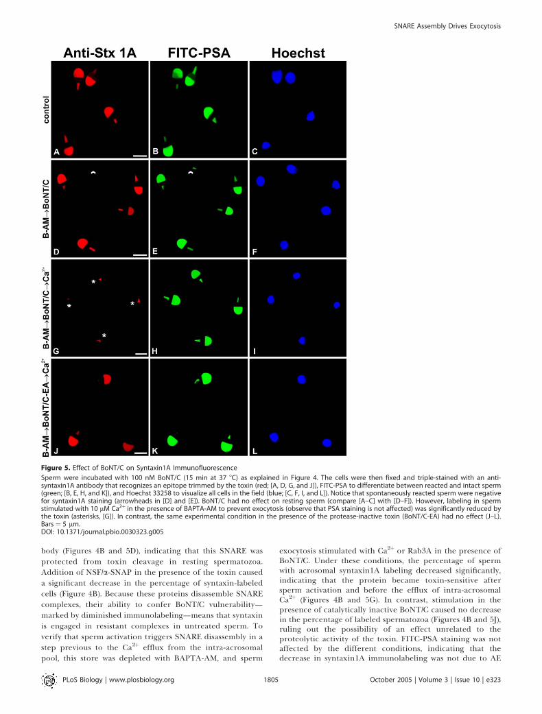
body (Figures 4B and 5D), indicating that this SNARE wasprotected from toxin cleavage in resting spermatozoa.Addition of NSF/a-SNAP in the presence of the toxin causeda significant decrease in the percentage of syntaxin-labeledcells (Figure 4B). Because these proteins disassemble SNAREcomplexes, their ability to confer BoNT/C vulnerability—marked by diminished immunolabeling—means that syntaxinis engaged in resistant complexes in untreated sperm. Toverify that sperm activation triggers SNARE disassembly in astep previous to the Ca2þ efflux from the intra-acrosomalpool, this store was depleted with BAPTA-AM, and sperm
exocytosis stimulated with Ca2þ or Rab3A in the presence ofBoNT/C. Under these conditions, the percentage of spermwith acrosomal syntaxin1A labeling decreased significantly,indicating that the protein became toxin-sensitive aftersperm activation and before the efflux of intra-acrosomalCa2þ (Figures 4B and 5G). In contrast, stimulation in thepresence of catalytically inactive BoNT/C caused no decreasein the percentage of labeled spermatozoa (Figures 4B and 5J),ruling out the possibility of an effect unrelated to theproteolytic activity of the toxin. FITC-PSA staining was notaffected by the different conditions, indicating that thedecrease in syntaxin1A immunolabeling was not due to AE
Figure 5. Effect of BoNT/C on Syntaxin1A Immunofluorescence
Sperm were incubated with 100 nM BoNT/C (15 min at 37 8C) as explained in Figure 4. The cells were then fixed and triple-stained with an anti-syntaxin1A antibody that recognizes an epitope trimmed by the toxin (red; [A, D, G, and J]), FITC-PSA to differentiate between reacted and intact sperm(green; [B, E, H, and K]), and Hoechst 33258 to visualize all cells in the field (blue; [C, F, I, and L]). Notice that spontaneously reacted sperm were negativefor syntaxin1A staining (arrowheads in [D] and [E]). BoNT/C had no effect on resting sperm (compare [A–C] with [D–F]). However, labeling in spermstimulated with 10 lM Ca2þ in the presence of BAPTA-AM to prevent exocytosis (observe that PSA staining is not affected) was significantly reduced bythe toxin (asterisks, [G]). In contrast, the same experimental condition in the presence of the protease-inactive toxin (BoNT/C-EA) had no effect (J–L).Bars¼ 5 lm.DOI: 10.1371/journal.pbio.0030323.g005
PLoS Biology | www.plosbiology.org October 2005 | Volume 3 | Issue 10 | e3231805
SNARE Assembly Drives Exocytosis
that would have released the outer acrosomal membranetogether with the apposed plasma membrane (Figure 5).
These results reaffirm the notion that SNAREs are toxin-protected in resting spermatozoa and that addition of NSF/a-SNAP unprotects them. Furthermore, activation of sperma-tozoa (with either Ca2þ or Rab3A) under conditions thatdeplete intra-acrosomal Ca2þ promotes their cleavage byBoNTs, indicating that cis SNARE complexes are disas-sembled at a step prior to the intra-acrosomal Ca2þ efflux.
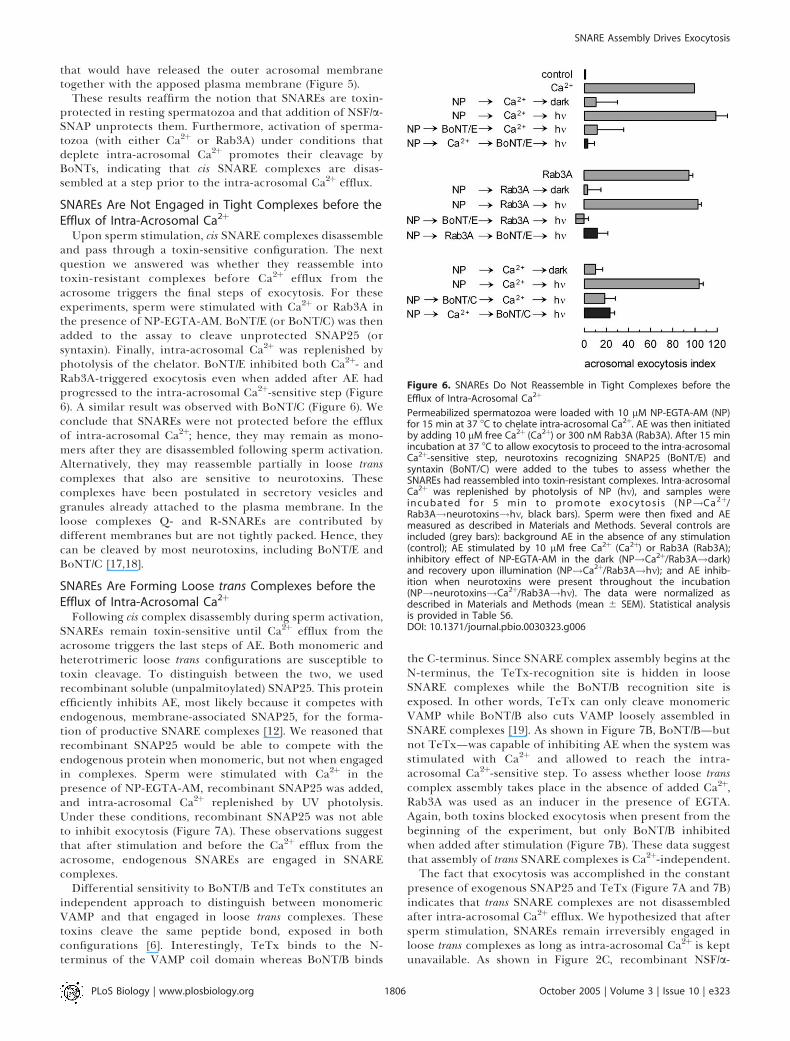
SNAREs Are Not Engaged in Tight Complexes before theEfflux of Intra-Acrosomal Ca2þ
Upon sperm stimulation, cis SNARE complexes disassembleand pass through a toxin-sensitive configuration. The nextquestion we answered was whether they reassemble intotoxin-resistant complexes before Ca2þ efflux from theacrosome triggers the final steps of exocytosis. For theseexperiments, sperm were stimulated with Ca2þ or Rab3A inthe presence of NP-EGTA-AM. BoNT/E (or BoNT/C) was thenadded to the assay to cleave unprotected SNAP25 (orsyntaxin). Finally, intra-acrosomal Ca2þ was replenished byphotolysis of the chelator. BoNT/E inhibited both Ca2þ- andRab3A-triggered exocytosis even when added after AE hadprogressed to the intra-acrosomal Ca2þ-sensitive step (Figure6). A similar result was observed with BoNT/C (Figure 6). Weconclude that SNAREs were not protected before the effluxof intra-acrosomal Ca2þ; hence, they may remain as mono-mers after they are disassembled following sperm activation.Alternatively, they may reassemble partially in loose transcomplexes that also are sensitive to neurotoxins. Thesecomplexes have been postulated in secretory vesicles andgranules already attached to the plasma membrane. In theloose complexes Q- and R-SNAREs are contributed bydifferent membranes but are not tightly packed. Hence, theycan be cleaved by most neurotoxins, including BoNT/E andBoNT/C [17,18].
SNAREs Are Forming Loose trans Complexes before theEfflux of Intra-Acrosomal Ca2þ
Following cis complex disassembly during sperm activation,SNAREs remain toxin-sensitive until Ca2þ efflux from theacrosome triggers the last steps of AE. Both monomeric andheterotrimeric loose trans configurations are susceptible totoxin cleavage. To distinguish between the two, we usedrecombinant soluble (unpalmitoylated) SNAP25. This proteinefficiently inhibits AE, most likely because it competes withendogenous, membrane-associated SNAP25, for the forma-tion of productive SNARE complexes [12]. We reasoned thatrecombinant SNAP25 would be able to compete with theendogenous protein when monomeric, but not when engagedin complexes. Sperm were stimulated with Ca2þ in thepresence of NP-EGTA-AM, recombinant SNAP25 was added,and intra-acrosomal Ca2þ replenished by UV photolysis.Under these conditions, recombinant SNAP25 was not ableto inhibit exocytosis (Figure 7A). These observations suggestthat after stimulation and before the Ca2þ efflux from theacrosome, endogenous SNAREs are engaged in SNAREcomplexes.
Differential sensitivity to BoNT/B and TeTx constitutes anindependent approach to distinguish between monomericVAMP and that engaged in loose trans complexes. Thesetoxins cleave the same peptide bond, exposed in bothconfigurations [6]. Interestingly, TeTx binds to the N-terminus of the VAMP coil domain whereas BoNT/B binds
the C-terminus. Since SNARE complex assembly begins at theN-terminus, the TeTx-recognition site is hidden in looseSNARE complexes while the BoNT/B recognition site isexposed. In other words, TeTx can only cleave monomericVAMP while BoNT/B also cuts VAMP loosely assembled inSNARE complexes [19]. As shown in Figure 7B, BoNT/B—butnot TeTx—was capable of inhibiting AE when the system wasstimulated with Ca2þ and allowed to reach the intra-acrosomal Ca2þ-sensitive step. To assess whether loose transcomplex assembly takes place in the absence of added Ca2þ,Rab3A was used as an inducer in the presence of EGTA.Again, both toxins blocked exocytosis when present from thebeginning of the experiment, but only BoNT/B inhibitedwhen added after stimulation (Figure 7B). These data suggestthat assembly of trans SNARE complexes is Ca2þ-independent.The fact that exocytosis was accomplished in the constant
presence of exogenous SNAP25 and TeTx (Figure 7A and 7B)indicates that trans SNARE complexes are not disassembledafter intra-acrosomal Ca2þ efflux. We hypothesized that aftersperm stimulation, SNAREs remain irreversibly engaged inloose trans complexes as long as intra-acrosomal Ca2þ is keptunavailable. As shown in Figure 2C, recombinant NSF/a-
Figure 6. SNAREs Do Not Reassemble in Tight Complexes before the
Efflux of Intra-Acrosomal Ca2þ
Permeabilized spermatozoa were loaded with 10 lM NP-EGTA-AM (NP)for 15 min at 37 8C to chelate intra-acrosomal Ca2þ. AE was then initiatedby adding 10 lM free Ca2þ (Ca2þ) or 300 nM Rab3A (Rab3A). After 15 minincubation at 37 8C to allow exocytosis to proceed to the intra-acrosomalCa2þ-sensitive step, neurotoxins recognizing SNAP25 (BoNT/E) andsyntaxin (BoNT/C) were added to the tubes to assess whether theSNAREs had reassembled into toxin-resistant complexes. Intra-acrosomalCa2þ was replenished by photolysis of NP (hm), and samples wereincuba ted for 5 min to pr omote exocytosis (NP!Ca 2þ/Rab3A!neurotoxins!hm, black bars). Sperm were then fixed and AEmeasured as described in Materials and Methods. Several controls areincluded (grey bars): background AE in the absence of any stimulation(control); AE stimulated by 10 lM free Ca2þ (Ca2þ) or Rab3A (Rab3A);inhibitory effect of NP-EGTA-AM in the dark (NP!Ca2þ/Rab3A!dark)and recovery upon illumination (NP!Ca2þ/Rab3A!hm); and AE inhib-ition when neurotoxins were present throughout the incubation(NP!neurotoxins!Ca2þ/Rab3A!hm). The data were normalized asdescribed in Materials and Methods (mean 6 SEM). Statistical analysisis provided in Table S6.DOI: 10.1371/journal.pbio.0030323.g006
PLoS Biology | www.plosbiology.org October 2005 | Volume 3 | Issue 10 | e3231806
SNARE Assembly Drives Exocytosis

SNAP can disassemble cis SNARE complexes in unstimulatedspermatozoa. We asked whether they could also disengageloose trans complexes. To this end, NP-EGTA-AM-loadedsperm were stimulated with Ca2þ or Rab3A and then treatedwith TeTx in the presence or absence of NSF/a-SNAP. Theresults, presented in Figure 7C, show that these chaperonesfailed to confer TeTx sensitivity when added after theexocytic process had progressed to the intra-acrosomalCa2þ-sensitive step. These observations indicate that transSNARE complexes cannot be disassembled by NSF/a-SNAP.The notion that trans SNARE complexes are resistant to NSF/a-SNAP opposes the original SNARE hypothesis [20] and,although suggested in the past [21], has to our knowledgenever before been shown in actual cells.
Immunofluorescence Experiments Show That VAMP2 IsEngaged in Loose trans SNARE Complexes afterStimulationAs shown in Figures 4B and 5, immunofluorescence is a
powerful technique to monitor SNARE assembly status asreflected by SNARE sensitivity to cleavage by neurotoxins. Wethus made use of the differential sensitivity of VAMP2 to TeTxand BoNT/B when engaged in loose complexes to show byVAMP2 immunostaining that these complexes form aftersperm stimulation. BAPTA-AM was used in all cases to chelateintra-acrosomal Ca2þ and prevent VAMP2-containing mem-brane loss inherent to the acrosome reaction. As expected,acrosomal labeling for VAMP2 was not affected by incubatingunstimulated sperm with TeTx or BoNT/B (Figures 8 and 9),consistent with the notion that VAMP2 is protected in cisSNARE complexes. A significant decrease in the percentage ofcells exhibiting acrosomal staining was observed when toxin-loaded spermwere challengedwith Ca2þ (Figures 8, 9G, and 9P).Thus, VAMP2 sensitization to cleavage was probably aconsequence of cis SNARE disassembly by the stimulant. Incontrast, when toxins were added after Ca2þ (i.e., after AE hadprogressed to the intra-acrosomal Ca2þ-sensitive step), VAMP2labeling was attenuated by BoNT/B (Figures 8 and 9J) but not byTeTx (Figures 8 and 9S). Loss of VAMP2 immunostainingfollowing BoNT/B treatment is due to lack of immunostainingof the cleavage product (aa 1–76) by the anti-VAMP2 antibody[22]. This pattern of VAMP2 sensitivity to BoNT/B coupled toresistance to TeTx implies its engagement in loose trans SNAREcomplexes. In conclusion, immunofluorescence data furthersupport the idea that, following sperm stimulation, SNAREs areengaged in loose trans complexes awaiting the release of intra-acrosomal Ca2þ that will trigger the final steps of AE.
Discussion
Exocytosis of the acrosome is a synchronized, all-or-nothingprocess that happens only once in the life of the spermatozoonand depends on bothRab3 activation and neurotoxin-sensitiveSNAREs. Because of these special features, it constitutes aparticularly attractive system to examine molecular aspects ofregulated exocytosis that are not amenable to experimentalmanipulation in other mammalian models. These include thecoupling between Rab and SNARE functions, the properties of
Figure 7. SNAREs Reassemble in Loose Complexes That Are Resistant to
NSF/a-SNAP before the Efflux of Intra-Acrosomal Ca2þ
(A) Permeabilized spermatozoa were loaded with 10 lM NP-EGTA-AM(NP) for 15 min at 37 8C to chelate intra-acrosomal Ca2þ. AE was theninitiated by adding 10 lM free Ca2þ (Ca2þ). After 15 min incubation at 378C to allow exocytosis to proceed to the intra-acrosomal Ca2þ-sensitivestep, 800 nM recombinant SNAP25 (SNAP25) was added to compete withendogenous SNAP25. Intra-acrosomal Ca2þ was replenished by photol-ysis of NP-EGTA-AM (hm), and the samples were incubated for 5 min topromote exocytosis (NP!Ca2þ!SNAP25!hm, black bar). Sperm werethen fixed and AE was measured as described in Materials and Methods.(B) Permeabilized spermatozoa were loaded with 10 lM NP-EGTA-AM(NP) for 15 min at 37 8C. AE was then initiated by adding 10 lM free Ca2þ
(Ca2þ) or 300 nM Rab3A (Rab3A). After 15 min incubation at 37 8C, 100nM neurotoxin recognizing VAMP (BoNT/B and TeTx) was added to thetubes to assess whether the SNAREs had reassembled in loose transcomplexes sensitive to BoNT/B but not to TeTx. After 15 min incubationat 37 8C, intra-acrosomal Ca2þwas replenished by photolysis of NP-EGTA-AM (hm), and the samples were incubated for 5 min to promoteexocytosis (NP!Ca2þ/Rab3A!neurotoxin!hm, black bars). Sperm werethen fixed and AE measured as described in Materials and Methods.(C) To assess whether NSF/a-SNAP can disassemble loose trans SNAREcomplexes, permeabilized sperm treated as in (B) were incubated withTeTx in the presence of 310 nM NSF and 500 nM a-SNAP (NP!Ca2þ/Rab3A!NSF/aSþTeTx!hm, black bars).Several controls were included in (A), (B), and (C) (grey bars): backgroundAE in the absence of any stimulation (control); AE stimulated by 10 lMfree Ca2þ (Ca2þ) or 300 nM Rab3A (Rab3A); inhibitory effect of NP-EGTA-AM in the dark (NP!Ca2þ/Rab3A!dark) and the recovery uponillumination (NP!Ca2þ/Rab3A!hm); inhibitory effect when SNAP25was present throughout the incubations (NP!SNAP25!Ca2þ!hm);inhibitory effect when the neurotoxins were present throughout the
incubations (NP!neurotoxin!Ca2þ/Rab3A!hm); and the effect of NSF/a-SNAP on SNARE complexes in unstimulated sperm (NSF/aSþTeTx!TPEN!Rab3A!hm). The data were normalized as describedin Materials and Methods (mean 6 SEM). Statistical analysis is providedin Table S7.DOI: 10.1371/journal.pbio.0030323.g007
PLoS Biology | www.plosbiology.org October 2005 | Volume 3 | Issue 10 | e3231807
SNARE Assembly Drives Exocytosis
loose trans SNARE complexes, the role of synaptotagmin in thedynamics of SNARE assembly, and the role of intravesicularCa2þ inmembrane fusion. Furthermore, AE is a central processin sperm physiology, and understanding the molecularmechanisms underlying it will be of outstanding importancefor our ability to regulate fertilization. Sperm contact withglycoproteins in the zona pellucida of the oocyte leads to theopening of store-operated Ca2þ channels in the spermplasmalemma followed by a massive entry of Ca2þ from themedium into the cytoplasm, leading to exocytosis [23]. OurSLO-permeabilized sperm model, in which Ca2þ can freelypermeate into the cells, resembles the state of open store-operated Ca2þ channels in intact cells and is therefore suitableto study stages of exocytosis occurring downstream of store-operated Ca2þ channel opening.
SNAREs are required in multiple fusion events mandatoryfor cell survival even under resting conditions. The ratio ofmonomeric to assembled SNAREs depends on the type andphysiological condition of the cell. Thus, while some studiesindicate that most SNAREs are free in the plasma membrane[24], others suggest that they are engaged in complexes [25,26].SNAP25 and syntaxins can form partial all-Q complexes (threehelix bundles from one SNAP25 and one syntaxin; [25]) orcomplexes composed of four helix bundles from two syntaxinsand one SNAP25 [27]. Pre-association of Q-SNAREs in a three-bundle complex creates the docking site for the cognate R-SNARE [28]. Unlike their ternary counterparts, binary com-plexes are unstable and sensitive to neurotoxins [25].Whateverthe steady-state configuration of SNAREs in neuroendocrinecells might be, exocytosis is blocked by neurotoxins, suggestingthat SNAREs go through toxin-sensitive stages [18,29]. Inresting human sperm, both R- and Q-SNAREs are protectedfrom toxin cleavage. Susceptibility to toxins is conferred byboth endogenous or exogenously addedNSF/a-SNAP. Thus, weconclude that sperm SNAREs are locked in ternary complexesin a cis configuration, in contrast to cells with active vesicularrecycling. Because sperm have only one chance to fertilize theoocyte, tight spatial and temporal regulation of the acrosomereaction is a prerequisite for their success. In this scenario, it isnot surprising that SNAREs are bound in an inactive state untilexocytosis is triggered.
The connection between the rise in cytosolic Ca2þoccurringupon sperm stimulation and Rab3A activation is not clear. Acalmodulin-mediated effect is possible, since Ca2þ/calmodulinbinds Rab3A [30] and promotes the exchange of GDP for GTPin the Rab3A–GDP dissociation inhibitor complex [31].Although we have shown that calmodulin has an inhibitoryeffect on AE in permeabilized sperm and that this effect is notmediated by binding to Rab3A [32], our results do not excludea role for calmodulin in Rab3A activation. Once Rab3A isactivated, cis SNAREs are disassembled by NSF/a-SNAP andbecome accessible to neurotoxins. In other systems, activeRabs recruit a variety of effectors that tether the membranesthat will fuse. NSF has been found associated with tetheringcomplexes [33]. Moreover, NSF binds several Rabs, includingRab3A [34]. Perhaps similar interactions favor the dissociationof cis SNAREs by sperm NSF/a-SNAP following Rab3Aactivation. The proximity of the tethered membranes plusthe availability of free Q- and R-SNAREs would allow theirassociation in loose trans complexes. The latter can form at thevery low Ca2þ concentrations achieved by EGTA and NP-EGTA-AM chelating Ca2þ in the medium/cytosol and in theacrosome, respectively. While not required for the assembly ofSNARE complexes from pure proteins [27], Ca2þ appears to benecessary for trans SNARE pairing during exocytosis in PC12
cells [35]. Perhaps this incongruity is due to the presence ofspecific regulatory proteins in these cells. We have shown thattrans complexes are resistant to NSF/a-SNAP, in agreementwith results obtained in a proteoliposome fusion assay [21]. Incontrast, trans SNAREs can be dissociated by the NSF/a-SNAPhomologs Sec18p/Sec17p in a yeast vacuole fusion assay [36].The reasons for this discrepancy are unknown, but it is worthmentioning that both the models (yeast vacuole fusion versusmammalian regulated exocytosis) and the experimental read-out (coimmunoprecipitation of SNAREs versus toxin sensitiv-ity) are quite different. Resistance to NSF/a-SNAP, SNAP25,and TeTx indicates that, following sperm stimulation, SNAREsare engaged in trans complexes that do not spontaneouslyrevert to the monomeric configuration from which suchcomplexes arose. Temporally, the arrest of exocytosis untilCa2þ is released from the acrosome correlates with SNAREsbeing assembled in loose trans complexes. Resistance to NSF/a-SNAP, SNAP25, and TeTx might also be explained if SNAREswere dispensable for the final fusion steps, as has beensuggested for yeast vacuole fusion and exocytosis of corticalgranules of sea urchin oocytes [36,37]. Our data do not supportthis view, however, since BoNTs and antibodies to SNAREscontinue to prevent exocytosis downstreamof intra-acrosomalCa2þ release. This indicates that SNAREs are required late inthe exocytotic cascade.A direct role for intravesicular Ca2þ in membrane fusion has
been proposed in several transport events, including theexocytosis of secretory granules [38,39]. Why would Ca2þeffluxfrom the acrosome be necessary for exocytosis? We favor theidea that tight apposition of the membranes near the formingfusion pore prevents free accessibility to cytosolic Ca2þ. Thus, alocal release from the acrosome is necessary to activate thefinal steps of membrane fusion. Synaptotagmins are likelyinvolved in this late Ca2þ-sensitive step. Synaptotagmin VI, atleast, is present in the acrosomal membrane of human sperm,and this protein is required at a step downstream of the intra-
Figure 8. VAMP2 Is Engaged in Loose SNARE Complexes before the
Efflux of Intra-Acrosomal Ca2þ
Permeabilized spermatozoa were loaded with 10 lM BAPTA-AM (B-AM)for 15 min at 37 8C to chelate intra-acrosomal Ca2þ. AE was then initiatedby adding 10 lM free Ca2þ (Ca2þ). After 15 min incubation at 37 8C toallow exocytosis to proceed to the intra-acrosomal Ca2þ-sensitive step,100 nM neurotoxins recognizing VAMP (BoNT/B or TeTx) were added tothe tubes and the samples were incubated for 15 min at 37 8C (B-AM!Ca2þ!neurotoxin, black bars). Samples were then immunolabeledwith an anti-VAMP2 antibody as described in Materials and Methods.Notice that at this stage VAMP2 immunolabeling was sensitive to BoNT/Bbut not to TeTx. Several other conditions are included (grey bars). Thetoxins did not affect VAMP2 staining in resting sperm (compare controlversus B-AM!neurotoxin). However, the toxins decreased the VAMP2labeling when present during stimulation (B-AM!neurotoxin!Ca2þ).Fluorescence was normalized as described in Materials and Methods(mean 6 SEM). Statistical analysis is provided in Table S8.DOI: 10.1371/journal.pbio.0030323.g008
PLoS Biology | www.plosbiology.org October 2005 | Volume 3 | Issue 10 | e3231808
SNARE Assembly Drives Exocytosis
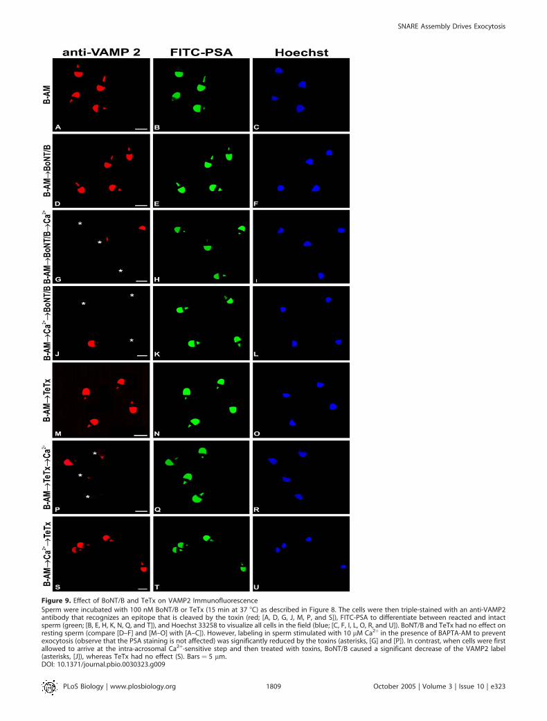
Figure 9. Effect of BoNT/B and TeTx on VAMP2 Immunofluorescence
Sperm were incubated with 100 nM BoNT/B or TeTx (15 min at 37 8C) as described in Figure 8. The cells were then triple-stained with an anti-VAMP2antibody that recognizes an epitope that is cleaved by the toxin (red; [A, D, G, J, M, P, and S]), FITC-PSA to differentiate between reacted and intactsperm (green; [B, E, H, K, N, Q, and T]), and Hoechst 33258 to visualize all cells in the field (blue; [C, F, I, L, O, R, and U]). BoNT/B and TeTx had no effect onresting sperm (compare [D–F] and [M–O] with [A–C]). However, labeling in sperm stimulated with 10 lM Ca2þ in the presence of BAPTA-AM to preventexocytosis (observe that the PSA staining is not affected) was significantly reduced by the toxins (asterisks, [G] and [P]). In contrast, when cells were firstallowed to arrive at the intra-acrosomal Ca2þ-sensitive step and then treated with toxins, BoNT/B caused a significant decrease of the VAMP2 label(asterisks, [J]), whereas TeTx had no effect (S). Bars ¼ 5 lm.DOI: 10.1371/journal.pbio.0030323.g009
PLoS Biology | www.plosbiology.org October 2005 | Volume 3 | Issue 10 | e3231809
SNARE Assembly Drives Exocytosis
acrosomal Ca2þ efflux (Figure 1 and [40]). Therefore, synapto-tagmin VI/Ca2þ may favor the full zippering of SNAREcomplexes and thus enable membrane fusion [41].
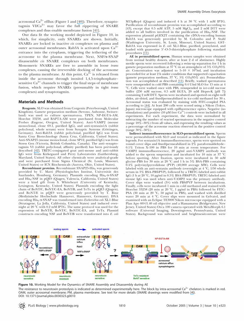
Our data fit the working model depicted in Figure 10, inwhich, for simplicity, only SNAREs are shown. Initially,SNAREs are locked in inactive cis complexes on plasma andouter acrosomal membranes. Rab3A is activated upon Ca2þ
entrance into the cytoplasm, triggering the tethering of theacrosome to the plasma membrane. Next, NSF/a-SNAPdisassemble cis SNARE complexes on both membranes.Monomeric SNAREs are free to assemble in loose transcomplexes, causing the irreversible docking of the acrosometo the plasma membrane. At this point, Ca2þ is released frominside the acrosome through inositol 1,4,5-trisphosphate–sensitive Ca2þ channels to trigger the final steps of membranefusion, which require SNAREs (presumably in tight transcomplexes) and synaptotagmin.
Materials and Methods
Reagents. SLO was obtained from Corgenix (Peterborough, UnitedKingdom). Gamete preparation medium (Serono, Aubonne, Switzer-land) was used to culture spermatozoa. TPEN, NP-EGTA-AM,Hoechst 33258, and BAPTA-AM were purchased from MolecularProbes (Eugene, Oregon, United States). Anti-VAMP2 (mousemonoclonal, clone 69.1, purified IgG), and anti-syntaxin1A (rabbitpolyclonal, whole serum) were from Synaptic Systems (Gottingen,Germany). Anti-Rab3A (rabbit polyclonal, purified IgG) was fromSanta Cruz Biotechnology (Santa Cruz, California, United States).Anti-SNAP25 (mouse monoclonal, clone SP12, purified IgG) was fromStress Gen (Victoria, British Columbia, Canada). The anti–synapto-tagmin VI (rabbit polyclonal, affinity purified) has been previouslydescribed [42]. TRITC-conjugated goat anti-mouse and anti-rabbitIgG were from Kirkegaard and Perry Laboratories (Gaithersburg,Maryland, United States). All other chemicals were analytical-gradeand were purchased from Sigma Chemical (St. Louis, Missouri,United States) or ICN Biochemicals (Aurora, Ohio, United States).
Recombinant proteins. Recombinant SNAP25-His6 was generouslyprovided by U. Matti (Physiologisches Institut, Universitat desSaarlandes, Homburg, Germany). Plasmids encoding His6-a-SNAPand His6-NSF in pQE9 (Qiagen, Valencia, California, United States)were a kind gift from S. Whiteheart (University of Kentucky,Lexington, Kentucky, United States). Plasmids encoding the lightchain of BoNT/C, BoNT/C-EA, BoNT/B, and TeTx in pQE3 (Qiagen),and BoNT/E in pQE9 were generously provided by T. Binz(Medizinische Hochschule Hannover, Hannover, Germany). DNAencoding His6-a-SNAP was transformed into Escherichia coli XL1-Blue(Stratagene, La Jolla, California, United States) and induced over-night at 20 8C with 0.2 mM IPTG. The same protocol was used for theexpression of BoNT/E, BoNT/C, BoNT/C-EA, and TeTx. Plasmidconstructs encoding NSF and BoNT/B were transformed into E. coli
M15pRep4 (Qiagen) and induced 4 h at 30 8C with 1 mM IPTG.Purification of recombinant proteins was accomplished according to[43], except that 0.5 mM ATP, 5 mM MgCl2, and 2 mM DTT wereadded to all buffers involved in the purification of His6-NSF. Theexpression plasmid pGEX2T containing the cDNA-encoding humanRab3A was generously provided by M. Colombo and P. Stahl(Washington University, St. Louis, Missouri, United States). GST-Rab3A was expressed in E. coli XL1-Blue, purified, prenylated, andloaded with guanosine 59-O-3-thiotriphosphate following standardprocedures [11].
AE in permeabilized sperm. Human semen samples were obtainedfrom normal healthy donors, after at least 2 d of abstinence. Highlymotile sperm were recovered following a swim-up separation for 1 h ingamete preparation medium at 37 8C in an atmosphere of 5% CO2/95%air. Concentration was adjusted to 5–10 3 106/ml, and incubationproceeded for at least 2 h under conditions that supported capacitation(gamete preparation medium, 37 8C, 5% CO2/95% air). Permeabiliza-tion was accomplished as described [11]. Briefly, washed spermatozoawere resuspended in cold PBS containing 0.4 U/ml SLO for 15 min at 48C. Cells were washed once with PBS, resuspended in ice-cold sucrosebuffer (250 mM sucrose, 0.5 mM EGTA, 20 mM Hepes-K [pH 7])containing 2 mM DTT. Sperm were incubated and spotted on eight-wellslides, air-dried, and fixed/permeabilized in ice-cold methanol for 30 s.Acrosomal status was evaluated by staining with FITC-coupled PSAaccording to [44]. At least 200 cells were scored using a Nikon (Tokyo,Japan) microscope equipped with epifluorescence optics. Negative (nostimulation) and positive (10 lM free Ca2þ) controls were included in allexperiments. For each experiment, the data were normalized bysubtracting the number of reacted spermatozoa in the negative control(range 18%–30%) from all values, and expressing the resulting values asa percentage of the acrosome reaction observed in the positive control(range 30%–50%).
Indirect immunofluorescence in SLO-permeabilized sperm. Spermwere permeabilized with SLO and treated as indicated in the figurelegends. For syntaxin1A immunofluorescence, sperm were spotted onround cover slips and fixed/permeabilized in 2% paraformaldehyde–0.1% Triton X-100 in PBS for 10 min at room temperature. ForVAMP2 immunofluorescence, 20 lg/ml anti-VAMP2 antibody wasadded to the sperm suspension and incubated for 10 min at 37 8Cbefore spotting. After fixation, sperm were incubated in 50 mMglycine-PBS for 30 min at 20 8C and 1 h in 5% BSA-PBS containing0.4% polyvinylpyrrolidone (PVP) (40,000 average MW). Cells werelabeled with an anti-syntaxin antibody (overnight at 4 8C, 1/50 wholeserum in 3% BSA-PBS/PVP), followed by a TRITC-labeled anti-rabbitIgG (1 h at 20 8C, 10 lg/ml in 0.5% BSA-PBS/PVP). TRITC-labeled antimouse IgG was used when anti-VAMP2 was the primary antibody.Cover slips were washed (33) with PBS/PVP between incubations.Finally, cells were incubated 1 min in cold methanol and stained withHoechst 33258 (20 min at 20 8C, 1 lg/ml in PBS) followed by FITC-PSA (30 min at 20 8C, 50 lg/ml in PBS), and washed with distilledwater 20 min at 4 8C. Cover slips were mounted in Gelvatol, andexamined with an Eclipse TE3000 Nikon microscope equipped with aPlan Apo 603/1.40 oil objective and a Hamamatsu (Bridgewater, NewJersey, United States) Orca 100 camera operated with MetaMorph 6.1software (Universal Imaging, Downingtown, Pennsylvania, UnitedStates). Background was subtracted and brightness/contrast were
Figure 10. Working Model for the Dynamics of SNARE Assembly and Disassembly during AE
The resistance to neurotoxin proteolysis is indicated as determined experimentally here. The block by intra-acrosomal Ca2þ chelators is marked in red.OAM, outer acrosomal membrane; PM, plasma membrane. See text for more details (SNARE drawings were modified from [4]).DOI: 10.1371/journal.pbio.0030323.g0010
PLoS Biology | www.plosbiology.org October 2005 | Volume 3 | Issue 10 | e3231810
SNARE Assembly Drives Exocytosis
adjusted to render an all-or-nothing labeling pattern using Jasc PaintShop Pro 6.02 (Jasc Software, http://www.corel.com). The presence ofimmunostaining in the acrosomal region was evaluated in at least 200cells in three independent experiments. Data were normalized withrespect to the percentage of positive cells observed in untreatedsamples (range 30%–70%).
Statistical analysis. Data were evaluated using one-way ANOVA.The Tukey-Kramer post hoc test was used for pairwise comparisons.The results are listed in Tables S1–S8, which correspond to datadepicted in Figures S1, 1, 2, 3, 4, 6, 7, and 8, respectively. Onlysignificant differences (p , 0.05) are discussed in the text.
Supporting Information
Figure S1. SNAREs Are Assembled in Neurotoxin-Resistant Com-plexes in Non-Permeabilized Resting Spermatozoa
Spermatozoa maintained in gamete preparation medium weretreated at 37 8C for 15 min with 150 nM BoNT/A (holotoxin) or 25nM BoNT/F (holotoxin). Next, 25 lM TPEN (a zinc chelator thatinactivates neurotoxins) was added, AE was activated by adding 10 lMA23187, and the incubation continued for an additional 15 min(black bars). Sperm were then fixed and AE was measured asdescribed in Materials and Methods. Several controls are included(grey bars): background AE in the absence of any stimulation(control); AE stimulated by 10 lM A23187 (Iono); lack of TPENeffect on exocytosis (TPEN!Iono); inhibitory effect of the neuro-toxins on exocytosis (neurotoxin!Iono); and block of neurotoxinactivity by TPEN (TPEN!neurotoxin! Iono). The data werenormalized as described in Materials and Methods (mean 6 standarderror of the mean [SEM]). Statistical analysis is provided in Table S1.
Found at DOI: 10.1371/journal.pbio.0030323.sg001 (409 KB JPG).
Table S1. Accessory Data and Statistical Analysis for Figure S1
Found at DOI: 10.1371/journal.pbio.0030323.st001 (38 KB DOC).
Table S2. Accessory Data and Statistical Analysis for Figure 1
Found at DOI: 10.1371/journal.pbio.0030323.st002 (50 KB DOC).
Table S3. Accessory Data and Statistical Analysis for Figure 2A and2C
Found at DOI: 10.1371/journal.pbio.0030323.st003 (62 KB DOC).
Table S4. Accessory Data and Statistical Analysis for Figure 3
Found at DOI: 10.1371/journal.pbio.0030323.st004 (39 KB DOC).
Table S5. Accessory Data and Statistical Analysis for Figure 4B
Found at DOI: 10.1371/journal.pbio.0030323.st005 (37 KB DOC).
Table S6. Accessory Data and Statistical Analysis for Figure 6
Found at DOI: 10.1371/journal.pbio.0030323.st006 (51 KB DOC).
Table S7. Accessory Data and Statistical Analysis for Figure 7
Found at DOI: 10.1371/journal.pbio.0030323.st007 (72 KB DOC).
Table S8. Accessory Data and Statistical Analysis for Figure 8
Found at DOI: 10.1371/journal.pbio.0030323.st008 (36 KB DOC).
Acknowledgments
The authors thank M. Furlan for technical assistance, Drs. Patterson,Stevens, and Matti for critical reading of the manuscript, and Drs.Matti, Stahl, Colombo, Binz, Whiteheart, and Fukuda for plasmidsand proteins. Thanks are also given to L. de Jong and Dr. Fernandezfor BoNT/A and BoNT/F (holotoxins), and to Dr. Alvarez forstatistical advice. This work was supported partly by an InternationalResearch Scholar Award from the Howard Hughes Medical Instituteto LSM and by grants from Consejo Nacional de InvestigacionesCientıficas y Tecnicas (Argentina) and Agencia Nacional de Promo-cion Cientıfica y Tecnologica (Argentina).
Competing interests. The authors have declared that no competinginterests exist.
Author contributions. CNT and LSM conceived and designed theexperiments. GAD, CMR, and CNT performed the experiments. GAD,CMR, CNT, and LSM analyzed the data. CNT and LSM wrote thepaper. &
References1. Burgoyne RD, Morgan A (2003) Secretory granule exocytosis. Physiol Rev
83: 581–632.2. Deneka M, Neeft M, van der Sluijs P (2003) Regulation of membrane
transport by Rab GTPases. Crit Rev Biochem Mol Biol 38: 121–142.3. Pfeffer SR (1999) Transport-vesicle targeting: Tethers before SNAREs. Nat
Cell Biol 1: E17–E22.4. Sollner TH (2003) Regulated exocytosis and SNARE function. Mol Membr
Biol 20: 209–220.5. Fasshauer D, Sutton RB, Brunger AT, Jahn R (1998) Conserved structural
features of the synaptic fusion complex: SNARE proteins reclassified as Q-and R-SNAREs. Proc Natl Acad Sci U S A 95: 15781–15786.
6. Schiavo G, Matteoli M, Montecucco C (2000) Neurotoxins affectingneuroexocytosis. Physiol Rev 80: 717–766.
7. Schiavo G, Benfenati F, Poulain B, Rossetto O, Polverino dL, et al. (1992)Tetanus and botulinum-B neurotoxins block neurotransmitter release byproteolytic cleavage of synaptobrevin. Nature 359: 832–835.
8. Hayashi T, Mcmahon H, Yamasaki S, Binz T, Hata Y, et al. (1994) Synapticvesicle membrane fusion complex: Action of clostridial neurotoxins onassembly. EMBO J 13: 5051–5061.
9. Yanagimachi R (1994) Mammalian fertilization. In: Knobil E, Neill JD,editors. The physiology of reproduction. New York: Raven Press. pp. 189–281.
10. Michaut M, Tomes CN, De Blas G, Yunes R, Mayorga LS (2000) Calcium-triggered acrosomal exocytosis in human spermatozoa requires thecoordinated activation of Rab3A and N-ethylmaleimide-sensitive factor.Proc Natl Acad Sci U S A 97: 9996–10001.
11. Yunes R, Michaut M, Tomes C, Mayorga LS (2000) Rab3A triggers theacrosome reaction in permeabilized human spermatozoa. Biol Reprod 62:1084–1089.
12. Tomes CN, Michaut M, De Blas G, Visconti P, et al. (2002) SNARE complexassembly is required for human sperm acrosome reaction. Dev Biol 243:326–338.
13. Tomes CN, De Blas GA, Michaut MA, Farre EV, Cherhitin O, et al. (2005) a-SNAP and NSF are required in a priming step during the human spermacrosome reaction. Mol Hum Reprod 11: 43–51.
14. De Blas G, Michaut M, Trevino CL, Tomes CN, Yunes R, et al. (2002) Theintraacrosomal calcium pool plays a direct role in acrosomal exocytosis. JBiol Chem 51: 49326–49331.
15. Aballay A, Sarrouf MN, Colombo MI, Stahl PD, Mayorga LS (1995) Zincdepletion blocks endosome fusion. Biochem J 312: 919–923.
16. Gore PJ, Singh SP, Brooks DE (1986) Composition of gangliosides fromovine testis and spermatozoa. Biochim Biophys Acta 876: 36–47.
17. Chen YA, Scales SJ, Scheller RH (2001) Sequential SNARE assemblyunderlies priming and triggering of exocytosis. Neuron 30: 161–170.
18. Xu T, Binz T, Niemann H, Neher E (1998) Multiple kinetic components ofexocytosis distinguished by neurotoxin sensitivity. Nat Neurosci 1: 192–200.
19. Hua SY, Charlton MP (1999) Activity-dependent changes in partial VAMPcomplexes during neurotransmitter release. Nat Neurosci 2: 1078–1083.
20. Sollner T, Whiteheart SW, Brunner M, Erdjument-Bromage H, GeromanosS, et al. (1993) SNAP receptors implicated in vesicle targeting and fusion.Nature 362: 318–324.
21. Weber T, Parlati F, McNew JA, Johnston RJ, Westermann B, et al. (2000)SNAREpins are functionally resistant to disruption by NSF and a-SNAP. JCell Biol 149: 1063–1072.
22. Synaptic Systems (2005) Synaptobrevin-1 (VAMP-1), synaptobrevin-2(VAMP-2), cellubrevin (VAMP-3): Major vesicle proteins involved infusion—Fact sheet. Gottingen (Germany): Synaptic Systems. Available:http://www.sysy.com/synaptobrevin/sbrevin_fs.html#vamp2. Accessed 29July 2005.
23. Darszon A, Beltran C, Felix R, Nishigaki T, Trevino CL (2001) Ion transportin sperm signaling. Dev Biol 240: 1–14.
24. Lang T, Margittai M, Holzler H, Jahn R (2002) SNAREs in native plasmamembranes are active and readily form core complexes with endogenousand exogenous SNAREs. J Cell Biol 158: 751–760.
25. Rickman C, Meunier FA, Binz T, Davletov B (2004) High affinity interactionof syntaxin and SNAP-25 on the plasma membrane is abolished bybotulinum toxin E. J Biol Chem 279: 644–651.
26. Xiao J, Xia Z, Pradhan A, Zhou Q, Liu Y (2004) An immunohistochemicalmethod that distinguishes free from complexed SNAP-25. J Neurosci Res75: 143–151.
27. Fasshauer D, Otto H, Eliason WK, Jahn R, Brunger AT (1997) Structuralchanges are associated with soluble N-ethylmaleimide-sensitive fusionprotein attachment protein receptor complex formation. J Biol Chem 272:28036–28041.
28. Fiebig KM, Rice LM, Pollock E, Brunger AT (1999) Folding intermediates ofSNARE complex assembly. Nat Struct Biol 6: 117–123.
29. Xu T, Rammner B, Margittai M, Artalejo AR, Neher E, et al. (1999)Inhibition of SNARE complex assembly differentially affects kineticcomponents of exocytosis. Cell 99: 713–722.
30. Coppola T, Perret-Menoud V, Luthi S, Farnsworth CC, Glomset JA, et al.(1999) Disruption of Rab3-calmodulin interaction, but not other effector
PLoS Biology | www.plosbiology.org October 2005 | Volume 3 | Issue 10 | e3231811
SNARE Assembly Drives Exocytosis
interactions, prevents Rab3 inhibition of exocytosis. EMBO J 18: 5885–5891.
31. Park JB, Kim JS, Lee JY, Kim J, Seo JY, et al. (2002) GTP binds to Rab3A in acomplex with Ca2þ/calmodulin. Biochem J 362: 651–657.
32. Yunes R, Tomes C, Michaut M, De Blas G, Rodriguez F, et al. (2002) Rab3Aand calmodulin regulate acrosomal exocytosis by mechanisms that do notrequire a direct interaction. FEBS Lett 525: 126–130.
33. McBride HM, Rybin V, Murphy C, Giner A, Teasdale R, et al. (1999)Oligomeric complexes link Rab5 effectors with NSF and drive membranefusion via interactions between EEA1 and syntaxin 13. Cell 98: 377–386.
34. Han SY, Park DY, Park SD, Hong SH (2000) Identification of Rab6 as an N-ethylmaleimide-sensitive fusion protein-binding protein. Biochem J 352:165–173.
35. Chen YA, Scales SJ, Patel SM, Doung YC, Scheller RH (1999) SNAREcomplex formation is triggered by Ca2þ and drives membrane fusion. Cell97: 165–174.
36. Ungermann C, Sato K, Wickner W (1998) Defining the functions of trans-SNARE pairs. Nature 396: 543–548.
37. Tahara M, Coorssen JR, Timmers K, Blank PS, Whalley T, et al. (1998)Calcium can disrupt the SNARE protein complex on sea urchin eggsecretory vesicles without irreversibly blocking fusion. J Biol Chem 273:33667–33673.
38. Scheenen WJ, Wollheim CB, Pozzan T, Fasolato C (1998) Ca2þ depletionfrom granules inhibits exocytosis. A study with insulin-secreting cells. J BiolChem 273: 19002–19008.
39. Mayer A (1999) Intracellular membrane fusion: SNAREs only? Curr OpinCell Biol 11: 447–452.
40. Michaut M, De Blas G, Tomes CN, Yunes R, Fukuda M, et al. (2001)Synaptotagmin VI participates in the acrosome reaction of humanspermatozoa. Dev Biol 235: 521–529.
41. Tucker WC, Weber T, Chapman ER (2004) Reconstitution of Ca2þ-regulated membrane fusion by synaptotagmin and SNAREs. Science 304:435–438.
42. Fukuda M, Mikoshiba K (1999) A novel alternatively spliced variant ofsynaptotagmin VI lacking a transmembrane domain. Implications fordistinct functions of the two isoforms. J Biol Chem 274: 31428–31434.
43. Qiagen (2003 June) The QIAexpressionist: A handbook for high-levelexpression and purification of 6xHis-tagged proteins. Valencia (California):Qiagen. Available: http://www1.qiagen.com/literature/handbooks/PDF/Pro-tein/Expression/QXP_QIAexpressionist/1024473_QXPHB_0603.pdf. Ac-cessed 29 July 2005.
44. Mendoza C, Carreras A, Moos J, Tesarik J (1992) Distinction between trueacrosome reaction and degenerative acrosome loss by a one-step stainingmethod using Pisum sativum agglutinin. J Reprod Fertil 95: 755–763.
PLoS Biology | www.plosbiology.org October 2005 | Volume 3 | Issue 10 | e3231812
SNARE Assembly Drives Exocytosis


