Embed Size (px)
Citation preview
International Journal of
Molecular Sciences
Review
Distant Metastasis in Colorectal Cancer Patients—DoWe Have New Predicting Clinicopathological andMolecular Biomarkers? A Comprehensive Review
Stanislav Filip 1,* , Veronika Vymetalkova 2,3,4 , Jiri Petera 1, Ludmila Vodickova 2,3,4,Ondrej Kubecek 1 , Stanislav John 1, Filip Cecka 5, Marketa Krupova 6, Monika Manethova 6 ,Klara Cervena 2,4 and Pavel Vodicka 2,3,4,*
1 Department of Oncology and Radiotherapy, Charles University, Faculty of Medicine in Hradec Kralove,Šimkova 870, 50001 Hradec Králové, Czech Republic; [email protected] (J.P.);[email protected] (O.K.); [email protected] (S.J.)
2 Department of Molecular Biology of Cancer, Institute of Experimental Medicine of the Czech Academy ofSciences, Videnska 1083, 14220 Prague, Czech Republic; [email protected] (V.V.);[email protected] (L.V.); [email protected] (K.C.)
3 Biomedical Centre, Faculty of Medicine in Pilsen, Charles University, Alej Svobody 1655, 32300 Pilsen,Czech Republic
4 Institute of Biology and Medical Genetics, First Faculty of Medicine, Charles University, Albertov 4,12800 Prague, Czech Republic
5 Department of Surgery, University Hospital in Hradec Kralove, Sokolská 581, 50005 Hradec Králové,Czech Republic; [email protected]
6 The Fingerland Department of Pathology, University Hospital in Hradec Kralove, Sokolská 581,50005 Hradec Králové, Czech Republic; [email protected] (M.K.);[email protected] (M.M.)
* Correspondence: [email protected] (S.F.); [email protected] (P.V.); Tel.: +420-495-834-618 (S.F.);+420-241-062-694 (P.V.)
Received: 21 May 2020; Accepted: 22 July 2020; Published: 24 July 2020�����������������
Abstract: Colorectal cancer (CRC) remains a serious health problem worldwide. Approximately halfof patients will develop distant metastasis after CRC resection, usually with very poor prognosisafterwards. Because patient performance after distant metastasis surgery remains very heterogeneous,ranging from death within 2 years to a long-term cure, there is a clinical need for a precise riskstratification of patients to aid pre- and post-operative decisions. Furthermore, around 20% ofidentified CRC cases are at IV stage disease, known as a metastatic CRC (mCRC). In this review,we overview possible molecular and clinicopathological biomarkers that may provide prognosticand predictive information for patients with distant metastasis. These may comprise sidednessof the tumor, molecular profile and epigenetic characteristics of the primary tumor and arisingmetastatic CRC, and early markers reflecting cancer cell resistance in mCRC and biomarkers identifiedfrom transcriptome. This review discusses current stage in employment of these biomarkers inclinical practice as well as summarizes current experience in identifying predictive biomarkers inmCRC treatment.
Keywords: colon cancer; predictive markers; biomarkers; liver metastasis; metastatic colorectal cancer
1. Introduction
Colorectal cancer (CRC) ranks as the third most frequent cancer and the second leading cause ofcancer-related death in developed countries [1]. Surgery represents one of the most important relevantapproaches for CRC patients, and the only curative option for patients with localized and locoregionaltumors, as well as for those with resectable distant metastases.
Int. J. Mol. Sci. 2020, 21, 5255; doi:10.3390/ijms21155255 www.mdpi.com/journal/ijms
Int. J. Mol. Sci. 2020, 21, 5255 2 of 24
The five-year survival rate for stages I–III CRC is up to 80%, while for stage IV CRC,which represents 20% of all cases, it is about 13% [2]. Liver metastasis develops in almost 60% of patientswith stage IV CRC [3]. The lung is the second most common site. In any case, the tumor-node-metastasis(TNM) staging system has been accepted in everyday practice due to its prognostic capability andsimplicity of N staging. The status of the lymph nodes is also an important factor in determining theapplication of adjuvant chemotherapy after surgical resection [4].
Tumor metastasis, a multistep process, comprises dissemination of the cancer cells from theprimary tumor to lymph nodes or remote areas, their survival in the microenvironment as singlecells or micrometastases, and colonization of the distant organ by adaption of the tumor cells to thetissue microenvironment and subsequent proliferation leading to macrometastatic outgrowth [5].The liver is the most frequent target of metastatic spread owing to the fact that most of the intestinalmesenteric drainage enters the hepatic portal venous system [6]. Nearly half of patients undergoingsurgical resection for their primary CRC will eventually develop liver metastases [7]. If not treated,liver metastases exhibit an unfavorable prognosis with median overall survival (OS) of 5–20 months [8].Currently, the surgical resection of isolated liver metastases represents the only curative approach.However, despite modern surgical techniques and adjuvant systemic therapy, only 20% of patients withdistant metastasis achieve long-term remission (which ranges from 25.8–31.4 months when standardchemotherapy is administered), while 60–70% of patients develop local or distant recurrence [9,10].According to historical data, only 5–10% of patients with liver metastases were resectable before theintroduction of novel diagnostic and therapeutic methods [8]. At present, the resectability rates havereached to 20–25%. The resection of isolated pulmonary metastases increases the survival rates to 40%across 5 years [11].
Particularly during the last decade, the clinical outcome for patients with stage IV mCRC hasimproved substantially due to a more strategic approach to the delivery of systemic therapy [12].Predictive biomarkers of chemotherapeutic efficacy are therefore required for choice of optimal mCRCtreatment [13]. In the case of RAS wild-type tumors, a cytotoxic doublet with an anti-epidermalgrowth factor receptor (EGFR) antibody (cetuximab or panitumumab) seems to provide best results,but the combination of FOLFOXIRI (FOL–folic acid, FO–fluorouracil, OX–oxaliplatin, IRI–irinotecan)and bevacizumab (Avastin) remains the option [14]. On the other hand, for RAS-mutated diseases,the combination of bevacizumab plus either a cytotoxic doublet (FOLFOX or FOLFIRI) or FOLFOXIRIcan be used [15,16]. However, by considering recent findings that right-sided mCRC may not respondwell to anti-EGFR therapies, this paradigm has been questioned [17]. Furthermore, there is no consensuson which patients with up-front resectable disease and high-risk features should be offered neoadjuvanttherapy. It is a common clinical practice to use adjuvant chemotherapy after surgical resection ofdistant metastasis, although there is still lack of evidence to support this approach in contrast to thesituation in primary CRC; moreover, it is not clear which patients draw benefit from adjuvant therapy.Considering the extensive molecular and clinical heterogeneity between CRC and distant metastasis,it becomes apparent that an individualized approach based on molecular profiling will be necessaryto achieve more promising results [18]. Currently, only clinical risk scores systems using standardpathological and clinical variables are at hand to stratify the patients with resected distant metastasis,as suggested by several authors in earlier studies [19–21]. The Fong clinical risk score (FCRS), a mostwell-known algorithm, assigns a single point for each of the following variables: A positive margin,extrahepatic disease, node-positive primary, disease-free interval from primary to metastases, numberof hepatic tumors >1, largest hepatic tumor >5 cm, and carcinoembryonic antigen level >200 ng/mL.Based on this algorithm, patients are effectively stratified into those with a low risk, who demonstrateda 5-year OS of 47%, versus patients with a high risk, who demonstrated a 5-year OS of 24% [19].
However, clinical risk scores have limited impact. They came almost exclusively from a cohortfrom one institution reflecting patterns of local practice and bias and were not successfully verifiedacross different institutions [22,23] in patients with longer follow-up or in neo-adjuvant chemotherapy
Int. J. Mol. Sci. 2020, 21, 5255 3 of 24
settings [24]. Furthermore, none of these scores has achieved a level of prognostic reliability sufficientto influence clinical decision-making [25,26].
Besides, existing histopathological and molecular classifications are also insufficient for distantmetastasis prediction that limits treatment strategy. Identification of cancer-related biomarkers canfacilitate early diagnosis, patient outcomes, and recurrence risk [27].
About one third of patients undergoing primary tumor surgery relapse at distant sites [28].Although certain histological factors, such as tumor differentiation, grade, and lymphovascularinvasion, have been identified as higher risk features, there is still a lack of understanding of themolecular factors that may affect the disease recurrence [29]. Extensive CRC research over the lastdecade has suggested several molecular biomarkers, both of prognostic and predictive value. Althoughplenty of biomarkers have been extensively analyzed, very few of them were confirmed to be validfor management of CRC including defects in DNA mismatch repair (MSI phenotype), and KRAS andBRAF mutations [30]. Since the liver represents the most frequent site of metastatic dissemination inCRC (30–50%), the majority of studies investigating molecular prognostic and predictive markers inpatients with mCRC included patients with liver metastasis, simply due to inadequate numbers ofpatients with metastases localized in other organs, such as lungs, accounting for 3% [31].
At the time of diagnosis, malignant tumors contain multiple cell populations with diversebiological heterogeneity that is not restricted to primary lesions [32,33]. The clonal origin and geneticheterogeneity of CRCs and heterogeneity between primary tumors and liver metastases have beenrecently addressed. It seems that the majority of CRC tumors is of polyclonal origin [34] and that halfof liver metastasis is genetically distinct from their primary carcinomas [35,36]. Generally, metastasesare now believed to carry identical mutations to the primary cancers from which they arise; however,other mutations occur during and after dissemination. Observed accelerated incidence of mutationsin metastasis might subsequently result in genetic heterogeneity between primary and metastaticcancers [37,38].
2. Tumor Heterogeneity in Colorectal Cancer
CRCs exhibit significant level of heterogeneity. The inter-tumor heterogeneity (also known asinterlesional heterogeneity) deals with differences between primary tumors appearing synchronouslyin the same patient, or between a primary tumor and its corresponding metastases. On the otherhand, intra-tumor heterogeneity (also known as intralesional heterogeneity) indicates the differenceswithin the tumor. Intra-tumor heterogeneity also means the presence of different morphological,inflammatory, genetic, or transcriptomic subclones in a single tumor, which affects the outcome ofthe disease and the therapeutic response. Heterogeneity can also be divided as spatial or temporal.Spatial heterogeneity characterizes variations in different tumor regions, i.e., either different geneticsubpopulations within the primary tumor or differences between the primary tumor and its metastaticlesions [39], while temporal heterogeneity assigns to changes developed over time in the individualtumor [33].
The investigation of tumor heterogeneity is of great clinical interest because it affects treatmentdecisions. Tumors consist of a mosaic of cancer cells with distinct features and different sensitivities toanti-cancer therapies. Natural selection results in clonal expansion, leading to different subclones withdifferent abilities for proliferation, migration, and invasion [40]. In CRC, tumor heterogeneity wasassociated with worse prognosis and patient outcomes [30,32]. Intra-tumor heterogeneity was alsoconsidered as one of the major reasons contributing to chemoresistance and treatment failure, and oneof the dominant causes of worse survival in patients with distant metastases [41].
Several studies focusing on the study of tumor heterogeneity are restricted to the comparison ofprimary tumors and adjacent non-malignant tissues; however, detailed research on correspondingmetastases is needed [42,43]. Thanks to the rapid development of new omics technologies, the currentresearch has also focused on comparing the difference between the primary tumor and distantmetastases. Several studies have reported high genomic concordance between primary CRC and
Int. J. Mol. Sci. 2020, 21, 5255 4 of 24
liver metastases [44,45]. The presence of lymph node metastases in CRC patients may be consideredas a prognostic factor. Besides, lymph node metastases are thought to be the precursors of distantmetastases and their surgical resection is necessary to achieve a “cancer-free” state [46]. Cady [47]proposed an alternative model that distant metastases occur independently of lymph node metastases.Recently, Naxerova et al. [48] observed that the majority of distant metastases and those in lymph nodesderived from independent subclones in the primary tumor, whereas in one third of cases they sharedcommon sub-clonal origin. These results pointed to two different relationships between lymphatic anddistant metastases.
Comprehensive gene expression profiling of the primary CRC and matched metastases mayidentify the molecular events involved in tumor progression [49]. Lee et al. [50] showed a highagreement of gene expression signature between the primary tumor and liver metastases. However,the fusion transcripts were expressed differently between primary CRC and liver metastases. Similarly,Vermaat et al. [51] noticed a considerable loss or gain of genetic variants between primary tumorsand corresponding metastases. It can be hypothesized that genetic analysis of metastases representspredictive value for selection of patients for specific treatment regimes.
Recently, liquid biopsy approach has gained a leading position in cancer research, and one of theadvantages that makes it particularly promising is its potential to overcome the problem of tumorheterogeneity [52–54]. The tissue biopsy from the specific part of tumor may not represent the geneticprofile of the tumor. Liquid biopsy is more representative of the entire tumor and enables real-timemonitoring of tumor progression.
3. Sidedness of the Primary Tumor
Tumors originating from the left side of the colon harbor distinct molecular properties, biologicalbehavior, and prognosis as compared to those arising from the right side [38]. Furthermore, tumorsidedness is now generally accepted as one of the factors guiding a treatment choice, as there aredifferences regarding the tumor response to targeted therapies. The patients with left-sided tumorstend to have liver-only metastases [55]. These patients have lower tumor burden in terms of thenumber of metastases and the number of segments involved, higher resectability rate, and markedlybetter survival than those with right-sided tumors [56]. Right-sided tumors are more often poorlydifferentiated and KRAS- and/or BRAF-mutated [57]. RAS mutations occur in 35–45% of all patientswith mCRC [58], while in the population with resectable liver metastases, it is 10–15% lower, indicatingthe negative prognostic value of a RAS mutational status and its association with the likelihood ofsurgical resection [59]. There is an emerging question regarding whether clonal selection during themetastasizing process leads to a shift in molecular patterns, and to which extent liver metastases retaintheir original molecular profile.
4. The Cancer Stem Cell Model
Recently, the cancer stem cell (CSC) model has been proposed to explain tumor heterogeneity.The detection of stem cells in solid tumors has also highlighted their important role tumorigenesis.Stem cells, often called CSCs, comprising a small minority of neoplastic cells within a tumor, have beenexperimentally characterized for the capacity to seed new tumors and create populations otherthan CSCs without the tumorigenic potential. CSC subpopulations were observed in a numberof malignancies [60] and CSC-rich tumors were associated with aggressive disease and worseprognosis [61].
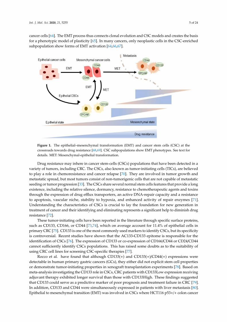
Epithelial to mesenchymal transition (EMT) is a reversible process important for tissue and organformation, through which non-motile epithelial cells with tight cell adhesions and apical basal polarityare converted to individual motile mesenchymal cells with a front to promote polarity [62]. When atumor metastasis arises, often by EMT [63], disseminated cancer cells require a self-renewal abilitysimilar to that of CSC to form macroscopic metastases (Figure 1). This increases the possibility that theEMT process, which allows cancer cells to spread, may also confer the ability of self-renewal to spread
Int. J. Mol. Sci. 2020, 21, 5255 5 of 24
cancer cells [64]. The EMT process thus connects clonal evolution and CSC models and creates the basisfor a phenotypic model of plasticity [65]. In many cancers, only neoplastic cells in the CSC-enrichedsubpopulation show forms of EMT activation [64,66,67].
Int. J. Mol. Sci. 2020, 21, x FOR PEER REVIEW 5 of 24
Figure 1. The epithelial–mesenchymal transformation (EMT) and cancer stem cells (CSC) at the crossroads towards drug resistance [68,69]. CSC subpopulations show EMT phenotypes. See text for details. MET: Mesenchymal-epithelial transformation.
Drug resistance may inhere in cancer stem-cells (CSCs) populations that have been detected in a variety of tumors, including CRC. The CSCs, also known as tumor-initiating cells (TICs), are believed to play a role in chemoresistance and cancer relapse [70]. They are involved in tumor growth and metastatic spread, but most tumors consist of non-tumorigenic cells that are not capable of metastatic seeding or tumor progression [33]. The CSCs share several normal stem cells features that provide a long existence, including the relative silence, dormancy, resistance to chemotherapeutic agents and toxins through the expression of drug efflux transporters, an active DNA-repair capacity and a resistance to apoptosis, vascular niche, stability to hypoxia, and enhanced activity of repair enzymes [71]. Understanding the characteristics of CSCs is crucial to lay the foundation for new generation in treatment of cancer and their identifying and eliminating represents a significant help to diminish drug resistance [72].
These tumor-initiating cells have been reported in the literature through specific surface proteins, such as CD133, CD166, or CD44 [73,74], which on average account for 11.4% of epithelial cells in primary CRC [75]. CD133 is one of the most commonly used markers to identify CSCs, but its specificity is controversial. Recent studies have shown that the AC133-CD133 epitome is responsible for the identification of CSCs [76]. The expression of CD133 or co-expression of CD166/CD44 or CD24/CD44 cannot sufficiently identify CSCs populations. This has raised some doubts as to the suitability of using CRC cell lines for screening CSC-specific therapies [77].
Rocco et al. have found that although CD133(+) and CD133(+)/CD44(+) expressions were detectable in human primary gastric cancers (GCs), they either did not explicit stem cell properties or demonstrate tumor-initiating properties in xenograft transplantation experiments [78]. Based on meta-analysis investigating the CD133 role in CSCs, CRC patients with CD133Low expression receiving adjuvant therapy exhibited longer survival than those with CD133High. These findings suggested that CD133 could serve as a predictive marker of poor prognosis and treatment failure in CRC [79]. In addition, CD133 and CD44 were simultaneously expressed in patients with liver metastasis [80]. Epithelial to mesenchymal transition (EMT) was involved in CSCs where HCT116 p53+/+ colon cancer cells with a high expression of CD133/CD44 showed EMT after long-term culture [81]. Similarly, Dylla et al. demonstrated that the CSCs fraction is increased in CRC tumors after chemotherapy and may help explain relapse following treatment [82]. In another clinical study, the CD133 CRC stem cell marker was followed. Three hundred and three patients with CRC stage I to III
Figure 1. The epithelial–mesenchymal transformation (EMT) and cancer stem cells (CSC) at thecrossroads towards drug resistance [68,69]. CSC subpopulations show EMT phenotypes. See text fordetails. MET: Mesenchymal-epithelial transformation.
Drug resistance may inhere in cancer stem-cells (CSCs) populations that have been detected in avariety of tumors, including CRC. The CSCs, also known as tumor-initiating cells (TICs), are believedto play a role in chemoresistance and cancer relapse [70]. They are involved in tumor growth andmetastatic spread, but most tumors consist of non-tumorigenic cells that are not capable of metastaticseeding or tumor progression [33]. The CSCs share several normal stem cells features that provide a longexistence, including the relative silence, dormancy, resistance to chemotherapeutic agents and toxinsthrough the expression of drug efflux transporters, an active DNA-repair capacity and a resistanceto apoptosis, vascular niche, stability to hypoxia, and enhanced activity of repair enzymes [71].Understanding the characteristics of CSCs is crucial to lay the foundation for new generation intreatment of cancer and their identifying and eliminating represents a significant help to diminish drugresistance [72].
These tumor-initiating cells have been reported in the literature through specific surface proteins,such as CD133, CD166, or CD44 [73,74], which on average account for 11.4% of epithelial cells inprimary CRC [75]. CD133 is one of the most commonly used markers to identify CSCs, but its specificityis controversial. Recent studies have shown that the AC133-CD133 epitome is responsible for theidentification of CSCs [76]. The expression of CD133 or co-expression of CD166/CD44 or CD24/CD44cannot sufficiently identify CSCs populations. This has raised some doubts as to the suitability ofusing CRC cell lines for screening CSC-specific therapies [77].
Rocco et al. have found that although CD133(+) and CD133(+)/CD44(+) expressions weredetectable in human primary gastric cancers (GCs), they either did not explicit stem cell propertiesor demonstrate tumor-initiating properties in xenograft transplantation experiments [78]. Based onmeta-analysis investigating the CD133 role in CSCs, CRC patients with CD133Low expression receivingadjuvant therapy exhibited longer survival than those with CD133High. These findings suggestedthat CD133 could serve as a predictive marker of poor prognosis and treatment failure in CRC [79].In addition, CD133 and CD44 were simultaneously expressed in patients with liver metastasis [80].Epithelial to mesenchymal transition (EMT) was involved in CSCs where HCT116 p53+/+ colon cancer
Int. J. Mol. Sci. 2020, 21, 5255 6 of 24
cells with a high expression of CD133/CD44 showed EMT after long-term culture [81]. Similarly,Dylla et al. demonstrated that the CSCs fraction is increased in CRC tumors after chemotherapy andmay help explain relapse following treatment [82]. In another clinical study, the CD133 CRC stemcell marker was followed. Three hundred and three patients with CRC stage I to III who underwentsurgical resection were found to have higher CD133 expression that correlated with poor prognosisafter radical resection [83]. The prognostic value of co-expression of two CSCs biomarkers, CD44 andCD133, with wild-type EGFR (wtEGFR) and EGFRvIII in CRC patients, was studied and CD133/CD44expression was associated with primary resistance to irinotecan and acquired resistance to anti-EGFRinhibitors in vitro. The results suggest that co-expression of these markers and EGFRvIII may bepotential biomarkers of poorer overall survival and resistance to therapy in patients with mCRC [84].
Based on results from a recent meta-analysis that analyzed the correlation of expression of surfacemarkers and CSCs (tumorigenicity) properties by monitoring tumor incidence and volume in vivo,authors suggested that CD133 expression may represent a strong biomarker for identifying of CSCs inprimary tumors and can be designed as a prognostic marker in CRC patients [85,86].
5. Distant Metastasis-Related Biomarkers in Clinical Practice
There were attempts to use clinical risk scores to stratify patients according to the risk of recurrenceafter metastasectomy, however, with only limited success. The first prognostic gene expressionsignature for OS after resected distant metastasis that was also externally validated showed dominanceover clinical risk scores and highlighted the prognostic potential of transcription profiling [55,59].Similarly, there is a need to identify biomarkers predictive of tumor response to targeted agents.Currently, the only validated and clinically used predictors of responses to anti-EGFR therapy arerepresented by KRAS, NRAS, and BRAF mutational status. A good concordance (almost to 95%) in thepresence of mutations in high risk genes, such as KRAS, TP53, APC, PIK3CA, BRAF, and NRAS, betweenthe primary tumor and its metastases have been reported [87,88]. However, tumor heterogeneity inmCRC have been identified. Intra-tumoral and inter-tumoral heterogeneity of KRAS mutations wasobserved in mCRC and corresponded with resistance or lower efficacy of anti-EGFR therapies [89].For instance, Jeantet et al. determined a high amount of heterogeneity in distribution of RAS mutationsin mCRCs; in detail, 33% of intra-tumoral and 36% of inter-tumoral heterogeneity [90]. Nevertheless,in light of recent findings that right-sided RAS and RAF wild-type tumors do not respond well toanti-EGFR therapies [55], there is an emerging question regarding whether anti-EGFR therapy shouldbe given only to liver metastasis originating from left-sided CRC. There is growing evidence that otherbiomarkers, including mutations of PI3KCA, FBXW7, SMAD4, and others, may evince predictive value.However, further validation is needed. It is becoming strikingly clear that multigene expression assaysmight help to select the most effective induction therapy as well as identify a high-risk populationsuitable for adjuvant therapy. It should be kept in mind, however, that multigene expression assaysare sensitive to sample quality and pre-analytical handling [91]. Moreover, gene expression may notreflect the final function of the protein. Similarly, the tumor microenvironment and immune systemplay a major role in tumor progression and response to therapy. The presence of specific immunecells within the tumor tissue, such as increased densities of T cell infiltrates with a high proportion ofCD8+ T and demonstrated that immune reactivity at the tumor site influences clinical outcome [92].Primary colorectal carcinomas were associated with a significant protection against tumor recurrenceand prognosis was especially marked in deficient mismatch repair system (dMMR/MSI) tumors [93].In detail, hypermutated dMMR/MSI of mCRCs evince higher sensitivity to inhibitors of immunecheckpoint that revive cytotoxic T cells to eliminate dMMR/MSI tumors cells [94]. Patients withdMMR/MSI tumors greatly benefit from immunotherapy, regardless of the tumor type, with diseasecontrol rates of 80% and OS superior to three years in chemo-resistant mCRC [95]. Consequently,all mCRCs should be regularly tested for MMR and MSI status [96].
In mCRC, the dMMR/MSI phenotype (around 5% of mCRCs) is associated with worse prognosisand chemoresistance [95,97]. However, recent studies have reported prolonged OS in dMMR/MSI mCRC
Int. J. Mol. Sci. 2020, 21, 5255 7 of 24
after anti-vascular endothelial growth factor (anti-VEGF) treatment, as compared with anti-EGFR,however without change in survival conferring to chemotherapy regimen, i.e., irinotecan-basedchemotherapy, in contrast to oxaliplatin-based chemotherapy [98]. Finally, recent nonrandomizedtrials point to the high efficacy of the immune checkpoint inhibitor in dMMR/MSI chemo-resistantmCRC as a reason for the high tumor mutational burden. Another response predictor to the immunecheckpoint inhibitor is the immunohistochemistry labeling of the programmed death-ligand 1 (PD-L1)protein [96,99].
Both immunological and molecular markers seem to provide promising prognostic and predictiveinformation in distant metastasis, but this topic warrants further research [93,94,98].
The management of mCRC has been markedly changing in recent years with the introductionof targeted therapies resulting in significant improvements in survival and quality of life of patients.Currently, the gold standard of systemic therapy in the first or second line is a combination ofchemotherapy and targeted therapy. The combined chemotherapy regimens are based on 5-fluorouracil(5-FU) and leucovorin with oxaliplatin (FOLFOX) or irinotecan (FOLFIRI), or both (FOLFOXIRI) [72].The monoclonal antibody (mAb) against vascular endothelial growth factor (VEGF) bevacizumab,VEGF-trap aflibercept and mAbs against epidermal growth factor receptor (EGFR) cetuximab andpanitumumab represent the widely used targeted agents combined with the first- or second-linechemotherapy [100].
Anti-EGFR mAbs are used for patients with tumors harboring the wild-type RAS gene,which represents a well-established predictive biomarker. The efficacy of anti-EGFR mAbs was restrictedto patients with tumors harboring the wild-type KRAS gene. Thus, KRAS gene mutations, occurringin 35–45% of cases, became the most important predictive biomarker in mCRC patients [101–104].Extended RAS analyses have demonstrated a lack of response to anti-EGFR mAbs also in patients withtumors harboring NRAS gene mutations, in 1–6% of cases [105–107].
It was also assumed that the BRAF oncogene evinces more prognostic significance [108].BRAF mutations can be used as an effective predictive biomarker for BRAF-targeted therapies.The combination of encorafenib (BRAF inhibitor), binimetinib (MEK inhibitor) and cetuximab in mCRCpatients with tumors harboring BRAF mutations resulted in significantly longer overall survival anda higher response rate to therapy [109]. mCRC microsatellite instability (MSI) phenotype in mCRCis associated with worse prognosis and chemoresistance to standard treatment [94]. On the otherhand, the MSI-high tumors often respond to immunotherapy, and two programmed cell death 1(PD1)-blocking mAbs, pembrolizumab and nivolumab, have been effective in mCRC patients withMSI-H tumors.
Considerable efforts are currently dedicated to identifying biomarkers associated with therapyresponse. The identification of these biomarkers is crucial for individualized treatment strategies inmCRC patients.
Despite RAS gene mutations representing effective and currently the most important predictivebiomarkers, there is still a proportion of patients with tumors harboring the wild-type RAS gene,who derive no or poor benefit from the systemic therapy containing anti-EGFR mAbs. Moreover,for the antiangiogenic targeted therapy represented by bevacizumab and aflibercept, there are nopredictive biomarkers available in the routine clinical practice. BRAF mutations and MSI-H representother recently established predictive biomarkers for novel therapies. However, they are relatively rare,occurring in approximately 5–10% and 3–5% of mCRC, respectively [94].
There is an urgent need for personalized medicine to select the optimal therapy from an expandingrange of the systemic treatment modalities in mCRC patients. The identification of biomarkers thatcould predict the response to a specific type of systemic therapy and/or be used in the effectivenoninvasive monitoring of mCRC patients treated with palliative systemic therapy might the shift inthe therapy towards a precision medicine.
Int. J. Mol. Sci. 2020, 21, 5255 8 of 24
6. Side Effects and Toxicity
The therapeutic CRC options have increased during the last 20 years, and the complexity ofdecision-making has also advanced. The treatment strategies in neoadjuvant, adjuvant, and palliativeapproaches differ, and treatment decisions influence not only the drug efficacy. Age, the presence ofsignificant comorbidities, and various treatment regimens and strategies provide medical oncologistswith a number of options to try to maximize patients’ quality of life and longevity.
Systemic chemotherapy plays a major role in the CRC management. However, cancer treatmentcan be significantly prolonged and obstructed due to the presence of chemotherapy-induced sideeffects which may require dose reduction or delay or discontinuation of treatment. Although 5-FUis one of the safest chemotherapeutics, several CRC patients still have serious side and toxic effects.Clinical manifestations of 5-FU toxicity include fever, fatigue, mucositis, stomatitis, nausea, vomiting,and diarrhea [110]. Other common toxic effects include leukopenia, neutropenia, thrombocytopenia,anemia, neuropathy, skin rash, and hand–foot syndrome [111]. Neurological abnormalities, such ascerebellar ataxia and changes in cognitive function, have also been reported rarely and have occurredin less than one percent of patients [112]. The oxaliplatin side effects includes peripheral neuropathy,nausea and vomiting, diarrhea and fatigue [113,114]. However, most of the patients do not experienceany of these side effects.
Although it has been reported that the patient outcomes have improved after the addition ofoxaliplatin or irinotecan to the 5-FU regimen, the toxicity has also increased [115]. The efficacy of5-FU/LV combined with oxaliplatin (FOLFOX) or irinotecan (FOLFIRI) in the first-line treatment ofmCRC is comparable [116]. The combination therapies FOLFOX and FOLFIRI have become establishedas efficacious cytotoxic regimens for the treatment of mCRC, resulting in overall improvement insurvival of approximately 2 years [117].
7. Drug Resistance in mCRC
Drug resistance is the major cause of treatment failure in CRC. Resistance can be intrinsic (primary)or acquired (secondary). Both intrinsic resistance, characterized by cancer cells that have little or noresponse to treatment from the beginning, and acquired resistance, where the tumor may becomecross-resistant to a range of chemotherapies, lead to treatment failure in over 90% of patients withmCRC [72].
Malignant tumors can possess intrinsic and/or acquired resistance and each is important indetermining initial and subsequent lines of treatment. Innate resistance is typically noted duringearly drug development or in early phase clinical trials of biologic efficacy. For instance, resistanceto EGFR antagonists was initially not well understood and in early studies only 10–20% of patientsexhibited a response to the EGFR-targeted therapies, cetuximab or panitumumab [118]. The subsequentelucidation of RAS mutations in CRC clarified a marker of innate resistance to these therapies andchanged their clinical use.
Different mechanisms of acquired resistance can exist for each cytotoxic therapy and each targetedpathway, but often acquired resistance to one drug confers resistance to other drugs which may workby different mechanisms of action, a concept referred to as multidrug resistance (MDR). In general,resistance to traditional cytotoxic therapy is accomplished by decreasing the delivery of drugs tothe cancer cell, either by increased efflux out of the cell mediated by ATP-dependent transporters,by decreased uptake into the cell, or by a change in enzymes involved in metabolism. Alternately,resistance can be conferred by changes within the cell itself by genetic or epigenetic modifications thatcan alter drug sensitivity.
Albeit targeted drugs may provide more adequate therapy, studies have displayed moderatebenefits and, like traditional chemotherapy, both primary and secondary resistance may arise [119].
Chemoresistance may be involved in the disease relapse and in the process of metastasisdevelopment. It restricts the improvement of clinical outcomes for cancer patients and persists asthe main obstruction to cancer therapy. Therefore, it is very important to understand the molecular
Int. J. Mol. Sci. 2020, 21, 5255 9 of 24
mechanisms of chemoresistance to find novel therapeutic approaches. Currently, it is believed thattherapy resistance is the consequence of a clone selection during therapy and the presence/generationof novel mutations in cancer cells that enable them to resist the cytotoxic attack [120]. Kim et al.have shown that CRC seems to follow the clonal evolution concept [121]. In this model, the gradualgain of mutations or non-genetic alterations leads to the development of specific sub-clones withdifferent abilities to adapt to the tumor microenvironment, resulting in inter- and intra-tumoralheterogeneity [33]. mCRC often had one clone that is dominant with several minor sub-clones, while apreeminent clone was found less frequently in early stage CRC. In distant metastases, KRAS and TP53mutation heterogeneity was rare. This reflection of decreasing tumor heterogeneity together with tumorprogression, particularly in distant metastases, was promoted by study of Kim et al., who demonstrateda higher number of mutated alleles in CRC metastases compared to matched primary tumors, aimingat contraction in genetic and non-genetic heterogeneity in distant metastases [96,121].
On the other hand, Sottoriva et al. proposed a different aspect of heterogeneity leading tochemoresistance by highlighting the timing of tumor mutations. This “big bang model” considersthat the mutations that are responsible for tumor formation and progression appear early in CRC.Therefore, the biological behavior of a tumor is set early and may clarify why some large tumors neverform metastases, and other small tumors develop metastases earlier. Thus, in this model, mutationsthat lead to evolution of different tumor sub-clones could be considered as passenger mutations ratherthan induced by Darwinian option of the “fittest” sub-clone, which leads to spatial dominance [122].
Clearly, new and effective strategies to overcome chemoresistance are urgently needed. Malignanttumors may have an intrinsic resistance and/or acquired resistance, and both are important indetermining initial and subsequent lines of treatment. Innate resistance of cancer cells is typically notedduring an early drug application. But for instance, resistance to EGFR antagonists was initially not wellunderstood and in early studies only 10–20% of patients exhibited a response to the EGFR-targetedtherapies cetuximab or panitumumab [118]. The subsequent elucidation of the role played by RASmutations in CRC clarified a marker of acquired resistance to these therapies and changed their clinicaluse [57].
8. Experience in Predicting Markers
Patients with distant metastasis show a wide heterogeneity of clinical outcomes. Many systemshave been developed to determine patient prognosis based on individual tumors and personalcharacteristics to understand who would benefit most from hepatic resection. Such a strategy also aidsin adapting an individualized treatment strategy that focuses on providing appropriate, timely, andpersonalized therapy. In addition, in the period of modern effective chemotherapy and multidisciplinarymanagement, traditional tumor and patient characteristics may have less impact on survival. Recentresearch has focused on the use of genetic biomarkers and molecular signatures to identify subgroupsof patients most likely to obtain benefits from a given therapy and to use better survival estimates toguide individualized patient treatment plans [123].
Gene expression profiling of cancer cells has been used for decade to accurately classify tumorsand to identify markers predicting the patient’s outcome. In the case of primary CRC tumors, numerousexpression studies have been conducted so far [124–126].
Plentiful studies analyzing either whole transcriptome or small sets of expressed genes reportedsignificant differences in the gene expression profiles between non-metastatic and mCRC [127,128].A few other studies also described variances in single gene expression between primary tumor and/orliver metastasis on one side, and lung metastasis on the other side [129,130]. More data are availablefrom expression-profiling analyses of clinical samples and/or animal models with CRC liver metastasis.In these studies, various gene signatures for colon-to-liver metastasis were suggested [131,132].Although many identified signatures or molecular markers have been validated by other groups,yet none of them has been used as a diagnostic or prognostic tool applicable to clinical practice asreviewed [133,134]. In order to obtain a real genetic signature for distant metastasis by transcription
Int. J. Mol. Sci. 2020, 21, 5255 10 of 24
profiling, further studies are needed to improve reproducibility and increase consistency, and thevalidation of results needs to be implemented. Before the clinical use can be implemented, prospectivestudies should be performed with a huge number of patients to accomplish reproducible results.Improving of our knowledge of molecular pathways involved in the development of distant metastasiswill lead to a better approach to the prevention and treatment of this disease.
Identification of appropriate biomarkers can significantly improve the choice of treatment strategiesfor patients with CRC. Most of these markers can notify the oncologists about the overall prognosisof the disease; nevertheless, they fail to make a therapeutic choice. In fact, the majority of identifiedbiomarkers, with the exception of the KRAS and BRAF genes and MSI status, did not predict therapeuticresponses or did not reach the clinical practice.
Recently, Koncina et al. [135] and Lee [136] outlined new clinicopathological and molecularbiomarkers for all CRC that can be translated into the clinical setting; however, only few of them areapplicable for mCRC.
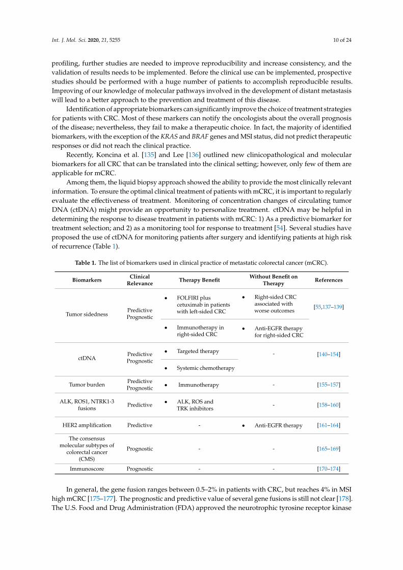
Among them, the liquid biopsy approach showed the ability to provide the most clinically relevantinformation. To ensure the optimal clinical treatment of patients with mCRC, it is important to regularlyevaluate the effectiveness of treatment. Monitoring of concentration changes of circulating tumorDNA (ctDNA) might provide an opportunity to personalize treatment. ctDNA may be helpful indetermining the response to disease treatment in patients with mCRC: 1) As a predictive biomarker fortreatment selection; and 2) as a monitoring tool for response to treatment [54]. Several studies haveproposed the use of ctDNA for monitoring patients after surgery and identifying patients at high riskof recurrence (Table 1).
Table 1. The list of biomarkers used in clinical practice of metastatic colorectal cancer (mCRC).
Biomarkers ClinicalRelevance Therapy Benefit Without Benefit on
Therapy References
Tumor sidednessPredictivePrognostic
• FOLFIRI pluscetuximab in patientswith left-sided CRC
• Right-sided CRCassociated withworse outcomes
[55,137–139]
• Immunotherapy inright-sided CRC
• Anti-EGFR therapyfor right-sided CRC
ctDNAPredictivePrognostic
• Targeted therapy - [140–154]
• Systemic chemotherapy
Tumor burden PredictivePrognostic • Immunotherapy - [155–157]
ALK, ROS1, NTRK1-3fusions Predictive • ALK, ROS and
TRK inhibitors- [158–160]
HER2 amplification Predictive - • Anti-EGFR therapy [161–164]
The consensusmolecular subtypes of
colorectal cancer(CMS)
Prognostic - - [165–169]
Immunoscore Prognostic - - [170–174]
In general, the gene fusion ranges between 0.5–2% in patients with CRC, but reaches 4% in MSIhigh mCRC [175–177]. The prognostic and predictive value of several gene fusions is still not clear [178].The U.S. Food and Drug Administration (FDA) approved the neurotrophic tyrosine receptor kinase
Int. J. Mol. Sci. 2020, 21, 5255 11 of 24
(NTRK)-inhibitor entrectinib and larotrectinib in NTRK-fusion-mutated tumors of all organ types,including CRC, provided they do not contain a known acquired resistance mutation, in 2019.
HER2 (Errb2) is a transmembrane receptor of the EGFR family and its activation stimulatescell proliferation and the inhibition of apoptosis. Amplifications of HER2 occur in 2–6% of mCRC,but reach up to 13% in wild-type KRAS/NRAS/BRAF mCRC [179]. HER2 has recently gainedmuch importance in CRC. Two recent clinical trials, MyPathway (NCT02091141) and HERACLES(NCT03225937), have shown encouraging clinical benefits for a dual HER2 blockade in mCRC patientswith HER2 amplification.
The CRC classification by consensus molecular subtypes of CRC (CMS) proposed byGuinney et al. [165] subtypes might serve as a prognostic biomarker. These 4 subtypes are: CMS,with MSI and immune activation (14%); CMS2, with canonical CRC alterations (37%); CMS3,with metabolic dysregulation (13%); and CMS4, with mesenchymal features (23%). However, from aclinical point of view, CMS does not seem to have superior value to routinely use clinical indicationcriteria for selecting patients for optimal treatment with either anti-EGFR or anti-VEGF agents. Overall,CMS categorization provides a detailed insight into CRC etiology but currently still has no real impacton clinical decision-making. However, the inclusion of patients with distinct CRC molecular subtypesrepresents an essential start for clinical translation. The prognostic significance of CMS subtypes inboth early and metastatic CRCs bolsters the fact that they could be used in the assessment of therapyresponses and might aid the treatment choice.
The immunoscore is a scoring system that is based on the quantification of cytotoxic and memoryT cells in the core of the tumor and in the tumor’s invasive margin [172,180–182]. It represents a strongprognostic marker in CRC [170,183–185] and has a dual advantage over TNM staging. The immunoscorehas been reported to be a better predictor than MSI alone [174]. Due to the important role of hostimmunity in controlling tumor progression, it is probably necessary to include the immunoscore in thecancer classification.
As stated before, the choice of the treatment strategy depends on various clinical factors andbiomarkers. Several other molecular biomarkers have been also suggested, such as miRNAs, the CpGisland methylator phenotype (CIMP). Recently, the expression of miR-31-3p was introduced as apromising predictive biomarker for anti-EGFR therapy in patients with the wild-type KRAS gene, andtreated with adjuvant chemotherapy. Low miR-31-3p expression in patients treated with standardchemotherapy and cetuximab was associated with longer progression-free survival compared topatients expressing high levels of miR-31-3p [186–190]. The CIMP-positive tumors (hypermethylationof at least three out of five pre-defined marker) displayed independent biomarker or recurrence inCRC [191–194]. Further studies are urgently needed to identify and validate new biomarkers toimprove outcomes in patients with CRC.
9. Conclusions and Future Perspectives
The metastatic process into specific organs is largely dependent on the ability of a tumor cellto interact with its microenvironment that is influenced also by its internal characteristics [195].The identification of these cell characteristics is of prime interest, as it could lead to the design of newtherapeutic strategies. CRC research is aimed at several areas, such as prevention, early stage canceridentification, prognostic and predictive markers recognition, new molecular targets identification,drug development, and clinical practice adjustment. Identification of major cancer pathways on genetic,proteomic, and epigenetic level contributes to our better understanding of CRC behavior. Recentyears witnessed substantial progress in understanding of microenvironment/microbial settlementin CRC onset, pathogenesis, and progression. The future precise risk stratification of patients withadvanced CRC and subsequent pre- and post-operative decisions must not omit consideration ofmicroenvironmental (microbial) context (e.g., based on liquid biopsy).
Despite the improving knowledge of the development and progression of CRC, results in commonclinical practice treatment show only little advance.
Int. J. Mol. Sci. 2020, 21, 5255 12 of 24
Starting in 2016, relevant sub-analysis of 6 important clinical trials (FIRE-3, CALGB/SWOG-80405,PEAK, Clinical Study No 20050181, CRYSTAL and PRIME) [196] opened the question of primary tumorsidedness as an additional character for molecular tumor profile and dual inhibition (VEGF/EGFR)treatment efficacy [197]. These findings resulted afterwards in standard use of consensus molecularsubtypes (CMS) in the majority of newly designed trials [165,175]. The findings in this area wouldhelp us to identify, for example, patients at higher risk of tumor recurrence after surgery andtreatment or development of distant metastasis. Moreover, small subpopulations of patients withdruggable molecular changes are already treated correspondingly, for instance BRAF-mutatedtumors (10%), HER2-positive tumors (3%), NTRK -fusion positive tumors (1–2%), FGFR (fibroblastgrowth factor receptor)-amplified tumors or ROS1 (receptor tyrosin kinase)- and ALK (anaplasticlymphoma kinase)-fusion-positive tumors (up to 1%) [175,198–200]. Most expectations are put intoimmunomodulation with PD-1 or PD-L1 antibodies, currently used in MSI/MMR-deficient tumors [201].Great efforts have been undertaken to change the immune cold tumor environment into the hot oneand to take the advantage of natural property of the immune system to precisely and accuratelytarget specific antigens [202]. Another interesting approach to treatment is the use of epigeneticmodification. Notably, HDACi (histone deacetylase inhibitors), namely Vorinostat, are already usedin hemato-oncology; however, the results are still waiting for application in the CRC treatment [203].Besides the detection of specific DNA mutations for predicting responses to anti-EGFR therapies [149],the liquid biopsy approach as measuring the plasma/serum concentrations of cell-free DNA (cfDNA)and/or circulating tumor DNA (ctDNA) emerged to be an effective indirect predictive biomarker inmCRC patients [53,204].
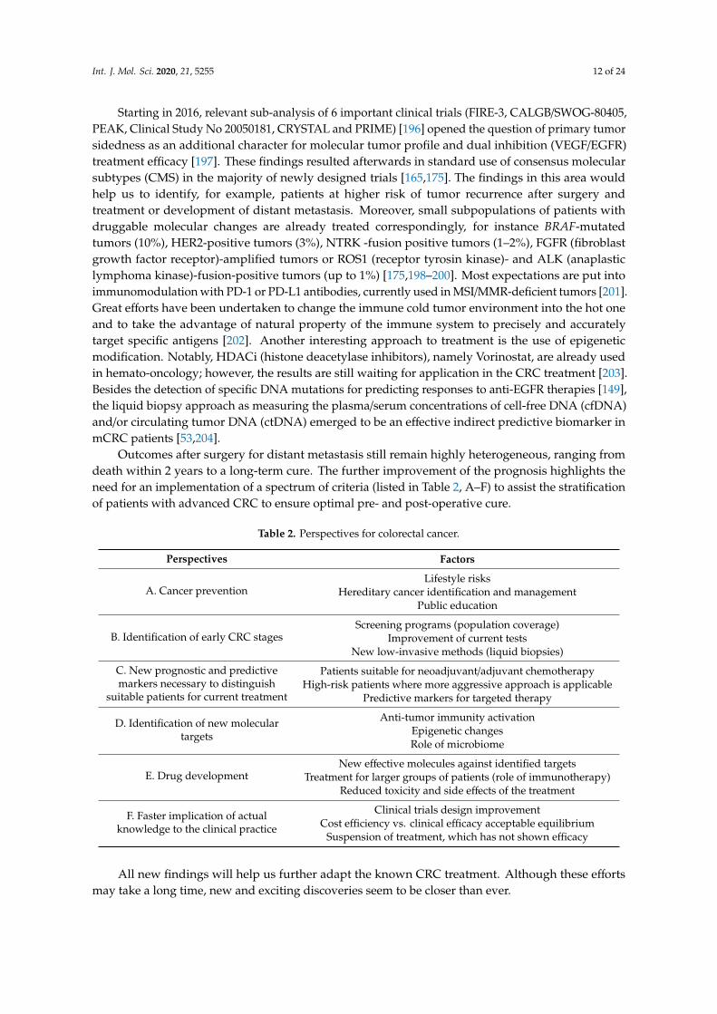
Outcomes after surgery for distant metastasis still remain highly heterogeneous, ranging fromdeath within 2 years to a long-term cure. The further improvement of the prognosis highlights theneed for an implementation of a spectrum of criteria (listed in Table 2, A–F) to assist the stratificationof patients with advanced CRC to ensure optimal pre- and post-operative cure.
Table 2. Perspectives for colorectal cancer.
Perspectives Factors
A. Cancer preventionLifestyle risks
Hereditary cancer identification and managementPublic education
B. Identification of early CRC stagesScreening programs (population coverage)
Improvement of current testsNew low-invasive methods (liquid biopsies)
C. New prognostic and predictivemarkers necessary to distinguish
suitable patients for current treatment
Patients suitable for neoadjuvant/adjuvant chemotherapyHigh-risk patients where more aggressive approach is applicable
Predictive markers for targeted therapy
D. Identification of new moleculartargets
Anti-tumor immunity activationEpigenetic changesRole of microbiome
E. Drug developmentNew effective molecules against identified targets
Treatment for larger groups of patients (role of immunotherapy)Reduced toxicity and side effects of the treatment
F. Faster implication of actualknowledge to the clinical practice
Clinical trials design improvementCost efficiency vs. clinical efficacy acceptable equilibrium
Suspension of treatment, which has not shown efficacy
All new findings will help us further adapt the known CRC treatment. Although these effortsmay take a long time, new and exciting discoveries seem to be closer than ever.

Int. J. Mol. Sci. 2020, 21, 5255 13 of 24
Author Contributions: S.F., V.V., J.P., O.K., S.J., and K.C. wrote and analyzed the trial and clinical study ofexperience; F.C. wrote about the clinical experience of the surgery in mCRC; V.V., L.V., M.K., P.V. and M.M. wrotethe experimental experience; and P.V. along with S.F. are the corresponding authors. All authors have read andagreed to the published version of the manuscript.
Funding: This short study was supported by the grant NV19-09-00237 from the Ministry of Health CZ andPROGRES Q40/06.
Conflicts of Interest: The authors declare no conflict of interests.
References
1. Siegel, R.L.; Miller, K.D.; Jemal, A. Cancer statistics, 2019. CA Cancer J. Clin. 2019, 69, 7–34. [CrossRef][PubMed]
2. Siegel, R.L.; Miller, K.D.; Goding Sauer, A.; Fedewa, S.A.; Butterly, L.F.; Anderson, J.C.; Cercek, A.; Smith, R.A.;Jemal, A. Colorectal cancer statistics, 2020. CA A Cancer J. Clin. 2020, 70, 145–164. [CrossRef] [PubMed]
3. Oki, E.; Ando, K.; Nakanishi, R.; Sugiyama, M.; Nakashima, Y.; Kubo, N.; Kudou, K.; Saeki, H.; Nozoe, T.;Emi, Y.; et al. Recent advances in treatment for colorectal liver metastasis. Ann. Gastroenterol. Surg. 2018, 2,167–175. [CrossRef] [PubMed]
4. Kim, H.J.; Choi, G. Clinical Implications of Lymph Node Metastasis in Colorectal Cancer: Current Status andFuture Perspectives. Ann. Coloproctol. 2019, 35, 109–117. [CrossRef]
5. Fessler, E.; Dijkgraaf, F.E.; De Sousa, E.; Melo, F.; Medema, J.P. Cancer stem cell dynamics in tumor progressionand metastasis: Is the microenvironment to blame? Cancer Lett. 2013, 341, 97–104. [CrossRef]
6. Zarour, L.R.; Anand, S.; Billingsley, K.G.; Bisson, W.H.; Cercek, A.; Clarke, M.F.; Coussens, L.M.; Gast, C.E.;Geltzeiler, C.B.; Hansen, L.; et al. Colorectal Cancer Liver Metastasis: Evolving Paradigms and FutureDirections. Cell Mol. Gastroenterol. Hepatol. 2017, 3, 163–173. [CrossRef]
7. Bozzetti, F.; Doci, R.; Bignami, P.; Morabito, A.; Gennari, L. Patterns of Failure Following Surgical Resectionof Colorectal Cancer Liver Metastases: Rationale for a Multimodal Approach. Ann. Surg. 1987, 205, 264–270.[CrossRef]
8. Misiakos, E.P.; Karidis, N.P.; Kouraklis, G. Current treatment for colorectal liver metastases.World J. Gastroenterol. 2011, 17, 4067–4075. [CrossRef]
9. Jones, R.P.; Jackson, R.; Dunne, D.F.J.; Malik, H.Z.; Fenwick, S.W.; Poston, G.J.; Ghaneh, P. Systematic reviewand meta-analysis of follow-up after hepatectomy for colorectal liver metastases. Br. J. Surg. 2012, 99,477–486. [CrossRef]
10. Yamazaki, K.; Nagase, M.; Tamagawa, H.; Ueda, S.; Tamura, T.; Murata, K.; Eguchi Nakajima, T.; Baba, E.;Tsuda, M.; Moriwaki, T.; et al. Randomized phase III study of bevacizumab plus FOLFIRI and bevacizumabplus mFOLFOX6 as first-line treatment for patients with metastatic colorectal cancer (WJOG4407G). Ann. Oncol.2016, 27, 1539–1546. [CrossRef]
11. Pastorino, U.; Buyse, M.; Friedel, G.; Ginsberg, R.J.; Girard, P.; Goldstraw, P.; Johnston, M.; McCormack, P.;Pass, H.; Putnam, J.B. Long-term results of lung metastasectomy: Prognostic analyses based on 5206 cases.J. Thorac. Cardiovasc. Surg. 1997, 113, 37–49. [CrossRef]
12. Abdalla, E.K.; Hicks, M.E.; Vauthey, J.N. Portal vein embolization: Rationale, technique and future prospects.Br. J. Surg. 2001, 88, 165–175. [CrossRef] [PubMed]
13. Okita, A.; Takahashi, S.; Ouchi, K.; Inoue, M.; Watanabe, M.; Endo, M.; Honda, H.; Yamada, Y.; Ishioka, C.Consensus molecular subtypes classification of colorectal cancer as a predictive factor for chemotherapeuticefficacy against metastatic colorectal cancer. Oncotarget 2018, 9, 18698–18711. [CrossRef]
14. Ciombor, K.K.; Bekaii-Saab, T. Emerging treatments in recurrent and metastatic colorectal cancer. J. Natl.Compr. Cancer Netw. 2013, 11 (Suppl. S4), S18–S27. [CrossRef] [PubMed]
15. Van Cutsem, E.; Cervantes, A.; Adam, R.; Sobrero, A.; Van Krieken, J.H.; Aderka, D.; Aranda Aguilar, E.;Bardelli, A.; Benson, A.; Bodoky, G.; et al. ESMO consensus guidelines for the management of patients withmetastatic colorectal cancer. Ann. Oncol. 2016, 27, 1386–1422. [CrossRef]
16. Turan, N.; Benekli, M.; Koca, D.; Ustaalioglu, B.O.; Dane, F.; Ozdemir, N.; Ulas, A.; Oztop, I.; Gumus, M.;Ozturk, M.A.; et al. Adjuvant systemic chemotherapy with or without bevacizumab in patients with resectedliver metastases from colorectal cancer. Oncology 2013, 84, 14–21. [CrossRef] [PubMed]
Int. J. Mol. Sci. 2020, 21, 5255 14 of 24
17. Nappi, A.; Berretta, M.; Romano, C.; Tafuto, S.; Cassata, A.; Casaretti, R.; Silvestro, L.; Divitiis, C.D.;Alessandrini, L.; Fiorica, F.; et al. Metastatic Colorectal Cancer: Role of Target Therapies and FuturePerspectives. Curr. Cancer Drug Targets 2018, 18, 421–429. [CrossRef]
18. Tsilimigras, D.I.; Ntanasis-Stathopoulos, I.; Bagante, F.; Moris, D.; Cloyd, J.; Spartalis, E.; Pawlik, T.M. Clinicalsignificance and prognostic relevance of KRAS, BRAF, PI3K and TP53 genetic mutation analysis for resectableand unresectable colorectal liver metastases: A systematic review of the current evidence. Surg. Oncol. 2018,27, 280–288. [CrossRef]
19. Fong, Y.; Fortner, J.; Sun, R.L.; Brennan, M.F.; Blumgart, L.H. Clinical score for predicting recurrence afterhepatic resection for metastatic colorectal cancer: Analysis of 1001 consecutive cases. Ann. Surg. 1999, 230,309–318, discussion 318–321. [CrossRef]
20. Iwatsuki, S.; Dvorchik, I.; Madariaga, J.R.; Marsh, J.W.; Dodson, F.; Bonham, A.C.; Geller, D.A.; Gayowski, T.J.;Fung, J.J.; Starzl, T.E. Hepatic resection for metastatic colorectal adenocarcinoma: A proposal of a prognosticscoring system. J. Am. Coll. Surg. 1999, 189, 291–299. [CrossRef]
21. Rees, M.; Tekkis, P.P.; Welsh, F.K.S.; O’Rourke, T.; John, T.G. Evaluation of long-term survival after hepaticresection for metastatic colorectal cancer: A multifactorial model of 929 patients. Ann. Surg. 2008, 247,125–135. [CrossRef] [PubMed]
22. Zakaria, S.; Donohue, J.H.; Que, F.G.; Farnell, M.B.; Schleck, C.D.; Ilstrup, D.M.; Nagorney, D.M. Hepaticresection for colorectal metastases: Value for risk scoring systems? Ann. Surg. 2007, 246, 183–191. [CrossRef][PubMed]
23. Nathan, H.; de Jong, M.C.; Pulitano, C.; Ribero, D.; Strub, J.; Mentha, G.; Gigot, J.-F.; Schulick, R.D.;Choti, M.A.; Aldrighetti, L.; et al. Conditional Survival after Surgical Resection of Colorectal Liver Metastasis:An International Multi-Institutional Analysis of 949 Patients. J. Am. Coll. Surg. 2010, 210, 755–764. [CrossRef][PubMed]
24. Roberts, K.J.; White, A.; Cockbain, A.; Hodson, J.; Hidalgo, E.; Toogood, G.J.; Lodge, J.P.A. Performanceof prognostic scores in predicting long-term outcome following resection of colorectal liver metastases.Br. J. Surg. 2014, 101, 856–866. [CrossRef]
25. Kattan, M.W.; Gönen, M.; Jarnagin, W.R.; DeMatteo, R.; D’Angelica, M.; Weiser, M.; Blumgart, L.H.; Fong, Y.A nomogram for predicting disease-specific survival after hepatic resection for metastatic colorectal cancer.Ann. Surg. 2008, 247, 282–287. [CrossRef]
26. Balachandran, V.P.; Arora, A.; Gönen, M.; Ito, H.; Turcotte, S.; Shia, J.; Viale, A.; Snoeren, N.; van Hooff, S.R.;Rinkes, I.H.M.B.; et al. A Validated Prognostic Multigene Expression Assay for Overall Survival in ResectedColorectal Cancer Liver Metastases. Clin. Cancer Res. 2016, 22, 2575–2582. [CrossRef]
27. Barbon, C.; Margonis, G.A.; Andreatos, N.; Rezaee, N.; Sasaki, K.; Buettner, S.; Damaskos, C.; Pawlik, T.M.;He, J.; Wolfgang, C.L.; et al. Colorectal Liver Metastases: Does the Future of Precision Medicine Lie inGenetic Testing? J. Gastrointest. Surg. 2018, 22, 1286–1296. [CrossRef]
28. Tie, J.; Lipton, L.; Desai, J.; Gibbs, P.; Jorissen, R.N.; Christie, M.; Drummond, K.J.; Thomson, B.N.J.; Usatoff, V.;Evans, P.M.; et al. KRAS mutation is associated with lung metastasis in patients with curatively resectedcolorectal cancer. Clin. Cancer Res. 2011, 17, 1122–1130. [CrossRef]
29. Sideris, M.; Papagrigoriadis, S. Molecular biomarkers and classification models in the evaluation of theprognosis of colorectal cancer. Anticancer Res. 2014, 34, 2061–2068.
30. Sagaert, X. Prognostic biomarkers in colorectal cancer: Where do we stand? Virchows Arch. 2014, 464, 379–391.[CrossRef]
31. Adam, R. Developing strategies for liver metastases from colorectal cancer. Semin. Oncol. 2007, 34, S7–S11.[CrossRef]
32. Molinari, C.; Marisi, G.; Passardi, A.; Matteucci, L.; De Maio, G.; Ulivi, P. Heterogeneity in Colorectal Cancer:A Challenge for Personalized Medicine? Int. J. Mol. Sci. 2018, 19, 3733. [CrossRef] [PubMed]
33. Blank, A.; Roberts, D.E.; Dawson, H.; Zlobec, I.; Lugli, A. Tumor Heterogeneity in Primary Colorectal Cancerand Corresponding Metastases. Does the Apple Fall Far From the Tree? Front. Med. (Lausanne) 2018, 5, 234.[CrossRef] [PubMed]
34. Ulintz, P.J.; Greenson, J.K.; Wu, R.; Fearon, E.R.; Hardiman, K.M. Lymph Node Metastases in Colon CancerAre Polyclonal. Clin. Cancer Res. 2018, 24, 2214–2224. [CrossRef]
35. Lee, S.Y.; Haq, F.; Kim, D.; Jun, C.; Jo, H.-J.; Ahn, S.-M.; Lee, W.-S. Comparative genomic analysis of primaryand synchronous metastatic colorectal cancers. PLoS ONE 2014, 9, e90459. [CrossRef]
Int. J. Mol. Sci. 2020, 21, 5255 15 of 24
36. Hunter, K.W.; Amin, R.; Deasy, S.; Ha, N.-H.; Wakefield, L. Genetic insights into the morass of metastaticheterogeneity. Nat. Rev. Cancer 2018, 18, 211–223. [CrossRef] [PubMed]
37. Mogensen, M.B.; Rossing, M.; Østrup, O.; Larsen, P.N.; Heiberg Engel, P.J.; Jørgensen, L.N.; Hogdall, E.V.;Eriksen, J.; Ibsen, P.; Jess, P.; et al. Genomic alterations accompanying tumour evolution in colorectal cancer:Tracking the differences between primary tumours and synchronous liver metastases by whole-exomesequencing. BMC Cancer 2018, 18, 752. [CrossRef]
38. Carethers, J.M.; Jung, B.H. Genetics and Genetic Biomarkers in Sporadic Colorectal Cancer. Gastroenterology2015, 149, 1177–1190.e3. [CrossRef]
39. Zellmer, V.R.; Zhang, S. Evolving concepts of tumor heterogeneity. Cell Biosci. 2014, 4, 69. [CrossRef]40. Nowell, P. The clonal evolution of tumor cell populations. Science 1976, 194, 23–28. [CrossRef]41. Jamal-Hanjani, M.; Quezada, S.A.; Larkin, J.; Swanton, C. Translational Implications of Tumor Heterogeneity.
Clin. Cancer Res. 2015, 21, 1258–1266. [CrossRef] [PubMed]42. The Cancer Genome Atlas Network Comprehensive molecular characterization of human colon and rectal
cancer. Nature 2012, 487, 330–337. [CrossRef] [PubMed]43. Kim, R.; Schell, M.J.; Teer, J.K.; Greenawalt, D.M.; Yang, M.; Yeatman, T.J. Co-Evolution of Somatic Variation
in Primary and Metastatic Colorectal Cancer May Expand Biopsy Indications in the Molecular Era. PLoS ONE2015, 10, e0126670. [CrossRef] [PubMed]
44. Brannon, A.R.; Vakiani, E.; Sylvester, B.E.; Scott, S.N.; McDermott, G.; Shah, R.H.; Kania, K.; Viale, A.;Oschwald, D.M.; Vacic, V.; et al. Comparative sequencing analysis reveals high genomic concordancebetween matched primary and metastatic colorectal cancer lesions. Genome Biol. 2014, 15, 454. [CrossRef][PubMed]
45. Vignot, S.; Lefebvre, C.; Frampton, G.M.; Meurice, G.; Yelensky, R.; Palmer, G.; Capron, F.; Lazar, V.;Hannoun, L.; Miller, V.A.; et al. Comparative analysis of primary tumour and matched metastasesin colorectal cancer patients: Evaluation of concordance between genomic and transcriptional profiles.Eur. J. Cancer 2015, 51, 791–799. [CrossRef] [PubMed]
46. Sleeman, J.P.; Cady, B.; Pantel, K. The connectivity of lymphogenous and hematogenous tumor celldissemination: Biological insights and clinical implications. Clin. Exp. Metastasis 2012, 29, 737–746.[CrossRef]
47. Cady, B. Lymph Node Metastases: Indicators, but Not Governors of Survival. Arch. Surg. 1984, 119, 1067.[CrossRef]
48. Naxerova, K.; Reiter, J.G.; Brachtel, E.; Lennerz, J.K.; van de Wetering, M.; Rowan, A.; Cai, T.; Clevers, H.;Swanton, C.; Nowak, M.A.; et al. Origins of lymphatic and distant metastases in human colorectal cancer.Science 2017, 357, 55–60. [CrossRef]
49. Koehler, A.; Bataille, F.; Schmid, C.; Ruemmele, P.; Waldeck, A.; Blaszyk, H.; Hartmann, A.; Hofstaedter, F.;Dietmaier, W. Gene expression profiling of colorectal cancer and metastases divides tumours according totheir clinicopathological stage. J. Pathol. 2004, 204, 65–74. [CrossRef]
50. Lee, J.-R.; Kwon, C.H.; Choi, Y.; Park, H.J.; Kim, H.S.; Jo, H.-J.; Oh, N.; Park, D.Y. Transcriptome analysisof paired primary colorectal carcinoma and liver metastases reveals fusion transcripts and similar geneexpression profiles in primary carcinoma and liver metastases. BMC Cancer 2016, 16, 539. [CrossRef]
51. Vermaat, J.S.; Nijman, I.J.; Koudijs, M.J.; Gerritse, F.L.; Scherer, S.J.; Mokry, M.; Roessingh, W.M.; Lansu, N.; deBruijn, E.; van Hillegersberg, R.; et al. Primary Colorectal Cancers and Their Subsequent Hepatic MetastasesAre Genetically Different: Implications for Selection of Patients for Targeted Treatment. Clin. Cancer Res.2012, 18, 688–699. [CrossRef] [PubMed]
52. Cervena, K.; Vodicka, P.; Vymetalkova, V. Diagnostic and prognostic impact of cell-free DNA in humancancers: Systematic review. Mutat. Res./Rev. Mutat. Res. 2019, 781, 100–129. [CrossRef] [PubMed]
53. Vymetalkova, V.; Cervena, K.; Bartu, L.; Vodicka, P. Circulating Cell-Free DNA and Colorectal Cancer:A Systematic Review. IJMS 2018, 19, 3356. [CrossRef] [PubMed]
54. Marcuello, M.; Vymetalkova, V.; Neves, R.P.L.; Duran-Sanchon, S.; Vedeld, H.M.; Tham, E.; van Dalum, G.;Flügen, G.; Garcia-Barberan, V.; Fijneman, R.J.; et al. Circulating biomarkers for early detection and clinicalmanagement of colorectal cancer. Mol. Asp. Med. 2019, 69, 107–122. [CrossRef] [PubMed]
Int. J. Mol. Sci. 2020, 21, 5255 16 of 24
55. Tejpar, S.; Stintzing, S.; Ciardiello, F.; Tabernero, J.; Van Cutsem, E.; Beier, F.; Esser, R.; Lenz, H.-J.; Heinemann, V.Prognostic and Predictive Relevance of Primary Tumor Location in Patients with RAS Wild-Type MetastaticColorectal Cancer: Retrospective Analyses of the CRYSTAL and FIRE-3 Trials. JAMA Oncol. 2017, 3, 194.[CrossRef] [PubMed]
56. Engstrand, J.; Nilsson, H.; Strömberg, C.; Jonas, E.; Freedman, J. Colorectal cancer liver metastases-apopulation-based study on incidence, management and survival. BMC Cancer 2018, 18, 78. [CrossRef][PubMed]
57. Shen, H.; Yang, J.; Huang, Q.; Jiang, M.-J.; Tan, Y.-N.; Fu, J.-F.; Zhu, L.-Z.; Fang, X.-F.; Yuan, Y.Different treatment strategies and molecular features between right-sided and left-sided colon cancers.World J. Gastroenterol. 2015, 21, 6470–6478. [CrossRef]
58. Vauthey, J.-N.; Zimmitti, G.; Kopetz, S.E.; Shindoh, J.; Chen, S.S.; Andreou, A.; Curley, S.A.; Aloia, T.A.;Maru, D.M. RAS mutation status predicts survival and patterns of recurrence in patients undergoinghepatectomy for colorectal liver metastases. Ann. Surg. 2013, 258, 619–626, discussion 626–627. [CrossRef]
59. Brudvik, K.W.; Kopetz, S.E.; Li, L.; Conrad, C.; Aloia, T.A.; Vauthey, J.-N. Meta-analysis of KRAS mutationsand survival after resection of colorectal liver metastases: KRAS status and survival after resection ofcolorectal liver metastases. Br. J. Surg. 2015, 102, 1175–1183. [CrossRef]
60. Alison, M.R.; Islam, S.; Wright, N.A. Stem cells in cancer: Instigators and propagators? J. Cell Sci. 2010, 123,2357–2368. [CrossRef]
61. Ailles, L.E.; Weissman, I.L. Cancer stem cells in solid tumors. Curr. Opin. Biotechnol. 2007, 18, 460–466.[CrossRef] [PubMed]
62. Kalluri, R.; Weinberg, R.A. The basics of epithelial-mesenchymal transition. J. Clin. Investig. 2009, 119,1420–1428. [CrossRef]
63. Thiery, J.P. Epithelial–mesenchymal transitions in development and pathologies. Curr. Opin. Cell Biol. 2003,15, 740–746. [CrossRef] [PubMed]
64. Mani, S.A.; Guo, W.; Liao, M.-J.; Eaton, E.N.; Ayyanan, A.; Zhou, A.Y.; Brooks, M.; Reinhard, F.; Zhang, C.C.;Shipitsin, M.; et al. The Epithelial-Mesenchymal Transition Generates Cells with Properties of Stem Cells.Cell 2008, 133, 704–715. [CrossRef] [PubMed]
65. Chaffer, C.L.; Brueckmann, I.; Scheel, C.; Kaestli, A.J.; Wiggins, P.A.; Rodrigues, L.O.; Brooks, M.; Reinhardt, F.;Su, Y.; Polyak, K.; et al. Normal and neoplastic nonstem cells can spontaneously convert to a stem-like state.Proc. Natl. Acad. Sci. USA 2011, 108, 7950–7955. [CrossRef] [PubMed]
66. Pang, R.; Law, W.L.; Chu, A.C.Y.; Poon, J.T.; Lam, C.S.C.; Chow, A.K.M.; Ng, L.; Cheung, L.W.H.; Lan, X.R.;Lan, H.Y.; et al. A Subpopulation of CD26+ Cancer Stem Cells with Metastatic Capacity in Human ColorectalCancer. Cell Stem Cell 2010, 6, 603–615. [CrossRef]
67. Mulholland, D.J.; Kobayashi, N.; Ruscetti, M.; Zhi, A.; Tran, L.M.; Huang, J.; Gleave, M.; Wu, H. PtenLoss and RAS/MAPK Activation Cooperate to Promote EMT and Metastasis Initiated from Prostate CancerStem/Progenitor Cells. Cancer Res. 2012, 72, 1878–1889. [CrossRef]
68. Fabregat, I.; Malfettone, A.; Soukupova, J. New Insights into the Crossroads between EMT and Stemness inthe Context of Cancer. JCM 2016, 5, 37. [CrossRef]
69. Tanabe, S.; Quader, S.; Cabral, H.; Ono, R. Interplay of EMT and CSC in Cancer and the Potential TherapeuticStrategies. Front. Pharmacol. 2020, 11, 904. [CrossRef]
70. Mansoori, M.; Madjd, Z.; Janani, L.; Rasti, A. Circulating cancer stem cell markers in breast carcinomas:A systematic review protocol. Syst. Rev. 2017, 6, 262. [CrossRef]
71. Settleman, J. Bet on drug resistance. Nature 2016, 529, 289–290. [CrossRef] [PubMed]72. Vodenkova, S.; Buchler, T.; Cervena, K.; Veskrnova, V.; Vodicka, P.; Vymetalkova, V. 5-fluorouracil and
other fluoropyrimidines in colorectal cancer: Past, present and future. Pharmacol. Ther. 2020, 206, 107447.[CrossRef] [PubMed]
73. Ricci-Vitiani, L.; Lombardi, D.G.; Pilozzi, E.; Biffoni, M.; Todaro, M.; Peschle, C.; De Maria, R. Identificationand expansion of human colon-cancer-initiating cells. Nature 2007, 445, 111–115. [CrossRef] [PubMed]
74. Catalano, V.; Di Franco, S.; Iovino, F.; Dieli, F.; Stassi, G.; Todaro, M. CD133 as a target for colon cancer.Expert Opin. Ther. Targets 2012, 16, 259–267. [CrossRef] [PubMed]
75. Dalerba, P.; Dylla, S.J.; Park, I.-K.; Liu, R.; Wang, X.; Cho, R.W.; Hoey, T.; Gurney, A.; Huang, E.H.;Simeone, D.M.; et al. Phenotypic characterization of human colorectal cancer stem cells. Proc. Natl. Acad.Sci. USA 2007, 104, 10158–10163. [CrossRef]
Int. J. Mol. Sci. 2020, 21, 5255 17 of 24
76. Ying, X.; Wu, J.; Meng, X.; Zuo, Y.; Xia, Q.; Chen, J.; Feng, Y.; Liu, R.; Li, L.; Huang, W. AC133 expressionassociated with poor prognosis in stage II colorectal cancer. Med. Oncol. 2013, 30, 356. [CrossRef]
77. Muraro, M.G.; Mele, V.; Däster, S.; Han, J.; Heberer, M.; Cesare Spagnoli, G.; Iezzi, G. CD133+, CD166+CD44+,and CD24+CD44+ phenotypes fail to reliably identify cell populations with cancer stem cell functionalfeatures in established human colorectal cancer cell lines. Stem Cells Transl. Med. 2012, 1, 592–603. [CrossRef]
78. Rocco, A.; Liguori, E.; Pirozzi, G.; Tirino, V.; Compare, D.; Franco, R.; Tatangelo, F.; Palaia, R.; D’Armiento, F.P.;Pollastrone, G.; et al. CD133 and CD44 cell surface markers do not identify cancer stem cells in primaryhuman gastric tumors. J. Cell. Physiol. 2012, 227, 2686–2693. [CrossRef]
79. Zhao, Y.; Peng, J.; Zhang, E.; Jiang, N.; Li, J.; Zhang, Q.; Zhang, X.; Niu, Y. CD133 expression may be useful asa prognostic indicator in colorectal cancer, a tool for optimizing therapy and supportive evidence for thecancer stem cell hypothesis: A meta-analysis. Oncotarget 2016, 7, 10023–10036. [CrossRef]
80. Huang, X.; Sheng, Y.; Guan, M. Co-expression of stem cell genes CD133 and CD44 in colorectal cancers withearly liver metastasis. Surg. Oncol. 2012, 21, 103–107. [CrossRef]
81. Chen, K.; Pan, F.; Jiang, H.; Chen, J.; Pei, L.; Xie, F.; Liang, H. Highly enriched CD133(+)CD44(+) stem-likecells with CD133(+)CD44(high) metastatic subset in HCT116 colon cancer cells. Clin. Exp. Metastasis 2011,28, 751–763. [CrossRef] [PubMed]
82. Dylla, S.J.; Beviglia, L.; Park, I.-K.; Chartier, C.; Raval, J.; Ngan, L.; Pickell, K.; Aguilar, J.; Lazetic, S.;Smith-Berdan, S.; et al. Colorectal cancer stem cells are enriched in xenogeneic tumors following chemotherapy.PLoS ONE 2008, 3, e2428. [CrossRef]
83. Park, Y.Y.; An, C.H.; Oh, S.T.; Chang, E.D.; Lee, J. Expression of CD133 is associated with poor prognosis instage II colorectal carcinoma. Medicine (Baltimore) 2019, 98, e16709. [CrossRef] [PubMed]
84. Khelwatty, S.A.; Essapen, S.; Bagwan, I.; Green, M.; Seddon, A.M.; Modjtahedi, H. Co-expression andprognostic significance of putative CSC markers CD44, CD133, wild-type EGFR and EGFRvIII in metastaticcolorectal cancer. Oncotarget 2019, 10, 1704–1715. [CrossRef] [PubMed]
85. Abbasian, M.; Mousavi, E.; Arab-Bafrani, Z.; Sahebkar, A. The most reliable surface marker for theidentification of colorectal cancer stem-like cells: A systematic review and meta-analysis. J. Cell. Physiol.2019, 234, 8192–8202. [CrossRef] [PubMed]
86. Akbari, M.; Shomali, N.; Faraji, A.; Shanehbandi, D.; Asadi, M.; Mokhtarzadeh, A.; Shabani, A.; Baradaran, B.CD133: An emerging prognostic factor and therapeutic target in colorectal cancer. Cell Biol. Int. 2020, 44,368–380. [CrossRef]
87. Jesinghaus, M.; Wolf, T.; Pfarr, N.; Muckenhuber, A.; Ahadova, A.; Warth, A.; Goeppert, B.; Sers, C.;Kloor, M.; Endris, V.; et al. Distinctive Spatiotemporal Stability of Somatic Mutations in MetastasizedMicrosatellite-stable Colorectal Cancer. Am. J. Surg. Pathol. 2015, 39, 1140–1147. [CrossRef]
88. Lang, H.; Baumgart, J.; Heinrich, S.; Tripke, V.; Passalaqua, M.; Maderer, A.; Galle, P.R.; Roth, W.; Kloth, M.;Moehler, M. Extended Molecular Profiling Improves Stratification and Prediction of Survival After Resectionof Colorectal Liver Metastases. Ann. Surg. 2019, 270, 799–805. [CrossRef]
89. Testa, U.; Pelosi, E.; Castelli, G. Colorectal cancer: Genetic abnormalities, tumor progression, tumorheterogeneity, clonal evolution and tumor-initiating cells. Med. Sci. 2018, 6, 31. [CrossRef] [PubMed]
90. Jeantet, M.; Tougeron, D.; Tachon, G.; Cortes, U.; Archambaut, C.; Fromont, G.; Karayan-Tapon, L. HighIntra- and Inter-Tumoral Heterogeneity of RAS Mutations in Colorectal Cancer. Int. J. Mol. Sci. 2016, 17, 15.[CrossRef]
91. Korenkova, V.; Slyskova, J.; Novosadova, V.; Pizzamiglio, S.; Langerova, L.; Bjorkman, J.; Vycital, O.; Liska, V.;Levy, M.; Veskrna, K.; et al. The focus on sample quality: Influence of colon tissue collection on reliability ofqPCR data. Sci. Rep. 2016, 6, 29023. [CrossRef] [PubMed]
92. Pagès, F.; Berger, A.; Camus, M.; Sanchez-Cabo, F.; Costes, A.; Molidor, R.; Mlecnik, B.; Kirilovsky, A.;Nilsson, M.; Damotte, D.; et al. Effector memory T cells, early metastasis, and survival in colorectal cancer.N. Engl. J. Med. 2005, 353, 2654–2666. [CrossRef] [PubMed]
93. Kawakami, H.; Zaanan, A.; Sinicrope, F.A. Microsatellite instability testing and its role in the management ofcolorectal cancer. Curr. Treat Options Oncol. 2015, 16, 30. [CrossRef] [PubMed]
94. Evrard, C.; Tachon, G.; Randrian, V.; Karayan-Tapon, L.; Tougeron, D. Microsatellite Instability: Diagnosis,Heterogeneity, Discordance, and Clinical Impact in Colorectal Cancer. Cancers 2019, 11, 1567. [CrossRef][PubMed]
Int. J. Mol. Sci. 2020, 21, 5255 18 of 24
95. Le, D.T.; Uram, J.N.; Wang, H.; Bartlett, B.R.; Kemberling, H.; Eyring, A.D.; Skora, A.D.; Luber, B.S.; Azad, N.S.;Laheru, D.; et al. PD-1 Blockade in Tumors with Mismatch-Repair Deficiency. N. Engl. J. Med. 2015, 372,2509–2520. [CrossRef] [PubMed]
96. Le, D.T.; Durham, J.N.; Smith, K.N.; Wang, H.; Bartlett, B.R.; Aulakh, L.K.; Lu, S.; Kemberling, H.; Wilt, C.;Luber, B.S.; et al. Mismatch repair deficiency predicts response of solid tumors to PD-1 blockade. Science2017, 357, 409–413. [CrossRef]
97. Venderbosch, S.; Nagtegaal, I.D.; Maughan, T.S.; Smith, C.G.; Cheadle, J.P.; Fisher, D.; Kaplan, R.; Quirke, P.;Seymour, M.T.; Richman, S.D.; et al. Mismatch repair status and BRAF mutation status in metastatic colorectalcancer patients: A pooled analysis of the CAIRO, CAIRO2, COIN, and FOCUS studies. Clin. Cancer Res.2014, 20, 5322–5330. [CrossRef]
98. Tougeron, D.; Sueur, B.; Zaanan, A.; de la Fouchardiére, C.; Sefrioui, D.; Lecomte, T.; Aparicio, T.; DesGuetz, G.; Artru, P.; Hautefeuille, V.; et al. Prognosis and chemosensitivity of deficient MMR phenotype inpatients with metastatic colorectal cancer: An AGEO retrospective multicenter study. Int. J. Cancer 2020, 147,285–296. [CrossRef]
99. Overman, M.J.; Lonardi, S.; Wong, K.Y.M.; Lenz, H.-J.; Gelsomino, F.; Aglietta, M.; Morse, M.A.; VanCutsem, E.; McDermott, R.; Hill, A.; et al. Durable Clinical Benefit With Nivolumab Plus Ipilimumab inDNA Mismatch Repair-Deficient/Microsatellite Instability-High Metastatic Colorectal Cancer. J. Clin. Oncol.2018, 36, 773–779. [CrossRef]
100. Xie, Y.-H.; Chen, Y.-X.; Fang, J.-Y. Comprehensive review of targeted therapy for colorectal cancer.Signal Transduct. Target. Ther. 2020, 5, 22. [CrossRef]
101. Vaughn, C.P.; ZoBell, S.D.; Furtado, L.V.; Baker, C.L.; Samowitz, W.S. Frequency of KRAS, BRAF, and NRASmutations in colorectal cancer. Genes Chromosom. Cancer 2011, 50, 307–312. [CrossRef] [PubMed]
102. Lièvre, A.; Bachet, J.-B.; Boige, V.; Cayre, A.; Le Corre, D.; Buc, E.; Ychou, M.; Bouché, O.; Landi, B.;Louvet, C.; et al. KRAS Mutations As an Independent Prognostic Factor in Patients With Advanced ColorectalCancer Treated With Cetuximab. JCO 2008, 26, 374–379. [CrossRef] [PubMed]
103. Karapetis, C.S.; Khambata-Ford, S.; Jonker, D.J.; O’Callaghan, C.J.; Tu, D.; Tebbutt, N.C.; Simes, R.J.;Chalchal, H.; Shapiro, J.D.; Robitaille, S.; et al. K-ras Mutations and Benefit from Cetuximab in AdvancedColorectal Cancer. N. Engl. J. Med. 2008, 359, 1757–1765. [CrossRef] [PubMed]
104. Amado, R.G.; Wolf, M.; Peeters, M.; Van Cutsem, E.; Siena, S.; Freeman, D.J.; Juan, T.; Sikorski, R.; Suggs, S.;Radinsky, R.; et al. Wild-Type KRAS Is Required for Panitumumab Efficacy in Patients With MetastaticColorectal Cancer. JCO 2008, 26, 1626–1634. [CrossRef] [PubMed]
105. Douillard, J.-Y.; Oliner, K.S.; Siena, S.; Tabernero, J.; Burkes, R.; Barugel, M.; Humblet, Y.; Bodoky, G.;Cunningham, D.; Jassem, J.; et al. Panitumumab–FOLFOX4 Treatment and RAS Mutations in ColorectalCancer. N. Engl. J. Med. 2013, 369, 1023–1034. [CrossRef]
106. Bokemeyer, C.; Kohne, C.-H.; Ciardiello, F.; Lenz, H.-J.; Heinemann, V.; Klinkhardt, U.; Beier, F.; Duecker, K.;Tejpar, S. Treatment outcome according to tumor RAS mutation status in OPUS study patients with metastaticcolorectal cancer (mCRC) randomized to FOLFOX4 with/without cetuximab. JCO 2014, 32, 3505. [CrossRef]
107. Sorich, M.J.; Wiese, M.D.; Rowland, A.; Kichenadasse, G.; McKinnon, R.A.; Karapetis, C.S. ExtendedRAS mutations and anti-EGFR monoclonal antibody survival benefit in metastatic colorectal cancer:A meta-analysis of randomized, controlled trials. Ann. Oncol. 2015, 26, 13–21. [CrossRef]
108. Rowland, A.; Dias, M.M.; Wiese, M.D.; Kichenadasse, G.; McKinnon, R.A.; Karapetis, C.S.; Sorich, M.J.Meta-analysis of BRAF mutation as a predictive biomarker of benefit from anti-EGFR monoclonal antibodytherapy for RAS wild-type metastatic colorectal cancer. Br. J. Cancer 2015, 112, 1888–1894. [CrossRef]
109. Kopetz, S.; Grothey, A.; Yaeger, R.; Van Cutsem, E.; Desai, J.; Yoshino, T.; Wasan, H.; Ciardiello, F.; Loupakis, F.;Hong, Y.S.; et al. Encorafenib, Binimetinib, and Cetuximab in BRAF V600E–Mutated Colorectal Cancer.N. Engl. J. Med. 2019, 381, 1632–1643. [CrossRef]
110. Latchman, J.; Guastella, A.; Tofthagen, C. 5-Fluorouracil Toxicity and Dihydropyrimidine DehydrogenaseEnzyme: Implications for Practice. Clin. J. Oncol. Nurs. 2014, 18, 581–585. [CrossRef]
111. Zhang, L.; Xing, X.; Meng, F.; Wang, Y.; Zhong, D. Oral fluoropyrimidine versus intravenous 5-fluorouracilfor the treatment of advanced gastric and colorectal cancer: Meta-analysis: Oral fluoropyrimidines forgastrointestinal cancer. J. Gastroenterol. Hepatol. 2018, 33, 209–225. [CrossRef] [PubMed]
Int. J. Mol. Sci. 2020, 21, 5255 19 of 24
112. Cordier, P.-Y.; Nau, A.; Ciccolini, J.; Oliver, M.; Mercier, C.; Lacarelle, B.; Peytel, E. 5-FU-inducedneurotoxicity in cancer patients with profound DPD deficiency syndrome: A report of two cases.Cancer Chemother. Pharmacol. 2011, 68, 823–826. [CrossRef] [PubMed]
113. Braun, M.S.; Seymour, M.T. Balancing the efficacy and toxicity of chemotherapy in colorectal cancer. Ther. Adv.Med. Oncol. 2011, 3, 43–52. [CrossRef] [PubMed]
114. Grothey, A. Clinical Management of Oxaliplatin-Associated Neurotoxicity. Clin. Colorectal Cancer 2005, 5,S38–S46. [CrossRef] [PubMed]
115. Boige, V.; Mendiboure, J.; Pignon, J.-P.; Loriot, M.-A.; Castaing, M.; Barrois, M.; Malka, D.; Trégouët, D.-A.;Bouché, O.; Le Corre, D.; et al. Pharmacogenetic Assessment of Toxicity and Outcome in Patients WithMetastatic Colorectal Cancer Treated With LV5FU2, FOLFOX, and FOLFIRI: FFCD 2000-05. JCO 2010, 28,2556–2564. [CrossRef] [PubMed]
116. Tournigand, C.; André, T.; Bonnetain, F.; Chibaudel, B.; Lledo, G.; Hickish, T.; Tabernero, J.; Boni, C.;Bachet, J.-B.; Teixeira, L.; et al. Adjuvant Therapy With Fluorouracil and Oxaliplatin in Stage II andElderly Patients (between ages 70 and 75 years) With Colon Cancer: Subgroup Analyses of the MulticenterInternational Study of Oxaliplatin, Fluorouracil, and Leucovorin in the Adjuvant Treatment of Colon CancerTrial. JCO 2012, 30, 3353–3360. [CrossRef]
117. Gustavsson, B.; Carlsson, G.; Machover, D.; Petrelli, N.; Roth, A.; Schmoll, H.-J.; Tveit, K.-M.; Gibson, F.A Review of the Evolution of Systemic Chemotherapy in the Management of Colorectal Cancer.Clin. Colorectal Cancer 2015, 14, 1–10. [CrossRef]
118. Bardelli, A.; Siena, S. Molecular mechanisms of resistance to cetuximab and panitumumab in colorectalcancer. J. Clin. Oncol. 2010, 28, 1254–1261. [CrossRef]
119. Hammond, W.A.; Swaika, A.; Mody, K. Pharmacologic resistance in colorectal cancer: A review. Ther. Adv.Med. Oncol. 2016, 8, 57–84. [CrossRef]
120. Mashouri, L.; Yousefi, H.; Aref, A.R.; Ahadi, A.M.; Molaei, F.; Alahari, S.K. Exosomes: Composition,biogenesis, and mechanisms in cancer metastasis and drug resistance. Mol. Cancer 2019, 18, 75. [CrossRef]
121. Kim, H.-S.; Do, S.-I.; Noh, B.-J.; Jeong, Y.I.; Park, S.J.; Kim, Y.W. Expression of phosphorylated extracellularsignal-regulated kinase at the invasive front of hepatic colorectal metastasis. Oncol. Lett. 2015, 9, 1261–1265.[CrossRef] [PubMed]
122. Sottoriva, A.; Kang, H.; Ma, Z.; Graham, T.A.; Salomon, M.P.; Zhao, J.; Marjoram, P.; Siegmund, K.; Press, M.F.;Shibata, D.; et al. A Big Bang model of human colorectal tumor growth. Nat. Genet. 2015, 47, 209–216.[CrossRef] [PubMed]
123. Chakedis, J.; Squires, M.H.; Beal, E.W.; Hughes, T.; Lewis, H.; Paredes, A.; Al-Mansour, M.; Sun, S.; Cloyd, J.M.;Pawlik, T.M. Update on current problems in colorectal liver metastasis. Curr. Probl. Surg. 2017, 54, 554–602.[CrossRef] [PubMed]
124. Huo, T.; Canepa, R.; Sura, A.; Modave, F.; Gong, Y. Colorectal cancer stages transcriptome analysis. PLoS ONE2017, 12, e0188697. [CrossRef]
125. Ma, S.; Ogino, S.; Parsana, P.; Nishihara, R.; Qian, Z.; Shen, J.; Mima, K.; Masugi, Y.; Cao, Y.; Nowak, J.A.; et al.Continuity of transcriptomes among colorectal cancer subtypes based on meta-analysis. Genome Biol. 2018,19, 142. [CrossRef]
126. Pira, G.; Uva, P.; Scanu, A.M.; Rocca, P.C.; Murgia, L.; Uleri, E.; Piu, C.; Porcu, A.; Carru, C.; Manca, A.; et al.Landscape of transcriptome variations uncovering known and novel driver events in colorectal carcinoma.Sci. Rep. 2020, 10, 432. [CrossRef]
127. Fehlker, M.; Huska, M.R.; Jöns, T.; Andrade-Navarro, M.A.; Kemmner, W. Concerted down-regulationof immune-system related genes predicts metastasis in colorectal carcinoma. BMC Cancer 2014, 14, 64.[CrossRef]
128. Johnston, P.G. Identification of clinically relevant molecular subtypes in colorectal cancer: The dawning of anew era. Oncologist 2014, 19, 568–573. [CrossRef]
129. Li, Z.; Chen, Y.; Ren, W.U.; Hu, S.; Tan, Z.; Wang, Y.; Chen, Y.; Zhang, J.; Wu, J.; Li, T.; et al.Transcriptome Alterations in Liver Metastases of Colorectal Cancer After Acquired Resistance to Cetuximab.Cancer Genom. Proteom. 2019, 16, 207–219. [CrossRef]
130. Zhang, Y.; Song, J.; Zhao, Z.; Yang, M.; Chen, M.; Liu, C.; Ji, J.; Zhu, D. Single-cell transcriptome analysisreveals tumor immune microenvironment heterogenicity and granulocytes enrichment in colorectal cancerliver metastases. Cancer Lett. 2020, 470, 84–94. [CrossRef]
Int. J. Mol. Sci. 2020, 21, 5255 20 of 24
131. Lin, A.Y.; Chua, M.-S.; Choi, Y.-L.; Yeh, W.; Kim, Y.H.; Azzi, R.; Adams, G.A.; Sainani, K.; van de Rijn, M.;So, S.K.; et al. Comparative profiling of primary colorectal carcinomas and liver metastases identifies LEF1as a prognostic biomarker. PLoS ONE 2011, 6, e16636. [CrossRef] [PubMed]
132. Lopez, G.; Boggio, F.; Ferrero, S.; Fusco, N.; Del Gobbo, A. Molecular and Immunohistochemical Markerswith Prognostic and Predictive Significance in Liver Metastases from Colorectal Carcinoma. Int. J. Mol. Sci.2018, 19, 14. [CrossRef] [PubMed]
133. Paschos, K.A.; Majeed, A.W.; Bird, N.C. Natural history of hepatic metastases from colorectalcancer–pathobiological pathways with clinical significance. World J. Gastroenterol. 2014, 20, 3719–3737.[CrossRef] [PubMed]
134. Yang, P.-S.; Hsu, H.-H.; Hsu, T.-C.; Chen, M.-J.; Wang, C.-D.; Yu, S.-L.; Hsu, Y.-C.; Li, K.-C. Genome-WideScan for Copy Number Alteration Association with Relapse-Free Survival in Colorectal Cancer with LiverMetastasis Patients. J. Clin. Med. 2018, 7, 446. [CrossRef] [PubMed]
135. Koncina, E.; Haan, S.; Rauh, S.; Letellier, E. Prognostic and Predictive Molecular Biomarkers for ColorectalCancer: Updates and Challenges. Cancers 2020, 12, 319. [CrossRef] [PubMed]
136. Lee, M.K.C.; Loree, J.M. Current and emerging biomarkers in metastatic colorectal cancer. Curr. Oncol. 2019,26, S7–S15. [CrossRef] [PubMed]
137. Baran, B.; Mert Ozupek, N.; Yerli Tetik, N.; Acar, E.; Bekcioglu, O.; Baskin, Y. Difference Between Left-Sidedand Right-Sided Colorectal Cancer: A Focused Review of Literature. Gastroenterol. Res. 2018, 11, 264–273.[CrossRef]
138. Loupakis, F.; Yang, D.; Yau, L.; Feng, S.; Cremolini, C.; Zhang, W.; Maus, M.K.H.; Antoniotti, C.; Langer, C.;Scherer, S.J.; et al. Primary Tumor Location as a Prognostic Factor in Metastatic Colorectal Cancer. JNCI J.Natl. Cancer Inst. 2015, 107. [CrossRef]
139. Petrelli, F.; Tomasello, G.; Borgonovo, K.; Ghidini, M.; Turati, L.; Dallera, P.; Passalacqua, R.; Sgroi, G.; Barni, S.Prognostic Survival Associated With Left-Sided vs. Right-Sided Colon Cancer: A Systematic Review andMeta-analysis. JAMA Oncol. 2017, 3, 211. [CrossRef]
140. Mas, L.; Bachet, J.-B.; Taly, V.; Bouché, O.; Taieb, J.; Cohen, R.; Meurisse, A.; Normand, C.; Gornet, J.-M.;Artru, P.; et al. BRAF Mutation Status in Circulating Tumor DNA from Patients with Metastatic ColorectalCancer: Extended Mutation Analysis from the AGEO RASANC Study. Cancers 2019, 11, 998. [CrossRef]
141. Bergheim, J.; Semaan, A.; Gevensleben, H.; Groening, S.; Knoblich, A.; Dietrich, J.; Weber, J.; Kalff, J.C.;Bootz, F.; Kristiansen, G.; et al. Potential of quantitative SEPT9 and SHOX2 methylation in plasmaticcirculating cell-free DNA as auxiliary staging parameter in colorectal cancer: A prospective observationalcohort study. Br. J. Cancer 2018, 118, 1217–1228. [CrossRef] [PubMed]
142. Siravegna, G.; Marsoni, S.; Siena, S.; Bardelli, A. Integrating liquid biopsies into the management of cancer.Nat. Rev. Clin. Oncol. 2017, 14, 531–548. [CrossRef] [PubMed]
143. Frenel, J.S.; Carreira, S.; Goodall, J.; Roda, D.; Perez-Lopez, R.; Tunariu, N.; Riisnaes, R.; Miranda, S.;Figueiredo, I.; Nava-Rodrigues, D.; et al. Serial Next-Generation Sequencing of Circulating Cell-Free DNAEvaluating Tumor Clone Response To Molecularly Targeted Drug Administration. Clin. Cancer Res. 2015, 21,4586–4596. [CrossRef] [PubMed]
144. Couraud, S.; Vaca-Paniagua, F.; Villar, S.; Oliver, J.; Schuster, T.; Blanche, H.; Girard, N.; Tredaniel, J.;Guilleminault, L.; Gervais, R.; et al. Noninvasive Diagnosis of Actionable Mutations by DeepSequencing of Circulating Free DNA in Lung Cancer from Never-Smokers: A Proof-of-Concept Studyfrom BioCAST/IFCT-1002. Clin. Cancer Res. 2014, 20, 4613–4624. [CrossRef]
145. Rothé, F.; Laes, J.-F.; Lambrechts, D.; Smeets, D.; Vincent, D.; Maetens, M.; Fumagalli, D.; Michiels, S.;Drisis, S.; Moerman, C.; et al. Plasma circulating tumor DNA as an alternative to metastatic biopsies formutational analysis in breast cancer. Ann. Oncol. 2014, 25, 1959–1965. [CrossRef]
146. Bettegowda, C.; Sausen, M.; Leary, R.J.; Kinde, I.; Wang, Y.; Agrawal, N.; Bartlett, B.R.; Wang, H.; Luber, B.;Alani, R.M.; et al. Detection of Circulating Tumor DNA in Early- and Late-Stage Human Malignancies.Sci. Transl. Med. 2014, 6, 224ra24. [CrossRef]
147. Demuth, C.; Spindler, K.-L.G.; Johansen, J.S.; Pallisgaard, N.; Nielsen, D.; Hogdall, E.; Vittrup, B.; Sorensen, B.S.Measuring KRAS Mutations in Circulating Tumor DNA by Droplet Digital PCR and Next-GenerationSequencing. Transl. Oncol. 2018, 11, 1220–1224. [CrossRef]
Int. J. Mol. Sci. 2020, 21, 5255 21 of 24
148. Newman, A.M.; Bratman, S.V.; To, J.; Wynne, J.F.; Eclov, N.C.W.; Modlin, L.A.; Liu, C.L.; Neal, J.W.;Wakelee, H.A.; Merritt, R.E.; et al. An ultrasensitive method for quantitating circulating tumor DNA withbroad patient coverage. Nat. Med. 2014, 20, 548–554. [CrossRef]
149. Strickler, J.H.; Loree, J.M.; Ahronian, L.G.; Parikh, A.R.; Niedzwiecki, D.; Pereira, A.A.L.; McKinney, M.;Korn, W.M.; Atreya, C.E.; Banks, K.C.; et al. Genomic Landscape of Cell-Free DNA in Patients with ColorectalCancer. Cancer Discov. 2018, 8, 164–173. [CrossRef]
150. Diaz Jr, L.A.; Williams, R.T.; Wu, J.; Kinde, I.; Hecht, J.R.; Berlin, J.; Allen, B.; Bozic, I.; Reiter, J.G.;Nowak, M.A.; et al. The molecular evolution of acquired resistance to targeted EGFR blockade in colorectalcancers. Nature 2012, 486, 537–540. [CrossRef]
151. Mohan, S.; Heitzer, E.; Ulz, P.; Lafer, I.; Lax, S.; Auer, M.; Pichler, M.; Gerger, A.; Eisner, F.; Hoefler, G.; et al.Changes in Colorectal Carcinoma Genomes under Anti-EGFR Therapy Identified by Whole-Genome PlasmaDNA Sequencing. PLoS Genet 2014, 10, e1004271. [CrossRef] [PubMed]
152. Montagut, C.; Argilés, G.; Ciardiello, F.; Poulsen, T.T.; Dienstmann, R.; Kragh, M.; Kopetz, S.; Lindsted, T.;Ding, C.; Vidal, J.; et al. Efficacy of Sym004 in Patients With Metastatic Colorectal Cancer With AcquiredResistance to Anti-EGFR Therapy and Molecularly Selected by Circulating Tumor DNA Analyses: A Phase 2Randomized Clinical Trial. JAMA Oncol. 2018, 4, e175245. [CrossRef] [PubMed]
153. Ng, S.B.; Chua, C.; Ng, M.; Gan, A.; Poon, P.S.; Teo, M.; Fu, C.; Leow, W.Q.; Lim, K.H.; Chung, A.; et al.Individualised multiplexed circulating tumour DNA assays for monitoring of tumour presence in patientsafter colorectal cancer surgery. Sci. Rep. 2017, 7, 40737. [CrossRef] [PubMed]
154. Kidess, E.; Heirich, K.; Wiggin, M.; Vysotskaia, V.; Visser, B.C.; Marziali, A.; Wiedenmann, B.; Norton, J.A.;Lee, M.; Jeffrey, S.S.; et al. Mutation profiling of tumor DNA from plasma and tumor tissue of colorectalcancer patients with a novel, high-sensitivity multiplexed mutation detection platform. Oncotarget 2015, 6,2549–2561. [CrossRef]
155. Schrock, A.B.; Ouyang, C.; Sandhu, J.; Sokol, E.; Jin, D.; Ross, J.S.; Miller, V.A.; Lim, D.; Amanam, I.;Chao, J.; et al. Tumor mutational burden is predictive of response to immune checkpoint inhibitors inMSI-high metastatic colorectal cancer. Ann. Oncol. 2019, 30, 1096–1103. [CrossRef]
156. Domingo, E.; Camps, C.; Kaisaki, P.J.; Parsons, M.J.; Mouradov, D.; Pentony, M.M.; Makino, S.; Palmieri, M.;Ward, R.L.; Hawkins, N.J.; et al. Mutation burden and other molecular markers of prognosis incolorectal cancer treated with curative intent: Results from the QUASAR 2 clinical trial and an Australiancommunity-based series. Lancet Gastroenterol. Hepatol. 2018, 3, 635–643. [CrossRef]
157. Fenizia, F.; Esposito Abate, R.; Pasquale, R.; Roma, C.; Lambiase, M.; Chicchinelli, N.; Graziano, P.; Botti, G.;Tatangelo, F.; Scognamiglio, G.; et al. Tumour mutation burden and microsatellite instability in colorectalcancer. Ann. Oncol. 2019, 30, v39. [CrossRef]
158. Sartore-Bianchi, A.; Ardini, E.; Bosotti, R.; Amatu, A.; Valtorta, E.; Somaschini, A.; Raddrizzani, L.; Palmeri, L.;Banfi, P.; Bonazzina, E.; et al. Sensitivity to Entrectinib Associated With a Novel LMNA-NTRK1 Gene Fusionin Metastatic Colorectal Cancer. JNCI J. Natl. Cancer Inst. 2016, 108. [CrossRef]
159. Amatu, A.; Somaschini, A.; Cerea, G.; Bosotti, R.; Valtorta, E.; Buonandi, P.; Marrapese, G.; Veronese, S.;Luo, D.; Hornby, Z.; et al. Novel CAD-ALK gene rearrangement is drugable by entrectinib in colorectalcancer. Br. J. Cancer 2015, 113, 1730–1734. [CrossRef]
160. Cesi, G.; Philippidou, D.; Kozar, I.; Kim, Y.J.; Bernardin, F.; Van Niel, G.; Wienecke-Baldacchino, A.; Felten, P.;Letellier, E.; Dengler, S.; et al. A new ALK isoform transported by extracellular vesicles confers drugresistance to melanoma cells. Mol. Cancer 2018, 17, 145. [CrossRef]
161. Yonesaka, K.; Zejnullahu, K.; Okamoto, I.; Satoh, T.; Cappuzzo, F.; Souglakos, J.; Ercan, D.; Rogers, A.;Roncalli, M.; Takeda, M.; et al. Activation of ERBB2 Signaling Causes Resistance to the EGFR-DirectedTherapeutic Antibody Cetuximab. Sci. Transl. Med. 2011, 3, 99ra86. [CrossRef] [PubMed]
162. Martin, V.; Landi, L.; Molinari, F.; Fountzilas, G.; Geva, R.; Riva, A.; Saletti, P.; De Dosso, S.; Spitale, A.;Tejpar, S.; et al. HER2 gene copy number status may influence clinical efficacy to anti-EGFR monoclonalantibodies in metastatic colorectal cancer patients. Br. J. Cancer 2013, 108, 668–675. [CrossRef] [PubMed]
163. Bertotti, A.; Migliardi, G.; Galimi, F.; Sassi, F.; Torti, D.; Isella, C.; Cora, D.; Di Nicolantonio, F.; Buscarino, M.;Petti, C.; et al. A Molecularly Annotated Platform of Patient-Derived Xenografts (“Xenopatients”) IdentifiesHER2 as an Effective Therapeutic Target in Cetuximab-Resistant Colorectal Cancer. Cancer Discov. 2011, 1,508–523. [CrossRef]
Int. J. Mol. Sci. 2020, 21, 5255 22 of 24
164. Raghav, K.; Loree, J.M.; Morris, J.S.; Overman, M.J.; Yu, R.; Meric-Bernstam, F.; Menter, D.; Korphaisarn, K.;Kee, B.; Muranyi, A.; et al. Validation of HER2 Amplification as a Predictive Biomarker for Anti–EpidermalGrowth Factor Receptor Antibody Therapy in Metastatic Colorectal Cancer. JCO Precis. Oncol. 2019, 1–13.[CrossRef]
165. Guinney, J.; Dienstmann, R.; Wang, X.; de Reyniès, A.; Schlicker, A.; Soneson, C.; Marisa, L.; Roepman, P.;Nyamundanda, G.; Angelino, P.; et al. The consensus molecular subtypes of colorectal cancer. Nat. Med.2015, 21, 1350–1356. [CrossRef] [PubMed]
166. Lenz, H.-J.; Ou, F.-S.; Venook, A.P.; Hochster, H.S.; Niedzwiecki, D.; Goldberg, R.M.; Mayer, R.J.;Bertagnolli, M.M.; Blanke, C.D.; Zemla, T.; et al. Impact of Consensus Molecular Subtype on Survival inPatients With Metastatic Colorectal Cancer: Results From CALGB/SWOG 80405 (Alliance). JCO 2019, 37,1876–1885. [CrossRef]
167. Isella, C.; Brundu, F.; Bellomo, S.E.; Galimi, F.; Zanella, E.; Porporato, R.; Petti, C.; Fiori, A.; Orzan, F.;Senetta, R.; et al. Selective analysis of cancer-cell intrinsic transcriptional traits defines novel clinicallyrelevant subtypes of colorectal cancer. Nat. Commun. 2017, 8, 15107. [CrossRef]
168. Sveen, A.; Bruun, J.; Eide, P.W.; Eilertsen, I.A.; Ramirez, L.; Murumägi, A.; Arjama, M.; Danielsen, S.A.;Kryeziu, K.; Elez, E.; et al. Colorectal Cancer Consensus Molecular Subtypes Translated to Preclinical ModelsUncover Potentially Targetable Cancer Cell Dependencies. Clin. Cancer Res. 2018, 24, 794–806. [CrossRef]
169. Berg, K.C.G.; Eide, P.W.; Eilertsen, I.A.; Johannessen, B.; Bruun, J.; Danielsen, S.A.; Bjørnslett, M.;Meza-Zepeda, L.A.; Eknæs, M.; Lind, G.E.; et al. Multi-omics of 34 colorectal cancer cell lines-a resource forbiomedical studies. Mol. Cancer 2017, 16, 116. [CrossRef]
170. Galon, J. Type, Density, and Location of Immune Cells Within Human Colorectal Tumors Predict ClinicalOutcome. Science 2006, 313, 1960–1964. [CrossRef]
171. Becht, E.; de Reyniès, A.; Giraldo, N.A.; Pilati, C.; Buttard, B.; Lacroix, L.; Selves, J.; Sautès-Fridman, C.;Laurent-Puig, P.; Fridman, W.H. Immune and Stromal Classification of Colorectal Cancer Is Associatedwith Molecular Subtypes and Relevant for Precision Immunotherapy. Clin. Cancer Res. 2016, 22, 4057–4066.[CrossRef] [PubMed]
172. Galon, J.; Pagès, F.; Marincola, F.M.; Angell, H.K.; Thurin, M.; Lugli, A.; Zlobec, I.; Berger, A.; Bifulco, C.;Botti, G.; et al. Cancer classification using the Immunoscore: A worldwide task force. J. Transl. Med. 2012, 10,205. [CrossRef]
173. Pagès, F.; Mlecnik, B.; Marliot, F.; Bindea, G.; Ou, F.-S.; Bifulco, C.; Lugli, A.; Zlobec, I.; Rau, T.T.;Berger, M.D.; et al. International validation of the consensus Immunoscore for the classification of coloncancer: A prognostic and accuracy study. Lancet 2018, 391, 2128–2139. [CrossRef]
174. Mlecnik, B.; Bindea, G.; Angell, H.K.; Maby, P.; Angelova, M.; Tougeron, D.; Church, S.E.; Lafontaine, L.;Fischer, M.; Fredriksen, T.; et al. Integrative Analyses of Colorectal Cancer Show Immunoscore Is a StrongerPredictor of Patient Survival Than Microsatellite Instability. Immunity 2016, 44, 698–711. [CrossRef] [PubMed]
175. Pietrantonio, F.; Di Nicolantonio, F.; Schrock, A.B.; Lee, J.; Tejpar, S.; Sartore-Bianchi, A.; Hechtman, J.F.;Christiansen, J.; Novara, L.; Tebbutt, N.; et al. ALK, ROS1, and NTRK Rearrangements in Metastatic ColorectalCancer. J. Natl. Cancer Inst. 2017, 109. [CrossRef] [PubMed]
176. Kheder, E.S.; Hong, D.S. Emerging Targeted Therapy for Tumors with NTRK Fusion Proteins. Clin. CancerRes. 2018, 24, 5807–5814. [CrossRef] [PubMed]
177. Ardini, E.; Bosotti, R.; Borgia, A.L.; De Ponti, C.; Somaschini, A.; Cammarota, R.; Amboldi, N.; Raddrizzani, L.;Milani, A.; Magnaghi, P.; et al. The TPM3-NTRK1 rearrangement is a recurring event in colorectal carcinomaand is associated with tumor sensitivity to TRKA kinase inhibition. Mol. Oncol. 2014, 8, 1495–1507. [CrossRef]
178. Cremolini, C.; Pietrantonio, F. How the Lab is Changing Our View of Colorectal Cancer. Tumori J. 2016, 102,541–547. [CrossRef]
179. Ross, J.S.; Fakih, M.; Ali, S.M.; Elvin, J.A.; Schrock, A.B.; Suh, J.; Vergilio, J.-A.; Ramkissoon, S.; Severson, E.;Daniel, S.; et al. Targeting HER2 in colorectal cancer: The landscape of amplification and short variantmutations in ERBB2 and ERBB3: ERBB2 and ERBB3 in CRC. Cancer 2018, 124, 1358–1373. [CrossRef]
180. Angell, H.; Galon, J. From the immune contexture to the Immunoscore: The role of prognostic and predictiveimmune markers in cancer. Curr. Opin. Immunol. 2013, 25, 261–267. [CrossRef]
Int. J. Mol. Sci. 2020, 21, 5255 23 of 24
181. Berghoff, A.S.; Fuchs, E.; Ricken, G.; Mlecnik, B.; Bindea, G.; Spanberger, T.; Hackl, M.; Widhalm, G.;Dieckmann, K.; Prayer, D.; et al. Density of tumor-infiltrating lymphocytes correlates with extent of brainedema and overall survival time in patients with brain metastases. OncoImmunology 2016, 5, e1057388.[CrossRef] [PubMed]
182. Galon, J.; Mlecnik, B.; Bindea, G.; Angell, H.K.; Berger, A.; Lagorce, C.; Lugli, A.; Zlobec, I.; Hartmann, A.;Bifulco, C.; et al. Towards the introduction of the ‘Immunoscore’ in the classification of malignant tumours:Immunoscore classification of malignant tumours. J. Pathol. 2014, 232, 199–209. [CrossRef] [PubMed]
183. Galon, J.; Pagès, F.; Marincola, F.M.; Thurin, M.; Trinchieri, G.; Fox, B.A.; Gajewski, T.F.; Ascierto, P.A.The immune score as a new possible approach for the classification of cancer. J. Transl. Med. 2012, 10, 1.[CrossRef] [PubMed]
184. Mlecnik, B.; Bindea, G.; Angell, H.K.; Sasso, M.S.; Obenauf, A.C.; Fredriksen, T.; Lafontaine, L.; Bilocq, A.M.;Kirilovsky, A.; Tosolini, M.; et al. Functional Network Pipeline Reveals Genetic Determinants Associatedwith in Situ Lymphocyte Proliferation and Survival of Cancer Patients. Sci. Transl. Med. 2014, 6, 228ra37.[CrossRef] [PubMed]
185. Pagès, F.; Kirilovsky, A.; Mlecnik, B.; Asslaber, M.; Tosolini, M.; Bindea, G.; Lagorce, C.; Wind, P.; Marliot, F.;Bruneval, P.; et al. In Situ Cytotoxic and Memory T Cells Predict Outcome in Patients with Early-StageColorectal Cancer. JCO 2009, 27, 5944–5951. [CrossRef]
186. Manceau, G.; Imbeaud, S.; Thiebaut, R.; Liebaert, F.; Fontaine, K.; Rousseau, F.; Genin, B.; Corre, D.L.;Didelot, A.; Vincent, M.; et al. Hsa-miR-31-3p Expression Is Linked to Progression-free Survival in Patientswith KRAS Wild-type Metastatic Colorectal Cancer Treated with Anti-EGFR Therapy. Clin. Cancer Res. 2014,20, 3338–3347. [CrossRef]
187. Mosakhani, N.; Lahti, L.; Borze, I.; Karjalainen-Lindsberg, M.-L.; Sundström, J.; Ristamäki, R.; Österlund, P.;Knuutila, S.; Sarhadi, V.K. MicroRNA profiling predicts survival in anti-EGFR treated chemorefractorymetastatic colorectal cancer patients with wild-type KRAS and BRAF. Cancer Genet. 2012, 205, 545–551.[CrossRef]
188. Mlcochova, J.; Faltejskova-Vychytilova, P.; Ferracin, M.; Zagatti, B.; Radova, L.; Svoboda, M.; Nemecek, R.;John, S.; Kiss, I.; Vyzula, R.; et al. MicroRNA expression profiling identifies miR-31-5p/3p as associated withtime to progression in wild-type RAS metastatic colorectal cancer treated with cetuximab. Oncotarget 2015, 6,38695–38704. [CrossRef]
189. Laurent-Puig, P.; Grisoni, M.-L.; Heinemann, V.; Liebaert, F.; Neureiter, D.; Jung, A.; Montestruc, F.;Gaston-Mathe, Y.; Thiébaut, R.; Stintzing, S. Validation of miR-31-3p Expression to Predict CetuximabEfficacy When Used as First-Line Treatment in RAS Wild-Type Metastatic Colorectal Cancer. Clin. Cancer Res.2019, 25, 134–141. [CrossRef]
190. Pugh, S.; Thiébaut, R.; Bridgewater, J.; Grisoni, M.-L.; Moutasim, K.; Rousseau, F.; Thomas, G.J.; Griffiths, G.;Liebaert, F.; Primrose, J.; et al. Association between miR-31-3p expression and cetuximab efficacy in patientswith KRAS wild-type metastatic colorectal cancer: A post-hoc analysis of the New EPOC trial. Oncotarget2017, 8, 93856–93866. [CrossRef]
191. Van Rijnsoever, M.; Elsaleh, H.; Joseph, D.; McCaul, K.; Iacopetta, B. CpG island methylator phenotype is anindependent predictor of survival benefit from 5-fluorouracil in stage III colorectal cancer. Clin. Cancer Res.2003, 9, 2898–2903. [PubMed]
192. Ahn, J.B.; Chung, W.B.; Maeda, O.; Shin, S.J.; Kim, H.S.; Chung, H.C.; Kim, N.K.; Issa, J.-P.J. DNA methylationpredicts recurrence from resected stage III proximal colon cancer. Cancer 2011, 117, 1847–1854. [CrossRef][PubMed]
193. Shiovitz, S.; Bertagnolli, M.M.; Renfro, L.A.; Nam, E.; Foster, N.R.; Dzieciatkowski, S.; Luo, Y.; Lao, V.V.;Monnat, R.J.; Emond, M.J.; et al. CpG Island Methylator Phenotype Is Associated With Response to AdjuvantIrinotecan-Based Therapy for Stage III Colon Cancer. Gastroenterology 2014, 147, 637–645. [CrossRef][PubMed]
194. Cervena, K.; Siskova, A.; Buchler, T.; Vodicka, P.; Vymetalkova, V. Methylation-Based Therapies for ColorectalCancer. Cells 2020, 9, 1540. [CrossRef] [PubMed]
195. Valastyan, S.; Weinberg, R.A. Tumor metastasis: Molecular insights and evolving paradigms. Cell 2011, 147,275–292. [CrossRef] [PubMed]
Int. J. Mol. Sci. 2020, 21, 5255 24 of 24
196. Arnold, D.; Lueza, B.; Douillard, J.-Y.; Peeters, M.; Lenz, H.-J.; Venook, A.; Heinemann, V.; Van Cutsem, E.;Pignon, J.-P.; Tabernero, J.; et al. Prognostic and predictive value of primary tumour side in patients withRAS wild-type metastatic colorectal cancer treated with chemotherapy and EGFR directed antibodies in sixrandomized trials. Ann. Oncol. 2017, 28, 1713–1729. [CrossRef]
197. Li, X.; Wang, M.; Liu, G.-Y.; Ma, J.-L. Dual VEGF/EGFR inhibition versus single targeted agent treatment inpatients with metastatic colorectal cancer: A meta-analysis of randomized trials. Int. J. Colorectal Dis. 2016,31, 1655–1656. [CrossRef]
198. Fromme, J.E.; Schildhaus, H.-U. [FGFR3 overexpression is a relevant alteration in colorectal cancer]. Pathologe2018, 39, 189–192. [CrossRef]
199. Greally, M.; Kelly, C.M.; Cercek, A. HER2: An emerging target in colorectal cancer. Curr. Probl. Cancer 2018,42, 560–571. [CrossRef]
200. Guler, I.; Askan, G.; Klostergaard, J.; Sahin, I.H. Precision medicine for metastatic colorectal cancer:An evolving era. Expert Rev. Gastroenterol. Hepatol. 2019, 13, 919–931. [CrossRef]
201. Svrcek, M.; Lascols, O.; Cohen, R.; Collura, A.; Jonchère, V.; Fléjou, J.-F.; Buhard, O.; Duval, A.MSI/MMR-deficient tumor diagnosis: Which standard for screening and for diagnosis? Diagnostic modalitiesfor the colon and other sites: Differences between tumors. Bull. Cancer 2019, 106, 119–128. [CrossRef][PubMed]
202. Grasso, C.S.; Giannakis, M.; Wells, D.K.; Hamada, T.; Mu, X.J.; Quist, M.; Nowak, J.A.; Nishihara, R.;Qian, Z.R.; Inamura, K.; et al. Genetic Mechanisms of Immune Evasion in Colorectal Cancer. Cancer Discov.2018, 8, 730–749. [CrossRef] [PubMed]
203. Singh, A.; Patel, P.; Patel, V.K.; Jain, D.K.; Veerasamy, R.; Sharma, P.C.; Rajak, H. Histone DeacetylaseInhibitors for the Treatment of Colorectal Cancer: Recent Progress and Future Prospects. Curr. CancerDrug Targets 2017, 17, 456–466. [CrossRef] [PubMed]
204. He, Y.; Ma, X.; Chen, K.; Liu, F.; Cai, S.; Han-Zhang, H.; Hou, T.; Xiang, J.; Peng, J. Perioperative CirculatingTumor DNA in Colorectal Liver Metastases: Concordance with Metastatic Tissue and Predictive Value forTumor Burden and Prognosis. Cancer Manag. Res. 2020, 12, 1621–1630. [CrossRef]
© 2020 by the authors. Licensee MDPI, Basel, Switzerland. This article is an open accessarticle distributed under the terms and conditions of the Creative Commons Attribution(CC BY) license (http://creativecommons.org/licenses/by/4.0/).


