Embed Size (px)
Citation preview

RESEARCH Open Access
Discovering and validating unknown phospho-sites from p38 and HuR protein kinases in vitroby Phosphoproteomic and Bioinformatic toolsElena López1,5*, Isabel López2, Julia Sequí3 and Antonio Ferreira4
Abstract
Background: The mitogen activated protein kinase (MAPK) pathways are known to be deregulated in manyhuman malignancies. Phosphopeptide identification of protein-kinases and site determination are major challengesin biomedical mass spectrometry (MS). P38 and HuR protein kinases have been reported extensively in the generalprinciples of signalling pathways modulated by phosphorylation, mainly by molecular biology and western blottingtechniques. Thus, although it has been demonstrated they are phosphorylated in different stress/stimuli conditions,the phosphopeptides and specific amino acids in which the phosphate groups are located in those protein kinaseshave not been shown completely.
Methods: We have combined different resins: (a) IMAC (Immobilized Metal Affinity Capture), (b) TiO2 (Titaniumdioxide) and (c) SIMAC (Sequential Elution from IMAC) to isolate phosphopeptides from p38 and HuR proteinkinases in vitro.Different phosphopeptide MS strategies were carried out by the LTQ ion Trap mass spectrometer (Thermo): (a)Multistage activation (MSA) and (b) Neutral loss MS3 (DDNLMS3).In addition, Molecular Dynamics (MD) bioinformatic simulation has been applied in order to simulate, over a periodof time, the effects of the presence of the extra phosphate group (and the associated negative charge) in theoverall structure and behaviour of the protein HuR.This study is supported by the Declaration of Helsinki and subsequent ethical guidelines.
Results: The combination of these techniques allowed for:(1) The identification of 6 unknown phosphopeptides of these protein kinases. (2) Amino acid site assignments ofthe phosphate groups from each identified phosphopeptide, including manual validation by inspection of all thespectra. (3) The analyses of the phosphopeptides discovered were carried out in four triplicate experiments toavoid false positives getting high reproducibility in all the isolated phosphopeptides recovered from both proteinkinases. (4) Computer simulation using MD techniques allowed us to get functional models of both structure andinteractions of the previously mentioned phosphorylated kinases and the differences between their phosphorylatedand un-phosphorylated forms.
Conclusion: Many research studies are necessary to unfold the whole signalling network (human proteome),which is so important to advance in clinical research, especially in the cases of malignant diseases.
* Correspondence: [email protected] core, Spanish National Cancer Research Centre (CNIO),C/Melchor Fernández Almagro, 3, 28029, Madrid, SpainFull list of author information is available at the end of the article
López et al. Journal of Clinical Bioinformatics 2011, 1:16http://www.jclinbioinformatics.com/content/1/1/16
JOURNAL OF CLINICAL BIOINFORMATICS
© 2011 López et al; licensee BioMed Central Ltd. This is an Open Access article distributed under the terms of the Creative CommonsAttribution License (http://creativecommons.org/licenses/by/2.0), which permits unrestricted use, distribution, and reproduction inany medium, provided the original work is properly cited.

IntroductionAs with other MAPK pathways, the p38 signalling cascadeinvolves sequential activation of MAPK kinase kinases(MAP3Ks) and MAPK kinases (MKKs) including MKK3,MKK4, and MKK6, which directly activate p38 throughphosphorylation in a cell-type- and stimulus-dependentmanner [1,2]. Once activated, p38 MAPKs phosphorylateserine/threonine residues on their substrates, such as tran-scription factors, cell cycle regulators as well as proteinkinases. The p38 signalling pathway allows cells to inter-pret a wide range of external signals, such as inflamma-tion, hyperosmorality, oxidative stress and respondappropriately by generating a plethora of different biologi-cal effects [3-14]. HuR has been implicated in processessuch carcinogenesis, proliferation, immune function orresponsiveness to DNA damage [15].It is of interest to note that numerous HuR-regulated
mRNAs encode proteins responsible for implementingfive major cancer traits:(a) Promote cell proliferation (p27, cyclin D, Cyclin E1
or EGF)(b) Increase cell survival (SIRT1, Mdm2 or p21)(c) Elevate local angiogenesis (VEGF, Cox-2 or HIF-
1alpha)(d) Invasion and metastasis (Snail, MMP-9, or uPA)(e) Evasion of immune recognition (TGF-beta).Moreover, HuR was broadly elevated in cancer tissue
compared to the corresponding non-cancer tissues. Ithas been widely reported that in the general principlesof signalling pathways p38 and HuR kinases are modu-lated by phosphorylation, mainly by western blottingtechniques. The phosphopeptides and the specific aminoacids in which the phosphate groups are located inthese low expressed proteins have not been completelyshown as yet [16-22].The analysis of the spatial and temporal aspects of pro-
tein phosphorylation is of great interest for the discoveryof functions of specific biological processes. An extensivemass spectrometry-based mapping of the phosphopro-teome progresses and computational analysis of phos-phorylation has been carried out. Phosphorylation-dependent signalling becomes increasingly important forclinical research and requires improvements for each dif-ferent sample. In addition, the linear sequence motifsthat surround phosphorylated residues have been suc-cessfully used to characterize kinase-substrate specificity.To complement phosphoproteomic research, bioinfor-matics offers a range of methods to analyze and to simu-late structural properties of the studied phosphoproteins.Both unphosphorylated and phosphorylated states of aresidue can be generated “in silico” and included in theappropriate 3D protein context. After this initial model-ling, Molecular Dynamics (MD) techniques can be
applied in order to simulate, over a period of time, theeffects of the presence of the extra phosphate group (andthe associated negative charge) in the overall structureand behaviour of the protein [23-25].We describe the successful strategy (also used by
other scientists [26-28]) for the discovery of 6 unknownphosphorylated peptides from p38 and HuR kinases.Our data comes from advances in MS strategies coupledto different resins (IMAC, TiO2 and SIMAC) that wehave applied, coupled to bioinformatics tools (MD simu-lation). The specific peptides discovered, which arephosphorylated in p38 and HuR protein kinases, areprovided. In addition, the specific amino acid assign-ments of the phosphate groups from the identified phos-phopeptides are also presented. Unknown phospho-sitesfrom these kinases in vitro have been discovered for thefirst time. Our data is supported by previous scientificstudies related to these protein phosphorylated kinases.It has have been reported that p38 and HuR kinases are
phosphorylated mainly by western blotting techniquesalthough not showing all amino acids in which the phos-phate groups are located. It should be pointed out that thephosphate groups can vary according to the conditions ofthe sample analysis (see references of p38 and HuR pre-viously mentioned [16-22]). In this study, MSA (multistageactivation) compared to DDNLMS3 (neutral loss MS3)gave more information for the suite of phosphopeptidesstudied when using SIMAC coupled to the ion Trap massspectrometer. Using bioinformatics MD simulations wehave proposed functional variations in both structure andinteractions of the previously mentioned phosphorylated-kinases comparing the phosphorylated and un-phosphory-lated forms previously described in vitro. Finally, we pointout possible developments or alternatives and complemen-tary tools with the intention of providing the communitywith improved and additional phosphorylation studies ofcellular signalling networks, this being such an importantissue owing to the fact that if we had complete knowledgeof the signalling-networks, many malignant diseases couldbe more fully understood and thus facilitate drug develop-ment for different pathologies. This article also aims toimprove the knowledge of p38 and HuR protein kinasesby identifying and validating new phosphopetides in vitro,with the knowledge that this is essential to advance in theknowledge of signalling networks (human proteome).These and many other advances will help clinical researchinvestigations, especially in relation to human malignantdiseases.
Materials and methodsStatement of ethical approvalThis study was conducted in compliance with the inter-national “Declaration of Helsinki.” An informed consent
López et al. Journal of Clinical Bioinformatics 2011, 1:16http://www.jclinbioinformatics.com/content/1/1/16
Page 2 of 16
about the procedures as well as permission from theEthical Committee of Carlos III Hospital of Health wasobtained. This study adhered to the tenets of theDeclaration of Helsinki. (http://www.wma.net/e/policy/b3.htm). (Declaration of Helsinki (1964), Belmont (1978)and agreement of Oviedo (1997) - the basic principlesfor human and biological samples research studies -)http://www.isciii.es/htdocs/index.jsp).(http://www.madrid.org/cs/Satellite?pagename=HospialCarlosIII,http://www.cnio.es “working links”)
Purification and Kinase assayRecombinant glutathione S-transferase (GST) fusion pro-teins were expressed in Escherichia coli BL21 (DE3) andpurified using standard protocols. p38beta was activatedwith MalE-MKK6DD (5:1 ratio) in 50 mM Tris-HCl, pH7.5, 10 mM MgCl2, 2 mM DTT pH 7.5 and 200 uM ATPfor 1 hour at 30°C. Kinase assay were carried out in a buf-fer A (50 mM Tris-HCl, pH 7.5, 10 mM MgCl2, 2 μMmicrocystin, 50 mM NaF, and 10 μM ATP) supplemen-ted with Phosphatase inhibitor cocktail 1 (P2850, 1:100)and Phosphatase inhibitor cocktail 2 (P5726, 1:100) fromSIGMA, containing 12 μg of HuR and 500 ng of activatedp38 for 30 min at 30°C.
Protein digestion in solutionProteins (10 μg) were subjected to digestion procedure fol-lowing the protocol described by Zhao and co-workerswith slight variations [29]. Digestion with Lysyl Endopepti-dase: the reduced and alkylated sample was incubated atroom temperature for 3 h with 1 μg of lysyl endopepti-dase/50 μg protein (WAKO). Digestion with Trypsin: thelysyl endopeptidase-digested sample was diluted with 50mM NH4HCO3 (Sigma) to make a 5 times dilution ofurea, since trypsin is not fully active at high concentrationsof urea. One microgram of modified trypsin (Promega)was added per 50 μg of lysyl endopeptidase-digested pro-tein and the sample was incubated at room temperaturefor 16-24 h. The digests were evaporated to about 20 μLin a SpeedVac centrifuge and subsequently 5 μl were usedfor TiO2, 5 μl for IMAC and 5 μl for SIMAC phosphopep-tide enrichments.
Dioxide Titanium phosphoenrichment (TiO2)Titanium dioxide-microcolumns with a length of ~2 mmwere packed in GELoader tips. A small plug of C8 mate-rial was stamped out of a 3M Empore C8 extraction diskusing an HPLC syringe needle and placed at the con-stricted end of the GELoader tip. The C8 disk serves onlyas a frit to retain the titanium dioxide beads within theGELoader tip.Note that the solvent used for either washing or load-
ing the sample onto the TiO2 microcolumn containsorganic solvent (50-80% CH3CN), which abrogates
adsorption of peptides to the C8 material. The TiO2
beads were suspended in 80% acetonitrile, 0.1% TFA,and an aliquot of this suspension (depending on the sizeof the column) was loaded onto the GELoader tip. Gen-tle air pressure created by a plastic syringe was used topack the column as described previously. The boundpeptides were eluted using 3 μl of NH4OH, pH 10.5. Anadditional elution step using 0.5 μL of 30% acetonitrilewas added to elute peptides, which had remained boundto the C8 membrane plug. The eluents were pooled andacidified using 100% formic acid prior to the desaltingstep and desalted using Poros-R3 coupled to C18-Disksmicrocolumns prior to MS analysis [30,31].
Immobilized Metal Affinity Capture (IMAC)phosphoenrichmentPurification of phosphorylated peptides was performedaccording to Nuhse and co-workers [32] and Lee and co-workers with minor changes [33]. Briefly 10 μl of iron-coated PHOS-selectTM metal chelate beads (Sigma) werewashed twice in 100 μl of washing/loading solution(0.25 M acetic acid, 30% acetonitrile) and resuspended in40 μl of washing/loading solution. An aliquot of this solu-tion (20 μl) was incubated with the peptide solution in atotal volume of 40 μl of washing/loading solution for30 min with constant rotating. After incubation, the solu-tion was loaded onto a constricted GELoader tip, and gen-tle air pressure was used to pack the beads. Subsequentlythe beads were washed extensively with the washing/load-ing solution. The bound peptides were eluted using 3 μl ofNH4OH, pH 10.5, and desalted using Poros R3 coupled toC18-Disks microcolumn prior to MS analysis.
Sequential Elution from IMAC (SIMAC)phosphoenrichmentFor each experiment 10 μl of iron-coated PHOS-selectTMmetal chelate beads IMAC (Sigma) were used. The beadswere washed twice in loading buffer (0.1% TFA, 50% acet-onitrile) as described previously [34]. The beads wereincubated with 30 μl of loading buffer and 4 μg of peptidemixture (tryptic digest). The beads were shaken in a Ther-momixer (Eppendorf) for 30 min at 20°C. After incuba-tion, the beads were packed in the constricted end of a200 μl GELoader tip (Alpha Laboratories) by applicationof air pressure forming an IMAC microcolumn. TheIMAC flow-through was collected in an Eppendorf tubefor further analysis by TiO2 chromatography (see below).The IMAC column was washed using 20 μl of loading buf-fer, which was pooled with the IMAC flow-through. Theputative monophosphorylated peptides and contaminatingnon-phosphorylated peptides were eluted from the IMACcolumn using 10 μl of 1% TFA, 20% acetonitrile, and thepossible multiple phosphorylated peptides were subse-quently eluted from the same IMAC microcolumn using
López et al. Journal of Clinical Bioinformatics 2011, 1:16http://www.jclinbioinformatics.com/content/1/1/16
Page 3 of 16

40 μl of ammonia water, pH 11.3 (10 μl of 25% ammoniasolution (Merck) in 490 μl of ultra-high quality water).The IMAC flow-through and the IMAC eluents weredried by lyophilization. Titanium Dioxide (TiO2) Chroma-tography after lyophilization, the pooled flow-through andwash from the IMAC microcolumn was enriched forphosphopeptides using TiO2 chromatography. For thecomplex mixture of the putative monophosphorylatedpeptide fraction (1% TFA) was also subjected to TiO2
chromatography as described below. A TiO2 microcolumnwas prepared by stamping out a small plug of C8 materialfrom a 3M EmporeTM C8 extraction disk (3M Bioanalyti-cal Technologies) and placing the plug in the constrictedend of a P10 tip (Eppendorff). The TiO2 beads (suspendedin 100% acetonitrile) were packed in the P10 tip where theC8 material prevented the beads from leaking. The TiO2
microcolumn was packed by the application of air pres-sure. Buffers used for loading or washing of the microcol-umn contained 80% acetonitrile to prevent non-specificbinding to the C8 membrane and the TiO2 beads. The lyo-philized sample was resuspended in 2 μl of 4 M urea and 3μl of 1% SDS and diluted five times in loading buffer (1 Mglycolic acid (Fluka) in 80% acetonitrile, 5% TFA) andloaded onto a TiO2 microcolumn of 5 mm [35]. The TiO2
microcolumn was washed with 5 μl of loading buffer andsubsequently with 30 μl of wash buffer (80% acetonitrile,5% TFA). The phosphopeptides bound to the TiO2 micro-columns were eluted using 50 μl of ammonium water (pH11.3) followed by elution using 0.5 μl of 30% acetonitrileto elute phosphopeptide bound to the C8 disk. The eluentwas acidified by adding 5 μl of 100% formic acid prior tothe desalting step.
Desalting the isolated phosphopetides bychromatography reversed phase (RP) using POROs R3coupled to C18 Disks, prior to MALDI and ESI MassSpectrometry analysisPoros Oligo R3 reversed phase material was from PerSep-tive Biosystems (Framingham, MA). GELoader tips werefrom Eppendorf (Eppendorf, Hamburg, Germany) andAlpha Laboratories (Hampshire, UK). Orthophosphoricacid (85%, v/v) was from J. T. Baker Inc. Ammonia solu-tion (25%) was from Merck. 3M Empore C8 disk wasfrom 3M Bioanalytical Technologies (St. Paul, MN). Allreagents used in the experiments were sequence grade,and the water was from a Milli-Q system (Millipore, Bed-ford, MA). The Poros Oligo R3 reversed phase resin (Per-Septive Biosystems) was dissolved in 70% acetonitrile.The R3 beads were loaded onto constricted GELoadertips, and gentle air pressure was used to pack the beadsto obtain R3 microcolumns of 2 mm. Each acidified sam-ple was loaded onto a R3 microcolumn. The R3 micro-columns were subsequently washed with 30 μl of 0.1%TFA, and the phosphopeptides were eluted directly onto
the MALDI target using 0.5 μl of 20 μg/μl DHB (Fluka),50% acetonitrile (ACN), 1% phosphoric acid. MALDI-MSanalysis was just carried out in order to check there weresufficient eluted peptides to be analyzed by LC-ES-MS,after the microcolumns applied for the isolation, cleaningand concentration of putative phosphorylated peptides.For LC-ESI/MSMS analysis of the phosphorylated pep-tides originating from the sample, the phosphopeptideswere desalted in a similar way; however, the phosphory-lated peptides were eluted from the Poros R3 columncoupled to C18 using 30 μl of 70% acetonitrile, 0.1% TFAfollowed by lyophilization. The phosphopeptides weresubsequently resuspended in 0.5 μl of 100% formic acidand 10 μl of Buffer A (0.1% formic acid, and 5% ACN)prior to LC-ESI/MSn analysis (see references previouslymentioned [30,31,35]).
Nano-LC-ESI-MSMS analysis using the LTQ ion Trap massspectrometerThe nano-LC-MS experiments were performed using aLTQ ion Trap mass spectrometer (Thermo Electron,Bremen, Germany). The sample (5 μl) was applied onto anEASY nano-LC system following protocols from ThermoCompany and Protein Research Group of Odense Univer-sity courtesy. Each elute was then entered into a C18reverse phase column (100 μm i.d., 10 cm long, 5 μm resinfrom Michrom Bioresources, Auburn, CA). The peptidemixtures were eluted with a 0-40% gradient (Buffer A,0.1% formic acid, and 5% ACN; Buffer B, 0.1% formic acidand 95% ACN) over 180 min and were then onlinedetected in LTQ ion Trap- mass spectrometer using adata-dependent TOP6 method. The general mass spectro-metric conditions were: spray voltage, 1.85 kV; no sheathand auxiliary gas flow; ion transfer tube temperature,1900C; 35% normalized collision energy using for MS/MS(MS2). Ion selection thresholds were: 500 counts for MS2.An activation q = 0.25 and activation time of 30 ms wereapplied in MS2 acquisitions. The mass spectrometer wasoperated in positive ion mode and a data-dependent auto-matic switch was employed between MS and MS/MSacquisition modes. For each cycle, one full MS scan in theLTQ ion Trap followed by ten MS2 in the LTQ at 5000on the six most intense ions. Selected ions were excludedfrom further selection for 90 s. Maximum ion accumula-tion times were 1000 ms for full MS scans and 120 ms forMS2 scans. For the pseudo- MS3 method or Multi StageActivation (MSA), an MSA was triggered if in the MS2 aneutral loss peak at -49, -32.7 or -24.5 Da was observedand that peak was one of the five most intense ions of theMS2 spectrum. To improve the fragmentation of phos-phopeptides, multi-stage activation (MSA) in the Xcalibursoftware was enabled for each MS/MS spectrum. When aneutral loss of 97.97, 48.99, or 32.66 Thomson (Th) wasdetected, the MSA was applied to further fragment the
López et al. Journal of Clinical Bioinformatics 2011, 1:16http://www.jclinbioinformatics.com/content/1/1/16
Page 4 of 16

ions. For the Neutral Loss MS3, MS Conditions were: theNanoMate® 100 was mounted to the Finnigan LTQ, and 5μL (like for MSA) samples were infused at a rate ofapproximately 100 nL/min. Mass Spectrometer: FinniganLTQ ion Trap. Ionization Mode: Nano-electrospray, IonPolarity: Positive, Spray Voltage: 1.55 kV, Spray Pressure:0.2 psi., Capillary Temperature: 150°C, Normalized Colli-sion Energies: 20-25% for MSn., Maximum Scan Time: 50ms., Number of Micro Scans Summed for Each Scan: 2-3.Neutral loss MS3 experiment activated for the loss of 98,49 and 32.7 (singly, doubly and triply charged phospho-peptides). [Mascot searches (http://proteomicsresource.washington.edu/mascot/search_form_select.html) werecarried out by “in-Mascot-house server of Centro Nacionalde Investigaciones Oncológicas CNIO, http://www.cnio.es”].
Database searching using an in-house MASCOT serverand the validation of the identified phosphopeptidesThe Mascot generic format file was produced by the fol-lowing process: the utilities provided by Thermo Elec-tron and Bioworks first converted Xcalibur binary(RAW) files into peak list (DTA) files, then the pro-grams of merge.pl and merge.bat provided by MASCOTpublic web merged all DTA files into a Mascot genericformat file.For peptide or protein identification, all the raw data
files were processed using BioWorks 3.3.1 (Thermo Fin-nigan, San Jose, CA) and the derived peak list wassearched using the Mascot search engine (MatrixScience, London, UK) against a real and false human IPIprotein database (V3.49), respectively. The followingsearch criteria were employed: full tryptic specificity wasrequired; two missed cleavages were allowed; carbamido-methylation (Cys) was set as fixed modification, whereasoxidation (Met), N-acetilation (protein), phosphorylation(STY), and intact phosphorylation (STY) were consideredas variable modifications. Initial mass deviation of pre-cursor ion and fragment ions was allowed up to 10 ppmand 0.5 Da, respectively. A peptide identified by Mascotwas accepted if it had a peptide score above 20 in all theexperiments performed. In addition, each phosphopep-tide spectrum assignment was manually validated. All ofthe potential phosphopeptides were confirmed by manualinterpretation of MS/MS and MSA ion spectra using thecriteria described by Mann and Jensen [36], Gruhler andco-workers [37], and Thingholm and co-workers [38].
Bioinformatics modelling and molecular dynamicssimulationsCrystal structure of MAP kinase p38beta protein and 3Dcoordinates of the quaternary structure of the first RNArecognition motif of human HuR protein were obtainedfrom the Protein Data Bank (PDB <http://www.pdb.org>
codes: 3GC8 - and 3HI9 -[39]- [40]-, respectively). As thepublished structure of HuR dimmer (dimmer) [40]showed a gap between residues 53 and 60 of the firstmonomer (chain B in 3HI9 structure), the coordinatesfor this external loop were completed by standard homol-ogy modelling procedures using the second monomer(chain D of 3HI9) as template. Model was built usingSWISS-MODEL server facilities at http://swissmodel.expasy.org//SWISS-MODEL.html, and its structuralquality was checked using the analysis programs providedby the same server (Anolea/Gromos/QMEAN4) [41-43].Molecular dynamics (MD) simulations of the behaviourof human HuR dimmer (dimmer) in both unphosphory-lated and phosphorylated states of Ser-48 residue wereperformed using the PMEMD module of AMBER10 andthe parm-99 parameter set [44]. Two independent MDsimulations were carried out: one for the modelled non-phosphorylated protein and a second one for the samesystem but containing a phosphorylated Ser residue inposition 48. To simulate phospho-Ser, a tailored-made“prep” file for AMBER was used, as described in Men-dieta and co-workers (2005) [45]. In order to neutralizethe system’s electrostatic charge, Cl- counterions wereplaced in a shell around the system using a grid of cou-lombic potentials. The electrostatically neutralized com-plexes were then embedded in a truncated octahedronsolvation box, keeping a distance of 12 Å between thelimits of the box and the closest atom of the solute. Bothcounterions and solvent were added using the LEAPmodule of AMBER. Initial relaxation of each complexwas completed by performing 10000 steps of energyminimization with a cut-off of 10.0 Å. Before starting theMD simulation, the temperature was raised from 0 to298K, in a 200 ps continuous heating phase. During thisstage, velocities were reassigned at each new temperatureaccording to the Maxwell-Boltzmann distribution, andpositions of the Ca trace of the solute were constrainedwith a force constant of 500 kcal mol-1rad-2 to impede aspurious disorganization of the structure during the heat-ing of the system. During the last 100 ps of the equilibra-tion phase of the MD, the force constant was reducedstepwise down to 0 for all constrained atoms. Final tra-jectory length of both MD simulation processes were of10 ns over the complete systems. During the full trajec-tory, SHAKE algorithm was used to constrain hydrogenbonds to their equilibrium values with an integrationtime step of 2 fs, updating the list of non-bonded pairsevery 25 steps and saving coordinated every 2 ps. Peri-odic boundary conditions were applied. Electrostaticinteractions were represented using the smooth particlemesh Ewald method with a grid spacing of about 1 Å.Final analysis of the trajectories was performed using theCARNAL module of AMBER10. The proteomics coupledto bioinformatics pipe-line strategy used for this research
López et al. Journal of Clinical Bioinformatics 2011, 1:16http://www.jclinbioinformatics.com/content/1/1/16
Page 5 of 16

study of p38 and Hur protein kinases is illustrated(Figure 1).
ResultsIdentified phosphopeptides(a) The aim of this study was to establish, as a routinepath, a method for identification and characterization ofindividual phosphorylated kinases p38 and HuR in vitrousing: TiO2, IMAC, SIMAC coupled to MSA and MS3NLon the LTQ ion Trap mass spectrometer (Thermo). Purifi-cation and fusion proteins were expressed in Escherichia
coli. The kinase assay was carried out incubating with dif-ferent types of protein phosphatase inhibitors in order toincrease the levels of protein-kinases phosphorylationprior to the analysis. In fact, sodium pervanadate, a tyro-sine phosphatase inhibitor, coupled to a combination oftwo phosphatase inhibitor cocktails from Sigma (one cock-tail containing serine/threonine phosphatase inhibitorsand one containing tyrosine phosphatase inhibitors) wasalso used. Protein kinases were digested with lysyl endo-peptidase and trypsin and subsequently enriched for phos-phorylated peptides using TiO2, IMAC and SIMAC
Figure 1 The work flow for proteomic and bioinformatics PTM analysis is illustrated. [A] Proteins isolated from kinase assays are in-solution digested into peptides using the proteases Lysyl Endopeptidase and Trypsin. The peptides containing specific post-translationalmodifications (phosphorylation) are enriched using different resins. Non-modified peptides are used to identify proteins. [B] Purified peptides areseparated on a miniaturized reverse phase chromatography column with an organic solvent gradient. Peptides eluting from the column areionized by electrospray at the tip of the column, directly in front of the mass spectrometer. [C] The electrosprayed ions are transferred into thevacuum of the mass spectrometer. In the mass spectrometer (MS mode) all ions are moved to the mass analyzer (ion Trap), where they aremeasured at high resolution. The mass analyser then selects a particular peptide ion and fragments it in a collision cell. For modified peptides,the peptide mass will be shifted by the mass of the modification, as will all fragments containing the modification, allowing the unambiguousplacement of the PTM on the sequence. [D] The mass and lists of fragment masses for each peptide are scanned against protein sequencedatabases, resulting in a list of identified peptides and proteins. The lists of proteins and their peptides are the basis for bioinformatics analysis,in order to acknowledge improvements.
López et al. Journal of Clinical Bioinformatics 2011, 1:16http://www.jclinbioinformatics.com/content/1/1/16
Page 6 of 16
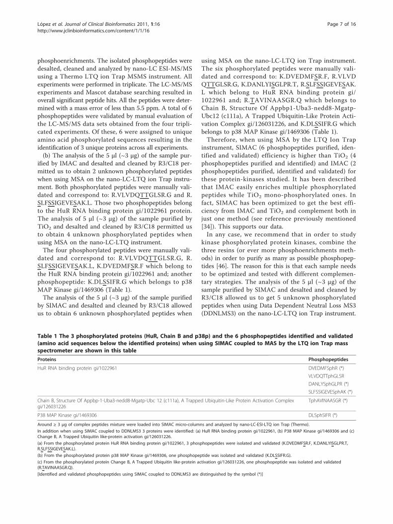
phosphoenrichments. The isolated phosphopeptides weredesalted, cleaned and analyzed by nano-LC ESI-MS/MSusing a Thermo LTQ ion Trap MSMS instrument. Allexperiments were performed in triplicate. The LC-MS/MSexperiments and Mascot database searching resulted inoverall significant peptide hits. All the peptides were deter-mined with a mass error of less than 5.5 ppm. A total of 6phosphopeptides were validated by manual evaluation ofthe LC-MS/MS data sets obtained from the four tripli-cated experiments. Of these, 6 were assigned to uniqueamino acid phosphorylated sequences resulting in theidentification of 3 unique proteins across all experiments.(b) The analysis of the 5 μl (~3 μg) of the sample pur-
ified by IMAC and desalted and cleaned by R3/C18 per-mitted us to obtain 2 unknown phosphorylated peptideswhen using MSA on the nano-LC-LTQ ion Trap instru-ment. Both phosphorylated peptides were manually vali-dated and correspond to: R.VLVDQTTGLSR.G and R.SLFSSIGEVESAK.L. Those two phosphopeptides belongto the HuR RNA binding protein gi/1022961 protein.The analysis of 5 μl (~3 μg) of the sample purified byTiO2 and desalted and cleaned by R3/C18 permitted usto obtain 4 unknown phosphorylated peptides whenusing MSA on the nano-LC-LTQ instrument.The four phosphorylated peptides were manually vali-
dated and correspond to: R.VLVDQTTGLSR.G, R.SLFSSIGEVESAK.L, K.DVEDMFSR.F which belong tothe HuR RNA binding protein gi/1022961 and; anotherphosphopeptide: K.DLSSIFR.G which belongs to p38MAP Kinase gi/1469306 (Table 1).The analysis of the 5 μl (~3 μg) of the sample purified
by SIMAC and desalted and cleaned by R3/C18 allowedus to obtain 6 unknown phosphorylated peptides when
using MSA on the nano-LC-LTQ ion Trap instrument.The six phosphorylated peptides were manually vali-dated and correspond to: K.DVEDMFSR.F, R.VLVDQTTGLSR.G, K.DANLYISGLPR.T, R.SLFSSIGEVESAK.L which belong to HuR RNA binding protein gi/1022961 and; R.TAVINAASGR.Q which belongs toChain B, Structure Of Appbp1-Uba3-nedd8-Mgatp-Ubc12 (c111a), A Trapped Ubiquitin-Like Protein Acti-vation Complex gi/126031226, and K.DLSSIFR.G whichbelongs to p38 MAP Kinase gi/1469306 (Table 1).Therefore, when using MSA by the LTQ Ion Trap
instrument, SIMAC (6 phosphopeptides purified, iden-tified and validated) efficiency is higher than TiO2 (4phosphopeptides purified and identified) and IMAC (2phosphopeptides purified, identified and validated) forthese protein-kinases studied. It has been describedthat IMAC easily enriches multiple phosphorylatedpeptides while TiO2 mono-phosphorylated ones. Infact, SIMAC has been optimized to get the best effi-ciency from IMAC and TiO2 and complement both injust one method (see reference previously mentioned[34]). This supports our data.In any case, we recommend that in order to study
kinase phosphorylated protein kinases, combine thethree resins (or ever more phosphoenrichments meth-ods) in order to purify as many as possible phosphopep-tides [46]. The reason for this is that each sample needsto be optimized and tested with different complemen-tary strategies. The analysis of the 5 μl (~3 μg) of thesample purified by SIMAC and desalted and cleaned byR3/C18 allowed us to get 5 unknown phosphorylatedpeptides when using Data Dependent Neutral Loss MS3(DDNLMS3) on the nano-LC-LTQ ion Trap instrument.
Table 1 The 3 phosphorylated proteins (HuR, Chain B and p38p) and the 6 phosphopeptides identified and validated(amino acid sequences below the identified proteins) when using SIMAC coupled to MAS by the LTQ ion Trap massspectrometer are shown in this table
Proteins Phosphopeptides
HuR RNA binding protein gi/1022961 DVEDMFSphR (*)
VLVDQTTphGLSR
DANLYSphGLPR (*)
SLFSSIGEVESphAK (*)
Chain B, Structure Of Appbp-1-Uba3-nedd8-Mgatp-Ubc 12 (c111a), A Trapped Ubiquitin-Like Protein Activation Complexgi/126031226
TphAVINAASGR (*)
P38 MAP Kinase gi/1469306 DLSphSIFR (*)
Around ≥ 3 μg of complex peptides mixture were loaded into SIMAC micro-columns and analyzed by nano-LC-ESI-LTQ ion Trap (Thermo).
In addition when using SIMAC coupled to DDNLMS3 3 proteins were identified: (a) HuR RNA binding protein gi/1022961, (b) P38 MAP Kinase gi/1469306 and (c)Change B, A Trapped Ubiquitin like-protein activation gi/126031226.
(a) From the phosphorylated protein HuR RNA binding protein gi/1022961, 3 phosphopeptides were isolated and validated (K.DVEDMFSR.F, K.DANLYISGLPR.T,R.SLFSSIGEVESAK.L).
(b) From the phosphorylated protein p38 MAP Kinase gi/1469306, one phosphopeptide was isolated and validated (K.DLSSIFR.G).
(c) From the phosphorylated protein Change B, A Trapped Ubiquitin like-protein activation gi/126031226, one phosphopeptide was isolated and validated(R.TAVINAASGR.Q).
[Identified and validated phosphopeptides using SIMAC coupled to DDNLMS3 are distinguished by the symbol (*)]
López et al. Journal of Clinical Bioinformatics 2011, 1:16http://www.jclinbioinformatics.com/content/1/1/16
Page 7 of 16

The five phosphorylated peptides were manually vali-dated and correspond to: K.DVEDMFSR.F, K.DAN-LYISGLPR.T, R.SLFSSIGEVESAK.L which belong toHuR RNA binding protein gi/1022961; K.DLSSIFR.Gwhich belongs to p38 MAP Kinase gi/1469306 and R.TAVINAASGR.Q which belongs to Chain B, StructureOf Appbp1-Uba3-nedd8-Mgatp-Ubc12 (c111a), ATrapped Ubiquitin-Like Protein Activation Complex gi/126031226 (Table 1).(c) SIMAC coupled to MAS and MS3-NL mass spectro-
metry analysis. The preferred approach for analyzing sam-ples using mass spectrometry is to produce structurallysignificant product ions using the process of ion dissocia-tion. A method commonly known as Data DependentNeutral Loss MS3 (DDNLMS3) (developed by Coon andco-workers [47]) analysis enables selective fragmentationby isolating a neutral loss ion fragment from an MS/MSexperiment and then subjecting it to further dissociation[48]. Despite of this, DDNLMS3 did not allow us to get asefficient results as when using MSA for our protein-kinases analyses. It is well known that the production ofneutral loss ions in MS/MS, is almost always accompaniedby partial fragmentation of the precursor ion and thesediagnostic fragment ions are subsequently lost when theneutral loss ions are isolated for MS3. Multistage activa-tion (or pseudo MS3) allowed us to get spectra that werethe combination of MS/MS and MS3 fragmentation andthus retaining the informative fragments from the precur-sor ion more efficiently. This is due to the fact that MSAproduced more structurally informative ions by eliminat-ing the ion isolation step between MS/MS and MS3 forthe study of phosphorylated protein kinases p38 and HuRin vitro. We observed that - in this research study relatedto the previously phosphorylated proteins after in vitrokinase reaction- multistage activation was a faster route toa more information- rich spectra since the ion-trap doesnot require refilling for the MS3 scan, as with the tradi-tional neutral loss experiment (DDNLMS3). We con-cluded during the first tests-analyses of the protein kinasesin vitro, that when compared to DDNLMS3, multistageactivation generated spectra with increased signal intensityand a greater number of structurally diagnostic ions forphosphorylated peptides. Thus we chose MSA as a routinepath for this kind of analysis (p38 and HuR phosphory-lated kinases in vitro). Further benefits of using multistageactivation are demonstrated in other studies of phospho-peptides, including large scale analysis [49]. The informa-tion-rich spectra generated using multistage activationwere particularly important for these compounds becausethere is often a significant loss of sequence informativefragment ions generated in MS/MS. For this study, moreions were identified with multistage activation than withMS/MS or MS3 in the DDNLMS3 method. In addition,the signal intensities were generally higher with multistage
activation compared to MS/MS or MS3 of DDNLMS3method. In fact, multistage activation resulted in moreinformation for the suite of phosphopeptides studied(Table 1) (see an example of the spectrum of an identifiedphosphorylated peptide when using SIMAC coupled toMSA in the LTQ ion Trap mass spectrometer and Mascot,Figure 2).Nevertheless, it must be pointed out that Jiang and co-
workers developed a specific classification filtering strategyfor their studies (using different samples) which signifi-cantly improved the coverage of the phosphoproteomeanalysis when using NLMS3 (see reference previouslymentioned [48]). In fact, Jiang and co-workers obtained ahigher coverage of the phosphopeptide identificationswhen processing and filtering specific methods which theydeveloped for the spectra from NLMS3, compared withMS2 and MSA strategies. In relation to this, we should saythat just one more phosphopeptide was identified and vali-dated when we used SIMAC coupled to MSA (new 6identified phoshopeptides) compared to when we coupledSIMAC to DDNLMS3 (5 new identified phosphopeptides).In addition, those 5 new phosphorylated peptides identi-fied and their phospho-site assignments in each specificamino acid are the same ones following both strategies(see Figure 3 and Table 1). Moreover, the 6 new phospho-peptides and phospho-site assignments showed highreproducibility in all cases during the four triplicateexperiments we carried out.All our MS analyses were carried out by CID. We
hypothesize that combining CID with ETD or ECD frag-mentation, it is probable that more and/or complementarydata would be obtained according to the methodologicalstudy of Navajas and co-workers [50]. ECD occurs only onthe peptide backbone - which is an advantage -, and labilephosphate groups are left intact on the resulting c- and z-fragment ions, thus, complementary identification of otherspecific phosphorylation sites would be enabled [51,52].As a result, we recommend using CID to start with, andwould recommend switching to ETD, in the event youwere not able to determine the phosphorylation site, if youhave the possibility of the required instrument [53-57].The phosphopeptides purified, identified and validated,including also the site-assignments of the phosphate groupare illustrated in Table 1.The efficiency and reproducibility of the phosphopep-
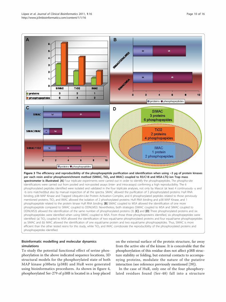
tide purification and identification when using ~3 μg ofprotein kinases per each resin and or phosphoenrich-ment method (SIMAC, TiO2 and IMAC) coupled to R3/C18 and MSA-LTQ ion Trap mass spectrometer is illu-strated in Figure 3.An example of a phospho-site assignment and manual
validation of the phosphorylated peptide (VLVDQTTphGLSR) obtained by Mascot analysis is illustrated inFigure 2.
López et al. Journal of Clinical Bioinformatics 2011, 1:16http://www.jclinbioinformatics.com/content/1/1/16
Page 8 of 16

Figure 2 Phospho-site assignment & manual validation of the phosphorylated peptide VLVDQTTphGLSR obtained by Mascot analysis.The monoisotopic mass of neutral peptide Mr (calc) resulted was 1267.6173. Fixed modifications chosen were: Carbamidomethyl (C), while forvariable modifications T7: Phospho (ST), with neutral losses 97.9769 (shown in table) was selected. The y5 ion and b7 are those which allowedidentification of the treonine (5) as phosphorylated (ph) amino acid (T in red colour). In addition the phosphate fingerprint of the neutral loss(NL) from the parent ion is also a positive signal of phosphorylation. Six b ions and 8 y ions were continuously matched respectively.
López et al. Journal of Clinical Bioinformatics 2011, 1:16http://www.jclinbioinformatics.com/content/1/1/16
Page 9 of 16
Bioinformatic modelling and molecular dynamicssimulationsTo study the potential functional effect of serine phos-phorylation in the above indicated sequence locations, 3Dstructural models for the phosphorylated state of bothMAP kinase p38beta (p38B) and HuR were generatedusing bioinformatics procedures. As shown in figure 4,phosphorylated Ser-279 of p38B is located in a loop placed
on the external surface of the protein structure, far awayfrom the active site of the kinase. It is conceivable that thephosphorylation of this residue does not affect p38B struc-ture stability or folding, but external contacts to accompa-nying proteins, modulate the nature of the putativeinteraction (see reference previously mentioned [39]).In the case of HuR, only one of the four phosphory-
lated residues found (Ser-48) fall into a structure
Figure 3 The efficiency and reproducibility of the phosphopeptide purification and identification when using ~3 μg of protein kinasesper each resin and/or phosphoenrichment method (SIMAC, TiO2 and IMAC) coupled to R3/C18 and MSA-LTQ ion Trap massspectrometer is illustrated. [A] Four triplicate experiments were carried out in order to identify the phosphopeptides. The phospho-siteidentifications were carried out from pooled and non-pooled assays (inter- and intra-assays) confirming a high reproducibility. The 6phosphorylated peptides identified were isolated and validated in the four triplicate analyses, not only by Mascot (at least 4 continuously -y and-b ions matched)but also by manual inspection of all the spectra. SIMAC allowed the purification of 3 phosphorylated proteins: HuR RNAbinding, p38 MAP Kinase and Trapped Ubiquitin-Like Protein Activation Complex, and 6 phosphorylated peptides related to those previouslymentioned proteins. TiO2 and IMAC allowed the isolation of 2 phoshorylated proteins: HuR RNA binding and p38 MAP Kinase, and 1phosphopeptide related to the protein kinase HuR RNA binding. [B] SIMAC coupled to MSA allowed the identification of one morephosphopeptide compared to SIMAC coupled to DDNLMS3. Nevertheless, both strategies (SIMAC coupled to MSA and SIMAC coupled toDDNLMS3) allowed the identification of the same number of phosphorylated proteins (3). [C] and [D] Three phosphorylated proteins and sixphosphopeptides were identified when using SIMAC coupled to MSA. From those three phosphoproteins identified, six phosphopeptides wereidentified: (a) TiO2 coupled to MSA allowed the identification of two equal/same phosphorylated proteins and four equal/same phosphopeptidesas SIMAC and (b) IMAC allowed the identification of one equal/same protein and two equal/same phosphopeptides. Thus, SIMAC is moreefficient than the other tested resins for this study, while TiO2 and IMAC corroborate the reproducibility of the phosphorylated proteins andphosphopeptides identified.
López et al. Journal of Clinical Bioinformatics 2011, 1:16http://www.jclinbioinformatics.com/content/1/1/16
Page 10 of 16

domain of the protein previously crystallized: the dim-merized first RNA recognition motif (see reference pre-viously mentioned [40]). Thus, only the putativestructural effect of the phosphorylated and non-phos-phorylated state of Ser-48 could be analyzed throughstructural bioinformatic tools including MolecularDynamics (MD) simulation.As shown in figure 5A, Ser-48 is located in the dim-
merization surface, surrounded by residues Glu-47 andLys-50 that form a pair of saline bonds potentially impli-cated in the stabilization of the dimmer. To test the effectof the presence of a phosphorylated Ser in the mainte-nance of the quaternary structure, two unrestricted MDcomputer simulations were performed in presence orabsence of a phosphorylated Ser in position 48, as indi-cated under “Materials and Methods”. Results obtainedafter 10ns of MD showed that, in the case of the unpho-sphorylated dimmer structure (non-phosphorylated Ser-
48), both monomers experimented a significant displace-ment from their initial relative positions, resulting in acomplete disorganization of the quaternary structure(Figure 5B -left-). Measurement of root mean squaredeviation (RMSD) values of the monomers and the dim-mer, as well as the distances between the C atoms of thecontact residues (Glu47A-Lys50B, Ser48A-Ser48B andLys50A-Glu48B) indicated a clear and irreversible displa-cement from the initial values during the first steps ofthe MD simulation (Figure 5C, upper plots). In contrast,when the same MD simulation was performed in pre-sence of a phosphorylated Ser in position 48 of bothmonomers, a clear stabilization of the dimmerized struc-ture was obtained, showing no displacement from theirinitial position (Figure 5B -right-) and exhibiting constantvalues of RMSD values and invariable RMSD values anddistances between contact residues (Figure 5C, lowerplots).
Figure 4 Location of phosphorylated Ser-279 in the protein structure of human MAP kinase p38beta (p38B) . A model forphosphorylated serine was located in the structural position of residue Ser-279 in the 3D crystallographic coordinates of p38B (Protein DataBank code: 3GC8). Position of the ATP binding site is indicated. Plot was generated using PyMOL (DeLano Scientific, San Carlos, CA).
López et al. Journal of Clinical Bioinformatics 2011, 1:16http://www.jclinbioinformatics.com/content/1/1/16
Page 11 of 16

These results suggest that the phosphorylation of Ser48in the first protein RNA recognition motif of HuR haspotentially a stabilizing effect, exerting a regulatory role onthe biological function of protein by regulating the mainte-nance of the dimmerized quaternary state, a requirementof the complex prior to RNA binding activity [58].
DiscussionReversible protein phosphorylation plays an importantrole in the regulation of many different processes, suchas cell growth, differentiation, migration, metabolism,and apoptosis. Identification of differentially phosphory-lated proteins by means of phospho-proteomic analysis
Figure 5 Effect of the phosphorylation of Ser-48 in the stability of the dimmer structure of the first RNA recognition motif of HuR. [A]Crystal structure of HuR dimmer (Protein Data Bank code: 3HI9, chains B and D) indicating the position of Ser-48, Glu-47 and Lys-50 in thedimmerization surface. [B] Relative spatial position of the two HuR monomers after 10ns of unrestricted Molecular Dynamics (MD) simulation ofboth non-phosphorylated (red structure, left) and phosphorylated (green structure, right) states of Ser-48. Position of initial dimmer structure,prior to the MD simulation, is included for comparison (in gray). Note the large displacement of the non-phosphorylated state in contrast to thestability exhibited by the dimmer in presence of phosphoSer48. Respective positions of Ser-48 and phosphoSer-48 are indicated. [C] Left: RMSDvalues measured for HuR dimmer (red) monomer A (blue) and monomer B (green) during the 10 ns trajectory of the unrestricted MD simulationof the dimmer in presence of non-phosphorylated Ser-48 (HUR plot, top panel) or phosphorylated pSer-48 (HURP plot, lower panel). Right:continuous measurement of Ca Ca distances between residues E47(A)-K50(B) (red), S48(A)-S48(B) (blue) and E47(A)-K50(B) (green), during theMD trajectory. Distortion of the HuR dimmer in the presence of Ser-48 (HUR plot, top panel) when compared to the phosphorylated state of theprotein (HURP plot, lower panel) is patent in both RMSD and Ca-Ca measurements. Structure plots were generated as in Figure 4.
López et al. Journal of Clinical Bioinformatics 2011, 1:16http://www.jclinbioinformatics.com/content/1/1/16
Page 12 of 16

coupled to bioinformatic tools provides insight into sig-nal transduction pathways that are activated in responseto, for example, growth factor stimulation or toxicant-induced apoptosis [59]. Four triplicate experiments werecarried out in order to identify the phosphopeptides ofthe protein kinases studied in this article (p38 and HuR).The phospho-site identifications were carried out frompooled and non-pooled assays (inter- and intra-assays)confirming a high reproducibility: the 6 isolated andidentified phosphorylated peptides were validated in thefour triplicate analyses by Mascot (score >20, and at least4-y and -b ions continuously matched) also includingmanually inspection via phosphate-fingerprints of all thespectra. It should be pointed that some spectra showed abetter resolution than others. Three different resins(IMAC, TiO2 and SIMAC) were used despite the factthat it is well known that SIMAC has been developedand described as a method to purify more phosphopep-tides for high-throughput analysis giving great efficiency(see references previously mentioned [34-36]). But it hasalso been demonstrated that the analyses of differentphosphorylated proteins when using different resins (e.g.TiO2, IMAC or ZrO2) -including SIMAC- are more effi-cient in a complementary way for specific samples. Thisis due to the fact that the intrinsic characteristic of eachprotein (e.g. amino acid composition). In fact, in order toget as many phosphopeptides as possible from the samesample, different tests must be carried out [e.g of otherpossibilities of phosphoenrichments: Strong cation andanion exchange (SCX and SAX), Calcium phosphate pre-cipitation, Hydrophilic interaction chromatography(HILIC)]. Moreover, phosphoproteomic analysis ofkinases implies more difficulties as they usually are lowexpressed proteins with the disadvantage that the un-phosphorylated form is more abundant than the phos-phorylated within the same sample to be analyzed. Inaddition, IMAC elutes easily multi-phopshorylated pep-tides than TiO2 and ZrO2. TiO2 and ZrO2 have the capa-city of binding strongly the multiple phosphorylatedpeptides, thus both last mentioned resins retain multi-phosphorylated peptides and do not give -usually- thechance to elute them. We chose TiO2 for our researchstudy instead of ZrO2 although they are of similar char-acteristics, but further experiments using ZrO2 or otherresins or different possibilities of phosphoenrichments(SCX and/orHILIC for e.g) could surely give complemen-tary and interesting data for signalling network researchadvances. The phosphopeptides found in the analyzedsample corresponding to p38 MAP kinase protein do notinclude the described phosphorylation sites in the T-loop(pThr-180 and pTyr-182). We hypothesize that the pro-tein was not phosphorylated in those precise sites in theoriginal sample, indicating a particular inactive state ofthe kinase activity [60,61]. In addition, we detected
unphosphorylated peptides after protein digestions byMS and Mascot analysis. These unphosphorylated pep-tides were very useful for the assured identification of thestudied protein kinases. During the evaluation of thedata, we found that the peptide scores of phosphopep-tides assigned by Mascot did not always correlate withthe quality of the spectra, and for this reason, all frag-mentation spectra were manually verified, in order toavoid false positives. A peptide was accepted when atleast four consecutive y- and b- ions were assigned toabundant signals in the fragmentation spectra. If this wasnot the case, then the amino acid sequence was inspectedto look for indications of why these y/b-ions were miss-ing. Peptides lacking a C-terminal arginine or lysine resi-due, due to a C-terminal position in the particularprotein sequence, were accepted, when at least four con-secutive b-ions had been assigned in the fragmentationspectra. Peptides with a C-terminal lysine residue and anN-terminal arginine residue due to missed cleavage werealso accepted, when at least four consecutive b-ions wereassigned in the fragmentation spectra. In addition, thespectra were inspected for the presence of proline resi-dues, which usually give rise to intense signals, suppres-sing other signals in the spectra. A neutral loss of 98 Thdue to the loss of phosphoric acid resulting from gas-phase Β-elimination of phosphoserine or phosphothreo-nine residues together with the presence of dehydroala-nine or dehydro-2-amino butyric acid or the presence ofintact phosphoserine or phosphothreonine residues inthe sequence were used as indicators of a serine or threo-nine phosphorylated peptide, respectively. The character-istic phosphotyrosine immonium ion at 216.05 Th or thepresence of an intact phosphotyrosine residue in thesequence was used for confirmation of tyrosine phos-phorylation if it was the case. The analysis by theLC-nano-ESI-LTQ ion Trap and the manual validation ofthe MS/MS and MSA spectra was performed by EL fol-lowing the rules of Mann and Jensen (see references[36,57]). (Table 1, Figure 2, Figure 3).One of the disadvantages of Electron Capture Dissocia-
tion (ECD) is that it has selectivity for disulfide bonds,due to the high radical affinity of the bond. Further ana-lysis related to these protein kinases will be carried out asit preserves the intact information about labile modifica-tions, which are not observed directly when using CID atthe same time with the knowledge that the drawback ofETD is less sensitive compared to CID, because of lowerionization efficiency. Thus more complementary datawould be available soon. A third phosphorylated protein(Trapped ubiquitin-like protein complex gi/126031226)was isolated and identified during the MS analysis of thep38 and HuR protein kinases. Apparently, this thirdphosphorylated protein seems to be linked to p38 andHuR kinases. The reason for that could be the kinase
López et al. Journal of Clinical Bioinformatics 2011, 1:16http://www.jclinbioinformatics.com/content/1/1/16
Page 13 of 16

assay. The origin of the Ubiquitin-like protein activationcomplex protein needs to be studied in a deep way, asalthough p38/MAPK is a fundamental actor for the net-work connectivity of signalling partners, no data yetclearly implicate gi/126031226 in these processes orinteractions (see references [53-59])MD bioinformatics simulations are often good indica-
tors of potential behaviour of protein-protein complexes;but they are only computational results, but coupled toexperimental proteomics data, they give relevant clues. Infact, this study suggests that the phosphorylation ofSer48 in the first protein RNA recognition motif of HuRhas potentially a stabilizing effect (Figure 5), exerting aregulatory role on the biological function of protein byregulating the maintenance of the dimmerized quatern-ary state, a requirement of the complex prior to RNAbinding activity (see reference [58]). In addition, a figureillustrating the 3D position of phosphorylated Ser-279 inthe structure of human MAP kinase p38beta (Figure 4)(p38B) indicates clearly that this precise phosphorylationsite is located far from the ATP binding site and also farfrom the T-loop, indicating a different role for this resi-due, which opens a new research door for this relevantprotein kinase [59-61].
ConclusionsOur proteomics studies have demonstrated that there are6 new phosphopeptides of the protein kinases p38 andHuR during in vitro assays. This was possible by testingdifferent resins coupled to different MS strategies to iso-late and identify phosphopeptides. The identified phos-phopeptides were manually validated and the phospho-siteassignments were also carried out in order to avoid falsepositives. Recently, several signalling studies for clinicalresearch are ongoing and each sample needs to be testedby different strategies in order to check which one adaptsbetter to your goals, the characteristics of the proteins tobe studied, and the possibility of getting complementarydata. The experiments carried out showed excellent repro-ducibility in the four triplicate experiments for the identifi-cation and validation of the identified phosphopeptides,also for the protein identifications of p38 and HuR kinases.A Trapped ubiquitin-like protein complex gi/126031226was also isolated and identified containing one phospho-peptide during this study. The origin of the Ubiquitin-likeactivation complex protein needs to be studied in a depthas although p38/MAPK is a fundamental actor for the net-work connectivity of signalling partners, no data yet clearlyimplicate gi/126031226 in these processes or interactions.Overall, the combination of SIMAC and MSA resultedmore efficient to isolate and identify phosphopeptidesfrom p38 and HuR protein kinases. It should be pointedout that when using other phosphoenrichment alternatives(SCX and HILIC) coupled to other MS strategies for this
study, we will be able to corroborate even more comple-mentary data related to the results presented.Bioinformatic studies, using structural tools, of reversible
phosphorylation in proteins will allow the generation ofuseful models for protein-protein contacts at the atomiclevel. These models can be used as input for sophisticatedtechniques, as molecular dynamics, that offer the possibi-lity of analyzing the potential effect of the phosphorylationand de-phosphorylation process of each residue in the glo-bal structure and behaviour of the protein. In the presentwork, the molecular dynamics analysis of the phosphoryla-tion of Ser-48 in HuR homodimmer offered a suitablehypothesis of how the phosphorylation state of Ser-48 canregulate the protein mechanism through controlling thehomodimmerization arrangement of the correspondingstructural domain. Regarding the 3D position of phos-phorylated Ser-279 in the structure of human MAP kinasep38, molecular simulation clearly indicates that this pre-cise phosphorylation locus is located far from the ATPbinding site and also far from the T-loop, indicating a dif-ferent role for this residue: this could open a new and rele-vant research door for this important protein kinase,which has been related as a connectivity-link for differentsignalling pathways.
List of AbbreviationsAQUA: Absolute Quantification; CID: Collision-Induced Dissociation; Da:Dalton (molecular mass); DIGE 2-D: Fluorescence Difference GelElectrophoresis; ECD: Electron Capture Dissociation; ESI: Electron SprayIonization; ETD: Electron Transfer Dissociation; FT-ICR: Fourier transform-IonCyclotron Resonance; HILIC: Hydrophilic interaction chromatography; HPLC:High-performance liquid chromatography or high-pressure liquidchromatography; H3PO4: Phosphoric acid; ICR: Ion Cyclotron Resonance;IMAC: Immobilized Metal Affinity Capture; IT: Ion Trap; iTRAQ: Isobaric Tagfor Relative and Absolute Quantification; kDa: kilodalton (molecular mass);LC: Liquid Chromatography; MALDI: Matrix-Assisted Laser Desorption/Ionization; MD: Molecular Dynamics; MOAC: Metal Oxide AffinityChromatography; Mr: Relative molecular mass (dimensionless); MRM:Multiple reaction monitoring; MS: Mass Spectrometry; MSA: MultiStageActivation; MS/MS: tandem mass spectrometry; m/z: Mass to charge ratio;PID: Primary Immunodeficiencies; PTM: Post-Translational Modification;SILAC: Stable Isotope Labelling with Amino acid in cell Culture; SIMAC:Sequential Elution from IMAC; TiO2: Titanium dioxide; TOF: Time Of Flight;ZrO2: Zirconium dioxide.
AcknowledgementsEL PhD is a recipient of a Postdoctoral Fellowship (Programme ClinicalProteomics of Ministerio de Ciencia e Innovación de España (MICINN). IL, MDPhD is a recipient of a Grant from “Fundación Leucemia & Linfoma de España).JL and AF are MD PhD and hold a tenured position at Spanish NationalHospitals Carlos III and La Paz respectively. We are very grateful to Biomol-Informatics S.L. (CSIC Bioinformatics Group of Severo Ochoa, Prof. PaulinoGómez-Puertas) for the welcomed and useful discussions. Special thanks Prof.Rune Matthiesen (Institute of Molecular Pathology and Immunology of theUniversity of Porto) who contributed to the publication of this article. We alsothank the great discussions of p38 and HuR protein kinases (Prof. Ángel R.Nebreda and Vanesa Lafarga PhD). Thanks also to the Molecular ModellingGroup (CSIC-UAM), and Prof. Keith Ashman for allowing use of the LTQ.
Author details1Phosphoproteomic core, Spanish National Cancer Research Centre (CNIO),C/Melchor Fernández Almagro, 3, 28029, Madrid, Spain. 2Hematology
López et al. Journal of Clinical Bioinformatics 2011, 1:16http://www.jclinbioinformatics.com/content/1/1/16
Page 14 of 16

Department, Hospital QUIRÓN, Madrid, Diego de Velázquez 1, 28223,Pozuelo, Madrid, Spain. 3Immunology Department, Hospital Carlos III, SinesioDelgado 28029, Madrid, Spain. 4Immunology Department, HospitalUniversitario La Paz, P° de la Castellana 261-28046, Madrid, Spain.5Inflammatory core, Centro de Investigación i+12 del Hospital Universitario12 de Octubre, Avda de Córdoba s/n 28041, Madrid, Spain.
Authors’ contributionsAuthor 1 (EL) carried out the proteomics, phosphoproteomics and massspectrometry studies for this article. Authors 2, 3, 4 (IL, JS, AF) carried out theclinical studies in order to support this article. (Contributions of PGP and RM)support the bioinformatic section coupled to proteomic, special thanks forPGP, RM and ARN for making possible the publication of this article. Allauthors read and approved the final manuscript.
Competing interestsThe authors declare that they have no competing interests.
Received: 20 April 2011 Accepted: 6 July 2011 Published: 6 July 2011
References1. Han J, Sun P: The pathways to tumor suppression via route p38. Trends
Biochem Sci 2007, 32(8):364-371, 2007.2. Lluis F, Perdiguero E, Nebreda AR, Munoz-Canoves P: Regulation of skeletal
muscle gene expression by p38 MAP kinases. Trends Cell Biol 2006,16(1):36-44, 2006.
3. Zarubin T, Han J: Activation and signalling of the p38 MAP kinasepathway. Cell Res 2005, 15(1):11-18, 2005.
4. Cuenda A, Rousseau S: p38 MAP-kinases pathway regulation, functionand role in human diseases. Biochim Biophys Acta 2007,1773(8):1358-1375, 2007.
5. Dolado I, Swat A, Ajenjo N, De Vita G, Cuadrado A, Nebreda AR: p38alphaMAP kinase as a sensor of reactive oxygen species in tumorigenesis.Cancer Cell 2007, 11(2):191-205, 2007.
6. Cuadrado A, Nebreda AR: Mechanisms and functions of p38 MAPKsignalling. Biochem J 2010, 429(3):403-417.
7. Kim HH, Gorospe M: Phosphorylated HuR shuttles in cycles. Cell Cycle2008, 7(20):3124-3126, 2008.
8. Abdelmohsen K, Pullmann R Jr, Lal A, Kim HH, Galban S, Yang X,Blethrow JD, Walker M, Shubert J, Gillespie DA, Furneaux H, Gorospe M:Phosphorylation of HuR by Chk2 regulates SIRT1 expression. Mol Cell2007, 25(4):543-557, 2007b.
9. Doller A, Huwiler A, Muller R, Radeke HH, Pfeilschifter J, Eberhardt W:Protein kinase C alpha-dependent phosphorylation of the mRNA-stabilizing factor HuR: implications for posttranscriptional regulation ofcyclooxygenase-2. Mol Biol Cell 2007, 18(6):2137-2148, 2007.
10. Jin SH, Kim TI, Yang KM, Kim WH: Thalidomide destabilizescyclooxygenase-2 mRNA by inhibiting p38 mitogen-activated proteinkinase and cytoplasmic shuttling of HuR. Eur J Pharmacol 2007, 558(1-3):14-20, 2007.
11. Kim HH, Yang X, Kuwano Y, Gorospe M: Modification at HuR(S242) altersHuR localization and proliferative influence. Cell Cycle 2008, 7(21), 2008b.
12. Kim HH, Abdelmohsen K, Lal A, Pullmann R Jr, Yang X, Galban S,Srikantan S, Martindale JL, Blethrow J, Shokat KM, Gorospe M: Nuclear HuRaccumulation through phosphorylation by Cdk1. Genes Dev 2008,22(13):1804-1815, 2008a.
13. Wang W, Furneaux H, Cheng H, Caldwell MC, Hutter D, Liu Y, Holbrook N,Gorospe M: HuR regulates p21 mRNA stabilization by UV light. Mol CellBiol 2000, 20(3):760-769, 2000.
14. Lafarga V, Cuadrado A, Lopez de Silanes I, Bengoechea R, Fernandez-Capetillo O, Nebreda AR: p38 Mitogen-activated protein kinase- and HuR-dependent stabilization of p21(Cip1) mRNA mediates the G(1)/Scheckpoint. Mol Cell Biol 2009, 29(16):4341-4351, 2009.
15. Kim HH, Abdelmohsen K, Gorospe M: Regulation of HuR by DNA DamageResponse Kinases. J Nucleic Acids 2010, 25:2010.
16. Lopez de Silanes I, Fan J, Yang X, Zonderman AB, Potapova O, Pizer ES,Gorospe M: Role of the RNA-binding protein HuR in coloncarcinogenesis. Oncogene 2003, 22(46):7146-7154, 2003.
17. Abdelmohsen K, Lal A, Kim HH, Gorospe M: Posttranscriptionalorchestration of an anti-apoptotic program by HuR. Cell Cycle 2007,6(11):1288-1292, 2007a.
18. Doller A, Pfeilschifter J, Eberhardt W: Signalling pathways regulatingnucleo-cytoplasmic shuttling of the mRNA-binding protein HuR. CellSignal 2008, 20(12):2165-2173, 2008.
19. Fernau NS, Fugmann D, Leyendecker M, Reimann K, Grether-Beck S,Galban S, Ale-Agha N, Krutmann J, Klotz LO: Role of HuR and p38MAPK inultraviolet B-induced post-transcriptional regulation of COX-2 expressionin the human keratinocyte cell line HaCaT. J Biol Chem 2010,285(6):3896-3904, 2010.
20. Subbaramaiah K, Marmo TP, Dixon DA, Dannenberg AJ: Regulation ofcyclooxgenase-2 mRNA stability by taxanes: evidence for involvement ofp38, MAPKAPK-2, and HuR. J Biol Chem 2003, 278(39):37637-37647, 2003.
21. Farooq F, Balabanian S, Liu X, Holcik M, MacKenzie A: p38 Mitogen-activated protein kinase stabilizes SMN mRNA through RNA bindingprotein HuR. Hum Mol Genet 2009, 18(21):4035-4045, 2009.
22. Shi Y, Gaestel M: In the cellular garden of forking paths: how p38 MAPKssignal for downstream assistance. Biol Chem 2002, 383(10):1519-1536,20002.
23. Thingholm TE, Larsen MR, Ingrell CR, Kassem M, Jensen ON: TiO(2)-basedphosphoproteomic analysis of the plasma membrane and the effects ofphosphatase inhibitor treatment. J Proteome Res 2008, 7(8):3304-13, 2008.
24. Thingholm TE, Jensen ON, Larsen MR: Enrichment and separation ofmono- and multiply phosphorylated peptides using sequential elutionfrom IMAC prior to mass spectrometric analysis. Methods Mol Biol 2009,527:67-78, (2009) Review.
25. Ashman K, Lopez-Villar E: Phosphoproteomics and cancer research. ClinTransl Oncol 2009, 11(6):356-62, (2009) Review.
26. Palmisano G, Thingholm TE: Strategies for quantitation ofphosphoproteomic data. Expert Rev Proteomics 2010, 7(3):439-56, (2010)Review.
27. López E, Matthiesen R, López I, Ashman K, Mendieta J, Wesselink JJ, Gómez-Puertas P, Ferreira A: Functional phosphoproteomics tools for currentimmunological disorders research. JOURNAL OF INTEGRATED OMICS 2011,1(1):1-16 [HTTP://WWW.JIOMICS.COM].
28. Miller ML, Blom N: Kinase-specific prediction of protein phosphorylationsites. Methods Mol Biol 2009, 527:299-310, 2009.
29. Zhao X, León IR, Bak S, Mogensen M, Wrzesinski K, Højlund K, Jensen ON:Phosphoproteome analysis of functional mitochondria isolated fromresting human muscle reveals extensive phosphorylation of innermembrane protein complexes and enzymes. Mol Cell Proteomics 2011,10(1):M110.000299, 2011.
30. Larsen MR, Thingholm TE, Jensen ON, Roepstorff P, Jørgensen TJ: Highlyselective enrichment of phosphorylated peptides from peptide mixturesusing titanium dioxide microcolumns. Mol Cell Proteomics 2005,4(7):873-86, 2005.
31. Thingholm TE, Jørgensen TJ, Jensen ON, Larsen MR: Highly selectiveenrichment of phosphorylated peptides using titanium dioxide. NatProtoc 2006, 1(4):1929-35, 2006.
32. Nühse TS, Stensballe A, Jensen ON, Peck SC: Large-scale analysis of in vitrophosphorylated membrane proteins by immobilized metal ion affinitychromatography and mass spectrometry. Mol Cell Proteomics 2003,2(11):1234-43.
33. Lee J, Xu Y, Chen Y, Sprung R, Kim SC, Xie S, Zhao Y: Mitochondrialphosphoproteome revealed by an improved IMAC method and MS/MS/MS. Mol Cell Proteomics 2007, 6(4):669-76, 2007.
34. Thingholm TE, Jensen ON, Robinson PJ, Larsen MR: SIMAC (sequentialelution from IMAC), a phosphoproteomics strategy for the rapidseparation of monophosphorylated from multiply phosphorylatedpeptides. Mol Cell Proteomics 2008, 7(4):661-71, 2008.
35. Jensen SS, Larsen MR: Evaluation of the impact of some experimentalprocedures on different phosphopeptide enrichment techniques. RapidCommun Mass Spectrom 2007, 21(22):3635-45, 2007.
36. Mann M, Jensen ON: Proteomic analysis of post-translationalmodifications. Nat Biotechnol 2003, 21(3):255-61, 2003.
37. Gruhler A, Olsen JV, Mohammed S, Mortensen P, Faergeman NJ, Mann M,Jensen ON: Quantitative phosphoproteomics applied to the yeastpheromone signalling pathway. Mol Cell Proteomics 2005, 4(3):310-27,2005.
38. Thingholm TE, Jensen ON, Larsen MR: Analytical strategies forphosphoproteomics. Proteomics 2009, 9(6):1451-68, (2009) Review.
39. Patel SB, Cameron PM, O’Keefe SJ, Frantz-Wattley B, Thompson J, O’Neill EA,Tennis T, Liu L, Becker JW, Scapin G: The three-dimensional structure of
López et al. Journal of Clinical Bioinformatics 2011, 1:16http://www.jclinbioinformatics.com/content/1/1/16
Page 15 of 16
MAP kinase p38beta: different features of the ATP-binding site inp38beta compared with p38alpha. Acta Crystallogr D Biol Crystallogr 2009,65(Pt 8):777-85, 2009.
40. Benoit RM, Meisner N-C, Kallen J, Graff P, Hemmig R, Cèbe1 R, Ostermeier C,Widmer H, Auer M: The X-ray Crystal Structure of the First RNARecognition Motif and Site-Directed Mutagenesis Suggest a PossibleHuR Redox Sensing Mechanism. J Mol Biol 2010, 397:1231-1244, 2010.
41. Guex N, Diemand A, Peitsch MC: Protein modelling for all. Trends BiochemSci 1999, 24:364-367, 1999.
42. Peitsch MC: ProMod and Swiss-Model: Internet-based tools forautomated comparative protein modelling. Biochem Soc Trans 1996,24:274-279, 1996.
43. Schwede T, Kopp J, Guex N, Peitsch MC: SWISS-MODEL: An automatedprotein homology-modeling server. Nucleic Acids Res 2003, 31:3381-3385,2003.
44. Case DA, Cheatham TE, Darden T, Gohlke H, Luo R, Merz KM Jr, Onufriev A,Simmerling C, Wang B, Woods RJ: The Amber biomolecular simulationprograms. J Comput Chem 2005, 26:1668-1688, 2005.
45. Mendieta J, Fuertes MA, Kunchitapatham R, Santa-María I, Moreno FJ,Alonso C, Gago F, Muñoz V, Avila J, Hernández F: PhosphorylationModulates the Alpha Helical Structure and Polymerization of a Peptidefrom the Third Tau Microtubule-Binding Repeat. Biochim Biophys Acta2005, 1721:16-26, 2005.
46. Thingholm TE, Larsen MR, Ingrell CR, Kassem M, Jensen ON: TiO(2)-basedphosphoproteomic analysis of the plasma membrane and the effects ofphosphatase inhibitor treatment. J Proteome Res 2008, 7(8):3304-13, 2008.
47. Schroeder MJ, Shabanowitz J, Schwartz JC, Hunt DF, Coon JJ: A neutralloss activationmethod for improved phosphopeptide sequence analysisby quadrupole ion trap mass spectrometry. Anal Chem 2004,76:3590-3598, 2004.
48. Jiang X, Ye M, Han G, Dong X, Zou H: Classification filtering strategy toimprove the coverage and sensitivity of phosphoproteome analysis. AnalChem 2010, 82(14):6168-75, (2010) 15.
49. Villén J, Beausoleil SA, Gygi SP: Evaluation of the utility of neutral-loss-dependent MS3 strategies in large-scale phosphorylation analysis.Proteomics 2008, 8(21):4444-52, 2008.
50. Steen H, Küster B, Mann M: Quadrupole time-of-flight versus triple-quadrupole mass spectrometry for the determination ofphosphopeptides by precursor ion scanning. J Mass Spectrom 2001,36(7):782-90.
51. Steen H, Mann M: A new derivatization strategy for the analysis ofphosphopeptides by precursor ion scanning in positive ion mode. J AmSoc Mass Spectrom 2002, 13(8):996-1003.
52. Navajas R, Paradela A, Albar JP: Immobilized metal affinitychromatography/reversed-phase enrichment of phosphopeptides andanalysis by CID/ETD tandem mass spectrometry. Methods Mol Biol 2011,681:337-48, 2011.
53. Kleinnijenhuis AJ, Kjeldsen F, Kallipolitis B, Haselmann KF, Jensen ON:Analysis of histidine phosphorylation using tandem MS and ion-electronreactions. Anal Chem 2007, 79(19):7450-6, 1.
54. Stensballe A, Andersen S, Jensen ON: Characterization of phosphoproteinsfrom electrophoretic gels by nanoscale Fe(III) affinity chromatographywith off-line mass spectrometry analysis. Proteomics 2001, 1(2):207-22.
55. Schroeder MJ, Webb DJ, Shabanowitz J, Horwitz AF, Hunt DF: Methods forthe detection of paxillin post-translational modifications and interactingproteins by mass spectrometry. J Proteome Res 2005, 4(5):1832-41.
56. Syka JE, Coon JJ, Schroeder MJ, Shabanowitz J, Hunt DF: Peptide andprotein sequence analysis by electron transfer dissociation massspectrometry. Proc Natl Acad Sci USA 2004, 101(26):9528-33, 29.
57. Thingholm TE, Jensen ON, Larsen MR: Enrichment and separation ofmono- and multiply phosphorylated peptides using sequential elutionfrom IMAC prior to mass spectrometric analysis. Methods Mol Biol 2009,527:67-78, 2009.
58. Meisner NC, Hintersteiner M, Mueller K, Bauer R, Seifert JM, Naegeli HU,Ottl J, Oberer L, Guenat C, Moss S, Harrer N, Woisetschlaeger M, Buehler C,Uhl V, Auer M: Identification and mechanistic characterization of low-molecular-weight inhibitors for HuR. Nat Chem Biol 2007, 3:508-515.
59. Besnard A, Galan-Rodriguez B, Vanhoutte P, Caboche J: Elk-1 atranscription factor with multiple facets in the brain. Front Neurosci 2011,5:35, 16.
60. López E, López I, Ferreira A, Sequí J: Clinical and TechnicalPhosphoproteomic Research. Proteome Sci 2011, 9(1):27, 2.
61. Kim J-E, Tannenbaum SR, White FM: Global phosphoproteome of HT-29human colon adenocarcinoma cells. J Proteome Res 2007, 4:1339-1346.
doi:10.1186/2043-9113-1-16Cite this article as: López et al.: Discovering and validating unknownphospho-sites from p38 and HuR protein kinases in vitro byPhosphoproteomic and Bioinformatic tools. Journal of ClinicalBioinformatics 2011 1:16.
Submit your next manuscript to BioMed Centraland take full advantage of:
• Convenient online submission
• Thorough peer review
• No space constraints or color figure charges
• Immediate publication on acceptance
• Inclusion in PubMed, CAS, Scopus and Google Scholar
• Research which is freely available for redistribution
Submit your manuscript at www.biomedcentral.com/submit
López et al. Journal of Clinical Bioinformatics 2011, 1:16http://www.jclinbioinformatics.com/content/1/1/16
Page 16 of 16


