Embed Size (px)
Citation preview
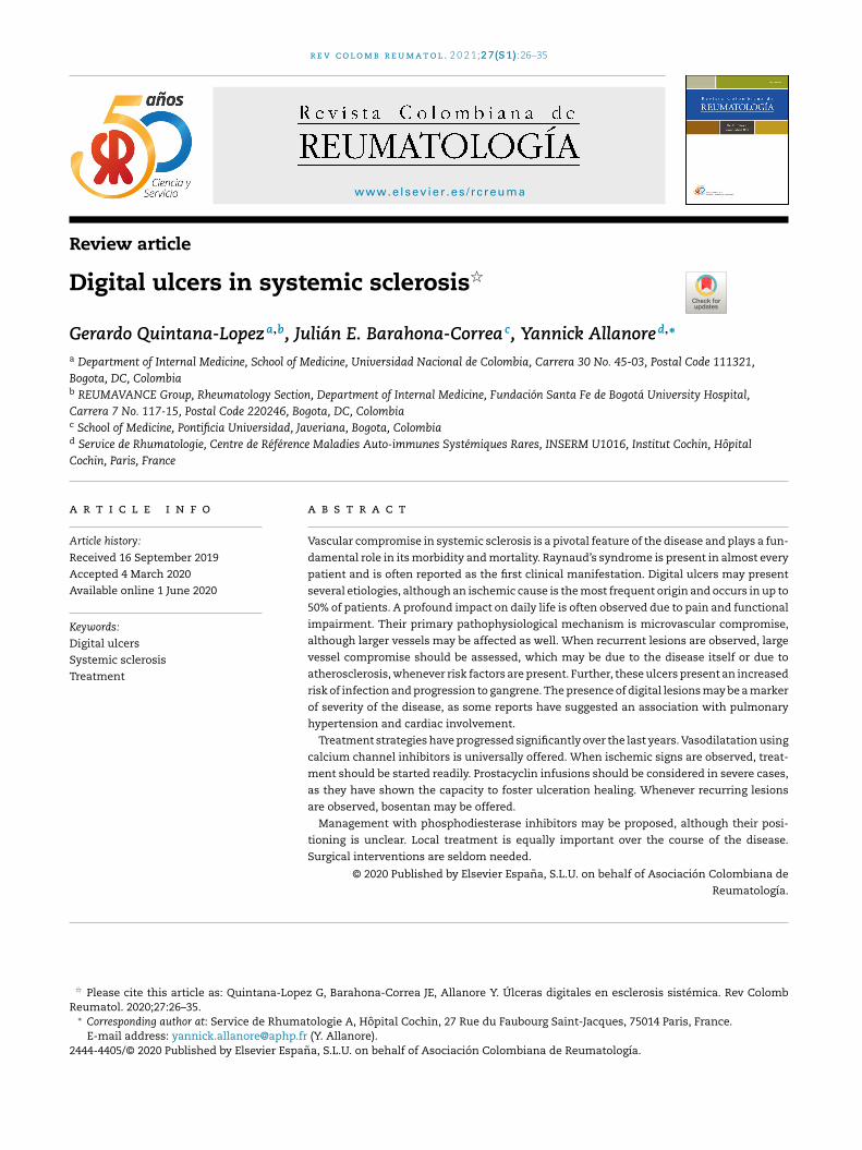
r e v c o l o m b r e u m a t o l . 2 0 2 1;2 7(S 1):26–35
w ww.elsev ier .es / rc reuma
Review article
Digital ulcers in systemic sclerosis�
Gerardo Quintana-Lopeza,b, Julián E. Barahona-Correa c, Yannick Allanored,∗
a Department of Internal Medicine, School of Medicine, Universidad Nacional de Colombia, Carrera 30 No. 45-03, Postal Code 111321,
Bogota, DC, Colombiab REUMAVANCE Group, Rheumatology Section, Department of Internal Medicine, Fundación Santa Fe de Bogotá University Hospital,
Carrera 7 No. 117-15, Postal Code 220246, Bogota, DC, Colombiac School of Medicine, Pontificia Universidad, Javeriana, Bogota, Colombiad Service de Rhumatologie, Centre de Référence Maladies Auto-immunes Systémiques Rares, INSERM U1016, Institut Cochin, Hôpital
Cochin, Paris, France
a r t i c l e i n f o
Article history:
Received 16 September 2019
Accepted 4 March 2020
Available online 1 June 2020
Keywords:
Digital ulcers
Systemic sclerosis
Treatment
a b s t r a c t
Vascular compromise in systemic sclerosis is a pivotal feature of the disease and plays a fun-
damental role in its morbidity and mortality. Raynaud’s syndrome is present in almost every
patient and is often reported as the first clinical manifestation. Digital ulcers may present
several etiologies, although an ischemic cause is the most frequent origin and occurs in up to
50% of patients. A profound impact on daily life is often observed due to pain and functional
impairment. Their primary pathophysiological mechanism is microvascular compromise,
although larger vessels may be affected as well. When recurrent lesions are observed, large
vessel compromise should be assessed, which may be due to the disease itself or due to
atherosclerosis, whenever risk factors are present. Further, these ulcers present an increased
risk of infection and progression to gangrene. The presence of digital lesions may be a marker
of severity of the disease, as some reports have suggested an association with pulmonary
hypertension and cardiac involvement.
Treatment strategies have progressed significantly over the last years. Vasodilatation using
calcium channel inhibitors is universally offered. When ischemic signs are observed, treat-
ment should be started readily. Prostacyclin infusions should be considered in severe cases,
as they have shown the capacity to foster ulceration healing. Whenever recurring lesions
are observed, bosentan may be offered.
Management with phosphodiesterase inhibitors may be proposed, although their posi-
tioning is unclear. Local treatment is equally important over the course of the disease.
Surgical interventions are seldom needed.
© 2020 Published by Elsevier Espana, S.L.U. on behalf of Asociacion Colombiana de
Reumatologıa.
� Please cite this article as: Quintana-Lopez G, Barahona-Correa JE, Allanore Y. Úlceras digitales en esclerosis sistémica. Rev ColombReumatol. 2020;27:26–35.
∗ Corresponding author at: Service de Rhumatologie A, Hôpital Cochin, 27 Rue du Faubourg Saint-Jacques, 75014 Paris, France.E-mail address: [email protected] (Y. Allanore).
2444-4405/© 2020 Published by Elsevier Espana, S.L.U. on behalf of Asociacion Colombiana de Reumatologıa.
r e v c o l o m b r e u m a t o l . 2 0 2 1;2 7(S 1):26–35 27
Úlceras digitales en esclerosis sistémica
Palabras clave:
Ulceras digitales
Sclerosis sistemica
Tratamiento
r e s u m e n
El compromiso vascular en la esclerosis sistémica es una característica fundamental de la
enfermedad y desempena un papel fundamental en su morbilidad y mortalidad. El sín-
drome de Raynaud está presente en casi todos los pacientes y con frecuencia es reportado
como la primera manifestación clínica. Las úlceras digitales pueden tener varias etiologías,
aunque una causa isquémica es el origen más frecuente y ocurre hasta en 50% de los
pacientes. A menudo se observa un profundo impacto en la vida diaria debido al dolor y
al deterioro funcional. Su mecanismo fisiopatológico primario es el compromiso microvas-
cular, aunque los vasos más grandes también pueden verse afectados. Cuando se observan
lesiones recurrentes, se debe evaluar el compromiso de los vasos grandes, que puede deberse
a la enfermedad en sí o a aterosclerosis, siempre que existan factores de riesgo. Además,
estas úlceras presentan un mayor riesgo de infección y progresión a gangrena. La presen-
cia de lesiones digitales puede ser un marcador de la gravedad de la enfermedad, ya que
algunos informes han sugerido una asociación con hipertensión pulmonar y compromiso
cardíaco.
Las estrategias de tratamiento han progresado significativamente en los últimos anos.
La vasodilatación con inhibidores de los canales de calcio se ofrece universalmente. Cuando
se observan signos isquémicos, el tratamiento debe iniciarse de inmediato. Las infusiones
de prostaciclina se deben considerar en casos graves, ya que han demostrado la capacidad
de promover la curación de la ulceración. Siempre que se observen lesiones recurrentes, se
puede administrar bosentán.
Se puede proponer el manejo con inhibidores de la fosfodiesterasa, aunque su posi-
cionamiento no está claro. El tratamiento local es igualmente importante durante el curso
de la enfermedad. Las intervenciones quirúrgicas rara vez son necesarias.
© 2020 Publicado por Elsevier Espana, S.L.U. en nombre de Asociacion Colombiana de
Reumatologıa.
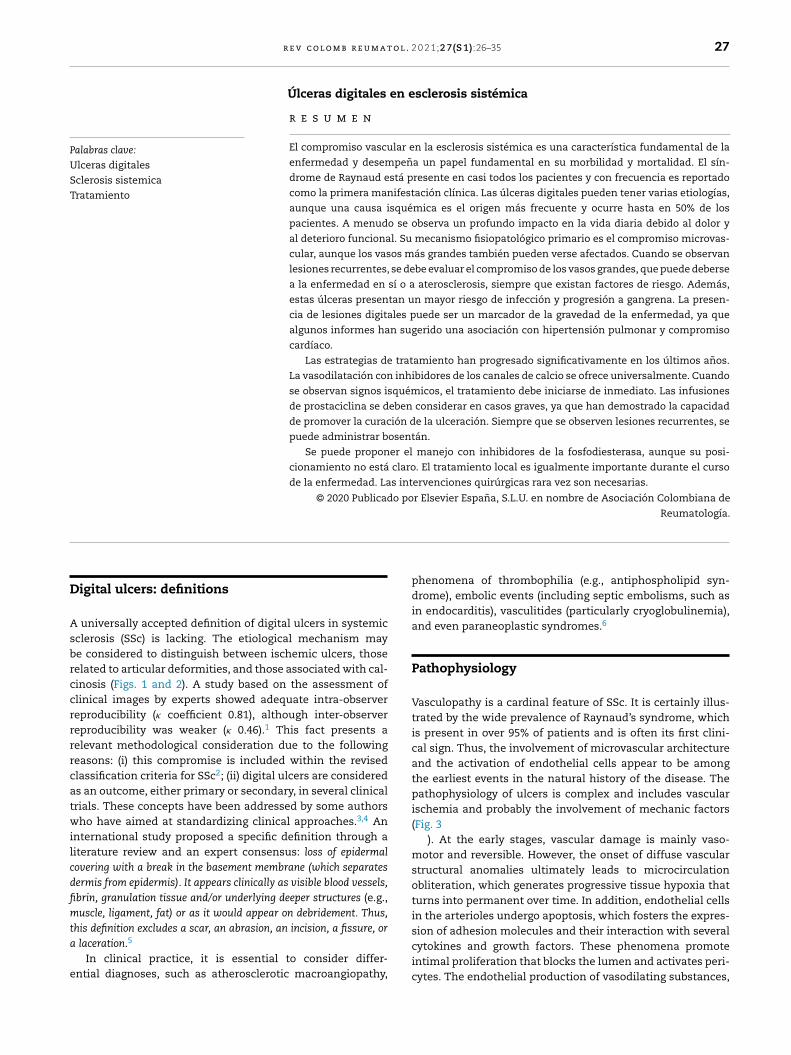
Digital ulcers: definitions
A universally accepted definition of digital ulcers in systemicsclerosis (SSc) is lacking. The etiological mechanism maybe considered to distinguish between ischemic ulcers, thoserelated to articular deformities, and those associated with cal-cinosis (Figs. 1 and 2). A study based on the assessment ofclinical images by experts showed adequate intra-observerreproducibility (� coefficient 0.81), although inter-observerreproducibility was weaker (� 0.46).1 This fact presents arelevant methodological consideration due to the followingreasons: (i) this compromise is included within the revisedclassification criteria for SSc2; (ii) digital ulcers are consideredas an outcome, either primary or secondary, in several clinicaltrials. These concepts have been addressed by some authorswho have aimed at standardizing clinical approaches.3,4 Aninternational study proposed a specific definition through aliterature review and an expert consensus: loss of epidermal
covering with a break in the basement membrane (which separates
dermis from epidermis). It appears clinically as visible blood vessels,
fibrin, granulation tissue and/or underlying deeper structures (e.g.,muscle, ligament, fat) or as it would appear on debridement. Thus,
this definition excludes a scar, an abrasion, an incision, a fissure, or
a laceration.5
In clinical practice, it is essential to consider differ-ential diagnoses, such as atherosclerotic macroangiopathy,
phenomena of thrombophilia (e.g., antiphospholipid syn-drome), embolic events (including septic embolisms, such asin endocarditis), vasculitides (particularly cryoglobulinemia),and even paraneoplastic syndromes.6
Pathophysiology
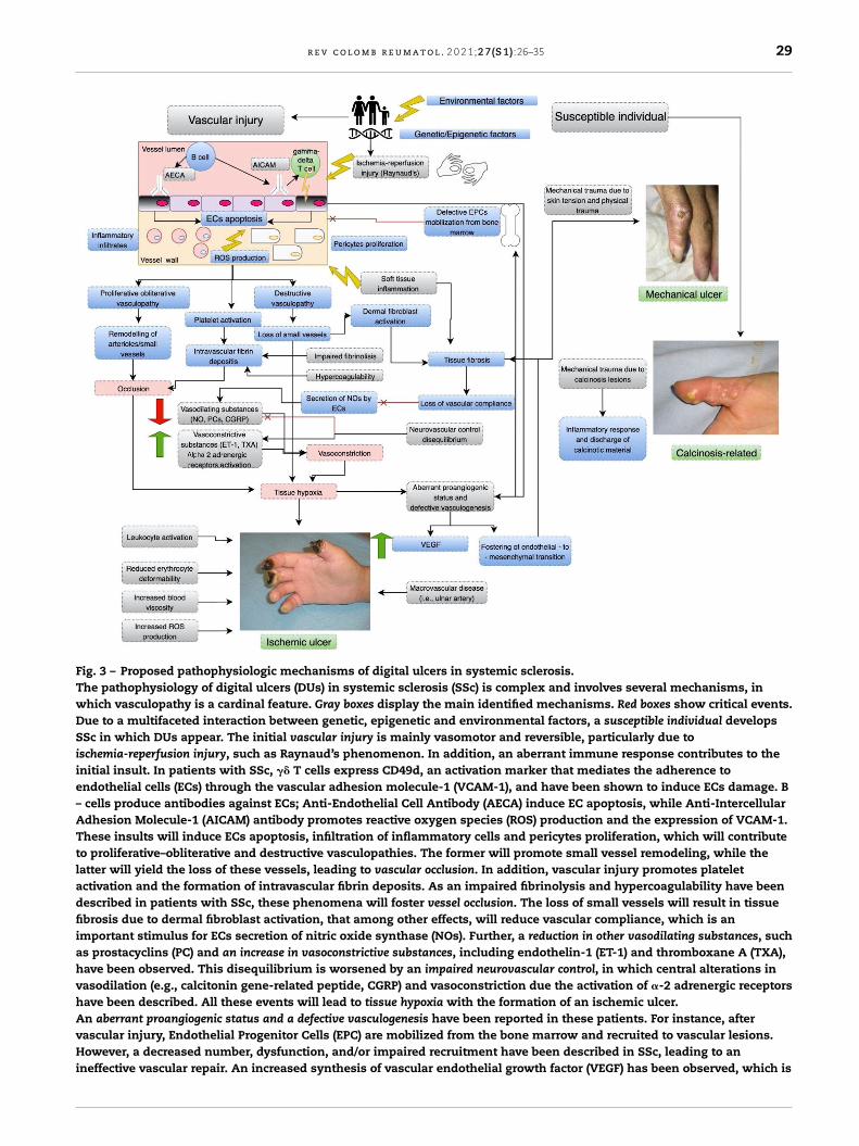
Vasculopathy is a cardinal feature of SSc. It is certainly illus-trated by the wide prevalence of Raynaud’s syndrome, whichis present in over 95% of patients and is often its first clini-cal sign. Thus, the involvement of microvascular architectureand the activation of endothelial cells appear to be amongthe earliest events in the natural history of the disease. Thepathophysiology of ulcers is complex and includes vascularischemia and probably the involvement of mechanic factors(Fig. 3
). At the early stages, vascular damage is mainly vaso-motor and reversible. However, the onset of diffuse vascularstructural anomalies ultimately leads to microcirculationobliteration, which generates progressive tissue hypoxia thatturns into permanent over time. In addition, endothelial cellsin the arterioles undergo apoptosis, which fosters the expres-sion of adhesion molecules and their interaction with severalcytokines and growth factors. These phenomena promoteintimal proliferation that blocks the lumen and activates peri-cytes. The endothelial production of vasodilating substances,
28 r e v c o l o m b r e u m a t o l . 2 0 2 1;2 7(S 1):26–35
Fig. 1 – Definition and etiology of digital ulcers in systemic sclerosis.Adapted from Suliman et al. [5].
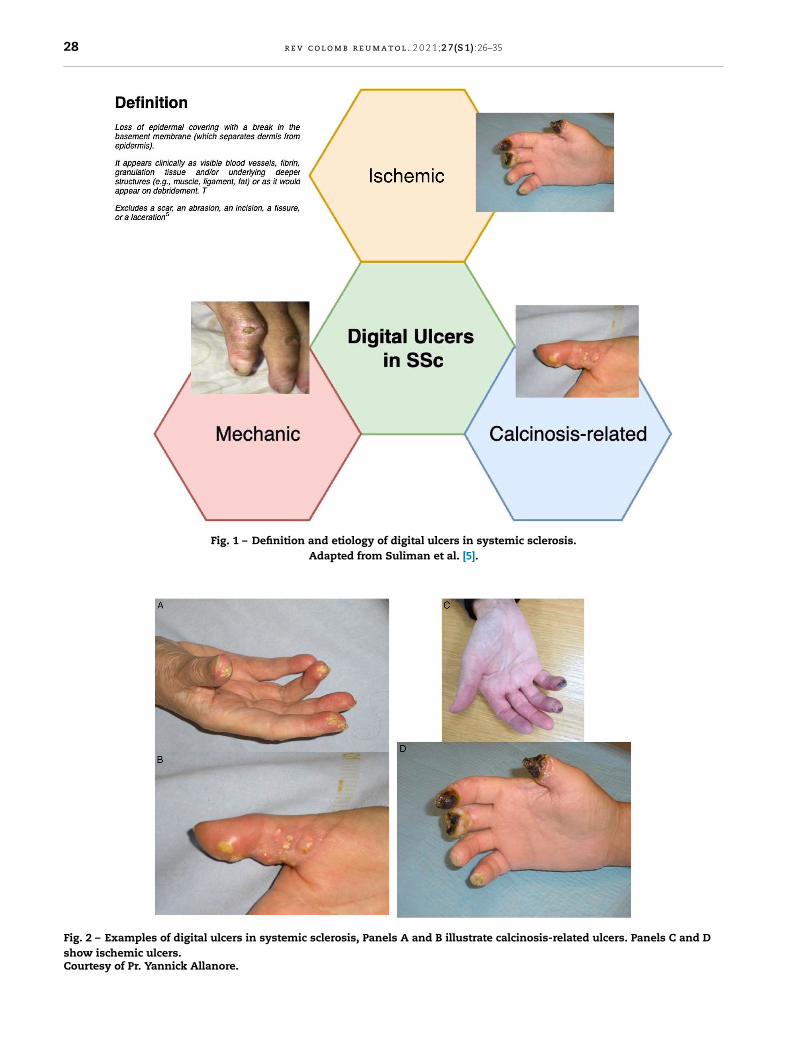
Fig. 2 – Examples of digital ulcers in systemic sclerosis, Panels A and B illustrate calcinosis-related ulcers. Panels C and Dshow ischemic ulcers.Courtesy of Pr. Yannick Allanore.
r e v c o l o m b r e u m a t o l . 2 0 2 1;2 7(S 1):26–35 29
Fig. 3 – Proposed pathophysiologic mechanisms of digital ulcers in systemic sclerosis.The pathophysiology of digital ulcers (DUs) in systemic sclerosis (SSc) is complex and involves several mechanisms, inwhich vasculopathy is a cardinal feature. Gray boxes display the main identified mechanisms. Red boxes show critical events.Due to a multifaceted interaction between genetic, epigenetic and environmental factors, a susceptible individual developsSSc in which DUs appear. The initial vascular injury is mainly vasomotor and reversible, particularly due toischemia-reperfusion injury, such as Raynaud’s phenomenon. In addition, an aberrant immune response contributes to theinitial insult. In patients with SSc, �� T cells express CD49d, an activation marker that mediates the adherence toendothelial cells (ECs) through the vascular adhesion molecule-1 (VCAM-1), and have been shown to induce ECs damage. B– cells produce antibodies against ECs; Anti-Endothelial Cell Antibody (AECA) induce EC apoptosis, while Anti-IntercellularAdhesion Molecule-1 (AICAM) antibody promotes reactive oxygen species (ROS) production and the expression of VCAM-1.These insults will induce ECs apoptosis, infiltration of inflammatory cells and pericytes proliferation, which will contributeto proliferative–obliterative and destructive vasculopathies. The former will promote small vessel remodeling, while thelatter will yield the loss of these vessels, leading to vascular occlusion. In addition, vascular injury promotes plateletactivation and the formation of intravascular fibrin deposits. As an impaired fibrinolysis and hypercoagulability have beendescribed in patients with SSc, these phenomena will foster vessel occlusion. The loss of small vessels will result in tissuefibrosis due to dermal fibroblast activation, that among other effects, will reduce vascular compliance, which is animportant stimulus for ECs secretion of nitric oxide synthase (NOs). Further, a reduction in other vasodilating substances, suchas prostacyclins (PC) and an increase in vasoconstrictive substances, including endothelin-1 (ET-1) and thromboxane A (TXA),have been observed. This disequilibrium is worsened by an impaired neurovascular control, in which central alterations invasodilation (e.g., calcitonin gene-related peptide, CGRP) and vasoconstriction due the activation of �-2 adrenergic receptorshave been described. All these events will lead to tissue hypoxia with the formation of an ischemic ulcer.An aberrant proangiogenic status and a defective vasculogenesis have been reported in these patients. For instance, aftervascular injury, Endothelial Progenitor Cells (EPC) are mobilized from the bone marrow and recruited to vascular lesions.However, a decreased number, dysfunction, and/or impaired recruitment have been described in SSc, leading to anineffective vascular repair. An increased synthesis of vascular endothelial growth factor (VEGF) has been observed, which is
30 r e v c o l o m b r e u m a t o l . 2 0 2 1;2 7(S 1):26–35
such as nitric oxide (NO) and prostacyclins, is reduced,whereas the production of vasoconstrictive substances, suchas endothelin-1, is augmented. Further, the neurovascularcontrol disequilibrium is mainly peripheral, although cen-tral alterations have been identified, such as the reductionof vasodilating substances (e.g., calcitonin gene-related pep-tide) and vasoconstriction due to activation of �-2 adrenergicreceptors. In addition, several other alterations that contributeto vascular obstruction have been described, namely, insuf-ficient fibrinolysis due to endothelial compromise, leukocyteactivation, a reduced erythrocyte deformability, an increasedblood viscosity, and a rise in free radicals (i.e., oxidativestress). Platelet activation induces overexpression of throm-boxane, a vasoconstrictive molecule.7,8 Another importantelement is the increased synthesis of VEGF (vascular endothelial
growth factor), which is stimulated by the augmented endothe-lial permeability and hypoxia. Its action may represent anattempt to repair vascular damage; however, it is either insuf-ficient or ineffective, as illustrated by the increase in serumconcentrations and its association with the presence of avas-cular areas in capillaroscopy. Further, VEGF appears to be amolecular link between vascular damage and fibrosis. In addi-tion, an alteration in compensatory mechanisms of vasculardamage appears to be present, namely angiogenesis (i.e., com-pensatory growth of new vessels from residual vessels) andvasculogenesis (i.e., de novo vessel formation).9,10 Some ofthese molecules may be potential biomarkers for ulcerationrisk.
Large vessels, such as digital or ulnar arteries, are occasion-ally affected, without the presence of atherosclerotic lesions.11
In recurrent lesions, magnetic resonance (MR) or computedtomography (CT) angiography may be indicated to determinethe need of reperfusion strategies.
Epidemiology
The literature on the epidemiology of digital ulcers is con-sistent and suggests that one out of every two patients willpresent at least one digital ulcer throughout the course oftheir disease.12–14,3 The first ulceration may appear early anda higher risk of recurrence will be present. The strongest riskfactor for the development of an ulcer is the fact of having aprevious ulcer. Regarding clinical associations, recent litera-ture has shown an increased risk in patients suffering fromthe diffuse cutaneous subtype and in the presence of anti-topoisomerase I autoantibodies.12–14,3 Male patients presentan increased risk of ulcers and vascular complications.15
Some reports have suggested an association with other signsof vasculopathy, such as the presence of telangiectasia, areduced carbon monoxide diffusion, and even with pulmonary
hypertension. A prospective study by EUSTAR has suggestedthat the presence of ulcerations may be a marker of reducedsurvival.14 These facts have led to propose the concept of aunified vascular phenotype during the course of SSc.8
Clinical consequences
Digital ulcers are often recurrent, multiple and tend to affectboth hands. Healing time depends on the severity of thelesions and on the time of medical intervention, although itis acknowledged that it is usually delayed. A study in which100 patients were followed and 1614 lesions were observed,suggested a mean healing time of 76 days.16 The presence ofunderlying calcinosis prolonged healing time (94 days). Someother negative factors were reported, such as the presenceof osteomyelitis, perilesional edema, abnormally dry or wetnecrosis, the presence of a pressure sore, visible underly-ing structures (e.g., tendons, ligaments), or gangrene.16 Localinfections are common and the isolation of Staphylococcus
aureus is frequent. Nonetheless, some other microorganisms,such as enterobacteria, may be isolated, thus highlighting theparamount importance of hygiene and patient’s education,particularly when chronic wounds are present. In the Euro-pean DUO registry, 32% of patients required antibiotic therapyfor a soft tissue infection.17 When osteoarticular infectionis suspected, a magnetic resonance imaging should be per-formed.
On the other hand, the risk of vascular compromise com-plications should be considered, such as critical ischemiathat may evolve into gangrene; sometimes these events areassociated with infections. In a study of 2080 patients, thevast majority had developed ulcers during the course of theirdisease (58%).18 A third of them (32%, n = 666) presented persis-tent or recurrent ulcers, and among those, 30% (n = 197) wereconsidered severe (due to the presence of gangrene, ampu-tation or the need of sympathectomy). In the randomizedtrial that assessed the effects of bosentan on the recurrenceof ulcers, among 188 patients, 11% required amputation.19
In the DUO registry, among 4944 recruited patients, 4642presented analyzable data regarding the development of gan-grene: 82% (n = 3787) were classified as “never”, 18% (n = 855) as“previous gangrene”, and 6% (n = 258) as “current gangrene”.The 3 groups were rather homogenous, although smokinghabits were more frequent in the previous and current gan-grene groups. The severity and complexity of the vascularcompromise was confirmed by the increased frequency of hos-pitalization and surgical intervention in the groups with ahistory of gangrene. Based on the data of patients with anadequate follow-up (n = 3809), multivariable analyses showedthat, at inclusion, previous smoking habits, a history of 3 or
stimulated by the augmented endothelial permeability and hypoxia. Its action may represent an attempt to repair vasculardamage, nonetheless it is either insufficient or ineffective. Some other events (see gray boxes) contribute to ischemic ulcersdevelopment. In addition, these defects foster endothelial-to-mesenchymal transition, that will lead to tissue fibrosis.Regarding mechanical ulcers, their mechanisms are mainly due to skin tension and physical trauma. Calcinosis-related ulcerspresent mechanical trauma due to calcinosis lesions, but will present an inflammatory response and the discharge ofcalcinotic material.Adapted from: Allanore et al. [8], Matucci-Cerinic et al. [9], and Asano et al. [7].
r e v c o l o m b r e u m a t o l . 2 0 2 1;2 7(S 1):26–35 31
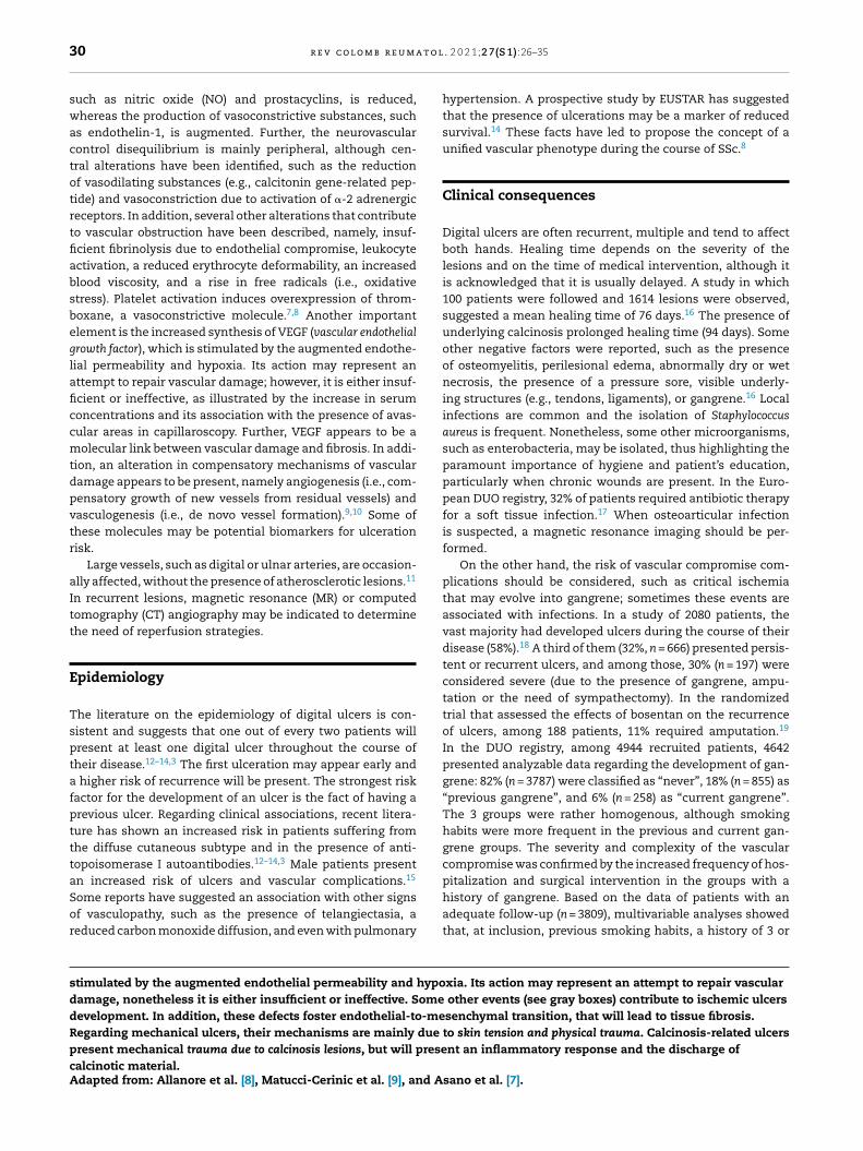
Fig. 4 – Main complications of digital ulcers in systemicsclerosis.
more digital ulcers, and a previous gangrene episode or sym-pathectomy, predicted the development of a new episode ofgangrene (n = 243).20
The cardinal symptom of ulcerations is pain, which is usu-ally continuous and present during night, yielding a negativeand significant functional impact. A recent analysis of the DUOregistry, based on 2327 patients, showed that a higher num-ber of ulcers at inclusion (0, 1–2 and ≥3) was associated with ahigher risk of inability to work during the previous month (8,42 and 48%, respectively), a higher disability to perform sim-ple daily tasks (35, 54 and 63%, respectively), and to a higherexpenditure due to the need of paid help (mean number ofhours: 17, 36 and 64, respectively).21 Digital ulcers are oneof the fundamental phenomena driving permanent disabil-ity in SSc.22 Reducing the number and the severity of ulcerswould allow to diminish the disability burden associated tothe disease, particularly when the functionality of the handis recovered.23 Fig. 4 summarizes the main consequences ofdigital ulcers in SSc.
Management
When digital ulcers appear, the management must be expe-ditious and should be based on patient’s awareness of clinicalchanges and the availability of medical interventions by thetreating physician. Early warning signs to be aware of are: anincreasing frequency of Raynaud’s phenomenon flares, a pro-longation of the cyanotic phase, and any other tissue changethat suggests digital necrosis.
Non-pharmacologic treatments
The patient should avoid cold stimuli that trigger Raynaud’scrisis. Smoking habits are equally harmful and interventions
aimed at stopping tobacco should be implemented.24,25 Medi-cations that trigger Raynaud’s phenomenon should be avoidedor suspended whenever possible. Regarding mechanical ulcersassociated with articular deformations, a low awarenessthreshold is encouraged, particularly when local lesions dueto trauma are present.
Local treatment is fundamental and patients should betaught about adequate care habits. The 3 objectives of localtreatment are: to prevent infectious complications, to protectwounds from trauma that may generate a local aggravationor pain, and to foster healing. Wound cleansing with salinesolution or soap and water is recommended. Using antisepticsolutions may generate contact eczema, irritant dermatitis,promote antibiotic resistance and may delay healing. Thus,local antibiotics should be avoided. Mechanical cleansing anddebridement should be performed under appropriate analge-sia. The election of adhesive dressings is based on the healingstage and the appearance of the ulcer. Hydrocolloid dressings(i.e., substances that form a gel when mixed with water) arebased on a humid milieu.26 Some other dressing options suchas hydrocellular polyurethane films may be used in this typeof wounds as well. Neutral tulles, Vaseline® or paraffin areadequate alternatives. In case of local superinfection, topicalantibacterial products such as flammazine (i.e., silver sulfadi-azine) are sometimes applied as a thick layer and covered bya dry compress, or a Vaseline® or silicone-soaked compress,and hold temporarily by a gauze bandage.
In severe or refractory cases, hyperbaric oxygen, whichis a developing alternative in diabetic ulcerations, has beenoffered to patients with SSc.27,28
Pharmacologic treatment
Vasodilators
Vasodilators play an essential role in the management of dig-ital ulcers in SSc.
Calcium channel inhibitors
Calcium channel inhibitors (CCI) are an effective strategy forthe management of digital ulcers, as they reduce the num-ber and the severity of Raynaud’s crises. One meta-analysissuggests a reduction in the number and the severity of flareswithin a period of 2 weeks of −8.3 [CI −15.7; −0.91] and of −0.7[−1.2; −0.17], respectively, for the CCI group as a whole, andof −10.2 [−20.1; −0.3] and −0.99 [−1.74; −0.24], respectively,for nifedipine.29 None of the included studies presented dataregarding the prevention or the evolution of digital ulcers. Onecross-over controlled trial that assessed 10 patients reporteda reduction in the number of digital ulcers, although withoutstatistical significance. Another trial compared iloprost andnifedipine (10–20 mg 3 times per day) during a 16 weeks follow-up, and showed a reduction of 4.3 compared to 1.4 digitalulcers (p < 0.001) in favor of nifedipine; however, the evolutionwas close to that of iloprost and the lack of a placebo controlundermined an adequate interpretation.30
32 r e v c o l o m b r e u m a t o l . 2 0 2 1;2 7(S 1):26–35
Angiotensin-converting enzyme inhibitors and angiotensin II
receptor antagonists
Although the rationale to propose these drug classes as a treat-ment strategy appeared solid, data on Raynaud’s syndrome donot support neither their utility nor their efficacy for digitalulcers. A large study over a 3 years follow-up period did notreport effectiveness for quinapril for the prevention of globalvascular progression of the disease.31,32
Prostacyclins
Prostacyclins analogs are potent vasodilators with anti-aggregation properties and anti-remodeling effects on smoothmuscle cells. Adverse effects are mainly complications ofvenous accesses, hypotension, vertigo, flushing, abdominalpain and diarrhea, skin allergies and sometimes jaw pain.Two therapeutic studies revealed that prostacyclin infusionsimproved the healing of ulcers and prevented the appearanceof new lesions.33,34 The infusion protocol was a continuousinfusion of 0.5–20 ng/kg/min over at least 6 h for 5 consec-utive days. Some other authors have adapted this protocolin severe patients using repeated infusions for 3–5 days dur-ing the winter.35 A sub-cutaneous preparation (trepostinil)was studied in a randomized trial; however it did not showa beneficial effect on ulcers.36 Moreover, an oral prepara-tion (selexipag), which was recently developed for pulmonaryhypertension, was not effective in a trial of SSc-associatedRaynaud’s phenomenon.37
Endothelin receptor antagonists
Endothelin-1 is the most potent vasoconstrictor substance.It presents vascular remodeling effects due to its action onsmooth-muscle cells. Its effect occurs through 2 receptors,namely, ETA and ETB, which are expressed by smooth musclecells and play a role in vasoconstriction and vascular hyper-plasia; ETB receptors are expressed by endothelial cells aswell and promote vasodilation. Bosentan is a non-selectiveendothelin antagonist that blocks both ETA and ETB. At first,it was approved for pulmonary hypertension, and thereupon,two phase 3 trials on ulcers revealed a significant effect onrecurrence prevention, but not on healing nor on Raynaud’ssyndrome.19,38 These findings were reinforced in a trial includ-ing 188 patients that presented at least 1 active digital ulcerwho were randomized and followed for 24 weeks. The totalnumber of ulcers was 1.9±0.2 for bosentan versus 2.7±0.3for placebo (p = 0.035; magnitude of improvement of 30%).19
A trend toward a higher effect in patients with a larger num-ber of ulcers at inclusion was observed. A three-fold increasein aminotransferases was detected in up to 10.5% of patientson bosentan; biological monitoring at least once a month isrecommended. This medication is usually offered to patientswith multiple or recurrent ulcers. Nevertheless, treatmentduration is not well defined and some groups employ thistreatment as a prevention strategy in high-risk patients duringcold seasons only, a long-term whereas others prefer usage.
Macitentan is another non-selective endothelin inhibitorthat exhibits a higher bioavailability than bosentan. It isapproved for pulmonary arterial hypertension. Nonetheless,in two large randomized studies, it did not prevent the appear-ance of new ulcers in SSc.39
Ambrisentan is a selective antagonist of ETA. It is approvedfor the treatment of pulmonary arterial hypertension. How-ever, it has not been evaluated in rigorous trials for themanagement of digital ulcers. Only one study with interimresults has been reported, which are rather inconclusive.40
Thus, it is not used in the context of digital ulcers.
Phosphodiesterase-5 inhibitors
Phosphodiesterase-5 inhibitors prevent the destruction ofcyclic GMP, thus increasing its bioavailability and fosteringa high concentration of NO. The latter acts as a vasodilatordue to the relaxation of smooth muscle cells, and as an anti-platelet aggregation agent.
In a meta-analysis comprising 6 randomized trials, phos-phodiesterase inhibitors were effective in reducing the activityof SSc-associated Raynaud’s syndrome, although with a mod-erate effect.41 Further, a study assessing as-needed sildenafil40 mg or 80 mg, showed a moderate benefit on the activity ofRaynaud’s syndrome in SSc.42
Regarding digital ulcers, a randomized trial that included83 patients compared sildenafil 20 mg three times per day orplacebo for 12 weeks.43 The primary outcome was the heal-ing time of each ulcer (192 ulcers were included). The medianhealing time was 63 (59–86) days in the placebo arm comparedto 56 (55–60) days in the sildenafil arm (p = 0.18). However,since week 8, the healing rate was superior in the sildenafilgroup (65% vs. 51%) and this benefit persisted through week 12(78% vs. 66%).43 In addition, excellent tolerance was reported.These results did not lead to a registration request for thismedication. This product, for which generic formulations areavailable, is often used in clinical practice as an off-label first-line medication in order to avoid the use of prostacyclins. Itis used by some teams prior to the application of bosentan,or either in association with this drug as a bridging therapybefore the use of prostacyclin. Tadalafil was studied in a ran-domized trial that supported the beneficial effects of this drugclass on ulcers, although with a low statistical power.44
Treatment combinations
Long-term and systematic use of CCI is recommended in SScfor the prevention of disease progression. Whenever an ulcerappears, CCI should be continued and potent vasodilatorsshould be associated, particularly prostacyclins to foster heal-ing, and bosentan to prevent recurrence in case of multipleulcers. Sildenafil is used as an off-label medication in bothsituations, although it is used as a bridging therapy for prosta-cyclins as well or in association with bosentan in severe orrefractory disease.45,46 Nonetheless, randomized trials assess-ing these combinations are lacking, while a great interest forthe treatment of SSc-associated pulmonary arterial hyperten-sion has been elicited.
Other treatment strategies
Interim results of a randomized trial have suggested thebeneficial effects of atorvastatin 40 mg per day due to areduced frequency of new ulcers, with an adequate toler-ance profile and without musculoskeletal adverse events.Several studies have shown potential further benefits in thiscontext, thus supporting larger trials.47 Anticoagulants andanti-aggregation therapies are occasionally offered to patients
r e v c o l o m b r e u m a t o l . 2 0 2 1;2 7(S 1):26–35 33
who develop ulcers. However, studies that support a benefitare lacking. In fact, an open-label study reported the absenceof effect of a low molecular weight heparin.48 Further, theassociation of salicylic acid and dipyridamole was not usefulin a study with a low statistical power.49 In clinical prac-tice, low dose salicylic acid is often given to patients with avascular compromise phenotype. One should always assessa risk-benefit balance for this intervention, as a high riskfor gastrointestinal bleeding is present in patients with SSc,particularly when risk factors for gastric ulcers are present;angiodysplasia-associated bleeding may be present as well.
Several anti-oxidative strategies have been assessed with-out compelling results (e.g., vitamin E, N-acetylcysteine,dimethyl sulfoxide). Vasodilators such as ketanserine, pra-zosin, and prostaglandins have been assessed in preliminarystudies.49 Immunosuppressants have not been studied in thisparticular context, though neither an increased risk has beenreported with rituximab, tocilizumab, or any other usual syn-thetic immunosuppressant.
Surgical interventions
Surgical interventions are seldom indicated for the man-agement of digital ulcers in SSc. Surgical debridement isindicated exceptionally in otherwise uncontrolled pain, or theneed to remove necrotic tissue as a potential risk factor forinfection. In case of mummification, whenever possible, it isequally recommended to avoid to precipitate scarring tissueremoval. Sympathectomy was previously offered, althoughit has been gradually abandoned.49 An equivalent therapythrough the injection of botulinum toxin may play a role nowa-days, although its evidence is based only on interim resultsparticularly focused on Raynaud’s syndrome.50 The rationalefor a local treatment is still strong. Several topical vasodilatorsor anti-oxidative agents have been studied without compellingresults.49 A recent development proposes the injection of fattytissue, which may contain progenitor cells that may fosterangiogenesis in an injured tissue and may promote healing.This tissue fraction appears to be rich in mesenchymal stemcells that may be equally beneficial. A pilot study in patientswith digital ulcers has been recently published and may offeralternatives for refractory cases.51
Conclusion
Digital ulcers are frequent over the course of SSc and are a sig-nificant source of morbidity. They occur due to vasculopathyand, besides general measures, their treatment is based onthe use of vasodilator agents. A better classification system ofthese lesions may guide care strategies and the formulationof standardized and personalized management protocols.
r e f e r e n c e s
1. Herrick AL, Roberts C, Tracey A, et al. Lack of agreementbetween rheumatologists in defining digital ulceration insystemic sclerosis. Arthritis Rheum. 2009;60:878–82.
2. van den Hoogen F, Khanna D, Fransen J, Johnson SR, Baron M,Tyndall A, et al. Classification criteria for systemic sclerosis:an American college of rheumatology/European league
against rheumatism collaborative initiative. Ann Rheum Dis.2013;72:1747–55.
3. Galluccio F, Allanore Y, Czirjak L, Furst DE, Khanna D,Matucci-Cerinic M. Points to consider for skin ulcers insystemic sclerosis. Rheumatology (Oxford). 2017;56Suppl 5:v67–71.
4. Hughes M, Tracey A, Bhushan M, Chakravarty K, Denton CP,Dubey S, et al. Reliability of digital ulcer definitions asproposed by the UK Scleroderma Study Group: a challenge forclinical trial design. J Scleroderma Relat Disord. 2018;3:170–4.
5. Suliman YA, Bruni C, Johnson SR, Praino E, Alemam M,Borazan N, et al. Defining skin ulcers in systemic sclerosis:systematic literature review and proposed World SclerodermaFoundation (WSF) definition. J Scleroderma Relat Disord.2017;2:115–20.
6. Sharp CA, Akram Q, Hughes M, Muir L, Herrick AL.Differential diagnosis of critical digital ischemia in systemicsclerosis: report of five cases and review of the literature.Semin Arthritis Rheum. 2016;46:209–16.
7. Asano Y, Sato S. Vasculopathy in scleroderma. SeminImmunopathol. 2015;37:489–500.
8. Allanore Y, Distler O, Matucci-Cerinic M, Denton CP. Review:defining a unified vascular phenotype in systemic sclerosis.Arthritis Rheumatol. 2018;70:162–70.
9. Matucci-Cerinic M, Manetti M, Bruni C, Chora I,Bellando-Randone S, Lepri G, et al. The “myth” of loss ofangiogenesis in systemic sclerosis: a pivotal earlypathogenetic process or just a late unavoidable event?Arthritis Res Ther. 2017;19:162.
10. Del Papa N, Pignataro F. The role of endothelial progenitors inthe repair of vascular damage in systemic sclerosis. FrontImmunol. 2018;9:1383.
11. Lescoat A, Yelnik CM, Coiffier G, Wargny M, Lamotte C,Cazalets C, et al. Ulnar artery occlusion and severity markersof vasculopathy in systemic sclerosis: a multicentercross-sectional study. Arthritis Rheumatol. 2018,http://dx.doi.org/10.1002/art.40799 [Epub ahead of print].
12. Hughes M, Pauling JD. Exploring the patient experience ofdigital ulcers in systemic sclerosis. Semin Arthritis Rheum.2018, http://dx.doi.org/10.1016/j.semarthrit.2018.08.001, pii:S0049-0172(18)30354-8 [Epub ahead of print] [Review].
13. Khimdas S, Harding S, Bonner A, et al. Associations withdigital ulcers in a large cohort of systemic sclerosis: resultsfrom the Canadian Scleroderma Research Group registry.Arthritis Care Res. 2011;63:142–9.
14. Mihai C, Landewé R, van der Heijde D, Walker UA, ConstantinPI, Gherghe AM, et al. Digital ulcers predict a worse diseasecourse in patients with systemic sclerosis. Ann Rheum Dis.2016;75:681–6.
15. Elhai M, Avouac J, Walker UA, Matucci-Cerinic M,Riemekasten G, Airò P, et al. A gender gap in primary andsecondary heart dysfunctions in systemic sclerosis: a EUSTARprospective study. Ann Rheum Dis. 2016;75:163–9.
16. Amanzi L, Braschi F, Fiori G, et al. Digital ulcers inscleroderma: staging, characteristics and sub-setting throughobservation of 1614 digital lesions. Rheumatology.2010;49:1374–82.
17. Denton CP, Krieg T, Guillevin L, et al. Demographic, clinicaland antibody characteristics of patients with digital ulcers insystemic sclerosis: data from the DUO Registry. Ann RheumDis. 2012;71:718–21.
18. Steen V, Denton CP, Pope JE, Matucci-Cerinic M. Digital ulcers:overt vascular disease in systemic sclerosis. Rheumatology(Oxford). 2009;48 Suppl. 3:iii19–24.
19. Matucci-Cerinic M, Denton CP, Furst DE, Mayes MD, Hsu VM,Carpentier P, et al. Bosentan treatment of digital ulcersrelated to systemic sclerosis: results from the RAPIDS-2randomised, double-blind, placebo-controlled trial. Ann
34 r e v c o l o m b r e u m a t o l . 2 0 2 1;2 7(S 1):26–35
Rheum Dis. 2011;70:32–8,http://dx.doi.org/10.1136/ard.2010.130658 [Epub 30.08.10].
20. Allanore Y, Denton CP, Krieg T, Cornelisse P, Rosenberg D,Schwierin B, et al. Clinical characteristics and predictors ofgangrene in patients with systemic sclerosis and digitalulcers in the Digital Ulcer Outcome Registry: a prospective,observational cohort. Ann Rheum Dis. 2016;75:1736–40.
21. Guillevin L, Hunsche E, Denton CP, et al. Functionalimpairment of systemic scleroderma patients with digitalulcerations: results from the DUO Registry. Clin ExpRheumatol. 2013;31:71–80.
22. Jaeger VK, Distler O, Maurer B, Czirják L, Lóránd V, Valentini G,et al. Functional disability and its predictors in systemicsclerosis: a study from the DeSScipher project within theEUSTAR group. Rheumatology (Oxford). 2018;57:441–50.
23. Mouthon L, Carpentier PH, Lok C, Clerson P, Gressin V,Hachulla E, et al. Controlling the digital ulcerative disease insystemic sclerosis is associated with improved handfunction. Semin Arthritis Rheum. 2017;46:759–66.
24. Jaeger VK, Valentini G, Hachulla E, Cozzi F, Distler O, Airó P,et al. Brief report: Smoking in systemic sclerosis: alongitudinal European scleroderma trials and research groupstudy. Arthritis Rheumatol. 2018;70:1829–34,http://dx.doi.org/10.1002/art.40557 [Epub 24.08.18].
25. Harrison BJ, Silman AJ, Hider SL, Herrick AL. Cigarettesmoking as a significant risk factor for digital vasculardisease in patients with systemic sclerosis. Arthritis Rheum.2002;46:3312–6.
26. Milburn PB, Singer JZ, Milburn MA. Treatment of sclerodermaskin ulcers with a hydrocolloid membrane. J Am AcadDermatol. 1989;21:200–4.
27. Sato T, Arai K. Hyperbaric oxygen therapy for digital ulcersdue to Raynaud’s disease. Case Rep Plast Surg Hand Surg.2018;5:72–4.
28. Mirasoglu B, Bagli BS, Aktas S. Hyperbaric oxygen therapy forchronic ulcers in systemic sclerosis – case series. Int JDermatol. 2017;56:636–40.
29. Thompson AE, Shea B, Welch V, Fenlon D, Pope JE.Calcium-channel blockers for Raynaud’s phenomenon insystemic sclerosis. Arthritis Rheum. 2001;44:1841–7.
30. Rademaker M, Cooke ED, Almond NE, Beacham JA, Smith RE,Mant TG, et al. Comparison of intravenous infusions ofiloprost and oral nifedipine in treatment of Raynaud’sphenomenon in patients with systemic sclerosis: a doubleblind randomised study. BMJ. 1989;298:561–4.
31. Gliddon AE, Doré CJ, Black CM, McHugh N, Moots R, DentonCP, et al. Prevention of vascular damage in scleroderma andautoimmune Raynaud’s phenomenon: a multicenter,randomized, double-blind, placebo-controlled trial of theangiotensin-converting enzyme inhibitor quinapril. ArthritisRheum. 2007;56:3837–46.
32. Dziadzio M, Denton CP, Smith R, Howell K, Blann A, Bowers E,et al. Losartan therapy for Raynaud’s phenomenon andscleroderma: clinical and biochemical findings in afifteen-week, randomized, parallel-group, controlled trial.Arthritis Rheum. 1999;42:2646–55.
33. Wigley FM, Wise RA, Seibold JR, et al. Intravenous iloprostinfusion in patients with Raynaud phenomenon secondary tosystemic sclerosis. A multicenter, placebo-controlled,double-blind study. Ann Intern Med. 1994;120:199–206.
34. Scorza R, Caronni M, Mascagni B, Berruti V, Bazzi S, Micallef E,et al. Effects of long-term cyclic iloprost therapy in systemicsclerosis with Raynaud’s phenomenon. A randomized,controlled study. Clin Exp Rheumatol. 2001;19:503–8.
35. Ingegnoli F, Schioppo T, Allanore Y, Caporali R, Colaci M,Distler O, et al. Practical suggestions on intravenous iloprostin Raynaud’s phenomenon and digital ulcer secondary tosystemic sclerosis: systematic literature review and expertconsensus. Semin Arthritis Rheum. 2018,http://dx.doi.org/10.1016/j.semarthrit.2018.03.019, pii:S0049-0172(18)30013-1 [Epub ahead of print] [Review].
36. Seibold J, Schiopu F, Denton E, et al. Digital ischemic ulcers inscleroderma treated with oral treprostinil diethanolamine: arandomized, double-blind, placebo-controlled, multicenterstudy. Arthritis Rheumatol. 2011;63:2483.
37. Denton CP, Hachulla É, Riemekasten G, Schwarting A,Frenoux JM, Frey A, et al. Efficacy and safety of selexipag inadults with Raynaud’s phenomenon secondary to systemicsclerosis: a randomized, placebo-controlled, phase II study.Arthritis Rheumatol. 2017;69:2370–9.
38. Korn JH, Mayes M, Matucci Cerinic M, Rainisio M, Pope J,Hachulla E, et al. Digital ulcers in systemic sclerosis:prevention by treatment with bosentan, an oral endothelinreceptor antagonist. Arthritis Rheum. 2004;50:3985–93.
39. Khanna D, Denton CP, Merkel PA, Krieg T, Le Brun FO, Marr A,et al. Effect of macitentan on the development of newischemic digital ulcers in patients with systemic sclerosis:DUAL-1 and DUAL-2 randomized clinical trials. JAMA.2016;315:1975–88.
40. Chung L, Ball K, Yaqub A, Lingala B, Fiorentino D. Effect of theendothelin type A-selective endothelin receptor antagonistambrisentan on digital ulcersin patients with systemicsclerosis: results of a prospective pilot study. J Am AcadDermatol. 2014;71:400–1.
41. Roustit M, Blaise S, Allanore Y, Carpentier PH, Caglayan E,Cracowski JL. Phosphodiesterase-5 inhibitors for thetreatment of secondary Raynaud’s phenomenon: systematicreview and meta-analysis of randomised trials. Ann RheumDis. 2013;72:1696–9.
42. Roustit M, Giai J, Gaget O, Khouri C, Mouhib M, Lotito A, et al.On-demand sildenafil as a treatment for Raynaudphenomenon: a series of n-of-1 trials. Ann Intern Med.2018;169:694–703, http://dx.doi.org/10.7326/M18-0517 [Epub30.10.18].
43. Hachulla E, Hatron PY, Carpentier P, Agard C, Chatelus E, JegoP, et al. Efficacy of sildenafil on ischaemic digital ulcer healingin systemic sclerosis: the placebo-controlled SEDUCE study.Ann Rheum Dis. 2016;75:1009–15.
44. Shenoy PD, Kumar S, Jha LK, Choudhary SK, Singh U, Misra R,et al. Efficacy of tadalafil in secondary Raynaud’sphenomenon resistant to vasodilator therapy: a double-blindrandomized cross-over trial. Rheumatology (Oxford).2010;49:2420–8.
45. Moinzadeh P, Riemekasten G, Siegert E, Fierlbeck G, Henes J,Blank N, et al. Vasoactive therapy in systemic sclerosis:real-life therapeutic practice in more than 3000 patients. JRheumatol. 2016;43:66–74.
46. Omarjee L, Fontaine C, Mahe G, Jaquinandi V. Improvement ofperipheral artery disease with sildenafil and bosentancombined therapy in a patient with limited cutaneoussystemic sclerosis: a case report. Medicine (Baltimore).2017;96:e6988.
47. Ladak K, Pope JE. A review of the effects of statins in systemicsclerosis. Semin Arthritis Rheum. 2016;45:698–705.
48. Tingey T, Shu J, Smuczek J, Pope J. Meta-analysis of healingand prevention of digital ulcers in systemic sclerosis.Arthritis Care Res (Hoboken). 2013;65:1460–71.

r e v c o l o m b r e u m a t o l . 2 0 2 1;2 7(S 1):26–35 35
49. Numata S, Iwata Y, Kobayashi M, Sato T, Sugiura K. Successfultreatment of continuous intra-arterial administration ofprostaglandin E1, urokinase and heparin for intractabledigital ulcers by upper extremity arterial occlusion in diffusecutaneous systemic sclerosis patient. J Dermatol.2017;44:e254–5.
50. Motegi SI, Sekiguchi A, Saito S, Ishibuchi H, Kishi C, YasudaM, et al. Successful treatment of Raynaud’s phenomenon anddigital ulcers in systemic sclerosis patients with botulinum
toxin B injection: assessment of peripheral vascular disorderby angiography and dermoscopic image of nail fold capillary. JDermatol. 2018;45:349–52.
51. Del Papa N, Di Luca G, Andracco R, Zaccara E, Maglione W,Pignataro F, et al. Regional grafting of autologous adiposetissue is effective in inducing prompt healing of indolentdigital ulcers in patients with systemic sclerosis: results of amonocentric randomized controlled study. Arthritis Res Ther.2019;21:7.


