Embed Size (px)
Citation preview
Citation: Galimberti, S.; Balducci, S.;
Guerrini, F.; Del Re, M.; Cacciola, R.
Digital Droplet PCR in Hematologic
Malignancies: A New Useful
Molecular Tool. Diagnostics 2022, 12,
1305. https://doi.org/10.3390/
diagnostics12061305
Academic Editor: Andreas Kjaer
Received: 1 May 2022
Accepted: 22 May 2022
Published: 24 May 2022
Publisher’s Note: MDPI stays neutral
with regard to jurisdictional claims in
published maps and institutional affil-
iations.
Copyright: © 2022 by the authors.
Licensee MDPI, Basel, Switzerland.
This article is an open access article
distributed under the terms and
conditions of the Creative Commons
Attribution (CC BY) license (https://
creativecommons.org/licenses/by/
4.0/).
diagnostics
Review
Digital Droplet PCR in Hematologic Malignancies: A NewUseful Molecular ToolSara Galimberti 1 , Serena Balducci 1 , Francesca Guerrini 1, Marzia Del Re 1 and Rossella Cacciola 2,*
1 Department of Clinical and Experimental Medicine, Section of Hematology, University of Pisa,56126 Pisa, Italy; [email protected] (S.G.); [email protected] (S.B.);[email protected] (F.G.); [email protected] (M.D.R.)
2 Department of Clinical and Experimental Medicine, Section of Hemostasis, University of Catania,95123 Catania, Italy
* Correspondence: [email protected]
Abstract: Digital droplet PCR (ddPCR) is a recent version of quantitative PCR (QT-PCR), useful formeasuring gene expression, doing clonality assays and detecting hot spot mutations. In respect ofQT-PCR, ddPCR is more sensitive, does not need any reference curve and can quantify one quarter ofsamples already defined as “positive but not quantifiable”. In the IgH and TCR clonality assessment,ddPCR recapitulates the allele-specific oligonucleotide PCR (ASO-PCR), being not adapt for detectingclonal evolution, that, on the contrary, does not represent a pitfall for the next generation sequencing(NGS) technique. Differently from NGS, ddPCR is not able to sequence the whole gene, but it isuseful, cheaper, and less time-consuming when hot spot mutations are the targets, such as occurswith IDH1, IDH2, NPM1 in acute leukemias or T315I mutation in Philadelphia-positive leukemias orJAK2 in chronic myeloproliferative neoplasms. Further versions of ddPCR, that combine differentprimers/probes fluorescences and concentrations, allow measuring up to four targets in the samePCR reaction, sparing material, time, and money. ddPCR is also useful for quantitating BCR-ABL1fusion gene, WT1 expression, donor chimerism, and minimal residual disease, so helping physiciansto realize that “patient-tailored therapy” that is the aim of the modern hematology.
Keywords: digital PCR; quantitative PCR; multiplexing PCR; MRD; clonality; NGS; point muta-tions; hematology
1. Introduction
Digital Droplet Polymerase Chain Reaction (ddPCR) is a specific, accurate and time-saving technique capables of accurately quantifying gene expression or detecting pointmutations applicable in several hematologic disorders, such as leukemias, lymphomas,myeloma, and chronic myeloproliferative neoplasms, and in transplant field. The ddPCRmight provide useful informations in prognostic and therapeutic setting.
2. Digital PCR: General Features and Applications
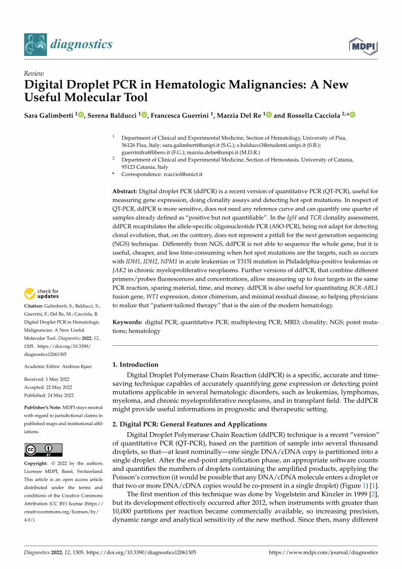
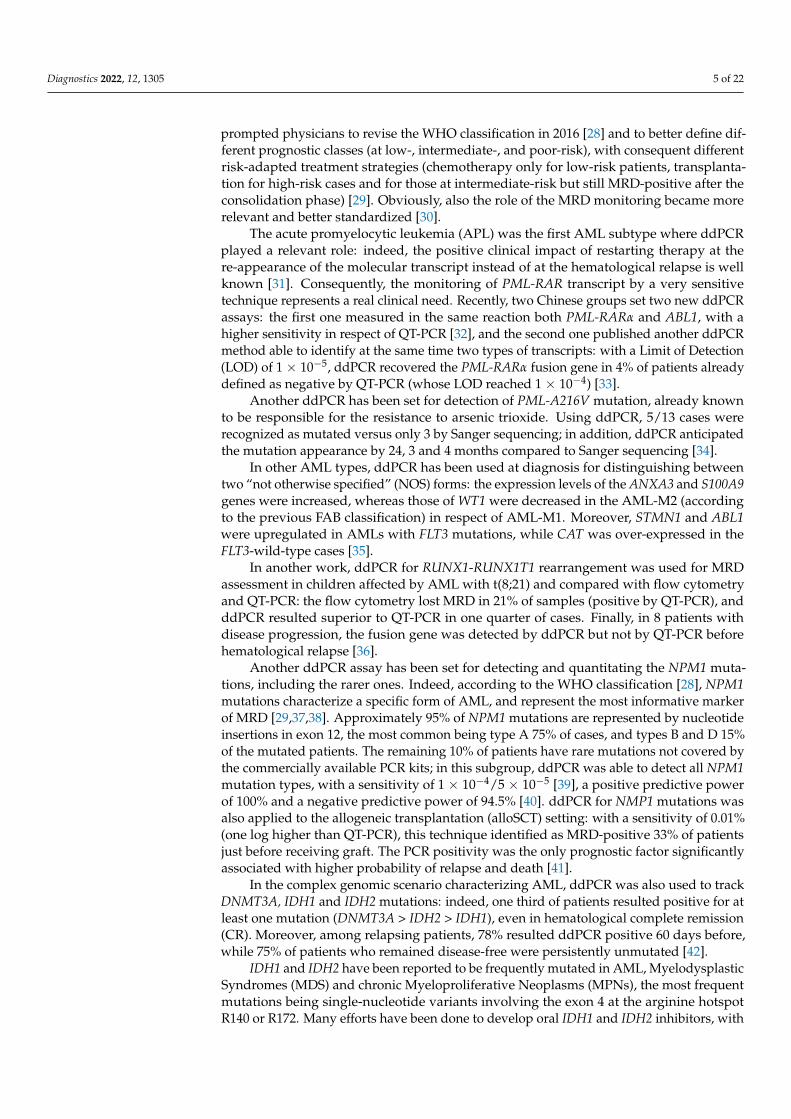
Digital Droplet Polymerase Chain Reaction (ddPCR) technique is a recent “version”of quantitative PCR (QT-PCR), based on the partition of sample into several thousanddroplets, so that—at least nominally—one single DNA/cDNA copy is partitioned into asingle droplet. After the end-point amplification phase, an appropriate software countsand quantifies the numbers of droplets containing the amplified products, applying thePoisson’s correction (it would be possible that any DNA/cDNA molecule enters a droplet orthat two or more DNA/cDNA copies would be co-present in a single droplet) (Figure 1) [1].
The first mention of this technique was done by Vogelstein and Kinzler in 1999 [2],but its development effectively occurred after 2012, when instruments with greater than10,000 partitions per reaction became commercially available, so increasing precision,dynamic range and analytical sensitivity of the new method. Since then, many different
Diagnostics 2022, 12, 1305. https://doi.org/10.3390/diagnostics12061305 https://www.mdpi.com/journal/diagnostics
Diagnostics 2022, 12, 1305 2 of 22
applications have been set and published: quantification of donor cell-free DNA in plasmaof transplanted patients [3], rapid detection of the most common pathogens in patients withbloodstream infection [4], extracellular RNA [5] or circulating miRNAs quantitation [6].
Diagnostics 2022, 12, x FOR PEER REVIEW 2 of 23
Figure 1. The phases of ddPCR technique. (a) The sample is partitioned in many thousands of drop-
lets. (b) In each droplet a target is amplified. (c) The endpoint amplification results are analyzed. (d)
A plot is generated, reading 2 fluorescence channels.
The first mention of this technique was done by Vogelstein and Kinzler in 1999 [2],
but its development effectively occurred after 2012, when instruments with greater than
10,000 partitions per reaction became commercially available, so increasing precision, dy-
namic range and analytical sensitivity of the new method. Since then, many different ap-
plications have been set and published: quantification of donor cell-free DNA in plasma
of transplanted patients [3], rapid detection of the most common pathogens in patients
with bloodstream infection [4], extracellular RNA [5] or circulating miRNAs quantitation
[6].
More recently, ddPCR has been used with success during the Coronavirus pandem-
ics: with a minimum cutoff of 0.04 copies/μL, ddPCR was able to quantify the Coronavirus
genome with a sensitivity and specificity of 97.6% and 100%, respectively. Interestingly,
in 12 out of 18 patients who converted back to Coronavirus positivity after a negative
phase, only ddPCR—and not QT-PCR—still detected viral genome, so reducing the diag-
nostic error during the recovery phase from the SARS-CoV-2 infection [7]. Moreover,
ddPCR accurately quantified Coronavirus genome from crude lysate, with high concord-
ance with measures from purified RNA, thus making more rapid and simpler the viral
genome detection [8].
The principal distinctive feature of ddPCR in respect of QT-PCR is that the former
does not require a reference standard curve, because the number of “amplified” droplets
are divided by the total number of droplets giving an absolute percentage (for example: if
1000 droplets are “positive” among 20,000 total droplets: 1000/20,000 = 0.05%). This is rel-
evant, making possible to quantitate new genes or mutations without need of cloning se-
quences into “ad hoc” plasmids whose presence in a laboratory significantly increases the
probability of environmental contamination. Moreover, the “absolute quantitation”
Figure 1. The phases of ddPCR technique. (a) The sample is partitioned in many thousands ofdroplets. (b) In each droplet a target is amplified. (c) The endpoint amplification results are analyzed.(d) A plot is generated, reading 2 fluorescence channels.
More recently, ddPCR has been used with success during the Coronavirus pandemics:with a minimum cutoff of 0.04 copies/µL, ddPCR was able to quantify the Coronavirusgenome with a sensitivity and specificity of 97.6% and 100%, respectively. Interestingly, in12 out of 18 patients who converted back to Coronavirus positivity after a negative phase,only ddPCR—and not QT-PCR—still detected viral genome, so reducing the diagnosticerror during the recovery phase from the SARS-CoV-2 infection [7]. Moreover, ddPCRaccurately quantified Coronavirus genome from crude lysate, with high concordance withmeasures from purified RNA, thus making more rapid and simpler the viral genomedetection [8].
The principal distinctive feature of ddPCR in respect of QT-PCR is that the formerdoes not require a reference standard curve, because the number of “amplified” dropletsare divided by the total number of droplets giving an absolute percentage (for example:if 1000 droplets are “positive” among 20,000 total droplets: 1000/20,000 = 0.05%). This isrelevant, making possible to quantitate new genes or mutations without need of cloningsequences into “ad hoc” plasmids whose presence in a laboratory significantly increasesthe probability of environmental contamination. Moreover, the “absolute quantitation”avoids the need of comparing a sample with itself in a different phase of disease, forexample before and after a specific treatment, that might overcome the problem of thematerial consumption.
Diagnostics 2022, 12, 1305 3 of 22
In terms of sensitivity, ddPCR is at least comparable to QT-PCR, and probably evenhigher, as shown in several different contexts. In non-Hodgkin’s lymphomas (NHLs) Dr.Drandi and coworkers showed a higher sensitivity for ddPCR (up to one log), especiallyin samples with very low tumor infiltration [9]. In Waldenstrom’s Macroglobulinemia(WM), it has been shown that ddPCR reached a sensitivity of 5 × 10⁻⁵, 1.5 log higherthan that offered by the Allele-Specific Oligonucleotide PCR (ASO-PCR), the techniqueclassically used for quantitating the rearrangement of the immunoglobulins heavy chain(IgH). In a series of 148 patients affected by WM, lymphoplasmacytic lymphoma (LPL) orIgM monoclonal gammopathy of undetermined significance (MGUS), 95% of cases showedthe MYD88L265P mutation; the concordance with QT-PCR was 74%, and the discordancewas always in favor of ddPCR [10]. In patients affected by acute B lymphoblastic leukemia(ALL), ddPCR has been compared to QT-PCR for assessment of minimal residual mea-surable disease (MRD): rearrangements of IgH or of immunoglobulins light chains (Ig,Ig), in addition to those of T cell receptors (TCRs) have been analyzed and concordantresults were observed in 88% of cases, without significant prevalence of one or the twotechniques in the discordant cases. On the contrary, 28% of samples defined as “positivebut not quantifiable” by QT-PCR resulted quantifiable by ddPCR, so suggesting its highersensitivity and accuracy [11]. Finally, in multiple myeloma (MM), ddPCR has been shownto have a comparable sensitivity of ASO-PCR in the MRD assessment [12].
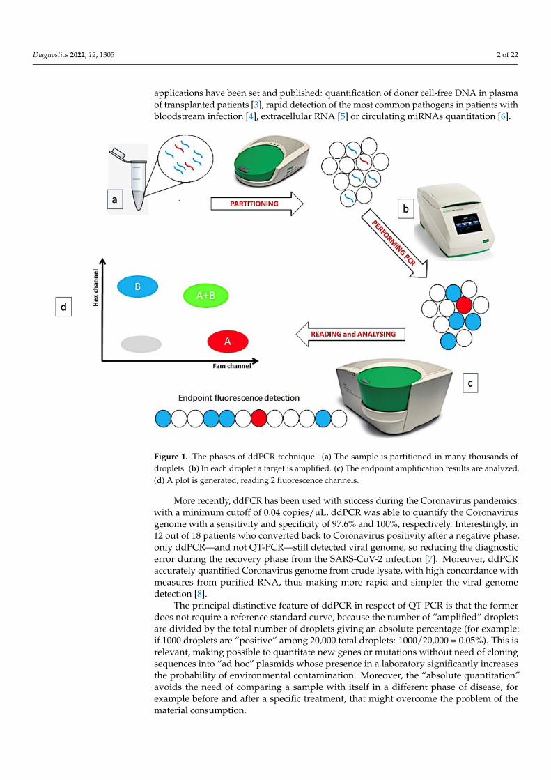
Unfortunately, no guidelines about ddPCR setting are today available; nevertheless,the progressive dissemination of this technique in many laboratories prompted the scientificcommunity to produce two useful documents for producing high-quality assays: the ISO20395:2019 rules (available at the website https://www.iso.org/obp/ui#iso:std:iso:20395:ed-1:v1:en, accessed on 28 February 2022) and another work that summarized the minimuminformation for publication of ddPCR experiments (dMIQE guidelines) [13]. In this paper,several technical aspects of ddPCR are discussed: amplicons <150 bp are preferred, thefundamental role of perfectly setting annealing temperature and probe concentrations, thepre-amplification step for low-level targets or the dilution step for too concentrated samples.Finally, the rules for adequately placing the threshold that allows distinguishing positiveevents from the background and the best number of replicates to do. Moreover, a furtherapplication of ddPCR also includes the multiplexing ddPCR [13]. In this case, differentfluorescent probes are simultaneously detected in different channels or, in the “higherorder multiplexing” version, it is possible to evaluate different targets by varying theconcentrations of different probes using the same fluorophore. This most recent version ofddPCR can be also performed by combination of more than one fluorophore with differentprobe concentrations, to detect up to 4 targets within a single reaction [1,14,15] (Figure 2).During the recent pandemics, the multiplexing ddPCR has been used to simultaneouslydetect the Coronavirus envelope, the viral RNA polymerase and the nucleocapsid genes,so avoiding the possible mismatch of primers and probes which could follow the viruschanges that might lead to false negative results [16].
In the non-invasive prenatal testing field, multiplexing ddPCR using universal lockednucleic acid probes correctly identified several fetal aneuploidies [17], while in oncologythis technique detected 4 different PIK3CA mutations on “liquid biopsy”, with a clinicalimpact in the management of metastatic breast cancer [18]. Moreover, an Italian groupset a multiplexing ddPCR in patients affected by chronic myeloid leukemia (CML) withunusual BCR-ABL1 atypical transcripts, not quantifiable by standardized QT-PCR, with anoptimal detection limit level (0.001%). The output of this technique is relevant, because itallows physicians to offer discontinuation of therapy with tyrosine kinase inhibitors evenin cases with persistent deep molecular response whose atypical transcripts are difficult tobe measured [19].
Diagnostics 2022, 12, 1305 4 of 22
Diagnostics 2022, 12, x FOR PEER REVIEW 4 of 23
At the 2022 national meeting of the Italian Society of Experimental Hematology, our
group presented a new kind of “higher order multiplexing ddPCR” able to simultane-
ously measure the expression of BMI1, EZH2, USP22 and GAPDH genes in 56 patients
affected by aggressive B-cell lymphoma (DLBCL). This assay allowed us to analyze very
small RNA quantities (the samples were paraffin-embedded and already employed for
the cell of origin definition by the Nanostring technology) [20,21].
In conclusion, ddPCR, in its different versions, represents a new, widely applicable,
specific, sensitive, and accurate quantitative technique. In the following manuscript, de-
tailed uses of ddPCR in different hematological fields are described.
Figure 2. Multiplex assay based on the amplitude of the amplifiers. (a) the different targets are de-
tected by probes labeled with the same fluorochrome (FAM or HEX) but used at different concen-
trations. This strategy allows to quantify four targets within a single reaction (A, B, C, D). (b) Targets
A and B have relative concentrations of 100% and 50% of FAM-labeled probe, respectively, while C
and D have relative concentrations of 100% and 50% of HEX-labeled probe. In the 2D plot, 16 pos-
sible clusters are generated: clusters that contain only one target, clusters that simultaneously con-
tain two targets and possibly clusters that contain three targets.
3. ddPCR Applications in Hematology
3.1. ddPCR in Acute Myeloid Leukemia and Myelodysplasias
Acute myeloid leukemia (AML) represents the prototype of a disease where the “tar-
get-therapy” is fundamental for improving patients’ survival [22]. In the recent years, the
availability of the anti-CD33 monoclonal antibody gemtuzumab ozogamicin [23], of the
FLT3 inhibitors (midostaurin and gilteritinib) [24,25] and of IDH1 and IDH2 inhibitors
(ivosidenib and enasidenib) [26,27] significantly changed the therapeutic scenario. The in-
creased probability of therapeutic success and the more defined disease genetic features
prompted physicians to revise the WHO classification in 2016 [28] and to better define
Figure 2. Multiplex assay based on the amplitude of the amplifiers. (a) the different targets aredetected by probes labeled with the same fluorochrome (FAM or HEX) but used at different concen-trations. This strategy allows to quantify four targets within a single reaction (A, B, C, D). (b) TargetsA and B have relative concentrations of 100% and 50% of FAM-labeled probe, respectively, while Cand D have relative concentrations of 100% and 50% of HEX-labeled probe. In the 2D plot, 16 possibleclusters are generated: clusters that contain only one target, clusters that simultaneously contain twotargets and possibly clusters that contain three targets.
At the 2022 national meeting of the Italian Society of Experimental Hematology, ourgroup presented a new kind of “higher order multiplexing ddPCR” able to simultaneouslymeasure the expression of BMI1, EZH2, USP22 and GAPDH genes in 56 patients affectedby aggressive B-cell lymphoma (DLBCL). This assay allowed us to analyze very smallRNA quantities (the samples were paraffin-embedded and already employed for the cell oforigin definition by the Nanostring technology) [20,21].
In conclusion, ddPCR, in its different versions, represents a new, widely applicable,specific, sensitive, and accurate quantitative technique. In the following manuscript,detailed uses of ddPCR in different hematological fields are described.
3. ddPCR Applications in Hematology3.1. ddPCR in Acute Myeloid Leukemia and Myelodysplasias
Acute myeloid leukemia (AML) represents the prototype of a disease where the“target-therapy” is fundamental for improving patients’ survival [22]. In the recent years,the availability of the anti-CD33 monoclonal antibody gemtuzumab ozogamicin [23], ofthe FLT3 inhibitors (midostaurin and gilteritinib) [24,25] and of IDH1 and IDH2 inhibitors(ivosidenib and enasidenib) [26,27] significantly changed the therapeutic scenario. Theincreased probability of therapeutic success and the more defined disease genetic features
Diagnostics 2022, 12, 1305 5 of 22
prompted physicians to revise the WHO classification in 2016 [28] and to better define dif-ferent prognostic classes (at low-, intermediate-, and poor-risk), with consequent differentrisk-adapted treatment strategies (chemotherapy only for low-risk patients, transplanta-tion for high-risk cases and for those at intermediate-risk but still MRD-positive after theconsolidation phase) [29]. Obviously, also the role of the MRD monitoring became morerelevant and better standardized [30].
The acute promyelocytic leukemia (APL) was the first AML subtype where ddPCRplayed a relevant role: indeed, the positive clinical impact of restarting therapy at there-appearance of the molecular transcript instead of at the hematological relapse is wellknown [31]. Consequently, the monitoring of PML-RAR transcript by a very sensitivetechnique represents a real clinical need. Recently, two Chinese groups set two new ddPCRassays: the first one measured in the same reaction both PML-RARα and ABL1, with ahigher sensitivity in respect of QT-PCR [32], and the second one published another ddPCRmethod able to identify at the same time two types of transcripts: with a Limit of Detection(LOD) of 1 × 10⁻⁵, ddPCR recovered the PML-RARα fusion gene in 4% of patients alreadydefined as negative by QT-PCR (whose LOD reached 1 × 10⁻⁴) [33].
Another ddPCR has been set for detection of PML-A216V mutation, already knownto be responsible for the resistance to arsenic trioxide. Using ddPCR, 5/13 cases wererecognized as mutated versus only 3 by Sanger sequencing; in addition, ddPCR anticipatedthe mutation appearance by 24, 3 and 4 months compared to Sanger sequencing [34].
In other AML types, ddPCR has been used at diagnosis for distinguishing betweentwo “not otherwise specified” (NOS) forms: the expression levels of the ANXA3 and S100A9genes were increased, whereas those of WT1 were decreased in the AML-M2 (accordingto the previous FAB classification) in respect of AML-M1. Moreover, STMN1 and ABL1were upregulated in AMLs with FLT3 mutations, while CAT was over-expressed in theFLT3-wild-type cases [35].
In another work, ddPCR for RUNX1-RUNX1T1 rearrangement was used for MRDassessment in children affected by AML with t(8;21) and compared with flow cytometryand QT-PCR: the flow cytometry lost MRD in 21% of samples (positive by QT-PCR), andddPCR resulted superior to QT-PCR in one quarter of cases. Finally, in 8 patients withdisease progression, the fusion gene was detected by ddPCR but not by QT-PCR beforehematological relapse [36].
Another ddPCR assay has been set for detecting and quantitating the NPM1 muta-tions, including the rarer ones. Indeed, according to the WHO classification [28], NPM1mutations characterize a specific form of AML, and represent the most informative markerof MRD [29,37,38]. Approximately 95% of NPM1 mutations are represented by nucleotideinsertions in exon 12, the most common being type A 75% of cases, and types B and D 15%of the mutated patients. The remaining 10% of patients have rare mutations not covered bythe commercially available PCR kits; in this subgroup, ddPCR was able to detect all NPM1mutation types, with a sensitivity of 1 × 10⁻⁴/5 × 10−5 [39], a positive predictive powerof 100% and a negative predictive power of 94.5% [40]. ddPCR for NMP1 mutations wasalso applied to the allogeneic transplantation (alloSCT) setting: with a sensitivity of 0.01%(one log higher than QT-PCR), this technique identified as MRD-positive 33% of patientsjust before receiving graft. The PCR positivity was the only prognostic factor significantlyassociated with higher probability of relapse and death [41].
In the complex genomic scenario characterizing AML, ddPCR was also used to trackDNMT3A, IDH1 and IDH2 mutations: indeed, one third of patients resulted positive for atleast one mutation (DNMT3A > IDH2 > IDH1), even in hematological complete remission(CR). Moreover, among relapsing patients, 78% resulted ddPCR positive 60 days before,while 75% of patients who remained disease-free were persistently unmutated [42].
IDH1 and IDH2 have been reported to be frequently mutated in AML, MyelodysplasticSyndromes (MDS) and chronic Myeloproliferative Neoplasms (MPNs), the most frequentmutations being single-nucleotide variants involving the exon 4 at the arginine hotspotR140 or R172. Many efforts have been done to develop oral IDH1 and IDH2 inhibitors, with
Diagnostics 2022, 12, 1305 6 of 22
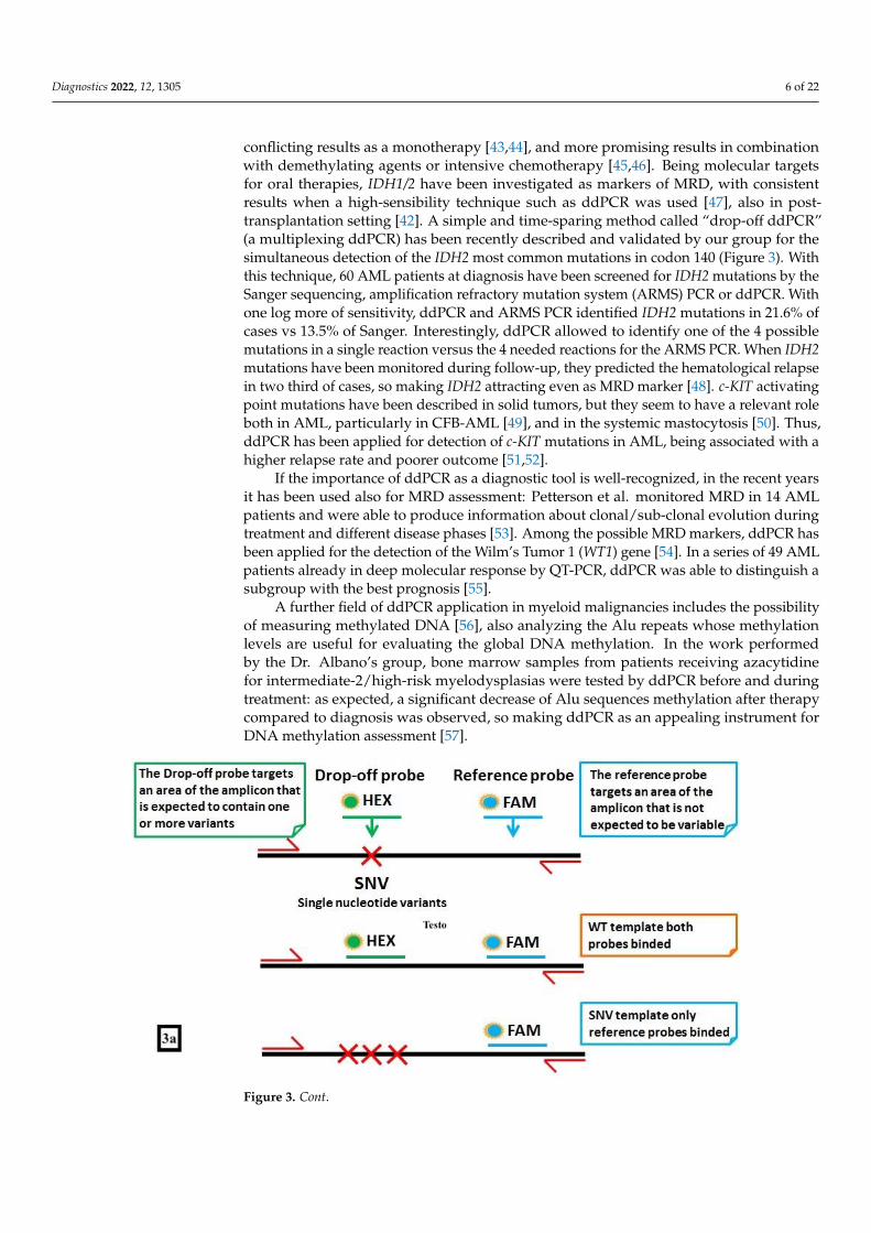
conflicting results as a monotherapy [43,44], and more promising results in combinationwith demethylating agents or intensive chemotherapy [45,46]. Being molecular targetsfor oral therapies, IDH1/2 have been investigated as markers of MRD, with consistentresults when a high-sensibility technique such as ddPCR was used [47], also in post-transplantation setting [42]. A simple and time-sparing method called “drop-off ddPCR”(a multiplexing ddPCR) has been recently described and validated by our group for thesimultaneous detection of the IDH2 most common mutations in codon 140 (Figure 3). Withthis technique, 60 AML patients at diagnosis have been screened for IDH2 mutations by theSanger sequencing, amplification refractory mutation system (ARMS) PCR or ddPCR. Withone log more of sensitivity, ddPCR and ARMS PCR identified IDH2 mutations in 21.6% ofcases vs 13.5% of Sanger. Interestingly, ddPCR allowed to identify one of the 4 possiblemutations in a single reaction versus the 4 needed reactions for the ARMS PCR. When IDH2mutations have been monitored during follow-up, they predicted the hematological relapsein two third of cases, so making IDH2 attracting even as MRD marker [48]. c-KIT activatingpoint mutations have been described in solid tumors, but they seem to have a relevant roleboth in AML, particularly in CFB-AML [49], and in the systemic mastocytosis [50]. Thus,ddPCR has been applied for detection of c-KIT mutations in AML, being associated with ahigher relapse rate and poorer outcome [51,52].
If the importance of ddPCR as a diagnostic tool is well-recognized, in the recent yearsit has been used also for MRD assessment: Petterson et al. monitored MRD in 14 AMLpatients and were able to produce information about clonal/sub-clonal evolution duringtreatment and different disease phases [53]. Among the possible MRD markers, ddPCR hasbeen applied for the detection of the Wilm’s Tumor 1 (WT1) gene [54]. In a series of 49 AMLpatients already in deep molecular response by QT-PCR, ddPCR was able to distinguish asubgroup with the best prognosis [55].
A further field of ddPCR application in myeloid malignancies includes the possibilityof measuring methylated DNA [56], also analyzing the Alu repeats whose methylationlevels are useful for evaluating the global DNA methylation. In the work performedby the Dr. Albano’s group, bone marrow samples from patients receiving azacytidinefor intermediate-2/high-risk myelodysplasias were tested by ddPCR before and duringtreatment: as expected, a significant decrease of Alu sequences methylation after therapycompared to diagnosis was observed, so making ddPCR as an appealing instrument forDNA methylation assessment [57].
Diagnostics 2022, 12, x FOR PEER REVIEW 6 of 23
combination with demethylating agents or intensive chemotherapy [45,46]. Being molec-
ular targets for oral therapies, IDH1/2 have been investigated as markers of MRD, with
consistent results when a high-sensibility technique such as ddPCR was used [47], also in
post-transplantation setting [42]. A simple and time-sparing method called “drop-off
ddPCR” (a multiplexing ddPCR) has been recently described and validated by our group
for the simultaneous detection of the IDH2 most common mutations in codon 140 (Figure
3). With this technique, 60 AML patients at diagnosis have been screened for IDH2 muta-
tions by the Sanger sequencing, amplification refractory mutation system (ARMS) PCR or
ddPCR. With one log more of sensitivity, ddPCR and ARMS PCR identified IDH2 muta-
tions in 21.6% of cases vs 13.5% of Sanger. Interestingly, ddPCR allowed to identify one
of the 4 possible mutations in a single reaction versus the 4 needed reactions for the ARMS
PCR. When IDH2 mutations have been monitored during follow-up, they predicted the
hematological relapse in two third of cases, so making IDH2 attracting even as MRD
marker [48]. c-KIT activating point mutations have been described in solid tumors, but
they seem to have a relevant role both in AML, particularly in CFB-AML [49], and in the
systemic mastocytosis [50]. Thus, ddPCR has been applied for detection of c-KIT muta-
tions in AML, being associated with a higher relapse rate and poorer outcome [51,52].
If the importance of ddPCR as a diagnostic tool is well-recognized, in the recent years
it has been used also for MRD assessment: Petterson et al. monitored MRD in 14 AML
patients and were able to produce information about clonal/sub-clonal evolution during
treatment and different disease phases [53]. Among the possible MRD markers, ddPCR
has been applied for the detection of the Wilm’s Tumor 1 (WT1) gene [54]. In a series of 49
AML patients already in deep molecular response by QT-PCR, ddPCR was able to distin-
guish a subgroup with the best prognosis [55].
A further field of ddPCR application in myeloid malignancies includes the possibility
of measuring methylated DNA [56], also analyzing the Alu repeats whose methylation
levels are useful for evaluating the global DNA methylation. In the work performed by
the Dr. Albano’s group, bone marrow samples from patients receiving azacytidine for in-
termediate-2/high-risk myelodysplasias were tested by ddPCR before and during treat-
ment: as expected, a significant decrease of Alu sequences methylation after therapy com-
pared to diagnosis was observed, so making ddPCR as an appealing instrument for DNA
methylation assessment [57].
Figure 3. Cont.
Diagnostics 2022, 12, 1305 7 of 22Diagnostics 2022, 12, x FOR PEER REVIEW 7 of 23
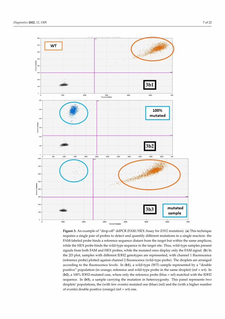
Figure 3. An example of “drop-off” ddPCR (FAM/HEX Assay for IDH2 mutation). (a) This tech-
nique requires a single pair of probes to detect and quantify different mutations in a single reaction:
the FAM-labeled probe binds a reference sequence distant from the target but within the same am-
plicon, while the HEX probe binds the wild-type sequence in the target site. Thus, wild-type samples
present signals from both FAM and HEX probes, while the mutated ones display only the FAM
signal. (b) In the 2D plot, samples with different IDH2 genotypes are represented, with channel 1
fluorescence (reference probe) plotted against channel 2 fluorescence (wild-type probe). The drop-
lets are arranged according to the fluorescence levels. In (b1), a wild-type (WT) sample represented
by a “double positive” population (in orange; reference and wild-type probe in the same droplet)
(ref + wt). In (b2), a 100% IDH2-mutated case, where only the reference probe (blue = ref) matched
with the IDH2 sequence. In (b3), a sample carrying the mutation in heterozygosity. This panel rep-
resents two droplets’ populations, the (with few events) mutated one (blue) (ref) and the (with a
higher number of events) double positive (orange) (ref + wt) one.
Figure 3. An example of “drop-off” ddPCR (FAM/HEX Assay for IDH2 mutation). (a) This techniquerequires a single pair of probes to detect and quantify different mutations in a single reaction: theFAM-labeled probe binds a reference sequence distant from the target but within the same amplicon,while the HEX probe binds the wild-type sequence in the target site. Thus, wild-type samples presentsignals from both FAM and HEX probes, while the mutated ones display only the FAM signal. (b) Inthe 2D plot, samples with different IDH2 genotypes are represented, with channel 1 fluorescence(reference probe) plotted against channel 2 fluorescence (wild-type probe). The droplets are arrangedaccording to the fluorescence levels. In (b1), a wild-type (WT) sample represented by a “doublepositive” population (in orange; reference and wild-type probe in the same droplet) (ref + wt). In(b2), a 100% IDH2-mutated case, where only the reference probe (blue = ref) matched with the IDH2sequence. In (b3), a sample carrying the mutation in heterozygosity. This panel represents twodroplets’ populations, the (with few events) mutated one (blue) (ref) and the (with a higher numberof events) double positive (orange) (ref + wt) one.
Diagnostics 2022, 12, 1305 8 of 22
3.2. Digital PCR in Acute Lymphoblastic Leukemia
Acute Lymphoblastic Leukemia (ALL) is the most frequent childhood neoplasia witha dismal outcome when diagnosed in adults; in the last few years a better knowledge of Band T-ALL genetic landscape and advanced tools for MRD monitoring allowed to refineindications for alloSCT and ameliorate prognosis [58,59].
MRD monitoring in chromosome Philadelphia-positive (Ph’-positive) B-ALL is basedon quantification of BCR-ABL1 transcript using QT-PCR; a recent Italian study appliedddPCR to patients enrolled into the GIMEMA LAL2116 trial, showing optimal sensitivity(1 × 10⁻⁵–5 × 10⁻⁶) and specificity (near to 100%). In follow-up samples, ddPCR was ableto reduce the proportion of positive-not-quantifiable (PNQ) cases, which represent a greyzone in the clinical practice, significantly increasing the proportion of quantifiable samples.Therefore, of the 5 cases that were negative by QT-PCR and positive by ddPCR duringfollow-up, 4/5 experienced a relapse, confirming the clinical relevance of a deeper MRDmonitoring [60]. Similar results were found in 2018 by Dr. Coccaro and colleagues [61] andby Dr. Guan and coworkers who applied ddPCR MRD monitoring in 10 relapsed/refractoryPh’-positive ALL patients treated with anti-CD19/CD22 CAR-T-cell cocktail therapy [62].Finally, Dr. Martinez and colleagues proposed and validated a one-step ddPCR assayfor the p190 BCR-ABL1 transcript that showed several advantages over QT-PCR: deepersensitivity, no need for a standard curve, no need for standardization material to be sharedbetween different laboratories for result comparison, and less propensity to the reactioninhibition [63].
In Ph’-positive neoplasms, the emergence of point mutations in Tyrosine KinaseInhibitors (TKIs)-ligand domain of ABL1 may represent a major barrier for success of TKIs,with T315I mutations rendering cells sensible only to the 3rd generation TKI, ponatinib. Ifthe role of ABL1 point mutations is well-recognized in Chronic Myeloid Leukemia (CML), inALL the prognostic relevance of detecting small clones of T315I-mutated cells at diagnosisremains to be fully elucidated [64]. Nevertheless, in 2020 Dr. Akahoshi et al. reportedthat the detection of even a small amount of T315I mutation by ddPCR at the time ofmolecular relapse after autologous transplantation may provide appropriate informationfor identifying patients who are likely to develop hematological relapse [65].
About other rarer BCR-partner fusion genes, such as t(8;22) BCR/FGFR1, which conferto ALL a particularly aggressive disease course and dismal outcome, a standardizedmethods for MRD detection is lacking and response monitoring is usually performed withcytogenetic techniques or qualitative PCR [66]. In 2018, Dr. Coccaro et al. reported theabsolute quantitation of the BCR/FGFR1 fusion gene by a new ddPCR assay with a LOD of0.01% [67].
In Ph’-negative ALLs (B or T), which represent most ALLs in childhood, MRD detec-tion is more complicated and based on the Immunoglobulin IgH, Ig, Ig and TCRs rearrange-ment analysis; nevertheless, a consistent fraction of samples with very-low MRD levelscannot be properly quantified and must be scored as positive not quantifiable (PNQ), thatrepresent a clinical dilemma (are they MRD positive or negative? Is it worth to proceed withtransplantation or not?) [68]. Trying to address this issue, Dr. Della Starza and coworkersproposed ddPCR as an alternative method for MRD monitoring [11,69] in samples frompatients enrolled in the GIMEMA LAL1308 and in the AIEOP-BFM ALL 2000 trials, findinga concordance rate of 70% between QT-PCR and ddPCR. The greater accuracy of ddPCRallowed to quantitate samples defined as PNQ by QT-PCR in a quarter of cases. To allowa better standardization, the group also proposed a fixed-threshold of positive-dropletnumber to define a sample as negative, PNQ or positive [69].
Another gene mutated in about 15–20% of pediatric B-ALL and in 50% of adult ALL isthe IKZF1 [70]. The presence of IKZF1 or BCR-ABL1 mutations has been reported to be anindependent risk factor of poor prognosis [71]. In 2019, Dr. Hashiguchi and Dr. Onozawadescribed the application of ddPCR for detection and quantification of IKZF1 genomicaberrations, suggesting a possible its role even as MRD marker [72].
Diagnostics 2022, 12, 1305 9 of 22
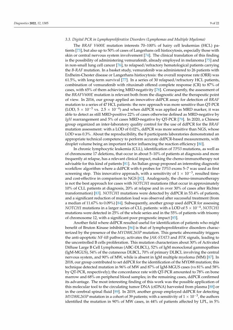
3.3. Digital PCR in Lymphoproliferative Disorders (Lymphomas and Multiple Myeloma)
The BRAF V600E mutation interests 70–100% of hairy cell leukemias (HCL) pa-tients [73], but also up to 50% of cases of Langerhans cell histiocytosis, especially those withskin or central nervous system involvement [74]. The clinical translation of this findingis the possibility of administering vemurafenib, already employed in melanoma [75] andin non-small lung cell cancer [76], to relapsed/refractory hematological patients carryingthe B-RAF mutation. In a basket study, vemurafenib was administered to 26 patients withErdheim-Chester disease or Langerhans histiocytosis: the overall response rate (ORR) was61.5%, with long-term survival [77]. In a series of 30 relapsed/refractory HCL patients,combination of vemurafenib with rituximab offered complete response (CR) to 87% ofcases, with 65% of them achieving MRD-negativity [78]. Consequently, the assessment ofthe BRAFV600E mutation is relevant both from the diagnostic and the therapeutic pointof view. In 2016, our group applied an innovative ddPCR assay for detection of BRAFmutation to a series of 47 HCL patients: the new approach was more sensitive than QT-PCR(LOD, 5 × 10⁻⁵ vs. 2.5 × 10⁻⁴) and when ddPCR was applied as MRD marker, it wasable to detect as still MRD-positive 22% of cases otherwise defined as MRD-negative byIgH rearrangement and 5% of cases MRD-negative by QT-PCR [79]. In 2020, a Chinesegroup organized an inter-laboratory quality control for the use of ddPCR for the BRAFmutation assessment: with a LOD of 0.02%, ddPCR was more sensitive than NGS, whoseLOD was 0.3%. About the reproducibility, the 8 participants laboratories demonstrated anappropriate technical competency to perform accurate ddPCR-based measurements, withdroplet volume being an important factor influencing the reaction efficiency [80].
In chronic lymphocytic leukemia (CLL), identification of TP53 mutations, as well asof chromosome 17 deletions, that occur in about 5–10% of patients at diagnosis and morefrequently at relapse, has a relevant clinical impact, making the chemo-immunotherapy notadvisable for this kind of patients [81]. An Italian group proposed an interesting diagnosticworkflow algorithm where a ddPCR with 6 probes for TP53 exons 5–7 was used as firstscreening step. This innovative approach, with a sensitivity of 1 × 10⁻³, resulted time-and cost-effective in comparison to NGS [82]. Analogously, the chemo-immunotherapyis not the best approach for cases with NOTCH1 mutations (that occur in approximately10% of CLL patients at diagnosis, 20% at relapse and in over 30% of cases after Richtertransformation) [83]. NOTCH1 mutations were detected by ddPCR in 53.4% of patients,and a significant reduction of mutation load was observed after successful treatment (froma median of 11.67% to 0.09%) [84]. Subsequently, another group used ddPCR for assessingNOTCH1 mutations in a larger series of CLL patients: with a LOD of 5 × 10⁻⁴, NOTCH1mutations were detected in 25% of the whole series and in the 55% of patients with trisomyof chromosome 12, with a significant poor prognostic impact [85].
Another field where ddPCR resulted useful for identification of patients who mightbenefit of Bruton Kinase inhibitors [86] is that of lymphoproliferative disorders charac-terized by the presence of the MYD88L265P mutation. This genetic abnormality triggersthe anti-apoptotic NF-kB pathway, activates the JAK-STAT3 and BTK signals, leading tothe uncontrolled B cells proliferation. This mutation characterizes about 30% of ActivatedDiffuse Large B Cell Lymphomas (ABC-DLBCL), 52% of IgM monoclonal gammopathies(IgM-MGUS), 54% of the cutaneous DLBCL, 70% of primary DLBCL involving the centralnervous system, and 90% of MW, while is absent in IgM multiple myeloma (MM) [87]. In2018, our group contributed to set ddPCR for the identification of the MYD88 mutation; thistechnique detected mutation in 96% of MW and 87% of IgM-MGUS cases (vs 81% and 58%by QT-PCR, respectively); the concordance rate with QT-PCR amounted to 78% on bonemarrow and 68% on peripheral blood samples; in the remaining cases, ddPCR confirmedits advantage. The most interesting finding of this work was the possible application ofthis molecular tool to the circulating tumor DNA (ctDNA) harvested from plasma [88] orin the cerebral spinal fluid [89]. In 2019, another group employed ddPCR for detectingMYD88L265P mutation in a cohort of 39 patients; with a sensitivity of 1 × 10⁻³, the authorsidentified the mutation in 90% of MW cases, in 44% of patients affected by LPL, in 5%

Diagnostics 2022, 12, 1305 10 of 22
of IgM MM, and no in CLL or mantle cell lymphoma (MCL) cases [90]. Another groupproposed a new ddPCR assay able to detect and quantify the hot spots mutations of EZH2,STAT6, MYD88, and CCND3 that characterize about 20% of B-cell lymphomas, especiallythe germinal center DLBCL (GB-DLBCL) and follicular lymphoma (FL) that seem to beassociated with resistance to treatment. ddPCR, with a sensitivity of 1 × 10⁻⁴, was accurateeither on paraffin-embedded samples or on ctDNA (the “liquid biopsy”) [91].
Because BCL2/JH rearrangement can be found only in 60% of FL and the assessment ofIgH rearrangement in this lymphoma is often difficult due to its hypermutated status [92],the possibility of assessing different molecular markers is intriguing. Among them, muta-tions of EZH2 are becoming relevant, even from the clinical point of view, after the recentintroduction in the therapeutic armamentarium of the oral EZH2 inhibitor. In patientswith relapsed/refractory FL, tazemetostat offered to EZH2-mutated patients 69% of ORand 13% of CR vs. 35% of ORR and 4% of CR to the wild-type subgroup [93]. A ddPCRfor detecting EZH2 mutations was set; interestingly, in a patient carrying two differentmutations in different tumor sites, the analysis of ctDNA revealed both EZH2 genomicaberrations, so demonstrating the optimal representativeness of liquid biopsy [94]. Even inearly-stage FL, ddPCR for BCL2/IgH rearrangement was compared to classical QT-PCR:the concordance between the two techniques amounted to 92%, and the fusion gene wasrecovered by ddPCR in 18% of cases otherwise negative by QT-PCR [95].
In 2020, the European cooperative group for ddPCR published an interesting manuscriptabout the employ of ddPCR in 416 samples from 166 patients affected by MCL. Firstly, theauthors observed a 90% of concordance rate among the 9 involved laboratories; then, theyproposed some rules for performing and analyzing ddPCR reactions, such as starting from500 ng of DNA, preferring 3 replicates, and considering as “positive” a sample showingat least 3 merged events, as “negative” that without events or with only one mergedevent, reserving the concept of “grey zone” (PNQ) to samples with two merged events.When ddPCR for IgH clonality and/or BCL1/IgH rearrangement was compared withQT-PCR, GeneScan PCR or flow cytometry, the respective sensitivities reached 1 × 10−5,1 × 10−4, 5 × 10⁻², and 1 × 10⁻⁴, showing once again the advantage of ddPCR in term ofsensitivity [96,97].
In the T angioimmunoblastic lymphoma, the ddPCR has been proposed for the G17Vmutant RHOA (that hyperactivates the TCR signal so prompting the abnormal T cellproliferation); with a LOD of 1 × 10⁻⁴, ddPCR was able to recognize mutation in 4 casesthat NGS defined as unmutated [98].
In Hodgkin’s lymphoma, ddPCR was used as confirmatory tool of STAT6 mutationson frozen biopsy tissue and ctDNA. NGS showed STAT6 mutations in about 30% of patients,being the most frequent recurrent mutations with those of XPO1 and B2M. With a sensitivityof 0.14%, ddPCR was able to recognize mutations in all cases already tested by NGS and inall cases ddPCR was able to detect mutations also on ctDNA [99].
Dr. Drandi and coworkers compared ddPCR to QT-PCR in a series of 69 patientswith FL, MCL and MM: the concordance was good, and both techniques reached the LODof 1 × 10⁻⁵; nevertheless, ddPCR was more accurate, because it was successful in 100%of cases, whereas QT-PCR failed in 4% of cases. This pivotal work clearly sustained thepossibility of replacing ASO-PCR for IgH clonality with ddPCR [9].
Focusing on MM, it is incontrovertible that the prognostic value of MRD assessmentis becoming a new target of treatment, thanks to the availability of drugs able to induceMRD eradication in up to 70% of patients [100]. A Japanese group recently revised theissue of MRD in autografts from 43 MM patients who underwent autologous stem celltransplantation comparing NGS (with a sensitivity of 1 × 10⁻⁷) to ASO-PCR (sensitivity1 × 10−4/1 × 10−5) and to ddPCR (sensitivity 1 × 10⁻⁵). Correlation between ddPCR andASO-PCR was satisfying (91%), with an advantage for ddPCR, while NGS resulted lessperformant [101] (Table 1).
Diagnostics 2022, 12, 1305 11 of 22
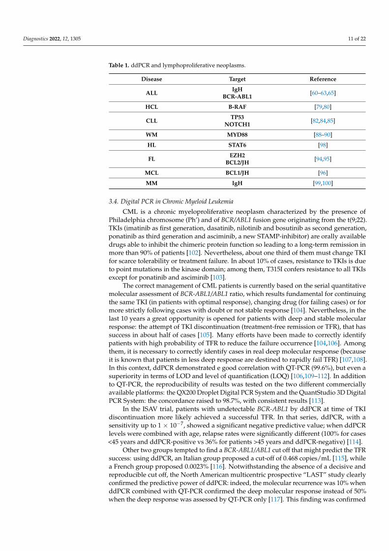
Table 1. ddPCR and lymphoproliferative neoplasms.
Disease Target Reference
ALL IgHBCR-ABL1 [60–63,65]
HCL B-RAF [79,80]
CLL TP53NOTCH1 [82,84,85]
WM MYD88 [88–90]
HL STAT6 [98]
FL EZH2BCL2/JH [94,95]
MCL BCL1/JH [96]
MM IgH [99,100]
3.4. Digital PCR in Chronic Myeloid Leukemia
CML is a chronic myeloproliferative neoplasm characterized by the presence ofPhiladelphia chromosome (Ph’) and of BCR/ABL1 fusion gene originating from the t(9;22).TKIs (imatinib as first generation, dasatinib, nilotinib and bosutinib as second generation,ponatinib as third generation and asciminib, a new STAMP-inhibitor) are orally availabledrugs able to inhibit the chimeric protein function so leading to a long-term remission inmore than 90% of patients [102]. Nevertheless, about one third of them must change TKIfor scarce tolerability or treatment failure. In about 10% of cases, resistance to TKIs is dueto point mutations in the kinase domain; among them, T315I confers resistance to all TKIsexcept for ponatinib and asciminib [103].
The correct management of CML patients is currently based on the serial quantitativemolecular assessment of BCR-ABL1/ABL1 ratio, which results fundamental for continuingthe same TKI (in patients with optimal response), changing drug (for failing cases) or formore strictly following cases with doubt or not stable response [104]. Nevertheless, in thelast 10 years a great opportunity is opened for patients with deep and stable molecularresponse: the attempt of TKI discontinuation (treatment-free remission or TFR), that hassuccess in about half of cases [105]. Many efforts have been made to correctly identifypatients with high probability of TFR to reduce the failure occurrence [104,106]. Amongthem, it is necessary to correctly identify cases in real deep molecular response (becauseit is known that patients in less deep response are destined to rapidly fail TFR) [107,108].In this context, ddPCR demonstrated e good correlation with QT-PCR (99.6%), but even asuperiority in terms of LOD and level of quantification (LOQ) [106,109–112]. In additionto QT-PCR, the reproducibility of results was tested on the two different commerciallyavailable platforms: the QX200 Droplet Digital PCR System and the QuantStudio 3D DigitalPCR System: the concordance raised to 98.7%, with consistent results [113].
In the ISAV trial, patients with undetectable BCR-ABL1 by ddPCR at time of TKIdiscontinuation more likely achieved a successful TFR. In that series, ddPCR, with asensitivity up to 1 × 10⁻⁷, showed a significant negative predictive value; when ddPCRlevels were combined with age, relapse rates were significantly different (100% for cases<45 years and ddPCR-positive vs 36% for patients >45 years and ddPCR-negative) [114].
Other two groups tempted to find a BCR-ABL1/ABL1 cut off that might predict the TFRsuccess: using ddPCR, an Italian group proposed a cut-off of 0.468 copies/mL [115], whilea French group proposed 0.0023% [116]. Notwithstanding the absence of a decisive andreproducible cut off, the North American multicentric prospective “LAST” study clearlyconfirmed the predictive power of ddPCR: indeed, the molecular recurrence was 10% whenddPCR combined with QT-PCR confirmed the deep molecular response instead of 50%when the deep response was assessed by QT-PCR only [117]. This finding was confirmed
Diagnostics 2022, 12, 1305 12 of 22
by other authors that used ddPCR for accurately identifying patients with deep responseor undetectable fusion gene at the time of TKI discontinuation [118].
In addition to the better quantitation of BCR-ABL1 transcript, another promising use ofddPCR seems to be its use for screening BCR-ABL1 mutations. Indeed, it has been recentlyestablished that NGS seems to be the best technique for these mutations’ identification: inthe “Next in CML” study, the percentage of mutated patients increased from 25% of Sangerto 47% of NGS. Interestingly, in 69 cases NGS allowed to identify the most appropriate TKI;in 10 patients, who resulted unmutated by Sanger, NGS detected the T315I mutation, withthe immediate start of ponatinib [119].
In the context of the Italian “Campus CML” working group, 44 samples were screenedfor T315I by Sanger, NGS and ddPCR: in our hands, the minimum mutational burdendetected was 0.02%; with this sensitivity, 25 samples were concordant between ddPCR andSanger, while 5 cases resulted mutated by ddPCR but not by Sanger. In respect of NGS,19 samples were concordant; 2 cases, mutated by NGS, resulted wild-type by ddPCR; onthe other hand, other 2 cases wild-type by NGS was mutated by ddPCR. The VAF of thesecases was 0.43% and 0.39%, values under the sensitivity limit of NGS. One of the 2 failingcases in ddPCR resulted mutated on genomic DNA but not on cDNA. These data, even ifpreliminary, sustain the possibility of using ddPCR for a rapid screening of T315I, with theimmediate therapeutic change [120].
The possibility of employing ddPCR on genomic DNA to identify quiescent leukemiastem cells is another feature distinguishing ddPCR from QT-PCR, as well shown by Dr.Albano and his group [121] and might be worth of further investigation (Table 2).
3.5. Digital PCR in Chronic Myeloproliferative Neoplasms
The chronic myeloproliferative neoplasms (MPNs), including essential thrombo-cythemia (ET), polycythemia vera (PV) and myelofibrosis (MF), are frequently charac-terized by the JAK2 mutations [122,123]. Because the presence of these mutations (or, inunmutated cases, of mutations of Calreticulin (CALR) or MPL) is one of the diagnosticcriteria [124], it is obvious that ddPCR was firstly set for the screening of JAK2 V617Fmutation (that is more common than mutations at exon 12). In 2015, our group publishedan innovative ddPCR method for identifying and quantitating in a single reaction the JAK2V617F mutation. In the 99 samples analyzed by both techniques, there was an optimalcorrelation between QT-PCR and ddPCR, with the latest technique showing half a loghigher sensitivity than the former one (5 × 10⁻⁴ vs. 1 × 10⁻³). PV and MF presented asimilar median mutation burden (40.45%), higher than that observed in ET (21.35%) [125],differences that we also confirmed by different grades of the spleen stiffness observed byultrasonography [126–128]. Finally, a Korean group compared a ddPCR assay for JAK2V617F mutation with the results from pyrosequencing, once again showing the superiorityof ddPCR [129].
About CALR, it has been reported a ddPCR assay with a sensitivity of 0.01% ableto quantitate the type 1 mutation; even in this case, ddPCR was predictive of the clinicaloutcome [130].
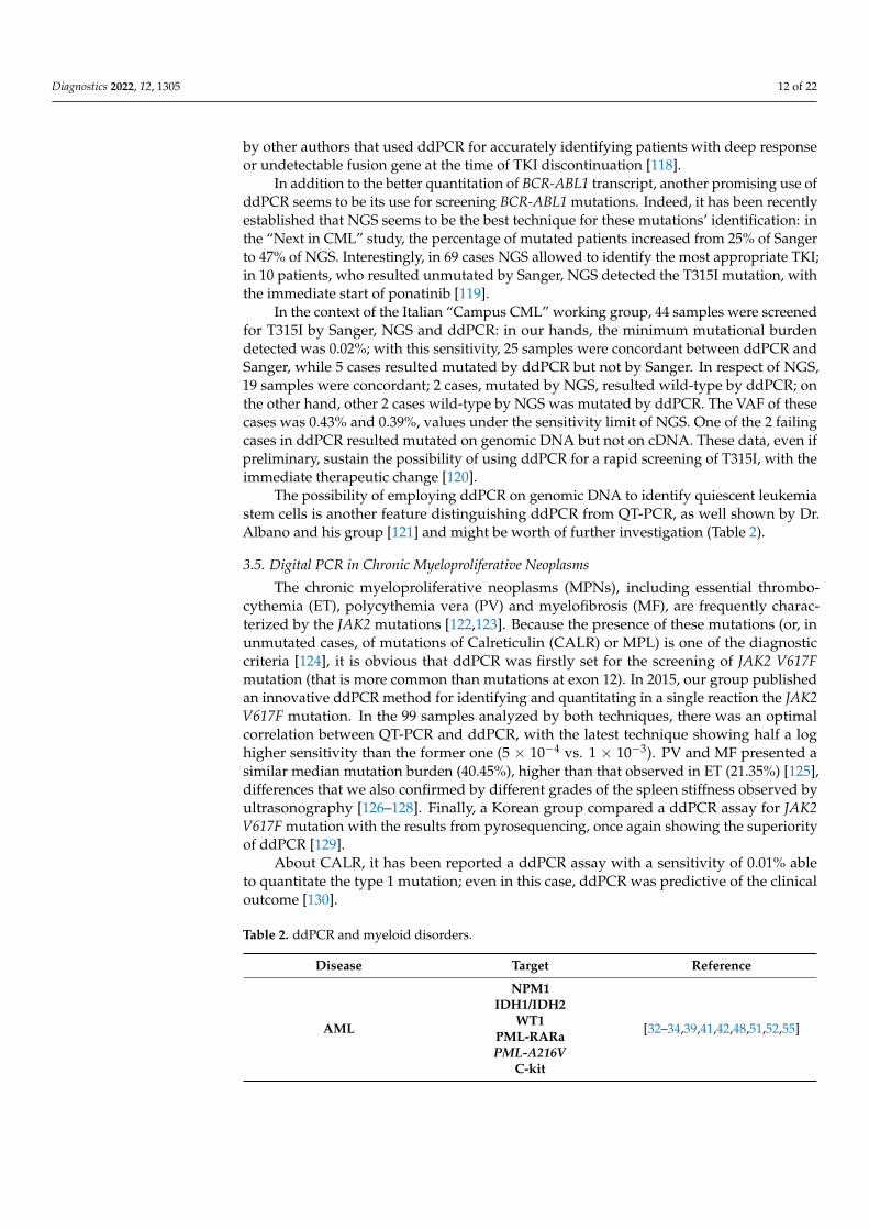
Table 2. ddPCR and myeloid disorders.
Disease Target Reference
AML
NPM1IDH1/IDH2
WT1PML-RARaPML-A216V
C-kit
[32–34,39,41,42,48,51,52,55]
Diagnostics 2022, 12, 1305 13 of 22
Table 2. Cont.
Disease Target Reference
CML BCR-ABL1T315I [108–113,119]
MDS Alu methylation [57]
MPNs JAK2CALR [123,124,127,129]
3.6. Digital PCR in Transplant and Immunoterapies
Allogeneic hematopoietic stem cell transplantation (AlloSCT) is a potentially curativetherapeutic option for several high-risk hematological malignancies (AML, ALL, MDS,lymphomas), especially if performed in CR. After AlloSCT the follow-up is principallybased on chimerism and, when possible, on disease specific MRD markers or persistence ofmutations: all these strategies allow to promptly detect and treat graft rejection or diseaserelapse [131]. Nevertheless, the correct timing, samples source—PB or BM—and techniquesfor chimerism evaluation as well as the exact threshold to distinguish complete donorchimerism from mixed chimerism are still matters of debate [132–134].
Currently, the standard methods to measure chimerism are QT-PCR-based analysisof Short Tandem Repeats (STR), with a sensitivity between 5% and 1%, according tothe diversity of donor/recipient fingerprint [135]. During the last few years, severalstudies tried to apply ddPCR to the chimerism assessment, even for levels <1% [136,137].One of the proposed strategies for children who underwent transplantation for primaryimmunodeficiency diseases included ddPCR for SRY and RPP30 genes that allowed detectthe male/female chimerism. This method revealed accurate and was able to analyze verysmall amount of genomic material (less than 10 ng) [137]. With a sensitivity of 8 × 10⁻⁵,the correlation between STR and ddPCR was higher than 99%, thus supporting the use ofddPCR also for the chimerism assessment [138].
In AlloSCT for malignancies when a suitable MRD marker is available, the betterclinical management could be obtained by integrating chimerism analysis with MRD moni-toring; Dr. Waterhouse et al. reported a combined use of ddPCR for chimerism and MRDin a series of 70 patients who underwent transplantation, mainly for myeloid malignancies.The authors reported a high concordance between mixed chimerism detection and MRDvalues, when NPM1, DNMT3A, MLL-PTD, IDH1 and KRAS were monitored [139].
In line with these results, a Japanese group assessed by ddPCR (sensitivity 1 × 10⁻⁵)the presence of T315I mutation in 25 patients with Ph’-positive ALL who underwentalloSCT. The hematological relapse was predicted by the persistence/reapperance of themutation after alloSCT, even at sub-clonal levels (median ratio T315I/ABL1 = 0.91%) [65].
Finally, a ddPCR assay was set for evaluating the immune reconstitution in MMpatients after autologous transplantation. Indeed, during TCR rearrangement, excisedDNA fragments create the TCR excision circles (TRECs) that have no clear functions butcan be used for determining the thymus activity and output [140]. Our group used anew ddPCR for measuring TRECs in 9 patients with MM who underwent autologoustransplantation and received high-dose zinc supplementation versus other 9 that did notreceive zinc. Interestingly, zinc supplementation supported the immune reconstitution:indeed, TRECs significantly increased from day +30 until day +100 only in the zinc group(6.1-fold vs 1.8 in the control group) [141].
Another innovative field of ddPCR application is represented by the immunotherapy,and in particular by the administration of CAR-T cells to patients affected by CD19+relapsed/refractory DLBCL, MCL or ALL [142]. Indeed, the shorter or longer persistenceof CAR-T seems to be predictive of success [143], whereas it is not still clear the impactof CAR-T persistence on adverse events, such as the cytokines release syndrome (CRS)or immune effector cell-associated neurotoxicity syndrome (ICANS) [144]. In 2020, two

Diagnostics 2022, 12, 1305 14 of 22
different German groups developed new ddPCR assays for monitoring patients receivingCAR-T. In the first work, published by Dr. Fehse and coworkers, starting from 120 ngof DNA the authors reached a sensitivity of 0.01%. Interestingly, the CAR-T expansionabove the median peak level of 11.2/mL was correlated with better clinical responses,whereas treatment was less effective in patients for whom CAR-T peaks were below themedian [145].
In the paper by Dr. Mika et al. detection and quantification of CAR-T were feasible inall patients, once again with a sensitivity of 1 × 10⁻⁴. As expected, significant differences inCAR-T expansion were observed: in 4 patients the initial CAR-T expansion was followedby decreasing numbers of copies; in the other 3, CAR-T were still detectable after 9 monthsfrom infusion and the CAR-T persistence and expansion were associated with better clinicalresponses; in this series, higher levels of CAR-T correlated also with ICANS but not withCRS occurrence [146].
A third group added to the ddPCR for quantitating CAR-T a ddPCR assay for mea-suring IL-6 gene expression. Differently from that expected, IL-6 gene levels were notpredictive for the development of CRS but might be useful for triggering tocilizumabtreatment at the first clinical signs of CRS. From the CAR-T expansion and clearancepoint of view, 4 different patterns have been described by these authors: that of rapidincrease and rapid decrease with complete disappearance of CAR-T, that rapid increaseand slow decrease with higher persistence, that of rapid increase and rapid decrease withlower persistence, and that of slow increase but rapid decrease with almost disappearance.Interestingly, patients assigned to the category “rapid increase and slow decrease withhigher persistence” seemed to have the best response rate, but also a higher risk of CRS,independently from the IL-6 gene expression [147] (Table 3).
Table 3. ddPCR and immunotherapies.
Disease Target Reference
ALLOGENEIC TRANSPLANTATIONChimerism
Chimerism & MRDT315I
[133,135–139]
AUTOLOGOUS TRANSPLANTATION TRECs [141]
IMMUNOTHERAPY CAR-T [145–147]
4. Conclusions
Born about 20 years ago, ddPCR is a new version of QT-PCR, more sensitive, specific,and accurate. With a LOD ranging from 10−4 and 10−5 according to different assays, ddPCRallows to quantitate about one quarter of samples already defined as PNQ by QT-PCR,so making more easily the patients’ management and follow-up. The versatility of thistechnique makes it available for measuring gene expression (without the need of a standardcurve or plasmids), but also for detecting single or multiple point mutations, either oncDNA but also on genomic DNA, both on bone marrow, peripheral blood or liquid biopsy.
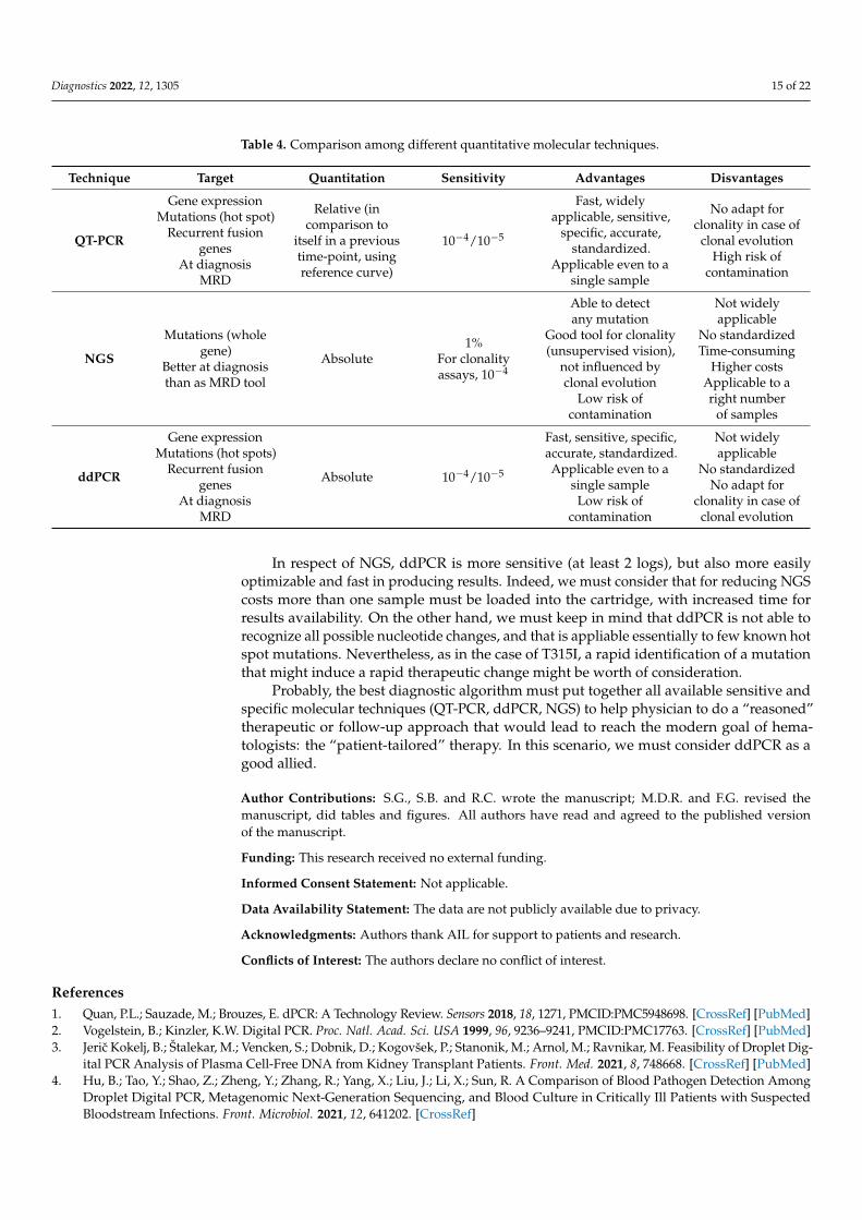
As above reported, many are the hematological contexts where ddPCR has been usedand implemented: acute leukemias, where it is able to quantitate NPM1 mutations but alsoWT1 expression; Ph’-positive leukemias, where it is used for measuring more accuratelythe BCR-ABL1/ABL1 ratio to also identify the patients best candidate to TFR but also forBCR-ABL1 mutations detection; the MPNs, where JAK2 and CALR mutations have a cleardiagnostic role, and the lymphoma/myeloma setting, where IgH and TCR clonality canbe combined with BCL2/JH and BCL1/JH fusion genes for assessing MRD. Finally, ddPCRcan be used for chimerism determination and for monitoring immune reconstitution andCAR-T persistence in patients who receive transplantation or the new immunotherapies(Table 4).
Diagnostics 2022, 12, 1305 15 of 22
Table 4. Comparison among different quantitative molecular techniques.
Technique Target Quantitation Sensitivity Advantages Disvantages
QT-PCR
Gene expressionMutations (hot spot)
Recurrent fusiongenes
At diagnosisMRD
Relative (incomparison to
itself in a previoustime-point, usingreference curve)
10−4/10−5
Fast, widelyapplicable, sensitive,
specific, accurate,standardized.
Applicable even to asingle sample
No adapt forclonality in case of
clonal evolutionHigh risk of
contamination
NGS
Mutations (wholegene)
Better at diagnosisthan as MRD tool
Absolute1%
For clonalityassays, 10−4
Able to detectany mutation
Good tool for clonality(unsupervised vision),
not influenced byclonal evolution
Low risk ofcontamination
Not widelyapplicable
No standardizedTime-consuming
Higher costsApplicable to aright number
of samples
ddPCR
Gene expressionMutations (hot spots)
Recurrent fusiongenes
At diagnosisMRD
Absolute 10−4/10−5
Fast, sensitive, specific,accurate, standardized.Applicable even to a
single sampleLow risk of
contamination
Not widelyapplicable
No standardizedNo adapt for
clonality in case ofclonal evolution
In respect of NGS, ddPCR is more sensitive (at least 2 logs), but also more easilyoptimizable and fast in producing results. Indeed, we must consider that for reducing NGScosts more than one sample must be loaded into the cartridge, with increased time forresults availability. On the other hand, we must keep in mind that ddPCR is not able torecognize all possible nucleotide changes, and that is appliable essentially to few known hotspot mutations. Nevertheless, as in the case of T315I, a rapid identification of a mutationthat might induce a rapid therapeutic change might be worth of consideration.
Probably, the best diagnostic algorithm must put together all available sensitive andspecific molecular techniques (QT-PCR, ddPCR, NGS) to help physician to do a “reasoned”therapeutic or follow-up approach that would lead to reach the modern goal of hema-tologists: the “patient-tailored” therapy. In this scenario, we must consider ddPCR as agood allied.
Author Contributions: S.G., S.B. and R.C. wrote the manuscript; M.D.R. and F.G. revised themanuscript, did tables and figures. All authors have read and agreed to the published versionof the manuscript.
Funding: This research received no external funding.
Informed Consent Statement: Not applicable.
Data Availability Statement: The data are not publicly available due to privacy.
Acknowledgments: Authors thank AIL for support to patients and research.
Conflicts of Interest: The authors declare no conflict of interest.
References1. Quan, P.L.; Sauzade, M.; Brouzes, E. dPCR: A Technology Review. Sensors 2018, 18, 1271, PMCID:PMC5948698. [CrossRef] [PubMed]2. Vogelstein, B.; Kinzler, K.W. Digital PCR. Proc. Natl. Acad. Sci. USA 1999, 96, 9236–9241, PMCID:PMC17763. [CrossRef] [PubMed]3. Jeric Kokelj, B.; Štalekar, M.; Vencken, S.; Dobnik, D.; Kogovšek, P.; Stanonik, M.; Arnol, M.; Ravnikar, M. Feasibility of Droplet Dig-
ital PCR Analysis of Plasma Cell-Free DNA from Kidney Transplant Patients. Front. Med. 2021, 8, 748668. [CrossRef] [PubMed]4. Hu, B.; Tao, Y.; Shao, Z.; Zheng, Y.; Zhang, R.; Yang, X.; Liu, J.; Li, X.; Sun, R. A Comparison of Blood Pathogen Detection Among
Droplet Digital PCR, Metagenomic Next-Generation Sequencing, and Blood Culture in Critically Ill Patients with SuspectedBloodstream Infections. Front. Microbiol. 2021, 12, 641202. [CrossRef]
Diagnostics 2022, 12, 1305 16 of 22
5. Yan, I.K.; Lohray, R.; Patel, T. Droplet Digital PCR for Quantitation of Extracellular RNA. Methods Mol. Biol. 2018, 1740,155–162. [CrossRef]
6. Cirillo, P.D.R.; Margiotti, K.; Mesoraca, A.; Giorlandino, C. Quantification of circulating microRNAs by droplet digital PCR forcancer detection. BMC Res. Notes 2020, 13, 351. [CrossRef]
7. Liu, C.; Shi, Q.; Peng, M.; Lu, R.; Li, H.; Cai, Y.; Chen, J.; Xu, J.; Shen, B. Evaluation of droplet digital PCR for quantification ofSARS-CoV-2 Virus in discharged COVID-19 patients. Aging 2020, 12, 20997–21003. [CrossRef]
8. Vasudevan, H.N.; Xu, P.; Servellita, V.; Miller, S.; Liu, L.; Gopez, A.; Chiu, C.Y.; Abate, A.R. Digital droplet PCR accuratelyquantifies SARS-CoV-2 viral load from crude lysate without nucleic acid purification. Sci. Rep. 2021, 11, 780. [CrossRef]
9. Drandi, D.; Kubiczkova-Besse, L.; Ferrero, S.; Dani, N.; Passera, R.; Mantoan, B.; Gambella, M.; Monitillo, L.; Saraci, E.; Ghione, P.;et al. Minimal Residual Disease Detection by Droplet Digital PCR in Multiple Myeloma, Mantle Cell Lymphoma, and FollicularLymphoma: A Comparison with Real-Time PCR. J. Mol. Diagn. 2015, 17, 652–660. [CrossRef]
10. Drandi, D.; Genuardi, E.; Dogliotti, I.; Ferrante, M.; Jiménez, C.; Guerrini, F.; Schirico, M.L.; Mantoan, B.; Muccio, V.; Lia, G.; et al.Highly sensitive MYD88L265P mutation detection by droplet digital polymerase chain reaction in Waldenström macroglobulinemia.Haematologica 2018, 103, 1029–1037. [CrossRef]
11. Della Starza, I.; Nunes, V.; Cavalli, M.; De Novi, L.A.; Ilari, C.; Apicella, V.; Vitale, A.; Testi, A.M.; Del Giudice, I.; Chiaretti, S.;et al. Comparative analysis between RQ-PCR and digital-droplet-PCR of immunoglobulin/T-cell receptor gene rearrangementsto monitor minimal residual disease in acute lymphoblastic leukaemia. Br. J. Haematol. 2016, 174, 541–549. [CrossRef] [PubMed]
12. Takamatsu, H. Comparison of Minimal Residual Disease Detection by Multiparameter Flow Cytometry, ASO-qPCR, DropletDigital PCR, and Deep Sequencing in Patients with Multiple Myeloma Who Underwent Autologous Stem Cell Transplantation. J.Clin. Med. 2017, 6, 91, Erratum in J. Clin. Med. 2017, 6, 106. [CrossRef] [PubMed]
13. dMIQE Group; Huggett, J.F. The Digital MIQE Guidelines Update: Minimum Information for Publication of Quantitative DigitalPCR Experiments for 2020. Clin. Chem. 2020, 66, 1012–1029, Erratum in Clin. Chem. 2020, 66, 1464. [CrossRef] [PubMed]
14. Taly, V.; Pekin, D.; Benhaim, L.; Kotsopoulos, S.K.; Le Corre, D.; Li, X.; Atochin, I.; Link, D.R.; Griffiths, A.D.; Pallier, K.; et al.Multiplex picodroplet digital PCR to detect KRAS mutations in circulating DNA from the plasma of colorectal cancer patients.Clin. Chem. 2013, 59, 1722–1731. [CrossRef] [PubMed]
15. Levy, C.N.; Hughes, S.M.; Roychoudhury, P.; Amstuz, C.; Zhu, H.; Huang, M.L.; Lehman, D.A.; Jerome, K.R.; Hladik, F.HIV reservoir quantification by five-target multiplex droplet digital PCR. STAR Protoc. 2021, 2, 100885, PMCID:PMC8517383.[CrossRef] [PubMed]
16. de Kock, R.; Baselmans, M.; Scharnhorst, V.; Deiman, B. Sensitive detection and quantification of SARS-CoV-2 by multiplexdroplet digital RT-PCR. Eur. J. Clin. Microbiol. Infect. Dis. 2021, 40, 807–813. [CrossRef]
17. Tan, C.; Chen, X.; Wang, F.; Wang, D.; Cao, Z.; Zhu, X.; Lu, C.; Yang, W.; Gao, N.; Gao, H.; et al. A multiplex droplet digital PCRassay for non-invasive prenatal testing of fetal aneuploidies. Analyst 2019, 144, 2239–2247. [CrossRef] [PubMed]
18. Corné, J.; Le Du, F.; Quillien, V.; Godey, F.; Robert, L.; Bourien, H.; Brunot, A.; Crouzet, L.; Perrin, C.; Lefeuvre-Plesse, C.; et al.Development of multiplex digital PCR assays for the detection of PIK3CA mutations in the plasma of metastatic breast cancerpatients. Sci. Rep. 2021, 11, 17316, PMCID:PMC8397758. [CrossRef] [PubMed]
19. Petiti, J.; Lo Iacono, M.; Dragani, M.; Pironi, L.; Fantino, C.; Rapanotti, M.C.; Quarantelli, F.; Izzo, B.; Divona, M.; Rege-Cambrin,G.; et al. Novel Multiplex Droplet Digital PCR Assays to Monitor Minimal Residual Disease in Chronic Myeloid LeukemiaPatients Showing Atypical BCR-ABL1 Transcripts. J. Clin. Med. 2020, 9, 1457. [CrossRef]
20. Bettelli, S.; Marcheselli, R.; Pozzi, S.; Marcheselli, L.; Papotti, R.; Forti, E.; Cox, M.C.C.; Di Napoli, A.; Tadmor, T.; Mansueto, G.R.;et al. Cell of origin (COO), BCL2/MYC status and IPI define a group of patients with Diffuse Large B-cell Lymphoma (DLBCL)with poor prognosis in a real-world clinical setting. Leuk. Res. 2021, 104, 106552. [CrossRef]
21. Galimberti, S.; Guerrini, F.; Volpe, G.; Grassi, S.; Ciabatti, E.; Forti, E.; Rapotti, R.; Bettelli, S.; Sacchi, S. A new digital PCR methodfor measuring the expression value of Polycomb genes in DLBCL. Haematologica 2022, 107, 35.
22. DiNardo, C.D.; Wei, A.H. How I treat acute myeloid leukemia in the era of new drugs. Blood 2020, 135, 85–96. [CrossRef] [PubMed]23. Lambert, J.; Pautas, C.; Terré, C.; Raffoux, E.; Turlure, P.; Caillot, D.; Legrand, O.; Thomas, X.; Gardin, C.; Gogat-Marchant, K.;
et al. Gemtuzumab ozogamicin for de novo acute myeloid leukemia: Final efficacy and safety updates from the open-label, phaseIII ALFA-0701 trial. Haematologica 2019, 104, 113–119. [CrossRef] [PubMed]
24. Stone, R.M.; Mandrekar, S.J.; Sanford, B.L.; Laumann, K.; Geyer, S.; Bloomfield, C.D.; Thiede, C.; Prior, T.W.; Döhner, K.; Marcucci,G.; et al. Midostaurin plus Chemotherapy for Acute Myeloid Leukemia with a FLT3 Mutation. N. Engl. J. Med. 2017, 377, 454–464.[CrossRef] [PubMed]
25. Perl, A.E.; Martinelli, G.; Cortes, J.E.; Neubauer, A.; Berman, E.; Paolini, S.; Montesinos, P.; Baer, M.R.; Larson, R.A.; Ustun,C.; et al. Gilteritinib or Chemotherapy for Relapsed or Refractory FLT3-Mutated AML. N. Engl. J. Med. 2019, 381, 1728–1740.[CrossRef] [PubMed]
26. DiNardo, C.D.; Stein, A.S.; Stein, E.M.; Fathi, A.T.; Frankfurt, O.; Schuh, A.C.; Döhner, H.; Martinelli, G.; Patel, P.A.; Raffoux,E.; et al. Mutant Isocitrate Dehydrogenase 1 Inhibitor Ivosidenib in Combination with Azacitidine for Newly Diagnosed AcuteMyeloid Leukemia. J. Clin. Oncol. 2021, 39, 57–65, Erratum in J. Clin. Oncol. 2021, 39, 341. [CrossRef]
27. Stein, E.M.; DiNardo, C.D.; Pollyea, D.A.; Fathi, A.T.; Roboz, G.J.; Altman, J.K.; Stone, R.M.; DeAngelo, D.J.; Levine, R.L.; Flinn,I.W.; et al. Enasidenib in mutant IDH2 relapsed or refractory acute myeloid leukemia. Blood 2017, 130, 722–731. [CrossRef]
Diagnostics 2022, 12, 1305 17 of 22
28. Arber, D.A.; Orazi, A.; Hasserjian, R.; Thiele, J.; Borowitz, M.J.; Le Beau, M.M.; Bloomfield, C.D.; Cazzola, M.; Vardiman, J.W.The 2016 revision to the World Health Organization classification of myeloid neoplasms and acute leukemia. Blood 2016, 127,2391–2405. [CrossRef] [PubMed]
29. Döhner, H.; Estey, E.; Grimwade, D.; Amadori, S.; Appelbaum, F.R.; Büchner, T.; Dombret, H.; Ebert, B.L.; Fenaux, P.; Larson, R.A.;et al. Diagnosis and management of AML in adults: 2017 ELN recommendations from an international expert panel. Blood 2017,129, 424–447. [CrossRef]
30. Schuurhuis, G.J.; Heuser, M.; Freeman, S.; Béné, M.C.; Buccisano, F.; Cloos, J.; Grimwade, D.; Haferlach, T.; Hills, R.K.; Hourigan,C.S.; et al. Minimal/measurable residual disease in AML: A consensus document from the European LeukemiaNet MRD WorkingParty. Blood 2018, 131, 1275–1291. [CrossRef]
31. Sanz, M.A.; Fenaux, P.; Tallman, M.S.; Estey, E.H.; Löwenberg, B.; Naoe, T.; Lengfelder, E.; Döhner, H.; Burnett, A.K.; Chen,S.J.; et al. Management of acute promyelocytic leukemia: Updated recommendations from an expert panel of the EuropeanLeukemiaNet. Blood 2019, 133, 1630–1643. [CrossRef] [PubMed]
32. Yuan, D.; Cui, M.; Yu, S.; Wang, H.; Jing, R. Droplet digital PCR for quantification of PML-RARα in acute promyelocytic leukemia:A comprehensive comparison with real-time PCR. Anal. Bioanal. Chem. 2019, 411, 895–903. [CrossRef] [PubMed]
33. Jiang, X.W.; Chen, S.Z.; Zhu, X.Y.; Xu, X.X.; Liu, Y. Development and validation of a droplet digital PCR assay for the evaluationof PML-RARα fusion transcripts in acute promyelocytic leukemia. Mol. Cell Probes. 2020, 53, 101617. [CrossRef]
34. Alfonso, V.; Iaccarino, L.; Ottone, T.; Cicconi, L.; Lavorgna, S.; Divona, M.; Cairoli, R.; Cristiano, A.; Ciardi, C.; Travaglini, S.; et al.Early and sensitive detection of PML-A216V mutation by droplet digital PCR in ATO-resistant acute promyelocytic leukemia.Leukemia 2019, 33, 1527–1530. [CrossRef]
35. Handschuh, L.; Kazmierczak, M.; Milewski, M.C.; Góralski, M.; Łuczak, M.; Wojtaszewska, M.; Uszczynska-Ratajczak, B.;Lewandowski, K.; Komarnicki, M.; Figlerowicz, M. Gene expression profiling of acute myeloid leukemia samples from adultpatients with AML-M1 and -M2 through boutique microarrays, real-time PCR and droplet digital PCR. Int. J. Oncol. 2018, 52,656–678. [CrossRef] [PubMed]
36. Chen, X.; Zong, S.; Yi, M.; Liu, C.; Wang, B.; Duan, Y.; Cheng, X.; Ruan, M.; Zhang, L.; Zou, Y.; et al. Minimal residualdisease monitoring via AML1-ETO breakpoint tracing in childhood acute myeloid leukemia. Transl. Oncol. 2021, 14, 101119.[CrossRef] [PubMed]
37. Forghieri, F.; Comoli, P.; Marasca, R.; Potenza, L.; Luppi, M. Minimal/Measurable Residual Disease Monitoring in NPM1-MutatedAcute Myeloid Leukemia: A Clinical Viewpoint and Perspectives. Int. J. Mol. Sci. 2018, 19, 3492. [CrossRef]
38. Gorello, P.; Cazzaniga, G.; Alberti, F.; Dell’Oro, M.G.; Gottardi, E.; Specchia, G.; Roti, G.; Rosati, R.; Martelli, M.F.; Diverio, D.;et al. Quantitative assessment of minimal residual disease in acute myeloid leukemia carrying nucleophosmin (NPM1) genemutations. Leukemia 2006, 20, 1103–1108. [CrossRef]
39. Mencia-Trinchant, N.; Hu, Y.; Alas, M.A.; Ali, F.; Wouters, B.J.; Lee, S.; Ritchie, E.K.; Desai, P.; Guzman, M.L.; Roboz, G.J.; et al.Minimal Residual Disease Monitoring of Acute Myeloid Leukemia by Massively Multiplex Digital PCR in Patients with NPM1Mutations. J. Mol. Diagn. 2017, 19, 537–548. [CrossRef]
40. Pettersson, L.; Johansson Alm, S.; Almstedt, A.; Chen, Y.; Orrsjö, G.; Shah-Barkhordar, G.; Zhou, L.; Kotarsky, H.; Vidovic, K.;Asp, J.; et al. Comparison of RNA- and DNA-based methods for measurable residual disease analysis in NPM1-mutated acutemyeloid leukemia. Int. J. Lab. Hematol. 2021, 43, 664–674. [CrossRef] [PubMed]
41. Bill, M.; Grimm, J.; Jentzsch, M.; Kloss, L.; Goldmann, K.; Schulz, J.; Beinicke, S.; Häntschel, J.; Cross, M.; Vucinic, V.; et al. Digitaldroplet PCR-based absolute quantification of pre-transplant NPM1 mutation burden predicts relapse in acute myeloid leukemiapatients. Ann. Hematol. 2018, 97, 1757–1765. [CrossRef] [PubMed]
42. Brambati, C.; Galbiati, S.; Xue, E.; Toffalori, C.; Crucitti, L.; Greco, R.; Sala, E.; Crippa, A.; Chiesa, L.; Soriani, N.; et al. Dropletdigital polymerase chain reaction for DNMT3A and IDH1/2 mutations to improve early detection of acute myeloid leukemiarelapse after allogeneic hematopoietic stem cell transplantation. Haematologica 2016, 101, e157–e161. [CrossRef] [PubMed]
43. Liu, X.; Gong, Y. Isocitrate dehydrogenase inhibitors in acute myeloid leukemia. Biomark. Res. 2019, 7, 22. [CrossRef] [PubMed]44. McMurry, H.; Fletcher, L.; Traer, E. IDH Inhibitors in AML-Promise and Pitfalls. Curr. Hematol. Malig. Rep. 2021, 16,
207–217. [CrossRef]45. Venugopal, S.; Takahashi, K.; Daver, N.; Maiti, A.; Borthakur, G.; Loghavi, S.; Short, N.J.; Ohanian, M.; Masarova, L.; Issa, G.; et al.
Efficacy and safety of enasidenib and azacitidine combination in patients with IDH2 mutated acute myeloid leukemia and noteligible for intensive chemotherapy. Blood Cancer J. 2022, 12, 10. [CrossRef]
46. Stein, E.M.; DiNardo, C.D.; Fathi, A.T.; Mims, A.S.; Pratz, K.W.; Savona, M.R.; Stein, A.S.; Stone, R.M.; Winer, E.S.; Seet, C.S.; et al.Ivosidenib or enasidenib combined with intensive chemotherapy in patients with newly diagnosed AML: A phase 1 study. Blood2021, 137, 1792–1803, PMCID:PMC8020270. [CrossRef] [PubMed]
47. Petrova, L.; Vrbacky, F.; Lanska, M.; Zavrelova, A.; Zak, P.; Hrochova, K. IDH1 and IDH2 mutations in patients with acute myeloidleukemia: Suitable targets for minimal residual disease monitoring? Clin. Biochem. 2018, 61, 34–39, Erratum in Clin Biochem. 2019,63, 161. [CrossRef] [PubMed]
48. Grassi, S.; Guerrini, F.; Ciabatti, E.; Puccetti, R.; Salehzadeh, S.; Metelli, M.R.; Di Vita, A.; Domenichini, C.; Caracciolo, F.; Orciuolo,E.; et al. Digital Droplet PCR is a Specific and Sensitive Tool for Detecting IDH2 Mutations in Acute Myeloid LeuKemia Patients.Cancers 2020, 12, 1738. [CrossRef]
Diagnostics 2022, 12, 1305 18 of 22
49. Ayatollahi, H.; Shajiei, A.; Sadeghian, M.H.; Sheikhi, M.; Yazdandoust, E.; Ghazanfarpour, M.; Shams, S.F.; Shakeri, S. PrognosticImportance of C-KIT Mutations in Core Binding Factor Acute Myeloid Leukemia: A Systematic Review. Hematol. Oncol. Stem CellTher. 2017, 10, 1–7. [CrossRef] [PubMed]
50. Pardanani, A. Systemic mastocytosis in adults: 2021 Update on diagnosis, risk stratification and management. Am. J. Hematol.2021, 96, 508–525. [CrossRef]
51. Tan, Y.; Liu, Z.; Wang, W.; Zhu, G.; Guo, J.; Chen, X.; Zheng, C.; Xu, Z.; Chang, J.; Ren, F.; et al. Monitoring of clonal evolution ofdouble C-KIT exon 17 mutations by Droplet Digital PCR in patients with core-binding factor acute myeloid leukemia. Leuk. Res.2018, 69, 89–93. [CrossRef] [PubMed]
52. Sasaki, K.; Tsujimoto, S.; Miyake, M.; Uchiyama, Y.; Ikeda, J.; Yoshitomi, M.; Shimosato, Y.; Tokumasu, M.; Matsuo, H.; Yoshida,K.; et al. Droplet digital polymerase chain reaction assay for the detection of the minor clone of KIT D816V in paediatric acutemyeloid leukaemia especially showing RUNX1-RUNX1T1 transcripts. Br. J. Haematol. 2021, 194, 414–422. [CrossRef] [PubMed]
53. Pettersson, L.; Chen, Y.; George, A.M.; Rigo, R.; Lazarevic, V.; Juliusson, G.; Saal, L.H.; Ehinger, M. Subclonal patterns infollow-up of acute myeloid leukemia combining whole exome sequencing and ultrasensitive IBSAFE digital droplet analysis.Leuk. Lymphoma. 2020, 61, 2168–2179. [CrossRef] [PubMed]
54. Bussaglia, E.; Pratcorona, M.; Carricondo, M.; Sansegundo, L.; Rubio, M.A.; Monter, A.; Brell, A.; Badell, I.; Esteve, J.; Arnan, M.;et al. Application of a digital PCR method for WT1 to myeloid neoplasms in CR and deep ELN WT1 molecular response (<10copies). Ann. Hematol. 2020, 99, 765–772. [CrossRef]
55. Koizumi, Y.; Furuya, D.; Endo, T.; Asanuma, K.; Yanagihara, N.; Takahashi, S. Quantification of Wilms’ tumor 1 mRNA by digitalpolymerase chain reaction. Int. J. Hematol. 2018, 107, 230–234. [CrossRef]
56. Yu, M.; Heinzerling, T.J.; Grady, W.M. DNA Methylation Analysis Using Droplet Digital PCR. Methods Mol. Biol. 2018, 1768,363–383. [CrossRef]
57. Orsini, P.; Impera, L.; Parciante, E.; Cumbo, C.; Minervini, C.F.; Minervini, A.; Zagaria, A.; Anelli, L.; Coccaro, N.; Casieri, P.;et al. Droplet digital PCR for the quantification of Alu methylation status in hematological malignancies. Diagn. Pathol. 2018,13, 98. [CrossRef]
58. Marks, D.I.; Rowntree, C. Management of adults with T-cell lymphoblastic leukemia. Blood 2017, 129, 1134–1142, Erratum in Blood2017, 129, 2204. [CrossRef]
59. Hein, K.; Short, N.; Jabbour, E.; Yilmaz, M. Clinical Value of Measurable Residual Disease in Acute Lymphoblastic Leukemia.Blood Lymphat. Cancer 2022, 12, 7–16. [CrossRef]
60. Ansuinelli, M.; Della Starza, I.; Lauretti, A.; Elia, L.; Siravo, V.; Messina, M.; De Novi, L.A.; Taherinasab, A.; Canichella,M.; Guarini, A.; et al. Applicability of droplet digital polymerase chain reaction for minimal residual disease monitoring inPhiladelphia-positive acute lymphoblastic leukaemia. Hematol. Oncol. 2021, 39, 680–686. [CrossRef]
61. Coccaro, N.; Anelli, L.; Zagaria, A.; Casieri, P.; Tota, G.; Orsini, P.; Impera, L.; Minervini, A.; Minervini, C.F.; Cumbo, C.;et al. Droplet Digital PCR Is a Robust Tool for Monitoring Minimal Residual Disease in Adult Philadelphia-Positive AcuteLymphoblastic Leukemia. J. Mol. Diagn. 2018, 20, 474–482. [CrossRef] [PubMed]
62. Guan, Y.; Zhang, M.; Zhang, W.; Wang, J.; Shen, K.; Zhang, K.; Yang, L.; Huang, L.; Wang, N.; Xiao, M.; et al. Clinical Utility ofDroplet Digital PCR to Monitor BCR-ABL1 Transcripts of Patients with Philadelphia Chromosome-Positive Acute LymphoblasticLeukemia Post-chimeric Antigen Receptor19/22 T-Cell Cocktail Therapy. Front. Oncol. 2021, 11, 646499. [CrossRef] [PubMed]
63. Martinez, R.J.; Kang, Q.; Nennig, D.; Bailey, N.G.; Brown, N.A.; Betz, B.L.; Tewari, M.; Thyagarajan, B.; Bachanova, V.; Mroz, P.One-Step Multiplexed Droplet Digital Polymerase Chain Reaction for Quantification of p190 BCR-ABL1 Fusion Transcript inB-Lymphoblastic Leukemia. Arch. Pathol. Lab. Med. 2022, 146, 92–100. [CrossRef] [PubMed]
64. Soverini, S.; Vitale, A.; Poerio, A.; Gnani, A.; Colarossi, S.; Iacobucci, I.; Cimino, G.; Elia, L.; Lonetti, A.; Vignetti, M.; et al.Philadelphia-positive acute lymphoblastic leukemia patients already harbor BCR-ABL kinase domain mutations at low levels atthe time of diagnosis. Haematologica 2011, 96, 552–557. [CrossRef]
65. Akahoshi, Y.; Nakasone, H.; Kawamura, K.; Kusuda, M.; Kawamura, S.; Takeshita, J.; Yoshino, N.; Misaki, Y.; Yoshimura, K.;Gomyo, A.; et al. Detection of T315I using digital polymerase chain reaction in allogeneic transplant recipients with Ph-positiveacute lymphoblastic anemia in the dasatinib era. Exp. Hematol. 2020, 81, 60–67, Erratum in Exp. Hematol. 2020, 85, 71. [CrossRef]
66. Wang, W.; Tang, G.; Kadia, T.; Lu, X.; Li, Y.; Huang, L.; Montenegro-Garreaud, X.; Miranda, R.N.; Wang, S.A. CytogeneticEvolution Associated With Disease Progression in Hematopoietic Neoplasms With t(8;22)(p11;q11)/BCR-FGFR1 Rearrangement.J. Natl. Compr. Cancer Netw. 2016, 14, 708–711. [CrossRef]
67. Coccaro, N.; Tota, G.; Zagaria, A.; Anelli, L.; Casieri, P.; Impera, L.; Minervini, A.; Minervini, C.F.; Orsini, P.; Cumbo, C.; et al.Monitoring minimal residual disease by ddPCR in acute lymphoblastic leukemia associated with the FGFR1 gene rearrangement.Int. J. Lab. Hematol. 2018, 40, e117–e120. [CrossRef]
68. van der Velden, V.H.; Cazzaniga, G.; Schrauder, A.; Hancock, J.; Bader, P.; Panzer-Grumayer, E.R.; Flohr, T.; Sutton, R.; Cave,H.; Madsen, H.O.; et al. Analysis of minimal residual disease by Ig/TCR gene rearrangements: Guidelines for interpretation ofreal-time quantitative PCR data. Leukemia 2007, 21, 604–611. [CrossRef]
69. Della Starza, I.; Nunes, V.; Lovisa, F.; Silvestri, D.; Cavalli, M.; Garofalo, A.; Campeggio, M.; De Novi, L.A.; Soscia, R.; Oggioni, C.;et al. Droplet Digital PCR Improves IG-/TR-based MRD Risk Definition in Childhood B-cell Precursor Acute LymphoblasticLeukemia. Hemasphere 2021, 5, e543. [CrossRef]
70. Medeiros, B.C. Deletion of IKZF1 and prognosis in acute lymphoblastic leukemia. N. Engl. J. Med. 2009, 360, 1787. [CrossRef]
Diagnostics 2022, 12, 1305 19 of 22
71. Martinelli, G.; Iacobucci, I.; Storlazzi, C.T.; Vignetti, M.; Paoloni, F.; Cilloni, D.; Soverini, S.; Vitale, A.; Chiaretti, S.; Cimino, G.; et al.IKZF1 (Ikaros) deletions in BCR-ABL1-positive acute lymphoblastic leukemia are associated with short disease-free survival andhigh rate of cumulative incidence of relapse: A GIMEMA AL WP report. J. Clin. Oncol. 2009, 27, 5202–5207. [CrossRef] [PubMed]
72. Hashiguchi, J.; Onozawa, M.; Okada, K.; Amano, T.; Hatanaka, K.C.; Nishihara, H.; Sato, N.; Teshima, T. Quantitative detectionof IKZF1 deletion by digital PCR in patients with acute lymphoblastic leukemia. Int. J. Lab. Hematol. 2019, 41, e38–e40.[CrossRef] [PubMed]
73. Maitre, E.; Cornet, E.; Troussard, X. Hairy cell leukemia: 2020 update on diagnosis, risk stratification, and treatment. Am. J.Hematol. 2019, 94, 1413–1422. [CrossRef] [PubMed]
74. Rodriguez-Galindo, C.; Allen, C.E. Langerhans cell histiocytosis. Blood 2020, 135, 1319–1331. [CrossRef]75. Chapman, P.B.; Hauschild, A.; Robert, C.; Haanen, J.B.; Ascierto, P.; Larkin, J.; Dummer, R.; Garbe, C.; Testori, A.; Maio, M.; et al.
Improved survival with vemurafenib in melanoma with BRAF V600E mutation. N. Engl. J. Med. 2011, 364, 2507–2516. [CrossRef]76. Mazieres, J.; Cropet, C.; Montané, L.; Barlesi, F.; Souquet, P.J.; Quantin, X.; Dubos-Arvis, C.; Otto, J.; Favier, L.; Avrillon, V.;
et al. Vemurafenib in non-small-cell lung cancer patients with BRAFV600 and BRAFnonV600 mutations. Ann. Oncol. 2020, 31,289–294. [CrossRef]
77. Diamond, E.L.; Subbiah, V.; Lockhart, A.C.; Blay, J.Y.; Puzanov, I.; Chau, I.; Raje, N.S.; Wolf, J.; Erinjeri, J.P.; Torrisi, J.; et al.Vemurafenib for BRAF V600-Mutant Erdheim-Chester Disease and Langerhans Cell Histiocytosis: Analysis of Data from theHistology-Independent, Phase 2, Open-label VE-BASKET Study. JAMA Oncol. 2018, 4, 384–388, Erratum in JAMA Oncol. 2019,5, 122. [CrossRef]
78. Tiacci, E.; De Carolis, L.; Simonetti, E.; Capponi, M.; Ambrosetti, A.; Lucia, E.; Antolino, A.; Pulsoni, A.; Ferrari, S.;Zinzani, P.L.; et al. Vemurafenib plus Rituximab in Refractory or Relapsed Hairy-Cell Leukemia. N. Engl. J. Med. 2021, 384,1810–1823. [CrossRef]
79. Guerrini, F.; Paolicchi, M.; Ghio, F.; Ciabatti, E.; Grassi, S.; Salehzadeh, S.; Ercolano, G.; Metelli, M.R.; Del Re, M.; Iovino, L.;et al. The Droplet Digital PCR: A New Valid Molecular Approach for the Assessment of B-RAF V600E Mutation in Hairy CellLeukemia. Front. Pharmacol. 2016, 7, 363. [CrossRef]
80. Dong, L.; Wang, X.; Wang, S.; Du, M.; Niu, C.; Yang, J.; Li, L.; Zhang, G.; Fu, B.; Gao, Y.; et al. Interlaboratory assess-ment of droplet digital PCR for quantification of BRAF V600E mutation using a novel DNA reference material. Talanta 2020,207, 120293. [CrossRef]
81. Hallek, M.; Cheson, B.D.; Catovsky, D.; Caligaris-Cappio, F.; Dighiero, G.; Döhner, H.; Hillmen, P.; Keating, M.; Montserrat, E.;Chiorazzi, N.; et al. iwCLL guidelines for diagnosis, indications for treatment, response assessment, and supportive managementof CLL. Blood 2018, 131, 2745–2760. [CrossRef] [PubMed]
82. Frazzi, R.; Bizzarri, V.; Albertazzi, L.; Cusenza, V.Y.; Coppolecchia, L.; Luminari, S.; Ilariucci, F. Droplet digital PCR is a sensitivetool for the detection of TP53 deletions and point mutations in chronic lymphocytic leukaemia. Br. J. Haematol. 2020, 189, e49–e52.[CrossRef] [PubMed]
83. Stilgenbauer, S.; Schnaiter, A.; Paschka, P.; Zenz, T.; Rossi, M.; Döhner, K.; Bühler, A.; Böttcher, S.; Ritgen, M.; Kneba, M.; et al.Gene mutations and treatment outcome in chronic lymphocytic leukemia: Results from the CLL8 trial. Blood 2014, 123, 3247–3254.[CrossRef] [PubMed]
84. Minervini, A.; Francesco Minervini, C.; Anelli, L.; Zagaria, A.; Casieri, P.; Coccaro, N.; Cumbo, C.; Tota, G.; Impera, L.;Orsini, P.; et al. Droplet digital PCR analysis of NOTCH1 gene mutations in chronic lymphocytic leukemia. Oncotarget 2016, 7,86469–86479. [CrossRef]
85. Hoofd, C.; Huang, S.J.; Gusscott, S.; Lam, S.; Wong, R.; Johnston, A.; Ben-Neriah, S.; Steidl, C.; Scott, D.W.; Bruyere, H.; et al.Ultrasensitive Detection of NOTCH1 c.7544_7545delCT Mutations in Chronic Lymphocytic Leukemia by Droplet Digital PCRReveals High Frequency of Subclonal Mutations and Predicts Clinical Outcome in Cases with Trisomy 12. J. Mol. Diagn. 2020, 22,571–578. [CrossRef]
86. Gertz, M.A. Waldenström macroglobulinemia: 2021 update on diagnosis, risk stratification, and management. Am. J. Hematol.2021, 96, 258–269. [CrossRef]
87. Yu, X.; Li, W.; Deng, Q.; Li, L.; Hsi, E.D.; Young, K.H.; Zhang, M.; Li, Y. MYD88 L265P Mutation in Lymphoid Malignancies.Cancer Res. 2018, 78, 2457–2462. [CrossRef]
88. Ferrante, M.; Furlan, D.; Zibellini, S.; Borriero, M.; Candido, C.; Sahnane, N.; Uccella, S.; Genuardi, E.; Alessandria, B.; Bianchi, B.;et al. MYD88L265P Detection in IgM Monoclonal Gammopathies: Methodological Considerations for Routine Implementation.Diagnostics 2021, 11, 779. [CrossRef]
89. Chen, K.; Ma, Y.; Ding, T.; Zhang, X.; Chen, B.; Guan, M. Effectiveness of digital PCR for MYD88L265P detection in vitreous fluidfor primary central nervous system lymphoma diagnosis. Exp. Ther. Med. 2020, 20, 301–308. [CrossRef]
90. Willenbacher, E.; Willenbacher, W.; Wolf, D.G.; Zelger, B.; Peschel, I.; Manzl, C.; Haun, M.; Brunner, A. Digital PCR in bone marrowtrephine biopsies is highly sensitive for MYD88L265P detection in lymphomas with plasmacytic/plasmacytoid differentiation. Br.J. Haematol. 2019, 186, 189–191. [CrossRef]
91. Alcaide, M.; Yu, S.; Bushell, K.; Fornika, D.; Nielsen, J.S.; Nelson, B.H.; Mann, K.K.; Assouline, S.; Johnson, N.A.; Morin, R.D.Multiplex Droplet Digital PCR Quantification of Recurrent Somatic Mutations in Diffuse Large B-Cell and Follicular Lymphoma.Clin Chem. 2016, 62, 1238–1247. [CrossRef] [PubMed]
Diagnostics 2022, 12, 1305 20 of 22
92. Galimberti, S.; Luminari, S.; Ciabatti, E.; Grassi, S.; Guerrini, F.; Dondi, A.; Marcheselli, L.; Ladetto, M.; Piccaluga, P.P.; Gazzola,A.; et al. Minimal residual disease after conventional treatment significantly impacts on progression-free survival of patients withfollicular lymphoma: The FIL FOLL05 trial. Clin. Cancer Res. 2014, 20, 6398–6405. [CrossRef] [PubMed]
93. von Keudell, G.; Salles, G. The role of tazemetostat in relapsed/refractory follicular lymphoma. Ther. Adv. Hematol. 2021, 12,20406207211015882. [CrossRef] [PubMed]
94. Nagy, Á.; Bátai, B.; Balogh, A.; Illés, S.; Mikala, G.; Nagy, N.; Kiss, L.; Kotmayer, L.; Matolcsy, A.; Alpár, D.; et al. Quantitative Anal-ysis and Monitoring of EZH2 Mutations Using Liquid Biopsy in Follicular Lymphoma. Genes 2020, 11, 785. [CrossRef] [PubMed]
95. Cavalli, M.; De Novi, L.A.; Della Starza, I.; Cappelli, L.V.; Nunes, V.; Pulsoni, A.; Del Giudice, I.; Guarini, A.; Foà, R. Comparativeanalysis between RQ-PCR and digital droplet PCR of BCL2/IGH gene rearrangement in the peripheral blood and bone marrowof early stage follicular lymphoma. Br. J. Haematol. 2017, 177, 588–596. [CrossRef]
96. Novella, E.; Giaretta, I.; Felice, F.; Madeo, D.; Piccin, A.; Castaman, G.; Rodeghiero, F. Fluorescent polymerase chain reaction andcapillary electrophoresis for IgH rearrangement and minimal residual disease evaluation in multiple myeloma. Haematologica2002, 87, 1157–1164.
97. Drandi, D.; Alcantara, M.; Benmaad, I.; Söhlbrandt, A.; Lhermitte, L.; Zaccaria, G.; Ferrante, M.; Genuardi, E.; Mantoan, B.;Villarese, P.; et al. Droplet Digital PCR Quantification of Mantle Cell Lymphoma Follow-up Samples from Four Prospective Trialsof the European MCL Network. Hemasphere 2020, 4, e347. [CrossRef]
98. Tanzima Nuhat, S.; Sakata-Yanagimoto, M.; Komori, D.; Hattori, K.; Suehara, Y.; Fukumoto, K.; Fujisawa, M.; Kusakabe, M.; Mat-sue, K.; Wakamatsu, H.; et al. Droplet digital polymerase chain reaction assay and peptide nucleic acid-locked nucleic acid clampmethod for RHOA mutation detection in angioimmunoblastic T-cell lymphoma. Cancer Sci. 2018, 109, 1682–1689. [CrossRef]
99. Bessi, L.; Viailly, P.J.; Bohers, E.; Ruminy, P.; Maingonnat, C.; Bertrand, P.; Vasseur, N.; Beaussire, L.; Cornic, M.; Etancelin, P.;et al. Somatic mutations of cell-free circulating DNA detected by targeted next-generation sequencing and digital droplet PCR inclassical Hodgkin lymphoma. Leuk. Lymphoma. 2019, 60, 498–502. [CrossRef]
100. Bertamini, L.; D’Agostino, M.; Gay, F. MRD Assessment in Multiple Myeloma: Progress and Challenges. Curr. Hematol. Malig.Rep. 2021, 16, 162–171. [CrossRef] [PubMed]
101. Takamatsu, H.; Wee, R.K.; Zaimoku, Y.; Murata, R.; Zheng, J.; Moorhead, M.; Carlton, V.E.H.; Kong, K.A.; Takezako, N.; Ito, S.; et al.A comparison of minimal residual disease detection in autografts among ASO-qPCR, droplet digital PCR, and next-generationsequencing in patients with multiple myeloma who underwent autologous stem cell transplantation. Br. J. Haematol. 2018, 183,664–668. [CrossRef] [PubMed]
102. Jabbour, E.; Kantarjian, H. Chronic myeloid leukemia: 2020 update on diagnosis, therapy and monitoring. Am. J. Hematol. 2020,95, 691–709. [CrossRef] [PubMed]
103. Smith, G.; Apperley, J.; Milojkovic, D.; Cross, N.C.P.; Foroni, L.; Byrne, J.; Goringe, A.; Rao, A.; Khorashad, J.; de Lavallade, H.;et al. A British Society for Haematology Guideline on the diagnosis and management of chronic myeloid leukaemia. Br. J.Haematol. 2020, 191, 171–193. [CrossRef] [PubMed]
104. Hochhaus, A.; Baccarani, M.; Silver, R.T.; Schiffer, C.; Apperley, J.F.; Cervantes, F.; Clark, R.E.; Cortes, J.E.; Deininger, M.W.;Guilhot, F.; et al. European LeukemiaNet 2020 recommendations for treating chronic myeloid leukemia. Leukemia 2020, 34,966–984. [CrossRef] [PubMed]
105. Saussele, S.; Richter, J.; Guilhot, J.; Gruber, F.X.; Hjorth-Hansen, H.; Almeida, A.; Janssen, J.J.W.M.; Mayer, J.; Koskenvesa,P.; Panayiotidis, P.; et al. Discontinuation of tyrosine kinase inhibitor therapy in chronic myeloid leukaemia (EURO-SKI): Aprespecified interim analysis of a prospective, multicentre, non-randomised, trial. Lancet Oncol. 2018, 19, 747–757. [CrossRef]
106. Atallah, E.; Sweet, K. Treatment-Free Remission: The New Goal in CML Therapy. Curr. Hematol. Malig. Rep. 2021, 16, 433–439.[CrossRef] [PubMed]
107. Baccarani, M.; Abruzzese, E.; Accurso, V.; Albano, F.; Annunziata, M.; Barulli, S.; Beltrami, G.; Bergamaschi, M.; Binotto, G.;Bocchia, M.; et al. Managing chronic myeloid leukemia for treatment-free remission: A proposal from the GIMEMA CML WP.Blood Adv. 2019, 3, 4280–4290. [CrossRef]
108. Dragani, M.; Rege Cambrin, G.; Berchialla, P.; Dogliotti, I.; Rosti, G.; Castagnetti, F.; Capodanno, I.; Martino, B.; Cerrano, M.;Ferrero, D.; et al. A Retrospective Analysis about Frequency of Monitoring in Italian Chronic Myeloid Leukemia Patients afterDiscontinuation. J. Clin. Med. 2020, 9, 3692. [CrossRef]
109. Chung, H.J.; Hur, M.; Yoon, S.; Hwang, K.; Lim, H.S.; Kim, H.; Moon, H.W.; Yun, Y.M. Performance Evaluation of the QXDxBCR-ABL %IS Droplet Digital PCR Assay. Ann. Lab. Med. 2020, 40, 72–75. [CrossRef]
110. Wang, W.J.; Zheng, C.F.; Liu, Z.; Tan, Y.H.; Chen, X.H.; Zhao, B.L.; Li, G.X.; Xu, Z.F.; Ren, F.G.; Zhang, Y.F.; et al. Droplet digitalPCR for BCR/ABL(P210) detection of chronic myeloid leukemia: A high sensitive method of the minimal residual disease anddisease progression. Eur. J. Haematol. 2018, 101, 291–296. [CrossRef]
111. Franke, G.N.; Maier, J.; Wildenberger, K.; Cross, M.; Giles, F.J.; Müller, M.C.; Hochhaus, A.; Niederwieser, D.; Lange, T. Comparisonof Real-Time Quantitative PCR and Digital Droplet PCR for BCR-ABL1 Monitoring in Patients with Chronic Myeloid Leukemia.J. Mol. Diagn. 2020, 22, 81–89. [CrossRef] [PubMed]
112. Cortés, A.A.; Olmedillas, S.; Serrano-López, J.; Lainez-González, D.; Castaño, T.; Iñiguez, R.; Lopez-Lorenzo, J.L.; García, A.;Atance, M.; Sánchez, R.N.S.; et al. Comparison of Droplet Digital PCR versus qPCR Measurements on the International Scale forthe Molecular Monitoring of Chronic Myeloid Leukemia Patients. Mol. Diagn. Ther. 2020, 24, 593–600. [CrossRef] [PubMed]
Diagnostics 2022, 12, 1305 21 of 22
113. Fava, C.; Bernardi, S.; Gottardi, E.M.; Lorenzatti, R.; Galeotti, L.; Ceccherini, F.; Cordoni, F.; Daraio, F.; Giugliano, E.; Jovanovski,A.; et al. Alignment of Qx100/Qx200 Droplet Digital (Bio-Rad) and QuantStudio 3D (Thermofisher) Digital PCR for Quantificationof BCR-ABL1 in Ph+ Chronic Myeloid Leukemia. Diseases 2021, 9, 35. [CrossRef] [PubMed]
114. Mori, S.; Vagge, E.; le Coutre, P.; Abruzzese, E.; Martino, B.; Pungolino, E.; Elena, C.; Pierri, I.; Assouline, S.; D’Emilio, A.; et al.Age and dPCR can predict relapse in CML patients who discontinued imatinib: The ISAV study. Am. J. Hematol. 2015, 90, 910–914.[CrossRef] [PubMed]
115. Bernardi, S.; Malagola, M.; Zanaglio, C.; Polverelli, N.; Dereli Eke, E.; D’Adda, M.; Farina, M.; Bucelli, C.; Scaffidi, L.; Toffoletti, E.;et al. Digital PCR improves the quantitation of DMR and the selection of CML candidates to TKIs discontinuation. Cancer Med.2019, 8, 2041–2055. [CrossRef] [PubMed]
116. Nicolini, F.E.; Dulucq, S.; Boureau, L.; Cony-Makhoul, P.; Charbonnier, A.; Escoffre-Barbe, M.; Rigal-Huguet, F.; Coiteux, V.; Varet,B.; Dubruille, V.; et al. Evaluation of Residual Disease and TKI Duration Are Critical Predictive Factors for Molecular Recurrenceafter Stopping Imatinib First-line in Chronic Phase CML Patients. Clin. Cancer Res. 2019, 25, 6606–6613. [CrossRef]
117. Atallah, E.; Schiffer, C.A.; Radich, J.P.; Weinfurt, K.P.; Zhang, M.J.; Pinilla-Ibarz, J.; Kota, V.; Larson, R.A.; Moore, J.O.; Mauro, M.J.;et al. Assessment of Outcomes After Stopping Tyrosine Kinase Inhibitors Among Patients with Chronic Myeloid Leukemia: ANonrandomized Clinical Trial. JAMA Oncol. 2021, 7, 42–50. [CrossRef]
118. Colafigli, G.; Scalzulli, E.; Porrazzo, M.; Diverio, D.; Loglisci, M.G.; Latagliata, R.; Guarini, A.; Foà, R.; Breccia, M. Digitaldroplet PCR at the time of TKI discontinuation in chronic-phase chronic myeloid leukemia patients is predictive of treatment-freeremission outcome. Hematol. Oncol. 2019, 37, 652–654. [CrossRef]
119. Soverini, S.; Bavaro, L.; De Benedittis, C.; Martelli, M.; Iurlo, A.; Orofino, N.; Sica, S.; Sorà, F.; Lunghi, F.; Ciceri, F.; et al.Prospective assessment of NGS-detectable mutations in CML patients with nonoptimal response: The NEXT-in-CML study. Blood2020, 135, 534–541, Erratum in Blood 2022, 139, 1601. [CrossRef]
120. Galimberti, S.; Guerrini, F.; Grassi, S.; Bocchia, M.; Bavaro, L.; Ciabatti, E.; Dragani, M.; Gottardi, E.M.; Izzo, B.; Lunghi, F.; et al.DIGITAL Droplet PCR Is a Fast and Effective Tool for Detecting T315i Mutation in Chronic Myeloid Leukemia. EHA Libr. 2020,294653, EP735.
121. Cumbo, C.; Impera, L.; Minervini, C.F.; Orsini, P.; Anelli, L.; Zagaria, A.; Coccaro, N.; Tota, G.; Minervini, A.; Casieri, P.; et al.Genomic BCR-ABL1 breakpoint characterization by a multi-strategy approach for “personalized monitoring” of residual diseasein chronic myeloid leukemia patients. Oncotarget 2018, 9, 10978–10986. [CrossRef] [PubMed]
122. Tefferi, A.; Barbui, T. Polycythemia vera and essential thrombocythemia: 2021 update on diagnosis, risk-stratification andmanagement. Am. J. Hematol. 2020, 95, 1599–1613. [CrossRef] [PubMed]
123. Tefferi, A. Primary myelofibrosis: 2021 update on diagnosis, risk-stratification and management. Am. J. Hematol. 2021, 96, 145–162.[CrossRef] [PubMed]
124. Barbui, T.; Thiele, J.; Gisslinger, H.; Kvasnicka, H.M.; Vannucchi, A.M.; Guglielmelli, P.; Orazi, A.; Tefferi, A. The 2016 WHOclassification and diagnostic criteria for myeloproliferative neoplasms: Document summary and in-depth discussion. Blood CancerJ. 2018, 8, 15. [CrossRef] [PubMed]
125. Fontanelli, G.; Baratè, C.; Ciabatti, E.; Guerrini, F.; Grassi, S.; Del Re, M.; Morganti, R.; Petrini, I.; Arici, R.; Barsotti, S.; et al.Real-Time PCR and Droplet Digital PCR: Two techniques for detection of the JAK2(V617F) mutation in Philadelphia-negativechronic myeloproliferative neoplasms. Int. J. Lab. Hematol. 2015, 37, 766–773. [CrossRef]
126. Benedetti, E.; Tavarozzi, R.; Morganti, R.; Bruno, B.; Bramanti, E.; Baratè, C.; Balducci, S.; Iovino, L.; Ricci, F.; Ricchiuto, V.; et al.Organ Stiffness in the Work-Up of Myelofibrosis and Philadelphia-Negative Chronic Myeloproliferative Neoplasms. J. Clin. Med.2020, 9, 2149. [CrossRef]
127. Zheng, C.F.; Zhao, X.X.; Chen, X.H.; Liu, Z.; Wang, W.J.; Luo, M.; Ren, Y.; Wang, H.W. Quantification of JAK2V617F mutation loadby droplet digital PCR can aid in diagnosis of myeloproliferative neoplasms. Int. J. Lab. Hematol. 2021, 43, 645–650. [CrossRef]
128. La Rocca, F.; Grieco, V.; Ruggieri, V.; Zifarone, E.; Villani, O.; Zoppoli, P.; Russi, S.; Laurino, S.; Falco, G.; Calice, G.; et al.Superiority of Droplet Digital PCR Over Real-Time Quantitative PCR for JAK2V617F Allele Mutational Burden Assessment inMyeloproliferative Neoplasms: A Retrospective Study. Diagnostics 2020, 10, 143. [CrossRef]
129. Wieseler, B.; Kerekes, M.F.; Vervoelgyi, V.; McGauran, N.; Kaiser, T. Impact of document type on reporting quality of clinical drugtrials: A comparison of registry reports, clinical study reports, and journal publications. BMJ 2012, 344, d8141. [CrossRef]
130. Anelli, L.; Zagaria, A.; Coccaro, N.; Tota, G.; Minervini, A.; Casieri, P.; Impera, L.; Minervini, C.F.; Brunetti, C.; Ricco, A.; et al.Droplet digital PCR assay for quantifying of CALR mutant allelic burden in myeloproliferative neoplasms. Ann. Hematol. 2016,95, 1559–1560. [CrossRef]
131. Baron, F.; Sandmaier, B.M. Chimerism and outcomes after allogeneic hematopoietic cell transplantation following nonmyeloabla-tive conditioning. Leukemia 2006, 20, 1690–1700. [CrossRef] [PubMed]
132. Bader, P.; Niethammer, D.; Willasch, A.; Kreyenberg, H.; Klingebiel, T. How and when should we monitor chimerism afterallogeneic stem cell transplantation? Bone Marrow Transplant. 2005, 35, 107–119. [CrossRef] [PubMed]
133. Antin, J.H.; Childs, R.; Filipovich, A.H.; Giralt, S.; Mackinnon, S.; Spitzer, T.; Weisdorf, D. Establishment of complete and mixeddonor chimerism after allogeneic lymphohematopoietic transplantation: Recommendations from a workshop at the 2001 TandemMeetings of the International Bone Marrow Transplant Registry and the American Society of Blood and Marrow Transplantation.Biol. Blood Marrow Transplant. 2001, 7, 473–485. [CrossRef] [PubMed]
Diagnostics 2022, 12, 1305 22 of 22
134. Mika, T.; Baraniskin, A.; Ladigan, S.; Wulf, G.; Dierks, S.; Haase, D.; Schork, K.; Turewicz, M.; Eisenacher, M.; Schmiegel, W.;et al. Digital droplet PCR-based chimerism analysis for monitoring of hematopoietic engraftment after allogeneic stem celltransplantation. Int. J. Lab. Hematol. 2019, 41, 615–621. [CrossRef] [PubMed]
135. Alizadeh, M.; Bernard, M.; Danic, B.; Dauriac, C.; Birebent, B.; Lapart, C.; Lamy, T.; Le Prisé, P.Y.; Beauplet, A.; Bories, D.; et al.Quantitative assessment of hematopoietic chimerism after bone marrow transplantation by real-time quantitative polymerasechain reaction. Blood 2002, 99, 4618–4625. [CrossRef] [PubMed]
136. Stahl, T.; Böhme, M.U.; Kröger, N.; Fehse, B. Digital PCR to assess hematopoietic chimerism after allogeneic stem cell transplanta-tion. Exp. Hematol. 2015, 43, 462–468.e1. [CrossRef] [PubMed]
137. Okano, T.; Tsujita, Y.; Kanegane, H.; Mitsui-Sekinaka, K.; Tanita, K.; Miyamoto, S.; Yeh, T.W.; Yamashita, M.; Terada, N.; Ogura, Y.;et al. Droplet Digital PCR-Based Chimerism Analysis for Primary Immunodeficiency Diseases. J. Clin. Immunol. 2018, 38,300–306. [CrossRef]
138. Kliman, D.; Castellano-Gonzalez, G.; Withers, B.; Street, J.; Tegg, E.; Mirochnik, O.; Lai, J.; Clancy, L.; Gottlieb, D.; Blyth, E.Ultra-Sensitive Droplet Digital PCR for the Assessment of Microchimerism in Cellular Therapies. Biol. Blood Marrow Transplant.2018, 24, 1069–1078. [CrossRef]
139. Waterhouse, M.; Pfeifer, D.; Duque-Afonso, J.; Follo, M.; Duyster, J.; Depner, M.; Bertz, H.; Finke, J. Droplet digital PCR for thesimultaneous analysis of minimal residual disease and hematopoietic chimerism after allogeneic cell transplantation. Clin. Chem.Lab. Med. 2019, 57, 641–647. [CrossRef]
140. Somech, R. T-cell receptor excision circles in primary immunodeficiencies and other T-cell immune disorders. Curr. Opin. AllergyClin. Immunol. 2011, 11, 517–524. [CrossRef]
141. Iovino, L.; Mazziotta, F.; Carulli, G.; Guerrini, F.; Morganti, R.; Mazzotti, V.; Maggi, F.; Macera, L.; Orciuolo, E.; Buda, G.; et al.High-dose zinc oral supplementation after stem cell transplantation causes an increase of TRECs and CD4+ naïve lymphocytesand prevents TTV reactivation. Leuk. Res. 2018, 70, 20–24. [CrossRef]
142. Holstein, S.A.; Lunning, M.A. CAR T-Cell Therapy in Hematologic Malignancies: A Voyage in Progress. Clin. Pharmacol. Ther.2020, 107, 112–122. [CrossRef] [PubMed]
143. Kersten, M.J.; Spanjaart, A.M.; Thieblemont, C. CD19-directed CAR T-cell therapy in B-cell NHL. Curr. Opin. Oncol. 2020, 32,408–417. [CrossRef]
144. Adkins, S. CAR T-Cell Therapy: Adverse Events and Management. J. Adv. Pract. Oncol. 2019, 10 (Suppl. 3), 21–28. [CrossRef]145. Fehse, B.; Badbaran, A.; Berger, C.; Sonntag, T.; Riecken, K.; Geffken, M.; Kröger, N.; Ayuk, F.A. Digital PCR Assays for Precise
Quantification of CD19-CAR-T Cells after Treatment with Axicabtagene Ciloleucel. Mol. Ther. Methods Clin. Dev. 2020, 16, 172–178.[CrossRef] [PubMed]
146. Mika, T.; Maghnouj, A.; Klein-Scory, S.; Ladigan-Badura, S.; Baraniskin, A.; Thomson, J.; Hasenkamp, J.; Hahn, S.A.; Wulf,G.; Schroers, R. Digital-Droplet PCR for Quantification of CD19-Directed CAR T-Cells. Front. Mol. Biosci. 2020, 7, 84.[CrossRef] [PubMed]
147. Pabst, T.; Joncourt, R.; Shumilov, E.; Heini, A.; Wiedemann, G.; Legros, M.; Seipel, K.; Schild, C.; Jalowiec, K.; Mansouri Taleghani, B.;et al. Analysis of IL-6 serum levels and CAR T cell-specific digital PCR in the context of cytokine release syndrome. Exp. Hematol.2020, 88, 7–14.e3. [CrossRef]


