Embed Size (px)
Citation preview

Available online at www.sciencedirect.com
www.elsevier.com/locate/ybbrc
Biochemical and Biophysical Research Communications 364 (2007) 528–533
Differential regulation of cell proliferation in neurogenic zonesin mice lacking cystine transport by xCT
Richard R. Liu a, Craig E. Brown a,b, Timothy H. Murphy a,b,c,*
a Kinsmen Laboratory, Department of Psychiatry, University of British Columbia, 4N1-2255 Wesbrook Mall, Vancouver, BC, Canada V6T 1Z3b Brain Research Center, University of British Columbia, Vancouver, BC, Canada V6T 1Z3
c Department of Cellular and Physiological Sciences, University of British Columbia, Vancouver, BC, Canada V6T 1Z3
Received 2 October 2007Available online 15 October 2007
Abstract
The cystine/glutamate exchanger (xCT) supplies intracellular cyst(e)ine for the production of glutathione, a major cellular anti-oxi-dant. xCT is enriched in brain regions associated with neurogenesis. Previous studies have shown that the malfunction of this proteingreatly attenuates cell proliferation in vitro and is associated with brain atrophy in vivo. Using mice that are homozygous for a func-tion-blocking deletion in xCT (Sut mice), we examined in vivo the role of xCT in cell proliferation in neurogenic regions of the subven-tricular zone (SVZ) and denate gyrus (DG) in the adult brain. Our results indicate that a high level of cellular proliferation in the adultbrain persists even in the absence of functional xCT. Furthermore, in both young adult and middle-aged mice (3 and 11 months old),rates of SVZ cell proliferation were comparable between Sut and wild-type controls, although there was trend towards reduced prolif-eration in Sut mice (12% and 9% reduction, respectively). To our surprise, rates of cell proliferation in the DG were elevated in both 3-and 11-month-old Sut mice relative to controls (22% and 28% increase, respectively). These results demonstrate that xCT expressionplays a role in regulating cellular proliferation in the DG, but not the SVZ of adult mice. Furthermore, unlike previous in vitro studies,our in vivo observations clearly indicate that xCT is not essential for ongoing cellular proliferation.� 2007 Elsevier Inc. All rights reserved.
Keywords: Neurogenesis; Proliferation; xCT; Cystine; Oxidative stress; Dentate gyrus; Subventricular zone; Bromodeoxyuridine
In mammals, neurogenesis persists throughout adult-hood in two regions: the subgranular zone (SGZ) of thedentate gyrus (DG) in the hippocampal formation [1] andthe subventricular zone (SVZ) of the lateral ventricles [2].In both neurogenic regions, these cells arise from stem cellprecursors, migrate towards their respective targets, differ-entiate and become integrated into the existing circuitry[3,4]. Although it is not entirely certain what functionalrole these new neurons play, growing evidence suggest thatthese cells participate in normal processes such as spatiallearning, memory storage, and olfactory discrimination[5–7]. In addition, alterations in the rate of cellular prolif-
0006-291X/$ - see front matter � 2007 Elsevier Inc. All rights reserved.
doi:10.1016/j.bbrc.2007.10.036
* Corresponding author. Address: Kinsmen Laboratory, Department ofPsychiatry, University of British Columbia, 4N1-2255 Wesbrook Mall,Vancouver, BC, Canada V6T 1Z3. Fax: +1 604 822 7981.
E-mail address: [email protected] (T.H. Murphy).
eration have also been implicated in the etiology of psychi-atric and neuropathological conditions such as depression[8] and epilepsy [9]. Considering the potential importanceof neurogenesis in multiple aspects of normal and abnor-mal brain functioning, it is essential that we understandwhat molecular mechanisms regulate the ongoing prolifer-ation of these cells.
Anti-oxidant proteins play an essential role in the sur-vival of brain cells exposed to various metabolic and oxida-tive challenges [10], therefore it would seem likely that theycould also influence the production of new cells [11].Indeed, studies have shown that certain conditions knownto promote oxidative stress such as traumatic brain injury[12] and ischemia [13], greatly impact rates of adult neuro-genesis. Glutathione (GSH) is a major cellular anti-oxidantthat plays a pivotal role in a cell’s defence against oxidativestress [14]. A decrease in cellular GSH levels increases the

R.R. Liu et al. / Biochemical and Biophysical Research Communications 364 (2007) 528–533 529
brain’s susceptibility to oxidative injuries [15,16]. The Na+
independent cystine–glutamate exchange antiporter (xCT)uptakes cystine which subsequently participates in themaintenance of intracellular cyst(e)ine which is essentialfor GSH production [17–19]. xCT can be detected in astro-cytes and developing neuronal preparations, although itslevels are a factor of 10 higher in the meninges and theependymal cells of periventricular regions [19,20]. Consis-tently, in vivo imaging of fluorescent indicators for GSHshow that it is highly enriched in the ependymal cells ofthe lateral ventricle, and the subgranular cell layer of thehippocampus [21]. Therefore, the abundance of GSH inthe developing brain and its enrichment in neurogenicregions of the adult suggest that factors involved in the reg-ulation of GSH, such as xCT, may be important for cellproliferation [19].
The subtle gray pigmentation mutant phenotype (Sut) isa natural truncation mutation in the gene Slc7a11 (genecoding for the light chain of xCT transporter), leading tonon-functional xCT protein [22]. In vitro work suggeststhat xCT is critical for cell proliferation given that melano-cytes [22], fibroblasts [18], astrocytes and meningeal [19]cells do not proliferate without the addition of the anti-oxi-dant b-mercaptoethanol (b-ME) which reduces extracellu-lar cystine to cysteine allowing it to bypass xCTtransport. Here, we utilized Sut mice to determine whatrole, if any the xCT exchanger protein plays in cellular pro-liferation in neurogenic regions of the adult brain in vivo.
Materials and methods
Animals. All experiments were approved by the University of BritishColumbia Animal Care Committee and were conducted in strict accor-dance with guidelines set by the Canadian Council on Animal Care. Sutmice breeding pairs were obtained from Dr. R. Swank (Roswell ParkCancer Institute, Buffalo, NY) [22]. The C3H/HeSnJ control backgroundstrain was obtained from The Jackson Laboratory (Bar Harbor, ME). Allmice were maintained at the University of British Columbia Animal CareFacility in a 12 h light/dark cycle with food and water ad libitum.
Injections and tissue processing. Labelling of new cells in the adult brainwas accomplished by administering two intraperitoneal injections (2 hapart, see Fig. 1A) of the thymidine analog bromodeoxyuridine (BrdU,100 mg/kg, Sigma–Aldrich). BrdU was dissolved at a concentration of10 mg/ml in 0.9% NaCl (w/v).
To examine rates of cellular proliferation, all mice were sacrificed 24 hafter the first BrdU injection. Mice were deeply anaesthetized using pen-tobarbital (100 mg/kg) and perfused intracardially through the left ven-tricle with 10 ml of phosphate buffered saline (PBS, 0.9% NaCl in 0.1 Mphosphate buffer, pH 7.4) followed by 10 ml of phosphate buffered 4%paraformaldehyde (PFA). Brains were removed and post-fixed for 2 daysin PFA at 4 �C, then immersed in 30% sucrose solution for another 2 days.Brains were cut frozen, at 40 lm in the coronal plane on a slidingmicrotome and collected into a series of six wells containing PBS with0.02% sodium azide.
BrdU immunohistochemisty. Free-floating sections were denatured byincubation in a solution containing 50% deionized formamide (Sigma–Aldrich) and 50% 2· saline citrate buffer (SSC, 0.9% NaCl in 0.03 M salinecitrate buffer, pH 7.6) at 65 �C for 2 h. After a wash in SSC buffer, sectionswere immersed in 2 N HCl at 37 �C for 30 min. To eliminate endogenousperoxidase activity, sections were treated with 0.3% H2O2 in dH2O for10 min. For BrdU immunolabeling, sections were incubated in primaryantibodies raised against BrdU (mouse anti-BrdU, 1:1000 dilution, Sigma–
Aldrich) with 2% normal horse serum (Vectastain) in PBS containing 0.3%Triton X-100 (PBS+), overnight at room temperature. Sections were thenimmersed in secondary antisera (biotinylated horse–anti-mouse IgG,1:1000 dilution, Vectastain ABC kit) in PBS+ at room temperature, fol-lowed by 1 h incubation in avidin–horseradish peroxidase complex (1:1000dilution, Vectastain ABC kit) at room temperature. BrdU labelled nucleiwere visualized by incubating sections in chromagen solution containing0.02% diaminobenzadine, 0.08% nickel chloride and 0.009% H2O2 in0.1 M Tris buffered saline for approximately 1–2 min at room tempera-ture. Sections were then washed three times in PBS at room temperature,mounted, dehydrated and coverslipped using Permount (Fisher Scientific).
Data analysis. To assess the role of xCT in cellular proliferation, anobserver blind to experimental condition would count BrdU positivenuclei in the SVZ and DG using a Zeiss Axiophot microscope in brightfield mode using a 40· objective (NA = 0.75). Cells in the uppermost focalplane were excluded to reduce double counting of split nuclei. For theSVZ, labelled nuclei were counted along the lateral walls of the lateralventricles for a total of five sections per mouse, beginning at 1.18 mmanterior of bregma (every sixth section was counted). For the DG, allBrdU labelled cells within two cell diameters from the inner edge of thegranule cell layer (GCL) of the DG were included in the analysis. Due tosystematic differences in brain size between genotypes, quantification ofcell proliferation rates were expressed as the total number of BrdU posi-tive nuclei per unit area. Measurements of brain area and morphologywere performed using NIH Image J software (v1.35) from images takenusing 4· objective lens on a Zeiss Axiphot microscope equipped with a 12-bit digital camera (Retiga EXi, Q imaging).
Results are presented as means ± SEM. Statistical analysis of raw datawas performed with Microsoft Excel. Comparisons between groups weredone using two-tailed Students t-tests. Probability values less than 0.05were deemed statistically significant.
Results
Subventricular zone and dentate gyrus structure
The Sut mice have previously been reported to havealterations in brain morphology [21]. Therefore morpho-logical measurements of the SVZ and DG GCL in 3- and11-month-old mice were first performed. There were no dif-ferences between mice in SVZ thickness, however, SVZlength and area were reduced in Sut mice relative to thewild-type C3H control (Table 1, 3 month n = 8, 11 monthn = 5). In the DG, Sut mice showed a reduction in GCLlength, an increase in thickness, but were comparable inarea when relative to C3H mice (Table 2, 3 month n = 8,11 month C3H n = 5, Sut n = 4). We also measured corti-cal thickness, as well as hemispheric, hippocampal, and lat-eral ventricular area. Our results are in agreement withprevious findings showing a reduction in the size of theSut mouse brain compared to its background strain controlC3H [19] (data not shown).
Cellular proliferation in the subventricular zone and dentate
gyrus
To examine the effect of xCT disruption on cellular pro-liferation, BrdU labelling was examined in the SVZ 24 hafter two systemic injections of BrdU (Fig. 1A). BrdU isa thymidine analogue that becomes incorporated into theDNA of cells undergoing S phase of mitotic division [23].Consistent with previous reports [2], BrdU labelled nuclei

Fig. 1. BrdU labelling of proliferating cells in the SVZ 24 h after injection. (A) Experimental design for proliferation studies. All mice received twoinjections of BrdU (i.p. 100 mg/kg) and were sacrificed 24 h from the time of the first injection. (B,C) Low-magnification bright field images showinganterior–posterior profile of BrdU labelling in the SVZ of wild-type C3H and Sut mice (each section is approximately 480 lm apart). (D,E) Higher-magnification images (20· objective) of boxed regions in B and C. (F,G) Quantitative analysis of BrdU positive nuclei in the SVZ of 3- and 11-month-oldmice.
Table 1Subventricular zone area, length and thickness in 3 (n = 8) and 11 month (n = 5) old mice
SVZ area (mm2) SVZ length (mm) SVZ thickness (mm)
3 month C3H 0.0838 ± 0.0016 1.85 ± 0.029 0.0453 ± 0.00086SUT 0.0765 ± 0.0017 1.70 ± 0.050 0.0451 ± 0.00051P value 0.0082 0.00088 0.86
11 month C3H 0.0785 ± 0.0024 1.85 ± 0.049 0.0425 ± 0.0013SUT 0.0750 ± 0.0026 1.72 ± 0.026 0.0437 ± 0.0016P value 0.343 0.106 0.562
Table 2Dentate gyrus granule cell layer area, length and thickness in 3 (n = 8) and 11 month old mice (C3H n = 5, Sut n = 4)
SVZ area (mm2) SVZ length (mm) SVZ thickness (mm)
3 month C3H 0.819 ± 0.015 4.16 ± 0.055 0.197 ± 0.0032SUT 0.774 ± 0.016 3.74 ± 0.065 0.207 ± 0.0025P value 0.0552 0.0002 0.0412
11 month C3H 0.778 ± 0.029 4.31 ± 0.105 0.180 ± 0.0035SUT 0.738 ± 0.042 3.86 ± 0.075 0.191 ± 0.0079P value 0.45 0.0137 0.20
530 R.R. Liu et al. / Biochemical and Biophysical Research Communications 364 (2007) 528–533
were found throughout the lateral portions of the SVZ(Fig. 1B–E). Quantitative analysis of BrdU labelling inthe SVZ did not reveal a significant effect of genotype oncell proliferation in either 3-month-old (t(14) = 1.983,P = 0.067, n = 8) or 11-month-old (t(8) = 1.336, P =0.218, n = 5) mice (Fig. 1F and G). However, in both 3-and 11-month-old Sut mice, there was a trend towards
fewer BrdU labelled nuclei in the SVZ relative to wild-typeC3H mice (12.1% and 9.4% reduction relative to controls in3 and 11 month olds, respectively). The fact that prolifera-tion rates did not differ significantly between genotypes(when normalized to area) suggests that xCT mediated cys-tine uptake is not an absolute requirement for cell prolifer-ation in the SVZ in vivo.
R.R. Liu et al. / Biochemical and Biophysical Research Communications 364 (2007) 528–533 531
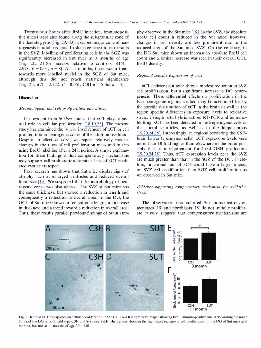
Twenty-four hours after BrdU injection, immunoposi-tive nuclei were also found along the subgranular zone ofthe dentate gyrus (Fig. 2A–D), a second major zone of neu-rogenesis in adult rodents. In sharp contrast to our resultsin the SVZ, labelling of proliferating cells in the SGZ wassignificantly increased in Sut mice at 3 months of age(Fig. 2E, 21.6% increase relative to controls, t(14) =2.978, P = 0.01, n = 8). At 11 months, there was a trendtowards more labelled nuclei in the SGZ of Sut mice,although this did not reach statistical significance(Fig. 2F, t(7) = 2.232, P = 0.061, C3H n = 5 Sut n = 4).
Discussion
Morphological and cell proliferation alterations
It is evident from in vitro studies that xCT plays a piv-otal role in cellular proliferation [18,19,22]. The presentstudy has examined the in vivo involvement of xCT in cellproliferation in neurogenic zones of the adult mouse brain.Despite an effect in vitro, we report relatively modestchanges in the rates of cell proliferation measured in vivo
using BrdU labelling after a 24 h period. A simple explana-tion for these findings is that compensatory mechanismsmay support cell proliferation despite a lack of xCT medi-ated cystine transport.
Past research has shown that Sut mice display signs ofatrophy such as enlarged ventricles and reduced overallbrain size [19]. We suspected that the morphology of neu-rogenic zones was also altered. The SVZ of Sut mice hasthe same thickness, but showed a reduction in length andconsequently a reduction in overall area. In the DG, theGCL of Sut mice showed a reduction in length, an increasein thickness and a trend toward a reduction in overall area.Thus, these results parallel previous findings of brain atro-
Fig. 2. Role of xCT transporter on cellular proliferation in the DG. (A–D) Brilining of the DG in both wild-type C3H and Sut mice. (E,F) Histograms showmonths, but not at 11 months of age *P < 0.01.
phy observed in the Sut mice [19]. In the SVZ, the absoluteBrdU cell count is reduced in the Sut mice; however,changes in cell density are less prominent due to thereduced area of the Sut mice SVZ. On the contrary, inthe DG Sut mice shown an increase in absolute BrdU cellcount and a similar increase was seen in their overall GCLBrdU density.
Regional specific expression of xCT
xCT deficient Sut mice show a modest reduction in SVZcell proliferation, but a significant increase in DG neuro-genesis. These differential effects on proliferation in thetwo neurogenic regions studied may be accounted for bythe specific distribution of xCT in the brain as well as theregion specific differences in exposure levels to oxidativestress. Using in situ hybridization, RT-PCR and immuno-blotting, xCT has been detected in both ependymal cells ofthe lateral ventricles, as well as in the hippocampus[19,20,24,25]. Interestingly, in regions bordering the CSF-brain barrer (ependymal cells), xCT expression levels weremore than 10-fold higher than elsewhere in the brain pos-sibly due to a requirement for local GSH production[19,20,24,25]. Thus, xCT expression levels near the SVZare much greater than that in the SGZ of the DG. There-fore, functional loss of xCT could have a larger impacton SVZ cell proliferation than SGZ cell proliferation aswe observed in Sut mice.
Evidence supporting compensatory mechanism for oxidative
stress
The observation that cultured Sut mouse astrocytes,meninges [19] and fibroblasts [18] do not initially prolifer-ate in vitro suggests that compensatory mechanisms are
ght field images showing BrdU immunopositive nuclei decorating the innering the significant increase in cell proliferation in the DG of Sut mice at 3
532 R.R. Liu et al. / Biochemical and Biophysical Research Communications 364 (2007) 528–533
effective in vivo that allow xCT to be dispensable. Interest-ingly, with Sut astrocytes and meninges grown in vitro,compensatory mechanisms become effective after 1 weekin culture, and the cells can survive independently of thereductant b-ME [19]. Additionally, cultured xCT deficientmouse cells also showed a low velocity cystine transportvia a Na+-dependent mechanisms resistant to inhibitionby glutamate an xCT blocker [18]. Lastly, it is evident thatxCT deficient mice have lower plasma [18] and cellular [19]GSH. This suggests that Sut mice are more likely to be in astate of oxidative stress that could induce anti-oxidantpathways such as Nrf2 activation. Nrf2 is a transcriptionfactor, which upon activation will trigger anti-oxidantpathways [26,27]. Thus, the loss of xCT function could trig-ger a global compensatory mechanism, which wouldexplain the elevation of cell proliferation in the DG SGZ.The elevation in oxidative stress in xCT deficient micemay also be associated with increased cell death. Thismay consequently trigger a regeneration mechanism asreflected by the elevation in cell proliferation in the DGof Sut mice. A similar phenomenon has been reported inischemic stroke, where oxidative stress is elevated andDG neurogenesis is increased [28–30].
In conclusion, we have shown that the natural loss offunctional xCT transport activity in Sut mice was accom-panied by relatively normal rates of cellular proliferationin the SVZ, and an enhancement of cell production in theDG. These results implicate xCT in the regulation of hip-pocampal neurogenesis and demonstrate that, unlike previ-ous in vitro studies, the absence of cystine transport viaxCT is not an essential component of ongoing cell produc-tion in the adult brain.
Acknowledgments
T.H. Murphy is a MSFHR Senior Scholar. This workwas supported by operating grants to T.H.M. from theCanadian Stroke Network, and the Heart and StrokeFoundation of BC and the Yukon.
References
[1] G. Kempermann, H.G. Kuhn, F.H. Gage, Genetic influence onneurogenesis in the dentate gyrus of adult mice, Proc. Natl. Acad. Sci.USA 94 (1997) 10409–10414.
[2] J. Luo, S.B. Daniels, J.B. Lennington, R.Q. Notti, J.C. Conover, Theaging neurogenic subventricular zone, Aging Cell 5 (2006) 139–152.
[3] G. Kempermann, D. Gast, G. Kronenberg, M. Yamaguchi, F.H.Gage, Early determination and long-term persistence of adult-generated new neurons in the hippocampus of mice, Development130 (2003) 391–399.
[4] B. Winner, C.M. Cooper-Kuhn, R. Aigner, J. Winkler, H.G. Kuhn,Long-term survival and cell death of newly generated neurons in theadult rat olfactory bulb, Eur. J. Neurosci. 16 (2002) 1681–1689.
[5] E. Gould, A. Beylin, P. Tanapat, A. Reeves, T.J. Shors, Learningenhances adult neurogenesis in the hippocampal formation, Nat.Neurosci. 2 (1999) 260–265.
[6] A. Tashiro, V.M. Sandler, N. Toni, C. Zhao, F.H. Gage, NMDA-receptor-mediated, cell-specific integration of new neurons in adultdentate gyrus, Nature 442 (2006) 929–933.
[7] L.A. Shapiro, K.L. Ng, Q.Y. Zhou, C.E. Ribak, Olfactory enrich-ment enhances the survival of newly born cortical neurons in adultmice, Neuroreport 18 (2007) 981–985.
[8] A. Sahay, R. Hen, Adult hippocampal neurogenesis in depression,Nat. Neurosci. 10 (2007) 1110–1115.
[9] J.M. Parent, T.W. Yu, R.T. Leibowitz, D.H. Geschwind, R.S. Sloviter,D.H. Lowenstein, Dentate granule cell neurogenesis is increased byseizures and contributes to aberrant network reorganization in theadult rat hippocampus, J. Neurosci. 17 (1997) 3727–3738.
[10] S. Orrenius, V. Gogvadze, B. Zhivotovsky, Mitochondrial oxidativestress: implications for cell death, Annu. Rev. Pharmacol. Toxicol. 47(2007) 143–183.
[11] E. Sleeper, C. Tamm, J. Frisen, B. Zhivotovsky, S. Orrenius, S.Ceccatelli, Cell death in adult neural stem cells, Cell Death Differ. 9(2002) 1377–1378.
[12] P.K. Dash, S.A. Mach, A.N. Moore, Enhanced neurogenesis in therodent hippocampus following traumatic brain injury, J. Neurosci.Res. 63 (2001) 313–319.
[13] J. Liu, K. Solway, R.O. Messing, F.R. Sharp, Increased neurogenesisin the dentate gyrus after transient global ischemia in gerbils, J.Neurosci. 18 (1998) 7768–7778.
[14] J. Martensson, A. Jain, E. Stole, W. Frayer, P.A. Auld, A. Meister,Inhibition of glutathione synthesis in the newborn rat: a model forendogenously produced oxidative stress, Proc. Natl. Acad. Sci. USA88 (1991) 9360–9364.
[15] T. Mizui, H. Kinouchi, P.H. Chan, Depletion of brain glutathione bybuthionine sulfoximine enhances cerebral ischemic injury in rats, Am.J. Physiol. 262 (1992) H313–H317.
[16] J.S. Bains, C.A. Shaw, Neurodegenerative disorders in humans: therole of glutathione in oxidative stress-mediated neuronal death, BrainRes. Brain Res. Rev. 25 (1997) 335–358.
[17] K. Miura, T. Ishii, Y. Sugita, S. Bannai, Cystine uptake andglutathione level in endothelial cells exposed to oxidative stress, Am.J. Physiol. 262 (1992) C50–C58.
[18] H. Sato, A. Shiiya, M. Kimata, K. Maebara, M. Tamba, Y.Sakakura, N. Makino, F. Sugiyama, K. Yagami, T. Moriguchi, S.Takahashi, S. Bannai, Redox imbalance in cystine/glutamate trans-porter-deficient mice, J. Biol. Chem. 280 (2005) 37423–37429.
[19] A.Y. Shih, H. Erb, X. Sun, S. Toda, P.W. Kalivas, T.H. Murphy,Cystine/glutamate exchange modulates glutathione supply for neu-roprotection from oxidative stress and cell proliferation, J. Neurosci.26 (2006) 10514–10523.
[20] H. Sato, M. Tamba, S. Okuno, K. Sato, K. Keino-Masu, M. Masu, S.Bannai, Distribution of cystine/glutamate exchange transporter,system x(c)-, in the mouse brain, J. Neurosci. 22 (2002) 8028–8033.
[21] X. Sun, A.Y. Shih, H.C. Johannssen, H. Erb, P. Li, T.H. Murphy,Two-photon imaging of glutathione levels in intact brain indicatesenhanced redox buffering in developing neurons and cells at thecerebrospinal fluid and blood–brain interface, J. Biol. Chem. 281(2006) 17420–17431.
[22] S. Chintala, W. Li, M.L. Lamoreux, S. Ito, K. Wakamatsu, E.V.Sviderskaya, D.C. Bennett, Y.M. Park, W.A. Gahl, M. Huizing, R.A.Spritz, S. Ben, E.K. Novak, J. Tan, R.T. Swank, Slc7a11 genecontrols production of pheomelanin pigment and proliferation ofcultured cells, Proc. Natl. Acad. Sci. USA 102 (2005) 10964–10969.
[23] H.G. Gratzner, Monoclonal antibody to 5-bromo- and 5-iododeoxy-uridine: a new reagent for detection of DNA replication, Science 218(1982) 474–475.
[24] V. La Bella, F. Valentino, T. Piccoli, F. Piccoli, Expression anddevelopmental regulation of the cystine/glutamate exchanger (xc-) inthe rat, Neurochem. Res. 32 (2007) 1081–1090.
[25] J. Burdo, R. Dargusch, D. Schubert, Distribution of the cystine/glutamate antiporter system xc- in the brain, kidney, and duodenum,J. Histochem. Cytochem. 54 (2006) 549–557.
[26] A.Y. Shih, D.A. Johnson, G. Wong, A.D. Kraft, L. Jiang, H. Erb,J.A. Johnson, T.H. Murphy, Coordinate regulation of glutathionebiosynthesis and release by Nrf2-expressing glia potently protectsneurons from oxidative stress, J. Neurosci. 23 (2003) 3394–3406.

R.R. Liu et al. / Biochemical and Biophysical Research Communications 364 (2007) 528–533 533
[27] G.E. Mann, J. Niehueser-Saran, A. Watson, L. Gao, T. Ishii, P. deWinter, R.C. Siow, Nrf2/ARE regulated antioxidant gene expressionin endothelial and smooth muscle cells in oxidative stress: implica-tions for atherosclerosis and preeclampsia, Sheng Li Xue Bao 59(2007) 117–127.
[28] H. Nakatomi, T. Kuriu, S. Okabe, S. Yamamoto, O. Hatano, N.Kawahara, A. Tamura, T. Kirino, M. Nakafuku, Regeneration ofhippocampal pyramidal neurons after ischemic brain injury by
recruitment of endogenous neural progenitors, Cell 110 (2002) 429–441.
[29] Y. Yagita, K. Kitagawa, T. Ohtsuki, K. Takasawa, T. Miyata, H.Okano, M. Hori, M. Matsumoto, Neurogenesis by progenitor cells inthe ischemic adult rat hippocampus, Stroke 32 (2001) 1890–1896.
[30] R. Zhang, L. Zhang, Z. Zhang, Y. Wang, M. Lu, M. Lapointe, M.Chopp, A nitric oxide donor induces neurogenesis and reducesfunctional deficits after stroke in rats, Ann. Neurol. 50 (2001) 602–611.

![[Physiotherapy and neurogenic lower urinary tract dysfunction in multiple sclerosis patients: a randomized controlled trial]](https://img.dokumen.tips/doc/110x75/6350b22db4766da83b034cbc/physiotherapy-and-neurogenic-lower-urinary-tract-dysfunction-in-multiple-sclerosis.jpg)


