Embed Size (px)
Citation preview
2004;64:4277-4285. Cancer Res Noriaki Sunaga, Kuniharu Miyajima, Makoto Suzuki, et al.
Small Cell Lung CancerversusNon-Small Cell Lung Cancer Different Roles for Caveolin-1 in the Development of
Updated version
http://cancerres.aacrjournals.org/content/64/12/4277
Access the most recent version of this article at:
Cited Articles
http://cancerres.aacrjournals.org/content/64/12/4277.full.html#ref-list-1
This article cites by 53 articles, 22 of which you can access for free at:
Citing articles
http://cancerres.aacrjournals.org/content/64/12/4277.full.html#related-urls
This article has been cited by 21 HighWire-hosted articles. Access the articles at:
E-mail alerts related to this article or journal.Sign up to receive free email-alerts
SubscriptionsReprints and
To order reprints of this article or to subscribe to the journal, contact the AACR Publications
Permissions
To request permission to re-use all or part of this article, contact the AACR Publications
Research. on March 20, 2014. © 2004 American Association for Cancercancerres.aacrjournals.org Downloaded from
Research. on March 20, 2014. © 2004 American Association for Cancercancerres.aacrjournals.org Downloaded from
[CANCER RESEARCH 64, 4277–4285, June 15, 2004]
Different Roles for Caveolin-1 in the Development of Non-Small Cell Lung Cancerversus Small Cell Lung Cancer
Noriaki Sunaga,1,2 Kuniharu Miyajima,1 Makoto Suzuki,1 Mitsuo Sato,1 Michael A. White,3 Ruben D. Ramirez,1
Jerry W. Shay,3 Adi F. Gazdar,1 and John D. Minna1,4,5*
1Hamon Center for Therapeutic Oncology Research and Departments of 3Cell Biology, 4Internal Medicine, and 5Pharmacology, University of Texas Southwestern Medical Centerat Dallas, Dallas, Texas, and 2First Department of Internal Medicine, Gunma University School of Medicine, Gunma, Japan
ABSTRACT
Caveolin-1 (CAV1), an essential structural constituent of caveolae thatplays an important role in cellular processes such as transport and sig-naling, has been implicated in the development of human cancers. How-ever, it is unclear whether CAV1 is acting like an oncogene or tumorsuppressor gene. We found that CAV1 expression was reduced or absentin 95% of small cell lung cancers (SCLCs; n � 21 lines), whereas it wasretained in 76% of non-small cell lung cancers (NSCLCs; n � 25 lines)compared with normal human lung epithelial cultures, where it wasabundantly expressed. CAV1 expression was tightly linked to the ability togrow attached to the plastic cell culture surface, whereas CAV1-nonex-pressing lung cancers of both SCLC and NSCLC type grew as suspensioncultures. In addition, attached lung cancer cultures expressed phospho-rylated focal adhesion kinase, whereas suspension cultures did not. Lackof CAV1 expression was tightly associated with CAV1 promoter methyl-ation (P < 0.0001) such that CAV1 methylation was found in 93% ofSCLCs (n � 15) and 9% of NSCLCs (n � 11), whereas 5-aza-2�deoxycy-tidine treatment restored CAV1 expression in SCLCs. Exogenous CAV1expression in SCLCs significantly inhibited soft-agar colony formationbut did not lead to attachment. By contrast, CAV1 knockdown in NSCLCsmediated by small interfering RNA against CAV1 led to inhibition ofcellular proliferation and soft-agar and liquid colony formation. Impor-tantly, CAV1 knockdown led to reduced phospho-focal adhesion kinaseand RalA, but not RalB, levels in NSCLC cells. These results suggestdifferent roles for CAV1 in SCLC, where CAV1 acts like a tumor sup-pressor gene, and NSCLC, where it appears required for survival andgrowth.
INTRODUCTION
Caveolae are flask-shaped invaginations of the plasma membranethat play an important role in cellular processes, including moleculetransport, cell adhesion, and signal transduction (1–3). Caveolin-1(CAV1) is an essential structural component of caveolae and func-tionally regulates the activity of many signaling molecules, such asG-proteins, Src family kinases, H-Ras, protein kinase C, epidermalgrowth factor receptor, endothelial nitric oxide synthase, and inte-grins, which are potentially involved in the development of humancancer, by generating preassembled signaling complexes (4–7). Thus,CAV1 could be a key molecule for growth-related signaling andcancer development.
The CAV1 gene has been considered as both a tumor suppressorgene and an oncogene. Down-regulation of CAV1 expression wasobserved in breast, lung, colon, and ovarian cancer, whereas onco-genic transformation of cells was associated with reduction of CAV1expression, and antisense-mediated down-regulation of CAV1 expres-sion was sufficient to drive oncogenic transformation in NIH 3T3
cells (8–15). Exogenous expression of CAV1 in oncogenically trans-formed cells and cancer cell lines inhibited cell growth and tumori-genesis (12, 16–18). This evidence indicates that CAV1 can act as atumor suppressor. By contrast, other studies have reported that CAV1expression was up-regulated in human cancers, including breast, lung,prostate, esophagus, colon, thyroid, and pancreas cancers, and that thisup-regulation was associated with metastases and poor prognosis(19–25). These results suggest that CAV1 can function other than asa tumor suppressor. Thus, studies have implicated CAV1 in thedevelopment of several human cancers, but it appears to play quitedifferent roles in the development of these cancers.
Human lung cancer is divided into two major histological types:small cell lung cancer (SCLC) and non-small cell lung cancer(NSCLC; Ref. 26). We evaluated endogenous expression of CAV1protein in several lung cancer cell lines and found that CAV1 proteinexpression was reduced or absent in 95% of SCLCs (n � 21 lines),whereas it was retained in 76% of NSCLCs (n � 25 lines) comparedwith normal human lung epithelial cultures, in which expression wasabundant. Methylation analysis revealed promoter hypermethylationof CAV1 in 93% of SCLCs (n � 15) but only 9% of NSCLCs(n � 11). Furthermore, exogenous reexpression of CAV1 in SCLCsled to significant inhibition of colony formation, whereas knockdownof CAV1 in NSCLCs by RNA interference (RNAi) technology led toinhibition of cellular proliferation and colony formation in this histo-logical type (27–31). These results suggest different roles for CAV1in SCLC, where it has the properties of a tumor suppressor, whereasin NSCLC it appears required for growth.
MATERIALS AND METHODS
Cell Lines. All of the lung cancer cell lines were obtained from the HamonCenter collection (University of Texas Southwestern Medical Center) gener-ated by the authors (32, 33). Most are also available from the American TypeCulture Collection. Breast cancer cell line MCF7 was obtained from theAmerican Type Culture Collection. Cells were cultured with RPMI 1640supplemented with 5% fetal bovine serum. Normal human bronchial epithelialand small-airway epithelial cells were obtained from Clonetics (San Diego,CA). The immortalized human bronchial epithelial cell lines BEAS-2B,HBEC1, HBEC2, HBEC3, and HBEC4 were also used. We recently estab-lished the immortalized human bronchial epithelial cell lines HBEC1, HBEC2,HBEC3, and HBEC4, retrovirally transfected with CDK4 and the catalyticcomponent of telomerase, which were obtained from bronchial biopsies.6
Preparation and Transfection of Synthetic Small Interfering RNA(siRNA). siRNAs targeting CAV1 (CAV1-1 siRNA), Tax (the human leuke-mia virus gene), and �-catenin were designed and prepared as describedpreviously (27, 34, 35). The siRNA sequences against CAV1 (CAV1-1 siRNA)were 5�-AGACGAGCUGAGCGAGAAGCA-3� (sense) and 5�-CUUCU-CGCUCAGCUCGUCUGC-3� (antisense) and (CAV1-2 siRNA) 5�-CAUC-UACAAGCCCAACAACTT-3� (sense) and 5�-GUUGUUGGGCUUGUA-GAUGTT-3� (antisense). The siRNA target sequences were tested in a BLASTsearch of GenBank (National Center for Biotechnology Information database)
Received 12/16/03; revised 3/12/04; accepted 4/9/04.Grant support: National Cancer Institute Lung Cancer Specialized Programs of
Research Excellence Grant P50CA70907 and the Gillson Longenbaugh Foundation.The costs of publication of this article were defrayed in part by the payment of page
charges. This article must therefore be hereby marked advertisement in accordance with18 U.S.C. Section 1734 solely to indicate this fact.
Requests for reprints: John D. Minna, Hamon Center for Therapeutic OncologyResearch, University of Texas Southwestern Medical Center at Dallas, 6000 Harry HinesBoulevard, Dallas, TX 75390-8593. Phone: (214) 648-4900; Fax: (214) 648-4940; E-mail:[email protected].
6R. D. Ramirez, M. Peyton, L. Girard, Y. Zou, J. M. Dimaio, S. Milchgrub, A. L.Smith, L. Gilbey, X. Zhang, S. Sheridan, K. Gandia, J. Pollack, M. Vaughn, W. E. Wright,J. W. Shay, and J. D. Minna. Immortalization of normal human bronchial epithelial cellsin the absence of viral oncoproteins, manuscript in preparation.
4277
Research. on March 20, 2014. © 2004 American Association for Cancercancerres.aacrjournals.org Downloaded from
to ensure that only the corresponding gene is the target. RNA oligonucleotideswere obtained from the core facility of the University of Texas SouthwesternMedical Center (see the RNA Oligonucleotide Synthesis Core web site fordetails).7 siRNAs were transfected into cells with use of Oligofectaminetransfection reagent (Invitrogen, Carlsbad, CA) as described (56). For siRNAtransfection into NCI-H187 and NCI-H524 cells, Lipofectamine 2000 trans-fection reagent was used (Invitrogen). Cells were grown and harvested forfurther analysis.
Immunofluorescent Staining. After 24 h of siRNA transfection, cellswere plated and grown on slides. After 48 h, the cells were washed with 60 mM
PIPES–25 mM HEPES–10 mM EGTA–1 mM MgCl2 (pH 7.4) and fixed for 10min at 37°C in 3% paraformaldehyde in 60 mM PIPES–25 mM HEPES–10 mM
EGTA–1 mM MgCl2 (pH 7.4). After additional washes with PBS, the cellswere permeabilized with 0.1% Triton in PBS for 10 min. After blocking with3% gelatin–3% BSA–0.2% Tween-20 for 1 h at 37°C, the cells were incubatedwith mouse monoclonal anti-�-tubulin (Calbiochem, San Diego, CA) and therabbit polyclonal anticaveolin (BD Transduction Laboratories, San Diego, CA)antibodies in gelatin/BSA blocking solution for 16 h at 4°C. The cells wereincubated with Alexa Fluor 568 antimouse IgG (H � L) (Molecular Probes,Eugene, OR) and Alexa Fluor 488 antirabbit IgG (H� L) (Molecular Probes)secondary antibodies for 1 h at 37°C. The stained cells were examined undera fluorescent microscope and photographed.
Western Blot Analysis. Cells were grown to 80–90% confluency andharvested, and cellular proteins were extracted with lysis buffer [40 mM
HEPES-NaOH (pH 7.4), 1% NP40, 0.5% sodium deoxycholate, 0.1% SDS,150 mM NaCl] containing Complete Mini, a cocktail of protease inhibitors(Roche, Indianapolis, IN). Total protein was separated on a SDS-polyacryl-amide gel and electroblotted to nitrocellulose membranes (Schleicher &Schuell, Beverly, NH). After blocking with 5% nonfat dry milk and 0.1%Tween 20 in Tris-buffered saline, membranes were incubated at 4°C for 16 hwith rabbit polyclonal anticaveolin (BD Transduction Laboratories), mousemonoclonal anti-�-catenin (BD Transduction Laboratories), rabbit polyclonalanti-phosphorylated focal adhesion kinase (FAK) pY397 (BIOSOURCE, Ca-marillo, CA), mouse monoclonal anti-FAK (BD Transduction Laboratories),mouse anti-RalA (BD Transduction Laboratories), rabbit anti-RalB (BD Trans-duction Laboratories), and mouse monoclonal antiactin (Sigma, Saint Louis,MO) antibodies. The membranes then were developed with peroxidase-labeledantibodies (Amersham Pharmacia, Piscataway, NJ) by Super Signal chemilu-minescence substrate (Pierce, Rockford, IL). Actin protein levels were used asa control for adequacy of equal protein loading.
Bisulfite Genomic DNA Sequencing. One �g of genomic DNA wasdenatured by NaOH and modified by bisulfite (Sigma) as described previously(36). The modified DNA was purified with use of a Wizard DNA purificationkit (Promega, Madison, WI), treated with NaOH to desulfonate the sample,precipitated with ethanol, and resuspended in water. Bisulfite-treated DNAwas then used as template in PCR reactions for sequencing analysis. Thesequences of the three primer pairs used are as follows: 5�-TGTGTATTTT-GTAAATATGGTATAATTTG-3� and 5�-CCATCTCTACCTTAAAACA-CAT-3�, 5�-GGGATTTATAAAGTTTAGATGTGA-3� and 5�-CATTTTC-CCTACTCTAAACCAC-3�, and 5�-ATAGGGTAGGATTGTGGATTGT-3�and 5�-TAAACACATCCCCAAAATTCTAAC-3�. The PCR conditions wereas follows: the reaction volume was 20 �l; initial denaturation was for 15 minat 95°C, followed by 40 cycles of 30 s at 94°C, 30 s at 55°C, and 30 s at 72°C,with final extension at 72°C for 10 min. PCR products were purified withexonuclease I and shrimp alkaline phosphatase (USB Corp., Cleveland, OH)and sequenced with an ABI 377 automatic DNA sequencer.
We evaluated the methylation status by measuring methylated CpG (mCpG)density as described previously (37). The bisulfite treatment converts unmethy-lated C to T but leaves methylated C as C. If only T is detected at this positionin sequencing, the status of this CpG dinucleotide is defined as fully unmethy-lated (open square), which was counted as 0 in the calculation of mCpGdensity. If only C is detected at this position in sequencing, the status of thisCpG dinucleotide is defined as fully methylated (filled square), which wascounted as 1 in the calculation of mCpG density. Accordingly, a 50% filledsquare represents C and T detected at equal peak height in sequencing at thisposition; thus, the status of this CpG is defined as one-half methylated and
one-half unmethylated and was counted as 0.5 in the calculation of mCpGdensity. A 75% filled square indicates that both C and T were detected insequencing at this position but the height of the C peak was larger than that ofthe T peak; thus the status of this CpG is defined as 75% methylated and 25%unmethylated and was counted as 0.75 in the calculation of mCpG density. A25% filled square indicates that both C and T were detected in sequencing atthis position but the height of the T peak was larger than that of the C peak;therefore, the status of this CpG is defined as 75% unmethylated and 25%methylated and was counted as 0.25 in the calculation of mCpG density. ThemCpG density per 100 bp was calculated by summing the scores of allmethylated CpGs in the region from nucleotide (nt) 29442 to nt 29676,dividing by 235 bp, and multiplying by 100.
Treatment with 5-Aza-2�-deoxycytidine and Reverse Transcription-PCR Analysis. NCI-H146 and NCI-H2171 SCLC cell lines were incubatedand grown in culture medium in the presence or absence of 4 �M 5-aza-2�-deoxycytidine (Sigma) for 6 days as described previously (36). Total RNA wasextracted from the cells by TRIzol Reagent (Invitrogen) according to themanufacturer’s instructions, and cDNA was synthesized using 2 �g of totalRNA with the SuperScript II First-Strand Synthesis using oligo(dT) primerSystem (Invitrogen). Aliquots of the reaction mixture were used for thesubsequent PCR amplification. The primer sequences for CAV1 amplificationwere 5�-CGACCCTAAACACCTCAACGATG-3� and 5�-GCAGACAG-CAAGCGGTAAAACC-3�. Glyceraldehyde-3-phosphate dehydrogenase(GAPDH) was used as an internal control to confirm the success of thereaction. The primer sequences for GAPDH amplification were 5�-CACTG-GCGTCTTCACCACCATG-3� and 5�-GCTTCACCACCTTCTTGATGTCA-3�. PCR conditions were as follows: the reaction volume was 20 �l; initialdenaturation was for 15 min at 95°C, followed by 25 cycles of 30 s at 94°C,30 s at 55°C, and 30 s at 72°C, with a final extension at 72°C for 10 min. PCRproducts were visualized on 2% agarose gels stained with ethidium bromide.
Methylation-Specific PCR. DNA methylation patterns in the CpG islandof the CAV1 gene were determined by the method of methylation-specific PCR(36). The primer sequences were 5�-TTATTTCGAAGCGTTTGGGAG-3�and 5�-AACACTCGTTTACATCTAATCG-3� for the methylated reaction,and 5�-TTATTTTGAAGTGTTTGGGAG-3� and 5�-AACACTCATTTACA-TCTAATCA-3� for the unmethylated reaction. PCR amplification was per-formed with bisulfite-treated DNA as a template, using specific primer se-quences for the methylated and unmethylated forms of the gene. PCRconditions were as follows: the reaction volume was 20 �l; initial denaturationwas for 15 min at 95°C, followed by 7 cycles of 30 s at 94°C, 55 s at 57°C,and 20 s at 72°C and 30 cycles of 20 s at 90°C, 30 s at 57°C, and 20 s at 72°C,with final extension at 72°C for 10 min. PCR products were analyzed on 2%agarose gels. We note that although CAV1 was not methylated in normal lungepithelium, it was methylated in B lymphocytes. Thus, caution must be used inapplying this assay to samples containing lymphocytes.
Construction and Transfection of CAV1-Expressing Plasmid Vector.The full-length cDNA encoding human CAV1 was amplified by reversetranscription-PCR with primers (5�-CGCGGATCCTCCTCACAGT-3� and 5�-CCGGAATTCTATCCTTGAAATG-3�) and RNA isolated from BEAS-2Bcells. The amplified DNA fragments were ligated into the BamHI-EcoRI sitesof the pcDNA3.1 (�) vector (Invitrogen) to construct the CAV1 expressionplasmid pcDNA3.1/CAV1. The correct orientation and sequence of the insertwere verified by DNA sequencing. The pcDNA3.1/CAV1 plasmid or thepcDNA3.1 plasmid was transfected into the cells with use of DMRIE-Creagent (Invitrogen) according to the manufacturers’ protocol.
Colony Formation Assay. The in vitro growth characteristics were testedby colony formation assay. After 48 h of transfection with siRNA or CAV1expression plasmid, cells were harvested, and the colony formation assay wasperformed as described previously (38) with some modifications. Briefly, 500viable cells unstained with trypan blue were plated in each well of 6-wellplates. Cells were cultured in RPMI 1640 supplemented with 5% serum, andsurviving colonies were counted 14 days later after staining with methyleneblue. For the anchorage-independent, soft-agar growth assays, 300 viable cellswere suspended and plated in 0.33% agar in complete medium and layeredover a 0.50% agar base containing complete medium in 12-well plates. After4 weeks, the number of foci was counted.
Bromodeoxyuridine (BrdUrd) Incorporation Assay. Twenty-four h aftertransfection with siRNA, 3000 cells were replated and cultured in 96-wellplates in replicates of 8. After 48 h, we examined DNA synthesis by quanti-7 http://cbi.swmed.edu/pages/oligonet_index.htm.
4278
ROLE OF CAV1 IN LUNG CANCER
Research. on March 20, 2014. © 2004 American Association for Cancercancerres.aacrjournals.org Downloaded from
tating BrdUrd incorporated into the newly synthesized DNA during cellproliferation with use of the BrdUrd Labeling and Detection Kit III (Roche)according to the manufacturers’ protocol. The absorbance (A405 nm/A492 nm)that was directly correlated to the level of BrdUrd incorporated into cellularDNA was determined with use of a microtiter plate reader.
PCR-Restriction Fragment Length Polymorphism Analysis. The CAV1missense mutation at codon 132 (P132L) was screened by PCR-restrictionfragment length polymorphism analysis as described previously (39). Theprimer sequences for CAV1 amplification were 5�-CTGTGCTCATGTTGT-GTCAC-3� and 5�-GTATTAGCAACTTGGAACTTG-3�. PCR conditionswere as follows: 50 �g of genomic DNA was used in a reaction volume of 20�l; initial denaturation was for 15 min at 95°C, followed by 35 cycles of 30 sat 94°C, 30 s at 55°C, and 60 s at 72°C, with final extension at 72°C for 10 min.The amplified DNA fragments were digested with NlaIII (New EnglandBiolabs, Beverly, MA) at 37°C for 3 h. Complete digestion of the 460-bp PCRproducts produced 236- and 222-bp fragments for the wild-type allele, whereasdigestion of the mutant allele produced a single, 460-bp fragment. The digestedDNA was electrophoresed on a 2% agarose gel and visualized by ethidiumbromide staining, and the genotypes were determined.
RESULTS
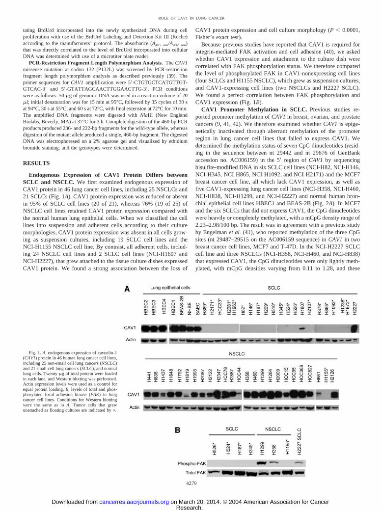
Endogenous Expression of CAV1 Protein Differs betweenSCLC and NSCLC. We first examined endogenous expression ofCAV1 protein in 46 lung cancer cell lines, including 25 NSCLCs and21 SCLCs (Fig. 1A). CAV1 protein expression was reduced or absentin 95% of SCLC cell lines (20 of 21), whereas 76% (19 of 25) ofNSCLC cell lines retained CAV1 protein expression compared withthe normal human lung epithelial cells. When we classified the celllines into suspension and adherent cells according to their culturemorphologies, CAV1 protein expression was absent in all cells grow-ing as suspension cultures, including 19 SCLC cell lines and theNCI-H1155 NSCLC cell line. By contrast, all adherent cells, includ-ing 24 NSCLC cell lines and 2 SCLC cell lines (NCI-H1607 andNCI-H2227), that grew attached to the tissue culture dishes expressedCAV1 protein. We found a strong association between the loss of
CAV1 protein expression and cell culture morphology (P � 0.0001,Fisher’s exact test).
Because previous studies have reported that CAV1 is required forintegrin-mediated FAK activation and cell adhesion (40), we askedwhether CAV1 expression and attachment to the culture dish werecorrelated with FAK phosphorylation status. We therefore comparedthe level of phosphorylated FAK in CAV1-nonexpressing cell lines(four SCLCs and H1155 NSCLC), which grew as suspension cultures,and CAV1-expressing cell lines (two NSCLCs and H2227 SCLC).We found a perfect correlation between FAK phosphorylation andCAV1 expression (Fig. 1B).
CAV1 Promoter Methylation in SCLC. Previous studies re-ported promoter methylation of CAV1 in breast, ovarian, and prostatecancers (9, 41, 42). We therefore examined whether CAV1 is epige-netically inactivated through aberrant methylation of the promoterregion in lung cancer cell lines that failed to express CAV1. Wedetermined the methylation status of seven CpG dinucleotides (resid-ing in the sequence between nt 29442 and nt 29676 of GenBankaccession no. AC006159) in the 5� region of CAV1 by sequencingbisulfite-modified DNA in six SCLC cell lines (NCI-H82, NCI-H146,NCI-H345, NCI-H865, NCI-H1092, and NCI-H2171) and the MCF7breast cancer cell line, all which lack CAV1 expression, as well asfive CAV1-expressing lung cancer cell lines (NCI-H358, NCI-H460,NCI-H838, NCI-H1299, and NCI-H2227) and normal human bron-chial epithelial cell lines HBEC1 and BEAS-2B (Fig. 2A). In MCF7and the six SCLCs that did not express CAV1, the CpG dinucleotideswere heavily or completely methylated, with a mCpG density range of2.23–2.98/100 bp. The result was in agreement with a previous studyby Engelman et al. (41), who reported methylation of the three CpGsites (nt 29487–29515 on the AC006159 sequence) in CAV1 in twobreast cancer cell lines, MCF7 and T-47D. In the NCI-H2227 SCLCcell line and three NSCLCs (NCI-H358, NCI-H460, and NCI-H838)that expressed CAV1, the CpG dinucleotides were only lightly meth-ylated, with mCpG densities varying from 0.11 to 1.28, and these
Fig. 1. A, endogenous expression of caveolin-1(CAV1) protein in 46 human lung cancer cell lines,including 25 non-small cell lung cancers (NSCLC)and 21 small cell lung cancers (SCLC), and normallung cells. Twenty �g of total protein were loadedin each lane, and Western blotting was performed.Actin expression levels were used as a control forequal protein loading. B, levels of total and phos-phorylated focal adhesion kinase (FAK) in lungcancer cell lines. Conditions for Western blottingwere the same as in A. Tumor cells that grewunattached as floating cultures are indicated by �.
4279
ROLE OF CAV1 IN LUNG CANCER
Research. on March 20, 2014. © 2004 American Association for Cancercancerres.aacrjournals.org Downloaded from

CpGs were completely unmethylated in NCI-H1299 as well as in theimmortalized bronchial epithelial cell lines BEAS2B and HBEC1.Methylation status of 14 CpG dinucleotides downstream (nt 29742–29981 of the AC006159 sequence) was further examined in fourSCLC cell lines (NCI-H146, NCI-H345, NCI-H865, and NCI-H2227)and in NCI-H1299 and BEAS-2B. In all of these cell lines, the mCpGdensities of the promoter region containing the 14 CpGs were �0.42/100 bp. Thus, we conclude that the 7 methylated CpG sites (nt29442–29676 on the AC006159 sequence) but not the 14 downstreamCpGs are possible targets for silencing of the CAV1 gene.
To confirm that promoter methylation leads to loss of CAV1expression in the lung cancer cells, we examined the effect of 5-aza-2�-deoxycytidine, a drug that inhibits DNA methylation, on CAV1expression. Treatment of the CAV1-nonexpressing SCLC cell lines(NCI-H146 and NCI-H2171) restored CAV1 expression with nochange in the expression of the housekeeping gene GAPDH (Fig. 2B),demonstrating that CAV1 is functionally methylated in these SCLCcell lines.
Strong Association between CAV1 Promoter Methylation andLoss of CAV1 Protein Expression. The methylation status of theCAV1 promoter region was further determined by methylation-specific PCR in 25 lung cancer cell lines, including 14 SCLCs and 11NSCLCs (Fig. 2C). Genomic DNA modified by sodium bisulfite wasused as template to amplify a part (nt 29487–29579 of the AC006159sequence) of the promoter region encompassing the five CpG sitesthat were heavily methylated in six SCLC cell lines and MCF7, which
lacked CAV1 protein expression (Fig. 2A). The NCI-H1155 NSCLCcell line and 13 SCLC cell lines that lacked CAV1 protein expressionexhibited the methylated band, whereas no methylated bands weredetected in 10 NSCLC cell lines and the NCI-H2227 SCLC line,which expressed CAV1 protein. We found a significant correlationbetween CAV1 methylation and loss of CAV1 protein expression(P � 0.0001 by Fisher’s exact test).
Absence of Mutations in the CAV1 Gene at Codon 132 (P132L)in Lung Cancer Cell Lines. A missense mutation in the CAV1 geneat codon 132 (P132L) was found in 16% of primary breast cancers, asreported by Hayashi et al. (39). We therefore screened the mutation atcodon 132 in 46 lung cancer cell lines by PCR-restriction fragmentlength polymorphism analysis. However, the CAV1 P132L mutationwas not detected in these cell lines, consistent with previous findingsin lung cancer (10, 22).
Effect of Exogenous Expression of CAV1 in CAV1-Nonexpress-ing SCLC Cells. To assess the functional role of CAV1 in SCLCs,we examined the effect of exogenous reexpression of CAV1 on invitro cell growth of the NCI-H187 and NCI-H524 SCLC cell lines,which lacked endogenous CAV1 expression, by colony formationassay in soft agar. Exogenous reexpression of CAV1 in the cellstransfected with the pcDNA3.1/CAV1 vector was confirmed by West-ern blot analysis at 48 h post-transfection (Fig. 3A). The transfectedcells were subjected to soft-agar growth assay, and colony formationwas determined. We found a significant decrease in colony formationin cells expressing exogenous CAV1 compared with those transfected
Fig. 2. Caveolin-1 (CAV1) promoter methylation in lung cancer cell lines. A, methylation status of each CpG dinucleotide in the promoter region of CAV1 was determined bysequencing sodium bisulfite-treated genomic DNA. The top panel illustrates the region examined (GenBank accession no. AC006159; nucleotides 29381–30060), the vertical barsrepresent CpG sites (numbered 1–21), and the open box represents the amplicon for methylation-specific PCR (MSP) assay. The bottom panel illustrates the methylation status (mCpG)of individual CpG sites in the cell lines. �, normal protein expression level; �, undetectable level by Western blotting. Open squares indicate that CpG sites are fully unmethylated;filled squares indicate that CpG sites are fully methylated; partially filled squares indicate various degrees of CpG methylation (see “Materials and Methods”). B, expression of CAV1after 5-aza-2�-deoxycytidine (5-Aza-CdR) treatment in NCI-H146 and NCI-H2171 SCLC cell lines. The cells were grown in the presence (�) and absence (�) of 4 �M
5-aza-2�-deoxycytidine for 6 days. Total RNA was isolated, cDNA was prepared, and reverse-transcribed PCR was performed for CAV1 with glyceraldehyde-3-phosphatedehydrogenase (GAPDH) as the control gene. The cDNA obtained from NCI-H1299 cells was used as a positive control. C, methylation-specific PCR for the detection of methylatedCAV1 5�-CpG sequences. Representative samples are shown. M, results with primers specific for methylated sequences; U, results with primers specific for unmethylated sequences.PCR products were visualized on 2% agarose gels stained with ethidium bromide. The cell lines expressing (�) and not expressing (�) CAV1 are indicated.
4280
ROLE OF CAV1 IN LUNG CANCER
Research. on March 20, 2014. © 2004 American Association for Cancercancerres.aacrjournals.org Downloaded from
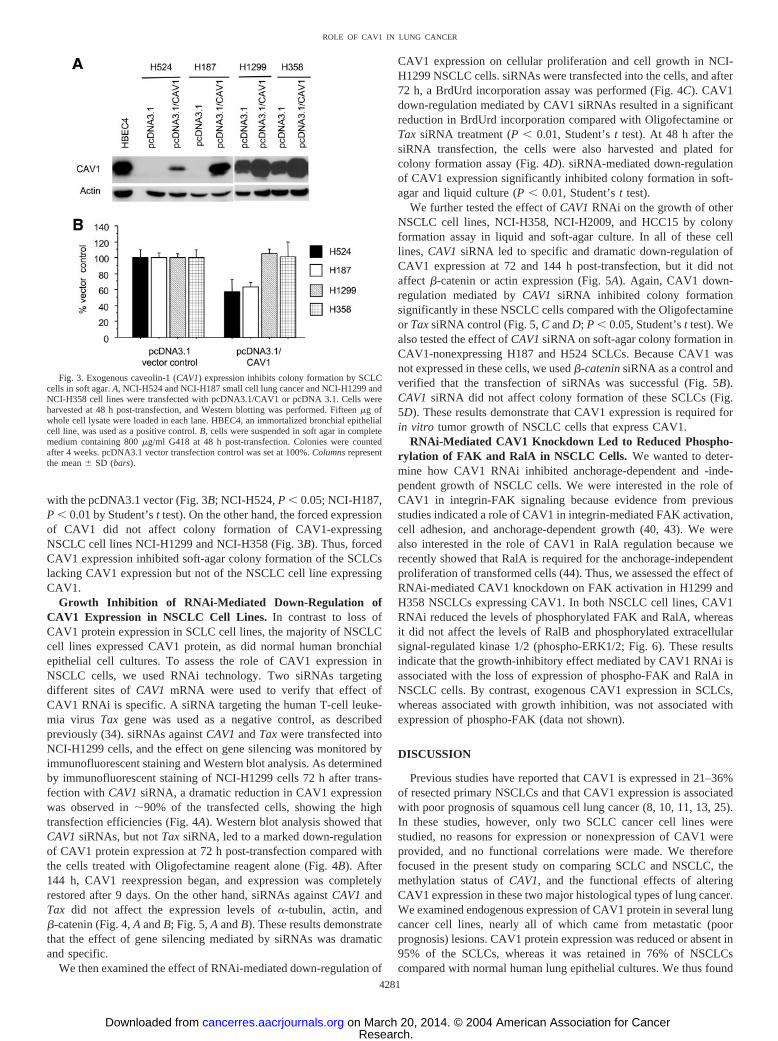
with the pcDNA3.1 vector (Fig. 3B; NCI-H524, P � 0.05; NCI-H187,P � 0.01 by Student’s t test). On the other hand, the forced expressionof CAV1 did not affect colony formation of CAV1-expressingNSCLC cell lines NCI-H1299 and NCI-H358 (Fig. 3B). Thus, forcedCAV1 expression inhibited soft-agar colony formation of the SCLCslacking CAV1 expression but not of the NSCLC cell line expressingCAV1.
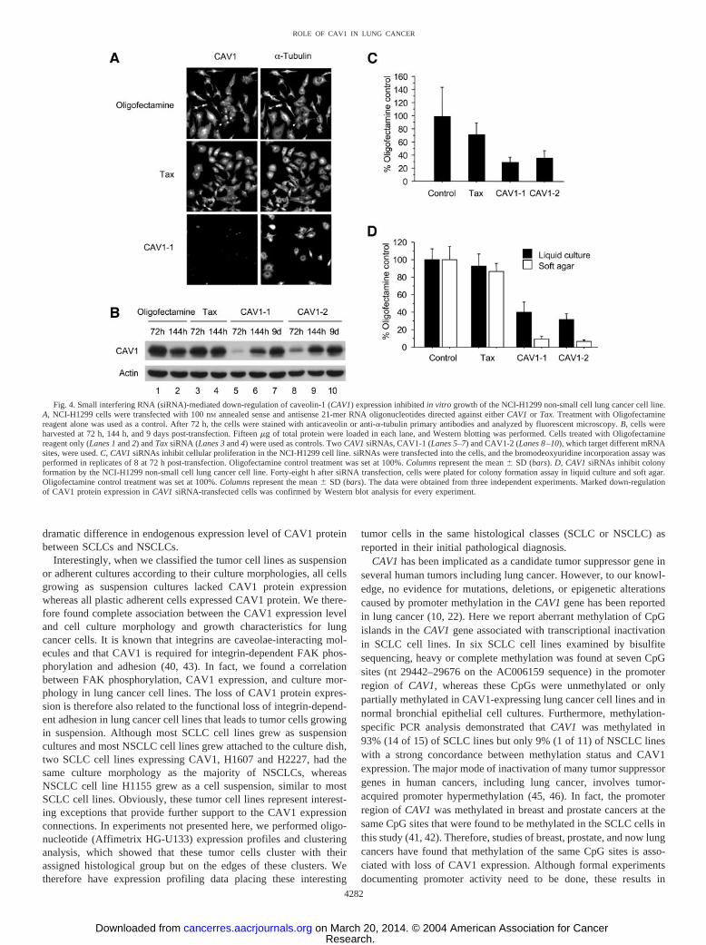
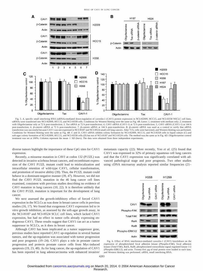
Growth Inhibition of RNAi-Mediated Down-Regulation ofCAV1 Expression in NSCLC Cell Lines. In contrast to loss ofCAV1 protein expression in SCLC cell lines, the majority of NSCLCcell lines expressed CAV1 protein, as did normal human bronchialepithelial cell cultures. To assess the role of CAV1 expression inNSCLC cells, we used RNAi technology. Two siRNAs targetingdifferent sites of CAV1 mRNA were used to verify that effect ofCAV1 RNAi is specific. A siRNA targeting the human T-cell leuke-mia virus Tax gene was used as a negative control, as describedpreviously (34). siRNAs against CAV1 and Tax were transfected intoNCI-H1299 cells, and the effect on gene silencing was monitored byimmunofluorescent staining and Western blot analysis. As determinedby immunofluorescent staining of NCI-H1299 cells 72 h after trans-fection with CAV1 siRNA, a dramatic reduction in CAV1 expressionwas observed in �90% of the transfected cells, showing the hightransfection efficiencies (Fig. 4A). Western blot analysis showed thatCAV1 siRNAs, but not Tax siRNA, led to a marked down-regulationof CAV1 protein expression at 72 h post-transfection compared withthe cells treated with Oligofectamine reagent alone (Fig. 4B). After144 h, CAV1 reexpression began, and expression was completelyrestored after 9 days. On the other hand, siRNAs against CAV1 andTax did not affect the expression levels of �-tubulin, actin, and�-catenin (Fig. 4, A and B; Fig. 5, A and B). These results demonstratethat the effect of gene silencing mediated by siRNAs was dramaticand specific.
We then examined the effect of RNAi-mediated down-regulation of
CAV1 expression on cellular proliferation and cell growth in NCI-H1299 NSCLC cells. siRNAs were transfected into the cells, and after72 h, a BrdUrd incorporation assay was performed (Fig. 4C). CAV1down-regulation mediated by CAV1 siRNAs resulted in a significantreduction in BrdUrd incorporation compared with Oligofectamine orTax siRNA treatment (P � 0.01, Student’s t test). At 48 h after thesiRNA transfection, the cells were also harvested and plated forcolony formation assay (Fig. 4D). siRNA-mediated down-regulationof CAV1 expression significantly inhibited colony formation in soft-agar and liquid culture (P � 0.01, Student’s t test).
We further tested the effect of CAV1 RNAi on the growth of otherNSCLC cell lines, NCI-H358, NCI-H2009, and HCC15 by colonyformation assay in liquid and soft-agar culture. In all of these celllines, CAV1 siRNA led to specific and dramatic down-regulation ofCAV1 expression at 72 and 144 h post-transfection, but it did notaffect �-catenin or actin expression (Fig. 5A). Again, CAV1 down-regulation mediated by CAV1 siRNA inhibited colony formationsignificantly in these NSCLC cells compared with the Oligofectamineor Tax siRNA control (Fig. 5, C and D; P � 0.05, Student’s t test). Wealso tested the effect of CAV1 siRNA on soft-agar colony formation inCAV1-nonexpressing H187 and H524 SCLCs. Because CAV1 wasnot expressed in these cells, we used �-catenin siRNA as a control andverified that the transfection of siRNAs was successful (Fig. 5B).CAV1 siRNA did not affect colony formation of these SCLCs (Fig.5D). These results demonstrate that CAV1 expression is required forin vitro tumor growth of NSCLC cells that express CAV1.
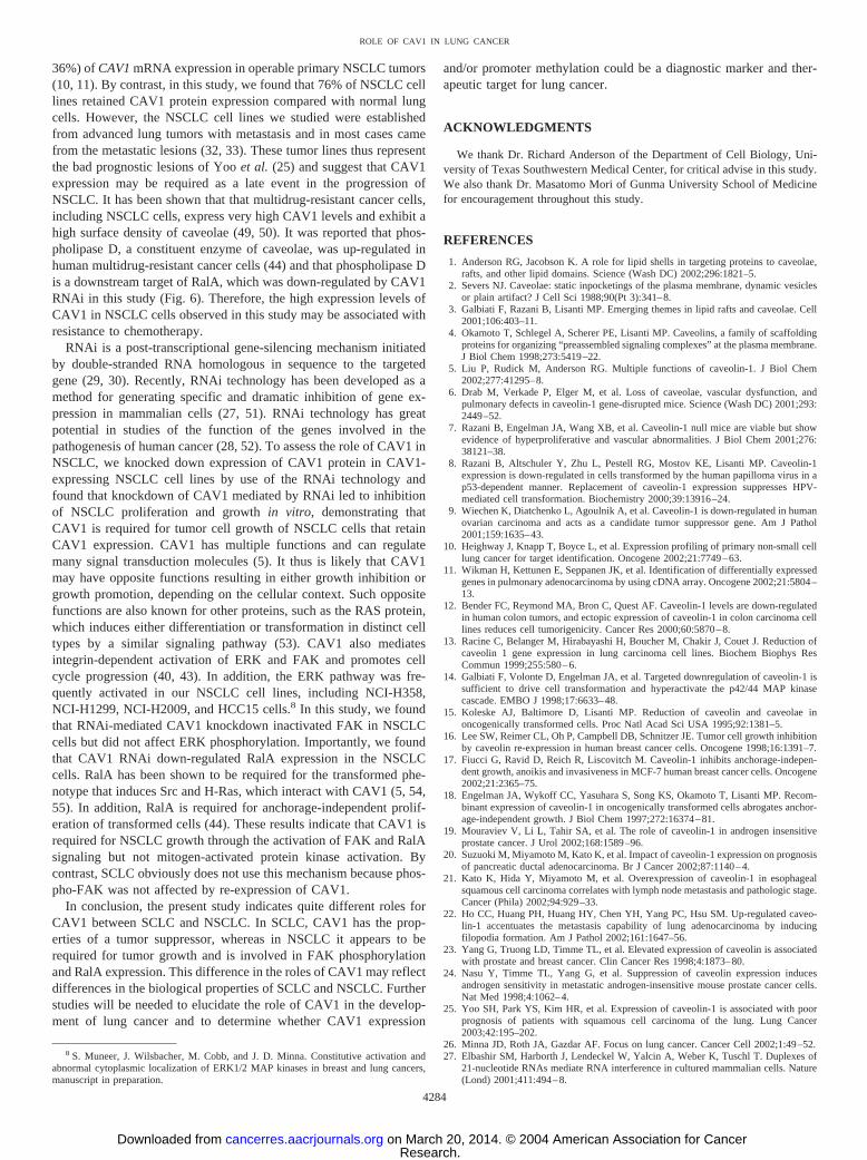
RNAi-Mediated CAV1 Knockdown Led to Reduced Phospho-rylation of FAK and RalA in NSCLC Cells. We wanted to deter-mine how CAV1 RNAi inhibited anchorage-dependent and -inde-pendent growth of NSCLC cells. We were interested in the role ofCAV1 in integrin-FAK signaling because evidence from previousstudies indicated a role of CAV1 in integrin-mediated FAK activation,cell adhesion, and anchorage-dependent growth (40, 43). We werealso interested in the role of CAV1 in RalA regulation because werecently showed that RalA is required for the anchorage-independentproliferation of transformed cells (44). Thus, we assessed the effect ofRNAi-mediated CAV1 knockdown on FAK activation in H1299 andH358 NSCLCs expressing CAV1. In both NSCLC cell lines, CAV1RNAi reduced the levels of phosphorylated FAK and RalA, whereasit did not affect the levels of RalB and phosphorylated extracellularsignal-regulated kinase 1/2 (phospho-ERK1/2; Fig. 6). These resultsindicate that the growth-inhibitory effect mediated by CAV1 RNAi isassociated with the loss of expression of phospho-FAK and RalA inNSCLC cells. By contrast, exogenous CAV1 expression in SCLCs,whereas associated with growth inhibition, was not associated withexpression of phospho-FAK (data not shown).
DISCUSSION
Previous studies have reported that CAV1 is expressed in 21–36%of resected primary NSCLCs and that CAV1 expression is associatedwith poor prognosis of squamous cell lung cancer (8, 10, 11, 13, 25).In these studies, however, only two SCLC cancer cell lines werestudied, no reasons for expression or nonexpression of CAV1 wereprovided, and no functional correlations were made. We thereforefocused in the present study on comparing SCLC and NSCLC, themethylation status of CAV1, and the functional effects of alteringCAV1 expression in these two major histological types of lung cancer.We examined endogenous expression of CAV1 protein in several lungcancer cell lines, nearly all of which came from metastatic (poorprognosis) lesions. CAV1 protein expression was reduced or absent in95% of the SCLCs, whereas it was retained in 76% of NSCLCscompared with normal human lung epithelial cultures. We thus found
Fig. 3. Exogenous caveolin-1 (CAV1) expression inhibits colony formation by SCLCcells in soft agar. A, NCI-H524 and NCI-H187 small cell lung cancer and NCI-H1299 andNCI-H358 cell lines were transfected with pcDNA3.1/CAV1 or pcDNA 3.1. Cells wereharvested at 48 h post-transfection, and Western blotting was performed. Fifteen �g ofwhole cell lysate were loaded in each lane. HBEC4, an immortalized bronchial epithelialcell line, was used as a positive control. B, cells were suspended in soft agar in completemedium containing 800 �g/ml G418 at 48 h post-transfection. Colonies were countedafter 4 weeks. pcDNA3.1 vector transfection control was set at 100%. Columns representthe mean � SD (bars).
4281
ROLE OF CAV1 IN LUNG CANCER
Research. on March 20, 2014. © 2004 American Association for Cancercancerres.aacrjournals.org Downloaded from
dramatic difference in endogenous expression level of CAV1 proteinbetween SCLCs and NSCLCs.
Interestingly, when we classified the tumor cell lines as suspensionor adherent cultures according to their culture morphologies, all cellsgrowing as suspension cultures lacked CAV1 protein expressionwhereas all plastic adherent cells expressed CAV1 protein. We there-fore found complete association between the CAV1 expression leveland cell culture morphology and growth characteristics for lungcancer cells. It is known that integrins are caveolae-interacting mol-ecules and that CAV1 is required for integrin-dependent FAK phos-phorylation and adhesion (40, 43). In fact, we found a correlationbetween FAK phosphorylation, CAV1 expression, and culture mor-phology in lung cancer cell lines. The loss of CAV1 protein expres-sion is therefore also related to the functional loss of integrin-depend-ent adhesion in lung cancer cell lines that leads to tumor cells growingin suspension. Although most SCLC cell lines grew as suspensioncultures and most NSCLC cell lines grew attached to the culture dish,two SCLC cell lines expressing CAV1, H1607 and H2227, had thesame culture morphology as the majority of NSCLCs, whereasNSCLC cell line H1155 grew as a cell suspension, similar to mostSCLC cell lines. Obviously, these tumor cell lines represent interest-ing exceptions that provide further support to the CAV1 expressionconnections. In experiments not presented here, we performed oligo-nucleotide (Affimetrix HG-U133) expression profiles and clusteringanalysis, which showed that these tumor cells cluster with theirassigned histological group but on the edges of these clusters. Wetherefore have expression profiling data placing these interesting
tumor cells in the same histological classes (SCLC or NSCLC) asreported in their initial pathological diagnosis.
CAV1 has been implicated as a candidate tumor suppressor gene inseveral human tumors including lung cancer. However, to our knowl-edge, no evidence for mutations, deletions, or epigenetic alterationscaused by promoter methylation in the CAV1 gene has been reportedin lung cancer (10, 22). Here we report aberrant methylation of CpGislands in the CAV1 gene associated with transcriptional inactivationin SCLC cell lines. In six SCLC cell lines examined by bisulfitesequencing, heavy or complete methylation was found at seven CpGsites (nt 29442–29676 on the AC006159 sequence) in the promoterregion of CAV1, whereas these CpGs were unmethylated or onlypartially methylated in CAV1-expressing lung cancer cell lines and innormal bronchial epithelial cell cultures. Furthermore, methylation-specific PCR analysis demonstrated that CAV1 was methylated in93% (14 of 15) of SCLC lines but only 9% (1 of 11) of NSCLC lineswith a strong concordance between methylation status and CAV1expression. The major mode of inactivation of many tumor suppressorgenes in human cancers, including lung cancer, involves tumor-acquired promoter hypermethylation (45, 46). In fact, the promoterregion of CAV1 was methylated in breast and prostate cancers at thesame CpG sites that were found to be methylated in the SCLC cells inthis study (41, 42). Therefore, studies of breast, prostate, and now lungcancers have found that methylation of the same CpG sites is asso-ciated with loss of CAV1 expression. Although formal experimentsdocumenting promoter activity need to be done, these results in
Fig. 4. Small interfering RNA (siRNA)-mediated down-regulation of caveolin-1 (CAV1) expression inhibited in vitro growth of the NCI-H1299 non-small cell lung cancer cell line.A, NCI-H1299 cells were transfected with 100 nM annealed sense and antisense 21-mer RNA oligonucleotides directed against either CAV1 or Tax. Treatment with Oligofectaminereagent alone was used as a control. After 72 h, the cells were stained with anticaveolin or anti-�-tubulin primary antibodies and analyzed by fluorescent microscopy. B, cells wereharvested at 72 h, 144 h, and 9 days post-transfection. Fifteen �g of total protein were loaded in each lane, and Western blotting was performed. Cells treated with Oligofectaminereagent only (Lanes 1 and 2) and Tax siRNA (Lanes 3 and 4) were used as controls. Two CAV1 siRNAs, CAV1-1 (Lanes 5–7) and CAV1-2 (Lanes 8–10), which target different mRNAsites, were used. C, CAV1 siRNAs inhibit cellular proliferation in the NCI-H1299 cell line. siRNAs were transfected into the cells, and the bromodeoxyuridine incorporation assay wasperformed in replicates of 8 at 72 h post-transfection. Oligofectamine control treatment was set at 100%. Columns represent the mean � SD (bars). D, CAV1 siRNAs inhibit colonyformation by the NCI-H1299 non-small cell lung cancer cell line. Forty-eight h after siRNA transfection, cells were plated for colony formation assay in liquid culture and soft agar.Oligofectamine control treatment was set at 100%. Columns represent the mean � SD (bars). The data were obtained from three independent experiments. Marked down-regulationof CAV1 protein expression in CAV1 siRNA-transfected cells was confirmed by Western blot analysis for every experiment.
4282
ROLE OF CAV1 IN LUNG CANCER
Research. on March 20, 2014. © 2004 American Association for Cancercancerres.aacrjournals.org Downloaded from
diverse tumors highlight the importance of these CpG sites for CAV1expression.
Recently, a missense mutation in CAV1 at codon 132 (P132L) wasdetected in invasive scirrhous breast cancers, and recombinant expres-sion of the CAV1 P132L mutant could lead to mislocalization andintracellular retention of wild-type CAV1, cellular transformation,and promotion of invasive ability (39). Thus, the P132L mutant couldbehave in a dominant-negative manner (39, 47). However, we did notfind the CAV1 P132L mutation in the 46 lung cancer cell linesexamined, consistent with previous studies describing no evidence ofCAV1 mutation in lung cancers (10, 22). It is therefore unlikely thatthe CAV1 P132L mutation is important for the development of lungcancer.
We next assessed the growth-inhibitory effect of forced CAV1expression in the SCLCs as was done in breast cancer cells in previousstudies (16, 17). We found that exogenous CAV1 expression led to invitro growth inhibition, as assessed by the soft-agar growth assay, inthe NCI-H187 and NCI-H524 SCLC cell lines, which lacked CAV1expression, but had no effect in tumor cells already expressing en-dogenous CAV1. These results suggest that CAV1 can act as a tumorsuppressor in SCLCs, as it does in breast cancer.
Although CAV1 has been implicated as a tumor suppressor gene,previous studies have reported CAV1 up-regulation in several humantumors, and the up-regulation was associated with tumor progressionand poor prognosis (19–24). CAV1 plays a role in prostate cancerprogression and protects prostate cancer cells from Myc-inducedapoptosis (19, 23, 48). As for lung cancer, the up-regulation of CAV1has been reported in lung adenocarcinoma with enhanced invasive/
metastasis capacity (22). More recently, Yoo et al. (25) found thatCAV1 was expressed in 32% of primary squamous cell lung cancersand that the CAV1 expression was significantly correlated with ad-vanced pathological stage and poor prognosis. Two other studiesusing cDNA microarray analysis reported similar frequencies (21–
Fig. 6. Effect of RNA interference-mediated caveolin-1 (CAV1) knockdown on theexpression of phosphorylated focal adhesion kinase (Phospho-FAK), focal adhesionkinase (Total FAK), RalA, RalB, phosphorylated extracellular-signal regulated kinase 1/2(Phospho-ERK1/2), and actin. Twenty-five �g of total protein were loaded in each lane,and Western blotting was performed. siRNA, small interfering RNA.
Fig. 5. A, specific small interfering RNA (siRNA)-mediated down-regulation of caveolin-1 (CAV1) protein expression in NCI-H2009, HCC15, and NCI-H358 NSCLC cell lines.siRNAs were transfected into NCI-H2009, HCC15, and NCI-H358 cells. Conditions for Western blotting were the same as Fig. 4B. Lanes: 1, treatment with medium only; 2, treatmentwith Oligofectamine only at 72 h post-transfection; 3, Tax siRNA at 72 h post-transfection; 4, CAV1 siRNA (CAV1-1) at 72 h post-transfection; 5, CAV1 siRNA (CAV1-1) at 144 hpost-transfection; 6, �-catenin siRNA at 72 h post-transfection; 7, �-catenin siRNA at 144 h post-transfection. B, �-catenin siRNA was used as a control to verify that siRNAtransfection was successful because CAV1 was not expressed in NCI-H187 and NCI-H524 small cell lung cancers. After 72 h, cells were harvested, and Western blotting was performed.Conditions for Western blotting were the same as Fig. 4B. C and D, CAV1 siRNA inhibits colony formation by NCI-H2009, HCC15, and NCI-H358 cells in liquid culture (C) andsoft-agar colony formation of NCI-H2009, HCC15, and NCI-H358 cells (D) but not of NCI-H187 and NCI-H524 cells. The method was the same as in Fig. 4D. Oligofectamine controltreatment was set at 100%. Columns represent the mean � SD (bars). The data were obtained from three independent experiments.
4283
ROLE OF CAV1 IN LUNG CANCER
Research. on March 20, 2014. © 2004 American Association for Cancercancerres.aacrjournals.org Downloaded from
36%) of CAV1 mRNA expression in operable primary NSCLC tumors(10, 11). By contrast, in this study, we found that 76% of NSCLC celllines retained CAV1 protein expression compared with normal lungcells. However, the NSCLC cell lines we studied were establishedfrom advanced lung tumors with metastasis and in most cases camefrom the metastatic lesions (32, 33). These tumor lines thus representthe bad prognostic lesions of Yoo et al. (25) and suggest that CAV1expression may be required as a late event in the progression ofNSCLC. It has been shown that that multidrug-resistant cancer cells,including NSCLC cells, express very high CAV1 levels and exhibit ahigh surface density of caveolae (49, 50). It was reported that phos-pholipase D, a constituent enzyme of caveolae, was up-regulated inhuman multidrug-resistant cancer cells (44) and that phospholipase Dis a downstream target of RalA, which was down-regulated by CAV1RNAi in this study (Fig. 6). Therefore, the high expression levels ofCAV1 in NSCLC cells observed in this study may be associated withresistance to chemotherapy.
RNAi is a post-transcriptional gene-silencing mechanism initiatedby double-stranded RNA homologous in sequence to the targetedgene (29, 30). Recently, RNAi technology has been developed as amethod for generating specific and dramatic inhibition of gene ex-pression in mammalian cells (27, 51). RNAi technology has greatpotential in studies of the function of the genes involved in thepathogenesis of human cancer (28, 52). To assess the role of CAV1 inNSCLC, we knocked down expression of CAV1 protein in CAV1-expressing NSCLC cell lines by use of the RNAi technology andfound that knockdown of CAV1 mediated by RNAi led to inhibitionof NSCLC proliferation and growth in vitro, demonstrating thatCAV1 is required for tumor cell growth of NSCLC cells that retainCAV1 expression. CAV1 has multiple functions and can regulatemany signal transduction molecules (5). It thus is likely that CAV1may have opposite functions resulting in either growth inhibition orgrowth promotion, depending on the cellular context. Such oppositefunctions are also known for other proteins, such as the RAS protein,which induces either differentiation or transformation in distinct celltypes by a similar signaling pathway (53). CAV1 also mediatesintegrin-dependent activation of ERK and FAK and promotes cellcycle progression (40, 43). In addition, the ERK pathway was fre-quently activated in our NSCLC cell lines, including NCI-H358,NCI-H1299, NCI-H2009, and HCC15 cells.8 In this study, we foundthat RNAi-mediated CAV1 knockdown inactivated FAK in NSCLCcells but did not affect ERK phosphorylation. Importantly, we foundthat CAV1 RNAi down-regulated RalA expression in the NSCLCcells. RalA has been shown to be required for the transformed phe-notype that induces Src and H-Ras, which interact with CAV1 (5, 54,55). In addition, RalA is required for anchorage-independent prolif-eration of transformed cells (44). These results indicate that CAV1 isrequired for NSCLC growth through the activation of FAK and RalAsignaling but not mitogen-activated protein kinase activation. Bycontrast, SCLC obviously does not use this mechanism because phos-pho-FAK was not affected by re-expression of CAV1.
In conclusion, the present study indicates quite different roles forCAV1 between SCLC and NSCLC. In SCLC, CAV1 has the prop-erties of a tumor suppressor, whereas in NSCLC it appears to berequired for tumor growth and is involved in FAK phosphorylationand RalA expression. This difference in the roles of CAV1 may reflectdifferences in the biological properties of SCLC and NSCLC. Furtherstudies will be needed to elucidate the role of CAV1 in the develop-ment of lung cancer and to determine whether CAV1 expression
and/or promoter methylation could be a diagnostic marker and ther-apeutic target for lung cancer.
ACKNOWLEDGMENTS
We thank Dr. Richard Anderson of the Department of Cell Biology, Uni-versity of Texas Southwestern Medical Center, for critical advise in this study.We also thank Dr. Masatomo Mori of Gunma University School of Medicinefor encouragement throughout this study.
REFERENCES
1. Anderson RG, Jacobson K. A role for lipid shells in targeting proteins to caveolae,rafts, and other lipid domains. Science (Wash DC) 2002;296:1821–5.
2. Severs NJ. Caveolae: static inpocketings of the plasma membrane, dynamic vesiclesor plain artifact? J Cell Sci 1988;90(Pt 3):341–8.
3. Galbiati F, Razani B, Lisanti MP. Emerging themes in lipid rafts and caveolae. Cell2001;106:403–11.
4. Okamoto T, Schlegel A, Scherer PE, Lisanti MP. Caveolins, a family of scaffoldingproteins for organizing “preassembled signaling complexes” at the plasma membrane.J Biol Chem 1998;273:5419–22.
5. Liu P, Rudick M, Anderson RG. Multiple functions of caveolin-1. J Biol Chem2002;277:41295–8.
6. Drab M, Verkade P, Elger M, et al. Loss of caveolae, vascular dysfunction, andpulmonary defects in caveolin-1 gene-disrupted mice. Science (Wash DC) 2001;293:2449–52.
7. Razani B, Engelman JA, Wang XB, et al. Caveolin-1 null mice are viable but showevidence of hyperproliferative and vascular abnormalities. J Biol Chem 2001;276:38121–38.
8. Razani B, Altschuler Y, Zhu L, Pestell RG, Mostov KE, Lisanti MP. Caveolin-1expression is down-regulated in cells transformed by the human papilloma virus in ap53-dependent manner. Replacement of caveolin-1 expression suppresses HPV-mediated cell transformation. Biochemistry 2000;39:13916–24.
9. Wiechen K, Diatchenko L, Agoulnik A, et al. Caveolin-1 is down-regulated in humanovarian carcinoma and acts as a candidate tumor suppressor gene. Am J Pathol2001;159:1635–43.
10. Heighway J, Knapp T, Boyce L, et al. Expression profiling of primary non-small celllung cancer for target identification. Oncogene 2002;21:7749–63.
11. Wikman H, Kettunen E, Seppanen JK, et al. Identification of differentially expressedgenes in pulmonary adenocarcinoma by using cDNA array. Oncogene 2002;21:5804–13.
12. Bender FC, Reymond MA, Bron C, Quest AF. Caveolin-1 levels are down-regulatedin human colon tumors, and ectopic expression of caveolin-1 in colon carcinoma celllines reduces cell tumorigenicity. Cancer Res 2000;60:5870–8.
13. Racine C, Belanger M, Hirabayashi H, Boucher M, Chakir J, Couet J. Reduction ofcaveolin 1 gene expression in lung carcinoma cell lines. Biochem Biophys ResCommun 1999;255:580–6.
14. Galbiati F, Volonte D, Engelman JA, et al. Targeted downregulation of caveolin-1 issufficient to drive cell transformation and hyperactivate the p42/44 MAP kinasecascade. EMBO J 1998;17:6633–48.
15. Koleske AJ, Baltimore D, Lisanti MP. Reduction of caveolin and caveolae inoncogenically transformed cells. Proc Natl Acad Sci USA 1995;92:1381–5.
16. Lee SW, Reimer CL, Oh P, Campbell DB, Schnitzer JE. Tumor cell growth inhibitionby caveolin re-expression in human breast cancer cells. Oncogene 1998;16:1391–7.
17. Fiucci G, Ravid D, Reich R, Liscovitch M. Caveolin-1 inhibits anchorage-indepen-dent growth, anoikis and invasiveness in MCF-7 human breast cancer cells. Oncogene2002;21:2365–75.
18. Engelman JA, Wykoff CC, Yasuhara S, Song KS, Okamoto T, Lisanti MP. Recom-binant expression of caveolin-1 in oncogenically transformed cells abrogates anchor-age-independent growth. J Biol Chem 1997;272:16374–81.
19. Mouraviev V, Li L, Tahir SA, et al. The role of caveolin-1 in androgen insensitiveprostate cancer. J Urol 2002;168:1589–96.
20. Suzuoki M, Miyamoto M, Kato K, et al. Impact of caveolin-1 expression on prognosisof pancreatic ductal adenocarcinoma. Br J Cancer 2002;87:1140–4.
21. Kato K, Hida Y, Miyamoto M, et al. Overexpression of caveolin-1 in esophagealsquamous cell carcinoma correlates with lymph node metastasis and pathologic stage.Cancer (Phila) 2002;94:929–33.
22. Ho CC, Huang PH, Huang HY, Chen YH, Yang PC, Hsu SM. Up-regulated caveo-lin-1 accentuates the metastasis capability of lung adenocarcinoma by inducingfilopodia formation. Am J Pathol 2002;161:1647–56.
23. Yang G, Truong LD, Timme TL, et al. Elevated expression of caveolin is associatedwith prostate and breast cancer. Clin Cancer Res 1998;4:1873–80.
24. Nasu Y, Timme TL, Yang G, et al. Suppression of caveolin expression inducesandrogen sensitivity in metastatic androgen-insensitive mouse prostate cancer cells.Nat Med 1998;4:1062–4.
25. Yoo SH, Park YS, Kim HR, et al. Expression of caveolin-1 is associated with poorprognosis of patients with squamous cell carcinoma of the lung. Lung Cancer2003;42:195–202.
26. Minna JD, Roth JA, Gazdar AF. Focus on lung cancer. Cancer Cell 2002;1:49–52.27. Elbashir SM, Harborth J, Lendeckel W, Yalcin A, Weber K, Tuschl T. Duplexes of
21-nucleotide RNAs mediate RNA interference in cultured mammalian cells. Nature(Lond) 2001;411:494–8.
8 S. Muneer, J. Wilsbacher, M. Cobb, and J. D. Minna. Constitutive activation andabnormal cytoplasmic localization of ERK1/2 MAP kinases in breast and lung cancers,manuscript in preparation.
4284
ROLE OF CAV1 IN LUNG CANCER
Research. on March 20, 2014. © 2004 American Association for Cancercancerres.aacrjournals.org Downloaded from
28. Borkhardt A. Blocking oncogenes in malignant cells by RNA interference–new hopefor a highly specific cancer treatment? Cancer Cell 2002;2:167–8.
29. Sharp PA. RNA interference—2001. Genes Dev 2001;15:485–90.30. Hannon GJ. RNA interference. Nature (Lond) 2002;418:244–51.31. Caplen NJ, Parrish S, Imani F, Fire A, Morgan RA. Specific inhibition of gene
expression by small double-stranded RNAs in invertebrate and vertebrate systems.Proc Natl Acad Sci USA 2001;98:9742–7.
32. Phelps RM, Johnson BE, Ihde DC, et al. NCI-Navy Medical Oncology Branch cellline data base. J Cell Biochem Suppl 1996;24:32–91.
33. Wistuba II, Bryant D, Behrens C, et al. Comparison of features of human lung cancercell lines and their corresponding tumors. Clin Cancer Res 1999;5:991–1000.
34. Verma UN, Surabhi RM, Schmaltieg A, Becerra C, Gaynor RB. Small interferingRNAs directed against beta-catenin inhibit the in vitro and in vivo growth of coloncancer cells. Clin Cancer Res 2003;9:1291–300.
35. Braasch DA, Jensen S, Liu Y, et al. RNA interference in mammalian cells bychemically-modified RNA. Biochemistry 2003;42:7967–75.
36. Harada K, Toyooka S, Maitra A, et al. Aberrant promoter methylation and silencingof the RASSF1A gene in pediatric tumors and cell lines. Oncogene 2002;21:4345–9.
37. Xu XL, Wu LC, Du F, et al. Inactivation of human SRBC, located within the11p15.5-p15.4 tumor suppressor region, in breast and lung cancers. Cancer Res2001;61:7943–9.
38. Burbee DG, Forgacs E, Zochbauer-Muller S, et al. Epigenetic inactivation ofRASSF1A in lung and breast cancers and malignant phenotype suppression. J NatlCancer Inst (Bethesda) 2001;93:691–9.
39. Hayashi K, Matsuda S, Machida K, et al. Invasion activating caveolin-1 mutation inhuman scirrhous breast cancers. Cancer Res 2001;61:2361–4.
40. Wei Y, Yang X, Liu Q, Wilkins JA, Chapman HA. A role for caveolin and theurokinase receptor in integrin-mediated adhesion and signaling. J Cell Biol 1999;144:1285–94.
41. Engelman JA, Zhang XL, Lisanti MP. Sequence and detailed organization of thehuman caveolin-1 and -2 genes located near the D7S522 locus (7q31.1). Methylationof a CpG island in the 5� promoter region of the caveolin-1 gene in human breastcancer cell lines. FEBS Lett 1999;448:221–30.
42. Cui J, Rohr LR, Swanson G, Speights VO, Maxwell T, Brothman AR. Hypermethy-lation of the caveolin-1 gene promoter in prostate cancer. Prostate 2001;46:249–56.
43. Wary KK, Mariotti A, Zurzolo C, Giancotti FG. A requirement for caveolin-1 andassociated kinase Fyn in integrin signaling and anchorage-dependent cell growth. Cell1998;94:625–34.
44. Chien Y, White MA. RAL GTPases are linchpin modulators of human tumour-cellproliferation and survival. EMBO Rep 2003;4:800–6.
45. Baylin SB, Esteller M, Rountree MR, Bachman KE, Schuebel K, Herman JG.Aberrant patterns of DNA methylation, chromatin formation and gene expression incancer. Hum Mol Genet 2001;10:687–92.
46. Zochbauer-Muller S, Fong KM, Virmani AK, Geradts J, Gazdar AF, Minna JD.Aberrant promoter methylation of multiple genes in non-small cell lung cancers.Cancer Res 2001;61:249–55.
47. Lee H, Park DS, Razani B, Russell RG, Pestell RG, Lisanti MP. Caveolin-1 mutations(P132L and null) and the pathogenesis of breast cancer: caveolin-1 (P132L) behavesin a dominant-negative manner and caveolin-1 (�/�) null mice show mammaryepithelial cell hyperplasia. Am J Pathol 2002;161:1357–69.
48. Timme TL, Goltsov A, Tahir S, et al. Caveolin-1 is regulated by c-myc andsuppresses c-myc-induced apoptosis. Oncogene 2000;19:3256–65.
49. Yang CP, Galbiati F, Volonte D, Horwitz SB, Lisanti MP. Upregulation ofcaveolin-1 and caveolae organelles in Taxol-resistant A549 cells. FEBS Lett1998;439:368 –72.
50. Lavie Y, Fiucci G, Liscovitch M. Upregulation of caveolin in multidrug resistantcancer cells: functional implications. Adv Drug Deliv Rev 2001;49:317–23.
51. Brummelkamp TR, Bernards R, Agami R. Stable suppression of tumorigenicity byvirus-mediated RNA interference. Cancer Cell 2002;2:243–7.
52. Harborth J, Elbashir SM, Bechert K, Tuschl T, Weber K. Identification of essentialgenes in cultured mammalian cells using small interfering RNAs. J Cell Sci 2001;114:4557–65.
53. Crespo P, Leon J. Ras proteins in the control of the cell cycle and cell differentiation.Cell Mol Life Sci 2000;57:1613–36.
54. Aguirre-Ghiso JA, Frankel P, Farias EF, et al. RalA requirement for v-Src- andv-Ras-induced tumorigenicity and overproduction of urokinase-type plasminogenactivator: involvement of metalloproteases. Oncogene 1999;18:4718–25.
55. Xu L, Frankel P, Jackson D, et al. Elevated phospholipase D activity in H-Ras- butnot K-Ras-transformed cells by the synergistic action of RalA and ARF6. Mol CellBiol 2003;23:645–54.
56. Suzuki M. Sunaga N, Shames DS, Toyooka S, Gazdar AF, Minna JD. RNA inter-ference-mediated knockdown of DNA methyltransferase 1 leads to promoter de-methylation and gene re-expression in human lung and breast cancer cells. CancerRes 2004;64:3137–43.
4285
ROLE OF CAV1 IN LUNG CANCER
Research. on March 20, 2014. © 2004 American Association for Cancercancerres.aacrjournals.org Downloaded from


