Embed Size (px)
Citation preview
ARTICLEReceived 25 Nov 2013 | Accepted 2 Jun 2014 | Published 27 Jun 2014
Degradable lipid nanoparticles with predictablein vivo siRNA delivery activityKathryn A. Whitehead1,w, J. Robert Dorkin2, Arturo J. Vegas1, Philip H. Chang1, Omid Veiseh1,
Jonathan Matthews1, Owen S. Fenton3, Yunlong Zhang1, Karsten T. Olejnik1, Volkan Yesilyurt1, Delai Chen1,
Scott Barros4, Boris Klebanov4, Tatiana Novobrantseva4, Robert Langer1,5,6 & Daniel G. Anderson1,5,6
One of the most significant challenges in the development of clinically viable delivery systems
for RNA interference therapeutics is to understand how molecular structures influence
delivery efficacy. Here, we have synthesized 1,400 degradable lipidoids and evaluate their
transfection ability and structure–function activity. We show that lipidoid nanoparticles
mediate potent gene knockdown in hepatocytes and immune cell populations on IV
administration to mice (siRNA EC50 values as low as 0.01 mg kg! 1). We identify four
necessary and sufficient structural and pKa criteria that robustly predict the ability of
nanoparticles to mediate greater than 95% protein silencing in vivo. Because these
efficacy criteria can be dictated through chemical design, this discovery could eliminate our
dependence on time-consuming and expensive cell culture assays and animal testing. Herein,
we identify promising degradable lipidoids and describe new design criteria that reliably
predict in vivo siRNA delivery efficacy without any prior biological testing.
DOI: 10.1038/ncomms5277
1 Koch Institute for Integrative Cancer Research, Massachusetts Institute of Technology, 77 Massachusetts Avenue, Cambridge, Massachusetts 02139, USA.2 Department of Biology, Massachusetts Institute of Technology, 77 Massachusetts Avenue, Cambridge, Massachusetts 02139, USA. 3 Department ofChemistry, Massachusetts Institute of Technology, 77 Massachusetts Avenue, Cambridge, Massachusetts 02139, USA. 4 Alnylam Pharmaceuticals, 300Third Street, Cambridge, Massachusetts 02142, USA. 5 Department of Chemical Engineering, Massachusetts Institute of Technology, 77 MassachusettsAvenue, Cambridge, Massachusetts 02139, USA. 6 The Institute for Medical Engineering and Science, Massachusetts Institute of Technology,77 Massachusetts Avenue, Cambridge, Massachusetts 02139 USA. w Present address: Department of Chemical Engineering, Carnegie Mellon University,5000 Forbes Avenue, Pittsburgh, Pennsylvania 15217, USA. Correspondence and requests for materials should be addressed to D.G.A. (email:[email protected]).
NATURE COMMUNICATIONS | 5:4277 | DOI: 10.1038/ncomms5277 | www.nature.com/naturecommunications 1
& 2014 Macmillan Publishers Limited. All rights reserved.
The development of drug delivery systems often involvesextensive characterization and in vitro testing before theconduct of preclinical studies in rodent or higher order
animal models. Unfortunately, progress towards the clinic hasbeen hindered because in vitro results generally do not correlatewell with in vivo data1–3. This has been particularly true for RNAinterference therapeutics (RNAi)4. While the past decade has seenan exponential increase in the number of in vitro short interferingRNA (siRNA) delivery studies, very few materials have beenreported to mediate potent gene silencing in vivo5–8, and only ahandful are being tested in clinical trials9.
One major challenge in the development of suitable deliverysystems is the identification of delivery vehicle chemistries withsafety and efficacy characteristics that support a sufficiently broadtherapeutic index for chronic indications. This requirement forany kind of RNAi therapeutic stems from the transient nature ofgene silencing effects (typically on the order of several days toseveral weeks in vivo) and the subsequent need for sustained,repeated siRNA dosing over the course of treatment10,11.Unfortunately, improvements in delivery vehicle potency do notalways result in an enlargement of the therapeutic index due toreductions in tolerated dose levels. In recent years, considerableprogress has been made in regards to potency6,12, but hasgenerally been done so using non-hydrolysable materials. Severalchallenges associated with delivery vehicle toxicity, degradabilityand potential for immune stimulation remain13–15. With thismotivation in mind, we sought to identify degradable materialsthat enable potent silencing in vivo without causing off-targettoxicities (for example, immune system stimulation, necrosis,hepatocellular injury). At the same time, we were interested in theestablishment of predictive structure–function relationships that
would potentially eliminate the need for costly and time-consuming in vitro screening procedures. One approachtowards these dual objectives is the high-throughput screeningof libraries of compounds, which can yield large quantitiesof structure–activity data while significantly increasing theprobability of identifying potent delivery compounds16–18.
Herein, we describe the discovery of several lipid nanoparticles(LNPs) that facilitate high levels of gene silencing in multiple cellsubtypes in mice, including hepatocytes, monocytes, macrophagesand dendritic cells. Furthermore, we establish a set of four‘efficacy criteria’ that robustly predict the ability of LNPs toefficiently deliver siRNA without any a priori biological testing.
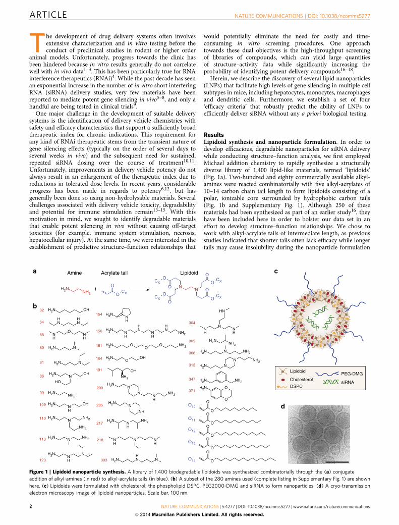
ResultsLipidoid synthesis and nanoparticle formulation. In order todevelop efficacious, degradable nanoparticles for siRNA deliverywhile conducting structure–function analysis, we first employedMichael addition chemistry to rapidly synthesize a structurallydiverse library of 1,400 lipid-like materials, termed ‘lipidoids’(Fig. 1a). Two-hundred and eighty commercially available alkyl-amines were reacted combinatorially with five alkyl-acrylates of10–14 carbon chain tail length to form lipidoids consisting of apolar, ionizable core surrounded by hydrophobic carbon tails(Fig. 1b and Supplementary Fig. 1). Although 250 of thesematerials had been synthesized as part of an earlier study16, theyhave been included here in order to bolster our data set in aneffort to develop structure–function relationships. We chose towork with alkyl-acrylate tails of intermediate length, as previousstudies indicated that shorter tails often lack efficacy while longertails may cause insolubility during the nanoparticle formulation
Amine LipidoidAcrylate taila c
d
b
Lipidoid
CholesterolDSPC
PEG-DMG
siRNA
OCX
OH2N
NH2
NN
OCX
O
O
O
OCX
OO
OCX
CX
O
OO
OO
OO
OO
O
H2N N NH2
H2NNH
N
H2NNH
NH2
H2NHN N
NH
NNH
HN
H2NNH2
H2N N NH2
H2N NN NH2
H2N N
H2N N
80
81
H2NNH2
99
H2NNH
OH109
H2NN
NH2110
NH2
113
123
217
303
304
305
306
313
H2NNH
154
H2NNH
HN
NH
HN
NH2156
H2N OO
O NH2161
H2NO
OH164
OHNH2
191
H2NN
NNH
NH2
200
H2NN
NH
205
NH
N NH
218
H2N OH32
H2N NH2347
O
OH2N371
NH
ONH
68
O14
O13
O12
O11
O10
+
HN
HN64
H2NN
OH
HO86
Figure 1 | Lipidoid nanoparticle synthesis. A library of 1,400 biodegradable lipidoids was synthesized combinatorially through the (a) conjugateaddition of alkyl-amines (in red) to alkyl-acrylate tails (in blue). (b) A subset of the 280 amines used (complete listing in Supplementary Fig. 1) are shownhere. (c) Lipidoids were formulated with cholesterol, the phospholipid DSPC, PEG2000-DMG and siRNA to form nanoparticles. (d) A cryo-transmissionelectron microscopy image of lipidoid nanoparticles. Scale bar, 100 nm.
ARTICLE NATURE COMMUNICATIONS | DOI: 10.1038/ncomms5277
2 NATURE COMMUNICATIONS | 5:4277 | DOI: 10.1038/ncomms5277 | www.nature.com/naturecommunications
& 2014 Macmillan Publishers Limited. All rights reserved.
process12,16. These acrylate-based lipidoids also containhydrolysable ester moieties, functional groups which arecommonly incorporated into delivery vehicles to promotephysiological degradation19–21. Proton NMR analysis indicatedthat a representative lipidoid, 304O13, degraded to theanticipated alkyl-alcohol product under hydrolytic conditions(Supplementary Figs 2 and 3). Conditions were chosen tofacilitate the clear observation of degradation products by NMR.It should be noted that, in vivo, lipidoids would be expected todegrade in the presence of liver-produced enzymes, particularlyesterases22,23.
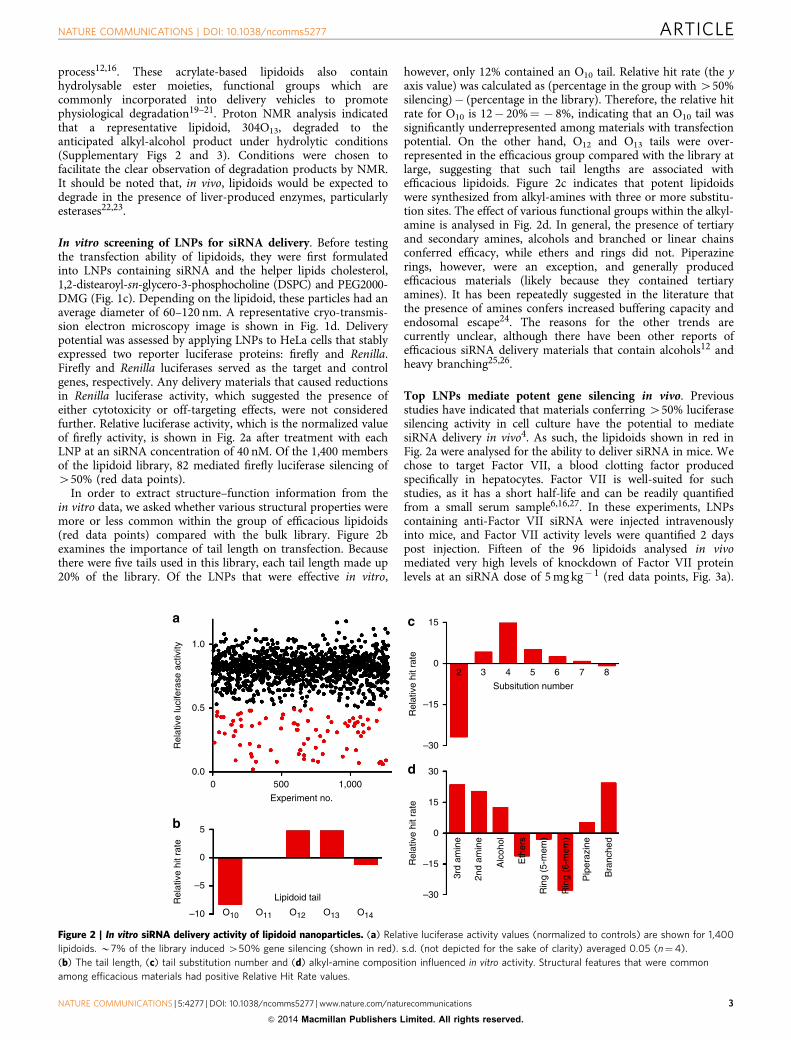
In vitro screening of LNPs for siRNA delivery. Before testingthe transfection ability of lipidoids, they were first formulatedinto LNPs containing siRNA and the helper lipids cholesterol,1,2-distearoyl-sn-glycero-3-phosphocholine (DSPC) and PEG2000-DMG (Fig. 1c). Depending on the lipidoid, these particles had anaverage diameter of 60–120 nm. A representative cryo-transmis-sion electron microscopy image is shown in Fig. 1d. Deliverypotential was assessed by applying LNPs to HeLa cells that stablyexpressed two reporter luciferase proteins: firefly and Renilla.Firefly and Renilla luciferases served as the target and controlgenes, respectively. Any delivery materials that caused reductionsin Renilla luciferase activity, which suggested the presence ofeither cytotoxicity or off-targeting effects, were not consideredfurther. Relative luciferase activity, which is the normalized valueof firefly activity, is shown in Fig. 2a after treatment with eachLNP at an siRNA concentration of 40 nM. Of the 1,400 membersof the lipidoid library, 82 mediated firefly luciferase silencing of450% (red data points).
In order to extract structure–function information from thein vitro data, we asked whether various structural properties weremore or less common within the group of efficacious lipidoids(red data points) compared with the bulk library. Figure 2bexamines the importance of tail length on transfection. Becausethere were five tails used in this library, each tail length made up20% of the library. Of the LNPs that were effective in vitro,
however, only 12% contained an O10 tail. Relative hit rate (the yaxis value) was calculated as (percentage in the group with 450%silencing)! (percentage in the library). Therefore, the relative hitrate for O10 is 12! 20%¼ ! 8%, indicating that an O10 tail wassignificantly underrepresented among materials with transfectionpotential. On the other hand, O12 and O13 tails were over-represented in the efficacious group compared with the library atlarge, suggesting that such tail lengths are associated withefficacious lipidoids. Figure 2c indicates that potent lipidoidswere synthesized from alkyl-amines with three or more substitu-tion sites. The effect of various functional groups within the alkyl-amine is analysed in Fig. 2d. In general, the presence of tertiaryand secondary amines, alcohols and branched or linear chainsconferred efficacy, while ethers and rings did not. Piperazinerings, however, were an exception, and generally producedefficacious materials (likely because they contained tertiaryamines). It has been repeatedly suggested in the literature thatthe presence of amines confers increased buffering capacity andendosomal escape24. The reasons for the other trends arecurrently unclear, although there have been other reports ofefficacious siRNA delivery materials that contain alcohols12 andheavy branching25,26.
Top LNPs mediate potent gene silencing in vivo. Previousstudies have indicated that materials conferring 450% luciferasesilencing activity in cell culture have the potential to mediatesiRNA delivery in vivo4. As such, the lipidoids shown in red inFig. 2a were analysed for the ability to deliver siRNA in mice. Wechose to target Factor VII, a blood clotting factor producedspecifically in hepatocytes. Factor VII is well-suited for suchstudies, as it has a short half-life and can be readily quantifiedfrom a small serum sample6,16,27. In these experiments, LNPscontaining anti-Factor VII siRNA were injected intravenouslyinto mice, and Factor VII activity levels were quantified 2 dayspost injection. Fifteen of the 96 lipidoids analysed in vivomediated very high levels of knockdown of Factor VII proteinlevels at an siRNA dose of 5 mg kg! 1 (red data points, Fig. 3a).
1.0
a
0.5
Rel
ativ
e lu
cife
rase
act
ivity
0.0
5
0
–5
Rel
ativ
e hi
t rat
e
–10 O10 O11 O12
Lipidoid tail
O13 O14
0 500Experiment no.
1,000
b
c
2 3 4Subsitution number
5 6 7 8
15
0
–15
–30
30
15
0
–15
3rd
amin
e
2nd
amin
e
Alc
ohol
Eth
ers
Rin
g (5
-mem
)
Rin
g (6
-mem
)
Pip
eraz
ine
Bra
nche
d
–30
Rel
ativ
e hi
t rat
eR
elat
ive
hit r
ate
d
Figure 2 | In vitro siRNA delivery activity of lipidoid nanoparticles. (a) Relative luciferase activity values (normalized to controls) are shown for 1,400lipidoids. B7% of the library induced 450% gene silencing (shown in red). s.d. (not depicted for the sake of clarity) averaged 0.05 (n¼4).(b) The tail length, (c) tail substitution number and (d) alkyl-amine composition influenced in vitro activity. Structural features that were commonamong efficacious materials had positive Relative Hit Rate values.
NATURE COMMUNICATIONS | DOI: 10.1038/ncomms5277 ARTICLE
NATURE COMMUNICATIONS | 5:4277 | DOI: 10.1038/ncomms5277 | www.nature.com/naturecommunications 3
& 2014 Macmillan Publishers Limited. All rights reserved.
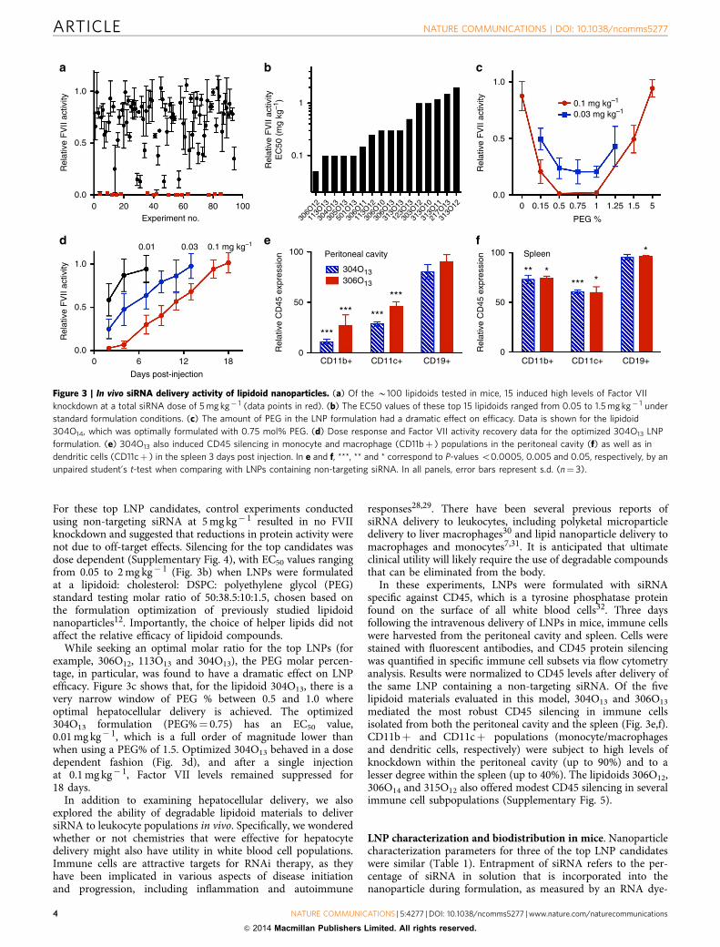
For these top LNP candidates, control experiments conductedusing non-targeting siRNA at 5 mg kg! 1 resulted in no FVIIknockdown and suggested that reductions in protein activity werenot due to off-target effects. Silencing for the top candidates wasdose dependent (Supplementary Fig. 4), with EC50 values rangingfrom 0.05 to 2 mg kg! 1 (Fig. 3b) when LNPs were formulatedat a lipidoid: cholesterol: DSPC: polyethylene glycol (PEG)standard testing molar ratio of 50:38.5:10:1.5, chosen based onthe formulation optimization of previously studied lipidoidnanoparticles12. Importantly, the choice of helper lipids did notaffect the relative efficacy of lipidoid compounds.
While seeking an optimal molar ratio for the top LNPs (forexample, 306O12, 113O13 and 304O13), the PEG molar percen-tage, in particular, was found to have a dramatic effect on LNPefficacy. Figure 3c shows that, for the lipidoid 304O13, there is avery narrow window of PEG % between 0.5 and 1.0 whereoptimal hepatocellular delivery is achieved. The optimized304O13 formulation (PEG%¼ 0.75) has an EC50 value,0.01 mg kg! 1, which is a full order of magnitude lower thanwhen using a PEG% of 1.5. Optimized 304O13 behaved in a dosedependent fashion (Fig. 3d), and after a single injectionat 0.1 mg kg! 1, Factor VII levels remained suppressed for18 days.
In addition to examining hepatocellular delivery, we alsoexplored the ability of degradable lipidoid materials to deliversiRNA to leukocyte populations in vivo. Specifically, we wonderedwhether or not chemistries that were effective for hepatocytedelivery might also have utility in white blood cell populations.Immune cells are attractive targets for RNAi therapy, as theyhave been implicated in various aspects of disease initiationand progression, including inflammation and autoimmune
responses28,29. There have been several previous reports ofsiRNA delivery to leukocytes, including polyketal microparticledelivery to liver macrophages30 and lipid nanoparticle delivery tomacrophages and monocytes7,31. It is anticipated that ultimateclinical utility will likely require the use of degradable compoundsthat can be eliminated from the body.
In these experiments, LNPs were formulated with siRNAspecific against CD45, which is a tyrosine phosphatase proteinfound on the surface of all white blood cells32. Three daysfollowing the intravenous delivery of LNPs in mice, immune cellswere harvested from the peritoneal cavity and spleen. Cells werestained with fluorescent antibodies, and CD45 protein silencingwas quantified in specific immune cell subsets via flow cytometryanalysis. Results were normalized to CD45 levels after delivery ofthe same LNP containing a non-targeting siRNA. Of the fivelipidoid materials evaluated in this model, 304O13 and 306O13mediated the most robust CD45 silencing in immune cellsisolated from both the peritoneal cavity and the spleen (Fig. 3e,f).CD11bþ and CD11cþ populations (monocyte/macrophagesand dendritic cells, respectively) were subject to high levels ofknockdown within the peritoneal cavity (up to 90%) and to alesser degree within the spleen (up to 40%). The lipidoids 306O12,306O14 and 315O12 also offered modest CD45 silencing in severalimmune cell subpopulations (Supplementary Fig. 5).
LNP characterization and biodistribution in mice. Nanoparticlecharacterization parameters for three of the top LNP candidateswere similar (Table 1). Entrapment of siRNA refers to the per-centage of siRNA in solution that is incorporated into thenanoparticle during formulation, as measured by an RNA dye-
1.0
a b c
0.5
Rel
ativ
e F
VII
activ
ity
Rel
ativ
e F
VII
activ
ityE
C50
(m
g kg
–1)
0.0
Experiment no.
0.01 0.03 0.1 mg kg–1
0 20 40 60 80 100
0.1
306O
12
113O
13
304O
13
305O
13
501O
13
306O
11
113O
12
306O
10
306O
13
313O
13
123O
13
303O
12
313O
10
313O
11
217O
13
313O
12
1
Rel
ativ
e F
VII
activ
ity
0.00 0.15 0.5 0.75
PEG %
0.03 mg kg–10.1 mg kg–1
1 1.25 1.5 5
0.5
1.0
d
1.0
0.5
Rel
ativ
e F
VII
activ
ity
Rel
ativ
e C
D45
exp
ress
ion
Rel
ativ
e C
D45
exp
ress
ion
0.00 6 12
Days post-injection18 CD11b+
***
CD11c+ CD19+ CD11b+ CD11c+ CD19+0
50
100
0
50
100Peritoneal cavity
304O13306O13
*** ***
***
Spleen
** **** *
*e f
Figure 3 | In vivo siRNA delivery activity of lipidoid nanoparticles. (a) Of the B100 lipidoids tested in mice, 15 induced high levels of Factor VIIknockdown at a total siRNA dose of 5 mg kg! 1 (data points in red). (b) The EC50 values of these top 15 lipidoids ranged from 0.05 to 1.5 mg kg! 1 understandard formulation conditions. (c) The amount of PEG in the LNP formulation had a dramatic effect on efficacy. Data is shown for the lipidoid304O14, which was optimally formulated with 0.75 mol% PEG. (d) Dose response and Factor VII activity recovery data for the optimized 304O13 LNPformulation. (e) 304O13 also induced CD45 silencing in monocyte and macrophage (CD11bþ ) populations in the peritoneal cavity (f) as well as indendritic cells (CD11cþ ) in the spleen 3 days post injection. In e and f, ***, ** and * correspond to P-values o0.0005, 0.005 and 0.05, respectively, by anunpaired student’s t-test when comparing with LNPs containing non-targeting siRNA. In all panels, error bars represent s.d. (n¼ 3).
ARTICLE NATURE COMMUNICATIONS | DOI: 10.1038/ncomms5277
4 NATURE COMMUNICATIONS | 5:4277 | DOI: 10.1038/ncomms5277 | www.nature.com/naturecommunications
& 2014 Macmillan Publishers Limited. All rights reserved.
binding assay33. These results are in keeping with a previousfinding that efficacious lipidoid nanoparticles often haveentrapment values of approximately 75% (ref. 4). Zeta potentialmeasurements were conducted under neutral pH conditions. pKavalues, which were obtained using a 2-(p-toluidino) naphthalene-6-sulphonic acid (TNS) assay, evaluated the pKa of thenanoparticle surface34. The pKa values of top LNP candidatescorroborate the results of another study in which surface pKavalues in the 6–7 range conveyed efficacy in vivo35.
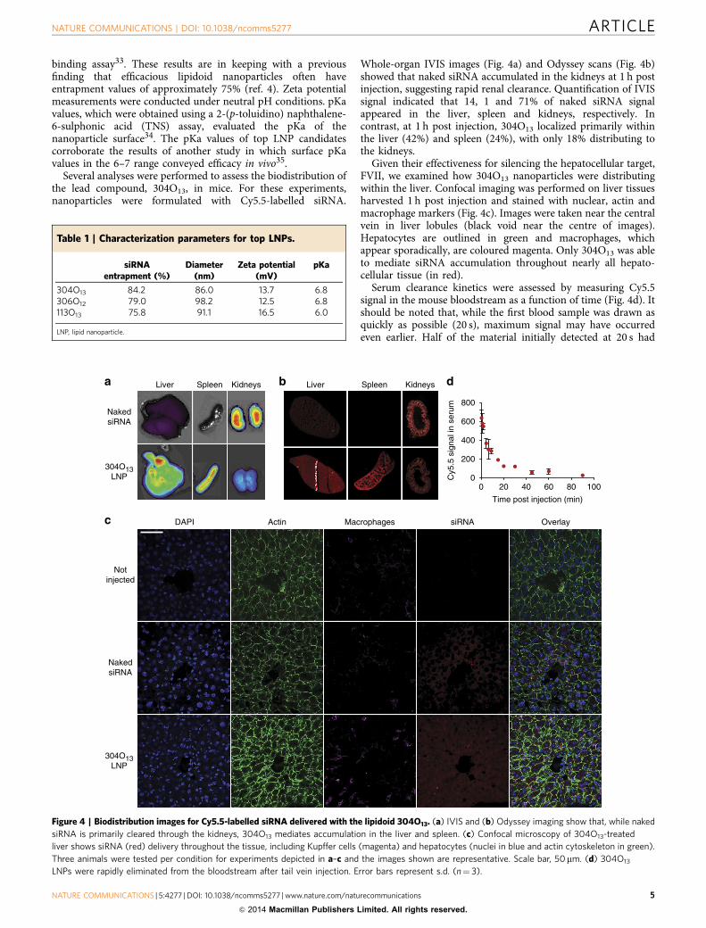
Several analyses were performed to assess the biodistribution ofthe lead compound, 304O13, in mice. For these experiments,nanoparticles were formulated with Cy5.5-labelled siRNA.
Whole-organ IVIS images (Fig. 4a) and Odyssey scans (Fig. 4b)showed that naked siRNA accumulated in the kidneys at 1 h postinjection, suggesting rapid renal clearance. Quantification of IVISsignal indicated that 14, 1 and 71% of naked siRNA signalappeared in the liver, spleen and kidneys, respectively. Incontrast, at 1 h post injection, 304O13 localized primarily withinthe liver (42%) and spleen (24%), with only 18% distributing tothe kidneys.
Given their effectiveness for silencing the hepatocellular target,FVII, we examined how 304O13 nanoparticles were distributingwithin the liver. Confocal imaging was performed on liver tissuesharvested 1 h post injection and stained with nuclear, actin andmacrophage markers (Fig. 4c). Images were taken near the centralvein in liver lobules (black void near the centre of images).Hepatocytes are outlined in green and macrophages, whichappear sporadically, are coloured magenta. Only 304O13 was ableto mediate siRNA accumulation throughout nearly all hepato-cellular tissue (in red).
Serum clearance kinetics were assessed by measuring Cy5.5signal in the mouse bloodstream as a function of time (Fig. 4d). Itshould be noted that, while the first blood sample was drawn asquickly as possible (20 s), maximum signal may have occurredeven earlier. Half of the material initially detected at 20 s had
Table 1 | Characterization parameters for top LNPs.
siRNAentrapment (%)
Diameter(nm)
Zeta potential(mV)
pKa
304O13 84.2 86.0 13.7 6.8306O12 79.0 98.2 12.5 6.8113O13 75.8 91.1 16.5 6.0
LNP, lipid nanoparticle.
Liver Spleen Kidneys Liver Spleen Kidneys
304O13LNP
304O13LNP
NakedsiRNA
Notinjected
DAPI
NakedsiRNA
Actin siRNA OverlayMacrophagesc
a b
0 20 40 60 80 1000
200
400
600
800
Time post injection (min)
Cy5
.5 s
igna
l in
seru
m
d
Figure 4 | Biodistribution images for Cy5.5-labelled siRNA delivered with the lipidoid 304O13. (a) IVIS and (b) Odyssey imaging show that, while nakedsiRNA is primarily cleared through the kidneys, 304O13 mediates accumulation in the liver and spleen. (c) Confocal microscopy of 304O13-treatedliver shows siRNA (red) delivery throughout the tissue, including Kupffer cells (magenta) and hepatocytes (nuclei in blue and actin cytoskeleton in green).Three animals were tested per condition for experiments depicted in a–c and the images shown are representative. Scale bar, 50mm. (d) 304O13
LNPs were rapidly eliminated from the bloodstream after tail vein injection. Error bars represent s.d. (n¼ 3).
NATURE COMMUNICATIONS | DOI: 10.1038/ncomms5277 ARTICLE
NATURE COMMUNICATIONS | 5:4277 | DOI: 10.1038/ncomms5277 | www.nature.com/naturecommunications 5
& 2014 Macmillan Publishers Limited. All rights reserved.
distributed to tissues by 6 min. At 90 min post injection, only 4%of the signal remained.
Safety assessment of lead LNP 304O13. We conducted a pre-liminary safety assessment of the lead LNP, 304O13, and com-pared it with a another previously discovered LNP formulation,C12-20012. C12-200 is a five-tailed, lipidoid that has the sameEC50 as 304O13 (0.01 mg kg! 1). It also has nearly identicalbiodistribution properties as 304O13 (data not shown). It waschosen for comparison purposes because it does not contain anyfunctional groups that are overtly sensitive to hydrolysis. Wechose to examine the effect of doses that were at least 100-foldhigher than the EC50. Serum cytokine levels for both materialswere assessed in mice 4h after a 3 mg kg! 1 IV bolus injection(total siRNA). The cytokines chosen were expected to reflect aninnate immune response either to foreign material13 or to liverirritation36. Interleukin-6, inducible protein-10, keratinocytechemoattractant and monocyte chemoattractant protein 1were elevated in the C12-200 group compared with bothphosphate-buffered saline (PBS) negative control and 304O13groups under these conditions (Supplementary Fig. 6). Clinicalchemistry parameters were evaluated for both materials 72 hafter a single dose of 3 mg kg! 1 and after four, once weeklydoses of 3 mg kg! 1 each. There were no toxicologicallysignificant increases in albumin, alanine transaminase, aspartatetransaminase, alkaline phosphatase, total bilirubin or gamma-glutamyl transferase for either 304O13 or C12-200 after single ormultiple doses (Supplementary Fig. 7).
Finally, histological analysis was performed through hematox-ylin and eosin staining on sections from the liver, spleen, kidneysand pancreas. In single-dose studies (0, 1, 2, 3, 5, 7.5,10 mg kg! 1), liver necrosis was observed in mice administeredZ7.5 mg kg! 1 of C12-200 and at 10 mg kg! 1 of 304O13.Pancreatic inflammation and islet cell enlargement were detectedat C12-200 doses Z2 mg kg! 1. A small amount of apoptosis insplenic red pulp was observed at 10 mg kg! 1 for 304O13.Multidose studies were also conducted in which mice receivedfour injections of 0.3, 1, 2, 3 or 5 mg kg! 1, once per week for4 weeks. Liver necrosis and inflammation were observed in miceadministered Z1 mg kg! 1 of C12-200. There was no sign ofliver toxicity in any of the 304O13 groups up to 5 mg kg! 1.
On the basis of this limited evaluation, the collective data suggestan improved toxicity profile for 304O13 compared with C12-200in mice. It should be noted that toxicity testing is materialspecific, and results for 304O13 cannot be broadly extended to theother materials in this study without further analysis.
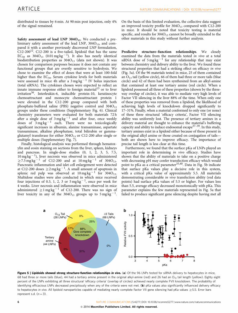
Predictive structure–function relationships. We closelyexamined the data from the materials tested in vivo at a totalsiRNA dose of 5 mg kg! 1 for any relationship that may existbetween chemistry and delivery ability to the liver. We found threestructural properties that had a striking effect on efficacy in vivo(Fig. 5a). Of the 96 materials tested in mice, 25 of them containedan O13 tail (yellow circle), 66 of them had three or more tails (bluecircle) and 42 of them had been synthesized from an alkyl-aminethat contained at least one tertiary amine (red circle). When alipidoid possessed all three of these properties (shown by the three-way overlap of circles), it was able to mediate very high levels ofFactor VII silencing in the liver 88% of the time. When only oneof these properties was removed from a lipidoid, the likelihood ofachieving high levels of knockdown dropped significantly to17–31%. Finally, when a material conformed to only one (or none)of these three structural ‘efficacy criteria’, Factor VII silencingability was uniformly lost. The presence of tertiary amines in adelivery material are thought to enhance the material’s bufferingcapacity and ability to induce endosomal escape37–39. In this study,tertiary amines exist in a lipidoid either because of those present inthe original alkyl amine or those created on conjugation of tails—both are shown here to improve efficacy. The importance ofprecise tail length is less clear at this time.
Furthermore, we found that the surface pKa of LNPs played animportant role in determining in vivo efficacy. Studies haveshown that the ability of materials to take on a positive chargewith decreasing pH may confer transfection efficacy which wouldpoint to pKa as a critical parameter35,40. Data in Fig. 5b indicatethat surface pKa values play a decisive role in this system,with a critical pKa value of approximately 5.5. All materialsdemonstrating considerable in vivo transfection ability (red datapoints) had surface pKa values of 5.5 or higher. For values lessthan 5.5, average efficacy decreased monotonically with pKa. Thisparameter explains the few materials represented in Fig. 5a thatfailed to produce significant gene silencing despite having met all
88%Give complete
knockdown
17%
31%
25%
0% 1.0
0.5
Rel
ativ
e F
VII
activ
ity
0.0
pKa3 4 5 6 7 8
0%0%
≥3 Tails(68 LNPs)
O13 Tail(26 LNPs)
Tertiaryamine
(44 LNPs)
a b
Figure 5 | Lipidoids showed strong structure–function relationships in vivo. (a) Of the 96 LNPs tested for siRNA delivery to hepatocytes in mice,68 had three or more tails (blue), 44 had a tertiary amine present in the original alkyl-amine (red) and 26 had an O13 tail length (yellow). Eighty eightpercent of the LNPs exhibiting all three structural ‘efficacy criteria’ (overlap of circles) achieved nearly complete FVII knockdown. The probability ofidentifying efficacious LNPs decreased precipitously when any of the criteria were not met. (b) pKa values also significantly influenced delivery efficacyto hepatocytes in vivo. All lipidoid nanoparticles capable of mediating nearly complete Factor VII gene silencing had pKa values Z5.5. Error barsrepresent s.d. (n¼ 3).
ARTICLE NATURE COMMUNICATIONS | DOI: 10.1038/ncomms5277
6 NATURE COMMUNICATIONS | 5:4277 | DOI: 10.1038/ncomms5277 | www.nature.com/naturecommunications
& 2014 Macmillan Publishers Limited. All rights reserved.
three structural efficacy criteria. Therefore, we concluded thatlipid nanoparticle efficacy is subject to a fourth criterion, which isthat the pKa must meet or exceed 5.5. Notably, the structuralcriteria can be determined on the basis of structure alone, whilethe pKa criteria requires only a simple, non-biological assay.Table 2 shows a four-way analysis of the efficacy criteria andconfirms the importance of conforming to all four criteria toguarantee high levels of gene silencing in vivo.
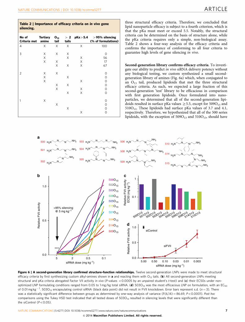
Second-generation library confirms efficacy criteria. To investi-gate our ability to predict in vivo siRNA delivery potency withoutany biological testing, we custom synthesized a small second-generation library of amines (Fig. 6a) which, when conjugated toan O13 tail, produced lipidoids that met the three structuralefficacy criteria. As such, we expected a large fraction of thissecond-generation ‘test’ library to be efficacious in comparisonwith first generation lipidoids. Once formulated into nano-particles, we determined that all of the second-generation lipi-doids resulted in surface pKa values Z5.5, except for 509O13 and510O13. These lipidoids had surface pKa values of 3.7 and 4.1,respectively. Therefore, we hypothesized that all of the 500 serieslipidoids, with the exception of 509O13 and 510O13, should have
Table 2 | Importance of efficacy criteria on in vivo genesilencing.
No ofCriteria met
Tertiaryamine
O13
tail4 2tails
pKa45.4 495% silencing(% of formulations)
4 X X X X 100
3 X X X 0X X X 56X X X 17
X X X 67
2 X X 0X X 0X X 0
X X 0X X 0
X X 0
1 X 0X 0
X 0X 0
500
501
502
503 506
508
509
504
505H2N
1.0
506O131
0.1
500O13501O13
502O13503O13
504O13505O13
506O13508O13
514O13515O13
Rel
ativ
e F
VII
activ
ityE
C50
(m
g kg
–1)
502O13
500O13
508O13
505O13
515O13
501O13
504O13
514O13
503O13
0.5
>95% silencing@ 5 mg kg–1
Rel
ativ
e F
VII
activ
ity
0.0
***
5 2 0.5siRNA dose (mg kg–1)
0.1
H2N
H2N
a
b
NN
NN
NN
NH2
NH2
NH2
H2N
H2N
H2N N N
NN
N N
NH2
NH2
NH2H2N
H2N
H2N NN
N NN
NN
NH2
NH2
NH2
510
514
515H2N
H2N
H2N N N
N N
N N N
N
NH2
NH2
NH2
NH2
c
1.0siControl
0.5
0.05.00 0.50 0.10
siRNA dose (mg kg–1)0.03 0.01 0.003
Rel
ativ
e F
VII
activ
ity
siFVII
d
Figure 6 | A second-generation library confirmed structure–function relationships. Twelve second-generation LNPs were made to meet structuralefficacy criteria by first synthesizing custom alkyl-amines shown in a and reacting them with O13 tails. (b) All second-generation LNPs meetingstructural and pKa criteria abrogated Factor VII activity in vivo (P-values o0.0005 by an unpaired student’s t-test) and (c) their EC50s under non-optimized LNP formulating conditions ranged from 0.05 to 1 mg/kg total siRNA. (d) 503O13 was the most efficacious LNP on formulation, with an EC50
of 0.01 mg kg! 1. 503O13 encapsulating control siRNA (black data point) did not result in FVII knockdown. Error bars represent s.d. (n¼ 3). Therewas a statistically significant difference between groups as determined by one-way analysis of variance (F(6,14)¼ 86.69, Po0.0001). Post hoccomparisons using the Tukey HSD test indicated that all tested doses of 503O13 resulted in silencing levels that were significantly different thanthe siControl (Po0.05).
NATURE COMMUNICATIONS | DOI: 10.1038/ncomms5277 ARTICLE
NATURE COMMUNICATIONS | 5:4277 | DOI: 10.1038/ncomms5277 | www.nature.com/naturecommunications 7
& 2014 Macmillan Publishers Limited. All rights reserved.
potent siRNA delivery activity in mice. When tested in vivo in theFactor VII model at a dose of 5 mg kg! 1, we found that ourpredictions were uniformly accurate (Fig. 6b). All materialsinduced greater than 95% silencing, with the exception of 509O13and 510O13, which each mediated only 10% silencing and are notincluded in the graph. The dose dependence of gene knockdownwas investigated in order to identify highly efficacious materialsin addition to those from the first generation library, and EC50values were found to vary from 0.05 to 1 mg kg! 1 (Fig. 6c).Formulation optimization of the best second-generation material,503O13, markedly decreased the EC50 value to 0.01 mg kg! 1
(Fig. 6d). Several second-generation materials also facilitatedsignificant CD45 knockdown in monocyte, macrophage, dendriticcell and B-cell populations (Supplementary Fig. 8). At the highestdose tested, 5 mg kg! 1, 500 series LNPs did not cause anyreductions in mouse bodyweights, suggesting that these second-generation materials were not acutely toxic (SupplementaryFig. 9).
DiscussionIn recent years, it has become increasingly important to considerthe degradability of siRNA delivery materials and their potentialfor immune stimulation. While earlier efforts focused on potency,clinical translation requires materials that are nontoxic enough toallow the repeated delivery that is typically needed for transientRNAi therapy. Part of what has made this so challenging from aresearch perspective is that potency can be measured via oneendpoint, but there are many potential endpoints whenevaluating toxicity and immunogenicity. This study has identifieda potent lipidoid nanoparticle, 304O13, that did not posepreliminary toxicity concerns at doses less than two orders ofmagnitude above than the EC50. In this study, we chose toevaluate cytokine expression and clinical chemical parameters atselected time points post injection and to perform histologicalanalysis on liver sections exposed to LNPs. Such work, althoughresource intensive, represents a fraction of potential endpointsthat may reveal toxicity issues for a particular compound41. Assuch, conclusions regarding the safety profile of 304O13 orextension to other members of the lipidoid library should be donewith caution.
The synthesis and biological testing of large libraries ofmaterials, such as the one described in Fig. 1, offer severaladvantages in the development of delivery systems. First, theprobability of discovering a potent delivery vehicle is significantlyincreased. Second, traditional drug delivery bias towards certainchemistries can be avoided if sufficient diversity is incorporatedinto the library. Third, and perhaps most importantly, the largequantity of screening results can be used to establish structure–function relationships that can inform the development ofsecond-generation libraries of increasingly efficacious materials.It is rare that structure–function studies reveal trends as strong asthose demonstrated in Fig. 5 and Table 2. Specifically, lipidoidssynthesized from O13 tails and alkyl-amines containing at leastone tertiary amine and at least three substitution sites had a highprobability of facilitating potent gene silencing in mice. If asimple, non-biological assay is performed to assess the pKa, LNPswith pKa values less than or equal to 5.4 can be ruled out. Thisnearly ensures high levels of in vivo knockdown. Of the fourcriteria, pKa appears to be the most influential in determiningin vivo efficacy in this system. It is the only criterion that, whennot met, abolishes the ability of an LNP to facilitate high levels ofgene silencing.
In summary, we have synthesized and tested a library of over1,400 lipid-like compounds for siRNA delivery in an attempt todevelop robust structure–function relationships that can informthe future work of drug delivery scientists. In the process, we
identified numerous degradable lipidoid formulations capable ofmediating very potent gene silencing in multiple biological targets(including hepatocytes and immune cells) in mice after IVadministration. One of the top formulations offered an improvedtoxicity profile in comparison with a lead non-degradable lipidoidformulation, and materials possessing potential clinical utility arepresently being evaluated in higher order animal models. Giventhe general difficulty of establishing structure–activity relation-ships in vivo, we were surprised to discover a set of four efficacycriteria (three structural and one pKa) that robustly predictin vivo siRNA delivery potency. Importantly, these materialdesign principles are able to predict in vivo activity without anyprior biological testing. In the context of future work, these newfindings may hasten delivery material development by reducingour dependence on time-consuming and expensive cell cultureand animal testing while underscoring a need for syntheticchemistry innovation.
MethodsLipidoid synthesis. Lipidoids were synthesized through the addition of alkyl-acrylates to amines. Amines were purchased from Sigma Aldrich (St Louis, MO),Alfa Aesar, Acros Organics and CHESS Organics. Briefly, 500 series amines weresynthesized by reacting secondary amines with sodium cyanide to form a nitriles,which were reduced to the primary amine products using lithium aluminiumhydride42. Acrylates were purchased from Scientific Polymer Products (Ontario,NY) and Hampford Research, Inc. (Stratford, CT). To synthesize lipidoids, amineswere combined with acrylates stoichiometrically in a glass scintillation vial andwere stirred at 90 !C for 3 days. In vitro experiments were conducted with crudematerials while in vivo experiments were performed with lipidoids that were eithercrude or purified via a Teledyne Isco Chromatography system (Lincoln, NE).Details of chemical characterization of a representative selection of compounds canbe found in Supplementary Note 1.
Lipidoid hydrolysis. To a 25 ml round bottom flask was added 304O13 (0.250 g,0.263 mmol, 1 equiv). For acidic hydrolysis, 10 ml of a solution of 6 N HCl wasadded to the flask to afford a cloudy heterogeneous solution. The reaction washeated to reflux to afford a clear, homogeneous solution and was stirred at refluxfor 24 h. For basic hydrolysis, 10 ml of a solution of KOH in EtOH/H2O(solution¼ 5.61 g KOH in 47.5 ml EtOH w/2.5 ml distilled H2O) was added to theflask to afford a clear colourless solution. The reaction was heated to reflux andstirred for 41 h. Both acidic and basic reactions were cooled to room temperatureand thin-layer chromatography analysis showed the presence of tridecanol (17.5%EtOAC/Hexanes) and the consumption of 304O13. Reactions were concentrated todryness under reduced pressure and diluted with CDCl3. The basic reaction wasfiltered to remove excess KOH. Proton NMR analysis was performed in CDCl3.Proton nuclear magnetic resonance spectra were recorded with a Bruker Avance400 spectrometer, are depicted in parts per million on the d scale, and are refer-enced from the residual protium in the NMR solvent (CDCl3: d 7.26 (CHCl3).
Formulation of LNPs. Lipidoids were formulated into nanoparticles for allapplications. Nanoparticles were formed by mixing lipidoids, cholesterol (SigmaAldrich), DSPC (Avanti Polar Lipids, Alabaster, AL) and mPEG2000-DMG (MW2660, gift from Alnylam Pharmaceuticals, Cambridge, MA) at a molar ratio of50:38.5:(11.5!X):X in a solution of 90% ethanol and 10% 10 mM sodium citrate(by volume). An siRNA solution was prepared by diluting siRNA in 10 mM sodiumcitrate such that the final weight ratio of lipidoid: siRNA was between 5:1 and 10:1,depending on the experiment. Equal volumes of lipid solution and siRNA solutionwere rapidly mixed together using either a microfluidic device43 or by pipet to formnanoparticles. Particles were diluted in PBS, Invitrogen, Carlsbad, CA and thendialysed against PBS for 90 min in 3,500 MWCO cassettes (Pierce/ThermoScientific, Rockford, IL).
In vitro transfection of cell lines with LNPs. HeLa cells (American Type CultureCollection, Manassas, VA) stably modified to express both firefly and Renillaluciferase were maintained at 37 !C in high glucose Dulbecco’s Modified EaglesMedium without phenol red (Invitrogen) supplemented with 10% fetal bovineserum (Invitrogen). 12–16 h before transfection, cells were seeded in white 96-wellplates at a density of 15,000 cells per well. Cells were transfected with a 40 nMconcentration of anti-firefly luciferase siRNA (Dharmacon, Lafayette, CO) that hadbeen formulated with lipidoids into nanoparticles. Firefly luciferase silencing wasassessed with a Dual-Glo Luciferase Assay kit (Promega, Madison, WI). Renillaluciferase activity served as a control.
ARTICLE NATURE COMMUNICATIONS | DOI: 10.1038/ncomms5277
8 NATURE COMMUNICATIONS | 5:4277 | DOI: 10.1038/ncomms5277 | www.nature.com/naturecommunications
& 2014 Macmillan Publishers Limited. All rights reserved.
In vivo gene silencing. All animal experiments were conducted using institu-tionally-approved protocols (IACUC). Female C57BL/6 mice at least 6 weeks of age(Charles River Laboratories, Wilmington, MA) received lateral tail vein injectionsof PBS (negative control) or lipidoid nanoparticles containing either non-targetingsiRNA (negative control) or anti-Factor VII siRNA diluted in PBS at a volumeof 0.01 ml g! 1. The sequence of the siFVII, provided by Alnylam Pharmaceuticals,was: sense: 50-GGAucAucucAAGucuuAcT*T-30 ; antisense: 50-GuAAGAcuuGA-GAuGAuccT*T-30 ,where 20-fluoro-modified nucleotides are in lower case andphosphorothioate linkages are represented by asterisks. Two days post injection, a100ml blood sample was obtained from mice, and serum levels of Factor VII wereanalysed using a Biophen FVII assay kit as described previously (Aniara Cor-poration, Mason, OH)6.
Biodistribution and immunostaining. Female C57BL/6 mice received tail veininjections LNPs containing siRNA that had been labelled with Cy5.5 on the 50-endof the sense strand (provided by Alnylam Pharmaceuticals). Animals were dosed at1 mg/kg of siRNA and volume of 0.01 ml g! 1. At 1 h post injection, mice wereeuthanized and organs were removed. Body-wide biodistribution was assessed byimaging whole, unprocessed organs with an IVIS Spectrum system (Caliper LifeSciences, Hopkinton, MA) at excitation and emission wavelengths of 675 and720 nm, respectively. For Odyssey and confocal imaging, organs were snap frozenon dry ice and embedded in optimal cutting temperature compound (Life Tech-nologies, Grand Island, NY). Cryostat sections were cut and collected on superfrostplus treated slides. Prepared frozen sections where kept at ! 20 !C until needed.Odyssey imaging was conducted on 20-mm-thick cryosections of tissue at a reso-lution of 21 mm44.
For confocal imaging, liver tissue was cryosectioned (12mm) and fixed using 4%paraformaldehyde at room temperature for 30 min. All solutions were prepared inPBS. Sections were washed two times with PBS, permeabilized for 30 min with 0.1%Triton X100 and blocked for 1 h with 5% normal goat serum. Sections then incubatedfor 1 h in an immunostaining cocktail solution consisting of 40 ,6-diamidino-2-phenylindole (3mM), Alexa Fluor 488 conjugated anti-mouse F4/80 (1:200 dilution,BioLegend, San Diego, CA), Alexa Fluor 555 Phalloidin (1:200 dilution, LifeTechnologies) and 5% normal goat serum. Slides were washed three times with 0.1%Tween 20 and mounted using ProLong Gold Antifade (Life Technologies). Sectionswere imaged using an LSM 700 point scanning confocal microscope (Carl Zeiss, Inc,Jena Germany) equipped with a $ 40 oil immersion objective.
Blood clearance. Blood clearance experiments were conducted by injecting LNPscontaining Cy5.5-labelled siRNA at an siRNA dose of 0.5 mg kg! 1. Blood sampleswere collected as a function of time via the retroorbital vein, with the exception offinal time points, which were collected via cardiac puncture. Serum, obtained bycentrifugation, was diluted 1:30 in PBS and imaged and quantified using anOdyssey CLx imaging system (LI-COR Biosciences, Lincoln, NE).
Histology. Animals that had received various doses of either 304O13 or C12-200LNPs were killed, and their organs were collected immediately. Organs were fixedovernight in 4% paraformaldehyde and transferred to 70% ethanol before paraffinembedding, sectioning and hematoxylin and eosin staining.
Serum chemistry and cytokine analysis. Post killing, cardiac sticks wereimmediately performed on animals that had been dosed with either 304O13 or C12-200 LNPs. A dose of 3 mg kg! 1 was used for cytokine and serum chemistryexperiments. Blood was centrifuged in serum separator tubes at 5,000 r.p.m. for10 min before analysis. Serum chemistry was evaluated on a Beckman OlympusAU400 Serum Chemistry Analyzer. Cytokines were analysed using Bio-Plex ProMouse Cytokine 23-Plex Assay kits (Luminex Corporation, Austin, TX) on theBio-Plex 200 system, according to the manufacturer’s instructions.
Nanoparticle characterization. LNPs were diluted to an siRNA concentration ofB4 mg ml! 1 in PBS buffer, pH 7.4. siRNA entrapment efficiency was determinedusing the Quant-iT RiboGreen RNA assay (Invitrogen). Particle sizes were mea-sured with a ZETAPals analyzer (Brookhaven Instruments, Holtsville, NY). Sizesreported are the average effective diameter of each LNP. Zeta potential measure-ments were acquired on a Zetasizer Nano ZS (Malvern, Westborough, MA), andreported values were the average of 10–25 runs.
pKa measurements. The surface pKa values of LNPs were determined asdescribed previously34. Briefly, solutions of 20 mM sodium phosphate, 25 mMcitrate, 20 mM ammonium acetate and 150 mM NaCl were titrated to pH valuesvarying by 0.5 from 2.0 to 12.0 and aliquoted into a black 96-well plate. LNPs and2-(p-toluidinyl)naphthalene-6-sulphonic acid (TNS, Sigma Aldrich) were dilutedinto these solutions for a final concentration of 20 and 6 mM, respectively.Fluorescence intensity was read on a Tecan M1000 plate reader at an excitation of322 nm and an emission of 431 nm. pKa values were calculated as the pHcorresponding to 50% LNP protonation, assuming minimum and maximumfluorescence values corresponded to zero and 100% protonation, respectively.
References1. Agu, R. U. & Ugwoke, M. I. In vitro and in vivo testing methods for respiratory
drug delivery. Expert Opin. Drug. Deliv. 8, 57–69 (2011).2. Sriamornsak, P. Application of pectin in oral drug delivery. Expert Opin. Drug.
Deliv. 8, 1009–1023 (2011).3. Godin, B. & Touitou, E. Transdermal skin delivery: predictions for humans
from in vivo, ex vivo and animal models. Adv. Drug Deliv. Rev. 59, 1152–1161(2007).
4. Whitehead, K. A. et al. In vitro–in vivo translation of lipid nanoparticles forhepatocellular siRNA delivery. ACS Nano 6, 6922–6929 (2012).
5. Davis, M. E. et al. Evidence of RNAi in humans from systemically administeredsiRNA via targeted nanoparticles. Nature 464, 1067–1070 (2010).
6. Semple, S. C. et al. Rational design of cationic lipids for siRNA delivery. Nat.Biotechnol. 28, 172–176 (2010).
7. Novobrantseva, T. I. et al. Systemic RNAi-mediated gene silencing in nonhumanprimate and rodent myeloid cells. Mol. Ther. Nucleic Acids 1, e4 (2012).
8. Rozema, D. B. et al. Dynamic PolyConjugates for targeted in vivo delivery ofsiRNA to hepatocytes. Proc. Natl Acad. Sci. USA 104, 12982–12987 (2007).
9. Kanasty, R., Dorkin, J. R., Vegas, A. & Anderson, D. Delivery materials forsiRNA therapeutics. Nat. Mater. 12, 967–977 (2013).
10. Strapps, W. R. et al. The siRNA sequence and guide strand overhangs aredeterminants of in vivo duration of silencing. Nucleic Acids Res. 38, 4788–4797(2010).
11. Foster, D. J. et al. Comprehensive evaluation of canonical versus Dicer-substrate siRNA in vitro and in vivo. RNA 18, 557–568 (2012).
12. Love, K. T. et al. Lipid-like materials for low-dose, in vivo gene silencing. Proc.Natl Acad. Sci. USA 107, 1864–1869 (2010).
13. Barros, S. A. & Gollob, J. A. Safety profile of RNAi nanomedicines. Adv. DrugDeliv. Rev. 64, 1730–1737 (2012).
14. Robbins, M., Judge, A. & MacLachlan, I. siRNA and Innate Immunity.Oligonucleotides 19, 89–102 (2009).
15. Whitehead, K. A. et al. Silencing or stimulation? sirna delivery and the immunesystem. Annu. Rev. Chem. Biomol. Eng. 2, 77–96 (2011).
16. Akinc, A. et al. A combinatorial library of lipid-like materials for delivery ofRNAi therapeutics. Nat. Biotechnol. 26, 561–569 (2008).
17. Siegwart, D. J. et al. Combinatorial synthesis of chemically diverse core-shellnanoparticles for intracellular delivery. Proc. Natl Acad. Sci. USA 108,12996–13001 (2011).
18. Mei, Y. et al. Combinatorial development of biomaterials for clonal growth ofhuman pluripotent stem cells. Nat. Mater. 9, 768–778 (2010).
19. Staubli, A., Ron, E. & Langer, R. Hydrolytically degradable amino acid-containing polymers. J. Am. Chem. Soc. 112, 4419–4424 (1990).
20. van Dijkhuizen-Radersma, R., Hesseling, S. C., Kaim, P. E., de Groot, K. &Bezemer, J. M. Biocompatibility and degradation of poly(ether–ester) micro-spheres: in vitro and in vivo evaluation. Biomaterials 23, 4719–4729 (2002).
21. Geng, Y. & Discher, D. E. Hydrolytic degradation of poly(ethylene oxide)-block-polycaprolactone worm micelles. J. Am. Chem. Soc. 127, 12780–12781(2005).
22. Pan, H., Jiang, H. & Chen, W. The biodegradability of electrospun dextran/PLGA scaffold in a fibroblast/macrophage co-culture. Biomaterials 29,1583–1592 (2008).
23. Jahangir, R. et al. The influence of protein adsorption and surface modifyingmacromolecules on the hydrolytic degradation of a poly (ether–urethane) bycholesterol esterase. Biomaterials 24, 121–130 (2003).
24. Benjaminsen, R. V., Mattebjerg, M. A., Henriksen, J. R., Moghimi, S. M. &Andresen, T. L. The possible ‘proton sponge’ effect of polyethylenimine (pei)does not include change in lysosomal pH. Mol. Ther. 21, 149–157 (2013).
25. Liu, Y., Samsonova, O., Sproat, B., Merkel, O. & Kissel, T. Biophysicalcharacterization of hyper-branched polyethylenimine-graft-polycaprolactone-block-mono-methoxyl-poly (ethylene glycol) copolymers (hy-PEI-PCL-mPEG)for siRNA delivery. J. Control. Release 153, 262–268 (2011).
26. Tagalakis, A. D., Saraiva, L., McCarthy, D., Gustafsson, K. T. & Hart, S. L.Comparison of nanocomplexes with branched and linear peptides for siRNAdelivery. Biomacromolecules 14, 761–770 (2013).
27. John, M. et al. Effective RNAi-mediated gene silencing without interruption ofthe endogenous microRNA pathway. Nature 449, 745–747 (2007).
28. Geissmann, F. et al. Development of monocytes, macrophages, and dendriticcells. Science 327, 656–661 (2010).
29. Grivennikov, S. I., Greten, F. R. & Karin, M. Immunity, inflammation, andcancer. Cell 140, 883–899 (2010).
30. Lee, S., Yang, S. C., Kao, C.-Y., Pierce, R. H. & Murthy, N. Solid polymericmicroparticles enhance the delivery of siRNA to macrophages in vivo. Nucleic.Acids Res. 37, e145 (2009).
31. Leuschner, F. et al. Therapeutic siRNA silencing in inflammatory monocytes inmice. Nat. Biotechnol. 29, 1005–1010 (2011).
32. Tonks, N. K., Charbonneau, H., Diltz, C. D. & Fischer, E. H. Demonstrationthat the leukocyte common antigen (CD45) is a protein tyrosine phosphatase.Biochemistry 27, 8695–8701 (1988).
NATURE COMMUNICATIONS | DOI: 10.1038/ncomms5277 ARTICLE
NATURE COMMUNICATIONS | 5:4277 | DOI: 10.1038/ncomms5277 | www.nature.com/naturecommunications 9
& 2014 Macmillan Publishers Limited. All rights reserved.
33. Nolan, T., Hands, R. E. & Bustin, S. A. Quantification of mRNA using real-timeRT-PCR. Nat. Protoc. 1, 1559–1582 (2006).
34. Heyes, J., Palmer, L., Bremner, K. & MacLachlan, I. Cationic lipid saturationinfluences intracellular delivery of encapsulated nucleic acids. J. Control. Release107, 276–287 (2005).
35. Jayaraman, M. M. et al. Maximizing the potency of sirna lipid nanoparticlesfor hepatic gene silencing in vivo. Angew. Chem. Int. Ed. 51, 8529–8533(2012).
36. Amanzada, A. et al. Induction of chemokines and cytokines before neutrophilsand macrophage recruitment in different regions of rat liver after TAAadministration. Lab. Invest. 94, 235–247 (2013).
37. Patil, M. L., Zhang, M. & Minko, T. Multifunctional triblock nanocarrier(PAMAM-PEG-PLL) for the efficient intracellular sirna delivery and genesilencing. ACS Nano 5, 1877–1887 (2011).
38. Ong, Z. Y. et al. Rational design of biodegradable cationic polycarbonates forgene delivery. J. Control. Release 152, 120–126 (2011).
39. Ou, M. et al. Novel biodegradable poly (disulfide amine) s for gene deliverywith high efficiency and low cytotoxicity. Bioconjug. Chem. 19, 626–633 (2008).
40. Zhang, J. J., Fan, H. H., Levorse, D. A. D. & Crocker, L. S. L. Ionization behaviorof amino lipids for siRNA delivery: determination of ionization constants, SAR,and the impact of lipid pKa on cationic lipid-biomembrane interactions.Langmuir 27, 1907–1914 (2011).
41. Judge, A. & MacLachlan, I. Overcoming the innate immune response to smallinterfering RNA. Hum. Gene. Ther. 19, 111–124 (2008).
42. Gamage, S. A. et al. Dicationic Bis(9-methylphenazine-1-carboxamides):relationships between biological activity and linker chain structure for aseries of potent topoisomerase targeted anticancer drugs. J. Med. Chem. 44,1407–1415 (2001).
43. Chen, D. et al. Rapid discovery of potent siRNA-containing lipid nanoparticlesenabled by controlled microfluidic formulation. J. Am. Chem. Soc. 134,6948–6951 (2012).
44. Lee, M. J.-E. et al. Rapid pharmacokinetic and biodistribution studies usingcholorotoxin-conjugated iron oxide nanoparticles: a novel non-radioactivemethod. PLoS ONE 5, e9536–e9536 (2010).
AcknowledgementsK.A.W. would like to thank D. Siegwart and C. Alabi for helpful discussions as well asF. Niroui and M. Ma for technical assistance. This work was funded by AlnylamPharmaceuticals and NIH grants EB000244, R01CA115527 and R01CA132091. K.A.W.was supported by an NIH F32 fellowship (award number EB009623). O.V. acknowledgessupport from CDMRP, Department of Defense, postdoctoral fellowship award(#W81XWH-13-1-0215).
Author contributionsK.A.W. designed and performed experiments, analysed data and wrote the paper. J.R.D.,P.H.C., J.M., O.V., O.F., Y.Z., K.T.O., V.Y. and B.K. designed and performed experi-ments. D.C. designed and fabricated microfluidic devices. A.J.V., S.B. and T.N. providedconceptual advice and technical support. R.L. and D.G.A. supervised the study. Allauthors discussed the results and commented on the manuscript.
Additional informationSupplementary Information accompanies this paper at http://www.nature.com/naturecommunications
Competing financial interests: R.L. is a shareholder and member of the scientificadvisory board of Alnylam. D.G.A. is a consultant with Alnylam Pharmaceuticals. R.L.and D.G.A. have sponsored research grants from Alnylam. Alnylam also has a license tocertain intellectual property invented at Massachusetts Institute of Technology byK.A.W., J.R.D., A.J.V., R.L., and D.G.A. The remaining authors declare no competingfinancial interest
Reprints and permission information is available online at http://npg.nature.com/reprintsandpermissions/
How to cite this article: Whitehead, K. A. et al. Degradable lipid nanoparticleswith predictable in vivo siRNA delivery activity. Nat. Commun. 5:4277doi: 10.1038/ncomms5277 (2014).
ARTICLE NATURE COMMUNICATIONS | DOI: 10.1038/ncomms5277
10 NATURE COMMUNICATIONS | 5:4277 | DOI: 10.1038/ncomms5277 | www.nature.com/naturecommunications
& 2014 Macmillan Publishers Limited. All rights reserved.


