Embed Size (px)
Citation preview

Deconstructing common variable immunodeficiency bygenetic analysisAlejandro A Schaffer1, Ulrich Salzer2, Lennart Hammarstrom3 andBodo Grimbacher4
Common variable immunodeficiency (CVID) is the most
common symptomatic primary immunodeficiency. Patients
have recurrent bacterial infections and an increased risk of
developing autoimmune diseases, lung damage, and selected
cancers. Since 2003, four genes have been shown to be
mutated in CVID patients: ICOS, TNFRSF13B (encoding TACI),
TNFRSF13C (encoding BAFF-R) and CD19. Heterozygous
mutations in TNFRSF13B are also associated with CVID,
whereas the other three genes are purely recessive. Recent
genetic linkage studies have also identified possible loci for
dominant CVID genes on chromosomes 4q, 5p and 16q. These
findings markedly improved the genetic diagnosis of CVID and
point towards new strategies for future genetic studies. In
addition, some CVID genes might be relevant to more common
diseases such as asthma and stroke.
Addresses1 National Center for Biotechnology Information, National Library of
Medicine, National Institutes of Health, Department of Heath and Human
Services, 8600 Rockvile Pike, Bethesda, MD 20894, USA2 Division of Rheumatology and Clinical Immunology, Medical Center,
University of Freiburg, Hugstetterstraße 55, 79106 Freiburg, Germany3 Division of Clinical Immunology, Karoliska Institutet at Karolisnka
University Hospital Huddinge, SE-141 86 Stockholm, Sweden4 Department of Immunology and Molecular Pathology, Royal Free
Hospital, University College London, Pond Street, London, NW3 2QG,
UK
Corresponding author: Grimbacher, Bodo
Current Opinion in Genetics & Development 2007, 17:201–212
This review comes from a themed issue on
Genetics of disease
Edited by Robert Nussbaum and Leena Peltonen
Available online 27th April 2007
0959-437X/$ – see front matter
# 2007 Elsevier Ltd. All rights reserved.
DOI 10.1016/j.gde.2007.04.002
IntroductionThe major achievement in the evolution of the adaptive
immune system is its capability to generate specific
antibodies directed against invading pathogens, and to
memorize this immune response. These antibodies,
generated as various immunoglobulin (Ig) isotypes (i.e.
IgM, IgG, IgA and IgE), represent an important com-
ponent of the humoral immune system and are thus kept
at a constant concentration in blood. Disorders affecting
www.sciencedirect.com
immunoglobulin production or turnover frequently lead
to hypogammaglobulinemia, rendering patients suscep-
tible to recurrent infection, especially by encapsulated
bacteria. The severity of this hypogammaglobulinemic
state can range from IgG subclass deficiencies in combi-
nation with IgA deficiency to complete absence of all
three major Ig isotypes (IgA, IgG and IgM), which is
termed ‘agammaglobulinemia’.
The diagnosis of common variable immunodeficiency
(CVID) is typically made in patients with a history of
recurrent bacterial infections and substantially lowered
serum levels of immunoglobulins. In particular, CVID
patients almost always have low IgG and IgA serum
levels, and about 85% also show a reduction of IgM in
serum [1]. The clinical course in CVID patients includes
an elevated risk of autoimmune diseases, lung compli-
cations and several types of cancer.
CVID is the most common primary immunodeficiency
that necessitates clinical attention, with an incidence
estimated at 1:10 000 to 1:50 000 [2]. Isolated IgA
deficiency is much more common in Caucasians, with
an incidence estimated at 1:700, but the vast majority of
cases are clinically unapparent. Several individuals have
been shown to gradually progress from IgA deficiency to
full-blown CVID [3–6]. How often this deterioration
occurs and over what time frame is hard to estimate,
because isolated IgA deficiency is often asymptomatic
and thus underdiagnosed.
In a large CVID patient cohort, Cunningham-Rundles
and Bodian [1] estimated that the difference between the
median age at the onset of initial symptoms and the
median age at diagnosis is five to six years. In the current
European database for CVID, this diagnostic delay was
shown to be four years [7]. Once patients are diagnosed
with CVID, immunoglobulin substitution is an effective
treatment in the majority of patients [8]. Immunoglobulin
administration is not a curative treatment, but a lifelong
replacement therapy that has to be applied either on a
monthly basis if given by the intravenous route or weekly
if subcutaneous immunoglobulin replacement therapy is
chosen. The safety and reliability of Ig replacement
therapy are overshadowed by various factors: possible
deleterious effects over decades of treatment [8,9];
economic constraints, because Ig therapy is expensive;
and the resources for the production of Ig for clinical
application are limited by the availability of eligible
Current Opinion in Genetics & Development 2007, 17:201–212

202 Genetics of disease
plasma donors. Therefore, the identification of genetic
factors that are involved in the etiology of CVID will
enable not only new diagnostics but also new leads for
possibly curative, therapeutic interventions.
It was recognized decades ago that CVID has a genetic
component [10]. The progression from IgA deficiency to
CVID mentioned above is most frequently observed in
families in which at least one immunodeficient individual
is diagnosed with CVID and in which the immunoglo-
bulin levels of available relatives are measured. Both by
this strategy and by strategies of ascertaining IgA-
deficient probands, Vorechovsky et al. [11] estimated that
approximately 20% of cases of IgA deficiency are familial,
whereby IgAD and CVID can cluster within single
families. For CVID alone we, and others, estimate the
frequency of familial occurrence to be about 10% [1].
There have been surprisingly few genetic studies of
CVID until the past few years. Early studies using genetic
linkage and association established that there is at least
one susceptibility locus in the HLA region on chromo-
some arm 6p. However, as with so many other HLA-
linked immune related diseases [12], pinning down the
CVID susceptibility genes and mutations on 6p has been
a quixotic challenge.
Both dominant and recessive inheritance have been
documented in CVID–IgAD families, with dominant
inheritance being far more common [11]. For recessive
inheritance, numerous CVID candidate genes have been
suggested on the basis of studies of single-gene knockout
mice that have low immunoglobulin levels and are suscept-
ible to infections. Within the past five years, studies of such
candidate genes outside 6p, and genome-wide linkage
studies of CVID have identified several causative or
associated genetic factors: three genes mutated in a small
number of recessive cases; one gene that is mutated in as
many as 10% of CVID cases; and some possible regions of
genetic linkage between polymorphic markers and the
CVID–IgAD phenotype.
In the central sections of this article, we review these
recent genetic findings. To put these findings into a
broader genetic context, we first describe some examples
of genetically defined differential diagnoses of CVID,
several of which have been made possible by the identi-
fication of other genes mutated in immunodeficiencies
that can mimic the clinical picture of CVID. In the later
sections, we speculate on the prospects for future genetic
studies of CVID and we discuss the possible relevance of
CVID-causing genes to two more common diseases,
asthma and stroke.
CVID is a diagnosis of exclusionMany conditions can manifest with the symptoms of
hypogammaglobulinemia and subsequent recurrent bac-
Current Opinion in Genetics & Development 2007, 17:201–212
terial infections. Still today, the diagnosis of CVID is
usually made when a variety of tests for other conditions
are negative. Only the discovery of some CVID-causing
genes, as described below, enabled us to make a definite,
genetically based diagnosis of CVID (i.e. by identification
of a mutation) in a small percentage of patients.
In Table 1, we highlight some monogenic immune dis-
eases that can mimic the clinical phenotype of CVID and
as such constitute important differential diagnoses.
Incomplete or abortive clinical courses of some multi-
system monogenic disorders such as ataxia telangiectasia
and myotonic dystrophy can also manifest with a CVID-
like phenotype. Additionally, exposure to non-genetic
factors such as drugs (e.g. gold salts) and viral infections
(e.g. Epstein-Barr virus [EBV]) can also result in states of
hypogammaglobulinemia.
In male CVID patients, especially when there is a family
history compatible with X-linked inheritance, two well-
known other immunodeficiency disorders need to be
excluded: in X-linked agammaglobulinemia (XLA),
B-cell development is blocked at the pre-B-cell stage,
resulting in severe peripheral B-cell lymphopenia and
usually very low immunoglobulin levels; in X-linked
lymphoproliferative syndrome (XLP) the clinical picture
contains the triad of an often fatal infection with EBV
associated with the development of lymphoma and hypo-
gammaglobulinemia [13].
A crucial distinction between XLA and XLP and CVID is
that the onset of XLA and XLP is usually shortly after
birth, whereas the diagnosis of CVID is usually reserved
for patients whose onset is above age 2. However, several
papers cited in Table 1 document XLA and XLP cases
with hypomorphic mutations that led to later age of onset
and misdiagnosis as CVID. XLA can be distinguished
from CVID by the nearly complete lack of B cells (<1% of
lymphocytes), whereas a distinguishing characteristic of
XLP is a very low number of natural killer T-cells [14��]and a proven history of a usually severe course of EBV-
induced infectious mononucleosis.
Hyper-IgM syndromes seem to be closely related to
CVID from a mechanistic point of view because they
also lack switched isotypes, which is one of the clinical
hallmarks of CVID. The observation of abnormally high
levels of IgM with concomitant reduction of switched
isotypes in some patients led to the name for these
disease entities. However, recent consensus statements
suggest that the hyper-IgM syndromes should rather be
called ‘class-switch recombination’ (CSR) defects to bet-
ter define the underlying pathomechanism in these
groups of patients. Consequently, many patients diag-
nosed with a CSR defect have serum IgM levels in the
normal range for their age, making it difficult to dis-
tinguish them from those with the CVID phenotype.
www.sciencedirect.com

Deconstructing common variable immunodeficiency by genetic analysis Schaffer, Salzer, Hammarstrom and Grimbacher 203
Table 1
Some monogenic disorders that are differential diagnoses for CVID.
Disease Inheritance Gene(s) involved References
X-linked agammaglobulinemia (Bruton’s disease) X-linked BTK [68–70]
Autosomal agammaglobulinemia due to m-heavy
chain deficiency
AR IGHM [71–73]
Rare autosomal agammaglobulinemias AR CD79A (Iga) [74–77]
IGLL1 (l5/14.1)
BLNK/SLP65
LRRC8 (translocation)
X-linked Hyper-IgM syndrome X-linked CD40L [78,79]
Autosomal Hyper-IgM syndromes Usually AR;
Mutations in AID can be AD
AID, CD40, UNG [80–83]
X-linked lymphoproliferative syndromes X-linked SH2D1A, XIAP (also known as BIRC4) [84,85,86��]
X-linked severe combined immunodeficiency
(SCID)
X-linked IL2RG [86��]
Autosomal SCID AR ADA, DCLRE1C, RAG1, RAG2, IL7R, CD45,
CD3D, CD3E
[87]
WHIM syndrome AD CXCR4 [88]
IgG subclass deficiencies AR Deletions involving the Ig heavy chain region
on chromosome 14
[89]
CVID is diagnosed by exclusion. For example, several rarer genetic disorders might mimic the symptoms and sometimes need to be excluded by
gene sequencing to substantiate the diagnosis of CVID. This table lists rare, but genetically defined, differential diagnoses of CVID, their mode
of inheritance and the respective mutated gene(s). Abbreviations: AID, activation-induced cytidine deaminase; AR, autosomal recessive;
AD, autosomal dominant; WHIM, warts, hypogammaglobulinemia, infections and myelokathexis.
CSR defects can be diagnosed most easily and reliably by
sequencing genes listed in Table 1; the CSR process itself
involves multiple steps, any one of which can be defec-
tive.
Candidate genes implicated in recent CVIDstudiesSince 2003, four genes have been found to have both
alleles mutated in at least one CVID patient (Table 2). All
four genes were considered as candidates on the basis of
the combined knowledge about B-cell biology and pub-
lished phenotypes of single-gene knockout mice. These
criteria could be used to justify the candidacy of dozens of
genes, and the majority of the genes we have tested have
no patently deleterious sequence changes in cohorts of
dozens of CVID patients. The rarity of mutations in ICOS(inducible co-stimulator), TNFRSF13C (tumor necrosis
factor superfamily member 13C) and CD19 (B-lympho-
cyte antigen CD19) demonstrates that by sequencing
Table 2
Four genes in which double mutations cause either CVID or IgAD (sin
and IgAD).
Gene Mutations Patients with 2 alleles mutated Pa
ICOS 1 9 0
TNFRSF13C 1 1 0
TNFRSF13B 17 20 38
CD19 2 4 0
In the past five years, four genes have been identified that have both allel
mutations in TNFRSF13B are associated with CVID, whereas heterozygot
gene names, the number of different mutations that have been described
one or two mutated alleles of the respective gene.
www.sciencedirect.com
alone it is impossible to exclude rare mutations in any
gene. We now present short reviews of the proteins
encoded by the four genes known to cause CVID.
ICOS is expressed exclusively on T cells after their
activation [15]. ICOS is a T-cell co-stimulatory molecule
that interacts with ICOS-L, which is expressed on the
surface of B cells and specialized antigen-presenting cells,
thereby supporting the collaboration among B, T and
dendritic cells in an ongoing immune response. Ligation
of ICOS-L to ICOS leads to a release of interleukin 10 from
germinal center T-cells, enabling B cells to undergo CSR
(see Figure 1). Furthermore, the ICOS–ICOS-L signaling
in germinal centers is crucial for the development and
function of the highly specialized subpopulation of
CXCR5+ follicular T-helper cells [16�]. The loss of ICOS
expression leads to absence of this special cell population,
consecutive failure to develop functional germinal centers
and, ultimately, impaired terminal B-cell differentiation
gle mutations in TNFRSF13B are also associated with CVID
tients with 1 allele mutated References
[29,90��]
[91]
[61��,92��] (Salzer et al., unpublished)
[18��]
es mutated in at least one CVID patient. Single, heterozygous
es for the other genes have no apparent phenotype. The table lists
for each of the four genes, and the numbers of patients carrying either
Current Opinion in Genetics & Development 2007, 17:201–212

204 Genetics of disease
Figure 2
The B cell receptor signaling complex. The B cell receptor (BCR) itself
has quite a weak capability of signaling into B cells. The BCR signal is
thus amplified by a transmembrane signaling complex consisting of
CD19, CD21 (also termed complement receptor (CR) 2), CD81 and
CD225 (also termed Leu-13). Not only does Antigen, opsonised by C3d,
bind the BCR but the C3d also binds to CD21. CD21 in turn is linked to
CD19, which has a long intracytoplasmatic signaling domain with at
least three important phosphorylation sites, well capable of amplifying
the BCR signal into the B cells.
Figure 1
T–B cell cooperation. After TCR engagement, the activation status of T
cells is fine-tuned by co-stimulatory signals. Ligation of CD80 or CD86
on antigen presenting cells (e.g. B cells) to CD28, which is constitutively
expressed on T cells, and ligation of ICOS-L to the inducible co-
stimulator (ICOS) of T cells, upregulates the T cell’s activation status. On
the contrary, engagement of CTLA-4 with CD80 or CD86, for example,
serves as a downregulatory mechanism. Ligation of ICOS-L and ICOS
lead to the secretion of a wide array of cytokines such as interleukin (IL)
10, IL-17, IL-21 and IL-23, with IL-10 being the most abundant one. If T
cells are missing this co-stimulatory signal, they are unable to provide B
cells with sufficient T cell help (cytokines) to perform a proper germinal
center formation and class-switch recombination.
and hypogammaglobulinemia [16�,17�]. Some of the
patients with ICOS mutations had normal levels of IgM
or showed a transient increase of antigen-specific IgM in
episodes with acute infection, but they had low levels of
IgG and IgA. ICOS deficiency is currently classified as a
cause of CVID rather than a CSR defect because of
historical reasons and the fact that, unlike the CSR-defect
genes in Table 1, the known functional connection be-
tween ICOS activity and CSR is quite indirect.
CD19, in contrast to ICOS, is expressed exclusively on B
cells. Indeed, surface expression of CD19 is often used in
flow cytometry to identify B cells. van Zelm et al. [18��]reported that B cells from four patients had severely
reduced CD19 expression, but had normal numbers of
B cells, as detected by the existence of other surface
markers such as CD20. The likely reason that CD19
deficiency is deleterious is that CD19 is part of a four-
protein complex, together with CD21, CD81 and CD225,
that helps receive and process the B-cell receptor–antigen
signal (see Figure 2). The impaired antigen-dependent
signaling of the B-cell receptor complex in CD19-
deficient humans, as evidenced by the reduced Ca2+
influx, results in inadequate numbers of mature B-cells,
which produce insufficient amounts of antibodies when
challenged with antigen — either by vaccination or by
recurrent infections.
Current Opinion in Genetics & Development 2007, 17:201–212
Both reported mutations in CD19 code for putative
proteins that have a premature truncation. The patients
were shown to have normal levels of CD19 mRNA, and
thus the truncated proteins might perform some function
in vivo. This differs from the scenario in ICOS deficiency,
in which the only known mutation causes a complete
protein knockout. CD19 mutations that affect surface
expression are likely to be rare because they should be
noticed accidentally in flow cytometry. However, CD19sequence changes with retained CD19 expression (e.g. at
phosphorylation sites in the CD19 intracellular domain)
might exist and could lead to a CVID phenotype when
both alleles are affected.
The other two proteins, BAFF-R (B-cell-activating fac-
tor receptor) and TACI (transmembrane activator and
calcium-modulating cyclophilin ligand interactor), are
receptors expressed at the surface of B cells and partici-
pate in a complex signaling network; the five most
studied proteins in this network are depicted in
Figure 3. BAFF-R receives signals from BAFF that
are essential for the survival of peripheral B-cells [19].
By contrast, BCMA (B-cell maturation antigen) expres-
sion is highly restricted to end stages of B-cell differen-
tiation and seems to be essential for the survival of
www.sciencedirect.com

Deconstructing common variable immunodeficiency by genetic analysis Schaffer, Salzer, Hammarstrom and Grimbacher 205
Figure 3
The APRIL–BAFF network: BAFF-R, TACI and BCMA are TNF-like receptors expressed in various stages of the B-cell development. Whereas
BAFF-R is more universally expressed and can be found on transitional and naıve B-cells, the expression of TACI is dependent on B-cell activation
and is mostly confined to the germinal center of the lymphoid organs. The expression of BCMA is mostly restricted to terminally differentiated
plasma blasts and plasma cells. BAFF binds all three receptors, but APRIL only binds to TACI and BCMA. BAFF binding to TACI seems to
downregulate the B cell’s activation status, but APRIL binding to TACI seems to induce class switch recombination to IgG and especially IgA.
long-lived bone marrow plasma cells [20]. The interaction
of APRIL (A proliferation-inducing ligand) with TACI
and with proteoglycans promotes IgA and IgG production
in a T cell-independent fashion [21,22�], whereas the
BAFF–TACI interaction keeps it in check [23�].
Based on knockout mouse models, any of the five genes in
Figure 3 could have been a plausible candidate for double
mutations in CVID patients. It is somewhat surprising
that, among the five, only TNFRSF13B has recurrent
double mutations. It is even more surprising that hetero-
zygosity for at least some mutations in TNFRSF13B was
reported to be associated with susceptibility to CVID and
possibly IgAD, since heterozygous TNFRSF13B+/� mice
are healthy and do not show signs of antibody deficiency
[24]. More recent data, however, suggests that mutations
in TNFRSF13B are not associated with sporadic selective
IgAD [25].
Because heterozygous TNFRSF13B mutations are seen in
approximately 10% of CVID patients, a major current
challenge is to understand which mutations are deleter-
www.sciencedirect.com
ious, by how much they increase the risk of developing an
antibody deficiency, and by what mechanism. The prin-
ciple that heterozygous mutations in TNF receptor
superfamily genes can increase susceptibility to human
disease was previously established through two other
immunological diseases: mutations in TNFRSF6, which
encodes Fas, are associated with autoimmune lympho-
proliferative syndrome (ALPS) [26]; and mutations in
TNFRSF1A, which encodes TNFR1, are associated with
TNFR1-associated periodic syndrome (TRAPS) [27]. For
CVID, ALPS and TRAPS, heterozygous mutations in the
TNF receptor superfamily member gene have variable
penetrance, even within families (for example [28]). It is
not known whether the variable penetrance is primarily
due to the effects of modifier genes, the effects of
environmental factors such as the infecting microbes,
or stochastic behavior of the TNF receptor proteins,
which naturally form homotrimers.
The discovery of four genes mutated in monogenic forms
of CVID–IgAD gives hope that more such genes can be
discovered by a candidate gene approach. The contrast
Current Opinion in Genetics & Development 2007, 17:201–212
206 Genetics of disease
between the B-cell-specific cellular function of TACI and
the T-cell specific expression of ICOS exemplifies the
extreme heterogeneity in the etiology of CVID. To date,
other genes encoding proteins that interact with the four
genes in Table 2 have not been found to have mutations
in any CVID patient (for example [29,30]). Given that the
candidate gene approach to identifying CVID genes
might be incomplete because so many immune-related
proteins have an incomplete functional annotation, we
have hedged our bets by also trying a positional approach
described in the next section.
Genetic linkage studiesThe genes described in the previous section were identi-
fied as being mutated in CVID patients, with minimal
usage of positional reasoning such as genetic linkage
analysis. There are dozens more genes that could be
functional candidates for CVID, and we are gradually
sequencing these for mutations [30] (Salzer et al., unpub-
lished). Nevertheless, we have some hope that genetic
linkage analysis could either lead to the identification of
some non-obvious CVID genes or help prioritize among
functional candidates. Especially for families with domi-
nant inheritance, candidate genes suggested by knockout
mouse models might be of limited relevance. Therefore,
we have undertaken genetic linkage studies in some
CVID families.
Many linkage or association studies in the 1990s focused
on the HLA region on chromosome 6p and suggested
that there is at least one susceptibility locus [31–37].
Whether there is one or two loci is still unresolved
[38]. The largest family collection grew to 101 multiplex
IgAD families, of which 43 have at least one patient with
CVID. Analysis of this dataset by model-free methods
defined the locus IGAD1 on chromosomes 6p but
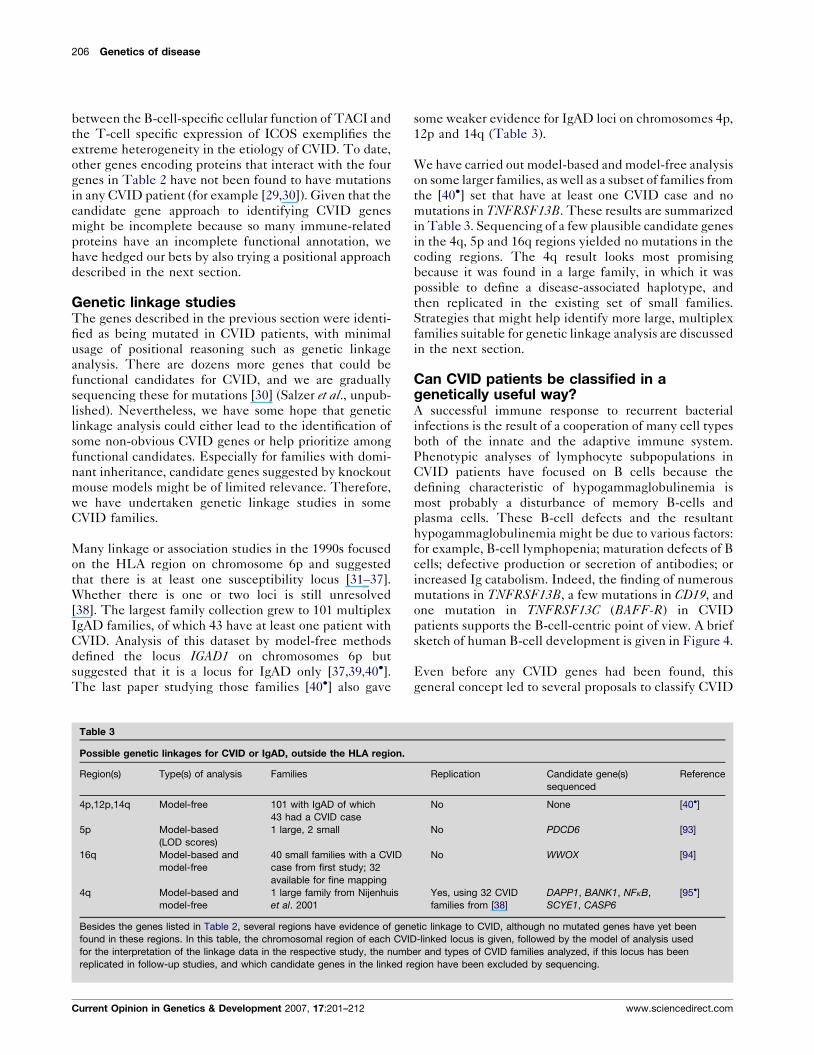
suggested that it is a locus for IgAD only [37,39,40�].The last paper studying those families [40�] also gave
Table 3
Possible genetic linkages for CVID or IgAD, outside the HLA region.
Region(s) Type(s) of analysis Families
4p,12p,14q Model-free 101 with IgAD of which
43 had a CVID case
5p Model-based
(LOD scores)
1 large, 2 small
16q Model-based and
model-free
40 small families with a CVID
case from first study; 32
available for fine mapping
4q Model-based and
model-free
1 large family from Nijenhuis
et al. 2001
Besides the genes listed in Table 2, several regions have evidence of gen
found in these regions. In this table, the chromosomal region of each CVI
for the interpretation of the linkage data in the respective study, the numb
replicated in follow-up studies, and which candidate genes in the linked re
Current Opinion in Genetics & Development 2007, 17:201–212
some weaker evidence for IgAD loci on chromosomes 4p,
12p and 14q (Table 3).
We have carried out model-based and model-free analysis
on some larger families, as well as a subset of families from
the [40�] set that have at least one CVID case and no
mutations in TNFRSF13B. These results are summarized
in Table 3. Sequencing of a few plausible candidate genes
in the 4q, 5p and 16q regions yielded no mutations in the
coding regions. The 4q result looks most promising
because it was found in a large family, in which it was
possible to define a disease-associated haplotype, and
then replicated in the existing set of small families.
Strategies that might help identify more large, multiplex
families suitable for genetic linkage analysis are discussed
in the next section.
Can CVID patients be classified in agenetically useful way?A successful immune response to recurrent bacterial
infections is the result of a cooperation of many cell types
both of the innate and the adaptive immune system.
Phenotypic analyses of lymphocyte subpopulations in
CVID patients have focused on B cells because the
defining characteristic of hypogammaglobulinemia is
most probably a disturbance of memory B-cells and
plasma cells. These B-cell defects and the resultant
hypogammaglobulinemia might be due to various factors:
for example, B-cell lymphopenia; maturation defects of B
cells; defective production or secretion of antibodies; or
increased Ig catabolism. Indeed, the finding of numerous
mutations in TNFRSF13B, a few mutations in CD19, and
one mutation in TNFRSF13C (BAFF-R) in CVID
patients supports the B-cell-centric point of view. A brief
sketch of human B-cell development is given in Figure 4.
Even before any CVID genes had been found, this
general concept led to several proposals to classify CVID
Replication Candidate gene(s)
sequenced
Reference
No None [40�]
No PDCD6 [93]
No WWOX [94]
Yes, using 32 CVID
families from [38]
DAPP1, BANK1, NFkB,
SCYE1, CASP6
[95�]
etic linkage to CVID, although no mutated genes have yet been
D-linked locus is given, followed by the model of analysis used
er and types of CVID families analyzed, if this locus has been
gion have been excluded by sequencing.
www.sciencedirect.com

Deconstructing common variable immunodeficiency by genetic analysis Schaffer, Salzer, Hammarstrom and Grimbacher 207
Figure 4
A brief sketch of human B cell development. After leaving the bone marrow as transitional B-cells, B cells circulate in the peripheral blood and
become naıve B-cells, most probably with help of the splenic environment. At that stage they need BAFF as a survival signal to prevent
apoptosis. Once engaged in a germinal center reaction in secondary lymphoid organs, B cells receive T cell help by providing the correct set
of co-stimulatory molecules such as CD28 and ICOS to select the B cells with the correct BCR, and provide help to form a germinal center
where B cells proliferate, undergo class switch and somatic hypermutation. From there, B cells either become plasma blasts and plasma
cells and home back to the bone marrow to secrete immunoglobulin or become memory B cells, which circulate in the blood and engage
in the secondary immune response to provide an immunological memory to infections and vaccinations. Plasma cells have been shown to
depend on the survival factor provided by APRIL signaling through BCMA.
patients on the basis of flow cytometric analyses of their
B-cell subpopulations (for example [41,42]). A consistent
finding in these classification approaches is that about
75% of CVID patients have substantially fewer switched
memory (defined as IgD�CD27+) B cells as compared
with those of healthy controls. These classifications are
now used to correlate the disturbed B-cell phenotype
with the disease course [43,44]. Additionally, recent stu-
dies identified a subgroup of patients who have reduced
numbers of nonswitched memory B-cells (IgD+CD27+)
and who suffer from an increased rate of pulmonary
infections and respiratory disease [45]. Once again, the
absence of switched memory cells in most of the CVID
patients, mirroring the lack of switched isotypes in serum,
suggests a close relationship between CVID and defects
of class switch recombination. It still has to be determined
if these classification models or other possibilities for
classification discussed below show concordance among
affected relatives.
Some recent findings suggest that a more global view of
the defective immune response in CVID could help
identify other genes that are mutated or dysregulated.
www.sciencedirect.com
The finding of CVID patients with a homozygous
mutation in ICOS, which is expressed exclusively in
T cells [15], gave molecular proof for the longstanding
hypothesis (for example [46]) that T-cell defects can be a
primary cause of CVID.
More recent studies have moved further upstream from the
T–B interaction to consider antigen-presenting dendritic
cells. Bayry et al. [47�] and Viallard et al. [48] have shown
association between CVID and low numbers of mature
dendritic cells, especially plasmocytoid (CD11cneg)
dendritic cells. One effect of diminished dendritic cell
function in presenting antigens is that CVID patient cells
tend to secrete subnormal levels of the cytokine IL-12 [49].
Another possibility is that defective plasmocytoid dendri-
tic cells could directly inhibit the development of long-
lived memory B-cells [50].
Another important aspect of terminal B-cell differen-
tiation that has been found to be impaired in CVID is
the process of somatic hypermutation. Somatic hypermu-
tation diversifies the transcripts that comprise both the
heavy chains and the light chains of immunoglobulins, so
Current Opinion in Genetics & Development 2007, 17:201–212

208 Genetics of disease
that different Ig molecules have enhanced affinity to
different antigens at the surface of B cells, thus support-
ing a stronger and more selective immune response.
Defective somatic hypermutation in an IgG heavy chain
gene was first reported by Levy et al. [51] in a cohort of
eight CVID patients. Schejbel et al. [52] recently
strengthened and extended this observation by using a
larger cohort of patients, and by showing that defective
somatic hypermutation is a better predictor of disease
severity than the B-cell-based classifications. Andersen
et al. [53] showed that defective somatic hypermutation
occurs also in IgG light chain genes and that this measure-
ment is also associated with the severity of respiratory
tract infections.
The process of somatic hypermutation also involves
proteins with distinct roles in DNA mismatch repair
[54] and several immunodeficiency diseases have been
shown to be caused by defects in DNA repair proteins [55].
Given these facts, one could speculate that the increased
rate of certain types of cancer in CVID and the defect in
somatic hypermutation might be related to a possible
mismatch repair defect. Predisposing mutations in such
genes could exist in CVID patients analogous to numerous
cancer susceptibility genes in which germline mutations,
present at birth, can lead to disease in adulthood, when
combined with deleterious somatic mutation events.
Especially interesting in this regard are genes such as
MLH1 [56] and BRCA1 [57] in which the same germline
mutation in relatives can lead to tumors in different tissues
and at rather different ages. These tumor suppressor genes
serve as counterpoints to the suggestion that CVID
is rarely associated with germline mutation because of
the following reasons: only a small percentage of cases are
familial; there are few large multiplex families; and most
patients have disease onset in adulthood.
One tool that has proven quite successful in cancer studies,
especially classification, is microarrays (for example
[58,59]). One might hope that a similar application of
microarrays to CVID patients will lead to the identification
of more homogenous subsets of patients as well as specific
genes that are dysregulated. However, we are aware of
only one published study that carried out a microarray
experiment on a CVID patient sample [60].
CVID genes and susceptibility to morecommon diseasesThe genes encoding a healthy immune system must
achieve a delicate balance to avoid both under-reaction
(e.g. immunodeficiency) and overreaction (e.g. autoim-
munity, allergy and excessive inflammation) to foreign
antigens. CVID is categorized as an immunodeficiencybecause all patients are unable to mount an adequate
antibody response to bacterial pathogens. However,
approximately 25% of all CVID patients have at least
one autoimmune disease [1], and a similar proportion has
Current Opinion in Genetics & Development 2007, 17:201–212
been documented within the subpopulation of CVID
patients carrying at least one mutation in TNFRSF13B[61��]. Thus, one might predict that genes mutated in
CVID patients might also be involved in susceptibility to
other immunological diseases.
In the case of ICOS, this possibility has been implicitly
considered since even before ICOS was discovered [15],
because the better known genes CD28 and CTLA4 are
adjacent to ICOS on human chromosome 2q. A variety of
studies have suggested that polymorphisms in this region
are associated with several autoimmune diseases (for
example [62] and references therein). However, almost
none of these studies have either specifically implicated
or specifically ruled out variants inside or close to ICOS.
One notable exception is the recent study by Shilling et al.[63�] showing that two polymorphisms at positions �691
and �1413 in the ICOS promoter region are associated
with allergy as measured by positive skin-prick tests
and elevated IgE. These results fit the over-reaction–
underreaction paradigm. Although Icos knockout mice do
not perfectly model human ICOS deficiency, these mice
do have low levels of IgE [64]. Thus, it mirrors previous
data that promoter polymorphisms leading to higher ICOSexpression also lead to elevated IgE and an over-reaction
to allergens.
The second recent example we highlight is more puz-
zling. Inoue et al. [65�] showed, using both multiplex
families and a case-control design, that a variety of het-
erozygous changes in TNFRSF13B are associated with
intracranial aneurysm and subsequent stroke. However,
no measurements of immunoglobulin levels were
reported. Conversely, we know of no evidence that
TACI-deficient CVID patients have an elevated risk of
stroke. Many studies have proposed that microbial infec-
tions are a risk factor of stroke [66]. TNFRSF13B hetero-
zygotes might be compromised in clearing infections that
possibly could cause damage to cerebral arteries, thereby
promoting the formation of aneurysms. An alternative
hypothesis is that the TACI protein, encoded by
TNFRSF13B, has a second, unknown function (e.g.
specific to the vascular endothelium of cerebral arteries).
ConclusionsGenetic studies of CVID–IgAD have identified
mutations explaining recessive inheritance in ICOS,
TNFRSF13B, CD19 and TNFRSF13C (BAFF-R). Hetero-
zygous missense mutations in TNFRSF13B also appear to
increase the risk of CVID, but which missense mutations
are deleterious and how much they increase risk requires
further investigation. Identifying other genes with het-
erozygous mutations and dominant inheritance has been
difficult because of the lack of mouse models and the lack
of large multiplex families. So far as we know, the most
www.sciencedirect.com

Deconstructing common variable immunodeficiency by genetic analysis Schaffer, Salzer, Hammarstrom and Grimbacher 209
CVID cases seen in a single published family is only six
[67�].
The diverse functions of the ICOS, TACI and CD19
proteins in the process of antibody production are con-
sistent with the repeatedly documented heterogeneity in
the clinical and laboratory measurements in CVID
patients. Future genetic studies of CVID will benefit
from new assays that can identify more homogeneous
subsets of patients. Microarray analysis, quantifications of
somatic hypermutation potential, and measurement of
dendritic cell subpopulations are three recently proposed
approaches to address the problem of CVID heterogen-
eity.
In summary, recent genetic research in CVID has made
some steps toward the ultimate goal of replacing the
existing and all-too-common diagnosis of exclusion with
numerous rare diagnoses of inclusion such as ‘ICOS
deficiency’, ‘CD19 deficiency’, ‘BAFF-R deficiency’
and ‘TACI deficiency’. However, these four genes can
be disease causing in at most 10% of CVID–IgAD
patients, and the recent results of genetic linkage studies
suggest that more CVID susceptibility genes can be
found through genetic studies.
AcknowledgementsOur research on CVID has been supported by the Intramural ResearchProgram at the National Institutes of Health, by grant SFB620-C2 fromthe Deutsche Forschungsgemeinschaft, by a grant from the Swedishresearch council, and by EU-Project QLRT-2001-01536 (IMPAD).
References and recommended readingPapers of particular interest, published within the period of review,have been highlighted as:
� of special interest
�� of outstanding interest
1. Cunningham-Rundles C, Bodian C: Common variableimmunodeficiency: Clinical and immunological features of 248patients. Clin Immunol 1999, 92:34-48.
2. No authors listed: Primary immunodeficiency diseases.Report of an IUIS Scientific Committee. InternationalUnion of Immunological Societies. Clin Exp Immunol 1999,118:1–28.
3. Ishizaka A, Nakanishi M, Yamada S, Sakiyama Y, Matsumoto S:Development of hypogammaglobulinaemia in a patient withcommon variable immunodeficiency. Eur J Pediatr 1989,149:175-176.
4. Espanol T, Catala M, Hernandez M, Caragol I, Bertran JM:Development of a common variable immunodeficiency inIgA-deficient patients. Clin Immunol Immunopathol 1996,80:333-335.
5. Johnson ML, Keeton LG, Zhu ZB, Volanakis JE, Cooper MD,Schroeder HW Jr: Age-related changes in serumimmunoglobulins in patients with familial IgA deficiencyand common variable immunodeficiency (CVID). Clin ExpImmunol 1997, 108:477-483.
6. Carvalho Neves Forte W, Ferreira De Carvalho Junior F,Damaceno N, Vidal Perez F, Gonzales Lopes C, Mastroti RA:Evolution of IgA deficiency to IgG subclass deficiency andcommon variable immunodeficiency. Allergol Immunopathol(Madr) 2000, 28:18-20.
www.sciencedirect.com
7. Eades-Perner A-M, Gathmann B, Knerr V, Guzman D, Veit D,Kindle G: Grimbacher B for the ESID Registry Working party:The European internet-based patient and research databasefor primary immunodeficiencies: results 2004–2006. Clin ExpImmunol 2007, 147:306-312.
8. Orange JS, Hossny EM, Weiler CR, Ballow M, Berger M,Bonilla FA, Buckley R, Chinen J, El-Gamal Y, Mazer BD et al.:Use of intravenous immunoglobulin in human disease: areview of evidence by members of the PrimaryImmunodeficiency Committee of the American Academy ofAllergy, Asthma and Immunology. J Allergy Clin Immunol 2006,117(4 Suppl):S525-S553.
9. Ziegner UHM, Kobayashi RH, Cunningham-Rundles C, Espanol T,Fasth A, Huttenlocher A, Korgstad P, Marthinsen L,Notarangelo LD, Pasic S et al.: Progressive neurodegenerationin patients with primary immunodeficiency disease on IVIGtreatment. Clin Immunol 2002, 102:19-24.
10. Kamin RM, Fudenberg HH, Douglas SD: A genetic defect in‘‘acquired’’ agammaglobulinemia. Proc Natl Acad Sci USA1968, 60:881-885.
11. Vorechovsky I, Zetterquist H, Paganelli R, Koskinen S,Webster ADB, Bjorkander J, Smith CIE, Hammarstrom L: Familyand linkage study of selective IgA deficiency and commonvariable immunodeficiency. Clin Immunol Immunopathol 1995,77:185-192.
12. de Bakker PIW, McVean G, Sabeti PC, Miretti MM, Green T,Marchini J, Ke XY, Monsuur AJ, Whittaker P, Delgado M et al.:A high-resolution HLA and SNP haplotype map for diseaseassociation studies in the extended human MHC. Nat Genet2006, 38:1166-1172.
13. Veillette A: Immune regulation by SLAM family receptorsand SAP-related adaptors. Nat Rev Immunol 2006,6:56-66.
14.��
Pasquier B, Yin L, Fondaneche M-C, Relouzat F, Bloch-Queyrat C,Lambert N, Fischer A, de Saint-Basile G, Latour S: Defective NKTcell development in mice and humans lacking the adapterSAP, the X-linked lymphoproliferative syndrome geneproduct. J Exp Med 2005, 201:695-701.
This study shows that SAP (signaling lymphocyte activation molecule-associated protein) is essential for the development of natural killer T-cellsin mice and humans, and that deficiency of natural killer T-cells might bethe explanation for the susceptibility of XLP patients for, often fatal, EBVinfections.
15. Hutloff A, Dittrich AM, Beier KC, Eljaschewitsch B, Kraft R,Anagnostopoulos I, Kroczek RA: ICOS is an inducible T-cellco-stimulator structurally and functionally related to CD28.Nature 1999, 397:263-266.
16.�
Bossaller L, Burger J, Draeger R, Grimbacher B, Knoth R,Plebani A, Durandy A, Baumann U, Schlesier M, Welcher AA et al.:ICOS deficiency is associated with a severe reduction ofCXCR5+CD4 germinal center Th cells. J Immunol 2006,177:4927-4932.
This study identifies the lack of CXCR5+CD4 germinal center T-helpercells as one of the key pathomechanisms in human and murine ICOSdeficiency.
17.�
Warnatz K, Bossaller L, Salzer U, Skrabl-Baumgartner A,Schwinger W, van der Burg M, van Dongen JJM, Orlowska-Volk M,Knoth R, Durandy A et al.: Human ICOS deficiency abrogatesthe germinal center reaction and provides a monogenicmodel for common variable immunodeficiency. Blood 2006,107:3045-3052.
This is the first in-depth clinical description of human ICOS deficiency.Although only described in nine patients worldwide, human ICOS defi-ciency mirrors the whole clinical spectrum of CVID.
18.��
van Zelm MC, Reisli I, van der Burg M, Castano D, vanNoesel CJM, van Tol MJD, Woellner C, Grimbacher B, Patino P,van Dongen JJM, Franco JL: An antibody-deficiency syndromedue to mutations in the CD19 gene. N Engl J Med 2006,354:1901-1912.
This study shows that deficiency of the B-cell marker CD19 can causeCVID.
19. Schneider P, MacKay F, Steiner V, Hofmann K, Bodmer JL,Holler N, Ambrose C, Lawton P, Bixler S, Acha-Orbea H et al.:
Current Opinion in Genetics & Development 2007, 17:201–212

210 Genetics of disease
BAFF, a novel ligand of the tumor necrosis factor family,stimulates B cell growth. J Exp Med 1999, 189:1747-1756.
20. O’Connor BP, Raman VS, Erickson LD, Cook WJ, Weaver LK,Ahonen C, Lin L-L, Mantchev GT, Bram RJ, Noelle RJ: BCMA isessential for the survival of long-lived bone marrow plasmacells. J Exp Med 2004, 199:91-98.
21. Litinskiy MB, Nardelli B, Hilbert DM, He B, Schaffer A, Casali P,Cerutti A: DCs induce CD40-independent immunoglobulinclass switching through Blys and APRIL. Nat Immunol 2002,3:822-829.
22.�
Sakurai D, Hase H, Kanno Y, Kojima H, Okumura K, Kobata T:TACI regulates IgA production by APRIL in collaboration withHSPG. Blood 2007, 109:2961-2967.
See annotation [23�].
23.�
Sakurai D, Kanno Y, Hase H, Kojima H, Okumura K, Kobata T:TACI attenuates antibody production costimulated by BAFF-Rand CD40. Eur J Immunol 2007, 37:110-118.
These two reports from Sakurai et al. [22�,23�] give an explanation on howTACI exerts its opposing effects on human B-cell function and differ-entiation. In their model, BAFF–TACI interactions negatively regulate B-cell function, whereas APRIL interactions with TACI and HSPG induce keyB-cell differentiation events.
24. von Bulow G-U, van Deursen JM, Bram RJ: Regulation of theT-independent humoral response by TACI. Immunity 2001,14:573-582.
25. Pan-Hammarstrom Q, Salzer U, Du L, Bjorkander J, Cunningham-Rundles C, Nelson DL, Bacchelli C, Gaspar B, Offer S, Behrens TWet al.: Role of mutations in TNFRSF13B in CVID and selectiveIgA deficiency. Nat Genet 2007, in press.
26. Fisher GH, Rosenberg FJ, Straus SE, Dale JK, Middleton LA,Lin AY, Strober W, Lenardo MJ, Puck JM: Dominant interferingFas gene mutations impair apoptosis in a human autoimmunelymphoproliferative syndrome. Cell 1995, 81:935-946.
27. McDermott MF, Aksentijevich I, Galon J, McDermott EM,Ogunkolade BW, Centola M, Mansfield E, Gadina M, Karenko L,Pettersson T et al.: Germline mutations in the extracellulardomains of the 55 kDa TNF receptor, TNFR1, define a family ofdominantly inherited autoinflammatory syndromes. Cell 1999,97:133-144.
28. Jackson CE, Fischer RE, Hsu AP, Anderson SM, Choi Y, Wang J,Dale JK, Fleisher TA, Middelton LA, Sneller MC et al.:Autoimmune lymphoproliferative syndrome with defectiveFas: genotype influences penetrance. Am J Hum Genet 1999,64:1002-1014.
29. Salzer U, Maul-Pavicic A, Cunningham-Rundles C, Urschel S,Belohradsky BH, Litzman J, Holm A, Franco JL, Plebani A,Hammarstrom L et al.: ICOS deficiency in patients with commonvariable immunodeficiency. Clin Immunol 2004, 113:234-240.
30. Losi CG, Salzer U, Gatta R, Lougaris V, Cattaneo G, Meini A,Soresina A, Grimbacher B, Plebani A: Mutational analysis ofhuman BLyS in patients with common variableimmunodeficiency. J Clin Immunol 2006, 26:396-399.
31. Schaffer FM, Palermos J, Zhu ZB, Barger BO, Cooper MD,Volanakis JE: Individuals with IgA deficiency and commonvariable immunodeficiency share polymorphisms of majorhistocompatibility complex class III genes. Proc Natl Acad SciUSA 1989, 86:8015-8019.
32. Olerup O, Smith CIE, Hammarstrom L: Different amino acids atposition 57 of the HLA-DQ beta chain associated withsusceptibility and resistance to IgA deficiency. Nature 1990,347:289-290.
33. Olerup O, Smith CI, Bjorkander J, Hammarstrom L: Shared HLAclass II-associated genetic susceptibility and resistance,related to the HLA-DQB1 gene, in IgA deficiency and commonvariable immunodeficiency. Proc Natl Acad Sci USA 1992,89:10653-10657.
34. Volanakis JE, Zhu ZB, Schaffer FM, Macon KJ, Palermos J,Barger BO, Go R, Campbell RD, Schroeder HW Jr, Cooper MD:Major histocompatibility complex class III genes andsusceptibility to immunoglobulin A deficiency and commonvariable immunodeficiency. J Clin Invest 1992, 89:1914-1922.
Current Opinion in Genetics & Development 2007, 17:201–212
35. Cucca F, Zhu Z-B, Khanna A, Cossu F, Congia M, Badiali M,Lampis R, Frau F, De Virgiliis S, Cao A et al.: Evaluation of IgAdeficiency in Sardinians indicates a susceptibility gene isencoded within the HLA class III region. Clin Exp Immunol 1998,111:76-80.
36. Schroeder HW Jr, Zhu ZB, March RE, Campbell RD, Berney SM,Nedospasov SA, Turetskaya RL, Atkinson TP, Go RC, Cooper MD,Volanakis JE: Susceptibility locus for IgA deficiency andcommon variable immunodeficiency in the HLA-DR3, -B8,-A1 haplotypes. Mol Med 1998, 4:72-86.
37. Vorechovsky I, Webster ADB, Plebani A, Hammarstrom L: Geneticlinkage of IgA deficiency to the major histocompatibilitycomplex: Evidence for allele segregation distortion, parent-of-origin penetrance differences, and the role of anti-IgAantibodies in disease predisposition. Am J Hum Genet 1999,64:1096-1109.
38. De la Concha EG, Fernandez-Arquero M, Gual L, Vigil P,Martinez A, Urcelay E, Ferreira A, Garcia-Rodriguez MC, Fontan G:MHC susceptibility genes to IgA deficiency are located indifferent regions on different HLA haplotypes. J Immunol 2002,169:4637-4643.
39. Vorechovsky I, Cullen M, Carrington M, Hammarstrom L,Webster ADB: Fine mapping of IGAD1 in IgA deficiency andcommon variable immunodeficiency: identification andcharacterization of haplotypes shared by affected members of101 multiple-case families. J Immunol 2000, 164:4408-4416.
40.�
Kralovicova J, Hammarstrom L, Plebani A, Webster ADB,Vorechovsky I: Fine-scale mapping at IGAD1 and genome-widegenetic linkage analysis implicate HLA-DQ/DR as a majorsusceptibility locus in selective IgA deficiency and commonvariable immunodeficiency. J Immunol 2003, 170:2765-2775.
This is the latest in a series of studies implicating one or more IgADsusceptibility factors in the HLA region. It describes the latest additions toa collection 101 multiplex IgAD families.
41. Warnatz K, Denz A, Drager R, Braun M, Groth C, Wolff-Vorbeck G,Eibel H, Schlesier M, Peter HH: Severe deficiency of switchedmemory B cells (CD27+IgMSIgDS) in subgroups of patientswith common variable immunodeficiency: a new approach toclassify a heterogeneous disease. Blood 2002, 99:1544-1551.
42. Piqueras B, Lavenu-Bombled C, Galicier L, Bergeron-Van derCruyssen F, Mouthon L, Chevret S, Debre P, Schmitt C,Oksenhendler E: Common variable immunodeficiencypatient classification based on impaired B cell memorydifferentiation correlates with clinical aspects. J Clin Immunol2003, 23:385-400.
43. Ko J, Radigan L, Cunningham-Rundles C: Immune competenceand switched memory B cells in common variableimmunodeficiency. Clin Immunol 2005, 116:37-41.
44. Alachkar H, Taubenheim N, Haeney MR, Durandy A, Arkwright PD:Memory switched B cell percentage and not serumimmunoglobulin concentration is associated with clinicalcomplications in children and adults with specific antibodydeficiency and common variable immunodeficiency.Clin Immunol 2006, 120:310-318.
45. Carsetti R, Rosado MM, Donnanno S, Guazzi V, Soresina A,Meini A, Plebani A, Aiuti F, Quinti I: The loss of IgM memory Bcells correlates with clinical disease in common variableimmunodeficiency. J Allergy Clin Immunol 2005, 115:412-417.
46. Waldmann TA, Durm M, Broder S, Blackman M, Blaese RM,Strober W: Role of suppressor T cells in pathogenesis ofcommon variable hypogammaglobulinemia. Lancet 1974,2:609-613.
47.�
Bayry J, Lacroix-Desmazes S, Kazatchkine MD, Galicier L,Lepelletier Y, Webster D, Levy Y, Eibl MM, Oksenhendler E,Hermine O, Kaveri SV: Common variable immunodeficiency isassociated with defective functions of dendritic cells.Blood 2004, 15:2441-2443.
This study expands the known cellular substrates of immunopathology inCVID to dendritic cells by showing various abnormalities in these cells inCVID patients.
48. Viallard J-F, Camou F, Andre M, Liferman F, Moreau J-F,Pellegrin J-L, Blanco P: Altered dendritic cell distribution in
www.sciencedirect.com
Deconstructing common variable immunodeficiency by genetic analysis Schaffer, Salzer, Hammarstrom and Grimbacher 211
patients with common variable immunodeficiency.Arthritis Res Ther 2005, 7:R1052-R1055.
49. Cunningham-Rundles C, Radigan L: Deficient IL-12 anddendritic cell function in common variable immundedeficiency. Clin Immunol 2005, 115:147-153.
50. Jego G, Palucka AK, Blanck J-P, Chalouni C, Pascual V,Banchereau J: Plasmacytoid dendritic cells induce plasma celldifferentiatiation through type I interferon and interleukin 6.Immunity 2003, 19:225-234.
51. Levy Y, Gupta N, Le Deist F, Garcia C, Fischer A, Weill J-C,Reynaud C-A: Defect in IgV gene somatic hypermutation inCommon Variable Immuno-Deficienciency syndrome.Proc Natl Acad Sci USA 1998, 95:13135-13140.
52. Schejbel L, Marquart H, Andersen V, Permin H, Andersen P,Svejgaard A, Barington T: Deficiency of somatic hypermutationof immunoglobulin G transcripts is a better predictor of severerespiratory tract infections than lack of memory B cells incommon variable immunodeficiency. J Clin Immunol 2005,25:392-403.
53. Andersen P, Permin H, Andersen V, Schejbel L, Garred P,Svejgaard A, Barington T: Deficiency of somatic hypermutationof the antibody light chain is associated with increasedfrequency of severe respiratory tract infection in commonvariable immunodeficiency. Blood 2005, 105:511-517.
54. Casali P, Pal Z, Xu Z, Zan H: DNA repair in antibody somatichypermutation. Trends Immunol 2006, 27:313-321.
55. Gennery AR: Primary immunodeficiency syndromesassociated with defective DNA double-strand break repair.Br Med Bull 2006, 77–78:71-85.
56. Papadopoulos N, Nicolaides NC, Wei Y-F, Ruben SM, Carter KC,Rosen CA, Haseltine WA, Fleischmann RD, Fraser CM, Adams MDet al.: Mutation of a mutL homolog in hereditary colon cancer.Science 1994, 263:1625-1629.
57. Miki Y, Swensen J, Shattuck-Eidens D, Futreal PA, Harshman K,Tavtigian S, Liu Q, Cochran C, Bennett LM, Ding W et al.: A strongcandidate for the breast and ovarian cancer susceptibilitygene BRCA1. Science 1994, 266:66-71.
58. Golub TR, Slonim DK, Tamayo P, Huard C, Gaasenbeek M,Mesirov JP, Coller H, Loh ML, Downing JR, Caligiuri MA et al.:Molecular classification of cancer: class discovery and classprediction by gene expression monitoring. Science 1999,286:531-537.
59. Dave SS, Fu K, Wright GW, Lam LT, Kluin P, Boerma E-J,Greiner TC, Weisenburger DD, Rosenwald A, Ott G et al.: StaudtLM for the lymphoma/leukemia molecular profiling project:molecular diagnosis of Burkitt’s lymphoma. N Engl J Med 2006,354:2431-2442.
60. Zeeberg BR, Qin H, Narasimhan S, Sunshine M, Cao H,Kane DW, Reimers M, Stephens RM, Bryant D, Burt SK et al.:High-throughput GoMiner, an ‘industrial-strength’ integrativegene ontology tool for interpretation of multiple-microarrayexperiments, with application to studies of CommonVariable Immune Deficiency (CVID). BMC Bioinformatics 2005,6:168.
61.��
Salzer U, Chapel HM, Webster ADB, Pan-Hammarstrom Q,Schmidt-Graeff A, Schlesier M, Peter HH, Rockstroh JK,Schneider P, Schaffer AA et al.: Mutations in TNFRSF13B, whichencodes TACI, are associated with common variableimmunodeficiency in humans. Nat Genet 2005, 37:820-828.
The authors identify the first commonly mutated gene in CVID and showthat mutations can be heterozygous as well as homozygous.
62. Ueda H, Howson JMM, Esposito L, Heward J, Snook H,Chamberlain G, Rainbow DB, Hunter KMD, Smith AN, Di Genova Get al.: Association of the T-cell regulatory gene CTLA4with susceptibility to autoimmune disease. Nature 2003,423:506-511.
63.�
Shilling RA, Pinto JM, Decker DC, Schneider DH, Bandukwala HS,Schneider JR, Camoretti-Mercado B, Ober C, Sperling AI: Cuttingedge: polymorphisms in the ICOS promoter region areassociated with allergic sensitization and Th2 cytokineproduction. J Immunol 2005, 175:2061-2065.
www.sciencedirect.com
This study illustrates the paradigm that genes mutated in CVID might alsobe relevant to more common immune-related diseases.
64. McAdam AJ, Greenwald RJ, Levin MA, Chernova T,Malenkovich N, Ling V, Freeman GJ, Sharpe AH: ICOS is criticalfor CD40-mediated antibody class switching. Nature 2001,409:102-105.
65.�
Inoue K, Mineharu Y, Inoue S, Yamada S, Matsuda F, Nozaki K,Takenaka K, Hashimoto N, Koizumi A: Search on chromosome17 centromere reveals TNFRSF13B as a susceptibility gene forintracranial aneurysm: A preliminary study. Circulation 2006,113:2002-2010.
This paper presents the intriguing result that the same gene can bemutated in either CVID or stroke.
66. Elkind MSV, Cole JW: Do common infections cause stroke?Semin Neurol 2006, 26:88-99.
67.�
Nijenhuis T, Klasen I, Weemaes CMR, Preijers F, de Vries E,van der Meer JWM: Common variable immunodeficiency(CVID) in a family: an autosomal dominant mode ofinheritance. Neth J Med 2001, 59:134-139.
This paper describes the CVID multiplex family with the most casespublished to date. See also study by Finck et al. [95�].
68. Tsukada S, Saffran DC, Rawlings DJ, Parolini O, Allen RC,Klisak I, Sparkes RS, Kubagawa H, Mohandas T, Quan S et al.:Deficient expression of a B cell cytoplasmic tyrosine kinase inhuman X-linked agammaglobulinemia. Cell 1993, 72:279-290.
69. Vetrie D, Vorechovsky I, Sideras P, Holland J, Davies A,Flinter F, Hammarstrom L, Kinnon C, Levinsky R, Bobrow M et al.:The gene involved in X-linked aggamaglobulinemia is amember of the src family of protein-tyrosine kinases.Nature 1993, 361:226-233.
70. Saffran DC, Parolin O, Fitch-Hilgenberg ME, Rawlings DJ,Afar DEH, Witte ON, Conley ME: A point mutation in the SH2domain of Bruton’s tyrosine kinase in atypical X-linkedagammaglobulinemia. N Engl J Med 1994, 330:1488-1491.
71. Yel L, Minegishi Y, Coustan-Smith E, Buckley RH, Trubel H,Pachman LM, Kitchingman GR, Campana D, Rohrer J, Conley ME:Mutations in the mu heavy-chain gene in patients withagammaglobulinemia. N Engl J Med 1996, 335:1486-1493.
72. Meffre E, Milili M, Blanco-Betancourt C, Antunes H,Nussenzweig MC, Schiff C: Immunoglobulin heavy chainexpression shapes the B cell receptor repertorie in human Bcell development. J Clin Invest 2001, 108:879-886.
73. Lopez Granados E, Porpiglia AS, Hogan MB, Matamoros N,Krasovec S, Pignata C, Smith CIE, Hammarstrom L, Bjorkander J,Belohradsky BH et al.: Clinical and molecular characterizationof patients with defects in m heavy chain gene. J Clin Invest2002, 110:1029-1035.
74. Minegishi Y, Coustan-Smith E, Rapalus L, Ersoy F, Campana D,Conley ME: Mutations in Iga (CD79a) result in a complete blockin B-cell development. J Clin Invest 1999, 104:1115-1121.
75. Minegishi Y, Coustan-Smith E, Wang Y-H, Cooper MD,Campana D, Conley ME: Mutations in the human k5/14.1gene result in B cell deficiency and agammaglobulinemia.J Exp Med 1998, 187:71-77.
76. Minegishi Y, Rohrer J, Coustan-Smith E, Lederman HM,Pappu R, Campana D, Chan AC, Conley ME: An essential rolefor BLNK in human B cell development. Science 1999,286:1954-1957.
77. Sawada A, Takihara Y, Kim JY, Matsuda-Hashii Y, Tokimasa S,Fujisaki H, Kubota K, Endo H, Onodera T, Ohta H et al.:A congenital mutation of the novel gene LRRC8 causesagammaglobulinemia in humans. J Clin Invest 2003,112:1707-1713.
78. Allen RC, Armitage RJ, Conley ME, Rosenblatt H, Jenkins NA,Copeland NG, Bedell MA, Edelhoff S, Disteche CM, Simoneaux DKet al.: CD40 ligand gene defects responsible for X-linkedhyper-IgM syndrome. Science 1993, 259:990-993.
79. Aruffo A, Farrington M, Hollenbaugh D, Li X, Milatovich A,Nonoyama S, Bajorath J, Grosmaire LS, Stenkamp R, Neubauer Met al.: The CD40 ligand, gp39, is defective in activated T cells
Current Opinion in Genetics & Development 2007, 17:201–212

212 Genetics of disease
from patients with X-linked hyper-IgM syndrome. Cell 1993,72:291-300.
80. Revy P, Muto T, Levy Y, Geissmann F, Plebani A, Sanal O,Catalan N, Forveille M, Dufourcq-Labelouse R, Gennery A et al.:Activation-induced cytidine deaminase (AID) deficiencycauses the autosomal recessive form of the Hyper-IgMsyndrome (HIGM2). Cell 2000, 102:565-575.
81. Ferrari S, Giliani S, Insalaco A, Al-Ghonaium A, Soresina AR,Loubser M, Avanzini MA, Marconi M, Badolato R, Ugazio AG et al.:Mutations of CD40 gene cause an autosomal recessive form ofimmunodeficiency with hyper IgM. Proc Natl Acad Sci USA2001, 98:12614-12619.
82. Imai K, Slupphaug G, Lee W-I, Revy P, Nonoyama S, Catalan N,Yel L, Forveille M, Kavli B, Krokan HE et al.: Human uracil-DNAglycosylase deficiency associated with profoundly impairedimmunoglobulin class-switch recombination. Nat Immunol2003, 4:1023-1028.
83. Imai K, Zhu Y, Revy P, Morio T, Mizutani S, Fischer A, Nonoyama S,Durandy A: Analysis of class switch recombination and somatichypermutation in patients affected with autosomal dominanthyper-IgM syndrome type 2. Clin Immunol 2005, 115:277-285.
84. Morra M, Silander O, Calpe S, Choi M, Oettgen H, Myers L,Etzioni A, Buckley R, Terhorst C: Alterations of the X-linkedlymphoproliferative disease gene SH2D1A in common variableimmunodeficiency syndrome. Blood 2001, 98:1321-1325.
85. Nistala K, Gilmour KC, Cranston T, Davies EG, Goldblatt D,Gaspar HB, Jones AM: X-linked lymphoproliferative disease:three atypical cases. Clin Exp Immunol 2001, 126:126-130.
86.��
Rigaud S, Fondaneche M-C, Lambert N, Pasuier B, Mateo V,Soulas P, Galicier L, Le Deist F, Rieux-Laucat F, Revy P et al.: XIAPdeficiency in humans causes an X-linked lymphoproliferativesyndrome. Nature 2006, 442:110-114.
The authors report on the second genetic defect, XIAP deficiency, inpatients with XLP.
87. Kalman L, Lindegren ML, Kobrynski L, Vogt R, Hannon H,Howard JT, Buckley R: Mutations in genes required for T-celldevelopment: IL7R, CD45, IL2RG, JAK3, RAG1, RAG2,ARTEMIS, and ADA and severe combined immunodeficiency:HuGE review. Genet Med 2004, 6:16-26.
Elsevier.com – linking scientists
Designed for scientists’ information needs, Elsevie
customer-focused navigation and an intuitive arch
greater prod
The easy-to-use navigational tools and structure
from one entry point. Users can perform rapid an
functionality, using the FAST technology of Scirus.
define their searches by any number of criteria to p
specific author or editor, book publication date,
physical sciences and social sciences – or by produ
1800 Elsevier journals, 2200 new books every year
In addition, tailored content for authors, editors an
on new products
Elsevier is proud to be a partner with the scientific
our mission and values at Elsevier.com. Discover
medical communities worldwide through partnershi
awards from The Els
As a world-leading publisher of scientific, technical
linking researchers and professionals to the best t
deepest coverage in a range of media types to
breakthroughs in research and discovery, and
Elsevier. Building insightswww.elsev
Current Opinion in Genetics & Development 2007, 17:201–212
88. Hernandez PA, Gorlin RJ, Lukens JN, Taniuchi S, Bohinjec J,Francois F, Klotman ME, Diaz GA: Mutations in the chemokinereceptor gene CXCR4 are associated with WHIM syndrome, acombined immunodeficiency disease. Nat Genet 2003, 34:70-74.
89. Pan Q, Hammarstrom L: Molecular basis of IgG subclassdeficiency. Immunol Rev 2000, 178:99-110.
90.��
Grimbacher B, Hutloff A, Schlesier M, Glocker E, Warnatz K,Drager R, Eibel H, Fischer B, Schaffer AA, Mages HW et al.:Homozygous loss of ICOS is associated with adult-onsetcommon variable immunodeficiency. Nat Immunol 2003,4:261-268.
This paper describes the first gene found to be mutated in CVID. It showshow a T-cell defect can lead to CVID.
91. Warnatz K, Salzer U, Gutenberger S, Taubenheim N, Theil K,Schlesier M, Grimbacher B, Eibel H, Peter HH: Finally found:human BAFF-R deficiency causes CVID. In: XIth Meeting of theEuropean Society for Immunodeficiencies 21–24 October 2004;Versailles [Abstract #B. 72].
92.��
Castigli E, Wilson SA, Garibyan L, Raschid R, Bonilla F,Schneider L, Geha RS: TACI is mutant in common variableimmunodeficiency and IgA deficiency. Nat Genet 2005,37:829-834.
Identifies the first commonly mutated gene in CVID and shows thatmutations can be heterozygous as well as homozygous.
93. Braig DU, Schaffer AA, Glocker E, Salzer U, Warnatz K, Peter HH,Grimbacher B: Linkage of autosomal dominant commonvariable immunodeficiency to chromosome 5p and evidencefor locus heterogeneity. Hum Genet 2003, 112:369-378.
94. Schaffer AA, Pfannstiel J, Webster ADB, Plebani A,Hammarstrom L, Grimbacher B: Analysis of families withcommon variable immunodeficiency (CVID) and IgA deficiencysuggests linkage of CVID to chromosome 16q. Hum Genet2006, 118:725-729.
95.�
Finck A, Van der Meer JWM, Schaffer AA, Pfannstiel J, Fieschi C,Plebani A, Webster ADB, Hammarstrom L, Grimbacher B: Linkageof autosomal-dominant common variable immunodeficiencyto chromosome 4q. Eur J Hum Genet 2006, 14:867-875.
The first replicated linkage for autosomal dominant CVID outside the HLAregion on chromosome 6p. See also study by Nijenhuis et al. [67�].
to new research and thinking
r.com is powered by the latest technology with
itecture for an improved user experience and
uctivity.
connect scientists with vital information – all
d precise searches with our advanced search
com, the free science search engine. Users can
inpoint information and resources. Search by a
subject area – life sciences, health sciences,
ct type. Elsevier’s portfolio includes more than
and a range of innovative electronic products.
d librarians provides timely news and updates
and services.
and medical community. Find out more about
how we support the scientific, technical and
ps with libraries and other publishers, and grant
evier Foundation.
and health information, Elsevier is dedicated to
hinking in their fields. We offer the widest and
enhance cross-pollination of information,
the sharing and preservation of knowledge.
. Breaking boundaries.ier.com
www.sciencedirect.com
![[Expression of leukocytic adhesion molecules in a patient with common variable immunodeficiency]](https://img.dokumen.tips/doc/110x75/6347bd95de40dd034d08d8ff/expression-of-leukocytic-adhesion-molecules-in-a-patient-with-common-variable-immunodeficiency.jpg)


