Embed Size (px)
Citation preview
2009;69:431-439. Cancer Res Stephen M. Wiesner, Stacy A. Decker, Jon D. Larson, et al. in Mice Using Plasmid DNA
Induction of Genetically Engineered Brain TumorsDe novo
Updated version
http://cancerres.aacrjournals.org/content/69/2/431
Access the most recent version of this article at:
Material
Supplementary
http://cancerres.aacrjournals.org/content/suppl/2009/01/12/69.2.431.DC1.html
Access the most recent supplemental material at:
Cited Articles
http://cancerres.aacrjournals.org/content/69/2/431.full.html#ref-list-1
This article cites by 45 articles, 12 of which you can access for free at:
Citing articles
http://cancerres.aacrjournals.org/content/69/2/431.full.html#related-urls
This article has been cited by 10 HighWire-hosted articles. Access the articles at:
E-mail alerts related to this article or journal.Sign up to receive free email-alerts
Subscriptions
Reprints and
To order reprints of this article or to subscribe to the journal, contact the AACR Publications
Permissions
To request permission to re-use all or part of this article, contact the AACR Publications
Research. on November 4, 2014. © 2009 American Association for Cancercancerres.aacrjournals.org Downloaded from
Research. on November 4, 2014. © 2009 American Association for Cancercancerres.aacrjournals.org Downloaded from
De novo Induction of Genetically Engineered Brain Tumors in
Mice Using Plasmid DNA
Stephen M. Wiesner,1Stacy A. Decker,
2Jon D. Larson,
3,6Katya Ericson,
4Colleen Forster,
5
Jose L. Gallardo,2Chunmei Long,
2Zachary L. Demorest,
4Edward A. Zamora,
2
Walter C. Low,4,6Karen SantaCruz,
5David A. Largaespada,
3,6and John R. Ohlfest
2,4,6
1Center for Allied Health Programs, Departments of 2Pediatrics, 3Genetics, Cell Biology, and Development, 4Neurosurgery,and 5Laboratory Medicine and Pathology, and 6Masonic Cancer Center, University of Minnesota, Minneapolis, Minnesota
Abstract
Spontaneous mouse models of cancer show promise to moreaccurately recapitulate human disease and predict clinicalefficacy. Transgenic mice or viral vectors have been requiredto generate spontaneous models of glioma, a lethal braintumor, because nonviral gene transfer is typically transient. Toovercome this constraint, we used the Sleeping Beautytransposable element to achieve chromosomal integration ofhuman oncogenes into endogenous brain cells of immuno-competent mice. Genetically engineered, spontaneous braintumors were induced with plasmid DNA in a matter of weeksin three separate mouse strains. The phenotype of tumorswas influenced by the combination of oncogenes delivered,resembling human astrocytoma or glioblastoma in the majo-rity of cases. At least five different genes can be cotransfectedsimultaneously including reporters, allowing measurement oftumor viability by in vivo imaging. This model can acceleratebrain tumor research in a variety of ways such as genera-tion of ‘‘humanized’’ models for high throughput drug screen-ing and candidate gene validation with exceptional speedand flexibility. [Cancer Res 2009;69(2):431–9]
Introduction
Malignant glioma (MG) is a devastating primary brain tumor.Gliomas are classified according the WHO criteria ranging fromgrade I, a typically treatable tumor, to grade IV, a glioblastomamultiforme (GBM; ref. 1). GBM is a lethal brain tumor claimingover 12,000 lives each year in the United States (2). Prognosis forpatients with high-grade MG is poor and has remained relativelyunchanged for decades. MG often displays marked genetic andphenotypic heterogeneity (3). For example, as many as threedifferent alleles of the Trp53 tumor suppressor gene are detectablein a single GBM; such tumors contain mixed regions appearing ashigh- or low-grade histologically and exhibit heterogeneousexpression of the epidermal growth factor receptor (EGFR)immunohistochemically (4, 5). The heterogeneity of MG mayaccount for the failure of therapies with a single mechanism ofaction. Animal models that recapitulate the complexity of human
MG would be useful to better understand glioma biology andpredict therapeutic response.The most widely used rodent model of MG involves intracerebral
transplantation of cultured glioma cells, a very different scenariofrom the human disease. There has been great interest inspontaneous models of MG in which the tumor evolves with thehost immune system with the expectation that they will morefaithfully mimic human disease progression and better predictclinical efficacy. A number of genetically engineered mice (GEM)are available, harboring constitutive or conditional alleles of genesassociated with MG development (6). Some models combinevarious GEM to test cooperativity of particular mutations in tumordevelopment (7–9). Spontaneous MG can also be induced byintracerebral injection of a retroviral vector encoding platelet-derived growth factor (10, 11). Hybrid models have also beencreated by using GEM that express a receptor for a replication-competent ALV splice acceptor (RCAS) avian retrovirus in a tissuespecific pattern, leading to MG after oncogene transfer (12, 13).This GEM has been bred to a second GEM expressing fireflyluciferase (FLuc) in mitotic cells to allow cell division to benoninvasively monitored using bioluminescent imaging (14).Bioluminescent imaging provides unparalleled convenience andspeed for determining the efficacy of therapeutic agents in livingmice (15). To date, all spontaneous mouse models of MG haverequired the use of GEM or viral vectors. Production of new GEMor viral vectors can take months to years to develop andcharacterize. In some models, mice develop tumors with incom-plete penetrance and exhibit relatively long survival times makingthem inconvenient for preclinical trials. We sought to develop amore flexible, rapid, spontaneous MG model that was independentof strain background while retaining the ability to monitor tumorviability with bioluminescence.Several investigators have previously achieved nonviral transfec-
tion of the murine brain with polyethylenimine/plasmid DNA (PEI/DNA) complexes (16–19). Unfortunately, gene expression afterplasmid DNA transfection is typically transient. To overcome thisconstraint, we used the Sleeping Beauty (SB) transposable elementdelivered as plasmid DNA to achieve chromosomal integraftionand long-term expression (17). SB is a synthetic transposableelement that was created by genetically engineering inactivetransposase gene sequences isolated from salmonid fish (20). SBis a two-part system composed of a transposon DNA substrate anda transposase enzyme. SB transposase mediates ‘‘cut and paste’’excision and insertion of transposon DNA into a TA dinucleotide ofthe host genomic DNA (21). The gene encoding the SB transposaseenzyme can be provided on the plasmid DNA backbone or on aseparate plasmid relative to transposon DNA.Here, we show that injection of PEI/DNA complexes into the
lateral cerebral ventricle of neonatal mice leads to SB-dependant
Note: Supplementary data for this article are available at Cancer Research Online(http://cancerres.aacrjournals.org/).
S.M. Wiesner and S.A. Decker contributed equally to this work and share firstauthorship.
Requests for reprints: John R. Ohlfest, Department of Pediatrics, University ofMinnesota, 515 Delaware Street SE, MMC 366, Minneapolis, MN 55455. Phone: 612-626-2491; Fax: 612-626-2490; E-mail: [email protected].
I2009 American Association for Cancer Research.doi:10.1158/0008-5472.CAN-08-1800
www.aacrjournals.org 431 Cancer Res 2009; 69: (2). January 15, 2009
Research Article
Research. on November 4, 2014. © 2009 American Association for Cancercancerres.aacrjournals.org Downloaded from
long-term gene expression. Using this method, we describe thedevelopment of a series of spontaneous MG models that expressseveral combinations of reporter genes, human oncogenes, andinhibitors of tumor suppressor function. This model is readilyadaptable for rapid high throughput drug screening, candidategene validation, and basic biology studies that could acceleratebrain tumor research in a variety of ways.
Materials and Methods
Animal care. Mice were purchased from The Jackson Laboratory or
Charles River Corporation for all experiments. Mating pairs were setup andcarefully monitored each day until they gave birth. All animals were
maintained in a specific pathogen–free facility. Experiments were
conducted according to the guidelines of the University of MinnesotaAnimal Care and Use Committee. Neonatal mice that were ages <2 d were
used for the studies with three exceptions (see Table 1).
Plasmid vectors. PT2/C-FLuc, pT/CMV-SV40-LgT, pT/CAGGS-
NRASV12, and PGK-SB13 were created as previously described (22). PT2/C-Luc//PGK-SB13 was created by excising the PGK-SB transposase
expression cassette from pPGK-SB13 as a Xmn I/Pme I fragment and
ligating into pT2/C-Luc as a Xmn I/Pme I fragment. PLXIN-EGFRvIII
containing the human EGFRvIII cDNA was a kind gift from Dr. Michael J.Ciesielski (Roswell Park Cancer Institute). PT3.5/CMV-EGFRvIII was created
by subcloning EGFRvIII from pLXIN-EGFRvIII into litmus 29 (New England
Biolabs) as a Spe I fragment, followed by ligation into pT3.5/CMV-GFP as aXho I/Age I fragment. MSCV-LTRmiR30-SV40 (23) contained a microRNA
short hairpin against Trp53 and a second expression cassette encoding
green fluorescent protein (GFP); it was a kind gift from Dr. Scott Lowe (Cold
Spring Harbor, NY, USA). The shP53 and GFP expression cassette–containing fragment was released from MSCV-LTRmiR30-SV40 as a Pvu II
fragment and ligated into PT2/HB (24) as an EcoR V fragment to generate
pT2/shP53/GFP4. The MSCV-AKT vector containing a human AKT cDNA
with a myristylation site was a kind gift from Dr. Scott Lowe. MSCV-AKTwas cut with EcoR I/Nco I to release the AKT cDNA and ligated into Litmus
29, followed by final ligation into pKT2/CLP as a Nco I/Bgl II fragment togenerate pKT2/CLP-AKT. Plasmids were purified using a maxiprep kit
(Invitrogen) and stored in 0.1X TE buffer (pH 8.0).
PEI/DNA administration. In all oncogene experiments, 25% mannitol
(20 AL) was injected i.p. immediately prior hypothermia anesthesia and PEI/DNA injection. Neonatal mice were then placed on ice for 3 min to induce
anesthesia before being secured in a cooled, ‘‘neonatal rat’’ stereotaxic frame
(Stoelting) maintained at 4jC to 8jC by a dry ice/ethanol reservoir. A 10 ALsyringe (Hamilton Company) fitted with a 30 gauge hypodermic needle(12.5j bevel; Hamilton Company) attached to a micropump (Stoelting) was
used to inject plasmids at a flow rate of 0.7 AL/min into the right lateral
cerebral ventricle. Coordinates for injection were +1.5AP, 0.7ML, and
�1.5DV from E. PEI/DNA complexes were prepared according to themanufacturer’s instructions to achieve N/P ratio of 7 (Polyplus Transfection;
ref. 25). One half to 2 AL of PEI/DNA solution was administered at a
maximum concentration of 0.5 Ag/AL. No incision was made for injection.The skull of a neonate was penetrated with the needle for all injections.
Luciferase assays and immunohistochemical staining. Luciferase
expression was assessed in vivo as previously described (17). For in vitro
luciferase assays, animals were deeply anesthetized before transcardialperfusion with PBS (pH 7.0–7.4). The brains were removed and
homogenized in 400 AL of 1X tissue lysis buffer (Promega). Luminescence
was determined by mixing 100 AL lysate with 20 AL of substrate solu-
tion provided in the luciferase tissue assay kit (Promega) and imme-diately measured on a luminometer with a 15 second exposure time.
Relative light units were normalized to milligrams of protein as determined
by Bradford assay.For histologic analysis, animals were perfused with PBS followed with
Z-fix (Anatech Ltd) or 4% paraformaldehyde. For immunofluorescence,
the brains were dissected and placed in 30% sucrose for 48 h for
cryoprotection. Brains were sectioned and immunofluorescent staining wascarried out using rabbit anti-GFP (Molecular Probes). Images were acquired
and processed as described (26). Immunohistochemistry (IHC) and H&E
staining was carried out using standard clinical techniques on formalin-fixed,
paraffin-embedded tissue as described (27). The EGFRvIII antibody was akind gift from Dr. Darrel Bigner (Duke University, Durham, North Carolina).
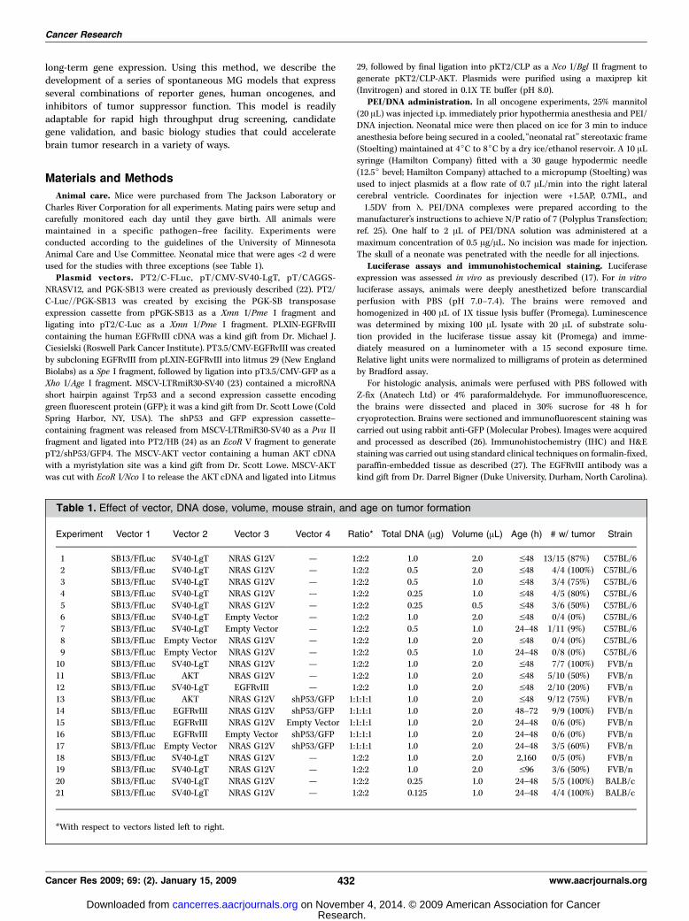
Table 1. Effect of vector, DNA dose, volume, mouse strain, and age on tumor formation
Experiment Vector 1 Vector 2 Vector 3 Vector 4 Ratio* Total DNA (Ag) Volume (AL) Age (h) # w/ tumor Strain
1 SB13/FfLuc SV40-LgT NRAS G12V — 1:2:2 1.0 2.0 V48 13/15 (87%) C57BL/62 SB13/FfLuc SV40-LgT NRAS G12V — 1:2:2 0.5 2.0 V48 4/4 (100%) C57BL/6
3 SB13/FfLuc SV40-LgT NRAS G12V — 1:2:2 0.5 1.0 V48 3/4 (75%) C57BL/6
4 SB13/FfLuc SV40-LgT NRAS G12V — 1:2:2 0.25 1.0 V48 4/5 (80%) C57BL/6
5 SB13/FfLuc SV40-LgT NRAS G12V — 1:2:2 0.25 0.5 V48 3/6 (50%) C57BL/66 SB13/FfLuc SV40-LgT Empty Vector — 1:2:2 1.0 2.0 V48 0/4 (0%) C57BL/6
7 SB13/FfLuc SV40-LgT Empty Vector — 1:2:2 0.5 1.0 24–48 1/11 (9%) C57BL/6
8 SB13/FfLuc Empty Vector NRAS G12V — 1:2:2 1.0 2.0 V48 0/4 (0%) C57BL/6
9 SB13/FfLuc Empty Vector NRAS G12V — 1:2:2 0.5 1.0 24–48 0/8 (0%) C57BL/610 SB13/FfLuc SV40-LgT NRAS G12V — 1:2:2 1.0 2.0 V48 7/7 (100%) FVB/n
11 SB13/FfLuc AKT NRAS G12V — 1:2:2 1.0 2.0 V48 5/10 (50%) FVB/n
12 SB13/FfLuc SV40-LgT EGFRvIII — 1:2:2 1.0 2.0 V48 2/10 (20%) FVB/n
13 SB13/FfLuc AKT NRAS G12V shP53/GFP 1:1:1:1 1.0 2.0 V48 9/12 (75%) FVB/n14 SB13/FfLuc EGFRvIII NRAS G12V shP53/GFP 1:1:1:1 1.0 2.0 48–72 9/9 (100%) FVB/n
15 SB13/FfLuc EGFRvIII NRAS G12V Empty Vector 1:1:1:1 1.0 2.0 24–48 0/6 (0%) FVB/n
16 SB13/FfLuc EGFRvIII Empty Vector shP53/GFP 1:1:1:1 1.0 2.0 24–48 0/6 (0%) FVB/n17 SB13/FfLuc Empty Vector NRAS G12V shP53/GFP 1:1:1:1 1.0 2.0 24–48 3/5 (60%) FVB/n
18 SB13/FfLuc SV40-LgT NRAS G12V — 1:2:2 1.0 2.0 2,160 0/5 (0%) FVB/n
19 SB13/FfLuc SV40-LgT NRAS G12V — 1:2:2 1.0 2.0 V96 3/6 (50%) FVB/n
20 SB13/FfLuc SV40-LgT NRAS G12V — 1:2:2 0.25 1.0 24–48 5/5 (100%) BALB/c21 SB13/FfLuc SV40-LgT NRAS G12V — 1:2:2 0.125 1.0 24–48 4/4 (100%) BALB/c
*With respect to vectors listed left to right.
Cancer Research
Cancer Res 2009; 69: (2). January 15, 2009 432 www.aacrjournals.org
Research. on November 4, 2014. © 2009 American Association for Cancercancerres.aacrjournals.org Downloaded from
Tissue culture and Western blot. Western blot was conducted as
described (22) using the following antibodies: mouse anti-LgT (Calbio-
chem), rabbit anti-NRAS, rabbit anti-p53, mouse anti-h actin, and rabbit
anti–extracellular signal-regulated kinase (all from Santa Cruz). Cell lineswere derived from mice as previously described using TrypLE enzymatic
digestion (28). All cells derived from spontaneous tumors were cultured in
media consisting of DMEM/F12, N2, and B27 supplements (1�), 1%penicillin/streptomycin (Invitrogen), and supplemented with 20 ng/mL EGF
and fibroblast growth factor (Peprotech). Cytokines were added every 2 to
3 d, and cells were passaged once to twice each week depending on density.
GL261 glioma cells were cultured as described (29).Southern blot and insertion cloning. Southern blot hybridization for
NRAS was performed as described (22). Transposon insertion sites were
identified by a combination of linker-mediated PCR (30), using the pCR4-
TOPOcloning vector and One-Shot TOP10 competent cells (Invitrogen) forshot-gun cloning, and pyrosequencing (454 Life Sciences; Roche) as
previously described (31). Genomic sequences directly flanking the
transposon were mapped using Ensemble.7
Statistical analysis. Survival was analyzed by log-rank test as described
(32). Graphing and statistical analysis was performed using Prism4 software
(Graph Pad Software) as described (32).
Results
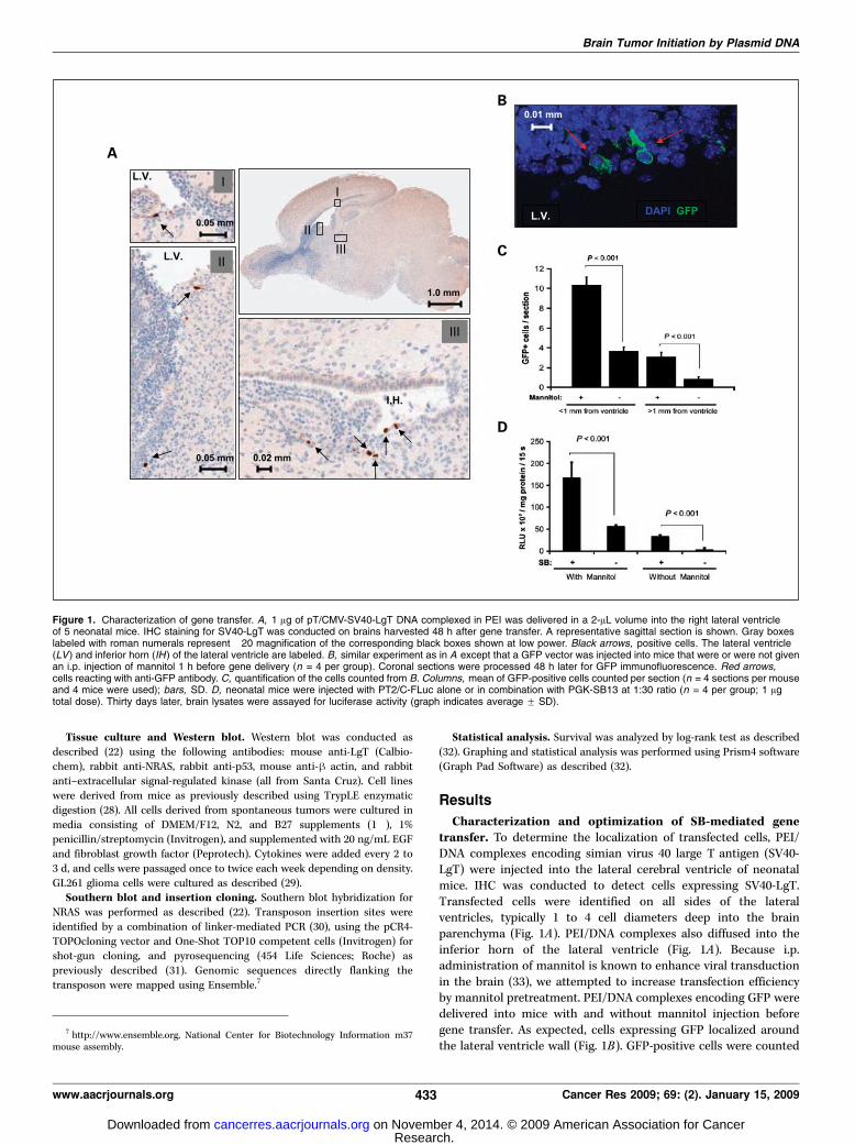
Characterization and optimization of SB-mediated genetransfer. To determine the localization of transfected cells, PEI/DNA complexes encoding simian virus 40 large T antigen (SV40-LgT) were injected into the lateral cerebral ventricle of neonatalmice. IHC was conducted to detect cells expressing SV40-LgT.Transfected cells were identified on all sides of the lateralventricles, typically 1 to 4 cell diameters deep into the brainparenchyma (Fig. 1A). PEI/DNA complexes also diffused into theinferior horn of the lateral ventricle (Fig. 1A). Because i.p.administration of mannitol is known to enhance viral transductionin the brain (33), we attempted to increase transfection efficiencyby mannitol pretreatment. PEI/DNA complexes encoding GFP weredelivered into mice with and without mannitol injection beforegene transfer. As expected, cells expressing GFP localized aroundthe lateral ventricle wall (Fig. 1B). GFP-positive cells were counted
Figure 1. Characterization of gene transfer. A, 1 Ag of pT/CMV-SV40-LgT DNA complexed in PEI was delivered in a 2-AL volume into the right lateral ventricleof 5 neonatal mice. IHC staining for SV40-LgT was conducted on brains harvested 48 h after gene transfer. A representative sagittal section is shown. Gray boxeslabeled with roman numerals represent �20 magnification of the corresponding black boxes shown at low power. Black arrows, positive cells. The lateral ventricle(LV) and inferior horn (IH) of the lateral ventricle are labeled. B, similar experiment as in A except that a GFP vector was injected into mice that were or were not givenan i.p. injection of mannitol 1 h before gene delivery (n = 4 per group). Coronal sections were processed 48 h later for GFP immunofluorescence. Red arrows,cells reacting with anti-GFP antibody. C, quantification of the cells counted from B. Columns, mean of GFP-positive cells counted per section (n = 4 sections per mouseand 4 mice were used); bars, SD. D, neonatal mice were injected with PT2/C-FLuc alone or in combination with PGK-SB13 at 1:30 ratio (n = 4 per group; 1 Agtotal dose). Thirty days later, brain lysates were assayed for luciferase activity (graph indicates average F SD).
7 http://www.ensemble.org, National Center for Biotechnology Information m37mouse assembly.
Brain Tumor Initiation by Plasmid DNA
www.aacrjournals.org 433 Cancer Res 2009; 69: (2). January 15, 2009
Research. on November 4, 2014. © 2009 American Association for Cancercancerres.aacrjournals.org Downloaded from
in serial sections to quantify the extent of transfection. Mannitolmore than doubled the number of GFP-positive cells within 1 mmof the lateral ventricle and also increased transfection deeper intothe brain parenchyma (Fig. 1C). To determine if SB could enhancetransgene expression in the brain, neonatal mice were intracere-brally injected with a FLuc transposon with or without trans-posase-encoding DNA. One month later, brain lysates were assayedfor FLuc activity as a measure of long-term expression. Luciferaseactivity was over seven times higher in brains injected with aplasmid encoding SB transposase relative to brains injectedwithout SB-encoding DNA (Fig. 1D). I.p. administration of mannitolbefore cotransfection with SB-encoding DNA further increasedFLuc activity 3-fold (Fig. 1D).Oncogene transfection resulted in development of sponta-
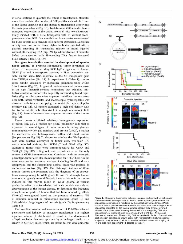
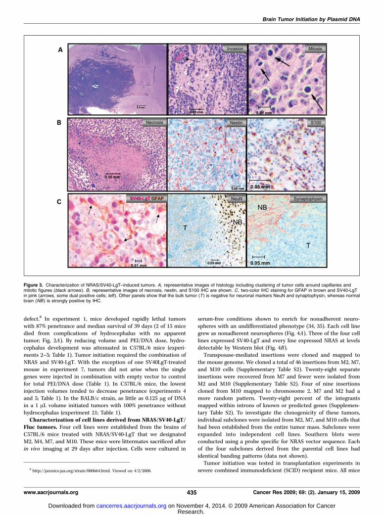
neous glioma. To promote spontaneous tumor formation, wedelivered transposons encoding SV40-LgT, a hyper-active humanNRAS (22), and a transposon carrying a FLuc expression cas-sette on the same DNA molecule as the SB transposase geneinto C57BL/6 mice (Fig. 2A). In experiment 1 (Table 1), tumorsarose rapidly, visualized by increasing bioluminescence within3 to 4 weeks (Fig. 2B). In general, well-demarcated tumors arosein the right (injected) cerebral hemisphere that exhibited infil-trative clusters of tumor cells frequently surrounding blood capil-laries (Fig. 3A). In some mice, apparent multifocal tumors arosenear both lateral ventricles and macroscopic hydrocephalus wasobserved with tumors occupying the ventricular space (Supple-mentary Fig. S1). All tumors exhibited a high cell density withtwo to five mitotic cells often visible in a single microscopic field(Fig. 3A). Areas of necrosis were apparent in some of the tumors(Fig. 3B).These tumors exhibited relatively homogenous expression
of nestin (Fig. 3B), a marker for neural progenitor cells that isexpressed in several types of brain tumors including gliomas.Immunopositivity for glial fibrillary acid protein (GFAP), a markerfor astrocytes, was heterogeneous within individual tumors(Supplementary Fig. S2). To determine whether the GFAP-positivecells were reactive astrocytes or tumor cells, two-color IHCwas conducted staining for SV40-LgT and GFAP (Fig. 3C).Numerous tumor cells were immunopositive for GFAP andSV40LgT (Fig. 3C), ruling out reactive astrocytes as the onlysource of GFAP immunoreactivity. Consistent with an astrocyticphenotype, tumor cells also stained positive for S100. These tumorswere negative for neuronal markers including NeuN and syn-aptophysin, but the surrounding normal brain was positive asan internal control (Fig. 3C). The histologic features of thesemurine tumors are consistent with the diagnosis of an astrocy-toma corresponding to WHO grade III and IV, although humantumors are typically more diffusively invasive. We refer to tumorsinduced in this murine mode as ‘‘GEM’’ glioma of variousgrades hereafter to acknowledge that such models are only anapproximation of the human disease. To determine the frequencyof each tumor grade, 13 tumors that had been induced by NRAS/SV40-LgT were graded based on the presence of necrosis; 77%of exhibited minimal or microscopic necrosis (grade III) and23% exhibited large regions of necrosis (grade IV; SupplementaryTable S1).The injection volume and concentration of PEI/DNA affected
penetrance and lethality of oncogene transfection. The highestinjection volume (2 AL) tended to result in the developmentof hydrocephalus that was apparent by an enlarged skull, parti-cularly in C57BL/6 mice, which are prone to this developmental
Figure 2. Oncogene transfection scheme, imaging, and survival. A, schematicof cotransfection technique used to induce tumors by oncogene transfer. SBtransposase expression is regulated by the phosphoglycerate kinase (PGK )promoter on the plasmid DNA backbone of the same vector harboring the FLuctransposon regulated by the CAG promoter. Double gray arrows, invertedrepeats of the transposon, marking the termini of the integrating DNA aftertransposition. B, neonatal mice were injected with SV40-LgT, NRAS, andFLuc vector loaded with SB-encoding DNA as detailed in Table 1. Survival aftereach injection condition is shown above with representative bioluminescenceimages from experiment 1 below. C, survival and bioluminescent imaging ofFVB/n mice from experiment 14 detailed in Table 1.
Cancer Research
Cancer Res 2009; 69: (2). January 15, 2009 434 www.aacrjournals.org
Research. on November 4, 2014. © 2009 American Association for Cancercancerres.aacrjournals.org Downloaded from
defect.8 In experiment 1, mice developed rapidly lethal tumorswith 87% penetrance and median survival of 39 days (2 of 15 micedied from complications of hydrocephalus with no apparenttumor; Fig. 2A). By reducing volume and PEI/DNA dose, hydro-cephalus development was attenuated in C57BL/6 mice (experi-ments 2–5; Table 1). Tumor initiation required the combination ofNRAS and SV40-LgT. With the exception of one SV40LgT-treatedmouse in experiment 7, tumors did not arise when the singlegenes were injected in combination with empty vector to controlfor total PEI/DNA dose (Table 1). In C57BL/6 mice, the lowestinjection volumes tended to decrease penetrance (experiments 4and 5; Table 1). In the BALB/c strain, as little as 0.125 Ag of DNAin a 1 AL volume initiated tumors with 100% penetrance withouthydrocephalus (experiment 21; Table 1).Characterization of cell lines derived from NRAS/SV40-LgT/
Fluc tumors. Four cell lines were established from the brains ofC57BL/6 mice treated with NRAS/SV40-LgT that we designatedM2, M4, M7, and M10. These mice were littermates sacrificed afterin vivo imaging at 29 days after injection. Cells were cultured in
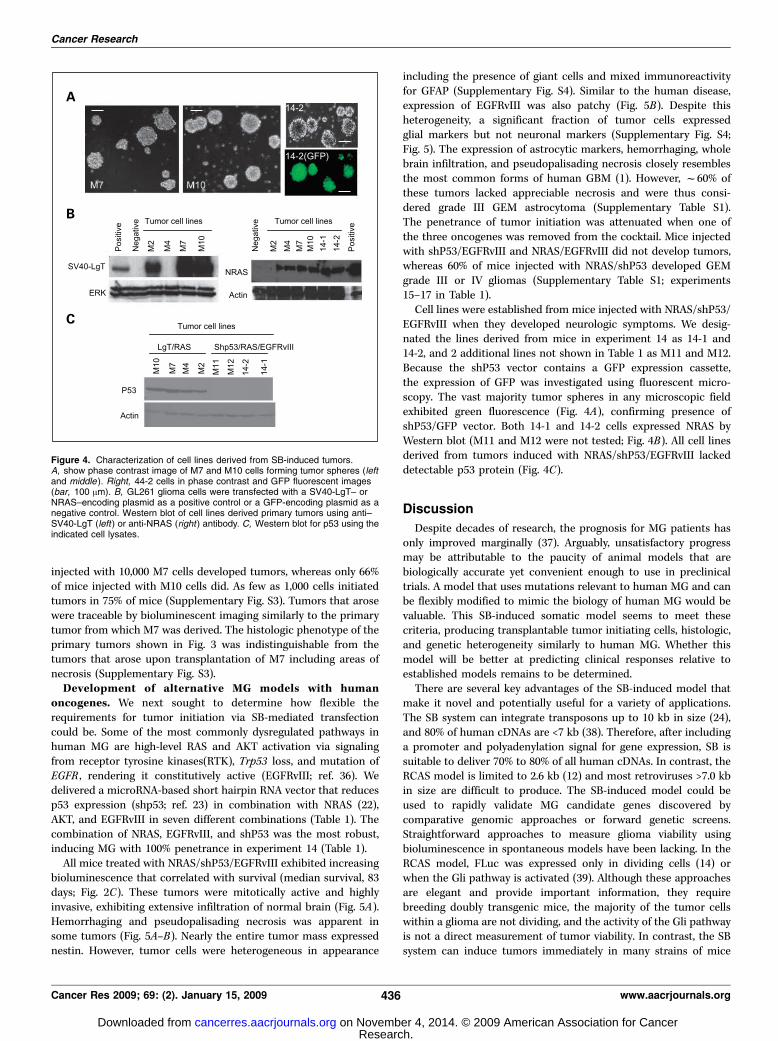
serum-free conditions shown to enrich for nonadherent neuro-spheres with an undifferentiated phenotype (34, 35). Each cell linegrew as nonadherent neurospheres (Fig. 4A). Three of the four celllines expressed SV40-LgT and every line expressed NRAS at levelsdetectable by Western blot (Fig. 4B).Transposase-mediated insertions were cloned and mapped to
the mouse genome. We cloned a total of 46 insertions from M2, M7,and M10 cells (Supplementary Table S2). Twenty-eight separateinsertions were recovered from M7 and fewer were isolated fromM2 and M10 (Supplementary Table S2). Four of nine insertionscloned from M10 mapped to chromosome 2. M7 and M2 had amore random pattern. Twenty-eight percent of the integrantsmapped within introns of known or predicted genes (Supplemen-tary Table S2). To investigate the clonogenicity of these tumors,individual subclones were isolated from M2, M7, and M10 cells thathad been established from the entire tumor mass. Subclones wereexpanded into independent cell lines. Southern blots wereconducted using a probe specific for NRAS vector sequence. Eachof the four subclones derived from the parental cell lines hadidentical banding patterns (data not shown).Tumor initiation was tested in transplantation experiments in
severe combined immunodeficient (SCID) recipient mice. All mice
Figure 3. Characterization of NRAS/SV40-LgT–induced tumors. A, representative images of histology including clustering of tumor cells around capillaries andmitotic figures (black arrows ). B, representative images of necrosis, nestin, and S100 IHC are shown. C, two-color IHC staining for GFAP in brown and SV40-LgTin pink (arrows, some dual positive cells; left). Other panels show that the bulk tumor (T ) is negative for neuronal markers NeuN and synaptophysin, whereas normalbrain (NB ) is strongly positive by IHC.
8 http://jaxmice.jax.org/strain/000664.html. Viewed on 4/2/2008.
Brain Tumor Initiation by Plasmid DNA
www.aacrjournals.org 435 Cancer Res 2009; 69: (2). January 15, 2009
Research. on November 4, 2014. © 2009 American Association for Cancercancerres.aacrjournals.org Downloaded from
injected with 10,000 M7 cells developed tumors, whereas only 66%of mice injected with M10 cells did. As few as 1,000 cells initiatedtumors in 75% of mice (Supplementary Fig. S3). Tumors that arosewere traceable by bioluminescent imaging similarly to the primarytumor from which M7 was derived. The histologic phenotype of theprimary tumors shown in Fig. 3 was indistinguishable from thetumors that arose upon transplantation of M7 including areas ofnecrosis (Supplementary Fig. S3).Development of alternative MG models with human
oncogenes. We next sought to determine how flexible therequirements for tumor initiation via SB-mediated transfectioncould be. Some of the most commonly dysregulated pathways inhuman MG are high-level RAS and AKT activation via signalingfrom receptor tyrosine kinases(RTK), Trp53 loss, and mutation ofEGFR , rendering it constitutively active (EGFRvIII; ref. 36). Wedelivered a microRNA-based short hairpin RNA vector that reducesp53 expression (shp53; ref. 23) in combination with NRAS (22),AKT, and EGFRvIII in seven different combinations (Table 1). Thecombination of NRAS, EGFRvIII, and shP53 was the most robust,inducing MG with 100% penetrance in experiment 14 (Table 1).All mice treated with NRAS/shP53/EGFRvIII exhibited increasing
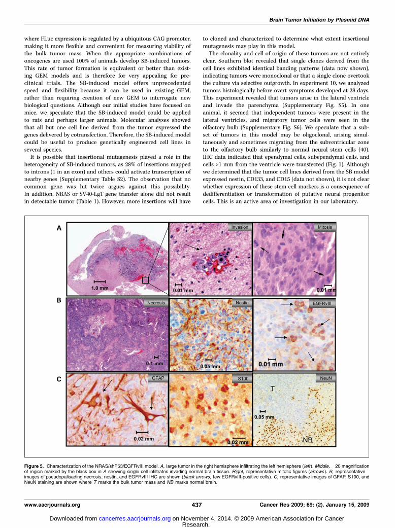
bioluminescence that correlated with survival (median survival, 83days; Fig. 2C). These tumors were mitotically active and highlyinvasive, exhibiting extensive infiltration of normal brain (Fig. 5A).Hemorrhaging and pseudopalisading necrosis was apparent insome tumors (Fig. 5A–B). Nearly the entire tumor mass expressednestin. However, tumor cells were heterogeneous in appearance
including the presence of giant cells and mixed immunoreactivityfor GFAP (Supplementary Fig. S4). Similar to the human disease,expression of EGFRvIII was also patchy (Fig. 5B). Despite thisheterogeneity, a significant fraction of tumor cells expressedglial markers but not neuronal markers (Supplementary Fig. S4;Fig. 5). The expression of astrocytic markers, hemorrhaging, wholebrain infiltration, and pseudopalisading necrosis closely resemblesthe most common forms of human GBM (1). However, f60% ofthese tumors lacked appreciable necrosis and were thus consi-dered grade III GEM astrocytoma (Supplementary Table S1).The penetrance of tumor initiation was attenuated when one ofthe three oncogenes was removed from the cocktail. Mice injectedwith shP53/EGFRvIII and NRAS/EGFRvIII did not develop tumors,whereas 60% of mice injected with NRAS/shP53 developed GEMgrade III or IV gliomas (Supplementary Table S1; experiments15–17 in Table 1).Cell lines were established from mice injected with NRAS/shP53/
EGFRvIII when they developed neurologic symptoms. We desig-nated the lines derived from mice in experiment 14 as 14-1 and14-2, and 2 additional lines not shown in Table 1 as M11 and M12.Because the shP53 vector contains a GFP expression cassette,the expression of GFP was investigated using fluorescent micro-scopy. The vast majority tumor spheres in any microscopic fieldexhibited green fluorescence (Fig. 4A), confirming presence ofshP53/GFP vector. Both 14-1 and 14-2 cells expressed NRAS byWestern blot (M11 and M12 were not tested; Fig. 4B). All cell linesderived from tumors induced with NRAS/shP53/EGFRvIII lackeddetectable p53 protein (Fig. 4C).
Discussion
Despite decades of research, the prognosis for MG patients hasonly improved marginally (37). Arguably, unsatisfactory progressmay be attributable to the paucity of animal models that arebiologically accurate yet convenient enough to use in preclinicaltrials. A model that uses mutations relevant to human MG and canbe flexibly modified to mimic the biology of human MG would bevaluable. This SB-induced somatic model seems to meet thesecriteria, producing transplantable tumor initiating cells, histologic,and genetic heterogeneity similarly to human MG. Whether thismodel will be better at predicting clinical responses relative toestablished models remains to be determined.There are several key advantages of the SB-induced model that
make it novel and potentially useful for a variety of applications.The SB system can integrate transposons up to 10 kb in size (24),and 80% of human cDNAs are <7 kb (38). Therefore, after includinga promoter and polyadenylation signal for gene expression, SB issuitable to deliver 70% to 80% of all human cDNAs. In contrast, theRCAS model is limited to 2.6 kb (12) and most retroviruses >7.0 kbin size are difficult to produce. The SB-induced model could beused to rapidly validate MG candidate genes discovered bycomparative genomic approaches or forward genetic screens.Straightforward approaches to measure glioma viability usingbioluminescence in spontaneous models have been lacking. In theRCAS model, FLuc was expressed only in dividing cells (14) orwhen the Gli pathway is activated (39). Although these approachesare elegant and provide important information, they requirebreeding doubly transgenic mice, the majority of the tumor cellswithin a glioma are not dividing, and the activity of the Gli pathwayis not a direct measurement of tumor viability. In contrast, the SBsystem can induce tumors immediately in many strains of mice
Figure 4. Characterization of cell lines derived from SB-induced tumors.A, show phase contrast image of M7 and M10 cells forming tumor spheres (leftand middle ). Right, 44-2 cells in phase contrast and GFP fluorescent images(bar, 100 Am). B, GL261 glioma cells were transfected with a SV40-LgT– orNRAS–encoding plasmid as a positive control or a GFP-encoding plasmid as anegative control. Western blot of cell lines derived primary tumors using anti–SV40-LgT (left ) or anti-NRAS (right ) antibody. C, Western blot for p53 using theindicated cell lysates.
Cancer Research
Cancer Res 2009; 69: (2). January 15, 2009 436 www.aacrjournals.org
Research. on November 4, 2014. © 2009 American Association for Cancercancerres.aacrjournals.org Downloaded from
where FLuc expression is regulated by a ubiquitous CAG promoter,making it more flexible and convenient for measuring viability ofthe bulk tumor mass. When the appropriate combinations ofoncogenes are used 100% of animals develop SB-induced tumors.This rate of tumor formation is equivalent or better than exist-ing GEM models and is therefore for very appealing for pre-clinical trials. The SB-induced model offers unprecedentedspeed and flexibility because it can be used in existing GEM,rather than requiring creation of new GEM to interrogate newbiological questions. Although our initial studies have focused onmice, we speculate that the SB-induced model could be appliedto rats and perhaps larger animals. Molecular analyses showedthat all but one cell line derived from the tumor expressed thegenes delivered by cotransfection. Therefore, the SB-induced modelcould be useful to produce genetically engineered cell lines inseveral species.It is possible that insertional mutagenesis played a role in the
heterogeneity of SB-induced tumors, as 28% of insertions mappedto introns (1 in an exon) and others could activate transcription ofnearby genes (Supplementary Table S2). The observation that nocommon gene was hit twice argues against this possibility.In addition, NRAS or SV40-LgT gene transfer alone did not resultin detectable tumor (Table 1). However, more insertions will have
to cloned and characterized to determine what extent insertionalmutagenesis may play in this model.The clonality and cell of origin of these tumors are not entirely
clear. Southern blot revealed that single clones derived from thecell lines exhibited identical banding patterns (data now shown),indicating tumors were monoclonal or that a single clone overtookthe culture via selective outgrowth. In experiment 10, we analyzedtumors histologically before overt symptoms developed at 28 days.This experiment revealed that tumors arise in the lateral ventricleand invade the parenchyma (Supplementary Fig. S5). In oneanimal, it seemed that independent tumors were present in thelateral ventricles, and migratory tumor cells were seen in theolfactory bulb (Supplementary Fig. S6). We speculate that a sub-set of tumors in this model may be oligoclonal, arising simul-taneously and sometimes migrating from the subventricular zoneto the olfactory bulb similarly to normal neural stem cells (40).IHC data indicated that ependymal cells, subependymal cells, andcells >1 mm from the ventricle were transfected (Fig. 1). Althoughwe determined that the tumor cell lines derived from the SB modelexpressed nestin, CD133, and CD15 (data not shown), it is not clearwhether expression of these stem cell markers is a consequence ofdedifferentiation or transformation of putative neural progenitorcells. This is an active area of investigation in our laboratory.
Figure 5. Characterization of the NRAS/shP53/EGFRvIII model. A, large tumor in the right hemisphere infiltrating the left hemisphere (left). Middle, �20 magnificationof region marked by the black box in A showing single cell infiltrates invading normal brain tissue. Right, representative mitotic figures (arrows ). B, representativeimages of pseudopalisading necrosis, nestin, and EGFRvIII IHC are shown (black arrows, few EGFRvIII-positive cells). C, representative images of GFAP, S100, andNeuN staining are shown where T marks the bulk tumor mass and NB marks normal brain.
Brain Tumor Initiation by Plasmid DNA
www.aacrjournals.org 437 Cancer Res 2009; 69: (2). January 15, 2009
Research. on November 4, 2014. © 2009 American Association for Cancercancerres.aacrjournals.org Downloaded from
Human MG evolves with the host immune system, a differentcircumstance than that modeled by injecting glioma cells into thebrain of adult mice. MG patients often develop tolerance to tumorantigens facilitated by an elevated fraction of regulatory T cells (41).Nonetheless, spontaneous antiglioma immune responses have beendocumented after successful treatment with radiation andchemotherapy (42, 43). In the current study, tumors developed inimmunocompetent mice presumably tolerized to foreign antigensused to drive MG formation and Treg frequency was not assessed.Future studies will address these features. We have previouslyshown that neonatal mice are tolerized to challenge with humanneoantigen delivered within 24 hours of birth (44). We made obser-vations that indicate mice in the current study were tolerant orignorant to the neoantigens we delivered using SB. The penetranceof tumor development dropped in mice older than 3 days des-pite equivalent transfection efficiency and we could not inducetumors with SV40-LgT/NRAS in adult mice (experiments 18 and 19;Table 1). Additionally, M7 cell transplants were rejected in 2/5immunocompetent C57BL/6 mice (data not shown) but not inSCID mice (Supplementary Fig. S3). The SB-induced somatictumors could be initiated in GEM for immunology studies, creating
‘‘humanized’’ models that express human tumor antigens andhuman MHC class I (45). Likewise, human RTK genes could drivetumor growth so that small molecule drugs or humanized antibodiescould be tested on the human RTK to diminish artifacts with speciesspecificity. This SB-induced somatic model offers a new tool tounderstand the biology of MG and realize effective targeted therapy.
Disclosure of Potential Conflicts of Interest
No potential conflicts of interest were disclosed.
Acknowledgments
Received 5/15/2008; revised 11/9/2008; accepted 11/10/2008.Grant support: NIH/NINDS 1R21-NS055738-01A2 (J.R. Ohlfest), Randy Shaver
Foundation (J.R. Ohlfest), Minnesota Medical Foundation (J.R. Ohlfest), NIH/NIDA T32DA022616 (S. Decker and W.C. Low), NIH/National Cancer Institute R01CA113636-01A1 (D.A. Largaespada), Minnesota Department of Employment and EconomicDevelopment SPAP-05-0013-P-FY06 (D.A. Largaespada).
The costs of publication of this article were defrayed in part by the payment of pagecharges. This article must therefore be hereby marked advertisement in accordancewith 18 U.S.C. Section 1734 solely to indicate this fact.
We thank Aaron Sarver for bioinformatics support in analyzing pyrosequenc-ing data.
Cancer Research
Cancer Res 2009; 69: (2). January 15, 2009 438 www.aacrjournals.org
References
1. Louis DN, Ohgaki H, Wiestler OD, et al. The 2007 WHOclassification of tumours of the central nervous system.Acta Neuropathol 2007;114:97–109.2. Davis FG, Kupelian V, Freels S, McCarthy B, SurawiczT. Prevalence estimates for primary brain tumors in theUnited States by behavior and major histology groups.Neuro-oncol 2001;3:152–8.3. Wiesner SM, Freese A, Ohlfest JR. Emerging conceptsin glioma biology: implications for clinical protocolsand rational treatment strategies. Neurosurg Focus 2005;19:E3.4. Ren ZP, Olofsson T, Qu M, et al. Molecular geneticanalysis of p53 intratumoral heterogeneity in humanastrocytic brain tumors. J Neuropathol Exp Neurol 2007;66:944–54.5. Cheng Y, Ng HK, Ding M, Zhang SF, Pang JC, Lo KW.Molecular analysis of microdissected de novo glioblas-tomas and paired astrocytic tumors. J Neuropathol ExpNeurol 1999;58:120–8.6. Weiss WA, Israel M, Cobbs C, et al. Neuropathology ofgenetically engineered mice: consensus report andrecommendations from an international forum. Onco-gene 2002;21:7453–63.7. Reilly KM, Loisel DA, Bronson RT, McLaughlin ME,Jacks T. Nf1;Trp53 mutant mice develop glioblastomawith evidence of strain-specific effects. Nat Genet 2000;26:109–13.8. Xiao A, Yin C, Yang C, Di Cristofano A, Pandolfi PP,Van Dyke T. Somatic induction of Pten loss in apreclinical astrocytoma model reveals major roles indisease progression and avenues for target discoveryand validation. Cancer Res 2005;65:5172–80.9. Zhu Y, Guignard F, Zhao D, et al. Early inactivation ofp53 tumor suppressor gene cooperating with NF1 loss in-duces malignant astrocytoma. Cancer Cell 2005;8:119–30.10. Assanah M, Lochhead R, Ogden A, Bruce J, GoldmanJ, Canoll P. Glial progenitors in adult white matter aredriven to form malignant gliomas by platelet-derivedgrowth factor-expressing retroviruses. J Neurosci 2006;26:6781–90.11. Uhrbom L, Hesselager G, Nister M, Westermark B.Induction of brain tumors in mice using a recombinantplatelet-derived growth factor B-chain retrovirus. Can-cer Res 1998;58:5275–9.12. Holland EC, Hively WP, DePinho RA, Varmus HE. Aconstitutively active epidermal growth factor receptor
cooperates with disruption of G1 cell-cycle arrest path-ways to induce glioma-like lesions in mice. Genes Dev1998;12:3675–85.13. Holland EC, Varmus HE. Basic fibroblast growthfactor induces cell migration and proliferation after glia-specific gene transfer in mice. Proc Natl Acad Sci U S A1998;95:1218–23.14. Uhrbom L, Nerio E, Holland EC. Dissecting tumormaintenance requirements using bioluminescence im-aging of cell proliferation in a mouse glioma model. NatMed 2004;10:1257–60.15. Momota H, Holland EC. Bioluminescence technologyfor imaging cell proliferation. Curr Opin Biotechnol2005;16:681–6.16. Abdallah B, Hassan A, Benoist C, Goula D, Behr JP,Demeneix BA. A powerful nonviral vector for in vivogene transfer into the adult mammalian brain: poly-ethylenimine. Hum Gene Ther 1996;7:1947–54.17. Ohlfest JR, Demorest ZL, Motooka Y, et al. Combi-natorial antiangiogenic gene therapy by nonviral genetransfer using the sleeping beauty transposon causestumor regression and improves survival in mice bear-ing intracranial human glioblastoma. Mol Ther 2005;12:778–88.18. Hirko AC, Buethe DD, Meyer EM, Hughes JA. Plasmiddelivery in the rat brain. Biosci Rep 2002;22:297–308.19. Zhang C, Yadava P, Hughes J. Polyethyleniminestrategies for plasmid delivery to brain-derived cells.Methods 2004;33:144–50.20. Ivics Z, Hackett PB, Plasterk RH, Izsvak Z. Molecularreconstruction of Sleeping Beauty, a Tc1-like transposonfrom fish, and its transposition in human cells. Cell1997;91:501–10.21. Ivics Z, Izsvak Z. Transposons for gene therapy! CurrGene Ther 2006;6:593–607.22. Carlson CM, Frandsen JL, Kirchhof N, McIvor RS,Largaespada DA. Somatic integration of an oncogene-harboring Sleeping Beauty transposon models livertumor development in the mouse. Proc Natl Acad SciU S A 2005;102:17059–64.23. Dickins RA, Hemann MT, Zilfou JT, et al. Probingtumor phenotypes using stable and regulated syntheticmicroRNA precursors. Nat Genet 2005;37:1289–95.24. Geurts AM, Yang Y, Clark KJ, et al. Gene transfer intogenomes of human cells by the sleeping beautytransposon system. Mol Ther 2003;8:108–17.25. Ohlfest JR, Lobitz PD, Perkinson SG, Largaespada DA.Integration and long-term expression in xenografted
human glioblastoma cells using a plasmid-basedtransposon system. Mol Ther 2004;10:260–8.26. Wu A, Oh S, Wiesner SM, et al. Persistence ofCD133(+) cells in human and mouse glioma cell lines:detailed characterization of gl261 glioma cells withcancer stem cell-like properties. Stem Cells Dev 2008;17:173–84.27. Wikstrand CJ, McLendon RE, Friedman AH, BignerDD. Cell surface localization and density of the tumor-associated variant of the epidermal growth factorreceptor, EGFRvIII. Cancer Res 1997;57:4130–40.28. Panchision DM, Chen HL, Pistollato F, Papini D, NiHT, Hawley TS. Optimized flow cytometric analysis ofCNS tissue reveals novel functional relationshipsbetween CD133, CD15 and CD24 expressing cells. StemCells 2007;25:1560–70.29. Wu A, Oh S, Ericson K, et al. Transposon-basedinterferon g gene transfer overcomes limitations ofepisomal plasmid for immunogene therapy of glioblas-toma. Cancer Gene Ther 2007;14:550–60.30. Collier LS, Carlson CM, Ravimohan S, Dupuy AJ,Largaespada DA. Cancer gene discovery in solidtumours using transposon-based somatic mutagenesisin the mouse. Nature 2005;436:272–6.31. Thomas RK, Nickerson E, Simons JF, et al. Sensitivemutation detection in heterogeneous cancer specimensby massively parallel picoliter reactor sequencing. NatMed 2006;12:852–5.32. Wu A, Oh S, Gharagozlou S, et al. In vivo vaccinationwith tumor cell lysate plus CpG oligodeoxynucleotideseradicates murine glioblastoma. J Immunother (1997)2007;30:789–97.33. Ghodsi A, Stein C, Derksen T, Martins I, AndersonRD, Davidson BL. Systemic hyperosmolality improves h-glucuronidase distribution and pathology in murineMPS VII brain following intraventricular gene transfer.Exp Neurol 1999;160:109–16.34. Lee J, Kotliarova S, Kotliarov Y, et al. Tumor stemcells derived from glioblastomas cultured in bFGF andEGF more closely mirror the phenotype and genotype ofprimary tumors than do serum-cultured cell lines.Cancer Cell 2006;9:391–403.35. Gunther HS, Schmidt NO, Phillips HS, et al.Glioblastoma-derived stem cell-enriched cultures formdistinct subgroups according to molecular and pheno-typic criteria. Oncogene 2008;27:2897–909.36. Fenstermaker RA, Ciesielski MJ. Deletion and tandemduplication of exons 2 - 7 in the epidermal growth factor
Research. on November 4, 2014. © 2009 American Association for Cancercancerres.aacrjournals.org Downloaded from

Brain Tumor Initiation by Plasmid DNA
www.aacrjournals.org 439 Cancer Res 2009; 69: (2). January 15, 2009
receptor gene of a human malignant glioma. Oncogene2000;19:4542–8.37. Stupp R, Mason WP, van den Bent MJ, et al.Radiotherapy plus concomitant and adjuvant temozo-lomide for glioblastoma. N Engl J Med 2005;352:987–96.38. Lander ES, Linton LM, Birren B, et al. Initialsequencing and analysis of the human genome. Nature2001;409:860–921.39. Becher OJ, Hambardzumyan D, Fomchenko EI, et al. Gliactivity correlates with tumor grade in platelet-derivedgrowth factor-induced gliomas. Cancer Res 2008;68:2241–9.40. Doetsch F, Caille I, Lim DA, Garcia-Verdugo JM,Alvarez-Buylla A. Subventricular zone astrocytes are
neural stem cells in the adult mammalian brain. Cell1999;97:703–16.41. Fecci PE, Mitchell DA, Whitesides JF, et al. Increasedregulatory T-cell fraction amidst a diminished CD4compartment explains cellular immune defects inpatients with malignant glioma. Cancer Res 2006;66:3294–302.42. Pallasch CP, Struss AK, Munnia A, et al. Autoanti-bodies against GLEA2 and PHF3 in glioblastoma:tumor-associated autoantibodies correlated with pro-longed survival. Int J Cancer 2005;117:456–9.43. Ueda R, Low KL, Zhu X, et al. Spontaneous im-mune responses against glioma-associated antigens in a
long term survivor with malignant glioma. J Transl Med2007;5:68.44. Ohlfest JR, Frandsen JL, Fritz S, et al. Phenotypiccorrection and long-term expression of factor VIII inhemophilic mice by immunotolerization and nonviralgene transfer using the Sleeping Beauty transposonsystem. Blood 2005;105:2691–8.45. Pascolo S, Bervas N, Ure JM, Smith AG, LemonnierFA, Perarnau B. HLA-A2.1-restricted education andcytolytic activity of CD8(+) T lymphocytes from h2microglobulin (h2m) HLA-A2.1 monochain transgenicH-2Db h2m double knockout mice. J Exp Med 1997;185:2043–51.
Research. on November 4, 2014. © 2009 American Association for Cancercancerres.aacrjournals.org Downloaded from


