Embed Size (px)
Citation preview
MOLECULAR AND CELLULAR BIOLOGY, June 2006, p. 4240–4256 Vol. 26, No. 110270-7306/06/$08.00�0 doi:10.1128/MCB.02124-05Copyright © 2006, American Society for Microbiology. All Rights Reserved.
Cyclin D1 Regulates Cellular Migration through the Inhibitionof Thrombospondin 1 and ROCK Signaling
Zhiping Li,1 Chenguang Wang,1 Xuanmao Jiao,1 Yinan Lu,1 Maofu Fu,1 Andrew A. Quong,1Chip Dye,2 Jianguo Yang,1 Maozheng Dai,2 Xiaoming Ju,1 Xueping Zhang,1 Anping Li,1
Peter Burbelo,2 E. Richard Stanley,3 and Richard G. Pestell1*Department of Cancer Biology, Kimmel Cancer Center, Thomas Jefferson University, Philadelphia, Pennsylvania 191071;
Department of Oncology, Lombardi Comprehensive Cancer Center, Georgetown University, 3970 Reservoir Road,NW, Box 571468, Washington, D.C. 20057-14682; and Department of
Developmental and Molecular Biology, Albert Einstein College of Medicine,Yeshiva University, Bronx, New York 104613
Received 2 November 2005/Returned for modification 13 December 2005/Accepted 13 March 2006
Cyclin D1 is overexpressed in human tumors, correlating with cellular metastasis, and is induced byactivating Rho GTPases. Herein, cyclin D1-deficient mouse embryo fibroblasts (MEFs) exhibited increasedadhesion and decreased motility compared with wild-type MEFs. Retroviral transduction of cyclin D1 reversedthese phenotypes. Mutational analysis of cyclin D1 demonstrated that its effects on cellular adhesion andmigration were independent of the pRb and p160 coactivator binding domains. Genomewide expression arraysidentified a subset of genes regulated by cyclin D1, including Rho-activated kinase II (ROCKII) and throm-bospondin 1 (TSP-1). cyclin D1�/� cells showed increased Rho GTP and ROCKII activity and signaling, withincreased phosphorylation of LIM kinase, cofilin (Ser3), and myosin light chain 2 (Thr18/Ser19). Cyclin D1repressed ROCKII and TSP-1 expression, and the migratory defect of cyclin D1�/� cells was reversed by ROCKinhibition or TSP-1 immunoneutralizing antibodies. cyclin E knockin to the cyclin D1�/� MEFs rescued theDNA synthesis defect of cyclin D1�/� MEFs but did not rescue either the migration defect or the abundanceof ROCKII. Cyclin D1 promotes cellular motility through inhibiting ROCK signaling and repressing themetastasis suppressor TSP-1.
The human cyclin D1 gene was initially cloned as a break-point rearrangement within parathyroid adenoma (42). Ho-mology of the cyclin D1 cDNA to the CLN gene in yeastsuggested the PRAD1 gene encoded the regulatory subunit ofa serine threonine kinase. In subsequent studies, cyclin D1 wasshown to bind the retinoblastoma (pRb) protein and throughphysical association with the cyclin-dependent kinase 4 or 6(cdk4 or cdk6) subunit to phosphorylate pRb. Phosphorylationof pRb by the cyclin D/cdk4 holoenzyme then alters the con-formation of pRb, correlating with sequential phosphorylationby cyclin E/cdk2 and the induction of DNA synthesis. Thecyclin D1 gene is overexpressed in human cancers, includingbreast, colon, and prostate cancer, and hematopoietic malig-nancies (23, 39). Targeted overexpression of cyclin D1 to themammary gland in transgenic mice was sufficient for the in-duction of mammary adenocarcinoma. Cyclin D1 is overex-pressed in metastatic cells (19, 30). Analysis of cyclin D1-deficient mice indicates a role for cyclin D1 in both cellularsurvival and DNA synthesis (3). Furthermore, cyclin D1-defi-cient mice are resistant to gastrointestinal tumors induced bymutation of the Apc gene (28) or tumor formation induced byeither mammary-targeted Ras or ErbB2 (82). Such observa-tions are consistent with previous studies demonstrating cyclin
D1 antisense abrogates epithelial growth of ErbB2-inducedtumors in vivo (34).
Mutational analysis of the human cyclin D1 cDNA has iden-tified several distinct domains involved in binding either pRb,cdk, the p160 coactivator, and histone deacetylases (22, 23, 59).The cdk-binding domain of cyclin D1 is required for the asso-ciation with cdk4 and sequential phosphorylation of pRb,which in turn, leads to the release of E2F binding proteins. Therelease of E2F proteins, in turn, leads to the sequential regu-lation of E2F-responsive genes associated with the induction ofDNA synthesis. The association of cyclin D1 with the p160coactivator SRC1 (AIB1) enhances ligand-independent ER�activity in cultured cells. Recent studies have demonstrated theregulation of several transcription factors through a cdk-inde-pendent mechanism, including MyoD, Neuro-D, the androgenreceptor, CEBP�, and peroxisome proliferator-activated re-ceptor gamma (PPAR�) (reviewed in reference 73). The abun-dance of cyclin D1 is rate limiting in progression through theG1 phase of the cell cycle in fibroblasts and mammary epi-thelial cells. Sustained extracellular signal-regulated kinase(ERK) activation induces cyclin D1 transcription and mRNAand protein abundance, which is required for mid-G1-phaseinduction of cyclin D1 (2, 56, 75). Tightly coordinated interac-tions between the Rho GTPases facilitate cell cycle progres-sion through regulating the expression of cyclin D1 and assem-bly of cyclin D/cdk complexes (12). Rac and Cdc42 inducecyclin D1 independently of ERK involving an NF-�B signalingpathway (12, 31, 79). Rho kinase suppresses Rac/Cdc42-depen-dent cyclin D1 induction through LIMK (56) independently of
* Corresponding author. Mailing address: Thomas Jefferson Univer-sity, Department of Cancer Biology, Kimmel Cancer Center, BluemleBuilding, Rm 1050, 233 South 10th St, Philadelphia, PA 19107. Phone:(215) 503-5649. Fax: (215) 503-9334. E-mail: [email protected].
4240
cofilin or actin polymerization. The inhibition of Rac/Cdc42signaling maintains mid-G1-phase ERK-dependent inductionof cyclin D1 (56).
The Rho family of small GTPases play an important role inthe regulation of cell motility via their effects on the cellularcytoskeleton and adhesion (5, 32). Rac and its effector, PAK,induce membrane ruffles and actin rearrangements includingstress fibers that control formation of lamellipodia and newfocal contacts at the leading edge that drive cellular motility(54). Rho regulates assembly of stress fibers and associatedfocal adhesions through its downstream effectors mouse Di-aphanous (mDia) and the Rho-activated kinase (ROCK) thatphosphorylate cytoskeletal proteins. Major ROCK substratesregulating cellular migration include LIM kinases, which phos-phorylate and regulate an actin-depolymerizing protein cofilin,and myosin light chain (MLC) kinase. Although Rho activitynegatively influences cell migration by increasing stress fiber-dependent adhesions to substratum, Rho activity is also re-quired for actomyosin contractility needed to drive cell bodyretraction at the rear of the cell (4). Dynamic activation andinactivation is tightly coordinated, and insufficient levels orexcessive Rho GTPase activity will prevent cell migration (52,57, 58, 71).
A variety of cytokines, chemokines, growth factors, extracel-lular matrix, and matrix-degrading proteins coordinate their sig-naling to affect migratory cues through the Rho family GTPases,and these factors are in turn regulated by Rho GTPases.Thrombospondin 1 (TSP-1), for example, is a matrix glyoco-protein that inhibits cellular metastasis and is repressed byoncogenic Ras (64). It is the first protein to be recognized as anaturally occurring inhibitor of angiogenesis (26). TSP-1 over-expression inhibits wound healing and tumorigenesis (55, 63,64, 65). Conversely, lack of functional TSP-1 increases tissuevascularization. The abundance of TSP-1 is tightly regulated,and it is the alteration from the physiological level that seemsto specifically affect migration. Thus, inhibition of TSP-1 fromTSP-1-oversecreting cells reverts abnormal migration, but im-munoneutralizing antibodies to TSP-1 do not affect migrationof normal cells (72).
In the present study, cyclin D1�/� mouse embryo fibroblasts(MEFs) exhibit increased adherence and cellular spreadingand decreased cellular motility compared with wild-type cells.Genomewide expression analysis of cyclin D1�/� MEFs trans-duced with a cyclin D1 expression vector that rescued thedefect in adhesion and migration identified a subset of genesgoverning migration that were regulated by cyclin D1, includ-ing TSP-1 and ROCKII. cyclin E knockin to the cyclin D1�/�
MEFs rescued the DNA synthesis defect of cyclin D1�/� MEFsbut did not rescue either the migration defect or the abun-dance of ROCKII. Increased ROCKII activity in cyclin D1�/�
cells was corroborated by increased phosphorylation of LIMkinase, cofilin, and MLC. TSP-1 immunoneutralization orROCK kinase inhibition rescued the migration defect of cyclinD1�/� MEFs. Thus, cyclin D1 inhibits the ROCKII and TSP-1signaling pathway to promote cellular migration.
MATERIALS AND METHODS
Mice. All animal experiments were performed in accordance with the guide-lines for the care and use of laboratory animals at Georgetown University andThomas Jefferson University. cyclin D1�/� mice and cyclin E knockin to the
cyclin D1�/� mice were maintained as described previously (24, 28, 60). Geno-typing was performed on tail DNA by PCR as described before (24, 60).
Cell culture. Cultures of cyclin D1�/�, cyclin D1�/�, and cyclin E knockin tothe cyclin D1�/� primary MEFs were prepared as described previously (74).Human kidney 293 and 293T cells were maintained in Dulbecco’s modifiedEagle’s medium (DMEM) containing penicillin and streptomycin (100 mg ofeach/liter) and supplemented with 10% fetal bovine serum (FBS).
Western blotting. Whole-cell lysates (50 �g) were separated by 10% sodiumdodecyl sulfate (SDS-PAGE), and the proteins were transferred to nitrocellulosemembrane for Western blotting as previously described (11). The followingantibodies were used for Western blotting: rabbit Ab-3 anti-cyclin D1 antibodyand mouse Ab-11 antithrombospondin (TSP) from Lab Vision/NeoMarker, Fre-mont, CA; an antibody against guanine nucleotide dissociation inhibitor (GDI)(35); mouse M2 anti-FLAG antibody, and antivinculin antibody from Sigma, St.Louis, MO; rabbit polyclonal antibody for Thr18/Ser19 phospho-myosin lightchain 2, rabbit anti-LIMK1, and rabbit anti-phospho-LIMK (Thr508/Thr505)from Cell Signaling Technology, Beverly, MA; rabbit polyclonal antibody forSer3 phospho-ADF/cofilin, mouse DCS-6 anti-cyclin D1 antibody, rabbit poly-clonal antipaxillin antibody, anti-ROCKII, mouse anti-human cyclin E antibody(HE12), and anti-�-tubulin antibody from Santa Cruz Biotechnology, SantaCruz, CA; and rabbit polyclonal anti-Y118 phospho-specific paxillin antibodyfrom Biosource International, Camarillo, CA.
Retroviral production and infection. The mouse stem cell virus (MSCV)-internal ribosome entry site (IRES)-green fluorescent protein (GFP) retrovirusvector and the ecotropic, packaging vector, pSV-��E-MLV, which providesecotropic packaging helper function, and infection methods were as describedpreviously (45). Briefly, the coding region of mouse cyclin D1 cDNA, 3 FLAG-tagged wild-type human cyclin D1, and a series of human cyclin D1 mutantcDNAs (74) were inserted into the MSCV-IRES-GFP vector at the EcoRI siteupstream of the IRES driving expression of GFP. MSCV retroviruses wereprepared by transient cotransfection with helper virus into 293T cells, usingcalcium phosphate precipitation. The retroviral supernatants were harvested48 h after transfection and filtered through a 0.45-�m filter. cyclin D1�/� MEFswere incubated with fresh retroviral supernatants in the presence of 8 �g/mlPolybrene for 24 h, cultured for a further 4 days, and subjected to fluorescence-activated cell sorting (FACS) (FACStar Plus; BD Biosciences, San Jose, CA) toselect for cells expressing GFP. GFP� cells were used for subsequent analysis.
Fluorescent and phase-contrast light microscopy. GFP� cells were examinedin six-well plates. Fluorescent microscopy and phase-contrast imaging were car-ried out using the 10 objective of an Olympus IX-70 laser-scanning confocalmicroscope.
Cell cycle analysis, phalloidin staining, and F-actin quantitation. DNA syn-thesis of MEFs was determined by propidium iodide (PI) staining and FACSanalysis as described previously (1). Phalloidin staining was conducted as previ-ously described (45). F-actin quantitation was carried out by FACS analysis (50).
Immunofluorescence. cyclin D1�/�, cyclin D1�/�, and cyclin D1�/� MEFsinfected with MSCV-cyclin D1-IRES-GFP or GFP vector control grown infour-well chamber slides were fixed with cold methanol for 20 min at 4°C. Theslides were air dried at room temperature. The primary antibodies used weremouse monoclonal antipaxillin (clone 5H11; Upstate) (1/100) and rabbit poly-clonal anti-phospho-paxillin (pY118) (Biosource) (1/100). The secondary anti-bodies used were Alexa Fluor 488-conjugated F(ab)2 fragment of goat anti-mouse immunoglobulin G (IgG) (Molecular Probes, Inc.) (1/250) and rhodaminered X-conjugated goat anti-rabbit IgG (Jackson ImmunoResearch Lab.) (1/50).The samples were visualized on an Olympus IX70 laser-scanning confocal mi-croscope with a 60 PlanApo oil immersion objective and using Olympus Fluo-view FV-300 software. The images were processed with MetaMorph (MolecularDevices).
SEM. For scanning electron microscopy (SEM), cells were plated on fibronec-tin-coated glass coverslips and grown to approximately 80% confluence. The cellswere fixed as described previously (45) to prevent agonal membrane artifacts.Dehydrated cells were critical point dried using liquid carbon dioxide in a Samdri790 critical point drier (Tousimis Research, Rockville, MD), sputter-coated withgold-palladium in a Vacuum Desk-1 sputter coater (Denton, Cherry Hill, NJ),mounted, and viewed in a AMRAY 1820D scanning electron microscope byusing an accelerated voltage of 20 kV for electron microscopy.
IRM. Interference reflection microscopy (IRM) was performed as described aspreviously (45). Briefly, IRM was performed using a 60 objective of an Olym-pus IX-70 laser-scanning confocal microscope in reflectance mode with polar-ization filters at either 488 or 568 nm. Direct adherence or apposition of the cellto the substrate is imaged as black (destructive interference), and greater dis-tance is viewed as white (direct reflection or constructive interference). Imageanalysis was performed on the images based on the assumption that more
VOL. 26, 2006 CYCLIN D1 REGULATES CELLULAR ADHESION AND MIGRATION 4241
adherent cells would have more dark pixels per unit area of spread than less-adherent cells. NIH ImageJ software was used to manually trace the boundariesof individual cells, and a grayscale histogram was derived for each cell. Thethreshold was set based on the shape of the histogram (15). Values below thethreshold corresponded to regions that appeared black. A ratio was derived foreach cell (percentage close apposition) that consisted of the sum of pixels belowthe threshold divided by the total number of pixels within the boundaries of thecells.
Adhesion assay. Adhesion assays were conducted as previously described (45).Briefly, cells (2 104) were seeded in serum-free medium in a 96-well platecoated with 10 �g/ml fibronectin (Sigma, St. Louis, MO). After 30 min, plateswere washed twice with phosphate-buffered saline (PBS), fixed, and stained withcrystal violet. Adherent cells were represented as A550.
Spreading assay. Spreading assays were conducted as described previously(47). Briefly, cells were plated on 60-mm plastic tissue culture dishes in DMEMcontaining 10% FBS for the indicated time points. Dark cells were considered tobe spread, and bright cells were considered to be unspread. Pictures of threeindependent fields were taken under the 10 objective. Experiments were donein triplicate and repeated at least three times.
Migration assay. Transwell migration assays were performed as describedbefore (38). Briefly, GFP-positive cells were seeded on 8-�m-pore-size Transwellfilter insert (Costar) coated with 10 �g/ml fibronectin (Sigma, St. Louis, MO).After 16 h of incubation at 37°C and 5% CO2, cells adherent to the upper surfaceof the filter were removed using a cotton applicator. Cells were fixed with 3.7%formaldehyde and stained with crystal violet, and the numbers of cells on thebottom were counted. Data are from three experiments done in triplicate (mean �standard error).
Wound healing. Cells were grown to confluence on 12-well plates, and themonolayers were wounded with a P10 micropipette tip (45). DMEM with 10%FBS and HEPES was changed immediately after scoring. The wound-healingvideos were taken at 20-min intervals (30 min in p16INK4a peptide experiments)using a Nikon Eclipse TE-300 inverted microscope system. The cell movementvelocity was determined by tracing single cells at different time points usingMetaMorph software (Molecular Devices Corporation, Downington, PA).
p16 peptides. Peptides corresponding to amino acids 84 to 103 of humanp16INK4a protein with a C-terminal sequence of 16 amino acids encoding theAntennapedia homeodomain (Penetratin) (20, 21, 27) were synthesized (Bio-synthesis, Inc. Lewisville, TX). Peptide 20 (DAAREGFLATLVVLHRAGARRQIKIWFQNRRMKWKK) with the D92A substitution has a lower 50% inhibi-tory concentration (IC50) to inhibit cdk4-cyclin D1 phosphorylation of aglutathione S-transferase (GST)-Rb protein in vitro and to arrest cell cycleprogression in G1 than the corresponding peptide containing the wild-type se-quence (20, 21). Peptide 21 (DAAREGFLDTLAALHRAGARRQIKIWFQNRRMKWKK) carrying VV95 and 96AA mutations, has an increased IC50 in vitroand has lost �60% of the cell cycle inhibitory capacity (20, 21). These peptideswere added to the cell culture medium at a concentration of 20 �M.
Time-lapse video. For time-lapse observation of cell movement, cells on 12-well plates were maintained in DMEM with 10% fetal calf serum (FCS) andHEPES. Cells were placed in a temperature and CO2 control incubator tomaintain the temperature at 37°C and CO2 at 5%. The cell movement videoswere taken at 5-min intervals by using a Nikon Eclipse TE-300 inverted micro-scope system. The cell movement velocity was determined by tracing the singlecells at different time points using MetaMorph software. To observe the effect ofROCK inhibition, cells were treated with 10 �M Y27632 (Calbiochem) for 30min before time-lapse recording. For assays with neutralizing antibody, the A4.1anti-TSP-1 monoclonal antibody (Neomarkers) was added at 10 �g/ml as previ-ously described (72).
Microarray analysis. Total RNA was isolated from retrovirus vector-infectedcyclin D1�/� MEFs (infected with either MSCV-cyclin D1-IRES-GFP or MSCV-IRES-GFP control vector) using Trizol and used to probe Affymetrix MU74v2arrays (Affymetrix, Santa Clara, CA). RNA quality was determined by gel elec-trophoresis. Probe synthesis and hybridization were performed according to themanufacturer’s manual (see the eukaryotic target preparation section at http://www.affymetrix.com/support). Three arrays were used for each condition. Anal-ysis of the arrays was performed using the statistical package R statistics package(14) and the limma library (62) of the Bioconductor software package. Arrayswere normalized using robust multiarray analysis (RMA), and the genes wereranked using the log odds ratios for differential expression (9, 53). Briefly, alinear model was constructed using a factorial design and differentially expressedgenes were obtained from cyclin D1�/� plus cyclin D1 or a cyclin D1�/� vectorcontrol. Finally, the top-ranked genes that are differentially expressed in a cyclinD1-dependent manner were determined based on their log odds ratio. Thesegenes were then clustered using hierarchical clustering with “complete” agglom-
eration, and each cluster was further analyzed based upon the known function ofthe genes contained in the cluster. All six arrays were analyzed and show goodseparation based upon their cyclin D1 expression levels (8, 62).
Luciferase assays. A luciferase reporter plasmid containing bp �2800 to �48of the murine thrombospondin 1 (mTSP-1) promoter was kindly provided byPaul Bornstein (University of Washington, Seattle) (7). HEK 293 cells were usedin mTSP-1 reporter assays. The cells were transiently transfected with increasingamounts of wild-type or KE mutant cyclin D1 (in a cytomegalovirus 10 [CMV10]vector) in combination with the reporter using Superfect reagent (QIAGEN,Valencia, CA), according to the manufacturer’s instructions. Forty-eight hoursposttransfection, luciferase assays were performed at room temperature using anAutolumat LB 953 (EG&G Berthold) as previously described (74). A CMV–�-galactosidase plasmid was cotransfected as a control for transfection efficiency.Luciferase activities were measured and normalized by the empty vector CMV10control.
Semiquantitative RT-PCR. Total cellular RNA was prepared from cyclinD1�/� and cyclin D1�/� P-3 MEFs. Semiquantitative reverse transcription-PCR(RT-PCR) was performed as previously described (72) using the following prim-ers: forward primer 5 GGGCTAGAGAAACCCCCCAC 3 and reverse primer5 CCAAAGGGAGAAAGTCC 3 to amplify murine TSP-1 and forward primer5 TGTTACCAACTGGGACGACA 3 and reverse primer 5 AAGGAAGGCTGGAAAAGAGC 3 to amplify murine �-actin.
Rho pulldown assay. The amount of activated Rho present in cyclin D1�/�
MEFs infected with wild-type cyclin D1 and vector was determined by using theRho activation assay kit (Upstate Cell Signaling Solutions, Lake Placid, NY),according to the manufacturer’s instruction. The amounts of bound (activated)and total Rho (RhoA, -B, and -C) were measured by immunoblotting.
ROCKII kinase assay. For ROCKII kinase activity assays, cell lysates (500 �gprotein in NP40 lysis buffer: 1% NP40, 150 mM NaCl, 50 mM Tris, pH 7.4) wereincubated with 10 �l of polyclonal anti-ROCKII (H-85; Santa Cruz Biotechnol-ogy) and 50 �l protein A agarose (Roche) overnight at 4°C. The immunocom-plexes were washed and subjected to kinase reaction in 20 �l of kinase buffer (50mM Tris, pH 7.4, 10 mM MgCl2, 3 mM NaCl, 1 mM dithiothreitol, 1 mM EDTA)in the presence of 10 �Ci of [�-32P]ATP and 20 �g of myelin basic protein(MBP) (Sigma). Reaction mixtures were incubated for 20 min at 37°C, andreactions were terminated by the addition of 4 �l of 6 SDS sample buffer.Samples were resolved by 15% SDS-polyacrylamide gel electrophoresis, and thegel was dried and subjected to autoradiography.
Online supplemental material. An online supplement includes the wound-healing videos (http://www.kimmelcancercenter.org/pestell/papers/zl/video/) ofcyclin D1�/� (see Fig. 5Avideo1 at the URL mentioned above), cyclin D1�/�
MEFs (Fig. 5Avideo2), cyclin D1�/� MEFs infected with wild type (Fig.5Bvideo1) or mutant GH (Fig. 5Bvideo2), LLAA (Fig. 5Bvideo3), KE (Fig.5Bvideo5) cyclin D1 or vector control (Fig. 5Bvideo4), and cyclin D1�/� MEFstreated with p16INK4a peptide 20 (Fig. 5Dvideo1) or p16INK4a peptide 21 (Fig.5Dvideo3) or without peptide treatment control (Fig. 5Dvideo2). Micro-array data of cyclin D1�/� plus cyclin D1 and vector control were also includedin the online supplemental material (http://www.kimmelcancercenter.org/pestell/papers/zl/D1vsGFP.xls). Supplements 1 and 2 may also be found online (http://www.kimmelcancercenter.org/pestell/papers/zl/Supplement1.pdf and http://www.kimmelcancercenter.org/pestell/papers/zl/Supplement2.pdf).
RESULTS
MEF morphology is changed by cyclin D1 deficiency, andthe cyclin D1 KE mutant failed to rescue this defect. By phase-contrast microscopy, cyclin D1�/� MEFs were more spreadthan wild-type MEFs (Fig. 1A), with no significant differencein cell diameter in suspension, assessed using a Multisizer 3Coulter counter (Fig. 1B). The flatter, rounder, less elongatedmorphology of the cyclin D1�/� MEFs was accompanied by anincreased F-actin with a cortical distribution (Fig. 1C) of F-actin and a reduction in cytoplasmic microfilament bundling.The MSCV-GFP retrovirus vector-infected cyclin D1�/�
MEFs with high efficiency without affecting cellular morphol-ogy, whereas the reintroduction of human or murine cyclin D1restored the elongated morphology and rescued the loss ofcytoplasmic stress fibers (Fig. 1C). F-actin abundance by FACSanalysis (data not shown) was not significantly different be-
4242 LI ET AL. MOL. CELL. BIOL.
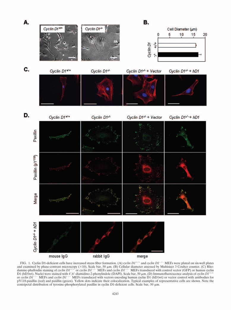
FIG. 1. Cyclin D1-deficient cells have increased stress fiber formation. (A) cyclin D1�/� and cyclin D1�/� MEFs were plated on six-well platesand examined by phase-contrast microscopy (10). Scale bar, 50 �m. (B) Cellular diameter assessed by Multisizer 3 Coulter counter. (C) Rho-damine-phalloidin staining of cyclin D1�/� or cyclin D1�/� MEFs and cyclin D1�/� MEFs transduced with control vector (GFP) or human cyclinD1 (hD1wt). Nuclei were stained with 4,6-diamidino-2-phenylindole (DAPI). Scale bar, 50 �m. (D) Immunofluorescence analysis of cyclin D1�/�
or cyclin D1�/� MEFs and cyclin D1�/� MEFs transduced with vectors encoding human cyclin D1 (hD1wt) or vector control with antibodies forpY118-paxillin (red) and paxillin (green). Yellow dots indicate their colocalization. Typical examples of representative cells are shown. Note thecentripetal distribution of tyrosine-phosphorylated paxillin in cyclin D1-deficient cells. Scale bar, 50 �m.
4243
tween cyclin D1�/� and cyclin D1�/� MEFs or between cyclinD1�/� MEFs infected with cyclin D1 and GFP vector control,suggesting altered F-actin distribution rather than altered geneexpression contributes to the less elongated phenotype in cy-clin D1�/� MEFs. In wild-type MEFs, infrequent costainingfor paxillin and tyrosine-phosphorylated paxillin (Y118) wasidentified. In contrast, sites of tyrosine-phosphorylated pax-illin were increased in number and centripetally distributedaround the circumference of cyclin D1-deficient cells (Fig.1D). Reintroduction of cyclin D1 reduced the number andreversed the circumferential distribution of focal contacts.
Control IgG showed only background staining (not shown).Overexposed images of IgG control staining showed nocostaining sites (Fig. 1D).
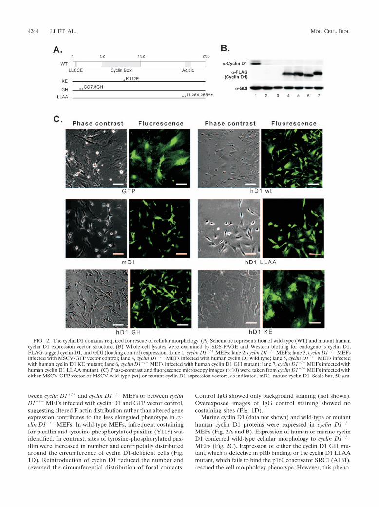
Murine cyclin D1 (data not shown) and wild-type or mutanthuman cyclin D1 proteins were expressed in cyclin D1�/�
MEFs (Fig. 2A and B). Expression of human or murine cyclinD1 conferred wild-type cellular morphology to cyclin D1�/�
MEFs (Fig. 2C). Expression of either the cyclin D1 GH mu-tant, which is defective in pRb binding, or the cyclin D1 LLAAmutant, which fails to bind the p160 coactivator SRC1 (AIB1),rescued the cell morphology phenotype. However, this pheno-
FIG. 2. The cyclin D1 domains required for rescue of cellular morphology. (A) Schematic representation of wild-type (WT) and mutant humancyclin D1 expression vector structure. (B) Whole-cell lysates were examined by SDS-PAGE and Western blotting for endogenous cyclin D1,FLAG-tagged cyclin D1, and GDI (loading control) expression. Lane 1, cyclin D1�/� MEFs; lane 2, cyclin D1�/� MEFs; lane 3, cyclin D1�/� MEFsinfected with MSCV-GFP vector control; lane 4, cyclin D1�/� MEFs infected with human cyclin D1 wild type; lane 5, cyclin D1�/� MEFs infectedwith human cyclin D1 KE mutant; lane 6, cyclin D1�/� MEFs infected with human cyclin D1 GH mutant; lane 7, cyclin D1�/� MEFs infected withhuman cyclin D1 LLAA mutant. (C) Phase-contrast and fluorescence microscopy images (10) were taken from cyclin D1�/� MEFs infected witheither MSCV-GFP vector or MSCV-wild-type (wt) or mutant cyclin D1 expression vectors, as indicated. mD1, mouse cyclin D1. Scale bar, 50 �m.
4244 LI ET AL. MOL. CELL. BIOL.
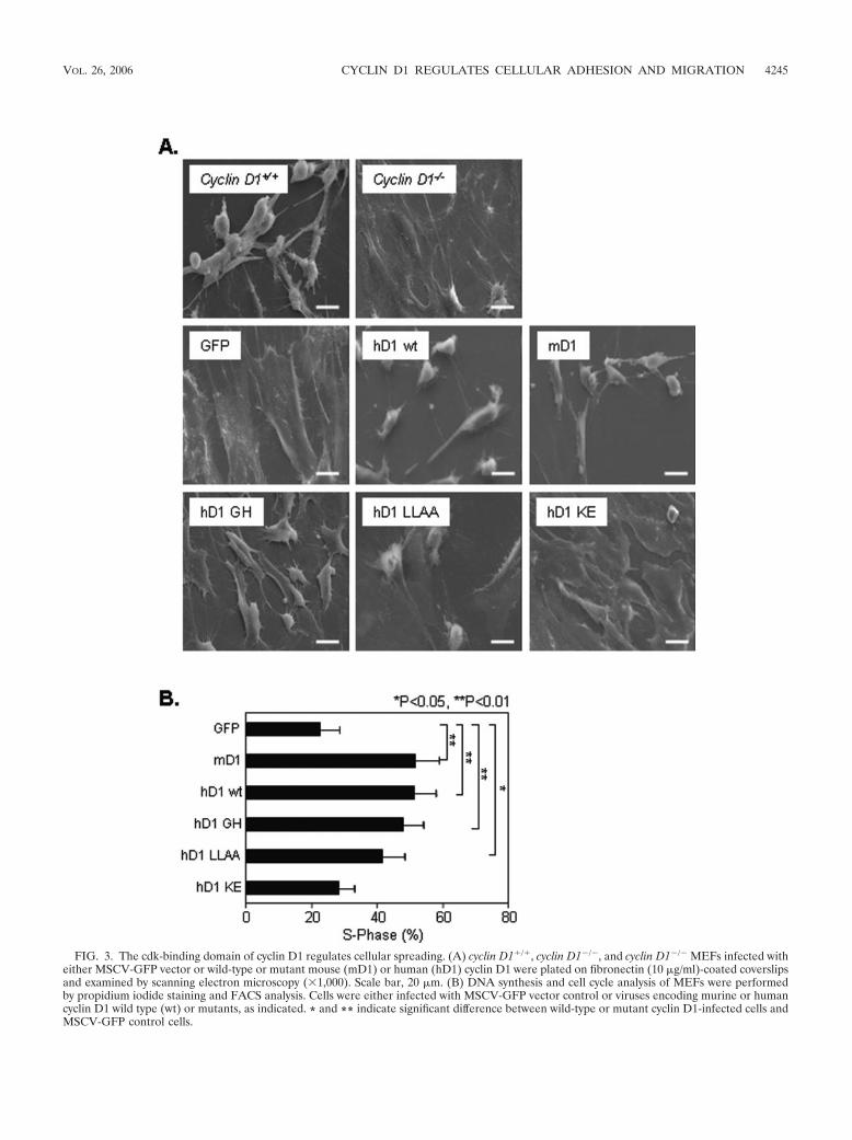
FIG. 3. The cdk-binding domain of cyclin D1 regulates cellular spreading. (A) cyclin D1�/�, cyclin D1�/�, and cyclin D1�/� MEFs infected witheither MSCV-GFP vector or wild-type or mutant mouse (mD1) or human (hD1) cyclin D1 were plated on fibronectin (10 �g/ml)-coated coverslipsand examined by scanning electron microscopy (1,000). Scale bar, 20 �m. (B) DNA synthesis and cell cycle analysis of MEFs were performedby propidium iodide staining and FACS analysis. Cells were either infected with MSCV-GFP vector control or viruses encoding murine or humancyclin D1 wild type (wt) or mutants, as indicated. * and ** indicate significant difference between wild-type or mutant cyclin D1-infected cells andMSCV-GFP control cells.
VOL. 26, 2006 CYCLIN D1 REGULATES CELLULAR ADHESION AND MIGRATION 4245
type was not rescued by the cdk4-binding-defective mutant ofcyclin D1 (KE mutant).
The cyclin D1 cdk-binding domain is required for reducingcellular spreading and inducing DNA synthesis. By scanningelectron microscopy (Fig. 3A), cyclin D1�/� MEFs were morespread than cyclin D1�/� MEFs. Expression of either murineor human cyclin D1 in cyclin D1�/� MEFs rescued the wild-type MEF phenotype. The pRb-binding-defective mutant (GHmutant) and SRC1-binding-defective mutant (LLAA mutant)were both capable of rescuing this phenotype, whereas thecdk-binding-defective mutant (KE mutant) was again unableto rescue (Fig. 3A). cyclin D1�/� MEFs and 3T3 cells exhibiteda reduced proportion of cells in S phase of the cell cycle (3).Expression of cyclin D1 wild-type or the pRb-binding-defectiveor SRC1-binding-defective mutants increased the proportionof cells in S phase from 22% to up to 51% (Fig. 3B) (P � 0.02).Mutation of the cdk4-binding site in cyclin D1 abrogated thisrescue of DNA synthesis. All cyclin D1�/� MEFs transducedwith the MSCV-cyclin D1-IRES-GFP vector reverted to the
fibroblastoid morphology, yet only 50% of the cells were in theDNA synthetic phase.
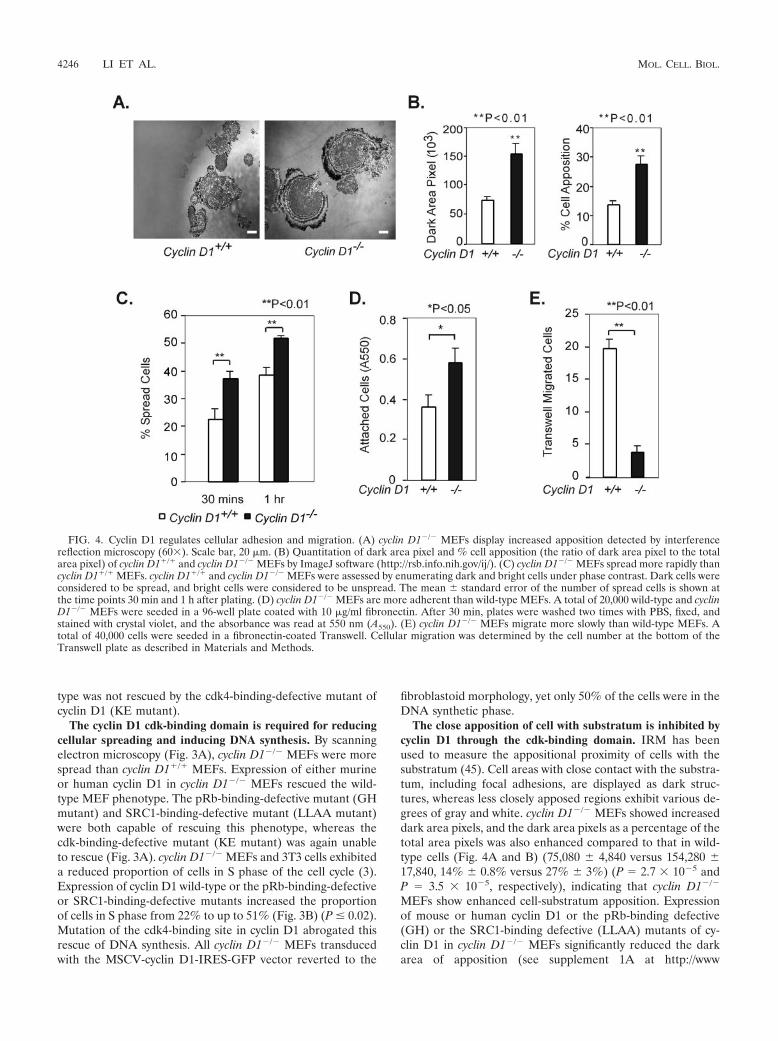
The close apposition of cell with substratum is inhibited bycyclin D1 through the cdk-binding domain. IRM has beenused to measure the appositional proximity of cells with thesubstratum (45). Cell areas with close contact with the substra-tum, including focal adhesions, are displayed as dark struc-tures, whereas less closely apposed regions exhibit various de-grees of gray and white. cyclin D1�/� MEFs showed increaseddark area pixels, and the dark area pixels as a percentage of thetotal area pixels was also enhanced compared to that in wild-type cells (Fig. 4A and B) (75,080 � 4,840 versus 154,280 �17,840, 14% � 0.8% versus 27% � 3%) (P 2.7 10�5 andP 3.5 10�5, respectively), indicating that cyclin D1�/�
MEFs show enhanced cell-substratum apposition. Expressionof mouse or human cyclin D1 or the pRb-binding defective(GH) or the SRC1-binding defective (LLAA) mutants of cy-clin D1 in cyclin D1�/� MEFs significantly reduced the darkarea of apposition (see supplement 1A at http://www
FIG. 4. Cyclin D1 regulates cellular adhesion and migration. (A) cyclin D1�/� MEFs display increased apposition detected by interferencereflection microscopy (60). Scale bar, 20 �m. (B) Quantitation of dark area pixel and % cell apposition (the ratio of dark area pixel to the totalarea pixel) of cyclin D1�/� and cyclin D1�/� MEFs by ImageJ software (http://rsb.info.nih.gov/ij/). (C) cyclin D1�/� MEFs spread more rapidly thancyclin D1�/� MEFs. cyclin D1�/� and cyclin D1�/� MEFs were assessed by enumerating dark and bright cells under phase contrast. Dark cells wereconsidered to be spread, and bright cells were considered to be unspread. The mean � standard error of the number of spread cells is shown atthe time points 30 min and 1 h after plating. (D) cyclin D1�/� MEFs are more adherent than wild-type MEFs. A total of 20,000 wild-type and cyclinD1�/� MEFs were seeded in a 96-well plate coated with 10 �g/ml fibronectin. After 30 min, plates were washed two times with PBS, fixed, andstained with crystal violet, and the absorbance was read at 550 nm (A550). (E) cyclin D1�/� MEFs migrate more slowly than wild-type MEFs. Atotal of 40,000 cells were seeded in a fibronectin-coated Transwell. Cellular migration was determined by the cell number at the bottom of theTranswell plate as described in Materials and Methods.
4246 LI ET AL. MOL. CELL. BIOL.
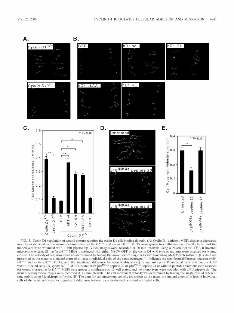
FIG. 5. Cyclin D1 regulation of wound closure requires the cyclin D1 cdk-binding domain. (A) Cyclin D1-deficient MEFs display a decreasedmotility as detected in the wound-healing assay. cyclin D1�/� and cyclin D1�/� MEFs were grown to confluence on 12-well plates, and themonolayers were wounded with a P10 pipette tip. Video images were recorded at 20-min intervals using a Nikon Eclipse TE-300 invertedmicroscope system. (B) cyclin D1�/� MEFs transduced with either MSCV-GFP or the cyclin D1 wild type or mutants were assessed for woundclosure. The velocity of cell movement was determined by tracing the movement of single cells with time using MetaMorph software. (C) Data arepresented as the mean � standard error of at least 6 individual cells of the same genotype. ** indicates the significant difference between cyclinD1�/� and cyclin D1�/� MEFs, and the significant difference between wild-type (wt) or mutant cyclin D1-infected cells and control GFPvector-infected cells. (D) cyclin D1�/� MEFs treated with p16INK4a peptide 20 or p16INK4a peptide 21 or without peptide treatment were assessedfor wound closure. cyclin D1�/� MEFs were grown to confluence on 12-well plates, and the monolayers were wounded with a P10 pipette tip. Thewound-healing video images were recorded at 30-min intervals. The cell movement velocity was determined by tracing the single cells at differenttime points using MetaMorph software. (E) The data for cell movement velocity are shown as the mean � standard error of at least 6 individualcells of the same genotype. **, significant difference between peptide-treated cells and untreated cells.
VOL. 26, 2006 CYCLIN D1 REGULATES CELLULAR ADHESION AND MIGRATION 4247
.kimmelcancercenter.org/pestell/papers/zl/Supplement1.pdf)(P � 0.008). In contrast, expression of the cdk-bindingdefective (KE) mutant, failed to reduce the closely apposedarea detected by IRM (P 0.12 and P 0.14, respectively)(see supplement 1B at http://www.kimmelcancercenter.org/pestell/papers/zl/Supplement1.pdf).
cyclin D1�/� MEFs spread significantly more quickly thanwild-type cells at 30 min and 1 h, as judged by the ratio of darkto bright cells under phase-contrast microscopy (P 0.009 andP 0.003, respectively) (Fig. 4C). Introduction of either themurine or human cyclin D1 cDNA into cyclin D1�/� MEFs byretroviral infection reduced the percentage of spread cellsfrom 58% to �30% at 30 min after plating (P 0.0007 andP 0.0004, respectively) (see supplement 2 at http://www.kimmelcancercenter.org/pestell/papers/zl/Supplement2.pdf).Both the pRb-binding-defective mutant (GH mutant) andthe SRC1-binding-defective mutant (LLAA mutant) de-creased the percentage of spread cells at 30 min (P 0.0032) (see supplement 2 at the URL above). However, thecdk-binding-defective mutant (KE mutant) failed to reducethe percentage of spread cells (see supplement 2 at the URLabove). Cyclin D1-deficient MEFs were more adherent thanwild-type cells on fibronectin (P 0.03) (Fig. 4D). Exoge-nous mouse and human cyclin D1 wild-type, GH mutant, orLLAA cyclin D1 mutants decreased adherence on fibronec-tin (P 8.7 10�10, P 1 10�6, P 9.1 10�6, P 4.4 10�6) (see supplement 2 at the URL above), while thecdk-binding-defective mutant (KE mutant) failed to reduceadhesion (see supplement 2 at the URL above).
p16INK4a inhibits and cyclin D1 enhances the velocity ofcellular migration. cyclin D1�/� MEFs migrated significantlyless than wild-type cells on fibronectin-coated Transwell plates(P 0.0008) (Fig. 4E). Retroviral introduction of the murineor human cyclin D1 cDNA into cyclin D1�/� MEFs increasedthe cell migration rate significantly (P 0.0006 and P 0.001,respectively) (see supplement 2C at the URL above). ThepRb-binding-defective cyclin D1 mutant (GH mutant) and theSRC1-binding-defective mutant (LLAA mutant) both in-creased cell migration (P 0.016 and P 0.0005, respectively)(see supplement 2C at the URL above), which was notenhanced by the cdk-binding-defective mutant (KE mutant).
By overnight time-lapse videomicroscopy, the single-cell mo-tility of cyclin D1�/� MEFs was significantly slower than themotility of wild-type cells (P 0.0006) (Fig. 5A; see Fig.5Avideo1 and -2 at http://www.kimmelcancercenter.org/pestell/papers/zl/video/). Reintroduction of the cyclin D1 wild type orthe cyclin D1 mutants GH or LLAA increased the velocity ofcell movement compared with that in vector control cells (P 0.0048, P 0.0009, and P 0.0002, respectively) (Fig. 5B;see Fig. 5Bvideo1 to -4 at http://www.kimmelcancercenter.org
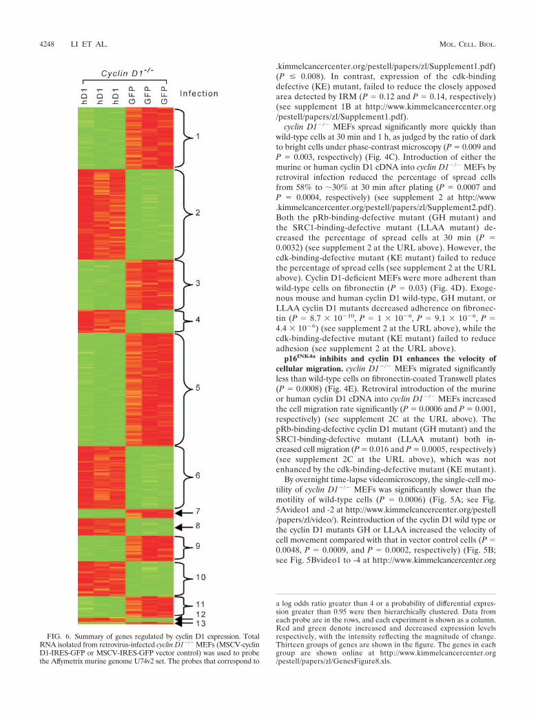
FIG. 6. Summary of genes regulated by cyclin D1 expression. TotalRNA isolated from retrovirus-infected cyclin D1�/� MEFs (MSCV-cyclinD1-IRES-GFP or MSCV-IRES-GFP vector control) was used to probethe Affymetrix murine genome U74v2 set. The probes that correspond to
a log odds ratio greater than 4 or a probability of differential expres-sion greater than 0.95 were then hierarchically clustered. Data fromeach probe are in the rows, and each experiment is shown as a column.Red and green denote increased and decreased expression levelsrespectively, with the intensity reflecting the magnitude of change.Thirteen groups of genes are shown in the figure. The genes in eachgroup are shown online at http://www.kimmelcancercenter.org/pestell/papers/zl/GenesFigure8.xls.
4248 LI ET AL. MOL. CELL. BIOL.

/pestell/papers/zl/video/), whereas there was no significant effectof the cdk-binding-defective mutant (KE mutant) (P 0.125)(Fig. 5B; see Fig. 5Bvideo5 at http://www.kimmelcancercenter.org/pestell/papers/zl/video/). Thus, cyclin D1 enhances the velocity ofcell motility.
A synthetic peptide that spans the two �-helixes of the thirdankyrin repeat of p16INK4a, interacts with the cdk, inhibitingcyclin D1-kinase activity. The derivative of this peptide carry-ing a D92A substitution (peptide 20) (Fig. 5D) has a lower IC50
compared with the wild-type sequence (20). The p16INK4a pep-tide 20 linked to the Antennapedia carrier sequence inhibitedwound closure compared with the no-peptide-treatmentcontrol (P 0.0016) (Fig. 5D; see Fig. 5Dvideo1 and -2 athttp://www.kimmelcancercenter.org/pestell/papers/zl/video/). Thep16INK4a peptide 21, which carries alanine substitutions of twovaline residues corresponding to positions 95 and 96 of thep16INK4a that dramatically increases its IC50 in vitro (20), hadreduced ability to inhibit MEF migration (P 0.98) (Fig. 5E;see Fig. 5Dvideo3 at http://www.kimmelcancercenter.org/pestell/papers/zl/video/). This finding is consistent with amodel in which cdk binding regulates MEF cellular migrationby cyclin D1.
Cyclin D1 regulates a molecular genetic cluster governingcellular adhesion and migration. We conducted a comprehen-
sive genomewide interrogation to identify gene targets regu-lated by cyclin D1 that may play a role in cell adhesion andmigration. cyclin D1�/� MEFs were infected with either wild-type cyclin D1 or the empty GFP vector as control. Analyseswere conducted in triplicate, and the subsets of genes differ-entially regulated by cyclin D1 were identified (Fig. 6 andTable 1; see also http://www.kimmelcancercenter.org/pestell/papers/zl/D1vsGFP.xls). Differential expression of genes pre-viously identified as regulating cellular migration and adhesionincluded those coding for TSP-1 and ROCKII. Given the cen-tral role of ROCKII and TSP-1 in regulating cellular migra-tion, we considered the possibility that cyclin D1 may regulatethe abundance and activity of these two key proteins.
The increase in actin stress fiber and focal contact formationin cyclin D1�/� MEFs is consistent with prior studies of cellswith increased ROCK activity (13, 70). Consistent with themicroarray analysis, ROCKII levels were increased in cyclinD1�/� MEFs compared to sibling controls. ROCKII kinaseactivity was increased in cyclin D1�/� cells (Fig. 7A). Additionof the ROCK kinase inhibitor Y-27632 increased cyclin D1�/�
cellular migration and movement velocity �6-fold (Fig. 7B andC). To determine whether the cyclin E could rescue the mi-gration defect of cyclin D1 deficiency, we examined MEFsderived from mice in which the cyclin D1 gene coding se-
TABLE 1. Adhesion- and migration-related genes regulated by cyclin D1a
Proteindesignation Name GenBank
accession no.
Fold change incyclin D1 vsGFP vector
BbProbability of
differentialexpression (%)
Ctgf Connective tissue growth factor NM_010217 �4.53 2.11E � 01 100.00Ptgs2 Prostaglandin-endoperoxide synthase 2 (cyclooxygenase 2) NM_011198 �4.35 1.62E � 01 100.00Tnc Tenascin C (hexabrachion) NM_011607 �3.86 1.99E � 01 100.00Selp Selectin P (granule membrane protein 140 kDa, antigen CD62) NM_011347 �3.25 1.64E � 01 100.00Col8a1 Collagen, type VIII, alpha 1 NM_007739 �2.57 1.65E � 01 100.00Fez1 Fasciculation and elongation protein zeta 1 (zygin I) NM_183171 �2.35 1.38E � 01 99.99Rhob Ras homolog gene family, member B NM_007483 �2.2 1.35E � 01 99.99TSP-1 Thrombospondin 1 NM_011580 �2.11 6.12E � 00 98.58Anxa1 Annexin A1 NM_010730 �1.9 5.53E � 00 97.88F11r F11 receptor NM_172647 �1.85 1.05E � 01 99.93Cdh2 Cadherin 2, type 1, N-cadherin (neuronal) NM_007664 �1.8 7.73E � 00 99.53Pkp2 Plakophilin 2 NM_026163 �1.67 6.15E � 00 98.61Ccl2 Chemokine (C-C motif) ligand 2 NM_011333 �1.65 8.16E � 00 99.65Mcam Melanoma cell adhesion molecule NM_023061 �1.59 5.80E � 00 98.24ROCKII Rho-associated coiled-coil-forming kinase 2 NM_009072 �1.57 5.85E � 00 98.30Tgfb1i1 Transforming growth factor-�1-induced transcript 1 NM_009365 �1.56 9.63E � 00 99.87Fbln2 Fibulin 2 NM_007992 �1.53 8.76E � 00 99.77Tln1 Talin 1 NM_011602 �1.5 4.86E � 00 96.67Map2k1 Mitogen-activated protein kinase kinase 1 NM_008927 �1.48 5.94E � 00 98.4Jup Junction plakoglobin NM_010593 �1.43 4.54E � 00 95.88Fxyd5 FXYD domain containing ion transport regulator 5 NM_008761 �1.37 4.44E � 00 95.6Fbln1 Fibulin 1 AK083573 1.36 4.28E � 00 95.1Serpine 2 Serine (or cysteine) proteinase inhibitor, clade E (nexin,
plasminogen activator inhibitor type 1), member 2NM_009255 1.59 6.23E � 00 98.69
Islr Immunoglobulin superfamily containing leucine-rich repeat NM_012043 1.61 8.31E � 00 99.69Gas6 Growth arrest-specific 6 NM_019521 1.63 8.36E � 00 99.70Ccl5 Chemokine (C-C motif) ligand 5 NM_013653 1.69 1.04E � 01 99.93Col6a1 Collagen, type VI, alpha 1 NM_009933 1.7 9.55E � 00 99.87Cx3cl1 Chemokine (C-X3-C motif) ligand 1 NM_009142 1.98 1.29E � 01 99.99Col6a2 Collagen, type VI, alpha 2 NM_146007 2.2 1.42E � 01 99.99Tpbg Trophoblast glycoprotein NM_011627 2.22 1.28E � 01 99.99Vegfc Vascular endothelial growth factor C NM_009506 2.81 1.15E � 01 99.97Tgfbi Transforming growth factor, beta induced, 68 kDa NM_009369 3.01 1.40E � 01 99.99
a Shown are genes differentially regulated by cyclin D1, which have adhesion- and/or migration-related function.b B is the log odds ratio of the gene being differentially expressed.
VOL. 26, 2006 CYCLIN D1 REGULATES CELLULAR ADHESION AND MIGRATION 4249

FIG. 7. Cyclin D1 promotes migration through inhibition of ROCK kinase. (A) Western blot analysis and ROCKII kinase assays in three pairsof cyclin D1�/� and cyclin D1�/� MEFs. Each lane represents a different animal. Vinculin was used as a loading control. �-, anti-. (B) Single-cellanalysis of cell movement velocity taken at 5-min intervals from cyclin D1�/� or cyclin D1�/� MEFs treated with either vehicle or the ROCK kinaseinhibitor Y27632. (C) Cellular movement velocity, shown as mean � standard error for n � 20 cells (��, P � 0.01) assessed at 5-min intervals for2 h. (D and E) Single-cell analysis of cell movement velocity was taken at 5-min intervals from cyclin D1�/� or cyclin E knockin to cyclin D1�/�
MEFs. Cellular movement velocity is shown as the mean � standard error for n � 10 cells (��, P � 0.01). (F) Western blot analysis for ROCKIIprotein expression in cyclin D1�/� and cyclin E knockin to cyclin D1�/� MEFs. Vinculin was used as a loading control. (G) DNA synthesis andcell cycle analysis of cyclin D1�/�, cyclin D1�/� MEFs, and cyclin D1�/� with human cyclin E knockin MEFs were performed by propidium iodidestaining and FACS analysis. An asterisk indicates the significant difference between cyclin D1�/� and cyclin D1�/� MEFs. (H) Rho protein andactivated Rho decreased by cyclin D1. Rho-GTP pull-down assay and Western blot analysis for activated Rho and total abundance of RhoA, -B,and -C, cyclin D1, and �-tubulin (loading control) in cyclin D1�/� MEFs transduced with vectors encoding wild-type cyclin D1 or vector control.
4250 LI ET AL. MOL. CELL. BIOL.
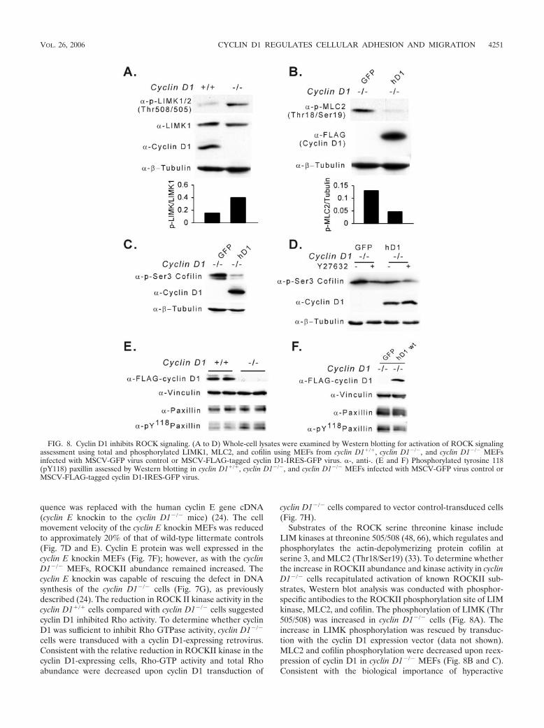
quence was replaced with the human cyclin E gene cDNA(cyclin E knockin to the cyclin D1�/� mice) (24). The cellmovement velocity of the cyclin E knockin MEFs was reducedto approximately 20% of that of wild-type littermate controls(Fig. 7D and E). Cyclin E protein was well expressed in thecyclin E knockin MEFs (Fig. 7F); however, as with the cyclinD1�/� MEFs, ROCKII abundance remained increased. Thecyclin E knockin was capable of rescuing the defect in DNAsynthesis of the cyclin D1�/� cells (Fig. 7G), as previouslydescribed (24). The reduction in ROCK II kinase activity in thecyclin D1�/� cells compared with cyclin D1�/� cells suggestedcyclin D1 inhibited Rho activity. To determine whether cyclinD1 was sufficient to inhibit Rho GTPase activity, cyclin D1�/�
cells were transduced with a cyclin D1-expressing retrovirus.Consistent with the relative reduction in ROCKII kinase in thecyclin D1-expressing cells, Rho-GTP activity and total Rhoabundance were decreased upon cyclin D1 transduction of
cyclin D1�/� cells compared to vector control-transduced cells(Fig. 7H).
Substrates of the ROCK serine threonine kinase includeLIM kinases at threonine 505/508 (48, 66), which regulates andphosphorylates the actin-depolymerizing protein cofilin atserine 3, and MLC2 (Thr18/Ser19) (33). To determine whetherthe increase in ROCKII abundance and kinase activity in cyclinD1�/� cells recapitulated activation of known ROCKII sub-strates, Western blot analysis was conducted with phosphor-specific antibodies to the ROCKII phosphorylation site of LIMkinase, MLC2, and cofilin. The phosphorylation of LIMK (Thr505/508) was increased in cyclin D1�/� cells (Fig. 8A). Theincrease in LIMK phosphorylation was rescued by transduc-tion with the cyclin D1 expression vector (data not shown).MLC2 and cofilin phosphorylation were decreased upon reex-pression of cyclin D1 in cyclin D1�/� MEFs (Fig. 8B and C).Consistent with the biological importance of hyperactive
FIG. 8. Cyclin D1 inhibits ROCK signaling. (A to D) Whole-cell lysates were examined by Western blotting for activation of ROCK signalingassessment using total and phosphorylated LIMK1, MLC2, and cofilin using MEFs from cyclin D1�/�, cyclin D1�/�, and cyclin D1�/� MEFsinfected with MSCV-GFP virus control or MSCV-FLAG-tagged cyclin D1-IRES-GFP virus. �-, anti-. (E and F) Phosphorylated tyrosine 118(pY118) paxillin assessed by Western blotting in cyclin D1�/�, cyclin D1�/�, and cyclin D1�/� MEFs infected with MSCV-GFP virus control orMSCV-FLAG-tagged cyclin D1-IRES-GFP virus.
VOL. 26, 2006 CYCLIN D1 REGULATES CELLULAR ADHESION AND MIGRATION 4251
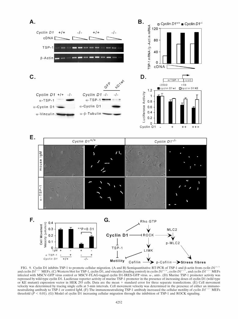
FIG. 9. Cyclin D1 inhibits TSP-1 to promote cellular migration. (A and B) Semiquantitative RT-PCR of TSP-1 and �-actin from cyclin D1�/�
and cyclin D1�/� MEFs. (C) Western blot for TSP-1, cyclin D1, and vinculin (loading control) in cyclin D1�/�, cyclin D1�/�, and cyclin D1�/� MEFsinfected with MSCV-GFP virus control or MSCV-FLAG-tagged cyclin D1-IRES-GFP virus. �-, anti-. (D) Murine TSP-1 promoter activity wasrepressed by wild-type cyclin D1. Luciferase reporter activity of murine TSP-1 promoter in the presence of increasing doses of cyclin D1 (wild typeor KE mutant) expression vector in HEK 293 cells. Data are the mean � standard error for three separate transfections. (E) Cell movementvelocity was determined by tracing single cells at 5-min intervals. Cell movement velocity was determined in the presence of either an immuno-neutralizing antibody to TSP-1 or control IgM. (F) The immunoneutralizing TSP-1 antibody increased the cellular motility of cyclin D1�/� MEFsthreefold (P � 0.01). (G) Model of cyclin D1 increasing cellular migration through the inhibition of TSP-1 and ROCK signaling.
4252
ROCKII in the migratory defect of cyclin D1�/� MEFs (Fig.7B and C) and the increased phosphorylation of cofilin byROCKII in cyclin D1�/� MEFs, addition of the ROCK inhib-itor Y27632 reduced the hyperphosphorylation of cofilin(Ser3) to the level of phosphorylation observed with cyclin D1rescue (Fig. 8D, lane 2 versus lane 3). Consistent with theenhanced formation of focal contacts, the levels of tyrosine-phosphorylated paxillin (Y118) were increased in cyclin D1�/�
cells (Fig. 8E) and reintroduction of cyclin D1 inhibited pax-illin Y118 phosphorylation (Fig. 8F).
In view of the microarray analysis indicating an increase inTSP-1 abundance in each of the cyclin D1�/� MEFs analyzed(Table 1) and the known role for TSP-1 as an inhibitor ofcellular migration, we examined further the relative abundanceof TSP-1 and its regulation by cyclin D1. Expression of theangiogenesis inhibitor TSP-1, which is repressed by inductionof the Ras-ROCK pathway (76), was induced in cyclin D1�/�
MEFs using quantitative RT-PCR and Western blotting (Fig.9A, B, and C). Reintroduction of cyclin D1 into cyclin D1�/�
MEFs inhibited TSP-1 abundance (Fig. 9C). The murineTSP-1 promoter activity was repressed by cyclin D1 40 to 50%in a dose-dependent manner (Fig. 9D). The cdk-binding-de-fective mutant of cyclin D1, which failed to rescue the cyclinD1�/� migratory defect, failed to repress the TSP-1 promoter(Fig. 9D). In cyclin D1�/� MEFs, neutralizing TSP-1 antibodyincreased migration of cyclin D1�/� cells �3-fold comparedwith normal mouse IgM control (Fig. 9E and F). Addition ofTSP-1 antibody to normal wild-type MEFs did not affect cel-lular migration, consistent with previous studies (72). The con-current addition of the TSP-1 antibody to the Y27632-treatedcyclin D1�/� cells provided no significant additional increasein cellular velocity compared with Y27632 alone (data notshown).
Collectively, these studies show that cyclin D1 inhibitsROCKII expression and function as evidenced by cyclin D1-mediated inhibition of ROCKII expression and kinase activity,and the reduction in phosphorylation of LIMK1 (Thr508/505),MLC2 (Thr18/Ser19), and cofilin (Ser3) (Fig. 9G). In addition,cyclin D1 inhibits TSP-1 transcription and abundance. Theincreased production of TSP-1 by cyclin D1�/� cells contrib-utes to the defect in cellular migration as reintroduction ofcyclin D1 or immunoneutralizing antibody to TSP-1 reducedthe migratory defect.
DISCUSSION
Herein, cyclin D1 promotes cellular migration and reducescellular adhesion, independently of its pRb-binding function,but dependent upon an intact cdk-binding domain. Cyclin D1reintroduction into cyclin D1�/� MEFs restored cellular mor-phology and migratory velocity. Microarray analysis identifiedROCKII and TSP-1 as targets of cyclin D1-dependent repres-sion. Immunoneutralizing antibodies to TSP-1 or addition ofROCKII inhibitor reversed the defective migration of cyclinD1-deficient cells. Together, these studies identify a novelmechanism by which cyclin D1 promotes cellular migrationthrough inhibiting expression and function of ROCKII andTSP-1. The extracellular matrix glycoprotein TSP-1 inhibitscellular metastases and tumor growth (64). TSP-1 is a potentinhibitor of both tumorigenesis and in vivo neovascularization.
Several oncogenes repress TSP-1, including oncogenic Ras,c-Myc, v-Src, and c-Jun (41, 51, 61, 67), as well as Id1 (72).Conversely, tumor suppressor proteins, including p53 andPTEN, induce TSP-1 abundance (17, 78). Ras repression ofTSP-1 involves a signaling pathway, which includes the smallmonomeric GTPases and the Rho effectors (76). The currentfindings in which cyclin D1 reduced TSP-1 protein and mRNAabundance, repressing the TSP-1 promoter, are consistent withour previous findings that cyclin D1 is induced as a distal targetof Ras, v-Src, and c-Jun (2, 35) and that each of these onco-genes represses TSP-1.
The current studies are consistent with recent findings link-ing factors that regulate cellular spreading and cell cycle pro-gression. AP-1 proteins are known to promote both DNAsynthesis and cellular motility (68, 71). Under several circum-stances, cellular adhesion and migration promote G1-phaseprogression. Increased Rho activity, as observed in the cyclinD1�/� cells in the current studies, has been shown to nativelyregulate motility by increasing stress fiber-dependent adhesion(16). Although cellular adhesion is frequently required for cellcycle progression to occur, high levels of cell adhesion withlarge focal contacts results in failed cellular migration. Rhomay also promote migration, being required for actomyosincontraction (4). Mutational analysis of the Rho/Rac chimerademonstrates distinct residues within the Rac effector domainregulate cellular morphology versus cyclin D1 expression andDNA synthesis (31, 80). Further evidence for a dissociationbetween the regulation of cellular morphology through Rhoactivity and DNA synthesis through pRb includes findings thatunlike Rac and Cdc42, activated RhoA, despite its ability toinduce morphological changes, does not inactivate pRb in NIH3T3 cells (25). Together, these studies suggest the regulation ofcell cycle progression and adhesion may be coupled or uncou-pled, depending upon the cell type and signaling pathway in-volved.
Cyclin D1 promoted cell migration by inhibiting ROCKIIexpression and activity. ROCK kinase inhibitor reversed thedefect in cellular motility in cyclin D1�/� cells, generating themore polarized morphology of wild-type cells. These findingsare consistent with studies that ROCKII small interferingRNA induces an elongated morphology and that ROCK in-hibitor treatment of cells induces a more polarized morphol-ogy (37) and increased cellular migration (6). Cyclin D1-defi-cient cells demonstrated evidence of increased ROCK activityusing the in vitro substrate of myelin basic protein (MBP) andwith increased phosphorylation of MLC2 (Thr18/Ser19),LIMK1/2 (Thr508/505), and cofilin (Ser3). The relationshipbetween the activity of ROCK and cellular migration is com-plex and cell type specific. Thus, the ROCK inhibitor Y-27632may either promote or inhibit cell migration (29, 46, 69, 81).The present findings are consistent with several recent find-ings. B-raf�/� MEFs demonstrated reduced ROCKII, reducedphosphocofilin, and increased migration (50), further confirm-ing the correlation of increased ROCKII and reduced migra-tion as seen in our studies. The possibility that the spreadphenotype in cyclin D1�/� MEFs was a function of early se-nescence was excluded by the finding that pH-sensitive �-ga-lactosidase staining (18) was similar between the cyclin D1�/�
and cyclin D1�/� MEFs used in the studies (data not shown).Several lines of evidence suggest the reduced migration of
VOL. 26, 2006 CYCLIN D1 REGULATES CELLULAR ADHESION AND MIGRATION 4253

cyclin D1 deficiency is not a direct consequence of reducedDNA synthesis. First, all cyclin D1�/� cells exhibited the mi-gratory defect, whereas only 7% of the cells were in the DNAsynthetic phase. Second, all cyclin D1�/� cells transduced bycyclin D1 demonstrated the change in cellular morphology,alterations in focal contact distribution and cellular migration,yet only a fraction of the cells were undergoing DNA synthesis.Third, the reintroduction of cyclin E through a knockin of thehuman cyclin E cDNA rescued the DNA synthetic defect ofcyclin D1�/� cells but did not rescue either the migrationdefect nor the ROCKII abnormality. Collectively these studiessuggest the effect of cyclin D1 on cell cycle progression andcellular migration may be dissociable functions. The effect ofRho, and its signaling components, including mDia, on cellularmorphology can be uncoupled from effects on DNA synthesis.ROCK inhibition stimulates passage through G1 phase in theabsence of cell spreading (37), and ROCK activity is not re-quired for cell-shape-dependent G1 progression in microvas-cular endothelial cells. mDia, which promotes actin polymer-ization and regulates the alignment of stress fibers by targetingmicrotubules to focal adhesions, fails to promote G1-phaseprogression (37). Thus, interaction between ROCK and itsregulation of cellular adhesion and DNA synthesis is disso-ciable and cell type dependent.
Cyclin D1 deficiency increased adhesion in both macro-phages and MEFs, changes correlating with the induction ofcircumferential cortical F-actin stress fibers in MEFs and inmacrophages (45). As with cyclin D1, genetic deletion of sev-eral other proteins known to regulate migration includingRhoB, cyclin B, and p27kip1, do not appear to affect embryonicdevelopment (6, 10, 38, 40, 44, 49). The stable adhesive struc-tures in cyclin D1-deficient cells are thought to contribute tothe increased adhesion and reduced migration of these cells. Ithas been proposed that an increase in either Rac (40) or Rho(6) activity and/or their effectors can contribute to reducedcellular migration. Cyclin D1 transcription is induced by theRho GTPases (12, 31, 79). Cyclin D1 induction in response togrowth factors as a delayed early response at 6 h requiresRho-dependent sustained ERK activation and results in theinduction of DNA synthesis. Rho also inhibits an alternativeRac/Cdc42-dependent induction of cyclin D1, thus preventingits premature induction (77). The inhibition of Rac-dependentexpression of cyclin D1 involves LIM kinase through an effectthat is independent of cofilin phosphorylation and actin poly-merization. Herein, cyclin D1 also functions as an upstreaminhibitor of Rho/ROCK/LIMK. As the effect of nuclear LIMkinase on cyclin D1 abundance regulates the duration of G1
phase (56), together these studies suggest cyclin D1 may func-tion as a fine-tuning feedback-regulating LIM kinase.
Cyclin D1 physically interacts with pRb, p160 (AIB1), cdks,and the cell cycle inhibitor proteins p21CIP1 and p27KIP1.p21CIP1 promotes cell motility in Ras-transformed cells,through forming a complex with ROCK and thereby blockingRho kinase action (36). p27KIP1 regulates actin dynamics, pro-moting cell migration (40, 43) independently of cyclin-cdkbinding, suggesting that the mechanisms by which p27KIP1 andcyclin D1 regulate motility are distinct (40). p27KIP1 can func-tion upstream of RhoA, inhibiting its activation (6). p21CIP1
and p27KIP1 can either reduce cdk activity or promote assem-bly and nuclear transport of D-type cyclins (59). Thus, new
possible interactions between cyclin D1 and p27KIP1 in regu-lating cellular migration may require further analysis. The as-sociation of cyclin D1 overexpression with poor prognosis andtumor metastasis (19, 30) raised the intriguing possibility thatcyclin D1 may play a distinct role in promoting cellular migra-tion and invasion. The present studies demonstrate cyclin D1promotes cellular migration through TSP-1 and ROCKII. Theidentification of compounds that selectively block the ATPpocket of cdk to selectively inhibit cellular kinase activity hasproven challenging. The current findings that the cyclin D1protein, through K112, promotes cellular migration may pro-vide an important new avenue for therapy of metastatic diseasein which cyclin D1 is overexpressed.
ACKNOWLEDGMENTS
We are particularly grateful to P. Sicinski for the cyclin D1�/� andcyclin E knockin to cyclin D1�/� mice. We thank D. Scardino forassistance with manuscript preparation. We are grateful for the assis-tance of Susette C. Mueller (Microscopy and Imaging Shared Re-source, Lombardi Cancer Center, Georgetown University) for techni-cal advice.
Support for these studies was from NIH R01CA70896, R01CA75503,R01CA86072, R01CA93596, R01CA107382, and Kimmel Cancer CenterSupport grant P30 CA56036 to R.G.P.; CA26504, P01 100324-02, and P30CA13330-31 to E.R.S.; and the Breast Cancer Alliance through an Ex-ceptional Project grant to A.A.Q.
REFERENCES
1. Albanese, C., M. D’Amico, A. T. Reutens, M. Fu, G. Watanabe, R. J. Lee, R. N.Kitsis, B. Henglein, M. Avantaggiati, K. Somasundaram, B. Thimmapaya, andR. G. Pestell. 1999. Activation of the cyclin D1 gene by the E1A-associatedprotein p300 through AP-1 inhibits cellular apoptosis. J. Biol. Chem. 274:34186–34195.
2. Albanese, C., J. Johnson, G. Watanabe, N. Eklund, D. Vu, A. Arnold, andR. G. Pestell. 1995. Transforming p21ras mutants and c-Ets-2 activate thecyclin D1 promoter through distinguishable regions. J. Biol. Chem. 270:23589–23597.
3. Albanese, C., K. Wu, M. D’Amico, C. Jarrett, D. Joyce, J. Hughes, J. Hulit,T. Sakamaki, M. Fu, A. Ben-Ze’ev, J. F. Bromberg, C. Lamberti, U. Verma,R. B. Gaynor, S. W. Byers, and R. G. Pestell. 2003. IKKalpha regulatesmitogenic signaling through transcriptional induction of cyclin D1 via Tcf.Mol. Biol. Cell 14:585–599.
4. Allen, W. E., G. E. Jones, J. W. Pollard, and A. J. Ridley. 1997. Rho, Rac andCdc42 regulate actin organization and cell adhesion in macrophages. J. CellSci. 110:707–720.
5. Amano, M., K. Chihara, K. Kimura, Y. Fukata, N. Nakamura, Y. Matsuura,and K. Kaibuchi. 1997. Formation of actin stress fibers and focal adhesionsenhanced by Rho-kinase. Science 275:1308–1311.
6. Besson, A., M. Gurian-West, A. Schmidt, A. Hall, and J. M. Roberts. 2004.p27Kip1 modulates cell migration through the regulation of RhoA activa-tion. Genes Dev. 18:862–876.
7. Bornstein, P., D. Alfi, S. Devarayalu, P. Framson, and P. Li. 1990. Charac-terization of the mouse thrombospondin gene and evaluation of the role ofthe first intron in human gene expression. J. Biol. Chem. 265:16691–16698.
8. Bouras, T., M. Fu, A. A. Sauve, F. Wang, A. A. Quong, N. D. Perkins, R. T.Hay, W. Gu, and R. G. Pestell. 2005. SIRT1 deacetylation and repression ofp300 involves lysine residues 1020/1024 within the cell cycle regulatory do-main 1. J. Biol. Chem. 280:10264–10276.
9. Boutros, P. C., I. D. Moffat, M. A. Franc, N. Tijet, J. Tuomisto, R. Pohjanvirta,and A. B. Okey. 2004. Dioxin-responsive AHRE-II gene battery: identifica-tion by phylogenetic footprinting. Biochem. Biophys. Res. Commun. 321:707–715.
10. Brandeis, M., I. Rosewell, M. Carrington, T. Crompton, M. A. Jacobs, J.Kirk, J. Gannon, and T. Hunt. 1998. Cyclin B2-null mice develop normallyand are fertile whereas cyclin B1-null mice die in utero. Proc. Natl. Acad. Sci.USA 95:4344–4349.
11. Bromberg, J. F., M. H. Wrzeszczynska, G. Devgan, Y. Zhao, R. G. Pestell, C.Albanese, and J. E. Darnell. 1999. Stat3 as an oncogene. Cell 98:295–303.
12. Burbelo, P., A. Wellstein, and R. G. Pestell. 2004. Altered Rho GTPase signalingpathways in breast cancer cells. Breast Cancer Res. Treat. 84:43–48.
13. Burridge, K., and K. Wennerberg. 2004. Rho and Rac take center stage. Cell116:167–179.
14. Chen, Y., V. Kamat, E. R. Dougherty, M. L. Bittner, P. S. Meltzer, and J. M.Trent. 2002. Ratio statistics of gene expression levels and applications tomicroarray data analysis. Bioinformatics 18:1207–1215.
4254 LI ET AL. MOL. CELL. BIOL.
15. Cox, D., J. S. Berg, M. Cammer, J. O. Chinegwundoh, B. M. Dale, R. E.Cheney, and S. Greenberg. 2002. Myosin X is a downstream effector ofPI(3)K during phagocytosis. Nat. Cell Biol. 4:469–477.
16. Cox, E. A., S. K. Sastry, and A. Huttenlocher. 2001. Integrin-mediatedadhesion regulates cell polarity and membrane protrusion through the Rhofamily of GTPases. Mol. Biol. Cell 12:265–277.
17. Dameron, K. M., O. V. Volpert, M. A. Tainsky, and N. Bouck. 1994. Controlof angiogenesis in fibroblasts by p53 regulation of thrombospondin-1. Sci-ence 265:1582–1584.
18. Dimri, G. P., X. Lee, G. Basile, M. Acosta, G. Scott, C. Roskelley, E. E.Medrano, M. Linskens, I. Rubelj, O. Pereira-Smith, et al. 1995. A biomarkerthat identifies senescent human cells in culture and in aging skin in vivo.Proc. Natl. Acad. Sci. USA 92:9363–9367.
19. Drobnjak, M., I. Osman, H. I. Scher, M. Fazzari, and C. Cordon-Cardo.2000. Overexpression of cyclin D1 is associated with metastatic prostatecancer to bone. Clin. Cancer Res. 6:1891–1895.
20. Fahraeus, R., S. Lain, K. L. Ball, and D. P. Lane. 1998. Characterization ofthe cyclin-dependent kinase inhibitory domain of the INK4 family as a modelfor a synthetic tumour suppressor molecule. Oncogene 16:587–596.
21. Fahraeus, R., and D. P. Lane. 1999. The p16(INK4a) tumour suppressorprotein inhibits alphavbeta3 integrin-mediated cell spreading on vitronectinby blocking PKC-dependent localization of alphavbeta3 to focal contacts.EMBO J. 18:2106–2118.
22. Fu, M., M. Rao, T. Bouras, C. Wang, K. Wu, X. Zhang, Z. Li, T. P. Yao, andR. G. Pestell. 2005. Cyclin D1 inhibits peroxisome proliferator-activatedreceptor gamma-mediated adipogenesis through histone deacetylase recruit-ment. J. Biol. Chem. 280:16934–16941.
23. Fu, M., C. Wang, Z. Li, T. Sakamaki, and R. G. Pestell. 2004. Minireview.Cyclin D1: normal and abnormal functions. Endocrinology 145:5439–5447.
24. Geng, Y., W. Whoriskey, M. Y. Park, R. T. Bronson, R. H. Medema, T. Li,R. A. Weinberg, and P. Sicinski. 1999. Rescue of cyclin D1 deficiency byknockin cyclin E. Cell 97:767–777.
25. Gjoerup, O., J. Lukas, J. Bartek, and B. M. Willumsen. 1998. Rac and Cdc42are potent stimulators of E2F-dependent transcription capable of promotingretinoblastoma susceptibility gene product hyperphosphorylation. J. Biol.Chem. 273:18812–18818.
26. Good, D. J., P. J. Polverini, F. Rastinejad, M. M. Le Beau, R. S. Lemons,W. A. Frazier, and N. P. Bouck. 1990. A tumor suppressor-dependent inhib-itor of angiogenesis is immunologically and functionally indistinguishablefrom a fragment of thrombospondin. Proc. Natl. Acad. Sci. USA 87:6624–6628.
27. Hosotani, R., Y. Miyamoto, K. Fujimoto, R. Doi, A. Otaka, N. Fujii, and M.Imamura. 2002. Trojan p16 peptide suppresses pancreatic cancer growth andprolongs survival in mice. Clin. Cancer Res. 8:1271–1276.
28. Hulit, J., C. Wang, Z. Li, C. Albanese, M. Rao, D. Di Vizio, S. Shah, S. W.Byers, R. Mahmood, L. H. Augenlicht, R. Russell, and R. G. Pestell. 2004.Cyclin D1 genetic heterozygosity regulates colonic epithelial cell differenti-ation and tumor number in ApcMin mice. Mol. Cell. Biol. 24:7598–7611.
29. Itoh, K., K. Yoshioka, et al. 1999. An essential part for Rho-associated kinasein the transcellular invasion of tumor cells. Nat. Med. 5:221–225.
30. Jares, P., P. L. Fernandez, E. Campo, A. Nadal, F. Bosch, G. Aiza, I. Nayach,J. Traserra, and A. Cardesa. 1994. PRAD-1/cyclin D1 gene amplificationcorrelates with messenger RNA overexpression and tumor progression inhuman laryngeal carcinomas. Cancer Res. 54:4813–4817.
31. Joyce, D., B. Bouzahzah, M. Fu, C. Albanese, M. D’Amico, J. Steer, J. U.Klein, R. J. Lee, J. E. Segall, J. K. Westwick, C. J. Der, and R. G. Pestell.1999. Integration of Rac-dependent regulation of cyclin D1 transcriptionthrough an NF-�B-dependent pathway. J. Biol. Chem. 274:25245–25249.
32. Kimura, K., M. Ito, M. Amano, K. Chihara, Y. Fukata, M. Nakafuku, B.Yamamori, J. Feng, T. Nakano, K. Okawa, A. Iwamatsu, and K. Kaibuchi.1996. Regulation of myosin phosphatase by Rho and Rho-associated kinase(Rho-kinase). Science 273:245–248.
33. Kureishi, Y., S. Kobayashi, M. Amano, K. Kimura, H. Kanaide, T. Nakano,K. Kaibuchi, and M. Ito. 1997. Rho-associated kinase directly inducessmooth muscle contraction through myosin light chain phosphorylation.J. Biol. Chem. 272:12257–12260.
34. Lee, R. J., C. Albanese, M. Fu, M. D’Amico, B. Lin, G. Watanabe, G. K.Haines III, P. M. Siegel, M.-C. Hung, Y. Yarden, J. M. Horowitz, W. J.Muller, and R. G. Pestell. 2000. Cyclin D1 is required for transformation byactivated Neu and is induced through an E2F-dependent signaling pathway.Mol. Cell. Biol. 20:672–683.
35. Lee, R. J., C. Albanese, R. J. Stenger, G. Watanabe, G. Inghirami, G. K. I.Haines, M. Webster, W. J. Muller, J. S. Brugge, R. J. Davis, and R. G.Pestell. 1999. pp60v-src induction of cyclin D1 requires collaborative interac-tions between the extracellular signal-regulated kinase, p38, and Jun kinasepathways: a role for cAMP response element-binding protein and activatingtranscription factor-2 in pp60v-src signaling in breast cancer cells. J. Biol.Chem. 274:7341–7350.
36. Lee, S., and D. M. Helfman. 2004. Cytoplasmic p21Cip1 is involved inRas-induced inhibition of the ROCK/LIMK/cofilin pathway. J. Biol. Chem.279:1885–1891.
37. Mammoto, A., S. Huang, K. Moore, P. Oh, and D. E. Ingber. 2004. Role of
RhoA, mDia, and ROCK in cell shape-dependent control of the Skp2-p27kip1 pathway and the G1/S transition. J. Biol. Chem. 279:26323–26330.
38. Manes, T., D. Q. Zheng, S. Tognin, A. S. Woodard, P. C. Marchisio, andL. R. Languino. 2003. Alpha(v)beta3 integrin expression up-regulates cdc2,which modulates cell migration. J. Cell Biol. 161:817–826.
39. Massague, J. 2004. G1 cell-cycle control and cancer. Nature 432:298–306.40. McAllister, S. S., M. Becker-Hapak, G. Pintucci, M. Pagano, and S. F.
Dowdy. 2003. Novel p27kip1 C-terminal scatter domain mediates Rac-depen-dent cell migration independent of cell cycle arrest functions. Mol. Cell. Biol.23:216–228.
41. Mettouchi, A., F. Cabon, N. Montreau, P. Vernier, G. Mercier, D. Blangy, H.Tricoire, P. Vigier, and B. Binetruy. 1994. SPARC and thrombospondingenes are repressed by the c-jun oncogene in rat embryo fibroblasts. EMBOJ. 13:5668–5678.
42. Motokura, T., T. Bloom, H. G. Kim, H. Juppner, J. V. Ruderman, H. M.Kronenberg, and A. Arnold. 1991. A novel cyclin encoded by a bcl1-linkedcandidate oncogene. Nature 350:512–515.
43. Nagahara, H., A. M. Vocero-Akbani, E. L. Snyder, A. Ho, D. G. Latham,N. A. Lissy, M. Becker-Hapak, S. A. Ezhevsky, and S. F. Dowdy. 1998.Transduction of full-length TAT fusion proteins into mammalian cells: TAT-p27Kip1 induces cell migration. Nat. Med. 4:1449–1452.
44. Nakayama, K., N. Ishida, M. Shirane, A. Inomata, T. Inoue, N. Shishido, I.Horii, D. Y. Loh, and K.-I. Nakayama. 1996. Mice lacking p27Kip1 displayincreased body size, multiple organ hyperplasia, retinal dysplasia, and pitu-itary tumors. Cell 85:707–720.
45. Neumeister, P., F. J. Pixley, Y. Xiong, H. Xie, K. Wu, A. Ashton, M. Cammer,A. Chan, M. Symons, E. R. Stanley, and R. G. Pestell. 2003. Cyclin D1governs adhesion and motility of macrophages. Mol. Biol. Cell 14:2005–2015.
46. Nobes, C. D., and A. Hall. 1999. Rho GTPases control polarity, protrusion,and adhesion during cell movement. J. Cell Biol. 144:1235–1244.
47. Oh, E.-S., H. Gu, T. M. Saxton, J. F. Timms, S. Hausdorff, E. U. Frevert,B. B. Kahn, T. Pawson, B. G. Neel, and S. M. Thomas. 1999. Regulation ofearly events in integrin signaling by protein tyrosine phosphatase SHP-2.Mol. Cell. Biol. 19:3205–3215.
48. Ohashi, K., K. Nagata, M. Maekawa, T. Ishizaki, S. Narumiya, and K.Mizuno. 2000. Rho-associated kinase ROCK activates LIM-kinase 1 byphosphorylation at threonine 508 within the activation loop. J. Biol. Chem.275:3577–3582.
49. Prendergast, G. C. 2001. Actin’ up: RhoB in cancer and apoptosis. Nat. Rev.Cancer 1:162–168.
50. Pritchard, C. A., L. Hayes, L. Wojnowski, A. Zimmer, R. M. Marais, andJ. C. Norman. 2004. B-Raf acts via the ROCKII/LIMK/cofilin pathway tomaintain actin stress fibers in fibroblasts. Mol. Cell. Biol. 24:5937–5952.
51. Rak, J., Y. Mitsuhashi, C. Sheehan, A. Tamir, A. Viloria-Petit, J. Filmus,S. J. Mansour, N. G. Ahn, and R. S. Kerbel. 2000. Oncogenes and tumorangiogenesis: differential modes of vascular endothelial growth factor up-regulation in ras-transformed epithelial cells and fibroblasts. Cancer Res.60:490–498.
52. Ren, X. D., W. B. Kiosses, D. J. Sieg, C. A. Otey, D. D. Schlaepfer, and M. A.Schwartz. 2000. Focal adhesion kinase suppresses Rho activity to promotefocal adhesion turnover. J. Cell Sci. 113:3673–3678.
53. Renn, S. C., N. Aubin-Horth, and H. A. Hofmann. 2004. Biologically mean-ingful expression profiling across species using heterologous hybridization toa cDNA microarray. BMC Genomics 5:42. [Online.]
54. Ridley, A. J., and A. Hall. 1992. The small GTP-binding protein rho regulatesthe assembly of focal adhesions and actin stress fibers in response to growthfactors. Cell 70:389–399.
55. Rodriguez-Manzaneque, J. C., T. F. Lane, M. A. Ortega, R. O. Hynes, J.Lawler, and M. L. Iruela-Arispe. 2001. Thrombospondin-1 suppresses spon-taneous tumor growth and inhibits activation of matrix metalloproteinase-9and mobilization of vascular endothelial growth factor. Proc. Natl. Acad. Sci.USA 98:12485–12490.
56. Roovers, K., E. A. Klein, P. Castagnino, and R. K. Assoian. 2003. Nucleartranslocation of LIM kinase mediates Rho-Rho kinase regulation of cyclinD1 expression. Dev. Cell 5:273–284.
57. Sahai, E., and C. J. Marshall. 2003. Differing modes of tumour cell invasionhave distinct requirements for Rho/ROCK signalling and extracellular pro-teolysis. Nat. Cell Biol. 5:711–719.
58. Sahai, E., M. F. Olson, and C. J. Marshall. 2001. Cross-talk between Ras andRho signalling pathways in transformation favours proliferation and in-creased motility. EMBO J. 20:755–766.
59. Sherr, C. J., and J. M. Roberts. 1999. CDK inhibitors: positive and negativeregulators of G1-phase progression. Genes Dev. 13:1501–1512.
60. Sicinski, P., J. L. Donaher, S. B. Parker, T. Li, A. Fazeli, H. Gardner, S. Z.Haslam, R. T. Bronson, S. J. Elledge, and R. A. Weinberg. 1995. Cyclin D1provides a link between development and oncogenesis in the retina andbreast. Cell 82:621–630.
61. Slack, J. L., and P. Bornstein. 1994. Transformation by v-src causes transientinduction followed by repression of mouse thrombospondin-1. Cell GrowthDiffer. 5:1373–1380.
62. Smyth, G. K. 2004. Linear models and empirical Bayes methods for assessing
VOL. 26, 2006 CYCLIN D1 REGULATES CELLULAR ADHESION AND MIGRATION 4255

differential expression in microarray experiments. Stat. Appl. Genet. Mol.Biol. 3:Article 3. [Online.] http://www.bepress.com/sagmb/vol3/iss1/art3.
63. Stellmach, V., O. V. Volpert, S. E. Crawford, J. Lawler, R. O. Hynes, and N.Bouck. 1996. Tumour suppressor genes and angiogenesis: the role of TP53 infibroblasts. Eur. J. Cancer 32A:2394–2400.
64. Streit, M., P. Velasco, L. F. Brown, M. Skobe, L. Richard, L. Riccardi, J.Lawler, and M. Detmar. 1999. Overexpression of thrombospondin-1 de-creases angiogenesis and inhibits the growth of human cutaneous squamouscell carcinomas. Am. J. Pathol. 155:441–452.
65. Streit, M., P. Velasco, L. Riccardi, L. Spencer, L. F. Brown, L. Janes, B.Lange-Asschenfeldt, K. Yano, T. Hawighorst, L. Iruela-Arispe, and M. Detmar.2000. Thrombospondin-1 suppresses wound healing and granulation tissueformation in the skin of transgenic mice. EMBO J. 19:3272–3282.
66. Sumi, T., K. Matsumoto, and T. Nakamura. 2001. Specific activation of LIMkinase 2 via phosphorylation of threonine 505 by ROCK, a Rho-dependentprotein kinase. J. Biol. Chem. 276:670–676.
67. Tikhonenko, A. T., D. J. Black, and M. L. Linial. 1996. Viral Myc oncopro-teins in infected fibroblasts down-modulate thrombospondin-1, a possibletumor suppressor gene. J. Biol. Chem. 271:30741–30747.
68. Tkach, V., E. Tulchinsky, E. Lukanidin, C. Vinson, E. Bock, and V. Berezin.2003. Role of the Fos family members, c-Fos, Fra-1 and Fra-2, in theregulation of cell motility. Oncogene 22:5045–5054.
69. Totsukawa, G., Y. Wu, Y. Sasaki, D. J. Hartshorne, Y. Yamakita, S. Yamashiro,and F. Matsumura. 2004. Distinct roles of MLCK and ROCK in the regu-lation of membrane protrusions and focal adhesion dynamics during cellmigration of fibroblasts. J. Cell Biol. 164:427–439.
70. Uehata, M., T. Ishizaki, H. Satoh, T. Ono, T. Kawahara, T. Morishita, H.Tamakawa, K. Yamagami, J. Inui, M. Maekawa, and S. Narumiya. 1997.Calcium sensitization of smooth muscle mediated by a Rho-associated pro-tein kinase in hypertension. Nature 389:990–994.
71. Vial, E., E. Sahai, and C. J. Marshall. 2003. ERK-MAPK signaling coordi-nately regulates activity of Rac1 and RhoA for tumor cell motility. CancerCell 4:67–79.
72. Volpert, O. V., R. Pili, H. A. Sikder, T. Nelius, T. Zaichuk, C. Morris, C. B.Shiflett, M. K. Devlin, K. Conant, and R. M. Alani. 2002. Id1 regulatesangiogenesis through transcriptional repression of thrombospondin-1. Can-cer Cell 2:473–483.
73. Wang, C., Z. Li, M. Fu, T. Bouras, and R. G. Pestell. 2004. Signal transduc-tion mediated by cyclin D1: from mitogens to cell proliferation: a moleculartarget with therapeutic potential. Cancer Treat. Res. 119:217–237.
74. Wang, C., N. Pattabiraman, J. N. Zhou, M. Fu, T. Sakamaki, C. Albanese, Z.Li, K. Wu, J. Hulit, P. Neumeister, P. M. Novikoff, M. Brownlee, P. E.Scherer, J. G. Jones, K. D. Whitney, L. A. Donehower, E. L. Harris, T.Rohan, D. C. Johns, and R. G. Pestell. 2003. Cyclin D1 repression of per-oxisome proliferator-activated receptor � expression and transactivation.Mol. Cell. Biol. 23:6159–6173.
75. Watanabe, G., R. J. Lee, C. Albanese, W. E. Rainey, D. Batlle, and R. G.Pestell. 1996. Angiotensin II (AII) activation of cyclin D1-dependent kinaseactivity. J. Biol. Chem. 271:22570–22577.
76. Watnick, R. S., Y. N. Cheng, A. Rangarajan, T. A. Ince, and R. A. Weinberg.2003. Ras modulates Myc activity to repress thrombospondin-1 expressionand increase tumor angiogenesis. Cancer Cell 3:219–231.
77. Welsh, C. F., K. Roovers, J. Villanueva, Y. Liu, M. A. Schwartz, and R. K.Assoian. 2001. Timing of cyclin D1 expression within G1 phase is controlledby Rho. Nat. Cell Biol. 3:950–957.
78. Wen, S., J. Stolarov, M. P. Myers, J. D. Su, M. H. Wigler, N. K. Tonks, andD. L. Durden. 2001. PTEN controls tumor-induced angiogenesis. Proc. Natl.Acad. Sci. USA 98:4622–4627.
79. Westwick, J. K., Q. T. Lambert, G. J. Clark, M. Symons, L. Van Aelst, R. G.Pestell, and C. J. Der. 1997. Rac regulation of transformation, gene expres-sion, and actin organization by multiple, PAK-independent pathways. Mol.Cell. Biol. 17:1324–1335.
80. Westwick, J. K., R. J. Lee, Q. T. Lambert, M. Symons, R. G. Pestell, C. J.Der, and I. P. Whitehead. 1998. Transforming potential of Dbl family pro-teins correlates with transcription from the cyclin D1 promoter but not withactivation of Jun NH2-terminal kinase, p38/Mpk2, serum response factor, orc-Jun. J. Biol. Chem. 273:16739–16747.
81. Wojciak-Stothard, B., and A. J. Ridley. 2003. Shear stress-induced endothe-lial cell polarization is mediated by Rho and Rac but not Cdc42 or PI3-kinases. J. Cell Biol. 161:429–439.
82. Yu, Q., Y. Geng, and P. Sicinski. 2001. Specific protection against breastcancers by cyclin D1 ablation. Nature 411:1017–1021.
4256 LI ET AL. MOL. CELL. BIOL.


