Embed Size (px)
Citation preview

Radiology Basics:Cross-‐sectional Imaging
CT, MRI, USS
An e-‐learning resourcefor medical students
andjunior doctors
Melisa Sia Vikas Shah

1
ForewordRadiology is often a neglected component of the undergraduate curriculum. Plain films are given much more importance than cross-sectional imaging, and rightly so. However, it is important for junior doctors to be able to identify certain important pathology on cross-sectional imaging, particularly in the ED where the interpretation of a radiologist may not be immediately available.
The aim of this book is to provide an easily accessible resource on cross-sectional imaging, aimed at the appropriate level for medical students on clinical attachments and junior doctors. An interactive e-book format has been chosen as this is a very visual subject, and also for ease of distribution. This book includes the underlying physics, important presentations and common pathologies, with a focus on acute conditions. Important cross-sectional anatomy is also presented, this may be useful for more junior students and for revision purposes for senior students.
Dr Melisa SiaFoundation Year 1 doctor; MBChB (Hons) University of Leicester
Dr Vikas ShahConsultant Radiologist, University Hospitals Leicester
DisclaimerThis book is intended for educational purposes only. All efforts have been made to minimise mistakes - if you do find any, please contact us and let us know! Please do not use this book to interpret images independently - seek the advice of your friendly consultant radiologist!
Contact UsAny feedback and comments are much welcomed and appreciated!Please send any correspondence to: [email protected]
You can find updates and news about new books on our website:http://www.RadiologyBasics.net
Find us on... Twitter @BasicRadiology, @Melisa_Sia, @DrVikasShah Facebook http://www.facebook.com/BasicRadiology
An e-‐learning resource for medical students and junior doctors
Radiology Basics: Cross-‐Sec9onal Imaging
< < < SWIPE to turn the page

How to Use This BookGeneral Controls
Swipe left/right to turn pages.
Tap the centre of the page to open the menu bar, where you can adjust brightness, search the text, or add a bookmark.
Zoom by pinching outwards with two fingers.
Scroll through an image stack using the arrows beneath it.
Look through an image gallery by swiping over the image, or by tapping on the thumbnails.
Tap this button to play animations.
Web links will open a new browser window for further reading (Internet connection required).
Pinch with two fingers to return to the main Table of Contents.
Swipe to scroll between chapters
Swipe to scroll between pages in selected chapter
Tap to select page
LayoutThis book is divided into five chapters.Chapter 1 - general overview of imaging modalities, with particular focus on cross-sectional imaging.Chapter 2, 3, 4 - each chapter covers one of the main body areas, starting with annotated normal studies to show anatomy, followed by common pathologies. and then a quiz section.Chapter 5 - requesting imaging and relevant guidelines.
There is a short quiz section at the end of each chapter. Select an answer and tap .You can tap the button for a more detailed explanation of the correct answer.
2

3
The contents of this book have been mapped to the RCR undergraduate curriculum and foundation programme competencies.
Royal College of Radiologists’ Undergraduate Learning Outcomes
System Clinical Presentation Example Conditions (in this book)
Cardiorespiratory
Chest pain Thoracic trauma Breathlessness Cough Haemoptysis
PneumoniaPleural effusionLung cancerMetastases to lungChronic lung disease
PneumothoraxPulmonary embolism Aortic dissectionRupture of AAA
Gastrointestinal
Abdominal pain Abdominal masses Abdominal traumaSwallowing disorders Bowel obstruction Bowel perforation Change in bowel habit Jaundice
Metastases to liverLiver abscessCholecystitisGallstonesPancreatitisPancreatic cancerBowel obstruction
Inflammatory bowel diseaseAppendicitisDiverticulitisPerforationAir in bladderBowel ischaemiaTraumatic lacerations
Renal
Urinary colic Haematuria Acute kidney injury Urinary obstruction Acute presentation of testicular disease
Renal cell carcinomaSimple renal cystsRenal stonesHydronephrosis
Neurology
Head injury Stroke Severe headache Seizures Altered consciousness Spinal cord compression
HaemorrhageInfarctMass lesionsDegenerative changesVenous sinus thrombosisSkull fracture
Musculoskeletal
Bone pain Joint painBone and soft tissue traumaBone and soft tissue infection Spinal injury Neck and back pain
Vertebral fractureMetastases to spine
O&GSuspected or abnormal pregnancy Abnormal vaginal bleeding; pelvic pain Pelvic mass Ultrasound in normal pregnancy
Normal pregnancyUterine fibroids
Multisystem diseasePrinciples of oncological disease staging by imaging Anaemia Pyrexia of unknown origin
Lymphoma
Royal College of Radiologists’ Foundation Programme Competencies
• Requests/arranges appropriate basic imaging (radiology), laboratory tests and other investigations in a timely fashion • Provides concise, accurate information when requesting investigation • Discusses the indications for and expectations of the test to radiology/laboratory staff • Interprets the results correctly within the context of the particular patient/presentation • Recognises that requesting investigations and seeking out then interpreting and acting upon their results is a crucial element of modern medical practice • Requests investigations appropriate for patients’ needs and the clinical context • Discusses requests appropriately and provides relevant information on the request form • Avoids unnecessary investigations and recognises that investigations are only needed if the result will impact on patient management • Interprets basic radiographs in the context of the patients’ history and presentation • Reviews reports when circumstances change
4

Overview of Imaging Modalities
X-rays, or plain films, are used as a first-line imaging investigation in most situations due to their low radiation dose and easy availability.
Nuclear imaging measures the uptake of various labelled radioactive isotopes. Bone scans use an IV tracer that concentrates in areas of high bone activity, including inflammation and infection. They are mainly used to show bone metastases (multiple myeloma is an exception which may not show). Arthritis will also show as hot spots. The renal collecting system (kidneys and bladder) show high uptake as they excrete tracer. Anterior and posterior images are taken. Other applications include V/Q scans and PET scans. (PET-CT = PET overlaid over CT)
Fluoroscopy is the use of X-rays to obtain live moving images. Common uses include barium studies, angiography, and interventional procedures.Barium studies: Swallow = Oesophagus; Meal = Stomach; Follow-through = Small bowel; Enema (double-contrast if air is also used) = Large bowel
Interventional radiology performs procedures under imaging guidance. Examples include the insertion of PICC and Hickman lines. Foams and coils are used to embolise blood vessels that are bleeding or supplying a tumour. Biopsies are done under CT and ultrasound guidance. Percutaneous coronary intervention (PCI) which involves angiography and stenting is an intervention that is now being carried out by cardiologists.
CT, MRI and Ultrasound - the cross-sectional imaging modalities - will be discussed further in the following pages.
Chapter 1
Introduc9on and Overview
5
Modality Terminology Contrast medium Radiation Prep Contraindications/Problems
X-ray Opacity vs Lucency Iodine (e.g. IV pyelogram) Yes - Pregnancy (relative)
CT Attenuation/Density Iodine Yes Hydration (low eGFR) Renal impairment, Pregnancy (relative)
MRI Signal intensity Gadolinium No Remove piercings Metals, Electronics, Claustrophobia
Ultrasound Echogenicity Air (‘microbubbles’) No Full bladder (gynae scans) Body habitus, Operator skill
Nuclear Uptake Radioactive labelled ‘tracer’ Yes - Pregnancy, Breastfeeding
Fluoroscopy Filling defect Barium/Air/Gastrografin Yes NBM/Bowel prep Poor mobility

CTPhysicsCT (computed tomography) uses X-rays to obtain images. A heated cathode releases high-energy electrons, which in turn release their energy as X-ray radiation. X-rays pass through tissues and hit a detector on the other side.
The more dense a tissue, the more X-rays it absorbs.• Bone: X-rays absorbed = Few X-rays reaching detector: White• Air: X-rays not absorbed = Lots of X-rays reaching detector: BlackCompared to plain film, CT is able to distinguish more subtle density differences and there is no overlap of structures.
Current CT machines use ‘Spiral CT’. An array consisting of a single radiation source with multiple detectors rotates around the patient, obtaining a block of data as the patient is moved through.
The information obtained can be reconstructed by a computer to form a 3D “volume”, which can then be “re-sliced” digitally to obtain thinner slices as well as slices in different planes.
Phases of a scan refer to when the images are taken, relative to time of contrast administration.
non or pre-contrast > arterial > venous > delayedThe arterial phase comes before the venous phase, because even though contrast is given into a vein, within approximately 30 seconds, the contrast has passed through the heart and into the arterial system.
• The chest is usually imaged in the arterial phase.• The abdomen is usually imaged in the (portal) venous phase.• Liver lesions are usually imaged with a multiple phase scan.
In a CT chest/abdo/pelvis, the lung bases/liver may be imaged twice: (overlap between chest - arterial phase, and abdomen - venous phase)
Artefacts can complicate interpretation. Examples:• metal (usually seen as radiating bright streaks): sternotomy wires,
aneurysm clips, dental fillings • high concentration of IV contrast: arm veins, heart• motion: minimised by asking patient to hold their breath
6
dental artefact contrast artefact
Axial slices: “as if looking at patient from the foot end of the bed”
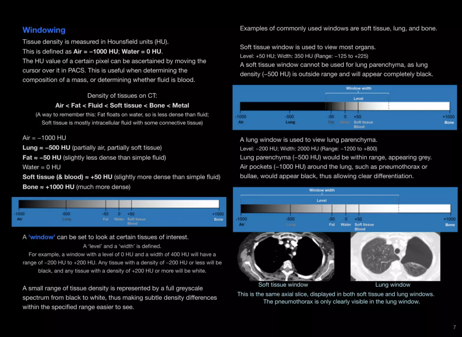
WindowingTissue density is measured in Hounsfield units (HU).This is defined as Air = −1000 HU; Water = 0 HU.The HU value of a certain pixel can be ascertained by moving the cursor over it in PACS. This is useful when determining the composition of a mass, or determining whether fluid is blood.
Density of tissues on CT: Air < Fat < Fluid < Soft tissue < Bone < Metal
(A way to remember this: Fat floats on water, so is less dense than fluid; Soft tissue is mostly intracellular fluid with some connective tissue)
Air = −1000 HULung ≈ −500 HU (partially air, partially soft tissue)Fat ≈ −50 HU (slightly less dense than simple fluid)Water = 0 HUSoft tissue (& blood) ≈ +50 HU (slightly more dense than simple fluid)Bone ≈ +1000 HU (much more dense)
A ‘window’ can be set to look at certain tissues of interest. A ‘level’ and a ‘width’ is defined.
For example, a window with a level of 0 HU and a width of 400 HU will have a range of −200 HU to +200 HU. Any tissue with a density of −200 HU or less will be
black, and any tissue with a density of +200 HU or more will be white.
A small range of tissue density is represented by a full greyscale spectrum from black to white, thus making subtle density differences within the specified range easier to see.
Examples of commonly used windows are soft tissue, lung, and bone.
Soft tissue window is used to view most organs.Level: +50 HU; Width: 350 HU (Range: −125 to +225) A soft tissue window cannot be used for lung parenchyma, as lung density (−500 HU) is outside range and will appear completely black.
A lung window is used to view lung parenchyma.Level: −200 HU; Width: 2000 HU (Range: −1200 to +800) Lung parenchyma (−500 HU) would be within range, appearing grey. Air pockets (−1000 HU) around the lung, such as pneumothorax or bullae, would appear black, thus allowing clear differentiation.
7
Soft tissue window Lung windowThis is the same axial slice, displayed in both soft tissue and lung windows. The pneumothorax is only clearly visible in the lung window.

RadiationIn the UK, radiation exposure for medical purposes (diagnostic/treatment) is regulated under Ionising Radiation (Medical Exposure) Regulations 2000 (IRMER 2000). It provides guidance for three roles:• Referrer (clinician) – provide adequate clinical details• Practitioner (radiologist) – ensure scan is justified• Operator (radiographer) – minimise amount of radiation
The ‘millisievert’ (mSv) is a unit used to measure radiation dose.The annual background radiation in the UK is 2.7 mSv.
Modality Dose Equivalent background radiation
DEXA/ Extremity X-ray 0.001 mSv Negligible
Chest X-ray 0.02 mSv 3 days
Abdo X-ray 0.7 mSv 3 months
Head CT 2 mSv 9 months
Chest CT 7 mSv 2.5 years
Abdo CT 8 mSv 3 years
Barium studies 5 mSv 2 years
Interventional procedures 15 - 70 mSv 5 - 25 years
Ref: Fred A. Mettler, Jr., et al., "Effective Doses in Radiology and Diagnostic Nuclear Medicine: A Catalog," Radiology Vol. 248, No. 1, pp. 254-263, July 2008.
Risks of radiation exposure:• Acute, high level exposure: radiotherapy treatment - affects fast dividing cells (GI tract and bone marrow) • Chronic, low level exposure: diagnostic tests - causes changes in DNA (teratogenic)
ContrastIodine-based IV contrast medium is used for most CTs.
Exceptions where contrast is usually not used:• CT KUBs (looking for renal stones)• CT heads (unless a mass lesion is suspected)• Poor renal function (eGFR <30) - non-contrast CT or alternative
modalities used
Risks of contrast administration:• Contrast-induced nephropathy - Creatinine increasing 25% within 3
days, with no other apparent reason. Usually self-limiting but can cause complications of kidney disease.
Incidence of ~2% in patients without risk factors (e.g. diabetes)• Anaphylaxis (immediate, within 1 hour)• Allergy (delayed, up to 7 days)
ContraindicationsNo absolute contraindications to CT, main concern is over contrast and radiation dose.• Patients with eGFR <60 but >30 can have contrast if they are given
prehydration (oral and IV fluids).• Pregnant women and children can have a CT if indicated (although
alternatives would be considered first).
Other uses of CT include high-resolution CT (HRCT) protocol for imaging interstitial lung disease, virtual 3D CT colonogram as an alternative to invasive colonoscopy, and CT-guided biopsies.
8

MRIPhysicsThe MRI (magnetic resonance imaging) machine generates an extremely strong magnetic field and pulses of radiofrequency energy, which align hydrogen nuclei in tissues and body water. The subsequent loss of alignment with time produces the MRI signal.
There are two main MRI sequences:T1: Water is dark - better for anatomy (soft tissue structures)T2: Water is bright - better for pathology (inflammation, oedema)
T1 - CSF is dark T2 - CSF is bright
Other commonly used types are:DWI (diffusion weighted imaging): Diffusion restriction is bright • useful for ischaemic strokes, abscesses, most tumoursFLAIR (fluid attenuated inversion recovery): Like T2, but Water is dark• useful for multiple sclerosis (periventricular lesions)STIR (soft tissue inversion recovery): Like T2, but fat is dark• useful for oedema in tissues, perianal abscesses
MRA (magnetic resonance angiography): Vessels are bright • useful for AVMs, aneurysms (can be done with or without contrast!)
ContrastGadolinium is a metal-based contrast given IV.It can rarely cause nephrogenic systemic fibrosis (similar to scleroderma) in patients with renal failure. Avoid in patients with eGFR <30.
ContraindicationsAlways go through the MRI checklist!
Metallic foreign bodies, especially from previous eye trauma, can cause serious damage if they move during the scan.If unsure of their presence, exclude with an X-ray.
Most modern implants (pacemakers, stents, joint replacements) are MRI safe, but they must always be checked to ensure compatibility.(They may, however, cause a black void artefact on the MRI image)
The MRI machine is very noisy and cramped. Some patients may not be able to tolerate it if they are claustrophobic or unable to lie still.
Monitoring leads can heat up excessively during the scan, causing burns to the patient.
Loose ferromagnetic objects can turn into projectiles if inadvertently brought into the room. (The machine is not turned off in between scans!)
9

UltrasoundPhysicsThe ultrasound probe generates inaudible high-frequency sound waves. The waves are reflected off the body structures and are detected by the probe.
Ultrasound waves are:• Reflected by Solid and Gas: Bright • Absorbed by Fluid: Dark
This reflection or absorption causes an additional effect distal to it:• Distal to a bright object: dark
acoustic shadowing• Distal to a dark object: bright
acoustic enhancement
Doppler: detects movement relative to probe, by whether reflected sound waves are compressed.• Red = moving towards probe• Blue = moving away from
probe
The blood within vessels is mobile, therefore will show up as colour on Doppler. This can be used to find the presence of vessels within an organ or tumour, or confirm lack of flow in a DVT. Doppler can also be used to measure the patent diameter of a vessel lumen. This is useful in carotid atherosclerosis. ‘Duplex’ ultrasound simply refers to the overlay of Doppler colours over the basic grey ultrasound image.
OrientationThe long axis of the probe “slices” through the body.There are two main planes in ultrasound - the transverse section (TS), and the longitudinal section (LS).In transverse section, the left side of the image is the right side of the patient.In longitudinal section (also known as sagittal section), the left side of the image is in the direction of the patient’s head.
The most common method of ultrasound is transthoracic (for heart i.e. echocardiogram) or transabdominal (for abdominal and pelvic organs) as it is less invasive, quicker and more acceptable.
In certain cases, a more invasive method may be indicated, such as transoesophageal, transrectal or transvaginal ultrasound. This is used particularly when the area of interest is within or adjacent to the wall of the lumen, for example intramural cancers and adjacent lymph nodes. Patient habitus may also necessitate the usage of these methods.
Ultrasound-guided biopsy can be performed on superficial lesions.Duplex ultrasonography helps with the insertion of central lines and the measurement of ABPI.
10

11
Check Answer
What is the correct order of tissue densities? (from least dense to most dense)
A. Air < Fat < Fluid < Soft tissue < Bone
B. Air < Fat < Soft tissue < Fluid < Bone
C. Air < Fluid < Soft tissue < Fat < Bone
D. Air < Fluid < Fat < Soft tissue < Bone
Quiz
Check Answer
What is the contrast medium used in MRI?
A. None
B. Iodine
C. Gastrografin
D. Gadolinium
Check Answer
How many times more radiation do you get from an abdominal X-ray, compared to a chest X-ray?
A. 0.3
B. 3
C. 30
D. 300
Check Answer
Is air bright or dark on ultrasound?
A. Bright
B. Dark

Anatomy
Chapter 2
Head and Spine
12
Axial CT Head

13
Sagittal T1 MRI Brain
Coronal T1 MRI Brain

Cervical SpineCount from C2
(elongated vertebral body and spine)
Thoracic SpineCount from T12
(follow the lowest rib)
Lumbar SpineCount from L5
(right above sacrum)
14
Sagittal CT Spine

PresentationsThe most common reasons for performing CT head is after head injury or a suspected stroke. There are NICE guidelines on the indications and urgency for imaging in these cases. (see page 66)
Trauma is one of the most common presentations in A&E.The main worry with head injuries is a bleed in one of several areas:• extradural, subdural, subarachnoid, intracerebral. Fractures, particularly involving the skull base, are also a concern. CT head (without contrast) is the preferred modality. Acute bleeds should be discussed with neurosurgery.There may also be an associated neck injury, in particular a cervical spine fracture, which may be imaged by either plain films or CT.
The purpose of urgent CT imaging in suspected stroke is mainly to check whether there is an intracranial bleed or an alternative cause for the symptoms such as a tumour. • If there is a bleed, the patient can be discussed with neurosurgery
for possible urgent operative management. • If there is no bleed, the patient can be thrombolysed (if indicated). Plain CT heads may look normal very early in an ischaemic stroke, as areas of ischaemia do not appear hypodense area until hours later. MRI brain, particularly the diffusion-weighted (DWI) sequence, can detect an ischaemic stroke earlier and shows it more clearly than CT.
The other major reason to perform neuroimaging is to look for a mass lesion, particularly tumours. Brain tumours are exceedingly rare, and when they occur, are much more likely to be secondary metastases than a primary lesion.
Monro-Kellie law: The total volume of brain matter + CSF + intracranial blood must be a constant. If any one component increases, the others must decrease.i.e. If there is a large haematoma or brain tumour within the skull, the CSF must be pushed out to compensate, causing effacement (compression) of ventricles. However, sometimes the pressure on the brainstem may cause blockage to CSF flow and a hydrocephalus.
Suspected spinal cord compression is investigated with MRI spine. Cauda equina syndrome is particularly concerning, as there is a risk of permanent disability if not treated urgently.
For paraspinal pathology, MRI delineates soft tissues well, which is particularly helpful in looking at collections and abscesses.
CT of the spine is used mainly to look at the bones - for example, vertebral collapse, crush fractures, or lytic lesions. MRI can also detect bony injuries well, but CT is the preferred modality in this setting.
15

16
Intracranial Haemorrhage - General PrinciplesIntracranial haemorrhage is a bleed inside the skull, i.e:• intra-axial, bleed within the brain itself (intraparenchymal, intraventricular); or• extra-axial, bleed between the brain and the skull (extradural, subdural, subarachnoid). As opposed to extracranial haemorrhage (outside the skull), i.e. soft tissue bruising/haematoma.
A lucid interval refers to an initial decrease in conscious level (GCS) due to the initial concussion from a head injury, which then improves for a period of time (usually a few hours) before deteriorating again, this time due to a gradually enlarging haematoma causing compression on brain structures.
Intracranial haemorrhages can exert mass effect on the brain. Signs include midline shift, effaced (slit-like) ventricles, and herniation of brain structures.Acute bleeds generally require urgent discussion with neurosurgery.
Appearance on CT:Acute (hours to days) - New blood is WhiteSubacute (days to weeks) - GreyChronic (weeks to months) - Old blood is Dark
Acute on Chronic - Layering effect

17
Epidural Haemorrhage Subdural HaemorrhageEpidural (also known as extradural) haemorrhage is usually due to head trauma, particularly to the region of the pterion, which tears the middle meningeal artery. This is therefore an arterial bleed.
It lies between the skull and the dura mater.
Appearance: Lens-shaped (lenticular).(as the dura is usually tightly adherent to the skull)
Subdural haemorrhage is more common in the elderly and alcoholics, who have cerebral atrophy. There is more strain on the bridging veins, which tear easily with minor trauma, and cause a venous bleed.
It lies between the dura mater and the arachnoid mater.
Appearance: Crescent-shaped.

18
Subarachnoid Haemorrhage Intraparenchymal HaemorrhageCan occur after trauma, or in individuals with arteriovenous malformations (AVMs), or berry aneurysms (particularly of the Circle of Willis). Patients describe a sudden onset of extremely severe pain (“thunderclap headache”). Blood in the subarachnoid space can cause meningeal irritation and result in symptoms similar to meningitis.
Appearance: High density blood in the sulci, basal cisterns and fissures:• suprasellar cistern (large pentagon/star shape), • quadrigeminal cistern (smaller W/smile shape below suprasellar)Blood may also extend into the ventricles.
Intracerebral haemorrhages occur within the substance of the brain itself. Common causes are hypertension, trauma and haemorrhagic stroke.
Appearance: Area of high density within the substance of the brain.
Intraventricular haemorrhage can be either primary, or secondary to extension from subarachnoid or intracerebral haemorrhage.
Appearance: Small amount of bright blood in the dependent part of the lateral ventricles. There is usually a CSF-blood level, as the denser blood sinks to the bottom.
Intraventricular Haemorrhage

19
Ischaemic Strokes / Infarcts
Ischaemic stroke on CT Ischaemic stroke on MRI - T1
Symptoms of ischaemic strokes vary depending on the blood vessel (and thus area of brain) affected. The Oxford Stroke Classification defines 4 types:• Total Anterior Circulation Stroke (TACS) - Anterior / Middle Cerebral Artery - all 3 of: higher dysfunction, hemiparesis, hemianopia• Partial Anterior Circulation Stroke (PACS) - Anterior / Middle Cerebral Artery - any 2 of: higher dysfunction, hemiparesis, hemianopia• Posterior Circulation Stroke (POCS) - Posterior Cerebral Artery - any 1 of: cerebellar symptoms, loss of consciousness, hemianopia• Lacunar Stroke (LACS) - Small Vessel Disease - any 1 of: hemiparesis, hemiparaesthesia
Appearance: CT - Low density area, that takes a few hours to develop. The ‘stroke window’ helps to see subtle difference in density. Other signs of acute infarct: Loss of differentiation of grey/white matter; Sulcal effacement (oedema); Bright MCA sign. Old infarcts have a lower density than acute infarcts.MRI - DWI is the best MRI sequence to detect stroke. On this sequence, infarct is a bright area, which can develop in just a few minutes.

20
Brain TumoursPrimary brain tumours are extremely rare. The brain is a relatively rare site for metastases compared to the liver or the lungs, but they are far more common than primary brain tumours. Cancers that most commonly metastasise to the brain include melanoma, lung, breast, renal and colon cancers. They can be asymptomatic, discovered incidentally; or they may cause symptoms due to their mass effect, such as cranial nerve palsy, seizures, or various other neurological signs.
Appearance:The appearances on CT and MRI vary but they are usually heterogenous (not a uniform texture) solid lesions. Sometimes the lesions themselves aren’t seen but the secondary vasogenic oedema or other sign of mass effect such as midline shift is seen. Tumours generally enhance with IV contrast as they are vascular. On MRI diffusion weighted imaging, they are bright due to high cellularity.The main differential for this appearance is an infective lesion such as a brain abscess.
Pre- and post-contrast CT images of a brain tumour (metastasis)

21
Small Vessel Disease Alzheimer’s diseaseAs we age, the small arterioles in the brain become more affected by arteriosclerosis. These small arterioles supply the subcortical, periventricular, and lacunar areas. These areas thus become ischaemic and low density on CT. This can lead to vascular dementia.
Appearance:Generalised low attenuation of ischaemic white matter.Frequently associated with age-related general cerebral atrophy,signs of which are enlarged ventricles and widened sulci.
Alzheimer's disease is the commonest type of dementia, causing memory impairment, loss of language skills and disorientation. This is due to the accumulation of amyloid plaques and neurofibrillary tangles. There is disproportionate atrophy of the brain, typically involving the hippocampus and the temporo-parietal cortex.
Appearance: Disproportionate hippocampal atrophy on CT or MRI.
Comparison of an Alzheimer’s patient (CT on left) vs normal (MRI on right)Note the difference in hippocampal size.

22
Venous Sinus Thrombosis Cerebral AneurysmIt is important to identify and treat these as a subarachnoid haemorrhage and cerebral haematoma from a ruptured aneurysm can cause sudden death.There are most commonly found involving the vessels of the Circle of Willis.
Appearance: Localised dilatation along one of the cerebral vessels.Possible associated findings: Aneurysm clips (metallic artefact), Old bleeds, Burr holes (defect in skull)
MRA showing an aneurysm at the bifurcation of the right middle cerebral artery.
Venous sinus thrombosis is very important but can be easily missed. It usually presents with headaches and neurological signs. Most occur in patients with prothrombotic risk factors, particularly young women. It can progress to a cerebral infarct and secondary haemorrhage.
Appearance:CT venogram - A filling defect within the contrast-filled venous sinuses (‘empty delta sign’). A high index of suspicion for this pathology is needed, as it is difficult to see on plain CT without contrast.
Patient 1: Non-contrast CT - thrombus seen as hyperdense area

23
Skull Fractures
Types of skull fracture: Linear (uncomplicated), Depressed (pushed in), Diastatic (suture widening), Basilar (base of skull)Basal skull fractures must not be missed in head trauma. Signs are bruising around the eyes (raccoon eyes) and over the mastoid process (Battle's sign), as well as CSF (i.e. clear fluid) leakage from the nose (rhinorrhoea) and ears (otorrhoea).
Appearance: It is easiest to see fractures on the bone window setting. Skull fractures may be particularly hard to spot if undisplaced, and may be mistaken for a suture, so it is important to 'follow' the line to see if it runs in a suture line. Facial fractures may bleed into the maxillary sinus, causing opacification or a fluid level.
Facial fracturesSkull fracture - Occipital fracture on plain CT, and reconstruction in 3D

24
Cervical Spine Fractures
A cervical spine X-ray is often performed to clear the cervical spine before removal of head blocks. The junction of C7 and T1 must be seen. If inadequate, cervical spine CT will be required. If a head CT is being done, the C-spine should be scanned at the same time (instead of doing X-rays).
Appearance: Cervical spine fractures are important to stabilise as they may cause paralysis. The classical ‘hangman’s fracture’ is a fracture of C2 that involves both pedicles.

25
Spinal Cord CompressionVertebral Compression Fracture
Vertebral collapse can be due to trauma in healthy bone (e.g. road traffic accidents), or secondary 'pathological fracture' in weakened bone (e.g. osteoporosis, lytic metastases). Severity is based on percentage of vertebral height loss, involvement of both the anterior and posterior parts of the bone, and impingement into the spinal canal. Treatment depends on whether the fracture is acute or chronic.
Appearance: (in acute fractures)On X-ray or CT, there is a cortical break and loss of height.On MRI, there is oedema of the bone. (bright on T2)On bone scan, there is high radiotracer uptake. (dark)
Spinal cord compression can be due to vertebral fractures, disc prolapse, or a mass lesion such as metastases growing into the spinal canal. Conus medullaris - level of L1 / L2 vertebraeCauda equina - below conus medullarisCompression of either the conus medullaris or the cauda equina can cause bladder/bowel dysfunction and saddle paraesthesia.
Appearance: Spinal cord compression may result in a bright oedematous area within the dark cord on T2 MRI. The bright CSF signal in front and behind the compressed section of cord is lost (as it has been displaced away). Look at the affected section on both sagittal and axial views to confirm.

26
Secondary Metastases to Spine
The cancers that most commonly metastasise to bone: Above diaphragm - lung, breast, thyroid; Below diaphragm - renal, prostate.Most bony metastases are lytic lesions. The exception is metastases from prostate which are sclerotic. Metastases from renal cancer are typically expansile, as well as lytic.A Technetium bone scan can be performed to determine extent of skeletal metastases. Anterior and posterior views are taken.
Appearance: CT will show lytic metastases as a hypoattenuating irregular region, which may extend into the spinal canal.Bone scan will show metastases (sclerotic) as areas of increased tracer uptake, known as 'hot spots' and are black on the scan. Lytic metastases and multiple myeloma are not well detected on bone scans. The tracer is excreted by kidneys, and so the kidneys and bladder will also show up as black areas. If the metastases are so extensive and the tracer uptake so high that there is little left to be excreted by the kidneys and the kidneys are not dark, this is called a 'super scan'. Inflammation such as that due to arthritis may also show up as hot spots. Paget’s disease will also cause increased uptake in affected bones.
Bone scan showing extensive skeletal metastases

27
Multiple Sclerosis
This is a chronic demyelinating disease of the central nervous system (brain and spinal cord) which usually affects young females. There is no definitive diagnostic test for MS. The diagnosis may be made when two distinct neurological episodes with no other cause occur. MRI and CSF analysis are the most useful tests.
Appearance: Bright plaques on MRI (T2 or FLAIR sequences). FLAIR is a modified T2 where fluid (CSF) signal is suppressed, so bright plaques are more obvious next to the ventricles (which are now dark).Classically periventricular with a finger-like appearance in sagittal section. Plaques can also be found in the cerebellum, brainstem, and spinal cord. If IV contrast is used and the plaques enhance, this indicates current disease activity.

28
Quiz
Check Answer
Type of haemorrhage?
A. Epidural (acute)
B. Epidural (chronic)
C. Subdural (acute)
D. Subdural (chronic)
E. Subarachnoid
Check Answer
What is the main pathology?
A. Normal scan
B. Infarct (new)
C. Infarct (old)
D. Subdural haemorrhage (chronic)
E. Intraventricular haemorrhage

29Check Answer
What is the main pathology?
A. Lytic metastasis
B. Sclerotic metastasis
C. Spinal cord compression
D. Crush fracture
E. Osteomyelitis
Check Answer
What is the main pathology?
A. Intraventricular haemorrhage
B. Small vessel disease
C. Hydrocephalus

30Check Answer
Drag the labels to the corresponding anatomical structures in the brain.
Medulla
Medulla
Pons
Pons
Occipital Lobe
Occipital Lobe
Midbrain
Midbrain
Thalamus
Thalamus
Parietal Lobe
Parietal Lobe

Anatomy
Chapter 3
Chest
31
Coronal & Axial CT Thorax

PresentationFor chest pain, we must first determine the likely source of the pain from history and examination. Possible sources of pain: Cardiovascular (heart, vessels), Respiratory (lungs), Gastrointestinal (oesophagus, stomach), Musculoskeletal (ribs, muscles).
Gastrointestinal and musculoskeletal chest pain (including suspected rib fractures) generally do not require imaging.
Chest pain from a respiratory source (e.g. pulmonary embolism, pneumonia) is usually pleuritic in nature (sharp, worse on inspiration).Chest X-rays are the first line investigation for diagnosis in most cases. Exceptions are a suspected tension pneumothorax (which must be treated immediately before doing any investigations), and suspected pulmonary embolism (investigated with CTPA or VQ scan as first line).
Chest pain from a vascular source (e,g. aortic dissection) is rare, but extremely important not to miss. It may be described as tearing pain.Thoracic trauma and connective tissue disorders predispose to this.If the patient is stable enough, they may be investigated with CT.A widened mediastinum on CXR is a less reliable sign.
Cough and breathlessness can have a respiratory or cardiac cause. Dry cough, fine crackles - pulmonary fibrosisProductive cough, coarse crackles - pneumonia or heart failure
Heart failure is not a radiological diagnosis, i.e. we do not do CXRs to diagnose heart failure. However, the signs of heart failure can be seen on CXR.
Pneumonias can be seen on CXR as inflammatory changes or frank consolidation, there is seldom any need to do CT for this unless an atypical pneumonia is suspected.
HRCT (high resolution CT) thorax is used to investigate interstitial lung disease and pulmonary fibrosis.
Haemoptysis, especially in a smoker, is concerning for lung cancer and is investigated initially with a CXR. If the CXR is clear but the symptoms persist, a CT thorax may be indicated.Infections (e.g. TB) and chronic coughing may also cause haemoptysis.
32

33
Figure Radiology Basics: Cross-sectional Imaging.1 Lorem Ipsum dolor amet, consectetur
Pneumonia Pleural EffusionTypical: Streptococcus pneumoniae; Haemophilus influenzaeAtypical: Mycoplasma; Legionella; Pneumocystis (‘PCP’); Viral; Fungal
CXR at 6 weeks post-treatment to ensure full resolution.(and exclude possible underlying lung mass)
Appearance:Typical: Airspace opacity with air bronchograms (usually one lobe) Atypical: Variable; ground glass opacity, nodules with ground glass “halo”
Transudate: Imbalance of hydrostatic and oncotic forces. Low protein. Commonly caused by organ failures (heart, liver, renal, thyroid failures).Exudate: Local pathology (infective, inflammatory, malignant). High protein.
A pleural tap (thoracocentesis) to obtain a sample of fluid for laboratory testing can be performed easily under ultrasound guidance.
Appearance: Fluid density at the dependent part of the hemithorax. i.e. at the bases in erect CXR; posteriorly in supine CT. Causes collapse of adjacent lung which appears denser (white).

34
Primary Lung CancerSmall cell (10%): Located centrally. Poorer prognosis. Associated paraneoplastic syndromes: ACTH; ADH; Lambert-Eaton myasthenic syndrome (LEMS).Non-small cell (90%): Adenocarcinoma is located peripherally. Most common type of lung cancer. Squamous cell carcinoma is located centrally. Associated paraneoplastic syndromes: PTH-like peptide (Hypercalcaemia - bones, stones, groans, moans); Hypertrophic pulmonary osteoarthropathy (HPOA) - expansion of long bones, DIP inflammation, finger clubbing.
Routes of spread:Direct - Bronchus, Chest wall, Aorta, OesophagusLymphatic - Hilar, Mediastinal lymph nodesHaematogenous - Bone, Brain, Liver, AdrenalTranscoelomic - Malignant pleural effusion
Appearance: Discrete mass of soft tissue density. Possibly associated with small lung nodules, mediastinal & hilar lymphadenopathy, or pleural effusions.Lymphadenopathy is enlargement of lymph node due to pathology. Definition of enlargement: >1 cm in the short axis (i.e. width, not length)

35
Secondary Metastases to Lung
The lungs are a common site for metastases, in particular from breast, bowel and renal primaries.
Appearance: Soft tissue nodules, usually multiple, located anywhere within the lung fields, but classically more in lower zones. Can measure anywhere from a few mm to >1 cm in diameter. Large “cannonball” metastases classically originate from a renal cell carcinoma primary. Metastatic nodules can occasionally be cavitating, particularly with squamous cell lesions, although if cavitation is seen, infective causes must be considered.
To differentiate small nodules from pulmonary vessels and lung parenchyma, ‘follow’ it by scrolling up and down a few slices. Nodules should appear and disappear within one or several slices, while vessels should continue for some distance and branch out.
The same method is used to differentiate lymph nodes from vessels, e.g. in the axillary region.

36
LymphomaMalignancy of B or T lymphocytes, with solid lymphoid tumours. It can involve nodal and extranodal sites. Subtypes include Hodgkin and Non-Hodgkin.
Staging is performed with a CT scan or a PET scan, using the Ann-Arbor classification.I: single node group; II: >1 node groups, same side of diaphragm; III: >1 node groups, both sides of diaphragm; IV: extranodal disease (liver, marrow). The letter 'B' is added to the stage (e.g. Stage IVB) if there are 'B symptoms' (fever, weight loss, night sweats). The absence of ‘B’ symptoms = ‘A’.
Causes of hilar lymphadenopathy on CXR: Lymphoma, TB, Sarcoidosis. (Note: Both Lymphoma and TB can cause the ‘B symptoms’!)
Appearance: Widespread lymphadenopathy (soft tissue density), involving one or more groups of lymph nodes,
In the video below, you can see a massive number of enlarged lymph nodes in the cervical and axillary areas.Can you spot any mediastinal lymph nodes?

37
Bronchiectasis Pulmonary FibrosisPulmonary fibrosis is caused predominantly by interstitial lung disease.
Extrinsic (occupational) and Intrinsic (autoimmune) causes result in different typical patterns of fibrosis, where the lung apex or base is more affected.Apex > Base: Occupational causes (Exception: Asbestosis)
Base > Apex: Autoimmune causes (Exception: Ankylosing spondylitis)
Appearance: Reticular shadowing; Honeycombing; Traction bronchiectasis.These are seen best on high-resolution CT but may also be seen on CXR.
Bronchiectasis is fixed dilation of part of the bronchial tree. Causes:• Congenital (cystic fibrosis, primary ciliary dyskinesia i.e. Kartagener syn)• Acquired (pneumonia, allergic bronchopulmonary aspergillosis).
Appearance: There is dilatation of bronchi, with or without thickening of bronchial walls and mucus plugging. ‘Tram-track sign’ can be seen on CT and CXR. The ‘Tree-in-bud sign’ and the ‘Signet ring sign’ can be seen on CT.

38
Emphysema PneumothoraxEmphysema is one of the two main entities of chronic obstructive pulmonary disease (COPD), the other being chronic bronchitis. • Smoking causes a centriacinar (peribronchiolar) pattern• Alpha-1-antitrypsin deficiency causes a panacinar (diffuse) pattern
There is destruction of alveolar walls, resulting in enlargement of airspaces. These become confluent and eventually form bullae. Bullae can rupture, leading to pneumothorax and pneumomediastinum.
CXR findings include hyperinflation, flat diaphragms, and bullae.
Appearance: Changes of confluent alveoli (hypodense areas of lung parenchyma) and bullae (small pockets of air) are typical.
Pneumothorax can be primary or secondary to underlying lung pathology. Primary: tall, thin, young males who smokeSecondary: asthma, emphysema, fibrosis, Marfan’s, Ehlers-Danlos, cancerOther: penetrating injury, blunt injury, rib fractures, biopsies, line insertions
It is usually diagnosed on CXR, but may also be diagnosed on CT if unsuspected previously and a CT was performed to exclude other causes of chest pain.
Appearance: A pocket or rim of air located outside the lung and adjacent to the chest wall, most commonly in the apices. Associated lung collapse. Only visible on lung window.

39
Pulmonary EmbolismRef: http://www.nice.org.uk/nicemedia/live/13767/59714/59714.pdf
Clear guidelines exist regarding the use of imaging in the investigation of suspected pulmonary embolism.
In pregnant women, D-dimers are unreliable and Wells’ score is not validated. Consider performing duplex ultrasound for DVT, and CXR to exclude other causes. V/Q scan (or perfusion-only scan) is generally favoured to minimise radiation exposure.The main concern with CTPA in pregnancy is the radiation to the mother’s breasts (as the fetus can be shielded), and the iodinedose to the foetus (needs thyroid screen when born).
Appearance: Clots are seen as ‘filling defects’. Contrast cannot fill areas occupied by clots, therefore the contrast (bright) surrounds the clots (darker).On V/Q scan, ventilation (inhaled tracer) is normal but perfusion (IV tracer) is abnormal, indicating a problem with the blood supply to a particular area.
The images below show the emboli highlighted in red. The first two images show multiple bilateral emboli, while the third image shows a saddle embolus.‘Saddle’ embolus sits in the bifurcation of the pulmonary trunk where it divides into the left and right main pulmonary arteries.
Algorithm 2 Diagnosis of PE
PE unlikely (≤ 4 points)
Two-level PE Wells score
Patient with signs or symptoms of PE
D-dimer test
No Yes
PE likely (> 4 points)
Diagnose PE and treat
No
Yes
Is deep vein thrombosis suspected?
Is CTPA* suitable** and available immediately?
Advise the patient it is not likely they have PE. Discuss with them the signs and symptoms of PE, and when and where to seek further medical help. Take into consideration alternative diagnoses. Advise the patient it is not likely they have
PE. Discuss with them the signs and symptoms of PE, and when and where to seek further medical help. Take into consideration alternative diagnoses.
Was the CTPA (or V/Q SPECT or planar scan) positive?
Yes No
*Computed tomography pulmonary angiogram **For patients who have an allergy to contrast media, or who have renal impairment, or whose risk from irradiation is high, assess the suitability of V/Q SPECT† or, if not available, V/Q planar scan, as an alternative to CTPA. †Ventilation/perfusion single photon emission computed tomography
Was the D-dimer test positive?
Was the CTPA (or V/Q SPECT or planar scan) positive?
Yes
No
Immediate interim parenteral anticoagulant therapy
Consider a proximal leg vein ultrasound scan. See Diagnosis of deep vein thrombosis
No
Immediate interim parenteral anticoagulant therapy
Yes
Is CTPA* suitable** and available immediately?
No Yes
Offer CTPA (or V/Q SPECT or planar scan)
Offer CTPA (or V/Q SPECT or planar scan) CTPA (or V/Q SPECT or
planar scan)
CTPA (or V/Q SPECT or planar scan)
Other causes excluded by assessment of general medical history, physical examination and chest X-ray
PE suspected
Tap image to view NICE guidelines on investigation of pulmonary embolism
based on Wells’ score

40
Aortic Dissection
aortic arch
Aortic dissection occurs when blood enters the aortic wall through a tear in the tunica intima, ‘dissecting’ a path (false lumen) between intima and media.If the dissection extends into smaller arteries (e.g. coronary, carotid, subclavian, mesenteric), it can cause ischaemia of the supplied area (i.e. MI, stroke, limb ischaemia, bowel ischaemia). Pericardial tamponade can also occur. The main risk factor is hypertension.
Classification systems are Stanford and DeBakey, both based on involvement of the ascending aorta (poorer prognosis).CXR may show widened mediastinum. CT chest (pre and post contrast, arterial phase) is the ideal investigation, to determine presence of aortic intramural haematoma, true lumen and extent of dissection.
Appearance: There is a true lumen and a false lumen, separated by an intimal flap (tunica intima which has been detached from tunica media). The false lumen is usually larger (higher pressure) and hypodense (darker as contrast delayed) compared to the true lumen, although this is not reliable. To differentiate them with more certainty, a normal part of the aorta should be found and followed as this will join into the true lumen.
The video below shows a dissection involving the ascending aorta (anterior), the aortic arch, and the descending aorta (posterior).Here the false lumen is brighter than the true lumen, which is not typical.

41Check Answer
Diagnosis?
A. Normal
B. Pleural effusion
C. Pneumonia
D. Lung collapse
E. Bronchiectasis
Check Answer
Diagnosis?
A. Normal
B. Pleural effusion
C. Pneumonia
D. Lung collapse
E. Bronchiectasis
Quiz

42Check Answer
Diagnosis?
A. Normal
B. COPD
C. Pulmonary fibrosis
D. Bronchiectasis
E. Pneumonia
Check Answer
Diagnosis?
A. Normal
B. COPD
C. Pulmonary fibrosis
D. Bronchiectasis
E. Pneumonia

43Check Answer
Diagnosis?
A. Normal
B. Pneumonia
C. Pulmonary embolism
D. Metastases to lung
E. Aortic dissection
Check Answer
Diagnosis?
A. Normal
B. Pneumonia
C. Pulmonary embolism
D. Metastases to lung
E. Aortic dissection

Anatomy
Chapter 4
Abdomen
44
Coronal & Axial CT Abdomen

45
Transabdominal Ultrasound Views
Coronal (Right Flank) Coronal (Left Flank) Transverse (Suprapubic)

PresentationCT is the most useful imaging modality in acute abdomen. The vast majority of intra-abdominal pathologies can be seen on CT.However, this involves a radiation dose, so care should be taken in young patients.
Abdominal X-rays are useful in diagnosing bowel obstruction, and erect chest x-rays can indicate perforation of viscus.
Ultrasound is most useful to detect gallstones, cholecystitis, and hydronephrosis. It can also detect pathology of the pancreas and appendicitis, although these areas are frequently poorly visualised due to overlying bowel gas. As it does not involve radiation, it is frequently used first line in children and young adults.
Suspected GI cancers are most commonly investigated with endoscopy. If a patient is unfit for colonoscopy, a CT ‘virtual’ colonoscopy can be done, with a 3D reconstruction of the intraluminal view. The disadvantage is that biopsies cannot be done.
Contrast studies (swallow/meal/follow-through/enema) are also frequently performed with suspected obstructing lesions. Barium is usually used, but if there is possible perforation, Gastrografin (water soluble) is used instead. Barium is highly irritant and will cause inflammation and fibrosis if it leaks out. A tumour in the wall will cause a ‘filling defect’ where it displaces the contrast. A circumferential wall lesion is seen as an ‘apple core lesion’.
Once cancer is confirmed, a staging CT (i.e chest/abdo/pelvis with contrast) is performed to inform treatment options and prognosis.
The best modality to detect renal stones, particularly in first presentation of renal colic, is CT KUB (kidneys ureters bladder). X-ray KUB may not detect small stones. Also, phleboliths (calcification within veins) are frequently found in the pelvis and are difficult to differentiate from renal stones on X-ray. CT KUB can show whether or not these are located within the renal tract.
Painful jaundice is investigated with USS to look for obstructing stones in the gallbladder and bile ducts. If bile duct dilatation is seen, but stones are not seen, the next step is MRCP (magnetic resonance cholangiopancreatography) which is better at detecting ductal stones.Painless jaundice is more concerning and would warrant an urgent CT with contrast for pancreatic cancer.
Gynaecological disorders are usually imaged with ultrasound as first line. This avoids radiation exposure, and the structures are viewed better than on CT. Transvaginal ultrasound is generally better than transabdominal ultrasound. Although it is more invasive, it provides better image quality, and is not affected by patient habitus.
Testicular disorders are also generally imaged with ultrasound, which can easily differentiate between a cystic lesion and a solid lesion.Pain from retroperitoneal structures (pancreas, kidneys, duodenum)tend to radiate to the back.
CT as well as ultrasound can be used by interventional radiologists to perform guided biopsies. The choice between CT and ultrasound is generally made by considering the proximity of the lesion of interest to the body surface, and whether there are any structures in the way. Preferred spots to take biopsies are immobile, peripheral structures such as lymph nodes, peritoneal lesions, and omental ‘cake’.
46

47
Secondary Metastases to Liver Liver AbscessGI cancers commonly metastasize to the liver, as venous blood returning from the bowel filters through the hepatic portal system first before rejoining the general circulation. Depending on the number and location, it may be possible to resect the affected segment(s) of liver.
Main differentials are simple liver cysts, benign lesions such as haemangioma and liver abscesses. To differentiate between them, the HU value may be helpful. Clinical features (such as fever) are also very useful to know.
Appearance: Irregular heterogenous areas of low attenuation.
Bacterial: Polymicrobial. e.g E. coli, Klebsiella, Streptococci, EnterococciFungal: Candida sp. Other: Amoebic (Entamoeba histolytica), Hydatid cysts
Appearance: Bacterial and fungal abscesses usually appear as multiple clustered lesions. Causative features such as biliary obstruction or diverticulitis or appendicitis may also be seen. Amoebic abscess usually appears as a solitary large lesion.

48
Gallstones CholecystitisUltrasound is the gold standard investigation. CT may miss some stones. Plain X-ray only shows 10% of stones. MRCP is useful for ductal stones.Locations: Gallbladder, Bile ducts, Small bowel (rare).
Appearance: On ultrasound, single or multiple hyperechoic objects in the gallbladder, with acoustic shadowing. On CT, stones may be hyperattenuating (calcified stones), isoattenuating (mixed), or hypoattenuating (cholesterol stones) with regards to bile. Isoattenuating stones can be missed. On MRI, stones are an area of low signal (dark).
Commonly due to gallstone disease.Biliary colic: Right upper quadrant painCholecystitis: Right upper quadrant pain + FeverAscending cholangitis: Right upper quadrant pain + Fever + Jaundice (Charcot’s triad)
Appearance: • Distended fluid-filled gallbladder• Thick enhancing (bright) gallbladder wall• Fluid surrounding gallbladder• ‘Fat stranding’ surrounding gallbladder (fluid density due to oedema in fat)

49
Pancreatic Cancer PancreatitisMost commonly arises from the head of pancreas. Very poor prognosis due to late diagnosis.
Appearance: Soft tissue mass which may cause obstruction and dilatation of the bile and pancreatic duct systems (“double-duct sign”). Mass may invade into surrounding structures (duodenum, vessels).
Release of pancreatic enzymes causes autodigestion of pancreatic tissue, pancreatic ducts, and the surrounding tissues (fat and blood vessels). Complications include peripancreatic fluid collections (if encapsulated, these are called pseudocysts); abscesses; necrosis of pancreatic tissue; thrombosis of splenic/portal veins; and pseudoaneurysms +/- haemorrhage.
Appearance: • Enlarged, oedematous pancreas• Fuzzy pancreas borders• Fluid around pancreas• ‘Fat stranding’ in retroperitoneum (fluid density due to oedema in fat)• Areas of non-enhancement indicate necrosis

50
Simple Renal Cysts Renal Cell CarcinomaSmall RCCs often diagnosed incidentally on CT for other reasons, or during investigations for haematuria.Originates from renal tubular epithelium (not transitional cell!).Risk factors are smoking and obesity. Not associated with simple cysts.
CT is the best first-line investigation. MRI may help with staging.
Appearance: Poorly-defined enhancing mass, extension into renal vein/IVC. Enlarged retroperitoneal lymph nodes.Metastases to bone are classically lytic and expansile.Metastases to lung are classically ‘cannonball’ (large and round).
Extremely common finding, increasing with age. Up to 50% in age >50. Essentially normal. Not premalignant. Cysts are ‘complex’ (i.e. not simple) if they have septa or contain high attenuation areas (soft tissue/blood/protein). Complex cysts can be associated with malignancy.
Appearance: Well-defined, round, fluid-containing lesions around the periphery of the kidney (“cortical”) or centrally (“parapelvic”). May be solitary or multiple. Cyst size ranges from a few mm to a few cm.

51
Renal Stones HydronephrosisCaused by a distal obstruction, e.g. stones, cancers, prostatic hypertrophy, pregnancy, congenital, large blood clot, retroperitoneal fibrosis; or backflow e.g. vesicoureteric reflux. The affected kidney frequently has impaired function. Further radiological tests to assess function include delayed phase contrast CT, intravenous pyelogram, or nuclear medicine tests.
Appearance: Enlarged renal calyces and renal pelvis. Look for visible cause of obstruction.
99% of stones are visible on CT KUB (done without contrast). CT shows the exact location of calcifications to see whether or not they are located within the renal tract. Any associated obstructing effect causing hydroureter or hydronephrosis can be seen. Inflammation around the ureter may also be seen which could indicate a recently passed stone.
‘Follow’ the ureter from where it leaves the renal pelvis, down along the psoas muscle, anterior to the sacroiliac joint, along the pelvic sidewall, until the ischial spine where it turns medially to reach the bladder.
Appearance: Very small (2-10mm) hyperdense object within the renal calyces, renal pelvis, or within the course of the ureters. Possible associated hydronephrosis and hydroureter.
In the video below, the path of the left ureter is outlined with a yellow arrow.The two renal stones are indicated with red arrows.
CT with contrast - hydronephrosis and hydroureter of left kidney

52
Small Bowel Obstruction Large Bowel ObstructionCauses of small bowel obstruction include adhesions, herniae and gallstone ileus. The ‘transition point’ is the point at which proximal bowel is dilated, and distal bowel is collapsed. This usually indicates site of obstruction.
Small bowel ileus occurs due to metabolic derangements or post-operatively, with dilatation but no transition point.
Appearance: Dilated small bowel loops proximal to a transition point; Multiple air-fluid levels; Possible ischaemic bowel (a complication)
Common causes of large bowel obstruction are cancers, diverticulosis and volvulus. An obstruction of the large bowel causes dilatation of proximal large bowel. However, the small bowel may or may not be dilated as well, depending on whether the ileocaecal valve is competent. It is competent in ~70% of people. A competent valve traps contents in the large bowel, so the small bowel is not dilated. However, this causes the large bowel to dilate much more and much quicker, putting it at a higher risk of perforation.
Appearance: Dilated large bowel loops proximal to a transition point; Multiple air-fluid levels; Dilated small bowel loops (if valve incompetent)
The 3-6-9 ruleDilated Small bowel: > 3 cmDilated Large bowel: > 6 cm
Dilated Caecum: > 9 cm

53
Small Bowel Inflammation Large Bowel InflammationUlcerative colitis affects a continuous section of large bowel, most commonly the rectum. Infective causes, such as Clostridium difficile infection, are also common. If the entire large bowel is affected, it is called a pancolitis. Both may cause toxic megacolon (seen as marked colonic dilatation).
Appearance: “Target sign” involving the large bowel (on CT)• Enhancement (bright) of serosa and mucosa due to more blood flow. • Low attenuation (dark), thickened wall due to wall oedema.• Ascites also seen with C Difficile colitis
CT with contrast
Most common cause is Crohn's disease, which affects patchy areas along the entire gut, most commonly the terminal ileum. Common complications are perforation (free air outside the bowel), abscesses, fistulae to other structures and obstructing strictures causing bowel dilatation.
Appearance: “Target sign” involving the small bowel (on CT)• Enhancement (bright) of serosa and mucosa due to more blood flow. • Low attenuation (dark) wall: oedema.

54
Appendicitis Diverticular DiseaseDiverticulitis typically occurs in those over 40, on a background of diverticulosis. It may be referred to as 'right-sided appendicitis' as it can present very similarly (classically periumbilical pain initially, later localising to the left iliac fossa). ’Diverticular disease' can refer to either diverticulosis or its complications.
Appearance: Multiple small outpouchings, particularly affecting the sigmoid colon. If these are inflamed (diverticulitis), there will be fat stranding, with possible localised perforation or collections.
Appendicitis can present at any age. In children, mesenteric adenitis is a common and harmless differential that should be considered. In older people, an underlying caecal carcinoma needs to be considered. The diagnosis can be made on US although the appendix is frequently not visible due to being obscured by overlying bowel gas.
Appearance: • Dilated, fluid-filled appendix• ‘Fat stranding’ around appendix• Possible associated soft tissue mass (lymphoid tissue, cancer); or hyperdense faecolith at base of appendix.

55
Ischaemic BowelBowel ischaemia may be caused by arterial insufficiency (thrombosis or embolism), venous obstruction, or as a result of bowel obstruction.
Appearance: Variety of appearances including hypo- or hyper-density of the bowel wall, bowel distension and congestion of the mesentery. Arterial filling defect.
Infarcted BowelInfarcted and gangrenous tissues will start to produce gas. Therefore, severely infarcted or necrotic bowel may have gas within its walls. The portal venous system drains the bowel walls. As a result, the portal vein fills with gas that drains into the liver. The liver shows a widespread branching pattern of portal venous gas, which is a premorbid sign. (This is different from pneumobilia, which looks similar but the air is actually within the intrahepatic biliary ducts, seen when there is a fistula between bowel and gallbladder)
Appearance: Air within bowel walls; Air in hepatic portal venous system

56
Traumatic InjuryTrauma can be either blunt force or sharp penetrating trauma. Common mechanisms include high speed road traffic accidents, falls from height and assault. Both blunt and penetrating trauma types can cause solid organ, bowel and mesenteric injuries. In high energy trauma, a whole body (head/neck/chest/abdo/pelvis) trauma series CT with two phases of contrast enhancement is usually done. Injuries to the liver, kidneys and spleen are associated with rib injuries. There may be a laceration with associated haematoma, with extension to involve the vessels at the hilum of the organ. There may also be active bleeding which would require emergent surgery or interventional radiology input to embolise the bleeding vessel.
Appearance: Disruption in continuity of the organ cortex, associated soft tissue injury, haematoma, contrast extravasation indicating active bleeding.

57
Air in Peritoneum Air in BladderCauses of air within bladder:• Catheterisation - recent or current• Fistula - between bladder and bowel
A fistula would allow infecting organisms to easily colonise the bladder leading to frequent UTIs and pyelonephritis.
Appearance: Air within the bladder, forming an air-fluid level with the urine inside the bladder.
Causes of air within intra- or retroperitoneal spaces:• Perforation - of hollow viscus (usually duodenum or sigmoid)• Recent laparoscopic surgery - from CO2 insufflation
Appearance: Free air within the peritoneum. Location of the air depends on the segment of perforated bowel i.e. intra- or retro-peritoneal. Air can extend into other spaces e.g. pneumomediastinum. Perforation of the rectum - air in mesorectum (mesentery surrounding rectum).

58
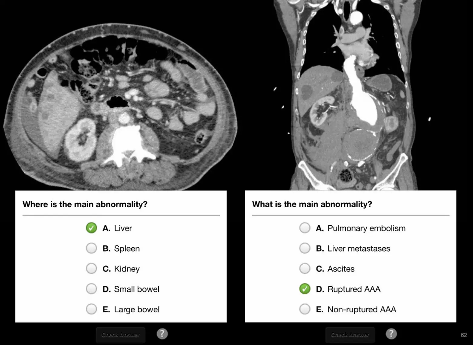
Abdominal Aortic AneurysmFocal dilatation of the abdominal aorta. Two-thirds of patients with ruptured AAAs die before reaching hospital. Of those who make it to emergency surgery, half die. Ultrasound used for diagnosis and screening. CT used for suspected leak, but this must not delay emergency surgery.
Screening for AAA is offered to all men when they turn 65. It is a quick ultrasound scan to measure the abdominal aorta, done in the community. If the test is negative (< 3 cm), there will be no further recall scans, unlike other screening programmes.
If the test is positive (> 3 cm), further action depends on the diameter of the aneurysm.3.0 - 4.4 cm = repeat in 1 year; 4.5 - 5.4 cm = repeat in 3 months; > 5.5 cm = immediate referral to vascular surgeons to consider elective repair.
Appearance: Aneurysm wall usually calcified. If ruptured, free blood (higher density than fluid e.g. in stomach/GB) with possible contrast leak.Mural thrombus common, seen as circumferential low density area.

59
Peritoneal and Omental DiseaseSoft tissue nodularity and thickening of the omentum and peritoneum can be seen in either metastatic disease (usually from gastric, pancreatic, colonic or ovarian carcinomas) or from infections such as tuberculosis. There may also be nodularity on the serosal surface of the bowel, and in metastatic disease in particular, the bowel may become encased in widespread malignant tissue. Ultrasound or CT guidance can be used to biopsy the omentum to identify the primary source.
Appearance: Nodular thickening of omentum deep to abdominal wall (“cake”) and peritoneal nodularity. Necrotic lymph nodes are a feature of TB.
GynaecologyThe preferred modality for initial diagnosis of a suspected gynaecological pathology is ultrasound (transabdominal or transvaginal).
Uterine fibroids can be very large and cause pain if they degenerate. Endometriosis may present as ovarian masses, partially cystic, or as deposits anywhere within the abdominal cavity.
Gynaecologic malignancies are staged with MRI & CT. Ovarian cancer tends to present late, frequently with spread to the peritoneum and omentum by diagnosis. A ‘Krukenberg tumour’ refers to a metastatic deposit in the ovary which originated from a primary elsewhere within the peritoneal cavity.
Appearance: Uterine fibroids can calcify and are seen as rounded masses with calcific rims. MRI can show the blood within endometriotic deposits on ovaries and the rectovaginal space.

60
AscitesAscites is seen in chronic liver disease, heart failure and abdominopelvic malignancies. It presents as abdominal distension.
It can be seen easily on ultrasound and CT. Ultrasound guided paracentesis/drainage can be carried out. A sample of ascitic fluid can be obtained for laboratory testing.
Appearance: Depending on the quantity, it is usually seen as fluid around the liver and spleen, surrounding the bowel, and in the pelvic cavity. It travels to dependent locations, i.e. the left and right paracolic gutters (folds of peritoneum) posteriorly.

61Check Answer
Diagnosis?
A. Hydronephrosis
B. Hydroureter
C. Renal abscess
D. Renal cell carcinoma
E. Renal cyst
Quiz
Check Answer
Diagnosis?
A. Normal
B. Ascites
C. Small bowel ischaemia
D. Small bowel obstruction
E. Small bowel inflammation
62Check Answer
Where is the main abnormality?
A. Liver
B. Spleen
C. Kidney
D. Small bowel
E. Large bowel
Check Answer
What is the main abnormality?
A. Pulmonary embolism
B. Liver metastases
C. Ascites
D. Ruptured AAA
E. Non-ruptured AAA

63Check Answer
Where is the bright object located?
A. Gallbladder
B. Stomach
C. Pancreas
D. Small bowel
E. Large bowel
Check Answer
Where is the bright object located?
A. Gallbladder
B. Stomach
C. Pancreas
D. Small bowel
E. Large bowel

64
How many abnormalities do you see?
(Check below for answer)
Check Answer
Is this a normal scan?
A. Normal scan
B. Abnormal scan

One of the responsibilities of a junior doctor on the wards is requesting appropriate imaging for patients, for diagnostic or monitoring purposes.It is an important skill to properly request imaging, to optimise efficiency and avoid undue delay.
Components of a request form• Patient details• Clinical details - signs and symptoms? previous imaging results? blood tests? • Clinical questions to be answered - specific finding to look for? provisional and differential diagnosis?• Investigation requested - modality?• Mode of transport - are there mobility problems?• Background medical information - Medical (renal disease, cardiac disease, diabetes, metformin, allergies), Contraindications (pregnancy, metal)• Referrer (requesting clinician) details
Chapter 5
Reques9ng Imaging
65
Generic Request Form Female of Childbearing Age Consent MRI Checklist
Tap images to enlarge
Providing clinical details
Written requests (include only relevant headings)• Scenario (PMH) e.g. Laparotomy 3/7 ago for bowel cancer.• Presenting Complaint e.g. Increasing SOB and cough.• On Examination e.g. RR 20, HR 100, Sats 90%,• Tests Done e.g. ABG normal.• Indication e.g. Wells Score 6.• Query e,g. ?PE ?Pneumonia• Request e.g. CTPA please
Verbal requests• Introduce your name, role and team e.g. Hi, I’m X, one of the junior doctors from obstetrics.• Patient details e.g. I would like to discuss a 30-year-old lady who is 32 weeks pregnant.• Query e.g. We suspect that she might have a PE.• History e.g. She was admitted yesterday with preeclampsia. Today she complained of shortness of breath and pleuritic chest pain. There are no signs of leg swelling but she has a previous history of unprovoked VTE.• Request e.g. We think that she probably needs a leg Doppler. Do you think this is the way to go? or We are not sure what the most suitable imaging in her case is. What would you recommend?• Hospital number
TipsKnow your patient• clinical condition? • urgency?• previous imaging?
Know why you are requesting the investigation • might it change management?
Give a structured, concise history focusing on • relevant background (chronic conditions, known diagnoses)• current admission• main signs and symptoms • investigations already performed• provisional diagnosis
Have information readily available• patient’s NHS/hospital number (for the radiologist to look at previous imaging)• relevant investigation results such as eGFR (ensure renal function adequate if contrast needed)
Discuss• Ask for advice if unsure about appropriateness of imaging, or
unsure about which modality to use
DO NOT• ‘order’ an investigation - it is a ‘request’• give the reason as ‘my consultant requested it’• ‘shop around’ for different radiologists
66

iRefer guidelines from the Royal College of Radiologists iRefer is one of the best evidence-based resources available on imaging guidelines. (a ‘NICE’ for radiology)It has been around for over 20 years and is targeted at GPs and other non-specialist medical practitioners.
Clear and concise recommendations are provided for most clinical presentations and conditions.
It can be accessed directly from any NHS computer at http://nww.irefer.nhs.uk/.To access the site from non-NHS computers, junior doctors can register for a free e-LfH account at http://portal.e-lfh.org.uk/Registration.
A ‘Launch iRefer’ button should then be available below the left hand menu.
67

Guidelines for Common Clinical Situations (Tap images to enlarge)
CT head for head injuries CT cervical spine for head injuries Wells score for DVT Imaging in acute stroke
CT head within 1 hour (urgent) in adults:• GCS <13 initially, or GCS <15 two hours after injury• Suspected skull fracture • Post-traumatic seizure• 2+ episodes of vomiting• Focal neurological deficit• Amnesia/Loss of consciousness + Coagulopathy
CT head within 8 hours in adults:• Retrograde amnesia >30 minutes (forgetting events before injury)• Amnesia/Loss of consciousness + Age ≥65 • Amnesia/Loss of consciousness + Dangerous mechanism of injury
68
Ref: NICE guidance CG176 Ref: NICE guidance CG176 Ref: NICE guidance CG144 Ref: ISWP National Clinical Guidance 4th ed
Ref: NICE guidance CG144 Ref: ISWP National Clinical Guidance 4th ed
ABCD2 score Age • >60 - 1 point Blood pressure • >140 sys or >90 diaClinical features • Unilateral weakness• Speech disturbance, no weaknessDuration of symptoms• >60 mins• 10-59 minsDiabetes• Yes
Points 1
1
21
21
1
Wells score for PE Imaging in TIA
CT vs X-ray of C-spine in unconscious
patient with head injuryRef: RCR iRefer guidelines
CTPA vs V/Q scan in suspected
pulmonary embolismRef: RCR iRefer guidelines
Imaging in suspected renal stones
without colicRef: RCR iRefer guidelines
Imaging in suspected renal stones
with colicRef: RCR iRefer guidelines

69
Check Answer
Frail 70-year-old lady, fallen on floor with head injury. LOC 2 mins. Not on warfarin. GCS 15. Neuro intact. No sign of skull fracture. No N+V.
A. CT head within 1 hr
B. CT head within 8 hrs
C. CT head within 24 hrs
D. CT head not required
Check Answer
Intoxicated 30-year-old man, assault with head injury. Poor history. GCS 12. Non compliant with neuro exam. No sign of skull fracture. Vomit x2.
A. CT head within 1 hr
B. CT head within 8 hrs
C. CT head within 24 hrs
D. CT head not required
Check Answer
62-year-old man, had 5 minutes of left arm weakness and slurred speech, now resolved. PMH: Diabetes. BP 135/71. How soon should he have brain imaging?
A. ABCD2 score = 3, image within 1 week
B. ABCD2 score = 4, image within 1 week
C. ABCD2 score = 4, image urgently
D. ABCD2 score = 5, image urgently
Check Answer
77-year-old lady, had 3 hours of left arm weakness and slurred speech, still ongoing. PMH: AF on warfarin. GCS 14. How soon should she have brain imaging?
A. Within 1 hour
B. Within 12 hours
C. Within 24 hours
D. Within 1 week
Quiz


