Embed Size (px)
Citation preview
JOURNAL OF MORPHOLOGY 211:95-118 (1992)
Cranial Ontogeny in the Direct-Developing Frog, Eleutherodacfylus coqui (Anura: Leptodactylidae) , Analyzed Using Whole-Mount lrnrnunohistochernistry
JAMES HANKEN, MICHAEL W. KLYMKOWSKY, CLIFF H. SUMMERS, DANIEL W. SEUFERT, AND NICOLE INGEBRIGTSEN Department of Environmental, Population, and Organismic Biology (J.H., N.I., D.W.S.), and Department of Molecular, Cellular, and Developmental Biology (M. W.K.), University of Colorado, Boulder, Colorado 80309; Department of Biology, University of South Dakota, Vermillion, South Dakota 57069 (C.H.S.)
ABSTRACT Direct development in amphibians is an evolutionarily derived life-history mode that involves the loss of the free-living, aquatic larval stage. We examined embryos of the direct-developing anuran Eleutherodactylus coqui (Leptodactylidae) to evaluate how the biphasic pattern of cranial ontogeny of metamorphosing species has been modified in the evolution of direct develop- ment in this lineage. We employed whole-mount immunohistochemistry using a monoclonal antibody against the extracellular matrix component Type I1 collagen, which allows visualization of the morphology of cartilages earlier and more effectively than traditional histological procedures; these latter proce- dures were also used where appropriate. This represents the first time that initial chondrogenic stages of cranial development of any vertebrate have been depicted in whole-mounts.
Many cranial cartilages typical of larval anurans, e.g., suprarostrals, cornua trabeculae, never form in Eleutherodactylus coqui. Consequently, many re- gions of the skull assume an adult, or postmetamorphic, morphology from the inception of their development. Other components, e.g., the lower jaw, jaw suspensorium, and the hyobranchial skeleton, initially assume a mid-metamor- phic configuration, which is subsequently remodeled before hatching. Thirteen of the adult complement of 17 bones form in the embryo, beginning with two bones of the jaw and jaw suspensorium, the angulosplenial and squamosal. Precocious ossification of these and other jaw elements is an evolutionarily derived feature not found in metamorphosing anurans, but shared with some direct-developing caecilians. Thus, in Eleutherodactylus cranial development involves both recapitulation and repatterning of the ancestral metamorphic ontogeny. These modifications, however, are not associated with any fundamen- tal change in adult morphology and cannot a t this time be causally linked to the evolutionary success of this extraordinarily speciose genus.
Direct development in amphibians is a life- history mode that involves the evolutionary loss of the free-living larval stage characteris- tic of metamorphosing taxa. It has evolved repeatedly in all three extant orders and char- acterizes the majority of species in some lin- eages (Duellman and Trueb, '86; Wake, '89). By comparing direct developers with related metamorphosing taxa, it is possible to exam- ine fundamental questions concerning both mechanistic and evolutionary aspects of devel- opment. These include the developmental ba-
sis of homology, the evolution of developmen- tal pathways and mechanisms for pattern formation and morphogenesis, and the exist- ence and mode of action of developmental constraints on morphological diversification (Elinson, '90; Elinson et al., '90; Hanken, '89; RafF, '87; Wake and Roth, '89). Yet, a t least in amphibians, direct development has been investigated primarily from ecological and life-history perspectives (e.g., Altig and Johnston, '89; Duellman, '89; McDiarmid, '78); there are few comprehensive studies of
D 1992 WILEY-LISS, INC.
96 J. HANKEN ET AL
the developmental basis of direct develop- ment in any species.
The present paper is the first in a projected series of studies of the cranial ontogeny in the direct-developing leptodactylid frog Eleutherodactylus coqui. We focus on the skull and associated tissues because of (1) the dramatic morphological transformation that these tissues undergo in metamorphosing taxa, especially anurans (Hanken and Hall, '88a,b; Hanken and Summers, '88a), and (2) the key role that cranial tissues, and espe- cially trophic and respiratory structures, play in the adaptive diversification of many am- phibian lineages (Wake and Roth, '89). Eleutherodactylus was chosen for several rea- sons. First, its ontogeny is among the most derived of any direct-developing anuran with respect to the ancestral biphasic pattern of development found in metamorphosing taxa. Indeed, it is widely regarded as the least recapitulatory, in terms of the presence of larval structures, of any direct-developing frog (Elinson, '90; Hanken et al., '90; Hanken and Summers, '88b; Hughes, '65, '66; Lynn, '61; Orton, '51). Second, embryos of the Puerto Rican species E. coqui are readily obtained from laboratory breeding colonies (Elinson et al., '90). Third, as the most speciose genus of terrestrial vertebrates (ca. 450 species are described at present, Hedges, '891, Eleuthero- dactylus provides an opportunity to evaluate hypotheses concerning the evolutionary sig- nificance of derived developmental patterns (sensu Lauder and Liem, '89). Until now, these hypotheses have been based primarily on the examination of other groups; e.g., ontogenetic repatterning in plethodontid sala- manders (Roth and Wake, '89; Wake and Roth, '89).
We do not provide a comprehensive ac- count of all aspects of cranial development in Eleutherodactylus coqui. Rather, we focus on those features that undergo the most dra- matic transformation between larval and adult stages in metamorphosing taxa. These include the cartilaginous components of the lower jaw and jaw suspensorium, the hyo- branchial skeleton, and the anterior neuro- cranium, as well as the ossification sequence. We have employed a new technique for whole- mount immunohistochemistry (Dent et al., '89; Dent and Klymkowsky, '89) using a mono- clonal antibody against the extracellular ma- trix component Type I1 collagen. This tech- nique allows visualization of the morphology of cartilages during initial stages of their development more effectively than tradi-
tional histological procedures, e.g., Alcian blue-stained whole-mounts and serial sec- tions, and leaves virtually no technical arti- facts (reviewed in Klymkowsky and Hanken, '91). Because this represents the first time that whole-mount immunohistochemistry has been applied to the study of skeletal development in amphibians, we also include brief descriptions of the postcranial skeleton at each stage in order to help put the pattern of cranial development in the context of skel- etogenesis overall and to supplement criteria for staging embryos. Our primary objectives are (1) to evaluate how and to what extent the characteristic biphasic pattern of cranial development in metamorphosing taxa has been modified during the evolution of direct development in Eleutherodactylus coqui and (2) to explore the implications of these re- sults concerning the developmental mecha- nisms for morphological evolution in amphib- ians, as well as other taxa with complex life cycles (e.g., echinoderms-Raff, '87).
MATERIALS AND METHODS Animal care
Embryonic and early posthatching speci- mens of Eleutherodactylus coqui were ob- tained primarily from spontaneous matings among wild-caught adults maintained as a laboratory breeding colony (Elinson et al., '90). A few clutches were collected in the field (El Verde Field Station, Luquillo Experimen- tal Forest, Puerto Rico) following natural matings; the eggs were reared in the labora- tory in Boulder. Once removed from the at- tending male, eggs were separated, briefly immersed in antibiotic (5 mg gentamicin sul- fate per 100 ml 10% Holtfreter's solution), and placed atop 100 gm sterile sand moist- ened with 5-6 ml deionized water in glass petri dishes (Taigen et al., '84). Eggs in closed dishes were incubated in the dark at 23°C and removed and preserved periodically. Newly hatched froglets were maintained at room temperature on moistened filter paper in glass petri dishes and fed wingless fruit- flies ad libitum.
Staging and terminology Embryonic stages were determined accord-
ing to the table provided by Townsend and Stewart ('85). Anatomical terminology fol- lows that advocated by Duellman and Trueb ('861, except as otherwise noted.
Histology Two kinds of whole-mounts were pre-
pared-ne using traditional methods for vi-
SKULL DEVELOPMENT IN ELEUTHERODACTYLUS 97
sualizing skeletal tissues, the other using immunohistochemistry (Table 1). For tradi- tional preparations, specimens were fixed and preserved in 10% neutral-buffered formalin (NBF; Humason, '79) and differentially stained for bone (alizarin red) and cartilage (Alcian blue) by using standard procedures (Dingerkus and Uhler, '77; Hanken and Wass- ersug, '81). For whole-mount immunohis- tochemistry (Dent and Klymkowsky, '89; Dent et al., '89; Klymkowsky and Hanken, '911, specimens were fixed overnight in Dent fixative (1 part DMSO, dimethyl sulfoxide: 4 parts methanol) and then bleached in a solu- tion of 1 part 30% hydrogen peroxide: 2 parts Dent fixative for 2-5 days. Investing jelly coats were removed manually, after which specimens were incubated at room tempera- ture with a monoclonal antibody against Type I1 collagen (II,B3/ 15A4; Linserimayer and Hendrix, '80) diluted 1:lOO into bovine calf serum supplemented with 5% DMSO and then incubated with an affinity-purified, per- oxidase-conjugated, goat anti-mouse anti- body (BioRad). After reaction with diami- nobenzidine, specimens were dehydrated and cleared in BABB (1 part benzyl alcohol: 2 parts benzyl benzoate).
Additional specimens preserved in either NBF or Smith fixative (Humason, '79) were prepared as 8-10-k.m serial sections differen- tially stained for connective tissues by using Alcian blue, direct red, celestine blue, and hematoxylin (Hall, '85).
Microscopy and photomicrography Specimens were examined by using either
a Wild M8 (whole-mounts) or a Leitz Dialux 22 (serial sections) microscope. Angles were measured in whole-mounts by using an ocu- lar protractor. Orientation of the palatoqua-
TABLE 1. Specimens examined
Whole-mounts Townsend- Serial
Stewart stage sections Alcian-alizarin Collagen - 5
6 2 7 5 8 3 9 1
10 1 11 1 12 1 13 1 14 1 15 2 15+' 1
2 2 2 2 1
2 3 2 3 4 3 5 3 5 3 5 2
- - - - -
- 15 ~~ ~ ~
'Preserved within 2 mo after hatching; mean snout-vent length equals 8.4 mm (range 6.8-10.1).
drate was defined as the angle, in lateral view, between the floor of the neurocranium and the long axis of the palatoquadrate de- fined by the articular and otic processes (Wassersug and Hoff, '82). Photographs were made on Kodak T-Max film (EI 100 or 400) by using a Wild MPS55 Photoautomat and tungsten illumination, plus a Wratten No. 8 (yellow) filter.
RESULTS Cartilaginous skull
Cartilage development is described prima- rily from whole-mounts (Table l ) . Immuno- stained preparations are effective for describ- ing individual cartilages as early as Stage 7, and prechondrogenic events a t even earlier stages. Alizarin-red-Alcian-blue-stained prep- arations are effective for Stages 10 and higher, yet immunostained preparations often pro- vide greater resolution and give more consis- tent results a t the same stages. Serial sec- tions are used to examine prechondrogenic condensations and to resolve specific ques- tions concerning structure, timing, and de- gree of fusion.
Prechondrogenic stages (Townsend-Stewart,
Collagen-rich areas are limited to the notochordal sheath and the otic vesicles, and intersegmental boundaries along the trunk and the first two or three caudal segments. The notochord, which is well stained along its entire length, extends from a point just anterior to the otic vesicles to the tip of the tail.
Whole-mounts a t this stage closely resemble those from Stage 5. In sec- tions, prechondrogenic condensations are be- ginning to form, e.g., surrounding otic vesi- cles, but these are not yet Alcian blue- positive.
Chondrogenic stages (T-S 7-15) Cartilage is evident
for the first time. In immunostained whole- mounts, the notochord is still deeply stained at its rostra1 tip and in the tail; the trunk portion is fainter. In general, intersegmental boundaries are fainter except for the neural arches, which first appear as paired rods on either side of the neural tube. The femur, tibia, and fibula are visible for the first time; distal hind limb and all forelimb cartilages cannot be seen.
In the head, paired cranial trabeculae are visible both in whole-mounts and serial sec-
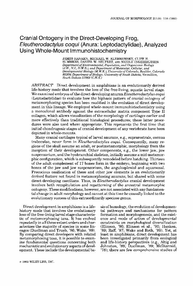
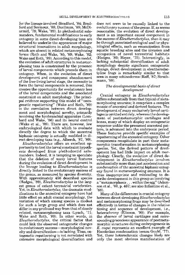
T-S 5-6) Stage 5 (Figs. lA,B, 2A,B).
Stage 6.
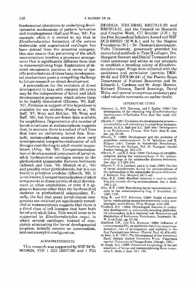
Stage 7 (Fig. 2C,D).
98 J. HANKEN ET AL
Figure 1
SKULL DEVELOPMENT IN ELEUTHERODACTYLUS 99
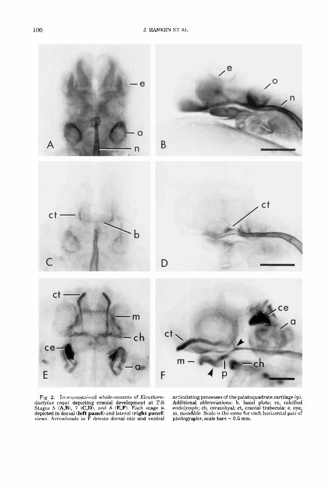
anteromedially; longitudinal arms (Meckel’s cartilages) extend posterolaterally to articu- late with the palatoquadrate cartilages. The rod-shaped palatoquadrate cartilage is lo- cated anterior to the auditory capsule. From its posterodorsal tip (otic process), it de- scends anteroventrally at an angle of 35” below the floor of the neurocranium to its articulation with Meckel’s cartilage beneath the orbit. It is separate from the neurocra- nium and lacks a commissura quadratocrani- alis anterior (de Beer, ’371, muscular process, and ascending process, none of which ever forms. In terms oftheir overall shape, degree of fusion, and the location of the jaw joint, Meckel’s, infrarostral, and palatoquadrate cartilages resemble a mid-metamorphic stage of development in metamorphosing anurans (e.g., Bombina orientalis, Hanken and Sum- mers, ’88a; Fig. ZC,D).
The hyobranchial skeleton comprises a con- tinuous ventral mass of cartilage that, in dorsal View, lies ktween the eyes and a d i - tory capsules (Figs. 2E,F, 6 ) . A longitudinal series Of four paired processes extends later- ally from the central region (hypobranchial Plate). The most anterior Process is largest and resembles the CeratohYal cartilage of lar- val frogs; the remaining processes (cerato- branchials 1-111) are progressively thinner, shorter, and fainter from anterior to poste- rior. All three ceratobranchials are rod- shaped and lack any of the secondary or tertiary branching characteristic of these car- tilages in larval frogs (cf. Hanken and Sum- mers, ’88a; Figs. 1A, 2A,B).
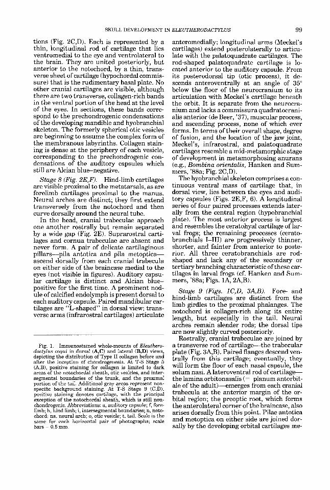
tions (Fig. 2C,D). Each is represented by a thin, longitudinal rod of cartilage that lies ventromedial to the eye and ventrolateral to the brain. They are united posteriorly, but anterior to the notochord, by a thin, trans- verse sheet of cartilage (hypochordal commis- sure) that is the rudimentary basal plate. NO other cranial cartilages are visible, although there are two transverse, collagen-rich bands in the ventral portion of the head at the level of the eyes. In sections, these bands corre- spend to the prechondrogenic condensations of the developing mandible and hyobranchial skeleton. The formerly spherical otic vesicles are beginning to assume the complex form of the membranous labyrinths. Collagen stain- ing is dense at the periphery of each vesicle, corresponding to the prechondrogenic con- densations of the auditory capsules which still are Alcian blue-negative.
Hind-limb cartilages are visible proximal to the metatarsals, as are forelimb cartilages proximal to the manus. Neural arches are distinct; they first extend transversely from the notochord and then curve dorsally around the neural tube.
In the head, cranial trabeculae approach one another rostrally but remain separated by a wide gap (Fig. 2E). Suprarostral carti- lages and cornua trabeculae are absent and never form. A pair of delicate cartilaginous pillars-pila antotica and pila metoptica- ascend dorsally from each cranial trabecula on either side of the braincase medial to the eyes (not visible in figures). Auditory capsu- lar cartilage is distinct and Alcian blue-
stage 8 (Fig. ~ E , F ) .
positive forthe first time. A prominent nod- ule of calcified endolymph is present dorsal to each auditory capsule. Paired mandibular car- tilages are “L-shaped” in dorsal view; trans- verse arms (infrarostral cartilages) articulate
Fig. 1. Immunostained whole-mounts of Eleuthero- dactylus coqui in dorsal (A,C) and lateral (B,D) views, depicting the distribution of Type I1 collagen before and after the inception of chondrogenesis. At T-S Stage 5 (A,B), positive staining for collagen is limited to dark areas of the notochordal sheath, otic vesicles, and inter- segmental boundaries of the trunk, and the proximal portion of the tail. Additional gray areas represent non- specific background staining. At T-S Stage 9 (C,D), positive staining denotes cartilage, with the principal exception of the notochordal sheath, which is still non- chondrogenic. Abbreviations: a, auditoly capsule; f, fore- limb; h, hind limb; i, intersegmental boundaries; n, noto- chord; na, neural arch; 0, otic vesicle; t, tail. Scale is the same for each horizontal pair of photographs; scale bars = 0.5 mm.
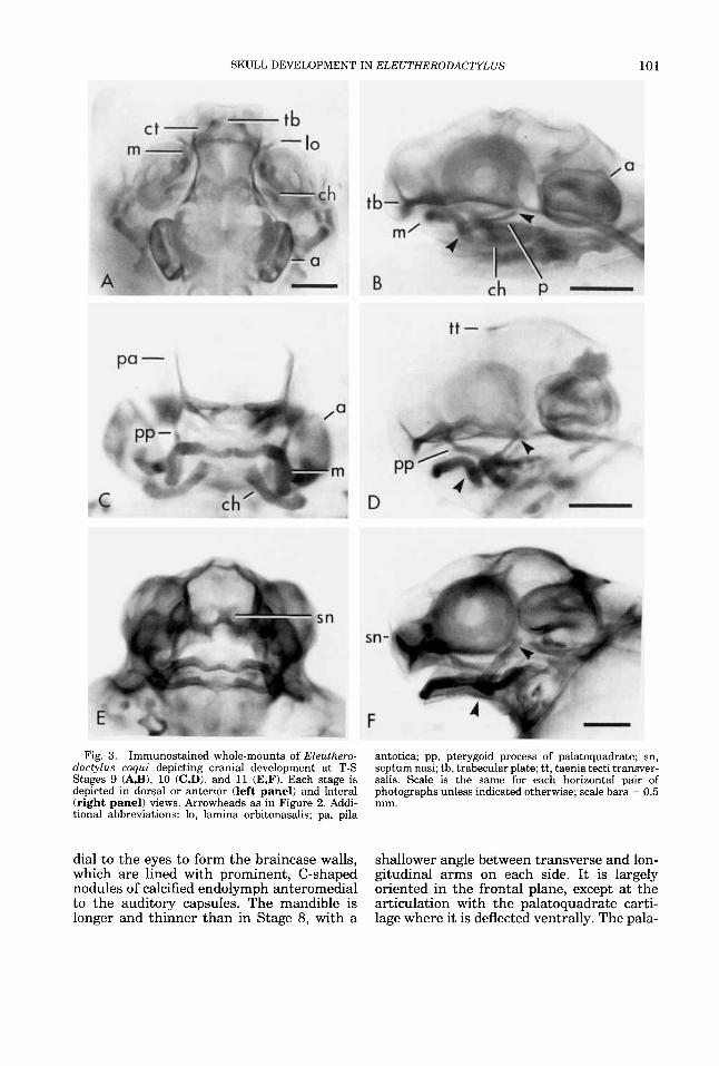
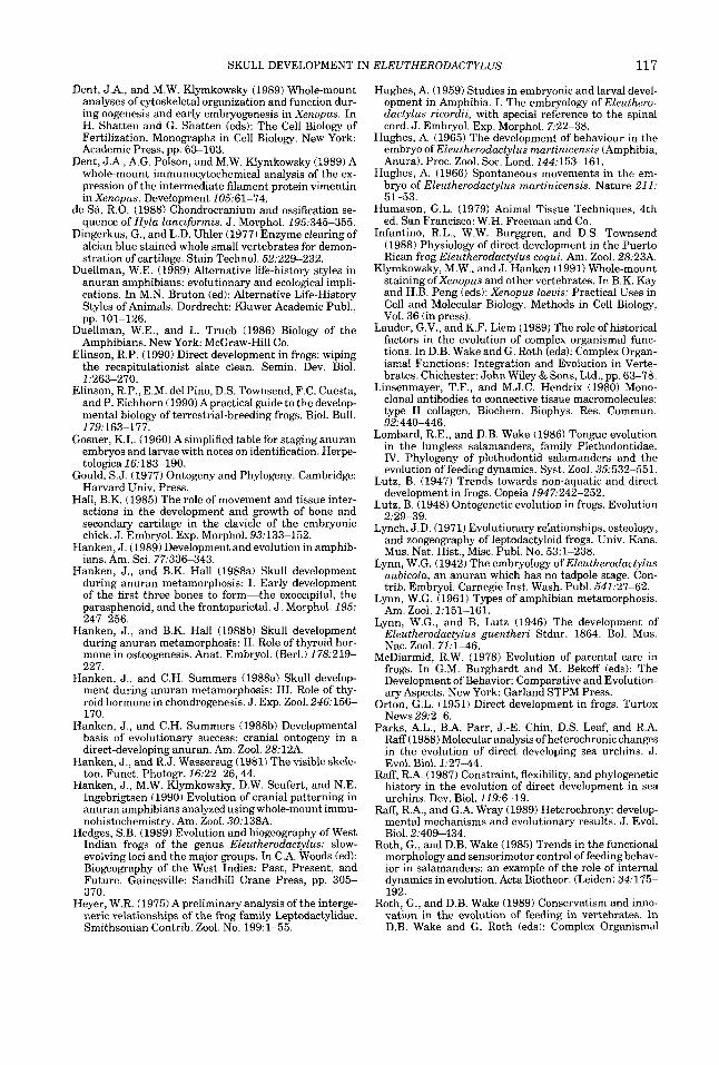
Stage 9 (Figs. lC,D, 3A,B). Fore- and hind-limb cartilages are distinct from the limb girdles to the proximal phalanges. The notochord is collagen-rich along its entire length, but especially in the tail. Neural arches remain slender rods; the dorsal tips are now slightly curved posteriorly.
Rostrally, cranial trabeculae are joined by a transverse rod of carti lagethe trabecular plate (Fig. 3A,B). Paired flanges descend ven- trally from this cartilage; eventually, they will form the floor of each nasal capsule, the solum nasi. A lateroventral rod of c a r t i l a g e the lamina orbitonasalis (= planum antorbit- ale of the adult)--+merges from each cranial trabecula at the anterior margin of the or- bital region; the preoptic root, which forms the anterolateral corner of the braincase, also arises dorsally from this point. Pilae antotica and metoptica on either side are joined dor- sally by the developing orbital cartilages me-
100 J. HANKEN ET AL.
Fig. 2. Immunostained whole-mounts of EZeuthero- dactylus coqui depicting cranial development at T-S Stages 5 (A,B), 7 (C,D), and 8 (E,F). Each stage is depicted in dorsal (left panel) and lateral (right panel) views. Arrowheads in F denote dorsal otic and ventral
articulating processes of the palatoquadrate cartilage (p). Additional abbreviations: b, basal plate; ce, calcified endolymph; ch, ceratohyal; ct, cranial trabecula; e, eye; m, mandible. Scale is the same for each horizontal pair of photographs; scale bars = 0.5 mm.
SKULL DEVELOPMENT IN ELEUTHERODACTYLUS 101
Fig. 3. Immunostained whole-mounts of Eleuthero- dactylus coqui depicting cranial development at T-S Stages 9 (A,B), 10 (C,D), and 11 (E,F). Each stage is depicted in dorsal or anterior (left panel) and lateral (right panel) views. Arrowheads as in Figure 2. Addi- tional abbreviations: lo, lamina orbitonasalis; pa, pila
antotica; pp, pterygoid process of palatoquadrate; sn, septum nasi; tb, trabecular plate; tt, taenia tecti transver- salis. Scale is the same for each horizontal pair of photographs unless indicated otherwise; scale bars = 0.5 mm.
dial to the eyes to form the braincase walls, which are lined with prominent, C-shaped nodules of calcified endolymph anteromedial to the auditory capsules. The mandible is longer and thinner than in Stage 8, with a
shallower angle between transverse and lon- gitudinal arms on each side. It is largely oriented in the frontal plane, except at the articulation with the palatoquadrate carti- lage where it is deflected ventrally. The pala-
102 J. HANKEN ET AL
toquadrate is oriented as in Stage 8 and re- mains well separated from the braincase. At the midpoint of the palatoquadrate, there is a ventral articulation with the ceratohyal carti- lage. Dorsal to this articulation, a prominent pterygoid process extends first medially and then anteriorly, closely following the curva- ture of the adjacent mandible, until it ascends to fuse with the braincase via the posterior maxillary process of the lamina orbitonasa- lis.
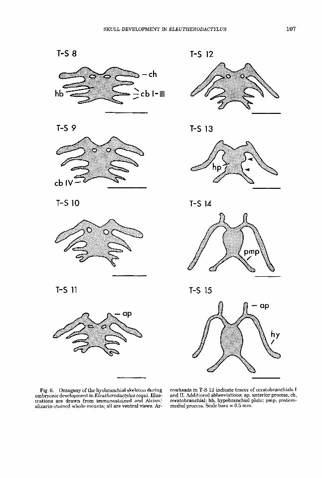
The hyobranchial skeleton is different from that of Stage 8 (Fig. 6). The hypobranchial plate is larger relative to ceratobranchials 1-111, and ceratobranchial IV is visible as a tiny transverse process emerging on either side from the posterolateral margin of the hypobranchial plate. The long axis of each ceratohyal has become obliquely oriented as the lateral articulation with the palatoquad- rate has migrated posteriorly.
Notochord and ver- tebrae are as in Stage 9; dorsal, recurved tips of neural arches extend farther posteriorly. All limb cartilages are distinct, including dis- tal phalanges.
The neurocranium is more robust (Fig. 3C,D). Trabecular and basal plates are more extensive, and the septum nasi, which forms initially as a transverse sheet of cartilage, separates the developing nasal capsules (cf. Fig. 3E,F). A thin, transverse band of carti- lage, the taenia tecti transversalis, forms a dorsal bridge over the braincase immediately anterior to the pila metoptica. Paired nod- ules of calcified endolymph closely follow the walls of the braincase medial to the auditory capsules and approach one another posteri- orly. The mandible is more elongate; the jaw articulation has advanced caudally to a level immediately anterior to the optic foramen. As in earlier stages, each side of the mandible comprises a single, continuous, cartilaginous rod. However, thin, crescentic, anteromedial segments resemble the infrarostral carti- lages of metamorphosing anuran larvae, and in this way they are distinct from laterally adjacent segments (Meckel's cartilage) (Fig. 3C). The palatoquadrate is more upright (40"); the otic process approaches, but re- mains separate from, the anterior border of the auditory capsule.
The hyobranchial skeleton has continued to diverge from its initial form (Fig. 6). The ceratohyal is more prominent and elongate; its primary axis forms an angle of approxi- mately 60" with the long axis of the skull.
Stage 10 (Fig. 3C,D).
Ceratobranchial I is as long as, and oriented parallel to, the ceratohyal, but it tapers dis- tally. Ceratobranchial (CB) I1 is much smaller than CB I and is oriented transversely. Cera- tobranchials I11 and IV are slightly shorter than CB I1 and emerge from the hypobran- chial plate by a common stalk before separat- ing after about one-half their length; they are oriented slightly posterolaterally.
Collagen staining of the notochord is faint in the head and trunk but still prominent in the tail. Neural arches bear prominent anterior and posterior pro- cesses, corresponding to the pre- and post- zygapophyses. The third presacral vertebra (second behind the atlas) bears an anterolat- era1 process that approaches the shoulder girdle. The craniovertebral articulation is es- tablished. All limb cartilages are distinct.
The neurocranium is well developed (Fig. 3E,F). Nasal capsular cartilages, in particu- lar, are more extensive; tectum nasi, solum nasi, and planum terminale are distinct. Nodules of calcified endolymph within the braincase are still prominent, but they are asymmetrical and more widely separated pos- teriorly than at earlier stages. The mandible is more robust and elongate; the jaw articula- tion lies ventral to the optic foramen. Antero- medial segments remain thinner than lateral segments (Figs. 3E, 7A). The palatoquadrate is more stout and upright in lateral view (50"), but it retains a solid connection with the neurocranium via the pterygoid process. The otic process remains separate from the neurocranium. The palatoquadrate a t this stage resembles that of a mid-metamorphic Rana depicted by de Beer ('37; P1. 75, Fig. 3).
The hyobranchial skeleton is prominent and beginning to resemble the hyale of a metamorphosed froglet (Fig. 6). The cerato- hyal is longer and more sinuous; the anterior processes are rudimentary. Ceratobranchial I is nearly as long as the ceratohyal, but its distal half is now much thinner than the base. Ceratobranchials 11-IV are much thin- ner and shorter than CB I and the cerato- hyal.
Immunostaining of the notochord is similar to that a t Stage 11. Vertebrae are better developed. Opposing neural arches are beginning to arch over the neural tube and approach one another, al- though they still are widely separated. In the trunk, anterior and posterior processes that correspond to the future pre- and postzyg-
Stage 11 (Fig. 3E,F).
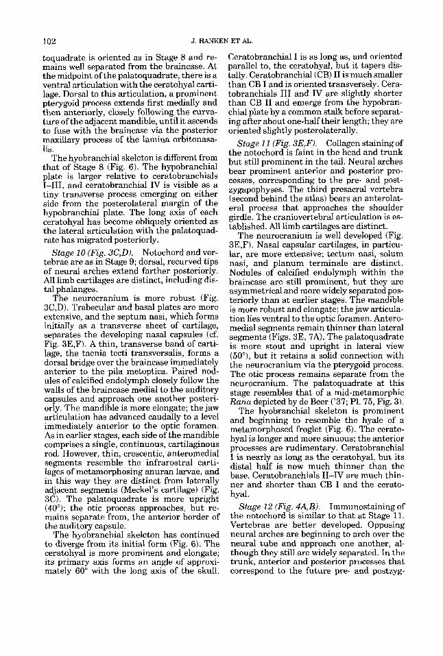
Stage 12 (Fig. 4A,B).
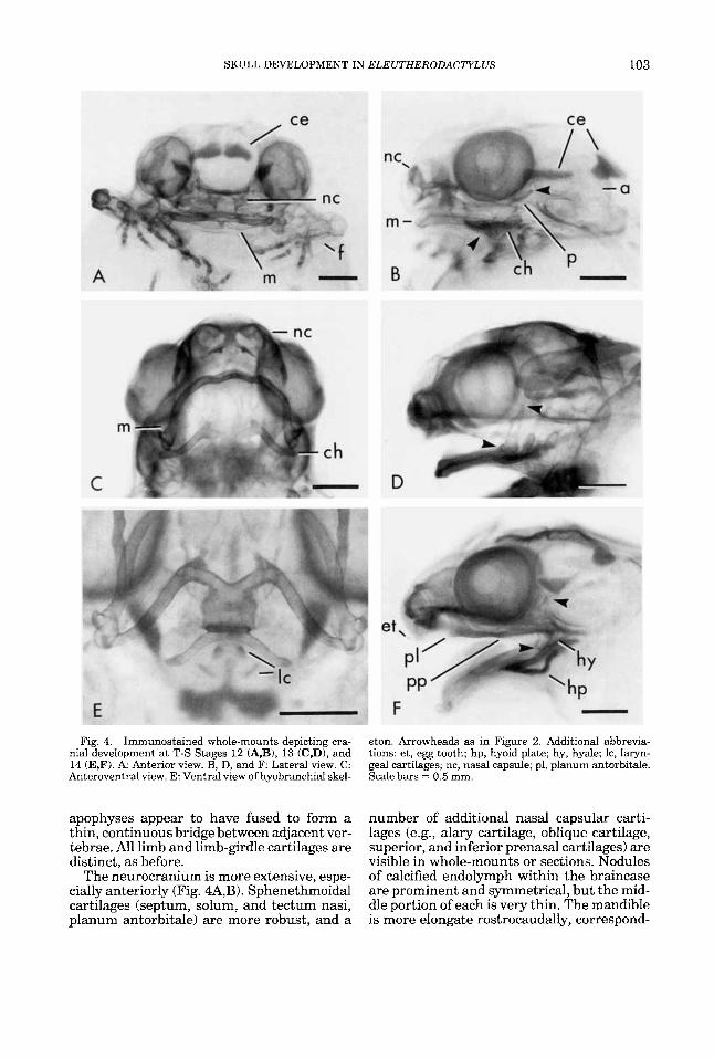
SKULL DEVELOPMENT IN ELEUTHERODACTYLUS 103
Fig. 4. Immunostained whole-mounts depicting cra- nial development a t T-S Stages 12 (A,B), 13 (C,D), and 14 (E,F). A Anterior view. B, D, and F: Lateral view. C: Anteroventral view. E: Ventral view of hyobranchial skel-
apophyses appear to have fused to form a thin, continuous bridge between adjacent ver- tebrae. All limb and limb-girdle cartilages are distinct, as before.
The neurocranium is more extensive, espe- cially anteriorly (Fig. 4A,B). Sphenethmoidal cartilages (septum, solum, and tectum nasi, planum antorbitale) are more robust, and a
eton. Arrowheads as in Figure 2. Additional abbrevia- tions: et, egg tooth; hp, hyoid plate; hy, hyale; Ic, laryn- geal cartilages; nc, nasal capsule; pl, planum antorbitale. Scale bars - 0.5 mm.
number of additional nasal capsular carti- lages (e.g., alary cartilage, oblique cartilage, superior, and inferior prenasal cartilages) are visible in whole-mounts or sections. Nodules of calcified endolymph within the braincase are prominent and symmetrical, but the mid- dle portion of each is very thin. The mandible is more elongate rostrocaudally, correspond-
104 J. HANKEN ET AL.
ing to the ongoing posteriad migration of the jaw articulation; it is beginning to assume the characteristic, adult, inverted U-shape in dorsal view. Anteromedial segments are still sickle-shaped and thinner than adjacent re- gions, but less so than in earlier stages. The palatoquadrate is stout, especially at the ar- ticular process, but tapers somewhat at the otic process; its long axis is oriented at 55". The otic process is close to the auditory cap- sule but the two elements remain separated by a small gap.
The hyobranchial skeleton is little changed from Stage 11 (Fig. 6). The ceratohyal articu- lates with the palatoquadrate midway be- tween articulating and otic processes (Fig. 4B).
A pair of rudimentary egg teeth are visible as tiny keratinized cones at the tip of the snout immediately above the mouth.
The notochord no longer stains positive for collagen in the head or trunk; staining in the caudal portion, which is smaller than in the preceding stage, is faint. In the postcranial skeleton, strong staining for collagen is limited to growth zones (e.g., long bone epiphyses, neural arches), except for the distal portions of the limbs, which remain well stained. Staining is much more intense in Alcian-alizarin prepa- rations. Centers of ossification are visible within both the trunk vertebrae (neural arches) and the appendicular skeleton. Oppos- ing neural arches approach one another dor- sally; pre- and postzygapophyses and trans- verse processes are distinct. The latter are especially pronounced opposite the pectoral and pelvic girdles.
The neurocranium is well formed, espe- cially anteriorly, where it closely resembles that of a postmetamorphic frog (Fig. 4C,D). Calcified endolymph within the braincase re- sembles that seen at Stage 12 (Fig. 4A,B) except that anterior and posterior segments on each side are connected by a threadlike bridge; posterior segments approach or artic- ulate a t the dorsal midline at the level of the synotic tectum. Additional deposits are visi- ble for the first time within each auditory capsule. The mandible is more elongate; the jaw articulation lies posterior to the optic foramen. Anteromedial segments (infraros- tral cartilages) now occupy a very small por- tion of the lower jaw; the symphysis is dis- tinctly notched along its ventral margin. The palatoquadrate is nearly vertical (85"); the otic process articulates and is beginning to fuse with the anterolateral face of the audi-
Stage 13 (Fig. 4C,D).
tory capsule. (Prechondrogenic condensa- tions linking these elements are visible in sections.) A rudimentary pseudobasal pro- cess is present on the medial aspect of each palatoquadrate between the articulation with the ceratohyal and the otic process.
Hyobranchial anatomy is variable. The skeleton in one specimen (not illustrated) is only slightly modified from that a t Stage 12. Ceratohyals are prominent and elongate. An- terior processes are rudimentary, and lateral segments (hyale) still articulate with the pala- toquadrate. Ceratobranchial I is arrayed par- allel to the hyale, but it is only slightly more than half as long and tapers distally. The threadlike CB I1 is arrayed transversely and is about half as long as CB I. Similarly, cera- tobranchial I11 is short and thin; CB IV is broader and, including the portion proximal to CB 111, is nearly as long as CB I. Another immunostained specimen is more advanced (Figs. 4C,D, 6). In it, a thin cartilaginous bridge extends anterodorsally from each hy- ale to fuse with the anteroventral margin of the auditory capsule; distal regions of CB 1-111 have been resorbed and cartilage is filling in the area between the proximal por- tions of the ceratohyal and CB I to form the hyoid plate.
The pair of cornified egg teeth rudiments is more prominent.
Stage 14 (Fig. 4E,F). The notochord is similar to that at Stage 13, only shorter cau- dally. Otherwise, collagen-positive areas gen- erally are limited to cartilaginous epiphyses throughout the entire postcranial skeleton. Opposing neural arches closely approach one another or articulate dorsally; vertebral cen- tra are well developed laterally, but are incom- plete above and below the notochord. Long bones, especially proximal elements, are ossi- fied for most of their length.
The cartilaginous walls, floor, and roof of the neurocranium are nearly fully formed, anteriorly and posteriorly (Fig. 4F). Promi- nent deposits of calcified endolymph lie within both the braincase and each auditory cap- sule, little changed from Stage 13. The man- dible also generally resembles that a t Stage 13. The short palatoquadrate is oriented at 95", reflecting the continued posteriad migra- tion of the jaw articulation, which now lies beneath the auditory capsule. The prominent pterygoid process extends anteriorly, subtend- ing the orbit, before fusing with the planum antorbitale of the nasal capsule (= lamina orbitonasalis of earlier embryo); the otic pro- cess is fused to the anterolateral face of the
SKULL DEVELOPMENT IN ELEUTHERODACTYLUS 105
auditory capsule. A small pseudobasal pro- cess extends medially from a point just dorsal to the jaw articulation to establish a second contact between the palatoquadrate and the auditory capsule.
The hyolaryngeal skeleton has changed substantially from earlier stages (Figs. 4E, 6). It now comprises an urn-shaped hyoid plate; paired hyale derived from the ceratohy- als, each with a prominent anterior process; and paired posteromedial processes, each de- rived from CB IV plus the basal portion once shared with CB 111. Ceratobranchials I, 11, and I11 are absent as discrete elements, al- though their basal portions, especially that of CB I, have been incorporated into the hyoid plate. The distal, recurved portion of each hyale is fused to the auditory capsule via the short cartilaginous segment described earlier (Stage 13). Rudimentary laryngeal carti- lages, variably present at Stage 13, are present posterodorsal to the posteromedial processes of the hyoid plate (Fig. 4E).
Paired egg teeth rudiments have merged to form a single, larger, and more prominent tooth.
The postcranial skeleton is well developed. Neural arches are well ossified; paired, basal ossification cen- ters on either side of the notochord corre- spond to the developing vertebral centra. Os- sification centers are visible throughout the appendicular skeleton, including many distal phalanges. Collagen-positive staining is lim- ited to the caudal portion of the notochord, which is continuing to resorb and is about as long as the thigh; to the distal tips of trans- verse processes; and to the dorsomedial seg- ments of the neural arches.
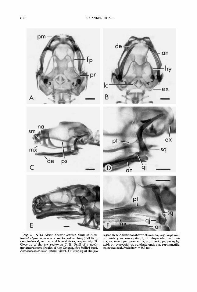
The cartilaginous skull and hyobranchium generally resemble those of Stage 14, al- though much of the cartilage is being re- placed by bone (Figs. 5A-D, 6). Except for the mandible and the hyobranchial skeleton, which remain collagen-positive, whole-mount immunostaining is less intense than at ear- lier stages. The adultlike mandible is elon- gate and sinuous, especially a t the symphysis (Fig. 7B). Anterior processes of the hyale are longer (Fig. 6). Calcified endolymph resem- bles that a t Stage 14. The median egg tooth is prominent.
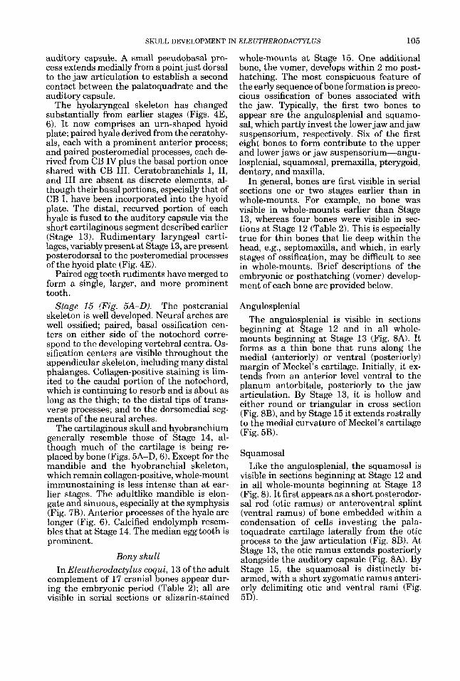
Bony skull In Eleutherodactylus coqui, 13 of the adult
complement of 17 cranial bones appear dur- ing the embryonic period (Table 2); all are visible in serial sections or alizarin-stained
Stage 15 (Fig. 5A-D).
whole-mounts at Stage 15. One additional bone, the vomer, develops within 2 mo post- hatching. The most conspicuous feature of the early sequence of bone formation is preco- cious ossification of bones associated with the jaw. Typically, the first two bones to appear are the angulosplenial and squamo- sal, which partly invest the lower jaw and jaw suspensorium, respectively. Six of the first eight bones to form contribute to the upper and lower jaws or jaw suspensorium-angu- losplenial, squamosal, premaxilla, pterygoid, dentary, and maxilla.
In general, bones are first visible in serial sections one or two stages earlier than in whole-mounts. For example, no bone was visible in whole-mounts earlier than Stage 13, whereas four bones were visible in sec- tions at Stage 12 (Table 2). This is especially true for thin bones that lie deep within the head, e.g., septomaxilla, and which, in early stages of ossification, may be difficult to see in whole-mounts. Brief descriptions of the embryonic or posthatching (vomer) develop- ment of each bone are provided below.
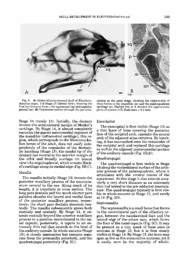
Angulosplenial The angulosplenial is visible in sections
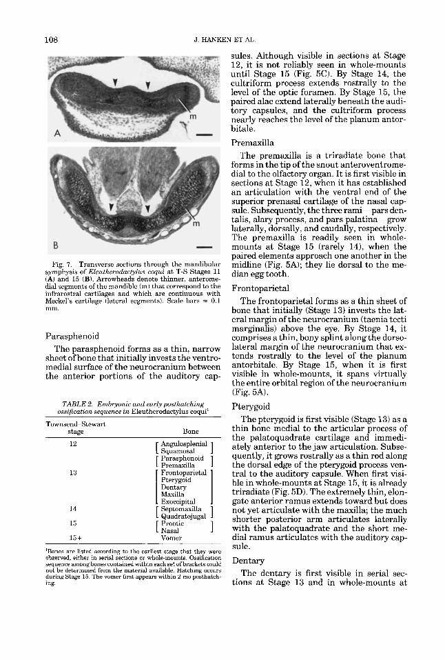
beginning at Stage 12 and in all whole- mounts beginning at Stage 13 (Fig. 8A). It forms as a thin bone that runs along the medial (anteriorly) or ventral (posteriorly) margin of Meckel’s cartilage. Initially, it ex- tends from an anterior level ventral to the planum antorbitale, posteriorly to the jaw articulation. By Stage 13, it is hollow and either round or triangular in cross section (Fig. SB), and by Stage 15 it extends rostrally to the medial curvature of Meckel’s cartilage (Fig. 5B).
Squamosal Like the angulosplenial, the squamosal is
visible in sections beginning at Stage 12 and in all whole-mounts beginning at Stage 13 (Fig. 8). It first appears as a short posterodor- sal rod (otic ramus) or anteroventral splint (ventral ramus) of bone embedded within a condensation of cells investing the pala- toquadrate cartilage laterally from the otic process to the jaw articulation (Fig. 8B). At Stage 13, the otic ramus extends posteriorly alongside the auditory capsule (Fig. 8A). By Stage 15, the squamosal is distinctly bi- armed, with a short zygomatic ramus anteri- orly delimiting otic and ventral rami (Fig. 5D).
106 J. HANKEN ET AL.
Fig. 5. A-C: Alcianializarin-stained skull of Eleu- therodactylus coqui several weeks posthatching (T-S 15+ 1, seen in dorsal, ventral, and lateral views, respectively. D: Close-up of the jaw region in C. E: Skull of a newly metamorphosed froglet of the Oriental fire-bellied toad, Bombina orientalis (lateral view). F: Close-up of the jaw
region in E. Additional abbreviations: an, angulosplenial; de, dentary; ex, exoccipital; fp, frontoparietal; mx, max- illa; na, nasal; pm, premaxilla; pr, prootic; ps, parasphe- noid; pt, pterygoid; qj, quadratojugal; sm, septomaxilla; sq, squamosal. Scale bars = 0.5 mm.
SKULL DEVELOPMENT IN ELEUTHERODACTYLUS 107
T-S 8 T-S 12
T-S 9 T-S 13
T-S 10
T-S 11
T-S 14
T-S 15
Fig. 6. Ontogeny of the hyobranchial skeleton during embryonic development in Ekutherodactylus coqui. Illus- trations are drawn from immunostained and Alcianl alizarin-stained whole-mounts; all are ventral views. Ar-
rowheads in T-S 13 indicate traces of ceratobranchials I and 11. Additional abbreviations: ap, anterior process; cb, ceratobranchial; hb, hypobranchial plate; pmp, postero- medial process. Scale bars = 0.5 mm.
108
13
J. HANKE
' Frontoparietal Pterygoid Dentary Maxilla . Exoccipital .
:N ET AL.
sules. Although visible in sections at Stage 12, it is not reliably seen in whole-mounts until Stage 15 (Fig. 5C). By Stage 14, the cultriform process extends rostrally to the level of the optic foramen. By Stage 15, the paired alae extend laterally beneath the audi- tory capsules, and the cultriform process nearly reaches the level of the planum antor- bitale. Premaxilla
The premaxilla is a triradiate bone that forms in the tip of the snout anteroventrome- dial to the olfactory organ. It is first visible in sections at Stage 12, when it has established an articulation with the ventral end of the superior prenasal cartilage of the nasal cap- sule. Subsequently, the three rami-pars den- talis, alary process, and pars palatina-grow laterally, dorsally, and caudally, respectively. The premaxilla is readily seen in whole- mounts at Stage 15 (rarely 14), when the paired elements approach one another in the midline (Fig. 5A); they lie dorsal to the me- dian egg tooth. Frontoparietal
The frontoparietal forms as a thin sheet of bone that initially (Stage 13) invests the lat- eral margin of the neurocranium (taenia tecti marginalis) above the eye. By Stage 14, it comprises a thin, bony splint along the dorso- lateral margin of the neurocranium that ex- tends rostrally to the level of the planum antorbitale. By Stage 15, when it is first visible in whole-mounts, it spans virtually the entire orbital region of the neurocranium (Fig. 5A).
Pterygoid The pterygoid is first visible (Stage 13) as a
thin bone medial to the articular process of the palatoquadrate cartilage and immedi- ately anterior to the jaw articulation. Subse- quently, it grows rostrally as a thin rod along the dorsal edge of the pterygoid process ven- tral to the auditory capsule. When first visi- ble in whole-mounts at Stage 15, it is already triradiate (Fig. 5D). The extremely thin, elon- gate anterior ramus extends toward but does not yet articulate with the maxilla; the much shorter posterior arm articulates laterally with the palatoquadrate and the short me- dial ramus articulates with the auditory cap- sule. Dentary
The dentary is first visible in serial sec- tions at Stage 13 and in whole-mounts a t
Fig. 7. Transverse sections through the mandibular symphysis of Eleutherodactylus coqui at T-S Stages 11 (A) and 15 (B). Arrowheads denote thinner, anterome- dial segments of the mandible (m) that correspond to the infrarostral cartilages and which are continuous with Meckel's cartilage (lateral segments). Scale bars = 0.1 mm.
Parasphenoid The parasphenoid forms as a thin, narrow
sheet of bone that initially invests the ventro- medial surface of the neurocranium between the anterior portions of the auditory cap-
TABLE 2. Embryonic and early posthatching ossification seauence in Eleutherodactvlus coaui'
Townsend-Stewart stage Bone
SKULL DEVELOPMENT IN ELEUTHERODACTYLUS 109
Fig. 8. A Alcianializarin-stained skull of Eleuthero- dactylus coqui, T-S Stage 13 (lateral view), showing the first two bones to form-the squamosal (sq) and angulos- plenial (an). B: Transverse section through the jaw artic-
Stage 14 (rarely 13). Initially, the dentary invests the anterolateral margin of Meckel’s cartilage. By Stage 14, it almost completely encircles the paired anteromedial segment of the mandible (infrarostral cartilage); this re- gion, which corresponds to the Mentomecke- lian bones of the adult, does not ossify inde- pendently of the remainder of the dentary. By hatching (Stage 151, the caudal tip of the dentary lies ventral to the anterior margin of the orbit and broadly overlaps (in lateral view) the angulosplenial, which invests Meck- el’s cartilage along its medial edge (Fig. 5B,C). Maxilla
The maxilla initially (Stage 13) invests the posterior maxillary process of the neurocra- nium ventral to the eye. Along much of its length, it is triradiate in cross section. The long pars dentalis and the much shorter pars palatina sheathe the lateral and medial faces of the posterior maxillary process, respec- tively; the short pars dentalis descends ven- trally. The maxilla subsequently grows both rostrally and caudally. By Stage 14, it ex- tends rostrally beyond the anterior maxillary process to a position ventrolateral to the na- sal capsule; posteriorly, it tapers to an ex- tremely thin rod that extends to the level of the auditory capsule. In whole-mounts (Stage 151, it closely approaches but remains sepa- rate from the premaxilla anteriorly, and the quadratojugal posteriorly (Fig. 5C).
ulation at the same stage, showing the relationship of these bones to the mandible (m) and the palatoquadrate cartilage (p). Dashed line in A denotes the approximate plane of section in B. Scale bars = 0.1 mm.
Exoccipital The exoccipital is first visible (Stage 13) as
a thin layer of bone covering the posterior face of the occipital arch, opposite the neural arch of the adjacent atlas vertebra. By hatch- ing, it has encroached onto the remainder of the occipital arch and replaced this cartilage as well as the adjacent posteromedial portion of the auditory capsule (Fig. 5B,D). Quadratojugal
The quadratojugal is first visible at Stage 14 along the ventrolateral surface of the artic- ular process of the palatoquadrate, where it articulates with the ventral ramus of the squamosal. At this stage it also extends ante- riorly a very short distance as an extremely thin rod lateral to the jaw adductor muscula- ture. The quadratojugal typically is first visi- ble in whole-mounts at Stage 15, and rarely at 14 (Fig. 5D). Septomaxilla
The septomaxilla is a small bone that forms in the ventrolateral part of the olfactory or- gan, between the nasolacrimal duct and the lateral edge of the solum nasi, which forms the floor of the nasal capsule (Fig. 5 0 . It may be present as a tiny speck of bone seen in sections at Stage 13, but it is first clearly visible a t Stage 14. By Stage 15, the bone may span as few as five consecutive sections, yet it is easily seen in the majority of whole-
110 J. HANKEN ET AL
mounts, embedded in nasal capsular carti- lage ventral to the external naris.
Prootic The prootic forms as a center of endochon-
dral ossification within the anteromedial wall of the auditory capsule adjacent to the basal plate (Fig. 5A). It is first visible at Stage 15 in both whole-mounts and serial sections. In all whole-mounts examined, the prootic forms in a region of capsular cartilage that lies be- tween nodules of calcified endolymph within the auditory capsule and the braincase.
Nasal The nasal initially forms as a thin, nearly
transverse sliver of bone that straddles the surface of the nasal capsule, posterior to the oblique cartilage and dorsal to the planum terminale (Fig. 5C). Its presence is presaged by a distinct condensation of cells seen in serial section at Stage 14, but calcified matrix is not present until Stage 15, when, typically, the bone also is visible in whole-mounts.
Vomer The vomer is a paired bone that is first
visible in whole-mounts preserved within 2 mo after hatching; it is absent from all pre- hatching specimens, both whole-mounts and serial sections. It initially forms ventral to the braincase, anteromedial to the orbit. It comprises an extremely thin, longitudinal process that extends rostrally toward the in- ternal naris and which is continuous cau- dally with a short, transverse dentigerous process. The structure of the vomer was not examined in sections.
DISCUSSION Recapitulation us. repatterning
A recurring question in the study of direct- developing anurans is whether there is an embryonic recapitulation of the ancestral bi- phasic pattern of development-including an embryonic “metamorphosis”--or if, instead, there has been a more fundamental repat- terning of early ontogeny. Such repatterning may involve the loss of ancestral, larval struc- tures, shifts in the relative timing of develop- mental events (i.e., heterochrony), and, per- haps most significantly, the appearance of novel morphologies not present in the ances- tor. From the limited comparative data avail- able, the degree of recapitulation vs. repat- terning varies widely, as one might expect from lineages that have evolved direct devel-
opment independently (reviewed by Lutz, ’47, ’48; Lynn, ’61; Orton, ’51). Eleutherodacty- lus is generally regarded as the least recapit- ulatory of all direct-developing anurans (Elin- son, ’90; Hughes, ’65, ’66; Lynn, ’61; Orton, ’51), although some authors refer to a meta- morphosis during embryonic development (Adamson et al., ’60; Infantino et al., ’88). Much of the difficulty in resolving this ques- tion stems from the difficulty in visualizing the morphology of early embryonic struc- tures, which typically are small, transient, and not fully differentiated. This problem is especially acute for the development of inter- nal features such as the skeleton, for which subtle aspects of form are difficult or time- consuming to depict by using traditional his- tological methods, either in whole-mounts or serial sections.
In this study, we employed a new whole- mount immunohistochemical technique that offers greater resolving power for depicting the structure of developing tissues with virtu- ally no preparation artifact (Dent et al., ’89; Dent and Klymkowsky, ’89; Klymkowsky and Hanken, ’91). For cartilages, it reveals the form of developing elements from the time of initial deposition of the extracellular matrix component Type I1 collagen at the inception of their development. This allowed us to de- pict the earliest stages of cranial chondrogen- esis and, thereby, address more completely the question of recapitulation vs. repattern- ing as it applies to the skull. Both phenomena were observed.
The most conspicuous evidence of repat- terning of cranial ontogeny in ELeutherodac- tylus coqui is the absence of a number of exclusively larval cartilages found in meta- morphosing anurans. These include the su- prarostral cartilages, which constitute the tadpole’s upper jaw; the commissura quadra- tocranialis anterior (de Beer, ’37), which unites the palatoquadrate with the neurocra- nium anteriorly; the cornua trabeculae, paired anterolateral extensions of the brain- case; and muscular, larval otic, and ascend- ing processes of the palatoquadrate. In the absence of these cartilages, the development of other cartilages that follow or replace them at metamorphosis has been advanced into earlier stages. For example, the postmetamor- phic complex of nasal cartilages (e.g., solum nasi, septum nasi, planum antorbitale) be- gins to form as early as Stage 9, only two stages after cartilage is first visible anywhere in the head.
SKULL DEVELOPMENT IN ELEUTHERODACTYLUS 111
Evolutionary modifications to the ontog- eny of other cranial cartilages are more com- plicated. While lacking the earliest, or larval characteristics of metamorphosing taxa, these structures do not initially display their final adult, or postmetamorphic, configuration. In- stead, they first assume a “mid-metamor- phic” stage of development that is subse- quently remodeled before hatching. In short, there is both repatterning and partial recapit- ulation of the ancestral ontogeny. The two principal examples of this phenomenon are components of the visceral skeleton: the lower jaw and jaw suspensorium, and the hyobran- chial skeleton.
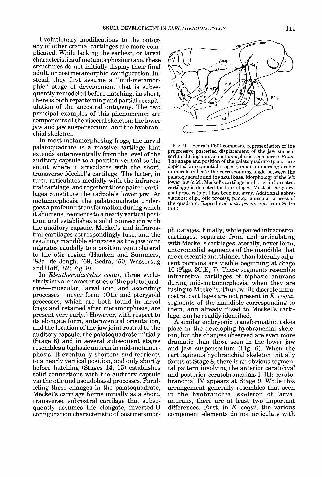
In most metamorphosing frogs, the larval palatoquadrate is a massive cartilage that extends anteroventrally from the level of the auditory capsule to a position ventral to the snout where it articulates with the short, transverse Meckel’s cartilage. The latter, in turn, articulates medially with the infraros- tral cartilage, and together these paired carti- lages constitute the tadpole’s lower jaw. At metamorphosis, the palatoquadrate under- goes a profound transformation during which it shortens, reorients to a nearly vertical posi- tion, and establishes a solid connection with the auditory capsule. Meckel’s and infraros- tral cartilages correspondingly fuse, and the resulting mandible elongates as the jaw joint migrates caudally to a position ventrolateral to the otic region (Hanken and Summers, ’88a; de Jongh, ’68; Sedra, ’50; Wassersug and Hoff, ’82; Fig. 9).
In Eleutherodactylus coqui, three exclu- sively larval characteristics of the palatoquad- rate-muscular, larval otic, and ascending processes-never form. (Otic and pterygoid processes, which are both found in larval frogs and retained after metamorphosis, are present very early.) However, with respect to its elongate form, anteroventral orientation, and the location of the jaw joint rostral to the auditory capsule, the palatoquadrate initially (Stage 8) and in several subsequent stages resembles a biphasic anuran in mid-metamor- phosis. I t eventually shortens and reorients to a nearly vertical position, and only shortly before hatching (Stages 14, 15) establishes solid connections with the auditory capsule via the otic and pseudobasal processes. Paral- leling these changes in the palatoquadrate, Meckel’s cartilage forms initially as a short, transverse, subrostral cartilage that subse- quently assumes the elongate, inverted-U configuration characteristic of postmetamor-
Fig. 9. Sedra’s (’50) composite representation of the progressive posteriad displacement of the jaw suspen- sorium during anuran metamorphosis, seen here in Rana. The shape and position of the palatoquadrate (p.a.q.1 are depicted in sequential stages (roman numerals); arabic numerals indicate the corresponding angle between the palatoquadrate and the skull base. Morphology of the left lower jaw (c.M., Meckel’s cartilage; and i.r.c., infrarostral cartilage) is depicted for four stages. Most of the ptery- goid process (p.pt.) has been cut away. Additional abbre- viations: ot.p., otic process; p.m.q., muscular process of the quadrate. Reproduced with permission from Sedra (’50).
phic stages. Finally, while paired infrarostral cartilages, separate from and articulating with Meckel’s cartilages laterally, never form, anteromedial segments of the mandible that are crescentic and thinner than laterally adja- cent portions are visible beginning at Stage 10 (Figs. 3C,E, 7). These segments resemble infrarostral cartilages of biphasic anurans during mid-metamorphosis, when they are fusing to Meckel’s. Thus, while discrete infra- rostral cartilages are not present in E. coqui, segments of the mandible corresponding to them, and already fused to Meckel’s carti- lage, can be readily identified.
A similar embryonic transformation takes place in the developing hyobranchial skele- ton, but the changes observed are even more dramatic than those seen in the lower jaw and jaw suspensorium (Fig. 6). When the cartilaginous hyobranchial skeleton initially forms at Stage 8, there is an obvious segmen- tal pattern involving the anterior ceratohyal and posterior ceratobranchials 1-111; cerato- branchial IV appears at Stage 9. While this arrangement generally resembles that seen in the hyobranchial skeleton of larval anurans, there are at least two important differences. First, in E. coqui, the various component elements do not articulate with
112 J. HANKEN ET AL
one another, but instead form a single contin- uous mass of cartilage; second, the cerato- branchials lack secondary and tertiary branching and are not united distally. Both of these features are characteristic of the hyobranchial skeleton during, but not be- fore, metamorphosis. By Stage 9, however, this configuration begins to be remodeled. Principal changes are reorientation of the ceratohyal, coincident with the posteriad mi- gration of its articulation with the pala- toquadrate, and growth and elaboration of the single, median hypobranchial plate. Con- sequently, by Stage 10, the morphology of the hyobranchial skeleton is substantially dif- ferent from its initial appearance, and remi- niscent of a mid-metamorphic stage in bipha- sic anurans.
A second period of intense change occurs between Stages 12 and 14. The hyale (for- merly ceratohyal) fuses to the auditory cap- sule and develops an anterior process. An urn-shaped hyoid plate forms that incorpo- rates the hypobranchial plate, basal portions of ceratobranchials 1-111, as well as new carti- lage. Ceratobranchials 1-111 are resorbed dis- tally and laryngeal cartilages begin to form. As a result of these modifications, a t hatch- ing, the hyobranchial skeleton generally has assumed the “postmetamorphic” configura- tion of the adult (Fig. 5B).
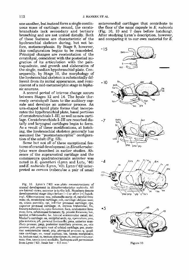
Some but not all of these exceptional fea- tures of cranial development in Eleutherodac- tylus were described in earlier studies. Ab- sence of the suprarostral cartilage and the commissura quadratocranialis anterior was noted in E. quentheri (Lynn and Lutz, ’46) and E. nubicola (Lynn, ’42). Lynn (’42) inter- preted as cornua trabeculae a pair of small
Fig. 10. Lynn’s (’42) wax-plate reconstructions of cranial development in Eleutherodactylus nubicola. All are lateral views; anterior is. to the left. Numbers denote developmental stage (days before [-I or after [ + I hatch- ing). Abbreviations: cau, columella auris; cf, carotid fora- men; ch, ceratohyal cartilage; cob, cartilage obliqua nasi; cp, crista parotica; cpi, inferior prenasal cartilage; cps, superior prenasal cartilage; ct, cornua trabeculae; fm, fissura metotica; fo, optic foramen; focn, oculomotor fora- men; forn, orbitonasal foramen; fp, prootic foramen; Ion, lamina orbitonasalis; Isc, lateral semicircular canal; mc, Meckel’s cartilage; oa, occipital arch; op, operculum; pao, pila antotica; pft, parietal fontanelle; pma, anterior max- illary process; pmp, posterior maxillary process; PO, otic process; pob, preoptic root of orbital cartilage; psc, poste- rior semicircular canal; ptp, pterygoid process; q, quad- rate cartilage; sn, nasal septum; tm, taenia marginalis; tn, tectum nasi; ts, tectum synoticum; tt, tectum transver- sum; ttm, taenia tecti medialis. Redrawn with permission from Lynn (’42). Scale bar = 0.5 mm.’
anteromedial cartilages that contribute to the floor of the nasal capsule in E. nubicola (Fig. 10, 10 and 7 days before hatching). After studying Lynn’s description, however, and comparing it to our own material for E.
- 10
h c ‘PtP
SC
+5
Figure 10
SKULL DEVELOPMENT IN ELEUTHERODACTYLUS 113
coqui, we regard these cartilages as distinct from the cornua trabeculae, which, when present, typically extend far anterior from the eventual location of the nasal capsule. Thus, we regard the cornua trabeculae as absent in E. nubicola, as well as in E. coqui. Absence of muscular, larval otic, and ascend- ing processes of the palatoquadrate has never been reported previously, although it is im- plicit in Lynn’s account of E. nubicola, which makes no mention of any of these features either in the text or figures.
The greatest difference between our ac- count of cartilaginous development in E. co- qui and those available for other species con- cerns the early development and initial patterning of the lower jaw and jaw suspen- sorium. Lynn (’421, working from wax-plate reconstructions from serial sections, accu- rately rendered a number of features in- volved in the embryonic remodeling of the palatoquadrate and lower jaw in E. nubicola (Fig. 10). These include the posteriad migra- tion of the jaw articulation, anteroposterior elongation of Meckel’s cartilage, fusion of the pterygoid process to the neurocranium, and fusion of the otic process to the auditory capsule. However, as our results with E. co- qui show, by the earliest developmental stage that he was able to depict using the methods then available (15 days before hatching, or approximately T-S 9), the morphology of the palatoquadrate and Meckel’s cartilage is al- ready different from the initial form. For example, the palatoquadrate is depicted by Lynn as initially a triradiate cartilage (Fig. lo), whereas we have shown that this stage is preceded by one in which the palatoquadrate is a straight rod displaced anteroventrally from near the auditory capsule (Fig. 2F). Similarly, while Lynn depicts the jaw articu- lation initially at the level of the anterior margin of the auditory capsule, associated with a relatively short but longitudinal Meck- el’s cartilage, in earlier stages the jaw articu- lation lies even further forward and Meckel’s cartilage is shorter and oriented more trans- versely. In both cases, the earlier stage repre- sents a more larval-like configuration. Fi- nally, infrarostral cartilages are regarded as absent in both E. nubicola (Lynn, ’42) and E. quentheri (Lynn and Lutz, ’46). However, neither study provides a detailed description of mandibular morphology that could be used to evaluate if these cartilages are instead incorporated into its anterior portions, as we claim for E. coqui.
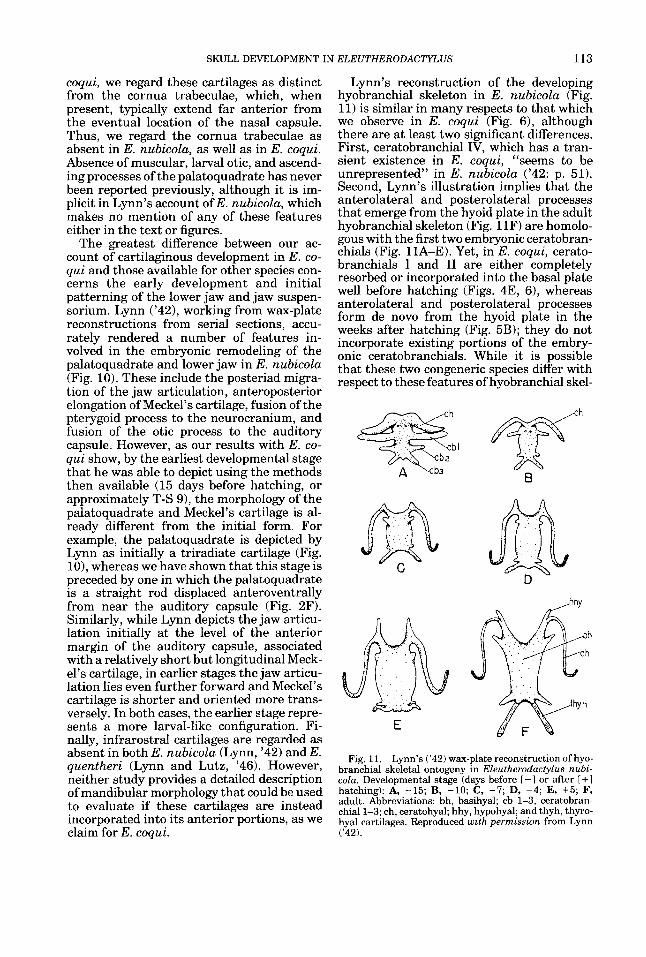
Lynn’s reconstruction of the developing hyobranchial skeleton in E. nubicola (Fig. 11) is similar in many respects to that which we observe in E. coqui (Fig. 6), although there are at least two significant differences. First, ceratobranchial IV, which has a tran- sient existence in E. coqui, “seems to be unrepresented” in E. nubicola (’42: p. 51). Second, Lynn’s illustration implies that the anterolateral and posterolateral processes that emerge from the hyoid plate in the adult hyobranchial skeleton (Fig. 11F) are homolo- gous with the first two embryonic ceratobran- chials (Fig. 11A-E). Yet, in E. coqui, cerato- branchials I and I1 are either completely resorbed or incorporated into the basal plate well before hatching (Figs. 4E, 61, whereas anterolateral and posterolateral processes form de novo from the hyoid plate in the weeks after hatching (Fig. 5B); they do not incorporate existing portions of the embry- onic ceratobranchials. While it is possible that these two congeneric species differ with respect to these features of hyobranchial skel-
m C
E
B
Fig. 11. Lynn’s (’42) wax-plate reconstruction of hyo- branchial skeletal ontogeny in Eleutherodactylus nubi- cola. Developmental stage (days before I -1 or after [ + 1 hatching): A, -15; B, -10; C, -7; D, -4; E, +5; F, adult. Abbreviations: bh, basihyal; cb 1-3, ceratobran- chiall-3; ch, ceratohyal; hhy, hypohyal; and thyh, thyro- hyal cartilages. Reproduced with permission from Lynn (’42).
114 J. HANKEN ET AL
eta1 development, we regard it as equally if not more probable that critical, albeit subtle, details may have been missed by Lynn, who depicted only five embryonic stages of hyo- branchial development.
By drawing attention to differences be- tween Lynn's work and ours, we do not mean to deprecate in any way his accomplish- ments, which represent outstanding contribu- tions to the study of direct development in amphibians. Rather, we offer these compari- sons to illustrate the additional resolving power provided by whole-mount immunohis- tochemistry, as compared to traditional meth- ods for visualizing embryonic form and mor- phogenesis, and to underscore how critical these early patterning events are for distin- guishing recapitulation and repatterning in the ontogeny and evolution of direct develop- ment. Cranial ossification
Cranial ossification in Eleutherodactylus coqui differs from that in metamorphosing anurans in two general ways, one of which- the ossification sequence-provides another example of repatterning. First, ossification is initiated during embryogenesis. Indeed, 13 of the adult complement of 17 bones form be- fore hatching, and one more forms within the next 2 mo, when the embryonic yolk supply is exhausted and the hatchling begins to feed. This is-very different from metamorphosing anurans, in which ossification typically com- mences at metamorphosis. In Bornbina orien- talis, for example, osteogenic differentiation is first visible at Gosner ('60) Stage 31, sev- eral weeks after the tadpole has begun to feed (Hanken and Hall, '88a). Consequently, the osteocranium of hatchling E. coqui bears a striking resemblance to that of a newly meta- morphosed froglet (cf. Fig. 5C-F). Thus, in the evolution of direct development in E. coqui, bone formation has been advanced into the embryonic period. It is unknown whether those mechanisms that mediate ossi- fication in metamorphosing taxa, such as en- docrine factors (Hanken and Hall, '88b), have been retained, or if new mechanisms of con- trol have been developed.
The second general difference concerns the cranial ossification sequence. Notwithstand- ing significant variation in the order of appearance of later-forming bones, metamor- phosing anurans show a remarkably consis- tent pattern of early ossification. The first bones to form typically are the exoccipital, frontoparietal, and parasphenoid, three
bones that invest or reinforce the braincase and otic region (Trueb, '85; de Sa, '88; Wiens, '89). The sequence in E. coqui is very dif- ferent, and primarily involves precocious os- sification of the jaws and jaw suspension (Table 2). While the parasphenoid is among the first bones to form, it is preceded by the angulosplenial and the squamosal, two bones which contribute to the jaw suspension and articulation. By the time the exoccipital and frontoparietal have formed, four additional bones are also present: the premaxilla, max- illa, and dentary, three tooth-bearing bones of the upper and lower jaws, and the ptery- goid, which braces the jaw suspension against the upper jaw and neurocranium.
The general pattern of ossification in E. coqui is consistent with the limited data on bone formation in three other species of Eleutherodactylus-guentheri (Lynn and Lutz, '46), nubicola (Lynn, '42), and ricordii (Hughes, '59). In each species, jaw elements are the first to ossify, which may represent a general and derived feature of skull develop- ment in the genus. However, whereas the sequence of ossification apparently is similar among species, the relative timing of bone differentiation is more variable. In E. nubi- cola, for example, formation of the septomax- illa, prootic, and nasal occurs after hatching, whereas these bones form during embryonic development in E. coqui. There are no data with which to assess if there are comparable interspecific differences in ossification once feeding begins.
Precocious ossification of jaw elements in direct-developing Eleutherodactylus is re- markably similar to the pattern of ossifica- tion in the viviparous caecilian Derrnophis rnexicanus (Wake and Hanken, '82). In this species, precocious ossification of the lower jaw and jaw suspension is part of a suite of derived developmental features that repre- sent an embryonic and fetal adaptation that permits intraoviductal feeding by the develop- ing offspring. The derived pattern of ossifica- tion in Eleutherodactylus may represent a similar trophic adaptation; the jaw elements serve as attachment sites for the prominent adductor and abductor musculature and pro- vide articulations that are important in ac- tive feeding, which commences soon after hatching.
Paradox of Eleutherodactylus In amphibians, the evolution of direct de-
velopment may have profound morphologi- cal, physiological, and ecological consequences
SKULL DEVELOPMENT IN ELEUTHERODACTYLUS 115
for the lineage involved (Bradford, '84; Brad- ford and Seymour, '88; Duellman, '89; McDi- armid, '78; Wake, '89). In plethodontid sala- manders, fundamental modifications to early ontogeny in some direct-developing taxa are believed to underlie the appearance of major structural innovations in adult morphology, which are absent in related metamorphosing forms (Roth and Wake, '85, '89; Wake, '82; Wake and Roth, '89). According to this model, the evolution of adult structures in metamor- phosing taxa is constrained by the presence of larval components which precede them in ontogeny. When, in the evolution of direct development and consequent abandonment of the free-living larval stage, the necessity to form the larval components is removed, this creates the opportunity for evolutionary loss of the larval components and the associated constraint on adult morphology. The princi- pal evidence supporting this model of "onto- genetic repatterning" (Wake and Roth, '89) is the correlation between direct develop- ment and morphological novelty, primarily involving the hyobranchial apparatus (Lom- bard and Wake, '86) and its neural control (Wake et al., '88). There are, however, few embryological data with which to evaluate directly the degree to which the ancestral biphasic ontogeny is actually modified in di- rect-developing taxa (e.g., Alberch, '87, '89).
Eleutherodactylus offers an excellent op- portunity to test the larval constraint hypoth- esis developed from plethodontid sala- manders. Indeed, it is tempting to speculate that the deletion of many larval features during the evolution of direct development in the lineage leading to Eleutherodactylus is directly linked to the evolutionary success of the genus, as measured by species diversity. With approximately 450 described species (Hedges, '89), Eleutherodactylus is the larg- est genus of extant terrestrial vertebrates. Yet, in Eleutherodactylus, the dramatic mod- ifications to the ancestral ontogeny have had little effect on adult cranial morphology, the variation of which among species is modest for such a large group and which does not differ in any profound way from that of closely related, metamorphosing taxa (Lynch, '71; Wake and Roth, '89). In other words, in Eleutherodactylus, the critical factor that could link the observed ontogenetic changes to evolutionary success-morphological nov- elty and diversification-is lacking. Thus, on- togenetic repatterning is not correlated with extensive morphological diversification and
does not seem to be causally linked to the evolutionary success of the genus. If, as seems reasonable, the evolution of direct develop- ment is an important causal component in the success of Eleutherodactylus, then it may be through associated ecological andlor phys- iological effects, such as emancipation from aquatic breeding sites and the invasion and occupation of novel terrestrial habitats (Hedges, '89; Heyer, '75). Interestingly, in lacking substantial diversification of adult morphology despite significant ontogenetic change, direct development in eleutherodac- tyline frogs is remarkably similar to that seen in many echinoderms (Ratf, '87; Strath- mann, '88).
The developmental basis of direct development
Cranial ontogeny in Eleutherodactylus differs substantially from that typical of meta- morphosing anurans; it comprises a complex mosaic of ancestral and derived features. The development of many exclusively larval carti- lages is omitted entirely, whereas the develop- ment of postmetamorphic cartilages and bones, many of which display an ontogenetic sequence unlike the presumed ancestral pat- tern, is advanced into the embryonic period. These features provide specific examples of repatterning of the ancestral ontogeny. Other components recapitulate in part their meta- morphic transformation in metamorphosing species. Yet, the derived pattern of devel- opment has had little impact on adult mor- phology. Clearly, the evolution of direct de- velopment in Eleutherodactylus involves substantially more than just acceleration and condensation of the ancestral biphasic ontog- eny found in metamorphosing anurans. It is thus inappropriate and misleading to de- scribe development in this group as involving a "metamorphosis. . . within the egg" (Adam- son et al., '60, p. 467; see also Infantino et al., '88).
Many of the differences in cranial ontogeny between direct-developing Eleutherodactylus and metamorphosing frogs may be described effectively in terms of changes in the relative timing and sequence of development, i.e., heterochrony (Elinson, '90). For example, the absence of larval cartilages and corre- sponding precocious appearance of postmeta- morphic structures during embryogenesis in E. coqui represents an excellent example of Haeckelian condensation (sensu Gould, '77). Yet, these heterochronic changes likely are only the most obvious manifestation of
116 J. HANKEN ET AL.
fundamental alterations to underlying devel- opmental mechanisms of pattern formation and morphogenesis (Raff and Wray, '89). For example, while it is correct to say that in Eleutherodactylus formation of the cornua trabeculae and suprarostral cartilages has been deleted from the ancestral ontogeny, this also means that patterning of the adult neurocranium involves a precursor arrange- ment that is significantly different from that in metamorphosing frogs. Explanation of de- rived ontogenetic patterns in terms of spe- cific perturbations of these basic developmen- tal mechanisms poses a compelling challenge to future research on direct development.
A precondition for the evolution of direct development in taxa with complex life cycles may be the independence of larval and adult developmental programs, which allows them to be readily dissociated (Elinson, '90; Raff, '87). Evidence in support of this hypothesis is available for sea urchins (Parks et al., '88; Raff, '87; Wray and McClay, '89; Wray and Raff, '891, but there are fewer data available for amphibians. Degeneration of a number of larval structures at metamorphosis suggests that, in anurans, there is a subset of cell lines that have an exclusively larval fate. Simi- larly, metamorphosing anurans show early developmental compartmentalization of cell lineages contributing to adult cranial muscu- lature (Alley, '89, '90). Compartmentaliza- tion of developmental precursors of larval vs. adult hyobranchial cartilages occurs in the plethodontid salamander Eurycea bislineata (Alberch and Gale, '86; Alberch et al., '85) and possibly other plethodontids, but it is not found in primitive urodeles (Alberch, '89). I t is not known if compartmentalization of adult components is characteristic of skull develop- ment in other amphibians, or even if it ap- plies to features other than the hyobranchial skeleton in plethodontid salamanders. Fi- nally, the fact that many larval cranial com- ponents are retained yet significantly remod- eled at metamorphosis suggests that there is a third class of cell lineages that have both larval and adult fates. This would seem to be supported by Eleutherodactylus coqui, in which several cartilages, instead of com- pletely eliminating the larval developmental program, initially assume an intermediate, mid-metamorphic configuration.
ACKNOWLEDGMENTS
This research was supported by NSF DCB- 9019624, NIH 1 R23 DE07190 and 1 F33
DE05610, NIH-BRSG RR07013-20 and RR07013-21, and the Council on Research and Creative Work, CU Boulder (J.H.); by the Pew Biomedical Scholars Award and NSF DCB-890522 (M.W.K.); and by the Hughes Foundation (N.I.). Dr. Thomas Linsenmayer, Tufts University, generously provided his monoclonal antibody to Type I1 collagen. Drs. Margaret Stewart and Daniel Townsend pro- vided assistance and advice in our attempts to establish a breeding colony of Eleuthero- dactylus coqui. Frogs were collected with the assistance and permission (permits DRN- 88-02 and DRN-90-24) of the Puerto Rican Department of Natural Resources and Sr. Eduardo L. Cardona and Sr. Jorge Moreno. Richard Elinson, David Jennings, David Wake, and several anonymous reviewers pro- vided valuable comments on earlier drafts.
LITERATURE CITED Adamson, L., R.G. Harrison, and I. Bayley (1960) The
development of the whistling frog Eleutherodactylus rnartznicenszs of Barbados. Proc. Zool. SOC. Lond. 133: 453469.
Alberch, P. (1987) Evolution of a developmental process- irreversibility and redundancy in amphibian metamor- phosis. In R.A. Raff and E.C. Raff (eds): Development as an Evolutionary Process. New York: Alan R. Liss, Inc., pp. 23-46.
Alberch, P. (1989) Development and the evolution of amphibian metamorphosis. In H. Splechtna and H. Hilgers (eds): Trends in Vertebrate Morphology, Fortschritte der Zoologie, Vol. 35. Stuttgart: Gustav Fischer Verlag, pp. 163-173.
Alberch, P., and E.A. Gale (1986) Pathways of cytodiffer- entiation during the metamorphosis of the epibran- chial cartilage in the salamander Eurycea bislineata. Dev. Biol. 117:233-244.
Alberch, P., G.A. Lewbart, and E.A. Gale (1985) The fate of larval chondrocytes during the metamorphosis of the epibranchial in the salamander Eurycea bislineata. J. Embryol. Exp. Morphol. 88:71-83.
Alley, K.E. (1989) Myofiber turnover is used to retrofit frog jaw muscles during metamorphosis. Am. J. Anat. 184:l-12.
Alley, K.E. (1990) Retrofitting larval neuromuscular cir- cuits in the metamorphosing frog. J. Neurobiol. 21: 1092-1107.
Altig, R., and G.F. Johnston (1989) Guilds of anuran larvae: relationships among developmental modes, mor- phologies, and habitats. Herp. Monogr. 3:81-109.
Bradford, D.F. (1984) Physiological features of embry- onic development in terrestrially-breeding plethodon- tid salamanders. In R.S. Seymour (ed): Respiration and Metabolism of Embryonic Vertebrates. Dordrecht: Dr. W. Junk Publ., pp. 87-98.
Bradford, D.F., and R.S. Seymour (1988) Influence of environmental PO, on embryonic hatching oxygen con- sumption, rate of development, and hatching in the frog Pseudophryne bibronz. Physiol. Zool. 61:475-482.
de Beer, G.R. (1937) The Development of the Vertebrate Skull. Oxford: Oxford University Press. (Paperback reprint: University of Chicago Press, Chicago, 1985.)
de Jongh, H.J. (1968) Functional morphology of the jaw apparatus of larvae and metamorphosing Rana ternpo- rarza L. Neth. J. Zool. 18rl-103.
SKULL DEVELOPMENT IN ELEUTHERODACTYLUS 117
Dent, J.A., and M.W. Klymkowsky (1989) Whole-mount analyses of cytoskeletal organization and function dur- ing oogenesis and early embryogenesis in Xenopus. In H. Shatten and G. Shatten (eds): The Cell Biology of Fertilization. Monographs in Cell Biology. New York: Academic Press, pp. 63-103.
Dent, J.A., A.G. Polson, and M.W. Klymkowsky (1989) A whole-mount immunocytochemical analysis of the ex- pression of the intermediate filament protein vimentin in Xenopus. Development 105:61-74.
de Sa, R.O. (1988) Chondrocranium and ossification se- quence of Hyla lanciformis. J. Morphol. 19.5345-355.
Dingerkus, G., and L.D. Uhler (1977) Enzyme clearing of alcian blue stained whole small vertebrates for demon- stration of cartilage. Stain Technol. 52:229-232.
Duellman, W.E. (1989) Alternative life-history styles in anuran amphibians: evolutionary and ecological impli- cations. In M.N. Bruton (ed): Alternative Life-History Styles of Animals. Dordrecht: Kluwer Academic Publ., pp. 101-126.
Duellman, W.E., and L. Trueb (1986) Biology of the Amphibians. New York: McGraw-Hill Co.
Elinson, R.P. (1990) Direct development in frogs: wiping the recapitulationist slate clean. Semin. Dev. Biol. 1 :263-270.
Elinson, R.P., E.M. del Pino, D.S. Townsend, F.C. Cuesta, and P. Eichhorn (1990) A practical guide to the develop- mental biology of terrestrial-breeding frogs. Biol. Bull. 179t163-177.
Gosner, K.L. (1960) A simplified table for staging anuran embryos and larvae with notes on identification. Herpe- tologica 16t183-190.
Gould, S.J. (1977) Ontogeny and Phylogeny. Cambridge: Harvard Univ. Press.
Hall, B.K. (1985) The role of movement and tissue inter- actions in the development and growth of bone and secondary cartilage in the clavicle of the embryonic chick. J . Embryol. Exp. Morphol. 93:133-152.
Hanken, J. (1989) Development and evolution in amphib- ians. Am. Sci. 77:336-343.
Hanken, J., and B.K. Hall (1988a) Skull development during anuran metamorphosis: I. Early development of the first three bones to form-the exoccipital, the parasphenoid, and the frontoparietal. J. Morphol. 195: 247-256.
Hanken, J., and B.K. Hall (1988b) Skull development during anuran metamorphosis: 11. Role of thyroid hor- mone in osteogenesis. Anat. Emhryol. (Berl.) 178:219- 227.
Hanken, J., and C.H. Summers (1988a) Skull develop- ment during anuran metamorphosis: 111. Role of thy- roid hormone in chondrogenesis. J. Exp. Zool. 246:156- 170.
Hanken, J., and C.H. Summers (1988b) Developmental basis of evolutionary success: cranial ontogeny in a direct-developing anuran. Am. Zool. 28r12A.
Hanken, J., and R.J. Wassersug (1981) The visible skele- ton. Funct. Photogr. 16~22-26, 44.
Hanken, J., M.W. Klymkowsky, D.W. Seufert, and N.E. Ingebrigtsen (1990) Evolution of cranial patterning in anuran amphibians analyzed using whole-mount immu- nohistochemistry. Am. Zool. 30:138A.
Hedges, S.B. (1989) Evolution and biogeography of West Indian frogs of the genus Eleutherodactylus: slow- evolving loci and the major groups. In C.A. Woods (ed): Biogeography of the West Indies: Past, Present, and Future. Gainesville: Sandhill Crane Press, pp. 305- 370.
Heyer, W.R. (1975) A preliminary analysis of the interge- neric relationships of the frog family Leptodactylidae. Smithsonian Contrib. Zool. No. 199:l-55.
Hughes, A. (1959) Studies in embryonic and larval devel- opment in Amphibia. I. The embryology of Eleuthero- dactylus ricordii, with special reference to the spinal cord. J . Emhryol. Exp. Morphol. 7t22-38.
Hughes, A. (1965) The development of behaviour in the embryo of Eleutherodactylus rnartinicensis (Amphibia, Anura). Proc. Zool. SOC. Lond. 144:153-161.
Hughes, A. (1966) Spontaneous movements in the em- bryo of Eleutherodactylus martinicensis. Nature 21 1: 51-53.
Humason, G.L. (1979) Animal Tissue Techniques, 4th ed. San Francisco: W.H. Freeman and Co.
Infantino, R.L., W.W. Burggren, and D.S. Townsend (1988) Physiology of direct development in the Puerto Rican frog Eleutherodactylus coqui. Am. Zool. 28:23A.
Klymkowsky, M.W., and J. Hanken (1991) Whole-mount staining ofXenopus and other vertebrates. In B.K. Kay and H.B. Peng (eds): Xenopus Zaeuis: Practical Uses in Cell and Molecular Biology. Methods in Cell Biology, Vol. 36 (in press).
Lauder, G.V., and K.F. Liem (1989) The role of historical factors in the evolution of complex organismal func- tions. In D.B. Wake and G. Roth (eds): Complex Organ- ismal Functions: Integration and Evolution in Verte- brates. Chichester: John Wiley &Sons, Ltd., pp. 63-78.
Linsenmayer, T.F., and M.J.C. Hendrix (1980) Mono- clonal antibodies to connective tissue macromolecules: type I1 collagen. Biochem. Biophys. Res. Commun. 92:440-446.
Lombard, R.E., and D.B. Wake (1986) Tongue evolution in the lungless salamanders, family Plethodontidae. IV. Phylogeny of plethodontid salamanders and the evolution of feeding dynamics. Syst. Zool. 35:532-551.
Lutz, B. (1947) Trends towards non-aquatic and direct development in frogs. Copeia 1947:242-252.
Lutz, B. (1948) Ontogenetic evolution in frogs. Evolution 2:29-39.
Lynch, J.D. (1971) Evolutionary relationships, osteology, and zoogeography of leptodactyloid frogs. Univ. Kans. Mus. Nat. Hist., Misc. Publ. No. 53:l-238.
Lynn, W.G. (1942) The embryology of Eleutherodactylus nubicola, an anuran which has no tadpole stage. Con- trib. Embryol. Carnegie Inst. Wash. Publ. 541:27-62.
Lynn, W.G. (1961) Types of amphibian metamorphosis. Am. Zool. 1:151-161.
Lynn, W.G., and B. Lutz (1946) The development of Eleutherodactylus guentheri Stdnr. 1864. Bol. Mus. Nac. 2001. 71:l-46.
McDiarmid, R.W. (1978) Evolution of parental care in frogs. In G.M. Burghardt and M. Bekoff (eds): The Development of Behavior: Comparative and Evolution- ary Aspects. New York: Garland STPM Press.
Orton, G.L. (1951) Direct development in frogs. Turtox News29t2-6.
Parks, A.L., B.A. Parr, J.-E. Chin, D.S. Leaf, and R.A. Raff(1988) Molecular analysis of heterochronic changes in the evolution of direct developing sea urchins. J . Evol. Biol. 1t27-44.
Raff, R.A. (1987) Constraint, flexibility, and phylogenetic history in the evolution of direct development-in sea urchins. Dev. Biol. 119~6-19.
Raff, R.A., and G.A. Wray (1989) Heterochrony: develop- mental mechanisms and evolutionary results. J . Evol. Biol. 2t409-434.
Roth, G., and D.B. Wake (1985) Trends in the functional morphology and sensorimotor control of feeding behav- ior in salamanders: an example of the role of internal dynamics in evolution. Acta Biotheor. (Leiden) 34: 175- 192.
Roth, G., and D.B. Wake (1989) Conservatism and inno- vation in the evolution of feeding in vertebrates. In D.B. Wake and G. Roth (eds): Complex Organismal
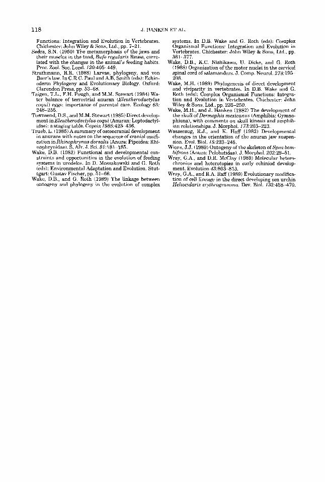
118 J. HANKEN ET AL
Functions: Integration and Evolution in Vertebrates, Chichester: John Wiley & Sons, Ltd., pp. 7-21.
Sedra, S.N. (1950) The metamorphosis of the jaws and their muscles in the toad, Bufo regularis Reuss, corre- lated with the changes in the animal’s feeding habits. Proc. Zool. SOC. Lond. 120t405-449.
Strathmann, R.R. (1988) Larvae, phylogeny, and von Baer’s law. In C.R.C. Paul andA.B. Smith (eds): Echin- oderm Phylogeny and Evolutionary Biology. Oxford: Clarendon Press, pp. 53-68.
Taigen, T.L., F.H. Pough, and M.M. Stewart (1984) Wa- ter balance of terrestrial anuran (Eleutherodactylus coqui) eggs: importance of parental care. Ecology 65: 248-255.
Townsend, D.S., and M.M. Stewart (1985) Direct develop- ment in Eleutherodactylus coqui (Anuran: Leptodactyl- idae): a staging table. Copeia 1985:423-436.
Trueb, L. (1985) A summary of osteocranial development in anurans with notes on the sequence of cranial ossifi- cation in Rhinophrynus dorsalis (Anura: Pipoidea: Rhi- nophrynidae). S. Afr. J. Sci. 81:181-185.
Wake, D.B. (1982) Functional and developmental con- straints and opportunities in the evolution of feeding systems in urodeles. In D. Mossakowski and G. Roth (eds): Environmental Adaptation and Evolution. Stut- tgart: Gustav Fischer, pp. 51-66.
Wake, D.B., and G. Roth (1989) The linkage between ontogeny and phylogeny in the evolution of complex
systems. In D.B. Wake and G. Roth (eds): Complex Organismal Functions: Integration and Evolution in Vertebrates, Chichester: John Wiley & Sons, Ltd., pp. 361-377.
Wake, D.B., K.C. Nishikawa, U. Dicke, and G. Roth (1988) Organization of the motor nuclei in the cervical spinal cord of salamanders. J . Comp. Neurol. 278t195- 208.
Wake, M.H. (1989) Phylogenesis of direct development and viviparity in vertebrates. In D.B. Wake and G. Roth (eds): Complex Organismal Functions: Integra- tion and Evolution in Vertebrates, Chichester: John Wiley & Sons, Ltd., pp. 235-250.
Wake, M.H., and J. Hanken (1982) The development of the skull of Dermophis mexicanus (Amphibia: Gymno- phiona), with comments on skull kinesis and amphib- ian relationships. J. Morphol. 173:203-223.
Wassersug, R.J., and K. Hoff (1982) Developmental changes in the orientation of the anuran jaw suspen- sion. Evol. Biol. 15.223-246.
Wiens, J.J. (1989) Ontogeny of the skeleton of Spea bom- bifrons (Anura: Pelobatidae). J. Morphol. 202:29-51.
Wray, G.A., and D.R. McClay (1989) Molecular hetero- chronies and heterotopies in early echiniod develop- ment. Evolution 43:803-813.
Wray, G.A., and R.A. Raff (1989) Evolutionary modifica- tion of cell lineage in the direct-developing sea urchin Heliocidaris erythrogramma. Dev. Biol. 132t458-470.


