Embed Size (px)
Citation preview

Copyright statement
This copy of the thesis has been supplied on condition that anyone who consults
it is understood to recognise that its copyright rests with its author and that no
quotation from the thesis and no information derived from it may be published
without the author's prior consent.
THE EFFECT OF IONISING RADIATION
ON MUSSELS
By
EMILY LAURA VERNON
A thesis submitted to the University of Plymouth in
partial fulfilment for the degree of
DOCTOR OF PHILOSOPHY
School of Biological and Marine Sciences
March 2019
I
ACKNOWLEDGMENTS
Completing this PhD has been one of the most challenging, exhausting,
wonderful experiences of my life, I am forever grateful to all the people who made
it possible.
First and foremost I would like to thank Professor Awadhesh Jha, your support
and guidance over the last 4 years has been invaluable. Thank you for giving me
the freedom to carry out this PhD in my own way, and for allowing me to grow as
a research scientist. I am also grateful to Professor Jim Smith and Professor Alex
Ford for their helpful advice. To Lorna Dallas, my friend, mentor and supervisor,
thank you for your patience, encouragement, and for teaching me more than I
could ever give credit for. Your passion and love of science is inspiring. I thank
the technical and academic staff of which I have had the pleasure of working with
over the years, namely Will Vevers, Andy Atfield, Jo Triner, Lee Hutt, Alex Taylor,
Will Blake, Nick Crocker and Tim Bean.
To my lovely friends, co-workers and fellow RATE PhD students (of which there
are far too many to mention, but you know who you are!), thank you for those
nights in Cuba, adventures in Chernobyl and endless walks around the campus,
and most importantly for the endless encouragement. A special mention to Fliss,
we went through this crazy journey together and I’m so glad we had each other
to see it through!
To Ed, my forever supportive, incredible partner, thank you for believing in me
even when I didn’t believe in myself. For those hours we spent collecting mussels
from Bude Canal (which haunts me to this day), for keeping my spirits up when
II
things went wrong in the lab (again!), for sticking by my side through all the ups
and downs, I am forever grateful. I’d also like to thank your lovely parents,
Stephen and Helen (and Leo the Cat!), for providing a place of refuge from PhD
life.
Last but by no means least, to my amazing family, Dad, Mum, Richard and of
course, as per tradition, Tom the cat, without your constant love and support I
would have never been able to finish my PhD.
I dedicate this thesis to my Mum and Dad, you made this possible.
I love you both dearly.

III
AUTHORS DECLARATION
At no time during the registration for the degree of Doctor of Philosophy has the
author been registered for any other University award without prior agreement of
the Doctoral College Quality Sub-Committee. Work submitted for this research
degree at the University of Plymouth has not formed part of any other degree
either at the University of Plymouth or at another establishment.
This work was jointly funded by the Natural Environment Research Council
(NERC), the Environment Agency (EA) and Radioactive Waste Management
Limited (RWM) under the Radioactivity and the Environment (RATE) programme
(Grant no.: NE/L000393/1).
Word count for the main body of this thesis:
~ 55,575
Signed:
Date: 17.09.2018

IV
V
ABSTRACT
The effect of ionising radiation on mussels.
Emily Laura Vernon
Ionising radiations have undoubtedly played a vital role in modern society. There
is however growing concern of their increasing presence in the environment
whether by permitted or accidental release, along with other contaminants. While
the effects of ionising radiation (IR) on human and mammalian models are well
studied, the impacts on aquatic organisms, which play important roles for
ecosystem sustainability are yet to be fully understood. This is particularly for
chronic, low dose, environmentally relevant exposures, bearing in mind that many
of the discharged radionuclides have long half-lives, to which biota are exposed.
In this context a multi-biomarker, multi-species approach was adopted to
investigate IR-induced (phosphorus-32, 32P) response, alone and in combination
with copper (Cu, an environmentally ubiquitous metal), in two ecologically
relevant bivalve species, the marine Mytilus galloprovincialis (MG) and
freshwater Dreissena polymorpha (DP) under laboratory conditions. The chosen
species play integral roles (ecological, economic and environmental) within
coastal and freshwater bodies. Accumulation patterns of Cu (18, 32, 56 µg L-1)
and 32P (0.10, 1, and 10 mGy d-1) in isolation varied between the species and
tissues. In turn, dose rates (32P) to specific tissues were found to exceed those
established for the whole-body for these species. This work demonstrated the
importance of determining dose rate at tissue level, and highlighted digestive
gland and gill as key tissues of interest for subsequent biological assays. In terms
of biomarker responses for DNA damage, given that DNA is an important target
VI
for the actions of IR, induction of novel biomarker gamma H2AX (γ-H2AX) was
performed alongside more classical techniques, including comet and
micronucleus (MN) assays, along with molecular approaches (i.e. transcriptional
expression of key genes involved in stress responses) and behavioural level
changes. Genotoxicity was well correlated with Cu concentration, and 32P dose
rate, both as single and combined stressors. Significant DNA damage was noted
at 32 µg L-1 (Cu), and 1 mGy d-1 (32P). In terms of relative sensitivity, overall, MG
appeared more sensitive for the induction of γ-H2AX and DNA strand breaks
(comet assay), both biomarkers of exposures. In contrast, DP was found to be
more susceptible for the induction of MN (biomarker of effects) in the target
tissues. The study also highlights that a single screening dose rate may not be
adequate to protect all species. The integrated, multi-biomarker, species and
tissue approach adopted in the current study provides a thorough, robust
methodology which could be further applied to other ecologically relevant
species, or reference organisms. The study contributes to the limited amount of
information related to understanding of IR-induced biological responses on
aquatic biota, alone and in combination with a relevant metallic contamination. It
goes some way towards providing the necessary scientific basis for the
development of adequate protective policies for both coastal and inland water
bodies.
VII
TABLE OF CONTENTS
ACKNOWLEDGMENTS ...................................................................................... I
AUTHORS DECLARATION ............................................................................... III
ABSTRACT ........................................................................................................ V
LIST OF FIGURES ......................................................................................... XIII
LIST OF TABLES .......................................................................................... XXIII
ABBREVIATIONS, SYMBOLS AND ACRONYMS ....................................... XXVI
Chapter 1: General introduction ..................................................................... 1
1.1 Environment and human health .................................................................... 3
1.2 Ionising radiation ........................................................................................... 4
1.2.1 Radioactivity in the aquatic environment ............................................. 4
1.2.2 Relative Biological Effectiveness (RBE) .............................................. 5
1.2.3 Radiation types and ecological relevance ........................................... 6
1.3 Levels of biological organisation ................................................................. 10
1.3.1 Radionuclide bioaccumulation and dosimetry ................................... 10
1.3.2 Biological end points.......................................................................... 11
1.4 Aquatic organisms as bioindicators ............................................................. 18
1.4.1 Marine and freshwater bivalves: Use in ecotoxicology ...................... 21
1.5 Experimental design: Laboratory or field, and external factors ................... 26
1.6 Overall aims of research ............................................................................. 28
1.7 Hypothesis……………………………………………………………………... 29
Chapter 2: Methods and method development ............................................ 31
2.1 Chemicals and suppliers ............................................................................. 32
2.2 Mussel collection and maintenance ............................................................ 32
2.2.1 Genetic identification of Mytilus spp. ..................................................... 36
2.3 Cellular extractions for biological assays .................................................... 39
2.3.1 Haemolymph extraction ..................................................................... 39
2.3.2 Tissue digestion ................................................................................ 40
2.4 Cell viability assessment with Trypan blue .................................................. 41
2.5 Biological assays ........................................................................................ 42
2.5.1 Micronucleus (MN) assay .................................................................. 42
2.5.2 Comet assay ..................................................................................... 44
VIII
2.5.2.1 Optimisation and validation of the comet assay using hydrogen
peroxide (H2O2)……………………………………………………………. 46
2.5.3 53BP1 and γ-H2AX assays ............................................................... 48
2.5.3.1 Validation of the γ-H2AX assay using H2O2 as a reference agent
………………………………………………………………………………… 54
2.5.4 Transcriptional expression of key genes ........................................... 55
2.5.4.1 RNA extraction……………………………………………………... 57
2.5.4.2 cDNA synthesis…………………………………………………….. 57
2.5.4.3 Real-time Polymerase chain reaction (qPCR)………………... 58
2.5.4.4 Data analysis and interpretation…………………………………. 58
2.6 Physiological observations .......................................................................... 59
2.6.2 Byssal attachment and valve activity ................................................. 59
2.7 Determination of metal concentration using ICP-OES and ICP-MS ............ 61
2.7.1 Tissue preparation ............................................................................. 61
2.7.2 Determination of metal in water samples .......................................... 61
2.8 Ionising Radiation ....................................................................................... 62
2.8.1 Selection of radionuclide and determination of radiation dose levels 62
2.8.2 Radiation safety and experimental design ......................................... 64
2.8.3 Liquid scintillation counting (LSC) ..................................................... 65
2.8.3.1 Tissue preparation for LSC……………………………………... 65
2.8.3.2 Liquid scintillation counting: Analysis of water and mussel tissue
samples………………………………………………………………………. 68
2.8.4 Dosimetry calculations and the ERICA tool ....................................... 69
2.8.5 Optimisation experiments .................................................................. 72
2.8.5.1 Pilot study………………………………………………………….. 72
2.8.5.2 LSC optimisation study…………………………………………… 76
2.9 Statistical analysis....................................................................................... 78
Chapter 3: Assessing relative sensitivity of marine and freshwater
bivalves following exposure to copper: Application of classical and novel
genotoxicological biomarkers ....................................................................... 81
3.1 Introduction ................................................................................................. 83
3.2 Materials and methods ................................................................................ 86
3.2.1 Chemicals and suppliers ................................................................... 86
3.2.2 Mussel exposure conditions .............................................................. 87
3.2.3 Sampling procedures ........................................................................ 89
IX
3.2.4 Biological assays ............................................................................... 90
3.2.4.1 Isolation of gill cells for genotoxicity assays……………………...90
3.2.4.2 Comet assay to determine DNA strand breaks………………... 90
3.2.4.3 Analysis of micronuclei (MN) formation………………………. 90
3.2.4.4 Induction of γ-H2AX foci……………………………………….. 90
3.2.5 Determination of Cu concentration in soft tissues and in water
samples ...................................................................................................... 91
3.2.5.1 Copper (Cu) analysis in tissues using Inductively Coupled
Plasma Mass Spectometry (ICP-
MS)…………………………………………………………………………. 91
3.2.5.2 Determination of Cu in water samples using ICP-
MS…………………………………………………………………………... 91
3.2.6 Statistical analysis ............................................................................. 91
3.3 Results ........................................................................................................ 92
3.3.1 Tissue specific Cu accumulation ....................................................... 92
3.3.2 Genotoxic effects and repair capacity in mussel gill cells .................. 95
3.4 Discussion ................................................................................................... 98
3.4.1 Tissue specific Cu accumulation ....................................................... 98
3.4.2 Cu induced genotoxicity in gill cells ................................................. 100
3.5 Conclusions .............................................................................................. 105
Chapter 4: Relative comparison of tissue specific bioaccumulation and
radiation dose estimation in marine and freshwater bivalves following
exposure to phosphorus-32 ......................................................................... 107
4.1 Introduction ............................................................................................... 109
4.2 Materials and methods .............................................................................. 112
4.2.1 Chemicals and suppliers ................................................................. 112
4.2.2 Mussel exposure conditions ............................................................ 113
4.2.3 Sampling procedures and liquid scintillation counting ..................... 114
4.2.4 Dosimetry and the ERICA TOOL ..................................................... 114
4.2.5 Statistical analysis ........................................................................... 115
4.3 Results ...................................................................................................... 115
4.3.1 Activity concentrations in water ....................................................... 115
4.3.2 Activity concentrations in bivalve soft tissue, shell and IMW……… 116
4.3.2.1 Soft tissue…………………………………………………………. 118
4.3.2.2 Internal mussel water (IMW) and shell…………………………. 119
4.3.2.3 Tissue specific 32P accumulation……………………………….. 119
X
4.3.2.4 Faecal matter and pseudofaeces……………………………….. 120
4.3.3 Dosimetry ........................................................................................ 121
4.4 Discussion ................................................................................................ 123
4.5 Conclusions .............................................................................................. 129
Chapter 5: Assessing relative biomarker responses in marine and
freshwater bivalve molluscs following exposure to phosphorus 32 (32P):
Application of genotoxicological and molecular biomarkers ................... 131
5.1 Introduction ............................................................................................... 133
5.2 Materials and methods .............................................................................. 136
5.2.1 Chemicals and suppliers ................................................................. 136
5.2.2 Mussel exposure conditions ............................................................ 136
5.2.3 Sampling procedures ...................................................................... 139
5.2.4 Biological assays ............................................................................. 140
5.2.4.1 Isolation of digestive gland and gill cells……………………….. 140
5.2.4.2 Comet assay to determine DNA strand breaks……………... 140
5.2.4.3 Analysis of micronuclei induction……………………………… 140
5.2.4.4 Induction of γ-H2AX foci………………………………………. 140
5.2.4.5 Determination of transcriptional levels of key genes……….. 141
5.2.5 Determination of 32P and Cu concentration in water samples ......... 141
5.2.5.1 Determination of 32P in water samples using liquid scintillation
counting…………………………………………………………………… 141
5.2.5.2 Determination of Cu concentration in water samples using
Inductively Coupled Plasma Mass Spectrometry (ICP-MS)……… 141
5.2.4 Dosimetry and the ERICA tool ......................................................... 141
5.2.5 Statistical analysis ........................................................................... 142
5.3 Results ...................................................................................................... 143
5.3.1 Genotoxic response following in vivo exposures to 32P ................... 143
5.3.1.1 Comet assay to determine DNA strand breaks……………... 145
5.3.1.2 Analysis of micronuclei (MN) induction……………………..... 145
5.3.1.3 Induction of γ-H2AX foci………………………………………... 145
5.3.2. Transcriptional expression of key genes ........................................ 146
5.3.3 Whole-body and tissue specific dose rates ..................................... 150
5.4 Discussion ................................................................................................ 150
5.4.1 Dosimetry and dose-response relationship ..................................... 150
5.4.2 32P induced genotoxic response in gill and digestive gland cells ..... 151
XI
5.4.2.1 Comet assay to determine DNA strand breaks………………. 151
5.4.2.2 Induction of γ-H2AX foci………………………………………… 153
5.4.2.3 Analysis of micronuclei (MN) induction………………………… 155
5.4.3 Expression of key genes ................................................................. 156
5.4.4 Environmental implications and future research .............................. 158
5.5 Conclusions .............................................................................................. 159
Chapter 6: Evaluation of interactive effects of phosphorus-32 and copper
on marine and freshwater bivalve molluscs ............................................... 161
6.1 Introduction ............................................................................................... 163
6.2 Materials and methods .............................................................................. 167
6.2.1 Chemicals and suppliers ................................................................. 167
6.2.2 Mussel exposure conditions ............................................................ 167
6.2.3 Sampling procedures....................................................................... 168
6.2.4 Biological assays ............................................................................. 169
6.2.4.1 Isolation of digestive gland and gill cells……………………… 169
6.2.4.2 Comet assay to determine DNA strand breaks…………….. 169
6.2.4.3 Analysis of micronuclei (MN) induction…………………….. 169
6.2.4.4 Induction of γ-H2AX foci………………………...………….….. 169
6.2.4.5 Determination of transcriptional expression of key genes…. 170
6.2.5 Behavioural observations: Valve movement and byssus
attachment…………………………………………………………………… 170
6.2.6 Water quality measurements and 32P and Cu analyses .................. 170
6.2.6.1 Determination of 32P in water samples using liquid scintillation
counting…………………………………………………………………… 170
6.2.6.2 Determination of Cu concentration in water samples using
Inductively Coupled Plasma Mass Spectrometry (ICP-MS)…………. 171
6.2.7 Dosimetry and the ERICA TOOL ..................................................... 171
6.2.8 Statistical analysis ........................................................................... 172
6.2.8.1 Multivariate analysis……………………………………………. 172
6.3 Results ...................................................................................................... 173
6.3.1 Genotoxic response following in vivo exposures to 32P and Cu ....... 174
6.3.1.1 Comet assay to determine DNA strand breaks…………….. 178
6.3.1.2 Analysis of micronuclei (MN) formation……………………….. 178
6.3.1.3 Induction of γ-H2AX foci………………………………………… 179
6.3.2 Transcriptional expression of key genes ......................................... 179
XII
6.3.3 Multivariate analysis of biomarker reactions .................................... 181
6.3.4 Behavioural observations: Valve movement and byssus
attachment……………………………………………………………………...182
6.4 Discussion ................................................................................................ 184
6.4.1 Biomarker interactions ..................................................................... 184
6.4.2 32P induced genotoxic response in gill and digestive gland cells ..... 185
6.4.3 Transcriptional expression of key genes ......................................... 189
6.4.4 Behavioural observations: Valve movement and byssus
attachment…………………………………………………………………….. 191
6.4.5 Environmental implications and future research .............................. 192
6.5 Conclusions .............................................................................................. 193
Chapter 7: General discussion and future perspectives ........................... 195
7.1 Environmental radiation and dosimetry ..................................................... 197
7.1.1 Radiation dosimetry ......................................................................... 197
7.2 Laboratory vs. field exposures .................................................................. 199
7.3 Mussels as bioindicators of environmental health ..................................... 201
7.3.1 M. galloprovincialis and D. polymorpha ........................................... 201
7.3.2 Life history stage, transgenerational effects and epigenetics .......... 203
7.4 Suitable biomarkers and radiation science ............................................... 205
7.4.1 Advancements in radiological research: The development of novel
biomarkers ............................................................................................... 206
7.4.1.1 Proteomics………………………………………………………… 206
7.4.1.2 Metabolomics……………………………………………………... 208
7.5 Conclusions .............................................................................................. 210
APPENDICES ................................................................................................ 213
University of Plymouth courses ................................................................... 213
External courses .......................................................................................... 213
Presentations and posters ........................................................................... 213
Press releases ............................................................................................. 215
Radiation protection document I: Bioaccumulation experiment ................... 216
Radiation protection document II: Biological end-point experiments ........... 234
List of accepted publications ....................................................................... 248
Radiation hazard map………………………………………………………..… 249
References .................................................................................................... 250
XIII
LIST OF FIGURES
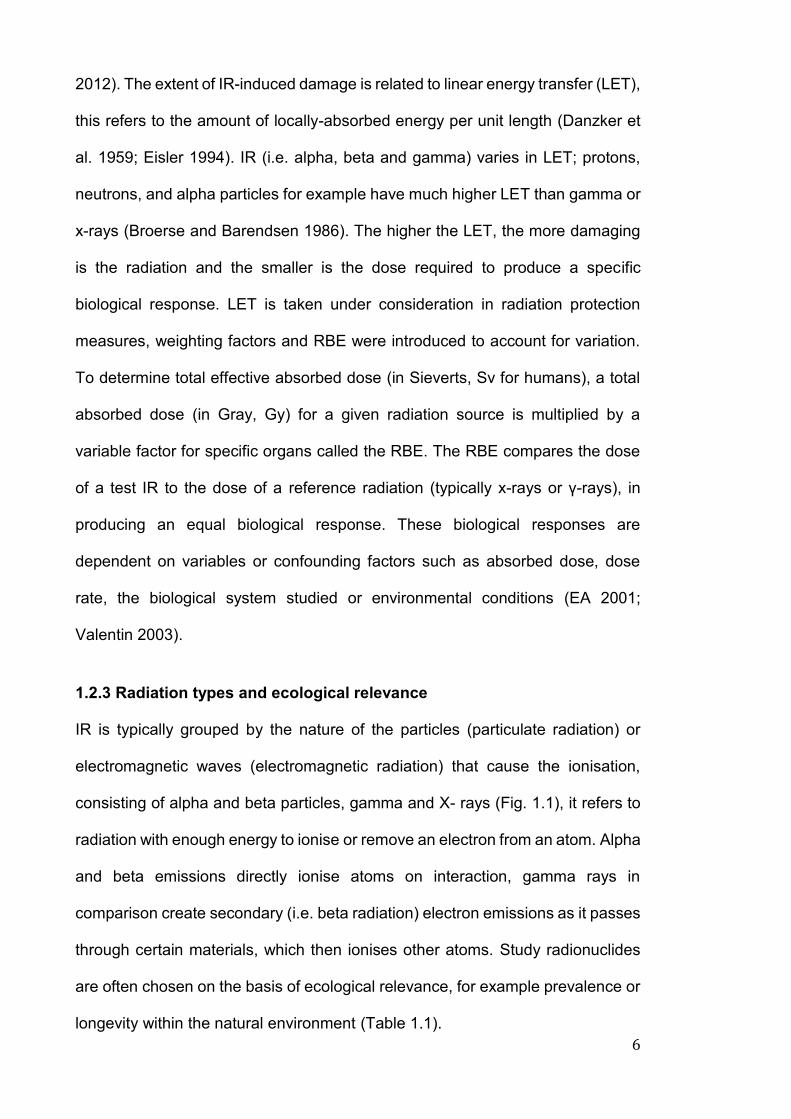
Figure 1.1. Penetration and shielding of the different types of IR…... [7]
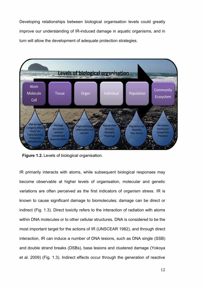
Figure 1.2. Levels of biological organisation…………………………. [12]
Figure 1.3. Direct and indirect IR-induced DNA damage……………. [13]
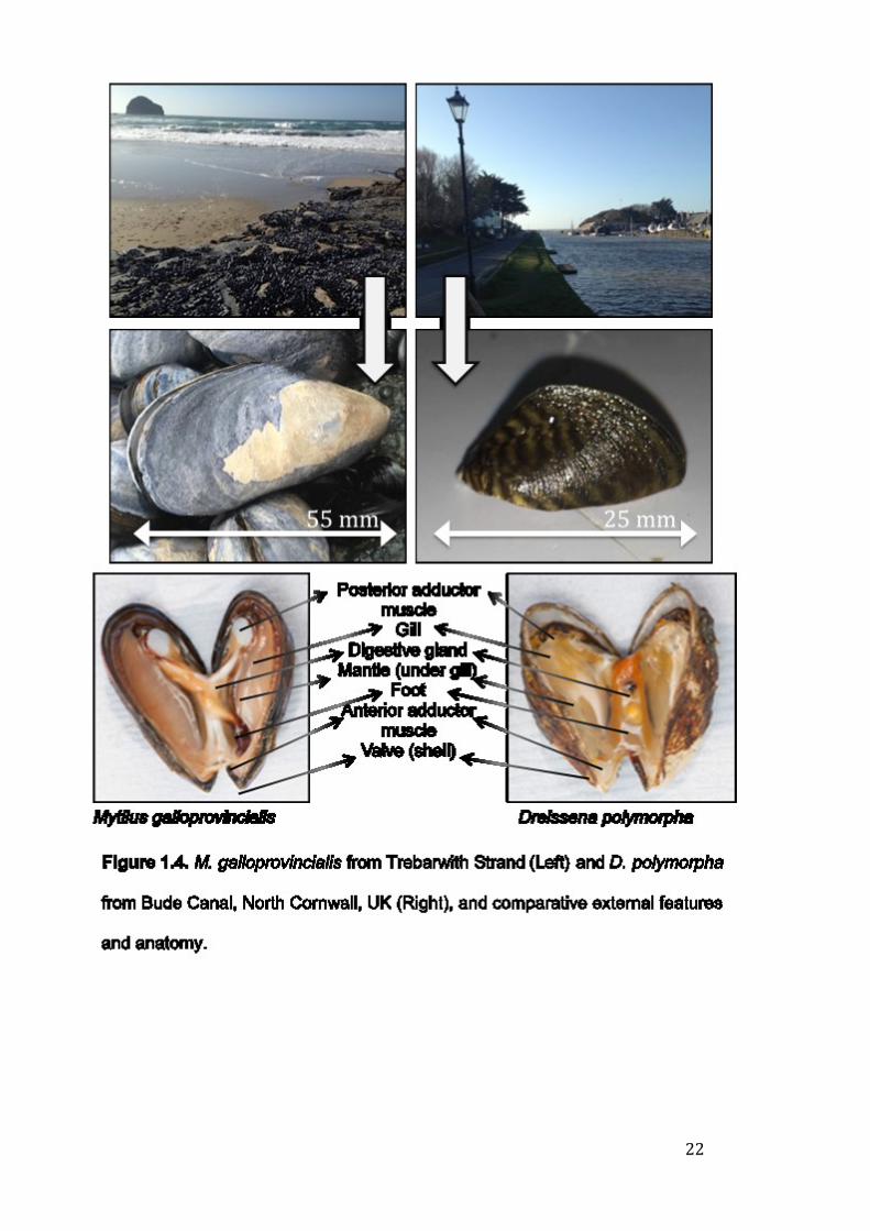
Figure 1.4. M. galloprovincialis from Trebarwith Strand (Left) and D.
polymorpha from Bude Canal, North Cornwall, UK (Right),
and comparative external features and anatomy………. [22]
Figure 2.1. Map to show location of collection sites for M.
galloprovincialis (Trebarwith, Cornwall) and D. polymorpha
(Bude, Cornwall) mussels in the UK………………………[33]
Figure 2.2. Cell viability (%) of D. polymorpha: Optimisation of water
types. (River, bottled and artificial) for maintenance in the
laboratory (n = 6)…………………………………………... [34]
Figure 2.3. Genetic identification of the Mytilus spp. (A) Positions of
PCR primers in the Glu-5’ gene in the M. edulis and M.
galloprovincialis and (B) sequences of the primers (modified
from (Inoue et al. 1995)…………………………………… [37]
Figure 2.4. Me 15/16 PCR products visualised on agarose gel (Mytilus
sp.) (Image: L. Dallas, 2014) (A) Mytilus edulis and (B)
Mytilus galloprovincialis…………………………………... [38]
Figure 2.5. Haemolymph extraction from adductor muscle (B, red
circles) in: M. galloprovincialis (A) and D. polymorpha
(C)………………………………………………………...… [39]
Figure 2.6. Different cell types in M. galloprovincialis (left) and D.
polymorpha (right): Haemocytes (A) gill cells (B) digestive
XIV
gland cells (C) mantle cells and (D) posterior adductor
muscle cells (E) (100 μm scale)………………………..… [41]
Figure 2.7. Cell viability in M. galloprovincialis haemocytes using the
Trypan blue exclusion method. Red circles surround viable
(clear) and non-viable (blue cells)………………..……… [42]
Figure 2.8. Simplified micronuclei (MN) formation (top). Normal cell (A)
cell with micronuclei (B) (bottom). Both cells are M.
galloprovincialis gill cells stained with ethidium bromide. [43]
Figure 2.9. Scoring comet assay slides: M. galloprovincialis gill cells:
Varying levels of DNA damage in response to hydrogen
peroxide exposure (top), head and tail of comet
(bottom)………………………………………………...…... [45]
Figure 2.10. DNA damage (% Tail DNA) in M. galloprovincialis (A, B and
C) and D. polymorpha (D, E and F) haemocytes, gill and
digestive gland cells, following exposure to varying
concentrations of hydrogen peroxide. Asterisks (*, ** or ***)
are indicative of significant differences (p < 0.05, 0.01,
0.001) from the corresponding control. SD is standard
deviation of mean data. n = 4………………………….… [47]
Figure 2.11. Diagram illustrating the recruitment of phosphorylated H2AX
following ATM activation (A). Bottom images show: Healthy
cell stained with DAPI (B), DAPI stained cell with foci
induction (C), and illustration showing cell and green foci (D)
(images B and C: D.polymorpha gill cells)………………. [48]
Figure 2.12. Average number of γ-H2AX foci in M. galloprovincialis and
D. polymorpha gill cells following exposure to varying
XV
concentrations of hydrogen peroxide for 1 h. SD is standard
deviation of mean data. Images show induction of γ-H2AX-
foci in D. polymorpha gill cells. n = 4………………..….. [51]
Figure 2.13. Optimisation of γ-H2AX assay. Images show microscope
image of DAPI stained M. galloprovincialis gill cells,
subsequent to varying blocks (Bovine serum albumin [BSA],
foetal bovine serum [FBS], milk powder and normal goat
serum)……………………………………………………… [53]
Figure 2.14. Average number of γ-H2AX foci in M. galloprovincialis and
D. polymorpha gill cells following exposure to varying
concentrations of hydrogen peroxide. SD is standard
deviation of mean data. Asterisks (*, ** or ***) are indicative
of significant differences (p < 0.05, 0.01, 0.001) from the
corresponding control. n = 4…………………..…….…… [54]
Figure 2.15. Diagram illustrating stages of gene expression protocol. [59]
Figure 2.16. (A) Byssus thread produced by M. galloprovinicalis, (B) M.
galloprovinicalis filter feeding and (C) D. polymorpha filter
feeding……………………………………………………… [60]
Figure 2.17. 32P shielding: Designs and measurements for shielding (A-
C), shielding with beaker covered with acrylic lid in individual
compartment (D), safety exposure using fluorescein as
surrogate for 32P (E-F)…………………………………….. [65]
Figure 2.18. Mechanism behind liquid scintillation counting (LSC)….. [66]
Figure 2.19. Whole mussel tissue homogenised in different volumes of DI
water (5, 10, 20 mL)……………………………………….. [67]
XVI
Figure 2.20. Series 32P dilution showing nominal and achieved values.
Error bars are SD of mean data………………………….. [69]
Figure 2.21. Diagram to show calculation of dose rates in M.
galloprovincialis and D. polymorpha. Ksib and Chi are
scaling parameters, representing the lengths of the minor
axes in terms of length of the major axis of the ellipsoid. [71]
Figure 2.22. 32P accumulation in M. galloprovincialis and D. polymorpha
whole soft tissue, over time. Error bars show standard
deviation of mean data. n = 3 (MG), 14 (DP). Significance
(*** is p < 0.001) is shown between species on each
sampling day………………………………………………. [75]
Figure 2.23. 32P accumulation in M. galloprovincialis and D. polymorpha
whole soft tissue, shell and internal mussel water. Asterisks
(*, ** or ***) are indicative of significant differences (p < 0.05,
0.01, 0.001) between species. Error bars show standard
deviation of mean data. n = 3 (MG), 14 (DP)……………. [77]
Figure 3.1. Overall experimental design: Experiment 1 (EXP1):
Determination of Genotoxic responses; Experiment 2
(EXP2): Determination of bioaccumulation of copper
(Cu)…………………………………………………………. [88]
Figure 3.2. Tissue specific accumulation of copper in M.
galloprovincialis (left) and D. polymorpha (right), microgram
per gram of mussel tissue (dry weight) in control and
exposed treatment groups. Asterisks (*, ** or ***) are
indicative of significant differences (p < 0.05, 0.01, 0.001)
XVII
from the corresponding control. SD is standard deviation of
mean data. n = 9…………………………………………… [93]
Figure 3.3. Pearson’s correlation analyses of Cu accumulation in gill
tissue. Top to bottom: DNA damage (% tail DNA), induction
of γ-H2AX foci and micronuclei (MN) formation in M.
galloprovincialis (left) and D. polymorpha (right). n = 9.. [94]
Figure 3.4. Genotoxic responses in M. galloprovincialis and D.
polymorpha gill cells following a 10 day exposure to copper
(Cu). (A) DNA damage (% tail DNA), (B) Induction of
micronuclei (MN) and (C) induction of γ-H2AX foci Asterisks
(*, ** or ***) are indicative of significant differences (p < 0.05,
0.01, 0.001) from the corresponding control. SD is standard
deviation of mean data. Images show (left) control cell and
(right) damaged cell. n = 9……………………………….... [96]
Figure 3.5. Pearson’s correlation analyses. Top to bottom: % tail DNA
and induction of γ-H2AX foci; % tail DNA and induction of
MN and induction of MN and γ-H2AX in M. galloprovincialis
(left) and D. polymorpha (right). n = 9………………….... [97]
Figure 4.1. Tissue specific accumulation of 32P in M. galloprovincialis
(MG, left) and D. polymorpha (DP, right), total activity per
gram of mussel tissue in control and irradiated treatment
groups. Asterisks (*, ** or ***) are indicative of significant
differences (p < 0.05, 0.01, 0.001) from the corresponding
control. Lower case letters denote variation between similar
tissues (species specific, i.e. there is a significant difference
in 32P bioconcentration in digestive gland between each
XVIII
treatment group). Upper case letters denote significant
variation in similar tissue and treatment group between
species (e.g. MG digestive gland tissue values [0.1, 1 and
10 mGy/d treatments] are significantly different than DP
values in the corresponding treatment, there is no difference
in control samples). SD is standard deviation of mean data.
IMW – Internal mussel water……………………………. [117]
Figure 4.2. Proportion of 32P in tissue after 10 day exposure in M.
galloprovincialis (left) and D. polymorpha (right). IMW –
Internal mussel water……………………………………. [118]
Figure 4.3. Activity levels (Bq g-1) in M. galloprovincialis and D.
polymorpha faecal matter (dry weight), following 32P
exposure. Asterisks (*, ** or ***) are indicative of significant
differences (p < 0.05, 0.01, 0.001) from the corresponding
control. SD is standard deviation of mean data……….. [120]
Figure 5.1 Schematic diagram showing experimental design. Images
from M. galloprovincialis digestive gland cells………… [138]
Figure 5.2 Genotoxic effects in M. galloprovincialis and D. polymorpha
gill cells following a 10-day exposure to 32P. Asterisks (*, **
or ***) are indicative of significant differences (p < 0.05, 0.01,
0.001) from the corresponding control. SD is standard
deviation of mean data. n = 9…………………………… [144]
Figure 5.3 Pearson’s correlation analyses of: (top to bottom) % tail DNA
and induction of MN, % tail DNA and γ-H2AX foci, and
induction of MN and γ-H2AX foci. M. galloprovincialis
XIX
digestive gland (left) and M. galloprovincialis gill (right). n =
9……………………………………………………………. [147]
Figure 5.4 Pearson’s correlation analyses of: (top to bottom) % tail DNA
and induction of MN, % tail DNA and γ-H2AX foci, and
induction of MN and γ-H2AX foci. D. polymorpha digestive
gland (left) and D. polymorpha gill (right). n = 9………. [148]
Figure 5.5 Relative expression ratios (RER) of key genes in M.
galloprovincialis and D. polymorpha gill and digestive gland
cells following a 10-day exposure to 32P. Data are
normalised for reference genes (ef1, actin) and controls.
Error bars indicate the 95% confidence intervals. Asterisks
(*, ** or ***) are indicative of significant differences (p < 0.05,
0.01, 0.001) from the corresponding control. n = 9……. [149]
Figure 6.1 Genotoxic effects and subsequent repair in M.
galloprovincialis and D. polymorpha gill and digestive gland
cells following a 10 day exposure to 32P and Cu, alone and
in combination. Asterisks (*, ** or ***) are indicative of
significant differences (p < 0.05, 0.01, 0.001) from the
corresponding control. Letters are indicative of significant
differences (p < 0.05) between species tissue (i.e. MG gill
tissue). SD is standard deviation of mean data. n = 9…. [175]
Figure 6.2a Scatter plots (A, C), principal component (PCA) and cluster
analysis of the biomarker data, not including behavioural
response. Vectors indicate the directionality of specific
biomarkers. (A, B) M. galloprovincialis digestive gland and
XX
(C, D) gill tissue. Asterisks (*) are indicative of significant
differences (p < 0.05)…………………………………….. [176]
Figure 6.2b Scatter plots (A, C), principal component (PCA) and cluster
analysis of the biomarker data, not including behavioural
response. Vectors indicate the directionality of specific
biomarkers. (A, B) D. polymorpha digestive gland and (C, D)
gill tissue. Asterisks (*) are indicative of significant
differences (p < 0.05)…………………………………….. [177]
Figure 6.3 Relative expression ratios (RER) of key genes in M.
galloprovincialis and D. polymorpha gill and digestive gland
cells following a 10-day exposure to 32P and Cu, alone and
in combination. Data are normalised for reference genes
(ef1, actin) and controls. Error bars indicate the 95%
confidence intervals. Asterisks (*, ** or ***) are indicative of
significant differences (p < 0.05, 0.01, 0.001) from the
corresponding control. n = 9…………………………….. [180]
Figure 6.4 Behavioural effects in M. galloprovincialis (MG) and D.
polymorpha (DP) following a 10 day exposure to 32P and Cu,
alone and in combination. (A) Number of attached mussels
per beaker, on days 1, 3, 5, 7, 9 and 11 of exposure, (Ba, b)
Average number of mussels (Ba: MG, Bb: DP) actively
filtering each experimental day, per beaker. SD is standard
deviation of mean data. n = 3……………………………. [183]

XXI
XXII
LIST OF TABLES
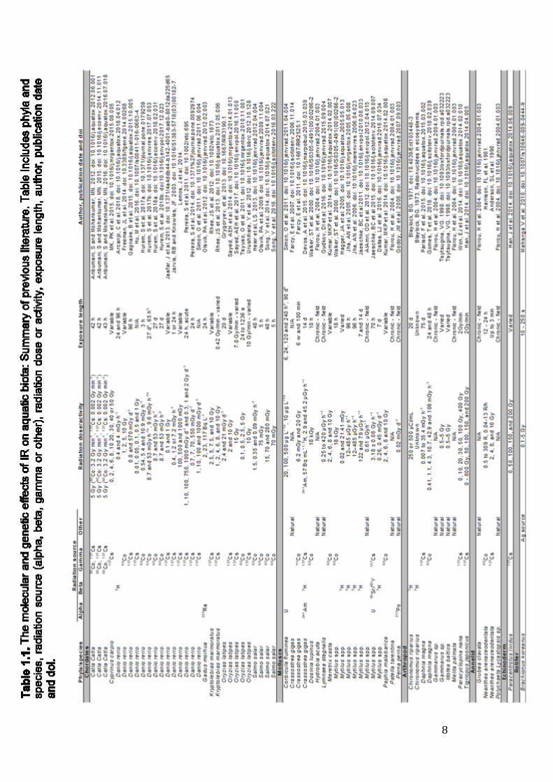
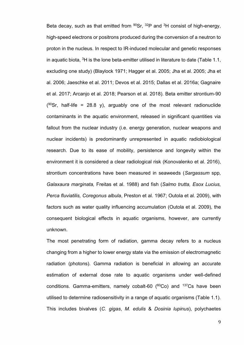
Table 1.1. The molecular and genetic effects of IR on aquatic biota:
Summary of previous literature. Table includes phyla and
species, radiation source (alpha, beta, gamma or other),
radiation dose or activity, exposure length, author,
publication date and doi…………………………………….. [8]
Table 1.2. The molecular and genetic effects of IR on aquatic biota:
Summary of previous literature (1971-2018). Table includes
phyla and species, endpoints investigated and source of
radiation used in exposure (α, alpha; β, beta; γ, gamma; *,
other [i.e. Natural]). No. of symbols represents the amount
of times each biomarker has been utilised in scientific
literature......................................................................... [20]
Table 2.1. Summary of primer pairs used for reference and target
genes in M. galloprovincialis and D. polymorpha: Name,
abbreviation, primer sequences and accession number. [56]
Table 2.2. Table outlining properties of phosphorus-32…………… [63]
Table 2.3. Alternative radionuclides considered for experimental
design………………………………………………………. [63]
Table 2.4. Table illustrating custom organism option in the ERICA tool;
D. polymorpha digestive gland (DG) and M. galloprovincialis
DG, occupancy factors and organism geometry. Ksib and
Chi are scaling parameters, representing the lengths of the
minor axes in terms of length of the major axis of the
ellipsoid…………………………………………………….. [70]
XXIII
Table 2.5. ERICA tool criteria used for estimating dose rate from water
concentrations. Kd is distribution coefficient, CR is
concentration ratio………………………………………… [72]
Table 2.6. Nominal dose rates (mGy d-1), total dose (mGy 10 d-1) and
required water concentration (Bq L-1) for 32P exposures,
using the ERICA tool default criteria…………………….. [73]
Table 2.7. Weight of M. galloprovincialis and D. polymorpha (g)…. [73]
Table 2.8. Water concentrations (Bq L-1), calculated by the ERICA tool,
that give to correct dose rates of 0.1, 1 and 10 mGy d-1 in M.
galloprovincialis and D. polymorpha……………………... [78]
Table 3.1. Water quality parameters (pH, temperature, salinity and
dissolved oxygen [DO]) and copper concentrations in water
(µg L-1). Data are presented as mean ± standard deviation
(SD)…………………………………………………………. [89]
Table 4.1. Activity levels in water samples (Bq L-1) per treatment in M.
galloprovincialis and D. polymorpha (SD is standard
deviation of mean data). Asterisks (*) denote nominal activity
concentrations……………………………………………. [115]
Table 4.2. Order of 32P accumulation in soft tissue, shell and IMW in M.
galloprovincialis and D. polymorpha individuals, order shows
tissue with the highest to lowest bioconcentration (Bq g-1) in
all treatment groups……………………………………… [116]
Table 4.3. Table to show (a) the expected dose rates in mGy d-1 and
μGy d-1 (for the ERICA tool), (b) the water activity
concentrations that give the correct dose rate (Bq L-1) for
both species *as calculated from preliminary experiments,
XXIV
(c) the average dose rate achieved in mGy d-1 and (d) ERICA
tool water activity concentrations that give the correct dose
(Bq L-1), corrected to 35 g whole mussel weight/beaker.[121]
Table 4.4. Table to show the expected and achieved dose rates (mGy
d-1) in M. galloprovincialis and D. polymorpha digestive gland
using custom geometry in the ERICA tool (Tier 2)…….. [122]
Table 5.1 Water quality parameters (pH, temperature, salinity and
dissolved oxygen [DO]), copper concentrations in water (µg
L-1) and 32P concentrations in water (Bq L-1), for both
species. Data is presented as mean ± standard
deviation…………………………………………………... [139]
Table 5.2 Table to show (a) the expected dose rates in mGy d-1 and
(b) the average dose rate achieved in M. galloprovincialis
and D. polmorpha whole-body, digestive gland and gill tissue
(mGy d-1)………………………………………………….. [142]
Table 6.1 Table to show (a) the expected dose rates in mGy d-1 and
(b) the average dose rate achieved in M. galloprovincialis
and D. polmorpha whole-body, digestive gland and gill tissue
(mGy d-1)………………………………………………….. [171]
Table 6.2 Water quality parameters (pH, temperature, salinity and
dissolved oxygen [DO]), copper concentrations in water (µg
L-1) and 32P concentrations in water (Bq L-1), for both
species. Data is presented as mean ± standard
deviation…………………………………………………. [174]

XXV

XXVI
ABBREVIATIONS, SYMBOLS AND ACRONYMS µg Microgram
µGy Microgray
μM Micromole
γ-H2AX Gamma Histone 2AX
2D-GE 2 dimensional gel electrophoresis
137Cs Caesium-137
3H Tritium
32P Phosphorus-32
32S Sulphur-32
90Sr Strontium-90
Al Aluminium
ALI Annual limit on intake
AM Adductor muscle
ATP Adenosine triphosphate
BaP Benzo(a)pyrene
Bq Becquerel
CA Chromosomal aberration
CAT Catalase
Cd Cadmium
cDna Complimentary DNA
CF Concentration factor
CR Concentration ratio
XXVII
CPM Counts per minute
Cu Copper
DAPI 4′,6-diamidino-2-phenylindole, dihydrochloride
DDR DNA damage response
DG Digestive gland
DI Deionised water
DO Dissolved oxygen
DOC Dissolved organic carbon
DP Dreissena polymorpha
DSB Double strand break
D.W Dry weight
EA Environment Agency
ERICA Environmental Risk from Ionising Contaminants:
Assessment and Management
EZ Exclusion Zone
FASSET Framework for Assessment of Environmental Impact
FPG Formamidopyrimidine glycosilase
GBq Gigabecquerel
GC-MS Gas chromatography-mass spectrometry
GST Glutathione-S-Transferase
Gy Gray
H2O2 Hydrogen peroxide
HCl Hydrochloric acid
Hg Mercury
XXVIII
HR-MS High-resolution mass spectrometry
HSP Heat Shock Protein
IAEA International Atomic Energy Agency
ICP-MS Inductively coupled plasma mass spectrometry
ICRP International Commission on Radiological Protection
IMW Internal mussel water
IR Ionising radiation
K Potassium
LC-MS Liquid chromatography-mass spectrometry
LD Lethal dose
LET Linear energy transfer
LMA Low melting point agarose
LSC Liquid Scintillation Counting
MBq Megabecquerel
ME Mytilus edulis
MG Mytilus galloprovincialis
mGy Milligray
MN Micronuclei
MoA/s Mode/s of action
NPP Nuclear power plant
mRNA Messenger RNA
MS Mass spectrometry
mSv Millisievert
NGS Next generation sequencing
XXIX
NMA Normal melting point agarose
NMR Nuclear magnetic resonance
OST Other soft tissue
PAH Polycyclic aromatic hydrocarbon
PBq Petabecquerel
PCA Principal Component Analysis
PCR Polymerase chain reaction
qPCR Quantitative PCR
RAPS Reference animals and plants
RBE Relative biological effectiveness
RE Ravenglass estuary
RER Relative mRNA expression ratio
RMS Radioactive materials supervisor
RNASeq RNA sequencing
ROS Reactive oxygen species
RPA Radiation protection advisor
SCE Sister Chromatid Exchange
SOD Superoxide dismutase
Sp/p Species
SSB Single strand break
Sv Sieverts
USSR Union of Soviet Socialist Republics

XXX

1
Chapter 1
General Introduction

2
3
1.1 Environment and human health Environmental quality is inherently and complexly linked with human health and
well-being. Anthropogenic environmental pollutants such as radionuclides,
metals, biological and physical agents (e.g. thermal stress, hypoxia) can threaten
both human and non-human biota through a series of complex transfer exposure
pathways and physiological processes (Frumkin 2001; Jha 2004; Jha 2008). For
the adequate protection of the environment and its inhabitants it is vital that
interactions between environment and organism, at different levels of biological
organisation (i.e. molecular to ecosystem levels) are understood. This in turn will
both improve tools for decision-making and provide more effective environmental
protection policies.
With 71% of the earth covered in water, the aquatic environment and inhabiting
organisms undoubtedly play a vast role in ecosystem structure and functioning,
offering endless ecological, economic, environmental and recreational services.
From an ecological perspective aquatic organisms are an integral part of their
environments, and often act as keystone species, providing ecosystem services.
As an example, bivalves are known to improve water quality, influence nutrient
dynamics and biogeochemical processes and provide habitats and nursery
grounds for other aquatic life (Edebo et al. 2000; Borthagaray and Carranza 2007;
Petersen et al. 2014). Furthermore, organisms such as fish and shellfish are an
important and often vital source of protein for both human and non-human biota,
many livelihoods rely on seafood as a means of income. Contaminated food is
the main pathway of radionuclides into the human body, environmental
contaminants discharged in aquatic ecosystems have the potential to reduce the
availability of an invaluable commodity (Howard et al. 2013; Steinhauser et al.
2014).
4
1.2 Ionising radiation
1.2.1 Radioactivity in the aquatic environment
With applications ranging from nuclear energy generation, diagnostic tools in
medical, pharmaceutical, research industries and consumer products, it is clear
that radioactive materials contribute significantly to modern day society.
Nonetheless radionuclides, in common with other contaminants have the
potential to enter the natural environment via various transfer pathways, with
aquatic ecosystems being the final recipient for many anthropogenic
contaminants (Pentreath 1988; Jha 2004). Although exposed to natural
background levels of IR (i.e. cosmic, geological), it is the environmental impact of
anthropogenic radionuclides, either by regulated or accidental release that is of
a growing concern to society, governments, industry and regulators (Hu et al.
2010).
Certain organisations (e.g. nuclear power industry, research organisations,
universities and hospitals) are permitted by authorities to discharge regulated
levels of radionuclides into the environment (Hu et al. 2010; IAEA 2010). For
example in 2011, 8.92 × 10¹⁵ Bq and 2.07 × 10¹⁵ Bq of liquid tritium (3H) was
discharged by the nuclear fuel reprocessing plants at La Hague, France and
Sellafield, UK, respectively (OSPARcommission 2011). In the same year total
liquid discharges of beta-emitters (not including 3H) from nuclear installations
under the OSPAR convention (1992 Convention for the Protection of the Marine
Environment of the North-East Atlantic), were 2.59 × 10¹³ Bq. Approximately 70%
was attributed to Sellafield, Springfield’s (UK, nuclear fuel production installation)
and La Hague (OSPARcommission 2011). Despite public and policy concerns
over safety, both human and environmental, nuclear power generation offers a
5
relatively low cost, low carbon form of energy. Combined depletion of natural
resources and rapid population growth will continue to drive the requirement for
nuclear production and reprocessing installations.
Radionuclides are also released into the environment via nuclear accidents, such
as from Chernobyl, Ukraine (formally USSR, 1986) and Fukushima, Japan (2011),
or nuclear weapons testing. Following the nuclear incident at the Fukushima
Daiichi nuclear power plant (NPP), resulting from the Tohoku earthquake a wide
range of terrestrial and aquatic environments were contaminated (Chino et al.
2011). Contamination occurred from atmospheric dispersion, direct release and
discharge with the primary source of highly radioactive water originating from a
trench surrounding the NPP (IAEA 2015). Oceanic releases were estimated at 1
– 6 petabecquerel (PBq) for caesium-137 (137Cs) and 10 - 20 PBq for iodine-131
(131I); strontium-90 (90Sr) and caesium-134 (134Cs) were also of concern
(Yamaguchi et al. 2014; IAEA 2015).
Considering the influx of radioactive contaminants into the aquatic environment,
whether from accidental or permitted release it is vital to quantify potential risks
to humans, and potential detrimental biological responses in biota (Dallas et al.
2012). Understanding IR-induced effects on various levels of biological
organisation, in a range of biota will help provide the necessary scientific
background for radiation protection.
1.2.2 Relative Biological Effectiveness (RBE)
Radiation weighting factors and relative biological effectiveness (RBE) are useful
tools in radiobiology. They allow for the evaluation of risks and potential
consequences of radioactive contamination on aquatic organisms (Dallas et al.
6
2012). The extent of IR-induced damage is related to linear energy transfer (LET),
this refers to the amount of locally-absorbed energy per unit length (Danzker et
al. 1959; Eisler 1994). IR (i.e. alpha, beta and gamma) varies in LET; protons,
neutrons, and alpha particles for example have much higher LET than gamma or
x-rays (Broerse and Barendsen 1986). The higher the LET, the more damaging
is the radiation and the smaller is the dose required to produce a specific
biological response. LET is taken under consideration in radiation protection
measures, weighting factors and RBE were introduced to account for variation.
To determine total effective absorbed dose (in Sieverts, Sv for humans), a total
absorbed dose (in Gray, Gy) for a given radiation source is multiplied by a
variable factor for specific organs called the RBE. The RBE compares the dose
of a test IR to the dose of a reference radiation (typically x-rays or γ-rays), in
producing an equal biological response. These biological responses are
dependent on variables or confounding factors such as absorbed dose, dose
rate, the biological system studied or environmental conditions (EA 2001;
Valentin 2003).
1.2.3 Radiation types and ecological relevance
IR is typically grouped by the nature of the particles (particulate radiation) or
electromagnetic waves (electromagnetic radiation) that cause the ionisation,
consisting of alpha and beta particles, gamma and X- rays (Fig. 1.1), it refers to
radiation with enough energy to ionise or remove an electron from an atom. Alpha
and beta emissions directly ionise atoms on interaction, gamma rays in
comparison create secondary (i.e. beta radiation) electron emissions as it passes
through certain materials, which then ionises other atoms. Study radionuclides
are often chosen on the basis of ecological relevance, for example prevalence or
longevity within the natural environment (Table 1.1).
7
High-LET alpha decay consists of heavy, short-range particles with two neutrons
and two protons being ejected from the nucleus of a radioactive atom. Typically
occurring from the heaviest nuclides (Uranium, U; Radium, Ra; Polonium, Po) it
is the least penetrating form of radiation, able to travel only a short distance
through air. Alpha radiation is most harmful when inhaled (e.g. radon gas),
swallowed or absorbed into an organism (Olsvik et al. 2012). Bioaccumulation of
alpha emitters, such as Polonium-210 (210Po) has been documented in aquatic
organisms including bivalves (Connan et al. 2007; Štrok and Smodiš 2011; Feroz
Khan et al. 2014), fish (Carvalho et al. 2011; Štrok and Smodiš 2011) and
cephalopods (Štrok and Smodiš 2011), due to the potential threat to human
health via consumption. In terms of radiotoxicity research, alpha emitters are
underrepresented (Table. 1.1).
Figure 1.1. Penetration and shielding of the different types of IR.
8
9
Beta decay, such as that emitted from 90Sr, 32P and 3H consist of high-energy,
high-speed electrons or positrons produced during the conversion of a neutron to
proton in the nucleus. In respect to IR-induced molecular and genetic responses
in aquatic biota, 3H is the lone beta-emitter utilised in literature to date (Table 1.1,
excluding one study) (Blaylock 1971; Hagger et al. 2005; Jha et al. 2005; Jha et
al. 2006; Jaeschke et al. 2011; Devos et al. 2015; Dallas et al. 2016a; Gagnaire
et al. 2017; Arcanjo et al. 2018; Pearson et al. 2018). Beta emitter strontium-90
(90Sr, half-life = 28.8 y), arguably one of the most relevant radionuclide
contaminants in the aquatic environment, released in significant quantities via
fallout from the nuclear industry (i.e. energy generation, nuclear weapons and
nuclear incidents) is predominantly unrepresented in aquatic radiobiological
research. Due to its ease of mobility, persistence and longevity within the
environment it is considered a clear radiological risk (Konovalenko et al. 2016),
strontium concentrations have been measured in seaweeds (Sargassum spp,
Galaxaura marginata, Freitas et al. 1988) and fish (Salmo trutta, Esox Lucius,
Perca fluviatilis, Coregonus albula, Preston et al. 1967; Outola et al. 2009), with
factors such as water quality influencing accumulation (Outola et al. 2009), the
consequent biological effects in aquatic organisms, however, are currently
unknown.
The most penetrating form of radiation, gamma decay refers to a nucleus
changing from a higher to lower energy state via the emission of electromagnetic
radiation (photons). Gamma radiation is beneficial in allowing an accurate
estimation of external dose rate to aquatic organisms under well-defined
conditions. Gamma-emitters, namely cobalt-60 (60Co) and 137Cs have been
utilised to determine radiosensitivity in a range of aquatic organisms (Table 1.1).
This includes bivalves (C. gigas, M. edulis & Dosinia lupinus), polychaetes
10
(Neanthes arenaceodentata), crustaceans (Daphnia magna) and fish (D. rerio,
S. salar, Catla catla, Cyprinus carpio, Kryptolebias marmoratus, Oryzias latipes)
(Harrison 1981, 87; Walker et al. 2000; Olsvik et al. 2010; Farcy et al. 2011; Rhee
et al. 2012, 13; Freeman et al. 2014; Song et al. 2014, 16; Kumar et al. 2015,
2017; Parisot et al. 2015; Anbumani and Mohankumar 2016; Hurem et al. 2017,
18). Such radionuclides allow researchers to elucidate potential mechanisms
involved in IR-induced biological responses under controlled, simplified
experimental settings. To note, radionuclides such as 32P and x-rays can be
adopted as a baseline to measure damage from differing radionuclides, or used
as a substitute for highly impacting radioactive emissions. Laboratory derived
data illustrating chronic IR-induced biological effects, with IR source and dose
rate representative of realistic environmental conditions are undoubtedly crucial
in understanding and predicting the impact of current and future radiation
exposures in the aquatic environment (Pereira et al. 2011; Gudkov et al. 2012;
Olsvik et al. 2012; Parisot et al. 2015; Dallas et al. 2016a).
1.3 Levels of biological organisation
1.3.1 Radionuclide bioaccumulation and dosimetry
In radiological studies accurate dosimetry is crucial (Stark et al. 2017). Aquatic
organisms can be exposed externally or internally to radionuclides of varying
physicochemical forms through water, sediment or via ingestion. The behaviour
and fate of radionuclides when accumulated into organs/tissue can differ,
resulting in variability of delivered dose. For ease of dose calculation, available
assessment tools (i.e. ERICA) often make certain adjustments when determining
dose such as (a) radionuclide within the organism is uniformly distributed,
therefore calculated dose is for the whole body and (b) organisms are ellipsoidal
11
in shape. More accurate environmental dosimetry and subsequent protective
policies can be achieved in part by determining tissue specific radionuclide
uptake and dose, before investigating biological response. It is essential to
establish the relationship between exposure, tissue specific uptake, dose rate
and effect on the aquatic biota, as varying tissue sensitivity could result in a
detrimental biological response at levels presumed to be acceptable. Many
approaches have been developed to determine suitable benchmarks or
screening dose rates which aim to filter out situations of no concern. As outlined
in a thorough review (Andersson et al. 2009), a generic (all species) “no effect”
dose rate of 10 μGy h−1 (0.24 mGy d-1) has been adopted as a screening value
(i.e. ERICA tool), dose rates under this value are thought to result in minimal risk
to the individual or population.
1.3.2 Biological end points
IR may cause adverse effects at all levels of biological organisation (Fig. 1.2),
from molecular to ecosystem levels (Clements 2000; Dallas et al. 2012). In non-
human biota, environmental protection is often deemed successful where no
observable effects are seen on ecosystems at a population level, or higher levels
of organisation. A conservative approach is to identify relationships between
contaminant and organism at an individual level (i.e. molecular, genetic to
reproductive level change), such information allows for the development of
acceptable levels of natural and anthropogenic radionuclides. Combined data
from omics technologies and classical methods (i.e. histopathology, population
genetics and ecology) can be linked through bioinformatics to illustrate potential
impacts of contaminant exposure in a given organism, or population (Miracle and
Ankley 2005; Dallas et al. 2012), such information is extremely valuable.
12
Developing relationships between biological organisation levels could greatly
improve our understanding of IR-induced damage in aquatic organisms, and in
turn will allow the development of adequate protection strategies.
IR primarily interacts with atoms, while subsequent biological responses may
become observable at higher levels of organisation, molecular and genetic
variations are often perceived as the first indicators of organism stress. IR is
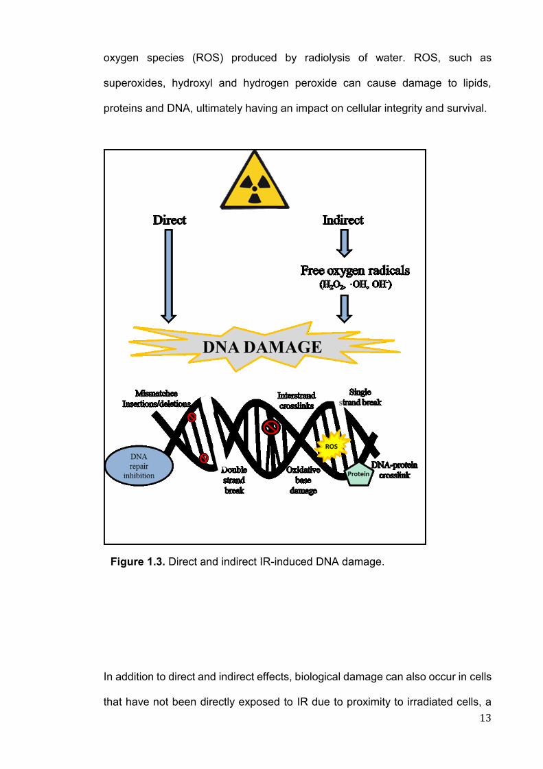
known to cause significant damage to biomolecules; damage can be direct or
indirect (Fig. 1.3). Direct toxicity refers to the interaction of radiation with atoms
within DNA molecules or to other cellular structures. DNA is considered to be the
most important target for the actions of IR (UNSCEAR 1982), and through direct
interaction, IR can induce a number of DNA lesions, such as DNA single (SSB)
and double strand breaks (DSBs), base lesions and clustered damage (Yokoya
et al. 2009) (Fig. 1.3). Indirect effects occur through the generation of reactive
13
oxygen species (ROS) produced by radiolysis of water. ROS, such as
superoxides, hydroxyl and hydrogen peroxide can cause damage to lipids,
proteins and DNA, ultimately having an impact on cellular integrity and survival.
In addition to direct and indirect effects, biological damage can also occur in cells
that have not been directly exposed to IR due to proximity to irradiated cells, a
Figure 1.3. Direct and indirect IR-induced DNA damage.
14
principle coined the bystander effect (Seymour and Mothersill 2004; Mothersill et
al. 2006; Mothersill and Seymour 2012; Chevalier et al. 2015). While the nature
of the communication system involved in producing a bystander response is not
yet fully understood, information from irradiated to neighbouring cells are thought
to be transmitted via chemical signalling processes (Seymour and Mothersill
2004). Bystander effects encompass a broad range of damage-mediated
endpoints, such as DNA damage, MN, sister chromatid exchange (SCE),
chromosomal aberrations (CAs), apoptosis and alterations in gene/protein
expression levels (Koturbash et al. 2008; Ilnytskyy and Kovalchuk 2011; Choi et
al. 2012; Hurem et al. 2017; Burdak-Rothkamm and Rothkamm 2018; Smith et
al. 2018a; Smith et al. 2018b). Bystander response has important implications for
radiation protection, where effects may also contribute to the final biological
consequences of radiation exposure.
Cells have developed numerous defence mechanisms to combat the harmful
effects of oxidative damage, such as enzymatic antioxidants (superoxide
dismutase [sod], catalase [cat], glutathione and peroxidases). Such enzymes
interact with ROS to convert them into more stable, removable molecules. Sod,
for example catalyses the breakdown of the superoxide anion into oxygen and
hydrogen peroxide. Generation of ROS, along with up/downregulation of
antioxidant defences can act as early warning signs, and biomarkers for
contaminant-induced oxidative damage. It is important, therefore, to elucidate
mechanisms underlying both the production and removal of free radicals in
organisms exposed to environmental stressors. Biological assays, such as
TBARS (lipid peroxidation) and the enzyme modified comet assay can be utilised
to determine oxidative damage in aquatic organisms. The latter, for example,
detects DNA bases with oxidative damage with the addition of lesion specific
15
repair enzymes, such as formamidopyrimidine glycosilase (FPG), endonuclease
III and human 8-hydroxyguanine DNA-glycosylase (hOGG1). García-Medina et
al (2011), Guilherme et al (2012), Michel and Vincent-Hubert (2012), Dallas et al
(2013) and Dallas et al (2016a) successfully utilised this technique in adult
bivalves (M. galloprovincialis, D. polymorpha) and fish (Anguilla anguilla,
Cyprinus carpio), to determine oxidative DNA damage following exposure to IR,
metals, aromatic hydrocarbons and organophosphate herbicides.
Cells have a complex range of responses allowing the ability to cope with
radiation-induced damage, which rely on molecular level change. Emerging
technologies in the field of omics have significant implications for both human
health and radiobiological research (Miracle and Ankley 2005). Omics
technologies generally refer to (a) transcriptomics, study of the complete set of
RNA transcripts produced by the genome, (b) proteomics, measurement of
protein levels and most recently (c) metabolomics, the study of endogenous and
exogenous low molecular mass metabolites present within a biological system.
Gene expression is arguably the first step towards response to any contaminant.
Genes, such as rad51 and p53 are highly conserved between species, animal
models can act as valuable additions to human data. Aquatic species allow for
specific examination of certain biological responses, which could not be
examined in more complex organisms. Omics tools (e.g. transcriptomics,
ecotoxicogenomics), incorporating techniques such as RNA-sequencing
(RNASeq) and genome-wide DNA microarrays are increasingly applied in
radiation research, and have been widely employed to study the effects of
radiation on humans and other mammalian species (i.e. mice, rats), however they
have not yet been fully utilised in aquatic organisms (Ogawa et al. 2007; Jaafar
et al. 2013; Li et al. 2018). Transcriptomic techniques (i.e. RNASeq, microarrays),
16
which allow the measurement of expression levels in thousands of genes
simultaneously can be utilised to identify early radiation responses, and to aid the
development of biomarkers to identify organisms susceptible to radiation, from
humans to aquatic organisms.
As levels of mRNA are not directly proportional to the expression level of the
proteins they code for, proteomics techniques are arguably more accurately
representative of the functional molecules within a cell. The study of proteomics
refers to the functional responses of gene expression; the proteins and peptides,
along with protein-protein interactions (Connon et al. 2012). It allows a systems-
based perspective of how proteins fluctuate, and therefore how aquatic
organisms may respond and adapt to various conditions (e.g. natural or
anthropogenic) that characterize the aquatic environment (Tomanek 2014). In
terms of radiobiological research, the potential advantage of proteomics is not yet
fully elucidated. As highlighted by Leszczynski (2014), few studies have
examined the proteome in human cells exposed to IR, but due to significant
variations in dose rates, exposure conditions and proteomics methods the studies
are not comparable.
Regardless of experimental approach, a major drawback in the ‘omics’ fields is
the lack of available annotated genomes, proteomes and metabolomes for most
aquatic organisms (Slattery et al. 2012). There is a requirement for large-scale
nucleotide sequencing of expressed sequence tags and genomic DNA for
organisms chosen for radiation studies. Correlations between ‘omics’
technologies and more established, validated biomarkers (e.g. DNA or
chromosomal damage) should strengthen the certainty of a correct radiation
exposure diagnosis in both human and non-human biota.

17
Whole organism (individual) level effects refer to variations in mortality,
physiology, behaviour and reproduction. The Framework for Assessment of
Environmental Impact (FASSET) proposed that following IR exposure, morbidity,
mortality and reproductive success of the organism should be assessed, such
responses were termed ‘umbrella endpoints’ (Brechignac and Howard 2001).
However, once an effect is manifested at an organism level, remedial measures
are often too late. While lower levels of biological organisation offer an early
warning system, it is difficult to predict the consequential health status of the
organism, which will vary depending on factors such as age, sexual maturity and
current health (Suter et al. 2005).
Biological responses at population, community or ecosystem levels typically
focus on species abundance and diversity, mortality and/or morbidity, species-
species interaction (i.e. predation, competition) or alterations in fecundity (Fig.
1.2). Identifying change at higher hierarchal levels offers numerous fundamental
advantages over those at lower levels (i.e. molecular, genetic). Such variations
are arguably more ecologically relevant due to the incorporation of multiple
species, giving an overview of the range of sensitivities to a given contaminant
(Attrill and Depledge 1997). However, as with individual effects, once an effect is
evident at population or community levels, it is often too late to offer counteractive
measures. Furthermore, the complex nature of ecosystem dynamics and
function, and influence of biotic and abiotic variation makes the direct route of
toxicity to an organism near impossible to elucidate.
In terms of environmental radiation protection, the main level of concern may be
populations, communities and ecosystems, however the effect of contaminants
are manifested at all levels of biological organisation. With regards to
18
radiobiological and toxicological studies, there is no ‘correct’ level at which to
study stress, a multi-biomarker approach over several levels of biological
organisation will provide insight into the effect of contaminants, its mechanistic
base and possible ecosystem wide consequences.
1.4 Aquatic organisms as bioindicators
IR is not an isolated threat, environmental contaminants such as pesticides,
metals and natural and synthetic chemicals (Oertel and Salánki 2003) all
contribute to the degradation of the aquatic environment. There is a need to
assess, monitor and maintain the health status of the natural environment for the
benefit of human and non-human biota. Biological monitoring refers to the use of
organisms (i.e. plants, animals or microorganisms) or their biological responses
(from molecular to individual levels) to determine the current condition or
alterations of the environment. Organisms used for biomonitoring are referred to
as bioindicator species, biological change within a model system can be used to
reflect changes in the natural environment, the presence of contaminants, or to
monitor alterations in pollutant levels over time.
Bioindicators share several characteristics. The organism must have good
indicator ability, such as the provision of a measureable response proportional to
the degree of contamination or degradation (Holt and Miller 2010). It is
advantageous for a species to be abundant within an ecosystem for ease of
sampling and to aid comparison between locations (Holt and Miller 2010). Finally,
it is beneficial for a bioindicator species to be well understood in terms of ecology,
life history and to be of economic or commercial relevance (Holt and Miller 2010).
In terms of molecular and genetic research, organisms with a complete,
19
published genome sequences (e.g. D. rerio, D. magna, C. gigas) are
advantageous.
Considering the 8.7 million known (2.2 million marine approx., Mora et al. 2013)
species on earth, investigating IR-induced biological response in each species is
not possible. A summary of phyla and species using in radiation research, with
specific focus on IR-induced genetic and molecular effects is displayed on table
1.2, along with specific end-points and radiation source utilised (i.e. alpha, beta,
gamma, other). In 2008, the International Commission on Radiological
Protection's (ICRP) Committee suggested 12 reference animals and plants
(RAPs), defined as ‘‘entities that provide a basis for the estimation of radiation
dose rate to a range of organisms which are typical, or representative of a
contaminated environment’’ (ICRP 2008). The use of RAPS is beneficial in
reducing the current fragmentary nature of radiobiological research by providing
focus and uniformity, it aims to reduce the current uncertainty surrounding the
biological effects from chronic, low-level exposures to radiation. Databases, such
as FREDERICA (Environmental Risk from Ionising Contaminants: Assessment
and Management) have been developed in order to collaborate available
information and improve understanding of environmental impact of radiations,
and to subsequently derive benchmarks of acceptable dose rates considered
protective of the structure and function of ecosystems (Copplestone et al. 2008).
Bioindicator species for radiation studies include amphibians, aquatic
invertebrates, aquatic plants, bacteria, birds, fish, fungi, insects, mammals,
mosses/lichens, reptiles, soil fauna, terrestrial plants and zooplankton
(Copplestone et al. 2008), representative of freshwater, marine and terrestrial
ecosystems.

20
21
1.4.1 Marine and freshwater bivalves: Use in ecotoxicology
This thesis focused on two mussel species, the marine species Mytilus
galloprovincialis (MG) and freshwater species Dreissena polymorpha (DP). The
use of two (or more) species should be a considered as a more robust, realistic
approach for ecotoxicological studies (Chapman 2002; Solomon and Sibley
2002; Schnug et al. 2014). Bivalve molluscs, particularly MG and DP were chosen
as they are (a) widespread, ecologically important representatives of both coastal
and inland water bodies (Bayne 1976; McDonald et al. 1991; Binelli et al. 2015),
(b) sessile, filter feeders capable of concentrating contaminants within their
tissues, where in turn mussel health is closely related to the quality status of the
aquatic environment to which they are found (Hawkins 1992; Souza et al. 2012)
and (c) the physiology, anatomy and ecology of both species is well understood
and their effectiveness within ecotoxicological studies well documented (Fig. 1.4).
22

23
The anatomy of both species is comparable (Fig. 1.4); main components include
shell valves, posterior/anterior muscle, gills, mantle, digestive organs, the foot
and byssus. In terms of toxicity tests, haemocytes (cells circulating within open
vascular system), gill and digestive gland cells are largely favoured. Two shell
valves are relatively equal in size and held together via a large posterior or
anterior adductor muscle. Suspension feeding and respiration occur via currents
of water directed across the gills, food particles (i.e. algae, phytoplankton) are
trapped by bands of lateral cilia on the gills and are directed to the mouth (Riisg
et al. 2011). Filtration rates are dependent on environmental conditions, such as
concentration of organic and inorganic particles and temperature (Riisg et al.
2011), close proximity to surrounding media makes gill a key organ of concern.
Whilst mussels are generally sessile, movement is allowed via a large, muscular
foot, it also serves as an anchor when stationary. Byssus threads, strong,
proteinaceous fibres that originate from specialised glands within the foot
(Silverman and Roberto 2010) allow the mussel to securely attach itself to a
substrate.
The Mediterranean Blue mussel, MG is a marine bivalve found predominantly on
rocky substrates of the intertidal and nearshore zones. The species has a broad
latitudinal distribution that extends from the Mediterranean to parts of Australia
and South America (McDonald et al. 1991). Its extensive biogeographic range is
mainly attributed to tolerance to environmental variability. Mytilus spp. are of high
ecological relevance; first, through the removal of particulates and excess
nitrogen from the aquatic environment they improve water quality (Edebo et al.
2000; Petersen et al. 2014; Zhou et al. 2014). Second, mussel beds are often
regarded to be ecosystem engineers. They provide a food source for many
aquatic organisms and can offer habitats, along with nursery grounds for juvenile
24
fish and invertebrates (Crooks 2002; Gutiérrez et al. 2003; Borthagaray and
Carranza 2007). MG are also cultured as a food source for human consumption,
acting as an important protein source and means of income, linking them directly
with human health.
In light of this the use of bivalves, such as MG and DP as bioindicator species for
both monitoring and research purposes is extensive, a prominent example being
the Mussel Watch Program (NOAA 2012). This contaminant monitoring program
aims to actively research, assess and monitor the health status of estuarine and
coastal environments via contaminant concentrations in bivalve tissue and
sediments, providing effective, integrative ecosystem based management.
Mytilus spp. are considered as surrogates for vertebrate models in laboratory
based toxicity exposures (Matthiessen 2008). Individuals from the genus Mytilus
have been used as sentinel organisms to monitor toxicity response to a range of
contaminants, including radionuclides (Walker et al. 2000; Hagger et al. 2005;
Jha et al. 2005, 6; AlAmri et al. 2012; Dallas et al. 2016a; Pearson et al. 2018),
metals (Mohamed et al. 2014; Poynton et al. 2014; Lewis et al. 2016; Xu et al.
2016), pharmaceuticals (Schmidt et al. 2011; Gonzalez-Rey et al. 2014;
Koutsogiannaki et al. 2014; Mezzelani et al. 2016) organic contaminants and
engineered nanoparticles (Di et al. 2011, 17; Gomes et al. 2014a, b; Hu et al.
2015; Rocha et al. 2016). The effects of specific contaminants on MG can be
determined on many biological levels (Table 1.2). Physiological responses such
as mortality, clearance rate, growth and morbidity have been utilised as indicators
of health, additionally subcellular and molecular markers. Biomarkers such as
MN induction, DNA strand breaks, and more recently the addition of ‘omics’
technologies have provided an informative snapshot of the health status of a
species within an environmental context.
25
DP, commonly known as the zebra mussel is a freshwater, invasive bivalve
species. While originating in Russia their adaptable, tolerant nature, rapid growth
and reproduction rate has allowed for their global distribution. Use as a
freshwater counterpart for Mytilus spp. in ecotoxicological studies has been
outlined by (Binelli et al. 2015).
Ubiquitous distribution, continuous availability throughout the year and ease of
sampling has resulted in DP being an ideal bioindicator for freshwater
environments, additionally high filtration rates result in high contaminant
accumulation rates directly into tissue (Baldwin et al. 2002; Bervoets et al. 2005;
Binelli et al. 2015). The rapid intake of toxicants allows for fast determination of
negative biological effects, DP therefore act as an early-warning system of
environmental stress (Binelli et al. 2015). DP is frequently used for biomonitoring
of freshwater habitats such as lakes and rivers (Richman and Somers 2005; Riva
et al. 2008; Voets et al. 2009; Alcaraz et al. 2011), and are utilised alongside
Mytilus spp. in the NOAA's mussel watch contaminant monitoring program
(NOAA 2012). In terms of ecological relevance, DP are an important food source
for some aquatic invertebrates, fish, reptiles and birds, toxic substances may be
transferred through the food web via consumption of contaminated individuals
(Ghedotti et al. 1995; Tucker et al. 1996). Biomarkers including DNA alterations
(DNA strand breaks or DNA adducts), MN formation and alterations in gene and
protein expression have been found as reliable indicators of genotoxic and
molecular level effects, caused by numerous contaminants in DP (Mersch and
Beauvais 1997; Riva et al. 2011; Vincent-Hubert et al. 2011; Châtel et al. 2012,
2015; Parolini et al. 2015, 16; Magni et al. 2018). Combined utilisation of marine
and freshwater species in this work will allow for determination of relative
26
sensitivity following IR exposure. To our knowledge, this is the first study to
investigate IR-induced response in the freshwater mussel, DP.
1.5 Experimental design: Laboratory or field, and external factors
A growth in scientific knowledge in the field of radiobiology from both laboratory
and field studies has been key in aiding radiobiological protection for both human,
non-human biota and the environment (Brechignac and Doi 2009).
The principal advantage of biological toxicity tests performed in the laboratory is
the ability to standardise methodologies. In terms of radiobiological studies it
allows for a controlled exposure of an organism to a known radiation dose,
providing direct evidence of IR as a causative agent of toxicity. Such controlled
exposure conditions, especially when single species focused are replicable,
reproducible, and easy to interpret (LaPoint et al 1989). Furthermore laboratory
tests allow for comparison, this may be in highlighting the relative sensitivities of
different aquatic organisms, or life history stages to a given contaminant, or
ranking contaminants by relative toxicity (LaPoint et al 1989). The main
disadvantage of such controlled exposures is the difficulties in extrapolating data
to field conditions. Field studies offer the advantage of authenticity, they account
for ecological (predator-prey relations, competition) or environmental (food
resources, water quality, other contaminants) variation. However it is not possible
to isolate a single causative factor. Toxicity may be related to the contaminant of
interest, another environmental stressor (e.g. metals) or a combination of many,
making results extremely difficult to interpret or replicate. Nuclear incidents, such
as Chernobyl and Fukushima have provided scientists with a real world research
site, allowing for the long term study of radiation induced biological effects in a
range of biota.
27
In radiobiological terms, exposure is generally classed as acute or chronic. Acute
exposures are often short and intense (typically hours or days), often referring to
a large, single dose or series of doses for a short duration of time. Acute IR
exposure can result from accidental release of radioactive material, from NPP
incidents (i.e. Chernobyl or Fukushima) or weapons testing. While often lacking
in environmental realism, acute toxicity tests can provide the basis for future,
chronic exposures in terms of experimental dose range (Inc. LD50), and the
immediate MoA involved in IR-induced stress response (Paget and Barnes 1964;
Walker et al. 2000). While such studies help to elucidate mechanisms of toxicity,
they are only environmentally valuable where an accidental release of a
contaminant occurs. Chronic exposure typically refers to continuous exposure
(weeks, months, years) to low or environmentally realistic radiation levels over a
long period of time. Chronic studies, whilst more time consuming, complex and
expensive could be regarded as more relevant in displaying a more accurate
representation of biological damage, as aquatic biota are typically exposed to
contaminants over long durations of time, and over many generations
(UNSCEAR 1996; Parisot et al. 2015). As some contaminants have differing
MoAs under acute and chronic conditions it is vital to investigate biological
damage under varying exposure setting.
28
1.6 Overall aims of research
In light of the above information, the main aims and objectives of this thesis are
as follows:
1. Establish the effects of metal contaminant copper (Cu2+, referred to as Cu
throughout text) on marine and freshwater mussels using a suite of biological
end points. This approach will also serve to validate our choice in biomarkers
in both MG and DP gill cells for use in subsequent studies on radionuclides
[Chapter 3];
2. Establish relative sensitivity between marine and freshwater bivalve species
in response to IR and metals, alone and in combination [Chapters, 3, 4, 5 and
6];
3. Determine tissue specific accumulation, dose rate and depuration (release via
excretion) of 32P in two different species of mussel. Accumulation patterns will
highlight key tissues of interest for future experiments investigating biological
response. [Chapter 4];
4. Determine genotoxic and molecular responses in two bivalve species
following 32P exposure [Chapter 5];
5. Establish the relationship between accumulation and radiation dose to
subsequent biological responses, in gill and digestive gland tissues [Chapter
5];
6. Determine the impact of Cu on the effects of 32P exposure in two bivalve
species [Chapter 6].
29
1.7 Hypothesis
1. Quantitatively and qualitatively, freshwater and marine mussels show
comparative biological responses following exposure to metals and
radionuclides, along with similar accumulation patterns (Cu and 32P);
2. DP can be used as a freshwater representative to MG in
ecotoxicological/radiation studies;
3. Tissue specific accumulation and dose rate could be correlated with biological
responses in bivalves;
4. Bivalves could serve as sensitive indicators to assess biological responses
following exposures to metals and radionuclides, either alone or in
combinations.
The experimental chapters in this thesis (Including chapters 3, 4, 5 and 6) are
written and formatted as papers for publication. Co-authors and journals are
listed at the start of each experimental chapter where appropriate.

30

31
Chapter 2
Methods and method development
32
2.1 Chemicals and suppliers
All chemicals, reagents and consumables were purchased from Fisher Scientific
Ltd, UK, Anachem Ltd, UK, Sigma-Aldrich Company Ltd, UK, VWR International
Ltd, USA, Greiner Bio-One Ltd, UK, Perkin Elmer Inc., USA, LabLogic Systems
Ltd, UK, Varicon Aqua Solutions Ltd, UK, Novus Biologicals LLC, USA, Bio-Rad
Laboratories Ltd, UK, Bioline Reagents Limited, UK or AMS Biotechnology
(Amsbio) Ltd, product details are noted in text as appropriate. Seawater was
obtained from Plymouth Sound, stored on site, aerated, filtered (10 μM) and UV
treated (VECTON UV water steriliser UV8, TMC, UK) before use.
2.2 Mussel collection and maintenance
Adult D. polymorpha (DP, 20-30 mm) and Mytilus sp. (45-60 mm) were collected
by hand from Bude Canal, Cornwall, UK (latitude: 50 49’ 41” N, longitude: 4 32’
58” W) and Trebarwith Strand, Cornwall, UK, as in Dallas et al (2013) and Vernon
et al (2018) (latitude 50 38’ 40" N, longitude 4 45’ 44" W), respectively (Fig. 1.4.,
2.1). DP were transported to the laboratory in sealed bottles (500 mL) containing
canal water, and Mytilus sp. in sealed plastic bags, both stored on ice in a cool
box (transportation < 2 h).
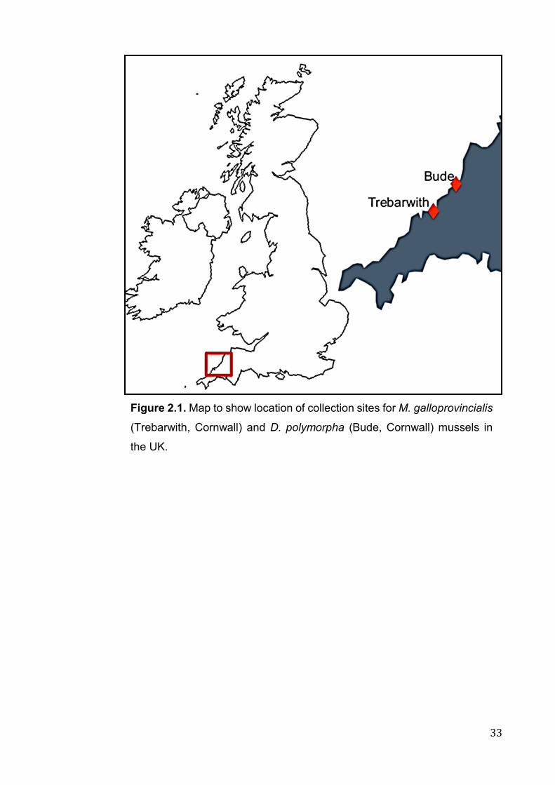
33
Figure 2.1. Map to show location of collection sites for M. galloprovincialis
(Trebarwith, Cornwall) and D. polymorpha (Bude, Cornwall) mussels in
the UK.
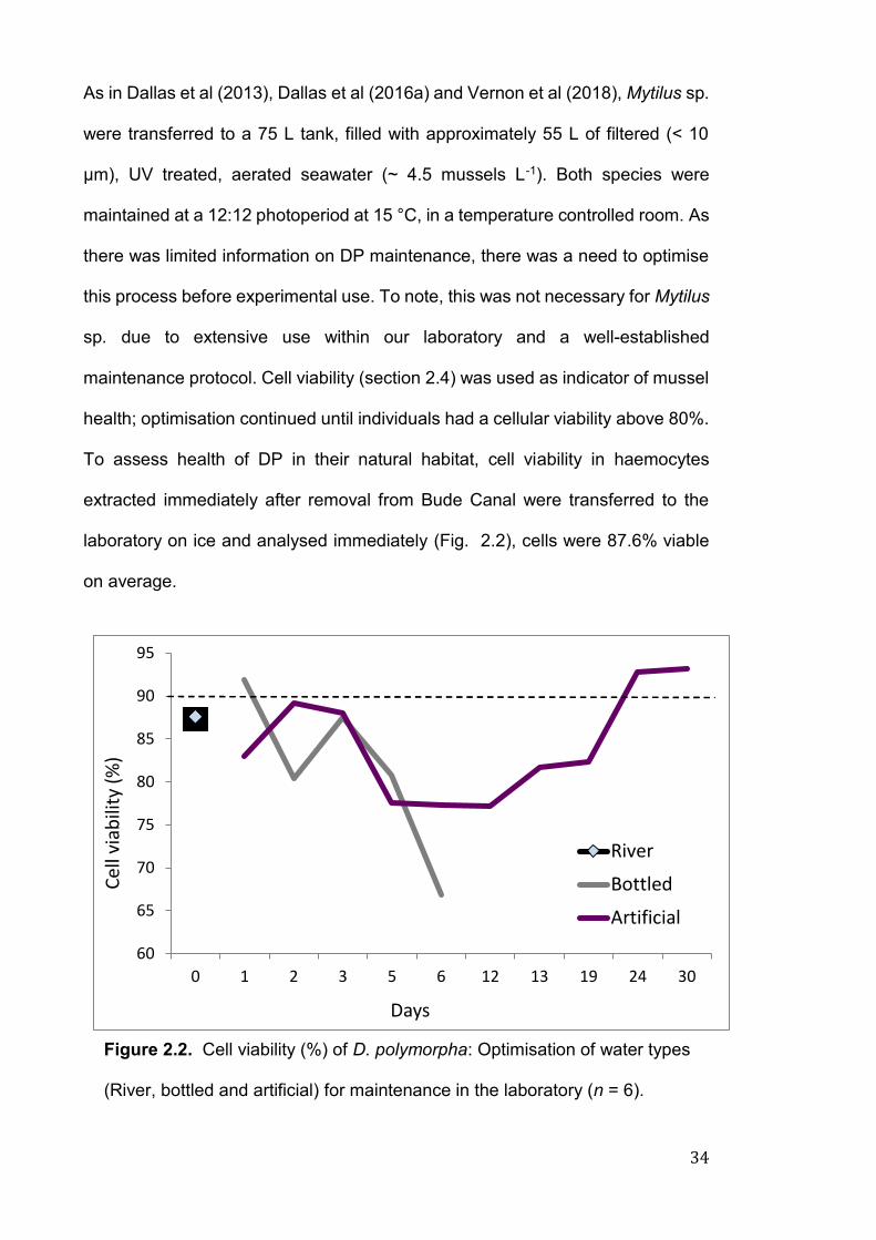
34
As in Dallas et al (2013), Dallas et al (2016a) and Vernon et al (2018), Mytilus sp.
were transferred to a 75 L tank, filled with approximately 55 L of filtered (< 10
μm), UV treated, aerated seawater (~ 4.5 mussels L-1). Both species were
maintained at a 12:12 photoperiod at 15 °C, in a temperature controlled room. As
there was limited information on DP maintenance, there was a need to optimise
this process before experimental use. To note, this was not necessary for Mytilus
sp. due to extensive use within our laboratory and a well-established
maintenance protocol. Cell viability (section 2.4) was used as indicator of mussel
health; optimisation continued until individuals had a cellular viability above 80%.
To assess health of DP in their natural habitat, cell viability in haemocytes
extracted immediately after removal from Bude Canal were transferred to the
laboratory on ice and analysed immediately (Fig. 2.2), cells were 87.6% viable
on average.
60
65
70
75
80
85
90
95
0 1 2 3 5 6 12 13 19 24 30
Cel
l via
bili
ty (
%)
Days
River
Bottled
Artificial
Figure 2.2. Cell viability (%) of D. polymorpha: Optimisation of water types
(River, bottled and artificial) for maintenance in the laboratory (n = 6).
35
DP were initially kept in a 1 L plastic container in the following water types (a) tap
water (main tap in Davy 422) and (b) bottled water (Highland Spring still water).
Tap water was initially used as an inexpensive, easily available option, however
it was not utilised due to possible variation of water quality with each water
change (i.e. metal concentrations, pH). Bottled water was tested as a controlled,
inexpensive choice. Cell viability was checked each day over 7 ± 3 days. On day
7, cell viability remained low at around 66.8 % (n = 6, bottled, Fig. 2.2). DP were
then transferred to a 15 L aquarium, filled with 10 L aerated DI water (~ 5 mussels
L-1) and an artificial river water solution (2M CaCl2.2H20, 8 mM MgSO4.7H2O,
40 mM, 5 mM KNO3, 0.7 M NaHCO3). Water test kits (Tetra, Blacksburg, VA,
USA) were used to assess and maintain optimum water quality parameters
(carbonates, general hardness, nitrite, nitrates, chlorine and ammonia), along
with dissolved oxygen (DO), salinity, pH and temperature (Hach HQ40D Multi-
meter, Hach-Lange, Dusseldorf, Germany). Cell viability was maintained at 90 %
(n = 6, Fig. 2.2).
During the holding period, mussels were fed three times per week. DP were fed
on dried Chlorella powder (3.2 mg/mussel per feed, Naturya, Bath, UK), Mytilus
sp. on a solution of Isochrysis galbana algae (8 x 10⁵ cells mL-1, Reed Mariculture,
Campbell, CA, USA). A 70% (DP) and a 100% water change was performed ~2
h after feeding. Mussels were allowed a 2-week acclimatisation period before use
in experiments. Due to the invasive nature of DP (Nalepa and Schloesser 1992;
Karatayev et al. 2007), all wastewater was spiked with salt (50 g L-1, NaCl) before
disposal as to prevent infestation and dispersal.
36
2.2.1 Genetic identification of Mytilus spp.
Hilbish et al (2002) reported the presence and distribution of Mytilus edulis, M.
galloproviancialis and their hybrids in the coastal regions of southwest England.
Based only on morphological characteristics alone these species are very
difficult, if not impossible to differentiate (Koehn 1991; McDonald et al. 1991).
Small genetic variations may account for differential biological response,
particularly at molecular level (Hilbish et al. 1994). Species homogeneity in our
experiments was therefore ensured based on the method of Inoue et al (1995).
In accordance to Dallas et al (2016a), Dallas et al (2018) and Pearson et al
(2018), species at the collection site were confirmed as M. galloprovincialis (MG).
To note, this was not necessary for DP.
A small section of gill tissue (~25 mg) was dissected from each individual (n = 20,
main collection site), snap frozen immediately in liquid nitrogen, and stored at -
80 ºC until analysis. Tissue was homogenised, Chelex solution added (500 µl,
Bio-rad, 10%), samples vortexed and incubated (56 ºC, 30 min, vortex every 10
min). Following incubation, samples were boiled (98 ºC, 8-10 min) and placed on
ice for 3 min. Samples were then spun (5 min, max speed ~ 32,310 g), and had
supernatant removed (in duplicate). Samples were stored (4 ºC short term, -20
ºC long term) until analysis.
Diagnostic Me 15 and Me 16 PCR primers (Fig. 2.3) were used to analyse the
genetic composition of species. These validated markers, which target the
adhesive protein gene sequence (Glu-5’ gene, GenBank accession no. D63778)
have successfully been utilised to identify the morphologically similar species
(Inoue et al. 1995; Coghlan and Gosling 2007; Dias et al. 2008; Kijewski et al.
2011; Pearson et al. 2018). Length of amplified fragments vary interspecifically
at 180, 126 and 168 base pairs for M. edulis, M. galloprovincialis and M. trossulus
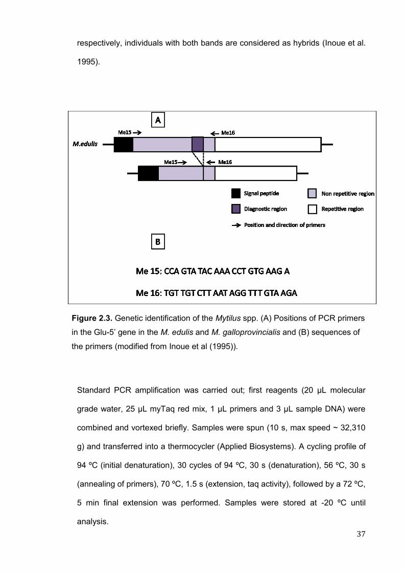
37
respectively, individuals with both bands are considered as hybrids (Inoue et al.
1995).
Standard PCR amplification was carried out; first reagents (20 µL molecular
grade water, 25 µL myTaq red mix, 1 µL primers and 3 µL sample DNA) were
combined and vortexed briefly. Samples were spun (10 s, max speed ~ 32,310
g) and transferred into a thermocycler (Applied Biosystems). A cycling profile of
94 ºC (initial denaturation), 30 cycles of 94 ºC, 30 s (denaturation), 56 ºC, 30 s
(annealing of primers), 70 ºC, 1.5 s (extension, taq activity), followed by a 72 ºC,
5 min final extension was performed. Samples were stored at -20 ºC until
analysis.
Figure 2.3. Genetic identification of the Mytilus spp. (A) Positions of PCR primers
in the Glu-5’ gene in the M. edulis and M. galloprovincialis and (B) sequences of
the primers (modified from Inoue et al (1995)).
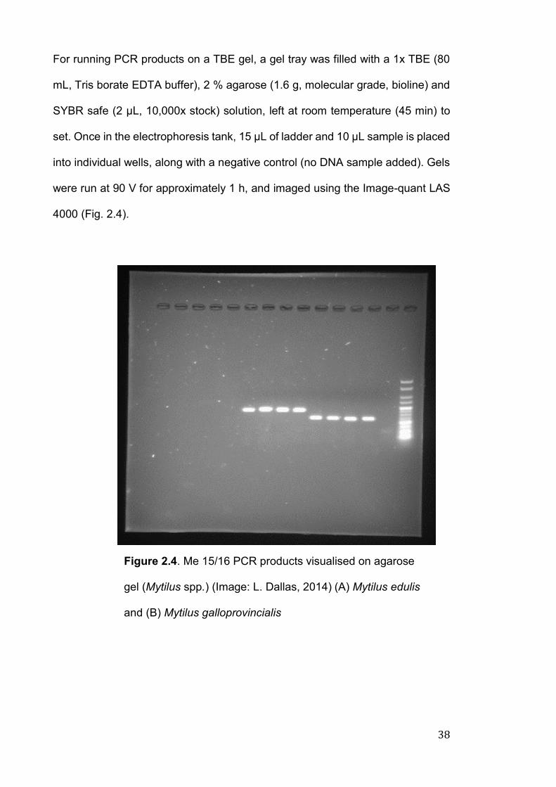
38
For running PCR products on a TBE gel, a gel tray was filled with a 1x TBE (80
mL, Tris borate EDTA buffer), 2 % agarose (1.6 g, molecular grade, bioline) and
SYBR safe (2 µL, 10,000x stock) solution, left at room temperature (45 min) to
set. Once in the electrophoresis tank, 15 µL of ladder and 10 µL sample is placed
into individual wells, along with a negative control (no DNA sample added). Gels
were run at 90 V for approximately 1 h, and imaged using the Image-quant LAS
4000 (Fig. 2.4).
Figure 2.4. Me 15/16 PCR products visualised on agarose
gel (Mytilus spp.) (Image: L. Dallas, 2014) (A) Mytilus edulis
and (B) Mytilus galloprovincialis
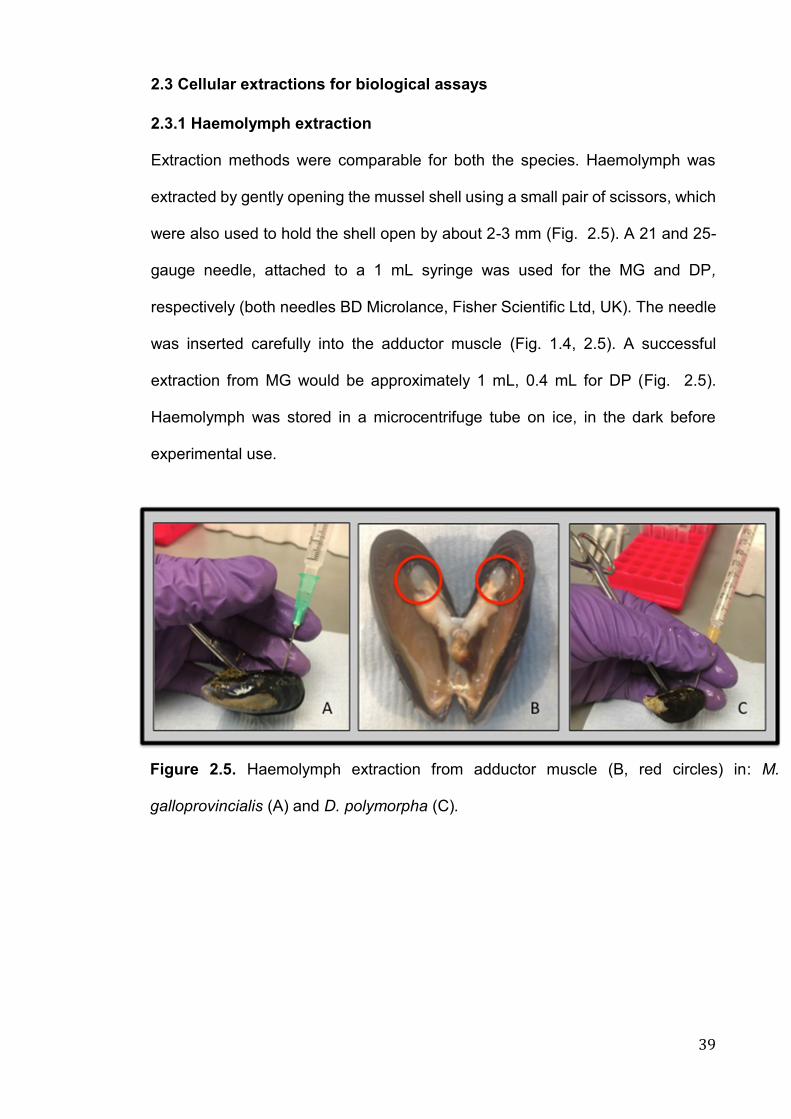
39
2.3 Cellular extractions for biological assays
2.3.1 Haemolymph extraction
Extraction methods were comparable for both the species. Haemolymph was
extracted by gently opening the mussel shell using a small pair of scissors, which
were also used to hold the shell open by about 2-3 mm (Fig. 2.5). A 21 and 25-
gauge needle, attached to a 1 mL syringe was used for the MG and DP,
respectively (both needles BD Microlance, Fisher Scientific Ltd, UK). The needle
was inserted carefully into the adductor muscle (Fig. 1.4, 2.5). A successful
extraction from MG would be approximately 1 mL, 0.4 mL for DP (Fig. 2.5).
Haemolymph was stored in a microcentrifuge tube on ice, in the dark before
experimental use.
Figure 2.5. Haemolymph extraction from adductor muscle (B, red circles) in: M.
galloprovincialis (A) and D. polymorpha (C).
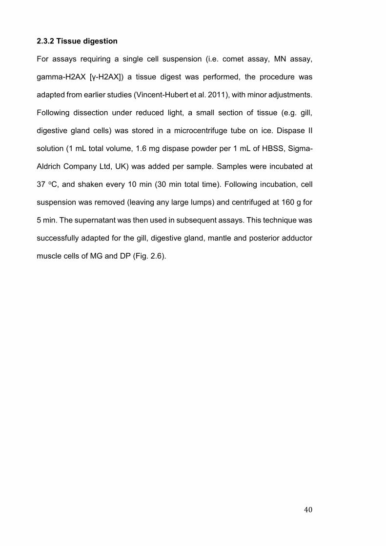
40
2.3.2 Tissue digestion
For assays requiring a single cell suspension (i.e. comet assay, MN assay,
gamma-H2AX [γ-H2AX]) a tissue digest was performed, the procedure was
adapted from earlier studies (Vincent-Hubert et al. 2011), with minor adjustments.
Following dissection under reduced light, a small section of tissue (e.g. gill,
digestive gland cells) was stored in a microcentrifuge tube on ice. Dispase II
solution (1 mL total volume, 1.6 mg dispase powder per 1 mL of HBSS, Sigma-
Aldrich Company Ltd, UK) was added per sample. Samples were incubated at
37 oC, and shaken every 10 min (30 min total time). Following incubation, cell
suspension was removed (leaving any large lumps) and centrifuged at 160 g for
5 min. The supernatant was then used in subsequent assays. This technique was
successfully adapted for the gill, digestive gland, mantle and posterior adductor
muscle cells of MG and DP (Fig. 2.6).
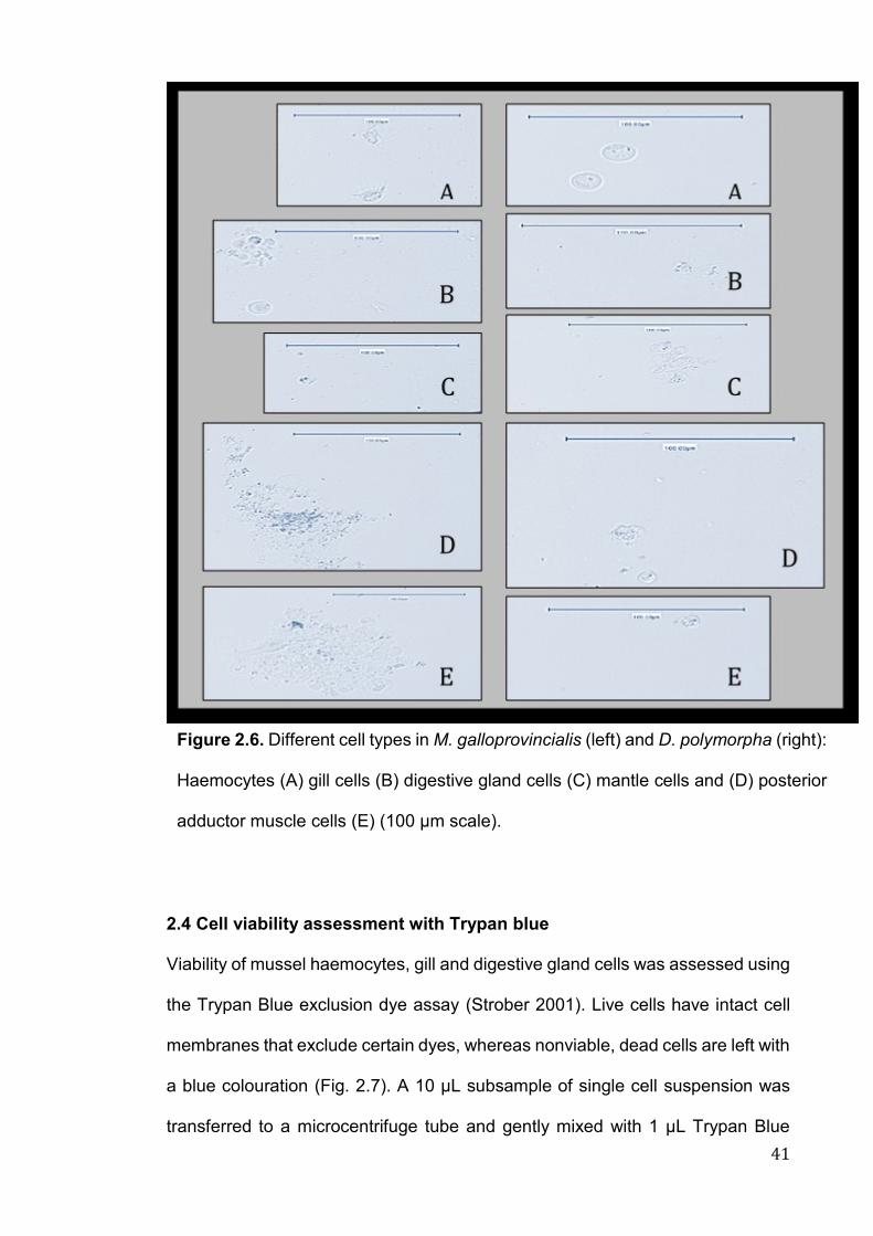
41
2.4 Cell viability assessment with Trypan blue
Viability of mussel haemocytes, gill and digestive gland cells was assessed using
the Trypan Blue exclusion dye assay (Strober 2001). Live cells have intact cell
membranes that exclude certain dyes, whereas nonviable, dead cells are left with
a blue colouration (Fig. 2.7). A 10 μL subsample of single cell suspension was
transferred to a microcentrifuge tube and gently mixed with 1 μL Trypan Blue
Figure 2.6. Different cell types in M. galloprovincialis (left) and D. polymorpha (right):
Haemocytes (A) gill cells (B) digestive gland cells (C) mantle cells and (D) posterior
adductor muscle cells (E) (100 μm scale).

42
(0.4%, Sigma), the solution was then transferred to a slide, and a coverslip was
applied. The number of viable cells (clear cytoplasm), and dead cells (blue
cytoplasm) were examined under light microscopy (x 40). Samples with >90%
viability were used in subsequent tests.
2.5 Biological assays
2.5.1 Micronucleus (MN) assay
The MN assay is a simple, reliable cytogenetic assay, which determines the
presence of micronuclei in the cytoplasm of interphase cells (Bolognesi and
Fenech 2012). MN formation is indicative of DNA damage or mutation, formation
mainly originates from either a whole chromosome, chromosome fragment
containing centromere or from acentric chromosome fragments (Fig. 2.8). The
assay detects the activity of clastogenic (chromosome breakage) and aneugenic
agents (abnormality in chromosome number within daughter cell). This assay can
be performed using mussel haemocytes or tissue cells, it has been successfully
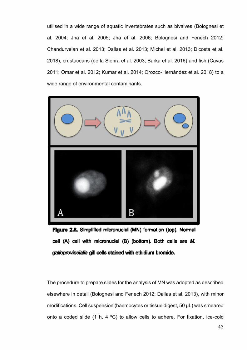
43
utilised in a wide range of aquatic invertebrates such as bivalves (Bolognesi et
al. 2004; Jha et al. 2005; Jha et al. 2006; Bolognesi and Fenech 2012;
Chandurvelan et al. 2013; Dallas et al. 2013; Michel et al. 2013; D’costa et al.
2018), crustaceans (de la Sienra et al. 2003; Barka et al. 2016) and fish (Cavas
2011; Omar et al. 2012; Kumar et al. 2014; Orozco-Hernández et al. 2018) to a
wide range of environmental contaminants.
The procedure to prepare slides for the analysis of MN was adopted as described
elsewhere in detail (Bolognesi and Fenech 2012; Dallas et al. 2013), with minor
modifications. Cell suspension (haemocytes or tissue digest, 50 μL) was smeared
onto a coded slide (1 h, 4 ºC) to allow cells to adhere. For fixation, ice-cold
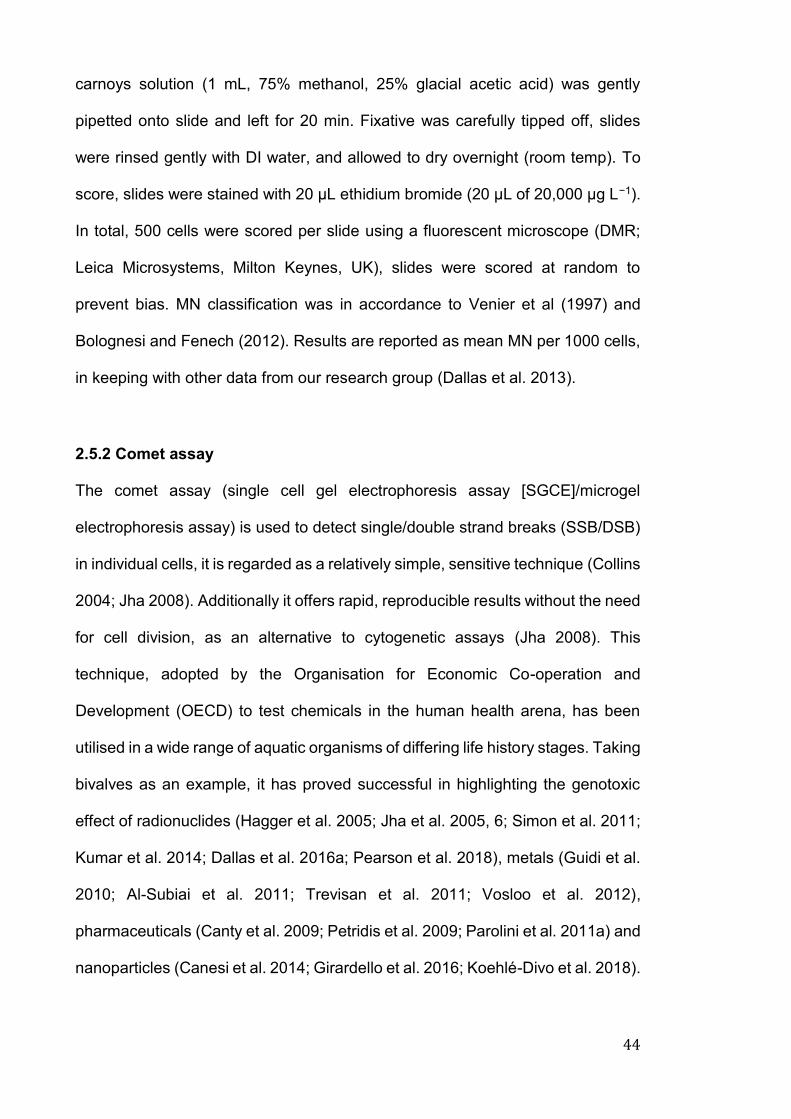
44
carnoys solution (1 mL, 75% methanol, 25% glacial acetic acid) was gently
pipetted onto slide and left for 20 min. Fixative was carefully tipped off, slides
were rinsed gently with DI water, and allowed to dry overnight (room temp). To
score, slides were stained with 20 μL ethidium bromide (20 μL of 20,000 µg L−1).
In total, 500 cells were scored per slide using a fluorescent microscope (DMR;
Leica Microsystems, Milton Keynes, UK), slides were scored at random to
prevent bias. MN classification was in accordance to Venier et al (1997) and
Bolognesi and Fenech (2012). Results are reported as mean MN per 1000 cells,
in keeping with other data from our research group (Dallas et al. 2013).
2.5.2 Comet assay
The comet assay (single cell gel electrophoresis assay [SGCE]/microgel
electrophoresis assay) is used to detect single/double strand breaks (SSB/DSB)
in individual cells, it is regarded as a relatively simple, sensitive technique (Collins
2004; Jha 2008). Additionally it offers rapid, reproducible results without the need
for cell division, as an alternative to cytogenetic assays (Jha 2008). This
technique, adopted by the Organisation for Economic Co-operation and
Development (OECD) to test chemicals in the human health arena, has been
utilised in a wide range of aquatic organisms of differing life history stages. Taking
bivalves as an example, it has proved successful in highlighting the genotoxic
effect of radionuclides (Hagger et al. 2005; Jha et al. 2005, 6; Simon et al. 2011;
Kumar et al. 2014; Dallas et al. 2016a; Pearson et al. 2018), metals (Guidi et al.
2010; Al-Subiai et al. 2011; Trevisan et al. 2011; Vosloo et al. 2012),
pharmaceuticals (Canty et al. 2009; Petridis et al. 2009; Parolini et al. 2011a) and
nanoparticles (Canesi et al. 2014; Girardello et al. 2016; Koehlé-Divo et al. 2018).
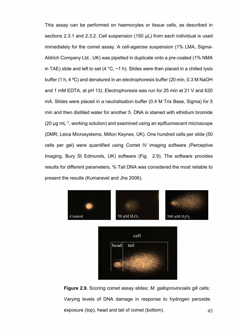
45
This assay can be performed on haemocytes or tissue cells, as described in
sections 2.3.1 and 2.3.2. Cell suspension (150 μL) from each individual is used
immediately for the comet assay. A cell-agarose suspension (1% LMA, Sigma-
Aldrich Company Ltd., UK) was pipetted in duplicate onto a pre-coated (1% NMA
in TAE) slide and left to set (4 °C, ~1 h). Slides were then placed in a chilled lysis
buffer (1 h, 4 ºC) and denatured in an electrophoresis buffer (20 min, 0.3 M NaOH
and 1 mM EDTA, at pH 13). Electrophoresis was run for 25 min at 21 V and 620
mA. Slides were placed in a neutralisation buffer (0.4 M Tris Base, Sigma) for 5
min and then distilled water for another 5. DNA is stained with ethidium bromide
(20 µg mL-1, working solution) and examined using an epifluorescent microscope
(DMR; Leica Microsystems, Milton Keynes, UK). One hundred cells per slide (50
cells per gel) were quantified using Comet IV imaging software (Perceptive
Imaging, Bury St Edmunds, UK) software (Fig. 2.9). The software provides
results for different parameters, % Tail DNA was considered the most reliable to
present the results (Kumaravel and Jha 2006).
Figure 2.9. Scoring comet assay slides: M. galloprovincialis gill cells:
Varying levels of DNA damage in response to hydrogen peroxide
exposure (top), head and tail of comet (bottom).
46
2.5.2.1 Optimisation and validation of the comet assay using hydrogen
peroxide (H2O2)
After a 2-week depuration period, gill, digestive gland tissue and haemolymph
was extracted as outlined in sections 2.3.2 and 2.3.1 respectively from MG and
DP (n = 4). Gill and digestive gland tissue was digested to acquire a single cell
suspension, cell suspensions were pooled to reduce inter-individual variability
and stored on ice in the dark until use. Aliquots (150 μL) of cells were transferred
into microcentrifuge tubes and spun (775 g, 2 min, 4 oC). Once supernatant was
removed and discarded (leaving approx. 10 μL), a H2O2 solution (Sigma H1009,
8.8 M, 100 μL, 0, 5, 50 or 500 μM in PBS) was added to the cellular pellet. H2O2
was used for validation as a known genotoxic agent, it has been successfully
utilised to validate genetic biomarkers in previous literature (Dallas et al. 2013;
Sarkar et al. 2015). Following incubation (1 h, 4 oC, dark) the samples were spun
(as before), supernatant removed, and samples processed as in section 2.5.2. In
both species, H2O2 concentrations produced an increase in percentage tail DNA
in comparison to the control (Fig. 2.10, p < 0.01 and p < 0.01 in haemolymph, p
< 0.01 and p < 0.01 in gill and p < 0.001 and p < 0.001 in digestive gland, in DP
and MG).
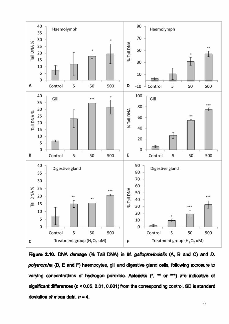
47
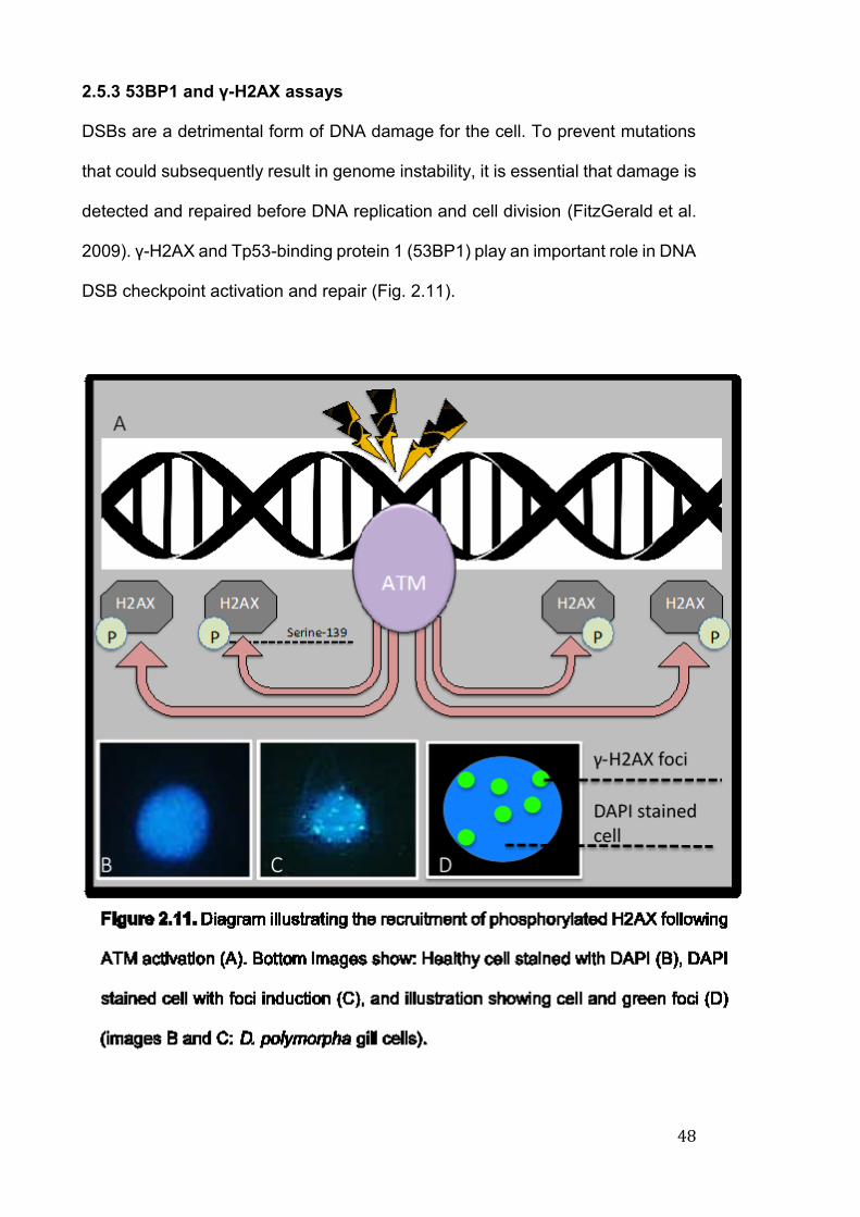
48
2.5.3 53BP1 and γ-H2AX assays
DSBs are a detrimental form of DNA damage for the cell. To prevent mutations
that could subsequently result in genome instability, it is essential that damage is
detected and repaired before DNA replication and cell division (FitzGerald et al.
2009). γ-H2AX and Tp53-binding protein 1 (53BP1) play an important role in DNA
DSB checkpoint activation and repair (Fig. 2.11).
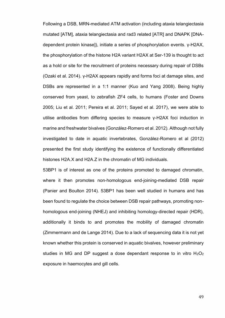
49
Following a DSB, MRN-mediated ATM activation (including ataxia telangiectasia
mutated [ATM], ataxia telangiectasia and rad3 related [ATR] and DNAPK [DNA-
dependent protein kinase]), initiate a series of phosphorylation events. γ-H2AX,
the phosphorylation of the histone H2A variant H2AX at Ser-139 is thought to act
as a hold or site for the recruitment of proteins necessary during repair of DSBs
(Ozaki et al. 2014). γ-H2AX appears rapidly and forms foci at damage sites, and
DSBs are represented in a 1:1 manner (Kuo and Yang 2008). Being highly
conserved from yeast, to zebrafish ZF4 cells, to humans (Foster and Downs
2005; Liu et al. 2011; Pereira et al. 2011; Sayed et al. 2017), we were able to
utilise antibodies from differing species to measure γ-H2AX foci induction in
marine and freshwater bivalves (González-Romero et al. 2012). Although not fully
investigated to date in aquatic invertebrates, González-Romero et al (2012)
presented the first study identifying the existence of functionally differentiated
histones H2A.X and H2A.Z in the chromatin of MG individuals.
53BP1 is of interest as one of the proteins promoted to damaged chromatin,
where it then promotes non-homologous end-joining-mediated DSB repair
(Panier and Boulton 2014). 53BP1 has been well studied in humans and has
been found to regulate the choice between DSB repair pathways, promoting non-
homologous end-joining (NHEJ) and inhibiting homology-directed repair (HDR),
additionally it binds to and promotes the mobility of damaged chromatin
(Zimmermann and de Lange 2014). Due to a lack of sequencing data it is not yet
known whether this protein is conserved in aquatic bivalves, however preliminary
studies in MG and DP suggest a dose dependant response to in vitro H2O2
exposure in haemocytes and gill cells.
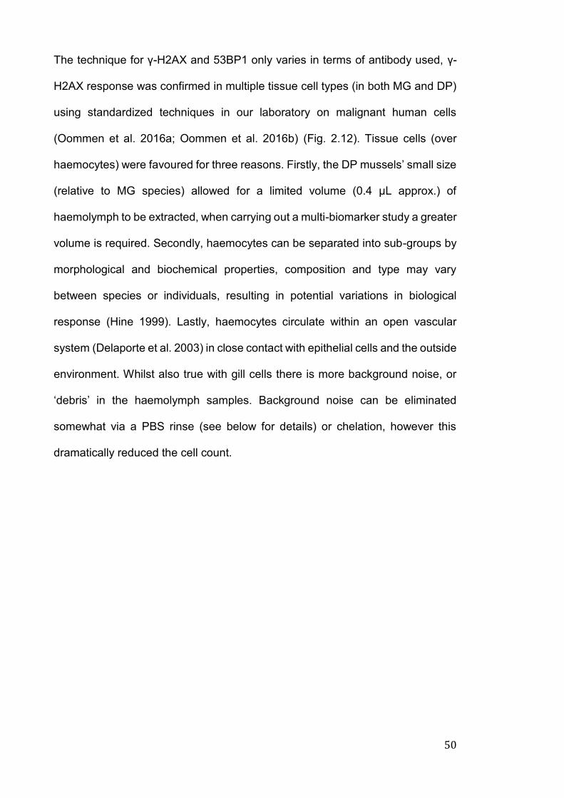
50
The technique for γ-H2AX and 53BP1 only varies in terms of antibody used, γ-
H2AX response was confirmed in multiple tissue cell types (in both MG and DP)
using standardized techniques in our laboratory on malignant human cells
(Oommen et al. 2016a; Oommen et al. 2016b) (Fig. 2.12). Tissue cells (over
haemocytes) were favoured for three reasons. Firstly, the DP mussels’ small size
(relative to MG species) allowed for a limited volume (0.4 μL approx.) of
haemolymph to be extracted, when carrying out a multi-biomarker study a greater
volume is required. Secondly, haemocytes can be separated into sub-groups by
morphological and biochemical properties, composition and type may vary
between species or individuals, resulting in potential variations in biological
response (Hine 1999). Lastly, haemocytes circulate within an open vascular
system (Delaporte et al. 2003) in close contact with epithelial cells and the outside
environment. Whilst also true with gill cells there is more background noise, or
‘debris’ in the haemolymph samples. Background noise can be eliminated
somewhat via a PBS rinse (see below for details) or chelation, however this
dramatically reduced the cell count.
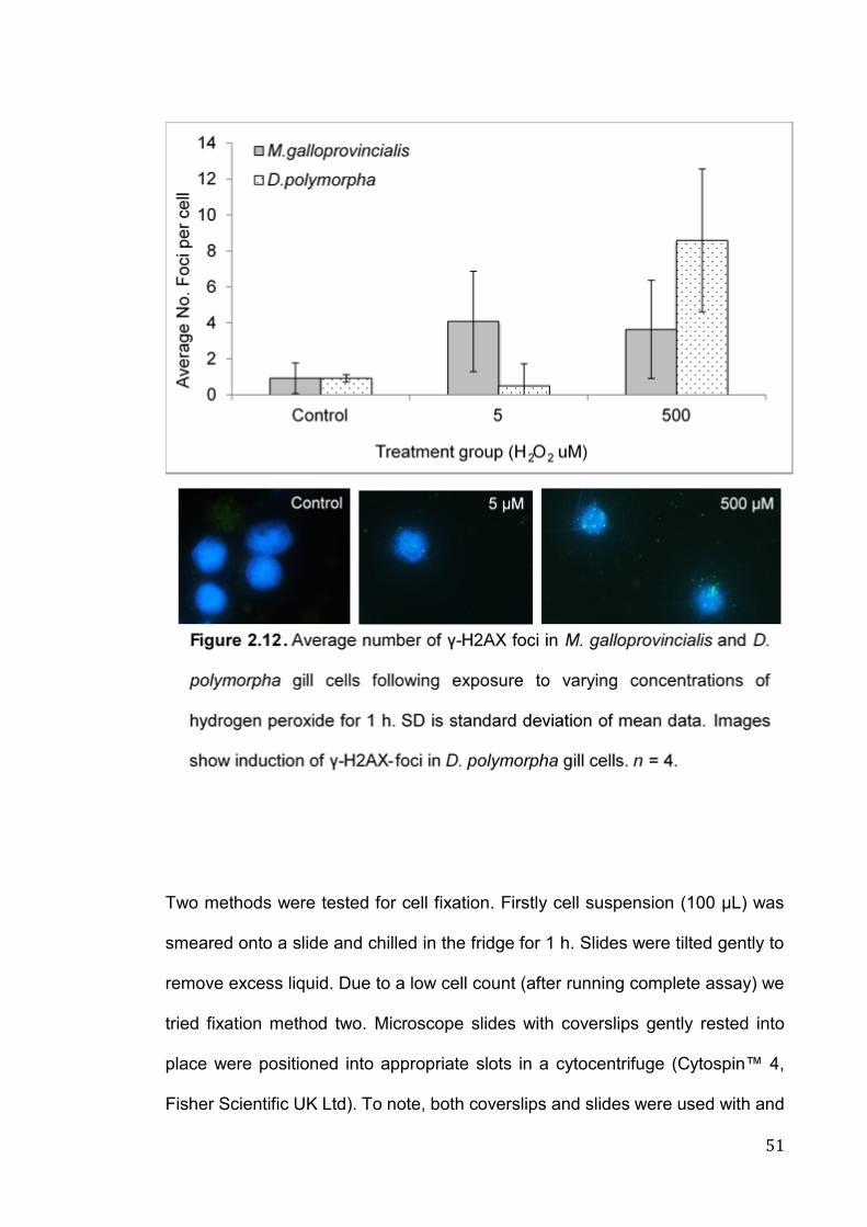
51
Two methods were tested for cell fixation. Firstly cell suspension (100 μL) was
smeared onto a slide and chilled in the fridge for 1 h. Slides were tilted gently to
remove excess liquid. Due to a low cell count (after running complete assay) we
tried fixation method two. Microscope slides with coverslips gently rested into
place were positioned into appropriate slots in a cytocentrifuge (Cytospin™ 4,
Fisher Scientific UK Ltd). To note, both coverslips and slides were used with and
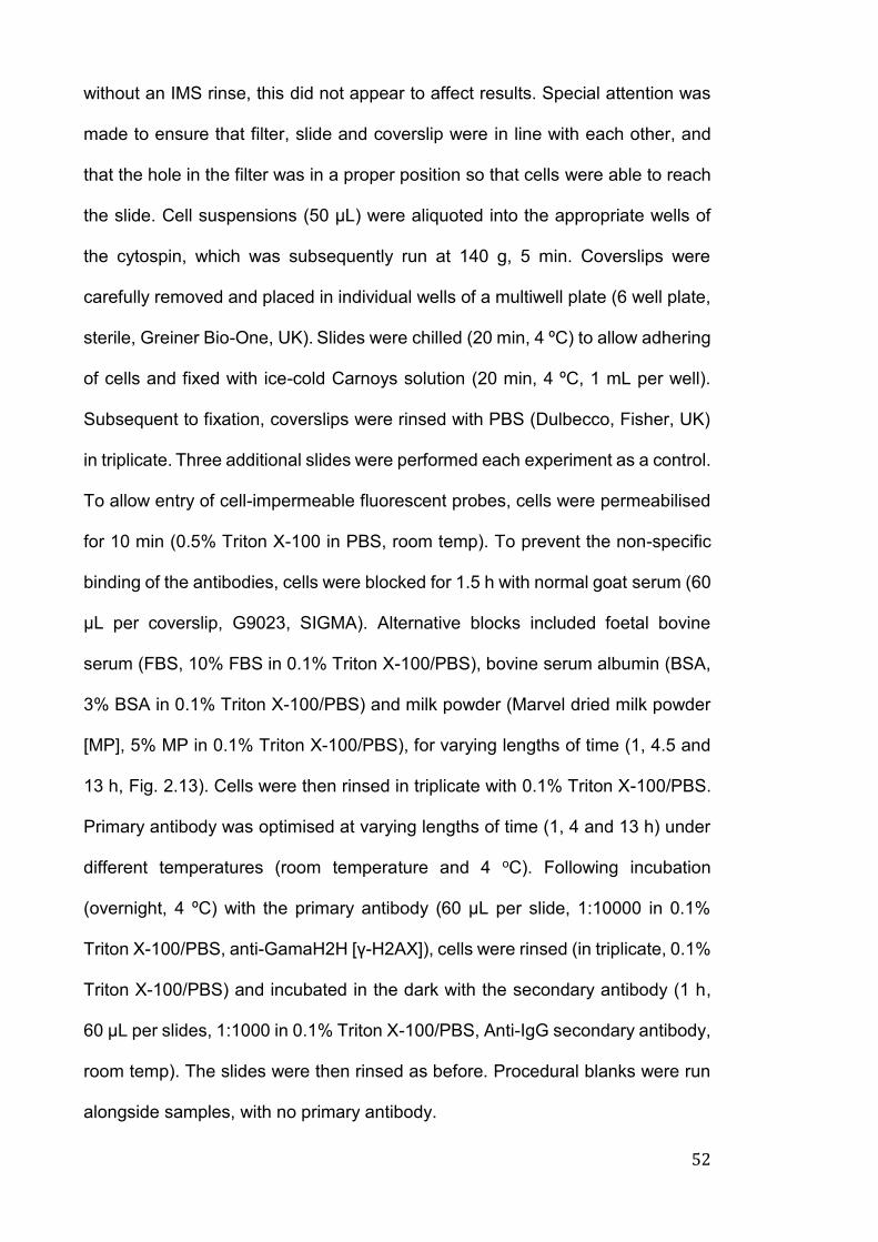
52
without an IMS rinse, this did not appear to affect results. Special attention was
made to ensure that filter, slide and coverslip were in line with each other, and
that the hole in the filter was in a proper position so that cells were able to reach
the slide. Cell suspensions (50 μL) were aliquoted into the appropriate wells of
the cytospin, which was subsequently run at 140 g, 5 min. Coverslips were
carefully removed and placed in individual wells of a multiwell plate (6 well plate,
sterile, Greiner Bio-One, UK). Slides were chilled (20 min, 4 ºC) to allow adhering
of cells and fixed with ice-cold Carnoys solution (20 min, 4 ºC, 1 mL per well).
Subsequent to fixation, coverslips were rinsed with PBS (Dulbecco, Fisher, UK)
in triplicate. Three additional slides were performed each experiment as a control.
To allow entry of cell-impermeable fluorescent probes, cells were permeabilised
for 10 min (0.5% Triton X-100 in PBS, room temp). To prevent the non-specific
binding of the antibodies, cells were blocked for 1.5 h with normal goat serum (60
μL per coverslip, G9023, SIGMA). Alternative blocks included foetal bovine
serum (FBS, 10% FBS in 0.1% Triton X-100/PBS), bovine serum albumin (BSA,
3% BSA in 0.1% Triton X-100/PBS) and milk powder (Marvel dried milk powder
[MP], 5% MP in 0.1% Triton X-100/PBS), for varying lengths of time (1, 4.5 and
13 h, Fig. 2.13). Cells were then rinsed in triplicate with 0.1% Triton X-100/PBS.
Primary antibody was optimised at varying lengths of time (1, 4 and 13 h) under
different temperatures (room temperature and 4 oC). Following incubation
(overnight, 4 ºC) with the primary antibody (60 µL per slide, 1:10000 in 0.1%
Triton X-100/PBS, anti-GamaH2H [γ-H2AX]), cells were rinsed (in triplicate, 0.1%
Triton X-100/PBS) and incubated in the dark with the secondary antibody (1 h,
60 µL per slides, 1:1000 in 0.1% Triton X-100/PBS, Anti-IgG secondary antibody,
room temp). The slides were then rinsed as before. Procedural blanks were run
alongside samples, with no primary antibody.
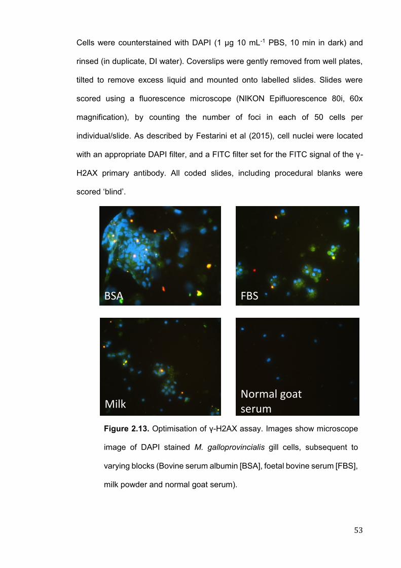
53
Cells were counterstained with DAPI (1 µg 10 mL-1 PBS, 10 min in dark) and
rinsed (in duplicate, DI water). Coverslips were gently removed from well plates,
tilted to remove excess liquid and mounted onto labelled slides. Slides were
scored using a fluorescence microscope (NIKON Epifluorescence 80i, 60x
magnification), by counting the number of foci in each of 50 cells per
individual/slide. As described by Festarini et al (2015), cell nuclei were located
with an appropriate DAPI filter, and a FITC filter set for the FITC signal of the γ-
H2AX primary antibody. All coded slides, including procedural blanks were
scored ‘blind’.
FBS BSA
Milk powder
Normal goat serum
Figure 2.13. Optimisation of γ-H2AX assay. Images show microscope
image of DAPI stained M. galloprovincialis gill cells, subsequent to
varying blocks (Bovine serum albumin [BSA], foetal bovine serum [FBS],
milk powder and normal goat serum).
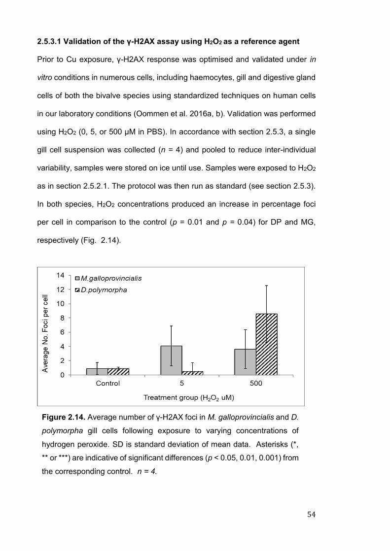
54
2.5.3.1 Validation of the γ-H2AX assay using H2O2 as a reference agent
Prior to Cu exposure, γ-H2AX response was optimised and validated under in
vitro conditions in numerous cells, including haemocytes, gill and digestive gland
cells of both the bivalve species using standardized techniques on human cells
in our laboratory conditions (Oommen et al. 2016a, b). Validation was performed
using H2O2 (0, 5, or 500 μM in PBS). In accordance with section 2.5.3, a single
gill cell suspension was collected (n = 4) and pooled to reduce inter-individual
variability, samples were stored on ice until use. Samples were exposed to H2O2
as in section 2.5.2.1. The protocol was then run as standard (see section 2.5.3).
In both species, H2O2 concentrations produced an increase in percentage foci
per cell in comparison to the control (p = 0.01 and p = 0.04) for DP and MG,
respectively (Fig. 2.14).
Figure 2.14. Average number of γ-H2AX foci in M. galloprovincialis and D.
polymorpha gill cells following exposure to varying concentrations of
hydrogen peroxide. SD is standard deviation of mean data. Asterisks (*,
** or ***) are indicative of significant differences (p < 0.05, 0.01, 0.001) from
the corresponding control. n = 4.
55
2.5.4 Transcriptional expression of key genes
DNA is the primary molecule of concern for effects from IR and genotoxic
chemicals, expression of key genes can provide an early signal of organism
stress. The following criteria were set when choosing genes of interest, (a) for
means of comparison, well documented markers of oxidative and stress
responses in both study species (preferably in both gill and digestive gland tissue)
and (b) available primer sequences. The genes (including primer sequences)
selected for this study have been listed in Table 2.1. These include superoxide
dismutase (sod), catalase (cat), glutathione S-transferase (gst), and heat shock
proteins 70 (hsp70) and 90 (hsp90). In accordance with Navarro et al (2011),
Lacroix et al (2014), Dallas et al (2016a) and Banni et al (2017), elongation factor
1 (ef1) and β-Actin (act) were chosen as reliable, widely utilised housekeeping
genes for both study species.
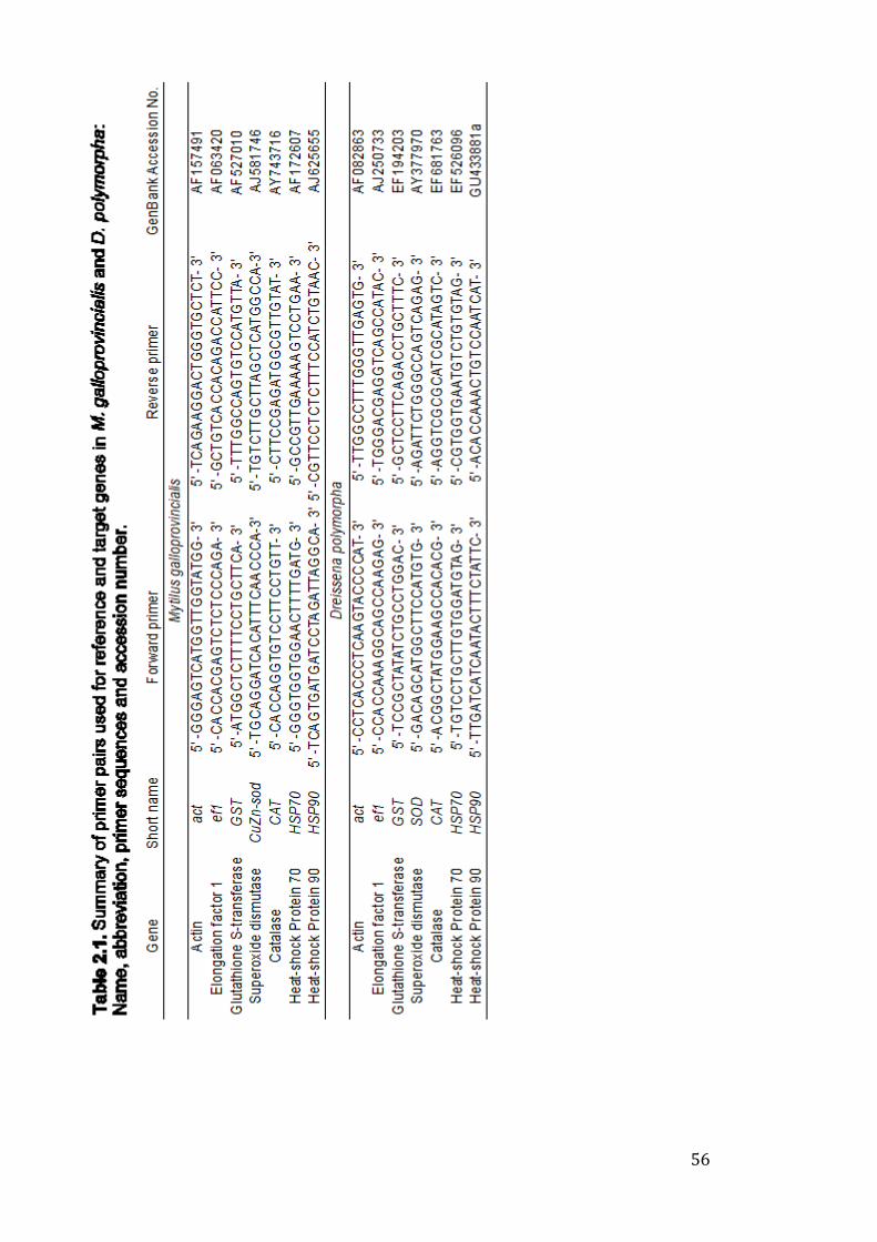
56
57
2.5.4.1 RNA extraction
Gill and digestive gland tissue was dissected immediately after exposure and
stored in RNAlater at -80 °C (R0901, Sigma-Aldrich Company Ltd UK) until use.
Total RNA was extracted using the GenElute™ Mammalian Total RNA Miniprep
Kit (RTN350, Sigma-Aldrich Company Ltd UK). To release RNA, biological tissue,
lysis buffer (guanidine thiocyanate and 2-mercaptoethanol) and glass beads (2
mm, Sigma-Aldrich) were transferred into a microcentrifuge tube, and
homogenised (FastPrep®, 30 s, 5 m/s) to form a smooth lysate (Fig. 2.15, A).
Lysates were transferred into a filtration column and spun (2 min, ~19000 g),
removing impurities such as cellular debris (Fig. 2.15, B). Subsequent to the
addition of 70% ethanol (Ethanol, Absolute 200 Proof, Fisher Scientific), the
filtrate was spun (2 min, ~19000 g) in a nucleic acid binding column (Fig. 2.15,
C). To remove impurities such as protein and salt residues, two wash steps were
carried out followed by a dry spin to ensure a clean eluent. Lastly, RNA was
released from the binding column via an elution buffer. The concentration and
purity of isolated RNA was determined by UV spectroscopy (Nanodrop 3300,
Thermo Scientific, Fig. 2.15, D), total RNA was stored at −80 °C.
2.5.4.2 cDNA synthesis
cDNA was synthesised using the Applied Biosystems High-Capacity cDNA
reverse transcription kit (Fisher Scientific, UK) with RNase inhibitor. A 10 μL RNA
sample (1 μg RNA) and 10 μL RT master mix (10X RT buffer, 25X dNTP mix,
10X RT random primers, MultiScribe™ reverse transcriptase, RNA inhibitor) was
added per tube (Fig. 2.15, E, F). Samples were sealed, spun to remove air
bubbles (~19000 g, 10 s) and placed in the thermal cycler (Applied Biosystems)
under the following conditions: 10 min, 25 °C; 120 min, 37°C; 5 min, 85 °C and
58
held at 4 °C. cDNA samples were diluted 1:5 with molecular water before qPCR
(Fig. 2.15, G).
2.5.4.3 Real-time Polymerase chain reaction (qPCR)
cDNA (1.9 μL), PowerUp™ SYBR™ Green Master Mix (7.5 μL), forward/reverse
primers (0.03 μL/primer) and molecular grade water (5 μL) were added to a 96
well plate (MicroAmp™ Fast Optical 96-Well Reaction Plate, 0.1 mL) to make a
total volume of 15 μL per well (Fig. 15, H). All samples were run in duplicate.
Plates were sealed (MicroAmp™ Optical Adhesive Film), spun and placed in the
qPCR (Applied Biosystems Step-One Plus real-time PCR system with StepOne
Software (v2.2.2; Applied BioSystems) under the following conditions: 2 min,
95 °C; 40 cycles of 95 °C for 15 s and 1 min, 60 °C (Fig. 2.15, I). A disassociation
curve (melt curve) was run to verify the consistency of the PCR products (Fig.
2.15, J).
2.5.4.4 Data analysis and interpretation
In accordance with Dallas (2016a, 2018), relative mRNA expression ratio (RER)
of key genes (sod, cat, gst, hsp70 and hsp90) was quantified using REST (v 1,
2009), from PCR efficiencies calculated using LinReg PCR software (LinRegPCR
version 2017.1) (Ramakers et al. 2003; Ruijter et al. 2009) and threshold cycle
(Cq, Fig. 2.15, K, L). Values were normalised to the geometric mean of Cq
determined for reference genes actin (act) and elongation factor 1 (ef1), using
control samples to calibrate.
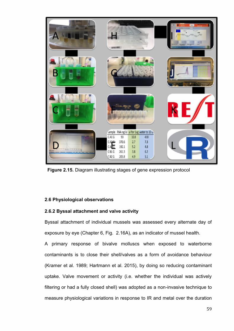
59
2.6 Physiological observations
2.6.2 Byssal attachment and valve activity
Byssal attachment of individual mussels was assessed every alternate day of
exposure by eye (Chapter 6, Fig. 2.16A), as an indicator of mussel health.
A primary response of bivalve molluscs when exposed to waterborne
contaminants is to close their shell/valves as a form of avoidance behaviour
(Kramer et al. 1989; Hartmann et al. 2015), by doing so reducing contaminant
uptake. Valve movement or activity (i.e. whether the individual was actively
filtering or had a fully closed shell) was adopted as a non-invasive technique to
measure physiological variations in response to IR and metal over the duration
Figure 2.15. Diagram illustrating stages of gene expression protocol

60
of the exposure (Chapter 6). Individuals were assessed by eye, three times daily
for valve activity (Fig. 2.16B, C).
61
2.7 Determination of metal concentration using ICP-OES and ICP-MS
2.7.1 Tissue preparation
For validation of biological assays, copper (Cu2+), as a metal toxicant and
reference agent was utilised. Determination of Cu in water and tissue samples
was carried out using ICP-MS (Inductively coupled plasma mass spectrometry)
as described before in publications from our laboratory (Al-Subiai et al. 2011;
Dallas et al. 2013; D'Agata et al. 2014). Briefly, all glass/plastic-ware and
dissection tools were acid washed (2% nitric acid, Fisher Scientific) at least 24 h
prior to use. Tissue was dissected and transferred into pre-weighed falcon tubes,
samples were placed in an oven to dry (60 oC, typically > 48 h). Tubes were re-
weighed and recorded for dry weight. For tissue digestion, 1 mL (volume
dependant on tissue weight, typically 2-3 mL for 0.1 g up to 10 mL for 1 g)
concentrated nitric acid (N/2272/PB17, Fisher Scientific) was added to each tube.
As procedural blanks, 6 tubes were tested alongside containing 1 mL acid without
tissue, along with a certified reference material (n = 3, TORT-2 Lobster
hepatopancreas, NRCC). Samples were boiled for 2 h (water bath, 80 oC), or until
tissue was fully digested. Once cool, digests were diluted with 4 mL Milli-Q water
and analysed as in section 2.7.2.
2.7.2 Determination of metal in water and tissue samples
To determine Cu concentration in water, samples (1 mL) were spiked with 50 μL
hydrochloric acid immediately after extraction, and stored at room temperature
until analysis. As in Dallas et al (2013), indium (115-In) and iridium (193-Ir) were
used as internal standards in both tissue and water samples. Seawater samples
were diluted 1:5 with DI water. Appropriate Cu standards were made to calibrate
62
the instrument before and during analysis of samples, in accordance to Al-Subiai
et al (2011). Using appropriate parameters (63Cu and 65Cu), standards and
samples were run using X Series II ICP-MS (Plasma Quad PQ2 Turbo, Thermo
Elemental, Winsford, UK) with PQ Vision 4.1.2 software. Procedural blanks were
run every 10 samples.
2.8 Ionising Radiation
2.8.1 Selection of radionuclide and determination of radiation dose levels
32P (1.709 MeV, Table 2.2), along with its relative low cost and ease to work with
was chosen as a study radionuclide as it can be used as a proxy for beta and
gamma emitters, such as environmentally relevant 137Cs and 90Sr. In addition, its
short half-life at 14.29 days is beneficial in terms of waste disposal. All waste
material, such as unused 32P solution, bivalve remains and general laboratory
waste (i.e. pipette tips, gloves) can be stored for a relatively short time period of
(approximately 4 months), before disposal. As illustrated in Table 2.3, several
other radionuclides were investigated for use in IR studies, however 32P was
deemed the most suitable. In terms of suitability of alternative radionuclides
iodine-131 had a half-life (8 d) too short to be utilised during experiments, both
33P and tritium lacked the decay energy to be used as proxies for gamma emitters
and 137Cs/90Sr were far more stringent in terms of safety regulations and allowed
dose levels in the laboratory. Experimental dose rates of 0.10, 1 and 10 mGy d-1
were based around a screening no-effect dose of 10 μGy h-1 (0.24 mGy d-1)
(Garnier-Laplace and Gilbin 2006; Andersson et al. 2008, 9; Garnier-Laplace et
al. 2008).
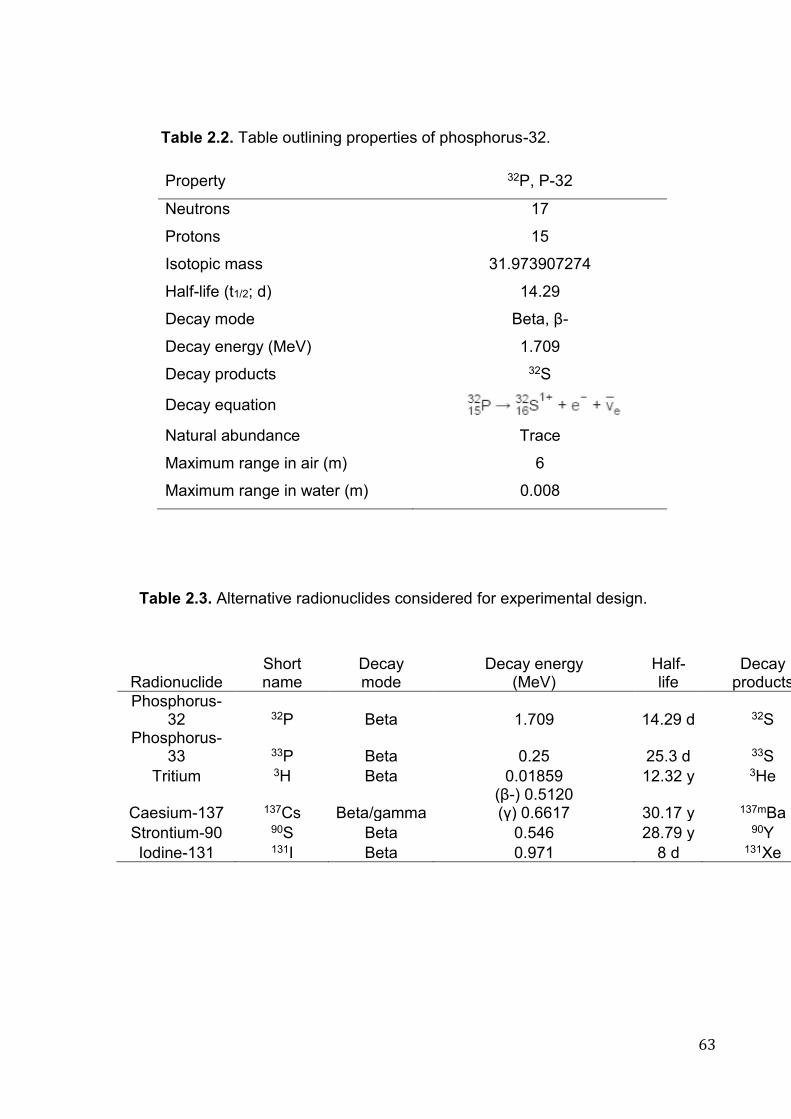
63
Property 32P, P-32
Neutrons 17
Protons 15
Isotopic mass 31.973907274
Half-life (t1/2; d) 14.29
Decay mode Beta, β-
Decay energy (MeV) 1.709
Decay products 32S
Decay equation
Natural abundance Trace
Maximum range in air (m) 6
Maximum range in water (m) 0.008
Radionuclide Short name
Decay mode
Decay energy (MeV)
Half-life
Decay products
Phosphorus-32 32P Beta 1.709 14.29 d 32S
Phosphorus-33 33P Beta 0.25 25.3 d 33S
Tritium 3H Beta 0.01859 12.32 y 3He
Caesium-137 137Cs Beta/gamma (β-) 0.5120 (γ) 0.6617 30.17 y 137mBa
Strontium-90 90S Beta 0.546 28.79 y 90Y
Iodine-131 131I Beta 0.971 8 d 131Xe
Table 2.2. Table outlining properties of phosphorus-32.
Table 2.3. Alternative radionuclides considered for experimental design.
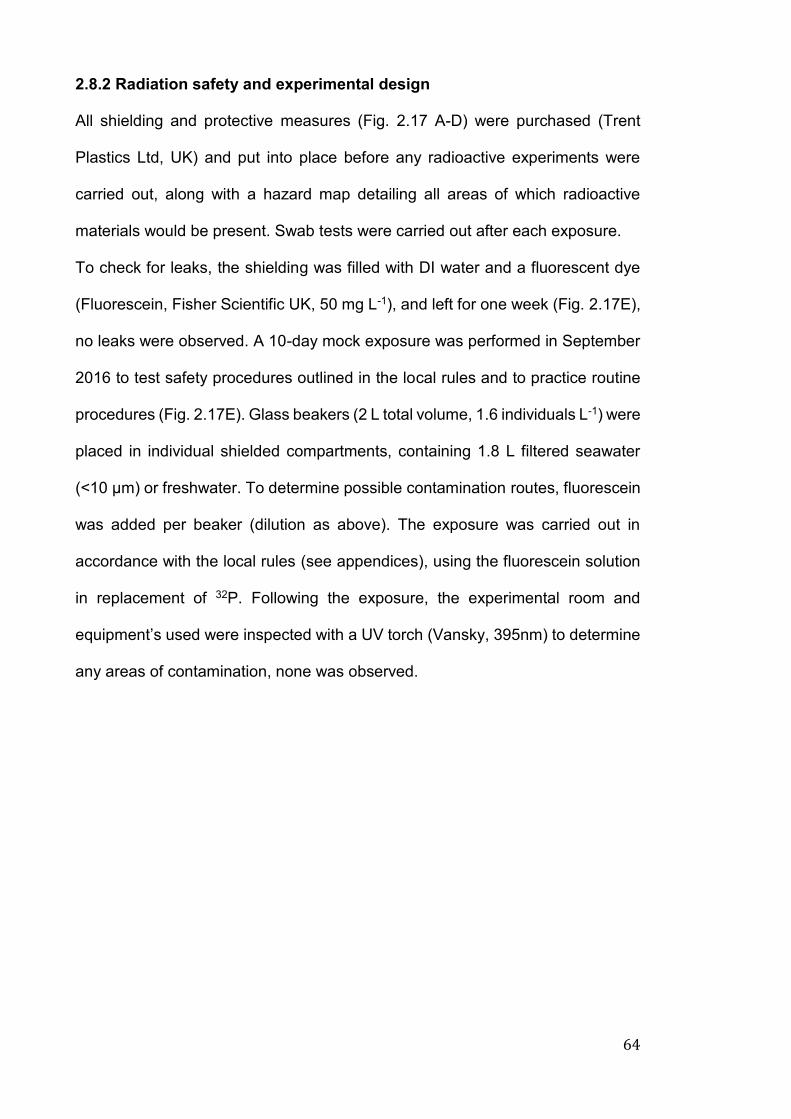
64
2.8.2 Radiation safety and experimental design
All shielding and protective measures (Fig. 2.17 A-D) were purchased (Trent
Plastics Ltd, UK) and put into place before any radioactive experiments were
carried out, along with a hazard map detailing all areas of which radioactive
materials would be present. Swab tests were carried out after each exposure.
To check for leaks, the shielding was filled with DI water and a fluorescent dye
(Fluorescein, Fisher Scientific UK, 50 mg L-1), and left for one week (Fig. 2.17E),
no leaks were observed. A 10-day mock exposure was performed in September
2016 to test safety procedures outlined in the local rules and to practice routine
procedures (Fig. 2.17E). Glass beakers (2 L total volume, 1.6 individuals L-1) were
placed in individual shielded compartments, containing 1.8 L filtered seawater
(<10 μm) or freshwater. To determine possible contamination routes, fluorescein
was added per beaker (dilution as above). The exposure was carried out in
accordance with the local rules (see appendices), using the fluorescein solution
in replacement of 32P. Following the exposure, the experimental room and
equipment’s used were inspected with a UV torch (Vansky, 395nm) to determine
any areas of contamination, none was observed.
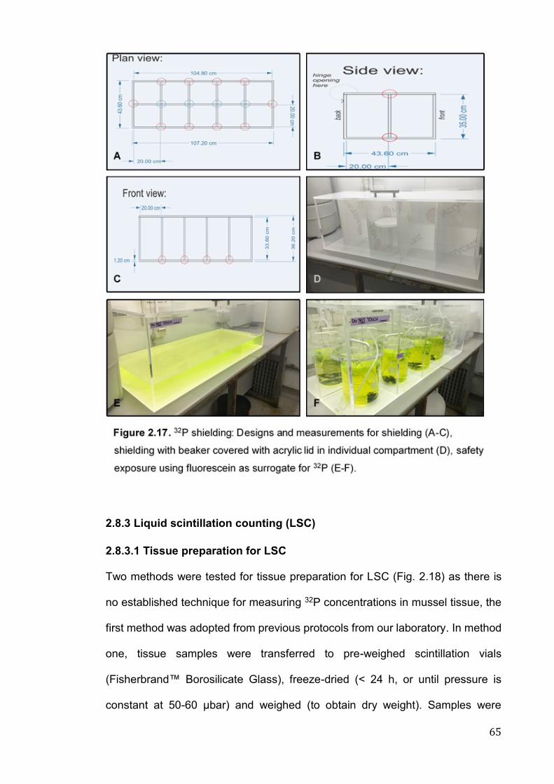
65
2.8.3 Liquid scintillation counting (LSC)
2.8.3.1 Tissue preparation for LSC
Two methods were tested for tissue preparation for LSC (Fig. 2.18) as there is
no established technique for measuring 32P concentrations in mussel tissue, the
first method was adopted from previous protocols from our laboratory. In method
one, tissue samples were transferred to pre-weighed scintillation vials
(Fisherbrand™ Borosilicate Glass), freeze-dried (< 24 h, or until pressure is
constant at 50-60 μbar) and weighed (to obtain dry weight). Samples were
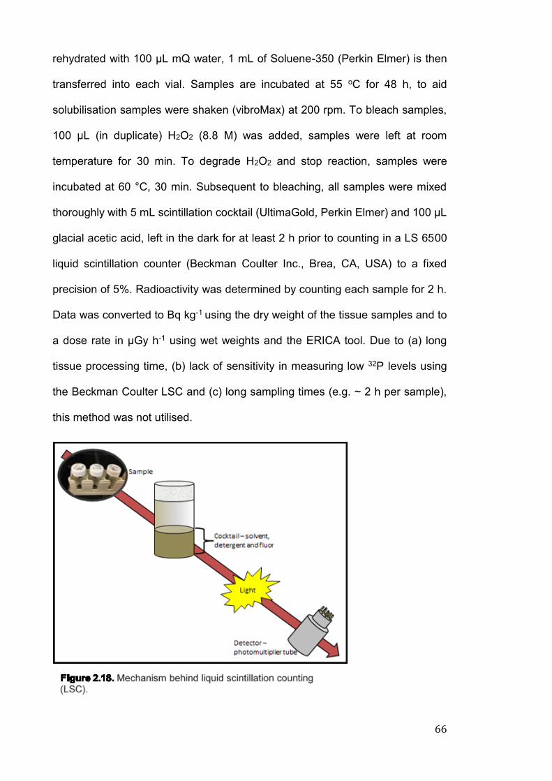
66
rehydrated with 100 µL mQ water, 1 mL of Soluene-350 (Perkin Elmer) is then
transferred into each vial. Samples are incubated at 55 oC for 48 h, to aid
solubilisation samples were shaken (vibroMax) at 200 rpm. To bleach samples,
100 μL (in duplicate) H2O2 (8.8 M) was added, samples were left at room
temperature for 30 min. To degrade H2O2 and stop reaction, samples were
incubated at 60 °C, 30 min. Subsequent to bleaching, all samples were mixed
thoroughly with 5 mL scintillation cocktail (UltimaGold, Perkin Elmer) and 100 μL
glacial acetic acid, left in the dark for at least 2 h prior to counting in a LS 6500
liquid scintillation counter (Beckman Coulter Inc., Brea, CA, USA) to a fixed
precision of 5%. Radioactivity was determined by counting each sample for 2 h.
Data was converted to Bq kg-1 using the dry weight of the tissue samples and to
a dose rate in μGy h-1 using wet weights and the ERICA tool. Due to (a) long
tissue processing time, (b) lack of sensitivity in measuring low 32P levels using
the Beckman Coulter LSC and (c) long sampling times (e.g. ~ 2 h per sample),
this method was not utilised.
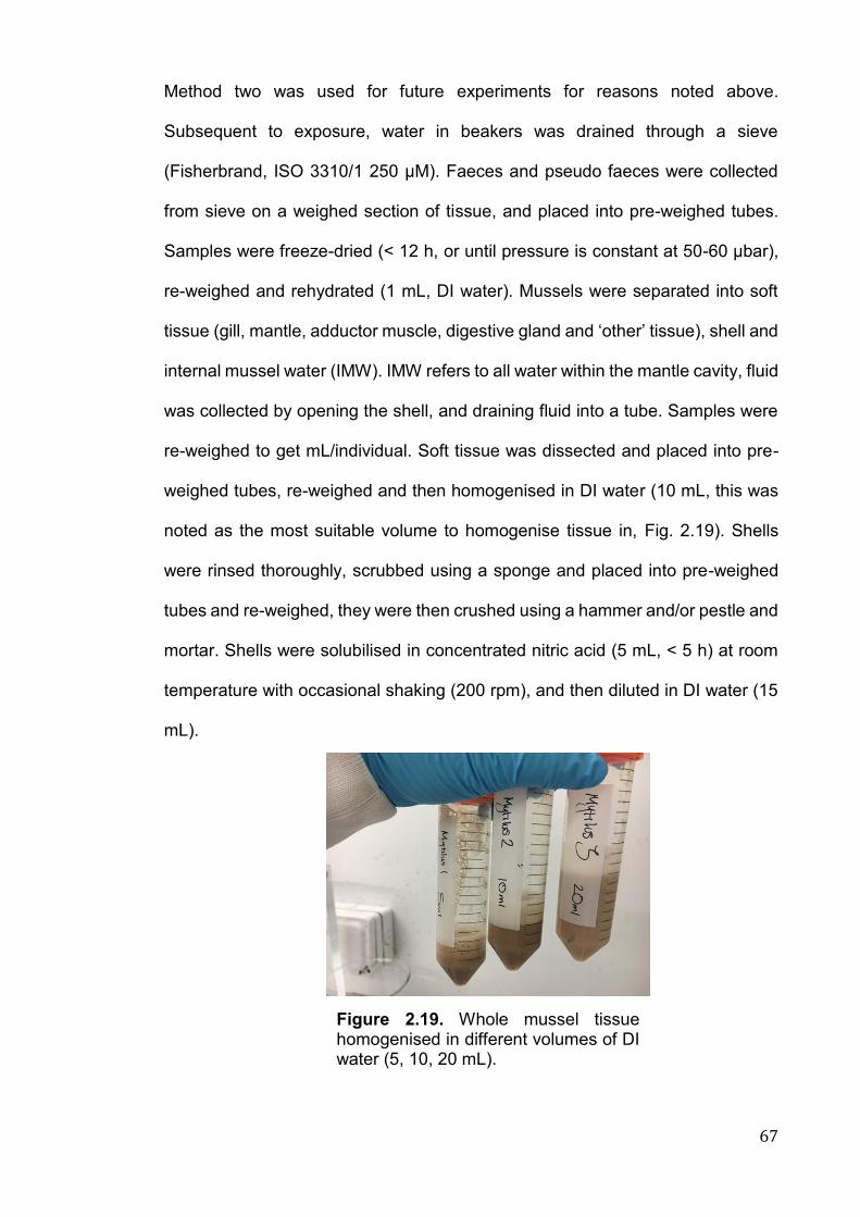
67
Method two was used for future experiments for reasons noted above.
Subsequent to exposure, water in beakers was drained through a sieve
(Fisherbrand, ISO 3310/1 250 µM). Faeces and pseudo faeces were collected
from sieve on a weighed section of tissue, and placed into pre-weighed tubes.
Samples were freeze-dried (< 12 h, or until pressure is constant at 50-60 μbar),
re-weighed and rehydrated (1 mL, DI water). Mussels were separated into soft
tissue (gill, mantle, adductor muscle, digestive gland and ‘other’ tissue), shell and
internal mussel water (IMW). IMW refers to all water within the mantle cavity, fluid
was collected by opening the shell, and draining fluid into a tube. Samples were
re-weighed to get mL/individual. Soft tissue was dissected and placed into pre-
weighed tubes, re-weighed and then homogenised in DI water (10 mL, this was
noted as the most suitable volume to homogenise tissue in, Fig. 2.19). Shells
were rinsed thoroughly, scrubbed using a sponge and placed into pre-weighed
tubes and re-weighed, they were then crushed using a hammer and/or pestle and
mortar. Shells were solubilised in concentrated nitric acid (5 mL, < 5 h) at room
temperature with occasional shaking (200 rpm), and then diluted in DI water (15
mL).
Figure 2.19. Whole mussel tissue homogenised in different volumes of DI water (5, 10, 20 mL).

68
Soft tissue, shell, IMW or faeces solution (1 mL, in duplicate) was mixed with 4
mL scintillant (ScintLogic U, LabLogic Systems Ltd, UK) in scintillation vials (20
mL, Fisherbrand™ Borosilicate Glass), 4 mL was also added to water samples.
Samples were left in dark for ~ 2 h prior to counting (Hidex 300SL), samples were
read (10 s) in triplicate.
2.8.3.2 Liquid scintillation counting: Analysis of water and mussel tissue
samples
To ensure accuracy of pipetting, a serial dilution of 32P was carried out (see Fig.
2.20). Sample data from the Beckman Coulter was presented in DPM
(disintegrations per min), and in counts per min (CPM) from the Hidex liquid
scintillation counter. Activity concentrations were background corrected by blank
subtracting from each sample, the blank was non-spiked fresh or seawater. Total
activity (Bq) per whole tissue was calculated by dividing by 60 to acquire DPS
(disintegrations per s), and then multiplying by the dilution factor where
appropriate. All samples were decay corrected where applicable to determine
total activity (Bq) per whole tissue (STA) on the day of sampling (tissue dissection
following exposure) using the following formulae:
STA = S0* 2^((N/365)/βy)
where S0 is the initial/starting sample activity (total activity [Bq] per whole tissue),
N is the number of days to decay correct for (i.e. -4 is 4 d), and βy is the half-life
of the radionuclide divided by 365 (days in one year, 32P = 0.039). Total activity
(Bq) per gram of tissue (wet weight) was calculated by dividing the total activity
(Bq) per whole tissue (decay adjusted) by the wet weight (g) of the tissue. In
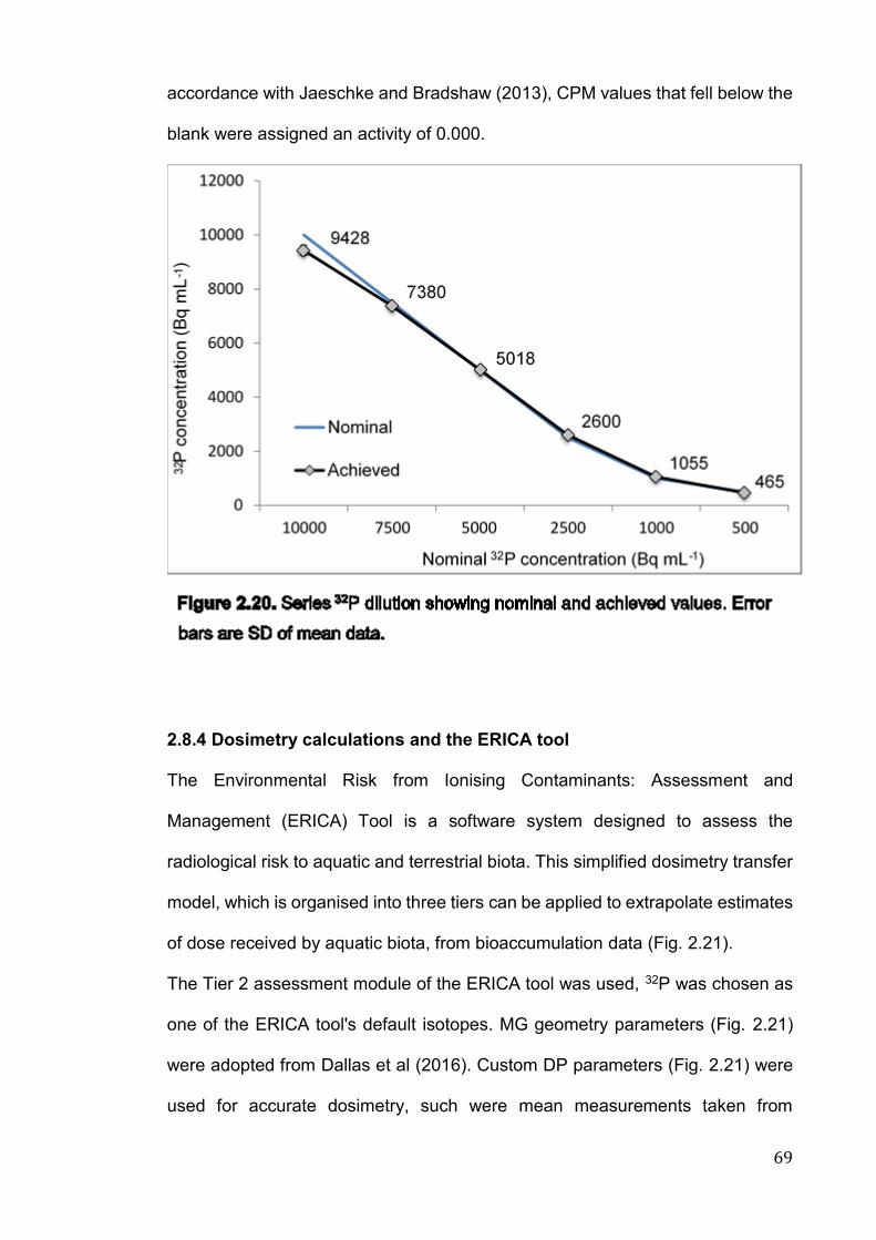
69
accordance with Jaeschke and Bradshaw (2013), CPM values that fell below the
blank were assigned an activity of 0.000.
2.8.4 Dosimetry calculations and the ERICA tool
The Environmental Risk from Ionising Contaminants: Assessment and
Management (ERICA) Tool is a software system designed to assess the
radiological risk to aquatic and terrestrial biota. This simplified dosimetry transfer
model, which is organised into three tiers can be applied to extrapolate estimates
of dose received by aquatic biota, from bioaccumulation data (Fig. 2.21).
The Tier 2 assessment module of the ERICA tool was used, 32P was chosen as
one of the ERICA tool's default isotopes. MG geometry parameters (Fig. 2.21)
were adopted from Dallas et al (2016). Custom DP parameters (Fig. 2.21) were
used for accurate dosimetry, such were mean measurements taken from
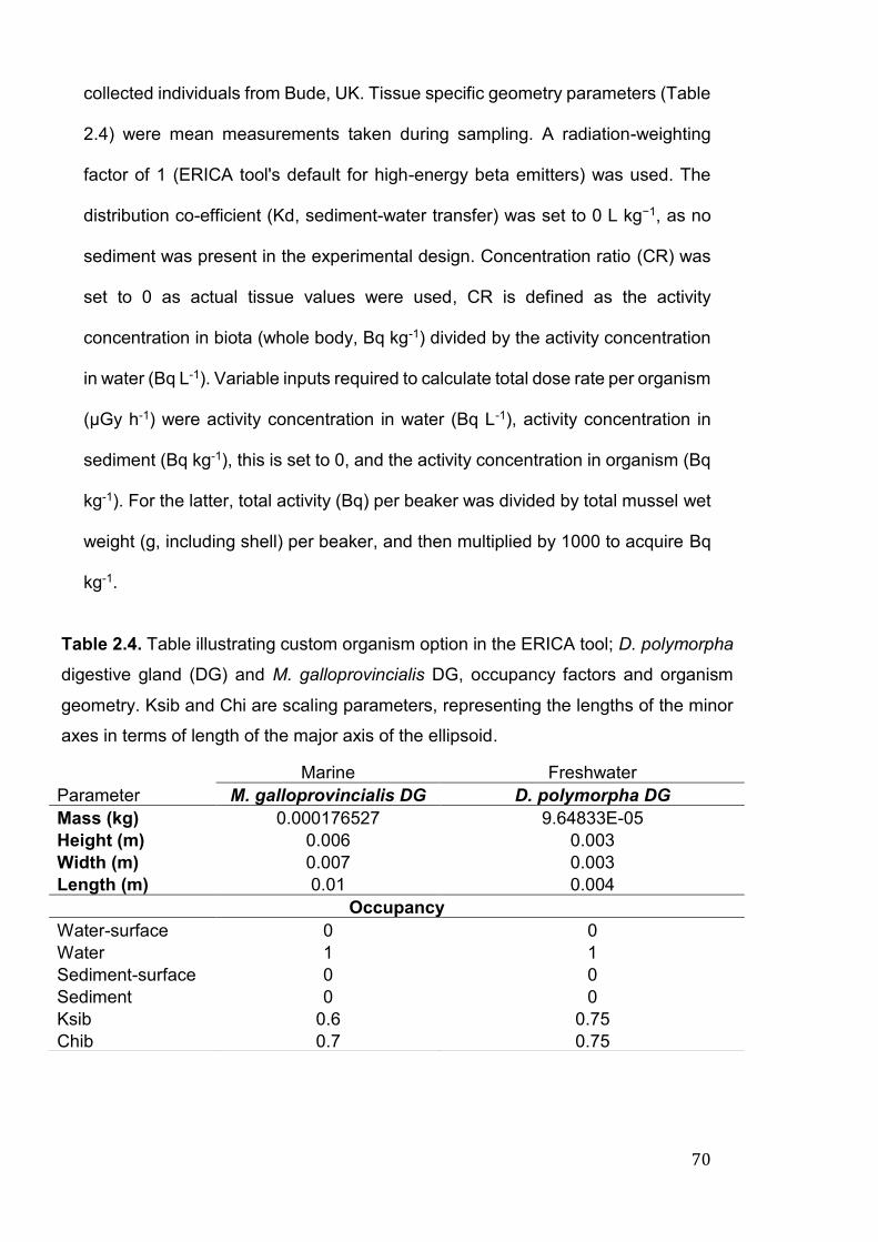
70
collected individuals from Bude, UK. Tissue specific geometry parameters (Table
2.4) were mean measurements taken during sampling. A radiation-weighting
factor of 1 (ERICA tool's default for high-energy beta emitters) was used. The
distribution co-efficient (Kd, sediment-water transfer) was set to 0 L kg−1, as no
sediment was present in the experimental design. Concentration ratio (CR) was
set to 0 as actual tissue values were used, CR is defined as the activity
concentration in biota (whole body, Bq kg-1) divided by the activity concentration
in water (Bq L-1). Variable inputs required to calculate total dose rate per organism
(μGy h-1) were activity concentration in water (Bq L-1), activity concentration in
sediment (Bq kg-1), this is set to 0, and the activity concentration in organism (Bq
kg-1). For the latter, total activity (Bq) per beaker was divided by total mussel wet
weight (g, including shell) per beaker, and then multiplied by 1000 to acquire Bq
kg-1.
Marine Freshwater
Parameter M. galloprovincialis DG D. polymorpha DG
Mass (kg) 0.000176527 9.64833E-05
Height (m) 0.006 0.003
Width (m) 0.007 0.003
Length (m) 0.01 0.004
Occupancy
Water-surface 0 0
Water 1 1
Sediment-surface 0 0
Sediment 0 0
Ksib 0.6 0.75
Chib 0.7 0.75
Table 2.4. Table illustrating custom organism option in the ERICA tool; D. polymorpha
digestive gland (DG) and M. galloprovincialis DG, occupancy factors and organism
geometry. Ksib and Chi are scaling parameters, representing the lengths of the minor
axes in terms of length of the major axis of the ellipsoid.
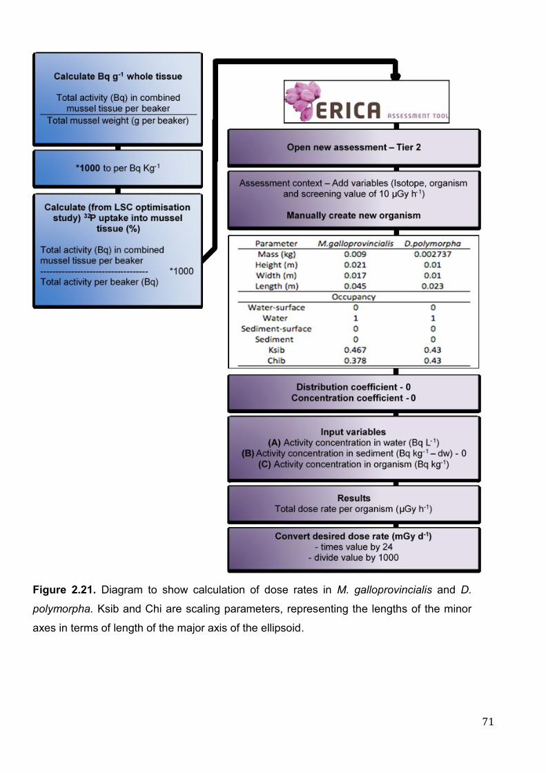
71
Figure 2.21. Diagram to show calculation of dose rates in M. galloprovincialis and D.
polymorpha. Ksib and Chi are scaling parameters, representing the lengths of the minor
axes in terms of length of the major axis of the ellipsoid.

72
2.8.5 Optimisation experiments
2.8.5.1 Pilot study
Starting activity levels (Bq L-1) to give the desired dose rates of 0.10, 1, 10 mGy
d-1 were calculated using the ERICA tool (Fig. 2.21) using the criteria listed in
Table 2.5 (Table 2.6). Activity levels were increased for the pilot experiment to
400 Bq/beaker (222 Bq L-1), as to be able to determine low activity levels
adequately using the Beckman Coulter. For accurate dosing and subsequent
dosimetry calculations, there was an equivalent weight of individual soft tissue
(wet weight, g) in each beaker. Before radioactive exposures, eleven individuals
(per species) were dissected, soft tissue was blotted dry and weighed to four
decimal places, shell was discarded. In accordance with Table 2.7, this equated
to 3 MG (mean. wet weight 1.9 g) to 14 DP (mean. wet weight 0.41 g).
M. galloprovincialis D. polymorpha
Tier 2 2
Isotope of interest 32P 32P
Environment Marine Freshwater
Organism Mollusc – bivalve Mollusc – bivalve
Kd (L kg−1) 21300 133
CR (1 Kg-1, wet
weight)
20000 60000
Occupancy Water-surface 0
Water 1
Sediment-surface 0
Sediment 0
Water-surface 0
Water 1
Sediment-surface 0
Sediment 0
Radiation weighting
factor
Defaults 1 Defaults 1
Table 2.5. ERICA tool criteria used for estimating dose rate from water
concentrations. Kd is distribution coefficient, CR is concentration ratio.
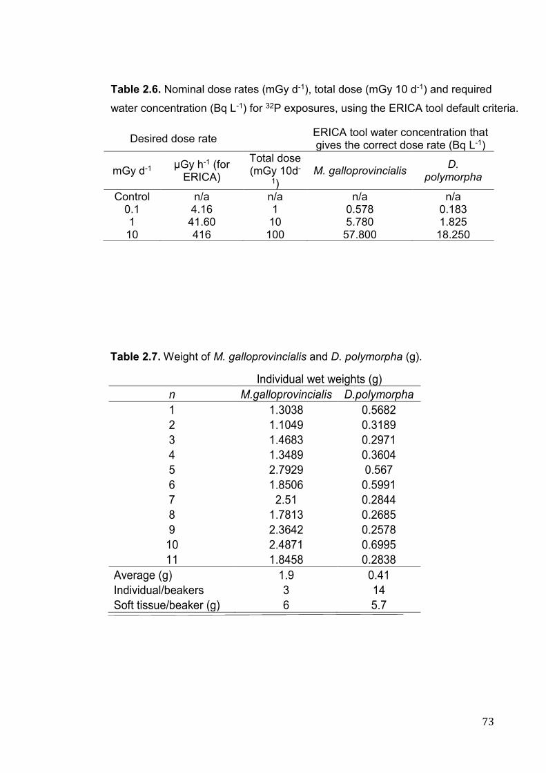
73
Desired dose rate ERICA tool water concentration that gives the correct dose rate (Bq L-1)
mGy d-1 µGy h-1 (for
ERICA)
Total dose (mGy 10d-
1) M. galloprovincialis
D. polymorpha
Control n/a n/a n/a n/a 0.1 4.16 1 0.578 0.183 1 41.60 10 5.780 1.825
10 416 100 57.800 18.250
Table 2.6. Nominal dose rates (mGy d-1), total dose (mGy 10 d-1) and required
water concentration (Bq L-1) for 32P exposures, using the ERICA tool default criteria.
Individual wet weights (g)
n M.galloprovincialis D.polymorpha
1 1.3038 0.5682
2 1.1049 0.3189
3 1.4683 0.2971
4 1.3489 0.3604
5 2.7929 0.567
6 1.8506 0.5991
7 2.51 0.2844
8 1.7813 0.2685
9 2.3642 0.2578
10 2.4871 0.6995
11 1.8458 0.2838
Average (g) 1.9 0.41
Individual/beakers 3 14
Soft tissue/beaker (g) 6 5.7
Table 2.7. Weight of M. galloprovincialis and D. polymorpha (g).
74
An 11-day pilot exposure was set up during September 2016 to determine
bioaccumulation in mussel soft tissue over time, following exposure to 32P.
Mussels were maintained as in section 2.2. Following depuration mussels were
transferred into 10 acid washed (5 per species), constantly aerated beakers,
contained within appropriate shielding (2 L-1 glass beakers, 1.6 MG or 7.8 DP L-
1).
Commercially available, radiolabelled-ATP (Adenosine triphosphate, γ-32P) was
obtained from Perkin Elmer (PerkinElmer, UK) in batches of 9.25 MBq (specific
activity: 370 MBq mL-1) and used as the source of radioactive 32P for our
experimental purposes. Radiolabelled ATP was utilised in our experiments as (a)
due to its readily bioavailable form would be accumulated readily into biological
tissue, (b) the ATP itself would not cause biological damage as the radioisotope
is almost chemically identical to the stable isotope, it therefore would not affect
future experiments and (c) it would not affect the chemical composition (i.e. pH,
salinity) of the sea/freshwater. Due to the short half-life of 32P, stock solution was
decay adjusted during the exposure.
Individuals were not fed during this exposure. Water quality parameters were pH
7.8 ± 0.32, temperature 15.3 ± 0.7 oC, dissolved oxygen (DO) 95 ± 19.6 % and
salinity 36.8 ± 0.28 for MG and pH 8 ± 0.29, temperature 14.9 ± 0.5 oC, DO 98.5
± 2.8 % and salinity 0.35 ± 0.08 for DP. On days 3, 5, 7, 9 and 11, soft tissue from
individuals from two beakers, one per species was processed for LSC as in
section 2.8.3.1 (Method 1. Water samples (100 ul, in duplicate) were taken each
day.
Due to inefficient sensitivity of the liquid scintillation counter, combined with long
tissue processing and counting times allowing for decay, 32P was not evident in
water samples. As displayed on Fig. 22, 32P accumulation is significantly (p <
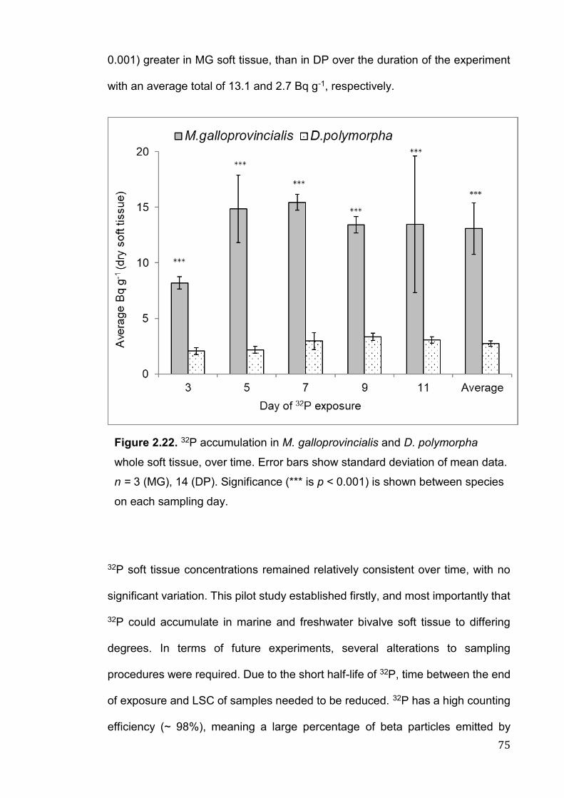
75
0.001) greater in MG soft tissue, than in DP over the duration of the experiment
with an average total of 13.1 and 2.7 Bq g-1, respectively.
32P soft tissue concentrations remained relatively consistent over time, with no
significant variation. This pilot study established firstly, and most importantly that
32P could accumulate in marine and freshwater bivalve soft tissue to differing
degrees. In terms of future experiments, several alterations to sampling
procedures were required. Due to the short half-life of 32P, time between the end
of exposure and LSC of samples needed to be reduced. 32P has a high counting
efficiency (~ 98%), meaning a large percentage of beta particles emitted by
Figure 2.22. 32P accumulation in M. galloprovincialis and D. polymorpha
whole soft tissue, over time. Error bars show standard deviation of mean data.
n = 3 (MG), 14 (DP). Significance (*** is p < 0.001) is shown between species
on each sampling day.
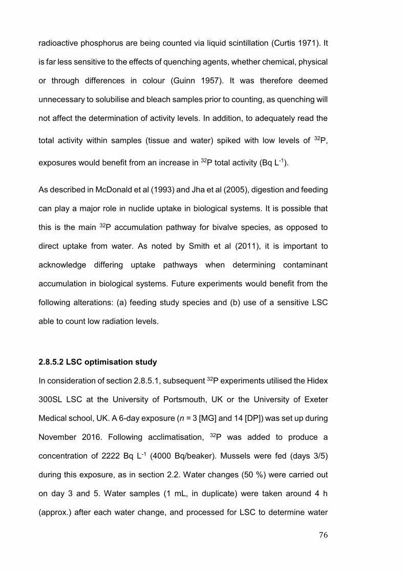
76
radioactive phosphorus are being counted via liquid scintillation (Curtis 1971). It
is far less sensitive to the effects of quenching agents, whether chemical, physical
or through differences in colour (Guinn 1957). It was therefore deemed
unnecessary to solubilise and bleach samples prior to counting, as quenching will
not affect the determination of activity levels. In addition, to adequately read the
total activity within samples (tissue and water) spiked with low levels of 32P,
exposures would benefit from an increase in 32P total activity (Bq L-1).
As described in McDonald et al (1993) and Jha et al (2005), digestion and feeding
can play a major role in nuclide uptake in biological systems. It is possible that
this is the main 32P accumulation pathway for bivalve species, as opposed to
direct uptake from water. As noted by Smith et al (2011), it is important to
acknowledge differing uptake pathways when determining contaminant
accumulation in biological systems. Future experiments would benefit from the
following alterations: (a) feeding study species and (b) use of a sensitive LSC
able to count low radiation levels.
2.8.5.2 LSC optimisation study
In consideration of section 2.8.5.1, subsequent 32P experiments utilised the Hidex
300SL LSC at the University of Portsmouth, UK or the University of Exeter
Medical school, UK. A 6-day exposure (n = 3 [MG] and 14 [DP]) was set up during
November 2016. Following acclimatisation, 32P was added to produce a
concentration of 2222 Bq L-1 (4000 Bq/beaker). Mussels were fed (days 3/5)
during this exposure, as in section 2.2. Water changes (50 %) were carried out
on day 3 and 5. Water samples (1 mL, in duplicate) were taken around 4 h
(approx.) after each water change, and processed for LSC to determine water
77
activity concentrations. Water quality parameters were pH 7.9 ± 0.33,
temperature 15 ± 0.1 oC, dissolved oxygen (DO) 99.23 ± 1.53 % and salinity 36.6
± 0.17 for MG and pH 8.3 ± 0.2, temperature 14.8 ± 0.2 oC, DO 98.9 ± 2 % and
salinity 0.4 ± 0.05 for DP. Samples (soft tissue, shell, IMW and water) were
processed in accordance to section 2.8.3.1 (Method 2).
Average total activity in water was 1935 and 1437 Bq/beaker (1.8 L-1), for MG
and DP, respectively. This study suggests that 54% and 74% of 32P is being
accumulated into MG and DP individuals (Whole body, Fig. 2.23). From this we
can employ the ERICA tool (section 2.8.4) to calculate correct water activity
concentrations to give the expected dose rates of 0.10, 1 and 10 mGy d-1 for each
species (Table 2.8).
Figure 2.23. 32P accumulation in M. galloprovincialis and D. polymorpha
whole soft tissue, shell and internal mussel water. Asterisks (*, ** or ***)
are indicative of significant differences (p < 0.05, 0.01, 0.001) between
species. Error bars show standard deviation of mean data. n = 3 (MG),
14 (DP).

78
2.9 Statistical analysis
All statistical analyses were carried out using the statistical software R (RStudio,
R 3.4.3 GUI 1.70 El Capitan build (7463), https://www.r-project.org/), unless
otherwise stated. All data was checked for normality distribution (Shapiro-Wilk
test) and homogeneity of variances (Levene’s test), with visual examination of
QQ-plots. The non-parametric Kruskal-Wallis test was used if assumptions were
not met; comparison between treatment groups was determined using a Dunn’s
pairwise comparison with Bonferroni correction. Where assumptions were met, a
one-way ANOVA was run with Tukey’s post hoc tests. Comparison between
treatment groups was determined using a Wilcoxon rank sum test with Holm-
Bonferroni correction. Regression analysis using a Pearson correlation
coefficient was used to determine any correlations between variables, all figures
displaying correlations include a best fit line (regression line). Level of
significance for all tests was set at p < 0.05 (*) and data presented as mean ±
standard deviation, unless otherwise stated.
Nominal dose rate ERICA tool water concentrations
mGy d-1 that give correct dose rate (Bq L-1)
M. galloprovincialis
0.1 709
1 7090
10 70900
D. polymorpha
0.1 571
1 5710
10 57100
Table 2.8. Water concentrations (Bq L-1), calculated by the ERICA tool, that give to
correct dose rates of 0.1, 1 and 10 mGy d-1 in M. galloprovincialis and D. polymorpha.

79

80
81
Chapter 3
Assessing relative sensitivity of marine and freshwater bivalves
following exposure to copper: Application of classical and
novel genotoxicological biomarkers
Under review
Vernon, EL and Jha, AN. 2018. Assessing relative sensitivity of marine and
freshwater bivalves following exposure to copper: Application of classical and
novel genotoxicological biomarkers. Mutation research/Genetic Toxicology and
Environmental Mutagenesis.

82
83
3.1 Introduction
Identification of most sensitive natural species following exposure to
anthropogenic contaminants is important from an environmental protection point
of view (Jha, 2004, 8; Canty et al. 2009; Dallas et al. 2013). In recent years, there
has been growing regulatory and scientific concerns for those contaminants
which have the potential to exert carcinogenic, mutagenic and endocrine
disrupting effects (Dallas et al. 2013). In this context, it is also emerging that the
contaminants could simultaneously induce their toxicity in a variety of ways, i.e.
the same contaminant could induce carcinogenic, mutagenic, immunotoxic and
endocrine disrupting effects (Jha 2008).
Metals are an important group of ubiquitous contaminants to which biota are
exposed in different habitats and ecological niches (Bolognesi et al. 1999; Dallas
et al. 2013). Exposure to metals can induce a variety of detrimental biological
effects via a range of mechanisms, including through the generation of ROS. In
addition to damage to cellular components such as lipids and proteins, DNA
damage (either directly or through generation of ROS), inhibition of DNA repair
capacity and disruption of cell cycle control are of particular concern (Stohs and
Bagchi 1995; Azqueta et al. 2009; Dallas et al. 2013; Xu et al. 2016). Although
various metals are essential for biological processes, copper (Cu) in particular
being important for growth, metabolism and enzymatic activities (Cid et al. 1995;
Gaetke and Chow 2003; Bopp et al. 2008; Kim et al. 2008), can be highly toxic
to organisms including bivalves at higher concentrations (Bolognesi et al. 1999;
Al Subiai et al. 2011). Cu (Cu2+) has been the focus of extensive research as a
widespread contaminant, present in coastal and inland water bodies globally and
is considered to be of greater environmental concern compared to other
contaminants such as pharmaceuticals (Eisler 1997; Donnachie et al. 2016).
84
Total dissolved Cu in contaminated environments have been found to reach
concentrations of 689 μg L-1 (Bryan and Gibbs 1983), with permitted levels in
England and Wales ranging from 1–28 μg L-1 in freshwater (dependant on water
hardness), and 5 μg L-1 in seawater (DEFRA 2014).
The link between metal (in particular Cu) exposure and genotoxicity, as
determined by induction of MN and DNA strand breaks (as determined by comet
assay) has been well documented in a range of aquatic species, including
bivalves (Geret et al. 2002; Brown et al. 2004; Villela et al. 2006; Al-Subiai et al.
2011; Trevisan et al. 2011; Mai et al. 2012; Vosloo et al. 2012; Maria et al. 2013;
Brooks et al. 2015; Lewis et al. 2016). Induction of DNA strand breaks has been
noted at environmentally relevant Cu concentrations in several bivalve species,
including Mytilus spp., (18-56 μg L-1), Perna perna (37.5-50 μg L-1), Limnoperna
fortunei (375-750 μg L-1) and Crassostrea gigas (embryos, 1-20 μg L-1)
(Bolognesi et al. 1999; Villela et al. 2006; Al-Subiai et al. 2011; Trevisan et al.
2011; Vosloo et al. 2012; Sussarellu et al. 2018).
Species-specific differences in bioaccumulation and resultant biological response
following exposure to environmental contaminants have been reported for
different marine bivalves (Pellerin and Amiard 2009; Brooks et al. 2015; Marisa
et al. 2018). Despite the fact that (a) freshwater and marine bivalves are
comparable in their external and internal anatomical structures (Vernon et al.
2018) and (b) contaminants including metals are highly relevant to both marine
and freshwater environments, there have been no attempts to compare the
relative bioaccumulation and biological responses in these ecologically relevant
organisms. As noted in section 1.4.1, the marine, Mytilus galloprovincialis (MG)
and freshwater mussel, Dreissena polymorpha (DP) were chosen for this study
as ecologically relevant representatives of both coastal and inland water bodies
85
(McDonald et al. 1991; Binelli et al. 2015). As sessile, filter feeding organisms,
both species are used extensively in monitoring programmes and in toxicological
research as accurate bioindicators of their environmental surroundings (NOAA
2012). The physiology, anatomy and ecology of MG and DP is well understood
and their effectiveness within ecotoxicological studies well documented (Binelli et
al. 2015), nevertheless, the relative sensitivity of marine and freshwater bivalves,
in terms of genotoxic damage is yet to be fully explored. As suggested by various
authors the use of two (or more) species should be a considered as more robust,
realistic approach for ecotoxicological studies (Chapman 2002; Solomon and
Sibley 2002; Schnug et al. 2014).
The extent of damage in individuals evident at higher levels of biological
organisation, subsequent to DNA strand breaks, is dependent on a range of
factors including exposure period, contaminant concentration, rate of uptake,
metabolism, accumulation and the efficiency of repair mechanisms (Jha 2008).
In recent years, immunostaining techniques such as γ-H2AX and 53BP1 have
been utilised as sensitive markers of DNA DSBs (Gerić et al. 2014; Oommen et
al. 2016b). The genotoxicity of various contaminants (e.g. metals, nanoparticles,
insecticides and radionuclides) using induction of γ-H2AX as a biomarker has
been displayed in zebrafish (Danio rerio) ZF4 cells, adult zebrafish liver tissue
and retina, and fathead minnow (Pimephales promelas) juveniles (Choi et al.
2010; Pereira et al. 2012, 13; Gagnaire et al. 2017; Paravani et al. 2018).
Concentration-dependent DNA damage in ZF4 cells was evident following
aluminium (10-100 µM) and cadmium (1- 100 µM) exposures (Pereira et al.
2013). γ-H2AX foci induction followed a similar trend, where number of foci per
cell increased with aluminium or cadmium concentration up to 30 µM, and then
decreased in 50-100 µM treatments (Pereira et al. 2013). Such findings aid to our
86
limited knowledge of DSB-repair mechanisms, subsequent to exposure to
environmentally relevant contaminants in non-mammalian models. In spite of
usefulness of this assay, particularly when validated alongside classical
genotoxicity assays (i.e. comet or MN assays), this highly sensitive approach is
yet to be applied in aquatic invertebrates.
In light of the above information, following exposure to a range of Cu2+
concentrations (referred to as Cu throughout text) the objectives of this study
were (a) to investigate the relative tissue specific accumulation of Cu in both
bivalve species, (b) to establish a concentration-response curve for genotoxic
responses in the adult life stages of the species (c) to determine relative
sensitivity between the two species for genotoxic responses using a range of
genotoxicity parameters (i.e. induction of MN, Comet and γ-H2AX) (d) to correlate
the nominal Cu concentrations in water with bioaccumulation and observed
genotoxic responses and (e) to determine potential correlations between different
genotoxicity parameters studied. With regards to species variation, we
hypothesised firstly that little disparity in genotoxic response will be evident.
Secondly, with increased DNA damage (DNA strand breaks and MN formation),
a greater induction of γ-H2AX foci will be evident.
3.2 Materials and methods
3.2.1 Chemicals and suppliers
All chemicals and reagents were purchased from Fisher Scientific UK, Anachem
Ltd. UK, Sigma-Aldrich Ltd. UK, VWR International Ltd USA or Greiner Bio-One
Ltd UK, unless stated otherwise. Product details are mentioned in text as
appropriate.
87
3.2.2 Mussel exposure conditions
Four 10-day Cu exposure experiments were performed between April 2016 and
December 2017, the first two to determine genotoxic responses and the latter for
tissue specific Cu accumulation measurements. Adult MG (shell length 44.5 ± 6.5
mm) and DP (shell length 26.7 ± 4.31 mm) were collected from Trebarwith strand
(as in Dallas et al (2013)) and Bude, Cornwall, UK (Chapter 2, section 2.2),
respectively (Vernon et al. 2018). DP and MG were maintained in accordance to
chapter 2, section 2.2.
Exposures of bivalves to Cu were staggered by one week for ease of analysis
and logistical reasons. Subsequent to a two-week depuration period after
collection, individual mussels were transferred into twelve acid washed glass
beakers, in triplicate (i.e. 1.7 mussels L-1), containing 1.8 L-1 of water and aerated
(Dallas et al. 2013). Individuals were then acclimatised for 48 h prior to exposure.
Beakers were labelled and assigned to one of the four treatment groups: 0
(control), 18, 32 and 56 µg L-1 Cu (as CuSO4.5H2O, 99% purity), there were three
replicates per treatment (Fig. 3.1). Cu concentrations were selected in
accordance to previous work from our laboratory and in line with environmental
realistic values (Al-Subiai et al. 2011).
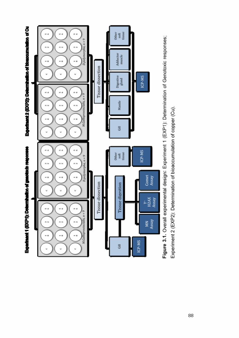
88
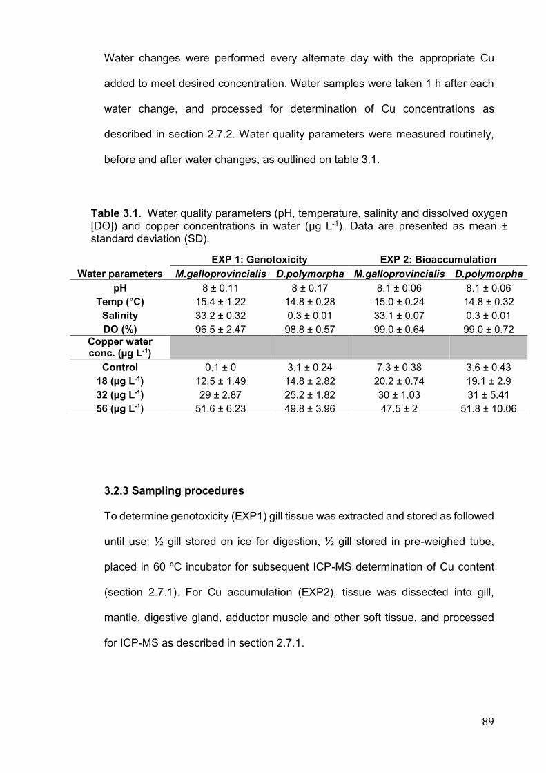
89
Water changes were performed every alternate day with the appropriate Cu
added to meet desired concentration. Water samples were taken 1 h after each
water change, and processed for determination of Cu concentrations as
described in section 2.7.2. Water quality parameters were measured routinely,
before and after water changes, as outlined on table 3.1.
3.2.3 Sampling procedures
To determine genotoxicity (EXP1) gill tissue was extracted and stored as followed
until use: ½ gill stored on ice for digestion, ½ gill stored in pre-weighed tube,
placed in 60 ºC incubator for subsequent ICP-MS determination of Cu content
(section 2.7.1). For Cu accumulation (EXP2), tissue was dissected into gill,
mantle, digestive gland, adductor muscle and other soft tissue, and processed
for ICP-MS as described in section 2.7.1.
EXP 1: Genotoxicity EXP 2: Bioaccumulation
Water parameters M.galloprovincialis D.polymorpha M.galloprovincialis D.polymorpha
pH 8 ± 0.11 8 ± 0.17 8.1 ± 0.06 8.1 ± 0.06
Temp (°C) 15.4 ± 1.22 14.8 ± 0.28 15.0 ± 0.24 14.8 ± 0.32
Salinity 33.2 ± 0.32 0.3 ± 0.01 33.1 ± 0.07 0.3 ± 0.01
DO (%) 96.5 ± 2.47 98.8 ± 0.57 99.0 ± 0.64 99.0 ± 0.72
Copper water conc. (µg L-1)
Control 0.1 ± 0 3.1 ± 0.24 7.3 ± 0.38 3.6 ± 0.43
18 (µg L-1) 12.5 ± 1.49 14.8 ± 2.82 20.2 ± 0.74 19.1 ± 2.9
32 (µg L-1) 29 ± 2.87 25.2 ± 1.82 30 ± 1.03 31 ± 5.41
56 (µg L-1) 51.6 ± 6.23 49.8 ± 3.96 47.5 ± 2 51.8 ± 10.06
Table 3.1. Water quality parameters (pH, temperature, salinity and dissolved oxygen [DO]) and copper concentrations in water (µg L-1). Data are presented as mean ± standard deviation (SD).
90
3.2.4 Biological assays
3.2.4.1 Isolation of gill cells for genotoxicity assays
The procedure to obtain gill cells for genotoxic assays was adopted from previous
studies (Vincent-Hubert et al. 2011), and outlined in section 2.3.2. Supernatant
was then used in subsequent assays providing cell viability, checked using the
Trypan Blue exclusion dye assay (section 2.4) (Strober 2001) was <90% across
all treatments (data not shown).
3.2.4.2 Comet assay to determine DNA strand breaks
The comet assay was performed using gill cell suspension (150 µL), as described
in section 2.5.2.
3.2.4.3 Analysis of micronuclei (MN) induction
Gill cell suspension was adhered and fixed as described in section 2.5.1, before
staining with 20 μL ethidium bromide (20 μL of 20 mg L−1). Cells (n = 500) were
scored per slide and results are reported as mean MN per 1000 cells, in keeping
with other data from our research group (Dallas et al. 2013).
3.2.4.4 Induction of γ-H2AX foci
γ-H2AX foci were determined in gill cells, following procedures outlined in 2.5.3.
All slides, including procedural blanks were coded and scored at random and 50
cells were counted per individual/slide.
91
3.2.5 Determination of Cu concentration in soft tissues and in water
samples
3.2.5.1 Cu analysis in tissues using Inductively Coupled Plasma Mass
Spectrometry (ICP-MS)
Cu determination in tissue samples (i.e. gill, mantle, digestive gland, adductor
muscle and ‘other’ soft tissue) and water was carried out using ICP-MS as
described before in publications from our laboratory (Al-Subiai et al. 2011; Dallas
et al. 2013; D'Agata et al. 2014) and in section 2.7. Procedural blanks, along with
a certified reference material (TORT-2, lobster hepatopancreas) were run
alongside samples.
3.2.5.2 Determination of Cu in water samples using ICP-MS
Cu samples in water were determined following procedures outlined in section
2.7.2.
3.2.6 Statistical analysis
All statistical analyses were carried out using the statistical software R (RStudio,
R 3.4.3 GUI 1.70 El Capitan build (7463), https://www.r-project.org/), in
accordance to principles detailed in section 2.9. Briefly, data was checked for
normality distribution (Shapiro-Wilk test) and homogeneity of variances (Levene’s
test). Appropriate tests were used to determine comparison between treatment
groups and correlations between variables.
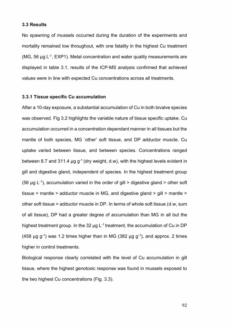
92
3.3 Results
No spawning of mussels occurred during the duration of the experiments and
mortality remained low throughout, with one fatality in the highest Cu treatment
(MG, 56 µg L-1, EXP1). Metal concentration and water quality measurements are
displayed in table 3.1, results of the ICP-MS analysis confirmed that achieved
values were in line with expected Cu concentrations across all treatments.
3.3.1 Tissue specific Cu accumulation
After a 10-day exposure, a substantial accumulation of Cu in both bivalve species
was observed. Fig 3.2 highlights the variable nature of tissue specific uptake. Cu
accumulation occurred in a concentration dependant manner in all tissues but the
mantle of both species, MG ‘other’ soft tissue, and DP adductor muscle. Cu
uptake varied between tissue, and between species. Concentrations ranged
between 8.7 and 311.4 μg g-1 (dry weight, d.w), with the highest levels evident in
gill and digestive gland, independent of species. In the highest treatment group
(56 μg L-1), accumulation varied in the order of gill > digestive gland > other soft
tissue > mantle > adductor muscle in MG, and digestive gland > gill > mantle >
other soft tissue > adductor muscle in DP. In terms of whole soft tissue (d.w, sum
of all tissue), DP had a greater degree of accumulation than MG in all but the
highest treatment group. In the 32 μg L-1 treatment, the accumulation of Cu in DP
(458 μg g-1) was 1.2 times higher than in MG (382 μg g-1), and approx. 2 times
higher in control treatments.
Biological response clearly correlated with the level of Cu accumulation in gill
tissue, where the highest genotoxic response was found in mussels exposed to
the two highest Cu concentrations (Fig. 3.3).
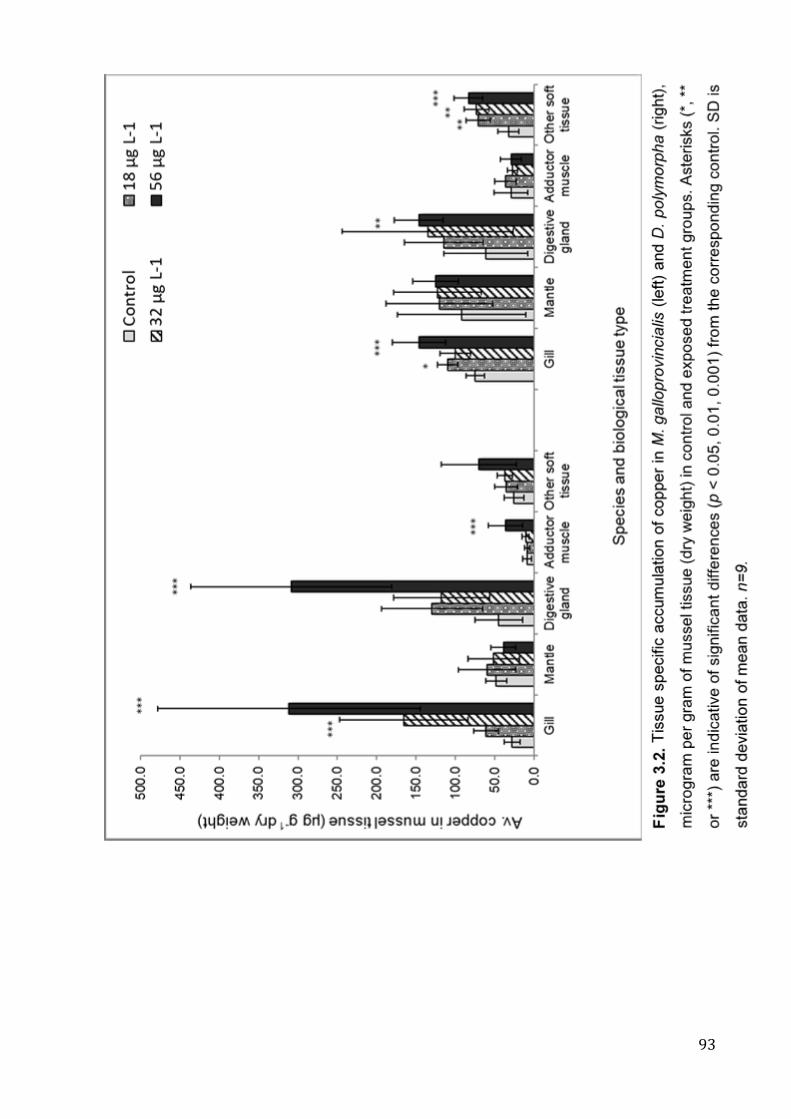
93
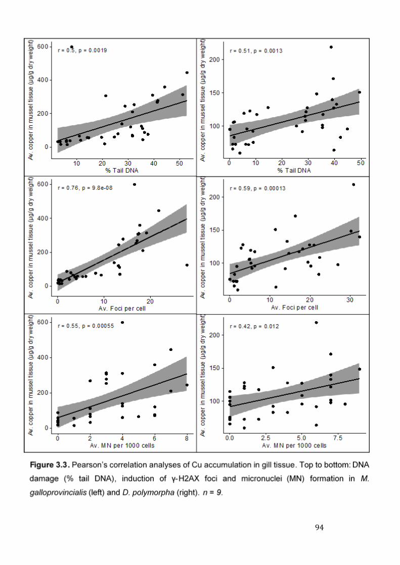
94
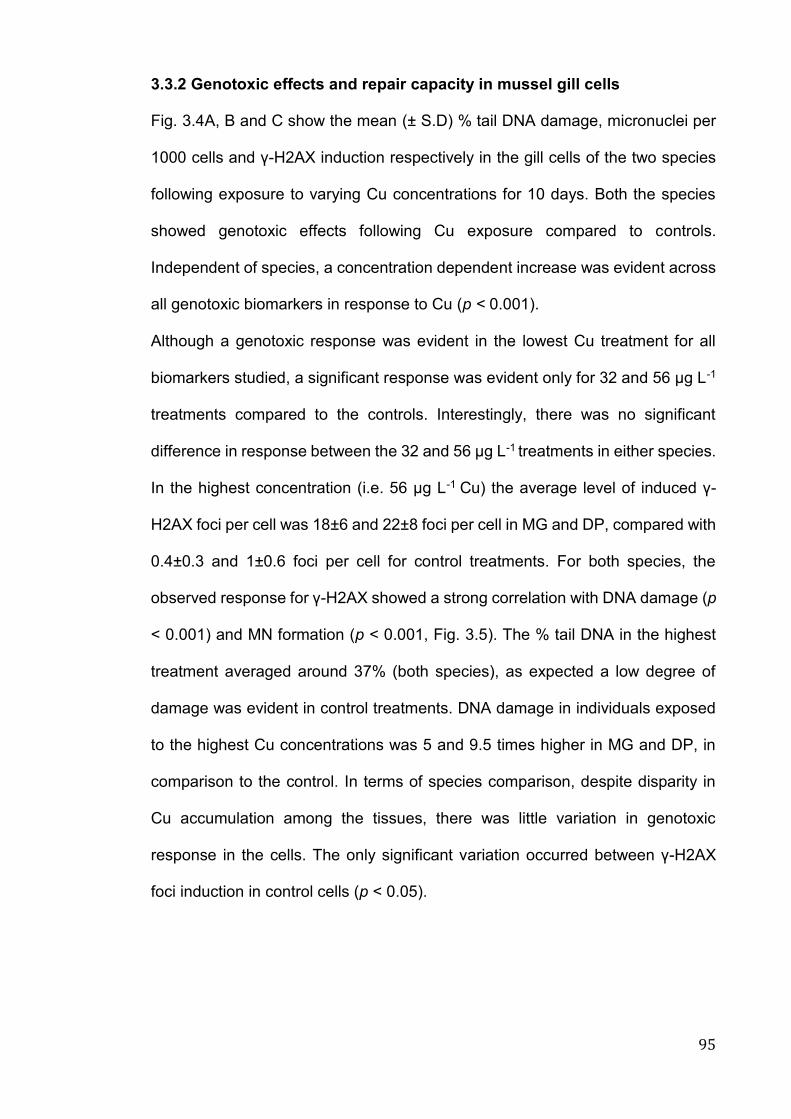
95
3.3.2 Genotoxic effects and repair capacity in mussel gill cells
Fig. 3.4A, B and C show the mean (± S.D) % tail DNA damage, micronuclei per
1000 cells and γ-H2AX induction respectively in the gill cells of the two species
following exposure to varying Cu concentrations for 10 days. Both the species
showed genotoxic effects following Cu exposure compared to controls.
Independent of species, a concentration dependent increase was evident across
all genotoxic biomarkers in response to Cu (p < 0.001).
Although a genotoxic response was evident in the lowest Cu treatment for all
biomarkers studied, a significant response was evident only for 32 and 56 μg L-1
treatments compared to the controls. Interestingly, there was no significant
difference in response between the 32 and 56 μg L-1 treatments in either species.
In the highest concentration (i.e. 56 μg L-1 Cu) the average level of induced γ-
H2AX foci per cell was 18±6 and 22±8 foci per cell in MG and DP, compared with
0.4±0.3 and 1±0.6 foci per cell for control treatments. For both species, the
observed response for γ-H2AX showed a strong correlation with DNA damage (p
< 0.001) and MN formation (p < 0.001, Fig. 3.5). The % tail DNA in the highest
treatment averaged around 37% (both species), as expected a low degree of
damage was evident in control treatments. DNA damage in individuals exposed
to the highest Cu concentrations was 5 and 9.5 times higher in MG and DP, in
comparison to the control. In terms of species comparison, despite disparity in
Cu accumulation among the tissues, there was little variation in genotoxic
response in the cells. The only significant variation occurred between γ-H2AX
foci induction in control cells (p < 0.05).
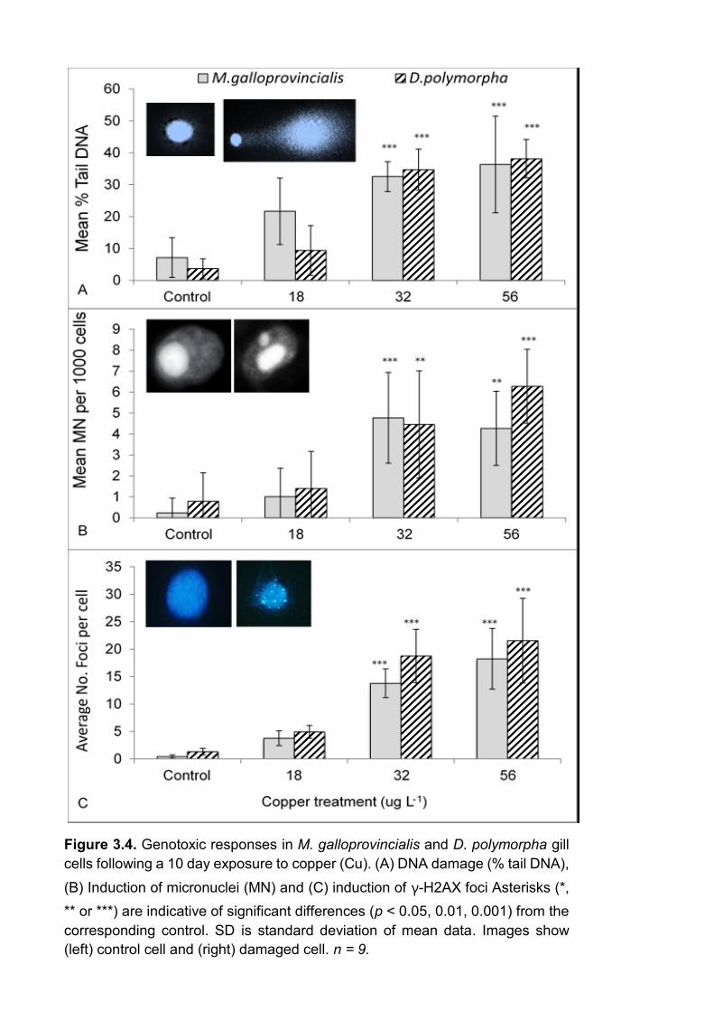
96
Figure 3.4. Genotoxic responses in M. galloprovincialis and D. polymorpha gill
cells following a 10 day exposure to copper (Cu). (A) DNA damage (% tail DNA),
(B) Induction of micronuclei (MN) and (C) induction of γ-H2AX foci Asterisks (*,
** or ***) are indicative of significant differences (p < 0.05, 0.01, 0.001) from the
corresponding control. SD is standard deviation of mean data. Images show
(left) control cell and (right) damaged cell. n = 9.
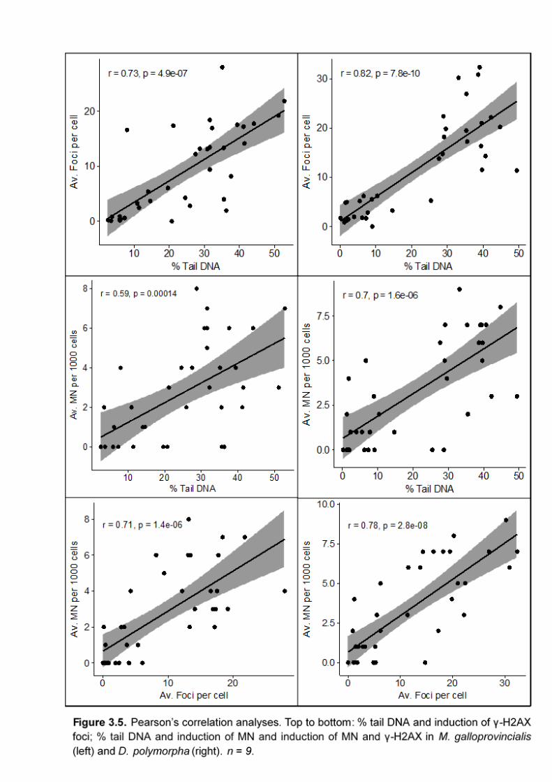
97
98
3.4 Discussion
3.4.1 Tissue specific Cu accumulation
It is well accepted that following uptake, waterborne contaminants are not
uniformly distributed among the tissues due to their inherent metabolic
capabilities (Jha et al. 2006; Faggio et al. 2018). The presence of metals in the
sediments has also been correlated with their accumulation in soft tissues of
bivalves under field conditions (Dallas et al. 2013). As expected, in our study Cu
concentrations varied among the tissues (Jing et al. 2006; Martins et al. 2011; Di
Salvatore et al. 2013; Jorge et al. 2016). The highest Cu concentrations were
found in the gill and digestive gland, particularly in the MG 56 μg L-1 treatment. In
this treatment, 40% (MG) and 28% (DP) of accumulated Cu was located in the
digestive gland, this is in contrast to previous reports where Cu in DP had been
shown to predominately accumulate in the foot (included in other soft tissue in
this study), followed by gill and digestive organs (Gundacker 1999). Mussel
digestive systems are well known to harbour contaminants via dietary uptake
pathways (Viarengo et al. 1981; Regoli 1998; Marigomez et al. 2002; Faggio et
al. 2018). Apart from inherent metabolic differences, uptake and bioaccumulation
is dependent on many factors including bioavailability, uptake mechanism and
biological factors (e.g. weight, gender, reproductive stage, feeding habits etc.).
These factors may help explain disparity between the studies (Jha 2008).
In line with findings from Sanders et al (1994), Zorita et al (2007) and Al-Subiai
et al (2011) a high degree of Cu bioaccumulation was evident in gill tissue of both
species. Bivalve gills due to proximity to surrounding media, and therefore the
primary sites of uptake of dissolved Cu are often regarded as a key tissue of
interest in ecotoxicological studies. In contrast to Al-Subiai et al (2011), who
found reduced Cu accumulation in 56 mg L-1 compared to 32 mg L-1 treatment
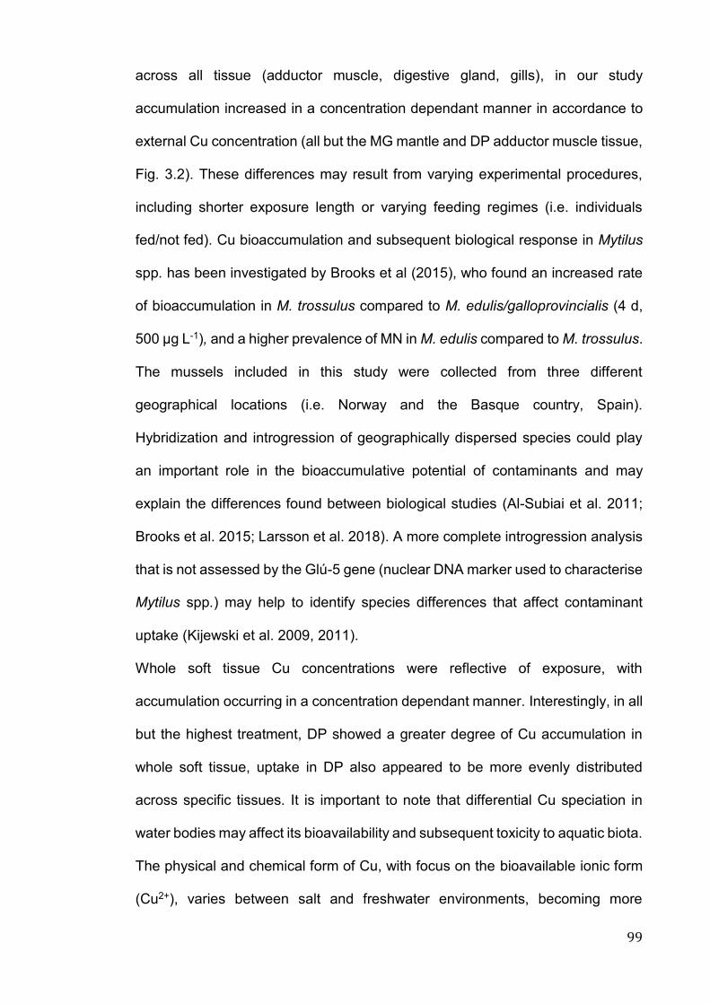
99
across all tissue (adductor muscle, digestive gland, gills), in our study
accumulation increased in a concentration dependant manner in accordance to
external Cu concentration (all but the MG mantle and DP adductor muscle tissue,
Fig. 3.2). These differences may result from varying experimental procedures,
including shorter exposure length or varying feeding regimes (i.e. individuals
fed/not fed). Cu bioaccumulation and subsequent biological response in Mytilus
spp. has been investigated by Brooks et al (2015), who found an increased rate
of bioaccumulation in M. trossulus compared to M. edulis/galloprovincialis (4 d,
500 μg L-1), and a higher prevalence of MN in M. edulis compared to M. trossulus.
The mussels included in this study were collected from three different
geographical locations (i.e. Norway and the Basque country, Spain).
Hybridization and introgression of geographically dispersed species could play
an important role in the bioaccumulative potential of contaminants and may
explain the differences found between biological studies (Al-Subiai et al. 2011;
Brooks et al. 2015; Larsson et al. 2018). A more complete introgression analysis
that is not assessed by the Glú-5 gene (nuclear DNA marker used to characterise
Mytilus spp.) may help to identify species differences that affect contaminant
uptake (Kijewski et al. 2009, 2011).
Whole soft tissue Cu concentrations were reflective of exposure, with
accumulation occurring in a concentration dependant manner. Interestingly, in all
but the highest treatment, DP showed a greater degree of Cu accumulation in
whole soft tissue, uptake in DP also appeared to be more evenly distributed
across specific tissues. It is important to note that differential Cu speciation in
water bodies may affect its bioavailability and subsequent toxicity to aquatic biota.
The physical and chemical form of Cu, with focus on the bioavailable ionic form
(Cu2+), varies between salt and freshwater environments, becoming more
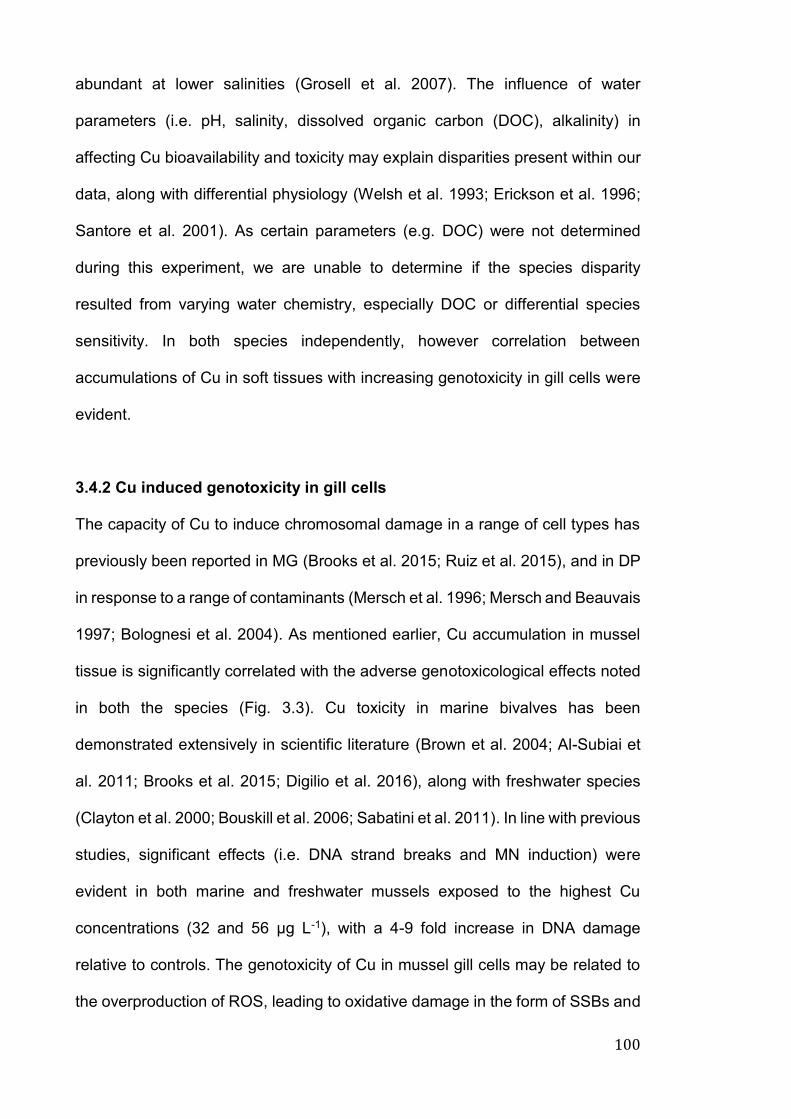
100
abundant at lower salinities (Grosell et al. 2007). The influence of water
parameters (i.e. pH, salinity, dissolved organic carbon (DOC), alkalinity) in
affecting Cu bioavailability and toxicity may explain disparities present within our
data, along with differential physiology (Welsh et al. 1993; Erickson et al. 1996;
Santore et al. 2001). As certain parameters (e.g. DOC) were not determined
during this experiment, we are unable to determine if the species disparity
resulted from varying water chemistry, especially DOC or differential species
sensitivity. In both species independently, however correlation between
accumulations of Cu in soft tissues with increasing genotoxicity in gill cells were
evident.
3.4.2 Cu induced genotoxicity in gill cells
The capacity of Cu to induce chromosomal damage in a range of cell types has
previously been reported in MG (Brooks et al. 2015; Ruiz et al. 2015), and in DP
in response to a range of contaminants (Mersch et al. 1996; Mersch and Beauvais
1997; Bolognesi et al. 2004). As mentioned earlier, Cu accumulation in mussel
tissue is significantly correlated with the adverse genotoxicological effects noted
in both the species (Fig. 3.3). Cu toxicity in marine bivalves has been
demonstrated extensively in scientific literature (Brown et al. 2004; Al-Subiai et
al. 2011; Brooks et al. 2015; Digilio et al. 2016), along with freshwater species
(Clayton et al. 2000; Bouskill et al. 2006; Sabatini et al. 2011). In line with previous
studies, significant effects (i.e. DNA strand breaks and MN induction) were
evident in both marine and freshwater mussels exposed to the highest Cu
concentrations (32 and 56 μg L-1), with a 4-9 fold increase in DNA damage
relative to controls. The genotoxicity of Cu in mussel gill cells may be related to
the overproduction of ROS, leading to oxidative damage in the form of SSBs and
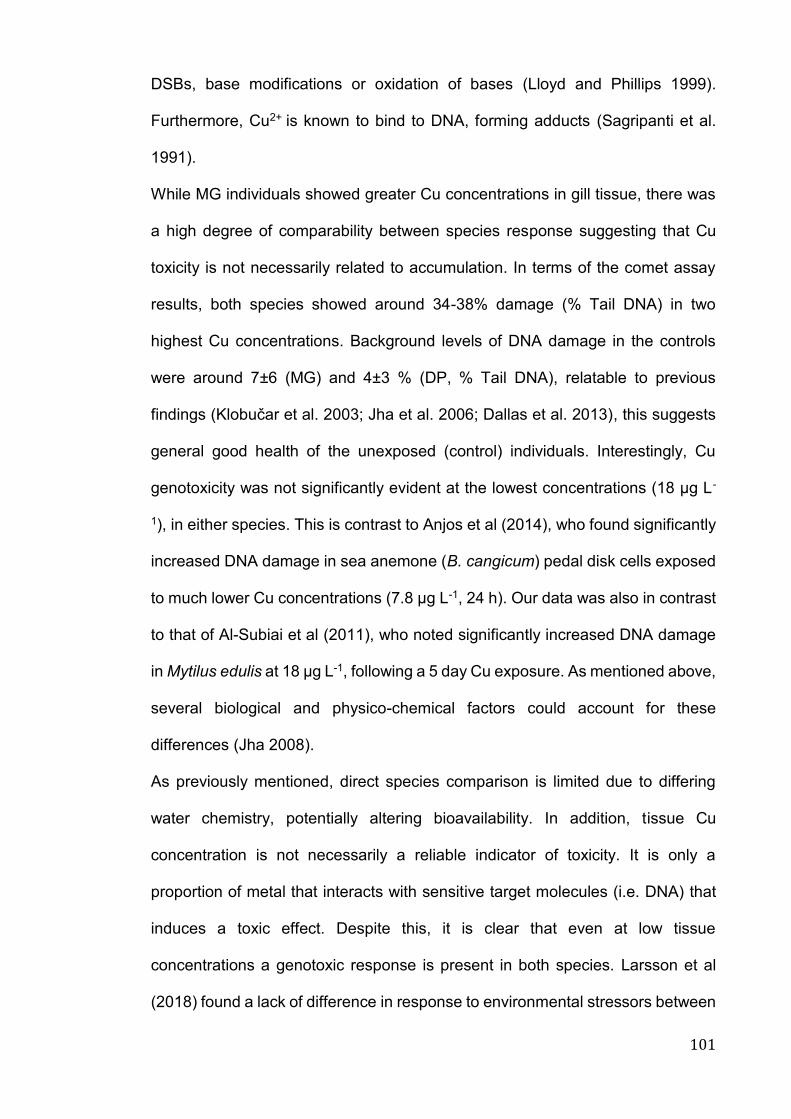
101
DSBs, base modifications or oxidation of bases (Lloyd and Phillips 1999).
Furthermore, Cu2+ is known to bind to DNA, forming adducts (Sagripanti et al.
1991).
While MG individuals showed greater Cu concentrations in gill tissue, there was
a high degree of comparability between species response suggesting that Cu
toxicity is not necessarily related to accumulation. In terms of the comet assay
results, both species showed around 34-38% damage (% Tail DNA) in two
highest Cu concentrations. Background levels of DNA damage in the controls
were around 7±6 (MG) and 4±3 % (DP, % Tail DNA), relatable to previous
findings (Klobučar et al. 2003; Jha et al. 2006; Dallas et al. 2013), this suggests
general good health of the unexposed (control) individuals. Interestingly, Cu
genotoxicity was not significantly evident at the lowest concentrations (18 μg L-
1), in either species. This is contrast to Anjos et al (2014), who found significantly
increased DNA damage in sea anemone (B. cangicum) pedal disk cells exposed
to much lower Cu concentrations (7.8 μg L-1, 24 h). Our data was also in contrast
to that of Al-Subiai et al (2011), who noted significantly increased DNA damage
in Mytilus edulis at 18 μg L-1, following a 5 day Cu exposure. As mentioned above,
several biological and physico-chemical factors could account for these
differences (Jha 2008).
As previously mentioned, direct species comparison is limited due to differing
water chemistry, potentially altering bioavailability. In addition, tissue Cu
concentration is not necessarily a reliable indicator of toxicity. It is only a
proportion of metal that interacts with sensitive target molecules (i.e. DNA) that
induces a toxic effect. Despite this, it is clear that even at low tissue
concentrations a genotoxic response is present in both species. Larsson et al
(2018) found a lack of difference in response to environmental stressors between
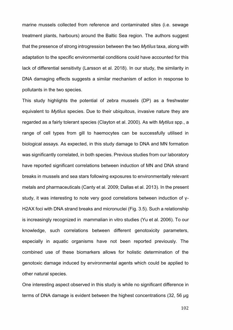
102
marine mussels collected from reference and contaminated sites (i.e. sewage
treatment plants, harbours) around the Baltic Sea region. The authors suggest
that the presence of strong introgression between the two Mytilus taxa, along with
adaptation to the specific environmental conditions could have accounted for this
lack of differential sensitivity (Larsson et al. 2018). In our study, the similarity in
DNA damaging effects suggests a similar mechanism of action in response to
pollutants in the two species.
This study highlights the potential of zebra mussels (DP) as a freshwater
equivalent to Mytilus species. Due to their ubiquitous, invasive nature they are
regarded as a fairly tolerant species (Clayton et al. 2000). As with Mytilus spp., a
range of cell types from gill to haemocytes can be successfully utilised in
biological assays. As expected, in this study damage to DNA and MN formation
was significantly correlated, in both species. Previous studies from our laboratory
have reported significant correlations between induction of MN and DNA strand
breaks in mussels and sea stars following exposures to environmentally relevant
metals and pharmaceuticals (Canty et al. 2009; Dallas et al. 2013). In the present
study, it was interesting to note very good correlations between induction of γ-
H2AX foci with DNA strand breaks and micronuclei (Fig. 3.5). Such a relationship
is increasingly recognized in mammalian in vitro studies (Yu et al. 2006). To our
knowledge, such correlations between different genotoxicity parameters,
especially in aquatic organisms have not been reported previously. The
combined use of these biomarkers allows for holistic determination of the
genotoxic damage induced by environmental agents which could be applied to
other natural species.
One interesting aspect observed in this study is while no significant difference in
terms of DNA damage is evident between the highest concentrations (32, 56 μg
103
L-1, both species), there is a slight increase in γ-H2AX foci in the 56 μg L-1
treatment. This could be a result of increased DSBs in the highest treatment, as
opposed to less detrimental DNA lesions (i.e. SSBs) at lower Cu concentrations.
γ-H2AX foci were present in the lowest Cu concentrations (18 μg L-1), where the
genotoxicity of Cu was not significantly apparent. Our data suggests that while
DNA damage was not evident using the comet or MN assays, possibly due to
lack of sensitivity, γ-H2AX is being recruited to damaged sites, in turn recruiting
other DNA repair machinery even at low Cu concentrations. In light of this, γ-
H2AX could be regarded as a more sensitive technique in measuring
genotoxicity, however in this study, to a limited degree.
In terms of relative sensitivity of the comet and γ-H2AX assays, one major
drawback of the comet assay is its inability to discriminate between DNA lesions,
such as SSBs and DSBs, alkali labile sites and DNA interstrand crosslinks
(Collins 2004, 8; Kumaravel and Jha 2006; Kumaravel et al. 2009; Liao et al.
2009). DSBs are considered to be most detrimental form of damage, they may
be repaired or result in apoptosis and/or mutation. γ-H2AX is increasingly used
as a biomarker in combination with classical and molecular techniques in
mammalian systems (e.g. Yu et al. 2006; Oommen et al. 2016a, b). Whilst these
techniques are considered to be simple and rapid (fast analysis time) in
comparison to many other established methodologies (Liao et al. 2009), their
relative sensitivity and effectiveness have not been compared sufficiently in
ecotoxicological research. In this study, we compare the classical and novel
techniques, examining their sensitivity as well as cost/time effectiveness.
Although the biological damage measured by Comet and γ-H2AX assays are
mechanistically different (one reflecting SSB/DSB, alkali labile site and another
only DSB), the alkaline comet assay appears to be more suitable for larger
104
sample sizes that have high levels of DNA damage. On the other hand γ-H2AX,
due to its high sensitivity and cost effectiveness, could be considered more
suitable to smaller studies which aim to determine DNA damage at lower levels
of exposure to those genotoxicants capable of effectively inducing DSBs, or
contaminants which could induce DSBs at higher concentrations. This is
particularly so as at higher levels of damage, induced foci can overlap making
the scoring difficult and time-consuming. Overall, γ-H2AX has proved useful as a
highly sensitive technique for detecting low-levels of DNA damage, its usefulness
in ecotoxicological research, when combined with more classical techniques is
clear.
Cu, as a model toxic metal is known to induce various types of damage to DNA
and chromatin with potential pathophysiological consequences (Lloyd and
Phillips 1999; Linder 2012). As mentioned earlier, one of the important
mechanisms of induction of damage by Cu is via generation of ROS, inducing
oxidative stress to biomolecules including DNA (Lloyd and Phillips 1999; Linder
2012). The modified comet assay using bacterial enzymes (e.g. FPG, Endo III)
has been used by different workers to determine DNA oxidation in fish and
mussels (Dallas et al. 2013; Mustafa et al. 2015). It would have been useful to
determine DNA oxidation using modified comet assay in this study as well to
determine relative contribution of DNA oxidation. This was however not feasible
due to logistical problems. In addition, Cu in common with other toxic metals and
metalloids (e.g. As, Co, Cd, Ni) could also interfere with DNA repair processes
and cell cycle control (Hartwig 2013). In common with mammalian studies,
elucidation of these fundamental processes in aquatic organisms following
exposures to environmental contaminants also warrants attention. Interpreting
105
these highly conserved processes would help to further strengthen human and
environmental links.
3.5 Conclusions
Our study has been the first to compare tissue specific accumulation and
genotoxic effects following exposure to Cu in marine and freshwater bivalve gill
cells. The zebra mussel, DP, is increasingly being utilised as a freshwater
counterpart of Mytilus spp. in biomonitoring and ecotoxicological research. Our
data highlights a clear relationship between external (water) and internal Cu
concentrations. The capacity to concentrate contaminants within tissue makes
MG and DP suitable bioindicator species to assess environmental health.
Cu induced comparable chromosomal and DNA damage in both mussel species,
despite variable bioaccumulation of Cu into gill tissue. Furthermore, γ-H2AX foci
formation was successfully applied as a useful biomarker of contaminant induced
genotoxicity. The usefulness of this assay, particularly when applied alongside
more classical, established techniques such as MN and comet assays is evident.
While we cannot definitively associate the comparability in genotoxic response to
differential species sensitivity, our results suggest that even low, environmentally
realistic Cu concentrations have the potential to cause stress to some bivalve
molluscs. For adequate protection of coastal and inland water bodies, future
research would benefit from using a multi-species, multi-biomarker approach
when investigating adverse effects at varying levels of biological organisation to
gain a true understanding of the real environmental threat of the contamination
to aquatic biota.

106
107
Chapter 4
Relative comparison of tissue specific bioaccumulation and
radiation dose estimation in marine and freshwater bivalves
following exposure to phosphorus-32
Published as part in:
Vernon, EL., Smith, JT., Jha, AN. 2018. Relative comparison of tissue specific
bioaccumulation and radiation dose estimation in marine and freshwater bivalves
following exposure to phosphorus-32. Journal of Environmental Radioactivity.

108
109
4.1 Introduction
Short lived radionuclides such as 32Phosphorus (32P, radiophosphorus), although
occurring in small quantities in the environment may be capable of accumulating
in aquatic biota (Smith et al. 2011). This is particularly so if the radionuclide is
continuously discharged in the environment, and the biota is chronically exposed.
In this context, 32P is discharged into aquatic systems from various sources. For
example, in England and Wales, 7, 5.2 and 5.7 GBq of 32P was discharged in
2015 as liquid waste from educational, medical (i.e. hospitals) and other
establishments (e.g. research, manufacturing and public sector) respectively
(RIFE 2015). In terms of environmental concentrations, 32P reference conditions
in Scotland (i.e. concentrations that result in a total ingested dose for humans of
0.10 mSv y-1 if consumed at 2 L day-1), are set at 57 Bq L-1 (DWQR 2014), with
recorded values (2005-2013) averaging 0.27 ± 0.21 Bq L-1 in the River Clyde
(Erskine Habour, King George V Dock), Scotland (SEPA 2013). While not as
environmentally relevant as radionuclides such as 137Cs, 32P was chosen due to
ease of use in an experimental setting and as a surrogate for gamma emitting
radionuclides, with sufficient penetrating energy to be detected outside the
tissues of interest. In addition, phosphorus in the natural environment serves as
an essential nutrient, and in common with non-radioactive phosphorus,
radioactive phosphorus (32P) is likely to have similar exposure pathways.
In terms of human health protection, contaminated organisms could pose a risk
to health via the food chain (Jha 2004, 8; Aoun et al. 2015; Yang et al. 2015). 32P
uptake in humans may occur via dietary pathways, with dose being higher in the
foetus and breastfed infants, than the adult (Oatway et al. 2008). Understanding
radionuclide concentration patterns in biota allows for the development of
adequate protection strategies, with the aim of reducing potential human dose
110
while maintaining environmental sustainability. Despite continuous and
prolonged use in industry and subsequent discharges, no studies to our
knowledge have investigated tissue specific accumulation of 32P in aquatic biota.
Bioaccumulative ability in aquatic bivalves, an important group of invertebrates of
ecological and economic importance, has been identified in scientific literature.
This is notably to ubiquitous, long-lived radionuclides such as 134Cs, 210Po, 210Pb
and 3H (Evans 1984; Jha et al. 2005; Kalaycı et al. 2013; Feroz Khan et al. 2012,
2014; Dallas et al. 2016a; Metian et al. 2016; Pearson et al. 2018). However
whole body accumulation and dose are often (but not always) the focus of such
studies. Sufficient data are not available for tissue specific accumulation of short-
lived radionuclides. It is well accepted that in common with other contaminants,
radionuclides accumulate in the biota in a tissue specific manner. Whole-body
determination of radionuclide bioaccumulation levels is important for risk
assessments, however for biomonitoring and biological response studies (i.e.
sensitive transcriptomics and proteomics studies), it is important that tissue
specific information is generated. Radionuclide uptake disparity amongst tissues
has been highlighted in studies from Jha et al (2005), Jaeschke et al (2011),
Dallas et al (2016a) and Pearson et al (2018), where tritium accumulation in
bivalve (Mytilus sp.) tissue was highly specific. Digestive gland
(hepatopancreas/gut), gill and foot showed higher concentrations following
exposure to varying amounts of tritium (5-15 MBq L−1). Such trends are followed
in green and brown mussels (P. perna, P. indica), where digestive gland showed
maximum 210Po/210Pb activity over other biological soft tissue and shell (Feroz
Khan and Godwin Wesley 2012). Furthermore in scallop (Pecten maximus) soft
tissue, 241Am was predominantly concentrated in the mantle and digestive gland,
whereas 134Cs was mainly present in the adductor muscle and mantle (Metian et

111
al. 2011). In environmental protection terms, understanding radionuclide
accumulation is necessary to relate exposure, to radiation dose and potential
biological responses. Exposure to IR can occur via multiple aqueous and dietary
pathways, the behaviour and fate of radionuclides when accumulated in specific
biological tissues or organs in the aquatic biota could be influenced by many
factors and may vary significantly under different exposure scenarios (Pearson
et al. 2018). Given that radionuclides accumulate differentially in the tissues, from
a biomonitoring perspective, whole-body bioaccumulation monitoring is therefore
not necessarily sufficient in fully protecting aquatic biota from the exposure. This
is particularly important as differential tissue sensitivity could result in a
detrimental biological response at levels presumed to be acceptable.
Dosimetry models, such as the Environmental Risk from Ionising Contaminants
Assessment and Management (ERICA) Tool have been developed to evaluate
radiological risk to aquatic and terrestrial biota (Brown et al. 2008). Risk is
assessed by comparing a dose rate in a reference organism to a dose rate of 10
μGy h-1 (0.24 mGy d-1), a “screening dose rate” whereby no effect to populations
of biota is expected (Garnier-Laplace and Gilbin 2006; Garnier-Laplace et al.
2008). Though dosimetry models are of great assistance in radiobiological
research, ERICA tool predicted dose rates presume homogeneous radionuclide
distribution within biota, which are represented as ellipsoidal shapes (Beresford
et al. 2007). In order to adequately estimate radiological risk to biota, we require
a greater knowledge of tissue specific radionuclide concentrations in a range of
organisms, the transfer pathways, concentration factor, dose rate and an
evaluation of any possible biological effects are required. Such data may also
help pinpoint key tissues of interest for biomonitoring purposes.
112
The marine species Mytilus galloprovincialis (MG) and freshwater Dreissena
polymorpha (DP) were selected in this study (Figure 1.4). The presence of
radionuclides is of concern for both marine and freshwater environments.
Although marine species might not be used to assess the risk in the freshwater
environment or vice-versa, it is nevertheless important to estimate relative
radionuclide accumulation in the biota belonging to same biological group. This
would help to identify the most sensitive species for environmental protection.
The present study had the following aims and objectives: (a) to determine tissue
specific accumulation and depuration (release via excretion) of 32P in two different
species of mussels (i.e. marine and freshwater), (b) to evaluate the application of
the ERICA tool in determining tissue specific radiation doses and (c) to identify
the accumulation pattern of 32P, as to highlight key tissues of interest for future
experiments investigating biological response. It was hypothesised that whole
body concentration of 32P would be comparable in freshwater and marine
bivalves, and that accumulation would be tissue specific.
4.2 Materials and methods
4.2.1 Chemicals and suppliers
Commercially available, radiolabelled-ATP (Adenosine triphosphate, γ-32P) was
obtained from Perkin Elmer (PerkinElmer, UK) in batches of 9.25 MBq (specific
activity: 370 MBq mL-1) and used as the source of radioactive 32P for our
experimental purposes. Radiolabelled-ATP was diluted with DI water to form
appropriate working solutions. Working solution added to beakers was decay
adjusted. Nitric acid was obtained from Fisher Scientific UK (Nitric acid 68%,
Primar Plus™) and scintillation cocktail from LabLogic systems Ltd. UK
(ScintLogic, UK). All other chemicals and reagents were purchased from
113
Anachem Ltd. UK, Sigma-Aldrich Company Ltd UK, VWR International Ltd USA
or Greiner Bio-One Ltd UK, unless stated otherwise. Additional product details
are mentioned in text as appropriate.
4.2.2 Mussel exposure conditions
Two ten-day exposures were performed between December and February 2016-
17. Adult MG and DP were collected and maintained in accordance to section
2.2. As Hilbish et al (2002) reported the occurrence and distribution of Mytilus
edulis, M. galloproviancialis and their hybrids in the coastal regions of south-west
England, we ensured species homogeneity in our experiments based on the
method of Inoue et al (1995), in accordance to section 2.2.1.
Three MG and fourteen DP individuals per beaker (total weight of 35 g/beaker)
were exposed to the following activity concentrations of 32P in triplicate: 709, 7090
or 70900 and 571, 5710 or 57100 Bq L-1, respectively, along with control
treatments. The sample size (number of individuals) used in the study was
decided to obtain a statistically robust set of data and was in line with previous
studies (Dallas et al. 2016b). Activity concentrations in water were calculated
from preliminary experiments (see section 2.8). Water changes (50 %) were
carried out on days 3, 5, 7 and 9 and mussels were fed during this exposure (2 h
before each water change), as described in detail elsewhere (Dallas et al. 2016a).
Water samples (1 mL, in duplicate) were taken around 30 min after each water
change and processed for liquid scintillation counting (LSC) to determine water
activity concentrations.
Water quality parameters were measured routinely before and after water
changes. Parameters were found to be within the expected range (pH 8.1 ± 1.2,
temperature 14.5 ± 1.8 ºC, dissolved oxygen (DO) 96.9 ± 8 % and salinity 36.7 ±
114
0.6 for MG and pH 8.1 ± 0.3, temperature 14.8 ± 0.9 ºC, DO 92.3 ± 4.1 % and
salinity 0.3 ± 0 for DP).
4.2.3 Sampling procedures and liquid scintillation counting
At the end of the exposure period, water from beakers was drained through a
sieve (Fisherbrand, ISO 3310/1 250 µM). Faeces and pseudo faeces were
collected from sieve on a weighed section of tissue, and placed into pre-weighed
tubes (Punt et al. 1998; Jha et al. 2005). Mussels were dissected and separated
into soft tissue (i.e. gill, mantle, adductor muscle, digestive gland and ‘other’
tissue), shell and internal mussel water (IMW). All samples were collected,
processed and analysed as in section 2.8.3.1. Activity concentrations were
background corrected by blank subtracting from each sample, the blank was non-
spiked fresh or seawater. In accordance with Jaeschke and Bradshaw (2013),
CPM values that fell below the blank were assigned an activity of 0.000. All
samples were decay corrected.
4.2.4 Dosimetry and the ERICA TOOL
The Tier 2 assessment module of the ERICA tool was used for dose estimation.
32P was chosen as one of the ERICA tool's default isotopes (Brown et al. 2008).
Tissue specific dose rate (e.g. 32P dose to digestive gland) was determined by
taking mean measurements during sampling (i.e. mass, height, width, length),
and developing custom geometry parameters on the ERICA tool (Table 2.4), the
ERICA tool was utilised in accordance to section 2.8.4.
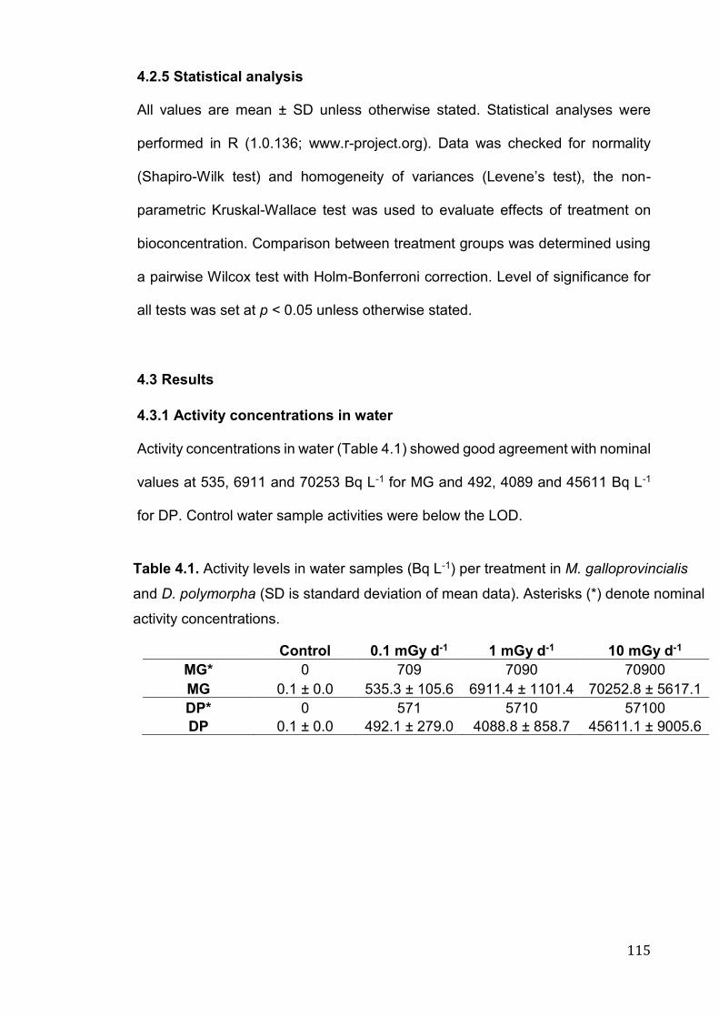
115
4.2.5 Statistical analysis
All values are mean ± SD unless otherwise stated. Statistical analyses were
performed in R (1.0.136; www.r-project.org). Data was checked for normality
(Shapiro-Wilk test) and homogeneity of variances (Levene’s test), the non-
parametric Kruskal-Wallace test was used to evaluate effects of treatment on
bioconcentration. Comparison between treatment groups was determined using
a pairwise Wilcox test with Holm-Bonferroni correction. Level of significance for
all tests was set at p < 0.05 unless otherwise stated.
4.3 Results
4.3.1 Activity concentrations in water
Activity concentrations in water (Table 4.1) showed good agreement with nominal
values at 535, 6911 and 70253 Bq L-1 for MG and 492, 4089 and 45611 Bq L-1
for DP. Control water sample activities were below the LOD.
Control 0.1 mGy d-1 1 mGy d-1 10 mGy d-1
MG* 0 709 7090 70900
MG 0.1 ± 0.0 535.3 ± 105.6 6911.4 ± 1101.4 70252.8 ± 5617.1
DP* 0 571 5710 57100
DP 0.1 ± 0.0 492.1 ± 279.0 4088.8 ± 858.7 45611.1 ± 9005.6
Table 4.1. Activity levels in water samples (Bq L-1) per treatment in M. galloprovincialis
and D. polymorpha (SD is standard deviation of mean data). Asterisks (*) denote nominal
activity concentrations.
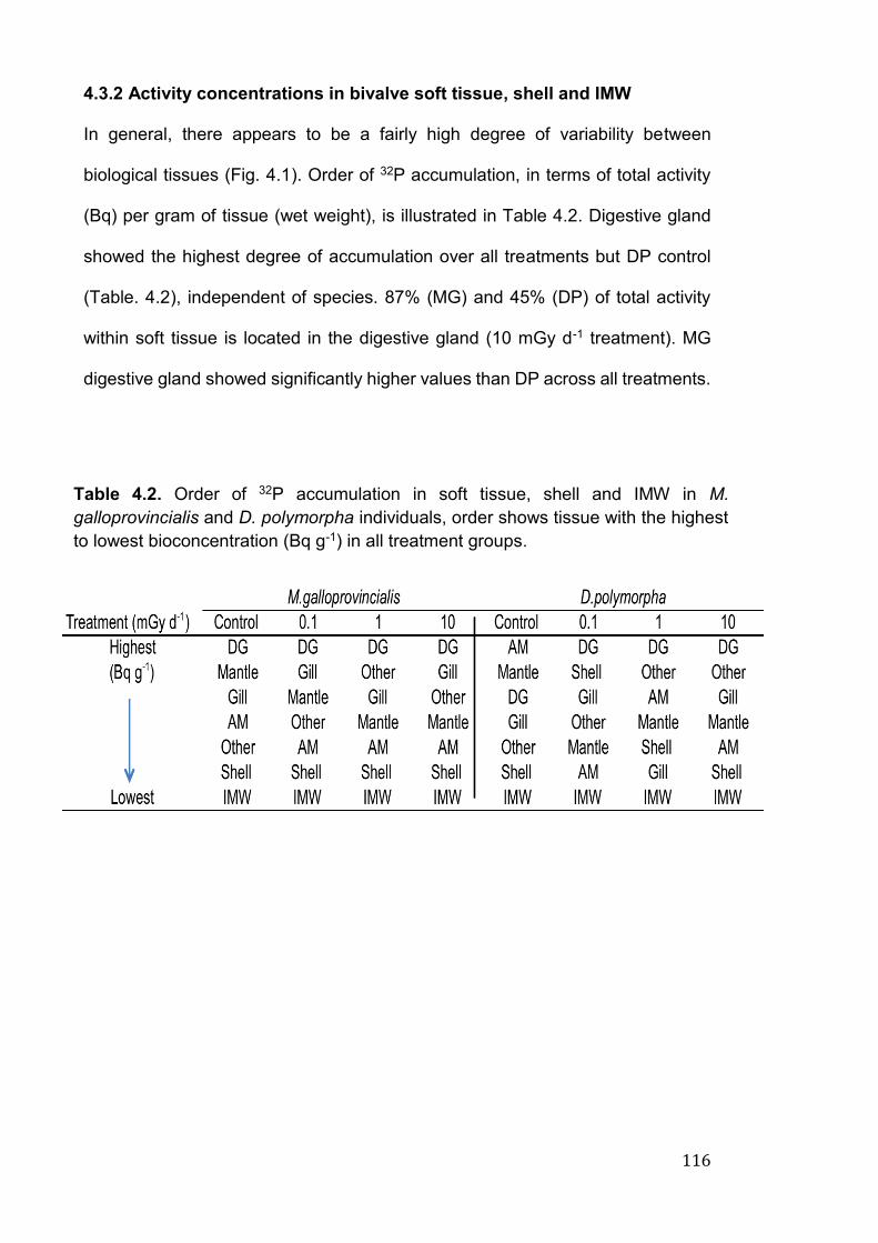
116
4.3.2 Activity concentrations in bivalve soft tissue, shell and IMW
In general, there appears to be a fairly high degree of variability between
biological tissues (Fig. 4.1). Order of 32P accumulation, in terms of total activity
(Bq) per gram of tissue (wet weight), is illustrated in Table 4.2. Digestive gland
showed the highest degree of accumulation over all treatments but DP control
(Table. 4.2), independent of species. 87% (MG) and 45% (DP) of total activity
within soft tissue is located in the digestive gland (10 mGy d-1 treatment). MG
digestive gland showed significantly higher values than DP across all treatments.
Table 4.2. Order of 32P accumulation in soft tissue, shell and IMW in M.
galloprovincialis and D. polymorpha individuals, order shows tissue with the highest
to lowest bioconcentration (Bq g-1) in all treatment groups.
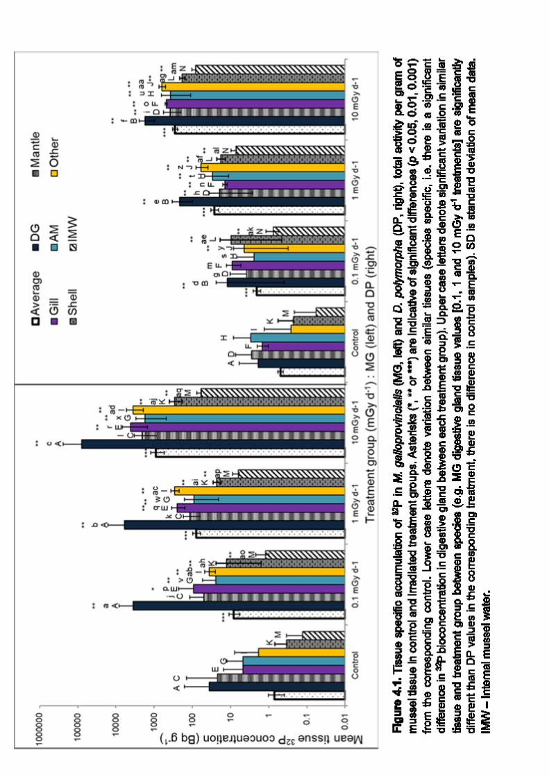
117
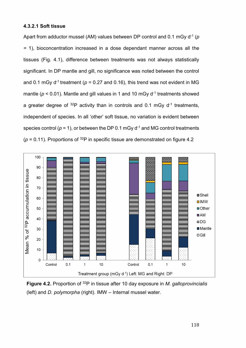
118
4.3.2.1 Soft tissue
Apart from adductor mussel (AM) values between DP control and 0.1 mGy d-1 (p
= 1), bioconcentration increased in a dose dependant manner across all the
tissues (Fig. 4.1), difference between treatments was not always statistically
significant. In DP mantle and gill, no significance was noted between the control
and 0.1 mGy d-1 treatment (p = 0.27 and 0.16), this trend was not evident in MG
mantle (p < 0.01). Mantle and gill values in 1 and 10 mGy d-1 treatments showed
a greater degree of 32P activity than in controls and 0.1 mGy d-1 treatments,
independent of species. In all ‘other’ soft tissue, no variation is evident between
species control (p = 1), or between the DP 0.1 mGy d-1 and MG control treatments
(p = 0.11). Proportions of 32P in specific tissue are demonstrated on figure 4.2
Figure 4.2. Proportion of 32P in tissue after 10 day exposure in M. galloprovincialis
(left) and D. polymorpha (right). IMW – Internal mussel water.
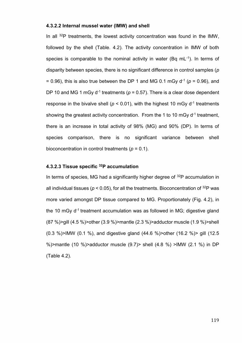
119
4.3.2.2 Internal mussel water (IMW) and shell
In all 32P treatments, the lowest activity concentration was found in the IMW,
followed by the shell (Table. 4.2). The activity concentration in IMW of both
species is comparable to the nominal activity in water (Bq mL-1). In terms of
disparity between species, there is no significant difference in control samples (p
= 0.96), this is also true between the DP 1 and MG 0.1 mGy d-1 (p = 0.96), and
DP 10 and MG 1 mGy d-1 treatments (p = 0.57). There is a clear dose dependent
response in the bivalve shell (p < 0.01), with the highest 10 mGy d-1 treatments
showing the greatest activity concentration. From the 1 to 10 mGy d-1 treatment,
there is an increase in total activity of 98% (MG) and 90% (DP). In terms of
species comparison, there is no significant variance between shell
bioconcentration in control treatments (p = 0.1).
4.3.2.3 Tissue specific 32P accumulation
In terms of species, MG had a significantly higher degree of 32P accumulation in
all individual tissues (p < 0.05), for all the treatments. Bioconcentration of 32P was
more varied amongst DP tissue compared to MG. Proportionately (Fig. 4.2), in
the 10 mGy d-1 treatment accumulation was as followed in MG; digestive gland
(87 %)>gill (4.5 %)>other (3.9 %)>mantle (2.3 %)>adductor muscle (1.9 %)>shell
(0.3 %)>IMW (0.1 %), and digestive gland (44.6 %)>other (16.2 %)> gill (12.5
%)>mantle (10 %)>adductor muscle (9.7)> shell (4.8 %) >IMW (2.1 %) in DP
(Table 4.2).
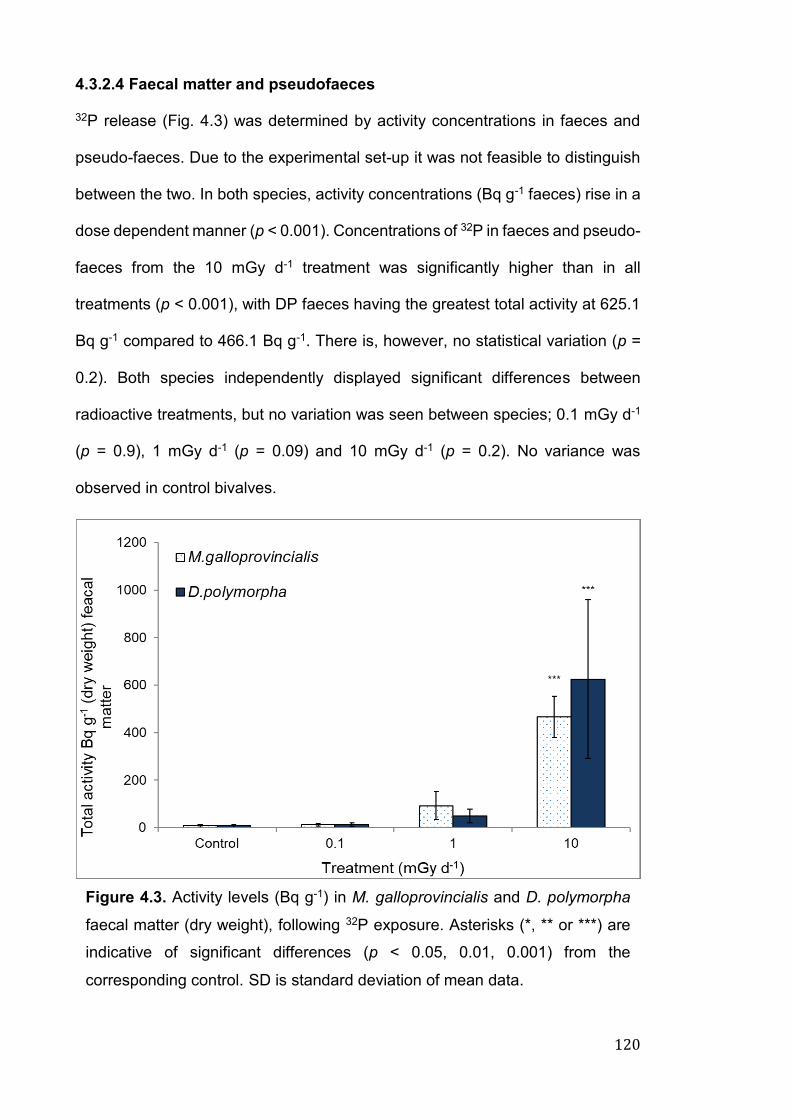
120
4.3.2.4 Faecal matter and pseudofaeces
32P release (Fig. 4.3) was determined by activity concentrations in faeces and
pseudo-faeces. Due to the experimental set-up it was not feasible to distinguish
between the two. In both species, activity concentrations (Bq g-1 faeces) rise in a
dose dependent manner (p < 0.001). Concentrations of 32P in faeces and pseudo-
faeces from the 10 mGy d-1 treatment was significantly higher than in all
treatments (p < 0.001), with DP faeces having the greatest total activity at 625.1
Bq g-1 compared to 466.1 Bq g-1. There is, however, no statistical variation (p =
0.2). Both species independently displayed significant differences between
radioactive treatments, but no variation was seen between species; 0.1 mGy d-1
(p = 0.9), 1 mGy d-1 (p = 0.09) and 10 mGy d-1 (p = 0.2). No variance was
observed in control bivalves.
Figure 4.3. Activity levels (Bq g-1) in M. galloprovincialis and D. polymorpha
faecal matter (dry weight), following 32P exposure. Asterisks (*, ** or ***) are
indicative of significant differences (p < 0.05, 0.01, 0.001) from the
corresponding control. SD is standard deviation of mean data.
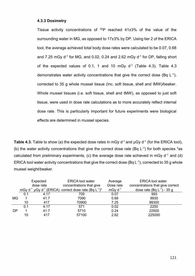
121
4.3.3 Dosimetry
Tissue activity concentrations of 32P reached 41±3% of the value of the
surrounding water in MG, as opposed to 17±3% by DP. Using tier 2 of the ERICA
tool, the average achieved total body dose rates were calculated to be 0.07, 0.68
and 7.25 mGy d-1 for MG, and 0.02, 0.24 and 2.62 mGy d-1 for DP, falling short
of the expected values of 0.1, 1 and 10 mGy d-1 (Table 4.3). Table 4.3
demonstrates water activity concentrations that give the correct dose (Bq L-1),
corrected to 35 g whole mussel tissue (Inc. soft tissue, shell and IMW)/beaker.
Whole mussel tissues (i.e. soft tissue, shell and IMW), as opposed to just soft
tissue, were used in dose rate calculations as to more accurately reflect internal
dose rate. This is particularly important for future experiments were biological
effects are determined in mussel species.
ERICA tool water Average ERICA tool water concentrations that give Dose rate concentrations that give correct
mGy d-1 µGy d-1 (ERICA) correct dose rate (Bq L-1)* mGy d-1 dose rate (Bq L-1) - 35 g0.1 4.17 709 0.07 9931 41.7 7090 0.68 993010 417 70900 7.25 993000.1 4.17 571 0.02 22501 41.7 5710 0.24 2250010 417 57100 2.62 225000
Expected
MG
DP
dose rate
Table 4.3. Table to show (a) the expected dose rates in mGy d-1 and μGy d-1 (for the ERICA tool),
(b) the water activity concentrations that give the correct dose rate (Bq L-1) for both species *as
calculated from preliminary experiments, (c) the average dose rate achieved in mGy d-1 and (d)
ERICA tool water activity concentrations that give the correct dose (Bq L-1), corrected to 35 g whole
mussel weight/beaker.
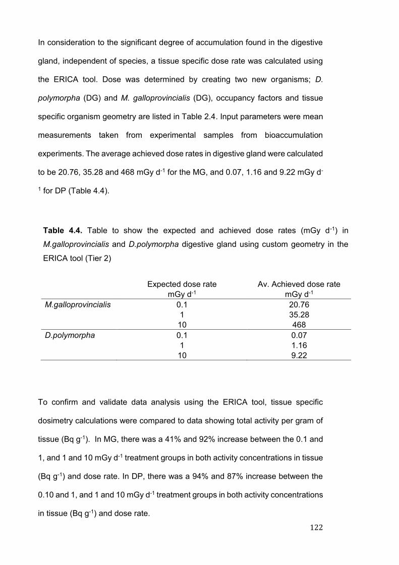
122
In consideration to the significant degree of accumulation found in the digestive
gland, independent of species, a tissue specific dose rate was calculated using
the ERICA tool. Dose was determined by creating two new organisms; D.
polymorpha (DG) and M. galloprovincialis (DG), occupancy factors and tissue
specific organism geometry are listed in Table 2.4. Input parameters were mean
measurements taken from experimental samples from bioaccumulation
experiments. The average achieved dose rates in digestive gland were calculated
to be 20.76, 35.28 and 468 mGy d-1 for the MG, and 0.07, 1.16 and 9.22 mGy d-
1 for DP (Table 4.4).
Expected dose rate Av. Achieved dose rate
mGy d-1 mGy d-1
M.galloprovincialis 0.1 20.76
1 35.28
10 468
D.polymorpha 0.1 0.07
1 1.16
10 9.22
To confirm and validate data analysis using the ERICA tool, tissue specific
dosimetry calculations were compared to data showing total activity per gram of
tissue (Bq g-1). In MG, there was a 41% and 92% increase between the 0.1 and
1, and 1 and 10 mGy d-1 treatment groups in both activity concentrations in tissue
(Bq g-1) and dose rate. In DP, there was a 94% and 87% increase between the
0.10 and 1, and 1 and 10 mGy d-1 treatment groups in both activity concentrations
in tissue (Bq g-1) and dose rate.
Table 4.4. Table to show the expected and achieved dose rates (mGy d-1) in
M.galloprovincialis and D.polymorpha digestive gland using custom geometry in the
ERICA tool (Tier 2)
123
Concentration factor values, calculated by dividing tissue specific 32P activity
concentrations (Bq kg-1, wet weight) by activity concentrations of the spiked
water, were as follows; 11.7, 11.2 and 11.9 in MG and 3.6, 4.6 and 5 for DP (in
0.1, 1 and 10 mGy d-1 treatments).
4.4 Discussion
From this study, it is evident that 32P accumulation is highly tissue specific and
variable between bivalve species. It is important to note that in this study 32P was
introduced in a highly bioavailable form (i.e. radiolabelled ATP), demonstrated
bioaccumulation patterns in this study may be reflective of this. MG, which
accumulated 41±3% of 32P present in the surrounding media as opposed to
17±3% by DP, showed a greater degree of 32P accumulation across all biological
tissues. Despite variance in uptake and accumulation, 32P excretion was
comparable between species. It could be assumed that the measured activity
concentration takes into account absorption, metabolism of ATP, subsequent
dispersal and partitioning of phosphorus in tissue specific manner at a given
sampling time. This phenomenon as a whole could be considered as tissue
specific accumulation of radiophosphorus. It is also possible that the tissues
could have achieved equilibrium over the exposure period. It would, however, be
difficult to predict tissue dose delivered by the available radionuclide
concentration in the surrounding media. Furthermore, equilibrium status is often
regarded as a flaw in the ERICA tool. In terms of dosimetry, the ERICA tool
proved valuable in calculating whole body and tissue specific dose rates. Average
achieved dose rates were 0.07, 0.68 and 7.25 mGy d-1 for MG, and 0.02, 0.24
and 2.62 mGy d-1 for DP, below expected values of 0.10, 1 and 10 mGy d-1. The
124
dose dependant nature of 32P accumulation gives evidence that both marine and
freshwater bivalves are suitable bioindicators of radioactive pollution.
In consideration of species, MG accumulated a higher degree of 32P in biological
tissue across all treatments. Such disparity may be a result of several biotic and
abiotic variables, including physiology (filtration rates, metabolism, and
reproductive stage), biochemistry and water chemistry (salinity, pH, dissolved
oxygen, radionuclide speciation) (Nalepa et al. 1991; Reinfelder et al. 1998;
Konovalenko et al. 2016; Pearson et al. 2018). The underlying mechanism which
leads to differences between freshwater and marine bivalves is unclear,
differential radionuclide accumulation between bivalves is a topic with little
attention to date. In terms of stable phosphorus (P), tissue bioconcentration has
been found to vary dependent on reproductive processes, high P concentrations
are evident during periods of spawning in Mytilus sp. and DP (Kuenzler 1961;
Jurkiewicz-Karnkowska 2002). Jurkiewicz-Karnkowska (2002) noted variability in
soft tissue P concentrations between three freshwater bivalves (DP, Anodonta
anatina and A. cygnea) inhabiting the Zegrzynski Reservoir, Poland, suggesting
species specificity in terms of stable P accumulation.
Feeding and digestion is often regarded as a predominant route of radionuclide
intake (McDonald et al. 1993). The digestive gland in bivalves plays a central role
in metabolism. It is important for intracellular digestion, as a storage site for
metabolic reserves during periods of stress, and as a site of nutrient distribution
to other organs, particularly reproductive tissue (Cartier et al. 2004). Under all
treatment groups the greatest 32P concentration was present in the digestive
gland, at 87% in MG and 45% in DP of the total activity within soft tissue (10 mGy
d-1), suggesting a dietary route of exposure. The findings are supported by earlier
studies by Jaeschke et al (2011) and Jha et al (2005) who reported preferential
125
tritium accumulation in Mytilus sp. digestive gland (tritiated glycine, 1.48 MBq L−1
and tritiated water, 3.7, 37 and 147 MBq L−1). This trend is continued in Mytilus
spp., following exposure to 241Am, 329Pu, 237Np and 63Ni, (McDonald et al. 1993;
Punt et al. 1998) and in marine amphipods exposed to 32P (Johannes 2003).
Variance between marine and freshwater bivalves may result from differential
physiological and genetic characteristics. In terms of physiology, filtration rates
have been noted as comparable between species, at 76.6 (DP) and 87.5 mL
mussel-1 h-1 (Mytilus edulis, ME), along with valve movement at 90.1 (DP) and 92
(ME) % of open valves under ambient conditions (Rajagopal et al. 2003). While
neither parameter were measured in this study; it is possible that DP individuals
are more inclined to close their valves when exposed to 32P, as a stress response.
This behaviour is documented in biofouling control research, where bivalves
close valves during periods of water chlorination as a protective strategy
(Rajagopal et al. 2003). Observed differences may also result from variable
feeding regimes (i.e. species fed different food types) and/or gut physiology.
Factors involved with digestion such as food density or quality, gut passage time,
volume or retention rate, enzymatic composition, digestive partitioning and
chemistry may effect 32P assimilation (Wang et al. 1995). Despite the disparity in
CF values between MG and DP, the trend between biological tissues is similar,
suggesting comparability in 32P accumulation pathways.
Average achieved dose rates in digestive gland were calculated at 20.76, 35.28
and 468 mGy d-1 for MG, and 0.07, 1.16 and 9.22 mGy d-1 for DP. This specific
tissue dose is substantially greater then calculated whole body doses, suggesting
that whole-body dose monitoring may be insufficient in wholly protecting aquatic
organisms from radionuclide exposure. Tissue specificity, in terms of
accumulation is well documented for many radionuclides. Strontium-90 for
126
example is a ‘bone seeker’, due to its biochemically similar behaviour to calcium,
following ingestion a large proportion will attach to the surface, or be absorbed
into bone (ATSDR 2004). In the context of biomonitoring and adequate
environmental protection, an understanding of tissue specific dose rates is of high
importance. Calculated whole body dose for MG and DP (0.1 and 1 mGy d-1
treatments) fell below the predicted ‘no effects’ screening value of 10 µGy h-1
(0.24 mGy d-1), suggesting a minimal risk to the individual or population.
However, in all but the DP 0.1 mGy d-1 treatment, digestive gland dose was above
the screening benchmark.
In the 10 mGy d-1 treatment, gill tissue had 1816% (MG) and 255% (DP) less
concentrated 32P then in the digestive gland (Bq g-1). As filter feeding organisms,
particulates within the water column are captured within cilia on the gills,
particulate matter is then carried via mucous strings to the mouth (Riisg et al.
2011). Gill tissue may therefore act as a major pathway for contaminants to enter
other biological tissue. IMW activity concentrations are comparable to the
expected activity in water (Bq mL-1) in both species, suggesting that bivalves are
unable to regulate 32P uptake via aqueous pathways. Past studies have often
highlighted gill as a tissue of key concern due to proximity to the surrounding
media, high surface area and water content. The relatively low activity in Mytilus
sp. gill tissue relative to the digestive gland is a trend found in other studies
following exposure to tritium (12 to 485 μGy h−1) and nickel (63Ni) (Punt et al.
1998; Jha et al. 2005). In terms of subsequent biological response, it is important
to note that while 32P may have accumulated to a lesser degree in some tissue,
the beta emission can penetrate approximately 0.76 cm of tissue/water (Terrance
2017). By proximity, higher dose rates may be evident in tissue or cells not
directly accumulating 32P to a high degree. In terms of gill tissue, while a relatively
127
low contaminant concentration is observed, its large surface area and proximity
to surrounding media may result in a higher absorbed dose.
In the natural environment, many factors may influence the filtration rate of
bivalves, along with feeding and depuration rate. Changing environmental
factors, such as water quality conditions, food availability, reproduction and
physiological condition may affect feeding behaviour (Riisg et al. 2011).
Laboratory conditions may not accurately reflect feeding, and therefore uptake
and depuration patterns of 32P in bivalves may vary. It is also possible that due
to different habitats, certain bivalve species are either more adapted to, or have
experienced more disturbances or stresses in the wild, and are therefore more
resilient to stresses under laboratory conditions. However relative response to a
particular stressor of similar magnitude in two different species, representing
different habitats, is difficult to estimate in the natural environment. From an
environmental protection perspective, an understanding of radionuclide transfer
pathways under environmentally realistic conditions, whether uptake is dietary
(ingestion of contaminated food) or through direct transfer from surrounding
media is important. One of the limitations of the study is that these laboratory-
based experiments were carried out in static exposure conditions, which differs
from real environmental situations. A flow-through exposure set-up would have
been a more realistic experimental design but due to health, safety, logistics and
economic reasons (requiring large amounts of radionuclides), a flow-through
experimental design was not feasible. Further studies using a wider range of
radionuclides and exposure conditions, which better reflect environmental
exposure conditions (e.g. flow through system) would be of great benefit.
Knowledge of the behaviour and transfer of radionuclides within aquatic systems
128
allows for an assessment of potential impacts and subsequent management
strategies.
Understanding excretion of contaminants is important firstly as a means of
determining possible chronic effects of assimilated contaminants, and secondly
in respects to human consumption. In terms of public health, depuration is
mandatory in bivalves harvested for human consumption as to remove
contaminants, predominantly bacteria (Lee et al. 2008). The effectiveness of
depuration in removing radionuclides is yet to be fully understood. Suspension
feeding bivalves produce faeces and pseudofaeces, the latter of which refers to
particles rejected before entering the gut. Excretion of 32P, measured in a
combination of faeces (from alimentary tract) and pseudofaeces (from mantle
cavity), do not appear to be consistent with that observed from uptake. In
irradiated treatments (0.10, 1 and 10 mGy d-1), 0.31%, 0.15% and 0.08% (MG)
and 0.4%, 0.15%, 0.34% (DP) of 32P from surrounding media was excreted;
significantly lower than the 41±3% (MG) and 17±3% (DP) of 32P accumulated
within biological tissue. While our findings suggest a slow depuration rate during
IR exposure, results are limited in showing a brief snapshot in time. It would be
of interest to monitor uptake and excretion, and therefore depuration rates over
both a longer duration, and following the removal of 32P in water.
The shell surface of aquatic bivalves is known to adsorb dissolved contaminants
from surrounding media (Zuykov et al. 2012), thus why in this study whole body
dose was not limited to just soft tissue. When removing both IMW and shell
concentrations from the data before ERICA tool analysis, the results follow
exactly the same pattern due to the influence of vast 32P concentrations in the
digestive gland. 32P biosorption in whole shell was concentration dependant in
both species, with an increase in total activity of 98% (MG) and 90% (DP)
129
between 1 and 10 mGy d-1 treatments. Proportionately DP showed higher
incorporation into shell, over all treatments, whereas per gram of shell, MG has
significantly greater 32P present. Mollusc shell is formed of a few calcified layers
and the periostracum, one thin, organic coating layer (Marin et al. 2012; Zuykov
et al. 2012). Species variation may be a result of differing shell microstructure
and topography, chemical and macromolecule composition (Marin et al. 2012).
As noted by Zuykov et al (2012), MG and DP do show disparity in shell
topography, where DP has a thinner periostracum and a lamellate surface (Immel
et al. 2016). In this study, the content of 32P in shell was far lower than in soft
tissue, this data contrasts to findings by Koide et al (1982), Clifton et al (1989)
and Metian et al (2011) following exposure to radionuclides or heavy metals. As
an example, in scallop (Pecten maximus), biosorption of 241Am into the shell was
far greater than soft tissue, however in the same species, 134Cs showed
preferential accumulation in soft tissue over shell (Metian et al. 2011). Bivalve
shells are widely used to monitor pollutants in the aquatic environment (Zuykov
et al. 2013). It is relevant to note that bioconcentration values taken from shell
are not reflective of soft tissue values.
4.5 Conclusions
This is the first study to compare uptake and depuration (via excretion) of short-
lived radionuclide, 32P in two anatomically similar bivalve species. Accumulation
of 32P is highly tissue specific, with the majority located within the digestive gland.
This is particularly important in the context of biomonitoring and adequate
environmental protection, where whole-body dose monitoring may not always be
sufficient to protect aquatic organisms from radionuclide exposure. Differential
sensitivity between biological tissues could result in harmful biological response
130
at activity levels presumed to be safe. The next step is to link radioactive
exposure, accumulation and dose rate, to consequent biological responses
(chapter 5). Accumulation within mussel tissues, even for short durations may
potentially have long lasting effects in both exposed individuals and subsequent
generations. Lastly, considering species variation in 32P accumulation, it is not
necessarily accurate to evaluate accumulation or biological hazard of ionising
radiations to the marine environment by using information gathered from
freshwater systems, and vice versa.
131
Chapter 5
Assessing relative biomarker responses in marine and
freshwater bivalve molluscs following exposure to phosphorus
32 (32P): Application of genotoxicological and molecular
biomarkers
In preparation:
Vernon, EL., Bean, TP., Jha, AN. 2018. Assessing relative biomarker responses
in marine and freshwater bivalve molluscs following exposure to phosphorus 32
(32P): Application of genotoxicological and molecular biomarkers.

132
133
5.1 Introduction
IR emitted from radionuclides discharged in the environment can potentially pose
short and long-term detrimental effects to both human and non-human biota, with
DNA being the most important target for their actions (UNSCEAR 1982; Dallas et
al. 2012). A generic (all species) “no effect” dose rate of 10 μGy h−1 (0.24 mGy d-
1) has been adopted as a screening value, dose rates under this value are thought
to result in minimal risk to the individual or population of natural species
(Andersson et al. 2008, 2009). There is, however, not enough experimental
information available in the literature to support this generic ‘no effect’ dose rate
for wider natural biota (Dallas et al. 2012). To ensure an adequate degree of
protection, therefore, there is a necessity to link radiation exposure to tissue
specific bioaccumulation and dose rate, and to subsequent biological responses
in a range of aquatic organisms to determine their relative sensitivity (Scoppa
1983; Dallas et al. 2012; Kumar et al. 2017; Carvalho 2018; Salbu et al. 2018;
Skipperud and Salbu 2018; Vernon et al. 2018).
As highlighted in section 1.3.2, molecular and genetic alterations are perceived
as an early warning signal of organism’s stress. Exposure to IR has the potential
to cause short and long term damage to aquatic organisms at different trophic
levels including invertebrates and fish, this has been demonstrated by the
detrimental biological responses caused by widespread, long-lived, radionuclides
such as 137Cs and 3H (Walker et al. 2000; Jha et al. 2006; Olsvik et al. 2010;
Farcy et al. 2011; Freeman et al. 2014; Dallas et al. 2016a; Arcanjo et al. 2018;
Hurem et al. 2018; Pearson et al. 2018).
The majority of studies aiming to determine potential detrimental effects of IR
have focused primarily on long-lived radionuclides. However, short-lived
radionuclides such as 32P, whilst occurring in small quantities within the
134
environment have the capacity to accumulate in aquatic biota, particularly when
they are chronically exposed (Smith et al. 2011). Once concentrated in tissues,
the radioisotope has the potential to induce significant molecular and genetic
level effects. Phosphorus-32 is chemically and radiologically unique, the mode of
action (MoA) is mediated by induction of DNA DSBs (Cheng et al. 2015).
Aqueous 32P gets incorporated into the ribose-phosphate backbone of replicating
DNA, isotopic decay (32P to sulfur-32, 32S) results in chemical breakage of DNA
(SSBs), and the release of high energy beta particles causes further DNA
damage through DSBs (Cheng et al. 2015). While not as environmentally
prominent as radionuclides such as 137Cs or 90Sr, 32P can be utilised as a
relatively cheap, easy to use (in terms of experimental design) surrogate for beta
and gamma emitting radionuclides (Vernon et al. 2018).
Given that DNA is the most important target for the actions of IR, several studies
have been carried out to determine its impact on DNA using a range of endpoints
(e.g. induction of DNA strand breaks, MN and chromosomal aberrations) in
aquatic species (Dallas et al. 2012). γ-H2AX foci induction following 32P exposure
(111 kBq, 24 h) has been displayed in HeLa S3 cells, mouse BALB/c CRL2836
cells and other malignant cell lines, along with other biological responses (Cheng
et al. 2015; Oommen et al. 2016a, 2016b). Aqueous 32P[PO4] has also been
investigated as a possible novel anti-cancer drug (Cheng et al. 2015). In terms of
aquatic biota, the use of this DDR biomarker (i.e. γ-H2AX) is somewhat limited,
and to date has only been utilised in fish (Danio rerio, Pimephales promelas,
Oryzias latipes) cells (Choi et al. 2010; Pereira et al. 2011; Gagnaire et al. 2017;
Sayed et al. 2017; Si et al. 2017).
With respect to IR induced mRNA alterations, there is limited information
available for aquatic invertebrates (Farcy et al. 2007, 2011; AlAmri et al. 2012;
135
Han et al. 2014a,b; Devos et al. 2015; Dallas et al. 2016a, Gomes et al. 2018).
In Mytilus sp., genes involved with protein folding, DNA DSB repair and cell cycle
checkpoint control (e.g. hsp70, hsp90, mt20, p53, rad51) were altered at dose
rates of 15.13-18.49 μGy h−1 (72 h, tritium, Dallas et al. 2016a). Similarly, AlAmri
et al (2012) noted altered rad51 mRNA expression levels, a gene involved in DSB
repair, in M. edulis exposed chronically to low dose rates of 0.61 μGy h−1. In
contrast, the expression of key stress genes (i.e. hsp70, hsp90, hsc72, gst, mdr,
cyp1a, sod, mt1&2 and p53) in oysters (C. gigas) larvae and spat exposed to
chronic dose rates of 29.3 and 27.4 mGy h−1 (14 d, 137Cs, 241Am), respectively,
remained unchanged in comparison to controls (Devos et al. 2015). This was
paralleled for the 9 target genes in C. gigas gill tissue following a 14-day exposure
to tritium (~15-18 μGy h−1, Devos et al. 2015). Such disparity in data could be
attributed to differential tissue and species sensitivity to IR, physiological factors
such as reproductive stage, age or general health status of the species or
potentially due to differences in experimental design including sources and
exposures of IR used (Jha 2008; Devos et al. 2015). In common with mammalian
studies, evidence suggests that aquatic invertebrates are vulnerable to IR-
induced damage at a molecular level. More studies are nevertheless required as
the limited amount of information available in the literature have evaluated
expression of genes and other biological responses following exposure of
organisms to external radiation sources, which could not be considered
environmentally relevant (Dallas et al. 2012).
In the background of above information and due to (a) the nature of 32P, (b) its
environmental relevance for both freshwater and marine environments and (c)
limited amount of information available with respect to its potential impact on
natural biota, in this study, we aimed to investigate genetic and molecular
136
alterations in two ecologically relevant bivalve species. Adopting a multi-
genotoxicological biomarker approach, we aimed to investigate IR-induced
damage, incorporating both classical (Comet and MN assays) and novel
techniques (i.e. γ-H2AX induction), along with transcriptional responses of key
genes to assess broader responses, in MG and DP individuals. The overall aims
and objectives of this study are: (a) to adopt a multi-biomarker approach to
establish genotoxic and molecular responses in two bivalve species following 32P
exposure, (b) to determine relative sensitivity of marine and freshwater adult
bivalves and (c) to link accumulation and radiation doses to subsequent biological
responses, in gill and digestive gland tissues. In terms of variation between
bivalve species subsequent to 32P exposure, we hypothesised that little disparity
in genotoxic or molecular response would be evident, increased DNA damage
would be paralleled by DDR, and lastly, genes related to oxidative stress would
be upregulated following 32P exposure.
5.2 Materials and methods
5.2.1 Chemicals and suppliers
Commercially available, radiolabelled-ATP (Adenosine triphosphate, γ-32P), was
obtained from Perkin Elmer (PerkinElmer, UK) in batches of 9.25 MBq (specific
activity: 370 MBq mL-1), and diluted with DI water to form appropriate working
solutions. Working solution added to beakers was decay adjusted.
5.2.2 Mussel exposure conditions
Two ten-day exposures were performed in June and September 2017 (Fig. 5.1).
DP and MG were maintained in accordance to Chapter 2, section 2.2.

137
Exposures of mussels to 32P and positive control (Cu) were staggered by one
week for ease of analysis and logistical reasons. MG and DP individuals (total
wet weight, 35 g L-1) per labelled beaker were exposed to the following activity
concentrations of ATP [γ-32P] in triplicate: 993, 9930, 99300 and 579, 5786,
57860 Bq L-1, respectively, to meet the expected dose rates of 0.10, 1.00 and 10
mGy d-1. Nominal 32P activity levels in water were calculated from preliminary
experiments (chapter 4, Vernon et al. 2018). A negative control and positive
control (copper [Cu], as CuSO4.5H2O, 99% purity, 56 μg L-1) were run alongside.
Water changes (50 %) were carried out on days 3, 5, 7 and 9. 32P activity levels
were determined using water samples (1 mL, in duplicate), taken ~30 minutes
after each water change and processed for liquid scintillation counting (LSC) as
in section 2.8.3.2. Mussels were fed 2 h prior to water changes as described in
earlier studies (Vernon et al. 2018). Water quality parameters (i.e. pH,
temperature, salinity and DO) were measured routinely, before and after water
changes. 32P and Cu concentrations were determined as per standard
procedures (described in sections 2.7.2 and 2.8.3), along with water quality
measurements, which are displayed in Table 5.1. Data from LSC counting and
ICP-MS analysis confirmed that achieved values were in line with expected
concentrations across all treatments.
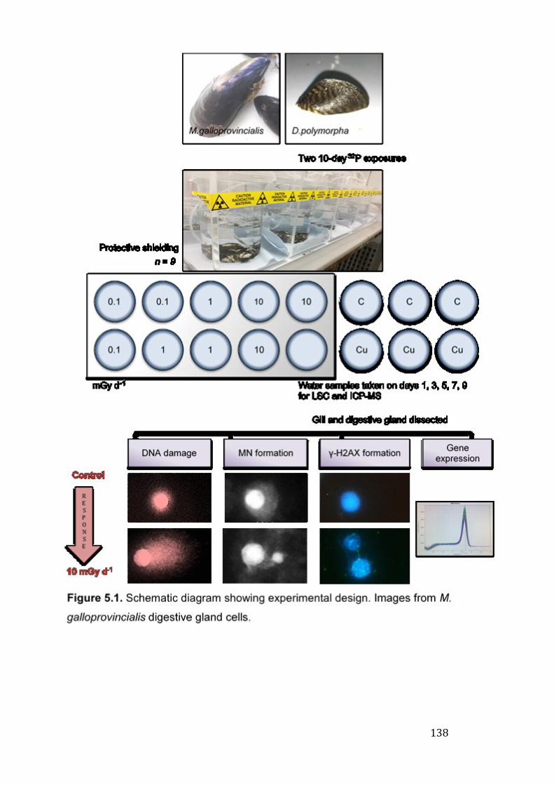
138
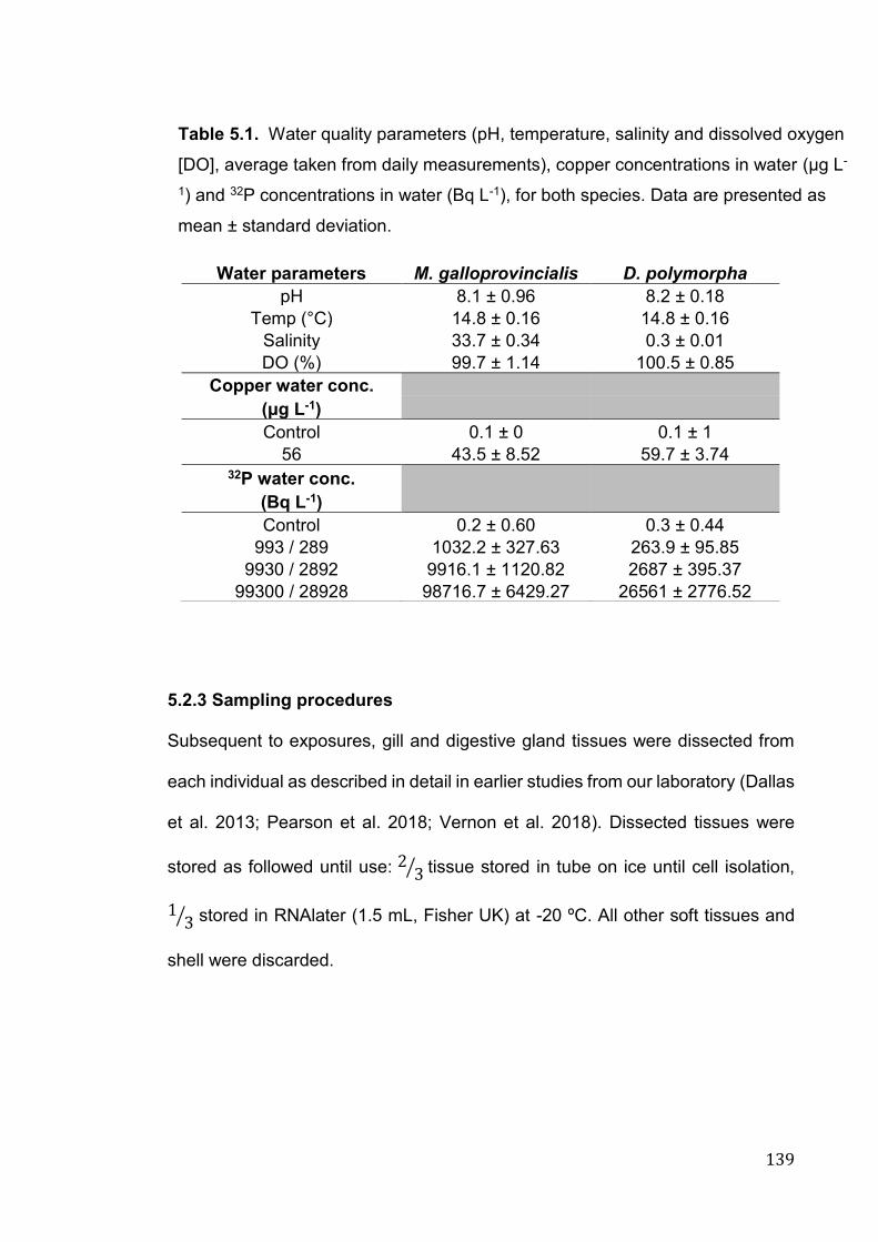
139
5.2.3 Sampling procedures
Subsequent to exposures, gill and digestive gland tissues were dissected from
each individual as described in detail in earlier studies from our laboratory (Dallas
et al. 2013; Pearson et al. 2018; Vernon et al. 2018). Dissected tissues were
stored as followed until use: 2 3⁄ tissue stored in tube on ice until cell isolation,
13⁄ stored in RNAlater (1.5 mL, Fisher UK) at -20 ºC. All other soft tissues and
shell were discarded.
Water parameters M. galloprovincialis D. polymorpha
pH 8.1 ± 0.96 8.2 ± 0.18
Temp (°C) 14.8 ± 0.16 14.8 ± 0.16
Salinity 33.7 ± 0.34 0.3 ± 0.01
DO (%) 99.7 ± 1.14 100.5 ± 0.85
Copper water conc.
(μg L-1)
Control 0.1 ± 0 0.1 ± 1
56 43.5 ± 8.52 59.7 ± 3.74 32P water conc.
(Bq L-1)
Control 0.2 ± 0.60 0.3 ± 0.44
993 / 289 1032.2 ± 327.63 263.9 ± 95.85
9930 / 2892 9916.1 ± 1120.82 2687 ± 395.37
99300 / 28928 98716.7 ± 6429.27 26561 ± 2776.52
Table 5.1. Water quality parameters (pH, temperature, salinity and dissolved oxygen
[DO], average taken from daily measurements), copper concentrations in water (µg L-
1) and 32P concentrations in water (Bq L-1), for both species. Data are presented as
mean ± standard deviation.
140
5.2.4 Biological assays
5.2.4.1 Isolation of digestive gland and gill cells
The procedure to obtain gill cells for genotoxic assays was adopted from previous
studies (Vincent-Hubert et al. 2011), and outlined in section 2.3.2. Supernatant
was then used in subsequent assays, providing cell viability, checked using the
Trypan Blue exclusion dye assay (section 2.4) (Strober 2001), was <90% across
all treatments (data not shown).
5.2.4.2 Comet assay to determine DNA strand breaks
The comet assay was performed using gill and digestive gland cell suspension
(150 µL), as described in section 2.5.2.
5.2.4.3 Analysis of micronuclei (MN) induction
Gill and digestive gland cell suspension was adhered and fixed as described in
section 2.5.1, before staining with 20 μL ethidium bromide (20 μL of 20 mg L−1).
Cells (n = 500) were scored per slide, and results are reported as mean MN per
1000 cells, in keeping with other data from our research group (Dallas et al.
2013).
5.2.4.4 Induction of γ-H2AX foci
γ-H2AX foci were determined in gill and digestive gland cells, following
procedures outlined in 2.5.3. All slides, including procedural blanks were coded
and scored at random, and 50 cells were counted per individual/slide.
141
5.2.4.5 Determination of transcriptional expression of key genes
Gill and digestive gland tissue was dissected immediately after exposure and
stored in RNAlater at -80 °C (R0901, Sigma-Aldrich Company Ltd UK) until use.
RNA extraction, cDNA synthesis and qPCR were performed in accordance to
sections 2.5.4.1 – 2.5.4.3.
5.2.5 Determination of 32P and Cu concentration in water samples
5.2.5.1 Determination of 32P in water samples using liquid scintillation
counting
All samples were collected, processed and analysed as in section 2.8.3.1. Activity
concentrations were background corrected by blank subtracting from each
sample, the blank was nonspiked fresh or seawater. In accordance with Jaeschke
and Bradshaw (2013), CPM values that fell below the blank were assigned an
activity of 0.000. All samples were decay corrected.
5.2.5.2 Determination of Cu concentration in water samples using
Inductively Coupled Plasma Mass Spectrometry (ICP-MS)
Cu concentrations in water samples were determined following procedures
outlined in section 2.7.2.
5.2.4 Dosimetry and the ERICA tool
The Tier 2 assessment module of the ERICA tool was used for dose estimation.
Phosphorus-32 was chosen as one of the ERICA tool's default isotopes (Brown
et al. 2008).
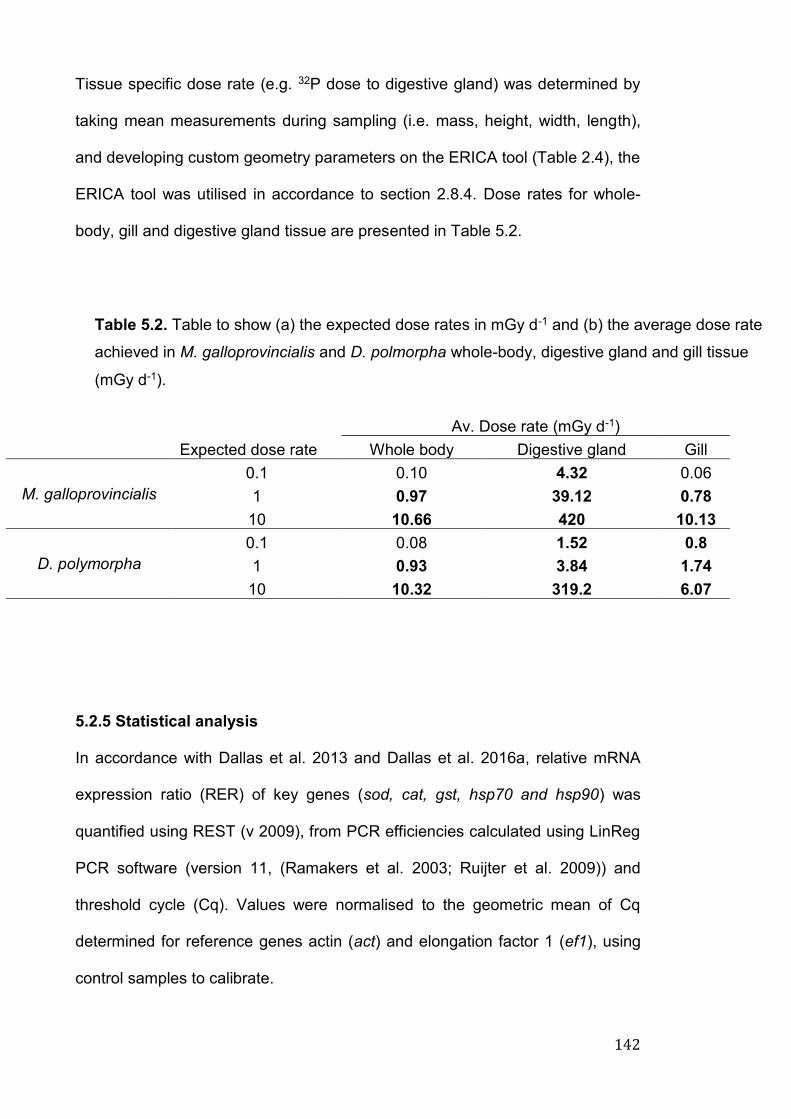
142
Tissue specific dose rate (e.g. 32P dose to digestive gland) was determined by
taking mean measurements during sampling (i.e. mass, height, width, length),
and developing custom geometry parameters on the ERICA tool (Table 2.4), the
ERICA tool was utilised in accordance to section 2.8.4. Dose rates for whole-
body, gill and digestive gland tissue are presented in Table 5.2.
Av. Dose rate (mGy d-1)
Expected dose rate Whole body Digestive gland Gill
M. galloprovincialis
0.1 0.10 4.32 0.06
1 0.97 39.12 0.78
10 10.66 420 10.13
D. polymorpha
0.1 0.08 1.52 0.8
1 0.93 3.84 1.74
10 10.32 319.2 6.07
5.2.5 Statistical analysis
In accordance with Dallas et al. 2013 and Dallas et al. 2016a, relative mRNA
expression ratio (RER) of key genes (sod, cat, gst, hsp70 and hsp90) was
quantified using REST (v 2009), from PCR efficiencies calculated using LinReg
PCR software (version 11, (Ramakers et al. 2003; Ruijter et al. 2009)) and
threshold cycle (Cq). Values were normalised to the geometric mean of Cq
determined for reference genes actin (act) and elongation factor 1 (ef1), using
control samples to calibrate.
Table 5.2. Table to show (a) the expected dose rates in mGy d-1 and (b) the average dose rate
achieved in M. galloprovincialis and D. polmorpha whole-body, digestive gland and gill tissue
(mGy d-1).
143
All statistical analyses were carried out using the statistical software R (RStudio,
R 3.4.3 GUI 1.70 El Capitan build (7463), https://www.r-project.org/). Data were
checked for normality distribution (Shapiro-Wilk test) and homogeneity of
variances (Levene’s test), with visual examination of QQ-plots. The non-
parametric Kruskal-Wallis test was used if assumptions were not met;
comparison between treatment groups was determined using a Dunn’s pairwise
comparison with Bonferroni correction. Where assumptions were met, a one-way
ANOVA was run with Tukey’s post hoc tests. To compare between treatment
groups, a Wilcoxon rank sum test with Holm-Bonferroni correction was used. Any
correlation between variables was determined using a Pearson’s correlation
coefficient. Level of significance for all tests was set at p < 0.05 (*) and data
presented as mean ± standard deviation, unless otherwise stated.
5.3 Results
During the experimental (exposure) periods, no spawning or mortality of the
mussels occurred in either of the species. Metal and 32P concentrations, along
with water quality measurements are presented in Table 5.1. Results of the ICP-
MS and LSC analyses confirmed that achieved values were in line with expected
concentrations across all treatments.
5.3.1 Genotoxic response following in vivo exposures to 32P
Fig. 5.2 shows the mean (± S.D) % tail DNA damage, MN per 1000 cells and γ-
H2AX induction in MG and DP gill cells, following exposure to varying 32P
concentrations for 10 days. All control (unexposed) treatments showed a low
degree of damage across all biomarkers, indicative of good health in study
species.
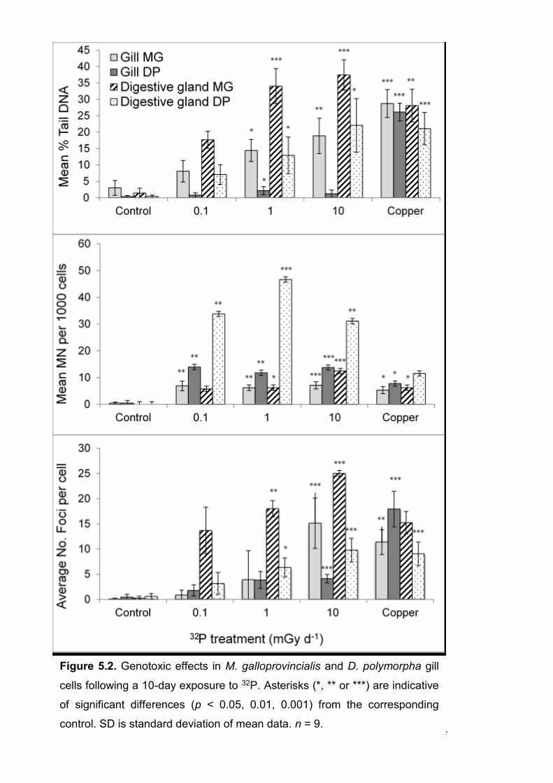
144
Figure 5.2. Genotoxic effects in M. galloprovincialis and D. polymorpha gill
cells following a 10-day exposure to 32P. Asterisks (*, ** or ***) are indicative
of significant differences (p < 0.05, 0.01, 0.001) from the corresponding
control. SD is standard deviation of mean data. n = 9.
145
5.3.1.1 Comet assay to determine DNA strand breaks
The results indicate that in terms of DNA damage (% Tail DNA, Fig. 5.2), both
the tissues showed a significant dose dependant increase in response to 32P
exposure (p < 0.01), particularly in MG tissue and DP digestive gland (p < 0.001).
Control samples showed low levels of damage at around 1-2%.
The highest level of damage was evident in MG digestive gland cells for both 1
and 10 mGy d-1 doses, with around 34-37% tail. The relatively low dose rate to
DP gill cells (Table 5.2) resulted in minimal damage to DNA across all treatments,
with a slight increase for 1 mGy d-1 dose (p < 0.05). MG tissues showed greater
DNA damage across all 32P treatments when compared to the equivalent DP
tissues (p < 0.01). Interestingly, there was no statistical difference between DNA
damage for the 1 and 10 mGy d-1 doses, a trend repeated for MN and γ-H2AX
formation across all tissue (apart from MG gill cells, γ-H2AX, p < 0.05).
5.3.1.2 Analysis of micronuclei (MN) induction
MN formation (Fig. 5.2) did not follow the dose-dependent response pattern
which was evident for DNA damage (comet response) and γ-H2AX induction,
while 32P had a significant effect (p < 0.001) there was little difference between
treatment groups. DP digestive gland showed significantly greater MN formation
then MG digestive gland, gill and DP gill, especially for 1 mGy d-1 dose at 47 MN
per 1000 cells. Similarly, MN induction in DP gill cells was higher than that in MG
gill, across all treatments, however not significantly so (p = 0.92).
5.3.1.3 Induction of γ-H2AX foci
A positive correlation (Fig. 5.3, 4) was evident between DNA damage and γ-H2AX
in all but the DP gill exposure (MG digestive gland: r = 0.91, p < 0.001, MG gill: r
146
= 0.6, p < 0.001, DP digestive gland: r = 0.79, p < 0.001). The 10 mGy d-1
treatment produced the greatest degree of damage across all species and
tissues, in comparison to controls (p < 0.001). In keeping with DNA damage, the
greatest number of γ-H2AX foci was in MG digestive gland cells, across all 32P
treatments with around 13-25 foci per cell. On average, the number of foci in MG
digestive gland was 3-4 fold greater than in DP digestive gland cells (p < 0.01).
Following exposure to a dose of 0.10 mGy d-1, there was no significant increase
in γ-H2AX or DNA damage, in either of the species or tissues.
5.3.2. Transcriptional expression of key genes
PCR efficiencies for studies genes were: actin (act): 1.79, elongation factor 1
(ef1): 1.79, catalase (cat): 1.81, glutathione-s-transferase (gst): 1.81, superoxide
dismutase (sod): 1.80, heat shock protein 70 (hsp70): 1.75 and heat shock
protein 90 (hsp90): 1.83 for MG, and act: 1.80, ef1: 1.79, cat: 1.82, gst: 1.79, sod:
1.78 and hsp70: 1.81 for DP. hsp90 data is not included for DP as the assay
failed to amplify. Relative gene expression of the selected genes are presented
in figure 5.5, there was limited variation across all biological tissue and species.
Gene hsp70 was significantly upregulated in DP gill following 32P exposure (p <
0.05), along with gst in the 1 mGy d-1 treatment, however to a limited degree.
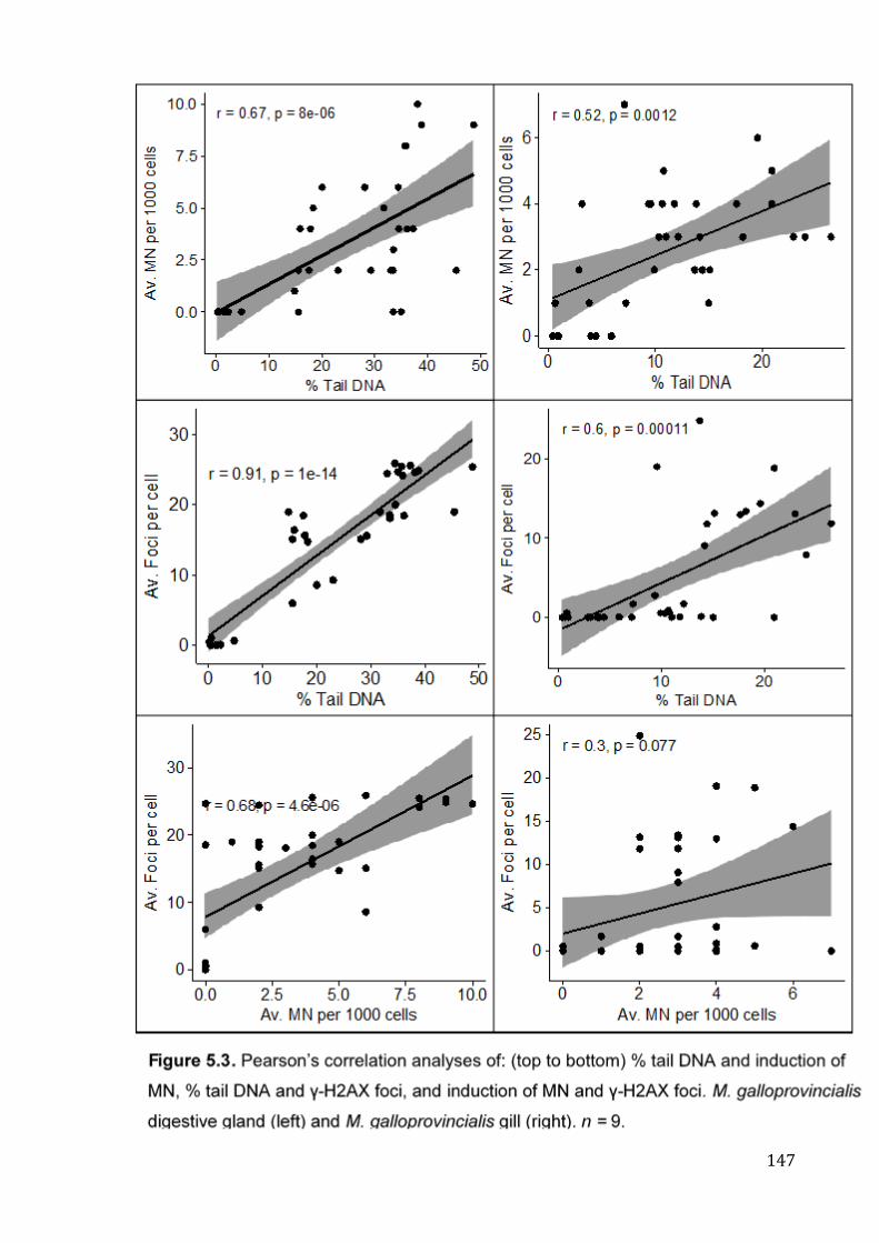
147

148
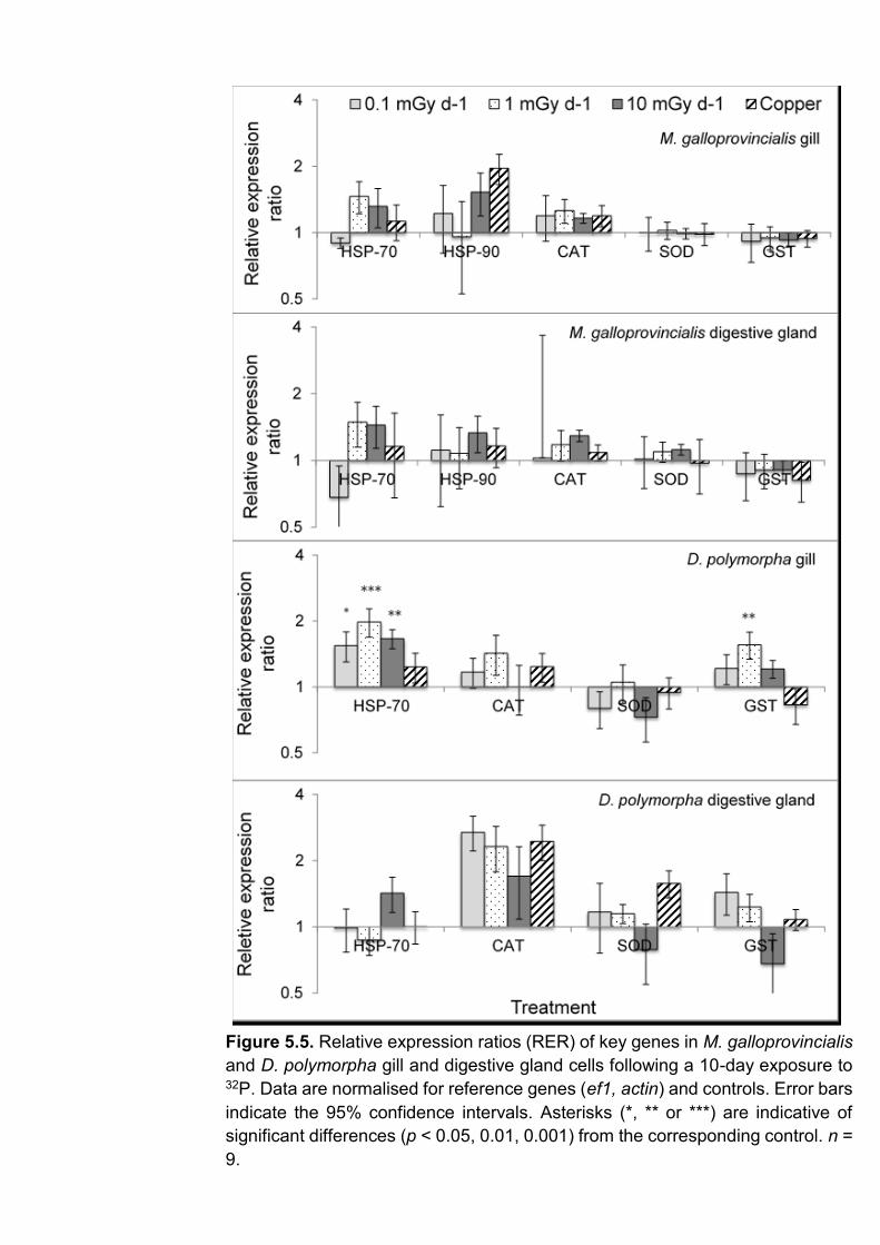
149
Figure 5.5. Relative expression ratios (RER) of key genes in M. galloprovincialis
and D. polymorpha gill and digestive gland cells following a 10-day exposure to 32P. Data are normalised for reference genes (ef1, actin) and controls. Error bars
indicate the 95% confidence intervals. Asterisks (*, ** or ***) are indicative of
significant differences (p < 0.05, 0.01, 0.001) from the corresponding control. n =
9.
150
5.3.3 Whole-body and tissue specific dose rates
Dose rates for whole body and specific tissues (i.e. gill and digestive gland) are
illustrated in Table 5.2. Whole body dose rates were 0.10, 0.97, 10.66 for MG and
0.08, 0.93, 10.32 mGy d-1 for DP, in line with predicted values. The highest dose
rates were evident in digestive gland tissue, with rates around 39 (MG) and 31-
fold (DP) higher than whole body dose in the 10 mGy d-1 treatment. Whole body
and gill tissue dose rates were comparable. A positive correlation was evident
between all genotoxic biomarkers except for DNA damage in DP gill, and MN
formation in both DP tissues (Fig. 5.4).
5.4 Discussion
5.4.1 Dosimetry and dose-response relationship
Our study explored the tissue specific effects following exposure to 32P on marine
and freshwater bivalves, using a multi-biomarker approach. It is important to note
that in the current study, 32P was introduced in a highly bioavailable form (i.e.
radiolabelled ATP). The observed biological responses may be reflective of this
form of radioactive phosphorus (Vernon et al. 2018). Our study firstly suggests
that gill and digestive gland tissues are sensitive and reliable cell types for
assessing IR-induced responses, this deviates from previous bivalve studies
where haemocytes are predominantly chosen as target cell types to study
biological responses (Jha et al. 2005; Jha et al. 2006; Jaeschke et al. 2015;
Dallas et al. 2016a; Pearson et al. 2018). Secondly, our study has highlighted the
necessity to evaluate response in several organs, as greater 32P activity levels
within digestive gland tissue in particular has induced a greater genotoxic
response. Whole body dose rates (Table 5.2) in this instance are likely to mask
the potential biological effect of 32P. At dose rates below the screening value of
151
10 μGy h−1 (0.24 mGy d-1) (Andersson et al. 2008; Andersson et al. 2009), whole
body dose estimations (MG 0.10, DP 0.08 mGy d-1) would suggest a minimal risk
to individual or population. However, MG digestive gland dose as an example
(4.32 mGy d-1), is 43-fold greater than whole body. As noted by Jha (2008) and
Jaeschke et al (2011), it is vital to determine tissue specific effects in response to
a wide range of contaminants, only then are we able to implement adequate
protection policies, and suitable radiation benchmarks.
5.4.2 32P induced genotoxic response in gill and digestive gland cells
5.4.2.1 Comet assay to determine DNA strand breaks
The present study revealed significant DNA damage (as % Tail DNA) in MG gill
tissue and the digestive glands of both species, highlighting strong correlations
between dose rate and the response. Our results are in line with previous studies
where exposure to doses between 12-485 μGy h−1 (tritium, 3H) induced significant
damage in haemocytes of Mytilus sp. (Jha et al. 2005, 2006; Dallas et al. 2016a;
Pearson et al. 2018). In both species the highest DNA damage was evident in
digestive gland cells, most likely due to high 32P accumulation, and therefore dose
rate. To our knowledge, few radiobiological studies have investigated IR-induced
response in digestive organs, despite them playing major roles in metabolism,
immune defence, and as a primary sink for many aquatic contaminants
(McDonald et al. 1993; Cartier et al. 2004; Dimitriadis et al. 2004; Banni et al.
2017; Faggio et al. 2018; Sforzini et al. 2018b). Mussel digestive glands
accumulate and process nutrients, which are distributed to reproductive tissues
during gonad development; as such, a key concern with digestive gland damage
is the possible influence on reproductive success (Sastry and Blake 1971;
Dimitriadis et al. 2004). Interestingly, no significant level of DNA damage is
152
evident at dose rates of 0.10 mGy d-1 in either tissue or species, this result is in
contrast to Jha et al (2006), where low doses of tritium (0.30 mGy d-1, 96 h)
caused a 2.8-fold increase in damage compared to control cells in haemocytes
of mussels. This trend is continued in Zebrafish (D. rerio, 96 hpf larvae), where
exposure to low dose-rates (137Cs, 0.8 mGy d-1) caused significantly increased
damage (Gagnaire et al. 2015). Disparity may be down to numerous factors,
including differential sensitivity between cell types or species, radionuclide LET
and exposure length, or physiological factors (i.e. reproductive stage,
metabolism, health status) (Nalepa et al. 1991; Jha 2008; Pearson et al. 2018).
In terms of DNA damage, little change was evident in DP gill tissue, with a slight
significant increase (statistically, but probably not biologically) noted for 1 mGy d-
1 dose. Mussel gills filter suspended particulates directly from the surrounding
media to specific organs (Jørgensen 1982). They play a major role in respiratory
processes, nutrient uptake and digestion (David and Fontanetti 2005; Gómez-
Mendikute et al. 2005). The large surface area and close proximity to aquatic
contaminants makes them a prime tissue for biomonitoring and ecotoxicological
studies and their sensitivity to numerous pollutants is well documented (Mersch
et al. 1996; Parolini et al. 2011b; Al-Subiai et al. 2012; Dallas et al. 2013, 2016a,
2018; Canesi et al. 2014). Interestingly, while DNA damage was relatively low in
DP gill, there was significant MN induction (at around 13 MN per 1000 cells)
across all 32P treatments. As MN, a biomarker of ‘effect’, are apparent following
cell division, any increase in its frequency can be regarded as a longer term sign
of damage, in comparison to the comet and γ-H2AX assays which provide a brief
snapshot in time. The lack of correlation between end points measured in DP gill
may be a result of DNA-DNA or DNA protein crosslinks that inhibit tail migration
during the comet assay (Hartmann et al. 2001; Klobučar et al. 2003).
153
As previously noted, 32P incorporates directly onto the ribose-phosphate
backbone of replicating DNA and isotopic decay (32P to 32S) breaks the initial
strand (SSB), and by close proximity emitted elections can cause DSBs, resulting
in a higher proportion of strand breaks than other beta emitters (i.e. 90Y, 131I,
(Cheng et al. 2015). The comet assay in this instance is useful in showing current,
non-specific (i.e. single or DSBs) DNA damage, often regarded as more sensitive
when combined with other biomarkers (Frenzilli et al. 2009). Future studies would
benefit from measuring DNA damage post exposure at multiple time points, as to
monitor possible DNA repair in different cell types. This would be particularly
relevant for MG digestive gland cells, where a high degree of DNA damage is
paralleled (r = 0.91, p < 0.001) by increased γ-H2AX foci, suggesting the
recruitment of DNA repair proteins (Kuo and Yang 2008).
5.4.2.2 Induction of γ-H2AX foci
IR has the potential to damage cellular DNA and to maintain genomic integrity
and function, repair enzymes/mechanisms are required (Hoeijmakers 2001;
Sancar 2004). The long-term biological impact of 32P is dependent on the
efficiency of such DNA repair mechanisms, as unrepaired DNA lesions may result
in genetic mutation, leading to cancer formation or cell death (cytotoxicity). As
noted by Kuo and Yang (2008), H2AX is rapidly phosphorylated to form γ-H2AX,
a crucial factor in DSB repair response, following irradiation with IR. γ-H2AX
induction has been noted at dose rates of 70-550 mGy d-1 in Zebrafish ZF4 cells
and embryos, in response to 137Cs exposure (Pereira et al. 2011; Pereira et al.
2014). In Medaka (O. latipes) fish erythrocytes, a high frequency of γ-H2AX foci
(2 h, 39.9 ± 45.05) were noted following an acute exposure (15 Gy) to 137Cs, and
in line with our study, were well correlated with DNA damage (as measured by
154
the comet assay) (Sayed et al. 2017). The present study suggests that mussels
are able to employ repair mechanisms following 32P induced DNA damage, while
no significant increase in γ-H2AX formation was evident at 0.10 mGy d-1, a dose
dependant response was demonstrated overall. If we compare data obtained
from comet and γ-H2AX assays, several similarities are apparent, and a good
correlation is evident between all end-points (all but DP gill cells). γ-H2AX foci
represent DSBs in a 1:1 manner (Kuo and Yang 2008), the strong correlation
between comet and γ-H2AX assays suggests that the majority of DNA damage
evident is in the form of DSBs, as opposed to SSBs, alkali-labile sites or DNA
cross-links.
γ-H2AX is dephosphorylated rapidly after DNA repair and is therefore time-
dependant. Foci peak in size at around 30 min and decrease in number over time
(Sedelnikova et al. 2003; Ivashkevich et al. 2011, 2012). As noted by Sayed et al
(2017), γ-H2AX foci per cell (Medaka erythrocytes) peaked 2 h after exposure,
and declined steadily over time (24 h). This study is limited however, by small,
inconsistent sample numbers and very few cells counted per sample, as
suggested, a minimum of 50-100 cells should be counted per sample (Redon et
al. 2009; Oommen et al. 2016b). Whilst care was taken to process the tissues for
analysis immediately after exposure, in our study it is highly possibly that all end-
points would vary over time. Monitoring during, immediately after, and h/days
post exposure would allow for a more clear indication of permanent, or repairable
effects.
Differing repair capacities are evident between the digestive glands of MG and
DP. In terms of MG digestive gland, a high degree of DNA damage and γ-H2AX
foci induction (~ 25 foci/cell, 10 mGy d-1), combined with relatively low MN
induction suggests efficient DNA repair mechanisms. In comparison, in DP
155
digestive gland, γ-H2AX foci induction is 2.5-4.3 fold lower than MG, but MN
formation is significantly greater. This could indicate a less efficient DNA repair
mechanism, or that the system was overwhelmed. It is important to note that γ-
H2AX foci form at both unprogrammed and programmed DSBs. Due to low foci
number in control samples (~0.3 foci/cell), we are confident in attributing foci
formation to 32P exposure in our study (Sedelnikova et al. 2003; Revet et al.
2011). To avoid misinterpretation of results, it is important that baseline levels of
cellular DSBs are determined prior to exposures (or via control treatments).
5.4.2.3 Analysis of micronuclei (MN) induction
MN represent fragments of chromosomes or whole chromosome formed via
misrepair of DNA DSBs (Fenech et al. 2011; Bolognesi and Fenech 2012). The
capacity of IR to induce MN has previously been reported in bivalves (M. edulis)
and fish (Catla catla, Cyprinus carpio, Oryzias latipes) (Jha et al. 2005, 2006;
Anbumani and Mohankumar 2012; Jaeschke et al. 2015; Kumar et al. 2015;
Sayed et al. 2017; Hurem et al. 2018). Our results suggest that MN formation is
the most sensitive endpoint to 32P, as significant MN formation was present even
at the lowest dose (0.10 mGy d-1, all but MG digestive gland). DP digestive gland
cells had a vast number of MN in comparison to all other 32P treatments at around
31-47 MN/1000 cells. This suggests that DNA damage overwhelmed the repair
capacity of the cells, resulting in less reversible, more permanent effects.
A correlation is evident between DNA damage and increased MN frequency in all
but DP gill (MG digestive gland: r = 0.67, p < 0.001, MG gill: r = 0.52, p < 0.01,
DP digestive gland: r = 0.38, p < 0.021). This is in contrast to previous studies
were strong positive correlations are noted (Bolognesi et al. 2004; Hagger et al.
2005; Jha et al. 2005; Canty et al. 2009; Kumar et al. 2015). No significant
156
variation was evident between 32P treatments, and there is little distinction
between species, cell type, or treatment, with MN frequency ranging between 6-
14 MN/1000 cells (excluding DP digestive gland). While the reason for this is
unclear, differing results suggest varying MoA’s between beta-emitting
radionuclides (tritium, 32P) on the DNA in mussels. Our study clearly shows the
benefit of adopting a multi-biomarker approach in measuring IR-induced genetic
damage. The combination of biomarkers aids the detection of differing aspects of
genotoxicity (DNA repair and response) and clastogenicity (Jha et al. 2005; Araldi
et al. 2015; Kumar et al. 2015).
5.4.3 Expression of key genes
IR has the potential to damage biological molecules, in turn altering their function.
Despite this there are few mechanistic studies about such processes in aquatic
invertebrates (Farcy et al. 2007; Farcy et al. 2011; AlAmri et al. 2012; Devos et
al. 2015; Dallas et al. 2016a). Interestingly, while genotoxic response to 32P was
evident in both species, there was little change in genes involved in cell stress
defence mechanisms (protein folding or regulation of oxidative stress). Slight up-
regulation of hsp70 (all 32P treatments, p < 0.05) and gst (1 mGy d-1, p < 0.001)
was noted in DP gill, albeit to a relatively limited degree.
IR generates ROS via the radiolysis of water (Barillet et al. 2011; Graupner et al.
2016). To minimise the detrimental effects of ROS, antioxidant enzymes such as
sod, cat and gst are employed. It could be expected that genes involved with
oxidative stress response, detoxification and/or cellular defence would be
upregulated when exposed to IR. This was noted following acute exposure to
137Cs in K. marmoratus embryos, where antioxidant enzyme-coding genes (e.g.
gst, cat, mn-sod, cu/zn-sod) were significantly upregulated at dose rates of 5 Gy
157
(Rhee et al. 2012). However, our findings are in line with Devos et al (2015) who
noted significant genotoxic response (i.e. DNA damage, as measured by the
comet assay) following exposure of C. gigas to 3H (0.07-1.1 mGy d-1), but no
change in gene expression levels (hsp70, hsp90, hsc72, gst, mdr, cyp1a, sod,
mt1&2 and p53). This was also noted in MG gill cells following exposure to tritium
(7 d, ~0.36 mGy d-1), where DNA damage significantly increased (p < 0.05) in
comparison to control treatments, but gene expression levels remained
unchanged (slight increase in hsp70-1 and rad51, 72 h) (Dallas et al. 2016a).
However, after 3 days (72 h, ~0.36 mGy d-1) significant upregulation was noted
in all genes (hsp70-1, hsp70-2, hsp90, mt20, p53, rad51) (Dallas et al. 2016a),
suggesting time as an important factor in the transcriptional expression of genes.
As suggested by Devos et al (2015), the lack of change noted at a molecular level
may suggest different sensitivities for end points, or disparity in the MoA of
toxicity. It is possible that evident genomic damage resulted from direct
interaction of 32P with DNA, either through isotopic decay or the subsequent
release of high-energy beta particles, and to a lesser degree via the generation
of ROS.
Our gene expression analysis included a limited number of stress response
genes, as the current study was limited to readily available gene sequences in
both species. Future studies would benefit from studying a wider range of genes,
particularly those involved in DNA damage and repair (i.e. p53, ogg1, rad51), or
via a more open ended approach. For example, adopting a high-throughput
transcriptomic (e.g. RNAseq) or proteomic approach, which allows the
measurement of expression levels in thousands of genes/proteins, could be
utilised to identify early IR-induced responses, and would aid the identification of
mechanisms involved in an organism’s toxicity response to IR.
158
5.4.4 Environmental implications and future research
While 32P occurs in small quantities in the environment, when accumulated into
biological tissue it is able to induce significant genomic damage. The marine and
freshwater species chosen for this study represent suitable models to investigate
32P induced toxicity. The multi-species approach adopted here could be regarded
as more robust, and realistic than single-species experiments (Chapman 2002;
Solomon and Sibley 2002; Canty et al. 2009; Schnug et al. 2014). It is important
to note that variations in 32P speciation and therefore bioavailability between salt
and freshwater may have influenced species response. We are however able to
establish a genotoxic response in both MG and DP, even at relatively low 32P
levels. Increased genomic instability may ultimately have a detrimental effect at
higher levels of biological organization, from individual to long-term population
level effects (Jha 2008).
In the natural environment, ionising radionuclides are part of a complex mix of
aquatic contaminants that can place combined pressure on biota. Field studies
are arguably more environmentally realistic in determining the true biological
effect of contaminants, taking into account the plethora of additional stressors
(i.e. predation, disease, population density, food availability). However, as noted
by Farcy et al (2007), the complexity of the natural environment makes it difficult
to link damage response to a particular source. Future studies would benefit from:
(a) combined field and laboratory studies, (b) laboratory experiments using a
more realistic, flow-through exposure set-up (to note, due to health, safety,
logistical and economic reasons a static exposure was utilised in this study), (c)
multi-stressor and/or multi-species exposures, and (d) use of a wide range of
radionuclides and exposure conditions. Knowledge of the behaviour, transfer and
159
biological impact of radionuclides within aquatic systems allows for the
development of adequate management and protective strategies.
5.5 Conclusions
In terms of the short-lived radionuclide, 32P, a greater tissue concentration is
paralleled by increased dose rate, and biological damage in two anatomically
similar bivalve species. In terms of DNA damage and DDR, the marine species,
MG appears to be slightly more sensitive on an immediate, short-term level,
possibly due to greater accumulation rates. However in terms of longer-term
damage, high MN formation in DP digestive gland cells suggests a more
permanent response. This low-dose, chronic study is the first to adopt a multi-
species, multi-biomarker approach in investigating tissue specific 32P induced
biological response, along with dose-response relationships. In terms of radiation
science, this approach could be readily adopted to study impact of other
radionuclides either alone or in combination with other environmental stressors.

160
161
Chapter 6
Evaluation of interactive effects of phosphorus-32 and copper
on marine and freshwater bivalve molluscs
In preparation:
Vernon, EL., Moore, NM., Bean, TP., Jha, AN. 2018. Combined toxicity of
phosphorus-32 and copper on marine and freshwater bivalve molluscs.

162
163
6.1 Introduction
Radionuclides and metals co-exist in the aquatic environment, their increased
prominence resulting primarily from human activities (e.g. industrial discharge,
nuclear power generation, accidents or weapons tests, mining, wastewater
treatment) (Hu et al. 2010). Nuclear industries as an example co-dispose of
radionuclides and non-radioactive waste. The requirement for large volumes of
water for cooling processes typically results in NPPs being located close to large
water bodies (i.e. oceans, lakes), where waste products are disposed. IR emitted
from discharged radionuclides can enter the environment via controlled or
accidental (nuclear accidents: Chernobyl, Fukushima) release, whilst discharges
of contaminants are largely regulated, little is known about interactive effects.
Exposure to a mixture of stressors, which in isolation may not induce significant
damage, may cause deleterious effects on organism health through additive or
synergistic mechanisms when acting in combination (Mothersill et al. 2007).
Alternatively, combinations may have an antagonistic effect, where the addition
of one stressor may offer protection against another. As demonstrated by Tran et
al (2007), selenium (Se, 4 µg L-1) had a protective effect in M. edulis haemocytes,
against exposure to known toxic agent, mercury (Hg2+, 20 µg L-1). To improve the
basis for environmental and human risk assessments, it is crucial to advance our
understanding on multi-stressor induced effects on biological, and ecological
levels.
It is well accepted that radionuclides and metals can readily concentrate in the
tissues of aquatic organisms, posing a threat to both humans via consumption,
and biota by trophic level transfer (Carvalho 2018). When present within a cell,
such contaminants have varying potential to cause significant damage to
164
molecules such as DNA, either directly or indirectly, the extent dependent on
concentration, LET, distribution and biological half-life. IR could interact with other
environmental stressors (metals, organics or physical agents such as
temperature) in different ways (i.e. additively, synergistically, antagonistically)
and could modify observed biological effects (Manti and D'Arco 2010).
In context, IR-induced biological response has been investigated in a wide range
of aquatic biota, predominately following exposures to long-lived radionuclides
such as 137Cs and 3H. Gene expression alterations, arguably the first step
towards response to any contaminant have been demonstrated in molluscs (C.
gigas, Mytilus spp.), arthropods (D. magna, T. japonicas), echinoderms (P. lividus)
and rotifer (B. koreanus), following exposure to IR, with focus predominantly on
specific cell stress marker genes, namely heat shock chaperone proteins (i.e.
hsp70, 90), along with markers of oxidative stress (gst, cat, sod) and DNA repair
(p53, rad51). At low 3H doses (~15.58 µGy h-1), Dallas et al (2016a) noted
upregulation of genes involved in protein folding, DNA DSB repair and cell cycle
checkpoint control in MG, transcriptome level effects were well correlated with
those at higher organisational levels (genetic, DNA damage). Given that IR is
capable of inducing a range of responses at different levels of biological
organisation, which are often determined simultaneously, adoption of a holistic
and integrated approach is required to assess the induced biological responses.
In light of this, mathematical models provide the conceptual and mathematical
formalism to integrate molecular, cellular and whole animal processes (Allen and
McVeigh 2004; Allen and Moore 2004; Moore and Noble 2004). Previous studies
have shown that Principal Component Analysis (PCA) and network complexity
can be used as an indicator of homeostasis or health in cellular systems (Moore
165
2010; Moore et al. 2015; Sforzini et al. 2015; Sforzini et al. 2018a). Modelling is
essential for the extrapolation of explanatory frameworks that facilitate the
development of a predictive capacity for estimating outcomes or risk associated
with stressful environmental conditions (Moore and Noble 2004; Moore 2010;
Moore et al. 2015). Previous studies on mussels and earthworms have shown
that there is a strong relationship between the first principal component (PC 1)
for responses of numerous stress biomarkers and lysosomal membrane stability
(LMS), as an indicator of cellular health (Moore et al. 2006; Sforzini et al. 2015;
Sforzini et al. 2017; Sforzini et al. 2018a). Multivariate statistical analysis,
including PCA was used to integrate multi-biomarker data in test organisms (MG
and DP) and specific tissues.
As IR is not an isolated threat to aquatic biota, several studies have utilised
laboratory exposure scenarios to determine combined toxicity of multiple
environmental stressors (Olsvik et al. 2010; Heier et al. 2013; Dallas et al. 2016a).
Olsvik et al (2010) and Heier et al (2013) investigated interactions between
radionuclides (60Co) and environmentally relevant metal concentrations (Cu, Al,
Cd) in Atlantic salmon (S. salar). The addition of Al and Cd appeared to reduce
the impact of gamma-irradiation by modifying transcriptional induction of
oxidative stress-responsive genes (including p53, glutathione reductase,
glutathione peroxidase, metallothionein) (Olsvik et al. 2010). This trend was not
continued following exposure to Al and Cu, suggesting varying MoA’s in toxicity
response. Potential IR-metal induced synergistic, antagonistic or additive effects
are yet to be fully explored in aquatic invertebrates.
In context, short-lived radionuclide 32P was utilised as a cost-effective, accessible
surrogate for more environmentally prominent beta and gamma emitting
radionuclides (e.g. 137Cs, 90Sr). As previously demonstrated (Vernon et al. 2018),
166
32P readily accumulated in the tissues of mussels, particularly digestive gland
(see chapter 4). Detrimental 32P-induced responses (DNA damage, MN and γ-
H2AX foci induction) have been noted at dose rates as low as 0.1 mGy d-1, in
both digestive gland and gill cells (see chapter 5).
Metals such as copper (Cu) can be highly toxic to organisms at concentrations
present within marine and freshwater environments. Cu-induced deleterious
effects in aquatic invertebrates are well studied (Al-Subiai et al. 2011; Vosloo et
al. 2012; Brooks et al. 2015; Xu et al. 2016), and much research has focused on
interactions between Cu and additional stressors (metals, ocean acidification,
temperature, pesticides, microplastics) (Clayton et al. 2000; Bouskill et al. 2006;
Trevisan et al. 2011; Maria et al. 2013; Lewis et al. 2016). However, to our
knowledge no studies have determined the interaction between IR and Cu in
mussels.
In this work, the effects of 32P and Cu, alone and in combination were studied in
two ecologically relevant adult bivalve species, MG and DP. Mussels are
excellent models for investigating the relevance of low metal/IR doses, they are
well established in ecotoxicological research and as such, a large number of
validated biological effect endpoints are available to be measured and quantified
in a range of cell types (i.e. haemocytes, digestive gland and gills cells). A suite
of biomarkers, from molecular to behavioural levels were measured in the
digestive gland and gill cells of two mussel species, following exposure to a range
of Cu and 32P concentrations/doses, alone and in combination. 32P dose rates of
0.10 and 1 mGy d-1 were reflective of a generic (all species) “no effect” screening
dose rate of 10 μGy h−1 (0.24 mGy d-1), the chosen Cu concentration (18 μg L-1)
was in line with environmentally realistic values and adopted from previous
167
validation studies described in chapter 3 (Andersson et al. 2008; Andersson et al.
2009). The overall aims and objectives of this study were (a) to adopt an
integrated, multi-biomarker approach in investigating the combined effects of Cu
and 32P in two bivalve species, (b) to determine relative sensitivity of marine and
freshwater adult bivalves, (c) to determine potential correlations between different
parameters (molecular, genetic and behavioural) studied and (d) to determine
relative sensitivity between different cell types (i.e. gill and digestive gland cells).
With regards to species variation, we hypothesised firstly that little disparity in
response will be evident. Secondly, Cu would have an additive effect on the 32P-
induced responses in mussels.
6.2 Materials and methods
6.2.1 Chemicals and suppliers
Radiolabelled-ATP (Adenosine triphosphate, γ-32P), was obtained from Perkin
Elmer (PerkinElmer, UK) in batches of 9.25 MBq (specific activity: 370 MBq mL-
1), and diluted with DI water to form appropriate working solutions, as in Vernon
et al (2018). All working solutions were decay adjusted throughout the exposure.
6.2.2 Mussel exposure conditions
Adult MG and DP were collected and maintained in accordance to section 2.2.
Ten-day exposures of both the mussel species were performed between
September-October 2017, and were staggered by two weeks for ease of analysis
and logistical reasons. Following collection and after a 2-week acclimation, MG
and DP individuals (total weight, 35 g L-1) per labelled beaker were exposed to
the following exposure scenarios in triplicate: (a) 0.1 mGy d-1, (b) 0.1 mGy d-1 +
Cu, (c) 1 mGy d-1 and (d) 1 mGy d-1 + Cu. The Cu (CuSO4.5H2O, 99% purity)
168
concentration used in combination with 32P was 18 μg L-1. Control and positive
control (Cu, 56 μg L-1) treatments were run alongside. 32P activity levels in water
were calculated in a previous study (section 2.8; Vernon et al. 2018), to meet
expected nominal dose rates, 32P activity concentrations for 0.1 mGy d-1 were 993
Bq L-1 (MG) and 579 Bq L-1 (DP), and for 1 mGy d-1, 9930 Bq L-1 (MG) and 5786
Bq L-1 (DP). Activity concentrations in water were calculated from preliminary
experiments (data not included). The selection of lower and higher concentrations
(positive control) of Cu were based on previous studies (Chapter 3).
Mussels were fed 2 h prior to water changes (50 %), on days 3, 5, 7 and 9, as
described in earlier studies (Vernon et al. 2018). Water quality parameters were
measured routinely, before and after water changes. Phosphorus-32 and Cu
activity levels were determined using water samples (1 mL, in duplicate), taken
~30 minutes after each water change and processed for ICP-MS or liquid
scintillation counting (LSC), as in section 2.7.2 and 2.8.3.2. Data from LSC
counting and ICP-MS analysis confirmed that achieved values were in line with
expected concentrations across all treatments.
6.2.3 Sampling procedures
After exposures, gill and digestive gland tissue was dissected from each
individual and stored as followed until use: 23⁄ tissue stored in tube on ice until
cell isolation, 1 3⁄ stored in RNAlater (1.5 mL, Fisher UK) at -20 ºC. All other soft
tissue and shell was discarded.
169
6.2.4 Biological assays
6.2.4.1 Isolation of digestive gland and gill cells
The procedure to obtain gill and digestive gland cells for genotoxic assays was
adopted from previous studies (Vincent-Hubert et al. 2011), and outlined in
section 2.3.2. Supernatant was then used in subsequent assays, providing cell
viability, checked using the Trypan Blue exclusion dye assay (section 2.4)
(Strober2001), was <90% across all treatments (data not shown).
6.2.4.2 Comet assay to determine DNA strand breaks
The comet assay was performed using gill and digestive gland cell suspension
(150 µL), as described in section 2.5.2.
6.2.4.3 Analysis of micronuclei (MN) induction
Gill and digestive gland cell suspension was adhered and fixed as described in
section 2.5.1, before staining with 20 μL ethidium bromide (20 μL of 20 mg L−1).
Cells (n = 500) were scored per slide and results are reported as mean MN per
1000 cells, in keeping with other data from our research group (Dallas et al. 2013).
6.2.4.4 Induction of γ-H2AX foci
γ-H2AX foci were determined in gill and digestive gland cells, following
procedures outlined in 2.5.3. All slides, including procedural blanks were coded
and scored at random, and 50 cells were counted per individual/slide.
6.2.4.5 Determination of transcriptional expression of key genes
Gill and digestive gland tissue was dissected immediately after exposure and
stored in RNAlater at -80 °C (R0901, Sigma-Aldrich Company Ltd UK) until use.
170
RNA extraction, cDNA synthesis and qPCR were performed in accordance to
sections 2.5.4.1 – 2.5.4.3.
6.2.5 Behavioural observations: Valve movement and byssus attachment
As noted in section 2.6.2, valve movement or activity (i.e. whether the individual
was actively filtering or had a fully closed shell) was assessed by eye, three times
daily during the course of the exposure (Rajagopal et al. 2003; Hartmann et al.
2015). Byssus attachment (i.e. whether the individual is attached to either the
glass beaker/other individuals) was assessed every alternate day of exposure by
eye, as an indicator of mussel health (Angarano et al. 2009; Ericson et al. 2010;
Martinović et al. 2016).
6.2.6 Water quality measurements and 32P and Cu analyses
6.2.6.1 Determination of 32P in water samples using liquid scintillation
counting
All samples were collected, processed and analysed as in section 2.8.3.1. Activity
concentrations were background corrected by blank subtracting from each
sample, the blank was nonspiked fresh or seawater. In accordance with Jaeschke
and Bradshaw (2013), CPM values that fell below the blank were assigned an
activity of 0.000. All samples were decay corrected.
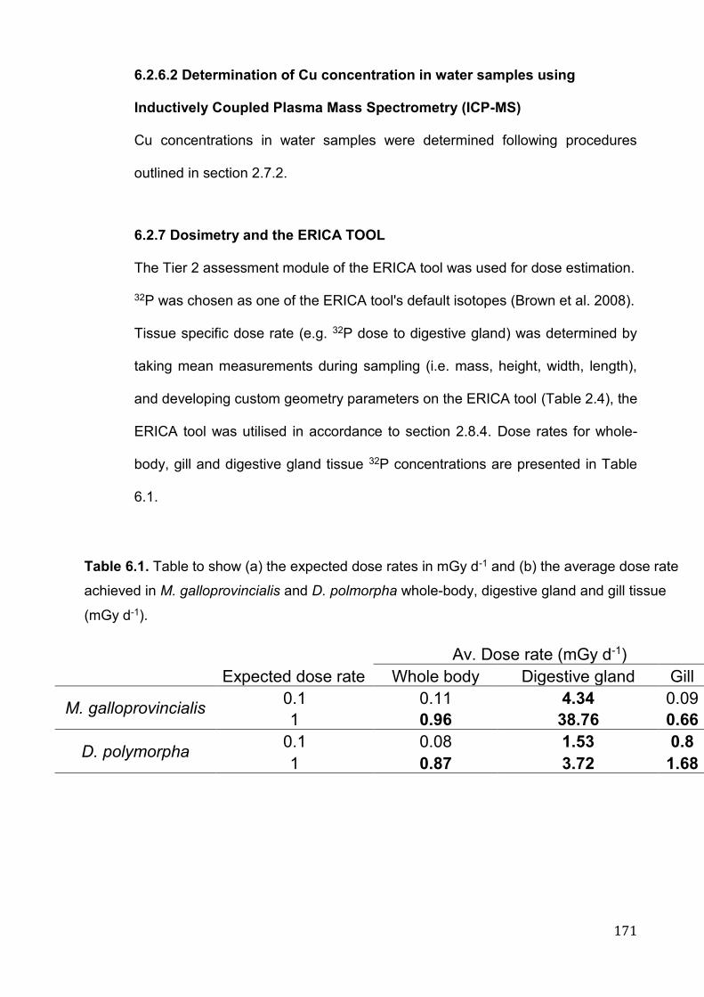
171
6.2.6.2 Determination of Cu concentration in water samples using
Inductively Coupled Plasma Mass Spectrometry (ICP-MS)
Cu concentrations in water samples were determined following procedures
outlined in section 2.7.2.
6.2.7 Dosimetry and the ERICA TOOL
The Tier 2 assessment module of the ERICA tool was used for dose estimation.
32P was chosen as one of the ERICA tool's default isotopes (Brown et al. 2008).
Tissue specific dose rate (e.g. 32P dose to digestive gland) was determined by
taking mean measurements during sampling (i.e. mass, height, width, length),
and developing custom geometry parameters on the ERICA tool (Table 2.4), the
ERICA tool was utilised in accordance to section 2.8.4. Dose rates for whole-
body, gill and digestive gland tissue 32P concentrations are presented in Table
6.1.
Av. Dose rate (mGy d-1)
Expected dose rate Whole body Digestive gland Gill
M. galloprovincialis 0.1 0.11 4.34 0.09
1 0.96 38.76 0.66
D. polymorpha 0.1 0.08 1.53 0.8
1 0.87 3.72 1.68
Table 6.1. Table to show (a) the expected dose rates in mGy d-1 and (b) the average dose rate
achieved in M. galloprovincialis and D. polmorpha whole-body, digestive gland and gill tissue
(mGy d-1).
172
6.2.8 Statistical analysis
In accordance with Dallas et al (2013) and Dallas et al (2016a), relative mRNA
expression ratio (RER) of genes was quantified using REST (v 2009), from PCR
efficiencies calculated using LinReg PCR software (version 11, Ramakers et al.
2003; Ruijter et al. 2009) and threshold cycle (Cq). Values were normalised to
the geometric mean of Cq, determined for reference genes actin (act) and
elongation factor 1 (ef1), using control values to calibrate.
Statistical analyses were operated using the statistical software R (RStudio, R
3.4.3 GUI 1.70 El Capitan build (7463), https://www.r-project.org/). Were
applicable, data was checked for normality distribution (Shapiro-Wilk test) and
homogeneity of variances (Levene’s test), with visual examination of QQ-plots.
The non-parametric Kruskal-Wallis test was used if assumptions were not met;
comparison between groups (i.e. specific tissue or species) was determined
using a Dunn’s pairwise comparison with Bonferroni correction. Where
assumptions were met, a one-way ANOVA was run with Tukey’s post hoc tests.
Comparison between groups was determined using a Wilcoxon rank sum test
with Holm-Bonferroni correction. Level of significance for all tests was set at p <
0.05 (*) and data presented as mean ± standard deviation, unless otherwise
stated.
6.2.8.1 Multivariate analysis
Biomarker data for gill and digestive gland tissue in both species were analysed
using non-parametric multivariate analysis software, PRIMER v 6.1.5 (PRIMER-
Є Ltd., U. Auckland, New Zealand; Clarke 1999; Clarke & Warwick. 2001). All
data were log transformed [logn(1+x)] and standardised to the same scale.
Correlations between biomarkers were tested using scatter plot matrices; while
173
PCA, hierarchical cluster analysis and non-metric multi-dimensional scaling
analysis (MDS, plots not shown), derived from Euclidean distance similarity
matrices, were used to visualize dissimilarities between sample groups. The
results were tested for significant differences between treatments using analysis
of similarity (PRIMER v6 - ANOSIM), which is an approximate analogue of the
univariate ANOVA, and reflects on differences between treatment groups in
contrast to differences among replicates within samples (the R statistic). Under
the null hypothesis H0 (“no difference between samples”), R = 0 and this was
tested by a non-parametric permutations approach; there should be little or no
effect on the average R value if the labels identifying which replicates belong to
which samples are randomly rearranged. Behavioural responses were excluded
from the PCA due to non-paired samples.
The PRIMER v6 - BIO-ENV routine (Spearman’s Rank Correlations) linking
multivariate biomarker response patterns was used to identify potential
“influential biomarkers”, small subsets of biomarkers capturing the full PCA
biomarker response pattern.
6.3 Results
No mortality or spawning occurred in either mussel species throughout the
duration of the experiment. Cu and 32P concentration, along with water quality
measurements are presented in Table 6.2. Whole body and specific tissue (i.e.
gill and digestive gland) dose rates are illustrated in Table 6.1. Whole body dose
rates were 0.11 and 0.96 for MG and 0.08, 0.87 mGy d-1 for DP, in line with
predicted values.
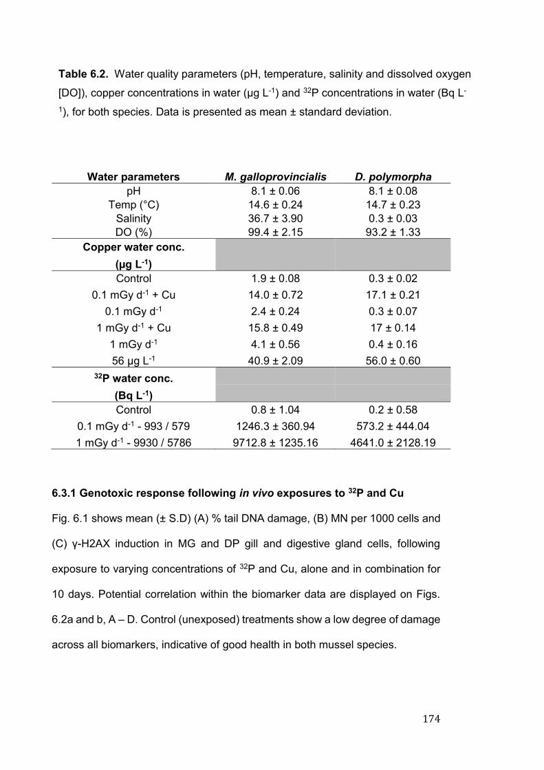
174
Water parameters M. galloprovincialis D. polymorpha
pH 8.1 ± 0.06 8.1 ± 0.08
Temp (°C) 14.6 ± 0.24 14.7 ± 0.23
Salinity 36.7 ± 3.90 0.3 ± 0.03
DO (%) 99.4 ± 2.15 93.2 ± 1.33
Copper water conc.
(µg L-1)
Control 1.9 ± 0.08 0.3 ± 0.02
0.1 mGy d-1 + Cu 14.0 ± 0.72 17.1 ± 0.21
0.1 mGy d-1 2.4 ± 0.24 0.3 ± 0.07
1 mGy d-1 + Cu 15.8 ± 0.49 17 ± 0.14
1 mGy d-1 4.1 ± 0.56 0.4 ± 0.16
56 µg L-1 40.9 ± 2.09 56.0 ± 0.60
32P water conc.
(Bq L-1)
Control 0.8 ± 1.04 0.2 ± 0.58
0.1 mGy d-1 - 993 / 579 1246.3 ± 360.94 573.2 ± 444.04
1 mGy d-1 - 9930 / 5786 9712.8 ± 1235.16 4641.0 ± 2128.19
6.3.1 Genotoxic response following in vivo exposures to 32P and Cu
Fig. 6.1 shows mean (± S.D) (A) % tail DNA damage, (B) MN per 1000 cells and
(C) γ-H2AX induction in MG and DP gill and digestive gland cells, following
exposure to varying concentrations of 32P and Cu, alone and in combination for
10 days. Potential correlation within the biomarker data are displayed on Figs.
6.2a and b, A – D. Control (unexposed) treatments show a low degree of damage
across all biomarkers, indicative of good health in both mussel species.
Table 6.2. Water quality parameters (pH, temperature, salinity and dissolved oxygen
[DO]), copper concentrations in water (µg L-1) and 32P concentrations in water (Bq L-
1), for both species. Data is presented as mean ± standard deviation.
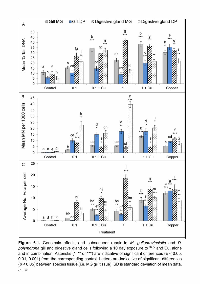
175
Figure 6.1. Genotoxic effects and subsequent repair in M. galloprovincialis and D.
polymorpha gill and digestive gland cells following a 10 day exposure to 32P and Cu, alone
and in combination. Asterisks (*, ** or ***) are indicative of significant differences (p < 0.05,
0.01, 0.001) from the corresponding control. Letters are indicative of significant differences
(p < 0.05) between species tissue (i.e. MG gill tissue). SD is standard deviation of mean data.
n = 9.
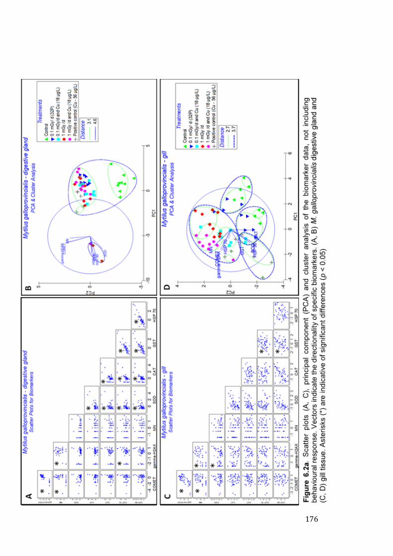
176

177
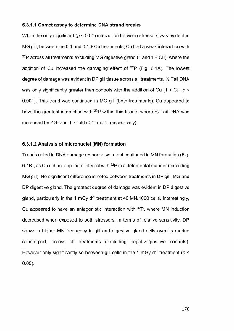
178
6.3.1.1 Comet assay to determine DNA strand breaks
While the only significant (p < 0.01) interaction between stressors was evident in
MG gill, between the 0.1 and 0.1 + Cu treatments, Cu had a weak interaction with
32P across all treatments excluding MG digestive gland (1 and 1 + Cu), where the
addition of Cu increased the damaging effect of 32P (Fig. 6.1A). The lowest
degree of damage was evident in DP gill tissue across all treatments, % Tail DNA
was only significantly greater than controls with the addition of Cu (1 + Cu, p <
0.001). This trend was continued in MG gill (both treatments). Cu appeared to
have the greatest interaction with 32P within this tissue, where % Tail DNA was
increased by 2.3- and 1.7-fold (0.1 and 1, respectively).
6.3.1.2 Analysis of micronuclei (MN) formation
Trends noted in DNA damage response were not continued in MN formation (Fig.
6.1B), as Cu did not appear to interact with 32P in a detrimental manner (excluding
MG gill). No significant difference is noted between treatments in DP gill, MG and
DP digestive gland. The greatest degree of damage was evident in DP digestive
gland, particularly in the 1 mGy d-1 treatment at 40 MN/1000 cells. Interestingly,
Cu appeared to have an antagonistic interaction with 32P, where MN induction
decreased when exposed to both stressors. In terms of relative sensitivity, DP
shows a higher MN frequency in gill and digestive gland cells over its marine
counterpart, across all treatments (excluding negative/positive controls).
However only significantly so between gill cells in the 1 mGy d-1 treatment (p <
0.05).
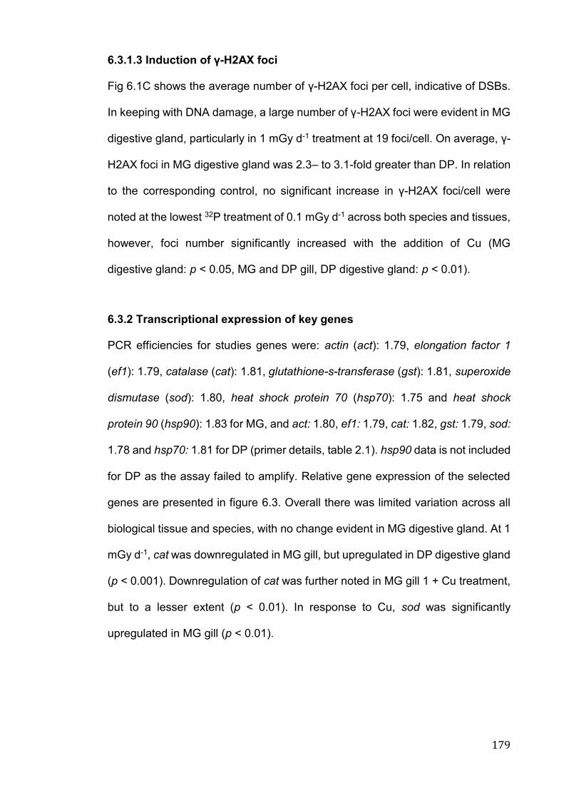
179
6.3.1.3 Induction of γ-H2AX foci
Fig 6.1C shows the average number of γ-H2AX foci per cell, indicative of DSBs.
In keeping with DNA damage, a large number of γ-H2AX foci were evident in MG
digestive gland, particularly in 1 mGy d-1 treatment at 19 foci/cell. On average, γ-
H2AX foci in MG digestive gland was 2.3– to 3.1-fold greater than DP. In relation
to the corresponding control, no significant increase in γ-H2AX foci/cell were
noted at the lowest 32P treatment of 0.1 mGy d-1 across both species and tissues,
however, foci number significantly increased with the addition of Cu (MG
digestive gland: p < 0.05, MG and DP gill, DP digestive gland: p < 0.01).
6.3.2 Transcriptional expression of key genes
PCR efficiencies for studies genes were: actin (act): 1.79, elongation factor 1
(ef1): 1.79, catalase (cat): 1.81, glutathione-s-transferase (gst): 1.81, superoxide
dismutase (sod): 1.80, heat shock protein 70 (hsp70): 1.75 and heat shock
protein 90 (hsp90): 1.83 for MG, and act: 1.80, ef1: 1.79, cat: 1.82, gst: 1.79, sod:
1.78 and hsp70: 1.81 for DP (primer details, table 2.1). hsp90 data is not included
for DP as the assay failed to amplify. Relative gene expression of the selected
genes are presented in figure 6.3. Overall there was limited variation across all
biological tissue and species, with no change evident in MG digestive gland. At 1
mGy d-1, cat was downregulated in MG gill, but upregulated in DP digestive gland
(p < 0.001). Downregulation of cat was further noted in MG gill 1 + Cu treatment,
but to a lesser extent (p < 0.01). In response to Cu, sod was significantly
upregulated in MG gill (p < 0.01).
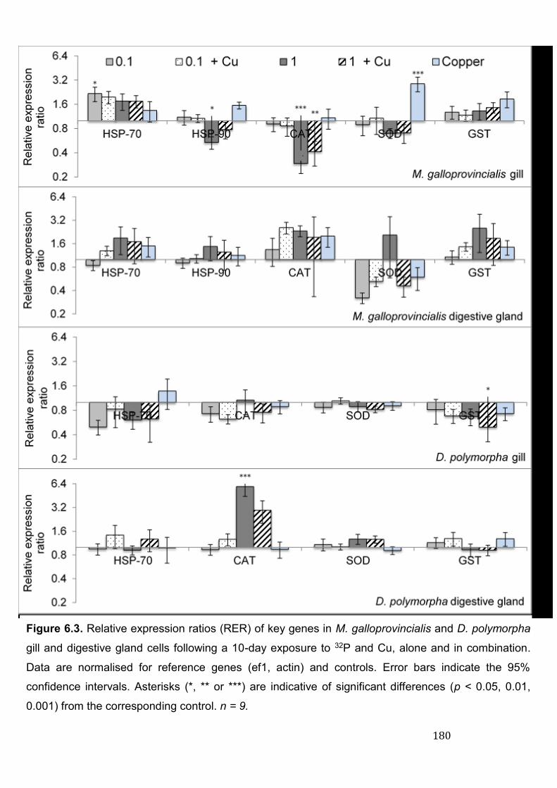
180
Figure 6.3. Relative expression ratios (RER) of key genes in M. galloprovincialis and D. polymorpha
gill and digestive gland cells following a 10-day exposure to 32P and Cu, alone and in combination.
Data are normalised for reference genes (ef1, actin) and controls. Error bars indicate the 95%
confidence intervals. Asterisks (*, ** or ***) are indicative of significant differences (p < 0.05, 0.01,
0.001) from the corresponding control. n = 9.
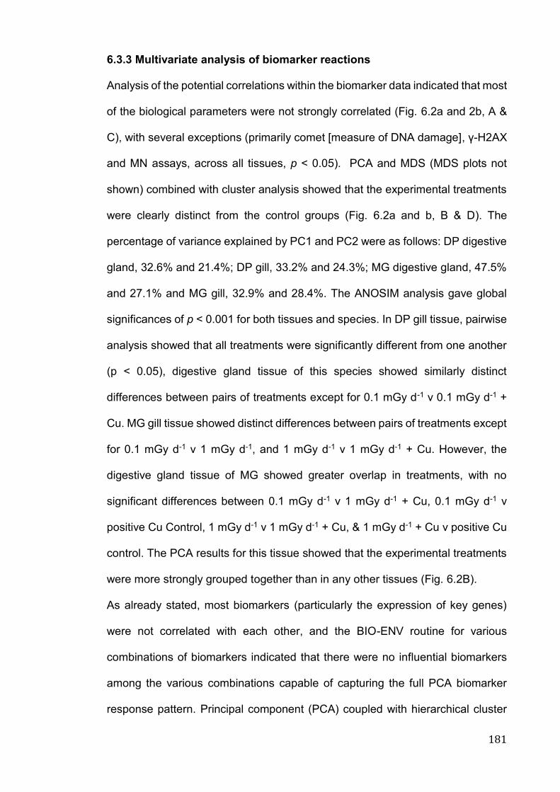
181
6.3.3 Multivariate analysis of biomarker reactions
Analysis of the potential correlations within the biomarker data indicated that most
of the biological parameters were not strongly correlated (Fig. 6.2a and 2b, A &
C), with several exceptions (primarily comet [measure of DNA damage], γ-H2AX
and MN assays, across all tissues, p < 0.05). PCA and MDS (MDS plots not
shown) combined with cluster analysis showed that the experimental treatments
were clearly distinct from the control groups (Fig. 6.2a and b, B & D). The
percentage of variance explained by PC1 and PC2 were as follows: DP digestive
gland, 32.6% and 21.4%; DP gill, 33.2% and 24.3%; MG digestive gland, 47.5%
and 27.1% and MG gill, 32.9% and 28.4%. The ANOSIM analysis gave global
significances of p < 0.001 for both tissues and species. In DP gill tissue, pairwise
analysis showed that all treatments were significantly different from one another
(p < 0.05), digestive gland tissue of this species showed similarly distinct
differences between pairs of treatments except for 0.1 mGy d-1 v 0.1 mGy d-1 +
Cu. MG gill tissue showed distinct differences between pairs of treatments except
for 0.1 mGy d-1 v 1 mGy d-1, and 1 mGy d-1 v 1 mGy d-1 + Cu. However, the
digestive gland tissue of MG showed greater overlap in treatments, with no
significant differences between 0.1 mGy d-1 v 1 mGy d-1 + Cu, 0.1 mGy d-1 v
positive Cu Control, 1 mGy d-1 v 1 mGy d-1 + Cu, & 1 mGy d-1 + Cu v positive Cu
control. The PCA results for this tissue showed that the experimental treatments
were more strongly grouped together than in any other tissues (Fig. 6.2B).
As already stated, most biomarkers (particularly the expression of key genes)
were not correlated with each other, and the BIO-ENV routine for various
combinations of biomarkers indicated that there were no influential biomarkers
among the various combinations capable of capturing the full PCA biomarker
response pattern. Principal component (PCA) coupled with hierarchical cluster
182
analysis and ANOSIM results for all treatments showed that both 32P and Cu had
a detrimental effect on the genetic integrity and oxidative stress status in the four
tissues tested (Figs. 6.2a and b, A - D).
6.3.4 Behavioural observations: Valve movement and byssus attachment
No significant variation was found between experimental treatments in terms of
active filtering (valve movement) or byssus attachment (Fig. 6.4), a possible
result of small n numbers. Control mussels showed increased attachment with
time, overall MG showed a greater propensity to attach than DP, with all 3
attached from exposure day 3 (Fig. 6.4a). Valve activity was variable throughout
the exposure, with MG generally showing more active filtration, independent of
treatment (Fig. 6.4b).
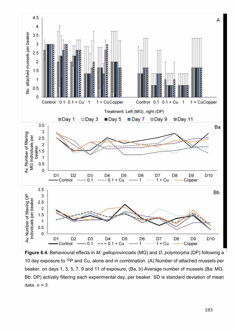
183
Figure 6.4. Behavioural effects in M. galloprovincialis (MG) and D. polymorpha (DP) following a
10 day exposure to 32P and Cu, alone and in combination. (A) Number of attached mussels per
beaker, on days 1, 3, 5, 7, 9 and 11 of exposure, (Ba, b) Average number of mussels (Ba: MG,
Bb: DP) actively filtering each experimental day, per beaker. SD is standard deviation of mean
data. n = 3.
184
6.4 Discussion
This novel study is the first to explore the interactive effects of IR and Cu on two
aquatic bivalve species using a tissue specific, multi-biomarker approach.
Qualities of radionuclides and speciation of metals (i.e. physical and chemical
properties) in aquatic systems has a large influence on bioavailability and
subsequent toxicity (Gunten and Beneš 1995; Richards et al. 2011). It is well
documented that Cu, focusing on the toxic ionic form (Cu2+), forms complexes
with natural organic matter, decreasing bioavailability and at lower salinities (i.e.
freshwater) becoming more abundant (Grosell et al. 2007). Despite this, little is
known on the possible interactions between Cu and lesser studied radionuclides
such as 32P, and the combined influence of water parameters [i.e. pH, salinity,
dissolved organic carbon (DOC), alkalinity]. Due to time and logistical constraints,
certain parameters (e.g., DOC) were not determined during this experiment. It is
possible that differing water chemistry (i.e. salt and freshwater), along with
chemical interactions between stressors could have affected the noted biological
effects in each species. However, correlation between accumulations of Cu and
32P (in isolation) in soft tissues with increasing genotoxicity in cells (gill and
digestive gland, Chapters 3, 4) were evident, in each species independently.
6.4.1 Biomarker interactions
Recent developments spanning multiple fields are leading to the discovery of
prognostic biomarkers that may be suitable for use as risk indicators of biological
damage (Moore et al. 2006; Jenkins et al. 2011; Ortiz et al. 2011; Berghella et al.
2014). It is probable that many biomarkers only exhibit a response in a part of the
“health status space” (Depledge et al. 1993; Moore et al. 2004; Moore et al. 2006),
where they indicate whether a reaction has taken place and may even indicate
185
health status within a narrow range, or what has induced the response, but they
do not generally indicate health status of the animal for the whole range from
healthy to irreversible damage (Köhler et al. 2002). In terms of environmental
prognostics, the first step is to relate biomarker responses to the health status of
individual organisms, by mapping said responses against an integrated “health
status” indicator (Köhler et al. 2002; Allen and Moore 2004; Moore et al. 2004;
Moore et al. 2006). PCA is an effective method for integrating biomarker data (i.e.
DNA damage, MN formation, gene expression etc.) into a “health status space”,
reducing the multi-dimensionality of the problem to a simple two dimensional
representation (Chatfield and Collins 1980; Allen and Moore 2004). PCA is
commonly used as a cluster analysis tool and effectively captures the variability
in a dataset in terms of principle components. PCA has facilitated modelling the
integrated responses of multiple biomarkers in the context of “health status space”
(Allen and Moore 2004; Moore et al. 2006). PCA and associated statistical tests
have previously shown that lysosomal biomarkers (as indicators of health status),
in combination with the comet assay provide an effective integrated assessment
of the adverse effects on physiological function and genetic integrity (genotoxicity)
(Sforzini et al. 2015; Sforzini et al. 2017; Dallas et al. 2018; Sforzini et al. 2018a).
In continuation with earlier studies, current findings demonstrate that PCA can
aid interpretation of multiple biomarker responses and pathological reactions to
multiple environmental stressors (i.e. IR and metals).
6.4.2 32P induced genotoxic response in gill and digestive gland cells
The majority of IR-induced toxicity studies have focused on single radionuclide
exposures. DNA damage as a biomarker has been noted at dose rates ranging
from 0.8-41666 µGy h-1 in a wide range of biota, including marine bivalves
186
(Mytilus spp., C. gigas, P. perna, C. fluminea, P. malabarica & M. casta) (Jha et
al. 2005, 2006; Godoy et al. 2008; Farcy et al. 2011; AlAmri et al. 2012; Kumar
et al. 2014). PCA and MDS, combined with cluster analysis demonstrate a clear
distinction between controls and experimental groups, across both species,
tissue and biomarker (Fig. 6.2a and b, B & D). In keeping with previous work (see
Chapter 5, section 5.3.1.1), no significant change in DNA damage was noted at
lower 32P doses of 0.10 mGy d-1 (Excluding DP digestive gland, present study).
Interestingly, where previous findings demonstrated significantly increased % Tail
DNA in both species and cell types at 1 mGy d-1, only MG digestive gland was
significantly increased. The present study had greater baseline levels of DNA
damage of ~5-11%, compared to <5% in previous literature, this along with
individual differences may account for such variation.
Of interest, exposure of the mussels to 32P alone caused no significant increase
in % Tail DNA (Excluding DP [0.1] and MG digestive gland [1 mGy d-1]), but when
combined with Cu, % Tail DNA was statistically greater than controls in MG gill
and DP digestive gland at 0.10 mGy d-1, and all species and tissues at 1 mGy d-
1. Cu in isolation, at environmentally relevant concentrations (18 µg L-1) was found
to have no significant effect on % Tail DNA in MG or DP gill cells (see Chapter 3,
section 3.3.2.2), when in combination with 32P there appears to be an additive
effect on mussels. Whilst not significantly so, the addition of Cu to IR exposures
also increased γ-H2AX foci induction, across both species, tissue and dose rate
(Excluding MG digestive gland). This apparent additive effect of Cu on the
genotoxicity of 32P on marine and freshwater mussels is the first reported. Cu-
contaminant induced effects have been noted in previous literature. In M. edulis,
Cu-induced (0.1 μM) damage to DNA and lipids was noted as significantly greater
under low pH conditions (reflective of ocean acidification), in comparison to
187
controls (Lewis et al. 2016). Similarly, combined Cu (10 µg L-1) and IR (60Co, 70
mGy) exposure was found to induce significantly depleted glutathione compared
to exposure to Cu alone, in presmolt S. salar (Heier et al. 2013).
Aquatic biota are continuously exposed to both endogenously and
environmentally generated contaminants, giving cause for the development of
highly effective biochemical mechanisms that afford the ability to protect and
defend on multiple biological levels. H2AX is quickly phosphorylated to form γ-
H2AX, a crucial factor in DSB repair response and therefore a relevant, useful
technique in radiation science, where the MoA of 32P in particular is mediated by
induction of DNA DSBs (Kuo and Yang 2008; Cheng et al. 2015). Pereira et al
(2011), Urushihara et al (2012), Pereira et al (2014) and Sayed et al (2017) have
successfully utilised this technique to assess DDR in fish (O. latipes and D. rerio)
following acute exposures to 137Cs, but to our knowledge, is yet to be utilised in
mussels.
The γ-H2AX assay is further validated when utilised alongside classical DNA
damage techniques, such as comet and MN assays, relationships have been
demonstrated between such biomarkers (Pereira et al. 2011; Sayed et al. 2017).
In our study, γ-H2AX was strongly correlated with DNA damage across all tissues
(p < 0.05), correlation was not noted between other molecular or genetic
biomarkers (Fig. 6.2a and 2b, A, C). The greatest induction of γ-H2AX foci was
noted in MG digestive gland, specifically at 1 mGy d-1 (~ 19 foci/cell), followed by
the 1 + Cu, positive Cu control, 0.1 + Cu and 0.1 mGy d-1 treatments. At
significantly higher dose rates of 10 mGy d-1 (137Cs), less than 5 foci were present
per zebrafish ZF4 cell (Pereira et al. 2011). As noted in section 5.4.2.2, γ-H2AX
foci occurs rapidly post-irradiation (30 min – 2 h) and decrease in number over

188
time, as both studies measured damage immediately (~10-40 min) post exposure,
varying radiosensitivity may be a result of species, radionuclide source or
associated MoA (Sedelnikova et al. 2003; Ivashkevich et al. 2011,2012). Trends
in γ-H2AX foci induction in MG digestive gland followed DNA damage, but not
MN formation, where MN/1000 cells remained uniform respective of experimental
treatment. The presence of MN is generally regarded as more permanent
damage, suggesting that the induced DSBs were easily repairable. This was
further noted in MG gill.
In keeping with previous studies (Chapter 5) a greater degree of MN formation is
noted in DP gill and digestive gland cells, relative to associated MG tissue across
all treatments, suggesting a significant impact on DNA integrity. Unexpectedly,
Cu has a weak antagonistic interaction with 32P in terms of MN induction, where
MN/1000 cells decrease in DP digestive gland (both 32P concentrations),
although not significantly so. It is possible that Cu-specific repair mechanisms, as
a by-product buffer against the more permanent effects induced by IR as an
isolated threat.
As described in previous literature (Vernon et al. 2018), 32P concentrates
differentially in gill and digestive gland tissue, with the latter accumulating 87 %
(MG) and 44% (DP) of 32P (proportion of whole body 32P concentration), at 10
mGy d-1. Similarly, Cu uptake varies between tissue, and between species,
(Chapter 3). In turn, there are clear differences in tissue specific dose rate (Table
6.1), and subsequent genotoxic response to IR and metals, highlighting the
importance of a multiple-tissue approach in ecotoxicological studies. Overall, the
addition of Cu arguably has the lowest impact on 32P-induced damage in MG
digestive gland, where MG gill tissue shows far less variation between treatments

189
(excluding % Tail DNA, 0.1 and 0.1 + Cu). In keeping with previous findings, in
terms of longer-term damage, high MN frequency in DP cells suggests a more
permanent response in freshwater mussels, in relation to MG (Chapter 5).
6.4.3 Transcriptional expression of key genes
In our study, transcriptional levels of key genes showed little variation between
differing treatments, such findings are in agreement with earlier studies described
in Chapter 5 (section 5.4.3). Excluding significant upregulation of sod in MG gill,
the Cu positive control (56 µg L-1) caused no variation in gene expression. This
in contrast to work by Xu et al (2018), where exposure to much lower Cu
concentrations (2 and 8 µg L-1) significantly induced the expressions of stress
genes (hsp70, hsp90, mt-10) in Mytilus coruscus (haemocytes), particularly 12 d
post exposure. Furthermore, significantly altered transcriptional profiles in fish (D.
rerio and S. salar) have been noted following IR exposure (137Cs or 60Co),
common MoAs associated with low-dose gamma radiation included the induction
of oxidative stress and DNA damage genes (Jaafar et al. 2013; Freeman et al.
2014; Song et al. 2014, 2016). As suggested by Devos et al (2015), and
highlighted in chapter 5, lack of change evident at molecular levels may be
suggestive of different sensitivities for end points, or disparity in the MoA of
toxicity. It is possible that evident genomic damage resulted from direct
interaction of 32P with DNA, through isotopic decay or release of high-energy beta
particles, and to a lesser degree via ROS generation.
Due to both logistical constraints and lack of readily available gene sequences in
MG and DP, we were only able to assess five key genes in this study. As noted
in Chapter 5 (section 5.4.3), this provides limited information pertaining to
molecular mechanistic responses to IR and Cu. Furthermore, while identification
190
of transcriptome variation is undoubtedly important and increasingly popular
within radiobiological research, limited information can be acquired by studying
genes in isolation. In recent years, there has been a movement from measuring
gene expression levels to proteins, as they more accurately represent the
functional molecules within a cell. It is frequently highlighted that mRNA is the
first step in a long sequence resulting in protein synthesis. Being transmitters of
genetic information the analysis of mRNA is not a direct reflection of the protein
content within a cell, for this reason many studies have found poor correlations
between the expression levels of mRNA and protein (Maier et al. 2009). Following
exposure to comparable Cu concentrations (10 µg L-1), Maria et al (2013) noted
altered proteins associated with oxidative stress (glutathione-S-transferase) and
digestion, growth and remodelling processes (chitin synthase) in MG gill tissue.
Findings were confirmed in MG gill and digestive gland (Gomes et al. 2014a).
Interestingly in MG gill tissue exposed to isolated Cu and benzo(a)pyrene (10 µg
L-1, BaP), concentrations produced higher protein alterations then when in
combination, suggesting an antagonistic interaction (Maria et al. 2013).
Proteomics research in relation to aquatic biota is limited by a lack of available
annotated genomes and proteomes for most aquatic organisms (Slattery et al.
2012), despite this, it is a clear direction for aquatic/ecotoxicological and
radiobiological research. Identifying known, and potentially novel molecular
targets (genes and proteins) involved, as well as activated signalling pathways in
a range of biological systems under multi-stress exposure scenarios may be the
first step in predicting potential larger scale impacts on more environmentally
relevant levels (Maria et al. 2013; Gomes et al. 2014a).
191
6.4.4 Behavioural observations: Valve movement and byssus attachment
To determine interactive effects of IR and metals on mussel health, we adopted
an integrated, multi-disciplinary approach. Whilst molecular and cellular
biomarkers are predominantly used in ecotoxicological studies, it is difficult to
extrapolate results to higher, arguably more relevant levels of biological
organisation (i.e. population, ecosystem). To aid the development of effective
policies for environmental protection, it is important to understand the relationship
between biomarker responses at multiple organisational levels (Moore et al.
2004).
In our study no relationship was evident between exposure and behavioural
observations, nor was there a correlation with molecular and genotoxic
biomarkers. Bivalve molluscs adhere to their surrounding environment via byssus
threads, strong proteinaceous fibres produced by the byssal gland on the base
of the foot. This allows for habituation on the hydrodynamically variable intertidal
zone (Smeathers and Vincent 1979; Rajagopal et al. 2005). Byssal attachment
can be utilised as an indicator of vitality, as byssal thread formation requires large
amounts of energy (Zardi et al. 2007). Reduced byssal attachment in Mytilus spp.
has been documented following exposure to antifouling agents (tributyltin
chloride), ocean acidification, hypoxia and pharmaceuticals (Ericson et al. 2010;
Sui et al. 2015; Martinović et al. 2016), and in DP exposed to cannabinoids and
organic compounds (BHA and TBHQ) (Cope et al. 1997; Angarano et al. 2009).
In contrast, byssus attachment remained fairly uniform following exposure to IR
and Cu, lack of notable variation is a possible result of the relatively short
exposure duration. Under more chronic regimes energy may be redirected
towards protective processes and away from byssal production.
192
In terms of valve movement, there was little change over the duration of the
exposure, irrespective of species. In the natural environment bivalves hold shell
valves open to facilitate necessary biological function (i.e. respiration and
feeding), valve closure can occur as a protective strategy under periods of
physiological stress (Kramer et al. 1989; Rajagopal et al. 2003; Redmond et al.
2017). Avoidance behaviour (fully closed shell) was not evident in this study,
suggesting (a) an inability to detect contaminants in surrounding media, or (b)
lack of sensitivity at particular behavioural levels. It is important to note that
behavioural endpoints are susceptible to individual variation, a larger study size
may better reflect stress response (Redmond et al. 2017).
6.4.5 Environmental implications and future research
Multivariate analysis is the first stage in developing numerical and network
models for environmental impact on the health of sentinel animals such as marine
and freshwater mussels (Allen and Moore 2004; Moore 2010; Sforzini et al. 2015,
2017, 2018a). PCA is an effective method for integrating biomarker data into a
“health status space” reducing the multi-dimensionality of the problem to a simple
two dimensional representation (Chatfield and Collins 1980; Allen and Moore
2004), however, PCA and cluster analysis do not integrate the various
biomarkers in a functionally meaningful way. The subsets of biomarkers used in
this study do not comprehensively support the development of a broad cellular
physiological network that is suited to providing a measure of “health status”,
however future studies could build on the current data sets in order to develop
such models in the future.
This study is certainly useful in establishing relationships between stressor and
response, PCA coupled with hierarchical cluster analysis and ANOSIM results for
193
all treatments showed that both 32P and Cu had deleterious effects on genetic
integrity and oxidative stress status in species and tissues tested (Fig. 6.2a and
b, A - D). However the controlled nature of laboratory experiments are not fully
reflective of field conditions. A continuing challenge for scientists is to understand
firstly, the complexity of contaminant interactions in differing environments, and
secondly, how multiple interactive stressors affect biota at all levels of biological
organisation, from molecular to ecosystem levels.
6.5 Conclusions
In conclusion, the results of this integrated, multi-biomarker study represent the
most extensive data to date, obtained on the combined effects of IR and metals
(Cu) in two environmentally relevant bivalve species. The results indicate that (a)
genotoxic response was reflective of exposure, where Cu had an overall additive
effect on 32P-induced damage across several (but not all) species, cell types and
dose rates, (b) freshwater mussels were susceptible to longer lasting damage,
marine mussels to more immediate effects, (c) selected genes were generally
unaltered in terms of transcriptional response to contaminants, independent of
species and (d) mussels were not responsive to IR and Cu, alone or in
combination at behavioural levels. Whilst it is difficult to extrapolate such findings
to exposures in realistic environmental conditions, these data contribute to the
limited information on the possible mechanisms involved in multi-stressor (IR and
metals) induced response, and subsequently highlight the importance of
investigating the interactive effects of pollutants on ecologically relevant mussel
species.

194

195
Chapter 7
General discussion and future perspectives

196
197
7.1 Environmental radiation and dosimetry
Radiobiological protection measures have adapted to become more inclusive of
both human and environmental protection (Brechignac and Howard 2001;
Copplestone et al. 2004; Brechignac and Doi 2009; Bréchignac et al. 2016). The
main objective of radiological research is to understand potential effects of
radionuclides on the organisms inhabiting the natural environment. The overall
aim being to provide the necessary scientific background for the protection for
both human and non-human biota (Bréchignac et al. 2016). Radiation research,
however, as with other environmental protection approaches is underpinned with
uncertainty (Brechignac and Doi 2009). Current issues in assessing the
ecological risks of radionuclides include (a) extrapolation of laboratory data to
field conditions, (b) chronic vs acute exposure, (c) external vs internal exposure,
(d) single vs multi-contaminant interactions, (e) differential radiosensitivity
between species and/or life stage, (f) importance of effects on different biological
levels (i.e. molecular, individual, ecosystem) and (g) accurate, adequate
dosimetry (Bréchignac 2003; Brechignac et al. 2012; Dallas et al. 2012; Mothersill
et al. 2018). The studies presented in this thesis aimed to address some, but not
all of the critical issues related to radiological protection, with focus specifically
on aquatic mussels as ecologically important species representing two different
habitats.
7.1.1 Radiation dosimetry
To achieve environmental protection, accurate dosimetry and the provision of
safe IR dose limits is paramount (Stark et al. 2017). In the past, application or
adoption of ‘Umbrella’ endpoints including mortality, morbidity, reproductive
success and mutations was considered to be important for environmental
198
protection (Brechignac and Howard 2001). It is however being realised that for
low level, chronic exposure conditions such as those adopted in this study,
molecular and cellular responses, in line with human and mammalian studies
could be more sensitive for an environmental protection point of view. In this
context, the majority (~ 67%, Fig. 1.1) of studies elucidating risks of IR at
molecular and genetic levels in aquatic biota, focus on acute, external exposure
to gamma emitters (60Co and 137Cs). To add to the existing information and to
more accurately reflect environmental values, chronic, low-dose exposure
regimes were adopted in this thesis, reflective of a ‘no effect’ screening value of
10 μGy h−1 (0.24 mGy d-1). At dose rates below suggested protective values (0.1
mGy d-1), 32P induced a significant biological response, particularly in MN
formation in DP gill, and the digestive gland of both species (Chapter 5). This by
no means suggests that current dose limits are incorrect, or that they should be
lowered, but it does highlight the need to investigate sensitivity in a broad range
of species, and across multiple tissues.
Accurate dosimetry for aquatic biota is complex, with dose rate and subsequent
biological response dependant on factors including internal (intake via food or
water) or external exposure, CR, bioaccumulation, RBE, exposure length
(chronic or acute), organism characteristics and other biotic and abiotic
influences (i.e. predation, reproductive stage, water quality) (Stark et al. 2017).
Biological responses induced by 32P were the product of external (surrounding
media) and internal dose, dose rates were calculated using the ERICA tool, in
whole-body and specific tissues. As highlighted in chapter 4, differential
sensitivity between biological tissues (e.g. digestive gland and gill) could result in
detrimental biological responses at levels presumed to be acceptable when
adopting a ‘whole-body’ approach.
199
In agreement with Dallas et al (2016b), the ERICA tool was suitable for estimating
radiation dose in ecotoxicological/radioecological studies on mussels. The
approach however is not without limitations. While the tool provides coverage for
many organism groups (Brown et al. 2016), including RAPS, we would
recommend the creation of custom geometry for the chosen study organism,
where discrepancies in geometry could result in inaccurate dosimetry
calculations. As an example, the default weight of marine bivalve molluscs on the
ERICA tool (as of Sept 2018) is 16.5 g, where the average weight of MG (in this
study) is ~11.7 ± 6.6 g. The use of custom geometry can be further applied to
determine tissue specific dose rate, to our knowledge this work is unique in
adopting the ERICA tool for this purpose, in terms of marine and freshwater
bivalves. While the determination of tissue specific accumulation (Bq kg-1) per
individual is lengthy and time-consuming, as highlighted in this study, dose rate
can vary significantly between organs/tissue.
7.2 Laboratory vs. field exposures
A limiting factor of this thesis is the exclusion of field data, where due to time
constraints, all exposures were conducted under controlled laboratory settings.
Field studies arguably provide more ecologically meaningful data (Brechignac
and Doi 2009). The inclusion of environmental variation could be considered a
more representative approach, particularly as the fate of radionuclides may vary
under differing environmental conditions. On the other hand, it is difficult to
extrapolate the direct cause of damage under field conditions and as illustrated
by Farcy et al (2007), it is important to be aware of confounding factors within the
aquatic environment that may mask or heighten the detrimental impact of
radionuclide exposure (e.g. IR-contaminant interaction). Specific to radiation
200
studies, there is an added degree of complexity in the limited number of
contaminated sites (e.g. AREVA reprocessing plant of La Hague, France,
Sellafield, UK, Fukishima, Japan and Chernobyl, Ukraine) available for
assessment. With such few contaminated sites comes a limited number of
species available as bioindicators. To our knowledge, just 6 studies have utilised
natural environments with increased background IR levels in determining genetic
or molecular response in aquatic biota (Tsytsugina and Polikarpov 2003; Florou
et al. 2004; Farcy et al. 2007; Godoy et al. 2008; AlAmri et al. 2012; Gudkov et
al. 2016a). Bivalve species adopted in this study are present in contaminated
environments, such as the Chernobyl cooling ponds (DP) and coastal areas
surrounding La Hague (MG) (Fetisov et al. 1992; Fiévet et al. 2006; Murphy et al.
2011). Future studies may benefit from a combined, integrative approach where
investigations in the field are underpinned by experimental, controlled laboratory
experiments, such as those in this study.
Laboratory-based, mechanistic studies are a useful tool in establishing a clear
link between dose and effect under standardised, controlled, reproducible
conditions. While it is often difficult to extrapolate data from a laboratory setting
and relate to field conditions, a conservative approach is to integrate acute
laboratory exposures, chronic environmental experiments and modelling studies
(Bréchignac et al. 2016). To more accurately reflect real-world conditions,
laboratory exposures can adopt practices including (a) flow-through exposure
set-ups, (b) environmentally realistic dose rates, (c) multi-species and/or multi-
stressor exposures, potentially spanning several trophic levels, (d) chronic
exposures and (e) realistic maintenance regimes (i.e. feeding, temperature). Both
laboratory and field studies are valuable to the advancement of radiobiological
research.
201
7.3 Mussels as bioindicators of environmental health
7.3.1 M. galloprovincialis and D. polymorpha
A main aim of this thesis was to address potential differences in species
sensitivity, as even genetically related species have been found to differ when
exposed to the same stressor (Saavedra et al. 2004; Wang 2010; Brooks et al.
2015). In addition, it is well accepted that amongst aquatic biota, and life stage,
there are substantial variations in radiosensitivity (Sazykina and Kryshev 2006;
Sazykina 2018). In previous literature, a clear disparity is evident between marine
and freshwater study species, where 72 % of invertebrate species are from a
marine environment, and 86 % of fish species are fresh water (Table. 1.2). In
terms of environmental radiation research, marine bivalves (Mytilus spp., P.
Perna, C. gigas) have been used extensively, also as biological indicators of
ecosystem health (Hagger et al. 2005; Jha et al. 2005; Jha et al. 2006; AlAmri et
al. 2012; Dallas et al. 2016a, 2016b; Pearson et al. 2018). In contrast, in terms of
IR-induced cellular response this is the first study to determine response in
freshwater bivalves.
The benefits of using mussels in toxicity studies have been made clear
throughout this thesis, and in other literature (Mersch and Beauvais 1997; Zhou
et al. 2008; Dallas et al. 2012; Binelli et al. 2015; Beyer et al. 2017), DP were
deemed suitable as inland representatives of Mytilus spp. In terms of relative
sensitivity, a similar mechanism of action for the induction of genotoxicity
between species was noted following exposure to Cu (chapter 3). While similar
32P accumulation patterns where noted between species (chapter 4, MG > DP,
total uptake), in terms of DNA damage and DDR (i.e. comet assay and gamma
H2AX assay, biomarkers of exposures), the marine species (MG) appeared
202
slightly more sensitive on an immediate, short-term level, possibly due to greater
accumulation (chapter 5). However in terms of longer-term damage, high MN
formation (a biomarker of effects) in DP digestive gland cells suggests a more
permanent response. Such patterns were evident with the addition of Cu (chapter
6). For a more robust, environmentally realistic exposure scenario, we
recommend the adoption of a multi-species approach in future studies. As
highlighted in Dallas et al (2012), there are many phyla (e.g. porifera, cnidara,
Platyhelminthes and marine chordates) where there is limited to no studies
outlining relative sensitivity to IR at cellular levels (i.e. molecular/genetic). A multi-
species approach would contribute to the limited data available on the
environmental impacts of IR.
Despite evident advantages of adopting mussels in ecotoxicological/radiation
studies there are also limitations, primarily the tolerant nature of both MG and
DP. It is suggested that studies should either concentrate on more sensitive
species, or use a range of biota of varying sensitivities in combination.
Furthermore, a drawback to using bivalves (particularly MG and DP) is the lack
of sequenced genome. Animal models such as Zebrafish (D. rerio), the nematode
worm (Caenorhabditis elegans), or the fruit fly (Drosophila melanogaster) are
widely used as relatively inexpensive organisms with a high level of genomic
sequence homology to humans, and fully sequenced genomes (Davis et al.
2014). Small fish models in particular, such as O. latipes and D. rerio offer fast
maturation, allowing for use in short-term, early life stage and transgenerational
toxicity tests (Koyama et al. 2008), in addition low husbandry costs, and ease of
maintenance are beneficial in terms of experimental design.
203
7.3.2 Life history stage, transgenerational effects and epigenetics
A limitation of this study is the exclusive use of adult organisms. Biological
characteristics such as organism gender, life and reproductive stage and
exposure history (i.e. transgenerational inheritance) can influence sensitivity, or
resistance to IR exposure. Early life stages of fish and aquatic invertebrates are
often reported as more sensitive to environmental toxicants (Jha 2004), despite
this they are unrepresented in scientific literature, with just one study focusing on
cellular effects of 3H on M. edulis embryo-larvae (Hagger et al. 2005). To fully
understand the impact IR on aquatic biota and future generations, it is vital that
all life stages are considered.
Environmental contaminant exposures may not only induce immediate
organismal responses, but can be observed over subsequent generations in
organisms whose tissues were not directly exposed to the stressor (Bhandari et
al. 2015). IR-induced transgenerational effects, i.e. effects seen over multiple
generations are poorly understood, yet they have the potential for broad
ecological impacts. In recent years, the freshwater flea (D. magna), marine
copepods (e.g. Paracyclopina nana and Tigriopus japonicas) and small fish
species (D. rerio, K. marmoratus) have been adopted as study species due to a
short life span and fast reproductive rate allowing for transgenerational research.
Parisot et al (2015) used the water flea to determine survival, growth,
reproduction and DNA alterations in successive generations (F0, F1 and F2)
following exposure to low dose gamma-emitters (137Cs, 0.007 to 35.4 mGy h−1).
The study found an accumulation and transmittance of DNA alterations across
three generations, in parallel to an increase in sensitivity of organisms. In
common with human and mammalian studies (Dubrova et al. 2000; Mughal et al.
2012; Grygoryev et al. 2013; Vandegehuchte and Janssen 2014),
204
transgenerational studies are undoubtedly an important future direction for
radiobiological research and protection. Understanding potential vulnerabilities of
future generations will allow the implementation of appropriate radiation
protection measures in the present.
A rapidly expanding field within toxicological research is epigenetics ( Kovalchuk
and Baulch 2008; Vandegehuchte and Janssen 2011, 2014; Merrifield and
Kovalchuk 2013; Mirbahai and Chipman 2014); heritable molecular variations
caused by mechanisms other than DNA sequence alteration (Jaenisch and Bird,
2003). The potential of epigenetics in human medicine, such as cancer research
and immune system effects has been increasingly observed in scientific literature
over the last decade (Weinhold 2006). In terms of radiation biology, epigenetic
mechanisms such as DNA methylation, histone modifications and small RNAs
have been studied in relation to IR-induced longer term biological effects in
human and mammalian systems (Aypar et al. 2011a,b; Ilnytskyy and Kovalchuk
2011; Merrifield and Kovalchuk 2013). However, to our knowledge just one study
has outlined epigenetic variation in aquatic organisms, Gombeau et al (2016)
demonstrated gender and tissue specific epigenetic changes (DNA methylation)
in D. rerio exposed to depleted uranium (2 and 20 mg L-1). Considering the
increase in epigenetic studies in human health and ecotoxicological research, it
seems likely that over the next few years the importance of understanding
epigenetic mechanisms within the field of radiobiology will become evident.
In terms of aquatic organisms, the freshwater flea (D. magna) is highlighted as
an ideal model organism for epigenetic research, due to its rapid life-cycle, ease
of culture and low cost (Connon et al. 2012). The water flea has previously been
applied in radiobiological research to determine survival, growth, reproduction
and DNA alterations in successive generations following exposure to γ-emitters
205
(Alonzo et al. 2008; Parisot et al. 2015). Epigenetic variations can in some
instances be transferred to subsequent generations, even when these
generations are no longer exposed to the external stressor which induced the
original epigenetic modification (Vandegehuchte and Janssen 2011). This could
have serious implications for future populations. For effective environmental
protection, research is required to determine the potential phenotypic and
population level impact of epigenetic modification in a range of aquatic
organisms, and to evaluate the persistence of radionuclide exposure-induced
epigenetic response in multiple subsequent generations.
7.4 Suitable biomarkers and radiation science
While the biological mechanisms behind IR-induced damage are being
increasingly explored, particularly in human and mammalian models, there are
still significant gaps in our understanding. Furthermore, a main uncertainty in
radiological protection is determining what should be protected, individual
organisms, populations or ecosystems (Copplestone et al. 2004). In recent years,
an integrated, ecosystem approach to radiological protection has been
recommended, where the environment is protected as a whole (Brechignac and
Doi 2009; Brechignac et al. 2012; Mothersill et al. 2018).
While not the focus of this thesis, it would be of benefit to establish relationships
between molecular and genetic level effects caused by IR, to higher levels of
biological organisation (i.e. individual, reproductive, population levels). Such
responses have been well established in marine and freshwater mussels in
response to a range of contaminants (Bacchetta and Mantecca 2009; Potet et al.
2016; Beyer et al. 2017). This would provide a thorough, environmentally relevant
206
understanding of IR-induced damage on aquatic biota, on both a short- and long-
term basis.
7.4.1 Advancements in radiological research: The development of novel
biomarkers
IR is widely exploited in human nuclear medicine, where radioactive decay of
specific radionuclides is used to target cancerous tumours (Sofou 2008; Cheng
et al. 2015; Gudkov et al. 2016b). Advancements in human-based molecular
techniques, such as the ‘omics’ approaches (e.g. transcriptomics, proteomics,
ecotoxicogenomics) are frequently crossed over to toxicological research, they
allow for the identification of novel biomarkers related to IR response (Thybaud
et al. 2007; Viant 2007; Tomanek 2014).
Techniques such as RNA-sequencing (RNASeq) and genome-wide DNA
microarrays are increasingly applied in radiation research, and have been widely
employed to study the effects of radiation on humans and other mammalian
species (i.e. mice, rats), however they have not yet been fully utilised in aquatic
organisms (Ogawa et al. 2007; Wang et al. 2009a; Jaafar et al. 2013; Li et al.
2018). The use of high-throughput, transcriptome-wide techniques, preferably
RNASeq (greater precision, sensitivity and accuracy than microarrays) should be
favoured in radiation studies where applicable, data generated in such studies
would allow identification of IR-specific genes, which can be utilised, or validated
on a smaller-scale using qPCR (Wang et al. 2009b).
7.4.1.1 Proteomics
As noted previously in chapter 6, there has been a movement from measuring
gene expression levels to proteins, as they more accurately represent the
207
functional molecules within a cell. The study of proteomics refers to the functional
responses of gene expression; the proteins and peptides, along with protein-
protein interactions (Connon et al. 2012), it allows a systems-based perspective
of how proteins vary, and therefore how aquatic organisms may respond and
adapt to various abiotic and biotic conditions that characterize the aquatic
environment (Tomanek 2014). The potential advantage of proteomics, using 2
dimensional gel electrophoresis (2D-GE), within radiation research is not yet fully
elucidated, as outlined by Leszczynski (2014) just a few studies have examined
the proteome in human cells exposed to IR, with dose rates, exposure conditions
and proteomics methods varying significantly from study to study.
2D-GE has been utilised to measure chemical-induced stress in aquatic
organisms including marine bacteria, polychaetes, bivalves and fish (Sanchez et
al. 2011; Slattery et al. 2012). While a lack of available annotated genomes and
proteomes for most aquatic bivalves hinders the use of proteomic techniques
(Slattery et al. 2012), marine mussels (M. edulis, M. galloprovincialis, M.
trossulus) have gained some coverage, where changes in protein expression
profiles were identified in response to a range of contaminants including flame-
retardants, pharmaceuticals, metals, nanoparticles and insecticides (Dondero et
al. 2010; Campos et al. 2012; Ji et al. 2013; Gomes et al. 2014a). Proteomics
within ecotoxicological research has been identified as a powerful tool; it
generates large amounts of meaningful data, allowing for the identification of
mechanisms involved in an organism’s toxicity response to environmental
contaminants. Given its use, it would of interest to investigate proteomic level
responses in MG and DP under the same 32P exposure regimes noted in this
work, enabling an understanding of IR-induced response at a more functional
level than gene expression. While challenging, to fully utilise proteomic
208
techniques it is essential that annotated genomes for a broad diversity of aquatic
organisms becomes available, particularly for ecologically relevant, or reference
(RAPs) species.
7.4.1.2 Metabolomics
Metabolomics is one of the newest ‘omics’ technologies, it can generally be
defined as the study of endogenous and exogenous low molecular mass
metabolites present within a biological system (organism, cell or tissue) under a
given set of conditions (Lankadurai et al. 2013). Applications to date include
toxicology, agricultural research (i.e. crop breeding and plant biotechnology) and
medical research including nutrition, disease diagnosis and prevention (Hall and
Hardy 2012; Gomez-Casati et al. 2013). Early investigations have primarily
focused on humans, plants and microbial metabolomes, through a wide spectrum
of technologic methods including liquid chromatography-mass spectrometry (LC-
MS), gas chromatography–mass spectrometry (GC-MS), and nuclear magnetic
resonance (NMR).
In more recent years the field of environmental metabolomics has emerged,
where techniques are utilised to investigate complex interactions between
organism and environment. Metabolomics offers several advantages over other
omics technologies. Firstly, in terms of biological organisation the metabolome
represents the final “omic”, unlike transcripts and proteomes, metabolites
represent functional, contextual entities representative of the surrounding
environment (Ryan and Robards 2006). In terms of analytical approach,
metabolomics provides a sensitive, high sample throughput with relative low
costs, and ease of sample preparation (Miller 2007). This allows for large-scale
studies, or vast sample numbers, potentially useful for determination of IR-
209
induced response in bivalves under field conditions (i.e. Chernobyl). Non-
targeted screening of several thousand compounds, made possible via the
development of high resolution mass spectrometry (HRMS) allows larger portions
of the metabolome to be studied; this has potential to elucidate biomarkers for
future radiological risk assessment (Lankadurai et al. 2013; Gómez-Canela et al.
2016). Lastly, metabolites are highly conserved across biological species
allowing for the transferability of analytical approaches, or comparisons between
aquatic biota within the same environment.
Environmental metabolomics to date has been successfully utilised in ecological
relevant organisms such as freshwater crustaceans (Gammarus pulex, D.
magna) and marine bivalves (M. galloprovincialis, edulis) (Taylor et al. 2009,
2010; Vandenbrouck et al. 2010; Cubero-Leon et al. 2012; Fasulo et al. 2012;
Cappello et al. 2013, 2015; Nagato et al. 2013; Ji et al. 2014; Gómez-Canela et
al. 2016), along with various freshwater fish (D. rerio, Carassius auratus,
Oncorhynchus mykiss, C. carpio, Odontesthes bonariensis) (Samuelsson et al.
2006; Kullgren et al. 2010; Carriquiriborde et al. 2012; Kokushi et al. 2012; Teng
et al. 2013; Xu et al. 2015).
Despite the potential of this rapidly emerging field, to our knowledge it has yet to
be utilised in radiobiological research focusing on aquatic organisms. In plants
(Moringa oleifera, Phaseolus vulgaris, Arabidopsis thaliana) however, exposure
to gamma radiation (60Co) was found to cause variations in metabolite distribution
patterns between irradiated and non-irradiated plants (Ramabulana et al. 2015,
2016). Additionally, a study by Laiakis et al (2016) provides the first metabolomics
study in urine from radiation exposed (137Cs) genetic mutant animal models (Mus
musculus). This provides evidence that this technology can be used to elucidate
210
effects of contaminants on metabolism by assessing bio-fluids, and in addition
identify biomarkers of radiation exposure (Laiakis et al. 2016).
Despite the obvious benefits of a metabolomics approach, such technologies
have their drawbacks. To determine highly diverse metabolite profiles there is a
requirement for sophisticated, relatively expensive instrumentation (Gomez-
Casati et al. 2013). Furthermore, a greater understanding and application of
bioinformatics is required to fully interpret the large, complex data sets that
metabolomics generates (Ryan and Robards 2006). Despite such challenges,
the vast potential and versatility of metabolomics technologies as a routine tool
for determining biological response in aquatic organisms to numerous types of
environmental stressors, including radionuclides is clear. Environmental
metabolomics should be viewed as complementary to other omics technologies
and more classical techniques in characterizing organism response to
environmental contaminants.
7.5 Conclusions
This thesis has contributed towards the elucidation of IR-induced biological
responses, alone and in combination with environmental relevant Cu, in marine
and freshwater mussels. Through this work, we have contributed to limited data
on the chronic, low dose effects of radionuclides, using a multi-biomarker, multi-
species approach, a similar approach could be adopted for other ecologically
relevant species to determining biological responses. A more thorough
understanding of the effects anthropogenic contaminants, such as radionuclides
can have on the environment will allow the provision of more effective, inclusive
radiobiological protection measures.

211

212
213
APPENDICES
University of Plymouth courses 2015: Keeping laboratory records
2015: Introduction to SPSS
2015: Introduction to LaTeX
External courses February 2015: RATE Research Group Winter School, Manchester, UK.
September 2015: Chernobyl Radioecology Summer School, Ukraine.
July 2017: Training Course on Marine Radioactivity, as part of the
Goldschmidt Conference, Paris, France.
Presentations and posters November 2014: TREE (Transfer – Exposure – Effects) research group
introductory presentation, Manchester – ‘The effect of IR on marine
invertebrates’
January 2015: LORISE (Long-lived radionuclides in the surface
environment) research meeting presentation, Manchester – ‘Assessing the
impact of IR on marine invertebrates’
January 2015: Plymouth University research group presentation, Plymouth
– ‘Comet assay: An assay used to detect DNA strand breaks in individual
cells’
January 2015: Plymouth University research group presentation, Plymouth
– ‘Molecular biology’
214
February 2015: GEOWASTE research meeting presentation,
Loughborough – ‘Assessing the impact of ionising radiation on aquatic
organisms’
February 2015: Plymouth University research group presentation, Plymouth
– ‘Protein structure’
February 2015: Plymouth University research group presentation, Plymouth
– ‘Post-translational modification and enzymes’
March 2015: Plymouth University research group presentation, Plymouth –
‘DNA repair’
March 2015: Plymouth University research group presentation, Plymouth –
‘The cell cycle and DNA replication’
April 2015: COGER (Co-ordinating Group for Environmental Radioactivity)
conference presentation, Nottingham – ‘Assessing the impact of IR on
bivalves’
May 2015: TREE research group presentation, Bristol, UK – ‘Assessing the
impact of IR on bivalves’
May 2015: Plymouth University research group presentation, Plymouth – ‘IR
and oxidative damage to DNA’
February 2016: Plymouth University research group presentation, Plymouth
– ‘Radioactivity in the environment’
April 2016: COGER conference presentation, Glasgow – ‘Assessing the
impact of IR on bivalves’
July 2016: Plymouth University research group presentation, Plymouth –
‘Phosphorus-32, experimental design’
November 2016: TREE Annual workshop presentation,– ‘Bioaccumulation
of 32P in bivalve molluscs’
215
April 2017: COGER conference presentation, Portsmouth, UK – ‘Tissue
specific bioaccumulation and release of 32P in bivalve molluscs’
November 2017: TREE Annual meeting presentation, Nottingham, UK –
‘32P induced biological damage in bivalve molluscs’
January 2018: RATE Final meeting poster, London – ‘Effects of radiation on
aquatic invertebrates’
May 2018: SETAC (Society of Environmental Toxicology and Chemistry)
conference poster and presentation, Rome, Italy – ‘Tissue specific 32P
accumulation and consequent biological effects on bivalve molluscs’
Press releases
Dec 2015. University of Plymouth news article – ‘Analysing the fallout of
radioactivity in the shadow of Chernobyl’. Mr Alan Williams.
216
Radiation protection document (RPD) I: Bioaccumulation experiment CORIF Local Rules CLR16_01
Researcher: Miss E. L. Vernon Supervisors: Professor A. Jha Dept: Biological Sciences Dept: Biological Sciences Tel: 07764486133 Tel. 01752 584633 Email: [email protected] Date: August 2016
Email: [email protected]
Local rules for the assessment of bioaccumulation of radioactivity in mussels (Mytilus galloprovincialis and Dreissena polymorpha) chronically exposed to 32P
Laboratories: Davy 110, 110A, 108, 420 & 422
1 General
Local Rules are provided in accordance with Regulation 17(1) of the Ionising
Radiations Regulations 1999 (IRR99). The aim of this experiment is to determine
and compare tissue specific bioaccumulation of phosphorus-32 (32P) in the
marine bivalve species, Mytilus galloprovincialis, after chronic exposure, and to
evaluate the ERICA tool’s ability to accurately predict tissue concentrations of 32P
and determine the doses received. There will be an in vivo exposure of mussels
to 32P for 10 days (0.1, 1, 10 mGy d-1), followed by the dissection and analysis of
individual tissue samples using scintillation counting techniques.
The supervisor will give training in the dispensing of the radioisotopes with the
aid of the Radioactive Materials Supervisor (RMS). The record keeping and
monitoring of the work area will form part of the training and will be assisted, as
appropriate, by the RMS. The researcher has been trained in appropriate lab
technique when handling radioactive materials, e.g. making sure that spills are
dealt with effectively, clear labelling of samples so that co-workers are fully aware
of the presence of any radioactive substances and the use of a dedicated
laboratory coat, gloves and safety glasses. The researcher has also been
instructed as to the proper method for disposal of waste and the importance of
217
record keeping in line with Environment Agency requirements. Personal
dosimetry, supplied via the RPA, will be worn.
2 Area description
Dilutions of 32P from the stock solution will be performed in a fume cupboard,
designated for use with radioactive materials, in Room 314c of the Davy Building
by the RMS, Nick Crocker. Exposure of mussels will be carried out in a controlled
temperature room (CR2) adjacent to Davy 420. This area is designated an ‘other
area’ in the PU Radiation Safety Handbook, therefore maximum total activity at
one time must be <1/10th annual limit on intake (ALI). ALI for 32P is 8.3 MBq for
ingestion and 6.3 MBq for inhalation, in accordance with the Plymouth University
radiation safety document 2011. This equates to the ICRP limit of effective dose
at 100 mSv in a 5 year period (20 mSv per year). The maximum total activity
permitted in CR2 is 0.63 MBq (630000 Bq), in accordance to the most stringent
ALI (inhalation).
The room will be cordoned off, locked when not in use, and clearly labelled with
trefoil as containing radiological material for the duration of the experiment. It is
impractical to carry out this work in Davy 110, due to the need for a controlled
temperature room (both species) with seawater on tap (Mytilus spp.) and a set
photoperiod. Dissection will also take place in CR2, to minimise transport of
contaminated animals. Further processing of tissue samples (e.g. digestion of
tissues into cell suspensions) will take place in Davy 110 or 314c. Preparation for
scintillation counting will be conducted in Davy 314c and CR2, with analysis at
the University of Portsmouth. For disposal, 32P treated water will be transported
in sealed plastic containers (polypropylene, minimum thickness 1cm), within an
additional larger plastic box, to the sink in Davy 110 designated for the disposal
218
of radioactive liquids. Accurate records will be kept of all disposals in the
laboratory record sheets and any discrepancies reported immediately to the
RMS. All potentially radioactive areas and areas where radioactive materials are
present will be noted on a hazard map within the door of CR2.
Experiments are designed to ensure that researcher doses are as low as
reasonably practical. The total activity and dose in the laboratory at any one time
during the experiment will be 515536 Bq (0.515536 MBq - including WSA during
dosing of beakers), this is under the maximum total activity permitted in CR2 of
0.63 MBq, and equivalent to a total worker dose to EV over the duration of the
experiment (including WSA, exposure, sampling and transportation) of 0.835
mSv.
3 Dose investigation levels
ATP γ32P will be purchased from Perkin-Elmer in batches of 15 MBq (the smallest
available – total activity varies per batch). In this form its specific activity is 600
MBq/ml and therefore the volume of each received batch is calculated as 25 µl.
In accordance with ERICA tool predicted dose rates, a total activity of 236097 Bq
for M. galloprovincalis will be required for the experiment. Considering the half-
life (14.29 days) of 32P, we will need to purchase 1 batch for the duration this
experiment. The university’s storage limit for ‘any other radionuclide expect alpha
emitter (in total)’ is 1 GBq, which includes 32P.
All 32P stock solutions will be stored and controlled by the RMS, Nick Crocker,
(Room 314a, Davy Building). The RMS will be consulted to ensure that holdings
of 32P do not exceed the storage limit, at the time of ordering, when added to the
other radionuclides already present. Stock record paperwork will be completed
when aliquots of the radioactive solution are removed from storage for use and
219
will be reconciled with disposal records at the end of every batch run. Key
elements of record keeping are: (i) Isotope store stock record, completed and
kept in Davy 314c, which will detail the remaining stock solution for each batch
purchased (decay corrected to certified date); (ii) CT room current holdings
record, completed and kept in CT2, will contain details of the activity currently
present in the cold room; (iii) the disposal record in 110 will be maintained and
reconciled with stock and CT room records at the end of each batch run and
reported to the RMS prior to next aliquot being drawn. All record sheets will be
returned to Nick Crocker on completion of the experiment.
3.1 Dose from storage stock (NC)
For the M. galloprovincialis bioaccumulation experiment: The main stock (11.79
MBq/25 µl [delivery date - 12.12.16] – 3.5 MBq on day of use [05.01.17]) will be
divided into two stocks the day before the experiment, a storage stock (2.1
MBq/15 μl) and working stock A (1.4 MBq/10 µl), both to be dispensed by Nick
Crocker (NC) in Davy 314c. Worker dose will be based on (a) proximity to storage
stock and (b) pipetting of storage stock. To note, pipetting of stock will be
performed behind a perplex screen in a fume hood, finger dosimeters will be
worn. Total maximum dose from the main stock (including dose from dispensing
WSA into 2 ml aliquots [see section 3.1], not including accidental spillage) is
0.512 mSv.
3.1.1 Proximity to storage stock
Total activity of solution in use: 3492997.619 Bq (3.5 MBq)
Dose rate (mSv hr-1) and pathway: Infinite plane source (1 m) – 0.168 mSv
h-1
o 1 MBq = 0.048 mSv for 1 h
220
o MBq = 0.168 mSv for 1 h
Total duration of exposure and number of times within the experiment: 30 s
exposure, once during experiment
Total dose for experimental step (mSv): 0.0014 mSv – [(0.168/60)/2]
3.1.2 Pipetting of storage stock and accidental spillage on skin
Total activity of solution in use: 3492997.619 Bq (3.5 MBq)
Dose rate (mSv h-1) and pathway: Contact with 5 mL plastic syringe
o 1 MBq = 23.9 mSv for 1 h
o 3.5 MBq = 83.65 mSv for 1 h
Total duration of exposure and number of times within the experiment: 10 s
exposure, once during experiment
Total dose for experimental step (mSv): 0.232 mSv [(83.65/60)/6]
Consequence of accidental spillage on skin (total dose): 6.4652 mSv (10 s) –
to note it is highly unlikely the entire stock volume would be on skin
o 0.05 ml 1 kBq droplet = 1.33 mSv h-1
o 0.05 ml 3.5 MBq droplet = 4655 mSv h-1 [(1.33*1000)*3.5
o 0.025 ml (stock volume, 25 μl) 3.5 MBq droplet = 2327.5 mSv h-1
o 0.025 ml 3.5 MBq droplet = 6.465 mSv (10 s)
3.2 Worker dose: Experimental researcher (EV dose – unless stated)
3.2.1 Dose from working stock
Working stock solution (working stock A, WSA, total activity 1397199.04 Bq/10
mL) taken into Davy 420 CR2 for experimental use will not exceed a total activity
of 279439.8 Bq/2 mL (0.27 MBq). Worker dose calculations will be based on (a)
dispensing of WSA by NC into 2 mL tubes, (b) proximity to WSA and (c) pipetting
221
of WSA into beakers. The total dose to NC from WSA will be 0.278 mSv. Total
dose from working stock (including whole body and extremity dose (pipetting) for
EV will be 0.558 mSv.
3.2.2 Dispensing of WSA into 2 mL aliquots (NC dose)
Total activity of solution in use: 1397199.04 Bq
Dose rate (mSv h-1) and pathway: Contact with 5 mL plastic syringe
o 1 MBq = 23.9 mSv for 1 h
o 1397199.04 Bq = 33.393057056 mSv for 1 h
Total duration of exposure and number of times within the experiment: 30 s
exposure, once
Total dose for experimental step: 0.278275475 mSv – [(33.393057056 /60)/2]
3.2.3 Pipetting of WSA into beakers
Total activity of solution in use: 279439.8 Bq
Dose rate (mSv h-1) and pathway: Contact with 5 mL plastic syringe -
o 1 MBq = 23.9 mSv for 1 h
o 279439.8 Bq = 6.67861122 mSv for 1 h
Total duration of exposure and number of times within the experiment: 1 min,
five times within the experiment – total 5 min
Total dose for experimental step (mSv): 0.556550935 mSv – [6.67861122/12]
3.2.4 Proximity to WSA (WSA stored in two plastic boxes to reduce
exposure)
Total activity of solution in use: 279439.8 Bq
Dose rate (mSv h-1) and pathway: 1 m infinite plane source
222
o 1 MBq = 0.048 mSv for 1 h
o 279439.8 Bq = 0.01341311 mSv for 1 h
Total duration of exposure and number of times within the experiment: 1 min,
five times within the experiment – total 5 min
Total dose for experimental step (mSv): 0.001117759 mSv – [0.01341311/12]
3.2.5 Dose from glass beakers – including water sampling
Worker dose calculations will be based on (a) proximity to beakers whilst
shielding lid is open (b) pipetting whilst taking water samples, these (1 mL in
duplicate) will be taken on days 1, 5 and 9. Pipetting and contact with glass
beakers will be kept to a minimum, efforts will be made to reduce this via acrylic
shielding. In addition, all dispensing will be carried out in bench-kote lined trays.
Dose to finger extremities and chest will be monitored using a dose meter, before,
during and after the exposure. Total dose from glass beakers (including whole
body and extremity dose (pipetting) for EV will be 0.0754 mSv.
3.2.5.1 Proximity to beakers during water changes
Total activity of solution in use: 236097 Bq
Dose rate (mSv h-1) and pathway: 1 m infinite plane source
o 1 MBq = 0.048 mSv for 1 h
o 236097 Bq = 0.011332656 mSv for 1 h
Total duration of exposure and number of times within the experiment: 30 min,
five times within the experiment – total 2.5 h
Total dose for experimental step: 0.02833164 mSv [0.011332656 *2.5]
3.2.5.2 Pipetting whilst taking water samples
223
Samples will be stored in plastic container in isotope store (to note- each beaker
is covered individually therefore worker will not be exposed to total activity of
beakers all at once). Exposure time was decreased to account for this.
Total activity of solution in use: 236097 Bq
Dose rate (mSv h-1) and pathway: Contact with 5 mL plastic syringe
o 1 MBq = 23.9 mSv for 1 h
o 236097 Bq = 5.6427183 mSv for 1 h
Total duration of exposure and number of times within the experiment: 10 s,
three times within the experiment – total 30 s
Total dose for experimental step: 0.047022653 mSv – [(5.6427183/60)/2]
3.2.6 Dose from handling radioactive mussels
Worker dose calculations are based on proximity to radioactive mussels. M.
galloprovincialis were found to uptake approximately 54% of 32P, therefore total
activity in samples is estimated at 127492.38 Bq – to note, worker will not be
exposed to all mussels at same time, therefore worker dose in reality will be far
lower. Total dose from handling radioactive mussels (including whole body and
extremity dose (pipetting) for EV will be 0.150 mSv.
Total activity of solution in use: 127492.38 Bq
Dose rate (mSv h-1) and pathway: Point source (30 cm)
o 1 MBq = 0.118 mSv for 1 h
o 127492.38 Bq = 0.015044101 mSv for 1 h
Total duration of exposure and number of times within the experiment: Total
sampling procedure time – 10 h once at end of exposure
Total dose for experimental step: 0.15044101 mSv – [0.015044101 *10]
224
3.2.7 Dose from transportation of samples
Worker dose calculations will be based on proximity to water and tissue samples.
Water samples (90 total) will have a maximum activity of 236 Bq. This is
calculated from the following: 15 (1 mL in duplicate – 2 per beaker) water samples
taken on days 1, 5 and 9 – totalling 472 Bq (not accounting for decay), this is
divided by 2 with the assumption that 54% of 32P (in accordance to collected data)
will be absorbed into mussel tissue. Tissue (shell, soft tissue and IMW) samples
(585 total) will have a maximum activity 118048.5 Bq (approx.), assuming that
approximately 54% of 32P has been absorbed into the mussels, and 10% of each
individual will be utilised for LSC. Total dose from the transportation of samples
(including whole body and extremity dose (pipetting) for EV will be 0.0511 mSv.
3.2.7.1 Proximity to water samples during transportation
Total activity of solution in use: 236 Bq
Dose rate (mSv h-1) and pathway: 1 m infinite plane source
o 1 MBq = 0.048 mSv for 1 h
o 236 Bq = 0.000011328 mSv for 1 h
Total duration of exposure and number of times within the experiment: 9 h
driving to/from the University of Portsmouth
Total dose for experimental step (mSv): 0.000101952 mSv [0.000011328*9]
3.2.7.2 Proximity to tissue samples during transportation
Total activity of solution in use: 118048.5 Bq
Dose rate (mSv h-1) and pathway: 1 m infinite plane source
o 1 MBq = 0.048 mSv for 1 h
o 118048.5 Bq = 0.005666328 mSv for 1 h
225
Total duration of exposure and number of times within the experiment: 9 h
driving to/from the University of Portsmouth
Total dose for experimental step: 0.050996952 mSv [0.005666328*9]
3.2.8 Total dose rate
The total annual dose rate to EV from the M. galloprovincialis bioaccumulation
experiment is 0.835 mSv, well under the 20 mSv per year limit. To note, this is
also well under the limit for women of reproductive capacity, where an equivalent
dose limit of 13 mSv in any consecutive period of 3 months applies. EV will keep
a record of accumulated dose for this and other related experiments to inform
future Local Rules as required.
4 Working instructions
4.1 Methods
All reagents will be kept in Room 422 and radioactive materials will be clearly
marked with trefoil warning signs on vessels and spill trays. Beakers, acrylic 12
mm shielding, air-stones, air pumps, electronic siphon hoses, water containers,
plastic storage boxes, stirrers and volumetric flasks will be kept in room 420 CR2.
The tray, pipettes, pipette tips and plastic gloves used will be kept in Room 110
and CR2, these will be double bagged and labelled in CR2 and brought to the
bins in Davy 110 for disposal. LSC will be conducted using the scintillation
counter at the University of Portsmouth (see section for 6 details). Weighing will
be conducted using the balances in Rooms 110 and 422.
226
4.2 Exposure scenario
Three M. galloprovincialis mussels per labelled beaker (1 L) will be exposed to
the following activity concentrations of 32P in triplicate: 709, 7090 or 70900 Bq L-
1, along with control and ATP (phosphorus only) treatments (15 beakers total).
Each beaker will be aerated via tubing fit through individual, circular acrylic
beaker covers. Dilutions of concentrated stock solution taken from Davy 314C
will be made with deionised water in CR2, obtained from taps in Davy 420. The
exposure experiments will take place over a period of 10 d, with 5 x half water
changes (500ml, on days 1, 3, 5, 7, 9). Siphoning will be performed using an
electric siphon which does not require holding during use, this will minimise
operator exposure. At each water change and on the final day the contaminated
water will be drained by siphoning the water directly into a large (50 L), sealable
plastic container (carboy) and taken to Room 110 for disposal. The electronic
siphon will be cleaned between water changes by transferring clean water into
the carboy to rinse out any remaining 32P. This water will be treated as
contaminated and disposed of with the beaker water in the designated sink in
Davy 110. 32P concentration in exposure vessels (beakers) will then be topped
up using the appropriate stock. On day 10, the exposure will finish so there will
be no renewal of 32P in that specific experiment. Water samples (1 mL) will be
taken on day 1, 5 and 10 for liquid scintillation counting to determine water activity
concentrations during the experiment. This exposure scenario results in a
maximum activity of 236097 Bq (0.236097 MBq) in Davy 420 CR2 at any one
time, which is lower than 1/10th total ALI (0.63 MBq - 630000 Bq) and below the
maximum recommended bench top activity (2 MBq). Following the exposure,
glassware (i.e. beakers) will be rinsed 3 times with DI water, this will be siphoned
off into the sealable plastic container (carboy) and taken to Room 110 for
227
disposal. Beakers will then be taken down to room 110 for a further rinse; they
will then be acid washed in room 422. All working areas will be monitored for
contamination before and after all radioactive exposures, suitable records will be
kept.
5 Sampling
At the end of the exposure period, mussels (45 individual’s total) will be dissected
into their soft tissue (adductor muscle, digestive gland, mantle, gills and ‘other’)
and shell, internal water will be drained directly into scintillation vial (in CR2) for
determination of 32P activity, as detailed below. After LSC analysis, any waste
mussel samples will be placed in a sealed plastic carboy in Davy 314C, this will
then be stored for 3/4 months before disposal as non-hazardous lab waste in
yellow bags, in accordance with the radioactive substance act 1993. All waste
will be checked will suitable detector prior to disposal. This will remain well below
the university storage limits of 1 GBq.
5.1 Determination of radioactivity within mussel tissues
Mussels will be dissected into soft tissue (adductor muscle, digestive gland,
mantle, gills and ‘other’), shell and internal mussel water. Soft tissue will be
placed into pre-weighed falcon tubes, re-weighed and homogenised in 10 mL DI
water. 1 mL samples will be aliquoted (in duplicate) into a scintillation vial and
mixed with 4 mL scintillation cocktail (LabLogic U). Mussel shells will be crushed
using a hammer and pestle and mortar, and placed into pre-weighed falcon
tubes. After re-weighing, shells will be solubilised in concentrated nitric acid (< 5
hr). Following solubilisation, 1 mL solution will be added to 4 mL scintillation
cocktail. 4 mL scintillation cocktail is added to water samples and internal mussel
water.
228
6 Transportation of samples to the University of Portsmouth, St Michael’s
building
Samples, in individual scintillation vials will be packed in scintillation boxes and
placed in sealed plastic bags, labelled with trefoil warning labels, surrounded by
blue roll. A layer of vermiculite will line the bottom of the box as a precaution. This
will then be placed into sealed, plastic boxes with warning labels on the inside.
Water samples (90 total) will have a maximum radiation activity of 236 Bq. This
is calculated from the following: 15 (1 mL in duplicate – 2 per beaker) water
samples taken on days 1, 3 and 5 – totalling 472 Bq (not accounting for decay),
this is divided by 2 with the assumption that 54% of 32P (in accordance to
collected data) will be absorbed into mussel tissue. Tissue (shell, soft tissue and
internal mussel water) samples (585 total) will have a radiation level of
approximately 118048.5 Bq. This is assuming that approximately 54% of 32P has
been absorbed into the mussels, and 10% of each individual will be utilised for
LSC. Maximum total will therefore be 11804.85 Bq.
In accordance with the Regulations for the safe transport of radioactive material,
SSR6 (2012 edition), which state that the activity limit for an exempt consignment
(32P specific) is 100000 Bq, samples will be transported to University of
Portsmouth by the researcher via car. Following LSC at the University of
Portsmouth (St Michael’s building, small instrument room), (Hidex 300 SL, allows
analysis of low radioactivity levels in samples) the data will be decay adjusted,
and converted to Bq g-1 using the weight of the samples and to a dose rate in
μGy h-1 using wet weights and the ERICA tool. Samples will be brought back to
229
Plymouth and disposed of as shown below. In regards to transportation, the driver
will;
1. be aware of contents and will know what actions to be taken in the unlikely
event of a spill,
2. ensure contact details and copy of the Local Rules are available with the
package for the emergency services should they not be able to talk with
the driver,
3. ensure relevant source records are updated,
4. monitor packages for both surface dose rate and contamination before
dispatch and recording the values,
5. produce a document to accompany the package that details the contents,
6. ensure that there is sufficient data that proves the activity levels are within
the exempt limits stated,
7. ensure that the receiving facility has the necessary Permits and facilities
to keep and work with the material,
8. ensure the RPS is aware of the proposals and comments as necessary.
7 Disposals
The entire experimental procedure for the researcher to be undertake under
these LRs is noted above. The exposure scenario described above will result in
a maximum total disposal activity of 236097 Bq 32P (in 684 individual scintillation
vials [max 118048.5 Bq] and via water waste [max 118048.5 Bq]). This is within
the university’s monthly aqueous disposal limit for 32P of 50 MBq. Maximum
theoretical disposal for the entire experimental procedure covered by these Local
Rules (assuming one batch of standard solution as defined in section 3 is
purchased per experiment and entire stock disposed of each time) would be 15
230
MBq, which is still well under this limit. Small amounts of solid radioactive waste
(waste tissue), sealed scintillation vials and other contaminated apparatus (i.e.
pipette tips) will be stored in a plastic bag in the 314C freezer for around two
months (4.5 half-lives) before disposal as solid waste in Davy 110. The levels of
activity associated with the solid waste will be below the bi-monthly disposal limit
of 40 KBq (The sum total of kBq of all other radionuclides in any one item of waste
does not exceed 40). All disposals will be recorded and decay corrected to the
certificate date to ensure records are accurate and comparable.
Disposals will be made of analysed solutions at the end of each experimental
batch run. New experiments will not be started until all disposals from prior
experiment have been undertaken and appropriate records made and approved
by the RMS and CORIF laboratory manager (Dr Alex Taylor). Under no
circumstances, will samples that have been analysed be stored in cupboards or
bench space in the Davy 108, 110, 110A laboratory suite.
8 Restricting exposure
Potential exposure from this experiment is low. All siphoning will be performed
using an electronic siphon so there will be nil by mouth and no prolonged
exposure to limbs or digits. Therefore, the amount of 32P ingested will be nil and
considerably below the 20 mSv ALI ingestion of 8.3 MBq (see section 3.1). As
noted from previous experiments, there is limited evaporative loss. Beakers are
covered with circular acrylic sheets/covers whilst within the shielding which
eliminates evaporative loss or contamination of the controlled temperature room.
This effectively reduces the evaporative loss (and associated dose) to zero. The
acrylic covers will only be removed during siphoning/refilling during which time
the operator will be at a distance from the beakers. After use, circular covers will
231
be rinsed (water directly siphoned into wastewater carboy) and stored in the
acrylic shielding. The proposed safety practices should be sufficient in
maintaining a safe working environment for the researchers. It is necessary to
exercise extreme care when handling the radioactive material, especially when
in the concentrated state prior to dilution. The safety record will be signed by the
Radiation Protection Supervisor (RPS) following each exposure. The researchers
will wear a designated red lab coat, safety glasses and gloves at all times when
working in Rooms 110, 110a and 420 CR2 (and a mask in the constant
temperature room). The RMS or researcher (student) will take three swabs before
and after each exposure to assess any increases in activity above typical levels
of the surfaces in the constant temperature room, activity at background level is
considered acceptable. The record of these measurements and indication of that
areas were ‘All Clear’ on assessment (and/or remedial action taken) will be kept
in a visible location in the laboratory.
9 Contingency arrangements
During the exposure period and at all water changes the beakers will be kept
within specially made 1 cm thick acrylic shielding, designed to contain all contents
if a leak should occur. In case of any spillages a vermiculite spill kit will be kept
in the lab, this will be used when any spill of liquid thought to contain radioactive
material is encountered, the area should then be cleaned with a detergent and
copious amounts of water. Any spillage onto skin will treated by immersing
contaminated area into saturated potassium permanganate solution (this will be
kept in CR2), followed by a rinse to decolourise it with 5% sodium sulphite
solution. This treatment will remove the dead cell layer containing the 32P, skin
should not be rubbed vigorously with a hard brush, the skin may be damaged and
232
contamination could enter the bloodstream. Area, equipment (Including lab coat,
glasses etc.) will be tested through swabbing for any residual contamination. The
emergency protocols detailed in the handbook will be applied, including minor
accidents and fire risk, essential safety procedures will be printed and readily
available in the CR2.
10 Radiological safety contacts
The Radiation Protection Advisor (RPA), available 24/7 for emergencies, is:
Cliff Ellis (BSc, MSc, MSRP)
HP2 Radiation Protection Services (www.hp2radiationprotectionservices.co.uk/)
Office: 01305 858506
Mobile: 07786 405769
Emergency Bleep: 07623 971247
Backup RPA: Robert Truman mobile 07786 405767
The Radiation Protection Supervisor (RPS) is:
Professor William Blake,
School of Geography, Earth & Environmental Sciences
University of Plymouth
Drake Circus, Plymouth PL4 8AA
tel. +44 (0)1752 585969
Email: [email protected]
The Deputy Radiation Protection Supervisor is:
Dr Alex Taylor
SoGEES,
Plymouth University
233
Tel: 01752 585940
The Radioactive Materials Supervisor is:
Mr. N. Crocker
School of Biomedical & Biological Sciences
Faculty of Science and Technology
University of Plymouth
Drake Circus
Plymouth
Devon
PL4 8AA
Tel: 01752 232928
Fax: 01752 232927
Email: [email protected]
All emergency contact details are held by Security.
11 Supporting information
Appropriate Risk Assessment/COSHH forms approved by the relevant authority
must be attached to validate Local Rules.
234
Radiation protection document II: Biological end-point experiments CORIF Local Rules CLR16_02
Researcher: Miss E. L. Vernon Supervisors: Professor A. Jha Dept: Biological Sciences Dept: Biological Sciences Tel: 07764486133 Tel. 01752 584633 Email: [email protected] Date: April 2017
Email: [email protected]
Local rules: to determine the genotoxic and molecular responses of adult freshwater (D.polymorpha) bivalves following exposure to 32P
Laboratories: Davy 110, 110A, 108, 420 & 422
1 General
Local Rules are provided in accordance with Regulation 17(1) of the Ionising
Radiations Regulations 1999 (IRR99). The aim of this experiment is to determine
and compare tissue specific genetic and molecular responses in Dreissena
polymorpha and Mytilus galloprovincialis (this local rules refers to the D.
polymorpha experiment due to higher activity levels), after chronic exposure to
phosphorus-32 (32P). There will be an in vivo exposure of mussels to 32P for 10
days (0.1, 1, 10 mGy d-1).
The supervisor will give training in the dispensing of the radioisotopes with the
aid of the Radioactive Materials Supervisor (RMS). Record keeping and
monitoring of work area will form part of the training and will be assisted, as
appropriate, by the RMS. The researcher has been trained in appropriate
laboratory techniques when handling radioactive materials, e.g. making sure that
spills are dealt with effectively, clear labelling of samples so that co-workers are
fully aware of the presence of any radioactive substances and use of a dedicated
laboratory coat, gloves and safety glasses. The researcher has also been
instructed as to the proper method for disposal of waste and the importance of
235
record keeping in line with EA requirements. Personal dosimetry, supplied via the
RPA, will be worn.
2 Area description
Dilutions of 32P from the stock solution will be performed in a fume cupboard,
designated for use with radioactive materials, in Room 314c of the Davy Building
by the RMS, Nick Crocker. Exposure of mussels will be carried out in a controlled
temperature room (CR2) adjacent to Davy 420. This area is designated an ‘other
area’ in the PU Radiation Safety Handbook, therefore maximum total activity at
one time must be <1/10th annual limit on intake (ALI). ALI for 32P is 8.3 MBq for
ingestion and 6.3 MBq for inhalation, in accordance with the University of
Plymouth radiation safety document 2011. This equates to the ICRP limit of
effective dose at 100 mSv in a 5 year period (20 mSv per year). The maximum
total activity permitted in CR2 is 0.63 MBq (630000 Bq), in accordance to the
most stringent ALI (inhalation).
The room will be cordoned off, locked when not in use, and clearly labelled with
trefoil as containing radiological material for the duration of the experiment. It is
impractical to carry out this work in Davy 110, due to the need for a controlled
temperature room (both species) with seawater on tap (M. galloprovincialis) and
a set photoperiod. Dissection will also take place in CR2, to minimise transport of
contaminated animals. Further processing of tissue samples (e.g. digestion of
tissues into cell suspensions) will take place in Davy 110 or 314c. Preparation for
scintillation counting will be conducted in Davy 314c and CR2, with analysis at
the University of Exeter.
236
For disposal, 32P treated water will be transported in sealed plastic containers
(polypropylene, minimum thickness 1cm), within an additional larger plastic box,
to the sink in Davy 110 designated for the disposal of radioactive liquids. Accurate
records will be kept of all disposals in the laboratory record sheets and any
discrepancies reported immediately to the RMS. All potentially radioactive areas
and areas where radioactive materials are present will be noted on a hazard map
within the door of CR2.
Experiments are designed to ensure that researcher doses are as low as
reasonably practical. Total activity and dose in the laboratory at any one time
during the experiment will be 374625 Bq (0.374625 MBq – not including stock) or
0.559625 MBq (including WSA [0.185 MBq] during dosing of beakers). This is
under the maximum total activity permitted in CR2 of 0.63 MBq, and equivalent
to a worker dose to EV over the duration of the experiment (including WSA,
exposure, sampling and transportation) of 0.56 mSv.
3 Dose investigation levels
ATP γ32P will be purchased from Perkin-Elmer in batches of 15 MBq (the smallest
available – total activity varies per batch). In this form its specific activity is 600
MBq/mL and therefore the volume of each received batch is calculated as 25 µL.
In accordance with ERICA tool predicted dose rates, a total activity of 374625 Bq
for D. polymorpha will be required for the experiment. Considering 32P’s half-life
we will need to purchase 1 batch for the duration this experiment. The university’s
storage limit for ‘any other radionuclide expect alpha emitter (in total)’ is 1 GBq,
which includes 32P.
237
All 32P stock solutions will be stored and controlled by the RMS in Room 314a
(Davy Building). The RMS will be consulted to ensure that holdings of 32P do not
exceed the storage limit at the time of ordering, when added to the other
radionuclides already present. Stock record paperwork will be completed when
aliquots of the radioactive solution are removed from storage for use and will be
reconciled with disposal records at the end of every batch run. Key elements of
record keeping are: (i) Isotope store stock record, completed and kept in Davy
314c, which will detail the remaining stock solution for each batch purchased
(decay corrected to certified date); (ii) CT room current holdings record,
completed and kept in CT2, will contain details of the activity currently present in
the cold room; (iii) the disposal record in 110 will be maintained and reconciled
with stock and CT room records at the end of each batch run and reported to the
RMS prior to next aliquot being drawn. All record sheets will be returned to the
RMS on completion of the experiment.
3.1 Dose from storage stock (NC)
For D. polymorpha genetic/molecular experiments: Main stock (Approx. 9.25
MBq/25 µl (varies on delivery)) will be divided into four stocks one day prior to
exposure;
1. Storage stock - 1.85 MBq/5 μl
2. Working stock A (for 10 mGy d-1 treatment) – 3.7 MBq/10 mL (aliquots taken
into CR2 to ensure total activity is below 0.63 MBq)
3. Working stock B (for 0.1 and 1 mGy d-1 treatment) - 3.7 MBq/1 mL
4. Working stock B.1 (for 0.1 and 1 mGy d-1 treatment) – 0.37 Mbq/10 mL
238
Worker dose will be based on (a) proximity to storage stock and (b) pipetting of
storage stock. To note, pipetting of stock will be performed behind a perplex
screen in a fume hood, finger dosimeters will be worn. Pipetting of all stocks will
be carried out with a pipette, not a plastic syringe. Calculated dose rates from
‘contact with 5 mL plastic syringe’ are therefore far greater. Total maximum dose
from the main stock (including dose from dispensing WSA into 0.5 mL aliquots
[see section 3.1], not including accidental spillage) is 1.3547 mSv (in reality far
lower due to use of pipette).
3.1.1 Proximity to storage stock
Total activity of solution in use: 9250000 Bq (9.25 MBq)
Dose rate and pathway: Infinite plane source (1 m) – 0.444 mSv h-1
o 1 MBq = 0.048 mSv for 1 h
o 9.25 MBq = 0.444 mSv for 1 h
Total duration of exposure and number of times within the experiment: 30 s
exposure, once during experiment
Total dose for experimental step: 0.0037 mSv – [(0.444/60)/2]
3.1.2 Pipetting of storage stock and accidental spillage on skin
Total activity of solution in use: 9250000 Bq (9.25 MBq)
Dose rate (mSv h-1) and pathway: Contact with 5 mL plastic syringe
o 1 MBq = 23.9 mSv for 1 h
o 3.5 MBq = 221.075 mSv for 1 h
Total duration of exposure and number of times within the experiment: 10 s
exposure, once during experiment
Total dose for experimental step: 0.614 mSv [(221.075/60)/6]
239
Consequence of accidental spillage on skin (total dose): 6.4652 mSv (10 s) –
to note it is highly unlikely the entire stock volume would be on skin
o 0.05 mL 1 kBq droplet = 1.33 mSv h-1
o 0.05 mL 9.25 MBq droplet = 12302.5 mSv h-1 [(1.33*1000)*9.25
o 0.025 mL (stock volume, 25 μL) 9.25 MBq droplet = 6151.25 mSv
h-1
o 0.025 mL 9.25 MBq droplet = 17.09 mSv (10 s)
3.2 Worker dose: Experimental researcher (EV dose – unless stated)
3.2.1 Dose from working stock
Working stock solutions (Includes working stock A and B and B.1, total activity
3700000 Bq/10 mL) taken into Davy 420 CR2 for experimental use will not
exceed a total activity of 185000 Bq/0.5 mL (0.185 MBq). Worker dose
calculations will be based on (a) dispensing of WSA by NC into 0.5 mL tubes, (b)
proximity to WSA and (c) pipetting of WSA into beakers. Total dose to NC from
WSA will be 0.737 mSv. Total dose from working stock (including whole body and
extremity dose (pipetting) for EV will be 0.75 mSv.
3.2.2 Dispensing of WSA into 0.5 ml aliquots (NC dose)
Total activity of solution in use: 3700000 Bq (3.7 MBq)
Dose rate (mSv hr-1) and pathway: Contact with 5 mL plastic syringe
o 1 MBq = 23.9 mSv for 1 h
o 3.7 MBq = 88.43 mSv for 1 h
Total duration of exposure and number of times within the experiment: 30 s
exposure, once
Total dose for experimental step: 0.737 mSv – [(88.43/60)/2]
240
3.2.3 Pipetting of WSA aliquot into beakers
Total activity of solution in use: 185000 Bq
Dose rate (mSv h-1) and pathway: Contact with 5 mL plastic syringe
o 1 MBq = 23.9 mSv for 1 h
o 185000 Bq = 4.4215 mSv for 1 h
Total duration of exposure and number of times within the experiment: 1
min, five times within the experiment – total 5 min
Total dose for experimental step: 0.368458333 mSv – [4.4215/12]
3.2.4 Proximity to WSA
WSA aliquots stored in two plastic boxes to reduce exposure
Total activity of solution in use: 185000 Bq
Dose rate (mSv h-1) and pathway: 1 m infinite plane source
o 1 MBq = 0.048 mSv for 1 h
o 185000 Bq = 0.00888 mSv for 1 h
Total duration of exposure and number of times within the experiment: 1
min, five times within the experiment – total 5 min
Total dose for experimental step: 0.00074 mSv – [0.00888/12]
3.2.5 Dose from glass beakers (Including water sampling)
Worker dose calculations are based on (a) proximity to beakers whilst shielding
lid is open, and (b) pipetting whilst taking water samples (1 mL in duplicate), taken
on day 1 and 9. Pipetting and contact with glass beakers will be kept to a
minimum, efforts will be made to reduce this via acrylic shielding. In addition, all
dispensing will be carried out in bench-kote lined trays. Dose to finger extremities
and chest will be monitored using a dose meter, before, during and after the
241
exposure. Total dose from glass beakers (including whole body and extremity
dose (pipetting) for EV will be 0.11956781 mSv.
3.2.5.1 Proximity to beakers during water changes
Total activity of solution in use: 374625 Bq
Dose rate (mSv h-1) and pathway: 1 m infinite plane source
o 1 MBq = 0.048 mSv for 1 h
o 374625 Bq = 0.017982 mSv for 1 h
Total duration of exposure and number of times within the experiment: 30 min,
five times within the experiment – total 2.5 h
Total dose for experimental step: 0.044955 mSv [0.017982 *2.5]
3.2.5.2 Pipetting whilst taking water samples
Samples will be stored in plastic container in isotope store (to note- each beaker
is covered individually therefore worker will not be exposed to total activity of
beakers all at once –exposure time is decreased to try and account for this.
Total activity of solution in use: 374625 Bq
Dose rate (mSv h-1) and pathway: Contact with 5 mL plastic syringe
o 1 MBq = 23.9 mSv for 1 h
o 374625 Bq = 8.9535375 mSv for 1 h
Total duration of exposure and number of times within the experiment: 10 s,
three times within the experiment – total 30 s
Total dose for experimental step: 0.074612813 mSv – [(8.9535375/60)/2]
3.2.6 Dose from handling radioactive mussels
Worker dose calculations are based on (a) proximity to radioactive mussels. From
previous experiments D. polymorpha were found to uptake approximately 17% of
242
32P, therefore the total activity in the samples will be estimated at 63686.25 Bq.
To note, worker will not be exposed to mussels at the same time, therefore dose
in reality will be far lower. Total dose from handling radioactive mussels (including
whole body and extremity dose (pipetting) for EV will be 0.075149775 mSv.
Total activity of solution in use: 63686.25 Bq
Dose rate (mSv h-1) and pathway: Point source (30 cm)
o 1 MBq = 0.118 mSv for 1 h
o 63686.25 Bq = 0.007514978 mSv for 1 h
Total duration of exposure and number of times within the experiment: Total
sampling procedure time – 10 h, end of exposure
Total dose for experimental step: 0.075149775 mSv – [0.007514978*10]
3.2.7 Dose from transportation of samples
Worker dose calculations will be based on proximity to water samples. Water
samples (50 total) will have a maximum radiation activity of 999 Bq. This is
calculated from the following: 12 (1 mL in duplicate, two per beaker) water
samples taken on days 1 and 9. Total dose from the transportation of samples
for EV will be 0.000191808 mSv.
3.2.7.1 Proximity to water samples during transportation
Total activity of solution in use: 999 Bq
Dose rate (mSv h-1) and pathway: 1 m infinite plane source
o 1 MBq = 0.048 mSv for 1 h
o 999 Bq = 0.000047952 mSv for 1 h
Total duration of exposure and number of times within the experiment: 4 h
Total dose for experimental step: 0.000191808 mSv [0.000047952 *4]
243
3.2.8 Total dose rate
Total annual dose rate to EV from the D. polymorpha genetic/molecular
experiment (including WSB stock solutions, exposure, sampling and
transportation) is 0.564107729 mSv, well under the 20 mSv per year limit. To
note, this is also well under the limit for women of reproductive capacity, where
an equivalent dose limit of 13 mSv in any consecutive period of 3 months applies.
4 Working instructions
4.1 Methods
All reagents will be kept in Room 422 and radioactive materials will be clearly
marked with trefoil warning signs on vessels and spill trays. Beakers, acrylic 12
mm shielding, air-stones, air pumps, electronic siphon hoses, water containers,
plastic storage boxes, stirrers and volumetric flasks will be kept in Room 420
CR2. The tray, pipettes, pipette tips and plastic gloves used will be kept in Room
110 and CR2, and these will be double bagged and labelled in CR2 and brought
to the bins in Davy 110 for disposal. Scintillation counting will be conducted using
the scintillation counter at the University of Exeter (see section for 6 details).
Weighing will be conducted using balances in Rooms 110 and 422.
4.2 Exposure scenario
D. polymorpha mussels (total weight 17.5 g, ~7 individuals) per labelled beaker
(500 mL) will be exposed to the following activity concentrations of 32P in
triplicate: 2250, 22500 or 225000 Bq L-1, along with control, and Cu (positive
control - 56 µg L-1) treatments (15 beakers total). Each beaker will be aerated via
tubing fit through individual, circular acrylic beaker covers. Dilutions of
concentrated stock solution taken from Davy 314C will be made with DI water in
244
CR2, obtained from taps in Davy 420. The exposure will take place over a period
of 10 d, with 4 x half water changes (250 mL, on days 3, 5, 7, 9). Siphoning will
be performed using an electric siphon which does not require holding during use,
this will minimise operator exposure. At each water change and on the final day
the contaminated water will be drained by siphoning the water directly into a large
(50 L), sealable plastic container (carboy) and taken to Room 110 for disposal.
The electronic siphon will be cleaned between water changes by transferring
clean water into the carboy to rinse out any remaining 32P. This water will be
treated as contaminated and disposed of with the beaker water in the designated
sink in Davy 110. The 32P concentration in exposure vessels (beakers) will then
be topped up using the appropriate stock. On day 10, the exposure will finish so
there will be no renewal of 32P in that specific experiment. Water samples (1 mL
– in duplicate) will be taken on days 1 and 9 for liquid scintillation counting to
determine water activity concentrations. This exposure scenario results in a
maximum activity of 0.559625 MBq in Davy 420 CR2 at any one time, which is
lower than 1/10th total ALI (0.63 MBq - 630000 Bq) and below the maximum
recommended bench top activity (2 MBq). Following the exposure, glassware (i.e.
beakers) will be rinsed 3 times with DI water, this will be siphoned off into the
sealable plastic container (carboy) and taken to Room 110 for disposal. Beakers
will then be taken down to room 110 for a further rinse; they will then be acid
washed in room 422. All working areas will be monitored for contamination before
and after all radioactive exposures, suitable records will be kept.
5 Sampling
At the end of the exposure period, mussels (109 individual’s total) will be
dissected into specific tissue (digestive gland and gills), shell and all other soft
tissue will be discarded. Any waste mussel samples will be placed in a sealed
245
plastic carboy in Davy 314C, this will then be stored for 3/4 months before
disposal as non-hazardous lab waste in yellow bags, in accordance with the
radioactive substance act 1993. All waste will be checked using a suitable
detector prior to disposal. This will remain well below the university storage limits
of 1 GBq.
5.1 Determination of genetic and molecular alterations
Following tissue extraction, a subsection of gill and digestive gland will be stored
in RNAlater for future molecular work. Samples will be stored in the isotope store
fridge until use. The remaining gill and digestive gland will be utilised for the
comet, γ-H2AX, trypan blue (cell viability) and MN assays. All genetic bioassays
will be performed in CR2, 314c or the CORIF lab. All equipment required
(centrifuge, incubator) will be moved into appropriate room prior to use.
6 Transportation of samples to the University of Exeter
Samples, in individual scintillation vials will be packed in scintillation boxes and
placed in sealed plastic bags, labelled with trefoil warning labels, surrounded by
blue roll. A layer of vermiculite will line the bottom of the box as a precaution. This
will then be placed into sealed, plastic boxes with warning labels on the inside.
Water samples (50 total) will have a maximum radiation activity of 999 Bq. This
is calculated from the following: 12 (1 mL in duplicate – so two per beaker) water
samples taken on days 1 and 9 – totalling 999 Bq (not accounting for decay. In
accordance with the Regulations for the safe transport of radioactive material,
SSR-6 (2012 edition), which state that the activity limit for an exempt consignment
(32P specific) is 100000 Bq, samples will be transported to the University of Exeter
246
by the researcher via car. Samples will be brought back to Plymouth and
disposed of as shown below. In regards to transportation, the driver will;
1. be aware of contents and will know what actions to be taken in the unlikely
event of a spill,
2. ensure contact details and copy of the Local Rules are available with the
package for the emergency services should they not be able to talk with
the driver,
3. ensure relevant source records are updated,
4. monitor packages for both surface dose rate and contamination before
dispatch and recording the values,
5. produce a document to accompany the package that details the contents,
6. ensure that there is sufficient data that proves the activity levels are within
the exempt limits stated,
7. ensure that the receiving facility has the necessary Permits and facilities
to keep and work with the material and
8. ensure the RPS is aware of the proposals and comments as necessary.
7 Disposals
The entire experimental procedure for the researcher to be undertake under
these LRs is noted above. The university’s monthly aqueous disposal limit for 32P
is 50 MBq, maximum theoretical disposal for the entire experimental procedure
covered by these Local Rules (assuming one batch of standard solution as
defined in section 3 is purchased per experiment and entire stock disposed of
each time) would be 9.25 MBq, which is well under this limit. Small amounts of
solid radioactive waste (waste tissue), sealed scintillation vials and other
contaminated apparatus (i.e. pipette tips) will be stored in a plastic bag in the
247
314C freezer for around two months (4.5 half-lives) before disposal as solid waste
in Davy 110. The levels of activity associated with the solid waste will be below
the bi-monthly disposal limit of 40 KBq (The sum total of kBq of all other
radionuclides in any one item of waste does not exceed 40). All disposals will be
recorded and decay corrected to the certificate date to ensure records are
accurate and comparable.
Disposals will be made of analysed solutions at the end of each experimental
batch run. New experiments will not be started until all disposals from prior
experiment have been undertaken and appropriate records made and approved
by the RMS and CORIF laboratory manager (Dr Alex Taylor). Under no
circumstances, will samples that have been analysed be stored in cupboards or
bench space in the Davy 108, 110, 110A laboratory suite.
8 Restricting exposure
See RPD I
9 Contingency arrangements
See RPD I

248
List of accepted publications
1. Vernon, EL., Smith, JT., Jha, AN. 2018. Relative comparison of tissue
specific bioaccumulation and radiation dose estimation in marine and
freshwater bivalves following exposure to phosphorus-32. Journal of
Environmental Radioactivity. 192. 312-320.10.1016/j.jenvrad.2018.07.005
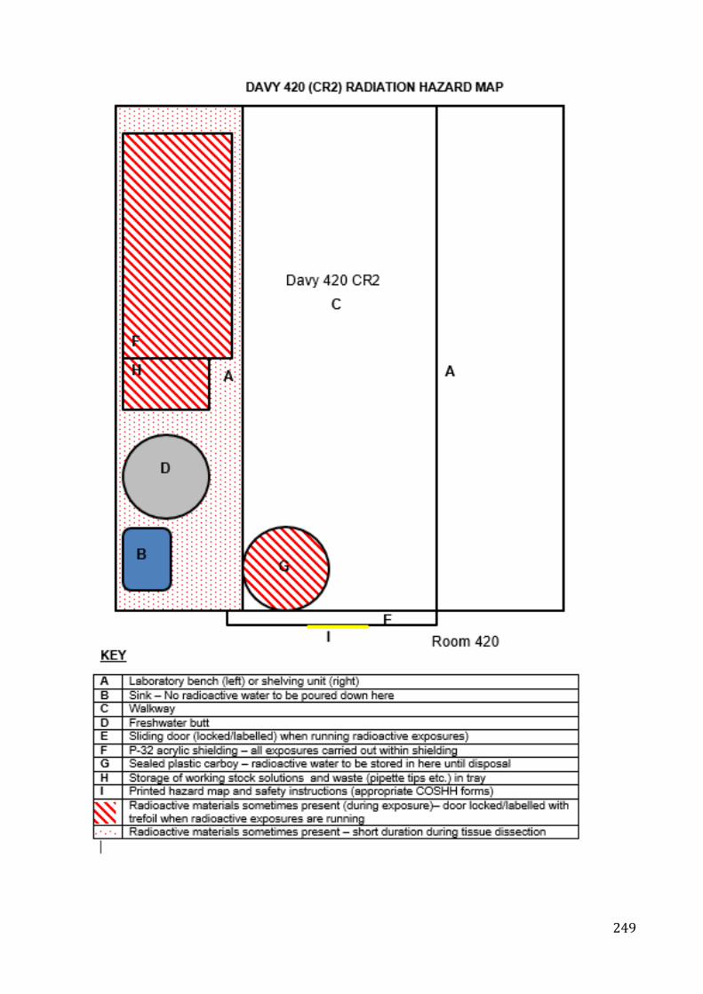
249
250
References
Al-Subiai SN, Arlt VM, Frickers PE, Readman JW, Stolpe B, Lead JR, Moody AJ,
Jha AN (2012) Merging nano-genotoxicology with eco-genotoxicology: An
integrated approach to determine interactive genotoxic and sub-lethal
toxic effects of C60 fullerenes and fluoranthene in marine mussels, Mytilus
sp. Mutation Research/Genetic Toxicology and Environmental
Mutagenesis 745: 92-103.
doi https://doi.org/10.1016/j.mrgentox.2011.12.019
Al-Subiai SN, Moody AJ, Mustafa SA, Jha AN (2011) A multiple biomarker
approach to investigate the effects of copper on the marine bivalve mollusc,
Mytilus edulis. Ecotoxicology and environmental safety 74: 1913-1920.
doi http://dx.doi.org/10.1016/j.ecoenv.2011.07.012
AlAmri OD, Cundy AB, Di Y, Jha AN, Rotchell JM (2012) Ionizing radiation-
induced DNA damage response identified in marine mussels, Mytilus sp.
Environmental Pollution 168: 107-112.
doi http://dx.doi.org/10.1016/j.envpol.2012.04.015
Alcaraz C, Caiola N, Ibanez C (2011) Bioaccumulation of pollutants in the zebra
mussel from hazardous industrial waste and evaluation of spatial
distribution using GAMs. The Science of the total environment 409: 898-
904. doi 10.1016/j.scitotenv.2010.11.015
Allen J, Moore M (2004) Environmental prognostics: Is the current use of
biomarkers appropriate for environmental risk evaluation? Marine
Environmental Research 58: 227-232.
doi https://doi.org/10.1016/j.marenvres.2004.03.119
251
Allen JI, McVeigh A (2004) Towards computational models of cells for
environmental toxicology. Journal of molecular history 35(7). 697-706.
doi: https://doi.org/10.1007/s10735-004-2674-8
Alonzo F, Gilbin R, Zeman FA, Garnier-Laplace J (2008) Increased effects of
internal alpha irradiation in Daphnia magna after chronic exposure over
three successive generations. Aquat Toxicol 87: 146-156.
doi 10.1016/j.aquatox.2008.01.015
Anbumani S, Mohankumar MN (2012) Gamma radiation induced micronuclei and
erythrocyte cellular abnormalities in the fish Catla catla. Aquat Toxicol 122-
123: 125-132. doi 10.1016/j.aquatox.2012.06.001
Anbumani S, Mohankumar MN (2016) Gene expression in Catla catla (Hamilton)
subjected to acute and protracted doses of gamma radiation. Aquatic
Toxicology 178: 153-157.
doi http://doi.org/10.1016/j.aquatox.2016.07.018
Andersson P, Beaugelin-Seiller K, Beresford N, Copplestone D, Della Vedova C,
Garnier-Laplace J, Howard B, Howe P, Oughton D, Wells Whitehouse CP
(2008) Numerical benchmarks for protecting biota from radiation in the
environment: Proposed levels, underlying reasoning and recommendation.
Andersson P, Garnier-Laplace J, Beresford NA, Copplestone D, Howard BJ,
Howe P, Oughton D, Whitehouse P (2009) Protection of the environment
from ionising radiation in a regulatory context (protect): proposed
numerical benchmark values. J Environ Radioact 100: 1100-1108.
doi 10.1016/j.jenvrad.2009.05.010
Angarano MB, McMahon RF, Schetz JA (2009) Cannabinoids inhibit zebra
mussel (Dreissena polymorpha) byssal attachment: a potentially green
antifouling technology. Biofouling 25: 127-138.
252
doi 10.1080/08927010802592743
Anjos VA, da Silva-Júnior FMR, Souza MM (2014) Cell damage induced by
copper: An explant model to study anemone cells. Toxicology in Vitro 28:
365-372. doi https://doi.org/10.1016/j.tiv.2013.11.013
Aoun M, El Samad O, Bou Khozam R, Lobinski R (2015) Assessment of
committed effective dose due to the ingestion of 210Po and 210Pb in
consumed Lebanese fish affected by a phosphate fertilizer plant. Journal
of Environmental Radioactivity 140: 25-29.
doi https://doi.org/10.1016/j.jenvrad.2014.10.014
Araldi RP, de Melo TC, Mendes TB, de Sá Júnior PL, Nozima BHN, Ito ET, de
Carvalho RF, de Souza EB, de Cassia Stocco R (2015) Using the comet
and micronucleus assays for genotoxicity studies: A review. Biomedicine
& Pharmacotherapy 72: 74-82.
doi https://doi.org/10.1016/j.biopha.2015.04.004
Arcanjo C, Armant O, Floriani M, Cavalie I, Camilleri V, Simon O, Orjollet D,
Adam-Guillermin C, Gagnaire B (2018) Tritiated water exposure disrupts
myofibril structure and induces mis-regulation of eye opacity and DNA
repair genes in zebrafish early life stages. Aquatic Toxicology 200: 114-
126. doi https://doi.org/10.1016/j.aquatox.2018.04.012
ATSDR (2004) Toxicological profile for strontium. Agency for Toxic Substances
and Disease Registry. Agency for Toxic Substances and Disease Registry.
CAS# 7440-24-6.
Attrill MJ, Depledge MH (1997) Community and population indicators of
ecosystem health: targeting links between levels of biological organisation.
Aquatic Toxicology 38: 183-197.
doi http://dx.doi.org/10.1016/S0166-445X(96)00839-9
253
Aypar U, Morgan WF, Baulch JE (2011a) Radiation-induced epigenetic
alterations after low and high LET irradiations. Mutation
Research/Fundamental and Molecular Mechanisms of Mutagenesis 707:
24-33. doi https://doi.org/10.1016/j.mrfmmm.2010.12.003
Aypar U, Morgan WF, Baulch JE (2011b) Radiation-induced genomic instability:
are epigenetic mechanisms the missing link? Int J Radiat Biol.;87(2). 179-
91. doi: 10.3109/09553002.2010.522686.
Azqueta A, Lorenzo Y, Collins AR (2009) In vitro comet assay for DNA repair: a
warning concerning application to cultured cells. Mutagenesis. 24(4). 379
- 81. doi: 10.1093/mutage/gep009
Bacchetta R, Mantecca P (2009) DDT polluted meltwater affects reproduction in
the mussel Dreissena polymorpha. Chemosphere 76: 1380-1385. doi
https://doi.org/10.1016/j.chemosphere.2009.06.020
Baldwin BS, Mayer MS, Dayton J, Pau N, Mendilla J, Sullivan M, Moore A, Ma A,
Mills EL (2002) Comparative growth and feeding in zebra and quagga
mussels (Dreissena polymorpha and Dreissena bugensis): implications for
North American lakes. Canadian Journal of Fisheries and Aquatic
Sciences 59: 680-694.
doi 10.1139/f02-043
Banni M, Sforzini S, Arlt VM, Barranger A, Dallas LJ, Oliveri C, Aminot Y,
Pacchioni B, Millino C, Lanfranchi G, Readman JW, Moore MN, Viarengo
A, Jha AN (2017) Assessing the impact of Benzo[a]pyrene on Marine
Mussels: Application of a novel targeted low density microarray
complementing classical biomarker responses. PLOS ONE 12: e0178460.
doi 10.1371/journal.pone.0178460
254
Barillet S, Adam-Guillermin C, Palluel O, Porcher J-M, Devaux A (2011) Uranium
bioaccumulation and biological disorders induced in zebrafish (Danio rerio)
after a depleted uranium waterborne exposure. Environmental Pollution
159: 495-502.
doi http://dx.doi.org/10.1016/j.envpol.2010.10.013
Barka S, Ouanes Z, Gharbi A, Gdara I, Mouelhi S, Hamza-Chaffai A (2016)
Monitoring genotoxicity in freshwater microcrustaceans: A new application
of the micronucleus assay. Mutation Research/Genetic Toxicology and
Environmental Mutagenesis 803–804: 27-33.
doi http://dx.doi.org/10.1016/j.mrgentox.2016.05.002
Bayne BL (1976) Marine mussels: their ecology and physiology. Cambridge
University Press
Beresford NA, Brown J, Copplestone D, Garnier-Laplace J, Howard BJ, Larsson
CM, Oughton D, Prohl G, Zinger I (2007) An integrated approach to the
assessmentand management of environmental risks from ionising
radiation.
Berghella AM, Contasta I, Marulli G, D'Innocenzo C, Garofalo F, Gizzi F,
Bartolomucci M, Laglia G, Valeri M, Gizzi M, Friscioni M, Barone M, Del
Beato T, Secinaro E, Pellegrini P (2014) Ageing gender-specific
"Biomarkers of Homeostasis", to protect ourselves against the diseases of
the old age.Immunity and aging. 6;11(1):3.
doi. 10.1186/1742-4933-11-3.
Bervoets L, Voets J, Covaci A, Chu S, Qadah D, Smolders R, Schepens P, Blust
R (2005) Use of Transplanted Zebra Mussels (Dreissena polymorpha) To
Assess the Bioavailability of Microcontaminants in Flemish Surface
Waters. Environmental science & technology 39: 1492-1505.
255
doi 10.1021/es049048t
Beyer J, Green NW, Brooks S, Allan IJ, Ruus A, Gomes T, Bråte ILN, Schøyen
M (2017) Blue mussels (Mytilus edulis spp.) as sentinel organisms in
coastal pollution monitoring: A review. Marine Environmental Research
130: 338-365. doi. https://doi.org/10.1016/j.marenvres.2017.07.024
Bhandari RK, vom Saal FS, Tillitt DE (2015) Transgenerational effects from early
developmental exposures to bisphenol A or 17α-ethinylestradiol in
medaka, Oryzias latipes. Scientific Reports 5: 9303.
doi. 10.1038/srep09303
https://www.nature.com/articles/srep09303#supplementary-information
Binelli A, Della Torre C, Magni S, Parolini M (2015) Does zebra mussel
(Dreissena polymorpha) represent the freshwater counterpart of Mytilus in
ecotoxicological studies? A critical review. Environmental Pollution 196:
386-403.
doi. http://dx.doi.org/10.1016/j.envpol.2014.10.023
Blaylock BG (1971) The production of chromosome aberration in Chironomus
riparius (diptera: chironomidae) by tritiated water. The Canadian
Entomologist 103: 448-453.
doi: 10.4039/Ent103448-3
Bolognesi C, Buschini A, Branchi E, Carboni P, Furlini M, Martino A, Monteverde
M, Poli P, Rossi C (2004) Comet and micronucleus assays in zebra mussel
cells for genotoxicity assessment of surface drinking water treated with
three different disinfectants. Science of The Total Environment 333: 127-
136. doi: https://doi.org/10.1016/j.scitotenv.2004.05.018
256
Bolognesi C, Fenech M (2012) Mussel micronucleus cytome assay. Nat
Protocols 7: 1125-1137
Bolognesi C, Landini E, Roggieri P, Fabbri R, Viarengo A (1999) Genotoxicity
biomarkers in the assessment of heavy metal effects in mussels:
experimental studies. Environmental and molecular mutagenesis 33: 287-
292
Bopp SK, Abicht HK, Knauer K (2008) Copper-induced oxidative stress in
rainbow trout gill cells. Aquatic Toxicology 86: 197-204.
doi http://dx.doi.org/10.1016/j.aquatox.2007.10.014
Borthagaray AI, Carranza A (2007) Mussels as ecosystem engineers: Their
contribution to species richness in a rocky littoral community. Acta
Oecologica 31: 243-250.
doi http://dx.doi.org/10.1016/j.actao.2006.10.008
Bouskill NJ, Handy RD, Ford TE, Galloway TS (2006) Differentiating copper and
arsenic toxicity using biochemical biomarkers in Asellus aquaticus and
Dreissena polymorpha. Ecotoxicology and environmental safety 65: 342-
349. doi http://dx.doi.org/10.1016/j.ecoenv.2005.07.027
Brechignac F, Bradshaw C, Carroll S, Fuma S, Hakonson L, Jaworska A,
Kapustka L, Kawaguchi I, Monte L, Oughton D, Sazykina T, Strand P
(2012) Towards an ecosystem approach for environmental protection with
emphasis on radiological hazards. IUR.
Brechignac F, Doi M (2009) Challenging the current strategy of radiological
protection of the environment: arguments for an ecosystem approach. J
Environ Radioact 100: 1125-1134. doi 10.1016/j.jenvrad.2009.06.022
Brechignac, B and Howard BJ (2001) Radioactive pollutants: Impact on the
environment.IRSN. 341.
257
Bréchignac F (2003) Protection of the environment: how to position
radioprotection in an ecological risk assessment perspective. Science of
The Total Environment 307: 35-54.
doi http://dx.doi.org/10.1016/S0048-9697(02)00545-4
Bréchignac F, Oughton D, Mays C, Barnthouse L, Beasley JC, Bonisoli-Alquati
A, Bradshaw C, Brown J, Dray S, Geras'kin S, Glenn T, Higley K, Ishida
K, Kapustka L, Kautsky U, Kuhne W, Lynch M, Mappes T, Mihok S, Møller
AP, Mothersill C, Mousseau TA, Otaki JM, Pryakhin E, Rhodes OE, Salbu
B, Strand P, Tsukada H (2016) Addressing ecological effects of radiation
on populations and ecosystems to improve protection of the environment
against radiation: Agreed statements from a Consensus Symposium.
Journal of Environmental Radioactivity 158-159: 21-29.
doi https://doi.org/10.1016/j.jenvrad.2016.03.021
Broerse JJ, Barendsen GW (1986) The Physics of Absorbed Dose and Linear
Energy Transfer. In: Burns FJ, Upton AC, Silini G (eds) Radiation
Carcinogenesis and DNA Alterations. Springer US, Boston, MA, pp 593-
609
Brooks SJ, Farmen E, Heier LS, Blanco-Rayón E, Izagirre U (2015) Differences
in copper bioaccumulation and biological responses in three Mytilus
species. Aquatic Toxicology 160: 1-12.
doi https://doi.org/10.1016/j.aquatox.2014.12.018
Brown JE, Alfonso B, Avila R, Beresford NA, Copplestone D, Hosseini A (2016)
A new version of the ERICA tool to facilitate impact assessments of
radioactivity on wild plants and animals. Journal of Environmental
Radioactivity 153: 141-148.
doi http://dx.doi.org/10.1016/j.jenvrad.2015.12.011
258
Brown JE, Alfonso B, Avila R, Beresford NA, Copplestone D, Pröhl G, Ulanovsky
A (2008) The ERICA Tool. Journal of Environmental Radioactivity 99:
1371-1383.
doi http://dx.doi.org/10.1016/j.jenvrad.2008.01.008
Brown RJ, Galloway TS, Lowe D, Browne MA, Dissanayake A, Jones MB,
Depledge MH (2004) Differential sensitivity of three marine invertebrates
to copper assessed using multiple biomarkers. Aquatic Toxicology 66:
267-278.
doi http://dx.doi.org/10.1016/j.aquatox.2003.10.001
Bryan GW, Gibbs PE (1983) Heavy Metals in the Fal Estuary, Cornwall: A Study
of Long-term Contamination by Mining Waste and Its Effects on Estuarine
Organisms. Marine Biological Association of the United Kingdom
Burdak-Rothkamm S, Rothkamm K (2018) Radiation-induced bystander and
systemic effects serve as a unifying model system for genotoxic stress
responses. Mutation Research/Reviews in Mutation Research 778: 13-22.
doi https://doi.org/10.1016/j.mrrev.2018.08.001
Campos A, Tedesco S, Vasconcelos V, Cristobal S (2012) Proteomic research
in bivalves: Towards the identification of molecular markers of aquatic
pollution. Journal of Proteomics 75: 4346-4359.
doi http://dx.doi.org/10.1016/j.jprot.2012.04.027
Canesi L, Frenzilli G, Balbi T, Bernardeschi M, Ciacci C, Corsolini S, Della Torre
C, Fabbri R, Faleri C, Focardi S, Guidi P, Kočan A, Marcomini A, Mariottini
M, Nigro M, Pozo-Gallardo K, Rocco L, Scarcelli V, Smerilli A, Corsi I
(2014) Interactive effects of n-TiO2 and 2,3,7,8-TCDD on the marine
bivalve Mytilus galloprovincialis. Aquatic Toxicology 153: 53-65.
doi http://dx.doi.org/10.1016/j.aquatox.2013.11.002
259
Canty MN, Hutchinson TH, Brown RJ, Jones MB, Jha AN (2009) Linking
genotoxic responses with cytotoxic and behavioural or physiological
consequences: Differential sensitivity of echinoderms (Asterias rubens)
and marine molluscs (Mytilus edulis). Aquatic Toxicology 94: 68-76.
doi http://dx.doi.org/10.1016/j.aquatox.2009.06.001
Cappello T, Maisano M, Giannetto A, Parrino V, Mauceri A, Fasulo S (2015)
Neurotoxicological effects on marine mussel Mytilus galloprovincialis
caged at petrochemical contaminated areas (eastern Sicily, Italy): 1H
NMR and immunohistochemical assays. Comparative Biochemistry and
Physiology Part C: Toxicology & Pharmacology 169: 7-15.
doi http://dx.doi.org/10.1016/j.cbpc.2014.12.006
Cappello T, Mauceri A, Corsaro C, Maisano M, Parrino V, Lo Paro G, Messina G,
Fasulo S (2013) Impact of environmental pollution on caged mussels
Mytilus galloprovincialis using NMR-based metabolomics. Marine pollution
bulletin 77: 132-139.
doi http://dx.doi.org/10.1016/j.marpolbul.2013.10.019
Carriquiriborde P, Marino DJ, Giachero G, Castro EA, Ronco AE (2012) Global
metabolic response in the bile of pejerrey (Odontesthes bonariensis,
Pisces) sublethally exposed to the pyrethroid cypermethrin. Ecotoxicology
and environmental safety 76: 46-54.
doi http://dx.doi.org/10.1016/j.ecoenv.2011.07.039
Cartier S, Pellerin J, Fournier M, Tamigneaux E, Girault L, Lemaire N (2004) Use
of an index based on the blue mussel (Mytilus edulis and Mytilus trossulus)
digestive gland weight to assess the nutritional quality of mussel farm sites.
Aquaculture 241: 633-654.
doi http://dx.doi.org/10.1016/j.aquaculture.2004.08.015
260
Carvalho FP (2018) Radionuclide concentration processes in marine organisms:
A comprehensive review. Journal of Environmental Radioactivity 186: 124-
130. doi https://doi.org/10.1016/j.jenvrad.2017.11.002
Carvalho FP, Oliveira JM, Malta M (2011) Radionuclides in deep-sea fish and
other organisms from the North Atlantic Ocean. ICES Journal of Marine
Science: Journal du Conseil 68: 333-340. doi 10.1093/icesjms/fsq088
Cavas T (2011) In vivo genotoxicity evaluation of atrazine and atrazine–based
herbicide on fish Carassius auratus using the micronucleus test and the
comet assay. Food and Chemical Toxicology 49: 1431-1435.
doi http://dx.doi.org/10.1016/j.fct.2011.03.038
Chandurvelan R, Marsden ID, Gaw S, Glover CN (2013) Waterborne cadmium
impacts immunocytotoxic and cytogenotoxic endpoints in green-lipped
mussel, Perna canaliculus. Aquatic Toxicology 142–143: 283-293. doi
http://dx.doi.org/10.1016/j.aquatox.2013.09.002
Chapman PM (2002) Integrating toxicology and ecology: putting the “eco” into
ecotoxicology. Marine Pollution Bulletin 44: 7-15.
doi https://doi.org/10.1016/S0025-326X(01)00253-3
Châtel A, Faucet-Marquis V, Gourlay-Francé C, Pfohl-Leszkowicz A, Vincent-
Hubert F (2015) Genotoxicity and activation of cellular defenses in
transplanted zebra mussels Dreissena polymorpha along the Seine river.
Ecotoxicology and environmental safety 114: 241-249.
doi http://dx.doi.org/10.1016/j.ecoenv.2014.03.023
Chatel A, Faucet-Marquis V, Perret M, Gourlay-France C, Uher E, Pfohl-
Leszkowicz A, Vincent-Hubert F (2012) Genotoxicity assessment and
detoxification induction in Dreissena polymorpha exposed to
benzo[a]pyrene. Mutagenesis 27: 703-711. doi 10.1093/mutage/ges036
261
Chatfield C, Collins A (1980) Introduction to Multivariate Analysis. Springer US
Cheng Y, Kiess AP, Herman JM, Pomper MG, Meltzer SJ, Abraham JM (2015)
Phosphorus-32, a Clinically Available Drug, Inhibits Cancer Growth by
Inducing DNA Double-Strand Breakage. PLOS ONE 10: e0128152. doi
10.1371/journal.pone.0128152
Chevalier F, Hamdi DH, Saintigny Y, Lefaix JL (2015) Proteomic overview and
perspectives of the radiation-induced bystander effects. Mutation
Research/Reviews in Mutation Research 763: 280-293.
doi https://doi.org/10.1016/j.mrrev.2014.11.008
Chino M, Nakayama H, Nagai H, Terada H, Katata G, Yamazawa H (2011)
Preliminary Estimation of Release Amounts of 131I and 137Cs Accidentally
Discharged from the Fukushima Daiichi Nuclear Power Plant into the
Atmosphere. Journal of Nuclear Science and Technology 48: 1129-1134.
doi 10.1080/18811248.2011.9711799
Choi JE, Kim S, Ahn JH, Youn P, Kang JS, Park K, Yi J, Ryu DY (2010) Induction
of oxidative stress and apoptosis by silver nanoparticles in the liver of adult
zebrafish. Aquatic Toxicology 100: 151-159.
doi https://doi.org/10.1016/j.aquatox.2009.12.012
Choi VW, Ng CY, Cheng SH, Yu KN (2012) alpha-Particle irradiated zebrafish
embryos rescued by bystander unirradiated zebrafish embryos. Environ
Sci Technol 46: 226-231. doi 10.1021/es2016928
Cid A, Herrero C, Torres E, Abalde J (1995) Copper toxicity on the marine
microalga Phaeodactylum tricornutum: effects on photosynthesis and
related parameters. Aquatic Toxicology 31: 165-174.
doi http://dx.doi.org/10.1016/0166-445X(94)00071-W
262
Clarke KR (1999) Non-metric multivariate analysis in community-level
ecotoxicology. Environ. Toxicol. Chem. 18, 117-127.
Clarke KR and Warwick RM (2001) Change in marine communities: an approach
to statistical analysis and interpretation. PRIMER-є, Plymouth, UK.
Clayton ME, Steinmann R, Fent K (2000) Different expression patterns of heat
shock proteins hsp 60 and hsp 70 in zebra mussels (Dreissena
polymorpha) exposed to copper and tributyltin. Aquatic Toxicology 47:
213-226. doi http://dx.doi.org/10.1016/S0166-445X(99)00022-3
Clements WH (2000) Integrating effects of contaminants across levels of
biological organization: an overview. Journal of Aquatic Ecosystem Stress
and Recovery 7: 113-116. doi 10.1023/a:1009927612391
Clifton RJ, Stevens HE, Hamilton EI (1989) Uptake and depuration of 241Am,
239+240Pu, 238Pu, 137Cs and 106Ru by Mytilus edulis under natural stress.
Marine Ecology Progress Series 54: 91-98
Coghlan B, Gosling E (2007) Genetic structure of hybrid mussel populations in
the west of Ireland: two hypotheses revisited. Marine Biology 150: 841-
852 doi 10.1007/s00227-006-0408-z
Collins AR (2004) The comet assay for DNA damage and repair: principles,
applications, and limitations. Mol Biotechnol 26: 249-261.
doi 10.1385/mb:26:3:249
Collins AR, Oscoz AA, Brunborg G, Gaivao I, Giovannelli L, Kruszewski M, Smith
CC, Stetina R (2008) The comet assay: topical issues. Mutagenesis 23:
143-151. doi 10.1093/mutage/gem051
Connan O, Germain P, Solier L, Gouret G (2007) Variations of 210Po and 210Pb in
various marine organisms from Western English Channel: contribution of
263
210Po to the radiation dose. Journal of Environmental Radioactivity 97:
168-188. doi http://dx.doi.org/10.1016/j.jenvrad.2007.04.004
Connon RE, Geist J, Werner I (2012) Effect-Based Tools for Monitoring and
Predicting the Ecotoxicological Effects of Chemicals in the Aquatic
Environment. Sensors (Basel, Switzerland) 12: 12741-12771.
doi 10.3390/s120912741
Cope WG, Bartsch MR, Marking LL (1997) Efficacy of candidate chemicals for
preventing attachment of zebra mussels (Dreissena polymorpha).
Environmental Toxicology and Chemistry 16: 1930-1934.
doi 10.1002/etc.5620160923
Copplestone D, Hingston J, Real A (2008) The development and purpose of the
FREDERICA radiation effects database. J Environ Radioact 99: 1456-
1463. doi 10.1016/j.jenvrad.2008.01.006
Copplestone D, Howard BJ, Brechignac F (2004) The ecological relevance of
current approaches for environmental protection from exposure to ionising
radiation. J Environ Radioact 74: 31-41.
doi 10.1016/j.jenvrad.2004.01.020
Crooks JA (2002) Characterizing ecosystem-level consequences of biological
invasions: the role of ecosystem engineers. Oikos 97: 153-166. doi
10.1034/j.1600-0706.2002.970201.x
Cubero-Leon E, Minier C, Rotchell JM, Hill EM (2012) Metabolomic analysis of
sex specific metabolites in gonads of the mussel, Mytilus edulis.
Comparative Biochemistry and Physiology Part D: Genomics and
Proteomics 7: 212-219. doi http://dx.doi.org/10.1016/j.cbd.2012.03.002
Curtis IP (1971) Techniques for Counting Carbon-14 and Phosphorus-32
Labelled Samples of Polluted Natural Waters. Water Pollution Research
264
Laboratory of the Department of the Environment, Stevenage, Hertfordsh
ire, England
D'Agata A, Fasulo S, Dallas LJ, Fisher AS, Maisano M, Readman JW, Jha AN
(2014) Enhanced toxicity of ‘bulk' titanium dioxide compared to ‘fresh' and
‘aged' nano-TiO2 in marine mussels (Mytilus galloprovincialis).
Nanotoxicology 8: 549-558.
doi 10.3109/17435390.2013.807446
Dallas LJ, Keith-Roach M, Lyons BP, Jha AN (2012) Assessing the impact of
ionizing radiation on aquatic invertebrates: a critical review. Radiat Res
177: 693-716
Dallas LJ, Bean TP, Turner A, Lyons BP, Jha AN (2013) Oxidative DNA damage
may not mediate Ni-induced genotoxicity in marine mussels: Assessment
of genotoxic biomarkers and transcriptional responses of key stress genes.
Mutation Research/Genetic Toxicology and Environmental Mutagenesis
754: 22-31. doi http://dx.doi.org/10.1016/j.mrgentox.2013.03.009
Dallas LJ, Bean TP, Turner A, Lyons BP, Jha AN (2016a) Exposure to tritiated
water at an elevated temperature: Genotoxic and transcriptomic effects in
marine mussels (M. galloprovincialis). Journal of Environmental
Radioactivity 164: 325-336.
doi https://doi.org/10.1016/j.jenvrad.2016.07.034
Dallas LJ, Devos A, Fievet B, Turner A, Lyons BP, Jha AN (2016b) Radiation
dose estimation for marine mussels following exposure to tritium: Best
practice for use of the ERICA tool in ecotoxicological studies. Journal of
Environmental Radioactivity 155–156: 1-6.
doi http://dx.doi.org/10.1016/j.jenvrad.2016.01.019
265
Dallas LJ, Turner A, Bean TP, Lyons BP, Jha AN (2018) An integrated approach
to assess the impacts of zinc pyrithione at different levels of biological
organization in marine mussels. Chemosphere 196: 531-539.
doi https://doi.org/10.1016/j.chemosphere.2017.12.144
Danzker M, Kessaris ND, Laughlin JS (1959) Absorbed Dose and Linear Energy
Transfer in Radiation Experiments. Radiology 72: 51-61.
doi 10.1148/72.1.51
David JAO, Fontanetti CS (2005) Surface morphology of Mytella falcata gill
filaments from three regions of the Santos estuary. Braz J Morphol Sci 22:
203-210
Davis EE, Frangakis S, Katsanis N (2014) Interpreting human genetic variation
with in vivo zebrafish assays. Biochimica et Biophysica Acta (BBA) -
Molecular Basis of Disease 1842: 1960-1970.
doi http://dx.doi.org/10.1016/j.bbadis.2014.05.024
de la Sienra E, Armienta MA, Gonsebatt ME (2003) Potassium dichromate
increases the micronucleus frequency in the crayfish Procambarus clarkii.
Environmental Pollution 126: 367-370.
doi http://dx.doi.org/10.1016/S0269-7491(03)00249-5
DEFRA (2014) Water Framework Directive implementation in England and Wales:
new and updated standards to protect the water environment. In:
Environment FaRA (ed)
Delaporte M, Soudant P, Moal J, Lambert C, Quéré C, Miner P, Choquet G,
Paillard C, Samain JF (2003) Effect of a mono-specific algal diet on
immune functions in two bivalve species - Crassostrea gigas and
Ruditapes philippinarum. Journal of Experimental Biology 206: 3053-3064.
doi 10.1242/jeb.00518
266
Depledge MH, Amaral-Mendes JJ, Daniel B, Halbrook RS, Kloepper-Sams P,
Moore MN, Peakall DB (1993) The Conceptual Basis of the Biomarker
Approach. In: Peakall DB, Shugart LR (eds) Biomarkers. Springer Berlin
Heidelberg, Berlin, Heidelberg, pp 15-29
Devos A, Dallas LJ, Voiseux C, Lecomte-Pradines C, Jha AN, Fiévet B (2015)
Assessment of growth, genotoxic responses and expression of stress
related genes in the Pacific oyster Crassostrea gigas following chronic
exposure to ionizing radiation. Marine Pollution Bulletin 95: 688-698. doi
http://dx.doi.org/10.1016/j.marpolbul.2015.03.039
Di Salvatore P, Calcagno JA, Ortíz N, Ríos de Molina MdC, Sabatini SE (2013)
Effect of seasonality on oxidative stress responses and metal
accumulation in soft tissues of Aulacomya atra, a mussel from the South
Atlantic Patagonian coast. Marine Environmental Research 92: 244-252.
doi https://doi.org/10.1016/j.marenvres.2013.10.004
Di Y, Aminot Y, Schroeder DC, Readman JW, Jha AN (2017) Integrated biological
responses and tissue-specific expression of p53 and ras genes in marine
mussels following exposure to benzo(alpha)pyrene and C60 fullerenes,
either alone or in combination. Mutagenesis 32: 77-90.
doi 10.1093/mutage/gew049
Di Y, Schroeder DC, Highfield A, Readman JW, Jha AN (2011) Tissue-specific
expression of p53 and ras genes in response to the environmental
genotoxicant benzo(alpha)pyrene in marine mussels. Environmental
science & technology 45: 8974-8981. doi 10.1021/es201547x
Dias PJ, Sollelis L, Cook EJ, Piertney SB, Davies IM, Snow M (2008)
Development of a real-time PCR assay for detection of Mytilus species
specific alleles: Application to a sampling survey in Scotland. Journal of
267
Experimental Marine Biology and Ecology 367: 253-258. doi
http://dx.doi.org/10.1016/j.jembe.2008.10.011
Digilio G, Sforzini S, Cassino C, Robotti E, Oliveri C, Marengo E, Musso D, Osella
D, Viarengo A (2016) Haemolymph from Mytilus galloprovincialis:
Response to copper and temperature challenges studied by 1H-NMR
metabonomics. Comparative Biochemistry and Physiology Part C:
Toxicology & Pharmacology.
doi http://dx.doi.org/10.1016/j.cbpc.2016.02.003
Dimitriadis VK, Domouhtsidou GP, Cajaraville MP (2004) Cytochemical and
Histochemical Aspects of the Digestive Gland Cells of the Mussel Mytilus
galloprovincialis (L.) in Relation to Function. Journal of Molecular Histology
35: 501-509. doi 10.1023/B:HIJO.0000045952.87268.76
Dondero F, Negri A, Boatti L, Marsano F, Mignone F, Viarengo A (2010)
Transcriptomic and proteomic effects of a neonicotinoid insecticide
mixture in the marine mussel (Mytilus galloprovincialis, Lam.). Science of
The Total Environment 408: 3775-3786.
doi http://dx.doi.org/10.1016/j.scitotenv.2010.03.040
Donnachie RL, Johnson AC, Sumpter JP (2016) A rational approach to selecting
and ranking some pharmaceuticals of concern for the aquatic environment
and their relative importance compared with other chemicals.
Environmental Toxicology and Chemistry 35: 1021-1027.
doi doi:10.1002/etc.3165
Dubrova YE, Plumb M, Gutierrez B, Boulton E, Jeffreys AJ (2000)
Transgenerational mutation by radiation. Nature 405: 37.
doi 10.1038/35011135
268
DWQR (2014) Monitoring of radioactivity in Scottish drinking water. In: Millican D
(ed)
D’costa AH, Kumar MK, Furtado S (2018) The Backwater Clam (Meretrix casta)
as a bioindicator species for monitoring the pollution of an estuarine
environment by genotoxic agents. Mutation Research/Genetic Toxicology
and Environmental Mutagenesis 825: 8-14.
doi https://doi.org/10.1016/j.mrgentox.2017.11.001
EA (2001) Impact Assessment of Ionising Radiation on Wildlife. Environment
agency,https://www.gov.uk/government/publications/impact-assessment-
of-ionising-radiation-on-wildlife
Edebo L, Haamer J, Lindahl O, Loo L-O, Piriz L (2000) Recycling of
macronutrients from sea to land using mussel cultivation. International
Journal of Environment and Pollution 13: 190-207.
doi doi:10.1504/IJEP.2000.002315
Eisler R (1994) Radiation Hazards to Fish, Wildlife, and Invertebrates: A Synoptic
Review, Report 29; Biological Report 26., Laurel, MD
Eisler R (1997) Copper hazards to fish, wildlife, and invertebrates: A synoptic
review. Biological Science Report: 98
Erickson RJ, Benoit DA, Mattson VR, Leonard EN, Nelson HP (1996) The effects
of water chemistry on the toxicity of copper to fathead minnows.
Environmental Toxicology and Chemistry 15: 181-193.
doi 10.1002/etc.5620150217
Ericson H, Thorsen G, Kumblad L (2010) Physiological effects of diclofenac,
ibuprofen and propranolol on Baltic Sea blue mussels. Aquat Toxicol 99:
223-231. doi 10.1016/j.aquatox.2010.04.017
269
Evans S (1984) Uptake and loss of 134Cs and 60Co by the Baltic bivalve Macoma
baltica in a laboratory microcosmos. Journal of Environmental
Radioactivity 1: 133-150.
doi http://dx.doi.org/10.1016/0265-931X(84)90004-3
Faggio C, Tsarpali V, Dailianis S (2018) Mussel digestive gland as a model tissue
for assessing xenobiotics: An overview. Science of The Total Environment
636: 220-229. doi https://doi.org/10.1016/j.scitotenv.2018.04.264
Farcy E, Voiseux C, Lebel J-M, Fievet B (2007) Seasonal changes in mRNA
encoding for cell stress markers in the oyster Crassostrea gigas exposed
to radioactive discharges in their natural environment. Science of The
Total Environment 374: 328-341.
doi http://dx.doi.org/10.1016/j.scitotenv.2006.11.014
Farcy E, Voiseux C, Robbes I, Lebel JM, Fievet B (2011) Effect of ionizing
radiation on the transcription levels of cell stress marker genes in the
Pacific oyster Crassostrea gigas. Radiat Res 176: 38-48
Fasulo S, Iacono F, Cappello T, Corsaro C, Maisano M, D'Agata A, Giannetto A,
De Domenico E, Parrino V, Lo Paro G, Mauceri A (2012) Metabolomic
investigation of Mytilus galloprovincialis (Lamarck 1819) caged in aquatic
environments. Ecotoxicology and environmental safety 84: 139-146. doi
http://dx.doi.org/10.1016/j.ecoenv.2012.07.001
Fenech M, Kirsch-Volders M, Natarajan AT, Surralles J, Crott JW, Parry J,
Norppa H, Eastmond DA, Tucker JD, Thomas P (2011) Molecular
mechanisms of micronucleus, nucleoplasmic bridge and nuclear bud
formation in mammalian and human cells. Mutagenesis. 26(1) 125-32. doi:
10.1093/mutage/geq052
270
Feroz Khan M, Godwin Wesley S (2012) Radionuclide monitoring in molluscs
inhabiting intertidal region near a nuclear installation, Gulf of Mannar, India.
Marine pollution bulletin 64: 436-444.
doi https://doi.org/10.1016/j.marpolbul.2011.11.010
Feroz Khan M, Godwin Wesley S, Rajan MP (2014) Polonium-210 in marine
mussels (bivalve molluscs) inhabiting the southern coast of India. Journal
of Environmental Radioactivity 138: 410-416.
doi http://dx.doi.org/10.1016/j.jenvrad.2014.06.023
Festarini A, Shultz C, Stuart M, Kim SB, Ferreri C (2015) Cellular responses to
tritium exposure in rainbow trout: hto- and obt-spiked feed exposure
experiments. CNL Nuclear Review 5: 155-172.
doi 10.12943/CNR.2015.00059
Fetisov AN, Rubanovich AV, Slipchenko TS, Shevchenko VA (1992) The
structure of Dreissena polymorpha populations from basins adjacent to the
Chernobyl atomic power station. Science of The Total Environment 112:
115-124. doi https://doi.org/10.1016/0048-9697(92)90242-K
FitzGerald JE, Grenon M, Lowndes NF (2009) 53BP1: function and mechanisms
of focal recruitment. Biochemical Society Transactions 37: 897-904. doi
10.1042/bst0370897
Fiévet B, Voiseux C, Rozet M, Masson M, Bailly du Bois P (2006) Transfer of
radiocarbon liquid releases from the AREVA La Hague spent fuel
reprocessing plant in the English Channel. Journal of Environmental
Radioactivity 90: 173-196.
doi https://doi.org/10.1016/j.jenvrad.2006.06.014
Florou H, Tsytsugina V, Polikarpov GG, Trabidou G, Gorbenko V, Chaloulou CH
(2004) Field observations of the effects of protracted low levels of ionizing
271
radiation on natural aquatic population by using a cytogenetic tool. Journal
of Environmental Radioactivity 75: 267-283.
doi http://dx.doi.org/10.1016/j.jenvrad.2004.01.003
Foster ER, Downs JA (2005) Histone H2A phosphorylation in DNA double-strand
break repair. FEBS Journal 272: 3231-3240.
doi 10.1111/j.1742-4658.2005.04741.x
Freeman JL, Weber GJ, Peterson SM, Nie LH (2014) Embryonic ionizing
radiation exposure results in expression alterations of genes associated
with cardiovascular and neurological development, function, and disease
and modified cardiovascular function in zebrafish. Front Genet 5: 268. doi
10.3389/fgene.2014.00268
Freitas ACS, Guimarães JRD, Gouvea VA, Penna Franca E (1988) Strontium-85
bioaccumulation by Sargassum spp. (brown seaweed) and Galaxaura
marginata (calcareous seaweed). Science of The Total Environment 75:
225-233. doi http://dx.doi.org/10.1016/0048-9697(88)90035-6
Frenzilli G, Nigro M, Lyons BP (2009) The Comet assay for the evaluation of
genotoxic impact in aquatic environments. Mutation Research/Reviews in
Mutation Research 681: 80-92.
doi https://doi.org/10.1016/j.mrrev.2008.03.001
Frumkin H (2001) Beyond toxicity: human health and the natural environment.
Am J Prev Med 20: 234-240
Gaetke LM, Chow CK (2003) Copper toxicity, oxidative stress, and antioxidant
nutrients. Toxicology 189: 147-163
Gagnaire B, Adam-Guillermin C, Festarini A, Cavalié I, Della-Vedova C, Shultz
C, Kim SB, Ikert H, Dubois C, Walsh S, Farrow F, Beaton D, Tan E, Wen
K, Stuart M (2017) Effects of in situ exposure to tritiated natural
272
environments: A multi-biomarker approach using the fathead minnow,
Pimephales promelas. Science of The Total Environment 599–600: 597-
611. doi https://doi.org/10.1016/j.scitotenv.2017.04.210
Gagnaire B, Cavalie I, Pereira S, Floriani M, Dubourg N, Camilleri V, Adam-
Guillermin C (2015) External gamma irradiation-induced effects in early-
life stages of zebrafish, Danio rerio. Aquat Toxicol 169: 69-78. doi
10.1016/j.aquatox.2015.10.005
García-Medina S, Razo-Estrada C, Galar-Martinez M, Cortéz-Barberena E,
Gómez-Oliván LM, Álvarez-González I, Madrigal-Bujaidar E (2011)
Genotoxic and cytotoxic effects induced by aluminum in the lymphocytes
of the common carp (Cyprinus carpio). Comparative Biochemistry and
Physiology Part C: Toxicology & Pharmacology 153: 113-118. doi
http://dx.doi.org/10.1016/j.cbpc.2010.09.005
Garnier-Laplace J, Copplestone D, Gilbin R, Alonzo F, Ciffroy P, Gilek M, Aguero
A, Bjork M, Oughton DH, Jaworska A, Larsson CM, Hingston JL (2008)
Issues and practices in the use of effects data from FREDERICA in the
ERICA Integrated Approach. J Environ Radioact 99: 1474-1483. doi
10.1016/j.jenvrad.2008.04.012
Garnier-Laplace J, Gilbin R (2006) Derivation of Predicted-No-Effect-Dose-Rate
values for ecosystems (and their sub-organisational levels) exposed to
radioactive substances. Swedish Radiation Protection Authority
Geret F, Serafim A, Barreira L, João Bebianno M (2002) Response of antioxidant
systems to copper in the gills of the clam Ruditapes decussatus. Marine
Environmental Research 54: 413-417.
doi http://dx.doi.org/10.1016/S0141-1136(02)00164-2
273
Gerić M, Gajski G, Garaj-Vrhovac V (2014) γ-H2AX as a biomarker for DNA
double-strand breaks in ecotoxicology. Ecotoxicology and Environmental
Safety 105: 13-21. doi http://dx.doi.org/10.1016/j.ecoenv.2014.03.035
Ghedotti MJ, Smihula JC, Smith GR (1995) Zebra Mussel Predation by Round
Gobies in the Laboratory. Journal of Great Lakes Research 21: 665-669.
doi http://dx.doi.org/10.1016/S0380-1330(95)71076-0
Girardello F, Custódio Leite C, Vianna Villela I, da Silva Machado M, Luiz Mendes
Juchem A, Roesch-Ely M, Neves Fernandes A, Salvador M, Antonio
Pêgas Henriques J (2016) Titanium dioxide nanoparticles induce
genotoxicity but not mutagenicity in golden mussel Limnoperna fortunei.
Aquatic Toxicology 170: 223-228.
doi http://dx.doi.org/10.1016/j.aquatox.2015.11.030
Godoy JM, de Oliveira MS, de Almeida CE, de Carvalho ZL, da Silva ER,
Fernandes Fda C, Pitanga FL, Danelon OM (2008) 210Po concentration in
Perna perna mussels: looking for radiation effects. J Environ Radioact 99:
631-640. doi 10.1016/j.jenvrad.2007.09.003
Gombeau K, Pereira S, Ravanat J-L, Camilleri V, Cavalie I, Bourdineaud J-P,
Adam-Guillermin C (2016) Depleted uranium induces sex- and tissue-
specific methylation patterns in adult zebrafish. Journal of Environmental
Radioactivity 154: 25-33.
doi https://doi.org/10.1016/j.jenvrad.2016.01.004
Gomes T, Chora S, Pereira CG, Cardoso C, Bebianno MJ (2014a) Proteomic
response of mussels Mytilus galloprovincialis exposed to CuO NPs and
Cu2+: An exploratory biomarker discovery. Aquatic Toxicology 155: 327-
336. doi http://dx.doi.org/10.1016/j.aquatox.2014.07.015
274
Gomes T, Pereira CG, Cardoso C, Sousa VS, Teixeira MR, Pinheiro JP,
Bebianno MJ (2014b) Effects of silver nanoparticles exposure in the
mussel Mytilus galloprovincialis. Marine Environmental Research 101:
208-214. doi http://dx.doi.org/10.1016/j.marenvres.2014.07.004
Gomes T, Song Y, Brede DA, Xie L, Gutzkow KB, Salbu B, Tollefsen KE (2018)
Gamma radiation induces dose-dependent oxidative stress and
transcriptional alterations in the freshwater crustacean Daphnia magna.
Science of the total environment. 628-629.
doi: 10.1016/j.scitotenv.2018.02.039
Gomez-Casati DF, Zanor MI, Busi M (2013) Metabolomics in Plants and Humans:
Applications in the Prevention and Diagnosis of Diseases. BioMed
Research International 2013: 11. doi 10.1155/2013/792527
Gonzalez-Rey M, Mattos JJ, Piazza CE, Bainy ACD, Bebianno MJ (2014) Effects
of active pharmaceutical ingredients mixtures in mussel Mytilus
galloprovincialis. Aquatic Toxicology 153: 12-26.
doi http://dx.doi.org/10.1016/j.aquatox.2014.02.006
González-Romero R, Rivera-Casas C, Frehlick LJ, Méndez J, Ausió J, Eirín-
López JM (2012) Histone H2A (H2A.X and H2A.Z) Variants in Molluscs:
Molecular Characterization and Potential Implications For Chromatin
Dynamics. PLoS ONE 7: e30006. doi 10.1371/journal.pone.0030006
Graupner A, Eide DM, Instanes C, Andersen JM, Brede DA, Dertinger SD, Lind
OC, Brandt-Kjelsen A, Bjerke H, Salbu B, Oughton D, Brunborg G, Olsen
AK (2016) Gamma radiation at a human relevant low dose rate is
genotoxic in mice. Scientific Reports 6: 32977. doi 10.1038/srep32977
https://www.nature.com/articles/srep32977#supplementary-information
275
Grosell M, Blanchard J, Brix KV, Gerdes R (2007) Physiology is pivotal for
interactions between salinity and acute copper toxicity to fish and
invertebrates. Aquatic Toxicology 84: 162-172.
doi https://doi.org/10.1016/j.aquatox.2007.03.026
Grygoryev D, Moskalenko O, Hinton TG, Zimbrick JD (2013) DNA damage
caused by chronic transgenerational exposure to low dose gamma
radiation in Medaka fish (Oryzias latipes). Radiat Res 180: 235-246. doi
10.1667/rr3190.1
Gudkov DI, Dzyubenko E, Shevtsova N, Pomortseva N, Kireev S, Nazarov A
(2012) Aquatic Biota within the Chernobyl Accident Exclusion Zone:
Consequences of the Long-Term Radiation Exposure. In: Mothersill CE,
Korogodina V, Seymour CB (eds) Radiobiology and Environmental
Security, pp 233-244
Gudkov DI, Shevtsova NL, Pomortseva NA, Dzyubenko EV, Kaglyan AE,
Nazarov AB (2016a) Radiation-induced cytogenetic and hematologic
effects on aquatic biota within the Chernobyl exclusion zone. Journal of
Environmental Radioactivity 151, Part 2: 438-448.
doi http://dx.doi.org/10.1016/j.jenvrad.2015.09.004
Gudkov SV, Shilyagina NY, Vodeneev VA, Zvyagin AV (2016b) Targeted
Radionuclide Therapy of Human Tumors. International Journal of
Molecular Sciences 17: 33. doi 10.3390/ijms17010033
Guidi P, Frenzilli G, Benedetti M, Bernardeschi M, Falleni A, Fattorini D, Regoli
F, Scarcelli V, Nigro M (2010) Antioxidant, genotoxic and lysosomal
biomarkers in the freshwater bivalve (Unio pictorum) transplanted in a
metal polluted river basin. Aquatic Toxicology 100: 75-83.
doi http://dx.doi.org/10.1016/j.aquatox.2010.07.009
276
Guilherme S, Gaivão I, Santos MA, Pacheco M (2012) DNA damage in fish
(Anguilla anguilla) exposed to a glyphosate-based herbicide – Elucidation
of organ-specificity and the role of oxidative stress. Mutation
Research/Genetic Toxicology and Environmental Mutagenesis 743: 1-9.
doi http://dx.doi.org/10.1016/j.mrgentox.2011.10.017
Guinn VP (1957) Liquid scintillation counting in industrial research. Shell
development company
Gundacker C (1999) Tissue-specific heavy metal (Cd, Pb, Cu, Zn) deposition in
a natural population of the zebra mussel Dreissena polymorpha pallas.
Chemosphere 38: 3339-3356.
doi http://dx.doi.org/10.1016/S0045-6535(98)00567-0
Gunten HRV, Beneš P (1995) Speciation of Radionuclides in the Environment
Radiochimica Acta, pp 1
Gutiérrez JL, Jones CG, Strayer DL, Iribarne OO (2003) Mollusks as ecosystem
engineers: the role of shell production in aquatic habitats. Oikos 101: 79-
90. doi 10.1034/j.1600-0706.2003.12322.x
Gómez-Canela C, Miller TH, Bury NR, Tauler R, Barron LP (2016) Targeted
metabolomics of Gammarus pulex following controlled exposures to
selected pharmaceuticals in water. Science of The Total Environment 562:
777-788. doi http://dx.doi.org/10.1016/j.scitotenv.2016.03.181
Gómez-Mendikute A, Elizondo M, Venier P, Cajaraville MP (2005)
Characterization of mussel gill cells in vivo and in vitro. Cell and Tissue
Research 321: 131-140.
doi 10.1007/s00441-005-1093-9
277
Hagger JAA, Franck, AA, Jha AN (2005) Genotoxic, cytotoxic, developmental
and survival effects of tritiated water in the early life stages of the marine
mollusc, Mytilus edulis. Aquatic Toxicology 74: 205-217.
doi http://dx.doi.org/10.1016/j.aquatox.2005.05.013
Hall RD, Hardy NW (2012) Practical applications of metabolomics in plant biology.
Methods in molecular biology (Clifton, NJ) 860: 1-10. doi 10.1007/978-1-
61779-594-7_1
Han J, Won EJ, Lee BY, Hwang UK, Kim IC, Yim JH, Leung KMY, Lee YS, Lee
JS (2014a) Gamma rays induce DNA damage and oxidative stress
associated with impaired growth and reproduction in the copepod
Tigriopus japonicus. Aquatic Toxicology 152: 264-272.
doi http://dx.doi.org/10.1016/j.aquatox.2014.04.005
Han J, Won EJ, Kim IC, Yim JH, Lee SJ, Lee JS (2014b) Sublethal gamma
irradiation affects reproductive impairment and elevates antioxidant
enzyme and DNA repair activities in the monogonont rotifer Brachionus
koreanus. Aquatic toxicology. 155. 101-9.
doi: 10.1016/j.aquatox.2014.06.009
Harrison FL (1981) Effect of low 60Co dose rates on sister chromatid exchange
incidence in the benthic worm. Neanthes arenaceodentata. Lawrence
Livermore National Lab., CA
Harrison FL (1987) Effects of radiation on frequency of chromosomal aberrations
and sister chromatid exchange in Neanthes arenaceodentata. IAEA.
Hartmann A, Elhajouji A, Kiskinis E, Poetter F, Martus HJ, Fjällman A, Frieauff W,
Suter W (2001) Use of the alkaline comet assay for industrial genotoxicity
screening: comparative investigation with the micronucleus test. Food and
278
Chemical Toxicology 39: 843-858. doi https://doi.org/10.1016/S0278-
6915(01)00031-X
Hartwig A (2013) Metal interaction with redox regulation: an integrating concept
in metal carcinogenesis. Free Radical Biology and Medicine 55, 63-72.
Hawkins BL (1992) Physiological interrelations, and the regulation of production.
In The mussel Mytilus: ecology, physiology, genetics and culture
Amsterdam: Elsevier Science Publishers
Heier LS, Teien HC, Oughton D, Tollefsen K-E, Olsvik PA, Rosseland BO, Lind
OC, Farmen E, Skipperud L, Salbu B (2013) Sublethal effects in Atlantic
salmon (Salmo salar) exposed to mixtures of copper, aluminium and
gamma radiation. Journal of Environmental Radioactivity 121: 33-42. doi
https://doi.org/10.1016/j.jenvrad.2012.04.004
Hilbish T, Carson E, Plante J, Weaver L, Gilg M (2002) Distribution of Mytilus
edulis, M. galloprovincialis, and their hybrids in open-coast populations of
mussels in southwestern England. Marine Biology 140: 137-142. doi
10.1007/s002270100631
Hilbish TJ, Bayne BL, Day A (1994) Genetics of Physiological Differentation
Within the Marine Mussel Genus mytilus. Evolution 48: 267-286. doi
10.2307/2410092
Hine PM (1999) The inter-relationships of bivalve haemocytes. Fish & Shellfish
Immunology 9: 367-385. doi http://dx.doi.org/10.1006/fsim.1998.0205
Hoeijmakers JHJ (2001) Genome maintenance mechanisms for preventing
cancer. Nature 411: 366-374
Holt EAM, Miller, SW (2010) Bioindicators: Using Organisms to Measure
Environmental Impacts. Nature Education Knowledge 3
279
Howard BJ, Beresford NA, Copplestone D, Telleria D, Proehl G, Fesenko S,
Jeffree RA, Yankovich TL, Brown JE, Higley K, Johansen MP, Mulye H,
Vandenhove H, Gashchak S, Wood MD, Takata H, Andersson P, Dale P,
Ryan J, Bollhöfer A, Doering C, Barnett CL, Wells C (2013) The IAEA
handbook on radionuclide transfer to wildlife. Journal of Environmental
Radioactivity 121: 55-74.
doi https://doi.org/10.1016/j.jenvrad.2012.01.027
Hu QH, Weng JQ, Wang JS (2010) Sources of anthropogenic radionuclides in
the environment: a review. Journal of Environmental Radioactivity 101:
426-437. doi https://doi.org/10.1016/j.jenvrad.2008.08.004
Hu W, Culloty S, Darmody G, Lynch S, Davenport J, Ramirez-Garcia S, Dawson
K, Lynch I, Doyle H, Sheehan D (2015) Neutral red retention time assay
in determination of toxicity of nanoparticles. Marine Environmental
Research 111: 158-161.
doi http://dx.doi.org/10.1016/j.marenvres.2015.05.007
Hurem S, Gomes T, Brede DA, Lindbo Hansen E, Mutoloki S, Fernandez C,
Mothersill C, Salbu B, Kassaye YA, Olsen A-K, Oughton D, Aleström P,
Lyche JL (2017) Parental gamma irradiation induces reprotoxic effects
accompanied by genomic instability in zebrafish (Danio rerio) embryos.
Environmental Research 159: 564-578.
doi https://doi.org/10.1016/j.envres.2017.07.053
Hurem S, Gomes T, Brede DA, Mayer I, Lobert VH, Mutoloki S, Gutzkow KB,
Teien H-C, Oughton D, Aleström P, Lyche JL (2018) Gamma irradiation
during gametogenesis in young adult zebrafish causes persistent
genotoxicity and adverse reproductive effects. Ecotoxicology and
Environmental Safety 154: 19-26.
280
doi https://doi.org/10.1016/j.ecoenv.2018.02.031
IAEA (2010) IAEA Annual Report for 2010.
https://www.iaea.org/publications/reports/annual-report-2010
IAEA (2015) The Fukushima Daiichi Accident. IAEA.
https://www-pub.iaea.org/MTCD/Publications/PDF/Pub1710-
ReportByTheDG-Web.pdf
ICRP (2008) Environmental Protection - The Concept and Use of Reference
Animals and Plants, pp 4-6
Ilnytskyy Y, Kovalchuk O (2011) Non-targeted radiation effects—An epigenetic
connection. Mutation Research/Fundamental and Molecular Mechanisms
of Mutagenesis 714: 113-125.
doi https://doi.org/10.1016/j.mrfmmm.2011.06.014
Immel F, Broussard C, Catherinet B, Plasseraud L, Alcaraz G, Bundeleva I, Marin
F (2016) The Shell of the Invasive Bivalve Species Dreissena polymorpha:
Biochemical, Elemental and Textural Investigations. PLoS ONE 11:
e0154264. doi 10.1371/journal.pone.0154264
Inoue K, Waite JH, Matsuoka M, Odo S, Harayama S (1995) Interspecific
variations in adhesive protein sequences of Mytilus edulis, M.
galloprovincialis, and M. trossulus. Biol Bull 189: 370-375
Ivashkevich A, Redon CE, Nakamura AJ, Martin RF, Martin OA (2012) Use of the
γ-H2AX assay to monitor DNA damage and repair in translational cancer
research. Cancer Letters 327: 123-133.
doi https://doi.org/10.1016/j.canlet.2011.12.025
Ivashkevich AN, Martin OA, Smith AJ, Redon CE, Bonner WM, Martin RF,
Lobachevsky PN (2011) γH2AX foci as a measure of DNA damage: A
computational approach to automatic analysis. Mutation
281
Research/Fundamental and Molecular Mechanisms of Mutagenesis 711:
49-60.
doi https://doi.org/10.1016/j.mrfmmm.2010.12.015
Jaafar L, Podolsky RH, Dynan WS (2013) Long-Term Effects of Ionizing
Radiation on Gene Expression in a Zebrafish Model. PLoS ONE 8: e69445.
doi 10.1371/journal.pone.0069445
Jaenisch R, Bird A (2003) Epigenetic regulation of gene expression: how the
genome integrates intrinsic and environmental signals. Nature genetics.
33. 245-54.
Jaeschke BC, Bradshaw C (2013) Bioaccumulation of tritiated water in
phytoplankton and trophic transfer of organically bound tritium to the blue
mussel, Mytilus edulis. J Environ Radioact 115: 28-33.
doi 10.1016/j.jenvrad.2012.07.008
Jaeschke BC, Lind OC, Bradshaw C, Salbu B (2015) Retention of radioactive
particles and associated effects in the filter-feeding marine mollusc Mytilus
edulis. Science of The Total Environment 502: 1-7.
doi https://doi.org/10.1016/j.scitotenv.2014.09.007
Jaeschke BC, Millward GE, Moody AJ, Jha AN (2011) Tissue-specific
incorporation and genotoxicity of different forms of tritium in the marine
mussel, Mytilus edulis. Environmental Pollution 159: 274-280.
doi http://dx.doi.org/10.1016/j.envpol.2010.08.033
Jenkins M, Smart T, Harbron C, Sabin T, Ratnayake J, Delmar P, Delmar P,
Herath A, Jarvis P, Matcham J (2011) A statistician's perspective on
biomarkers in drug development. Pharmaceutical statistics. 10(6). 494-
507. doi: 10.1002/pst.532
282
Jha AN (2004) Genotoxicological studies in aquatic organisms: an overview.
Mutation Research/Fundamental and Molecular Mechanisms of
Mutagenesis 552: 1-17.
doi http://dx.doi.org/10.1016/j.mrfmmm.2004.06.034
Jha AN, Dogra Y, Turner A, Millward GE (2005) Impact of low doses of tritium on
the marine mussel, Mytilus edulis: Genotoxic effects and tissue-specific
bioconcentration. Mutation Research/Genetic Toxicology and
Environmental Mutagenesis 586: 47-57.
doi http://dx.doi.org/10.1016/j.mrgentox.2005.05.008
Jha AN, Dogra Y, Turner A, Millward GE (2006) Are low doses of tritium genotoxic
to Mytilus edulis? Marine Environmental Research 62, Supplement 1:
S297-S300. doi http://dx.doi.org/10.1016/j.marenvres.2006.04.023
Jha AN (2008) Ecotoxicological applications and significance of the comet assay.
Mutagenesis 23: 207-221. doi 10.1093/mutage/gen014
Ji C, Wei L, Zhao J, Wu H (2014) Metabolomic analysis revealed that female
mussel Mytilus galloprovincialis was sensitive to bisphenol A exposures.
Environmental Toxicology and Pharmacology 37: 844-849. doi
http://dx.doi.org/10.1016/j.etap.2014.02.015
Ji C, Wu H, Wei L, Zhao J, Yu J (2013) Proteomic and metabolomic analysis
reveal gender-specific responses of mussel Mytilus galloprovincialis to
2,2′,4,4′-tetrabromodiphenyl ether (BDE 47). Aquatic Toxicology 140–141:
449-457. doi http://dx.doi.org/10.1016/j.aquatox.2013.07.009
Jing G, Li Y, Xie L, Zhang R (2006) Metal accumulation and enzyme activities in
gills and digestive gland of pearl oyster (Pinctada fucata) exposed to
copper. Comparative Biochemistry and Physiology Part C: Toxicology &
Pharmacology 144: 184-190.
283
doi https://doi.org/10.1016/j.cbpc.2006.08.005
Johannes RE (2003) Uptake and release of phosphorus by a benthic marine
amphipod. Limnology and Oceanography 9. doi 10.4319/lo.1964.9.2.0235.
Jorge MB, Lauer MM, Martins CDMG, Bianchini A (2016) Impaired regulation of
divalent cations with acute copper exposure in the marine clam
Mesodesma mactroides. Comparative Biochemistry and Physiology Part
C: Toxicology & Pharmacology 179: 79-86.
doi https://doi.org/10.1016/j.cbpc.2015.09.003
Jurkiewicz-Karnkowska E (2002) Differentiation of Phosphorus Concentration in
Selected Mollusc Species from the Zegrzynski Reservoir (Central Poland):
Implications for P Accumulation in Mollusc Communities. Polish Journal of
Environmental Studies 11: 355-359
Jørgensen CB (1982) Fluid mechanics of the mussel gill: The lateral cilia. Marine
Biology 70: 275-281. doi 10.1007/BF00396846
Kalaycı G, Belivermiş M, Kılıç Ö, Topcuoğlu S, Çotuk Y (2013) Investigation of
radiocesium biokinetics in Manila clam (Ruditapes philippinarum). Journal
of Radioanalytical and Nuclear Chemistry 295: 239-244.
doi 10.1007/s10967-012-1880-1
Karatayev AY, Boltovskoy D, Padilla DK, Burlakova LE (2007) The invasive
bivalves Dreissena polymorpha and Limnoperna fortunei: parallels,
contrasts, potential spread and invasion impacts. Journal of Shellfish
Research 26: 205-213.
doi 10.2983/0730-8000(2007)26[205:TIBDPA]2.0.CO;2
Kijewski T, Wijsman JWM, Hummel H, Wenne R (2009) Genetic composition of
cultured and wild mussels Mytilus from The Netherlands and transfers
284
from Ireland and Great Britain. Aquaculture 287: 292-296. doi
https://doi.org/10.1016/j.aquaculture.2008.10.048
Kijewski T, Śmietanka B, Zbawicka M, Gosling E, Hummel H, Wenne R (2011)
Distribution of Mytilus taxa in European coastal areas as inferred from
molecular markers. Journal of Sea Research 65: 224-234. doi
http://dx.doi.org/10.1016/j.seares.2010.10.004
Kim BE, Nevitt T, Thiele DJ (2008) Mechanisms for copper acquisition,
distribution and regulation. Nature chemical biology 4: 176-185. doi
10.1038/nchembio.72
Klobučar GIV, Pavlica M, Erben R, Papeš D (2003) Application of the
micronucleus and comet assays to mussel Dreissena polymorpha
haemocytes for genotoxicity monitoring of freshwater environments.
Aquatic Toxicology 64: 15-23. doi https://doi.org/10.1016/S0166-
445X(03)00009-2
Koehlé-Divo V, Cossu-Leguille C, Pain-Devin S, Simonin C, Bertrand C, Sohm B,
Mouneyrac C, Devin S, Giambérini L (2018) Genotoxicity and
physiological effects of CeO2 NPs on a freshwater bivalve (Corbicula
fluminea). Aquatic Toxicology 198: 141-148.
doi https://doi.org/10.1016/j.aquatox.2018.02.020
Koehn RK (1991) The Biology and Cultivation of Mussels. The genetics and
taxonomy of species in the genus Mytilus. Aquaculture 94: 125-145. doi
http://dx.doi.org/10.1016/0044-8486(91)90114-M
Kohler A, Wahl E, Soffker K (2002) Functional and morphological changes of
lysosomes as prognostic biomarkers of toxic liver injury in a marine flatfish
(Platichthys flesus (L.)). Environmental toxicology and chemistry. 21(11).
2434-44.
285
Koide M, Lee DS, Goldberg ED (1982) Metal and transuranic records in mussel
shells, byssal threads and tissues. Estuarine, Coastal and Shelf Science
15: 679-695. doi http://dx.doi.org/10.1016/0272-7714(82)90079-8
Kokushi E, Uno S, Harada T, Koyama J (2012) 1H NMR-based metabolomics
approach to assess toxicity of bunker a heavy oil to freshwater carp,
Cyprinus carpio. Environmental Toxicology 27: 404-414.
doi 10.1002/tox.20653
Konovalenko L, Bradshaw C, Andersson E, Lindqvist D, Kautsky U (2016)
Evaluation of factors influencing accumulation of stable Sr and Cs in lake
and coastal fish. Journal of Environmental Radioactivity 160: 64-79. doi
http://dx.doi.org/10.1016/j.jenvrad.2016.04.022
Koturbash I, Kutanzi K, Hendrickson K, Rodriguez-Juarez R, Kogosov D,
Kovalchuk O (2008) Radiation-induced bystander effects in vivo are sex
specific. Mutation Research 642: 28-36.
doi 10.1016/j.mrfmmm.2008.04.002
Koutsogiannaki S, Franzellitti S, Fabbri E, Kaloyianni M (2014) Oxidative stress
parameters induced by exposure to either cadmium or 17β-estradiol on
Mytilus galloprovincialis hemocytes. The role of signaling molecules.
Aquatic Toxicology 146: 186-195.
doi http://dx.doi.org/10.1016/j.aquatox.2013.11.005
Kovalchuk O, Baulch JE (2008) Epigenetic changes and nontargeted radiation
effects--is there a link? Environ Mol Mutagen 49: 16-25.
doi 10.1002/em.20361
Koyama J, Kawamata M, Imai S, Imai S, Fukunaga M, Uno S, Kakuno A (2008)
A Java medaka: a proposed new marine test fish for ecotoxicology.
Environmental toxicology. 23(3). 487-91. doi: 10.1002/tox.20367.
286
Kramer KJM, Jenner HA, de Zwart D (1989) The valve movement response of
mussels: a tool in biological monitoring. Hydrobiologia 188: 433-443. doi
10.1007/bf00027811
Kuenzler EJ (1961) Phosphorus budget of a mussel population. Limnology and
Oceanography 6: 400-415. doi 10.4319/lo.1961.6.4.0400
Kullgren A, Samuelsson LM, Larsson DG, Bjornsson BT, Bergman EJ (2010) A
metabolomics approach to elucidate effects of food deprivation in juvenile
rainbow trout (Oncorhynchus mykiss). American journal of physiology
Regulatory, integrative and comparative physiology 299: R1440-1448. doi
10.1152/ajpregu.00281.2010
Kumar MK, Shyama SK, Sonaye BS, Naik UR, Kadam SB, Bipin PD, D’costa A,
Chaubey RC (2014) Evaluation of γ-radiation-induced DNA damage in two
species of bivalves and their relative sensitivity using comet assay.
Aquatic Toxicology 150: 1-8.
doi http://dx.doi.org/10.1016/j.aquatox.2014.02.007
Kumar MK, Soorambail SK, Bhagatsingh Harisingh S, D’costa A, Ramesh
Chandra C (2015) The effect of gamma radiation on the Common carp
(Cyprinus carpio): In vivo genotoxicity assessment with the micronucleus
and comet assays. Mutation Research/Genetic Toxicology and
Environmental Mutagenesis 792: 19-25.
doi http://doi.org/10.1016/j.mrgentox.2015.08.005
Kumar MK, Shyama SK, D’Costa A, Kadam SB, Sonaye BH, Chaubey RC (2017)
Evaluation of DNA damage induced by gamma radiation in gill and muscle
tissues of Cyprinus carpio and their relative sensitivity. Ecotoxicology and
Environmental Safety 144: 166-170.
doi https://doi.org/10.1016/j.ecoenv.2017.06.022
287
Kumaravel TS, Jha AN (2006) Reliable Comet assay measurements for detecting
DNA damage induced by ionising radiation and chemicals. Mutation
Research/Genetic Toxicology and Environmental Mutagenesis 605: 7-16.
doi http://dx.doi.org/10.1016/j.mrgentox.2006.03.002
Kumaravel TS, Vilhar B, Faux SP, Jha AN (2009) Comet Assay measurements:
a perspective. Cell Biology and Toxicology 25: 53-64. doi 10.1007/s10565-
007-9043-9
Kuo LJ, Yang LX (2008) Gamma-H2AX - a novel biomarker for DNA double-
strand breaks. In Vivo 22: 305-309
Lacroix C, Coquille V, Guyomarch J, Auffret M, Moraga D (2014) A selection of
reference genes and early-warning mRNA biomarkers for environmental
monitoring using Mytilus spp. as sentinel species. Mar Pollut Bull 86: 304-
313. doi 10.1016/j.marpolbul.2014.06.049
Laiakis EC, Pannkuk EL, Diaz-Rubio ME, Wang YW, Mak TD, Simbulan-
Rosenthal CM, Brenner DJ, Fornace Jr AJ (2016) Implications of
genotypic differences in the generation of a urinary metabolomics radiation
signature. Mutation Research/Fundamental and Molecular Mechanisms of
Mutagenesis 788: 41-49.
doi http://dx.doi.org/10.1016/j.mrfmmm.2016.03.003
Lankadurai BP, Nagato EG, Simpson MJ (2013) Environmental metabolomics:
an emerging approach to study organism responses to environmental
stressors. Environmental Reviews 21: 180-205. doi 10.1139/er-2013-0011
LaPoint T, Fairchild J, Little E, Finger S (1989) Laboratory and field techniques
in ecotoxicological research: Strengths and limitations. Aquatic
Ecotoxicology: Fundamental Concepts and Methodologies 2
288
Larsson J, Smolarz K, Świeżak J, Turower M, Czerniawska N, Grahn M (2018)
Multi biomarker analysis of pollution effect on resident populations of blue
mussels from the Baltic Sea. Aquatic Toxicology 198: 240-256. doi
https://doi.org/10.1016/j.aquatox.2018.02.024
Lee R, Lovatelli A, Ababouch L (2008) Bivalve depuration: fundamental and
practical aspects. FAO, Rome
Leszczynski D (2014) Radiation proteomics: a brief overview. Proteomics 14:
481-488. doi 10.1002/pmic.201300390
Lewis C, Ellis RP, Vernon E, Elliot K, Newbatt S, Wilson RW (2016) Ocean
acidification increases copper toxicity differentially in two key marine
invertebrates with distinct acid-base responses. Scientific Reports 6:
21554. doi 10.1038/srep21554
Linder MC (2012) The relationship of copper to DNA damage and damage
prevention in humans. Mutation Research/Fundamental and Molecular
Mechanisms of Mutagenesis 733: 83-91.
doi https://doi.org/10.1016/j.mrfmmm.2012.03.010
Li X, Zha X, Wang Y, Jia R, Hu B, Zhao B (2018) Toxic effects and foundation of
proton radiation on the early-life stage of zebrafish development.
Chemosphere 200: 302-312.
doi https://doi.org/10.1016/j.chemosphere.2018.02.141
Liao W, A McNutt M, Zhu WG (2009) The Comet assay: A sensitive method for
detecting DNA damage in individual cells. Methods. 48(1). 46-53. doi:
10.1016/j.ymeth.2009.02.016
Liu M, Tee C, Zeng F, Sherry JP, Dixon B, Bols NC, Duncker BP (2011)
Characterization of p53 expression in rainbow trout. Comparative
289
Biochemistry and Physiology Part C: Toxicology & Pharmacology 154:
326-332. doi https://doi.org/10.1016/j.cbpc.2011.06.018
Lloyd DR, Phillips DH (1999) Oxidative DNA damage mediated by copper(II),
iron(II) and nickel(II) Fenton reactions: evidence for site-specific
mechanisms in the formation of double-strand breaks, 8-
hydroxydeoxyguanosine and putative intrastrand cross-links. Mutation
Research/Fundamental and Molecular Mechanisms of Mutagenesis 424:
23-36.
doi https://doi.org/10.1016/S0027-5107(99)00005-6
Magni S, Gagné F, André C, Della Torre C, Auclair J, Hanana H, Parenti CC,
Bonasoro F, Binelli A (2018) Evaluation of uptake and chronic toxicity of
virgin polystyrene microbeads in freshwater zebra mussel Dreissena
polymorpha (Mollusca: Bivalvia). Science of The Total Environment 631-
632: 778-788. doi https://doi.org/10.1016/j.scitotenv.2018.03.075
Mai H, Cachot J, Brune J, Geffard O, Belles A, Budzinski H, Morin B (2012)
Embryotoxic and genotoxic effects of heavy metals and pesticides on early
life stages of Pacific oyster (Crassostrea gigas). Marine pollution bulletin
64: 2663-2670. doi http://dx.doi.org/10.1016/j.marpolbul.2012.10.009
Maier T, Guell M, Serrano L (2009) Correlation of mRNA and protein in complex
biological samples. FEBBS Letters 17;585: 3966-73.
doi https://doi.org/10.1016/j.febslet.2009.10.036
Manti L, D'Arco A (2010) Cooperative biological effects between ionizing radiation
and other physical and chemical agents. Mutat Res 704: 115-122. doi
10.1016/j.mrrev.2010.03.005
290
Maria VL, Gomes T, Barreira L, Bebianno MJ (2013) Impact of benzo(a)pyrene,
Cu and their mixture on the proteomic response of Mytilus galloprovincialis.
Aquat Toxicol 144-145: 284-295. doi 10.1016/j.aquatox.2013.10.009
Marigomez I, Soto M, Cajaraville MP, Angulo E, Giamberini L (2002) Cellular and
subcellular distribution of metals in molluscs. Microscopy research and
technique. 1;56(5). 358-92
Marin F, Le Roy N, Marie B (2012) The formation and mineralization of mollusk
shell. Frontiers in bioscience (Scholar edition) 4: 1099-1125
Marisa I, Matozzo V, Martucci A, Franceschinis E, Brianese N, Marin MG (2018)
Bioaccumulation and effects of titanium dioxide nanoparticles and bulk in
the clam Ruditapes philippinarum. Marine Environmental Research 136:
179-189. doi https://doi.org/10.1016/j.marenvres.2018.02.012
Martinović R, Kolarević S, Kračun-Kolarević M, Kostić J, Jokanović S, Gačić Z,
Joksimović D, Đurović M, Kljajić Z, Vuković-Gačić B (2016) Comparative
assessment of cardiac activity and DNA damage in haemocytes of the
Mediterranean mussel Mytilus galloprovincialis in exposure to tributyltin
chloride. Environmental toxicology and pharmacology 47: 165-174 doi
10.1016/j.etap.2016.09.019
Martins CDMG, Barcarolli IF, de Menezes EJ, Giacomin MM, Wood CM,
Bianchini A (2011) Acute toxicity, accumulation and tissue distribution of
copper in the blue crab Callinectes sapidus acclimated to different
salinities: In vivo and in vitro studies. Aquatic Toxicology 101: 88-99. doi
https://doi.org/10.1016/j.aquatox.2010.09.005
Matthiessen P (2008) An assessment of endocrine disruption in mollusks and the
potential for developing internationally standardized mollusk life cycle test
291
guidelines. Integrated environmental assessment and management 4:
274-284. doi 10.1897/ieam_2008-003.1
McDonald JH, Seed R, Koehn RK (1991) Allozymes and morphometric
characters of three species of Mytilus in the Northern and Southern
Hemispheres. Marine Biology 111: 323-333. doi 10.1007/bf01319403
McDonald P, Baxter MS, Fowler SW (1993) Distribution of radionuclides in
mussels, winkles and prawns. Part 2. Study of organisms under laboratory
conditions using alpha-autoradiography. Journal of Environmental
Radioactivity 18: 203-228.
doi http://dx.doi.org/10.1016/0265-931X(93)90028-6
Merrifield M, Kovalchuk O (2013) Epigenetics in radiation biology: a new research
frontier. Frontiers in genetics. 4;40. doi: 10.3389/fgene.2013.00040
Mersch J, Beauvais MN (1997) The micronucleus assay in the zebra mussel,
Dreissena polymorpha, to in situ monitor genotoxicity in freshwater
environments. Mutation Research/Genetic Toxicology and Environmental
Mutagenesis 393: 141-149.
doi http://dx.doi.org/10.1016/S1383-5718(97)00099-5
Mersch J, Beauvais MN, Nagel P (1996) Induction of micronuclei in haemocytes
and gill cells of zebra mussels, Dreissena polymorpha, exposed to
clastogens. Mutation Research/Genetic Toxicology 371: 47-55. doi
http://dx.doi.org/10.1016/S0165-1218(96)90093-2
Metian M, Pouil S, Hédouin L, Oberhänsli F, Teyssié J-L, Bustamante P, Warnau
M (2016) Differential bioaccumulation of 134Cs in tropical marine
organisms and the relative importance of exposure pathways. Journal of
Environmental Radioactivity 152: 127-135.
doi https://doi.org/10.1016/j.jenvrad.2015.11.012
292
Metian M, Warnau M, Teyssié JL, Bustamante P (2011) Characterization of
241Am and 134Cs bioaccumulation in the king scallop Pecten maximus:
investigation via three exposure pathways. Journal of Environmental
Radioactivity 102: 543-550.
doi https://doi.org/10.1016/j.jenvrad.2011.02.008
Mezzelani M, Gorbi S, Da Ros Z, Fattorini D, d'Errico G, Milan M, Bargelloni L,
Regoli F (2016) Ecotoxicological potential of non-steroidal anti-
inflammatory drugs (NSAIDs) in marine organisms: Bioavailability,
biomarkers and natural occurrence in Mytilus galloprovincialis. Marine
Environmental Research.
doi http://dx.doi.org/10.1016/j.marenvres.2016.03.005
Michel C, Bourgeault A, Gourlay-Francé C, Palais F, Geffard A, Vincent-Hubert
F (2013) Seasonal and PAH impact on DNA strand-break levels in gills of
transplanted zebra mussels. Ecotoxicology and Environmental Safety 92:
18-26. doi http://dx.doi.org/10.1016/j.ecoenv.2013.01.018
Michel C, Vincent-Hubert F (2012) Detection of 8-oxodG in Dreissena
polymorpha gill cells exposed to model contaminants. Mutation
Research/Genetic Toxicology and Environmental Mutagenesis 741: 1-6.
doi http://dx.doi.org/10.1016/j.mrgentox.2011.10.001
Miller MG (2007) Environmental metabolomics: a SWOT analysis (strengths,
weaknesses, opportunities, and threats). Journal of proteome research 6:
540-545. doi 10.1021/pr060623x
Miracle AL, Ankley GT (2005) Ecotoxicogenomics: linkages between exposure
and effects in assessing risks of aquatic contaminants to fish.
Reproductive Toxicology 19: 321-326.
doi http://dx.doi.org/10.1016/j.reprotox.2004.06.007
293
Mirbahai L, Chipman JK (2014) Epigenetic memory of environmental organisms:
A reflection of lifetime stressor exposures. Mutation Research/Genetic
Toxicology and Environmental Mutagenesis 764-765: 10-17.
doi https://doi.org/10.1016/j.mrgentox.2013.10.003
Mohamed B, Hajer A, Susanna S, Caterina O, Flavio M, Hamadi B, Aldo V (2014)
Transcriptomic responses to heat stress and nickel in the mussel Mytilus
galloprovincialis. Aquatic Toxicology 148: 104-112.
doi http://dx.doi.org/10.1016/j.aquatox.2014.01.004
Moore M (2010) Is toxicological pathology characterised by a loss of system
complexity?. Marine Environmental Research 69: 37-41
Moore M, Shaw J, Ferrar Adams D, Viarengo A (2015) Protective effect of fasting-
induced autophagy and reduction of age-pigment in the hepatopancreatic
cells of a marine snail. Marine Environmental Research 107: 35-44
Moore MN, Depledge MH, Readman JW, Paul Leonard DR (2004) An integrated
biomarker-based strategy for ecotoxicological evaluation of risk in
environmental management. Mutation Research/Fundamental and
Molecular Mechanisms of Mutagenesis 552: 247-268.
doi https://doi.org/10.1016/j.mrfmmm.2004.06.028
Moore MN, Icarus Allen J, McVeigh A (2006) Environmental prognostics: an
integrated model supporting lysosomal stress responses as predictive
biomarkers of animal health status. Marine Environmental Resarch 61:
278-304
Moore MN, Noble D (2004) Editorial: Computational modelling of cell & tissue
processes & function. Journal of Molecular Histology 35: 655-658. doi
10.1007/s10735-004-2653-0
294
Mora C, Frazier AG, Longman RJ, Dacks RS, Walton MM, Tong EJ, Sanchez JJ,
Kaiser LR, Stender YO, Anderson JM, Ambrosino CM, Fernandez-Silva I,
Giuseffi LM, Giambelluca TW (2013) The projected timing of climate
departure from recent variability. Nature 502: 183-187.
doi 10.1038/nature12540
Mothersill C, Abend M, Bréchignac F, Iliakis G, Impens N, Kadhim M, Møller AP,
Oughton D, Powathil G, Saenen E, Seymour C, Sutcliffe J, Tang F-R,
Schofield PN (2018) When a duck is not a duck; a new interdisciplinary
synthesis for environmental radiation protection. Environmental Research
162: 318-324. doi https://doi.org/10.1016/j.envres.2018.01.022
Mothersill C, Bucking C, Smith RW, Agnihotri N, O'Neill A, Kilemade M, Seymour
CB (2006) Communication of Radiation-Induced Stress or Bystander
Signals between Fish in Vivo. Environmental Science & Technology 40:
6859-6864. doi 10.1021/es061099y
Mothersill C, Salbu B, Heier LS, Teien HC, Denbeigh J, Oughton D, Rosseland
BO, Seymour CB (2007) Multiple stressor effects of radiation and metals
in salmon (Salmo salar). Journal of Environmental Radioactivity 96: 20-31.
doi http://dx.doi.org/10.1016/j.jenvrad.2007.01.025
Mothersill C, Seymour C (2012) Changing paradigms in radiobiology. Mutation
Research 750: 85-95. doi 10.1016/j.mrrev.2011.12.007
Mughal SK, Myazin AE, Zhavoronkov LP, Rubanovich AV, Dubrova YE (2012)
The dose and dose-rate effects of paternal irradiation on transgenerational
instability in mice: a radiotherapy connection. PLoS One 7: e41300. doi
10.1371/journal.pone.0041300
Murphy JF, Nagorskaya LL, Smith JT (2011) Abundance and diversity of aquatic
macroinvertebrate communities in lakes exposed to Chernobyl-derived
295
ionising radiation. Journal of Environmental Radioactivity 102: 688-694.
doi https://doi.org/10.1016/j.jenvrad.2011.04.007
Mustafa SA, Karieb SS, Davies SJ, Jha AN (2015) Assessment of oxidative
damage to DNA, transcriptional expression of key genes, lipid peroxidation
and histopathological changes in carp Cyprinus carpio L. following
exposure to chronic hypoxic and subsequent recovery in normoxic
conditions. Mutagenesis 30, 107-116.
Nagato EG, D'Eon J C, Lankadurai BP, Poirier DG, Reiner EJ, Simpson AJ,
Simpson MJ (2013) (1)H NMR-based metabolomics investigation of
Daphnia magna responses to sub-lethal exposure to arsenic, copper and
lithium. Chemosphere 93: 331-337.
doi 10.1016/j.chemosphere.2013.04.085
Nalepa TF, Gardner WS, Malczyk JM (1991) Phosphorus cycling by mussels
(Unionidae : Bivalvia) in Lake St. Clair. Hydrobiologia 219: 239-250
Nalepa TF, Schloesser DW (1992) Zebra mussels biology, impacts, and control.
CRC Press
Navarro A, Faria M, Barata C, Piña B (2011) Transcriptional response of stress
genes to metal exposure in zebra mussel larvae and adults. Environmental
Pollution 159: 100-107. doi http://dx.doi.org/10.1016/j.envpol.2010.09.018
NOAA (2012) Pollution :Changes Over Time: How NOAA’s Mussel Watch
Program is Adapting to the Needs of Coastal Communities. In: Lauenstein
G (ed)
Oatway WB, Simmonds JR, Harrison JD (2008) Guidance on the application of
dose coefficients for the embryo, fetus and breastfed infant in dose
assessments for members of the public. Advice from the Health Protection
Agency. . Health Protection Agency
296
Oertel N, Salánki J (2003) Biomonitoring and Bioindicators in Aquatic
Ecosystems. In: Ambasht RS, Ambasht NK (eds) Modern Trends in
Applied Aquatic Ecology. Springer US, Boston, MA, pp 219-246
Ogawa K, Murayama S, Mori M (2007) Predicting the tumor response to
radiotherapy using microarray analysis (Review). Oncology reports. 18(5).
1243-8
Olsvik PA, Berntssen MHG, Hylland K, Eriksen DØ, Holen E (2012) Low impact
of exposure to environmentally relevant doses of 226Ra in Atlantic cod
(Gadus morhua) embryonic cells. Journal of Environmental Radioactivity
109: 84-93. doi http://doi.org/10.1016/j.jenvrad.2012.02.003
Olsvik PA, Heier LS, Rosseland BO, Teien HC, Salbu B (2010) Effects of
combined gamma-irradiation and metal (Al+Cd) exposures in Atlantic
salmon (Salmo salar L.). J Environ Radioact 101: 230-236. doi
10.1016/j.jenvrad.2009.11.004
Omar WA, Zaghloul KH, Abdel-Khalek AA, Abo-Hegab S (2012) Genotoxic
effects of metal pollution in two fish species, Oreochromis niloticus and
Mugil cephalus, from highly degraded aquatic habitats. Mutation
Research/Genetic Toxicology and Environmental Mutagenesis 746: 7-14.
doi http://dx.doi.org/10.1016/j.mrgentox.2012.01.013
Oommen D, Dodd NJF, Yiannakis D, Moyeed R, Jha AN (2016a) Linking
genotoxicity and cytotoxicity with membrane fluidity: A comparative study
in ovarian cancer cell lines following exposure to auranofin. Mutation
Research/Genetic Toxicology and Environmental Mutagenesis 809: 43-49.
doi https://doi.org/10.1016/j.mrgentox.2016.09.003
Oommen D, Yiannakis D, Jha AN (2016b) BRCA1 deficiency increases the
sensitivity of ovarian cancer cells to auranofin. Mutation
297
Research/Fundamental and Molecular Mechanisms of Mutagenesis 784-
785: 8-15. doi https://doi.org/10.1016/j.mrfmmm.2015.11.002
Orozco-Hernández L, Gutiérrez-Gómez AA, SanJuan-Reyes N, Islas-Flores H,
García-Medina S, Galar-Martínez M, Dublán-García O, Natividad R,
Gómez-Oliván LM (2018) 17β-Estradiol induces cyto-genotoxicity on
blood cells of common carp (Cyprinus carpio). Chemosphere 191: 118-
127. doi https://doi.org/10.1016/j.chemosphere.2017.10.030
Ortiz A, Massy ZA, Fliser D, Lindholm B, Wiecek A, Martinez-Castelao A, Covic
A, Goldsmith D, Suleymanlar G, London GM, Zoccali C (2011) Clinical
usefulness of novel prognostic biomarkers in patients on hemodialysis.
Nature reviews: Nephrology. 1;8(3). 141-50.
doi: 10.1038/nrneph.2011.170.
OSPARcommission (2011) Liquid discharges from nuclear installations, 2011.
Radioactive substances series.
Outola I, Saxén RL, Heinävaara S (2009) Transfer of 90Sr into fish in Finnish lakes.
Journal of Environmental Radioactivity 100: 657-664.
doi http://dx.doi.org/10.1016/j.jenvrad.2009.05.004
Ozaki T, Bu Y, Nagase H (2014) NFBD1/MDC1: DNA damage response, cell
cycle regulation and carcinogenesis. Cancer Research Frontiers 1: 49-59.
doi 10.17980/2015.49
Paget GE, Barnes JM (1964) CHAPTER 6 - Toxicity Tests. In: Laurence DR,
Bacharach AL (eds) Evaluation of Drug Activities. Academic Press, pp
135-166
Panier S, Boulton SJ (2014) Double-strand break repair: 53BP1 comes into focus.
Nat Rev Mol Cell Biol 15: 7-18. doi 10.1038/nrm3719
298
Paravani EV, Simoniello MF, Poletta GL, Zolessi FR, Casco VH (2018)
Cypermethrin: Oxidative stress and genotoxicity in retinal cells of the adult
zebrafish. Mutation Research/Genetic Toxicology and Environmental
Mutagenesis 826: 25-32.
doi https://doi.org/10.1016/j.mrgentox.2017.12.010
Parisot F, Bourdineaud JP, Plaire D, Adam-Guillermin C, Alonzo F (2015) DNA
alterations and effects on growth and reproduction in Daphnia magna
during chronic exposure to gamma radiation over three successive
generations. Aquatic Toxicology 163: 27-36.
doi http://dx.doi.org/10.1016/j.aquatox.2015.03.002
Parolini M, Binelli A, Provini A (2011a) Chronic effects induced by ibuprofen on
the freshwater bivalve Dreissena polymorpha. Ecotoxicology and
Environmental Safety 74: 1586-1594.
doi http://dx.doi.org/10.1016/j.ecoenv.2011.04.025
Parolini M, Magni S, Castiglioni S, Binelli A (2016) Genotoxic effects induced by
the exposure to an environmental mixture of illicit drugs to the zebra
mussel. Ecotoxicology and environmental safety 132: 26-30.
doi http://dx.doi.org/10.1016/j.ecoenv.2016.05.022
Parolini M, Magni S, Traversi I, Villa S, Finizio A, Binelli A (2015) Environmentally
relevant concentrations of galaxolide (HHCB) and tonalide (AHTN)
induced oxidative and genetic damage in Dreissena polymorpha. Journal
of Hazardous Materials 285: 1-10.
doi http://dx.doi.org/10.1016/j.jhazmat.2014.11.037
Parolini M, Quinn B, Binelli A, Provini A (2011b) Cytotoxicity assessment of four
pharmaceutical compounds on the zebra mussel (Dreissena polymorpha)
299
haemocytes, gill and digestive gland primary cell cultures. Chemosphere
84: 91-100. doi https://doi.org/10.1016/j.chemosphere.2011.02.049
Pearson HBC, Dallas LJ, Comber SDW, Braungardt CB, Worsfold PJ, Jha AN
(2018) Mixtures of tritiated water, zinc and dissolved organic carbon:
Assessing interactive bioaccumulation and genotoxic effects in marine
mussels, Mytilus galloprovincialis. Journal of Environmental Radioactivity
187: 133-143. doi https://doi.org/10.1016/j.jenvrad.2017.12.018
Pellerin J, Amiard JC (2009) Comparison of bioaccumulation of metals and
induction of metallothioneins in two marine bivalves (Mytilus edulis and
Mya arenaria). Comparative Biochemistry and Physiology Part C:
Toxicology & Pharmacology 150: 186-195.
doi https://doi.org/10.1016/j.cbpc.2009.04.008
Pentreath RJ (1988) Radionuclides in the Aquatic Environment. In: Harley J,
Schmidt G, Silini G (eds) Radionuclides in the Food Chain. Springer
London, pp 99-119
Pereira S, Bourrachot S, Cavalie I, Plaire D, Dutilleul M, Gilbin R, Adam-
Guillermin C (2011) Genotoxicity of acute and chronic gamma-irradiation
on zebrafish cells and consequences for embryo development. Environ
Toxicol Chem 30: 2831-2837. doi 10.1002/etc.695
Pereira S, Camilleri V, Floriani M, Cavalié I, Garnier-Laplace J, Adam-Guillermin
C (2012) Genotoxicity of uranium contamination in embryonic zebrafish
cells. Aquatic Toxicology 109: 11-16.
doi https://doi.org/10.1016/j.aquatox.2011.11.011
Pereira S, Cavalie I, Camilleri V, Gilbin R, Adam-Guillermin C (2013)
Comparative genotoxicity of aluminium and cadmium in embryonic
300
zebrafish cells. Mutation Research/Genetic Toxicology and Environmental
Mutagenesis 750: 19-26.
doi https://doi.org/10.1016/j.mrgentox.2012.07.007
Pereira S, Malard V, Ravanat JL, Davin AH, Armengaud J, Foray N, Adam-
Guillermin C (2014) Low Doses of Gamma-Irradiation Induce an Early
Bystander Effect in Zebrafish Cells Which Is Sufficient to Radioprotect
Cells. PLoS ONE 9: e92974. doi 10.1371/journal.pone.0092974
Petersen JK, Hasler B, Timmermann K, Nielsen P, Tørring DB, Larsen MM,
Holmer M (2014) Mussels as a tool for mitigation of nutrients in the marine
environment. Marine Pollution Bulletin 82: 137-143.
doi http://dx.doi.org/10.1016/j.marpolbul.2014.03.006
Petridis P, Jha AN, Langston WJ (2009) Measurements of the genotoxic potential
of (xeno-)oestrogens in the bivalve mollusc Scrobicularia plana, using the
Comet assay. Aquatic Toxicology 94: 8-15.
doi http://dx.doi.org/10.1016/j.aquatox.2009.05.010
Potet M, Devin S, Pain-Devin S, Rousselle P, Giambérini L (2016) Integrated
multi-biomarker responses in two dreissenid species following metal and
thermal cross-stress. Environmental Pollution 218: 39-49.
doi https://doi.org/10.1016/j.envpol.2016.08.025
Poynton HC, Robinson WE, Blalock BJ, Hannigan RE (2014) Correlation of
transcriptomic responses and metal bioaccumulation in Mytilus edulis L.
reveals early indicators of stress. Aquatic Toxicology 155: 129-141. doi
http://dx.doi.org/10.1016/j.aquatox.2014.06.015
Preston A, Jefferies DF, Dutton JWR (1967) The concentrations of caesium-137
and strontium-90 in the flesh of brown trout taken from rivers and lakes in
the British isles between 1961 and 1966: the variables determining the
301
concentrations and their use in radiological assessments. Water research
1: 475-496. doi http://dx.doi.org/10.1016/0043-1354(67)90025-5
Punt AG, Millward GE, Jones MB (1998) Uptake and depuration of 63Ni by Mytilus
edulis. The Science of the total environment 214: 71-78
Rajagopal S, Van der Velde G, Van der Gaag M, Jenner HA (2003) How effective
is intermittent chlorination to control adult mussel fouling in cooling water
systems? Water research 37: 329-338. doi https://doi.org/10.1016/S0043-
1354(02)00270-1
Rajagopal S, van der Velde G, van der Gaag M, Jenner HA (2005) Byssal
detachment underestimates tolerance of mussels to toxic compounds.
Marine pollution bulletin 50: 20-29.
doi https://doi.org/10.1016/j.marpolbul.2004.08.015
Ramabulana T, Mavunda RD, Steenkamp PA, Piater LA, Dubery IA, Madala NE
(2015) Secondary metabolite perturbations in Phaseolus vulgaris leaves
due to gamma radiation. Plant Physiology and Biochemistry 97: 287-295.
doi http://dx.doi.org/10.1016/j.plaphy.2015.10.018
Ramabulana T, Mavunda RD, Steenkamp PA, Piater LA, Dubery IA, Madala NE
(2016) Perturbation of pharmacologically relevant polyphenolic
compounds in Moringa oleifera against photo-oxidative damages imposed
by gamma radiation. Journal of Photochemistry and Photobiology B:
Biology 156: 79-86. doi http://dx.doi.org/10.1016/j.jphotobiol.2016.01.013
Ramakers C, Ruijter JM, Deprez RH, Moorman AF (2003) Assumption-free
analysis of quantitative real-time polymerase chain reaction (PCR) data.
Neuroscience letters 339: 62-66
Redmond KJ, Berry M, Pampanin DM, Andersen OK (2017) Valve gape
behaviour of mussels (Mytilus edulis) exposed to dispersed crude oil as
302
an environmental monitoring endpoint. Marine Pollution Bulletin 117: 330-
339. doi https://doi.org/10.1016/j.marpolbul.2017.02.005
Redon CE, Dickey JS, Bonner WM, Sedelnikova OA (2009) γ-H2AX as a
biomarker of DNA damage induced by ionizing radiation in human
peripheral blood lymphocytes and artificial skin. Advances in Space
Research 43: 1171-1178. doi https://doi.org/10.1016/j.asr.2008.10.011
Regoli F (1998) Trace Metals and Antioxidant Enzymes in Gills and Digestive
Gland of the Mediterranean Mussel Mytilus galloprovincialis. Archives of
Environmental Contamination and Toxicology 34: 48-63.
doi 10.1007/s002449900285
Reinfelder JR, Fisher NS, Luoma SN, Nichols JW, Wang WX (1998) Trace
element trophic transfer in aquatic organisms: A critique of the kinetic
model approach. Science of The Total Environment 219: 117-135. doi
https://doi.org/10.1016/S0048-9697(98)00225-3
Revet I, Feeney L, Bruguera S, Wilson W, Dong TK, Oh DH, Dankort D, Cleaver
JE (2011) Functional relevance of the histone γH2Ax in the response to
DNA damaging agents. Proceedings of the National Academy of Sciences
108: 8663
Rhee JS, Kim BM, Kang CM, Lee YM, Lee JS (2012) Gamma irradiation-induced
oxidative stress and developmental impairment in the hermaphroditic fish,
Kryptolebias marmoratus embryo. Environ Toxicol Chem 31: 1745-1753.
doi 10.1002/etc.1873
Rhee JS, Kim BM, Kim RO, Seo JS, Kim IC, Lee YM, Lee JS (2013) Co-
expression of antioxidant enzymes with expression of p53, DNA repair,
and heat shock protein genes in the gamma ray-irradiated hermaphroditic
303
fish Kryptolebias marmoratus larvae. Aquat Toxicol 140-141: 58-67. doi
10.1016/j.aquatox.2013.05.006
Richards R, Chaloupka M, Sanò M, Tomlinson R (2011) Modelling the effects of
‘coastal’ acidification on copper speciation. Ecological Modelling 222:
3559-3567. doi http://dx.doi.org/10.1016/j.ecolmodel.2011.08.017
Richman L, Somers K (2005) Can We Use Zebra and Quagga Mussels for
Biomonitoring Contaminants in the Niagara River? Water, Air, and Soil
Pollution 167: 155-178. doi 10.1007/s11270-005-0083-6
RIFE (2015) Radioactivity in Food and the Environment. Radioactivity in Food
and the Environment
Riisg, HU, Egede PP, Barreiro Saavedra I (2011) Feeding Behaviour of the
Mussel, Mytilus edulis: New Observations, with a Mini review of Current
Knowledge. Journal of Marine Biology. doi 10.1155/2011/312459
Riva C, Binelli A, Provini A (2008) Evaluation of several priority pollutants in zebra
mussels (Dreissena polymorpha) in the largest Italian subalpine lakes.
Environmental pollution (Barking, Essex : 1987) 151: 652-662. doi
10.1016/j.envpol.2007.03.016
Riva C, Binelli A, Rusconi F, Colombo G, Pedriali A, Zippel R, Provini A (2011) A
proteomic study using zebra mussels (D. polymorpha) exposed to
benzo(α)pyrene: The role of gender and exposure concentrations. Aquatic
Toxicology 104: 14-22.
doi http://dx.doi.org/10.1016/j.aquatox.2011.03.008
Rocha TL, Gomes T, Durigon EG, Bebianno MJ (2016) Subcellular partitioning
kinetics, metallothionein response and oxidative damage in the marine
mussel Mytilus galloprovincialis exposed to cadmium-based quantum dots.
Science of The Total Environment 554–555: 130-141.
304
doi http://dx.doi.org/10.1016/j.scitotenv.2016.02.168
Ruijter JM, Ramakers C, Hoogaars WM, Karlen Y, Bakker O, van den Hoff MJ,
Moorman AF (2009) Amplification efficiency: linking baseline and bias in
the analysis of quantitative PCR data. Nucleic Acids Res 37: e45. doi
10.1093/nar/gkp045
Ruiz P, Katsumiti A, Nieto JA, Bori J, Jimeno-Romero A, Reip P, Arostegui I,
Orbea A, Cajaraville MP (2015) Short-term effects on antioxidant enzymes
and long-term genotoxic and carcinogenic potential of CuO nanoparticles
compared to bulk CuO and ionic copper in mussels Mytilus
galloprovincialis. Marine Environmental Research 111: 107-120. doi
https://doi.org/10.1016/j.marenvres.2015.07.018
Ryan D, Robards K (2006) Metabolomics: The greatest omics of them all?
Analytical chemistry 78: 7954-7958. doi 10.1021/ac0614341
Saavedra Y, González A, Fernández P, Blanco J (2004) Interspecific Variation of
Metal Concentrations in Three Bivalve Mollusks from Galicia. Archives of
Environmental Contamination and Toxicology 47: 341-351.
doi 10.1007/s00244-004-3021-5
Sabatini SE, Rocchetta I, Nahabedian DE, Luquet CM, Eppis MR, Bianchi L, Ríos
de Molina MDC (2011) Oxidative stress and histological alterations
produced by dietary copper in the fresh water bivalve Diplodon chilensis.
Comparative Biochemistry and Physiology Part C: Toxicology &
Pharmacology 154: 391-398.
doi https://doi.org/10.1016/j.cbpc.2011.07.009
Sagripanti JL, Goering PL, Lamanna A (1991) Interaction of copper with DNA and
antagonism by other metals. Toxicology and Applied Pharmacology 110:
477-485. doi http://dx.doi.org/10.1016/0041-008X(91)90048-J
305
Salbu B, Kashparov V, Lind OC, Garcia-Tenorio R, Johansen MP, Child DP,
Roos P, Sancho C (2018) Challenges associated with the behaviour of
radioactive particles in the environment. Journal of Environmental
Radioactivity 186: 101-115.
doi https://doi.org/10.1016/j.jenvrad.2017.09.001
Samuelsson LM, Förlin L, Karlsson G, Adolfsson-Erici M, Larsson DGJ (2006)
Using NMR metabolomics to identify responses of an environmental
estrogen in blood plasma of fish. Aquatic Toxicology 78: 341-349. doi
http://dx.doi.org/10.1016/j.aquatox.2006.04.008
Sancar LALB, Keziban Ünsal-Kaçmaz, Stuart Linn (2004) Molecular Mechanisms
of Mammalian DNA Repair and the DNA Damage Checkpoints. Annual
Review of Biochemistry 73: 39-85.
doi doi:10.1146/annurev.biochem.73.011303.073723
Sanchez BC, Ralston-Hooper K, Sepúlveda MS (2011) Review of recent
proteomic applications in aquatic toxicology. Environmental Toxicology
and Chemistry 30: 274-282. doi 10.1002/etc.402
Sanders BM, Martin LS, Howe SR, Nelson WG, Hegre ES, Phelps DK (1994)
Tissue-Specific Differences in Accumulation of Stress Proteins in Mytilus
edulis Exposed to a Range of Copper Concentrations. Toxicology and
Applied Pharmacology 125: 206-213.
doi http://dx.doi.org/10.1006/taap.1994.1066
Santore RC, Di Toro DM, Paquin PR, Allen HE, Meyer JS (2001) Biotic ligand
model of the acute toxicity of metals. 2. Application to acute copper toxicity
in freshwater fish and Daphnia. Environmental Toxicology and Chemistry.
20(10). 2397-402.

306
Sarkar A, Bhagat J, Ingole BS, Rao DP, Markad VL (2015) Genotoxicity of
cadmium chloride in the marine gastropod Nerita chamaeleon using comet
assay and alkaline unwinding assay. Environ Toxicol 30: 177-187. doi
10.1002/tox.21883
Sastry AN, Blake NJ (1971) Regulation of gonad development in the bay scallop,
aequipecten irradians lamarck. The Biological Bulletin 140: 274-283. doi
10.2307/1540074
Sayed AEDH, Igarashi K, Watanabe-Asaka T, Mitani H (2017) Double strand
break repair and γ-H2AX formation in erythrocytes of medaka (Oryzias
latipes) after γ-irradiation. Environmental Pollution 224: 35-43. doi
http://doi.org/10.1016/j.envpol.2016.11.050
Sazykina T, Kryshev II (2006) Radiation effects in wild terrestrial vertebrates –
the EPIC collection. Journal of Environmental Radioactivity 88: 11-48. doi
http://dx.doi.org/10.1016/j.jenvrad.2005.12.009
Sazykina TG (2018) Population sensitivities of animals to chronic ionizing
radiation-model predictions from mice to elephant. Journal of
Environmental Radioactivity 182: 177-182.
doi https://doi.org/10.1016/j.jenvrad.2017.11.013
Schmidt W, O’Rourke K, Hernan R, Quinn B (2011) Effects of the
pharmaceuticals gemfibrozil and diclofenac on the marine mussel (Mytilus
spp.) and their comparison with standardized toxicity tests. Marine
pollution bulletin 62: 1389-1395.
doi http://dx.doi.org/10.1016/j.marpolbul.2011.04.043
Schnug L, Jensen J, Scott-Fordsmand JJ, Leinaas HP (2014) Toxicity of three
biocides to springtails and earthworms in a soil multi-species (SMS) test
system. Soil Biology and Biochemistry 74: 115-126.
307
doi https://doi.org/10.1016/j.soilbio.2014.03.007
Scoppa P (1983) Accumulation of radionuclides in aquatic organisms. Inorganica
Chimica Acta 79: 231.doi https://doi.org/10.1016/S0020-1693(00)95270-8
Sedelnikova OA, Pilch DR, Redon C, Bonner WM (2003) Histone H2AX in DNA
damage and repair. Cancer Biology and Therapy. 2:3. 233-235.
SEPA (2013) Phosphorus-32 concentrations in the River Clyde, Scotland
between 2005-2013. Scottish Environment Protection Agency
Seymour CB, Mothersill C (2004) Radiation-induced bystander effects —
implications for cancer. Nature Reviews Cancer 4: 158.
doi 10.1038/nrc1277
Sforzini S, Moore MN, Boeri M, Bencivenga M, Viarengo A (2015) Effects of
PAHs and dioxins on the earthworm Eisenia andrei: A multivariate
approach for biomarker interpretation. Environmental Pollution 196: 60-71.
doi https://doi.org/10.1016/j.envpol.2014.09.015
Sforzini S, Moore MN, Mou Z, Boeri M, Banni M, Viarengo A (2017) Mode of
action of Cr(VI) in immunocytes of earthworms: Implications for animal
health. 138. 298-308. Ecotoxicology and Environmental Safety. doi:
10.1016/j.ecoenv.2017.01.013
Sforzini S, Moore MN, Oliveri C, Volta A, Jha A, Banni M, Viarengo A (2018a)
Probable role of mTOR in autophagic and lysosomal reactions to
environmental stressors in molluscs. Aquatic toxicology 195: 114-128
Sforzini S, Oliveri C, Orrù A, Chessa G, Pacchioni B, Millino C, Jha AN, Viarengo
A, Banni M (2018b) Application of a new targeted low density microarray
and conventional biomarkers to evaluate the health status of marine
mussels: A field study in Sardinian coast, Italy. The Science of the total
environment 628-629: 319-328. doi 10.1016/j.scitotenv.2018.01.293
308
Si J, Zhou R, Song Je, Gan L, Zhou X, Di C, Liu Y, Mao A, Zhao Q, Wang Y,
Zhang H (2017) Toxic effects of 56Fe ion radiation on the zebrafish (Danio
rerio) embryonic development. Aquatic Toxicology 186: 87-95. doi
http://doi.org/10.1016/j.aquatox.2017.02.028
Silverman HG, Roberto FF (2010) Byssus Formation in Mytilus. In: von Byern J,
Grunwald I (eds) Biological Adhesive Systems: From Nature to Technical
and Medical Application. Springer Vienna, Vienna, pp 273-283
Simon O, Massarin S, Coppin F, Hinton TG, Gilbin R (2011) Investigating the
embryo/larval toxic and genotoxic effects of γ irradiation on zebrafish eggs.
Journal of Environmental Radioactivity 102: 1039-1044.
doi http://doi.org/10.1016/j.jenvrad.2011.06.004
Skipperud L, Salbu B (2018) Radionuclides: Sources, Speciation, Transfer and
Impacts in the Aquatic and Terrestrial Environment. In: Dellasala DA,
Goldstein MI (eds) Encyclopedia of the Anthropocene. Elsevier, Oxford,
pp 195-206
Slattery M, Ankisetty S, Corrales J, Marsh-Hunkin KE, Gochfeld DJ, Willett KL,
Rimoldi JM (2012) Marine proteomics: a critical assessment of an
emerging technology. J Nat Prod 75: 1833-1877. doi 10.1021/np300366a
Smeathers JE, Vincent JFV (1979) Mechanical properties of mussel byssus
threads. Journal of Molluscan Studies 45: 219-230.
doi 10.1093/oxfordjournals.mollus.a065497
Smith JT, Bowes MJ, Cailes CR (2011) A review and model assessment of (32)P
and (33)P uptake to biota in freshwater systems. J Environ Radioact 102:
317-325. doi 10.1016/j.jenvrad.2010.12.006
Smith RW, Moccia RD, Mothersill CE, Seymour CB (2018a) Irradiation of rainbow
trout at early life stages results in a proteomic legacy in adult gills. Part B;
309
the effect of a second radiation dose, after one year, on the proteomic
responses in the irradiated and non-irradiated bystander fish.
Environmental Research 163: 307-313.
doi https://doi.org/10.1016/j.envres.2018.01.020
Smith RW, Moccia RD, Seymour CB, Mothersill CE (2018b) Irradiation of rainbow
trout at early life stages results in a proteomic legacy in adult gills. Part A;
proteomic responses in the irradiated fish and in non-irradiated bystander
fish. Environmental Research 163: 297-306.
doi https://doi.org/10.1016/j.envres.2017.12.023
Sofou S (2008) Radionuclide carriers for targeting of cancer. Internation Journal
of Nanomedicine. 3(2). 181-199.
Solomon KR, Sibley P (2002) New concepts in ecological risk assessment: where
do we go from here? Marine Pollution Bulletin 44: 279-285. doi
https://doi.org/10.1016/S0025-326X(01)00252-1
Song Y, Salbu B, Teien HC, Evensen Ø, Lind OC, Rosseland BO, Tollefsen KE
(2016) Hepatic transcriptional responses in Atlantic salmon (Salmo salar)
exposed to gamma radiation and depleted uranium singly and in
combination. Science of The Total Environment 562: 270-279. doi
http://dx.doi.org/10.1016/j.scitotenv.2016.03.222
Song Y, Salbu B, Teien HC, Heier LS, Rosseland BO, Tollefsen KE (2014) Dose-
dependent hepatic transcriptional responses in Atlantic salmon (Salmo
salar) exposed to sublethal doses of gamma radiation. Aquatic Toxicology
156: 52-64. doi http://dx.doi.org/10.1016/j.aquatox.2014.07.021
Souza DS, Ramos AP, Nunes FF, Moresco V, Taniguchi S, Leal DA, Sasaki ST,
Bicego MC, Montone RC, Durigan M, Teixeira AL, Pilotto MR, Delfino N,
Franco RM, Melo CM, Bainy AC, Barardi CR (2012) Evaluation of tropical
310
water sources and mollusks in southern Brazil using microbiological,
biochemical, and chemical parameters. Ecotoxicology and environmental
safety 76: 153-161. doi 10.1016/j.ecoenv.2011.09.018
Stark K, Goméz-Ros JM, Vives i Batlle J, Lindbo Hansen E, Beaugelin-Seiller K,
Kapustka LA, Wood MD, Bradshaw C, Real A, McGuire C, Hinton TG
(2017) Dose assessment in environmental radiological protection: State of
the art and perspectives. Journal of Environmental Radioactivity 175-176:
105-114. doi https://doi.org/10.1016/j.jenvrad.2017.05.001
Steinhauser G, Brandl A, Johnson TE (2014) Comparison of the Chernobyl and
Fukushima nuclear accidents: A review of the environmental impacts.
Science of The Total Environment 470-471: 800-817.
doi https://doi.org/10.1016/j.scitotenv.2013.10.029
Stohs SJ, Bagchi D (1995) Oxidative mechanisms in the toxicity of metal ions.
Free radical biology & medicine 18: 321-336
Strober W (2001) Trypan blue exclusion test of cell viability. Curr Protoc Immunol
Appendix 3: Appendix 3B. doi 10.1002/0471142735.ima03bs21
Štrok M, Smodiš B (2011) Levels of 210Po and 210Pb in fish and molluscs in
Slovenia and the related dose assessment to the population.
Chemosphere 82: 970-976.
doi http://dx.doi.org/10.1016/j.chemosphere.2010.10.075
Sui Y, Hu M, Huang X, Wang Y, Lu W (2015) Anti-predatory responses of the
thick shell mussel Mytilus coruscus exposed to seawater acidification and
hypoxia. Mar Environ Res 109: 159-167.
doi 10.1016/j.marenvres.2015.07.008
311
Sussarellu R, Lebreton M, Rouxel J, Akcha F, Rivière G (2018) Copper induces
expression and methylation changes of early development genes in
Crassostrea gigas embryos. Aquatic Toxicology 196: 70-78.
doi https://doi.org/10.1016/j.aquatox.2018.01.001
Suter GW, Norton SB, Fairbrother A (2005) Individuals versus organisms versus
populations in the definition of ecological assessment endpoints.
Integrated environmental assessment and management 1: 397-400
Hartmann JT, Beggel S, Auerswald K, Stoeckle B, Geist J (2015) Establishing
mussel behavior as a biomarker in ecotoxicology. Aquatic Toxciology. 170.
279-288. doi: https://doi.org/10.1016/j.aquatox.2015.06.014
Taylor NS, Weber RJ, White TA, Viant MR (2010) Discriminating between
different acute chemical toxicities via changes in the daphnid metabolome.
Toxicological sciences : an official journal of the Society of Toxicology 118:
307-317. doi 10.1093/toxsci/kfq247
Taylor NS, Weber RJM, Southam AD, Payne TG, Hrydziuszko O, Arvanitis TN,
Viant MR (2009) A new approach to toxicity testing in Daphnia magna:
application of high throughput FT-ICR mass spectrometry metabolomics.
Metabolomics 5: 44-58. doi 10.1007/s11306-008-0133-3
Teng Q, Ekman DR, Huang W, Collette TW (2013) Impacts of 17α-
ethynylestradiol exposure on metabolite profiles of zebrafish (Danio rerio)
liver cells. Aquatic Toxicology 130–131: 184-191.
doi http://dx.doi.org/10.1016/j.aquatox.2013.01.011
Terrance A (2017) Phosphorus-32. In: Terrance A (ed) Occupational safety and
environmental health. University of Michigan
312
Thybaud V, Le Fevre AC, Boitier E (2007) Application of toxicogenomics to
genetic toxicology risk assessment. Environmental and Molecular
Mutagenesis. 48(5). 369-79.
Tomanek L (2014) Proteomics to study adaptations in marine organisms to
environmental stress. Journal of Proteomics 105: 92-106. doi
http://dx.doi.org/10.1016/j.jprot.2014.04.009
Tran D, Moody AJ, Fisher AS, Foulkes ME, Jha AN (2007) Protective effects of
selenium on mercury-induced DNA damage in mussel haemocytes.
Aquatic Toxicology 84: 11-18.
doi https://doi.org/10.1016/j.aquatox.2007.05.009
Trevisan R, Ferraz Mello D, Fisher AS, Schuwerack PM, Dafre AL, Moody AJ
(2011) Selenium in water enhances antioxidant defenses and protects
against copper-induced DNA damage in the blue mussel Mytilus edulis.
Aquatic Toxicology 101: 64-71.
doi http://dx.doi.org/10.1016/j.aquatox.2010.09.003
Tsytsugina VG, Polikarpov GG (2003) Radiological effects on populations of
Oligochaeta in the Chernobyl contaminated zone. Journal of
Environmental Radioactivity 66: 141-154.
doi 10.1016/s0265-931x(02)00120-0
Tucker JK, Cronin FA, Soergel DW, Theiling CH (1996) Predation on Zebra
Mussels (Dreissena polymorpha) by Common Carp (Cyprinus carpio).
Journal of Freshwater Ecology 11: 363-372.
doi 10.1080/02705060.1996.9664459
UNSCEAR (1982) Ionizing radiation: sources and biological effects. United
Nations Scientific Committee on the Effects of Atomic Radiation
313
Urushihara Y, Kobayashi J, Matsumoto Y, Komatsu K, Oda S, Mitani H (2012)
DNA-PK inhibition causes a low level of H2AX phosphorylation and
homologous recombination repair in Medaka (Oryzias latipes) cells.
Biochemical and Biophysical Research Communications 429: 131-136.
doi http://doi.org/10.1016/j.bbrc.2012.10.128
Valentin J (2003) Relative biological effectiveness (RBE), quality factor (Q), and
radiation weighting factor (wR): ICRP Publication 92. Annals of the ICRP
33: 1-121. doi http://dx.doi.org/10.1016/S0146-6453(03)00024-1
Vandegehuchte M, Janssen C (2011) Epigenetics and its implications for
ecotoxicology. Ecotoxicology 20: 607-624. doi 10.1007/s10646-011-0634-
0
Vandegehuchte MB, Janssen CR (2014) Epigenetics in an ecotoxicological
context. Mutation Research/Genetic Toxicology and Environmental
Mutagenesis 764-765: 36-45.
doi https://doi.org/10.1016/j.mrgentox.2013.08.008
Vandenbrouck T, Jones OAH, Dom N, Griffin JL, De Coen W (2010) Mixtures of
similarly acting compounds in Daphnia magna: From gene to metabolite
and beyond. Environment International 36: 254-268.
doi http://dx.doi.org/10.1016/j.envint.2009.12.006
Venier P, Maron S, Canova S (1997) Detection of micronuclei in gill cells and
haemocytes of mussels exposed to benzo[a]pyrene. Mutation
Research/Genetic Toxicology and Environmental Mutagenesis 390: 33-44.
doi https://doi.org/10.1016/S0165-1218(96)00162-0
Vernon EL, Smith JT, Jha AN (2018) Relative comparison of tissue specific
bioaccumulation and radiation dose estimation in marine and freshwater
314
bivalve molluscs following exposure to phosphorus-32. Journal of
Environmental Radioactivity 192: 312-320.
doi https://doi.org/10.1016/j.jenvrad.2018.07.005
Viant MR (2007) Metabolomics of aquatic organisms the new ‘omics’ on the block.
Marine Ecology Progress Series 332: 301-306
Viarengo A, Zanicchi G, Moore MN, Orunesu M (1981) Accumulation and
detoxication of copper by the mussel Mytilus galloprovincialis Lam: A study
of the subcellular distribution in the digestive gland cells. Aquatic
Toxicology 1: 147-157. doi https://doi.org/10.1016/0166-445X(81)90011-4
Villela IV, de Oliveira IM, da Silva J, Henriques JAP (2006) DNA damage and
repair in haemolymph cells of golden mussel (Limnoperna fortunei)
exposed to environmental contaminants. Mutation Research/Genetic
Toxicology and Environmental Mutagenesis 605: 78-86.
doi http://dx.doi.org/10.1016/j.mrgentox.2006.02.006
Vincent-Hubert F, Arini A, Gourlay-Francé C (2011) Early genotoxic effects in gill
cells and haemocytes of Dreissena polymorpha exposed to cadmium,
B[a]P and a combination of B[a]P and Cd. Mutation Research/Genetic
Toxicology and Environmental Mutagenesis 723: 26-35.
doi http://dx.doi.org/10.1016/j.mrgentox.2011.03.008
Voets J, Redeker ES, Blust R, Bervoets L (2009) Differences in metal
sequestration between zebra mussels from clean and polluted field
locations. Aquatic Toxicology 93: 53-60.
doi http://dx.doi.org/10.1016/j.aquatox.2009.03.006
Vosloo D, Sara J, Vosloo A (2012) Acute responses of brown mussel (Perna
perna) exposed to sub-lethal copper levels: integration of physiological
and cellular responses. Aquat Toxicol 106-107: 1-8.
315
doi 10.1016/j.aquatox.2011.10.001
Walker ST, Mantle D, Bythell JC, Thomason JC (2000) Oxidative-stress:
comparison of species specific and tissue specific effects in the marine
bivalves Mytilus edulis (L.) and Dosinia lupinus (L.). Comp Biochem
Physiol B Biochem Mol Biol 127: 347-355
Wang RL, Biales A, Bencic D, Lattier D, Kostich M, Villeneuve D, Ankley GT,
Lazorchak J, Toth G (2009a) DNA microarray application in ecotoxicology:
Experimental design, microarray scanning, and factors affecting
transcriptional profiles in a small fish species. Environmental Toxicology
and Chemistry 27: 652-663. doi 10.1897/07-191.1
Wang W, Fisher NS, Luoma S (1995) Assimilation of trace elements ingested by
the mussel Mytilus edulis: effects of algal food abundance Marine Ecology
Progress Series 129: 165-176
Wang WX (2010) Comparison of metal uptake rate and absorption efficiency in
marine bivalves. Environmental Toxicology and Chemistry 20: 1367-1373.
doi 10.1002/etc.5620200628
Wang Z, Gerstein M, Snyder M (2009b) RNA-Seq: a revolutionary tool for
transcriptomics. Nature Reviews Genetics 10: 57. doi 10.1038/nrg2484
Weinhold B (2006) Epigenetics: The Science of Change. Environmental Health
Perspectives 114: A160-A167
Welsh PG, Skidmore JF, Spry DJ, Dixon DG, Hodson PV, Hutchinson NJ, Hickie
BE (1993) Effect of pH and Dissolved Organic Carbon on the Toxicity of
Copper to Larval Fathead Minnow (Pimephales promelas) in Natural Lake
Waters of Low Alkalinity. Canadian Journal of Fisheries and Aquatic
Sciences 50: 1356-1362. doi 10.1139/f93-155
316
Xu HD, Wang JS, Li MH, Liu Y, Chen T, Jia AQ (2015) (1)H NMR based
metabolomics approach to study the toxic effects of herbicide butachlor on
goldfish (Carassius auratus). Aquat Toxicol 159: 69-80.
doi 10.1016/j.aquatox.2014.11.020
Xu K, Tang Z, Liu S, liao Z, Xia H, Liu L, Wang Z, Qi P (2018) Effects of low
concentrations copper on antioxidant responses, DNA damage and
genotoxicity in thick shell mussel Mytilus coruscus. Fish & Shellfish
Immunology 82: 77-83. doi https://doi.org/10.1016/j.fsi.2018.08.016
Xu M, Jiang L, Shen K-N, Wu C, He G, Hsiao CD (2016) Transcriptome response
to copper heavy metal stress in hard-shelled mussel (Mytilus coruscus).
Genomics. 152-154. doi https://doi.org/10.1016/j.gdata.2015.12.010
Yamaguchi M, Kitamura A, Oda Y, Onishi Y (2014) Predicting the long-term 137Cs
distribution in Fukushima after the Fukushima Dai-ichi nuclear power plant
accident: a parameter sensitivity analysis. Journal of Environmental
Radioactivity 135: 135-146.
doi http://dx.doi.org/10.1016/j.jenvrad.2014.04.011
Yang B, Ha Y, Jin J (2015) Assessment of radiological risk for marine biota and
human consumers of seafood in the coast of Qingdao, China.
Chemosphere 135: 363-369.
doi https://doi.org/10.1016/j.chemosphere.2015.04.097
Yokoya A, Cunniffe SM, Watanabe R, Kobayashi K, O'Neill P (2009) Induction of
DNA strand breaks, base lesions and clustered damage sites in hydrated
plasmid DNA films by ultrasoft X rays around the phosphorus K edge.
Radiat Res 172: 296-305. doi 10.1667/rr1609.1
Yu Y, Zhu W, Diao H, Zhou C, Chen FF, Yang J (2006) A comparative study of
using comet assay and γH2AX foci formation in the detection of N-methyl-

317
N′-nitro-N-nitrosoguanidine-induced DNA damage. Toxicology in vitro : an
international journal published in association with BIBRA 20: 959-965. doi
10.1016/j.tiv.2006.01.004
Zardi GI, McQuaid CD, Nicastro KR (2007) Balancing survival and reproduction:
seasonality of wave action, attachment strength and reproductive output
in indigenous Perna perna and invasive Mytilus galloprovincialis mussels.
Marine Ecology Progress Series 334: 155-163
Zhou Q, Zhang J, Fu J, Shi J, Jiang G (2008) Biomonitoring: An appealing tool
for assessment of metal pollution in the aquatic ecosystem. Analytica
Chimica Acta 606: 135-150. doi https://doi.org/10.1016/j.aca.2007.11.018
Zhou Y, Zhang S, Liu Y, Yang H (2014) Biologically Induced Deposition of Fine
Suspended Particles by Filter-Feeding Bivalves in Land-Based Industrial
Marine Aquaculture Wastewater. PLoS ONE 9: e107798. doi
10.1371/journal.pone.0107798
Zimmermann M, de Lange T (2014) 53BP1: pro choice in DNA repair. Trends
Cell Biol 24: 108-117. doi 10.1016/j.tcb.2013.09.003
Zorita I, Bilbao E, Schad A, Cancio I, Soto M, Cajaraville MP (2007) Tissue- and
cell-specific expression of metallothionein genes in cadmium- and copper-
exposed mussels analyzed by in situ hybridization and RT–PCR.
Toxicology and Applied Pharmacology 220: 186-196. doi
https://doi.org/10.1016/j.taap.2007.01.003
Zuykov M, Pelletier E, Harper DAT (2013) Bivalve mollusks in metal pollution
studies: From bioaccumulation to biomonitoring. Chemosphere 93: 201-
208. doi https://doi.org/10.1016/j.chemosphere.2013.05.001
Zuykov M, Pelletier E, Saint-Louis R, Checa A, Demers S (2012) Biosorption of
thorium on the external shell surface of bivalve mollusks: The role of shell

318
surface microtopography. Chemosphere 86: 680-683. doi
https://doi.org/10.1016/j.chemosphere.2011.11.023


