Embed Size (px)
Citation preview
Control of human thymocyte migration byNeuropilin-1/Semaphorin-3A-mediated interactionsYves Lepelletier*, Salete Smaniotto†‡, Reda Hadj-Slimane§¶, Dea Maria Serra Villa-Verde†, Ana Cristina Nogueira†,Mireille Dardenne*, Olivier Hermine*, and Wilson Savino†�
*Centre National de la Recherche Scientifique Unite Mixte de Recherche 8147, Universite Rene Descartes Paris V, Hopital Necker, 75743 Paris, France;†Laboratory on Thymus Research, Department of Immunology, Oswaldo Cruz Institute, Oswaldo Cruz Foundation, 21045-900, Rio de Janeiro, Brazil;‡Department of Morphology, Federal University of Alagoas, Maceio, 57072-970, Maceio, Brazil; §Institut National de la Sante et de la Recherche MedicaleUnite 648, Universite Paris V Rene Descartes, 45 Rue des Saints Peres, 75006 Paris, France; and ¶TRAGEX Pharma, Tour Reflets, 75015 Paris, France
Communicated by S. M. McCann, Pennington Biomedical Research Center, Baton Rouge, LA, January 29, 2007 (received for review May 8, 2006)
It is largely established that molecules first discovered in thenervous system are also found in the immune system. Neuropilin-1(NP-1) was initially identified to mediate semaphorin-induced che-morepulsion during brain development and is also involved inperipheral T cell/dendritic cell interactions. Herein, we studied NP-1during T cell development in the human thymus. NP-1 is expressedin both cortex and medulla of thymic lobules, being found indistinct CD4/CD8-defined thymocyte subsets. NP-1 is also found inthymic epithelial cells (TEC) in situ and in vitro, and is recruited atthe site of TEC–thymocyte contact. Moreover, NP-1 was rapidlyup-regulated during thymocyte stimulation by T cell receptor (TCR)and IL-7 or after adhesion to TEC. Semaphorin-3A (Sema-3A), anatural ligand of NP-1, is also present in human thymus, both in TECand thymocytes, being up-regulated in thymocytes after TCR engage-ment. Functionally, Sema-3A decreases the adhesion capacity ofNP-1� thymocytes and induces their migration by a repulsive effect.In conclusion, we show here that NP-1/Sema-3A-mediated interac-tions participate in the control of human thymocyte development.
extracellular matrix � integrins � thymocyte adhesion and migration
Migration of thymocytes is a crucial event in intrathymic T celldifferentiation. At least two large families of molecules have
been implicated as playing a role in thymocyte migration: extra-cellular matrix (ECM) proteins and chemokines (1–3). Moreover,recent data point to a combined effect of ECM and chemokines onthymocyte migration (3, 4). Nevertheless, one can expect that suchmigration is under a broader control that comprises yet unknownmolecular interactions.
It is established that molecules typically discovered in the nervoussystem are also found in the immune system and vice versa (5). Forexample, the chemokine CXCL12 is able to drive neuron migration(6), whereas stem cell factor controls neuron migration throughCD117/c-kit activation (7). Accordingly, we might expect thatinteractions involved in cell migration within the nervous tissuesalso exist in the immune system, more particularly in the thymus.The candidate approached herein is neuropilin-1 (NP-1), a 130-kDatransmembrane protein receptor, initially identified to mediate thechemorepulsive activity of semaphorins during embryonic braindevelopment (8, 9). Semaphorins correspond to a large family oftransmembrane and secreted glycoproteins that function in repul-sive growth cone and axon guidance. NP-1 interacts directly withone member of the semaphorin family, semaphorin-3A (Sema-3A)(9), and induces cytoskeleton changes ultimately driving repulsionof axons (10). Additionally, it is expressed in endothelial cells,playing a role in angiogenesis (11). We have shown that NP-1 isexpressed on dendritic cells (DCs) and peripheral T cells (12). Atthis level, NP-1 seems to be involved in the immunological synapseformation and colocalized with the T cell receptor (TCR) on T cells,during DC–T cell contact (12). In angiogenesis, Sema-3A wasshown to inhibit adhesion of endothelial cells, indicating a role inthe migratory activity of this cell type (13). All of these data
prompted us to study the putative role of NP-1/Sema-3A interactionon human thymocyte adhesion and migration.
We show here that NP-1 and Sema-3A are constitutively ex-pressed in the human thymus in both thymic epithelial cells (TEC)and CD4/CD8-defined thymocytes. TEC–thymocyte adhesion en-hances NP-1 expression on thymocytes. This effect may be partiallyattributed to IL-7 secreted by TEC and to TCR engagement,because both stimuli enhance NP-1 surface expression on thymo-cytes. Moreover, NP-1-mediated thymocyte adhesion is inhibited bySema-3A, and this activity is mainly because of the decrease in theintegrin-mediated adhesion capacity of thymocytes on ECM sub-strata. Lastly, Sema-3A induces a chemorepulsive activity on thy-mocytes and on thymic DC.
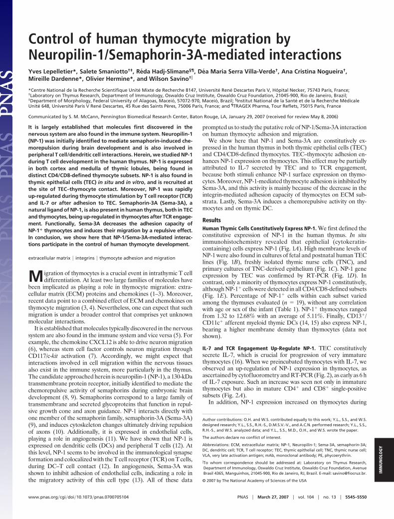
ResultsHuman Thymic Cells Constitutively Express NP-1. We first defined theconstitutive expression of NP-1 in the human thymus. In situimmunohistochemistry revealed that epithelial (cytokeratin-containing) cells express NP-1 (Fig. 1A). High membrane levels ofNP-1 were also found in cultures of fetal and postnatal human TEClines (Fig. 1B), freshly isolated thymic nurse cells (TNC), andprimary cultures of TNC-derived epithelium (Fig. 1C). NP-1 geneexpression by TEC was confirmed by RT-PCR (Fig. 1D). Incontrast, only a minority of thymocytes express NP-1 constitutively,although NP-1� cells were detected in all CD4/CD8-defined subsets(Fig. 1E). Percentage of NP-1� cells within each subset variedamong the thymuses evaluated (n � 19), without any correlationwith age or sex of the infant (Table 1). NP-1� thymocytes rangedfrom 1.32 to 12.68% with an average of 5.11%. Finally, CD13�/CD11c� afferent myeloid thymic DCs (14, 15) also express NP-1,bearing a higher membrane density than thymocytes (data notshown).
IL-7 and TCR Engagement Up-Regulate NP-1. TEC constitutivelysecrete IL-7, which is crucial for progression of very immaturethymocytes (16). When we preincubated thymocytes with IL-7, weobserved an up-regulation of NP-1 expression in thymocytes, asascertained by cytofluorometry and RT-PCR (Fig. 2), as early as 6 hof IL-7 exposure. Such an increase was seen not only in immaturethymocytes but also in mature CD4� and CD8� single-positivesubsets (Fig. 2A).
In addition, NP-1 expression increased on thymocytes during
Author contributions: O.H. and W.S. contributed equally to this work; Y.L., S.S., and W.S.designed research; Y.L., S.S., R.H.-S., D.M.S.V.-V., and A.C.N. performed research; Y.L., S.S.,R.H.-S., and W.S. analyzed data; and Y.L., S.S., M.D., O.H., and W.S. wrote the paper.
The authors declare no conflict of interest.
Abbreviations: ECM, extracellular matrix; NP-1, Neuropilin-1; Sema-3A, semaphorin-3A;DC, dendritic cell; TCR, T cell receptor; TEC, thymic epithelial cell; TNC, thymic nurse cell;VLA, very late activation antigen; mAb, monoclonal antibody; PE, phycoerythrin.
�To whom correspondence should be addressed at: Laboratory on Thymus Research,Department of Immunology, Oswaldo Cruz Institute, Oswaldo Cruz Foundation, AvenueBrasil 4365, Manguinhos, 21045-900, Rio de Janeiro, RJ, Brazil. E-mail: [email protected].
© 2007 by The National Academy of Sciences of the USA
www.pnas.org�cgi�doi�10.1073�pnas.0700705104 PNAS � March 27, 2007 � vol. 104 � no. 13 � 5545–5550
IMM
UN
OLO
GY
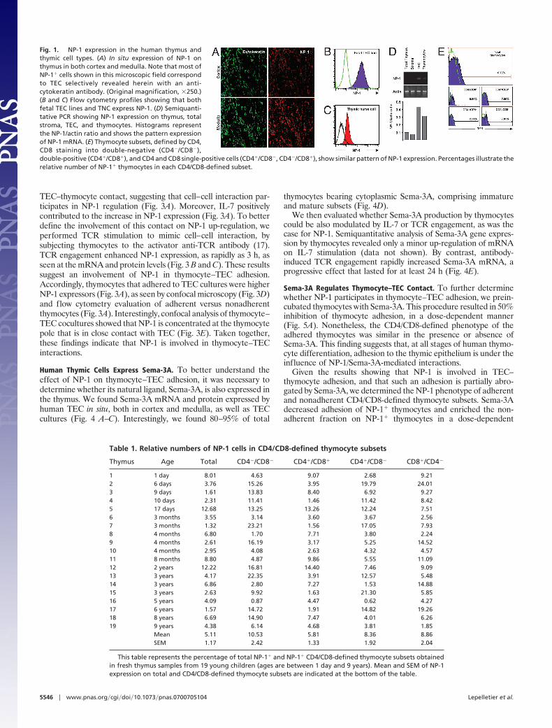
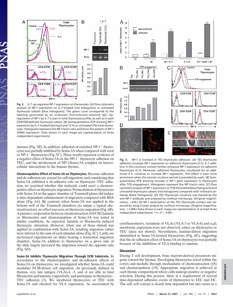
TEC–thymocyte contact, suggesting that cell–cell interaction par-ticipates in NP-1 regulation (Fig. 3A). Moreover, IL-7 positivelycontributed to the increase in NP-1 expression (Fig. 3A). To betterdefine the involvement of this contact on NP-1 up-regulation, weperformed TCR stimulation to mimic cell–cell interaction, bysubjecting thymocytes to the activator anti-TCR antibody (17).TCR engagement enhanced NP-1 expression, as rapidly as 3 h, asseen at the mRNA and protein levels (Fig. 3 B and C). These resultssuggest an involvement of NP-1 in thymocyte–TEC adhesion.Accordingly, thymocytes that adhered to TEC cultures were higherNP-1 expressors (Fig. 3A), as seen by confocal microscopy (Fig. 3D)and flow cytometry evaluation of adherent versus nonadherentthymocytes (Fig. 3A). Interestingly, confocal analysis of thymocyte–TEC cocultures showed that NP-1 is concentrated at the thymocytepole that is in close contact with TEC (Fig. 3E). Taken together,these findings indicate that NP-1 is involved in thymocyte–TECinteractions.
Human Thymic Cells Express Sema-3A. To better understand theeffect of NP-1 on thymocyte–TEC adhesion, it was necessary todetermine whether its natural ligand, Sema-3A, is also expressed inthe thymus. We found Sema-3A mRNA and protein expressed byhuman TEC in situ, both in cortex and medulla, as well as TECcultures (Fig. 4 A–C). Interestingly, we found 80–95% of total
thymocytes bearing cytoplasmic Sema-3A, comprising immatureand mature subsets (Fig. 4D).
We then evaluated whether Sema-3A production by thymocytescould be also modulated by IL-7 or TCR engagement, as was thecase for NP-1. Semiquantitative analysis of Sema-3A gene expres-sion by thymocytes revealed only a minor up-regulation of mRNAon IL-7 stimulation (data not shown). By contrast, antibody-induced TCR engagement rapidly increased Sema-3A mRNA, aprogressive effect that lasted for at least 24 h (Fig. 4E).
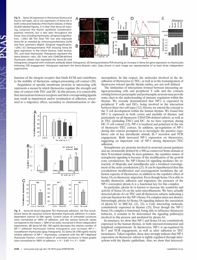
Sema-3A Regulates Thymocyte–TEC Contact. To further determinewhether NP-1 participates in thymocyte–TEC adhesion, we prein-cubated thymocytes with Sema-3A. This procedure resulted in 50%inhibition of thymocyte adhesion, in a dose-dependent manner(Fig. 5A). Nonetheless, the CD4/CD8-defined phenotype of theadhered thymocytes was similar in the presence or absence ofSema-3A. This finding suggests that, at all stages of human thymo-cyte differentiation, adhesion to the thymic epithelium is under theinfluence of NP-1/Sema-3A-mediated interactions.
Given the results showing that NP-1 is involved in TEC–thymocyte adhesion, and that such an adhesion is partially abro-gated by Sema-3A, we determined the NP-1 phenotype of adherentand nonadherent CD4/CD8-defined thymocyte subsets. Sema-3Adecreased adhesion of NP-1� thymocytes and enriched the non-adherent fraction on NP-1� thymocytes in a dose-dependent
Fig. 1. NP-1 expression in the human thymus andthymic cell types. (A) In situ expression of NP-1 onthymus in both cortex and medulla. Note that most ofNP-1� cells shown in this microscopic field correspondto TEC selectively revealed herein with an anti-cytokeratin antibody. (Original magnification, �250.)(B and C) Flow cytometry profiles showing that bothfetal TEC lines and TNC express NP-1. (D) Semiquanti-tative PCR showing NP-1 expression on thymus, totalstroma, TEC, and thymocytes. Histograms representthe NP-1/actin ratio and shows the pattern expressionof NP-1 mRNA. (E) Thymocyte subsets, defined by CD4,CD8 staining into double-negative (CD4�/CD8�),double-positive (CD4�/CD8�), and CD4 and CD8 single-positive cells (CD4�/CD8�, CD4�/CD8�), show similar pattern of NP-1 expression. Percentages illustrate therelative number of NP-1� thymocytes in each CD4/CD8-defined subset.
Table 1. Relative numbers of NP-1 cells in CD4/CD8-defined thymocyte subsets
Thymus Age Total CD4�/CD8� CD4�/CD8� CD4�/CD8� CD8�/CD4�
1 1 day 8.01 4.63 9.07 2.68 9.212 6 days 3.76 15.26 3.95 19.79 24.013 9 days 1.61 13.83 8.40 6.92 9.274 10 days 2.31 11.41 1.46 11.42 8.425 17 days 12.68 13.25 13.26 12.24 7.516 3 months 3.55 3.14 3.60 3.67 2.567 3 months 1.32 23.21 1.56 17.05 7.938 4 months 6.80 1.70 7.71 3.80 2.249 4 months 2.61 16.19 3.17 5.25 14.5210 4 months 2.95 4.08 2.63 4.32 4.5711 8 months 8.80 4.87 9.86 5.55 11.0912 2 years 12.22 16.81 14.40 7.46 9.0913 3 years 4.17 22.35 3.91 12.57 5.4814 3 years 6.86 2.80 7.27 1.53 14.8815 3 years 2.63 9.92 1.63 21.30 5.8516 5 years 4.09 0.87 4.47 0.62 4.2717 6 years 1.57 14.72 1.91 14.82 19.2618 8 years 6.69 14.90 7.47 4.01 6.2619 9 years 4.38 6.14 4.68 3.81 1.85
Mean 5.11 10.53 5.81 8.36 8.86SEM 1.17 2.42 1.33 1.92 2.04
This table represents the percentage of total NP-1� and NP-1� CD4/CD8-defined thymocyte subsets obtainedin fresh thymus samples from 19 young children (ages are between 1 day and 9 years). Mean and SEM of NP-1expression on total and CD4/CD8-defined thymocyte subsets are indicated at the bottom of the table.
5546 � www.pnas.org�cgi�doi�10.1073�pnas.0700705104 Lepelletier et al.
manner (Fig. 5B). In addition, adhesion of enriched NP-1� thymo-cytes was partially inhibited by Sema-3A when compared with totalor NP-1� thymocytes (Fig. 5C). These results represent evidence ofa negative effect of Sema-3A on the NP-1� thymocyte adhesion onTEC, and the involvement of NP-1/Sema-3A complex on hetero-cellular interactions in the human thymus.
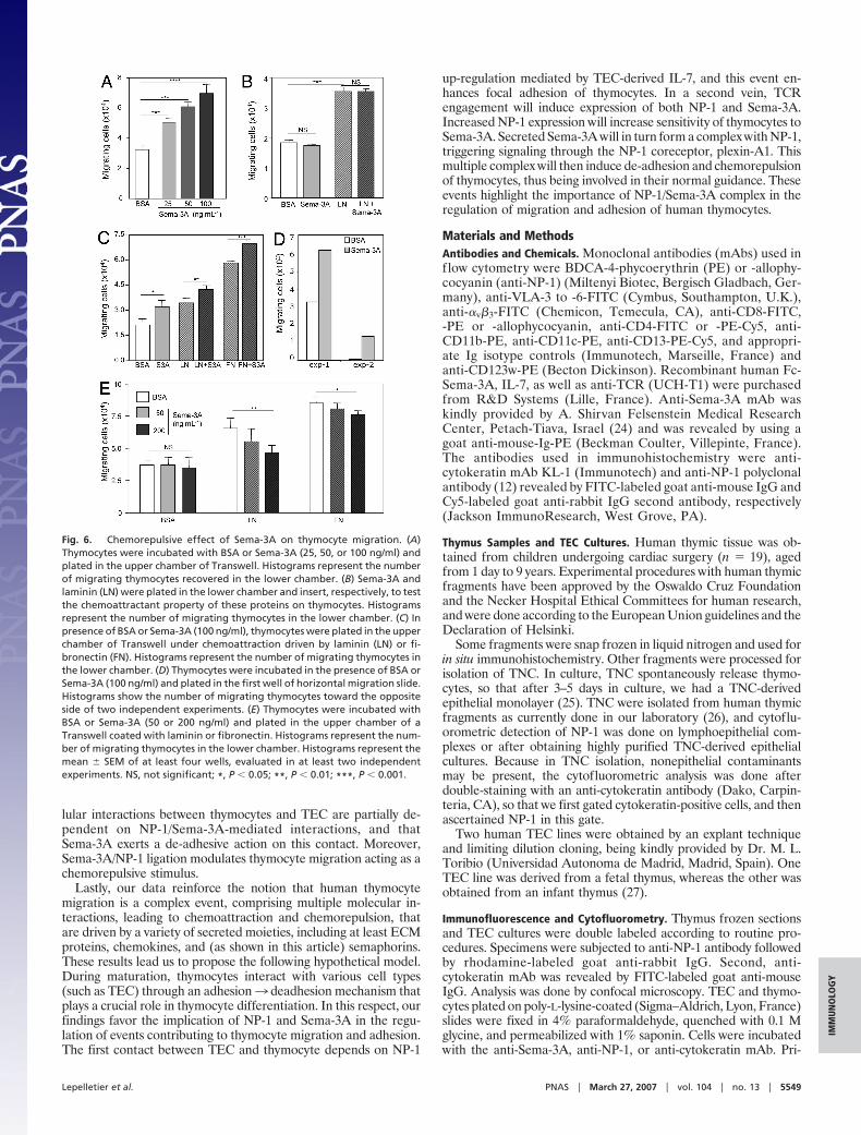
Chemorepulsive Effect of Sema-3A on Thymocytes. Because adhesionand de-adhesion are crucial for cell migration, and considering thatSema-3A exhibited a de-adhesive role on thymocyte–TEC adhe-sion, we searched whether this molecule could exert a chemore-pulsive effect on thymocyte migration. Preincubation of thymocyteswith Sema-3A in the upper chamber of Transwell plates did inducea dose-dependent enhancement of migration compared with BSAalone (Fig. 6A). By contrast, when Sema-3A was applied in thebottom well of the Transwell chambers (to mimic a typical che-moattraction), no effect was seen on thymocyte migration (Fig. 6B).A putative cooperation between chemoattraction of ECM (lamininor fibronectin) and chemorepulsion of Sema-3A was tested insimilar conditions. As expected, laminin or fibronectin inducedthymocyte attraction. However, when one of these stimuli wasapplied in combination with Sema-3A, resulting migration valueswere inferior to the sum of each stimulus alone (Fig. 6C). Lastly, weperformed experiments on slides bearing a horizontal migrationchamber. Sema-3A addition to thymocytes on a given side ofthe slide largely increased the migration toward the opposite side(Fig. 6D).
Sema-3A Inhibits Thymocyte Migration Through ECM Substrata. Inaccordance to the chemorepulsive and de-adhesion effects ofSema-3A on thymocytes, we investigated whether Sema-3A couldmodulate ECM-driven cell migration. As previously defined inthymus, very late antigen (VLA)-4, -5, and -6 are able to bindfibronectin and laminin, respectively, and participate in thymocyte–TEC adhesion (3). We incubated thymocytes or TEC withSema-3A and checked for VLA expression. As ascertained by
cytofluorometry, variations of VLAs (VLA-3 to VLA-6) and �v�3membrane expression were not observed, either on thymocytes orTEC (data not shown). Nevertheless, laminin-driven migrationdiminished in Sema-3A-treated thymocytes (Fig. 6E), suggestingthat the de-adhesion effect of Sema-3A on thymocytes was partiallybecause of the inhibition of VLA binding to laminin.
DiscussionDuring T cell development, bone marrow-derived precursors mi-grate toward the thymus. Developing thymocytes travel within thecortex and medulla through mechanisms of chemoattraction (18)and chemorepulsion (19). Appropriate migration is achieved ateach thymic compartment where cells undergo positive or negativeselection. During this process, there is a requirement of severaltime-dependent adhesion events of thymocytes to TEC and DC.The cell–cell contact is clearly time dependent but also varies as a
Fig. 2. IL-7 up-regulates NP-1 expression on thymocytes. (A) Flow cytometryanalysis of NP-1 expression on IL-7-treated (red histograms) or untreatedthymocyte subsets (blue histograms). The green curve corresponds to thelabeling generated by an irrelevant fluorochrome-matched IgG. Up-regulation of NP-1 by IL-7 is seen in total thymocyte profile, as well as in eachCD4/CD8-defined thymocyte subset. (B) Semiquantitative PCR showing NP-1expression by IL-7-treated (during 6 and 12 h) or untreated (T0) total thymo-cytes. Histograms represent the NP-1/actin ratio and show the pattern of NP-1mRNA expression. Data shown in each image are representative of threeindependent experiments.
Fig. 3. NP-1 is involved in TEC–thymocyte adhesion. (A) TEC–thymocyteadhesion increases NP-1 expression on adherent thymocytes (2 h). IL-7 addi-tion in this coculture context further enhances NP-1 expression on adherentthymocytes (2 h). Moreover, adherent thymocytes, cocultured for an addi-tional 4 h, continue to increase NP-1 expression. This effect is even moreprominent when the second coculture period is preceded by wash. (B) Semi-quantitative PCR showing increase in NP-1 gene expression in thymocytesafter TCR engagement. Histograms represent the NP-1/actin ratio. (C) Flowcytometry analysis of NP-1 expression on TCR stimulated (blue histograms) anduntreated thymocyte subsets (red histograms) compared with irrelevant an-tibody (black histograms). (D) TEC–thymocyte coculture was stained by theanti-NP-1 antibody and analyzed by confocal microscopy. (Original magnifi-cation, �250.) (E) NP-1 polarization at the TEC–thymocyte contact was ob-served by using Z-stack analysis by confocal microscopy. (Original magnifica-tion, �1,000.) Data shown in each image are representative of at least threeindependent experiments. ***, P � 0.001.
Lepelletier et al. PNAS � March 27, 2007 � vol. 104 � no. 13 � 5547
IMM
UN
OLO
GY
function of the integrin receptor that binds ECM and contributesto the stability of thymocyte–antigen-presenting cell contact (20).
Acquisition of specific membrane proteins in interacting cellsrepresents a means by which thymocytes regulate the strength andtime of contact with TEC and DC. In this process, it is conceivablethat interactions between receptors and their corresponding ligandsmay result in impairment and/or termination of adhesion, associ-ated to a migratory effect, secondary to chemoattraction or che-
morepulsion. In this respect, the molecules involved in the de-adhesion of thymocytes to TEC, as well as in the transmigration ofthymocytes toward specific thymic niches, are not well defined.
The similarities of interactions formed between interacting an-tigen-presenting cells and peripheral T cells and the contactsexisting between presynaptic and postsynaptic neurons may providesome clues to the understanding of immune regulation within thethymus. We recently demonstrated that NP-1 is expressed byperipheral T cells and DCs, being involved in the interactionbetween these two cell types (12). Herein, we extend the concept tothe T cell development within the human thymus. We found thatNP-1 is expressed in both cortex and medulla of thymus, andparticularly on all thymocyte CD4/CD8-defined subsets, as well asin TEC (including TNC) and DC. As we have reported, duringDC–T cell contact (12), NP-1 is localized and polarized at the siteof thymocyte–TEC contact. In addition, up-regulation of NP-1during this contact prompted us to investigate the putative regu-latory role of key intrathymic stimuli, IL-7 secretion and TCRengagement. Both increased NP-1 expression on thymocytes,suggesting an important role of NP-1 during thymocyte–TECadhesion.
Semaphorins are proteins involved in neuronal axonal guidanceand are structurally defined by a 500-aa conserved Sema domain attheir N-terminal ending. In axon guidance, the repulsive nature ofsemaphorin signaling is because of the modification of the growthcone cytoskeleton: the NP-1/Sema-3A signaling mediates the re-traction of filopodia and lamellipodia and a localized rearrange-ment of the actin cytoskeleton (21). It can be hypothesized that thiscytoskeleton modification and rearrangement modulates the ad-hesion capacity of thymocytes, in addition to the repulsive effect ofSema-3A on thymocyte migration. Even though Sema-3A is able tomodify thymocyte adhesion and migration, the presence of theNP-1 coreceptor plexin-A is a functional key for this complex.
In particular, plexin-A1 is known to increase the sensitivity andactivity of Sema-3A on the actin microfilaments. We have actuallydetected plexin-A1 on TEC and all thymocyte subsets, indicating arelevant function for the NP-1/Sema-3A complex (data not shown).Interestingly, plexin-A1/Sema-3A signaling induces the associationof plexin-A1 to MICAL (22, 23), a CasL interacting molecule,constitutively expressed in thymus (23). Even though the NP-1/Sema-3A complex is functional, being able to influence thymocytebehavior, it remains to be determined the signaling pathway(s)involved in this process and mediated by plexin-A1.
In summary, we show that NP-1 and Sema-3A are constitutivelyexpressed in the human thymus, in both microenvironmental andlymphoid compartments. In thymocytes, NP-1 is up-regulated byIL-7 and TCR engagement, as well as after adhesion to TECmonolayers. Taken together, these data strongly indicate that NP-1expression on thymocyte subsets is tightly regulated through inter-actions with the thymic epithelium. Also, we show that heterocel-
Fig. 4. Sema-3A expression in the human thymus andthymic cell types. (A) In situ expression of Sema-3A inboth cortex and medulla of the thymic lobules. In thesedouble-labeled figures, it is clear that Sema-3A stain-ing comprises the thymic epithelial (cytokeratin-positive) network, but is also seen throughout thetissue, thus including thymocytes. (Original magnifica-tion, �250.) (B) The fetal TEC line also expressesSema-3A as revealed by immunocytochemistry (Left)and flow cytometry (Right). (Original magnification,�250.) (C) Semiquantitative PCR showing Sema-3Agene expression in the whole thymus, total stroma,TEC, and total thymocytes. Histograms represent theSema-3A/actin ratio. (D) Total and CD4/CD8-definedthymocyte subsets that expressed the Sema-3A (redhistograms) compared with irrelevant antibody (black histograms). (E) Semiquantitative PCR showing an increase in Sema-3A gene expression on thymocytesfollowing TCR engagement. Histograms represent the Sema-3A/actin ratio. Data shown in each image are representative of at least three independentexperiments.
Fig. 5. Sema-3A down-regulates TEC–thymocyte adhesion. (A) The recom-binant Sema-3A repulsive isoform decreases thymocyte adhesion in a dose-dependent manner (5–500 ng/ml). Control values of untreated cocultureswere normalized as 100% of adhesion, and the various Sema-3A valuescorrespond to the means � SEM of six wells, recovered in three independentexperiments. (B) Sema-3A (50–100 ng/ml) decreases the relative number ofNP-1�-adhered thymocytes (white histograms) and increases NP-1�-nonadherent thymocytes (gray histograms). (C) Sema-3A (100 ng/ml) mainlyinhibits adhesion of NP-1� thymocytes, compared with the NP-1-depletedthymocyte fraction. Control values of untreated cocultures in these graphswere normalized to 100% of adhesion. *, P � 0.05; ***, P � 0.001.
5548 � www.pnas.org�cgi�doi�10.1073�pnas.0700705104 Lepelletier et al.
lular interactions between thymocytes and TEC are partially de-pendent on NP-1/Sema-3A-mediated interactions, and thatSema-3A exerts a de-adhesive action on this contact. Moreover,Sema-3A/NP-1 ligation modulates thymocyte migration acting as achemorepulsive stimulus.
Lastly, our data reinforce the notion that human thymocytemigration is a complex event, comprising multiple molecular in-teractions, leading to chemoattraction and chemorepulsion, thatare driven by a variety of secreted moieties, including at least ECMproteins, chemokines, and (as shown in this article) semaphorins.These results lead us to propose the following hypothetical model.During maturation, thymocytes interact with various cell types(such as TEC) through an adhesion3 deadhesion mechanism thatplays a crucial role in thymocyte differentiation. In this respect, ourfindings favor the implication of NP-1 and Sema-3A in the regu-lation of events contributing to thymocyte migration and adhesion.The first contact between TEC and thymocyte depends on NP-1
up-regulation mediated by TEC-derived IL-7, and this event en-hances focal adhesion of thymocytes. In a second vein, TCRengagement will induce expression of both NP-1 and Sema-3A.Increased NP-1 expression will increase sensitivity of thymocytes toSema-3A. Secreted Sema-3A will in turn form a complex with NP-1,triggering signaling through the NP-1 coreceptor, plexin-A1. Thismultiple complex will then induce de-adhesion and chemorepulsionof thymocytes, thus being involved in their normal guidance. Theseevents highlight the importance of NP-1/Sema-3A complex in theregulation of migration and adhesion of human thymocytes.
Materials and MethodsAntibodies and Chemicals. Monoclonal antibodies (mAbs) used inflow cytometry were BDCA-4-phycoerythrin (PE) or -allophy-cocyanin (anti-NP-1) (Miltenyi Biotec, Bergisch Gladbach, Ger-many), anti-VLA-3 to -6-FITC (Cymbus, Southampton, U.K.),anti-�v�3-FITC (Chemicon, Temecula, CA), anti-CD8-FITC,-PE or -allophycocyanin, anti-CD4-FITC or -PE-Cy5, anti-CD11b-PE, anti-CD11c-PE, anti-CD13-PE-Cy5, and appropri-ate Ig isotype controls (Immunotech, Marseille, France) andanti-CD123w-PE (Becton Dickinson). Recombinant human Fc-Sema-3A, IL-7, as well as anti-TCR (UCH-T1) were purchasedfrom R&D Systems (Lille, France). Anti-Sema-3A mAb waskindly provided by A. Shirvan Felsenstein Medical ResearchCenter, Petach-Tiava, Israel (24) and was revealed by using agoat anti-mouse-Ig-PE (Beckman Coulter, Villepinte, France).The antibodies used in immunohistochemistry were anti-cytokeratin mAb KL-1 (Immunotech) and anti-NP-1 polyclonalantibody (12) revealed by FITC-labeled goat anti-mouse IgG andCy5-labeled goat anti-rabbit IgG second antibody, respectively(Jackson ImmunoResearch, West Grove, PA).
Thymus Samples and TEC Cultures. Human thymic tissue was ob-tained from children undergoing cardiac surgery (n � 19), agedfrom 1 day to 9 years. Experimental procedures with human thymicfragments have been approved by the Oswaldo Cruz Foundationand the Necker Hospital Ethical Committees for human research,and were done according to the European Union guidelines and theDeclaration of Helsinki.
Some fragments were snap frozen in liquid nitrogen and used forin situ immunohistochemistry. Other fragments were processed forisolation of TNC. In culture, TNC spontaneously release thymo-cytes, so that after 3–5 days in culture, we had a TNC-derivedepithelial monolayer (25). TNC were isolated from human thymicfragments as currently done in our laboratory (26), and cytoflu-orometric detection of NP-1 was done on lymphoepithelial com-plexes or after obtaining highly purified TNC-derived epithelialcultures. Because in TNC isolation, nonepithelial contaminantsmay be present, the cytofluorometric analysis was done afterdouble-staining with an anti-cytokeratin antibody (Dako, Carpin-teria, CA), so that we first gated cytokeratin-positive cells, and thenascertained NP-1 in this gate.
Two human TEC lines were obtained by an explant techniqueand limiting dilution cloning, being kindly provided by Dr. M. L.Toribio (Universidad Autonoma de Madrid, Madrid, Spain). OneTEC line was derived from a fetal thymus, whereas the other wasobtained from an infant thymus (27).
Immunofluorescence and Cytofluorometry. Thymus frozen sectionsand TEC cultures were double labeled according to routine pro-cedures. Specimens were subjected to anti-NP-1 antibody followedby rhodamine-labeled goat anti-rabbit IgG. Second, anti-cytokeratin mAb was revealed by FITC-labeled goat anti-mouseIgG. Analysis was done by confocal microscopy. TEC and thymo-cytes plated on poly-L-lysine-coated (Sigma–Aldrich, Lyon, France)slides were fixed in 4% paraformaldehyde, quenched with 0.1 Mglycine, and permeabilized with 1% saponin. Cells were incubatedwith the anti-Sema-3A, anti-NP-1, or anti-cytokeratin mAb. Pri-
Fig. 6. Chemorepulsive effect of Sema-3A on thymocyte migration. (A)Thymocytes were incubated with BSA or Sema-3A (25, 50, or 100 ng/ml) andplated in the upper chamber of Transwell. Histograms represent the numberof migrating thymocytes recovered in the lower chamber. (B) Sema-3A andlaminin (LN) were plated in the lower chamber and insert, respectively, to testthe chemoattractant property of these proteins on thymocytes. Histogramsrepresent the number of migrating thymocytes in the lower chamber. (C) Inpresence of BSA or Sema-3A (100 ng/ml), thymocytes were plated in the upperchamber of Transwell under chemoattraction driven by laminin (LN) or fi-bronectin (FN). Histograms represent the number of migrating thymocytes inthe lower chamber. (D) Thymocytes were incubated in the presence of BSA orSema-3A (100 ng/ml) and plated in the first well of horizontal migration slide.Histograms show the number of migrating thymocytes toward the oppositeside of two independent experiments. (E) Thymocytes were incubated withBSA or Sema-3A (50 or 200 ng/ml) and plated in the upper chamber of aTranswell coated with laminin or fibronectin. Histograms represent the num-ber of migrating thymocytes in the lower chamber. Histograms represent themean � SEM of at least four wells, evaluated in at least two independentexperiments. NS, not significant; *, P � 0.05; **, P � 0.01; ***, P � 0.001.
Lepelletier et al. PNAS � March 27, 2007 � vol. 104 � no. 13 � 5549
IMM
UN
OLO
GY
mary antibody was revealed by a goat anti-mouse or -rabbit IgGconjugated to Cy-5, respectively. Specimens were examined with aconfocal laser microscope (LSM 510; Carl Zeiss, Oberkochen,Germany).
For flow cytometry, cells were fixed in 4% paraformaldehydeand incubated for 15 min at 4°C in PBS, 2% BSA, containing 0.05%saponin, with anti-Sema-3A or control isotype-matched irrelevantantibodies. Specific labeling was revealed by using a goat anti-mouse-Ig-PE. Membrane staining was performed by using FITC-and allophycocyanin-conjugated anti-CD4, anti-CD8 antibodies.Specimens were analyzed with a FACSCalibur (Becton Dickinson).In some experiments, four-color labeling was done to detect NP-1in DCs. In this case, we performed simultaneous labeling for NP-1,CD4, CD8, and a given DC marker, CD11c or CD13.
RNA Isolation and Semiquantitative RT-PCR. Total RNA was ex-tracted from 107 total thymus, total stroma, TEC, unstimulated andIL-7- or TCR-stimulated thymocytes, at different stages, and re-verse transcribed by using the first-strand cDNA synthesis kitaccording to the manufacturer’s instructions (Amersham Bio-sciences, Les Ulis, France). The PCR was carried out with cDNAas template, 50 pmol of both primers in the reaction mixture [50mM KCl, 10 mM Tris�HCl, pH 8.3, 1.5 mM MgCl2, 0.1% TritonX-100, 0.4 mM each dNTP (Promega, Madison, WI), 2.5 units ofAmpliTaq polymerase (Applied Biosystems, Foster City, CA)].After denaturation at 94°C for 5 min, samples underwent 25amplification cycles. Sense and antisense primers were as follows,respectively: 5�-CTAGAAGCATTTGCGGTGGACGATG-GAGGG-3� and 5�-TGACGGGGTCACCCACACTGTGC-CCATCTA-3� for �-actin; 5�-ACTCACTGTTCAGACTTA-3�and 5�-AGAGACTTCATGCAGCTC-3� for Sema-3A; and 5�-CTGGTGAGCCCTGTGGTTTATTCC-3� and 5�-ACTATTGT-CATCCACAGCAATCCC-3� for NP-1. PCR products were ana-lyzed on 1.5% agarose gel. Semiquantitative analysis wasperformed by the Gel Doc 2000 System (Bio-Rad, Hercules, CA).Sema-3A or NP-1 cDNA was normalized to �-actin cDNA con-centration, and Sema-3A/�-actin or NP-1/�-actin cDNA ratios werecalculated.
Thymocyte Stimulation. Freshly isolated thymocytes were culturedin presence of IL-7 (10 ng/ml) during 6–12 h. Then, cells wereharvested and cytofluorometry analysis of NP-1 expression ormRNA extractions were performed to follow both NP-1 andSema-3A protein and mRNA regulation, respectively. To induceTCR engagement, thymocytes were plated in the presence ofcoated anti-TCR antibody (UCHT-1; 5 �g/ml) or uncoated wellsduring 3–24 h at 37°C. Thymocytes were harvested at differenttimes and stained with mAbs to follow NP-1 expression, or used toextract mRNA to detect NP-1 and Sema-3A mRNA by semiquan-titative RT-PCR.
Cell Adhesion. Human TEC cultures were harvested, and 4 � 105
cells were replated in 75 cm2 flasks (Nunc, Roskilde, Denmark).After 24 h, thymocytes were cocultured (50 thymocytes per TEC)for 2 h at 37°C. Adherent cells were counted and phenotyped bycytofluorometry. We tested the inhibitory effect of Sema-3A onthymocyte adhesion by preincubating thymocytes with variousconcentrations of the protein (5–500 ng/ml), or the medium alone,for 2 h at 37°C. Nonadherent cells were gently washed out, andadhered thymocytes were counted and phenotyped.
Chemotaxis Assay. Thymocyte migratory activity was assessed exvivo in 5-�m pore size Transwell plates (Corning Costar, Corning,NY), as previously reported (28). Membrane inserts were coatedwith BSA, laminin, or fibronectin for 1 h at 37°C, followed by 1 hof blocking with 1% BSA. Thymocytes (2.5 � 106) with Sema-3A(25–100 ng/ml) were plated in the upper chamber in 100 �l of 0.5%BSA/RPMI, and 600 �l of 0.5% BSA/RPMI were added to thelower chamber. After 3 h, cells that migrated into lower chamberswere removed, counted, and phenotyped for the detection of NP-1,CD4, and CD8. In some experiments, Sema-3A was placed in thebottom well of the Transwell chamber to test a possible chemoat-tractant effect of the molecule.
In the horizontal migration assay (Biovalley, Conches, France),cells were plated in the first chamber side in presence of BSA orSema-3A (100 ng/ml). After 30–60 min, we counted those cellsmigrating toward the opposite chamber through a capillary ridge.
Purification of NP-1� Thymocytes. Total thymocytes (109 cells) wereincubated during 30 min at 4°C in presence of allophycocyanin-coupled anti-NP-1 mAb, washed in 2% FCS/PBS solution, andincubated with anti-allophycocyanine magnetic beads during 30min at 4°C before being plated in a magnetic sorter (MiltenyiBiotec). NP-1� thymocytes were directly eluted, whereas NP-1�
cells were retained in the column. Then, total, NP-1�, and NP-1�
thymocytes were used in culture to test the effect of Sema-3A (100ng/ml) on thymocyte adhesion to TEC monolayers.
Statistical Analysis. The results were analyzed by independentsample two-tailed and unpaired Student’s t test, and were presentedas means � standard error.
We thank the cardiac chirurgical pediatric offices (Necker Hospital, Paris,France) and Laranjeiras Hospital in Rio de Janeiro. This work wasdeveloped in the Institut National de la Sante et de la Recherche Medicale(INSERM)/Fundacao Oswaldo Cruz (FIOCRUZ) Associated Laboratoryof Immunology. Y.L. is a recipient of a Ligue National contre le Cancergrant. This work was supported by grants from INSERM/FIOCRUZ andComissao de Aperfeicoamento de Pessoal de Nıvel Superior/Comite Fran-cais d’Evaluation de la Cooperation Universitaire avec le Bresil conjointFrench–Brazilian programs, Conselho Nacional de Desenvolvimento Ci-entifico e Tecnologico (Brazil), Les Programmes Internationaux de Coop-eration Scientifique Centre National de la Recherche Scientifique (France),and Ligue National contre le Cancer (France).
1. Ansel KM, Cyster JG (2001) Curr Opin Immunol 13:172–179.2. Annunziato F, Romagnani P, Cosmi L, Lazzeri E, Romagnani S (2001) Trends Immunol
22:277–281.3. Savino W, Mendes-da-Cruz DA, Silva JS, Dardenne M, Cotta-de-Almeida V (2002) Trends
Immunol 23:305–313.4. Savino W, Mendes-Da-Cruz DA, Smaniotto S, Silva-Monteiro E, Villa-Verde DM (2004)
J Leukoc Biol 75:951–961.5. Steinman L (2004) Nat Immunol 5:575–581.6. Limatola C, Di Bartolomeo S, Trettel F, Lauro C, Ciotti MT, Mercanti D, Castellani L, Eusebi
F (2003) J Neuroimmunol 134:61–71.7. Sun L, Lee J, Fine HA (2004) J Clin Invest 113:1364–1374.8. He Z, Tessier-Lavigne M (1997) Cell 90:739–751.9. Kolodkin AL, Levengood DV, Rowe EG, Tai YT, Giger RJ, Ginty DD (1997) Cell 90:753–762.
10. Fan J, Mansfield SG, Redmond T, Gordon-Weeks PR, Raper JA (1993) J Cell Biol 121:867–878.11. Miao HQ, Soker S, Feiner L, Alonso JL, Raper JA, Klagsbrun M (1999) J Cell Biol 146:233–242.12. Tordjman R, Lepelletier Y, Lemarchandel V, Cambot M, Gaulard P, Hermine O, Romeo PH
(2002) Nat Immunol 3:477–482.13. Serini G, Valdembri D, Zanivan S, Morterra G, Burkhardt C, Caccavari F, Zammataro L, Primo
L, Tamagnone L, Logan M, et al. (2003) Nature 424:391–397.14. Vandenabeele S, Hochrein H, Mavaddat N, Winkel K, Shortman K (2001) Blood 97:1733–1741.15. Bendriss-Vermare N, Barthelemy C, Durand I, Bruand C, Dezutter-Dambuyant C, Moulian N,
Berrih-Aknin S, Caux C, Trinchieri G, Briere F (2001) J Clin Invest 107:835–844.
16. Offner F, Plum J (1998) Leuk Lymphoma 30:87–99.17. Schwinzer R, Sommermeyer H, Schlitt HJ, Schmidt RE, Wonigeit K (1991) Cell Immunol
136:318–328.18. Kim CH, Pelus LM, White JR, Broxmeyer HE (1998) Blood 91:4434–4443.19. Cyster JG (2002) J Clin Invest 109:1011–1012.20. Ayres-Martins S, Lannes-Vieira J, Farias-De-Oliveira DA, Brito JM, Villa-Verde DM, Savino
W (2004) Cell Immunol 229:21–30.21. Fournier AE, Nakamura F, Kawamoto S, Goshima Y, Kalb RG, Strittmatter SM (2000) J Cell
Biol 149:411–422.22. Terman JR, Mao T, Pasterkamp RJ, Yu HH, Kolodkin AL (2002) Cell 109:887–900.23. Suzuki T, Nakamoto T, Ogawa S, Seo S, Matsumura T, Tachibana K, Morimoto C, Hirai H
(2002) J Biol Chem 277:14933–14941.24. Shirvan A, Ziv I, Fleminger G, Shina R, He Z, Brudo I, Melamed E, Barzilai A (1999)
J Neurochem 73:961–971.25. Guyden JC, Pezzano M (2003) Int Rev Cytol 223:1–37.26. Ribeiro-Carvalho MM, Farias-de-Oliveira DA, Villa-Verde DM, Savino W (2002) Neuroim-
munomodulation 10:142–152.27. Fernandez E, Vicente A, Zapata A, Brera B, Lozano JJ, Martinez C, Toribio ML (1994) Blood
83:3245–3254.28. Cotta-de-Almeida V, Villa-Verde DM, Lepault F, Pleau JM, Dardenne M, Savino W (2004) Eur
J Immunol 34:1578–1587.
5550 � www.pnas.org�cgi�doi�10.1073�pnas.0700705104 Lepelletier et al.


