Embed Size (px)
Citation preview
Review began 06/15/2021 Review ended 07/01/2021 Published 07/13/2021
© Copyright 2021Ghosh et al. This is an open access articledistributed under the terms of theCreative Commons Attribution LicenseCC-BY 4.0., which permits unrestricteduse, distribution, and reproduction in anymedium, provided the original author andsource are credited.
Concomitant Acute Rheumatic Fever and AcutePost Streptococcal GlomerulonephritisSomshukla Ghosh , Kelli King-Morris , Joshua Shultz
1. Internal Medicine, University of Central Florida College of Medicine, Orlando, USA 2. Nephrology, Orlando VeteransAffairs Medical Center, Orlando, USA
Corresponding author: Somshukla Ghosh, [email protected]
AbstractAcute rheumatic fever (RF) and acute post Streptococcal glomerulonephritis (APSGN) are non-suppurativecomplications of a Group A Streptococcus (GAS) infection. The concomitant incidence of both complicationsin a patient is rare because nephritogenic and rheumatogenic strains belong to different serotypes of GroupA beta-hemolytic Streptococcus (GABHS). We present a case of a 47-year-old female who had concomitantacute RF and APSGN from a Streptococcus pyogenes infection. It is important to have a high clinical suspicionfor the sequela of GABHS infection in the setting of cardiac and renal disease following upper respiratoryinfection (URI) symptoms even in adults and in geographic locations with the nearly undetectable burden ofacute RF because of the importance of secondary prophylaxis with an antibiotic.
Categories: Cardiology, Infectious Disease, NephrologyKeywords: acute rheumatic fever, acute post streptococcal glomerulonephritis, rheumatic heart disease, antibioticprophylaxis, group a β-haemolytic streptococci
IntroductionAcute rheumatic fever (RF) is a non-suppurative complication secondary to tonsillopharyngitis causedby Streptococcus pyogenes and acute post Streptococcal glomerulonephritis (APSGN) is a type IIIhypersensitivity reaction usually following tonsillopharyngitis or skin infection caused by S. pyogenes [1]. Itis extremely rare for a patient to have concomitant acute RF and APSGN following a Streptococcaltonsillopharyngitis because the rheumatogenic and nephritogenic strains of Group A beta-hemolyticStreptococcus (GABHS) are different. However, such cases have been reported, and can be explained by co-infection with both rheumatogenic and nephritogenic strains.
The United States has a very low incidence of acute RF and it is no longer a reportable disease [2]. Acute RFoccurs most commonly in children aged between 5 and 15 years. We present a case of acute RF withconcurrent APSGN in an adult with no travel history outside of the United States in more than a decade.
Timely diagnosis of acute RF is critical because of the need for secondary prophylaxis with an antibiotic toavoid further valvular damage with a subsequent GABHS infection. This is because patients with asubsequent GABHS infection are at a higher risk for a recurrent attack for acute RF and the severity ofrheumatic heart disease increases with subsequent episodes. Also, timely diagnosis and early treatment ofAPSGN are important for a reduction in morbidity.
Case PresentationA 47-year-old female with no significant past medical history, including RF in childhood, presented to theemergency department (ED) with the chief complaint of acute onset respirophasic chest pain for two days.The pain was described as sharp in quality, 7/10 in intensity, unrelated to rest or activity, and non-radiating.She also complained of generalized fatigue, bilateral shoulder and knee pain, bilateral leg swelling, and darkfrothy urine, all of which started a few days prior to the onset of chest pain. Other review of systems wasnegative. She reported a sore throat which started three weeks prior to admission and lasted approximatelyone week before spontaneously resolving. The patient was originally from Honduras; however, she had nohistory of travel outside of the United States in the past 14 years. She was living in Florida during the sameperiod. Of note, her children were sick with pharyngitis a month prior to her presentation, however, they didnot seek medical treatment.
On physical examination, her temperature was 98.2°F, blood pressure was 160/90 mmHg, heart rate was 84beats/min, respiratory rate was 12 breaths/min, and oxygen saturation was 99% on ambientair. Cardiopulmonary examination revealed bilateral diffuse lung crackles, normal S1 and S2, a holosystolic3/6 murmur at the cardiac apex which radiated to her axilla. She also had bilateral 2+ pitting pedal edema.Examination of the skin was normal without any evidence of pyoderma or cutaneous abscess. Labs weresignificant for creatinine of 1.57 mg/dL, mildly elevated troponin of 0.14 ng/mL, elevated brain natriureticpeptide (BNP) of 16667 pg/mL, and erythrocyte sedimentation rate (ESR) of 69 mm/h (Table 1) . The chest X-
1 2 1
Open Access CaseReport DOI: 10.7759/cureus.16357
How to cite this articleGhosh S, King-Morris K, Shultz J (July 13, 2021) Concomitant Acute Rheumatic Fever and Acute Post Streptococcal Glomerulonephritis . Cureus13(7): e16357. DOI 10.7759/cureus.16357
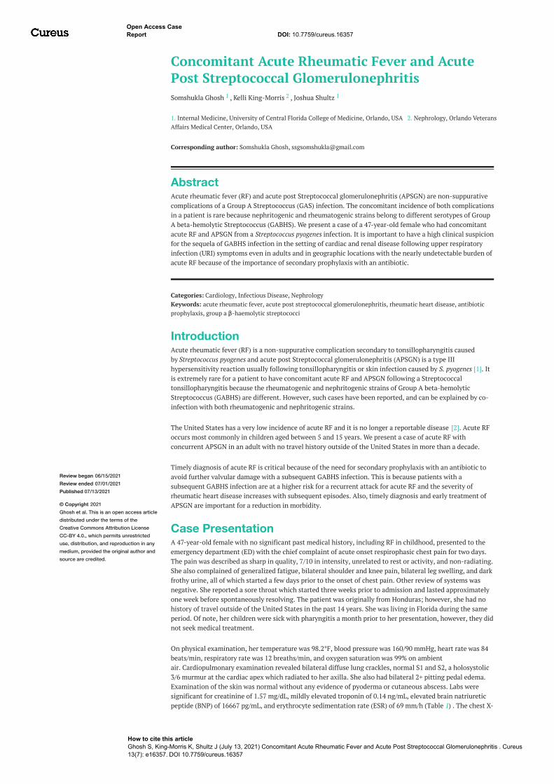
ray showed bilateral interstitial edema (Figure 1) and the echocardiogram showed severe mitral regurgitation(Figure 2), grade 1 diastolic dysfunction, and ejection fraction of 55%-60%.
FIGURE 1: Chest X-ray showing bilateral interstitial edema.
2021 Ghosh et al. Cureus 13(7): e16357. DOI 10.7759/cureus.16357 2 of 10
FIGURE 2: Parasternal long-axis view of echocardiogram showingsevere mitral regurgitation.
Group A Streptococcal antigen was negative, but antistreptolysin O (ASO) antibody was positive at 900IU/mL (Table 1).
2021 Ghosh et al. Cureus 13(7): e16357. DOI 10.7759/cureus.16357 3 of 10
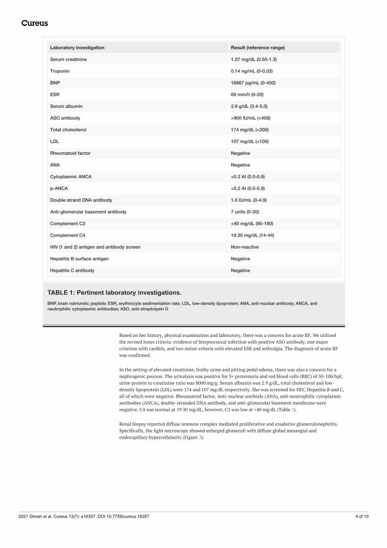
Laboratory investigation Result (reference range)
Serum creatinine 1.57 mg/dL (0.55-1.3)
Troponin 0.14 ng/mL (0-0.03)
BNP 16667 pg/mL (0-450)
ESR 69 mm/h (0-20)
Serum albumin 2.9 g/dL (3.4-5.0)
ASO antibody >900 IU/mL (<408)
Total cholesterol 174 mg/dL (<200)
LDL 107 mg/dL (<100)
Rheumatoid factor Negative
ANA Negative
Cytoplasmic ANCA <0.2 AI (0.0-0.9)
p-ANCA <0.2 AI (0.0-0.9)
Double strand DNA antibody 1.0 IU/mL (0-4.9)
Anti-glomerular basement antibody 7 units (0-20)
Complement C3 <40 mg/dL (90-180)
Complement C4 19.30 mg/dL (14-44)
HIV (1 and 2) antigen and antibody screen Non-reactive
Hepatitis B surface antigen Negative
Hepatitis C antibody Negative
TABLE 1: Pertinent laboratory investigations.BNP, brain natriuretic peptide; ESR, erythrocyte sedimentation rate; LDL, low-density lipoprotein; ANA, anti-nuclear antibody; ANCA, antineutrophilic cytoplasmic antibodies; ASO, anti-streptolysin O
Based on her history, physical examination and laboratory, there was a concern for acute RF. We utilizedthe revised Jones criteria: evidence of Streptococcal infection with positive ASO antibody, one majorcriterion with carditis, and two minor criteria with elevated ESR and arthralgia. The diagnosis of acute RFwas confirmed.
In the setting of elevated creatinine, frothy urine and pitting pedal edema, there was also a concern for anephrogenic process. The urinalysis was positive for 3+ proteinuria and red blood cells (RBC) of 50-100/hpf,urine protein to creatinine ratio was 8000 mg/g. Serum albumin was 2.9 g/dL, total cholesterol and low-density lipoprotein (LDL) were 174 and 107 mg/dL respectively. She was screened for HIV, Hepatitis B and C,all of which were negative. Rheumatoid factor, Anti-nuclear antibody (ANA), anti neutrophilic cytoplasmicantibodies (ANCA), double-stranded DNA antibody, and anti-glomerular basement membrane werenegative. C4 was normal at 19.30 mg/dL, however, C3 was low at <40 mg/dL (Table 1).
Renal biopsy reported diffuse immune complex mediated proliferative and exudative glomerulonephritis.Specifically, the light microscopy showed enlarged glomeruli with diffuse global mesangial andendocapillary hypercellularity (Figure 3).
2021 Ghosh et al. Cureus 13(7): e16357. DOI 10.7759/cureus.16357 4 of 10
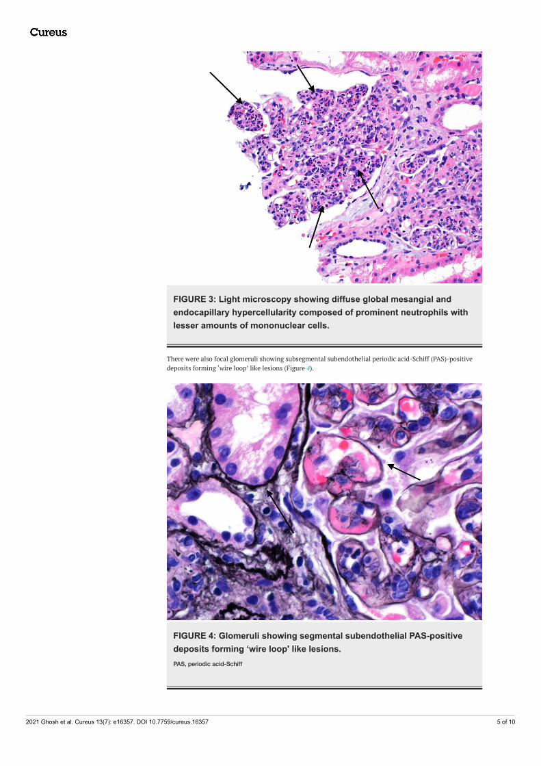
FIGURE 3: Light microscopy showing diffuse global mesangial andendocapillary hypercellularity composed of prominent neutrophils withlesser amounts of mononuclear cells.
There were also focal glomeruli showing subsegmental subendothelial periodic acid-Schiff (PAS)-positivedeposits forming ‘wire loop’ like lesions (Figure 4).
FIGURE 4: Glomeruli showing segmental subendothelial PAS-positivedeposits forming ‘wire loop' like lesions.PAS, periodic acid-Schiff
2021 Ghosh et al. Cureus 13(7): e16357. DOI 10.7759/cureus.16357 5 of 10
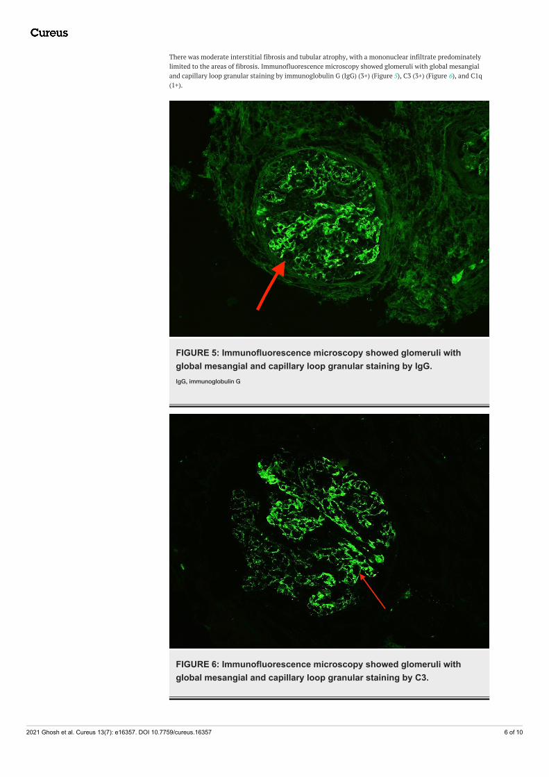
There was moderate interstitial fibrosis and tubular atrophy, with a mononuclear infiltrate predominatelylimited to the areas of fibrosis. Immunofluorescence microscopy showed glomeruli with global mesangialand capillary loop granular staining by immunoglobulin G (IgG) (3+) (Figure 5), C3 (3+) (Figure 6), and C1q(1+).
FIGURE 5: Immunofluorescence microscopy showed glomeruli withglobal mesangial and capillary loop granular staining by IgG.IgG, immunoglobulin G
FIGURE 6: Immunofluorescence microscopy showed glomeruli withglobal mesangial and capillary loop granular staining by C3.
2021 Ghosh et al. Cureus 13(7): e16357. DOI 10.7759/cureus.16357 6 of 10
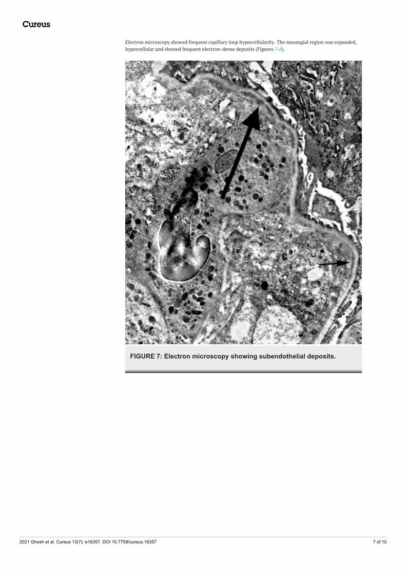
Electron microscopy showed frequent capillary loop hypercellularity. The mesangial region was expanded,hypercellular and showed frequent electron-dense deposits (Figures 7-8).
FIGURE 7: Electron microscopy showing subendothelial deposits.
2021 Ghosh et al. Cureus 13(7): e16357. DOI 10.7759/cureus.16357 7 of 10
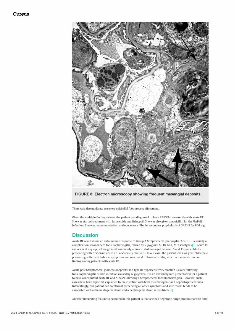
FIGURE 8: Electron microscopy showing frequent mesangial deposits.
There was also moderate to severe epithelial foot process effacement.
Given the multiple findings above, the patient was diagnosed to have APSGN concurrently with acute RF.She was started treatment with furosemide and lisinopril. She was also given amoxicillin for the GABHSinfection. She was recommended to continue amoxicillin for secondary prophylaxis of GABHS for lifelong.
DiscussionAcute RF results from an autoimmune response to Group A Streptococcal pharyngitis. Acute RF is usually acomplication secondary to tonsillopharyngitis, caused by S. pyogenes M-18, M-1, M-3 serotypes [3]. Acute RFcan occur at any age, although most commonly occurs in children aged between 5 and 15 years. Adultspresenting with first onset acute RF is extremely rare [4-5]. In our case, the patient was a 47-year-old femalepresenting with constitutional symptoms and was found to have valvulitis, which is the most commonfinding among patients with acute RF.
Acute post Streptococcal glomerulonephritis is a type III hypersensitivity reaction usually followingtonsillopharyngitis or skin infection caused by S. pyogenes. It is an extremely rare presentation for a patientto have concomitant acute RF and APSGN following a Streptococcal tonsillopharyngitis. However, suchcases have been reported, explained by co-infection with both rheumatogenic and nephritogenic strains.Interestingly, our patient had sorethroat preceeding all other symptoms and sore throat tends to beassociated with a rheumatogenic strain and a nephrogenic strain is less likely [6].
Another interesting feature to be noted in this patient is that she had nephrotic range proteinuria with renal
2021 Ghosh et al. Cureus 13(7): e16357. DOI 10.7759/cureus.16357 8 of 10
biopsy findings consistent with APSGN. Only 5% of patients with APSGN are known to have nephrotic rangeproteinuria [7]. It is unclear if the presence of nephrotic range proteinuria is a feature of concurrent acuteRF and APSGN.
In the literature, it is evident that the incidence of such concurrent non-suppurative complication of GroupA streptococci (GAS) infection is very rare (possibly less than 30 cases worldwide) and of these most arechildren and adolescents aged less than 18 years [4]. The most recent reported case of an adult withconcurrent acute RF and APSGN is the first reported case from Australia, even in the setting of Australiahaving a high burden of acute RF [8]. However, historically the United States is a low burden setting foracute RF, and in the last five decades of the 20th century, the incidence of acute RF in the United States hasdecreased to the extent to which it is no longer a reportable disease. The incidence of acute RF in Floridathrough 1970-1990 was 5-20/100,000 persons and was one of the pockets of significant incidence of acuteRF in the United States. The only states with a higher incidence were California, Tennessee, and Alabamawith 20-40/100,000 persons. The incidence of acute RF diminished significantly over the years with only onestate (West Virginia) with a measurable incidence at less than 5/100,000 persons [2]. To our knowledge, thisis the first reported case of concurrent acute RF and APSGN in Florida.
Diagnosis of acute RF confers the need for secondary prophylaxis with antibiotics as these patients are atrisk for subsequent complications and future GABHS infections which may result in worsening valvulardamage. Treatment entails administration of intramuscular Benzathine Penicillin G every four weeks forpatients with suspected noncompliance to oral antibiotics. As per the American Heart Association, patientswith rheumatic heart disease should receive secondary prophylaxis until 40 years of age or 10 years aftertheir last attack, whichever is longer. However, in patients with continued high risk for GABHS infection,lifelong secondary prophylaxis is warranted. Our patient was recommended to take lifelong secondaryprophylaxis with amoxicillin because she already had congestive heart failure from rheumatic heart diseaseand hence subsequent valvular damage secondary to recurrent acute RF could potentially prove to be lethal.
Regarding APSGN, delay in onset of treatment is associated with poor outcomes. Timing of diagnosis affectsmorbidity which is improved with early treatment. Long-term consequences such as hypertension,proteinuria, and hematuria should be monitored for resolution and may take years to resolve [9].
ConclusionsThis case highlights the importance of high clinical suspicion for the sequela of GABHS infection in thesetting of cardiac and renal disease that follows upper respiratory infection (URI) symptoms, even in adultsand in a geographic location with low incidence of non-suppurative complications of GABHS infection.Timely diagnosis and treatment are critical to prevent morbidity and mortality. Also, secondary prophylaxisagainst GABHS infection is of prime importance in patients with rheumatic heart disease.
Additional InformationDisclosuresHuman subjects: Consent was obtained or waived by all participants in this study. Conflicts of interest: Incompliance with the ICMJE uniform disclosure form, all authors declare the following: Payment/servicesinfo: All authors have declared that no financial support was received from any organization for thesubmitted work. Financial relationships: All authors have declared that they have no financialrelationships at present or within the previous three years with any organizations that might have aninterest in the submitted work. Other relationships: All authors have declared that there are no otherrelationships or activities that could appear to have influenced the submitted work.
References1. Walker MJ, Barnett TC, McArthur JD, et al.: Disease manifestations and pathogenic mechanisms of Group A
Streptococcus. Clin Microbiol Rev. 2014, 27:264-301. 10.1128/CMR.00101-132. Seckeler MD, Hoke TR: The worldwide epidemiology of acute rheumatic fever and rheumatic heart disease .
Clin Epidemiol. 2011, 3:67-84. 10.2147/CLEP.S129773. Dinkla K, Rohde M, Jansen WT, Kaplan EL, Chhatwal GS, Talay SR: Rheumatic fever-associated
Streptococcus pyogenes isolates aggregate collagen. J Clin Invest. 2003, 111:1905-1912. 10.1172/JCI172474. Gerber MA, Baltimore RS, Eaton CB, Gewitz M, Rowley AH, Shulman ST, Taubert KA: Prevention of
rheumatic fever and diagnosis and treatment of acute Streptococcal pharyngitis: a scientific statement fromthe American Heart Association Rheumatic Fever, Endocarditis, and Kawasaki Disease Committee of theCouncil on Cardiovascular Disease in the Young, the Interdisciplinary Council on Functional Genomics andTranslational Biology, and the Interdisciplinary Council on Quality of Care and Outcomes Research:endorsed by the American Academy of Pediatrics. Circulation. 2009, 119:1541-1551.10.1161/CIRCULATIONAHA.109.191959
5. Shulman ST, Bisno AL, Clegg HW, et al.: Clinical practice guideline for the diagnosis and management ofgroup A streptococcal pharyngitis: 2012 update by the Infectious Diseases Society of America. Clin InfectDis. 2012, 55:1279-1282. 10.1093/cid/cis847
6. Stollerman GH: Rheumatogenic and nephritogenic streptococci . Circulation. 1971, 43:915-921.10.1161/01.cir.43.6.915
2021 Ghosh et al. Cureus 13(7): e16357. DOI 10.7759/cureus.16357 9 of 10
7. Rawla P, Padala SA, Ludhwani D : Poststreptococcal Glomerulonephritis. StatPearls Publishing, TreasureIsland, FL; 2021.
8. Nakauyaca AV, Ralph AP, Majoni WS, Kangaharan N: Case report: concurrent rheumatic fever and acutepost-streptococcal glomerulonephritis in a high-burden setting. Am J Trop Med Hyg. 2019, 101:1054-1057.10.4269/ajtmh.18-0954
9. Gebreyesus LG, Aregay AF, Gebrekidan KG, Alemayehu YH: Factors associated with treatment outcome ofacute post streptococcal glomerulonephritis among patients less than 18 years in Mekelle City, PublicHospitals, North Ethiopia. BMC Res Notes. 2018, 11:693. 10.1186/s13104-018-3794-7
2021 Ghosh et al. Cureus 13(7): e16357. DOI 10.7759/cureus.16357 10 of 10