Embed Size (px)
Citation preview
HAL Id: hal-00602989https://hal.archives-ouvertes.fr/hal-00602989
Submitted on 24 Jun 2011
HAL is a multi-disciplinary open accessarchive for the deposit and dissemination of sci-entific research documents, whether they are pub-lished or not. The documents may come fromteaching and research institutions in France orabroad, or from public or private research centers.
L’archive ouverte pluridisciplinaire HAL, estdestinée au dépôt et à la diffusion de documentsscientifiques de niveau recherche, publiés ou non,émanant des établissements d’enseignement et derecherche français ou étrangers, des laboratoirespublics ou privés.
Computational design of a lipase for catalysis of theDiels-Alder reaction
Mats Linder, Anders Hermansson, John Liebeschuetz, Tore Brinck
To cite this version:Mats Linder, Anders Hermansson, John Liebeschuetz, Tore Brinck. Computational design of a lipasefor catalysis of the Diels-Alder reaction. Journal of Molecular Modeling, Springer Verlag (Germany),2010, 17 (4), pp.833-849. �10.1007/s00894-010-0775-8�. �hal-00602989�
Editorial Manager(tm) for Journal of Molecular Modeling Manuscript Draft Manuscript Number: JMMO1313R1 Title: Computational design of a lipase for catalysis of the Diels-Alder reaction Article Type: Original paper Keywords: Candida Antarctica lipase B, near attack conformer, molecular dynamics, density functional theory, in silico, promiscous reactivity, rational engineering Corresponding Author: Prof. Tore Brinck, Ph.D. Corresponding Author's Institution: Royal Institute of Technology (KTH) First Author: Mats Linder, MS Order of Authors: Mats Linder, MS; Anders Hermansson, BS; John Liebschuetz, PhD; Tore Brinck, Ph.D. Abstract: Combined molecular docking, molecular dynamics (MD) and density functional theory (DFT) studies have been employed to study catalysis of the Diels-Alder reaction by a modified lipase. Six variants of the versatile enzyme {\em Candida Antarctica} lipase B (CALB) have been rationally engineered {\em in silico} based on the specific characteristics of the pericyclic addition. A kinetic analysis reveals that hydrogen bond stabilization of the transition state and substrate binding are key components of the catalytic process. In the case of substrate binding, which has the greater potential for optimization, both binding strength and positioning of the substrates are important for catalytic efficiency. The binding strength is determined by hydrophobic interactions and can be tuned by careful selection of solvent and substrates. The MD simulations show that substrate positioning is sensitive to cavity shape and size, and can be controlled by a few rational mutations. The well-documented S105A mutation is essential to enable sufficient space in the vicinity of the oxyanion hole. Moreover, bulky residues on the edge of the active site hinders the formation of a sandwich-like near-attack conformer (NAC), and the I189A mutation is needed to obtain enough space above the face of the $\alpha$,$\beta$-double bond on the dienophile. The double mutant S105A/I189A performs quite well for two of three dienophiles. Based on binding constants and NAC energies obtained from MD simulations combined with activation energies from DFT computations, relative catalytic rates ($v_{cat}/v_{uncat}$) of up to $10^3$ are predicted. Response to Reviewers: We are happy with the favorable comments by the reviewers. We have have corrected the manuscript as suggested by reviewer 1 1. A sentence explaining catalytic promiscuity has been added. 2. A paragraph discussing the relationship between the NAC concept and the Reaction Force has been added. We have shortened the manuscript as suggested by reviewer 2. In particular, we have moved information regarding the structural relaxation of protein structure in MD simulations to the supplementary material.

Noname manuscript No.(will be inserted by the editor)
Computational Design of aLipase for Catalysis of theDiels-Alder reaction
Mats Linder · AndersHermansson · JohnLiebschuetz · ToreBrinck
Received: date / Accepted: date
Abstract Combined molecular docking, molecular dy-namics (MD) and density functional theory (DFT) stud-ies have been employed to study catalysis of the Diels-Alder reaction by a modified lipase. Six variants of theversatile enzyme Candida Antarctica lipase B (CALB)have been rationally engineered in silico based on thespecific characteristics of the pericyclic addition. A ki-netic analysis reveals that hydrogen bond stabilizationof the transition state and substrate binding are keycomponents of the catalytic process. In the case of sub-strate binding, which has the greater potential for opti-mization, both binding strength and positioning of thesubstrates are important for catalytic efficiency. Thebinding strength is determined by hydrophobic inter-actions and can be tuned by careful selection of sol-vent and substrates. The MD simulations show thatsubstrate positioning is sensitive to cavity shape andsize, and can be controlled by a few rational mutations.The well-documented S105A mutation is essential toenable sufficient space in the vicinity of the oxyanionhole. Moreover, bulky residues on the edge of the ac-tive site hinders the formation of a sandwich-like near-attack conformer (NAC), and the I189A mutation isneeded to obtain enough space above the face of theα,β-double bond on the dienophile. The double mu-tant S105A/I189A performs quite well for two of three
Mats Linder, Anders Hermansson and Tore BrinckPhysical Chemistry, Royal Institute of Technology (KTH)Tel.: +46-(0)8-7908210
E-mail: [email protected], [email protected]
John LiebeschuetzCambridge Crystallography Data Centre
Tel.: +44-(0)1223-762532
Fax.: +44-(0)1223-33603E-mail: [email protected]
dienophiles. Based on binding constants and NAC ener-gies obtained from MD simulations combined with ac-tivation energies from DFT computations, relative cat-alytic rates (vcat/vuncat) of up to 103 are predicted.
Keywords Diels-Alder · Rational design · CALB ·DFT · Molecular Dynamics
1 Introduction
The Diels-Alder reaction is remarkably rare in nature,[1] which may be attributed to a large activation en-ergy due to the highly ordered transition state. To date,only a few naturally occurring enzymes coined ”Diels-Alderases” have been characterized, [2] and these arevery substrate-specific. The mechanistic studies per-formed on Macrophomate Synthase (MPS), the only en-zyme performing bimolecular Diels-Alder, [3] also showthat the catalytic mechanism is likely to be a two-step,Michael-Aldol pathway, rather than a concerted [4 +2] addition. [4] Attempts to employ artificial ’enzymemimics’, for example cyclodextrin, catalytic antibodiesand other macrocycles [5] that utilize substrate recog-nition and transition state (TS) binding, have yieldedonly modest results due to product inhibition. [6]
To find a versatile Diels-Alderase is highly desirable,owing to the synthetic use of creating up to four stere-ocenters in one concerted step. The concept of non-covalent, hydrogen bond (H-bond) catalysis of Diels-Alder using carbonyl-activated dienophiles has under-gone rapid development in the last decade, [7, 8, 9] andsince activation of carbonyl-containing substrates to nu-cleophilic attack is very common in enzymes, in partic-ular the large hydrolase family (EC 3.x), the idea toutilize a promiscuous member of such an enzyme fam-ily for Diels-Alderase activity is not far-fetched, and hasbeen suggested in the literature. [10]
Among the hydrolase family, lipases are by far themost commonly utilized for in vitro catalysis of or-ganic reactions, such as (dynamic) kinetic resolution ofsecondary alcohols, transesterifications and acylations.[11, 12, 13, 14] Many lipases have been found to be sta-ble under a broad range of temperature and pH, andactive in organic solvents, [15, 16] which greatly ex-pands their utility in synthetic applications. More re-cently, evidence has emerged of catalytic promiscuity[17, 18] (i.e. the ability of an enzyme to exhibit cataly-sis of other than its native reaction) towards e.g. aldolcondensations, [19, 20] epoxidations [21] and Michaeladditions, [22, 23, 24, 25, 26] not seldom enhanced bypoint mutations. [27] An often-seen feature in promis-cuous catalysis is that only parts of the catalytic ma-chinery are used, so that removal of one or more of
For Production's Reference OnlyClick here to download Manuscript: Brinck_2010-DielsAlder_revised.pdf
2
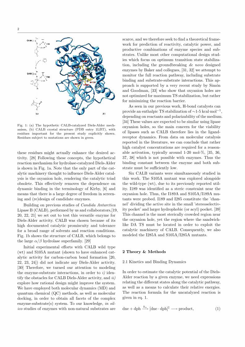
Fig. 1: (a) The hypothetic CALB-catalyzed Diels-Alder mech-
anism, (b) CALB crystal structure (PDB entry 1LBT), withresidues important for the present study explicitly shown.
Residues subject to mutations are shown in green.
these residues might actually enhance the desired ac-tivity. [28] Following these concepts, the hypotheticalreaction mechanism for hydrolase-catalyzed Diels-Alderis shown in Fig. 1a. Note that the only part of the cat-alytic machinery thought to influence Diels-Alder catal-ysis is the oxyanion hole, rendering the catalytic triadobsolete. This effectively removes the dependence ondynamic binding in the terminology of Kirby, [6] andmeans that there is a large degree of freedom in screen-ing and (re)design of candidate enzymes.
Building on previous studies of Candida AntarcticaLipase B (CALB), performed by us and collaborators,[19,20, 22, 21] we set out to test this versatile enzyme forDiels-Alder activity. CALB was chosen because of itshigh documented catalytic promiscuity and tolerancefor a broad range of solvents and reaction conditions.Fig. 1b shows the structure of CALB, which belongs tothe large α/β hydrolase superfamily. [29]
Initial experimental efforts with CALB wild type(wt) and S105A mutant (shown to have enhanced cat-alytic activity for carbon-carbon bond formation [20,22, 23, 24]) did not indicate any Diels-Alder activity.[30] Therefore, we turned our attention to modelingthe enzyme-substrate interactions, in order to i) iden-tify the obstacles for CALB Diels-Alder activity, and ii)explore how rational design might improve the system.We have employed both molecular dynamics (MD) andquantum chemical (QC) methods, as well as moleculardocking, in order to obtain all facets of the complexenzyme-substrate(s) system. To our knowledge, in sil-ico studies of enzymes with non-natural substrates are
scarce, and we therefore seek to find a theoretical frame-work for prediction of reactivity, catalytic power, andproductive combinations of enzyme species and sub-strates. Unlike most other computational design stud-ies which focus on optimum transition state stabiliza-tion, including the groundbreaking de novo designedenzymes by Baker and collegues, [31, 32] we attempt tomonitor the full reaction pathway, including substratebinding and substrate-substrate interactions. This ap-proach is supported by a very recent study by Simonand Goodman, [33] who show that oxyanion holes arenot optimized for maximum TS stabilization, but ratherfor minimizing the reaction barrier.
As seen in our previous work, H-bond catalysts canprovide an enthalpic TS stabilization of∼1-5 kcal·mol−1,depending on reactants and polarizability of the medium.[34] These values are expected to be similar using lipaseoxyanion holes, so the main concern for the viabilityof lipases such as CALB therefore lies in the ligand-receptor dynamics. From data on molecular catalysisreported in the literature, we can conclude that ratherhigh catalyst concentrations are required for a reason-able activation, typically around 1-20 mol-%, [35, 36,37, 38] which is not possible with enzymes. Thus thebinding constant between the enzyme and both sub-strate must be sufficiently low.
Six CALB variants were simultaneously studied inthis work. The S105A mutant was explored alongsidethe wild-type (wt), due to its previously reported util-ity. I189 was identified as a steric constraint near theoxyanion hole. Thus, the I189A and S105A/I189A mu-tants were probed. I189 and I285 constitute the ’chan-nel’ dividing the active site in the small ’stereoselectiv-ity pocket’ and larger hydrophobic (or acyl) pocket. [39]This channel is the most sterically crowded region nearthe oxyanion hole, yet the region where the sandwich-like DA TS must be located in order to exploit thecatalytic machinery of CALB. Consequently, we alsomodeled the I285A and S105A/I285A mutants.
2 Theory & Methods
2.1 Kinetics and Binding Dynamics
In order to estimate the catalytic potential of the Diels-Alder reaction by a given enzyme, we need expressionsrelating the different states along the catalytic pathway,as well as a means to calculate their relative energies.The reaction formula for the uncatalyzed reaction isgiven in eq. 1.
dne + dph ku−→ [dne · dph]‡ −→ product, (1)
3
for which the rate expression is
vu = ku[dne][dph] (2)
As a first approximation, we can consider the case wherewe have no specific interaction between the diene andthe enzyme active site, i.e. there is no productive bind-ing of the diene (note that the dienophile is assumedto bind to the oxyanion hole with the binding constantKM , as in 1a).
dne + dph + EK−1
M⇀↽ dne + [dph · E]kc1−→ [dne · dph · E]‡ −→ product
(3)
This reaction can be described by the well-known Micha-elis-Menten kinetics.[40]
KM =[E][dph][dph · E]
=([E0]− [dph·E])[dph]
[dph · E](4)
The rate expression is then given by eq. 5.
vcat = kc1[dne][dph · E] =kc1[E0][dne][dph]KM + [dph]
(5)
Dividing eq. 5 by eq. 2, we obtain the ratio
vcatvu
=kc1ku
[E0]KM + [dph]
(6)
We see that according to eq. 6, the catalytic effect ismaximized by using high enzyme and low dienophileconcentrations. Moreover, the relative rate is indepen-dent of diene concentration, and as long as KM <<
[dph], the value of KM has little influence on the rela-tive rate. It should be noted that this model simplifiesthe comparison between the catalyzed and the uncat-alyzed reaction. The entropic cost of bringing the dieneto the dienophile is similar for the two reactions, andthe solvation/desolvation effects are also similar. Thus,the relative rate can easily be estimated from two setsof quantum chemical calculations; one on the uncat-alyzed reaction and one on the catalyzed reaction witha simple model of the active site.
kc1ku
= e−(∆G‡c1−∆G‡u)/RT ≈ e−(∆E‡c1−∆E
‡u)/RT (7)
Eq. 6 describes well the kinetics we have for a reac-tion catalyzed by an organic catalyst without a bindingpocket. It is also expected to work well for enzymeswhen we have a nonpolar diene and a nonpolar sol-vent, i.e. when there is no binding to the active site dueto hydrophobic or electrostatic effects. Clearly for thismodel, small organic catalyst are more favored sincethey allow for higher catalyst concentration.
In an enzyme it is also possible to have a specificbindning of the diene to the active site. This can leadto increased reaction rates, since the entropic cost ofbringing the reactants together is compensated for bythe attractive binding of diene. In this case the modelhave to be refined according to eq. 8.
dne + dph + EK−1
M⇀↽ dne + [dph · E]K−1
L⇀↽ [dne · dph · E]kc2−→ [dne · dph · E]‡ −→ product
(8)
By neglecting any bias on the order of substrate bind-ing, one can equvivalently write
dne + dph + EK−1
K⇀↽ dph + [dne · E]K−1
M⇀↽ [dne · dph · E]kc2−→ [dne · dph · E]‡ −→ product
(9)
The rate expression is in this case given by eq. 10. [41,42]
vcat =kc2[E0][dne][dph]
KMKL +KM [dne] +KL[dph] + [dne][dph](10)
The rate relative the uncatalyzed reaction is given by
vcatvu
=kc2ku
[E0]KMKL +KM [dne] +KL[dph] + [dne][dph]
(11)
The difference between kc1 and kc2 is the referencestate. By comparing eqs. 3, 8 and 9, one realizes that
kc2 = kc1KL (12)
Inserting this expression into eq. 11 yields the eq. 13.
vcatvu
=kc1ku
[E0]
KM + [dph] + KM [dne]KL
+ [dne][dph]KL
(13)
We note that for large KL, i.e. no specific binding of thediene by the enzyme, eq. 13 reduces to eq. 6. However,it is important to note that the value of kc1 is not inde-pendent of KL. When KL is low there are interactionsin the active site that stabilize the diene relative itsfree solvated state. These interactions are also likely tobe present at the transition state, and lower its energyrelative the free state. Thus, the approach to calculatekc1ku
from quantum chemical calculations using a simplemodel of the active site is no longer applicable. Fig. 2illustrates the relation between the various states dis-cussed in the text, as well as the difference between kc1and kc2.
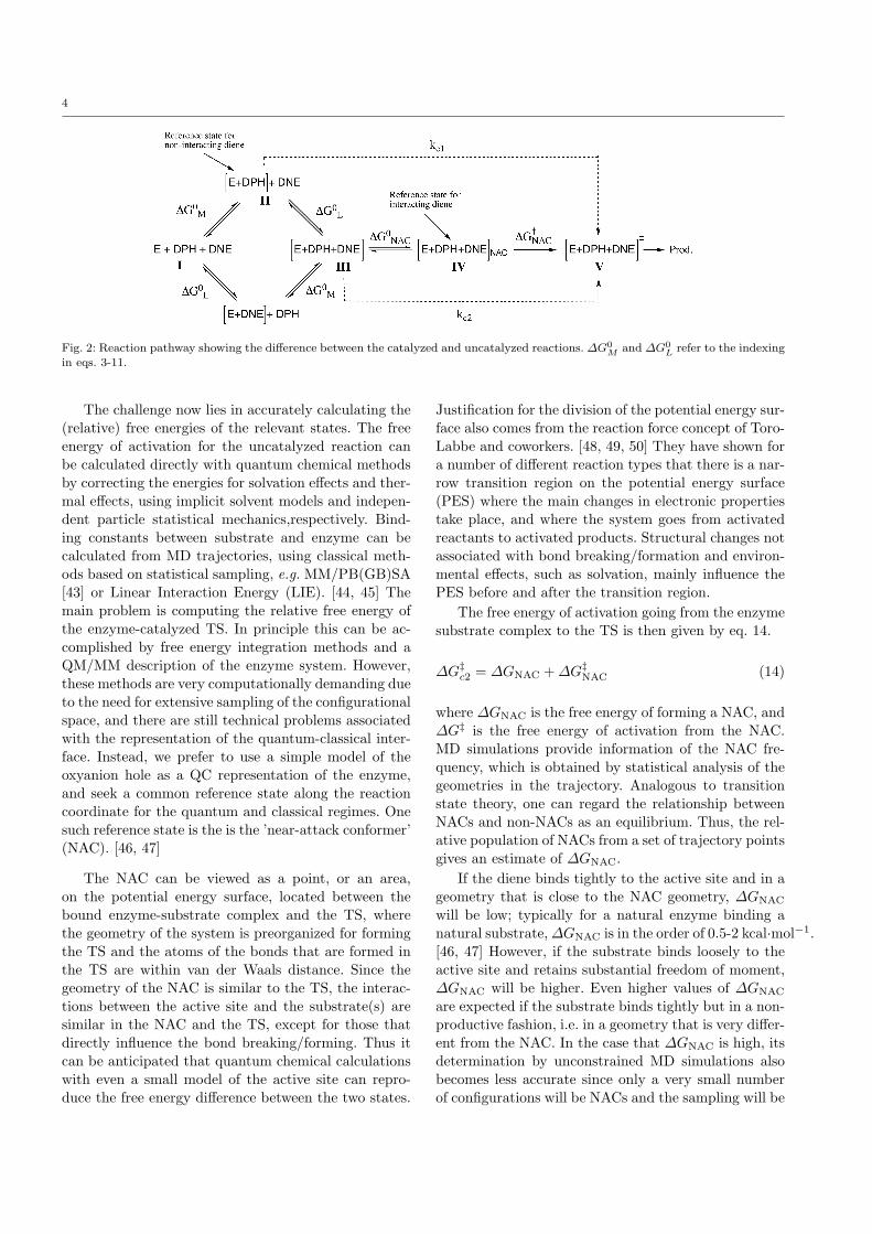
4
Fig. 2: Reaction pathway showing the difference between the catalyzed and uncatalyzed reactions. ∆G0M and ∆G0
L refer to the indexing
in eqs. 3-11.
The challenge now lies in accurately calculating the(relative) free energies of the relevant states. The freeenergy of activation for the uncatalyzed reaction canbe calculated directly with quantum chemical methodsby correcting the energies for solvation effects and ther-mal effects, using implicit solvent models and indepen-dent particle statistical mechanics,respectively. Bind-ing constants between substrate and enzyme can becalculated from MD trajectories, using classical meth-ods based on statistical sampling, e.g. MM/PB(GB)SA[43] or Linear Interaction Energy (LIE). [44, 45] Themain problem is computing the relative free energy ofthe enzyme-catalyzed TS. In principle this can be ac-complished by free energy integration methods and aQM/MM description of the enzyme system. However,these methods are very computationally demanding dueto the need for extensive sampling of the configurationalspace, and there are still technical problems associatedwith the representation of the quantum-classical inter-face. Instead, we prefer to use a simple model of theoxyanion hole as a QC representation of the enzyme,and seek a common reference state along the reactioncoordinate for the quantum and classical regimes. Onesuch reference state is the is the ’near-attack conformer’(NAC). [46, 47]
The NAC can be viewed as a point, or an area,on the potential energy surface, located between thebound enzyme-substrate complex and the TS, wherethe geometry of the system is preorganized for formingthe TS and the atoms of the bonds that are formed inthe TS are within van der Waals distance. Since thegeometry of the NAC is similar to the TS, the interac-tions between the active site and the substrate(s) aresimilar in the NAC and the TS, except for those thatdirectly influence the bond breaking/forming. Thus itcan be anticipated that quantum chemical calculationswith even a small model of the active site can repro-duce the free energy difference between the two states.
Justification for the division of the potential energy sur-face also comes from the reaction force concept of Toro-Labbe and coworkers. [48, 49, 50] They have shown fora number of different reaction types that there is a nar-row transition region on the potential energy surface(PES) where the main changes in electronic propertiestake place, and where the system goes from activatedreactants to activated products. Structural changes notassociated with bond breaking/formation and environ-mental effects, such as solvation, mainly influence thePES before and after the transition region.
The free energy of activation going from the enzymesubstrate complex to the TS is then given by eq. 14.
∆G‡c2 = ∆GNAC +∆G‡NAC (14)
where ∆GNAC is the free energy of forming a NAC, and∆G‡ is the free energy of activation from the NAC.MD simulations provide information of the NAC fre-quency, which is obtained by statistical analysis of thegeometries in the trajectory. Analogous to transitionstate theory, one can regard the relationship betweenNACs and non-NACs as an equilibrium. Thus, the rel-ative population of NACs from a set of trajectory pointsgives an estimate of ∆GNAC.
If the diene binds tightly to the active site and in ageometry that is close to the NAC geometry, ∆GNAC
will be low; typically for a natural enzyme binding anatural substrate,∆GNAC is in the order of 0.5-2 kcal·mol−1.[46, 47] However, if the substrate binds loosely to theactive site and retains substantial freedom of moment,∆GNAC will be higher. Even higher values of ∆GNAC
are expected if the substrate binds tightly but in a non-productive fashion, i.e. in a geometry that is very differ-ent from the NAC. In the case that ∆GNAC is high, itsdetermination by unconstrained MD simulations alsobecomes less accurate since only a very small numberof configurations will be NACs and the sampling will be
5
insufficient. This is particularly true if stringent criteriafor NAC geometries are enforced.
On the basis of ∆GNAC obtained from the MD sim-ulation and ∆G‡NAC from the quantum chemical calcu-lation it is possible to estimate ∆G‡c2. Combined withthe free energy estimate of the uncatalyzed reaction,kc2/ku can be calculated (c.f. Fig. 2):
kc2ku
= e−(∆G‡c2−∆G‡u)/RT (15)
Thus, by a combination of MD simulation and quan-tum chemical calculations it is possible to estimate thecatalytic performance for different substrates and en-zyme combinations using eq. 11. There is, unfortunately,substantial uncertainty involved in the determination ofthe different energy terms, which makes it hard to ob-tain accurate global energy estimates. However, relativerates are likely to be of better precision. In particular,for ranking the performance of different enzyme vari-ants, e.g. mutations, the ∆GNAC can be of great im-portance. For this purpose less stringent NAC criteriacan be employed, which improves the statistical signif-icance of the results. A first estimate of the quality ofa given enzyme variant or substrate is however mosteasily obtained by visual and statistical analysis. Theability to form NACs has for this purpose been usedas an important indicator of the viability of a givenenzyme variant.
2.2 Enzyme variants
The CALB model was obtained from the PDB struc-ture 1LBT. [39] The entry was chosen due to the pres-ence of a large lipid analog covalently bound to the cat-alytic S105, which putatively induces a fully active con-formation of the enzyme. The all-atom RMSD to twoother common structures in modeling is small; 1TCA[51] = 0.29, 1LBS [39] = 0.44. The 1LBT structure wasstripped of all water molecules and protonated (defaultprotonation state) in the viewer Hermes, a part of theGOLD Suite. [52] This structure was subsequently usedas the basis for mutant models, which were generatedin SwissPDB viewer. [53]
2.3 Molecular docking
Selection of dienophiles and generation of initial stud-ies were accomplished by docking simulations, usingthe GOLD Suite. [54, 55, 56, 57] The docking proce-dure was conducted according to the following proto-col: The built-in genetic algorithm (GA) was used to
generate different poses, which were evaluated with theChemScore scoring function. [58, 59] The space of pos-sible poses was limited to relevant ones by inducinghydrogen bond constraints between the dienophile car-bonyl oxygen and T40-NH, T40-OγH and Q106-NH. Tosomewhat counteract the limitations created by theseconstraints, the ’diverse solution’ flag was switched on,using default parameters. 105 GA runs were used foreach docking pose, and 50 poses were generated foreach molecule. Statistics were calculated on the top 10of these solutions, based on total score. The poses werefurther analyzed according to a composite scoring func-tion, which is explained in detail in the Electronic Sup-plementary Material (ESM). After numerical and visualanalysis, the best poses of each dienophile were savedfor docking of the diene. The same set of parameterswere used as for docking the dienophiles, except now,no constraints were used to limit the possible poses.
Docking in the MD-relaxed variants was performedanalogously. Since the average structures (Section 3.1)resulted in unphysical positions of a large portion ofhydrogens, all hydrogens were stripped off and the en-zymes were reprotonated in GOLD. The H-bond con-straints were limited to T40-NH and Q106-NH sincethe T40-OγH was seen to contribute less to binding ofthe substrates, and turned away somewhat during re-laxation.
2.4 Molecular dynamics simulations
All MD simulation were performed with the AMBER 9& 10 packages, [60] and the AMBER 99 force field [61]was employed throughout. All initial structures weretaken from molecular docking. Force field parametersfor the nonprotein atoms (dienophiles and diene) weregenerated from the general Amber force field (gaff),[62] using the Antechamber program. The generationof gaff-parameters was based on AM1 geometries, andthe AM1-BCC approach was used for partial charges.[63, 64] All amino acid residues were assumed to be intheir natural protonation state.
The LEaP program was used to create topologyfiles, neutralize the enzyme complexes with Sodium ions,and add an octahedral box of solvent in a minimumlayer of 8 A. Acetonitrile (ACN) was treated by theavailable united-atom model, [65] while chloroform (CHL)[66] and water (WAT, described by TIP3P) [67] param-eters were taken from all-atom models.
Simulations were performed under periodic bound-ary conditions and the long-range electrostatics washandled by particle Mesh Ewald summation. Initial min-imization was performed in two steps of 1000 and 2500iterations, respectively, where the protein was held fixed
6
during the first step. Next, the systems were heatedto 300 K over 20 ps with mild restraints on the pro-tein atoms (10 kcal·mol−1·A). Unconstrained simula-tions followed for 2-4 ns at 300 K and 1.0 bar. 2 fs timesteps were used, both during warmup and the followingsimulations, and hydrogen movements were constrainedby the SHAKE algorithm. [68]
Trajectories were based on snapshots taken every0.2 ps. Statistics were obtained using the PTRAJ toolwithin AMBER. For structural analysis, an average struc-ture was generated based on the last 1000 snapshots(200 ps). In these structures, hydrogen positions werepoorly defined, so when performing subsequent dock-ing, all hydrogens were stripped off and the relaxed en-zymes were reprotonated in GOLD.
For simulations of substrates 1 and 3 in water (seesections 3.4 and 3.5), the binding free energies of bothdiene and dienophile were estimated using the LIE meth-od. [44, 45] LIE approximates ∆Gbind by an electro-static contribution V elec (Coulomb interactions) and anon-polar (van der Waals) contribution V vdw. Thesecontributions are weighted by coefficients α and β, cal-ibrated to Free Energy Perturbation (FEP) data andempirical data, respectively.
∆Gbind = α[〈V elecl−s 〉bound − 〈V elecl−s 〉free]+β[〈V vdwl−s 〉bound − 〈V vdwl−s 〉free],
(16)
where α has been determined to 0.43 and β to 0.18 forthe neutral molecules used here.[69] 300 samples weretaken from the last half of the 2 ns trajectories. The LIEmethod requires explicit simulations of the free solvatedsubstrates; they were thus simulated for 500 ps, fromwhich 200 samples were used. Both average values andstandard deviation were stable with respect to a 5-foldincrease in sampling rate.
It should be noted that we also attempted to cal-culate binding free energies using the MM/PBSA andMM/GBSA approaches, but they lead to more diver-gent results and irrational magnitudes.
2.5 DFT calculations
A model of the oxyanion hole was created from the par-ent crystal structure 1LBT, using the docked structuresas starting guesses for the dienophile and TS. The cen-tral peptide atoms of the backbone Q106 and T40 nitro-gens were included together with adjacent α-carbons, inorder to correctly model the electrostatics of the hydro-gen bonds. The T40 side chain was included as well amethyl group on residue 105, representing both serineand alanine. Finally, the entire Q106 side chain was in-cluded because it is seen to H-bond to the back of the
T40 OH-group in CALB crystal structures. A graphicalrepresentation of the oxyanion hole model is providedin ESM Fig. 5.
Geometry optimizations were performed at the B3-LYP/6-31G(d) level, [70, 71, 72, 73] for the [E·dph],NAC-[E·dph·dne] (the C4
dne–Cαdph and C1dne–Cβdph dis-
tances being fixed at 4.0 A, see the discussion in Section3.3) and [E·dph·dne]‡ states. Gaussian 03 was used forall calculations. [74] An endo conformation of the TSwas chosen, since it is generally accepted to be the ki-netically favored geometry. [75] Because of the trun-cated oxyanion hole model, several atoms were keptfrozen throughout the optimization, as illustrated inESM Fig. 5. As this generally leads to more than onenegative force constant in the calculated Hessian, nothermodynamic corrections were made on the oxyanionhole structures. Where stated, estimates of the correc-tions were instead made by using the Hessian from thecorresponding uncatalyzed reactions. This is a reason-able estimate since eigenstates of the enzyme are ex-pected to change little during the course of reaction.The transition state were detected by identifying themost negative imaginary frequency as being the forma-tion of the two incipient C-C bonds.
Single point calculations on the optimized struc-tures were performed using a 6-31G++(d,p) basis setand the C-PCM solvent model. [76, 77] We used a di-electric constant of ε = 4 to simulate the polarity ofa typical enzymatic environment, and the shape of thecavity was determined by the United Atom TopologicalModel. [78]
2.6 Graphical representation
All molecular representations were created using theUCSF Chimera package. [79, 80] Diagrams were createdusing XMGrace [81] and Plot. [82]
3 Results and discussion
3.1 Enzyme variants
To test for relative stability and look for unexpecteddistortions of the enzyme variants, 4 ns MD simulationswere carried out on each variant in the three solvents.In all simulations, the backbone RMS trajectories levelout after 1 ns, indicating that 4 ns is enough simulationtime for this study.
For structural comparison, average structures of thelast 200 ps of simulations were abstracted. Their back-bone RMS deviations (RMSDs) with respect to the
7
crystal structure are shown in ESM Table 1. No ma-jor structural change occurs in any solvent, comparedto that of the wild type, which suggests that all mutantsare potentially stable. As expected, the water simula-tions display the lowest RMSDs with respect to thecrystal structure, while the CHL-relaxed variants showthe highest RMSD values.
An extensive analysis of solvent and mutant influ-ences on the structure is given in the ESM. Here, wesummarize by noting that the essential catalytic ma-chinery is well-preserved, but that the T40 side-chainmoves away from its crystal position where the OH-group stabilizes the oxyanion. A question thus arisesconcerning its role in catalysis, which will be addressedbelow. The S105 residue, where present, tends to moveinto the designated oxyanion hole region, an observa-tion that advocates mutation. Finally, the I189A andI285A mutations did not generate any major structuraldistortions, and should thus be viable.
ACN was chosen for further MD studies of enzyme-substrate interactions, since it does not significantly dis-tort the cavity, while it is a moderately polar solvent,which is believed to improve the affinity between thesubstrates and the hydrophobic active site. Moreover,using an organic solvent is of interest in synthesis, and(immobilized) CALB is known to operate well in dryenvironments.
3.2 Molecular docking
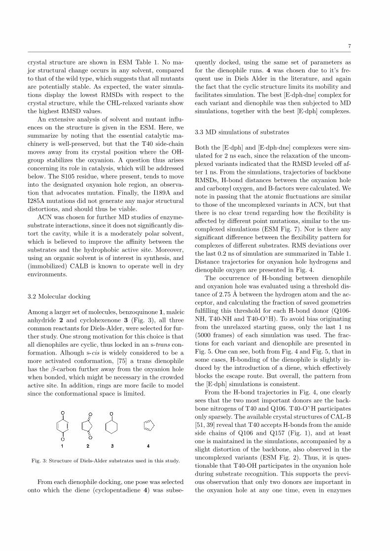
Among a larger set of molecules, benzoquinone 1, maleicanhydride 2 and cyclohexenone 3 (Fig. 3), all threecommon reactants for Diels-Alder, were selected for fur-ther study. One strong motivation for this choice is thatall dienophiles are cyclic, thus locked in an s-trans con-formation. Alhough s-cis is widely considered to be amore activated conformation, [75] a trans dienophilehas the β-carbon further away from the oxyanion holewhen bonded, which might be necessary in the crowdedactive site. In addition, rings are more facile to modelsince the conformational space is limited.
Fig. 3: Structure of Diels-Alder substrates used in this study.
From each dienophile docking, one pose was selectedonto which the diene (cyclopentadiene 4) was subse-
quently docked, using the same set of parameters asfor the dienophile runs. 4 was chosen due to it’s fre-quent use in Diels Alder in the literature, and againthe fact that the cyclic structure limits its mobility andfacilitates simulation. The best [E·dph·dne] complex foreach variant and dienophile was then subjected to MDsimulations, together with the best [E·dph] complexes.
3.3 MD simulations of substrates
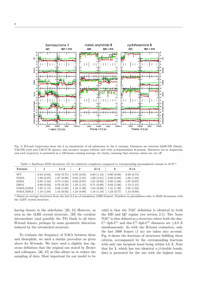
Both the [E·dph] and [E·dph·dne] complexes were sim-ulated for 2 ns each, since the relaxation of the uncom-plexed variants indicated that the RMSD leveled off af-ter 1 ns. From the simulations, trajectories of backboneRMSDs, H-bond distances between the oxyanion holeand carbonyl oxygen, and B-factors were calculated. Wenote in passing that the atomic fluctuations are similarto those of the uncomplexed variants in ACN, but thatthere is no clear trend regarding how the flexibility isaffected by different point mutations, similar to the un-complexed simulations (ESM Fig. 7). Nor is there anysignificant difference between the flexibility pattern forcomplexes of different substrates. RMS deviations overthe last 0.2 ns of simulation are summarized in Table 1.Distance trajectories for oxyanion hole hydrogens anddienophile oxygen are presented in Fig. 4.
The occurrence of H-bonding between dienophileand oxyanion hole was evaluated using a threshold dis-tance of 2.75 A between the hydrogen atom and the ac-ceptor, and calculating the fraction of saved geometriesfulfilling this threshold for each H-bond donor (Q106-NH, T40-NH and T40-OγH). To avoid bias originatingfrom the unrelaxed starting guess, only the last 1 ns(5000 frames) of each simulation was used. The frac-tions for each variant and dienophile are presented inFig. 5. One can see, both from Fig. 4 and Fig. 5, that insome cases, H-bonding of the dienophile is slightly in-duced by the introduction of a diene, which effectivelyblocks the escape route. But overall, the pattern fromthe [E·dph] simulations is consistent.
From the H-bond trajectories in Fig. 4, one clearlysees that the two most important donors are the back-bone nitrogens of T40 and Q106. T40-OγH participatesonly sparsely. The available crystal structures of CAL-B[51, 39] reveal that T40 accepts H-bonds from the amideside chains of Q106 and Q157 (Fig. 1), and at leastone is maintained in the simulations, accompanied by aslight distortion of the backbone, also observed in theuncomplexed variants (ESM Fig. 2). Thus, it is ques-tionable that T40-OH participates in the oxyanion holeduring substrate recognition. This supports the previ-ous observation that only two donors are important inthe oxyanion hole at any one time, even in enzymes
8
Fig. 4: H-bond trajectories from the 2 ns simulations of all substrates in the 6 variants. Distances are between Q106-NH (black),
T40-NH (red) and T40-OγH (green), and acceptor oxygen without and with cyclopentadiene 4 present. Distances are in Angstromsand each trajectory is presented as a 100-frame running average, for clarity, meaning that extreme values are cut off.
Table 1: Backbone RMS deviations (A) for substrate complexes compared to corresponding uncomplexed variant in ACN.a
Variant 1 1+4 2 2+4 3 3+4
WT 0.83 (0.93) 0.94 (0.75) 0.76 (0.85) 0.80 (1.13) 0.90 (0.80) 0.99 (0.75)S105A 1.00 (0.87) 1.07 (0.88) 0.94 (1.01) 1.06 (1.01) 0.98 (0.86) 1.06 (1.04)
I189A 0.85 (1.04) 0.75 (1.03) 0.92 (0.97) 1.01 (0.92) 0.92 (1.06) 1.07 (0.97)
I285A 0.89 (0.92) 0.76 (0.76) 1.34 (1.41) 0.71 (0.99) 0.84 (1.06) 1.13 (1.31)S105A/I189A 1.05 (1.15) 0.98 (1.09) 1.16 (1.09) 1.04 (0.86) 1.41 (1.39) 1.05 (1.04)
S105A/I285A 1.18 (1.04) 1.16 (0.92) 1.24 (0.90) 1.16 (1.19) 1.24 (0.77) 1.13 (0.88)
a Based on average structures from the last 0.2 ns of simulation (1000 frames). Numbers in parentheses refer to RMS deviations with
the 1LBT crystal structure.
having donors in the sidechains. [20, 21] However, asseen in the 1LBS crystal structure, [39] the covalentintermediate (and possibly the TS) binds to all threeH-bond donors, perhaps by some geometric distortioninduced by the tetrahedral structure.
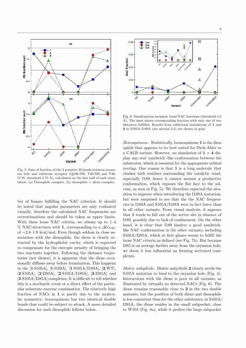
To evaluate the frequency of NACs between dieneand dienophile, we used a similar procedure as givenabove for H-bonds. We have used a slightly less rig-orous definition that the original one stated by Bruiceand colleagues, [46, 47] as this allows us to reduce thesampling of data. Most important for our model to be
valid is that the NAC definition is identical in boththe MD and QC regime (see section 2.1). The ’looseNAC’ is thus defined as a structure where both the dne-C1–dph-Cα and dne-C4–dph-Cβ distances are ≤4.0 Asimultaneously. As with the H-bond evaluation, onlythe last 5000 frames (1 ns) are taken into account.Fig. 6 shows the fractions of structures fulfilling thesecriteria, accompanied by the corresponding fractionswith only one incipient bond being within 4.0 A. Notethat for 1, which has two identical α,β-double bonds,data is presented for the one with the highest num-
9
Fig. 5: Sum of fraction of the 3 putative H-bonds between oxyan-
ion hole and substrate acceptor (Q106-NH, T40-NH and T40-OγH, threshold 2.75 A), calculated on the last half of each simu-
lation. (a) Dienophile complex, (b) dienophile + diene complex.
ber of frames fulfilling the NAC criterion. It shouldbe noted that angular parameters are only evaluatedvisually, therefore the calculated NAC frequencies areoverestimations and should be taken as upper limits.With these loose NAC criteria, we obtain up to 1–4% NAC-structures with 1, corresponding to a ∆GNAC
of ∼2.8–1.9 kcal/mol. Even though seldom in close as-sociation with the dienophile, the diene is clearly at-tracted by the hydrophobic cavity, which is expectedto compensate for the entropic penalty of bringing thetwo reactants together. Following the distance trajec-tories (not shown), it is apparent that the diene occa-sionally diffuses away before termination. This happensin the [1·S105A], [1·I189A], [1·S105A/I189A], [2·WT],[2·S105A], [2·I285A], [2·S105A/I189A], [3·I285A] and[3·S105A/I285A] complexes. It is difficult to tell whetherthis is a stochastic event or a direct effect of the partic-ular substrate-enzyme combination. The relatively highfraction of NACs in 1 is partly due to the molecu-lar symmetry; benzoquinone has two identical doublebonds that could be subject to attack. A more detaileddiscussion for each dienophile follows below.
Fig. 6: Simultaneous incipient bond NAC fractions (threshold 4.4A). The inset shows corresponding fraction with only one of two
distances fulfilled. Results from additional simulations of 1 and
3 in S105A/I189A (see section 3.4) are shown in gray.
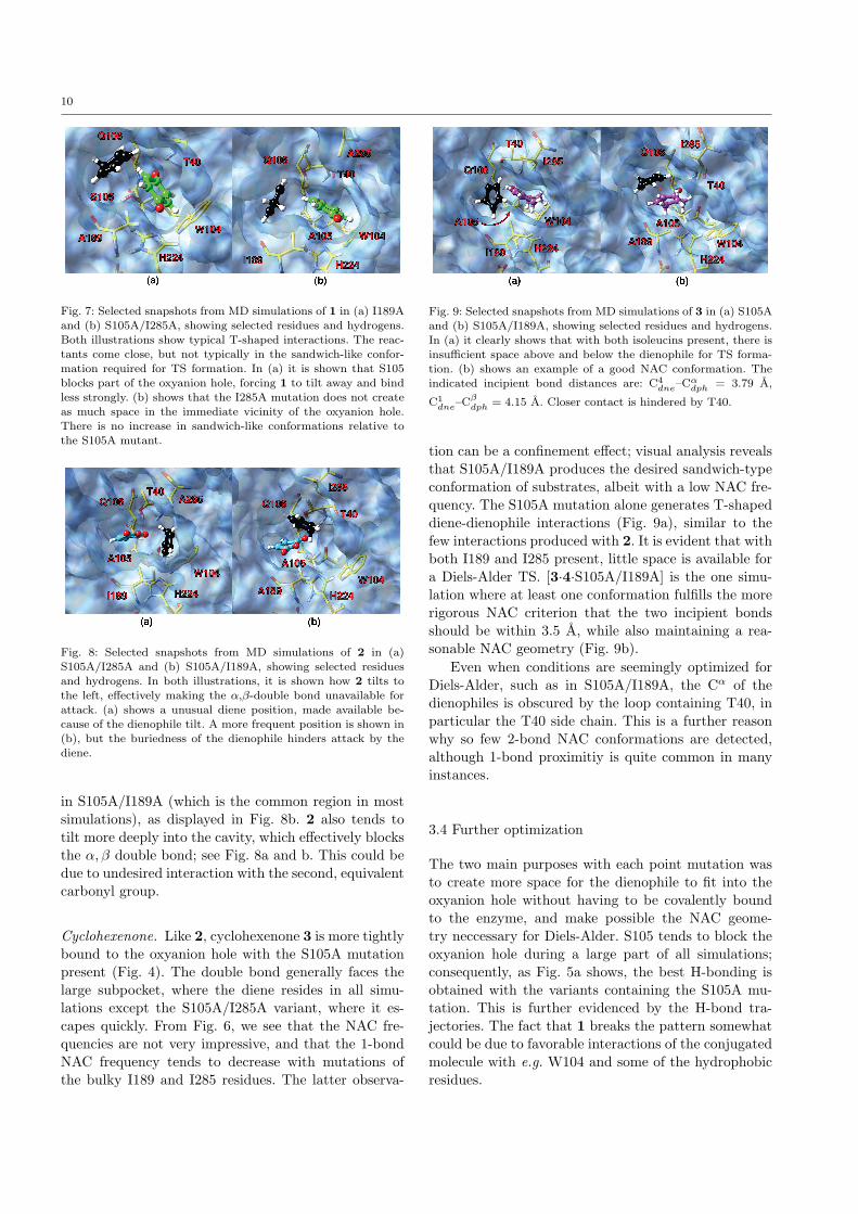
Benzoquinone. Statistically, benzoquinone 1 is the dien-ophile that appears to be best suited for Diels-Alder ina CALB variant. However, no simulation of 1 + 4 dis-play any real ’sandwich’-like conformation between thesubstrates, which is essential for the appropriate orbitaloverlap. One reason is that 1 is a long molecule thatclashes with residues surrounding the catalytic triad,especially I189, hence it cannot assume a productiveconformation, which exposes the flat face to the sol-vent, as seen in Fig. 7a. We therefore expected the situ-ation to improve when introducing the I189A mutation,but were surprised to see that the the NAC frequen-cies in I189A and S105A/I189A were in fact lower thanin all other variants. From visual analysis, it appearsthat 1 tends to fall out of the active site in absence ofI189, possibly due to lack of confinement. On the otherhand, it is clear that I189 hinders a good sandwich-like NAC conformation in the other variants, includingS105A/I285A, which at first glance seems to fulfill theloose NAC criteria as defined (see Fig. 7b). But becauseI285 is on average further away from the oxyanion hole,we deem it less influential on forming activated com-plexes.
Maleic anhydride. Maleic anhydride 2 clearly needs theS105A mutation to bind to the oxyanion hole (Fig. 4).Interactions with the diene is poor in all variants, asillustrated by virtually no detected NACs (Fig. 6). Thediene remains reasonably close to 2 in the two doublemutants, but the position of both diene and dienophileis less consistent than for the other substrates; in S105A/I285A, the diene resides in the small subpocket, closeto W104 (Fig. 8a), while it prefers the large subpocket
10
Fig. 7: Selected snapshots from MD simulations of 1 in (a) I189A
and (b) S105A/I285A, showing selected residues and hydrogens.
Both illustrations show typical T-shaped interactions. The reac-tants come close, but not typically in the sandwich-like confor-
mation required for TS formation. In (a) it is shown that S105
blocks part of the oxyanion hole, forcing 1 to tilt away and bindless strongly. (b) shows that the I285A mutation does not create
as much space in the immediate vicinity of the oxyanion hole.
There is no increase in sandwich-like conformations relative tothe S105A mutant.
Fig. 8: Selected snapshots from MD simulations of 2 in (a)S105A/I285A and (b) S105A/I189A, showing selected residues
and hydrogens. In both illustrations, it is shown how 2 tilts to
the left, effectively making the α,β-double bond unavailable forattack. (a) shows a unusual diene position, made available be-
cause of the dienophile tilt. A more frequent position is shown in(b), but the buriedness of the dienophile hinders attack by thediene.
in S105A/I189A (which is the common region in mostsimulations), as displayed in Fig. 8b. 2 also tends totilt more deeply into the cavity, which effectively blocksthe α, β double bond; see Fig. 8a and b. This could bedue to undesired interaction with the second, equivalentcarbonyl group.
Cyclohexenone. Like 2, cyclohexenone 3 is more tightlybound to the oxyanion hole with the S105A mutationpresent (Fig. 4). The double bond generally faces thelarge subpocket, where the diene resides in all simu-lations except the S105A/I285A variant, where it es-capes quickly. From Fig. 6, we see that the NAC fre-quencies are not very impressive, and that the 1-bondNAC frequency tends to decrease with mutations ofthe bulky I189 and I285 residues. The latter observa-
Fig. 9: Selected snapshots from MD simulations of 3 in (a) S105A
and (b) S105A/I189A, showing selected residues and hydrogens.
In (a) it clearly shows that with both isoleucins present, there isinsufficient space above and below the dienophile for TS forma-
tion. (b) shows an example of a good NAC conformation. The
indicated incipient bond distances are: C4dne–Cαdph = 3.79 A,
C1dne–Cβdph = 4.15 A. Closer contact is hindered by T40.
tion can be a confinement effect; visual analysis revealsthat S105A/I189A produces the desired sandwich-typeconformation of substrates, albeit with a low NAC fre-quency. The S105A mutation alone generates T-shapeddiene-dienophile interactions (Fig. 9a), similar to thefew interactions produced with 2. It is evident that withboth I189 and I285 present, little space is available fora Diels-Alder TS. [3·4·S105A/I189A] is the one simu-lation where at least one conformation fulfills the morerigorous NAC criterion that the two incipient bondsshould be within 3.5 A, while also maintaining a rea-sonable NAC geometry (Fig. 9b).
Even when conditions are seemingly optimized forDiels-Alder, such as in S105A/I189A, the Cα of thedienophiles is obscured by the loop containing T40, inparticular the T40 side chain. This is a further reasonwhy so few 2-bond NAC conformations are detected,although 1-bond proximitiy is quite common in manyinstances.
3.4 Further optimization
The two main purposes with each point mutation wasto create more space for the dienophile to fit into theoxyanion hole without having to be covalently boundto the enzyme, and make possible the NAC geome-try neccessary for Diels-Alder. S105 tends to block theoxyanion hole during a large part of all simulations;consequently, as Fig. 5a shows, the best H-bonding isobtained with the variants containing the S105A mu-tation. This is further evidenced by the H-bond tra-jectories. The fact that 1 breaks the pattern somewhatcould be due to favorable interactions of the conjugatedmolecule with e.g. W104 and some of the hydrophobicresidues.
11
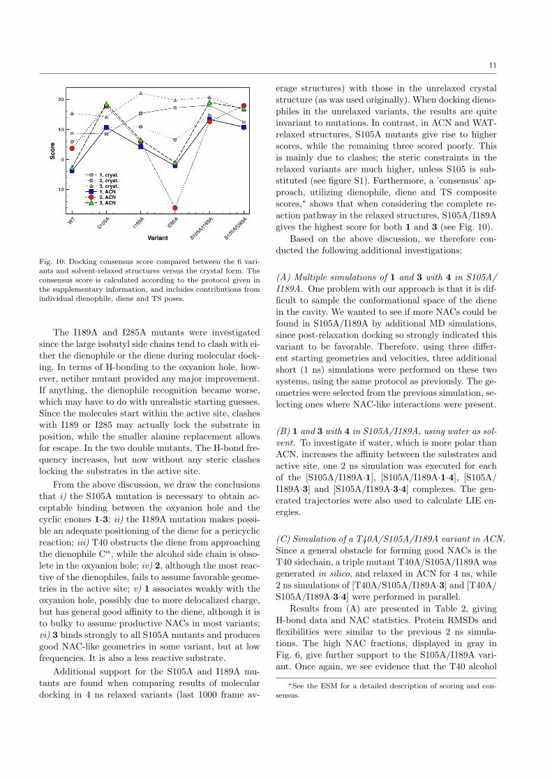
Fig. 10: Docking consensus score compared between the 6 vari-
ants and solvent-relaxed structures versus the crystal form. Theconsensus score is calculated according to the protocol given in
the supplementary information, and includes contributions from
individual dienophile, diene and TS poses.
The I189A and I285A mutants were investigatedsince the large isobutyl side chains tend to clash with ei-ther the dienophile or the diene during molecular dock-ing. In terms of H-bonding to the oxyanion hole, how-ever, netiher mutant provided any major improvement.If anything, the dienophile recognition became worse,which may have to do with unrealistic starting guesses.Since the molecules start within the active site, clasheswith I189 or I285 may actually lock the substrate inposition, while the smaller alanine replacement allowsfor escape. In the two double mutants, The H-bond fre-quency increases, but now without any steric clasheslocking the substrates in the active site.
From the above discussion, we draw the conclusionsthat i) the S105A mutation is necessary to obtain ac-ceptable binding between the oxyanion hole and thecyclic enones 1-3; ii) the I189A mutation makes possi-ble an adequate positioning of the diene for a pericyclicreaction; iii) T40 obstructs the diene from approachingthe dienophile Cα, while the alcohol side chain is obso-lete in the oxyanion hole; iv) 2, although the most reac-tive of the dienophiles, fails to assume favorable geome-tries in the active site; v) 1 associates weakly with theoxyanion hole, possibly due to more delocalized charge,but has general good affinity to the diene, although it isto bulky to assume productive NACs in most variants;vi) 3 binds strongly to all S105A mutants and producesgood NAC-like geometries in some variant, but at lowfrequencies. It is also a less reactive substrate.
Additional support for the S105A and I189A mu-tants are found when comparing results of moleculardocking in 4 ns relaxed variants (last 1000 frame av-
erage structures) with those in the unrelaxed crystalstructure (as was used originally). When docking dieno-philes in the unrelaxed variants, the results are quiteinvariant to mutations. In contrast, in ACN and WAT-relaxed structures, S105A mutants give rise to higherscores, while the remaining three scored poorly. Thisis mainly due to clashes; the steric constraints in therelaxed variants are much higher, unless S105 is sub-stituted (see figure S1). Furthermore, a ’consensus’ ap-proach, utilizing dienophile, diene and TS compositescores,∗ shows that when considering the complete re-action pathway in the relaxed structures, S105A/I189Agives the highest score for both 1 and 3 (see Fig. 10).
Based on the above discussion, we therefore con-ducted the following additional investigations:
(A) Multiple simulations of 1 and 3 with 4 in S105A/I189A. One problem with our approach is that it is dif-ficult to sample the conformational space of the dienein the cavity. We wanted to see if more NACs could befound in S105A/I189A by additional MD simulations,since post-relaxation docking so strongly indicated thisvariant to be favorable. Therefore, using three differ-ent starting geometries and velocities, three additionalshort (1 ns) simulations were performed on these twosystems, using the same protocol as previously. The ge-ometries were selected from the previous simulation, se-lecting ones where NAC-like interactions were present.
(B) 1 and 3 with 4 in S105A/I189A, using water as sol-vent. To investigate if water, which is more polar thanACN, increases the affinity between the substrates andactive site, one 2 ns simulation was executed for eachof the [S105A/I189A·1], [S105A/I189A·1·4], [S105A/I189A·3] and [S105A/I189A·3·4] complexes. The gen-erated trajectories were also used to calculate LIE en-ergies.
(C) Simulation of a T40A/S105A/I189A variant in ACN.Since a general obstacle for forming good NACs is theT40 sidechain, a triple mutant T40A/S105A/I189A wasgenerated in silico, and relaxed in ACN for 4 ns, while2 ns simulations of [T40A/S105A/I189A·3] and [T40A/S105A/I189A·3·4] were performed in parallel.
Results from (A) are presented in Table 2, givingH-bond data and NAC statistics. Protein RMSDs andflexibilities were similar to the previous 2 ns simula-tions. The high NAC fractions, displayed in gray inFig. 6, give further support to the S105A/I189A vari-ant. Once again, we see evidence that the T40 alcohol
∗See the ESM for a detailed description of scoring and con-
sensus.

12
Table 2: H-bond and NAC statistics for the three 1 ns simulations of 1 and 3 with 4 in S105A/I189A.a
dph H-bond d [A]b NAC fractionsc
Entry Q88-NH T40-NH T40-OγH Loosed Tighte
1 1, I 2.87 (0.61) 3.01 (0.61) 3.47 (0.07) 0.053 0.0016
2 1, II 2.37 (0.91) 2.30 (0.91) 3.62 (¡0.01) 0.081 0.00223 1, IIIf 3.05 (0.35) 2.47 (0.81) 4.21 (0.01) 0 0
4 3, I 2.75 (0.52) 2.31 (0.91) 3.76 (¡0.01) 0.037 0.0004
5 3, II 2.36 (0.87) 2.14 (0.98) 3.83 (¡0.01) 0.020 0.0002
6 3, III 2.58 (0.75) 2.14 (0.98) 3.43 (¡0.01) 0.005 0.0002
a 1 ns simulations, 5000 saved geometries. b Distances are averaged over all 5000 frames. Numbers in parentheses show the fraction
of distances below a threshold value of 2.75 A. cBased on the two-bond criterium described in the text. d4.0 A threshold. e 3.5 Athreshold. f The diene left the cavity shortly after the beginning of the simulation, similar to the original 2 ns run.
is no participant in the oxyanion hole in our system. In-triguingly, the loose NAC geometries are now quite fre-quent in two of three simulations with each substrate.We also detect some ’tight’ NAC poses, although theyare still very scarce. With this set of simulations, wehave shown that 1 can bind to the S105A/I189A andform NACs, and that the null result from the first setupwas not generalizable, but that multiple simulations arepreferable for sufficient sampling. However, 1 seems tobe bonded more weakly to the oxyanion hole than 3,as implied by the average H-bond distances in Table2. Visual analysis of the trajectories also reveals that1 tumbles around in the active site more than 3, andoccasionally falls out of the oxyanion hole for a shorttime. The best single simulation in this set is entry 2 inTable 2, in which a sandwich-like NAC is maintainedalmost constantly (see the supplementary informationfor an animation). These data further multiple trajec-tories are indeed important for efficient sampling of theconformational space, which is a standard approach ofe.g. Pleiss and coworkers. [83, 84]
The saturated framework of 3 makes it more diffi-cult to form sandwich-like interactions with 4 than for1. There is a smaller volume available for the diene tomove in to form NACs with 3, and this volume is of-ten occupied by surrounding residues, most dominantlyI285, T40 and Val140. The prevalent interaction is a T-shaped geometry, with 4 on the double bond side of 3.Still, 4 displays good affinity with the active site anddienophile.
Results from the water simulations (B) are givenin Table 3. We note that while 3 sticks tightly to theoxyanion hole, 1 tumbles around slightly, as observedin ACN. Hence the high average H-bond distances. Wesee a 14-fold increase in NAC fractions for 3, comparedto the initial simulation in ACN, and the NAC fractionfor 1 is even higher. The relationship is more or lessin concert with the findings in (A). More importantly,in both simulations, there is an event where the dienetemporarily falls out of active site but readily moves
back in. In ACN, this does not occur; if the diene startsto diffuse out, it does not stop. Thus, a highly polarsolvent is preferable when working with substrates suchas those in the present study. The issue of Diels-Aldercatalysis by water[75] is not explored further here, butclearly, there will be competition.
An extensive analysis of the T40A/S105A/I189Atriple mutant (C) is given in the ESM. Here it suffices tosay that while H-bonding of the dienophile was surpris-ingly improved, the formation of NACs with the dienewas poor. Both phenomena are believed to arise from arelief of steric crowding around the oxyanion hole. Thiscauses allows the dienophile to bind harder and becomemore buried, which in turn makes it less accessible tothe diene. This finding highlights the importance of del-icate tuning around the catalytic site in order to alignboth reactants.
3.5 Binding Constants
We have estimated the binding constants (KM and KL)of substrates 1, 3 and 4 to S105A/I189A in water us-ing the LIE method, see Table 4. The results for thedienophiles are in agremeent with earlier experimentaland theoretical studies on the binding of small ketonesand aldehydes to CALB and other lipases; the bindingconstants in water are typically in the range of 0.1-1 M,and generally decrease with increasing hydrophobicityof the substrates. [21, 85, 86] Our lowest binding con-stant is obtained for cyclopentadiene 4, which has nospecific binding to the active site. This supports the in-terpretation that the main driving force for binding ishydrophobic interactions. In earlier study on the epox-idation of butenal in CALB wt and S105A we obtainedexperimentally a KM value of 0.2 M in aqueous buffer,whereas no saturation was found in ACN up to con-centrations of 3M. [21] Thus, to obtain sufficiently lowbinding constants we conclude that nonpolar substratesand water as solvent should be used.
13
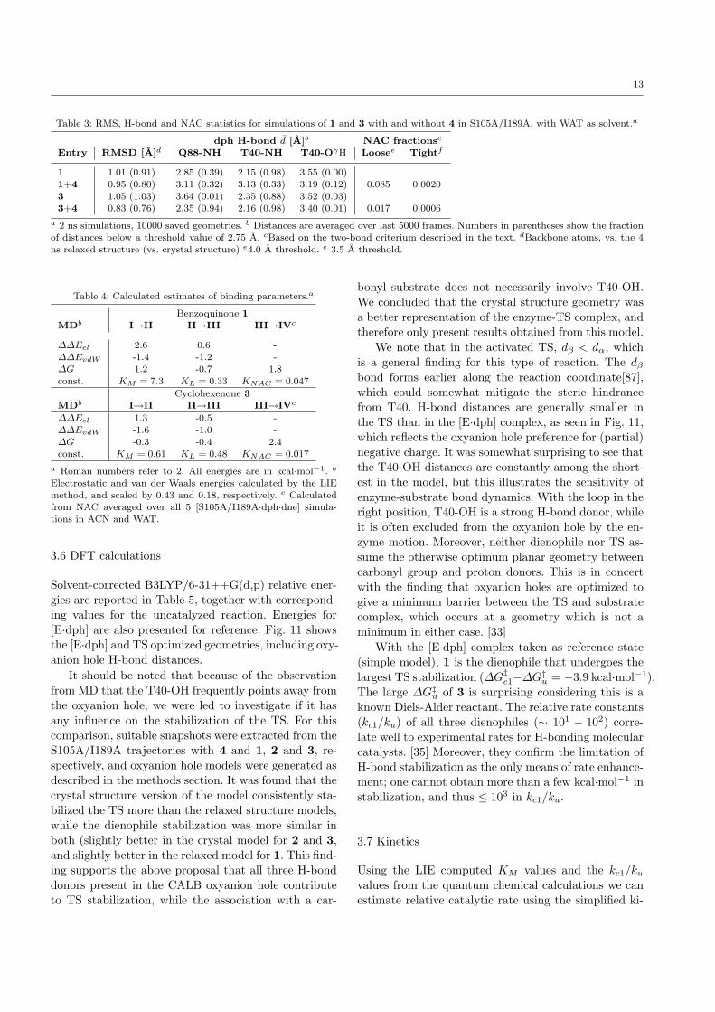
Table 3: RMS, H-bond and NAC statistics for simulations of 1 and 3 with and without 4 in S105A/I189A, with WAT as solvent.a
dph H-bond d [A]b NAC fractionsc
Entry RMSD [A]d Q88-NH T40-NH T40-OγH Loosee Tightf
1 1.01 (0.91) 2.85 (0.39) 2.15 (0.98) 3.55 (0.00)
1+4 0.95 (0.80) 3.11 (0.32) 3.13 (0.33) 3.19 (0.12) 0.085 0.00203 1.05 (1.03) 3.64 (0.01) 2.35 (0.88) 3.52 (0.03)
3+4 0.83 (0.76) 2.35 (0.94) 2.16 (0.98) 3.40 (0.01) 0.017 0.0006
a 2 ns simulations, 10000 saved geometries. b Distances are averaged over last 5000 frames. Numbers in parentheses show the fraction
of distances below a threshold value of 2.75 A. cBased on the two-bond criterium described in the text. dBackbone atoms, vs. the 4ns relaxed structure (vs. crystal structure) e4.0 A threshold. e 3.5 A threshold.
Table 4: Calculated estimates of binding parameters.a
Benzoquinone 1
MDb I→II II→III III→IVc
∆∆Eel 2.6 0.6 -
∆∆EvdW -1.4 -1.2 -
∆G 1.2 -0.7 1.8const. KM = 7.3 KL = 0.33 KNAC = 0.047
Cyclohexenone 3
MDb I→II II→III III→IVc
∆∆Eel 1.3 -0.5 -
∆∆EvdW -1.6 -1.0 -∆G -0.3 -0.4 2.4
const. KM = 0.61 KL = 0.48 KNAC = 0.017
a Roman numbers refer to 2. All energies are in kcal·mol−1. b
Electrostatic and van der Waals energies calculated by the LIE
method, and scaled by 0.43 and 0.18, respectively. c Calculatedfrom NAC averaged over all 5 [S105A/I189A·dph·dne] simula-
tions in ACN and WAT.
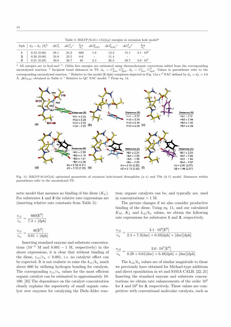
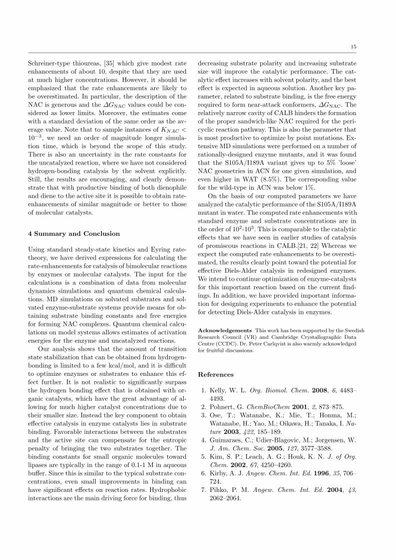
3.6 DFT calculations
Solvent-corrected B3LYP/6-31++G(d,p) relative ener-gies are reported in Table 5, together with correspond-ing values for the uncatalyzed reaction. Energies for[E·dph] are also presented for reference. Fig. 11 showsthe [E·dph] and TS optimized geometries, including oxy-anion hole H-bond distances.
It should be noted that because of the observationfrom MD that the T40-OH frequently points away fromthe oxyanion hole, we were led to investigate if it hasany influence on the stabilization of the TS. For thiscomparison, suitable snapshots were extracted from theS105A/I189A trajectories with 4 and 1, 2 and 3, re-spectively, and oxyanion hole models were generated asdescribed in the methods section. It was found that thecrystal structure version of the model consistently sta-bilized the TS more than the relaxed structure models,while the dienophile stabilization was more similar inboth (slightly better in the crystal model for 2 and 3,and slightly better in the relaxed model for 1. This find-ing supports the above proposal that all three H-bonddonors present in the CALB oxyanion hole contributeto TS stabilization, while the association with a car-
bonyl substrate does not necessarily involve T40-OH.We concluded that the crystal structure geometry wasa better representation of the enzyme-TS complex, andtherefore only present results obtained from this model.
We note that in the activated TS, dβ < dα, whichis a general finding for this type of reaction. The dβbond forms earlier along the reaction coordinate[87],which could somewhat mitigate the steric hindrancefrom T40. H-bond distances are generally smaller inthe TS than in the [E·dph] complex, as seen in Fig. 11,which reflects the oxyanion hole preference for (partial)negative charge. It was somewhat surprising to see thatthe T40-OH distances are constantly among the short-est in the model, but this illustrates the sensitivity ofenzyme-substrate bond dynamics. With the loop in theright position, T40-OH is a strong H-bond donor, whileit is often excluded from the oxyanion hole by the en-zyme motion. Moreover, neither dienophile nor TS as-sume the otherwise optimum planar geometry betweencarbonyl group and proton donors. This is in concertwith the finding that oxyanion holes are optimized togive a minimum barrier between the TS and substratecomplex, which occurs at a geometry which is not aminimum in either case. [33]
With the [E·dph] complex taken as reference state(simple model), 1 is the dienophile that undergoes thelargest TS stabilization (∆G‡c1−∆G‡u = −3.9 kcal·mol−1).The large ∆G‡u of 3 is surprising considering this is aknown Diels-Alder reactant. The relative rate constants(kc1/ku) of all three dienophiles (∼ 101 − 102) corre-late well to experimental rates for H-bonding molecularcatalysts. [35] Moreover, they confirm the limitation ofH-bond stabilization as the only means of rate enhance-ment; one cannot obtain more than a few kcal·mol−1 instabilization, and thus ≤ 103 in kc1/ku.
3.7 Kinetics
Using the LIE computed KM values and the kc1/kuvalues from the quantum chemical calculations we canestimate relative catalytic rate using the simplified ki-
14
Table 5: B3LYP/6-31++G(d,p) energies in oxyanion hole modela
Dph dβ − dα [A]b ∆G‡u ∆G‡c1c kc1
ku∆GdNAC ∆G‡NAC
e ∆G‡c2f kc2
ku
1 0.52 (0.00) 28.1 24.2 660 1.8 13.2 15.1 3.1 · 109
2 0.20 (0.00) 24.8 23.5 8.6 - 11.4 - -3 0.21 (0.28) 40.0 36.7 46 2.4 26.3 28.7 3.0 · 107
a All energies are in kcal·mol−1. Gibbs free energies are estimated using thermodynamic corrections added from the correspondinguncatalyzed reaction. b Incipient bond distances in TS: dα = C4
dne–Cαdph, dβ = C1dne–Cβdph. Values in parentheses refer to the
corresponding uncatalyzed reaction. c Relative to the model [E·dph] complexes depicted in Fig. 11a-c.d NAC defined by dα = dβ = 4.0
A. ∆GNAC obtained in Table 4. e Relative to QC NAC model. f From eq. 14.
Fig. 11: B3LYP/6-31G(d) optimized geometries of oxyanion hole-bound dienophiles (a–c) and TSs (d–f) model. Distances withinparentheses refer to the uncatalyzed TS.
netic model that assumes no binding of the diene (KL).For substrates 1 and 3 the relative rate expressions are(inserting relative rate constants from Table 5):
vc1vu
=660[E0]
7.3 + [dph]
vc1vu
=46[E0]
0.61 + [dph]
Inserting standard enzyme and substrate concentra-tions (10−5 M and 0.001 − 1 M, respectively) in theabove expressions, it is clear that without binding ofthe diene, vc1/vu < 0.001, i.e. no catalytic effect canbe expected. It is not realistic to raise the kc1/ku muchabove 660 by utilizing hydrogen bonding for catalysis.The corresponding vc1/vu values for the most efficientorganic catalyst can be estimated to approximately 10-100. [35] The dependence on the catalyst concentrationclearly explains the superiority of small organic cata-lyst over enzymes for catalyzing the Diels-Alder reac-
tion; organic catalysts can be, and typically are, usedin concentrations > 1 M.
The picture changes if we also consider productivebinding of the diene. Using eq. 11, and our calculatedKM , KL and kc2/ku values, we obtain the followingrate expressions for substrates 1 and 3, respectively.
vc2vu
=3.1 · 109[E0]
2.4 + 7.3[dne] + 0.33[dph] + [dne][dph]
vc2vu
=3.0 · 107[E0]
0.29 + 0.61[dne] + 0.48[dph] + [dne][dph]
The kc2/ku values are of similar magnitude to thosewe previously have obtained for Michael-type additionsand direct epoxidation in wt and S105A CALB. [22, 21]Inserting the standard enzyme and substrate concen-trations we obtain rate enhancements of the order 103
for 1 and 102 for 3, respectively. These values are com-petitive with conventional molecular catalysts, such as
15
Schreiner-type thioureas, [35] which give modest rateenhancements of about 10, despite that they are usedat much higher concentrations. However, it should beemphasized that the rate enhancements are likely tobe overestimated. In particular, the description of theNAC is generous and the ∆GNAC values could be con-sidered as lower limits. Moreover, the estimates comewith a standard deviation of the same order as the av-erage value. Note that to sample instances of KNAC <
10−3, we need an order of magnitude longer simula-tion time, which is beyond the scope of this study.There is also an uncertainty in the rate constants forthe uncatalyzed reaction, where we have not consideredhydrogen-bonding catalysis by the solvent explicitly.Still, the results are encouraging, and clearly demon-strate that with productive binding of both dienophileand diene to the active site it is possible to obtain rate-enhancements of similar magnitude or better to thoseof molecular catalysts.
4 Summary and Conclusion
Using standard steady-state kinetics and Eyring rate-theory, we have derived expressions for calculating therate-enhancements for catalysis of bimolecular reactionsby enzymes or molecular catalysts. The input for thecalculations is a combination of data from moleculardynamics simulations and quantum chemical calcula-tions. MD simulations on solvated substrates and sol-vated enzyme-substrate systems provide means for ob-taining substrate binding constants and free energiesfor forming NAC complexes. Quantum chemical calcu-lations on model systems allows estimates of activationenergies for the enzyme and uncatalyzed reactions.
Our analysis shows that the amount of transitionstate stabilization that can be obtained from hydrogen-bonding is limited to a few kcal/mol, and it is difficultto optimize enzymes or substrates to enhance this ef-fect further. It is not realistic to significantly surpassthe hydrogen bonding effect that is obtained with or-ganic catalysts, which have the great advantage of al-lowing for much higher catalyst concentrations due totheir smaller size. Instead the key component to obtaineffective catalysis in enzyme catalysts lies in substratebinding. Favorable interactions between the substratesand the active site can compensate for the entropicpenalty of bringing the two substrates together. Thebinding constants for small organic molecules towardlipases are typically in the range of 0.1-1 M in aqueousbuffer. Since this is similar to the typical substrate con-centrations, even small improvements in binding canhave significant effects on reaction rates. Hydrophobicinteractions are the main driving force for binding, thus
decreasing substrate polarity and increasing substratesize will improve the catalytic performance. The cat-alytic effect increases with solvent polarity, and the besteffect is expected in aqueous solution. Another key pa-rameter, related to substrate binding, is the free energyrequired to form near-attack conformers, ∆GNAC. Therelatively narrow cavity of CALB hinders the formationof the proper sandwich-like NAC required for the peri-cyclic reaction pathway. This is also the parameter thatis most productive to optimize by point mutations. Ex-tensive MD simulations were performed on a number ofrationally-designed enzyme mutants, and it was foundthat the S105A/I189A variant gives up to 5% ’loose’NAC geometries in ACN for one given simulation, andeven higher in WAT (8.5%). The corresponding valuefor the wild-type in ACN was below 1%.
On the basis of our computed parameters we haveanalyzed the catalytic performance of the S105A/I189Amutant in water. The computed rate enhancements withstandard enzyme and substrate concentrations are inthe order of 102-103. This is comparable to the catalyticeffects that we have seen in earlier studies of catalysisof promiscous reactions in CALB.[21, 22] Whereas weexpect the computed rate enhancements to be overesti-mated, the results clearly point toward the potential foreffective Diels-Alder catalysis in redesigned enzymes.We intend to continue optimization of enzyme-catalystsfor this important reaction based on the current find-ings. In addition, we have provided important informa-tion for designing experiments to enhance the potentialfor detecting Diels-Alder catalysis in enzymes.
Acknowledgements This work has been supported by the Swedish
Research Council (VR) and Cambridge Crystallographic Data
Centre (CCDC). Dr. Peter Carlqvist is also warmly acknowledgedfor fruitful discussions.
References
1. Kelly, W. L. Org. Biomol. Chem. 2008, 6, 4483–4493.
2. Pohnert, G. ChemBioChem 2001, 2, 873–875.3. Ose, T.; Watanabe, K.; Mie, T.; Honma, M.;
Watanabe, H.; Yao, M.; Oikawa, H.; Tanaka, I. Na-ture 2003, 422, 185–189.
4. Guimaraes, C.; Udier-Blagovic, M.; Jorgensen, W.J. Am. Chem. Soc. 2005, 127, 3577–3588.
5. Kim, S. P.; Leach, A. G.; Houk, K. N. J. of Org.Chem. 2002, 67, 4250–4260.
6. Kirby, A. J. Angew. Chem. Int. Ed. 1996, 35, 706–724.
7. Pihko, P. M. Angew. Chem. Int. Ed. 2004, 43,2062–2064.

16
8. Schreiner, P. R. Chem. Soc. Rev. 2003, 32, 289–296.
9. Zhang, Z.; Schreiner, P. R. Chem. Soc. Rev. 2009,38, 1187–1198.
10. Zhang, X.; DeChancie, J.; Gunaydin, H.;Chowdry, A. B.; Clemente, F. R.; Smith, A.J. T.; Handel, T. M.; Houk, K. N. J. Org. Chem.2007, 73, 889–899.
11. Kazlauskas, R. J.; Weissfloch, A. N. E.; Rappa-port, A. T.; Cuccia, L. A. J. Org. Chem. 1991,56, 2656–2665.
12. Stecher, H.; Faber, K. Synthesis 1997, 1–16.13. Kazlauskas, R.; Weber, H. Curr. Opin. Chem. Biol.
1998, 2, 121 – 126.14. Pamies, O.; Backvall, J.-E. Chem. Rev. 3262, 103,
3247.15. Zaks, A.; Klibanov, A. M. Proc. Natl. Acad. Sci.
1985, 82, 3192–3196.16. Klibanov, A. M. Nature 2001, 409, 241–246.17. O’Brien, P. J.; Herschlag, D. Chem. Biol. 1999, 6,
R91–R105.18. Hult, K.; Berglund, P. Trends Biotech. 2007, 25,
231–238.19. Branneby, C.; Carlqvist, P.; Magnusson, A.;
Hult, K.; Brinck, T.; Berglund, P. J. Am. Chem.Soc. 2003, 125, 874–875.
20. Branneby, C.; Carlqvist, P.; Hult, K.; Brinck, T.;Berglund, P. J. Mol. Catal. B 2004, 31, 123–128.
21. Svedendahl, M.; Carlqvist, P.; Branneby, C.;Allnr, O.; Frise, A.; Hult, K.; Berglund, P.;Brinck, T. ChemBioChem 2008, 3, 2443–2451.
22. Carlqvist, P.; Svedendahl, M.; Branneby, C.;Hult, K.; Brinck, T.; Berglund, P. ChemBioChem2004, 6, 331–336.
23. Svedendahl, M.; Hult, K.; Berglund, P. J. Am.Chem. Soc. 2005, 127, 17988–17989.
24. Svedendahl, M.; Jovanovic, B.; Berglund, P. Chem-CatChem 2009, 1, 252–258.
25. Strohmeier, G. A.; Sovic, T.; Steinkellner, G.; Hart-ner, F. S.; Andryushkova, A.; Purkarthofer, T.;Glieder, A.; Gruber, K.; Griengl, H. Tetrahedron2009, 65, 5663–5668.
26. Torre, O.; Alfonso, I.; Gotor, V. Chem. Commun.2004, 1724–1725.
27. Kazlauskas, R. J. Curr. Opin. Chem. Biol. 2005,9, 195–201.
28. Peracchi, A. Trends Biochem. Sci. 2001, 26, 497–503.
29. Ollis, D. L.; Cheah, E.; Cygler, M.; Dijkstra, B.;Frolow, F.; Franken, S. M.; Harel, M.; Reming-ton, S. J.; Silman, I.; Schrag, J.; Sussman, J. L.;Verschueren, K. H.; Goldman, A. Protein Eng.1992, 5, 197–211.
30. Svedendahl, M.; Berglund, P. Personal communi-cation, 2009.
31. Rothlisberger, D.; Khersonsky, O.; Wolla-cott, A. M.; Jiang, L.; DeChancie, J.; Betker, J.;Gallaher, J. L.; Althoff, E. A.; Zanghellini, A.;Dym, O.; Albeck, S.; Houk, K. N.; Tawfik, D. S.;Baker, D. Nature 2008, 453, 190–195.
32. Jiang, L.; Althoff, E. A.; Clemente, F. R.; Doyle, L.;Rothlisberger, D.; Zanghellini, A.; Gallaher, J. L.;Betker, J. L.; Tanaka, F.; Barbas, I., C. F.; Hil-vert, D.; Houk, K. N.; Stoddard, B. L.; Baker, D.Science 2008, 319, 1387–1381.
33. Simon, L.; Goodman, J. M. J. Org. Chem. 2010,75, 1831–1840.
34. Linder, M.; Brinck, T. Org. Biomol. Chem. 2009,7, 1304–1311.
35. Wittkopp, A.; Schreiner, P. R. Chem. Eur. J. 2003,9, 407–414.
36. N. Thadani, A.; R. Stankovic, A.; H. Rawal, V.Proc. Natl. Acad. Sci. 2004, 101, 5846–5850.
37. Ishihara, K.; Nakano, K.; Akakura, M. Org. Lett.2008, 10, 2893–2896.
38. Kano, T.; Tanaka, Y.; Osawa, K.; Yurino, T.;Maruoka, K. Chem. Comm. 2009, 15, 1956–1958.
39. Uppenberg, J.; Oehrner, N.; Norin, M.; Hult, K.;Kleywegt, G. J.; Patkar, S.; Waagen, V.; Anthon-sen, T.; Jones, T. A. Biochemistry 1995, 34, 16838–16851.
40. Michaelis, L.; Menten, M. L. Biochem. Z. 1913, 49,333.
41. Cleland, W. W. Enzymes 1970, 2, 1–65.42. Dalziel, K. Enzymes 1970, 11, 2–60.43. Kollman, P. A.; Massova, I.; Reyes, C.; Kuhn, B.;
Huo, S.; Chong, L.; Lee, M.; Lee, T.; Duan, Y.;Wang, W.; Donini, O.; Cieplak, P.; Srinivasan, J.;Case, D. A.; Cheatham, T. E. Acc. Chem. Res.2000, 33, 889–897.
44. Aqvist, J.; Medina, C.; Samuelsson, J. Protein Eng.1994, 7, 385–391.
45. Marelius, J.; Hansson, T.; Aqvist, J. Int. J. Quan-tum Chem. 1998, 69, 77–88.
46. Lightstone, F. C.; Bruice, T. C. J. Am. Chem. Soc.1996, 118, 2595–2605.
47. Bruice, T. C.; Lightstone, F. C. Acc. Chem. Res.1999, 32, 127–136.
48. Toro-Labbe, A.; Gutierrerez-Oliva, S.; Murray, J.;Politzer, P. Mol. Phys. 2007, 105, 2619–2625.
49. Labet, V.; Morell, C.; Grand, A.; Toro-Labbe, A.J. Phys. Chem. A 2008, 112, 11487–11494.
50. Toro-Labbe, A.; Gutierrez-Oliva, S.; Murray, J.;Politzer, P. J. Mol. Model. 2009, 15, 707–710.
51. Uppenberg, J.; Hansen, M. T.; Patkar, S.;Jones, T. A. Structure 1994, 2, 453–454.
17
52. GOLD 4.1, 2008, http://www.ccdc.cam.ac.uk/-products/life sciences/gold/.
53. Guex, N.; Peitsch, M. Electrophoresis 1997, 18,2714–2723, http://www.expasy.org/spdbv/.
54. Jones, G.; Willet, P.; Glen, R. J. Mol. Biol. 1995,245, 43–53.
55. Jones, G.; Willett, P.; Glen, R. C.; Leach, A. R.;Taylor, R. J. Mol. Biol. 1997, 267, 727–748.
56. Nissink, J. W. M.; Murray, C.; Hartshorn, M.; Ver-donk, M. L.; Cole, J. C.; Taylor, R. Proteins 2002,49, 457–471.
57. Verdonk, M. L.; Cole, J. C.; Hartshorn, M. J.; Mur-ray, C. W.; Taylor, R. D. Proteins 2003, 52, 609–623.
58. Eldridge, M. D.; Murray, C. W.; Auton, T. R.;Paolini, G. V.; Mee, R. P. J. Comput.-Aided Mol.Des. 1997, 11, 425–445.
59. Baxter, C. A.; Murray, C. W.; Clark, D. E.; West-head, D. R.; Eldridge, M. D. Proteins 1998, 33,367–382.
60. Case, D. et al. AMBER 10, 2008.61. Wang, J. M.; Cieplak, P.; Kollman, P. A. J. Com-
put. Chem. 2000, 21, 1049–1074.62. Wang, J. M.; Wolf, R. M.; Caldwell, J. W.; Koll-
man, P. A. J. Comput. Chem. 2004, 25, 1157–1174.63. Jakalian, A.; Bush, B. L.; Jack, D. B.; Bayly, C. I.
J. Comput. Chem. 2000, 21, 132–146.64. Jakalian, A.; Jack, D. B.; Bayly, C. I. J. Comput.
Chem. 2002, 23, 1623–1641.65. Guardia, E.; Pinzon, R.; Casulleras, J.; Oro-
zoco, M.; Luque, F. J. Mol. Simulat. 2001, 26, 287–306.
66. Cieplak, P.; Caldwell, J. W.; Kollman, P. A. J.Comput. Chem. 2001, 22, 1048–1057.
67. Jorgensen, W.; Chandrasekhar, J.; Madura, J.;Klein, M. J. Chem. Phys. 1983, 79, 926–935.
68. Ryckaert, J.-P.; Ciccotti, G.; Berendsen, H. J. C. J.Comput. Phys. 1977, 23, 327–341.
69. Almlof, M.; Brandsdal, B. O.; Aqvist, J. J. Comput.Chem. 2004, 25, 1242–1254.
70. Becke, A. D. Phys. Rev. A 1988, 38, 3098–3100.71. Becke, A. D. J. Chem. Phys. 1993, 98, 1372–1377.72. Lee, C.; Yang, W.; Parr, R. G. Phys. Rev. B 1988,
37, 785–789.73. Ditchfield, R.; Hehre, W. J.; Pople, J. A. J. Chem.
Phys. 1971, 54, 724–728.74. Frisch, M. J. et al. Gaussian 03, Revision C.02,
2003.75. Kong, S.; Evanseck, J. J. Am. Chem. Soc. 2000,
122, 10418–10427.76. Barone, V.; Cossi, M. J. Phys. Chem. A 1998, 102,
1995–2001.
77. Cossi, M.; Rega, N.; Scalmani, G.; Barone, V. J.Comput. Chem. 2003, 24, 669–681.
78. Barone, V.; Cossi, M.; Tomasi, J. J. Chem. Phys.1997, 107, 3210–3221.
79. UCSF Chimera, 2004,http://www.cgl.ucsf.edu/chimera.
80. Pettersen, E.; Goddard, T.; Huang, C.; Couch, G.;Greenblatt, D.; Meng, E.; Ferrin, T. J. Comput.Chem. 2004, 25, 1605–1612.
81. Turner, P. J. Grace v.5.1.20, 2007, http://plasma-gate.weizmann.ac.il/Grace.
82. Wesemann, M. Plot, v. 0.997, 2007,http://plot.micw.eu/.
83. Trodler, P.; Pleiss, J. BMC Struct. Biol. 2008, 8,9.
84. Trodler, P.; Schmid, R.; Pleiss, J. BMC Struct.Biol. 2009, 9, 38.
85. Nini, L.; Sarda, L.; Comeau, L.-C.; Boitard, E.;Dubes, J.-P.; Chahinian, H. Biochim. Biophys. Acta2001, 1534, 34–44.
86. Chahinian, H.; Nini, L.; Boitard, E.; Dubes, J.-P.;Comeau, L.-C.; Sarda, L. Lipids 2002, 37, 653–662.
87. Garcia, J.; Mayoral, J.; Salvatella, L. J. Am. Chem.Soc. 1996, 118, 11680–11681.

Click here to download high resolution image
Noname manuscript No.(will be inserted by the editor)
Computational Studies of a Lipase-mediated Diels-Alderreaction – Electronic Supplementary Material
Mats Linder · Anders Hermansson ·Tore Brinck · John Liebschuetz
Received: date / Accepted: date
1 Analysis of relaxed enzyme variants
Here follows a thorough analysis of solvent and mutant effects on the structureof the CALB variants. Backbone RMSDs of the average relaxed structures aregiven in Table 1.
The flexibility of each residue has been taken as the B-factor of the back-bone atoms, displayed in Fig. 1. We note that the ’nucleophilic elbow’, [1] towhich S/A105 belongs, is rigid in all variants and solvents. The loop containingT40 is also rigid, although residues 30-50 have B-factors of up to 25 in somevariants. This region appears to be most flexible in CHL. Furthermore, theα5 and α10 helices (residues 142-146 and 268-287, respectively) are amongthe most flexible in all simulations, together with the terminal regions, inagreement with recent studies. [2, 3] The relative flexibility of different regionsin different solvents varies, as does the relative flexibility between differentvariants. On the whole, each variant is most flexible in water, which is gen-erally accepted to be the case for most enzymes. [2, 4] But the difference issmaller than one might expect, perhaps due to the high documented flexibilitycaused by the AMBER FF99 force field, which seems to offset the need for
Mats LinderPhysical Chemistry, Royal Institute of TechnologyTel.: +46-(0)8-7908429E-mail: [email protected]
Tore BrinckPhysical Chemistry, Royal Institute of TechnologyTel.: +46-(0)8-790xxxxE-mail: [email protected]
John LiebeschuetzCambridge Crystallography Data CentreTel.: +44-(0)1223-762532Fax.: +44-(0)1223-33603E-mail: [email protected]
Electronic supplementary materialClick here to download attachment to manuscript: Brinck_2010-DielsAlder-ESM_revised.pdf

2
Table 1: Backbone RMS deviations (A) of relaxed structuresa
Variant ACN CHL WAT
WT 1.02 1.46 1.04S105A 1.08 1.38 0.84I189A 1.01 1.33 0.76I285A 1.04 1.46 0.96S105A/I189A 1.07 1.64 1.00S105A/I285A 1.32 1.44 0.92
a Average structure from last 0.2 out of 4 ns simulations, relative to the 1LBT crystalstructure.
Fig. 1: B-factor per residue, calculated from the 4 ns simulation of the 6 CALB variants, in3 solvents.
surface-bound water molecules. Interestingly, the α10 helix is very flexible inchloroform in the wt, I189A, S105A/I189A and S105A/I189A variants.
Fig. 2 shows the active sites of the superimposed, relaxed structures in eachsolvent. In agreement with Fig. 1, it is seen that the nucleophilic elbow back-bone is hardly distorted. However, we observe significant movement of severalside chains, most notably T40, S105 and W104. In species without the S105Amutation (Fig. 2a, c and d), the S105 OH group moves towards the oxyanionhole in all solvents. The T40 OH group moves away from its crystal positionin all solvents and variants except I285A (Fig. 2d). W104 tends to move closerto the catalytic triad, especially in WAT, which effectively makes the activesite smaller. A similar, albeit less prominent movement is observed in CHL
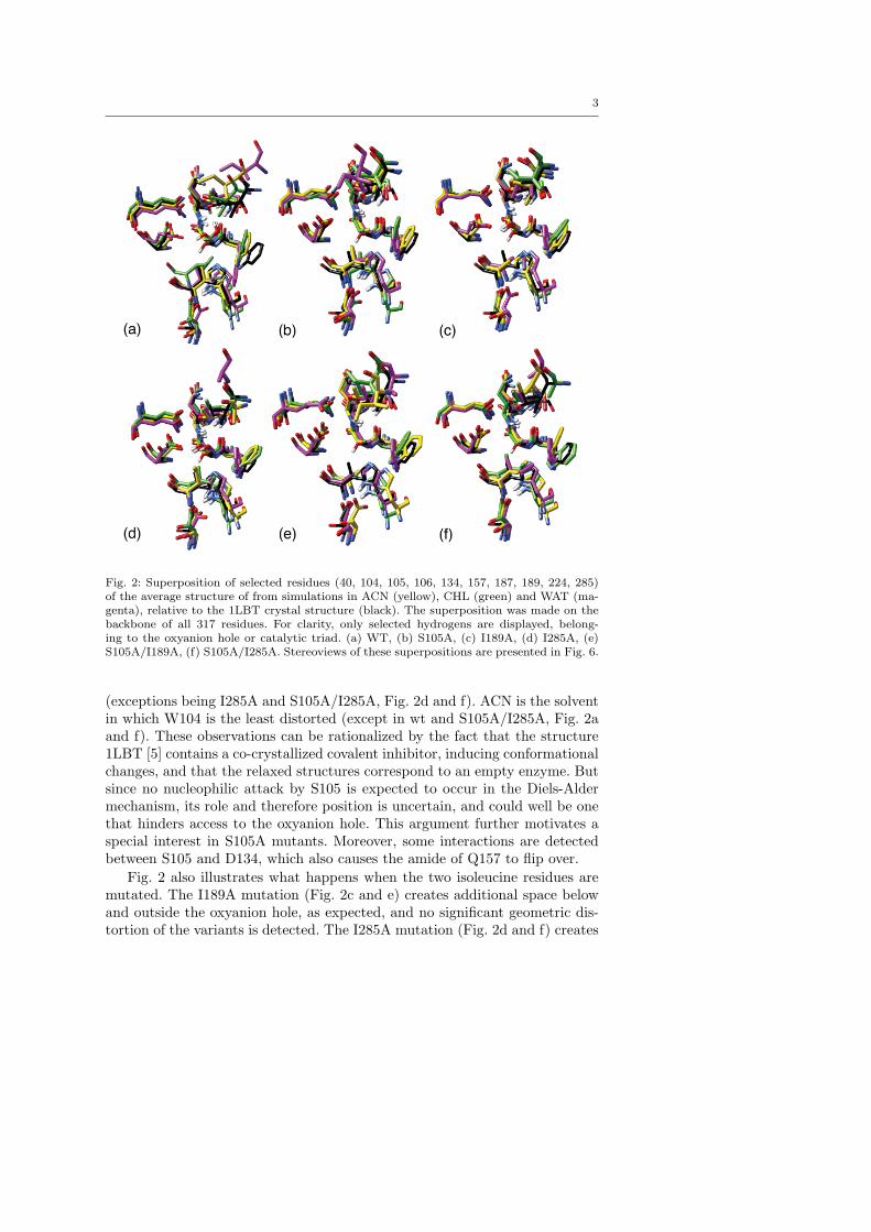
3
Fig. 2: Superposition of selected residues (40, 104, 105, 106, 134, 157, 187, 189, 224, 285)of the average structure of from simulations in ACN (yellow), CHL (green) and WAT (ma-genta), relative to the 1LBT crystal structure (black). The superposition was made on thebackbone of all 317 residues. For clarity, only selected hydrogens are displayed, belong-ing to the oxyanion hole or catalytic triad. (a) WT, (b) S105A, (c) I189A, (d) I285A, (e)S105A/I189A, (f) S105A/I285A. Stereoviews of these superpositions are presented in Fig. 6.
(exceptions being I285A and S105A/I285A, Fig. 2d and f). ACN is the solventin which W104 is the least distorted (except in wt and S105A/I285A, Fig. 2aand f). These observations can be rationalized by the fact that the structure1LBT [5] contains a co-crystallized covalent inhibitor, inducing conformationalchanges, and that the relaxed structures correspond to an empty enzyme. Butsince no nucleophilic attack by S105 is expected to occur in the Diels-Aldermechanism, its role and therefore position is uncertain, and could well be onethat hinders access to the oxyanion hole. This argument further motivates aspecial interest in S105A mutants. Moreover, some interactions are detectedbetween S105 and D134, which also causes the amide of Q157 to flip over.
Fig. 2 also illustrates what happens when the two isoleucine residues aremutated. The I189A mutation (Fig. 2c and e) creates additional space belowand outside the oxyanion hole, as expected, and no significant geometric dis-tortion of the variants is detected. The I285A mutation (Fig. 2d and f) creates
4
more space above the oxyanion hole; this is expected to be the face of attackfrom the diene, but the net effect is more difficult to predict, since I285 residesslightly further away from the oxyanion hole and tends to be more flexible, asseen in both Fig. 1 and Fig. 2.
2 Docking composite and consensus scoring
The ChemScore [6, 7] scoring function in GOLD generates a total score basedon an estimate of Gibbs Free Energy of binding, ∆Gbinding, which is modifiedby a set of penalty terms (dependent of what type of constraints are applied).In this study, the score is given by eq. 1.
CS = −∆Gbinding − Pclash − cPinternal, (1)
where c is a coefficient. ∆Gbinding, in turn, is given by eq. 2.
∆Gbinding = v0 + v1PHb + v2Pmetal + v3Plipo + v2Prot, (2)
where all P ’s are block functions with empirical parameters that are used todescribe different types of interactions,[8] and vi are regression coefficients.Similarily, Pclash and Pinternal are block functions describing energy penaltieswhen the ligand clashes with protein atoms or is distorted.
Since molecular docking software is designed mainly for drug discovery andsimilar applications, there is a bias in the scoring function to promote largeligands with many contacts. But since we seek to design a catalytic processinvolving two substrates that are turned over by the active site, our demandson a quite different to drug discovery. Most importantly, there must be solvent-accessible space left in the active site after docking the first ligand (in our casethe dienophile). A ligand filling out the entire active site will aquire a highscore in GOLD, but is unfit as a Diels-Alder reactant. Furthermore, we wishto promote H-bonding to the oxyanion hole and, since we induce a bindingconstraint between oxyanion hole donors and dienophile, penalize clashes thatmay arise because of this. Last, the smaller the ligand, the larger the numberof ways in which it may dock into the active site. In order to find ligands thatbind unambiguously in one conformation but is still small enough to enableDiels-Alder, we seek to include a term describing the conformational variety.
A simple way to promote these parameters is to create a ’composite’ scoringfunction, explicitly including terms that describe H-bonding, steric clashes andconformational variety. We used eq. 3:
Scomp(n) =(〈CS〉iatoms
)1/2
+ 〈PHb〉 − 〈Pclash〉 − 〈RMSD〉, (3)
which is based on average scores of the top n docking poses from a set ofsolutions. The total CS score is normalized by the number of heavy atoms inthe ligand.[9] PHb and Pclash are taken directly from the ChemScore output
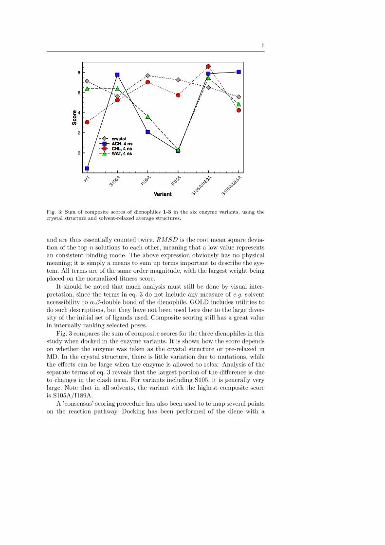
5
Fig. 3: Sum of composite scores of dienophiles 1-3 in the six enzyme variants, using thecrystal structure and solvent-relaxed average structures.
and are thus essentially counted twice. RMSD is the root mean square devia-tion of the top n solutions to each other, meaning that a low value representsan consistent binding mode. The above expression obviously has no physicalmeaning; it is simply a means to sum up terms important to describe the sys-tem. All terms are of the same order magnitude, with the largest weight beingplaced on the normalized fitness score.
It should be noted that much analysis must still be done by visual inter-pretation, since the terms in eq. 3 do not include any measure of e.g. solventaccessibility to α,β-double bond of the dienophile. GOLD includes utilities todo such descriptions, but they have not been used here due to the large diver-sity of the initial set of ligands used. Composite scoring still has a great valuein internally ranking selected poses.
Fig. 3 compares the sum of composite scores for the three dienophiles in thisstudy when docked in the enzyme variants. It is shown how the score dependson whether the enzyme was taken as the crystal structure or pre-relaxed inMD. In the crystal structure, there is little variation due to mutations, whilethe effects can be large when the enzyme is allowed to relax. Analysis of theseparate terms of eq. 3 reveals that the largest portion of the difference is dueto changes in the clash term. For variants including S105, it is generally verylarge. Note that in all solvents, the variant with the highest composite scoreis S105A/I189A.
A ’consensus’ scoring procedure has also been used to to map several pointson the reaction pathway. Docking has been performed of the diene with a
6
dienophile already bound to the oxyanion hole (the pose being selected fromthe set of dienophile solutions), as well TS models with fixed incipient bonddistances. These were generated based on DFT-optimized structures, and werethen treated as a single molecule by GOLD. The consensus score was calculatedaccording to equation 4.
Sconsensus = Scomp,dph + Scomp,dne + Scomp,TS . (4)
Calculated consensus score for each dienophile in ACN-relaxed variantsand crystal structures are displayed in figure 10 in the main article.
3 Investigations on a triple mutant T40A/S105A/I189A
The triple mutant (C) statistics are summarized in Fig. 4a. The RMSD of therelaxed uncomplexed variant to the crystal structure is 1.2. Structurally, themost remarkable feature is that the oxyanion loop (in ACN), now containingA40, is somewhat more distorted than in previous variants, which has theeffect that the the Q106-N–A40-N distance increases by 0.8 A relative to thecrystal structure. A similar distortion can be seen in the S105A/I189A andS105A/I285A variants, but is accompanied by a concordant movement of thenucleophilic elbow, resulting in a smaller increase or even reduction of theN–N distance, as illustrated in Fig. 4b. We also note that the RMSD is notcompletely leveled off in the 4 ns simulation, which is indicative of largergeometric distortions caused by three mutations in close vicinity.
The backbone RMSDs based on the last 1000 frames of both triple mutantcomplexes with 3 and 3 + 4 were 1.1 A with respect to the crystal structure,and 1.0 A with respect to the relaxed uncomplexed structure. Quite surpris-ingly, the H-bonding ability of the modified oxyanion hole was remarkable,both with and without the precence of a diene, as shown in 4a. Based on thelast 5000 recorded frames, 90-97 % distances were within the 2.75 A thresholdin both the dienophile and diene simulations (with average distances between2.2 and 2.4 A), but the diene did not form any NAC and fell out of the oxyan-ion hole after ≈ 1 ns. To investigate whether or not this was a stochastic event,we performed two additional 2 ns simulations with 3 and 4. Backbone RMSDsand H-bond statistics were similar to the first run, but neither produced anyNAC. In one case, the diene remained in the active site, but mostly in a T-shaped interaction with 3. We attribute the finding that the additional T40Amutation did not give better NAC formation to that the dienophile is allowedto become more buried (hence the enhanced H-bonding), and this introducesnew hindering residues, effectively blocking attack on the double bond. Mostnotable is Q157, which has taken over the role of obscuring the dienophile Cα.See Fig. 4c for an illustration.
To summarize, the triple mutant provides enhanced binding of the dienophiledue to the modification of the oxyanion hole, but structural modifications inthe active site leads to poor coordination of the diene. These results are stillimportant, as they suggest that spacious oxyanion holes with two donors, such

7
Fig. 4: (a) Simulation statistics of the T40A/S105A/I189A variant. The color legend is as fol-lows: For RSMD and B-factor, gray=naked enzyme; black=dph simulation; red=dph+dne,1st run; green=dph+dne, 2nd run; blue=dph+dne, 3rd run. For H-bond trajectories (100-frame running averages), Q106 and A40 are represented by black & gray (dph run),red & orange (dph+dne, 1st run), green & dark green (dph+dne, 2nd run), and blue& violet (dph+dne, 3rd run). (b) A comparison of the oxyanion hole geometry betweenT40A/S105A/I189A (yellow, thick sticks), the 1LBT crystal (green), S105A/I189A (gray)and S105A/I285A (black). All structures refer to the last 200 ps averages of 4 ns relaxationin ACN. His224 is shown for the sake of orientation. The distances displayed are calculatedbetween Q106-N and T/A40-N. (c) Representative snapshots of the [enz·3·4] complexesin T40A/S105A/I189A (yellow, green substrates) and S105A/I189A (black, magenta sub-strates). Note how the smaller methyl side chain on A40 allows the dienophile to becomemore buried, and how Q157 moves closer in the triple mutant, both resulting in less accessi-bility to the α,β-double bond. In contrast, the S105A/I189A variant displays a much moreideal NAC-like conformation of the substrates, although the dienophile Cα is still obscuredby the T40 side chain.
as those present in Pseudomonas cepacia and Candida rugosa lipases, mightbe preferable over the one in CALB for binding the dienophile. Furthermore,these enzymes have comparatively large cavities, [10, 11, 12] which we haveshown is an important feature to obtain the neccessary Diels-Alder TS con-formation.
References
1. Holmqvist, M. Curr. Prot. Peptide Sci. 2000, 1, 209–235.2. Trodler, P.; Pleiss, J. BMC Struct. Biol. 2008, 8, 9.3. Skjot, M.; De Maria, L.; Chatterjee, R.; Svendsen, A.; Patkar, S. A.; Os-
tergaard, P. R.; Brask, J. ChemBioChem 2009, 10, 520–527.4. Klibanov, A. M. Trends Biotechnol. 1997, 15, 97–101.

8
5. Uppenberg, J.; Oehrner, N.; Norin, M.; Hult, K.; Kleywegt, G. J.;Patkar, S.; Waagen, V.; Anthonsen, T.; Jones, T. A. Biochemistry 1995,34, 16838–16851.
6. Eldridge, M. D.; Murray, C. W.; Auton, T. R.; Paolini, G. V.; Mee, R. P.J. Comput.-Aided Mol. Des. 1997, 11, 425–445.
7. Baxter, C. A.; Murray, C. W.; Clark, D. E.; Westhead, D. R.; El-dridge, M. D. Proteins 1998, 33, 367–382.
8. 2010, For more details, see the GOLD documentation,http://www.ccdc.cam.ac.uk/support/do-cu-men-tation/gold/4 1/gold/.
9. Pan., Y.; Huang, N.; Cho, S.; MacKerrell, A. J. J. Chem. Inf. Comput.Sci. 2003, 43, 267–272.
10. Schrag, J. D.; Li, Y.; Cygler, M.; Lang, D.; Burgdorf, T.; Hecht, H.-J.;Schmid, R.; Schomburg, D.; Rydel, T. J.; Oliver, J. D.; Strickland, L. C.;Dunaway, C. M.; Larson, S. B.; Day, J.; McPherson, A. Structure 1997,5, 187–202.
11. Lang, D.; Mannesse, M.; De Haas, G.; Verheij, B., H.M. Dijkstra Eur. J.Biochem. 1998, 254, 333–340.
12. Grochulski, P.; Bouthillier, F.; Kazlauskas, R. J.; Serreqi, A. N.;Schrag, J. D.; Ziomek, E.; Cygler, M. Biochemistry 1994, 33, 3494–3500.
9
4 Supplementary figures
Supplementary figures as referred to in the main article.
Fig. 5: The oxyanion hole model. Frozen atoms are colored in magenta.
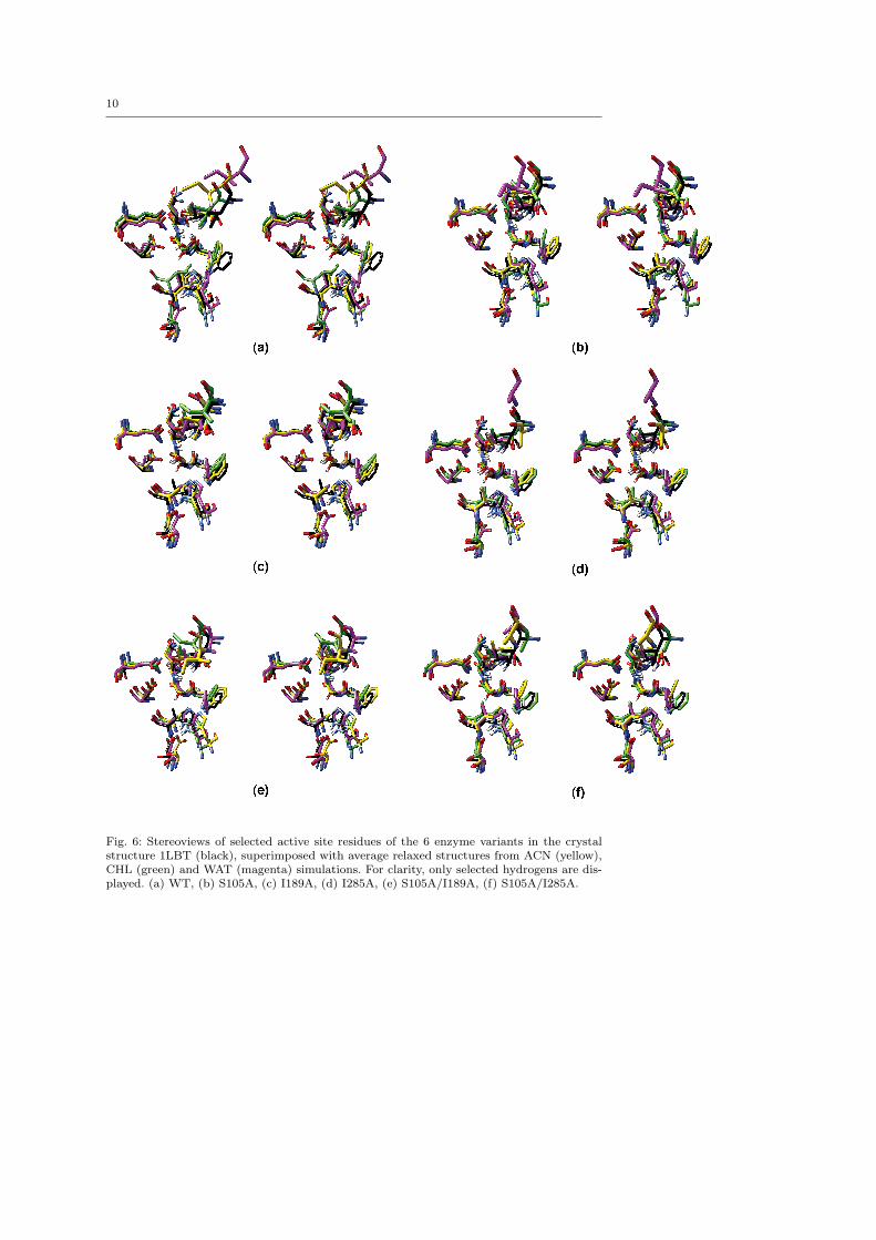
10
Fig. 6: Stereoviews of selected active site residues of the 6 enzyme variants in the crystalstructure 1LBT (black), superimposed with average relaxed structures from ACN (yellow),CHL (green) and WAT (magenta) simulations. For clarity, only selected hydrogens are dis-played. (a) WT, (b) S105A, (c) I189A, (d) I285A, (e) S105A/I189A, (f) S105A/I285A.
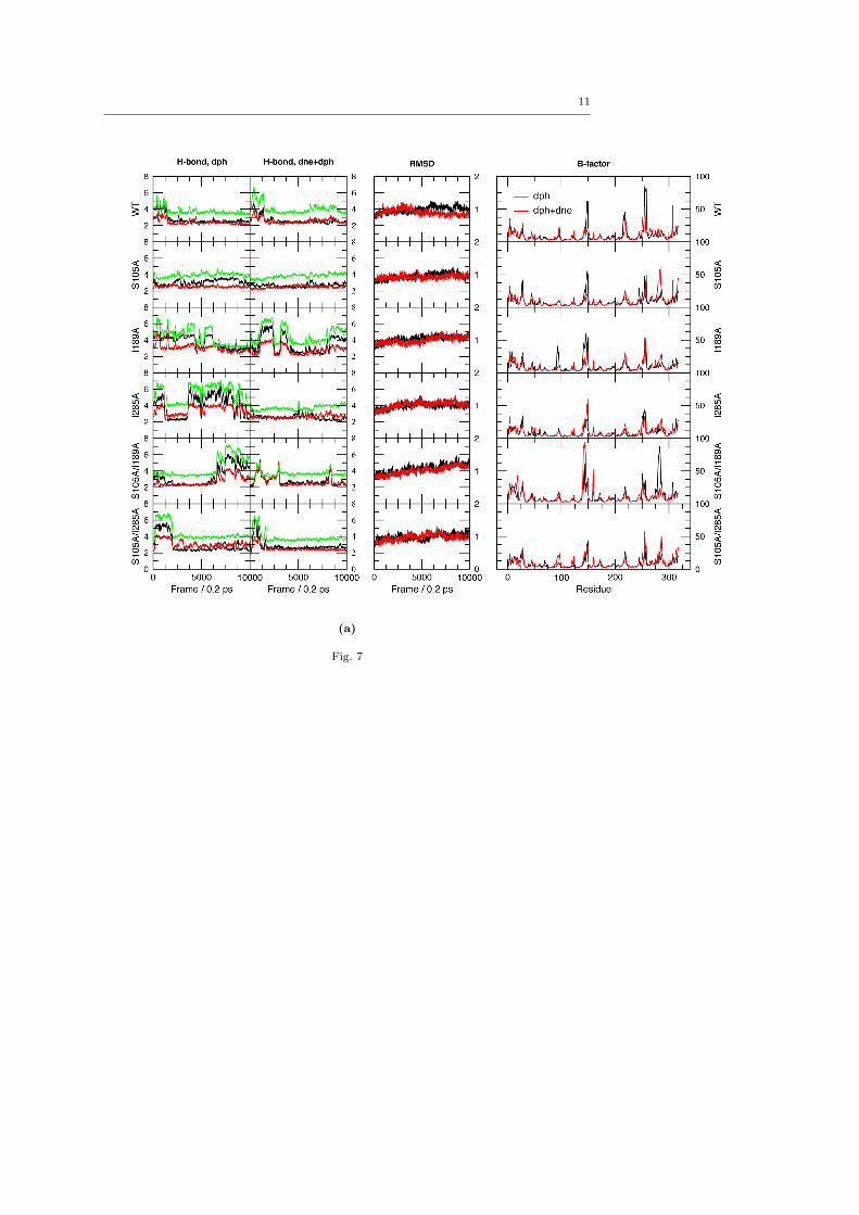
11
(a)
Fig. 7

12
(b)
Fig. 7: (cont.)
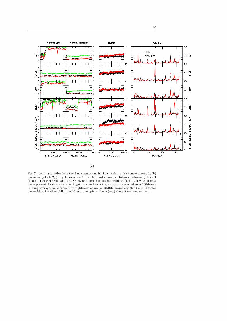
13
(c)
Fig. 7: (cont.) Statistics from the 2 ns simulations in the 6 variants. (a) benzoquinone 1, (b)maleic anhydride 2, (c) cyclohexenone 3. Two leftmost columns: Distance between Q106-NH(black), T40-NH (red) and T40-OγH, and acceptor oxygen without (left) and with (right)diene present. Distances are in Angstroms and each trajectory is presented as a 100-framerunning average, for clarity. Two rightmost columns: RMSD trajectory (left) and B-factorper residue, for dienophile (black) and dienophile+diene (red) simulation, respectively.
This is pdfTeX, Version 3.1415926-1.40.10 (Web2C 2009) (format=latex 2010.4.6) 31 MAY 2010 12:28 entering extended mode %&-line parsing enabled. **brinck_2010-dielsalder_revised.tex (./brinck_2010-dielsalder_revised.tex LaTeX2e <2009/09/24> Babel <v3.8l> and hyphenation patterns for english, usenglishmax, dumylang, noh yphenation, german-x-2009-06-19, ngerman-x-2009-06-19, ancientgreek, ibycus, ar abic, basque, bulgarian, catalan, pinyin, coptic, croatian, czech, danish, dutc h, esperanto, estonian, farsi, finnish, french, galician, german, ngerman, mono greek, greek, hungarian, icelandic, assamese, bengali, guajrati, hindi, assames e, malayalam, marathi, oriya, panjabi, tamil, telugu, indonesian, interlingua, irish, italian, kurmanji, latin, latvian, lithuanian, mongolian, mongolian2a, b okmal, nynorsk, polish, portuguese, romanian, russian, sanskrit, serbian, slova k, slovenian, spanish, swedish, turkish, turkmen, ukenglish, ukrainian, upperso rbian, welsh, loaded. (c:/TexLive2009/texmf/tex/latex/aries/svjour3.cls Document Class: svjour3 2006/03/15 v3.1 LaTeX document class for Springer journals (c:/TexLive2009/texmf-dist/tex/latex/base/fleqn.clo File: fleqn.clo 1998/08/17 v1.1c Standard LaTeX option (flush left equations) \mathindent=\dimen102 ) Class Springer-SVJour3 Info: extra/valid Springer sub-package (-> *.clo) (Springer-SVJour3) not found in option list of \documentclass (Springer-SVJour3) - autoactivating "global" style. (c:/TexLive2009/texmf/tex/latex/aries/svglov3.clo File: svglov3.clo 2006/02/03 v3.1 style option for standardised journals SVJour Class option: svglov3.clo for standardised journals ) LaTeX Font Info: Redeclaring math symbol \Gamma on input line 146. LaTeX Font Info: Redeclaring math symbol \Delta on input line 147. LaTeX Font Info: Redeclaring math symbol \Theta on input line 148. LaTeX Font Info: Redeclaring math symbol \Lambda on input line 149. LaTeX Font Info: Redeclaring math symbol \Xi on input line 150. LaTeX Font Info: Redeclaring math symbol \Pi on input line 151. LaTeX Font Info: Redeclaring math symbol \Sigma on input line 152. LaTeX Font Info: Redeclaring math symbol \Upsilon on input line 153. LaTeX Font Info: Redeclaring math symbol \Phi on input line 154. LaTeX Font Info: Redeclaring math symbol \Psi on input line 155. LaTeX Font Info: Redeclaring math symbol \Omega on input line 156. \logodepth=\dimen103 \headerboxheight=\dimen104 \betweenumberspace=\dimen105 \aftertext=\dimen106 \headlineindent=\dimen107 \c@inst=\count79 \c@auth=\count80 \instindent=\dimen108 \authrun=\box26 \authorrunning=\toks14
Click here to download Manuscript: Brinck_2010-DielsAlder_revised.tex
\titrun=\box27 \titlerunning=\toks15 \combirun=\box28 \c@lastpage=\count81 \rubricwidth=\dimen109 \c@section=\count82 \c@subsection=\count83 \c@subsubsection=\count84 \c@paragraph=\count85 \c@subparagraph=\count86 \spthmsep=\dimen110 \c@theorem=\count87 \c@case=\count88 \c@conjecture=\count89 \c@corollary=\count90 \c@definition=\count91 \c@example=\count92 \c@exercise=\count93 \c@lemma=\count94 \c@note=\count95 \c@problem=\count96 \c@property=\count97 \c@proposition=\count98 \c@question=\count99 \c@solution=\count100 \c@remark=\count101 \c@figure=\count102 \c@table=\count103 \abovecaptionskip=\skip41 \belowcaptionskip=\skip42 \figcapgap=\dimen111 \tabcapgap=\dimen112 \figgap=\dimen113 \bibindent=\dimen114 \@tempcntc=\count104 ) (c:/TexLive2009/texmf-dist/tex/generic/babel/babel.sty Package: babel 2008/07/06 v3.8l The Babel package (c:/TexLive2009/texmf-dist/tex/generic/babel/english.ldf Language: english 2005/03/30 v3.3o English support from the babel system (c:/TexLive2009/texmf-dist/tex/generic/babel/babel.def File: babel.def 2008/07/06 v3.8l Babel common definitions \babel@savecnt=\count105 \U@D=\dimen115 ) \l@canadian = a dialect from \language\l@american \l@australian = a dialect from \language\l@british \l@newzealand = a dialect from \language\l@british ) (c:/TexLive2009/texmf-dist/tex/generic/babel/swedish.ldf Language: swedish 2005/03/31 v2.3d Swedish support from the babel system Package babel Info: Making " an active character on input line 90. )) (c:/TexLive2009/texmf-dist/tex/latex/graphics/graphicx.sty Package: graphicx 1999/02/16 v1.0f Enhanced LaTeX Graphics (DPC,SPQR)
(c:/TexLive2009/texmf-dist/tex/latex/graphics/keyval.sty Package: keyval 1999/03/16 v1.13 key=value parser (DPC) \KV@toks@=\toks16 ) (c:/TexLive2009/texmf-dist/tex/latex/graphics/graphics.sty Package: graphics 2009/02/05 v1.0o Standard LaTeX Graphics (DPC,SPQR) (c:/TexLive2009/texmf-dist/tex/latex/graphics/trig.sty Package: trig 1999/03/16 v1.09 sin cos tan (DPC) ) (c:/TexLive2009/texmf-dist/tex/latex/latexconfig/graphics.cfg File: graphics.cfg 2009/08/28 v1.8 graphics configuration of TeX Live ) Package graphics Info: Driver file: dvips.def on input line 91. (c:/TexLive2009/texmf-dist/tex/latex/graphics/dvips.def File: dvips.def 1999/02/16 v3.0i Driver-dependant file (DPC,SPQR) )) \Gin@req@height=\dimen116 \Gin@req@width=\dimen117 ) (./mciteplus.sty Package: mciteplus 2008/01/15 v1.0 enhanced mcite \@mcitetmptoksA=\toks17 \@mcitetmpdima=\dimen118 \@mcitetmpdimb=\dimen119 \@mcitetmpcnta=\count106 \@mcitetmpcntb=\count107 \c@mcitebibitemcount=\count108 \c@mcitesubitemcount=\count109 ) (c:/TexLive2009/texmf-dist/tex/latex/natbib/natbib.sty Package: natbib 2009/11/07 8.31a (PWD, AO) \bibhang=\skip43 \bibsep=\skip44 LaTeX Info: Redefining \cite on input line 694. \c@NAT@ctr=\count110 ) (c:/TexLive2009/texmf-dist/tex/latex/base/makeidx.sty Package: makeidx 2000/03/29 v1.0m Standard LaTeX package ) (c:/TexLive2009/texmf-dist/tex/latex/jknapltx/mathrsfs.sty Package: mathrsfs 1996/01/01 Math RSFS package v1.0 (jk) \symrsfs=\mathgroup4 ) (c:/TexLive2009/texmf-dist/tex/latex/amsmath/amsmath.sty Package: amsmath 2000/07/18 v2.13 AMS math features \@mathmargin=\skip45 For additional information on amsmath, use the `?' option. (c:/TexLive2009/texmf-dist/tex/latex/amsmath/amstext.sty Package: amstext 2000/06/29 v2.01 (c:/TexLive2009/texmf-dist/tex/latex/amsmath/amsgen.sty File: amsgen.sty 1999/11/30 v2.0 \@emptytoks=\toks18 \ex@=\dimen120 )) (c:/TexLive2009/texmf-dist/tex/latex/amsmath/amsbsy.sty Package: amsbsy 1999/11/29 v1.2d \pmbraise@=\dimen121 ) (c:/TexLive2009/texmf-dist/tex/latex/amsmath/amsopn.sty Package: amsopn 1999/12/14 v2.01 operator names )
\inf@bad=\count111 LaTeX Info: Redefining \frac on input line 211. \uproot@=\count112 \leftroot@=\count113 LaTeX Info: Redefining \overline on input line 307. \classnum@=\count114 \DOTSCASE@=\count115 LaTeX Info: Redefining \ldots on input line 379. LaTeX Info: Redefining \dots on input line 382. LaTeX Info: Redefining \cdots on input line 467. \Mathstrutbox@=\box29 \strutbox@=\box30 \big@size=\dimen122 LaTeX Font Info: Redeclaring font encoding OML on input line 567. LaTeX Font Info: Redeclaring font encoding OMS on input line 568. Package amsmath Warning: Unable to redefine math accent \vec. \macc@depth=\count116 \c@MaxMatrixCols=\count117 \dotsspace@=\muskip10 \c@parentequation=\count118 \dspbrk@lvl=\count119 \tag@help=\toks19 \row@=\count120 \column@=\count121 \maxfields@=\count122 \andhelp@=\toks20 \eqnshift@=\dimen123 \alignsep@=\dimen124 \tagshift@=\dimen125 \tagwidth@=\dimen126 \totwidth@=\dimen127 \lineht@=\dimen128 \@envbody=\toks21 \multlinegap=\skip46 \multlinetaggap=\skip47 \mathdisplay@stack=\toks22 LaTeX Info: Redefining \[ on input line 2666. LaTeX Info: Redefining \] on input line 2667. ) (c:/TexLive2009/texmf-dist/tex/latex/tools/multicol.sty Package: multicol 2008/12/05 v1.6h multicolumn formatting (FMi) \c@tracingmulticols=\count123 Package multicol Warning: May not work with the twocolumn option on input line 135. \mult@box=\box31 \multicol@leftmargin=\dimen129 \c@unbalance=\count124 \c@collectmore=\count125 \doublecol@number=\count126
\multicoltolerance=\count127 \multicolpretolerance=\count128 \full@width=\dimen130 \page@free=\dimen131 \premulticols=\dimen132 \postmulticols=\dimen133 \multicolsep=\skip48 \multicolbaselineskip=\skip49 \partial@page=\box32 \last@line=\box33 \mult@rightbox=\box34 \mult@grightbox=\box35 \mult@gfirstbox=\box36 \mult@firstbox=\box37 \@tempa=\box38 \@tempa=\box39 \@tempa=\box40 \@tempa=\box41 \@tempa=\box42 \@tempa=\box43 \@tempa=\box44 \@tempa=\box45 \@tempa=\box46 \@tempa=\box47 \@tempa=\box48 \@tempa=\box49 \@tempa=\box50 \@tempa=\box51 \@tempa=\box52 \@tempa=\box53 \@tempa=\box54 \c@columnbadness=\count129 \c@finalcolumnbadness=\count130 \last@try=\dimen134 \multicolovershoot=\dimen135 \multicolundershoot=\dimen136 \mult@nat@firstbox=\box55 \colbreak@box=\box56 ) (c:/TexLive2009/texmf-dist/tex/latex/caption/caption.sty Package: caption 2010/01/09 v3.1m Customizing captions (AR) (c:/TexLive2009/texmf-dist/tex/latex/caption/caption3.sty Package: caption3 2010/01/14 v3.1m caption3 kernel (AR) \captionmargin=\dimen137 \captionmargin@=\dimen138 \captionwidth=\dimen139 \caption@indent=\dimen140 \caption@parindent=\dimen141 \caption@hangindent=\dimen142 Package caption Info: Unknown document class (or package), (caption) standard defaults will be used on input line 1084. )
Package caption Warning: Unsupported document class (or package) detected, (caption) usage of the caption package is not recommended. See the caption package documentation for explanation. Package caption Info: \@makecaption = \long macro:#1#2->\captionstyle \ifx \@ca ptype \fig@type \vskip \figcapgap \fi \setbox \@tempboxa \hbox {{\floatlegendst yle #1\floatcounterend }\capstrut #2}\ifdim \wd \@tempboxa >\hsize {\floatlegen dstyle #1\floatcounterend }\capstrut #2\par \else \hbox to\hsize {\leftlegendgl ue \unhbox \@tempboxa \hfil }\fi \ifx \@captype \fig@type \else \vskip \tabcapg ap \fi . \c@ContinuedFloat=\count131 ) (c:/TexLive2009/texmf-dist/tex/latex/footmisc/footmisc.sty Package: footmisc 2009/09/15 v5.5a a miscellany of footnote facilities \FN@temptoken=\toks23 \footnotemargin=\dimen143 \c@pp@next@reset=\count132 Package footmisc Info: Declaring symbol style bringhurst on input line 855. Package footmisc Info: Declaring symbol style chicago on input line 863. Package footmisc Info: Declaring symbol style wiley on input line 872. Package footmisc Info: Declaring symbol style lamport-robust on input line 883. Package footmisc Info: Declaring symbol style lamport* on input line 903. Package footmisc Info: Declaring symbol style lamport*-robust on input line 924 . ) (./brinck_2010-dielsalder_revised.aux) \openout1 = `brinck_2010-dielsalder_revised.aux'. LaTeX Font Info: Checking defaults for OML/cmm/m/it on input line 13. LaTeX Font Info: ... okay on input line 13. LaTeX Font Info: Checking defaults for T1/cmr/m/n on input line 13. LaTeX Font Info: ... okay on input line 13. LaTeX Font Info: Checking defaults for OT1/cmr/m/n on input line 13. LaTeX Font Info: ... okay on input line 13. LaTeX Font Info: Checking defaults for OMS/cmsy/m/n on input line 13. LaTeX Font Info: ... okay on input line 13. LaTeX Font Info: Checking defaults for OMX/cmex/m/n on input line 13. LaTeX Font Info: ... okay on input line 13. LaTeX Font Info: Checking defaults for U/cmr/m/n on input line 13. LaTeX Font Info: ... okay on input line 13. Unknown journal: specify \journalname{<name of your journal>} in preambel Package caption Info: Begin \AtBeginDocument code. Package caption Info: Incompatible package detected (regarding \@caption). (caption) \@caption = \long macro:#1[#2]#3->\par \addcontentsline { \csname ext@#1\endcsname }{#1}{\protect \numberline {\csname the#1\endcsname }{ \ignorespaces #2}}\begingroup \@parboxrestore \@makecaption {\csname fnum@#1\en dcsname }{\ignorespaces #3}\par \endgroup . Package caption Warning: \caption will not be redefined since it's already (caption) redefined by a document class or package which is (caption) unknown to the caption package. See the caption package documentation for explanation.
Package caption Info: End \AtBeginDocument code. SVJour3 warning: Missing \journalname LaTeX Font Warning: Font shape `OT1/cmr/m/n' in size <8.5> not available (Font) size <8> substituted on input line 30. LaTeX Font Warning: Font shape `OT1/cmr/m/n' in size <16> not available (Font) size <17.28> substituted on input line 30. LaTeX Font Warning: Font shape `OT1/cmr/bx/n' in size <16> not available (Font) size <17.28> substituted on input line 30. LaTeX Font Info: Try loading font information for U+rsfs on input line 30. (c:/TexLive2009/texmf-dist/tex/latex/jknapltx/ursfs.fd File: ursfs.fd 1998/03/24 rsfs font definition file (jk) ) LaTeX Font Info: Calculating math sizes for size <8.5> on input line 30. LaTeX Font Warning: Font shape `OT1/cmr/m/n' in size <4.25> not available (Font) size <5> substituted on input line 30. LaTeX Font Warning: Font shape `OML/cmm/m/it' in size <8.5> not available (Font) size <8> substituted on input line 30. LaTeX Font Warning: Font shape `OML/cmm/m/it' in size <4.25> not available (Font) size <5> substituted on input line 30. LaTeX Font Warning: Font shape `OMS/cmsy/m/n' in size <8.5> not available (Font) size <8> substituted on input line 30. LaTeX Font Warning: Font shape `OMS/cmsy/m/n' in size <4.25> not available (Font) size <5> substituted on input line 30. LaTeX Font Warning: Font shape `OMX/cmex/m/n' in size <8.5> not available (Font) size <8> substituted on input line 30. LaTeX Font Warning: Font shape `U/rsfs/m/n' in size <8.5> not available (Font) size <8> substituted on input line 30. LaTeX Font Warning: Font shape `U/rsfs/m/n' in size <4.25> not available (Font) size <5> substituted on input line 30.
Overfull \hbox (1.64522pt too wide) in paragraph at lines 33--34 \OT1/cmr/m/n/10 nam-ics (MD) and den-sity func-tional the-ory (DFT) stud- [] [1 ] Overfull \hbox (5.75746pt too wide) in paragraph at lines 44--45 \OT1/cmr/m/n/10 Li-pase B (CALB), per-formed by us and collaborators,[[], [] ! LaTeX Error: Cannot determine size of graphic in calb-scheme.png (no Bounding Box). See the LaTeX manual or LaTeX Companion for explanation. Type H <return> for immediate help. ... l.48 ...x{0.30}{\includegraphics{calb-scheme.png}} Try typing <return> to proceed. If that doesn't work, type X <return> to quit. File: calb-scheme.png Graphic file (type eps) <calb-scheme.png> Underfull \hbox (badness 1383) in paragraph at lines 49--49 \OT1/cmr/m/n/8 residues im-por-tant for the present study ex-plic-itly shown. [] Overfull \hbox (4.0769pt too wide) in paragraph at lines 55--56 \OT1/cmr/m/n/10 pro-vide an en-thalpic TS sta-bi-liza-tion of $\OMS/cmsy/m/n/10 ^^X$\OT1/cmr/m/n/10 1-5 kcal$\OMS/cmsy/m/n/10 ^^A$\OT1/cmr/m/n/10 mol$[]$, [] Overfull \hbox (5.03635pt too wide) in paragraph at lines 55--56 \OT1/cmr/m/n/10 de-pend-ing on re-ac-tants and po-lar-iz-abil-ity of the medium . [] Overfull \hbox (0.25838pt too wide) in paragraph at lines 70--72 \OT1/cmr/m/n/10 As a first ap-prox-i-ma-tion, we can con-sider the case where [] Overfull \hbox (2.59172pt too wide) in paragraph at lines 77--79 \OT1/cmr/m/n/10 This re-ac-tion can be de-scribed by the well-known Micha- []
Underfull \hbox (badness 10000) has occurred while \output is active [][] [] [2] ! LaTeX Error: Cannot determine size of graphic in dynamics.png (no BoundingBox ). See the LaTeX manual or LaTeX Companion for explanation. Type H <return> for immediate help. ... l.133 ...phics[width=0.75\textwidth]{dynamics.png} Try typing <return> to proceed. If that doesn't work, type X <return> to quit. File: dynamics.png Graphic file (type eps) <dynamics.png> [3] Underfull \vbox (badness 10000) has occurred while \output is active [] Underfull \vbox (badness 10000) has occurred while \output is active [] Underfull \hbox (badness 10000) has occurred while \output is active [][] [] [4] Overfull \hbox (20.21135pt too wide) in paragraph at lines 149--150 \OT1/cmr/m/n/10 nat-u-ral sub-strate, $\OML/cmm/m/it/10 ^^AG[]$ \OT1/cmr/m/n/10 is in the or-der of 0.5-2 kcal$\OMS/cmsy/m/n/10 ^^A$\OT1/cmr/m/n/10 mol$[]$. [] Overfull \hbox (1.2585pt too wide) in paragraph at lines 157--158 \OT1/cmr/m/n/10 zyme com-bi-na-tions us-ing eq. 11[]. There is, un-for-tu-natel y, [] [5] Overfull \hbox (2.06403pt too wide) in paragraph at lines 170--171 \OT1/cmr/m/n/10 files, neu-tral-ize the en-zyme com-plexes with Sodium ions, [] Overfull \hbox (8.45302pt too wide) in paragraph at lines 170--171 \OT1/cmr/m/n/10 avail-able united-atom model, [[]] while chlo-ro-form (CHL)
[] Overfull \hbox (2.14742pt too wide) in paragraph at lines 172--173 \OT1/cmr/m/n/10 han-dled by par-ti-cle Mesh Ewald sum-ma-tion. Ini-tial min- [] Overfull \hbox (7.78625pt too wide) in paragraph at lines 174--175 \OT1/cmr/m/n/10 within AM-BER. For struc-tural anal-y-sis, an av-er-age struc- [] Overfull \hbox (3.11844pt too wide) in paragraph at lines 176--177 \OT1/cmr/m/n/10 di-ene and dienophile were es-ti-mated us-ing the LIE meth- [] Underfull \vbox (badness 1253) has occurred while \output is active [] Underfull \hbox (badness 10000) has occurred while \output is active [][] [] [6] Overfull \hbox (1.11954pt too wide) in paragraph at lines 209--210 \OT1/cmr/m/n/10 Among a larger set of molecules, ben-zo-quinone \OT1/cmr/bx/n/1 0 1\OT1/cmr/m/n/10 , maleic [] ! LaTeX Error: Cannot determine size of graphic in BMC_reactants.png (no Boundi ngBox). See the LaTeX manual or LaTeX Companion for explanation. Type H <return> for immediate help. ... l.213 ...0.7}{\includegraphics{BMC_reactants.png}} Try typing <return> to proceed. If that doesn't work, type X <return> to quit. File: BMC_reactants.png Graphic file (type eps) <BMC_reactants.png> ! LaTeX Error: Cannot determine size of graphic in BMC_Hb.png (no BoundingBox). See the LaTeX manual or LaTeX Companion for explanation. Type H <return> for immediate help.
... l.225 ...raphics[width=0.85\textwidth]{BMC_Hb.png} Try typing <return> to proceed. If that doesn't work, type X <return> to quit. File: BMC_Hb.png Graphic file (type eps) <BMC_Hb.png> LaTeX Font Warning: Font shape `OT1/cmr/bx/n' in size <8.5> not available (Font) size <8> substituted on input line 226. [7] Underfull \hbox (badness 10000) has occurred while \output is active [][] [] [8] ! LaTeX Error: Cannot determine size of graphic in Hb_dph_dne.png (no BoundingB ox). See the LaTeX manual or LaTeX Companion for explanation. Type H <return> for immediate help. ... l.252 ...x{0.08}{\includegraphics{Hb_dph_dne.png}} Try typing <return> to proceed. If that doesn't work, type X <return> to quit. File: Hb_dph_dne.png Graphic file (type eps) <Hb_dph_dne.png> ! LaTeX Error: Cannot determine size of graphic in nac2_4.png (no BoundingBox). See the LaTeX manual or LaTeX Companion for explanation. Type H <return> for immediate help. ... l.262 ...lebox{0.31}{\includegraphics{nac2_4.png}} Try typing <return> to proceed. If that doesn't work, type X <return> to quit. File: nac2_4.png Graphic file (type eps) <nac2_4.png> Overfull \hbox (2.2018pt too wide) in paragraph at lines 259--267 \OT1/cmr/m/n/10 [\OT1/cmr/bx/n/10 3$\OMS/cmsy/m/n/10 ^^A$\OT1/cmr/m/n/10 S105A/ I285A] com-plexes. It is dif-fi-cult to tell whether
[] ! LaTeX Error: Cannot determine size of graphic in ben_snapshots_trans.png (no BoundingBox). See the LaTeX manual or LaTeX Companion for explanation. Type H <return> for immediate help. ... l.272 ...includegraphics{ben_snapshots_trans.png}} Try typing <return> to proceed. If that doesn't work, type X <return> to quit. File: ben_snapshots_trans.png Graphic file (type eps) <ben_snapshots_trans.png> Overfull \hbox (1.84682pt too wide) in paragraph at lines 269--276 \OT1/cmr/m/it/10 Ben-zo-quinone. [] \OT1/cmr/m/n/10 Sta-tis-ti-cally, ben-zo-q uinone \OT1/cmr/bx/n/10 1 \OT1/cmr/m/n/10 is the dien- [] ! LaTeX Error: Cannot determine size of graphic in mal_snapshots_trans.png (no BoundingBox). See the LaTeX manual or LaTeX Companion for explanation. Type H <return> for immediate help. ... l.281 ...includegraphics{mal_snapshots_trans.png}} Try typing <return> to proceed. If that doesn't work, type X <return> to quit. File: mal_snapshots_trans.png Graphic file (type eps) <mal_snapshots_trans.png> Overfull \hbox (2.95287pt too wide) in paragraph at lines 278--285 \OT1/cmr/m/n/10 is less con-sis-tent than for the other sub-strates; in S105A/ [] [9] ! LaTeX Error: Cannot determine size of graphic in cyh_snapshots_trans.png (no BoundingBox). See the LaTeX manual or LaTeX Companion for explanation. Type H <return> for immediate help. ... l.291 ...includegraphics{cyh_snapshots_trans.png}}
Try typing <return> to proceed. If that doesn't work, type X <return> to quit. File: cyh_snapshots_trans.png Graphic file (type eps) <cyh_snapshots_trans.png> Underfull \hbox (badness 10000) has occurred while \output is active [][] [] [10] ! LaTeX Error: Cannot determine size of graphic in consensus_BMC.png (no Boundi ngBox). See the LaTeX manual or LaTeX Companion for explanation. Type H <return> for immediate help. ... l.307 ....30}{\includegraphics{consensus_BMC.png}} Try typing <return> to proceed. If that doesn't work, type X <return> to quit. File: consensus_BMC.png Graphic file (type eps) <consensus_BMC.png> LaTeX Font Info: Try loading font information for OMS+cmr on input line 312. (c:/TexLive2009/texmf-dist/tex/latex/base/omscmr.fd File: omscmr.fd 1999/05/25 v2.5h Standard LaTeX font definitions ) LaTeX Font Info: Font shape `OMS/cmr/m/n' in size <7> not available (Font) Font shape `OMS/cmsy/m/n' tried instead on input line 312. LaTeX Font Info: Font shape `OMS/cmr/m/n' in size <5.94997> not available (Font) Font shape `OMS/cmsy/m/n' tried instead on input line 312. Overfull \hbox (9.18231pt too wide) in paragraph at lines 320--321 \OT1/cmr/m/it/10 (C) Sim-u-la-tion of a T40A/S105A/I189A vari-ant in ACN. [] [11] Overfull \hbox (1.5352pt too wide) in paragraph at lines 394--395 \OT1/cmr/m/n/10 the [E$\OMS/cmsy/m/n/10 ^^A$\OT1/cmr/m/n/10 dph] and TS op-ti-m ized ge-ome-tries, in-clud-ing oxy- [] Underfull \hbox (badness 10000) has occurred while \output is active [][] [] [12]
LaTeX Warning: File `TS_diagram2.png' not found on input line 415. ! LaTeX Error: File `TS_diagram2.png' not found. See the LaTeX manual or LaTeX Companion for explanation. Type H <return> for immediate help. ... l.415 ...cs[width=0.85\textwidth]{TS_diagram2.png} Try typing <return> to proceed. If that doesn't work, type X <return> to quit. ! LaTeX Error: Cannot determine size of graphic in TS_diagram2.png (no Bounding Box). See the LaTeX manual or LaTeX Companion for explanation. Type H <return> for immediate help. ... l.415 ...cs[width=0.85\textwidth]{TS_diagram2.png} Try typing <return> to proceed. If that doesn't work, type X <return> to quit. File: TS_diagram2.png Graphic file (type eps) <TS_diagram2.png> Underfull \vbox (badness 10000) has occurred while \output is active [] Overfull \hbox (9.81189pt too wide) in paragraph at lines 422--423 \OT1/cmr/m/n/10 largest TS sta-bi-liza-tion ($\OML/cmm/m/it/10 ^^AG[] \OMS/cmsy /m/n/10 ^^@ \OML/cmm/m/it/10 ^^AG[] \OT1/cmr/m/n/10 = \OMS/cmsy/m/n/10 ^^@\OT1/ cmr/m/n/10 3\OML/cmm/m/it/10 :\OT1/cmr/m/n/10 9$ kcal$\OMS/cmsy/m/n/10 ^^A$\OT1 /cmr/m/n/10 mol$[]$). [] [13] Underfull \hbox (badness 10000) has occurred while \output is active [][] [] [14] Overfull \hbox (0.25847pt too wide) in paragraph at lines 452--453 \OT1/cmr/m/n/10 rate-enhancements for catal-y-sis of bi-molec-u-lar re-ac-tions [] Overfull \hbox (1.14732pt too wide) in paragraph at lines 456--457
\OT1/cmr/m/n/10 mu-tant in wa-ter. The com-puted rate en-hance-ments with [] Overfull \hbox (12.14326pt too wide) in paragraph at lines 458--459 []\OT1/cmr/m/n/8 This work has been sup-ported by the Swedish [] Underfull \vbox (badness 3108) has occurred while \output is active [] [15] (./brinck_2010-dielsalder_revised.bbl Underfull \hbox (badness 10000) in paragraph at lines 77--85 []\OT1/cmr/m/n/10 Zhang, X.; DeChancie, J.; Gu-nay-din, H.; [] Underfull \hbox (badness 1460) in paragraph at lines 77--85 \OT1/cmr/m/n/10 Chowdry, A. B.; Clemente, F. R.; Smith, A. [] Underfull \hbox (badness 4217) in paragraph at lines 141--148 []\OT1/cmr/m/n/10 Branneby, C.; Car-lqvist, P.; Mag-nus-son, A.; [] Missing character: There is no à in font cmr10! Missing character: There is no ˆ in font cmr10! Underfull \hbox (badness 3158) in paragraph at lines 157--165 []\OT1/cmr/m/n/10 Svedendahl, M.; Car-lqvist, P.; Bran-neby, C.; [] Underfull \hbox (badness 2173) in paragraph at lines 157--165 \OT1/cmr/m/n/10 Allnr, O.; Frise, A.; Hult, K.; Berglund, P.; [] Underfull \hbox (badness 3158) in paragraph at lines 166--173 []\OT1/cmr/m/n/10 Carlqvist, P.; Sve-den-dahl, M.; Bran-neby, C.; [] Underfull \hbox (badness 10000) in paragraph at lines 239--249 []\OT1/cmr/m/n/10 Rothlisberger, D.; Kher-son-sky, O.; Wol-la- [] Underfull \vbox (badness 10000) has occurred while \output is active []
Underfull \hbox (badness 10000) has occurred while \output is active [][] [] [16] Underfull \hbox (badness 5245) in paragraph at lines 398--404 []\OT1/cmr/m/n/10 Uppenberg, J.; Hansen, M. T.; Patkar, S.; [] Underfull \hbox (badness 2096) in paragraph at lines 405--411 []\OT1/cmr/m/it/10 GOLD 4.1\OT1/cmr/m/n/10 , 2008, http://www.ccdc.cam.ac.uk/- [] Underfull \hbox (badness 1852) in paragraph at lines 501--507 []\OT1/cmr/m/n/10 Guardia, E.; Pin-zon, R.; Ca-sulleras, J.; Oro- [] Underfull \vbox (badness 10000) has occurred while \output is active [] Underfull \hbox (badness 10000) in paragraph at lines 610--616 []\OT1/cmr/m/it/10 UCSF Chimera\OT1/cmr/m/n/10 , 2004, [] Underfull \hbox (badness 10000) in paragraph at lines 631--636 []\OT1/cmr/m/n/10 Wesemann, M. \OT1/cmr/m/it/10 Plot, v. 0.997\OT1/cmr/m/n/10 , 2007, [] ) [17] (./brinck_2010-dielsalder_revised.aux) LaTeX Font Warning: Size substitutions with differences (Font) up to 1.28pt have occurred. LaTeX Warning: Label(s) may have changed. Rerun to get cross-references right. ) Here is how much of TeX's memory you used: 4027 strings out of 93775 62484 string characters out of 1149208 144637 words of memory out of 3000000 7276 multiletter control sequences out of 15000+50000 11436 words of font info for 43 fonts, out of 1200000 for 2000 714 hyphenation exceptions out of 8191 36i,20n,38p,4309b,522s stack positions out of 5000i,500n,10000p,200000b,5000s Output written on brinck_2010-dielsalder_revised.dvi (17 pages, 113932 bytes).


![[Charles N. Satterfield] Heterogeneous Catalysis i(BookZZ.org)](https://img.dokumen.tips/doc/110x75/63546b559ef77da53804b20b/charles-n-satterfield-heterogeneous-catalysis-ibookzzorg.jpg)


