Embed Size (px)
Citation preview
Complimentary Contributor Copy

Complimentary Contributor Copy

SURGERY - PROCEDURES, COMPLICATIONS, AND RESULTS
ADVANCES IN
EXPERIMENTAL SURGERY
VOLUME 2
No part of this digital document may be reproduced, stored in a retrieval system or transmitted in any form orby any means. The publisher has taken reasonable care in the preparation of this digital document, but makes noexpressed or implied warranty of any kind and assumes no responsibility for any errors or omissions. Noliability is assumed for incidental or consequential damages in connection with or arising out of informationcontained herein. This digital document is sold with the clear understanding that the publisher is not engaged inrendering legal, medical or any other professional services. Complimentary Contributor Copy

SURGERY - PROCEDURES, COMPLICATIONS,
AND RESULTS
Additional books in this series can be found on Nova’s website
under the Series tab.
Additional e-books in this series can be found on Nova’s website
under the e-Books tab.
Complimentary Contributor Copy
SURGERY - PROCEDURES, COMPLICATIONS, AND RESULTS
ADVANCES IN
EXPERIMENTAL SURGERY
VOLUME 2
HUIFANG CHEN
AND
PAULO N. MARTINS
EDITORS
Complimentary Contributor Copy
Copyright © 2018 by Nova Science Publishers, Inc.
All rights reserved. No part of this book may be reproduced, stored in a retrieval system or
transmitted in any form or by any means: electronic, electrostatic, magnetic, tape, mechanical
photocopying, recording or otherwise without the written permission of the Publisher.
We have partnered with Copyright Clearance Center to make it easy for you to obtain permissions
to reuse content from this publication. Simply navigate to this publication’s page on Nova’s website
and locate the “Get Permission” button below the title description. This button is linked directly to
the title’s permission page on copyright.com. Alternatively, you can visit copyright.com and search
by title, ISBN, or ISSN.
For further questions about using the service on copyright.com, please contact:
Copyright Clearance Center
Phone: +1-(978) 750-8400 Fax: +1-(978) 750-4470 E-mail: [email protected].
NOTICE TO THE READER
The Publisher has taken reasonable care in the preparation of this book, but makes no expressed or
implied warranty of any kind and assumes no responsibility for any errors or omissions. No liability
is assumed for incidental or consequential damages in connection with or arising out of information
contained in this book. The Publisher shall not be liable for any special, consequential, or exemplary
damages resulting, in whole or in part, from the readers’ use of, or reliance upon, this material. Any
parts of this book based on government reports are so indicated and copyright is claimed for those
parts to the extent applicable to compilations of such works.
Independent verification should be sought for any data, advice or recommendations contained in
this book. In addition, no responsibility is assumed by the publisher for any injury and/or damage
to persons or property arising from any methods, products, instructions, ideas or otherwise contained
in this publication.
This publication is designed to provide accurate and authoritative information with regard to the
subject matter covered herein. It is sold with the clear understanding that the Publisher is not
engaged in rendering legal or any other professional services. If legal or any other expert assistance
is required, the services of a competent person should be sought. FROM A DECLARATION OF
PARTICIPANTS JOINTLY ADOPTED BY A COMMITTEE OF THE AMERICAN BAR
ASSOCIATION AND A COMMITTEE OF PUBLISHERS.
Additional color graphics may be available in the e-book version of this book.
Library of Congress Cataloging-in-Publication Data
ISBN: 978-1-53612-774-4 (eBook)
Published by Nova Science Publishers, Inc. † New York
Complimentary Contributor Copy

CONTENTS
Foreword vii
Pierre Daloze
Preface ix
Huifang Chen and Paulo N. Martins
Section I. General and Specific Experimental Surgeries Models 1
Chapter 1 Laparoscopic Experimental Surgery Models 3
Natalie Bath, Demetrius Litwin and Paulo N. Martins
Chapter 2 Experimental Models of Sepsis 37
Norbert Nemeth, Mihai Oltean and Bela Fulesdi
Chapter 3 Bariatric Experimental Surgery Models 73
Osman Bilgin Gulcicek
Chapter 4 Animal Models of Heart Failure 81
Mouer Wang
Chapter 5 Hepatopancreatobiliary Surgery Experimental Models 107
Paulo N. Martins, Natalie Bath and Adel Bozorgzadeh
Section II. Other Experimental Models 157
Chapter 6 Animal Models of Acute Graft-Versus-Host and Host-Versus-Graft
Responses and Disease 159
Abraham Matar and Raimon Duran-Struuck
Chapter 7 Microfluidic Devices and Micro-Dissected Tissue to Predict
Therapeutic Response in Patients with Prostate Cancer 183
Robin Guay-Lord, Muhammad Abdul Lateef, Kayla Simeone,
Benjamin Péant,Jennifer Kendall-Dupont,
Anne-Marie Mes-Masson, Thomas Gervais and Fred Saad
Complimentary Contributor Copy
Contents vi
Chapter 8 Of Mice and Rats: Animal Models for Use in Laparoscopy and
Laparotomy with Human Ovarian Cancer Cell Line Intraperitoneal
Xenografts 211
Philippe Sauthier, Anne-Marie Mes-Masson, Louise Champoux,
Michèle Bally and Diane M. Provencher
Section III. Advance Techniques in Experimental Surgery 227
Chapter 9 Organ Bioengineering through Decellularization and
Recellularization Approaches 229
Maria Jaramillo and Basak E. Uygun
Chapter 10 Wound Healing and Scarring 255
A. Samandar Dowlatshahi
Chapter 11 Organ Preservation 269
Cheng Yang, Jiawei Li and Ruiming Rong
Chapter 12 Machine Organ Preservation 289
Hazel L. Marecki, Isabel Brüggenwirth and Paulo N. Martins
Chapter 13 Microcirculation 317
Norbert Nemeth and Andrea Szabo
Chapter 14 Experimental Gene Therapy Using Naked DNA 359
Eiji Kobayashi
Chapter 15 Robotic Surgery in Experimental Medicine 373
Sandra S. Y. Kim, David Ian Harriman and Christopher Nguan
About the Editors 403
Index 405
Complimentary Contributor Copy

FOREWORD
Pierre Daloze, CM, CQ, MD, FRCSC Professor Emeritus,
Department of Surgery, University of Montreal, Canada
Experimental surgery is a key link for developments in clinical surgery, research and
teaching. Throughout the history of medicine, many discoveries and techniques developed from
experimental surgery. Most modern surgeons are now learning or improving their surgical
techniques firstly via experimental surgery. Reviewing the 20th century surgical developing
history, most clinical achievements were related to experimental surgery. Some were real
landmarks, such as successful vascular sutures, opening the striking advances of vascular
surgery and organ transplantation. In recent years, experimental surgery has achieved new
advances, like laparoscopic and robotic surgery, tissue engineering and gene therapy, which
are now applied in clinic and have saved many patients.
Both editors of this book, Drs. Huifang Chen and Paulo Martins as well as their colleagues
have been contributing to experimental surgery at the University of Montreal, Canada and the
University of Massachusetts, US. Their achievements in experimental surgical models in small
and large animals, including nonhuman primates, have been applied in clinical trials.
It is my pleasure to write this foreword for this impressive book, Advances in Experimental
Surgery, which Drs. Chen and Martins provide as a reference for surgeons, residents, surgical
researchers, physicians, immunologists, veterinarians and nurses in surgery. I am sure these
two volumes will provide an abundance of imperative information for these individuals and
their patients.
Complimentary Contributor Copy

Complimentary Contributor Copy
PREFACE
Huifang Chen and Paulo N. Martins
Experimental surgery is an important link for developments in clinical surgery, research
and teaching. Experimental surgery has been a part of the most important surgical discoveries
in the past century. Since 1901, nine Nobel Prizes have been awarded to the pioneers who had
remarkable achievements in basic or practical surgery. In recent years, experimental surgery
has achieved new advances, like laparoscopic and robotic surgery, tissue engineering, and gene
therapy which are widely applied in clinic surgery.
The present book covers wide experimental surgery in preclinical research models
subdivided into two volumes. Volume I introduces basic surgical notions, techniques, and
different surgical models involved in basic experimental surgery, and reviews the
biomechanical models, ischemia/reperfusion injury models, repair and regeneration models,
and organ and tissue transplantation models, respectively. Volume II introduces several specific
experimental models such as laparoscopic and bariatric experimental surgical models. The
second volume also introduces graft-versus-host diseases, and other experimental models. A
review of the advances and development of recent techniques such as tissue engineering, organ
preservation, wound healing and scarring, gene therapy and robotic surgery is included. This
book documents the enormous volume of knowledge scientists have acquired in the field of
experimental surgery.
The editors have invited experts from the United States, Canada, France, Germany, China,
Japan, Korea, UK, Sweden, Netherlands, Hungary and Turkey to contribute 15 chapters in the
fields of their expertise. This volume is a compilation of basic experimental surgery and
updated advances of new developments in this field that will be invaluable to any experimental
surgery lab.
Complimentary Contributor Copy
Huifang Chen and Paulo N. Martins x
The editors are grateful to Dr. Pierre Daloze for writing the foreword and reviewing these
volumes. The editors also want to thank all of the authors for their contributions and the time
they spent to make this book successful. We also appreciate Dr. Muhammad Zafarullah and Dr.
Lijun Song for contributing their valuable time in proofreading the entirety of this book.
Dr. Huifang Chen, MD., PhD.
Professor of Surgery
Laboratory of Experimental Surgery, Research Center, CHUM
10th floor, R10. 480, University of Montreal
900 rue Saint Denis, Montreal, Quebec, Canada H2X 0A9
E. Mail: [email protected]
Dr. Paulo N. Martins, MD., PhD, FAST, FACS
Department of Surgery
Division of Transplantation
UMass Memorial Medical Center
University of Massachusetts
Worcester, MA, USA
E. Mail: [email protected]
Complimentary Contributor Copy

SECTION I. GENERAL AND SPECIFIC EXPERIMENTAL
SURGERIES MODELS
Complimentary Contributor Copy

Complimentary Contributor Copy

In: Advances in Experimental Surgery. Volume 2 ISBN: 978-1-53612-773-7
Editors: Huifang Chen and Paulo N. Martins © 2018 Nova Science Publishers, Inc.
Chapter 1
LAPAROSCOPIC EXPERIMENTAL SURGERY MODELS
Natalie Bath MD1, Demetrius Litwin MD
and Paulo N. Martins MD, PhD * 1Department of Surgery, UMass Memorial Medical Center, University Campus,
Worcester, MA 2Department of Surgery, Division of Organ Transplantation,
UMass Memorial Medical Center, University Campus, Worcester, MA
ABSTRACT
Laparoscopy and minimally invasive surgery has continued to play an increasingly
large role across all surgical fields including general surgery, gynecology, colorectal
surgery, hepatobiliary surgery, and urology. By minimizing trauma to the abdominal wall,
allowing faster recovery postoperatively, decreasing length of hospital stay, and improving
cosmesis, laparoscopy has become the gold standard approach for many operations due to
these improved outcomes. However, laparoscopic surgery demands its own unique set of
surgical skills due to loss of tactile feedback, operating within a three-dimensional space
as seen on a two-dimensional monitor, and the importance of recognizing spatial
relationships. A learning curve is found to exist as one is further developing these skills. In
order to further develop these skills prior to utilizing them in the operating room, live
animal models and laparoscopic simulators have been developed. Live animal models
including pigs, rabbits, and rats have been used for training, and it has been found that in
vivo models appear to most closely resemble operating on patients; therefore, animal
models are the ideal training model to be used in laparoscopic surgery training. As
laparoscopy continues to become more popular throughout surgery, it also continues to
evolve in order to further reduce the invasiveness of surgery as seen in robotic surgery,
single incision laparoscopic surgery (SILS), and natural orifice translumenal endoscopic
surgery (NOTES). These surgical techniques are the future of laparoscopic surgery and as
their use becomes more widespread, training on experimental models specific to these
methods will become even more important in order to optimize surgical outcomes for
patients.
* Corresponding author: Paulo N. Martins, MD, PhD, Department of Surgery, Division of Organ Transplantation, 55
Lake Avenue North, Worcester, MA 01655, Tel: 508-334-2023,
Email: [email protected]. Complimentary Contributor Copy
Natalie Bath, Demetrius Litwin and Paulo N. Martins 4
Keywords: laparoscopic surgery, experimental laparoscopy, learning curve, animal models,
laparoscopic simulator, future laparoscopy
ABBREVIATIONS
dVSS de Vinci Skills Simulator
FLS Fundamentals of Laparoscopic Surgery
MISTVR Minimally Invasive Surgery Trainer Virtual Reality
NOTES Natural orifice translumenal endoscopic surgery
OSATS Objective Structured Assessment of Technical Skill
SEP Sim-Surgery Educational Plateform
SILS Single Incision Laparoscopic Surgery
TAPP Transabdominal Preperitonal Practice
TEP Totally Extraperitonal
VR Virtual Reality
IMPORTANCE OF LAPAROSCOPIC TRAINING
Laparoscopic surgery requires a unique and at times more complex skill set than open
surgery. In order to further develop these skills prior to utilizing them in the operating room,
live animal models and laparoscopic simulators have been developed. Live animal models
including pigs, rabbits, and rats have been used for training, and it has been found that in vivo
models appear to most closely resemble operating on a live patient.
Laparoscopic surgery traces its roots to the early twentieth century when pioneers such as
Dimitri Ott, Georg Kelling, and Hans Christian Jacobeus first began using laparoscopy as a
diagnostic technique (Vecchio 2000). However, it was not until the early 1980s that a paradigm
shift was made in the field of laparoscopy when laparoscopic procedures evolved from an
invasive diagnostic tool to an efficient instrument for surgical treatment (Buia 2015). Over the
last thirty years, laparoscopy has played an increasingly more predominant role across all
surgical sub-specialties. As procedures become more complex and technically challenging, a
need for additional robust laparoscopic skills training has been created. With ongoing
improvements in surgical training, imaging, tools and instruments, laparoscopic surgery has
been able to be safely used in innumerable fields. Additionally, experimental laparoscopic
surgery helps to create a path for continuing evolution and new roles for laparoscopy to play in
the future.
Laparoscopy has multiple advantages over open surgery such as minimizing trauma to the
abdominal wall, which ultimately results in faster recovery, reduced hospital stay, and faster
return to normal activity. As a result, laparoscopy has become the gold standard for many
procedures despite lack of randomized controlled trials. Moreover, laparoscopy has been able
to be used for oncological surgeries without providing inferior oncological results when
compared to open surgery (Buia 2015).
Open appendectomy has been performed since the late 1800s with laparoscopic
appendectomy gaining popularity during the 1980s. Meta-analyses comparing the two Complimentary Contributor Copy

Laparoscopic Experimental Surgery Models 5
procedures have indicated that while operating times were increased with open
appendectomies, there was found to be shorter hospital stay, less postoperative pain, faster
recovery, and lower complication rates found in the laparoscopic appendectomy group. Meta-
analysis of randomized controlled trials comparing laparoscopic to open appendectomy have
found that although operative time was shorter for the open procedure, laparoscopic surgery
was associated with earlier consumption of liquid and solid diet, shorter hospital stay, decreased
use of oral and parenteral analgesics, and decreased wound infection amongst several other
advantages (Li 2010; Ohtani 2012).
Similar to appendectomy, open cholecystectomy initially was the gold standard for
treatment of symptomatic cholelithiasis for over one hundred years. However, with the gaining
prevalence of minimally invasive techniques, laparoscopic cholecystectomy gained
prominence in the 1980s and allowed a greater liberalization of indications for surgery
(Antoniou 2014). Meta-analyses comparing these two approaches have showed that while no
significant differences were found between these approaches with regards to mortality,
complication rates, and operative time, laparoscopic cholecystectomy is associated with
significantly shorter hospital stay and quicker overall recovery (Keus 2006).
The benefits of laparoscopy can also be extended to anti-reflux and adrenal surgery. Open
Nissen fundoplication during which the gastric fundus is wrapped around the distal esophagus
has been used in treatment for moderate to severe gastroesophageal reflux disease since the
1950s, but the laparoscopic technique was not introduced until 1991 by Dallemagne (Memon
2015). It was hypothesized that the benefits of laparoscopic surgery including less post-
operative pain, faster recovery time, improved cosmesis, and reduced rate of wound infection
could also be conferred to laparoscopic anti-reflux surgery. However, initial studies
demonstrated complications including esophageal and gastric perforation, paraesophageal
herniation and wrap migration into the chest (Peters 2009). As experience with laparoscopic
anti-reflux surgery has increased in addition to improved surgical techniques and training, the
laparoscopic approach has proved to be a safe and effective alternative to an open technique in
that laparoscopy allows a faster recovery, reduced risk of complications, and similar treatment
outcomes between groups.
Despite the fact that minimally invasive surgery has facilitated improved surgical outcomes
amongst various specialties, there are drawbacks to laparoscopy over open surgery. Work hour
restrictions for residents, cost of operating room time, and increased learning curve for
laparoscopic training creates the need for surgical trainees to acquire a large part of these skills
outside of the operating room and in a more efficient manner (Roberts 2006; Trehan 2015;
Alaker 2016). Furthermore, laparoscopic surgery requires a unique and at times more complex
skill set than open surgery, which furthers the need for training facilities outside of the operating
room in order for these skills to be further developed without putting the patient’s safety at risk
(Alaker 2016). In order to fill the need for additional training outside the operating room,
surgical simulation has emerged as an opportunity to instruct surgical trainees on how to safely
develop laparoscopic skills away from the time and financial constraints of the operating room.
These simulators allow the issues of patient safety and risk management concerns to be
addressed more efficiently and effectively while also helping trainees acquire skills needed to
perform complex procedures before utilizing these skills on live patients (Roberts 2006).
Complimentary Contributor Copy
Natalie Bath, Demetrius Litwin and Paulo N. Martins 6
LAPAROSCOPIC SURGERY LEARNING CURVE
Laparoscopy has continued to evolve over the last two decades, and this evolution has
allowed it to gain a larger presence amongst many fields ranging from bariatric surgery to
colorectal surgery to urology. As laparoscopic surgery has become increasingly more
widespread as the method of choice for many procedures, this increased exposure has allowed
surgeons across specialties to apply basic laparoscopic skills to more technically demanding
surgeries. For both the novice and expert laparoscopic surgeon, a learning curve exists when
learning how to perform basic and advanced tasks, respectively. Specific to laparoscopic
surgery, laparoscopy requires a new, different skill set than what is required by open surgery.
Laparoscopy can prove to be difficult to learn and is very different from open surgery due to
loss of tactile perception, using a two-dimensional video screen to work within a three-
dimensional space, spatial relationships, developing ambidextrous skills, and the fulcrum effect
(Grantcharov 2009; Greco 2010; Buckley 2014). Due to these added challenges, a steeper
learning curve exists within laparoscopic surgery when compared to open surgery, and it is
during this phase that an increased incidence of serious complications may occur (Grantcharov
2009). As a result, it is imperative that trainees complete adequate training prior to operating
on patients. In order to facilitate the teaching and assessment of trainees’ fundamental
knowledge and technical skills in laparoscopy, The Fundamentals of Laparoscopic Surgery
(FLS) program was developed. The FLS curriculum consists of five simulation stations, which
aim to teach and assess basic laparoscopic surgery skills: peg transfer, precision cutting, loop
ligation, and suturing with extracorporeal and intracorporeal knot tying (Zendejas 2016). The
FLS curriculum, which originally evolved from the McGill Inanimate System for Training and
Evaluation of Laparoscopic Skills (MISTELS), also consists of web-based didactics, hands-on
skills training and a written exam. Both the Society of American Gastrointestinal and
Endoscopic Surgeons (SAGES) and the American College of Surgeons (ACS) have endorsed
the FLS training program, which is now a prerequisite for board eligibility in general surgery.
The FLS program has been found to be effective in regards to training; however, fewer studies
exist which further explore the validity of the assessment component of FLS (Fried 2004).
Acquisition of skill in both laparoscopic and open surgery by trainees has been described
in the literature as a “learning curve”. The learning curve can be defined as the observation that
repetitive performance of motor skills results in improvement over time. This improvement is
most notable early on with sequentially smaller improvements as the number of repetitions
increase (Feldman 2009). The learning curve can be divided into three main parameters: the
starting point, the learning rate, and learning plateau. The starting point is the skill level at
which performance begins. It is at the starting point and early portion of the curve that higher
incidence of complications, increased rates of conversion to laparoscopic to open, and longer
operating times are seen to be experienced. The learning rate is the slope along the curve which
is measured by how quickly a level of performance is reached, and the learning plateau is the
plateau or asymptote level at which performance flattens and no longer improves (Cook 2007).
When applied to surgical skills, learning curves vary between surgeons and are influenced by
multiple factors including a surgeon’s innate ability, previous experience, motivation, available
technology, task complexity, case-mix and operative findings, and other members of the
surgical team (Cook 2004; Sachdeva 2007). In applications to surgery, identifying where a
trainee’s skill level is located on their own learning curve can help to predict whether or not the
Complimentary Contributor Copy
Laparoscopic Experimental Surgery Models 7
trainee’s skills will continue to improve. Traditionally, learning curves have been statistically
analyzed by dividing the curve into halves or thirds with subsequent comparison of outcomes
between earlier and later time periods. Although this approach may be useful for quantifying
improvements over a period of time, it does not describe the underlying curve itself and does
not allow for precise estimation of where the curve flattens and at what level this occurs.
Furthermore, it does not allow for the estimation of individual skill differences between
surgeons (Feldman 2009). By fitting an inverse curve to the performance curve of trainees, the
learning curve effect can be estimated including the starting point, speed of learning, and
plateau. Although not calculated on a regular basis amongst trainees, this technique may have
a role in laparoscopic simulation when comparing groups of trainees who have been exposed
to different educational interventions (Feldman 2009).
Although it has been shown that laparoscopic simulation helps trainees progress along the
learning curve, some trainees possess an aptitude for laparoscopic skills which others may lack.
This innate ability may allow for others to progress more quickly along the learning curve, and
conversely, a lack of ability may prevent others from achieving adequate laparoscopic skills.
In a study which examined this hypothesis, two groups of medical students with disparate innate
ability were selected. Innate ability was defined as exceptional visuospatial ability, depth
perception and psychomotor ability as measured by validated tests. Each group was tested on
a laparoscopic skill trainer until they achieved proficiency in performing a laparoscopic
appendectomy. The mean number of attempts to complete the procedure for the more skilled
group was six attempts versus fourteen in the less skilled group. Furthermore, three participants
in the less skilled group did not reach proficiency after eighteen attempts. This study
demonstrated that high innate ability is directly related to a faster learning curve in achieving
proficiency for a laparoscopic task (Buckley 2012).
Aptitude testing can further be applied to more difficult laparoscopic skills such as
laparoscopic suturing and intracorporeal knot tying. As indicated in previously mentioned
studies, aptitude can be used to predict the rate at which a trainee is able to become proficient
at basic skills. In turn, one’s aptitude can also be used to predict how quickly one is able to
achieve proficiency at more complex laparoscopic procedures. Students in the high aptitude
group were found to have higher visual spatial, perceptual and psychomotor ability than those
in the low aptitude group. After didactic teaching on how to perform laparoscopic suturing,
students in each group were asked to perform this task. Figure 1.1 compares path length,
economy of movement, and time scores between surgical novices with low or high aptitude.
Group A achieved proficiency faster than group B. In the low-aptitude group, 30% achieved
proficiency after a mean attempt of fourteen while 40% demonstrated improvement but did not
achieve proficiency over the course of sixteen attempts; 30% failed to progress altogether and
were unable to progress along the learning curve as seen in Figure 1.1B. Group A achieved
proficiency in a shorter amount of time in both economy of movement and time scores as shown
in Figure 1.3C and 1.1E, respectively. Figures 1.1D and 1.1F demonstrate the results seen in
group B: 30% reached proficiency, 40% showed improvement but did not reach proficiency,
and 30% failed to progress. Figure 1.1 demonstrates the mean number of attempts to achieve
proficiency. For group A the average was seven (range, 4−10) in comparison to group B who
required on average fourteen attempts (range, 10−16). In group B, 30% failed to progress and
dropped out of the study. From this study, it can be concluded that distinct learning curves
based on innate ability also exist for technically more challenging tasks such as laparoscopic
suturing. A significant proportion of those with lower aptitude were unable to achieve Complimentary Contributor Copy
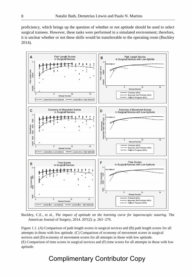
Natalie Bath, Demetrius Litwin and Paulo N. Martins 8
proficiency, which brings up the question of whether or not aptitude should be used to select
surgical trainees. However, these tasks were performed in a simulated environment; therefore,
it is unclear whether or not these skills would be transferrable to the operating room (Buckley
2014).
Buckley, C.E., et al., The impact of aptitude on the learning curve for laparoscopic suturing. The
American Journal of Surgery, 2014. 207(2): p. 263−270.
Figure 1.1. (A) Comparison of path length scores in surgical novices and (B) path length scores for all
attempts in those with low aptitude. (C) Comparison of economy of movement scores in surgical
novices and (D) economy of movement scores for all attempts in those with low aptitude.
(E) Comparison of time scores in surgical novices and (F) time scores for all attempts in those with low
aptitude.
Complimentary Contributor Copy
Laparoscopic Experimental Surgery Models 9
Although it has been established that separate learning curves exist for those with and
without an innate aptitude for laparoscopy, it is important to further delineate what makes this
distinction between groups and whether or not this discrepancy is surmountable. Visual-spatial
ability is thought to play a large role in achieving proficiency in both open and laparoscopic
procedures. A group of thirty-seven surgical residents were asked to complete six tests in order
to assess visual-spatial ability. After these tests were completed, residents were then asked to
complete a Z-plasty procedure. This procedure consists of one or more Z-shaped incisions
being made with the diagonals forming one straight line and two triangular sections, which is
used for cosmetic reasons or can be functional in order to elongate and relax scars. Residents
with higher visual-spatial scores performed better in the Z-plasty procedure in comparison to
those with lower scores. However, after additional practice, residents with lower scores were
also able to achieve competency (Wanzel 2002). This finding suggests that despite the fact that
learning curves differed between the groups, additional practice and feedback may be able to
make up for the innate differences between groups. In a separate study that examined visual-
spatial ability and manual dexterity among dental students, surgical residents, and staff
surgeons, it was found that higher visual-spatial ability scores were associated with a more
skilled performance among surgical novices. However, superior visual-spatial ability among
the more advanced trainees and experts was not found to correlate with superior performance
on procedures. Therefore, these findings suggest that although aptitude testing may not be able
to be used to predict who will perform well in a surgical career; however, it may be able to
predict who would likely benefit from additional surgical training prior to entering the operating
room (Wanzel 2003).
As previously stated, it has been established that laparoscopic procedures are associated
with a learning phase during which there is an increased prevalence of serious complications.
The learning curve for laparoscopic procedures is typically based on the time to procedure
completion and the number of complications; however, multiple factors play a role in
complications in the operating room in addition to surgeon experience such as the entire team
involved in patient care at that time. In addition to describing time to proficiency, learning curve
patterns may potentially also be used to identify surgical trainees who will be unable to achieve
proficiency despite additional training (Grantcharov 2004). A virtual-reality trainer was used
to measure the following parameters which have previously been validated in studies: time to
complete task, number of errors, and economy of motion score (Taffinder 1998). Figure 1.2
shows the four learning curves that were identified: surgeons who (1) demonstrated proficiency
from the beginning of the learning session (5.4%); (2) advanced with practice and achieved
predefined expert criteria between two and nine repetitions (70.3%); (3) slow and steady
acquisition of skills but unable to achieve proficiency level within ten repetitions (16.2%); and
(4) underperformed from the beginning and showed no tendency to skills improvement (8.1%)
(Grantcharov 2009; Kramp 2016). Trainees in the first group demonstrated impressive innate
ability, potentially indicating a strong future in minimally invasive surgery; whereas those in
the fourth group not only did not demonstrate any aptitude for laparoscopic surgery at the
beginning, but they also failed to show any progression towards achieving proficiency.
Although the role of surgeon involves skills such as communication and strong clinical
judgment, technical skills play a large role. Therefore, one’s performance on laparoscopic
trainers should not be viewed in isolation; however, technical performance should be taken into
consideration in order to help determine one’s success in pursuing a surgical career
(Grantcharov 2009). Complimentary Contributor Copy

Natalie Bath, Demetrius Litwin and Paulo N. Martins 10
Grantcharov, T.P. and P. Funch-Jensen, Can everyone achieve proficiency with the laparoscopic
technique? Learning curve patterns in technical skills acquisition. The American Journal of Surgery,
2009. 197(4): p. 447−449.
Figure 1.2. Learning curves. Time to perform the task. (a) Error scores. (b) Economy-of-motion scores.
(c) Dotted line indicates the proficiency level for each parameter.
In addition to visual-spatial ability, several other cognitive abilities can be assessed in order
to more completely understand one’s aptitude in laparoscopy. A cognitive aptitude test may be
used to assess trainees prior to laparoscopic simulator training. Ultimately, a surgeon’s
cognitive aptitude is related to the learning curve for minimally invasive surgery and the
possibility of success in achieving proficiency (Luursema 2010; Luursema 2012). Spatial
memory describes the ability to record information about one’s environment and its spatial
orientation. It can be viewed as an indicator of one’s ability to learn the procedural aspects of
tasks. Spatial memory is also related to the early learning phase of basic laparoscopic tasks
(Luursema 2010). Perceptual speed is the ability to quickly identify a given shape or dissimilar
shape from a number of alternatives. It is related to efficiency of movement in early learning,
and it is also related to the associative phase of learning, which indicates that part of
laparoscopic tasks might become automated during training. The fourth element of cognitive
ability is reasoning ability. Increases in perceptual speed, a skill crucial due to the time critical
nature of laparoscopic surgery, are found to be related to improved laparoscopic performance.
The fourth element, reasoning ability, was found to be related to early learning of laparoscopic
procedures. Reasoning ability was found to be less of an influence as skill level increased
(Keehner 2006).
Since laparoscopic surgery requires a different mental skill set, it should be expected that
the cognitive abilities of visual-spatial ability, spatial memory, perceptual speed, and reasoning Complimentary Contributor Copy
Laparoscopic Experimental Surgery Models 11
ability should predict the learning curve for basic tasks on a laparoscopic simulator.
Laparoscopic surgery requires each of these skills; therefore trainees who perform exceedingly
well on cognitive aptitude tests are expected to show high levels of performance in the
following areas: (1) number of practice sessions in order to achieve proficiency; (2) time to task
completion; (3) damage to surrounding tissue; (4) efficiency of movement on basic
laparoscopic simulator tasks. Despite these expectations, cognitive aptitude was found to
neither be related to total number of sessions required to achieve proficiency in a task, nor did
it predict learning rate during training. Participants with lower cognitive aptitude scores were
not found to require more practice sessions to reach proficiency in comparison to those with
higher scores. Although trainees became quicker and more efficient in performing basic
laparoscopic tasks on a simulator, damage to surrounding tissue remained constant. Cognitive
aptitude did not affect the amount of tissue damage that was found. Although learning rates for
trainees with discrepancies between their cognitive aptitude scores were similar, trainees with
higher levels of cognitive aptitude were found to consistently outperform those with lower
levels. Overall, there was no effect of cognitive aptitude on the learning curve across multiple
sessions (Groenier 2014).
All four aspects of cognitive aptitude were associated with higher efficiency of movement.
Visual-spatial and reasoning ability were associated with less time to complete a given task.
The role of cognitive aptitude did change, however, when the influence of a cognitive ability
was corrected for the effect of the other cognitive abilities. Perceptual speed remained
positively associated with efficiency of movement; spatial memory and perceptual speed were
associated with the amount of tissue damage.
ANIMAL MODELS OF LAPAROSCOPIC SURGERY TRAINING
Operating skills can be acquired via three methods, all of which have benefits and
drawbacks. The first method involves the use of bench models, for which benefits include lower
cost, ready availability, and reuse of materials (Martin 1997). Basic laparoscopy skills may be
learned and further honed on trainer boxes or with virtual reality models, but as the trainee
becomes more skilled, the need for more challenging and life-like models is required for skills
to be further developed. As a result, animal models have become a popular way to simulate
procedures in the operating room. The porcine model is the most commonly used model due to
the pig’s anatomical and physiological similarities to humans (Kobayashi 2012). The second
method involves practicing of skills in the animal laboratory. The animal model represents an
important step in the training process due to the fact that the direct step from the pelvic trainer
model to the operating room may be too difficult for a trainee. The animal model also allows
trainees to gain confidence in trocar use and positioning (Piechaud 2006). Although this method
is expensive and also presents the issue of limited resources and funding, the use of animals
very closely simulates operating on actual patients. The cadaver as a model also offers a very
effective way to gain an understanding of anatomy through a laparoscopic view. Cadavers also
provide macroscopic anatomy and improved spatial perception of anatomy. However, cadavers
present certain disadvantages due to the fact that they do not bleed, the visual field is also clear,
and no hemostasis can be done (Piechaud 2006). The third method takes place in the operating
room under the supervision of a preceptor where trainees can gain real life experience. The
Complimentary Contributor Copy

Natalie Bath, Demetrius Litwin and Paulo N. Martins 12
drawback to this method is that this can potentially be a dangerous situation in which trainees
are further developing their skills on real patients. Training in the operating room is also limited
by the fact that time and financial constraints limit the ability for trainees to perfect their skills
here.
As more complex procedures are now being performed with laparoscopy in addition to
shortened residency work hours, the need for intensive, efficient laparoscopy training is
necessary for novices. This training can be conducted in a safe, controlled, and standardized
environment within a simulation center; however, limited access to training centers does not
always make this training possible. In order to provide additional training to general surgeons
and graduating surgical residents, intensive training programs have been developed throughout
the country as an alternative to simulation centers. Most intensive training programs are set up
at locations throughout the country where general surgeons, residents, and fellows can travel to
in order to complete an intensive training program over the course of several days. Intensive
training programs may consist of lectures, laparoscopic videos, and practical training either on
animal training models or laparoscopic trainers. Skill improvement is assessed using objective
structured assessment of technical skill (OSATS). Most participants are found to have an
improvement in their overall performance after finishing these courses (Castillo 2015).
For those that have access to simulation centers at their local institutions, there are
numerous methods that can be used for preclinical training including virtual reality, box trainer
simulators, operations on cadavers, or animal models. Although virtual reality or box trainer
simulators have been used successfully by beginning trainees to acquire skills, they may be too
simple and do not correlate closely enough to the actual procedure in the operating room.
Therefore, human cadaveric and animal models have been introduced for more real-to-life
training. Animal models have been found to be the ideal model due their realistic anatomy and
surgical workflow similar to humans. Animal models are also ideal because they allow
pathological findings such as cholelithiasis and inflammatory findings to be present, which is
not possible in virtual reality or cadaveric models. Animal models are needed for two purposes.
First, they allow new, minimally invasive procedures to be evaluated for their feasibility and
effectiveness in comparison to standard techniques. Secondly, animal models provide training
that most accurately resembles operating on real patients; therefore, it may also be used for
training of both conventional and new methods of laparoscopic surgery (Ryska 2016). Due to
both of these considerations, animal models are used throughout multiple surgical sub-
specialties for preclinical training.
HEPATOBILIARY SURGERY
Training in laparoscopic hepatobiliary surgery with animal models has not gained large
traction internationally due in part to its level of difficulty; however, success has been found
when using the porcine model for training. Several differences exist between human and
porcine liver anatomy, which can present a challenge to new trainees, but with the appropriate
amount of preoperative preparation, the porcine model can offer a very realistic training model
for laparoscopic liver surgery (Figures 1.3−6) (Martins 2013). Bovine and porcine livers are
most frequently used due to their similarity to human livers in terms of size and density. These
animals are also farm animals that are readily available and relatively inexpensive (Ong 2013).
Complimentary Contributor Copy
Laparoscopic Experimental Surgery Models 13
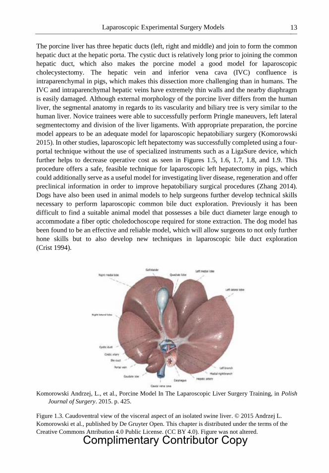
The porcine liver has three hepatic ducts (left, right and middle) and join to form the common
hepatic duct at the hepatic porta. The cystic duct is relatively long prior to joining the common
hepatic duct, which also makes the porcine model a good model for laparoscopic
cholecystectomy. The hepatic vein and inferior vena cava (IVC) confluence is
intraparenchymal in pigs, which makes this dissection more challenging than in humans. The
IVC and intraparenchymal hepatic veins have extremely thin walls and the nearby diaphragm
is easily damaged. Although external morphology of the porcine liver differs from the human
liver, the segmental anatomy in regards to its vascularity and biliary tree is very similar to the
human liver. Novice trainees were able to successfully perform Pringle maneuvers, left lateral
segmentectomy and division of the liver ligaments. With appropriate preparation, the porcine
model appears to be an adequate model for laparoscopic hepatobiliary surgery (Komorowski
2015). In other studies, laparoscopic left hepatectomy was successfully completed using a four-
portal technique without the use of specialized instruments such as a LigaSure device, which
further helps to decrease operative cost as seen in Figures 1.5, 1.6, 1.7, 1.8, and 1.9. This
procedure offers a safe, feasible technique for laparoscopic left hepatectomy in pigs, which
could additionally serve as a useful model for investigating liver disease, regeneration and offer
preclinical information in order to improve hepatobiliary surgical procedures (Zhang 2014).
Dogs have also been used in animal models to help surgeons further develop technical skills
necessary to perform laparoscopic common bile duct exploration. Previously it has been
difficult to find a suitable animal model that possesses a bile duct diameter large enough to
accommodate a fiber optic choledochoscope required for stone extraction. The dog model has
been found to be an effective and reliable model, which will allow surgeons to not only further
hone skills but to also develop new techniques in laparoscopic bile duct exploration
(Crist 1994).
Komorowski Andrzej, L., et al., Porcine Model In The Laparoscopic Liver Surgery Training, in Polish
Journal of Surgery. 2015. p. 425.
Figure 1.3. Caudoventral view of the visceral aspect of an isolated swine liver. © 2015 Andrzej L.
Komorowski et al., published by De Gruyter Open. This chapter is distributed under the terms of the
Creative Commons Attribution 4.0 Public License. (CC BY 4.0). Figure was not altered.
Complimentary Contributor Copy
Natalie Bath, Demetrius Litwin and Paulo N. Martins 14
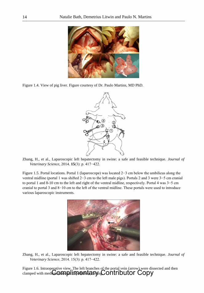
Figure 1.4. View of pig liver. Figure courtesy of Dr. Paulo Martins, MD PhD.
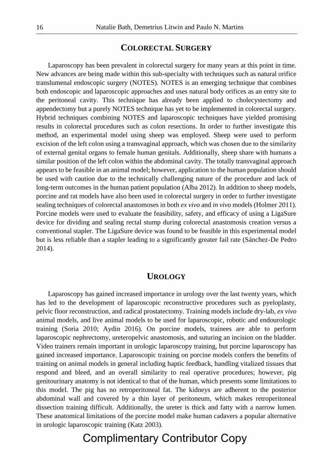
Zhang, H., et al., Laparoscopic left hepatectomy in swine: a safe and feasible technique. Journal of
Veterinary Science, 2014. 15(3): p. 417−422.
Figure 1.5. Portal locations. Portal 1 (laparoscope) was located 2−3 cm below the umbilicus along the
ventral midline (portal 1 was shifted 2−3 cm to the left male pigs). Portals 2 and 3 were 3−5 cm cranial
to portal 1 and 8-10 cm to the left and right of the ventral midline, respectively. Portal 4 was 3−5 cm
cranial to portal 3 and 8−10 cm to the left of the ventral midline. These portals were used to introduce
various laparoscopic instruments.
Zhang, H., et al., Laparoscopic left hepatectomy in swine: a safe and feasible technique. Journal of
Veterinary Science, 2014. 15(3): p. 417−422.
Figure 1.6. Intraoperative view. The left branches of the portal vein (arrow) were dissected and then
clamped with medium titanium clips before cutting. Complimentary Contributor Copy

Laparoscopic Experimental Surgery Models 15
Zhang, H., et al., Laparoscopic left hepatectomy in swine: a safe and feasible technique. Journal of
Veterinary Science, 2014. 15(3): p. 417−422.
Figure 1.7. Simulated intraoperative view. A knot was tied between the ends of the snipped sutures with
the aid of needle-holding forceps (F).
Zhang, H., et al., Laparoscopic left hepatectomy in swine: a safe and feasible technique. Journal of
Veterinary Science, 2014. 15(3): p. 417−422.
Figure 1.8. Simulated intraoperative view. A knot was tied with the aid of needle-holding forceps (F)
inserted from the third trocar.
Zhang, H., et al., Laparoscopic left hepatectomy in swine: a safe and feasible technique. Journal of
Veterinary Science, 2014. 15(3): p. 417−422.
Figure 1.9. Intraoperative view. The raw area observed immediately after liver resection. Complimentary Contributor Copy
Natalie Bath, Demetrius Litwin and Paulo N. Martins 16
COLORECTAL SURGERY
Laparoscopy has been prevalent in colorectal surgery for many years at this point in time.
New advances are being made within this sub-specialty with techniques such as natural orifice
translumenal endoscopic surgery (NOTES). NOTES is an emerging technique that combines
both endoscopic and laparoscopic approaches and uses natural body orifices as an entry site to
the peritoneal cavity. This technique has already been applied to cholecystectomy and
appendectomy but a purely NOTES technique has yet to be implemented in colorectal surgery.
Hybrid techniques combining NOTES and laparoscopic techniques have yielded promising
results in colorectal procedures such as colon resections. In order to further investigate this
method, an experimental model using sheep was employed. Sheep were used to perform
excision of the left colon using a transvaginal approach, which was chosen due to the similarity
of external genital organs to female human genitals. Additionally, sheep share with humans a
similar position of the left colon within the abdominal cavity. The totally transvaginal approach
appears to be feasible in an animal model; however, application to the human population should
be used with caution due to the technically challenging nature of the procedure and lack of
long-term outcomes in the human patient population (Alba 2012). In addition to sheep models,
porcine and rat models have also been used in colorectal surgery in order to further investigate
sealing techniques of colorectal anastomoses in both ex vivo and in vivo models (Holmer 2011).
Porcine models were used to evaluate the feasibility, safety, and efficacy of using a LigaSure
device for dividing and sealing rectal stump during colorectal anastomosis creation versus a
conventional stapler. The LigaSure device was found to be feasible in this experimental model
but is less reliable than a stapler leading to a significantly greater fail rate (Sánchez-De Pedro
2014).
UROLOGY
Laparoscopy has gained increased importance in urology over the last twenty years, which
has led to the development of laparoscopic reconstructive procedures such as pyeloplasty,
pelvic floor reconstruction, and radical prostatectomy. Training models include dry-lab, ex vivo
animal models, and live animal models to be used for laparoscopic, robotic and endourologic
training (Soria 2010; Aydin 2016). On porcine models, trainees are able to perform
laparoscopic nephrectomy, ureteropelvic anastomosis, and suturing an incision on the bladder.
Video trainers remain important in urologic laparoscopy training, but porcine laparoscopy has
gained increased importance. Laparoscopic training on porcine models confers the benefits of
training on animal models in general including haptic feedback, handling vitalized tissues that
respond and bleed, and an overall similarity to real operative procedures; however, pig
genitourinary anatomy is not identical to that of the human, which presents some limitations to
this model. The pig has no retroperitoneal fat. The kidneys are adherent to the posterior
abdominal wall and covered by a thin layer of peritoneum, which makes retroperitoneal
dissection training difficult. Additionally, the ureter is thick and fatty with a narrow lumen.
These anatomical limitations of the porcine model make human cadavers a popular alternative
in urologic laparoscopic training (Katz 2003).
Complimentary Contributor Copy

Laparoscopic Experimental Surgery Models 17
BARIATRIC SURGERY
Obesity continues to be a growing problem in the United States with an estimated 15.8
million adults qualifying for bariatric surgery (Ponce). Bariatric surgery techniques target
weight loss through restriction, malabsorption, or a mix of both. The two most popular weight
loss surgeries currently being performed are vertical sleeve gastrectomy (restrictive) and Roux-
en-Y gastric bypass (mixed). Within laparoscopic and bariatric surgery, there has been a
tendency for large laboratory animals such as pigs to be used. Bariatric experimental surgery
on animals is useful for developing and perfecting techniques. Animal experimentation in
bariatric surgery can also be used to study the relationship between metabolism and surgery in
order to find improvements in treatment in the co-morbidities associated with morbid obesity.
Lastly, research in this area can also focus on manipulating intake via central and vagal control
(Del Castillo Dejardin 2004). Although it is now an abandoned malabsorptive technique, the
jejunoileal bypass was previously the most studied bariatric surgical technique. Genetically-
determined Zucker-type obese rats were used for the first time in bariatric research. From this
research, subsequent studies of hormonal mechanisms responsible for weight loss and
resolution of co-morbidities such as type 2 diabetes took place (Polyzogopoulou 2003). Current
studies are aimed at further investigating the relationship between metabolism and surgery,
specifically with gastric bypass in Zucker-type obese rats. This animal research looks at peptide
hormones such as ghrelin, which regulates food intake and therefore plays a role in weight loss.
In addition to Zucker-type obese rats, dogs and pigs have been used with the restrictive and
mixed techniques with the goal of improving techniques for laparoscopy. Gastric persistalsis in
reduction of intake is also being studied in rats, dogs, pigs, and rabbits in order to discover new
options for patients in bariatric surgery. In order to draw meaningful conclusions, it is important
to use obese animals in this area of research (Del Castillo Dejardin 2004).
GENERAL SURGERY
Inguinal hernia repair is one of the most frequently performed surgical procedures with
over 800,000 hernia repairs performed annually in the USA. Despite the common nature of this
procedure, key problems still exist following repair including complications secondary to
invasive fixation of mesh, poor quality of tissue ingrowth induced by the mesh, and early mesh
migration to and penetration into neighboring organs after implantation (Scheidbach 2004;
Panaro 2015). Porcine animal models have been used for laparoscopic training purposes for
hernia repairs for quite some time, but this animal model is further being utilized in order to
compare surgical approaches and type of mesh used in hernia repairs. Polypropylene mesh is a
common mesh used in millions of hernia repair operations, but variations in the type of
polypropylene mesh used in repair result in a difference in their biocompatibility and handling
characteristics during surgery. Porcine models may be used to further characterize these
differences between mesh including shrinkage, chronic inflammatory reaction, and mesh
incorporation into the surrounding tissue (Scheidbach 2004). Studies have also been done using
the porcine model in order to further investigate various approaches to hernia repair including
NOTES technique, totally extraperitoneal (TEP) and transabdominal preperitoneal (TAPP)
(Dhumane 2013). Although this area of research may not purely focus on laparoscopic training, Complimentary Contributor Copy
Natalie Bath, Demetrius Litwin and Paulo N. Martins 18
the results of such studies have a direct impact on the future standard of care in inguinal hernia
repairs in patients.
PEDIATRIC SURGERY
Laparoscopic training outside of the operating room is of particular importance in pediatric
surgery due to the increasingly complicated surgeries that are now being performed
laparoscopically; however, the number of pediatric patients requiring these complicated
procedures is usually small, which makes training outside of the operating room of the utmost
importance in order to reduce complications and operating error. The porcine model is the most
widely used animal model in laparoscopic training in fields such as adult minimally invasive
surgery, colorectal, urologic and gynecological surgery. Despite this fact, very few studies have
described the role of animal models for laparoscopic training in the pediatric population.
Although the pig can be used in pediatric surgery training, the rabbit model could be helpful
for pediatric surgery training due to its smaller size, which is comparable to the operative field
encountered when operating on neonates and small children (Kirlum 2005). The piglet also
poorly simulates the pediatric population due to its larger size weighing between 20 and 40 kg.
In comparison, a rabbit that weighs 3−4 kg will have an abdominal cavity that measures
approximately 15 cm long, 10 cm wide, and 8 cm high, which mimics the dimensions of the
abdominal cavity in a newborn baby. Rabbits have been found to be good surgical models for
repair of inguinal hernia and hiatal hernia due to their patent inguinal ring with retractile or
intraabdominal testes and naturally gaping diaphragmatic hiatus, respectively. In contrast, it is
difficult to mobilize the esophagus or perform cholecystectomy in the pig model due to its huge,
multi-lobulated liver (Esposito 2016). Laparoscopic nephrectomy can also be challenging to
perform in the porcine model due to the difficult access to the kidneys caused by large,
distended bowel loops. Nephrectomy is more straight-forward in the rabbit model due to the
anterior location of the kidneys and scant intraabdominal and perineal fat (Molinas 2004). Other
laparoscopic procedures can be performed additionally including intestinal anastomosis,
splenectomy, and congenital diaphragmatic hernia repair. In an analysis of trainees’
performances on rabbit versus porcine models, it was found that surgeons had higher
performance scores and statistically shorter operative times for advanced procedures in the
rabbit model. Overall, the rabbit model appears to provide a viable approach to technical
training in basic and advanced laparoscopic procedures (Esposito 2016).
ALTERNATIVES TO ANIMAL SURGERY IN LAPAROSCOPIC SURGERY
Due to high costs, regulatory constraints, opposed public opinion, and complex logistics
(maintenance, anesthesia) associated with animal experiments, other alternatives for training
have been proposed. Simulation-based training allows trainees to further hone their
laparoscopic skills in a safe, non-threatening environment prior to entering the operating room.
The repetition of tasks and simulated procedures has been shown to reduce the risk of adverse
surgical events and further decrease operating time (Hyltander 2002). Although training that
occurs in the OR, on cadaver, and animal models may be a better method than simulation Complimentary Contributor Copy

Laparoscopic Experimental Surgery Models 19
training, it appears that skills acquired in a simulation setting are transferable to the OR such
as laparoscopic suturing and knot tying skills (Sutherland 2006; Al-Kadi 2013; Zendejas 2013).
Simulation-based training can lead to demonstrable benefits of surgical skills in the operating
room, and it is an effective way to teach laparoscopic surgery skills, increase translation of
laparoscopic skills to the OR, and increase patient safety (Vanderbilt 2015). Numerous
laparoscopic training tools exist, each of which have their own benefits and drawbacks.
A box trainer is a simple box that uses a camera, screen, light source and instruments. It
incorporates conventional laparoscopic equipment in order to perform basic skills. It can be
used for training on animal parts in addition to synthetic inanimate models. Complex task
trainers are hybrid models that provide visual, audio and touch cues and use integrated hardware
in order to replicate a clinical setting. They may also be composed of a partial component of a
simulator or body part such as an arm or a leg. Video trainers are more sophisticated box trainers
that have embedded motion sensors and recorders which measure distance and direction moved
in order to calculate economy of movement. Table 1.1 lists the different types of training tools
and their individual capabilities (Vanderbilt 2015; Alaker 2016).
In addition to physical box trainers and video trainers, virtual reality plays an increasingly
large role in laparoscopic training; multiple modalities that utilize virtual reality for
laparoscopic simulation training exist. Virtual reality uses screen-based computer software and
hardware similar to that used in laparo-endoscopic surgery. Virtual reality (VR) can further be
broken down into low-fidelity and high-fidelity groups. Low fidelity VR simulators use
computer-training programs that provide an abstract environment to teach basic laparo-
endoscopic skills whereas high-fidelity uses motorized instruments in order to provide haptic
feedback and more closely resembles live tissue. Minimally Invasive Surgical Trainer – Virtual
Reality (MIST VR), a part-task virtual reality laparoscopic simulator, not only simulates
surgical tasks and operations but is also able to provide an objective assessment of psychomotor
skills by the operator. It will then generate an overall score, taking into consideration errors
committed and time taken to complete six different tasks. When assessing surgical novices and
practicing surgeons, experienced surgeons scored consistently and significantly better than
their counterparts in all tasks, suggesting that the simulator is measuring surgically relevant
parameters. Therefore, MIST-VR appears to be a validated tool in objectively assessing
laparoscopic skills (Chaudhry 1999). In a systematic review and meta-analysis comparing
virtual reality to other simulation models, it was found that virtual reality simulation was
significantly more effective than video trainers and at least as good as box trainers in
laparoscopic simulation training (Nagendran 2013; Alaker 2016). Moreover, virtual reality
training appears to be a superior training method in comparison to standard laparoscopic
training in the operating room (Gurusamy 2009; Al-Kadi 2012; Larsen 2012; Willaert 2013).
Virtual or box trainer simulators have been shown to be effective at the beginning of
training; however, fresh-frozen cadavers are another method of providing laparoscopic training
that more closely resembles operating on patients. Cadavers provide full haptic and tactile
feedback, which is vital in laparoscopic simulation and is unfortunately difficult to incorporate
into virtual systems (Buckley 2014). They can also be stored at a low temperature for an
indefinite amount of time (Sharma 2012). In comparison of cost, an initial fee is incurred to
purchase with a small added cost for maintenance for simulators whereas cadaver bodies are
typically donated free of charge; however, high cost is associated with maintenance of a cadaver
laboratory and cremation of used bodies. Other drawbacks to use of cadavers for training
Complimentary Contributor Copy

Natalie Bath, Demetrius Litwin and Paulo N. Martins 20
Table 1.1. Laparoscopic training tools, definitions, and procedures commonly
used in surgery
Type of
simulation Definition
Camera
Navigation
Clipping
and
cutting
Suturing
and
knot
tying
Lifting
and
grasping
Dissection
Box Trainer A box that
incorporates
conventional
laparoscopic
equipment to perform
basic skills, is
versatile, and enables
training on animal
parts and synthetic
models
+ + + + +
Task Trainer A partial component
of a simulator or
simulation modality
such as an arm, leg or
torso
+ + +
MIST-VR Virtual reality
simulator with six
different tasks to
simulate maneuvers
performed during
laparoscopic
cholecystectomy in a
computerized
environment
+ + + +
LapMentor/
LapMentor II
Virtual reality
simulator consisting
of camera and two
calibrated working
instruments. Motion
of the instruments is
translated to a two-
dimensional computer
screen for student
practices
+ + + + +
LapSim A computer-based
simulator creating a
virtual laparoscopic
setting through a
computer operating
system, a video
monitor, a
laparoscopic interface
containing two pistol-
grip instruments, and
a diathermy pedal
without haptic
feedback
+ + + + +
EndoTower EndoTower software
consists of an angled
telescope simulator
composed of rotating
camera and telescopic
components.
+ +
Complimentary Contributor Copy

Laparoscopic Experimental Surgery Models 21
MISTELS/FLS
trainer
McGill Inanimate
System for Training
and Evaluation of
Laparoscopic Skills
(MISTELS)- this
inexpensive, portable,
and flexible system
allows students to
practice in a virtual
Endotrainer box
+ + +
SIMENDO
VR
Computer software
used to train eye-hand
coordination skills by
camera navigation
and basic drills.
+ + +
URO Mentor A hybrid simulator,
consisting of a
personal computer
based system linked
to a mannequin with
real endoscopes.
Cystoscopic and
ureteroscopic
procedures are
performed using
either flexible or semi
rigid endoscopes
+ + + +
Da Vinci Skills
Simulator
A portable simulator
containing a variety
of exercises and
scenarios specifically
designed to give users
the opportunity to
improve their
proficiency with
surgical controls.
+ + + + +
Vanderbilt, A.A., et al., Randomized Controlled Trials: A Systematic Review of Laparoscopic Surgery
and Simulation-Based Training. Global Journal of Health Science, 2015. 7(2): p. 310−327. This is
an open-access article distributed under the terms and conditions of the Creative Commons
Attribution license (http://creativecommons.org/licenses/by/3.0/). Table was not altered
include limited availability, avital tissues, and inability to injure vasculature or other structures
(Sharma 2012; Ryska 2016).
Robotic surgery continues to grow in popularity worldwide. Although it is a derivative of
minimally invasive surgery, the skills required to perform robotic surgery are unique from those
required in either open or laparoscopic surgery. There are currently four commercially available
simulators: the da Vinci Skills simulator (dVSS), the Mimic dV-Trainer (dV-Trainer), the
Robotic Surgical Simulator, and Sim-Surgery Educational Platform (SEP). All four trainers are
able to assess robotic skill. Three of the four simulators (dVSS, dV-Trainer, RoSS) demonstrate
the ability to improve basic robotic skills, with proficiency-based training being the most
effective training style. The skills obtained on a VR training curriculum appear to be similar to
those obtained through dry laboratory simulation (Bric 2016).
An alternative to virtual reality simulators and fresh frozen cadavers is the Open-source
Heidelberg laparoscopy phantom (OpenHELP), which combines realistic anatomy and tissue Complimentary Contributor Copy

Natalie Bath, Demetrius Litwin and Paulo N. Martins 22
properties with reusable simulation specimens. OpenHELP is based on a digital three-
dimensional model obtained from a CT scan of a male body and uses a flexible torso, via an
elastic abdominal wall, or rigid, with a plastic shell, filled with artificial organs made from
silicon casts. Pneumoperitoneum could successfully be attained by both methods (Kenngott
2015). As NOTES become more prevalent, training models specific for this technique are
needed. A new training model for the NOTES procedures was developed known as the
endoscopic-laparoscopic interdisciplinary training entity (ELITE). This model is composed of
a synthetic package with all the intrabdominal organs, including the greater omentum. The
synthetic appendix is made from gelatin material, which allows for dissection with high-
frequency tools. The ELITE training model appears to be well suited for the training of NOTES
procedures (Gillen 2012).
In addition to in vivo training, pieces of animal tissues may also be removed from the
animal specimen and be used to recreate the anatomic conditions of a specific procedure. When
used for this purpose, the animal tissue is traditionally placed in a pelvic trainer or dry box in
order to more closely simulate a specific laparoscopic surgery for the trainee. Animal tissue
may also be placed in human body simulators in order to more closely imitate both human
anatomy and realistic operating conditions by including real tissue that will allow tactile
feedback and will respond when being manipulated. The advantages of using pieces of animal
tissue include high availability, low cost, lack of need for anesthesia, and no animal
maintenance (van Velthoven 2006).
LAPAROSCOPIC SURGERY IN SMALL ANIMAL MODELS
A wide variety of small animal models exist in order to provide a sophisticated training
model for the advanced laparoscopic trainee. Each of these models not only have advantages
and disadvantages, but they also possess characteristics which may make them better suited for
certain surgical sub-specialties over other small animal models. Overall, the ideal training
model should provide the skills required, be inexpensive, universally available, and
anatomically and physiologically identical to the anesthetized patient. Mice, rats, rabbits, and
guinea pigs are universally accepted and are bred for both surgical and medical research due to
cheaper costs, easy handling and care, and a more favorable public opinion. Although primates
such as monkeys and apes most closely represent humans physiologically and anatomically,
the use of primates is limited only to special circumstances in which any other animal model
would be deemed unreliable. Animal models are typically utilized for reproduction of the
following systems for training purposes due to the relative ease of reproducing surgical
conditions: musculoskeletal, genitourinary, integumentary, vascular, cardiothoracic, and
digestive system (Hassan 2005).
Mice, despite being studied extensively and being ideal for immunologic or oncologic
studies, are too small to be used in laparoscopic procedures. On the other hand, the larger animal
model, the rat, has been previously used for laparoscopic training such as in transperitoneal
laparoscopic research (Gutt 1998). The rat model is inexpensive and easily handled due to size.
This model has previously been described as a model for retroperitoneal mini laparoscopic
nephrectomy, but the rat can also be used universally throughout laparoscopic sub-specialties
as a way for the trainee to acquire advanced laparoscopic skills and microsurgical suturing
Complimentary Contributor Copy
Laparoscopic Experimental Surgery Models 23
skills (Kaouk 2000). However, due to the limited workspace, the rat model requires the trainee
to have already acquired the skills necessary to work with 3-mm instruments in a very small
space.
In comparison to the rat model, the rabbit model is not only inexpensive but also provides
a larger workspace, which makes it a more realistic model. This model has been used as an in
vivo training model for laparoscopic skills such as knot-tying, suturing, and using cutting and
coagulation modes for dissection (Molinas 2004). The rabbit model has been shown to be well-
suited for the use as a new training tool that emphasizes the repetition of simple procedures due
to its ability to easily monitor progress when comparing learning curves between experienced
and inexperienced surgeons such as gynecologists and medical students, respectively. With
additional practice, shorter operating times and fewer complications were noted. This small
animal model is also being used in experimental settings to evaluate the oxidative stress
production on a transplanted kidney during laparoscopy (Demirbas 2004). As previously
discussed, the rabbit model provides a very realistic model for pediatric surgery training due to
the small operative field similar to that seen in the pediatric population. Due to the potential
risks of carbon dioxide pneumoperitoneum in the neonate and infant population, the concept of
gasless laparoscopy was first developed in the rabbit model. In the gasless model, the animals
were neither intubated nor mechanically ventilated. After incision was made and the
peritoneum was entered, an abdominal wall elevator was inserted which provided excellent
visualization of the abdominal cavity (Luks 1995).
At the Center for the Development of Advanced Medical Technology (CDAMTec) in
Japan, both wild-type and genetically modified pigs are used as multi-purpose models for
medical skill education, the development of therapeutic strategies, and innovations for new
tools for endo- and laparoscopic procedures. Unlike dogs, few spontaneous disease models
exist in pigs. Pigs also share similar immune systems with humans as seen in inbred pigs such
as the Clawn minipig, which further allows genetically modified pigs to be used in experimental
models for liver, intestine, kidney, pancreas, and lung transplantation (Kobayashi 2012). At the
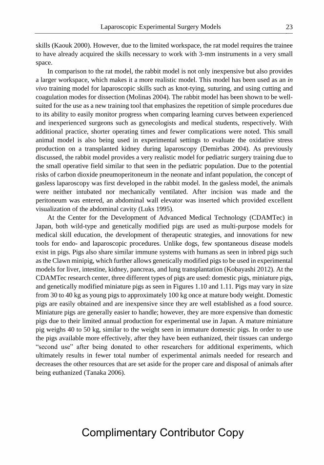
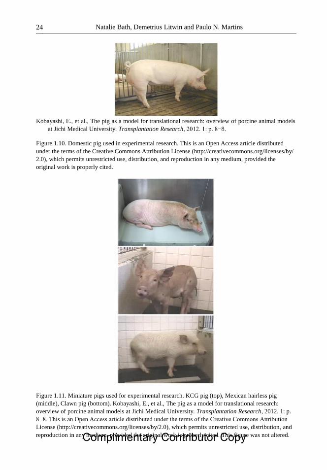
CDAMTec research center, three different types of pigs are used: domestic pigs, miniature pigs,
and genetically modified miniature pigs as seen in Figures 1.10 and 1.11. Pigs may vary in size
from 30 to 40 kg as young pigs to approximately 100 kg once at mature body weight. Domestic
pigs are easily obtained and are inexpensive since they are well established as a food source.
Miniature pigs are generally easier to handle; however, they are more expensive than domestic
pigs due to their limited annual production for experimental use in Japan. A mature miniature
pig weighs 40 to 50 kg, similar to the weight seen in immature domestic pigs. In order to use
the pigs available more effectively, after they have been euthanized, their tissues can undergo
“second use” after being donated to other researchers for additional experiments, which
ultimately results in fewer total number of experimental animals needed for research and
decreases the other resources that are set aside for the proper care and disposal of animals after
being euthanized (Tanaka 2006).
Complimentary Contributor Copy
Natalie Bath, Demetrius Litwin and Paulo N. Martins 24
Kobayashi, E., et al., The pig as a model for translational research: overview of porcine animal models
at Jichi Medical University. Transplantation Research, 2012. 1: p. 8−8.
Figure 1.10. Domestic pig used in experimental research. This is an Open Access article distributed
under the terms of the Creative Commons Attribution License (http://creativecommons.org/licenses/by/
2.0), which permits unrestricted use, distribution, and reproduction in any medium, provided the
original work is properly cited.
Figure 1.11. Miniature pigs used for experimental research. KCG pig (top), Mexican hairless pig
(middle), Clawn pig (bottom). Kobayashi, E., et al., The pig as a model for translational research:
overview of porcine animal models at Jichi Medical University. Transplantation Research, 2012. 1: p.
8−8. This is an Open Access article distributed under the terms of the Creative Commons Attribution
License (http://creativecommons.org/licenses/by/2.0), which permits unrestricted use, distribution, and
reproduction in any medium, provided the original work is properly cited. This figure was not altered. Complimentary Contributor Copy
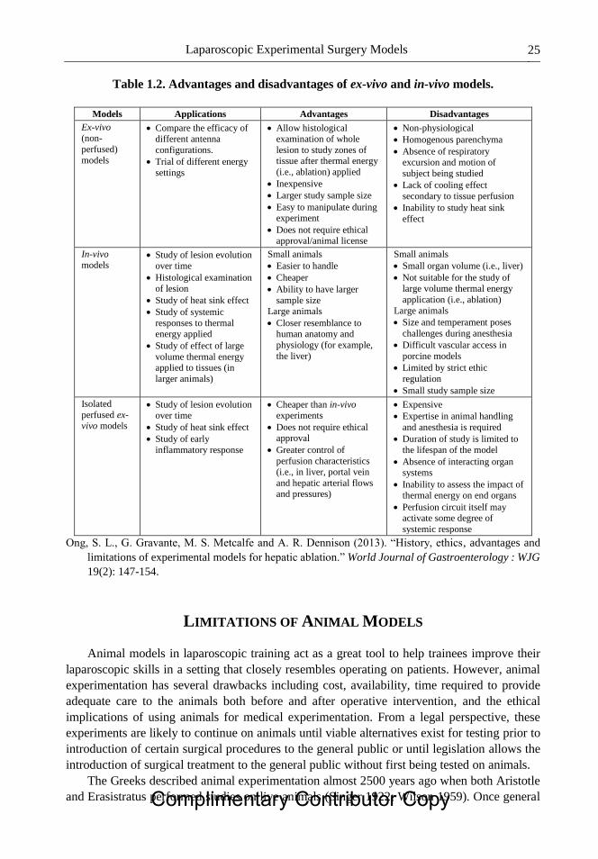
Laparoscopic Experimental Surgery Models 25
Table 1.2. Advantages and disadvantages of ex-vivo and in-vivo models.
Models Applications Advantages Disadvantages
Ex-vivo (non-
perfused)
models
Compare the efficacy of different antenna
configurations.
Trial of different energy
settings
Allow histological examination of whole
lesion to study zones of tissue after thermal energy
(i.e., ablation) applied
Inexpensive
Larger study sample size
Easy to manipulate during experiment
Does not require ethical approval/animal license
Non-physiological
Homogenous parenchyma
Absence of respiratory excursion and motion of
subject being studied
Lack of cooling effect
secondary to tissue perfusion
Inability to study heat sink
effect
In-vivo
models Study of lesion evolution
over time
Histological examination of lesion
Study of heat sink effect
Study of systemic
responses to thermal energy applied
Study of effect of large
volume thermal energy applied to tissues (in
larger animals)
Small animals
Easier to handle
Cheaper
Ability to have larger
sample size Large animals
Closer resemblance to human anatomy and
physiology (for example,
the liver)
Small animals
Small organ volume (i.e., liver)
Not suitable for the study of large volume thermal energy
application (i.e., ablation)
Large animals
Size and temperament poses
challenges during anesthesia
Difficult vascular access in
porcine models
Limited by strict ethic
regulation
Small study sample size
Isolated
perfused ex-vivo models
Study of lesion evolution over time
Study of heat sink effect
Study of early
inflammatory response
Cheaper than in-vivo experiments
Does not require ethical approval
Greater control of
perfusion characteristics (i.e., in liver, portal vein
and hepatic arterial flows and pressures)
Expensive
Expertise in animal handling
and anesthesia is required
Duration of study is limited to
the lifespan of the model
Absence of interacting organ systems
Inability to assess the impact of thermal energy on end organs
Perfusion circuit itself may activate some degree of
systemic response
Ong, S. L., G. Gravante, M. S. Metcalfe and A. R. Dennison (2013). “History, ethics, advantages and
limitations of experimental models for hepatic ablation.” World Journal of Gastroenterology : WJG
19(2): 147-154.
LIMITATIONS OF ANIMAL MODELS
Animal models in laparoscopic training act as a great tool to help trainees improve their
laparoscopic skills in a setting that closely resembles operating on patients. However, animal
experimentation has several drawbacks including cost, availability, time required to provide
adequate care to the animals both before and after operative intervention, and the ethical
implications of using animals for medical experimentation. From a legal perspective, these
experiments are likely to continue on animals until viable alternatives exist for testing prior to
introduction of certain surgical procedures to the general public or until legislation allows the
introduction of surgical treatment to the general public without first being tested on animals.
The Greeks described animal experimentation almost 2500 years ago when both Aristotle
and Erasistratus performed studies on live animals (Singer 1922; Wilson 1959). Once general Complimentary Contributor Copy

Natalie Bath, Demetrius Litwin and Paulo N. Martins 26
anesthesia emerged in the 1840s, this allowed unconscious animals to be studied, which led to
a turning point in the medical and scientific communities’ ability to use animal models in
surgical experimentation. Animals can be used either as ex vivo or in vivo models. The major
limitations of ex vivo models include absence of blood flow and vitalized tissues, which will
respond to movement and maneuvers performed by the surgeon, and they also have limited
lifespans during which valid data can be obtained. The absence of other interacting organs in
ex vivo models may also affect the extent of systemic responses during experimentation. The
use of living animals with in vivo studies requires specific laboratory facilities and research
teams with expertise in anesthetizing and handling different species. The size of the animal also
poses a barrier, for example when a larger animal such as a pig or cow needs to be anesthetized,
moved, or examined post-operative (Hildebrand 2007; Ong 2013). The advantages and
disadvantages of ex vivo and in vivo models for experimentation are compared in Table 1.2.
FUTURE OF LAPAROSCOPIC SURGERY
Laparoscopic surgery has evolved immensely from its initial use as a diagnostic procedure
to the first laparoscopic cholecystectomy being performed in 1985 to its widespread use across
all surgical subspecialties. Laparoscopy has further developed over the last thirty years in part
due to the development of new instruments, which allows more complex operations to be
performed. Laparoscopic camera equipment is now available in high-definition, with automatic
focus and zoom, and three-dimensional images. Several different types of energy devices are
now available including monopolar electrocautery scissors, ultrasonic coagulating shears, and
electrothermal bipolar vessel sealers, which can be used for dissection and division of tissue,
bowel, and vascular structures (Blackmore 2014). The development of new surgical
instruments and imaging devices expands not only the type of surgery but also the surgical
fields that are now able to use laparoscopy. In addition to these new contributions to the field,
laparoscopy continues to evolve through different methods in which laparoscopic surgery can
be performed including robotic surgery, single incision laparoscopic surgery (SILS), natural
orifice translumenal endoscopic surgery (NOTES), and three-dimensional laparoscopy.
Robotic surgery was first seen in the field of urology and cardiac surgery with over 50,000
robotic prostatectomies being performed in 2007 in the USA alone. It was not used in colorectal
surgery until 2002 (Blackmore 2014). Robotic surgery confers several advantages including
high-definition three-dimensional visualization, increased freedom of movement, and
minimization of tremor. These advantages provide for a more controlled, delicate dissection
compared to laparoscopy. Several urologic procedures are now routinely being performed
robotically including radical prostatectomy, transperitoneal and retroperitoneal partial
nephrectomy, radical nephrectomy with caval thrombectomy, and pyeloplasty (Rogers 2008;
Abaza 2011; Patel 2013; Stepanian 2016). Studies have indicated that robotic-assisted partial
nephrectomy is associated with excellent outcomes in terms of perioperative complications and
functional results when performed by experienced surgeons. Robotic surgery could potentially
also be applied to removal of complex tumors (Novara). Robotic technology has also been
extended to liver surgery and represents an accepted alternative to open and laparoscopic
techniques in certain cases. Robotic liver surgery allows precise dissection and microsuturing
in narrow operative fields, and with this technology, there is greater potential for more major
Complimentary Contributor Copy
Laparoscopic Experimental Surgery Models 27
resections and difficult segmentectomies to be performed through minimally invasive methods.
Current data suggests that robotic liver resections have at the very least similar perioperative
and postoperative outcomes while conferring the general benefits of minimally invasive
surgery. Advantages of minimally invasive robotic surgery appear to be able to be applied
additionally to right hepatectomy for living liver donors including faster post-operative
recovery. Similar early allograft dysfunction, complications, and one-year recipient liver
function were found between the open and robotic groups (Chen 2016). The technology used
in robotic surgery also helps integrate and facilitate newer technologies such as near-infrared
fluorescence (Giulianotti 2016). However, there are limitations to robotic surgery, which
include higher associated costs and the need for specialized training for the primary surgeon,
secondary surgeon, and OR nurses.
The next step to decrease the invasiveness and improve cosmesis of laparoscopic surgery
is to decrease the size and number of incisions made as seen in single incision laparoscopic
surgery (SILS). Through this technique, all instruments are introduced into the body cavity
through a single access port. As laparoscopy has resulted in decreased trauma to the abdominal
wall, improved pain control, and quicker recovery post-operatively, single-site incision surgery
has further capitalized on these advantages of minimally invasive surgery by decreasing the
number of incisions made. However, SILS requires a skill set and has ergonomic demands,
which cannot directly be adapted from laparoscopic surgery. Therefore, in order to avoid higher
complication rates as seen in both early laparoscopic cases and now initial SILS cases,
evidence- and competency-based SILS curriculum should be instituted.
The first reported SILS cholecystectomy was performed in 1995 with new applications to
other fields such as appendectomies and complex bariatric procedures. As was seen in the early
days of laparoscopic surgery, increased complication rates at the advent of SILS makes it clear
that appropriate training should occur before utilizing this technique on patients. The
Laparoendoscopic Single-site Surgery Consortium for Assessment and Research (LESSCAR)
set out a 2009 whitepaper, which addresses some of the issues regarding appropriate training
for new techniques. LESSCAR recommends a stepwise format for training with a progression
from inanimate models to animal procedures to supervised human SILS cases. These are the
same principles that can be applied to laparoscopic or robotic training. As seen in case studies
that examine SILS outcomes, there exists a significant learning curve even for laparoscopic
surgeons who adopt SILS as seen through greater operative time and increased conversion from
single to multi-port laparoscopy. Skills required for SILS do not appear to be automatically
conferred from laparoscopic surgery, which further makes it important that adequate, safe
training is available in order to make surgeons proficient (Pucher 2013). Needlescopic
cholecystectomy is an additional method, which further reduces traumatic stress during and
after surgery and hastens full recovery. A meta-analysis comparing needlescopic versus
laparoscopic cholecystectomy indicated that needlescopic is not only safe and effective
procedure for management of gallstone disease, but it is superior to laparoscopy for less post-
operative pain and better cosmetic results despite being associated with longer operative time
and higher conversion rate.
Natural orifice translumenal endoscopic surgery (NOTES) has gained traction in the
surgical community as a technique to further advance minimally invasive techniques towards
being truly non-invasive (Figure 1.12). This method requires a flexible endoscope to be inserted
via a natural orifice such as the mouth, anus, vagina or umbilicus in order to gain access to the
abdominal cavity thereby without leaving a scar. NOTES has gained popularity as a method to Complimentary Contributor Copy

Natalie Bath, Demetrius Litwin and Paulo N. Martins 28
perform cholecystectomy or appendectomy. NOTES is truly a non-invasive surgery since it
eliminates external postoperative wounds and other advantages including reduction in trauma
to the abdominal wall, decreased recovery time, decreased clinical costs, and improved overall
cosmetic results (Spivak 1997; MacFadyen 2005; Richards 2005; Ponsky 2006). Initial clinical
outcomes following transvaginal hybrid NOTES cholecystectomy indicate that this method
appears to be safe when performed by appropriately trained surgeons and may result in a faster
return to normal activities and decreased postoperative pain (Sodergren 2015).
As with any new operative technique, appropriate training and investigation into post-
operative outcomes should be performed prior to routine use of the NOTES technique.
Differences between laparoscopic and NOTES procedures are that the NOTES technique
requires the surgeon to operate an endoscope and other instruments through a single access
point, rather than three or four other ports as frequently used in laparoscopy. Many NOTES
procedures are currently being performed via hybrid fashion in which both an endoscope and
trans-abdominal instruments are used; however, there is still significant loss of traction and the
in-line instrument approach using the instrument ports in the endoscope hand-piece remain
unfamiliar to most surgeons. Additionally, there is no gastrointestinal lumen to support the
endoscope; thus, the distal end of the endoscope is manipulated in the open abdominal cavity
using the incision site, internal organs and gravitational force to navigate and position the
endoscope and instruments. As a result, the middle section of the endoscope is prone to rolling
and twisting in the abdomen. Similar to SILS, the skill set required to operate an endoscope
and associated surgical instruments is unique and cannot be directly transferred from
laparoscopic and endoscopic skills (Korzeniowski 2016). Virtual reality simulators have found
to be effective training tools for other minimally invasive techniques; however, a virtual reality
trainer specific to NOTES procedures is required in order to proceed with further training. As
a result, the natural orifice virtual surgery (NOViSE) simulator was developed due to the lack
of established curriculum for NOTES training. NOViSE is a first prototype whose main focus
is teaching the endoscopic manipulation skills required for NOTES and includes integration of
a force-feedback-enabled virtual reality simulator for NOTES training with a virtual flexible
endoscope. NOViSE is currently only able to simulate trans-gastric hybrid cholecystectomy
procedure using a flexible endoscope as seen in Figure 1.12. Promising initial results following
its implementation as a training device indicate that may be able to contribute to surgical
training and to improving technical skills for NOTES procedures without putting patients at
risk (Korzeniowski 2016).
One of the challenges of laparoscopic surgery is that surgeons face the loss of depth
perception and spatial orientation due to the fact that they are working in a three-dimensional
(3D) space but being guided by two-dimensional (2D) images. This disadvantage leads to a
high visual and cognitive load. Therefore, 3D imaging was developed as an alternative to
conventional 2D imaging. 3D imaging was introduced as early as the 1990s; however, its use
is still not standard at hospitals, which may be secondary to the degraded view of the operative
field resulting from poor image resolution, the necessity to wear uncomfortable eyewear (which
alone can lead to eye strain, headaches, dizziness, and physical discomfort), and high cost.
However, as technology has further evolved and high-definition monitors are now available,
these disadvantages may be minimized when using the newest technology (Sorensen 2016).
Complimentary Contributor Copy
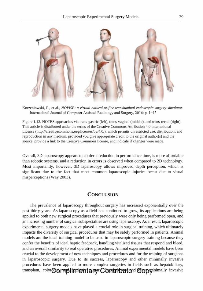
Laparoscopic Experimental Surgery Models 29
Korzeniowski, P., et al., NOViSE: a virtual natural orifice transluminal endoscopic surgery simulator.
International Journal of Computer Assisted Radiology and Surgery, 2016: p. 1−13
Figure 1.12. NOTES approaches via trans-gastric (left), trans-vaginal (middle), and trans-rectal (right).
This article is distributed under the terms of the Creative Commons Attribution 4.0 International
License (http://creativecommons.org/licenses/by/4.0/), which permits unrestricted use, distribution, and
reproduction in any medium, provided you give appropriate credit to the original author(s) and the
source, provide a link to the Creative Commons license, and indicate if changes were made.
Overall, 3D laparoscopy appears to confer a reduction in performance time, is more affordable
than robotic systems, and a reduction in errors is observed when compared to 2D technology.
Most importantly, however, 3D laparoscopy allows improved depth perception, which is
significant due to the fact that most common laparoscopic injuries occur due to visual
misperceptions (Way 2003).
CONCLUSION
The prevalence of laparoscopy throughout surgery has increased exponentially over the
past thirty years. As laparoscopy as a field has continued to grow, its applications are being
applied to both new surgical procedures that previously were only being performed open, and
an increasing number of surgical subspecialties are using laparoscopy. As a result, laparoscopic
experimental surgery models have played a crucial role in surgical training, which ultimately
impacts the diversity of surgical procedures that may be safely performed in patients. Animal
models are the ideal training model to be used in laparoscopic surgery training because they
confer the benefits of ideal haptic feedback, handling vitalized tissues that respond and bleed,
and an overall similarity to real operative procedures. Animal experimental models have been
crucial to the development of new techniques and procedures and for the training of surgeons
in laparoscopic surgery. Due to its success, laparoscopy and other minimally invasive
procedures have been applied to more complex surgeries in fields such as hepatobiliary,
transplant, colorectal, and pediatric surgery. Laparoscopy and other minimally invasive Complimentary Contributor Copy
Natalie Bath, Demetrius Litwin and Paulo N. Martins 30
techniques such as robotic surgery and NOTES require their own set of surgical skills, which
makes it imperative that proper training is available prior to both novice surgeons using basic
techniques and experienced surgeons using advanced or new techniques on patients.
Laparoscopic trainers, virtual reality, cadavers, and in vivo animal models are all options for
laparoscopic training with in vivo animal models providing the most real-to-life models for
practice due to their anatomical and physiologic similarities to humans. As minimally invasive
surgery continues to evolve from laparoscopy to robotic, single-incision laparoscopic surgery,
and NOTES procedures, the need for additional animal training will be a necessity in order to
ensure optimal outcomes for patients.
REFERENCES
Abaza, RR. Robotic surgery and minimally invasive management of renal tumors with vena
caval extension. Current opinion in urology, 2011, 21(2), 104−109.
Al-Kadi, AS; Donnon, T. Using simulation to improve the cognitive and psychomotor skills of
novice students in advanced laparoscopic surgery: a meta-analysis. Med. Teach., 2013, 35
Suppl 1, S47−55.
Al-Kadi, AS; Donnon, T; Oddone Paolucci, E; Mitchell, P; Debru, E; Church, N. The effect of
simulation in improving students’ performance in laparoscopic surgery: a meta-analysis.
Surg. Endosc., 2012, 26(11), 3215−3224.
Alaker, M; Wynn, GR; Arulampalam, T. Virtual reality training in laparoscopic surgery: A
systematic review & meta-analysis. Int. J. Surg., 2016, 29, 85−94.
Alba Mesa, F; Amaya Cortijo, A; Romero Fernandez, TM; Komorowski, AL; Sanchez
Hurtado, MA; Sanchez Margallo, FM. Totally transvaginal resection of the descending
colon in an experimental model. Surgical Endoscopy, 2012, 26(3), 877−881.
Antoniou, SA; Antoniou, GA; Koch, OO; Pointner, R; Granderath, FA. Meta-analysis of
laparoscopic vs open cholecystectomy in elderly patients. World Journal of
Gastroenterology: WJG, 2014, 20(46), 17626−17634.
Aydin, A; Raison, N; Khan, MS; Dasgupta, P; Ahmed, K. Simulation-based training and
assessment in urological surgery. Nat. Rev. Urol., 2016, 13(9), 503−519.
Blackmore, AE; Wong, MT; Tang, CL. Evolution of laparoscopy in colorectal surgery: an
evidence-based review. World J. Gastroenterol., 2014, 20(17), 4926−4933.
Bric, JD; Lumbard, DC; Frelich, MJ; Gould, JC. Current state of virtual reality simulation in
robotic surgery training: a review. Surgical Endoscopy, 2016, 30(6), 2169−2178.
Buckley, CE; Kavanagh, DO; Nugent, E; Ryan, D; Traynor, OJ; Neary, PC. The impact of
aptitude on the learning curve for laparoscopic suturing. The American Journal of Surgery,
2014, 207(2), 263−270.
Buckley, CE; Kavanagh, DO; Traynor, O; Neary, PC. Does aptitude matter? Journal of the
American College of Surgeons, 2012, 215(3), S116.
Bui, A; Stockhausen, F; Hanisch, E. Laparoscopic surgery: A qualified systematic review.
World J. Methodol, 2015, 5(4), 238−254.
Castillo, R; Buckel, E; León, F; Varas, J; Alvarado, J; Achurra, P; Aggarwal, R; Jarufe, N;
Boza, C. Effectiveness of Learning Advanced Laparoscopic Skills in a Brief Intensive
Laparoscopy Training Program. Journal of Surgical Education, 2015, 72(4), 648−653.
Complimentary Contributor Copy

Laparoscopic Experimental Surgery Models 31
Chaudhr, A; Sutton, C; Wood, J; Stone, R; McCloy, R. Learning rate for laparoscopic surgical
skills on MIST VR, a virtual reality simulator: quality of human-computer interface.
Annals of The Royal College of Surgeons of England, 1999, 81(4), 281−286.
Chen, PD; Wu, CY; Hu, RH; Ho, CM; Lee, PH; Lai, HS; Lin, MT; Wu, YM. Robotic liver
donor right hepatectomy – a pure, minimally invasive approach. Liver Transplantation,
2016, 22 (11), 1509−1518.
Cook, JA; Ramsay, CR; Fayers, P. Using the literature to quantify the learning curve: A case
study. International Journal of Technology Assessment in Health Care, 2007, 23(2),
255−260.
Cook, JA; Ramsaya, CR; Fayers, P. Statistical evaluation of learning curve effects in surgical
trials. Clinical Trials, 2004, 1(5), 421−427.
Crist, DW; Davoudi, MM; Parrino, PE; Gadacz, TR. An experimental model for laparoscopic
common bile duct exploration. Surg. Laparosc. Endosc., 1994, 4(5), 336−339.
Del Castillo Dejardin, D; Sabench Pereferrer, F; Hernandez Gonzale, M; Blanco Blasco, S;
Abello Sala, M. The evolution of experimental surgery in the field of morbid obesity. Obes.
Surg., 2004, 14(9), 1263−1272.
Demirbas, M; Samli, M; Aksoy, Y; Guler, C; Kilinc, A; Dincel, C. Comparison of changes in
tissue oxidative-stress markers in experimental model of open, laparoscopic, and
retroperitoneoscopic donor nephrectomy. Journal of endourology, 2004, 18(1), 105−108.
Dhumane, P; Donatelli, G; Chung, H; Dallemagne, B; Marescaux, J. Feasibility of
transumbilical flexible endoscopic preperitoneoscopy (FLEPP) and its utility for inguinal
hernia repair: experimental animal study. Surg. Innov., 2013, 20(1), 5−12.
Esposito, CC. Training Models in Pediatric Minimally Invasive Surgery: Rabbit Model Versus
Porcine Model: A Comparative Study. Journal of laparoendoscopic & advanced surgical
techniques. Part A, 2016, 26(1), 79−84.
Feldman, LS; Cao, J; Andalib, A; Fraser, S; Fried, GM. A method to characterize the learning
curve for performance of a fundamental laparoscopic simulator task: Defining “learning
plateau” and “learning rate”. Surgery, 2009, 146(2), 381−386.
Fried, GM; Feldman, LS; Vassiliou, MC; Fraser, SA; Stanbridge, D; Ghitulescu, G; Andrew,
CG. Proving the Value of Simulation in Laparoscopic Surgery. Annals of Surgery, 2004,
240(3), 518−528.
Gillen, S; Gröne, J; Knödgen, F; Wolf, P; Meyer, M; Friess, H; Buhr, HJ; Ritz, JP; Feussner,
H; Lehmann, KS. Educational and training aspects of new surgical techniques, experience
with the endoscopic–laparoscopic interdisciplinary training entity (ELITE) model in
training for a natural orifice translumenal endoscopic surgery (NOTES) approach to
appendectomy. Surgical Endoscopy, 2012, 26(8), 2376−2382.
Giulianotti, PC; Bianco, FM; Daskalaki, D; Gonzalez-Ciccarelli, LF; Kim, J; Benedetti, E.
Robotic liver surgery: technical aspects and review of the literature. Hepatobiliary Surgery
and Nutrition, 2016, 5(4), 311−321.
Grantcharov, TP; Funch-Jensen, P. Can everyone achieve proficiency with the laparoscopic
technique? Learning curve patterns in technical skills acquisition. The American Journal
of Surgery, 2009, 197(4), 447−449.
Grantcharov, TPTP. [Evaluation of surgical competence]. Ugeskrift for læger, 2004, 166(21),
2023−2025.
Greco, EFEF. Identifying and classifying problem areas in laparoscopic skills acquisition: can
simulators help? Academic medicine, 2010, 85(10 suppl), S5−S8. Complimentary Contributor Copy
Natalie Bath, Demetrius Litwin and Paulo N. Martins 32
Groenier, M; Schraagen, JMC; Miedema, HAT; Broeders, IAJM. The role of cognitive abilities
in laparoscopic simulator training. Advances in Health Sciences Education, 2014, 19(2),
203−217.
Gurusamy, KS; Aggarwal, R; Palanivelu, L; Davidson, BR. Virtual reality training for surgical
trainees in laparoscopic surgery. Cochrane Database Syst Rev, 2009, (1), Cd006575.
Gutt, CNC. Standardized technique of laparoscopic surgery in the rat. Digestive surgery, 1998,
15(2), 135−139.
Hassan, A; Kadima, KB; Remi-adewumi, BD; Awasum, CA; Abubakar, MT. Animal models
in surgical training, choice and ethics. Nigerian journal of surgical research, 2005, 7(3−4),
260−267.
Hildebrand, P; Kleemann, M; Roblick, U; Mirow, L; Bruch, HP; Bürk, C. Development of a
perfused ex vivo tumor-mimic model for the training of laparoscopic radiofrequency
ablation. Surgical Endoscopy, 2007, 21(10), 1745−1749.
Holmer, C; Winter, H; Kröger, M; Nagel, A; Jaenicke, A; Lauster, R; Kraft, M; Buhr, HJ; Ritz,
JP. Bipolar radiofrequency-induced thermofusion of intestinal anastomoses—feasibility of
a new anastomosis technique in porcine and rat colon. Langenbeck’s Archives of Surgery,
2011, 396(4), 529−533.
Hyltander, A; Liljegren, E; Rhodin, PH; Lönroth, H. The transfer of basic skills learned in a
laparoscopic simulator to the operating room. Surgical Endoscopy And Other
Interventional Techniques, 2002, 16(9), 1324−1328.
Kaouk, JH; Gill, IS; Meraney, AM; Desai, MM; Carvalhal, EF; Fergany, AF; Sung, GT.
Retroperitoneal minilaparoscopic nephrectomy in the rat model. Urology, 2000, 56(6),
1058−1062.
Katz, RR. Cadaveric versus porcine models in urological laparoscopic training. Urologia
internationalis, 2003, 71(3), 310−315.
Keehner, M; Lippa, Y; Montello, DR; Tendick, F; Hegarty, M. Learning a spatial skill for
surgery: How the contributions of abilities change with practice. Applied cognitive
psychology, 2006, 20(4), 487−504.
Kenngott, HG; Wünscher, JJ; Wagner, M; Preukschas, A; Wekerle, AL; Neher, P; Suwelack,
S; Speidel, S; Nickel, F; Oladokun, D; Maier-Hein, L; Dillmann, R; Meinzer, HP; Müller-
Stich, BP. Open HELP (Heidelberg laparoscopy phantom): development of an open-source
surgical evaluation and training tool. Surgical Endoscopy, 2015, 29(11), 3338−3347.
Keus, F; de Jong, J; Gooszen, HG; Laarhoven, CJ. Laparoscopic versus open cholecystectomy
for patients with symptomatic cholecystolithiasis. Cochrane Database of Systematic
Reviews, 2006, (4), CD006231
Kirlum, HJ; Heinrich, M; Tillo, N; Till, H. Advanced Paediatric Laparoscopic Surgery:
Repetitive Training in a Rabbit Model Provides Superior Skills for Live Operations. Eur.
J. Pediatr. Surg., 2005, 15(03), 149−152.
Kobayashi, E; Hishikawa, S;Teratani, T; Lefor, AT. The pig as a model for translational
research: overview of porcine animal models at Jichi Medical University. Transplantation
Research, 2012, 1, 1−8.
Komorowski Andrzej, L; Mituś Jerzy, W; Hurtado Miguel Angel, S; Margallo Francisco
Miguel, S. Porcine Model In The Laparoscopic Liver Surgery Training. Polish Journal of
Surgery, 2015, 87, 425.
Complimentary Contributor Copy
Laparoscopic Experimental Surgery Models 33
Korzeniowski, P; Barrow, A; Sodergren, MH; Hald, N; Bello, F. NOViSE: a virtual natural
orifice transluminal endoscopic surgery simulator. International Journal of Computer
Assisted Radiology and Surgery, 2016, 1−13.
Kramp, KH; van Det, MJ; Hoff, C; Veeger, NJ; ten Cate Hoedemaker, HO; Pierie, JP. The
predictive value of aptitude assessment in laparoscopic surgery: a meta-analysis. Med.
Educ., 2016, 50(4), 409−427.
Larsen, CR; Oestergaard, J; Ottesen, BS; Soerensen, JL. The efficacy of virtual reality
simulation training in laparoscopy: a systematic review of randomized trials. Acta. Obstet.
Gynecol. Scand., 2012, 91(9), 1015−1028.
Li, X; Zhang, J; Sang, L; Zhang, W; Chu, Z; Li, X; Liu, Y. Laparoscopic versus conventional
appendectomy - a meta-analysis of randomized controlled trials. BMC Gastroenterology,
2010, 10(1), 1−8.
Luks, FI; Peers, KHE; Deprest, JA; Lerut, TE. Gasless laparoscopy in infants: The rabbit
model. Journal of Pediatric Surgery, 1995, 30(8), 1206−1208.
Luursema, JM; Buzink, SN; Verwey, WB; Jakimowicz, JJ. Visuo-spatial ability in colonoscopy
simulator training. Advances in Health Sciences Education, 2010, 15(5), 685−694.
Luursema, JM; Verwey, WB; Burie, R. Visuospatial ability factors and performance variables
in laparoscopic simulator training. Learning and Individual Differences, 2012, 22(5),
632−638.
MacFadyen, BV; Cuschieri, A. Endoluminal surgery. Surgical Endoscopy and Other
Interventional Techniques, 2005, 19(1), 1−3.
Martin, JA; Regehr, G; Reznick, R; Macrae, H; Murnaghan, J; Hutchison, C; Brown, M.
Objective structured assessment of technical skill (OSATS) for surgical residents. British
Journal of Surgery, 1997, 84(2), 273−278.
Martins, P; Markmann, JF; Hertl, M; (2013). Liver transplantation in the pig. Experimental
Organ Transplantation. H. Chen and S. Quin, Nova Science Publishers, 271−296.
Memon, MA; Subramanya, MS; Hossain, MB; Yunus, RM; Khan, S; Memon, B. Laparoscopic
Anterior Versus Posterior Fundoplication for Gastro-esophageal Reflux Disease: A Meta-
analysis and Systematic Review. World Journal of Surgery, 2015, 39(4), 981−996.
Molinas, CR; Binda, MM; Mailova, K; Koninckx, PR. The rabbit nephrectomy model for
training in laparoscopic surgery. Human Reproduction, 2004, 19(1), 185−190.
Nagendran, M; Gurusamy, KS; Aggarwal, R; Loizidou, M; Davidson, BR. Virtual reality
training for surgical trainees in laparoscopic surgery. Cochrane Database Syst. Rev., 2013,
(8), Cd006575.
Novara, G; La Falce, S; Kungulli, A; Gandaglia, G; Ficarra, V; Mottrie, A. Robot-assisted
partial nephrectomy. International Journal of Surgery, 2016, 36, 554−559.
Ohtani, H; Tamamori, Y; Arimoto, Y; Nishiguchi, Y; Maeda, K; Hirakawa, K. Meta-analysis
of the Results of Randomized Controlled Trials that Compared Laparoscopic and Open
Surgery for Acute Appendicitis. Journal of Gastrointestinal Surgery, 2012, 16(10),
1929−1939.
Ong, SL; Gravante, G; Metcalfe, MS; Dennison, AR. History, ethics, advantages and
limitations of experimental models for hepatic ablation. World Journal of
Gastroenterology: WJG, 2013, 19(2), 147−154.
Panaro, F; Matos-Azevedo, AM; Fatas, JA; Marin, J; Navarro, F; Zaragoza-Fernandez, C.
Endoscopic and histological evaluations of a newly designed inguinal hernia mesh implant,
Complimentary Contributor Copy

Natalie Bath, Demetrius Litwin and Paulo N. Martins 34
Experimental studies on porcine animal model and human cadaver. Ann. Med. Surg.
(Lond), 2015, 4(2), 172−178.
Patel, M; Porter, J. Robotic retroperitoneal partial nephrectomy. World Journal of Urology,
2013, 31(6), 1377−1382.
Peters, MJ; Mukhtar, A; Yunus, RM; Khan, S; Pappalardo, J; Memon, B; Memon, MA. Meta-
Analysis of Randomized Clinical Trials Comparing Open and Laparoscopic Anti-Reflux
Surgery. Am. J. Gastroenterol., 2009, 104(6), 1548−1561.
Piechaud, PT; Pansadoro, A. Transfer of skills from the experimental model to the patients.
Curr. Urol. Rep., 2006, 7(2), 96−99.
Polyzogopoulou, EV; Kalfarentzos, F; Vagenakis, AG; Alexandrides, TK. Restoration of
Euglycemia and Normal Acute Insulin Response to Glucose in Obese Subjects With Type
2 Diabetes Following Bariatric Surgery. Diabetes, 2003, 52(5), 1098−1103.
Ponce, J; DeMaria, EJ; Nguyen, NT; Hutter, M; Sudan, R; Morton, JM. American Society for
Metabolic and Bariatric Surgery estimation of bariatric surgery procedures in 2015 and
surgeon workforce in the United States. Surgery for Obesity and Related Diseases, 2016,
12(9), 1637−1639.
Ponsky, JL. Endoluminal surgery: past, present and future. Surgical Endoscopy And Other
Interventional Techniques, 2006, 20(2), S500−S502.
Pucher, PH; Sodergren, MH; Singh, P; Darzi, A; Parakseva, P. Have we learned from lessons
of the past? A systematic review of training for single incision laparoscopic surgery. Surg.
Endosc., 2013, 27(5), 1478−1484.
Richards, WO; Rattner, DW. Endoluminal and transluminal surgery: no longer if, but when.
Surgical Endoscopy And Other Interventional Techniques, 2005, 19(4), 461−463.
Roberts, KE; Bell, RL; Duffy, AJ. Evolution of surgical skills training. World J. Gastroenterol.,
2006, 12(20), 3219−3224.
Rogers, CG; Singh, A; Blatt, AM; Linehan, WM; Pinto, PA. Robotic Partial Nephrectomy for
Complex Renal Tumors, Surgical Technique. European Urology, 2008, 53(3), 514−523.
Ryska, O; Serclova, Z; Martinek, J; Dolezel, R; Kalvach, J; Juhas, S; Juhasova, J; Bunganic,
B; Laszikova, E; Ryska, M. A new experimental model of calculous cholecystitis suitable
for the evaluation and training of minimally invasive approaches to cholecystectomy.
Surgical Endoscopy, 2016, 1−8.
Sachdeva, AK; Russell, TR. Safe Introduction of New Procedures and Emerging Technologies
in Surgery: Education, Credentialing, and Privileging. Surgical Oncology Clinics of North
America, 2007, 16(1), 101−114.
Sánchez-De Pedro, F; Moreno-Sanz, C; Morandeira-Rivas, A; Tenías-Burillo, JM; Alhambra-
Rodríguez De Guzmán, C. Colorectal anastomosis facilitated by the use of the LigaSure®
sealing device: comparative study in an animal model. Surgical Endoscopy, 2014, 28(2),
508−514.
Scheidbach, H; Tamme, C; Tannapfel, A; Lippert, H; Köckerling, F. In vivo studies comparing
the biocompatibility of various polypropylene meshes and their handling properties during
endoscopic total extraperitoneal (TEP) patchplasty: an experimental study in pigs. Surgical
Endoscopy And Other Interventional Techniques, 2004, 18(2), 211−220.
Sharma, M; Horgan, A. Comparison of Fresh-Frozen Cadaver and High-Fidelity Virtual
Reality Simulator as Methods of Laparoscopic Training. World Journal of Surgery, 2012,
36(8), 1732−1737.
Singer, CJ. Greek biology and Greek medicine. Oxford: Clarendon Press, 1922. Complimentary Contributor Copy

Laparoscopic Experimental Surgery Models 35
Sodergren, MH; Markar, S; Pucher, PH; Badran, IA; Jiao, LR; Darzi, A. Safety of transvaginal
hybrid NOTES cholecystectomy: a systematic review and meta-analysis. Surgical
Endoscopy, 2015, 29(8), 2077−2090.
Sorensen, SM; Savran, MM; Konge, L; Bjerrum, F. Three-dimensional versus two-dimensional
vision in laparoscopy: a systematic review. Surg. Endosc., 2016, 30(1), 11−23.
Soria, F; Delgado, MI; Rioja, LA; Blas, M; Pamplona, M; Duran, E; Uson, J; Sanchez, FM.
Ureteral double-J wire stent effectiveness after endopyelotomy: an animal model study.
Urol. Int., 2010, 85(3), 314−319.
Spivak, H; Hunter, JG. Endoluminal surgery. Surgical Endoscop., 1997, 11(4), 321−325.
Stepanian, S; Patel, M; Porter, J. Robot-assisted Laparoscopic Retroperitoneal Lymph Node
Dissection for Testicular Cancer: Evolution of the Technique. European Urology, 2016,
70(4), 661−667.
Sutherland, LM; Middleton, PF; Anthony, A; Hamdorf, J; Cregan, P; Scott, D; Maddern, GJ.
Surgical simulation: a systematic review. Ann. Surg., 2006, 243(3), 291−300.
Taffinder, NN. Validation of virtual reality to teach and assess psychomotor skills in
laparoscopic surgery: results from randomised controlled studies using the MIST VR
laparoscopic simulator. Studies in health technology and informatics, 1998, 50, 124−130.
Tanaka, H; Kobayashi, E. Education and research using experimental pigs in a medical school.
Journal of Artificial Organs, 2006, 9(3), 136−143.
Trehan, A; Barnett-Vanes, A; Carty, MJ; McCulloch, P; Maruthappu, M. The impact of
feedback of intraoperative technical performance in surgery: a systematic review. BMJ
Open, 2015, 5(6), e006759.
van Velthoven, RF; Hoffmann, P. Methods for laparoscopic training using animal models.
Current Urology Reports, 2006, 7(2), 114−119.
Vanderbilt, AA; Grover, AC; Pastis, NJ; Feldman, M; Granados, DD; Murithi, LK; Mainous,
AG. Randomized Controlled Trials: A Systematic Review of Laparoscopic Surgery and
Simulation-Based Training. Global Journal of Health Science, 2015, 7(2), 310−327.
Vecchio, R; MacFayden, BV; Palazzo, F. History of laparoscopic surgery. Panminerva. Med.,
2000, 42(1), 87−90.
Wanzel, KR; Hamstra, SJ; Anastakis, DJ; Matsumoto, ED; Cusimano, MD. Effect of visual-
spatial ability on learning of spatially-complex surgical skills. The Lancet, 2002,
359(9302), 230−231.
Wanzel, KR; Hamstra, SJ; Caminiti, MF; Anastakis, DJ; Grober, ED; Reznick, RK. Visual-
spatial ability correlates with efficiency of hand motion and successful surgical
performance. Surgery, 2003, 134(5), 750−757.
Way, LW; Stewart, L; Gantert, W; Liu, K; Lee, CM; Whang, K; Hunter, JG. Causes and
Prevention of Laparoscopic Bile Duct Injuries: Analysis of 252 Cases From a Human
Factors and Cognitive Psychology Perspective. Annals of Surgery, 2003, 237(4), 460−469.
Willaert, W; Van De Putte, D; Van Renterghem, K; Van Nieuwenhove, Y; Ceelen, W; Pattyn,
P. Training models in laparoscopy: a systematic review comparing their effectiveness in
learning surgical skills. Acta. Chir. Belg., 2013, 113(2), 77−95.
Wilson, LGL. Erasistratus, Galen, and the pneuma. Bulletin of the history of medicine, 1959,
33, 293−314.
Zendejas, B; Brydges, R; Hamstra, SJ; Cook, DA. State of the evidence on simulation-based
training for laparoscopic surgery: a systematic review. Ann. Surg., 2013, 257(4), 586−593.
Complimentary Contributor Copy

Natalie Bath, Demetrius Litwin and Paulo N. Martins 36
Zendejas, B; Ruparel, RK; Cook, DA. Validity evidence for the Fundamentals of Laparoscopic
Surgery (FLS) program as an assessment tool: a systematic review. Surg. Endosc., 2016,
30(2), 512−520.
Zhang, H; Liu, T; Wang, Y; Liu, HF; Zhang, JT; Wu, YS; Lei, L; Wang, HB. Laparoscopic left
hepatectomy in swine: a safe and feasible technique. Journal of Veterinary Science, 2014,
15(3), 417−422.
Complimentary Contributor Copy

In: Advances in Experimental Surgery. Volume 2 ISBN: 978-1-53612-773-7
Editors: Huifang Chen and Paulo N. Martins © 2018 Nova Science Publishers, Inc.
Chapter 2
EXPERIMENTAL MODELS OF SEPSIS
Norbert Nemeth1,*, Mihai Oltean2 and Bela Fulesdi3 1Department of Operative Techniques and Surgical Research, Faculty of Medicine,
University of Debrecen, Debrecen, Hungary 2The Transplantation Institute, Sahlgrenska University Hospital, Gothenburg, Sweden
3Department of Anesthesiology and Intensive Care, Faculty of Medicine,
University of Debrecen, Debrecen, Hungary
ABSTRACT
Sepsis is a clinical syndrome manifesting as a complex systemic response to infection.
In spite of significant developments in diagnostic, monitoring and intensive care, sepsis
remains a major cause of morbidity and mortality as well as a significant economic burden.
Despite inherent limitations, experimental models remain an essential tool for
understanding the intricate mechanisms and pathways activated during sepsis and for
developing new therapeutic approaches. However, most strategies proved as effective
experimentally, have been disappointing in the clinical trials conducted so far. These
unsatisfactory attempts in translating the treatment options to bedside may be attributed to
the involvement of multiple mediators and pathways in sepsis with only few components
being recognized and targeted but also to general or specific shortcomings of each animal
model. Herein, we provide a brief overview of the pathophysiology of sepsis and concise
descriptions of relevant experimental models including considerations for further
refinements (i.e., animal age, gender and co-morbidities) that are further required to ensure
an effective translation of the findings into the clinical setting.
Keywords: sepsis, animal models, clinical versus experimental sepsis
* Corresponding author; Norbert Nemeth M.D., Ph.D., Department of Operative Techniques and Surgical Research,
Faculty of Medicine, University of Debrecen, 22 Moricz Zs. krt., H-4032 Debrecen, Hungary , Tel: +36 52 416
915, E-mail: [email protected]. Complimentary Contributor Copy

Norbert Nemeth, Mihai Oltean and Bela Fulesdi 38
ABBREVIATIONS
ARDS acute respiratory distress syndrome
AST aspartate aminotransferase
ATP adenosine triphosphate
CASP colon ascendens stent peritonitis model
CLP cecal ligation and puncture model
DIC disseminated intravascular coagulation
EVLW extra-vascular lung water
GCS Glasgow Coma Scale
HR heart rate
ICU intensive care unit
IL interleukin
LPS lipopolysaccharide
MAP mean arterial pressure
MODS multiorgan dysfunction syndrome
MOF multiple organ failure
NFκB nuclear factor kappa B
NO nitric oxide
NOD nucleotide-binding oligomerization domain-like (receptor)
PEEP positive end-expiratory pressure
PiCCO Pulse index Continuous Cardiac Output monitoring device
SIRS systemic inflammatory response syndrome
SOFA sequential organ failure assessment (score)
TLR toll-like receptor
TNF-α tumor necrosis factor alpha
INTRODUCTION
The last decades witnessed a tremendous increase in the volume of research and number
of publications on sepsis, both in clinical and experimental setting (Figure 2.1). Despite the
advancement of the diagnostic and therapeutic possibilities and the accumulating evidence-
based treatment options, sepsis remains a main cause of death in the intensive care units (ICU)
of developed countries. According to a recent report, there have been 31.5 million cases of
sepsis annually, among them 19.4 million severe cases, with potentially 5.3 million deaths
occurring annually (Daniels 2011; Fleischmann 2016).
A better understanding of the pathophysiology of the sepsis and the development of novel
therapeutical methods require a great deal of animal experimentation. Publications on animal
sepsis have increased in logarithmic fashion in the past decades (Figure 2.1). Although rodents
remain the most common species used, other species are also subjected to sepsis studies (Figure
2.2). Depending on the goals of each particular study and the peculiarities of various models
and species, each existing model has different advantages but also result in several limitations
and difficulties when extrapolating results into the clinical scenarios or trials (Fink 2001;
Esmon 2004; Bara 2014; Kingsley 2016). Complimentary Contributor Copy
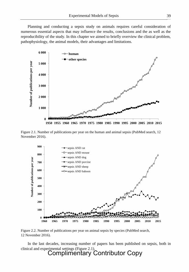
Experimental Models of Sepsis 39
Planning and conducting a sepsis study on animals requires careful consideration of
numerous essential aspects that may influence the results, conclusions and the as well as the
reproducibility of the study. In this chapter we aimed to briefly overview the clinical problem,
pathophysiology, the animal models, their advantages and limitations.
Figure 2.1. Number of publications per year on the human and animal sepsis (PubMed search, 12
November 2016).
Figure 2.2. Number of publications per year on animal sepsis by species (PubMed search,
12 November 2016).
In the last decades, increasing number of papers has been published on sepsis, both in
clinical and experimental settings (Figure 2.1).
0
1 000
2 000
3 000
4 000
5 000
6 000
1950 1955 1960 1965 1970 1975 1980 1985 1990 1995 2000 2005 2010 2015
Nu
mb
er o
f p
ub
lica
tio
ns
per
yea
r
human
other species
0
100
200
300
400
500
600
700
800
900
1960 1965 1970 1975 1980 1985 1990 1995 2000 2005 2010 2015
Nu
mb
er o
f p
ub
lica
tion
s p
er y
ear
sepsis AND rat
sepsis AND mouse
sepsis AND dog
sepsis AND porcine
sepsis AND sheep
sepsis AND baboon
Complimentary Contributor Copy
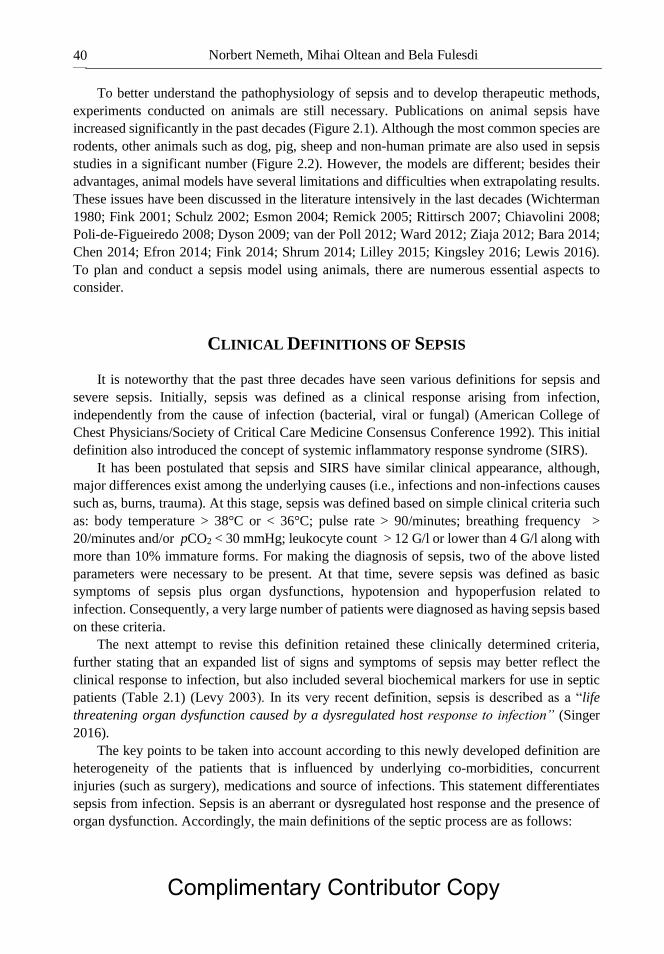
Norbert Nemeth, Mihai Oltean and Bela Fulesdi 40
To better understand the pathophysiology of sepsis and to develop therapeutic methods,
experiments conducted on animals are still necessary. Publications on animal sepsis have
increased significantly in the past decades (Figure 2.1). Although the most common species are
rodents, other animals such as dog, pig, sheep and non-human primate are also used in sepsis
studies in a significant number (Figure 2.2). However, the models are different; besides their
advantages, animal models have several limitations and difficulties when extrapolating results.
These issues have been discussed in the literature intensively in the last decades (Wichterman
1980; Fink 2001; Schulz 2002; Esmon 2004; Remick 2005; Rittirsch 2007; Chiavolini 2008;
Poli-de-Figueiredo 2008; Dyson 2009; van der Poll 2012; Ward 2012; Ziaja 2012; Bara 2014;
Chen 2014; Efron 2014; Fink 2014; Shrum 2014; Lilley 2015; Kingsley 2016; Lewis 2016).
To plan and conduct a sepsis model using animals, there are numerous essential aspects to
consider.
CLINICAL DEFINITIONS OF SEPSIS
It is noteworthy that the past three decades have seen various definitions for sepsis and
severe sepsis. Initially, sepsis was defined as a clinical response arising from infection,
independently from the cause of infection (bacterial, viral or fungal) (American College of
Chest Physicians/Society of Critical Care Medicine Consensus Conference 1992). This initial
definition also introduced the concept of systemic inflammatory response syndrome (SIRS).
It has been postulated that sepsis and SIRS have similar clinical appearance, although,
major differences exist among the underlying causes (i.e., infections and non-infections causes
such as, burns, trauma). At this stage, sepsis was defined based on simple clinical criteria such
as: body temperature > 38°C or < 36°C; pulse rate > 90/minutes; breathing frequency >
20/minutes and/or pCO2 < 30 mmHg; leukocyte count > 12 G/l or lower than 4 G/l along with
more than 10% immature forms. For making the diagnosis of sepsis, two of the above listed
parameters were necessary to be present. At that time, severe sepsis was defined as basic
symptoms of sepsis plus organ dysfunctions, hypotension and hypoperfusion related to
infection. Consequently, a very large number of patients were diagnosed as having sepsis based
on these criteria.
The next attempt to revise this definition retained these clinically determined criteria,
further stating that an expanded list of signs and symptoms of sepsis may better reflect the
clinical response to infection, but also included several biochemical markers for use in septic
patients (Table 2.1) (Levy 2003). In its very recent definition, sepsis is described as a “life
threatening organ dysfunction caused by a dysregulated host response to infection” (Singer
2016).
The key points to be taken into account according to this newly developed definition are
heterogeneity of the patients that is influenced by underlying co-morbidities, concurrent
injuries (such as surgery), medications and source of infections. This statement differentiates
sepsis from infection. Sepsis is an aberrant or dysregulated host response and the presence of
organ dysfunction. Accordingly, the main definitions of the septic process are as follows:
Complimentary Contributor Copy
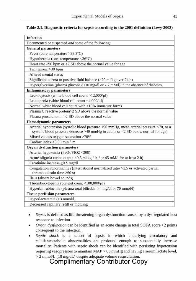
Experimental Models of Sepsis 41
Table 2.1. Diagnostic criteria for sepsis according to the 2001 definition (Levy 2003)
Infection
Documented or suspected and some of the following:
General parameters
Fever (core temperature >38.3°C)
Hypothermia (core temperature <36°C)
Heart rate >90 bpm or >2 SD above the normal value for age
Tachypnea: >30 bpm
Altered mental status
Significant edema or positive fluid balance (>20 ml/kg over 24 h)
Hyperglycemia (plasma glucose >110 mg/dl or 7.7 mM/l) in the absence of diabetes
Inflammatory parameters
Leukocytosis (white blood cell count >12,000/µl)
Leukopenia (white blood cell count <4,000/µl)
Normal white blood cell count with >10% immature forms
Plasma C reactive protein>2 SD above the normal value
Plasma procalcitonin >2 SD above the normal value
Hemodynamic parameters
Arterial hypotension (systolic blood pressure <90 mmHg, mean arterial pressure <70, or a
systolic blood pressure decrease >40 mmHg in adults or <2 SD below normal for age)
Mixed venous oxygen saturation >70%
Cardiac index >3.5 l min−1 m
Organ dysfunction parameters
Arterial hypoxemia (PaO2/FIO2 <300)
Acute oliguria (urine output <0.5 ml kg−1 h−1 or 45 mM/l for at least 2 h)
Creatinine increase ≥0.5 mg/dl
Coagulation abnormalities (international normalized ratio >1.5 or activated partial
thromboplastin time >60 s)
Ileus (absent bowel sounds)
Thrombocytopenia (platelet count <100,000/µl)
Hyperbilirubinemia (plasma total bilirubin >4 mg/dl or 70 mmol/l)
Tissue perfusion parameters
Hyperlactatemia (>3 mmol/l)
Decreased capillary refill or mottling
Sepsis is defined as life-threatening organ dysfunction caused by a dys-regulated host
response to infection.
Organ dysfunction can be identified as an acute change in total SOFA score >2 points
consequent to the infection.
Septic shock is a subset of sepsis in which underlying circulatory and
cellular/metabolic abnormalities are profound enough to substantially increase
mortality. Patients with septic shock can be identified with persisting hypotension
requiring vasopressors to maintain MAP > 65 mmHg and having a serum lactate level,
> 2 mmol/L (18 mg/dL) despite adequate volume resuscitation.
Complimentary Contributor Copy
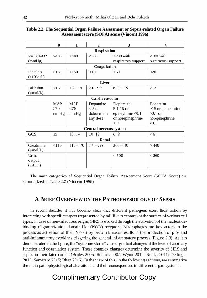
Norbert Nemeth, Mihai Oltean and Bela Fulesdi 42
Table 2.2. The Sequential Organ Failure Assessment or Sepsis-related Organ Failure
Assessment score (SOFA) score (Vincent 1996)
0 1 2 3 4
Respiration
PaO2/FiO2
(mmHg)
>400 <400 <300 <200 with
respiratory support
<100 with
respiratory support
Coagulation
Platelets
(x103/µL)
>150 <150 <100 <50 <20
Liver
Bilirubin
(µmol/L)
<1.2 1.2−1.9 2.0−5.9 6.0−11.9 >12
Cardiovascular
MAP
>70
mmHg
MAP
<70
mmHg
Dopamine
< 5 or
dobutamine
any dose
Dopamine
5.1-15 or
epinephrine <0.1
or norepinephrine
< 0.1
Dopamine
>15 or epinephrine
>0.1 or
norepinephrine
>0.1
Central nervous system
GCS 15 13−14 10−12 6−9 < 6
Renal
Creatinine
(µmol/L)
<110 110−170 171−299 300−440 > 440
Urine
output
(mL/D)
< 500 < 200
The main categories of Sequential Organ Failure Assessment Score (SOFA Score) are
summarized in Table 2.2 (Vincent 1996).
A BRIEF OVERVIEW ON THE PATHOPHYSIOLOGY OF SEPSIS
In recent decades it has become clear that different pathogens exert their action by
interacting with specific targets (represented by toll-like receptors) at the surface of various cell
types. In case of non-infectious origin, SIRS is evoked through the activation of the nucleotide-
binding oligomerization domain-like (NOD) receptors. Macrophages are key actors in the
process as activation of their NF-κB by protein kinases results in the production of pro- and
anti-inflammatory cytokines triggering the general inflammatory process (Figure 2.3). As it is
demonstrated in the figure, the “cytokine storm” causes gradual changes at the level of capillary
function and coagulation system. These complex changes determine the severity of SIRS and
sepsis in their later course (Brides 2005; Remick 2007; Wynn 2010; Nduka 2011; Dellinger
2013; Semeraro 2015; Bhan 2016). In the view of this, in the following sections, we summarize
the main pathophysiological alterations and their consequences in different organ systems.
Complimentary Contributor Copy

Experimental Models of Sepsis 43
Figure 2.3. The involvement of macrophages in the septic process.
Causes of Capillary Dysfunction in Sepsis
The capillary network is responsible for the transport of oxygen and the main nutritional
products to the tissues. Its function is influenced by the distribution of the capillary network in
certain tissue, by the actual condition of the capillaries (dilated or constricted), by the inter-
capillary distance and by hemorheological factors (Further details in Chapter 13).
Sepsis is currently considered as a microcirculatory disorder (Astiz 1995; Ince 2005, 2016;
Trzeciak 2007, 2008; Tyagi 2009; Ostergaard 2015). The microcirculatory failure usually is
generalized; however, it is often tested on the sublingual surface and/or on skin (Young 1995;
Trzeciak 2007, 2008; Wester 2011; Kiss 2015). Factors leading to microcirculatory problems
in sepsis are complex. Several mechanisms act on the pre-capillary, capillary and post-capillary
regions of the microcirculation, as summarized in Table 2.3.
The mechanisms leading to deterioration of micro-rheological factors are dominantly non-
specific. Red blood cell deformability can be impaired by (1) local metabolic and acid-base
changes (pH, lactate concentration, oxygenation level, micro-environmental osmolarity); (2)
decrease in ATP and consequent failure of active pump mechanisms, a condition that leads to
alteration in surface to volume ratio; as well as by (3) reactive oxygen species (Baskurt 2007;
Piagnerelli 2003; Nemeth 2012). Red blood cells can be damaged mechanically as well due to
the presence of thrombi and micro-thrombi (DIC), or even by the associated mechanical trauma
to blood during therapeutic efforts such as the direct removal of toxins from the bloodstream
by high-volume hemofiltration (Kameneva 2007; Borthwick 2013).
TLR2 TLR4 TLR5 TLR3,7,8,9
Protein kinase
Cytokine production
Tissue factorNOD
1/2
Peptidoglycan LPS Flagellin Nucleic acid
MACROPHAGE
NO PMN Endothel Complement Hypothalamus
Lipid peroxidation
Local inflammation
Capillary leak
Lysosomal enzymes
Capillary leak Fever
Coagulation system
MicrothrombiVasodilation
Complimentary Contributor Copy
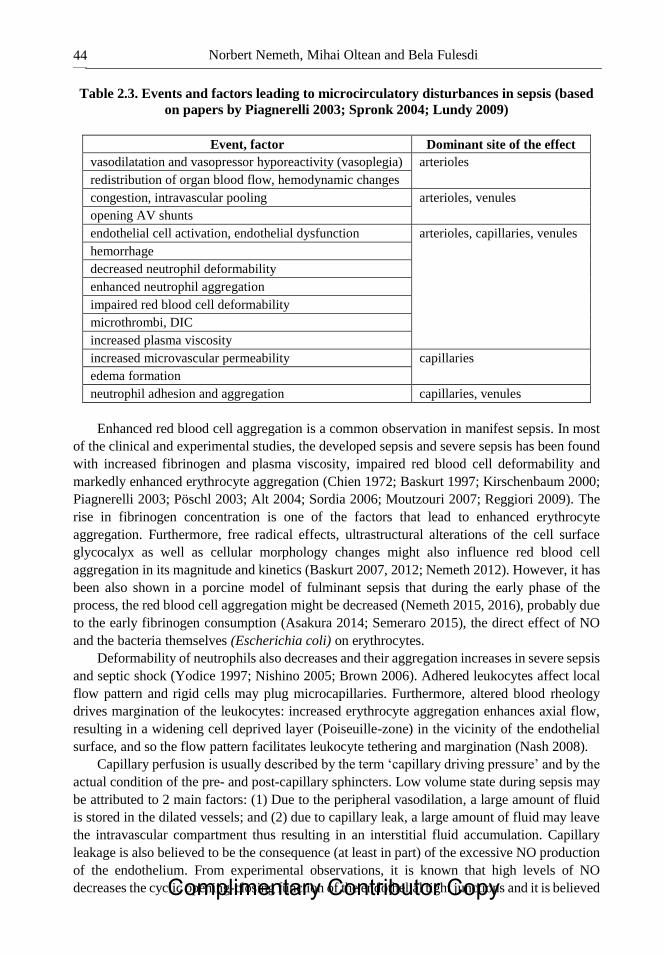
Norbert Nemeth, Mihai Oltean and Bela Fulesdi 44
Table 2.3. Events and factors leading to microcirculatory disturbances in sepsis (based
on papers by Piagnerelli 2003; Spronk 2004; Lundy 2009)
Event, factor Dominant site of the effect
vasodilatation and vasopressor hyporeactivity (vasoplegia) arterioles
redistribution of organ blood flow, hemodynamic changes
congestion, intravascular pooling arterioles, venules
opening AV shunts
endothelial cell activation, endothelial dysfunction arterioles, capillaries, venules
hemorrhage
decreased neutrophil deformability
enhanced neutrophil aggregation
impaired red blood cell deformability
microthrombi, DIC
increased plasma viscosity
increased microvascular permeability capillaries
edema formation
neutrophil adhesion and aggregation capillaries, venules
Enhanced red blood cell aggregation is a common observation in manifest sepsis. In most
of the clinical and experimental studies, the developed sepsis and severe sepsis has been found
with increased fibrinogen and plasma viscosity, impaired red blood cell deformability and
markedly enhanced erythrocyte aggregation (Chien 1972; Baskurt 1997; Kirschenbaum 2000;
Piagnerelli 2003; Pöschl 2003; Alt 2004; Sordia 2006; Moutzouri 2007; Reggiori 2009). The
rise in fibrinogen concentration is one of the factors that lead to enhanced erythrocyte
aggregation. Furthermore, free radical effects, ultrastructural alterations of the cell surface
glycocalyx as well as cellular morphology changes might also influence red blood cell
aggregation in its magnitude and kinetics (Baskurt 2007, 2012; Nemeth 2012). However, it has
been also shown in a porcine model of fulminant sepsis that during the early phase of the
process, the red blood cell aggregation might be decreased (Nemeth 2015, 2016), probably due
to the early fibrinogen consumption (Asakura 2014; Semeraro 2015), the direct effect of NO
and the bacteria themselves (Escherichia coli) on erythrocytes.
Deformability of neutrophils also decreases and their aggregation increases in severe sepsis
and septic shock (Yodice 1997; Nishino 2005; Brown 2006). Adhered leukocytes affect local
flow pattern and rigid cells may plug microcapillaries. Furthermore, altered blood rheology
drives margination of the leukocytes: increased erythrocyte aggregation enhances axial flow,
resulting in a widening cell deprived layer (Poiseuille-zone) in the vicinity of the endothelial
surface, and so the flow pattern facilitates leukocyte tethering and margination (Nash 2008).
Capillary perfusion is usually described by the term ‘capillary driving pressure’ and by the
actual condition of the pre- and post-capillary sphincters. Low volume state during sepsis may
be attributed to 2 main factors: (1) Due to the peripheral vasodilation, a large amount of fluid
is stored in the dilated vessels; and (2) due to capillary leak, a large amount of fluid may leave
the intravascular compartment thus resulting in an interstitial fluid accumulation. Capillary
leakage is also believed to be the consequence (at least in part) of the excessive NO production
of the endothelium. From experimental observations, it is known that high levels of NO
decreases the cyclic opening-closing function of the endothelial tight junctions and it is believed Complimentary Contributor Copy

Experimental Models of Sepsis 45
that this is a key factor in capillary leakage (Gomez-Jimenez 1995; Morel 2013). Additionally,
activated leucocytes and complement enzymes may also damage capillary wall integrity.
Based on experimental observations, dominantly precapillary sphincters in gram-positive
sepsis are dilated, while in case of gram-negative pathogens, this phenomenon occurs at the
level of postcapillary sphincters. Thus, capillary perfusion itself is also influenced by the actual
pathogen causing the septic process. Taking into account that capillary driving pressure is also
influenced by systemic blood pressure, low systemic blood pressure that develops in severe
sepsis, may result in a decreased capillary driving pressure. Due to local regulatory
mechanisms, when driving pressure decreases, capillary sphincters get further dilated in order
to enhance local blood flow. Unfortunately, as shown in Figure 2.3, coagulation system is also
activated during the septic process leading to micro-thrombi formation and thus resulting in
worsening of the local oxygen and nutritional transport.
Alterations in lymphatic flow also show a blend picture in sepsis. Endotoxaemia can be
associated with an increase in thoracic duct lymph flow (Kutner 1967; Williemas 1973; Smith
1988). When the thoracic duct was ligated in a canine model of peritonitis sepsis, it had a
negative effect on liver function and survival; bacterial count in peritoneal fluid, endotoxin
level in portal vein, nitrite and AST level were significantly elevated (Guler 1999). In a porcine
model, Lattuada and Hedenstierna (Lattuada 2006) found that during endotoxin sepsis (LPS
infusion), the lymph flow from the abdomen increased. The flow was measured by ultrasonic
probe placed around the thoracic duct. They have also observed that mechanical ventilation
with high PEEP impedes lymphatic drainage and also cold increases the lymph production,
while flow increased and lymphatic drainage of abdominal edema improved by spontaneous
breathing (Lattuada 2006). Elias and co-workers in their ovine model found that endotoxin (i.v.,
3.3 – 33 µg/kg) reduces lymphatic pump activity, contributing to the edema formation in sepsis
(Elias 1987). Intrahepatic lymph stasis in endotoxaemia in rats can be caused by decreased
pumping activity of extra- and intrahepatic large lymph vessels (Shibayama 1992). It has been
showed in LPS-induced sepsis model in guinea pigs that there is an early increase in mesenteric
lymph flow rate being mediated by vascular hyperpermeability and plasma albumin leakage
(Nemoto 2011).
Microcirculatory and Mitochondrial Distress Syndrome
As described above, the excessive systemic reaction to the inflammatory insult finally leads
to a critical imbalance of oxygen and nutritional products supply and demand. In the recent
decade, it has been speculated that compensatory mechanisms in sepsis may be described as a
2-phase process. In the beginning, microvascular compensation dominates. During this phase,
the decreased tissue perfusion and tissue hypoxia result in enhanced production of potent
vasodilatory substances (mainly NO) (Gomez-Jimenez 1995). This reaction aims to increase
the tissue perfusion. Whenever this compensation fails, in the second mitochondrial phase, cells
are trying to turn into an “energy-saving mode” by decreasing their energy needs. The main
actor in this process is again the overproduced NO. It passes easily through the cellular and
mitochondrial membranes as oxygen and blocks cytochrome oxidase at complex IV. This
compensatory mechanism however is a double-edged sword; although it decreases cellular
energy need (and thus contributes to survival) but only until a certain limit: as a secondary
consequence of complex IV block, reactive nitrogen species (most importantly peroxinitrites) Complimentary Contributor Copy

Norbert Nemeth, Mihai Oltean and Bela Fulesdi 46
are produced. Large amounts of reactive nitrogen species contribute to cell necrosis in severe,
end-stage states of sepsis.
Multiple Organ Failure (MOF) in Sepsis
The continuum of sepsis is depicted in Figure 2.4. Based on the underlying
pathophysiological mechanisms, different organs may be affected during the septic process
(Albert 2004; Langenberg 2005; Remick 2007; Wynn 2010; Nduka 2011; Bhan 2016; Doi
2016).
In the cardiovascular system, newly developing atrial fibrillation can be considered as
early warning sign of developing sepsis. Beside this, decreased central venous and
mean arterial blood pressures as well as a decreased peripheral vascular resistance are
typical signs of septic cardiovascular affection. Cardiac contractility may eventually
be higher in the early phase of sepsis, however, in the late phase of septic shock,
cardiac contractility also decreases due to inappropriate oxygenation of the
myocardium. Recent clinical observations suggest that PiCCO monitoring enables
checking the cardiovascular consequences.
Pulmonary consequences: Based on experimental and pathological observations, there
is an inflammatory exudate accumulation within the alveoli. This exudate worsens the
effectiveness of the alveolo-capillary gas exchange. It contributes to the washout of
the surfactant, thus leading to alveolar collapse and atelectasis formation. Additionally
capillary leak also affects the pulmonary capillaries and contributes to extravascular
fluid accumulation within the lung. This extravascular lung water may lead to further
compression of the alveoli (due to extraalveolar forces) and also leads to an increase
of the alveolo-capillary distance that worsens gas exchange. The most sensitive
parameter that describes the function of the alveolo-capillary gas exchanges is
PaO2/FiO2 ratio. Extravascular lung water can be measured by PiCCO monitoring
(EVLW).
Consciousness disturbances of different severity are early signs of sepsis development.
At early stages, sepsis causes confusions or delirious states. The correct terminology
for the cerebral affection in sepsis is sepsis-associated encephalopathy. This is a
multifactorially-determined functional disturbance that develops along with the
progression of the septic process, leading to somnolence, or coma in late phases.
Cerebral functions can be monitored by the GCS score and EEG.
In early stages of systemic hypoperfusion, the urine output decreases and septic
patients are prone to development of acute kidney injury. Along with the advancement
of severe sepsis, oliguria may turn to anuria and serum creatinine progressively
increases.
Complimentary Contributor Copy

Experimental Models of Sepsis 47
Figure 2.4. The continuum of sepsis.
Hypoperfusion of the liver has different consequences: albumin and vitamin K-
dependent coagulation factor production decreases. In severe cases where
parenchymal damage is advanced, bilirubin increases (by canalicular damage and
hepatocytolysis) (> 70 mmol/l) and icterus may develop.
Severe hypoperfusion also affects gastrointestinal organs. As a consequence, bowel
ischemia, ileus, pancreas apoplexy and acalculous cholecystitis may develop.
EXPERIMENTAL MODELS FOR STUDYING SEPSIS
According to Fink (2001), the ‘ideal’ animal sepsis model should be (1) capable of
predicting both positive and negative results of a trial; (2) inexpensive; (3) reproducible (within
and among laboratories); (4) humane and fully respecting the 3Rs; (5) able to provide a
mortality endpoint; (6) characterized by hemodynamic, hematologic, and biochemical features
similar to those observed in human sepsis (Fink 2001). Unfortunately there is no ‘ideal’ model
known to date.
Experimental sepsis can be induced via generating toxemia by intravenous or
intraperitoneal administration of live bacteria (pure or mixed cultures), inhalation,
intrabronchial instillation, implantation or seeding into tissues or by disruption of the host-
barriers (mechanically or by ischemia), or in combination. Behind these major approaches to
develop sepsis models, there are numerous variations and sub-models that can be classified in
the following main categories:
endotoxin models
bacteremia models
Production of
anti-inflammatory cytokines
Tissue damage
Depression of the immune system
Susceptibility to infections
Organ failure
Release of pro-inflammatory
cytokines, chemokines,
complement factors,
ROS and NO
Hyperactive response
Clinical signs of sepsis Severe sepsis/septic shock
Hypoactive response
Activation of:
• Macrophages
• PMN
• Lymphocytes
• Endothelium
• Complement system
• Coagulation system
Insult (infection, non-infectious)
Complimentary Contributor Copy

Norbert Nemeth, Mihai Oltean and Bela Fulesdi 48
peritonitis models
pneumonia models
meningitis models
soft tissue infection models (abscess models)
Table 2.4 summarizes some major characteristics of these models. Each of these strategies
reproduces one or several pathophysiological mechanisms activated during sepsis, yet the
multitude of existing approaches indicate limitations of each model to mimic the clinical
scenario. Many of the limitations depend on the species of animals or bacteria used while others
depend on technical and conceptual details behind each model.
Not only the way of sepsis induction, but the choice of the animal species itself is also
crucial. Table 2.5 gives a summary of various species in consideration of sepsis models (based
on Fink 2001).
Following sections provide the technical description of the methods for inducing
experimental sepsis as well as brief considerations on their advantages and limitations.
Endotoxin Models
Endotoxin is a lipoprotein-carbohydrate complex that can be found in the cell wall of all
gram-negative bacteria. Endotoxins were originally isolated by André Boivin (Bactéries et
Virus, Paris, Presses Universitaires de France, 1941).
The simplest way is to inject single bolus of LPS intravenously (as the most common way)
or intraperitoneally. The pro-inflammatory processes occur suddenly and last briefly when
endotoxicosis is induced by LPS injection (single bolus). Endotoxin can be administered via
intraperitoneal injection as well (Laschke 2007). In case of infection, the inflammatory stimulus
is much more obvious with gradual development and persisting longer. It has been shown that
TNF-ɑ concentration is much higher and showing a peak around 60−90 minutes after LPS
injection (i.v. or i.p.) compared to a focal infection model (e.g., fecal peritonitis) (Eskandari
1992; Fink 2001). After endotoxin administration, plasma concentration of host response
mediators such as TNF-α, IL-1, IL-6 and IL-10 rapidly increase as an acute and transient
elevation. LPS binds to TLR-4 and so activates inflammatory pathways, including NF-κβ.
The sensitivity of various animal species to LPS and the physiologic and immunologic
response to this stimulus is different. Humans are very sensitive to LPS; in volunteers 2−4
ng/kg LPS may produce obvious symptoms. In mice 1−25 ng/kg LPS is needed to generate
septic symptoms. A very interesting study aimed at reaching a similar IL-6 response in mice
and humans revealed that 2 ng of endotoxin administered intravenously in humans were enough
to elicit the same IL-6 plasma levels as 500 ng given intraperitoneally to mice (Copeland 2005).
Other species such as rabbit, pig and sheep are also sensitive to endotoxin, similarly to human.
Interestingly, the rodents or even monkeys such as baboons are resistant to endotoxin. About 5
µg/kg LPS is lethal in sheep, 100 µg/kg is the LD80 in rabbits, while mice and baboons needs
incomparably higher dose for lethality (LD70 dose is about 6 mg/kg in baboon; LD100 dose is
about 20−80 mg/kg in the mouse) (Fink 2001). Overall, endotoxemia models are relatively far
from the clinical sepsis scenario. Furthermore, the resistance of rodents to LPS also puts a
serious limitation of extrapolating the results to the human (Kingsley 2016).
Complimentary Contributor Copy
Experimental Models of Sepsis 49
There are several techniques and materials for increasing the sensitivity of rodents to LPS
such as D-galactosamine, a non-ionic polyoxyethylene-polypropylene copolymer (Pluronic
F127), carageenan, berrylium phosphate, lead acetate or carbon tetrachloride. Administration
of platelet-activating factor or heat-killed bacteria a week prior to LPS injection can also cause
enhanced sensitivity (Katschinski 1992; Rabinovici 1993; Fink 2001; Gumenscheimer 2002).
Rittirsch and co-workers reported in their paper that the LPS endotoxemia differs from
cecal ligation and puncture (CLP) model in rodents. After LPS infusion, the symptoms such as
rapid hypothermia developed within a few hours, together with leukopenia followed by
leukocytosis. The scenario is also characterized by an early increase and high transient levels
of pro-inflammatory cytokines (IL-1β, TNF-α, IL-6) and the protective effect of anti-TNF-α is
shown. In CLP model, the symptoms developed within 12−24 hours, leukocytosis or
leukopenia was also present, whereas, the pro-inflammatory cytokines (IL-1β, TNF-α, IL-6,
C5a) were expressed later at moderately high levels. Anti-LPS or anti-TNF-α were not
protective (Rittirsch 2007).
Intravenous, intraperionoteal or intraarticular administration of zymosan (a polysaccharide
cell wall component derived from Saccharomyces cerevisiae), producing a prolonged
activation of the alternative complement pathway and generalized inflammation, is another
example for endotoxaemia models, mostly in rodents (mouse, rat) and rabbit (Sharkey 1984;
Volman 2005; Cash 2009; Guerra 2016; Hong 2016;).
Bacteremia Models
Intravenous administration of live Escherichia coli is used for studying sepsis since
decades. The early studies used typically large animals (dogs, pigs, monkeys) (Guenter 1969;
Cryer 1971; Hinshaw 1976; Wichterman 1980). Later, rodent usage has been also spread out
widely. Contrary to the rapid bolus infusion, which results in rapid hypoglycemia and death,
further models have been established to provide more time for the response reactions
approaching closer to the clinical scenarios. Postel et al. developed a canine model in which
the animals were continuously infused with Pseudomonas aeruginosa for 5 hours (106/ml/min
for 2−5 hours or 4×10⁷/ml/min for 5 hours), and observed the animals for 24 hours. This model
provided more time for developing wider response reactions and for observation of kinetics
(Postel 1975).
In sepsis models with bacterial infusion, the exact number of bacteria can be estimated only
that might influence the reproducibility compared to the better standardized endotoxin infusion
models (Fink 2001). The most commonly used bacteria are the gram-negative Escherichia coli.
Other gram-positive bacteria, such as Staphylococcus aureus, Staphylococcus epidermidis,
Streptococcus pyogenes or Pseudomonas aeruginosa, or fungi such as Candida albicans can
be also used in various animal species (mouse, rat, rabbit, dog, pig, sheep, baboon).
PERITONITIS MODELS
The surgical models are particularly relevant models of sepsis since a significant number
of sepsis cases are the result of intraabdominal leakage of intestinal content after abdominal Complimentary Contributor Copy

Norbert Nemeth, Mihai Oltean and Bela Fulesdi 50
surgery or perforated diverticulitis or appendicitis (Browne 1976; Wichterman 1980; Krisher
2001; Wryzykowski 2005; Catry 2016). These models generate a polymicrobial sepsis which
closely resembles the clinical situation in terms of mechanism and clinical course. The
peritonitis may be induced by perforating the bowel or by instillation of bacteria or fecal
material in the peritoneal cavity.
Cecal Ligation and Puncture (CLP)
This simple and reproducible model has become a gold standard in sepsis research because
of its relative technical simplicity and versatility (Wichterman 1980; Hubbard 2005; Dejager
2011; Toscano 2011; Schabbauer 2012; Siempos 2014). Nonetheless, ensuring reproducibility
requires that a few important details should be carefully considered throughout the procedure
including preoperative fasting of the animals to generate similar intestinal contents as well as
consistency in several other technical details. In addition, short-acting methods of anesthesia
(i.e., isoflurane inhalation) should be chosen in order to minimize prolonged anesthesia-induced
cardiovascular alterations postoperatively (Hubbard 2005). Following midline incision, the
cecum is identified and carefully exteriorized. Then, the cecum is ligated distal to the ileocecal
valve and perforated using one or two needle punctures (Figure 2.5). Both the length of the
ligated cecum (Singleton 2003) and the needle size (Ebong 1999; Otero-Anton 2001) can be
used to influence the CLP outcome and give a lethal or nonlethal sepsis. For instance, two
punctures created with an 18G needle result in 100% mortality in BALB/c mice, while the same
procedure yields mortality rates of 50% or 0% when performed with 21G or 25G needles,
respectively (Ebong 1999).
The cecum is gently compressed to expel a small amount of intestinal content through the
perforation, thereafter is returned to the abdomen and the wound is closed in two layers. The
cecum will become necrotic within 24 hours but it may ultimately be resected to remove the
source of infection as part of the treatment strategy.
Figure 2.5. Cecal ligation and puncture (CLP) in anesthetized rat. White arrow shows the site of the
ligature. The excluded cecum punctured through by a 19 G needle.
The postoperative care is a significant element of this particular experimental model as
fluid resuscitation and antibiotic treatment may influence significantly both the
pathophysiology and animal survival (Zanotti-Cavazzoni 2009). In the rat, this model induces Complimentary Contributor Copy
Experimental Models of Sepsis 51
a complex cardiovascular response with an early, hyperdynamic phase characterized by
increased cardiac output, increased tissue perfusion and decreased peripheral resistance. This
is followed by a late, hypodynamic phase characterized by reduced microvascular blood flow
in various tissues and increased peripheral resistance (Yang 2002; Yang 1999). Results in mice
and rats indicate that fluid resuscitation (typically isotonic saline as subcutaneous injection)
and antibiotics may increase the 48 hour survival rate from 0 to around 20−40% (Hollenberg
2001) depending on type of fluid, volume and concentration (Zanotti-Cavazzoni 2009).
Colon Ascendens Stent Peritonitis (CASP)
The model of colon ascendens stent peritonitis (Zantl 1998) is more recent than the
previously described CLP and has been introduced following numerous observations that
animals undergoing cecal ligation and puncture often develop only localized peritonitis
(Schabbauer 2012). The cecal perforations are spontaneously covered by the surrounding
intestinal loops leading to abscess formation (Maier 2004). This in its turn may result in two
different pathologic conditions not entirely comparable (diffuse peritonitis vs. large abscess)
and with different response to various treatment alternatives.
The surgery is preceded by the preparation of plastic venous catheters of variable sizes. In
the rat model, the survival rate is strictly dependent on the diameter of the implanted stent and
a stent diameter of 14G leads to 100% mortality whereas smaller stent diameters generates
sublethal models (16G stent leads to a 71% mortality whereas mortality is 53% when a 18G
stent is used) (Lustig 2007). Similarly, approximately 70% of the mice expired within 48 hours
when a 16G stent was used whereas the death rate dropped to 50% with an 18G stent (Maier
2004). After creating a marking at a distance of 3 mm from the tip, the catheter is
circumferentially incised with a 1 mm scalpel beyond the marking sparing only a narrow bridge
of plastic.
Inhalational anesthesia is advocated by the majority of the authors (ether, isoflurane,
sevoflurane) (Lohner 2013; Islam 2016; Schöneborn 2016) due to the lower impact on the
hemodynamics as well as the short postoperative recovery. The abdominal wall is opened
through midline incision and the ascending colon is exposed using soft cotton earbuds. After
adequate exposure of the ascending colon, the prepared catheter is inserted into the lumen of
the ascending colon through the antimesenteric wall approximately 10 mm from the ileocecal
valve and fixed with two stitches of non-absorbable suture (7/0) (Figure 2.6). Thereafter, the
inner needle of the stent is removed and the stent is cut at the marked site. To verify the adequate
intraluminal placement of the stent and the immediate initiation of the fecal leakage, stool is
milked from the cecum into the ascending colon and the stent is filled until a small drop of stool
appears at its end. The procedure is completed through abdominal wall closure in two layers
and fluid resuscitation (Zantl 1998).
Bacterial Suspension Inoculation (Intraabdominal Abscess Models)
Classic models include the inoculation of defined bacterial isolates (e.g., E. coli, B. fragilis)
mixed with barium sulfate and autoclaved rat fecal material, suspended and loaded into gelatin
capsule and implanted intraperitoneally, or agar pellets containing bacteria, or suspension Complimentary Contributor Copy

Norbert Nemeth, Mihai Oltean and Bela Fulesdi 52
containing hemoglobin, mucin and bacteria (E. coli), or impregnated fibrin clot (Onderdonk
1976; Wichterman 1980; Fink 2001; Kingsley 2016). Inoculation of autologous feces and other
clinically-relevant model dominantly in pigs (for instance simulating the case of bowel
anastomosis insufficiency), in which a definitive amount (e.g., 0.5 g/kg) of autologous feces is
suspended in saline solution (e.g., 200 ml), incubated (e.g., for 6 hours at 38°C) and inoculated
into the peritoneal cavity through drains, or via incision and/or Veres needle (Chvojka 2008;
Zsikai 2012) (Table 2.6).
Interventions that Partly Destroy Barriers of Gastrointestinal Tract
Intestinal ischemia models. An example is the creation of an ischemic bowel loop results
in severe diffuse peritonitis (Wichterman 1980). Hau and Simmons (1977) and which reported
90% mortality rate within 10 days after making a loop of terminal ileum ischemia in dogs.
Simpler intestinal ischemia-reperfusion models with longer follow-up period, allowing time for
development of symptoms, are also capable for mimicking abdominal sepsis.
Cholecystitis models. In the canine study of Perbellini and co-workers, E. coli was injected
directly into the gall bladder after ligating the cystic artery. The ischemic and infected gall
bladder showed similarities to clinically seen cholecystitis (Perbellini 1978; Wichterman 1980).
Pneumonia Models
In this simple and clinically relevant model type, Streptocossus pneumoniae, Klebsiella
pneumoniae, Pseudomonas aeruginosa are the most common bacteria which are introduced via
a tracheal route (tube in rodents, or even with bronchoscope in large animals), or via nasal route
(Fink 2011; Kinglsey 2016).
Figure 2.6. Colon ascendens stent peritonitis (CASP) in anesthetized rat. White arrow shows the fixed
stent (size: 16 G).
Meningitis Models
Several murine models have been developed using intracerebral, intraperitoneal,
intravenous, intranasal or intracisternal inoculation methods (Chiavolini 2008). In the latter
category, a well-established model is inoculation by injecting 10 μL of bacterial suspension
into the cisterna magna using a 32 G needle (Mook-Kanamori 2012; 2015).
Complimentary Contributor Copy
Experimental Models of Sepsis 53
Meningitis in rats can be produced by intranasal inoculation of Haemophilus influenzae in
newborn rats, or in young adult animals by intraperitoneal inoculation (Smith 1984).
Streptococcus pneumoniae is also used for meningitis models. Intraperitoneal route provokes
hematogenous meningitis. The intranasal inoculation can act non-hematogenously as well
along the olfactory nerves (Chiavolini 2008).
Soft Tissue Infection Models (Abscess Models)
Intramuscular abscess models. Hermreck and Thal (1969) described a model in which 5-
cm wicks of umbilical tape were seeded with the dog’s own feces and immersed into porcine
mucin solution. These wicks then were embedded into the thigh (unilaterally) of the dog
subcutaneously and intramuscularly. The animals were followed-up for a week.
Dermal abscess models. In 1964, Walker et al. published a rat model mimicking dermal
abscess. Pseudomonas aeruginosa, isolated from a patient with septicemia after second and
third degree burn injury, was used to seed subcutaneous tissues. On the dorsum, a 5×7 cm area
was excised (~20% of the body surface), and the fascia in the base of the wound was seeded by
the bacterial culture. The area then was covered with split-thickness (0.012 inch) skin autograft.
In their study, a comparison was made with intraperitoneal and subcutaneous injection of the
bacterial culture as well (Walker 1964).
Intramuscular, subdermal bacterial injections, subcutaneous pockets filled with gauze and
injected bacteria, infected cotton suture are further examples for soft tissue infections models
(Fink 2001).
EXTRAPOLATION OF THE RESULTS:
EXPERIMENTAL VERSUS CLINICAL SEPSIS
The final goal of all medical research is and must be the benefit to the patient, by exploring
and understanding the nature of various disorders and its pathophysiology mechanisms as well
as developing preventive or therapeutic agents. In this process, animal studies are still
necessary, since in the translational process, there must be a stage of in vivo study before any
human application.
Unfortunately, numerous pharmacological agents which have been promising and effective
in animal models, failed in large well-designed human clinical trials (e.g., methylprednisolone
sodium succinate, IL1-RA, CDP571, BN 5021, BB-822, TCV-309, TAK-242, lenercept,
tifacogin) (Fink 2014; Lakshmikanth 2016).
Practically speaking, all the experimental sepsis models fail to reproduce entirely the
clinical scenario. This explains the relatively large number of models used to induce severe
systemic infections and continuous search for a better mirror to the clinical situation.
Accordingly, numerous authors have discussed the comparability of animal sepsis models and
human sepsis (Wichterman 1980; Fink 2001; Esmon 2004; Rittirsch 2007; Nemzek 2008; Poli-
de-Figueiredo 2008; Dyson 2009; Fink 2014; Lilley 2015).
Complimentary Contributor Copy
Norbert Nemeth, Mihai Oltean and Bela Fulesdi 54
In the clinical practice, the most frequent infection sites are the lung, the abdomen and
urinary tract. Gram-positive and gram-negative infections show almost the same frequency,
followed by fungi and parasites. The initial insult or infection activates the innate immune
system bursting the pro-inflammatory cascades and mediators with SIRS as result. Anti-
inflammatory factors appear as compensatory mechanism but any imbalance can cause further
deterioration: exaggerated pro-inflammatory effect results in uncontrolled, excessive
inflammatory response and consecutive effects; stronger anti-inflammatory steps may lead to
immunosuppression (Rittirsch 2007).
Virtually all the experimental sepsis models in rodents fail to reproduce entirely the clinical
scenario due to limitations related to design or the species used. Moreover, seemingly minor
technical details (Ebong 1999; Singleton 2003) or the choice of the animals used in terms of
age and gender impacts on the pathophysiology and the outcome (Turnbull 2003). Compared
with humans, rodents are quite resistant to endotoxin, have a distinct hemodynamic profile, and
limited blood volume (Copeland 2005). All these factors explain the relatively large number of
models used to induce severe systemic infections and the ongoing search for refined designs to
better mirror the clinical situation.
Microbiological Actors Causing Sepsis
In human sepsis, gram-positive bacteria and fungi act for causing sepsis in higher incidence
than gram-negative organisms, however, in animal studies mostly gram-negative bacteria have
been used (Poli de Figueiredo 2008).
Experimental Considerations Related to the Pathomechanism of Sepsis
In clinical scenario, usually there is a septic focus which intermittently or persistently
scatters bacteria. Different ways of sepsis induction act differently, as it has been discussed
previously and overviewed in Table 2.4.
The Time Factor (Follow-Up)
For development of sepsis, time is needed that differs according to the way of induction
(Table 2.4). Endotoxemia and infusion of bacteria provide prompt effects, while infection site
models obviously need more time to develop the response reactions and the septic symptoms.
The choice of the model depends on the primary aim of the study itself.
Complimentary Contributor Copy

Table 2.4. Summary of various septic models by its nature, origin, most commonly used species, advantages and disadvantages
(based on papers by Fink 2001; Esmon 2004; Volman 2005; Rittirsch 2007; Poli de Figueiredo 2008; Engel 2011; Mai 2012;
Fink 2014; Kingsley, 2016)
Sepsis
origin Model Species
Characteristic
hemodynamic
response
Persistent
inflammatory
response
Cytokine
response
characteristics
Advantages Disadvantages Special notes
Toxemia Endotoxin
(LPS) administration
Most
commonly used:
mouse, rat
More rarely
used:
rabbit, dog, pig,
sheep, non-
human primates
immediate early
hypodynamic phase
no Early rise of
TNF-α, rapid transient
increase
Easy model.
LPS dosage is well-controllable.
Innate immune
system activation can be well
studied.
Well-suited to test the mechanism of
inflammatory
cascade activation and the efficacy of
anti-inflammatory
compounds.
Far from clinical
scenario. Different sensitivity
amongst the species
(relatively endotoxin resistant species:
rodents, cat, dog;
sensitivity with enhanced response:
rabbit, sheep, non-
human primates). Dose dependency
(concentration and
way of administration)
Toxin can be given:
i.v., i.a., i.p. LPS effect can be
enhanced by
administration of D-galactosamine, pluronic
F 127, carageenan, lead
acetate, beryllium phosphate, carbon
tetrachloride, platelet-
activating factor, co-administration of heat-
killed bacteria (e.g., P.
acnes, C. parvum, M. bovis), mostly in
rodents.
TLR agonist
administration
(i.v.)
rodents Not specified. no Rapid immune
activation and
inflammation, elevation of
IL-6 and IFN-β
within 2 h
Technically easy
model.
Nonspecific
indicator of
inflammation, short half-time of agents,
immunological
effects often controversial
Mostly TLR-9, TLR-7,
TLR-3 agonists
Administration of zymosan
rodents (most frequently
rabbits), rarely
in sheep
early drop in cardiac index
and stroke
volume index
no Prolonged activation of
the alternative
complement pathway,
generalized
inflammation
Easy to perform. Can be used for
studying sterile
peritonitis, MODS and acute lung
injury as well.
Early mortality rate is high.
Toxin can be given: i.v., i.a., i.p.,
intraarticularly.
Acute inflammation peaking within few
hours. Marked
hypoxaemia, interstitial edema,
pulmonary neutrophil
sequestration.
Complimentary Contributor Copy

Table 2.4. (Continued)
Sepsis
origin Model Species
Characteristic
hemodynamic
response
Persistent
inflammatory
response
Cytokine
response
characteristics
Advantages Disadvantages Special notes
Bacteremia Intravascular
infusion of
live bacteria
rodents, dog,
pig, sheep,
non-human
primates
hypotensive
shock
no Rise of TNF-α
and IL-1β,
rapid transient
increase
DIC presents. Relatively far from
clinical scenario.
Most commonly used
bacteria:
E. coli
Others: S. aureus, P. aeruginosa, S.
pyogenes, S.
epidermidis, C. albicans
Variations: Bolus
(single or multiple), increasing
concentration via
infusion pump.
Peritonitis cecal ligation
and puncture
(CLP)
most commonly
rodents
hemodynamic
and metabolic
phases
yes moderate
increase
Clinically highly
relevant.
Not specified. -
colon
ascendens stent
peritonitis
(CASP)
prolonged
hemodynamic instability
yes steady,
continuous systemic
increase
Clinically highly
relevant. Lethality is
accompanied by
MOF.
Not specified. -
polymicrobial peritoneal
contamination
and infection (PCI)
early hyperdynamic
response
yes Clinically highly relevant.
Not specified. e.g., intraperitoneal injection of a human
feces suspension
implantation
model (implanting
fibrin clot
impregnated with live
bacteria)
rat dog, pig yes Similar to
human sepsis.
Clinically highly
relevant.
Not specified. Most commonly used
bacteria: E. coli
Others:
B. fragilis in combination with E.
coli
Complimentary Contributor Copy
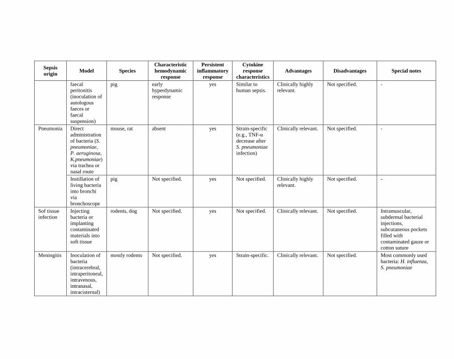
Sepsis
origin Model Species
Characteristic
hemodynamic
response
Persistent
inflammatory
response
Cytokine
response
characteristics
Advantages Disadvantages Special notes
faecal peritonitis
(inoculation of
autologous
faeces or
faecal
suspension)
pig early hyperdynamic
response
yes Similar to human sepsis.
Clinically highly relevant.
Not specified. -
Pneumonia Direct
administration
of bacteria (S. pneumoniae,
P. aeruginosa,
K. pneumoniae) via trachea or
nasal route
mouse, rat absent yes Strain-specific
(e.g., TNF-α
decrease after S. pneumoniae
infection)
Clinically relevant. Not specified. -
Instillation of
living bacteria
into bronchi via
bronchoscope
pig Not specified. yes Not specified. Clinically highly
relevant.
Not specified. -
Sof tissue infection
Injecting bacteria or
implanting contaminated
materials into
soft tissue
rodents, dog Not specified. yes Not specified. Clinically relevant. Not specified. Intramuscular, subdermal bacterial
injections, subcutaneous pockets
filled with
contaminated gauze or cotton suture
Meningitis Inoculation of
bacteria
(intracerebral,
intraperitoneal,
intravenous, intranasal,
intracisternal)
mostly rodents Not specified. yes Strain-specific. Clinically relevant. Not specified. Most commonly used
bacteria: H. influenza,
S. pneumoniae
Complimentary Contributor Copy
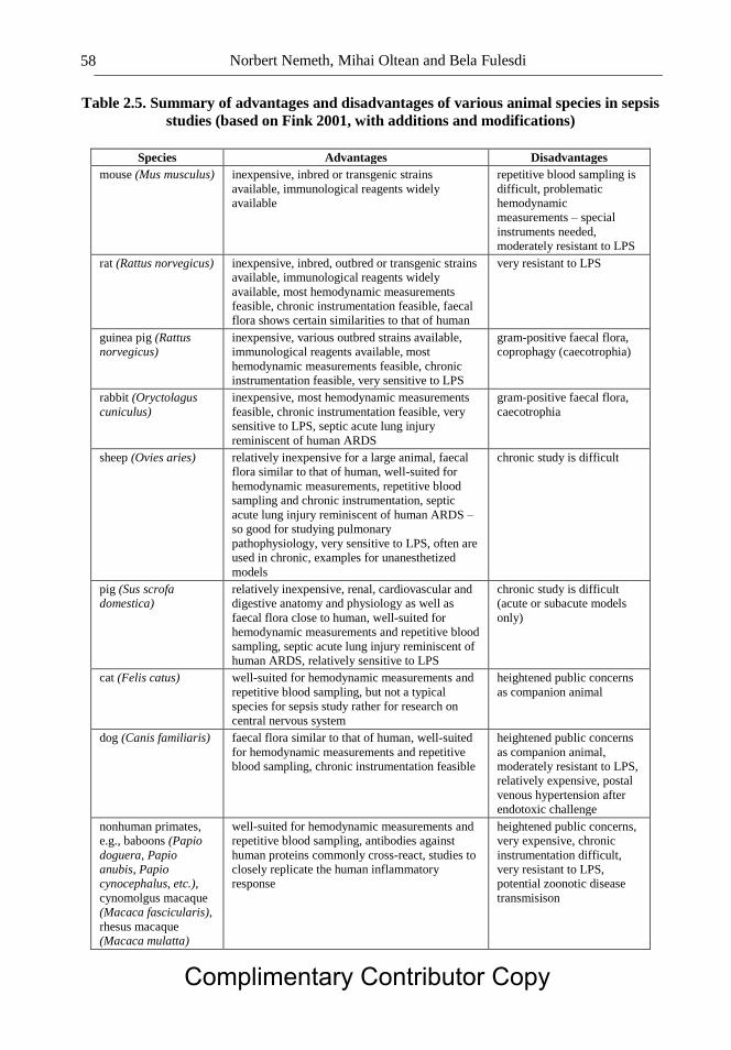
Norbert Nemeth, Mihai Oltean and Bela Fulesdi 58
Table 2.5. Summary of advantages and disadvantages of various animal species in sepsis
studies (based on Fink 2001, with additions and modifications)
Species Advantages Disadvantages
mouse (Mus musculus) inexpensive, inbred or transgenic strains
available, immunological reagents widely
available
repetitive blood sampling is
difficult, problematic
hemodynamic
measurements – special
instruments needed,
moderately resistant to LPS
rat (Rattus norvegicus)
inexpensive, inbred, outbred or transgenic strains
available, immunological reagents widely
available, most hemodynamic measurements
feasible, chronic instrumentation feasible, faecal
flora shows certain similarities to that of human
very resistant to LPS
guinea pig (Rattus
norvegicus)
inexpensive, various outbred strains available,
immunological reagents available, most
hemodynamic measurements feasible, chronic
instrumentation feasible, very sensitive to LPS
gram-positive faecal flora,
coprophagy (caecotrophia)
rabbit (Oryctolagus
cuniculus)
inexpensive, most hemodynamic measurements
feasible, chronic instrumentation feasible, very
sensitive to LPS, septic acute lung injury
reminiscent of human ARDS
gram-positive faecal flora,
caecotrophia
sheep (Ovies aries)
relatively inexpensive for a large animal, faecal
flora similar to that of human, well-suited for
hemodynamic measurements, repetitive blood
sampling and chronic instrumentation, septic
acute lung injury reminiscent of human ARDS –
so good for studying pulmonary
pathophysiology, very sensitive to LPS, often are
used in chronic, examples for unanesthetized
models
chronic study is difficult
pig (Sus scrofa
domestica)
relatively inexpensive, renal, cardiovascular and
digestive anatomy and physiology as well as
faecal flora close to human, well-suited for
hemodynamic measurements and repetitive blood
sampling, septic acute lung injury reminiscent of
human ARDS, relatively sensitive to LPS
chronic study is difficult
(acute or subacute models
only)
cat (Felis catus)
well-suited for hemodynamic measurements and
repetitive blood sampling, but not a typical
species for sepsis study rather for research on
central nervous system
heightened public concerns
as companion animal
dog (Canis familiaris)
faecal flora similar to that of human, well-suited
for hemodynamic measurements and repetitive
blood sampling, chronic instrumentation feasible
heightened public concerns
as companion animal,
moderately resistant to LPS,
relatively expensive, postal
venous hypertension after
endotoxic challenge
nonhuman primates,
e.g., baboons (Papio
doguera, Papio
anubis, Papio
cynocephalus, etc.),
cynomolgus macaque
(Macaca fascicularis),
rhesus macaque
(Macaca mulatta)
well-suited for hemodynamic measurements and
repetitive blood sampling, antibodies against
human proteins commonly cross-react, studies to
closely replicate the human inflammatory
response
heightened public concerns,
very expensive, chronic
instrumentation difficult,
very resistant to LPS,
potential zoonotic disease
transmisison
Complimentary Contributor Copy
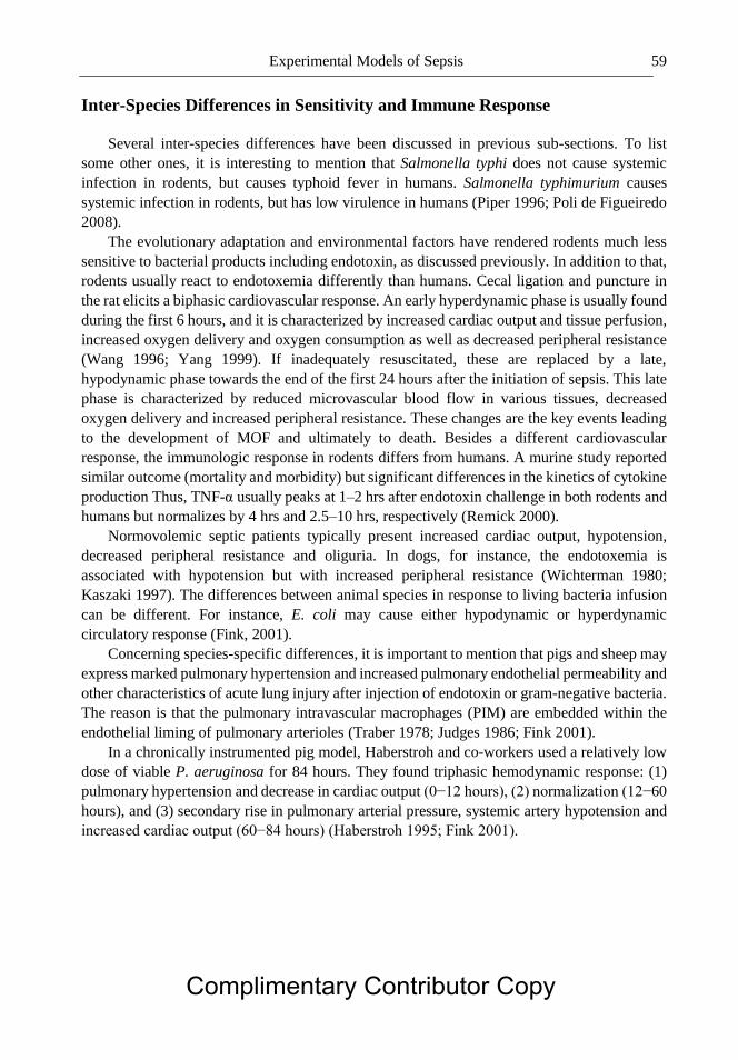
Experimental Models of Sepsis 59
Inter-Species Differences in Sensitivity and Immune Response
Several inter-species differences have been discussed in previous sub-sections. To list
some other ones, it is interesting to mention that Salmonella typhi does not cause systemic
infection in rodents, but causes typhoid fever in humans. Salmonella typhimurium causes
systemic infection in rodents, but has low virulence in humans (Piper 1996; Poli de Figueiredo
2008).
The evolutionary adaptation and environmental factors have rendered rodents much less
sensitive to bacterial products including endotoxin, as discussed previously. In addition to that,
rodents usually react to endotoxemia differently than humans. Cecal ligation and puncture in
the rat elicits a biphasic cardiovascular response. An early hyperdynamic phase is usually found
during the first 6 hours, and it is characterized by increased cardiac output and tissue perfusion,
increased oxygen delivery and oxygen consumption as well as decreased peripheral resistance
(Wang 1996; Yang 1999). If inadequately resuscitated, these are replaced by a late,
hypodynamic phase towards the end of the first 24 hours after the initiation of sepsis. This late
phase is characterized by reduced microvascular blood flow in various tissues, decreased
oxygen delivery and increased peripheral resistance. These changes are the key events leading
to the development of MOF and ultimately to death. Besides a different cardiovascular
response, the immunologic response in rodents differs from humans. A murine study reported
similar outcome (mortality and morbidity) but significant differences in the kinetics of cytokine
production Thus, TNF-α usually peaks at 1–2 hrs after endotoxin challenge in both rodents and
humans but normalizes by 4 hrs and 2.5–10 hrs, respectively (Remick 2000).
Normovolemic septic patients typically present increased cardiac output, hypotension,
decreased peripheral resistance and oliguria. In dogs, for instance, the endotoxemia is
associated with hypotension but with increased peripheral resistance (Wichterman 1980;
Kaszaki 1997). The differences between animal species in response to living bacteria infusion
can be different. For instance, E. coli may cause either hypodynamic or hyperdynamic
circulatory response (Fink, 2001).
Concerning species-specific differences, it is important to mention that pigs and sheep may
express marked pulmonary hypertension and increased pulmonary endothelial permeability and
other characteristics of acute lung injury after injection of endotoxin or gram-negative bacteria.
The reason is that the pulmonary intravascular macrophages (PIM) are embedded within the
endothelial liming of pulmonary arterioles (Traber 1978; Judges 1986; Fink 2001).
In a chronically instrumented pig model, Haberstroh and co-workers used a relatively low
dose of viable P. aeruginosa for 84 hours. They found triphasic hemodynamic response: (1)
pulmonary hypertension and decrease in cardiac output (0−12 hours), (2) normalization (12−60
hours), and (3) secondary rise in pulmonary arterial pressure, systemic artery hypotension and
increased cardiac output (60−84 hours) (Haberstroh 1995; Fink 2001).
Complimentary Contributor Copy
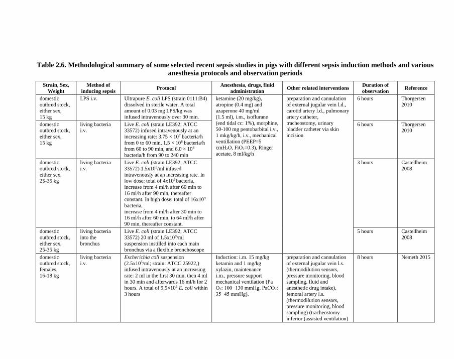
Table 2.6. Methodological summary of some selected recent sepsis studies in pigs with different sepsis induction methods and various
anesthesia protocols and observation periods
Strain, Sex,
Weight
Method of
inducing sepsis Protocol
Anesthesia, drugs, fluid
administration Other related interventions
Duration of
observation Reference
domestic
outbred stock,
either sex,
15 kg
LPS i.v. Ultrapure E. coli LPS (strain 0111:B4)
dissolved in sterile water. A total
amount of 0.03 mg LPS/kg was
infused intravenously over 30 min.
ketamine (20 mg/kg),
atropine (0.4 mg) and
azaperone 40 mg/ml
(1.5 ml), i.m., isoflurane
(end tidal cc: 1%), morphine, 50-100 mg pentobarbital i.v.,
1 mkg/kg/h, i.v., mechanical
ventillation (PEEP=5 cmH2O, FiO2=0.3), Ringer
acetate, 8 ml/kg/h
preparation and cannulation
of external jugular vein l.d.,
carotid artery l.d., pulmonary
artery catheter,
tracheostomy, urinary bladder catheter via skin
incision
6 hours Thorgersen
2010
domestic outbred stock,
either sex,
15 kg
living bacteria i.v.
Live E. coli (strain LE392; ATCC 33572) infused intravenously at an
increasing rate: 3.75 × 10⁷ bacteria/h
from 0 to 60 min, 1.5 × 10⁸ bacteria/h from 60 to 90 min, and 6.0 × 10⁸
bacteria/h from 90 to 240 min
6 hours Thorgersen 2010
domestic
outbred stock,
either sex, 25-35 kg
living bacteria
i.v.
Live E. coli (strain LE392; ATCC
33572) 1.5x10⁸/ml infused
intravenously at an increasing rate. In low dose: total of 4x10⁹ bacteria,
increase from 4 ml/h after 60 min to
16 ml/h after 90 min, thereafter constant. In high dose: total of 16x109
bacteria,
increase from 4 ml/h after 30 min to 16 ml/h after 60 min, to 64 ml/h after
90 min, thereafter constant.
3 hours Castellheim
2008
domestic
outbred stock,
either sex, 25-35 kg
living bacteria
into the
bronchus
Live E. coli (strain LE392; ATCC
33572) 20 ml of 1.5x10⁹/ml
suspension instilled into each main bronchus via a flexible bronchoscope
5 hours Castellheim
2008
domestic
outbred stock,
females,
16-18 kg
living bacteria
i.v.
Escherichia coli suspension
(2.5x10⁵/ml; strain: ATCC 25922,)
infused intravenously at an increasing
rate: 2 ml in the first 30 min, then 4 ml
in 30 min and afterwards 16 ml/h for 2 hours. A total of 9.5×10⁶ E. coli within
3 hours
Induction: i.m. 15 mg/kg
ketamin and 1 mg/kg
xylazin, maintenance
i.m., pressure support
mechanical ventilation (Pa O2: 100−130 mmHg, PaCO2:
35−45 mmHg).
preparation and cannulation
of external jugular vein l.s.
(thermodilution sensors,
pressure monitoring, blood
sampling, fluid and anesthetic drug intake),
femoral artery l.s.
(thermodilution sensors, pressure monitoring, blood
sampling) (tracheostomy
inferior (assisted ventilation)
8 hours Nemeth 2015
Complimentary Contributor Copy

Strain, Sex,
Weight
Method of
inducing sepsis Protocol
Anesthesia, drugs, fluid
administration Other related interventions
Duration of
observation Reference
domestic
outbred stock, either sex,
27−35 kg
faecal peritonitis
(autologous faeces
inoculation)
0.5 g/kg of autologous faeces
suspended in 200 ml saline was inoculated into the peritoneal cavity
through drains.
Induction: i.v. 0.5 mg
atropine, 1 to 2 mg/kg propofol 2% and 2 mg/kg
ketamine. Mechanically
ventillation (PEEP=10
cmH2O, FiO2=0.4, tidal
volume: 10 ml/kg)
Surgical anaesthesia: intravenous 5-10 mg/kg/hour
thiopental and 5-15
μg/kg/hour fentanyl. Pancuronium (4 to 6
mg/hour), infusion of Plasma
Lyte solution (7−15 ml/kg/hour), 20% glucose to
maintain blood glucose
levels between 4.5 and 7 mmol/l.
central venous and
pulmonary artery catherers, external jugular vein and
femoral artery cannulation.
Midline laparotomy,
drainage, urinary blader
catheterization via
epicystostomy.
22 hours
(after 8 hours of stabilization)
Chvojka
2008
Vietnamese
minipig, either sex,
24 ± 3 kg
faecal peritonitis
(autologous faeces
inoculation)
0.5 g/kg of autologous faeces
suspended in 200 ml saline was incubated for 6 hours at 38 °C, then
the suspension was intraperitoneally
inoculated via a small incision above the umbilical scar and through a
Veres-needle.
Anesthesia for sepsis
induction: i.m. 20 mg/kg ketamin and 2 mg/kg
xylazin, then 6 mg/kg/h i.v.
propofol. After the induction process,
animals were sedated and analgesic was used (4 mg/kg
ketamin, 0.6 mg/kg
nalbuphin). 15 hours later the animals were re-
anaesthetized for invasive
monitoring.
Ringer-lactate 15 ml/kg/h,
Voluvel 6% 130 kDa/0.4 5
ml/kg/h, and in case of need: 0.015 µg/kg/h i.v.
Unilateral preparation and
cannulation of external jugular vein (pressure
monitoring, sampling,
infusion). Endotracheal intubation.
Unilateral preparation and cannulation of femoral artery
and vein (thermodilution
sensors, pressure monitoring).
Urinary bladder
catheterization via inguinal
incision.
24 hours
(measurements started 15 hours
after sepsis
induction)
Zsikai 2012
Complimentary Contributor Copy

Norbert Nemeth, Mihai Oltean and Bela Fulesdi 62
Effect of Age, Gender, and Co-Morbidities
Age is an important issue both in clinical and experimental scenarios (Watters 2001; Starr
2014). In most of the experimental models, the animals are usually young adults and healthy
animals, without co-morbidities. Agent is a toxin or a defined type of micro-organism. The
treatment protocol (if any) is standardized and initiated soon after the onset of sepsis. The
animals are usually anesthetized. Coprophagy is common.
In humans, sepsis often is seen at extreme ages (neonates or elderly), the origin is more
often an infection site usually not recognized or frequently, the infection is polymicrobial. The
treatment is initiated usually late (hours or days) after the start of infection and frequently the
antibiotics are ineffective and are very different between cases. The presence of the co-
morbidities is increasingly common (atherosclerosis, hypertension, diabetes mellitus, cancer,
etc.) (Esmon 2004).
Gender differences have been also observed. Receptors for sex hormones have been
identified on various immune cells, suggesting direct effects of these hormones on immune
function (Albertsmeier 2014).
There are models with immunosuppressed host and so-called sequential challenge models.
Co-morbidities can be induced by acute ethanol ingestion, CCl4-induced cirrhosis, hemorrhagic
shock, trauma, splenectomy, thermal injury, etc., mostly in rodents (Fink 2001).
Anesthesia, Drugs, Resuscitation
The clinical management of septic animals versus septic patients is a very important point
of view, such as volume therapy, electrolytes, antibiotics, ventilation, etc. These protocols in
experiments have unquestionable importance (Naumann 2015). Table 2.6 summarizes
methodological details of some selected porcine sepsis models, which include the sepsis
induction, the instrumentation, the anesthesia, drugs, resuscitation protocols as well as the
follow-up periods are different.
CONCLUSION
Unfortunately, sepsis remains a severe clinical challenge with numerous unsolved
problems. Animal models are both useful and necessary to explore the pathophysiology and
develop effective therapeutic strategies. Nonetheless, besides their advantages, the
experimental models of sepsis have numerous limitations that need to be considered when
translating the experimental findings into clinical trials.
Some of the main factors have to be taken into consideration when planning, conducting
and evaluating experimental sepsis models:
the method of sepsis induction
the agent (type of toxin or microorganism)
choice of animal species
Complimentary Contributor Copy

Experimental Models of Sepsis 63
species-specific characteristics in immune response sensitivity, in physiological and
even anatomical features
age and gender of the animals
effect of anesthesia, drugs, fluid intake, resuscitation
co-morbidities (absence or presence)
duration of the follow-up (observation period)
In general it can be also concluded that small animal models (e.g., mouse, rat) are useful
to test certain pathophysiological pathways and for developing/testing drugs, while large
animal models (e.g., pig, non-human primate) may provide a better scenario for testing novel
treatment strategies being closer to the clinical practice by size, physiology and opportunities
for intervention.
REFERENCES
Albert, M; Losser, MR; Hayon, D; Faivre, V; Payen, D. Systemic and renal macro- and
microcirculatory responses to arginine vasopressin in endotoxic rabbits. Crit. Care Med.,
2004, 32(9), 1891–1898.
Albertsmeier, M; Pratschke, S; Chaudry, I; Angele, MK. Gender-specific effects on immune
response and cardiac function after trauma hemorrhage and sepsis. Viszeralmedizin., 2014,
30(2), 91–96.
Alt, E; Amann-Vesti, BR; Madl, C; Funk, G; Koppensteiner, R. Platelet aggregation and blood
rheology in severe sepsis/septic shock: relation to the Sepsis-related Organ Failure
Assessment (SOFA) score. Clin. Hemorheol. Microcirc., 2004, 30(2), 107–115.
American College of Chest Physicians/Society of Critical Care Medicine Consensus
Conference: definitions for sepsis and organ failure and guidelines for the use of innovative
therapies in sepsis. Crit. Care Med., 1992, 20(6), 864–874.
Asakura, H. Classifying types of disseminated intravascular coagulation: clinical and animal
models. J. Int. Care, 2014, 2(1), 20–26.
Astiz, ME; DeGent, GE; Lin, RY; Rackow, EC. Microvascular function and rheologic changes
in hyperdynamic sepsis. Crit. Care Med., 1995, 23(2), 265–271.
Bara, M; Joffe, AR. The ethical dimension in published animal research in critical care: the
public face of science. Crit. Care., 2014, 18(1), R15–R21.
Baskurt, OK; Neu, B; Meiselman, HJ. Red Blood Cell Aggregation. Boca Raton: CRC Press,
2012.
Baskurt, OK; Temiz, A; Meiselman, HJ. Red blood cell aggregation in experimental sepsis. J.
Lab. Clin. Med., 1997, 130(2), 183–190.
Baskurt, OK. Mechanisms of blood rheology alterations. In: Baskurt OK, Hardeman MR,
Rampling MW, Meiselman HJ, editors. Handbook of Hemorheology and Hemodynamics.
Amsterdam: IOS Press, 2007. pp. 170–190.
Bhan, C; Dipankar, P; Chakraborty, P; Sarangi, PP. Role of cellular events in the
pathophysiology of sepsis. Inflamm. Res,. 2016, 65(11), 853–868.
Borthwick, EM; Hill, C; Rabindranath, KS; Maxwell, AP; McAuley, DF; Blackwood, B. High-
volume haemofiltration for sepsis. Cochrane Database Syst. Rev., 1, 2013, CD008075. Complimentary Contributor Copy
Norbert Nemeth, Mihai Oltean and Bela Fulesdi 64
Bridges, EJ; Dukes, S. Cardiovascular aspects of septic shock: pathophysiology, monitoring,
and treatment. Crit. Care Nurse., 2005, 25(2), 14–40.
Brown, KA; Brain, SD; Pearson, JD; Edgeworth, JD; Lewis, SM; Treacher, DF. Neutrophils in
development of multiple organ failure in sepsis. The Lancet, 2006, 368(9530),
157–169.
Browne, MK; Leslie, GB. Animal models of peritonitis. Surg. Gynecol. Obstet., 1976, 143(5),
738–740.
Cash, JL; White, GE; Greaves, DR. Chapter 17. Zymosan‐induced peritonitis as a simple
experimental system for the study of inflammation. Methods in Enzymology (Chemokines,
Part B), 2009, 461, 379–396.
Castellheim, A; Thorgersen, EB; Hellerud, BC; Pharo, A; Johansen, HT; Brosstad, F; Gaustad,
P; Brun, H; Fosse, E; Tonnessen, TI; Nielsen, EW; Mollnes, TE. New biomarkers in an
acute model of live Escherichia coli-induced sepsis in pigs. Scand. J. Immunol., 2008,
68(1), 75–84.
Catry, J; Brouquet, A; Peschaud, F; Vychnevskaia, K; Abdalla, S; Malafosse, R; Lambert, B;
Costaglioli, B; Benoist, S; Penna, C. Sigmoid resection with primary anastomosis and
ileostomy versus laparoscopic lavage in purulent peritonitis from perforated diverticulitis:
outcome analysis in a prospective cohort of 40 consecutive patients. Int. J. Colorectal. Dis.,
2016, 31(10), 1693–1699.
Chen, P; Stanojcic, M; Jeschke, MG. Differences between murine and human sepsis. Surg.
Clin. North. Am., 2014, 94(6), 1135–1149.
Chiavolini, D; Pozzi, G; Ricci, S. Animal models of Streptococcus pneumoniae disease. Clin.
Microbiol. Rev., 2008, 21(4), 666–685.
Chien, S; Usami, S; Dellenback, J; Magazinovic, V. Blood rheology after hemorrhage and
endotoxin. Adv. Exp. Med. Biol., 1972, 33, 75–93.
Chvojka, J; Sykora, R; Krouzecky, A; Radej, J; Varnerova, V; Karvunidis, T; Hes, O; Novak,
I; Radermacher, P; Matejovic, M. Renal haemodynamic, microcirculatory, metabolic and
histopathological responses to peritonitis-induced septic shock in pigs. Crit. Care., 2008,
12(6), R164. http://ccforum.biomedcentral.com/articles/10.1186/cc7164.
Copeland, S; Warren, HS; Lowry, SF; Calvano, SE; Remick, D. Acute inflammatory response
to endotoxin in mice and humans. Clin. Diagn. Lab. Immunol., 2005, 12(1),
60–67.
Cryer, PE; Herman, CM; Sode, J. Carbohydrate metabolism in the baboon subjected to gram-
negative (E. coli) septicemia. I. Hyperglycemia with depressed plasma insulin
concentrations. Ann. Surg., 1971, 174(1), 91–100.
Daniels, R. Surviving the first hours in sepsis: getting the basics right (an intensivist’s
perspective). J. Antimicrob. Chemother., 2011, 66(Suppl 2), 11–13.
Dejager, L; Pinheiro, I; Dejonckheere, E; Libert, C. Cecal ligation and puncture: the gold
standard model for polymicrobial sepsis? Trends Microbiol., 2011, 19(4), 198–208.
Dellinger, RP; Levy, MM; Rhodes, A; Annane, D; Gerlach, H; Opal, SM; Sevransky, JE;
Sprung, CL; Douglas, IS; Jaeschke, R; Osborn, TM; Nunnally, ME; Townsend, SR;
Reinhart, K; Kleinpell, RM; Angus, DC; Deutschman, CS; Machado, FR; Rubenfeld, GD;
Webb, SA; Beale, RJ; Vincent, JL; Moreno, R. Surviving Sepsis Campaign Guidelines
Committee including the Pediatric Subgroup. Surviving sepsis campaign: international
guidelines for management of severe sepsis and septic shock: 2012. Crit. Care Med., 2013,
41(2), 580–637. Complimentary Contributor Copy
Experimental Models of Sepsis 65
Doi, K. Role of kidney injury in sepsis. J. Int. Care, 2016, 4, 17–22.
Dyson, A; Singer, M. Animal models of sepsis: why does preclinical efficacy fail to translate
to the clinical setting? Crit. Care Med., 2009, 37(1 Suppl), S30–S37.
Ebong, S; Call, D; Nemzek, J; Bolgos, G; Newcomb, D; Remick, D. Immunopathologic
alterations in murine models of sepsis of increasing severity. Infect. Immun., 1999, 67(12),
6603–6610.
Efron, PA; Mohr, AM; Moore, FA; Moldawer, LL. The future of murine sepsis and trauma
research models. J. Leukoc. Biol., 2015, 98(6), 945–952.
Elias, RM; Johnston, MG; Hayashi, A; Nelson, W. Decreased lymphatic pumping after
intravenous endotoxin administration in sheep. Am. J. Physiol., 1987, 253(6 Pt 2), H1349–
H1357.
Engel, AL; Holt, GE; Lu, H. The pharmacokinetics of Toll-like receptor agonists and the impact
on the immune system. Expert Rev. Clin. Pharmacol., 2011, 4(2), 275–289.
Eskandari, MK; Bolgos, G; Miller, C; Nguyen, DT; DeForge, LE; Remick, DG. Anti-tumor
necrosis factor antibody therapy fails to prevent lethality after cecal ligation and puncture
or endotoxemia. J. Immunol., 1992, 148(), 2724–2730.
Esmon, CT. Why do animal models (sometimes) fail to mimic human sepsis? Crit. Care Med.,
2004, 32(5 Suppl), S219−S222.
Fink, MP. Animal models of sepsis and the multiple organ dysfunction syndrome. In: Souba
WW, Wilmore DW, editors. Surgical Research. Amsterdam: Elsevier Academic Press,
2001, pp. 875−891.
Fink, MP. Animal models of sepsis. Virulence, 2014, 5(1), 143–153.
Fleischmann, C; Scherag, A; Adhikari, NK; Hartog, CS; Tsaganos, T; Schlattmann, P; Angus,
DC; Reinhart, K. International Forum of Acute Care Trialists. Assessment of Global
Incidence and Mortality of Hospital-treated Sepsis. Current Estimates and Limitations. Am.
J. Respir. Crit. Care Med., 2016, 193(3), 259–272.
Franks, Z; Carlisle, M; Rondina, MT. Current challenges in understanding immune cell
functions during septic syndromes. BMC Immunology, 2015, 16, 11–16.
Gomez-Jimenez, J; Salgado, A; Mourelle, M; Martin, MC; Segura, RM; Peracaula, R;
Moncada, S. L-arginine: nitric oxide pathway in endotoxemia and human septic shock.
Crit. Care Med., 1995, 23(2), 253-258.
Guenter, CA; Fiorica, V; Hinshaw, LB. Cardiorespiratory and metabolic responses to liver E.
coli and endotoxin in the monkey. J. Appl. Physiol., 1969, 26(6), 780–786.
Guerra, GC; de Menezes, MS; de Araújo, AA; de Araújo Júnior, RF; de Medeiros, CA.
Olmesartan prevented intra-articular inflammation induced by zymosan in rats. Biol.
Pharm. Bull., 2016, 39(11), 1793–1801.
Guler, O; Ugras, S; Aydin, M; Dilek, FH; Dilek, ON; Karaayvaz, M. The effect of lymphatic
blockage on the amount of endotoxin in portal circulation, nitric oxide synthesis, and the
liver in dogs with peritonitis. Surg. Today, 1999, 29(8), 735–740.
Gumenscheimer, M; Mitov, I; Galanos, C; Freudenberg, MA. Beneficial or deleterious effects
of a preexisting hypersensitivity to bacterial components on the course and outcome of
infection. Infect. Immun., 2002, 70(10), 5596–5603.
Haberstroh, J; Breuer, H; Lücke, I; Massarrat, K; Früh, R; Mand, U; Hagerdorn, L; von Specht,
BU. Effect of recombinant human granulocyte colony-stimulating factor on hemodynamic
and cytokine response in a porcine model of Pseudomonas sepsis. Shock, 1995, 4(3), 216–
224. Complimentary Contributor Copy
Norbert Nemeth, Mihai Oltean and Bela Fulesdi 66
Hau, T; Simmons, RL. Animal models of peritonitis. Surg. Gynecol. Obstet., 1977, 144(5),
755–756.
Hermreck, AS; Thal, AP. Mechanisms for the high circulatory requirements in sepsis and septic
shock. Ann. Surg., 1969, 170(4), 677–695.
Hinshaw, LB; Beller, BK; Archer, LT; Benjamin, B. Hypoglycemic response of blood to live
Escherichia coli organisms and endotoxin. J. Surg. Res., 1976, 21(3), 141–150.
Hollenberg, SM; Dumasius, A; Easington, C; Colilla, SA; Neumann, A; Parrillo, JE.
Characterization of a hyperdynamic murine model of resuscitated sepsis using
echocardiography. Am. J. Respir. Crit. Care Med., 2001, 164(5), 891–895.
Hong, Y; Sun, LI; Sun, R; Chen, H; Yu, Y; Xie, K. Combination therapy of molecular hydrogen
and hyperoxia improves survival rate and organ damage in a zymosan-induced generalized
inflammation model. Exp. Ther. Med., 2016, 11(6), 2590–2596.
Hubbard, WJ; Choudhry, M; Schwacha, MG; Kerby, JD; Rue, LW; 3rd. Bland, KI; Chaudry,
IH. Cecal ligation and puncture. Shock., 2005, 24(Suppl 1), 52–57.
Ince, C; Mik, EG. Microcirculatory and mitochondrial hypoxia in sepsis, shock, and
resuscitation. J. Appl. Physiol., 2016, 120(2), 226–235.
Ince, C. The microcirculation is the motor of sepsis. Crit. Care., 2005, 9(Suppl 4), S13–S19.
Islam, S; Jarosch, S; Zhou, J; Parquet Mdel, C; Toguri, JT; Colp, P; Holbein, BE; Lehmann, C.
Anti-inflammatory and anti-bacterial effects of iron chelation in experimental sepsis. J.
Surg. Res., 2016, 200(1), 266–273.
Judges, D; Sharkey, P; Cheung, H; Craig, I; Driedger, AA; Sibbald, WJ; Finley, R. Pulmonary
microvascular fluid flux in a large animal model of sepsis: evidence for increased
pulmonary endothelial permeability accompanying surgically induced peritonitis in sheep.
Surgery, 1986, 99(2), 222–234.
Kameneva, MV; Antaki, JF. Mechanical trauma to blood. In: Baskurt OK, Hardeman MR,
Rampling MW, Meiselman HJ, editors. Handbook of Hemorheology and Hemodynamics.
Amsterdam: IOS Press, 2007, pp. 206–227.
Kaszaki, J; Wolfrad, A; Boros, M; Baranyi, L; Okada, H; Nagy, S. Effects of antiendothelin
treatment on the early hemodynamic changes in hyperdynamic endotoxemia. Acta Chir.
Hung., 1997, 36(1-4), 152–153.
Katschinski, T; Galanos, C; Coumbos, A; Freudenberg, MA. Gamma interferon mediates
Proprionibacterium acnes-induced hypersensitivity to lipopolysaccharide. Infect. Immun.,
1992, 60(5), 1994–2001.
Kingsley, SM; Bhat, BV. Differential paradigms in animal models of sepsis. Curr. Infect. Dis.
Rep., 2016, 18(9), 26–36.
Kirschenbaum, LA; Aziz, M; Astiz, ME; Saha, DC; Rackow, EC. Influence of rheologic
changes and platelet-neutrophil interactions on cell filtration in sepsis. Am. J. Respir. Crit.
Care Med., 2000, 161(5), 1602–1607.
Kiss, F; Molnar, L; Hajdu, E; Deak, A; Molnar, A; Berhes, M; Szabo, J; Nemeth, N; Fulesdi,
B. Skin microcirculatory changes reflect early the circulatory deterioration in a fulminant
sepsis model in the pig. Acta Cir. Bras., 2015, 30(7), 470–477.
Klag, T; Cantara, G; Sechtem, U; Athanasiadis, A. Interleukin-6 kinetics can be useful for early
treatment monitoring of severe bacterial sepsis and septic shock. Infect Dis Rep., 2016,
8(1), 6213. http:// www.pagepress.org/journals/index.php/idr/ article/view/6213/ 5524
Complimentary Contributor Copy
Experimental Models of Sepsis 67
Krisher, SL; Browne, A; Dibbins, A; Tkacz, N; Curci, M. Intra-abdominal abscess after
laparoscopic appendectomy for perforated appendicitis. Arch. Surg., 2001, 136(4), 438–
441.
Kutner, FR; Schwartz, SI; Adams, JT. The effect of adrenergic blockade on lymph flow in
endotoxin shock. Ann. Surg., 1967, 165(4), 518–527.
Lakshmikanth, CL; Jacob, SP; Chaithra, VH; de Castro-Faria-Neto, HC; Marathe, GK. Sepsis:
in search of cure. Inflamm. Res., 2016, 65(8), 587–602.
Langenberg, C; Bellomo, R; May, C; Wan, L; Egi, M; Morgera, S. 2005. Renal blood flow in
sepsis. Crit. Care., 9(4), R363–R374.
Laschke, MW; Menger, MD; Wang, Y; Lindell, G; Jeppsson, B; Thorlacius, H. Sepsis-
associated cholestasis is critically dependent on P-selectin-dependent leukocyte
recruitment in mice. Am. J. Physiol. Gastrointest. Liver Physiol., 2007, 292(5), G1396–
G1402.
Lattuada, M; Hedenstierna, G. Abdominal lymph flow in an endotoxin sepsis model: influence
of spontaneous breathing and mechanical ventilation. Crit. Care Med., 2006, 34(11), 2792–
2798.
Levy, MM; Fink, MP; Marshall, JC; Abraham, E; Angus, D; Cook, D; Cohen, J; Opal, SM;
Vincent, JL; Ramsay, G. International Sepsis Definitions Conference. 2001 SCCM/
ESICM/ACCP/ATS/SIS International Sepsis Definitions Conference. Intensive Care
Med., 2003, 29(4), 530–538.
Lewis, AJ; Seymour, CW; Rosengart, MR. Current murine models of sepsis. Surg. Infect.
(Larchmt)., 2016, 17(4), 385–393.
Lilley, E; Armstrong, R; Clark, N; Gray, P; Hawkins, P; Mason, K; López-Salesansky, N;
Stark, AK; Jackson, SK; Thiemermann, C; Nandi, M. Refinement of animal models of
sepsis and septic shock. Shock, 2015, 43(4), 304–316.
Lohner, R; Schwederski, M; Narath, C; Klein, J; Duerr, GD; Torno, A; Knuefermann, P; Hoeft,
A; Baumgarten, G; Meyer, R; Boehm, O. Toll-like receptor 9 promotes cardiac
inflammation and heart failure during polymicrobial sepsis. Mediators Inflamm., 2013,
2013, 261049. https://www.hindawi.com/journals/mi/2013/261049/.
Lundy, DJ; Trzeciak, S. Microcirculatory dysfunction in sepsis. Crit. Care Clin., 2009, 25(4),
721–731.
Lustig, MK; Bac, VH; Pavlovic, D; Maier, S; Gründling, M; Grisk, O; Wendt, M; Heidecke,
CD; Lehmann, C. Colon ascendens stent peritonitis – a model of sepsis adopted to the rat:
physiological, microcirculatory and laboratory changes. Shock, 2007, 28(1), 59–64.
Mai, S; Khan, M; Liaw, P; Fox-Robichaud, A. Experimental sepsis models. In: Azevedo L,
editor. Sepsis - An ongoing and significant challenge. InTech, 2012. http://www.
intechopen.com/ books/sepsis-an-ongoing-and-significant-challenge/experimental-sepsis-
models.
Maier, S; Traeger, T; Entleutner, M; Westerholt, A; Kleist, B; Hüser, N; Holzmann, B; Stier,
A; Pfeffer, K; Heidecke, CD. Cecal ligation and puncture versus colon ascendens stent
peritonitis: two distinct animal models for polymicrobial sepsis. Shock, 2004, 21(6), 505–
511.
Martins, GS. Sepsis, severe sepsis and septic shock: changes in incidence, pathogens and
outcomes. Expert Rev. Anti Infect. Ther., 2012, 10, 701−706.
Complimentary Contributor Copy

Norbert Nemeth, Mihai Oltean and Bela Fulesdi 68
Mook-Kanamori, B; Geldhoff, M; Troost, D; van der Poll, T; van de Beek, D. Characterization
of a pneumococcal meningitis mouse model. BMC Infect. Dis., 2012, 12, 71.
Mook-Kanamori, BB; Valls Serón, M; Geldhoff, M; Havik, SR; van der Ende, A; Baas, F; van
der Poll, T; Meijers, JC; P Morgan, B; Brouwer, MC; van de Beek, D. Thrombin-
activatable fibrinolysis inhibitor influences disease severity in humans and mice with
pneumococcal meningitis. J. Thromb. Haemost., 2015, 13(11), 2076–2086.
Morel, J; Li, JY; Eyenga, P; Meiller, A; Gustin, MP; Bricca, G; Molliex, S; Viale, JP. Early
adverse changes in liver microvascular circulation during experimental septic shock are
not linked to an absolute nitric oxide deficit. Microvasc. Res., 2013, 90, 187–191.
Moutzouri, AG; Skoutelis, AT; Gogos, CA; Missirlis, YF; Athanassiou, GM. Red blood cell
deformability in patients with sepsis: a marker for prognosis and monitoring of severity.
Clin. Hemorheol. Microcirc., 2007, 36(4), 291–299.
Nash, GB; Watts, T; Thornton, C; Barigou, M. Red cell aggregation as a factor influencing
margination and adhesion of leukocytes and platelets. Clin. Hemorheol. Microcirc., 2008,
39(1-4), 303–310.
Naumann, DN; Dretzke, J; Hutchings, S; Midwinter, MJ. Protocol for a systematic review of
the impact of resuscitation fluids on the microcirculation after haemorrhagic shock in
animal models. Systematic Reviews, 2015, 4, 135–141.
Nduka, OO; Parrillo, JE. The pathophysiology of septic shock. Crit. Care Nurs. Clin. North
Am., 2011, 23(1), 41–66.
Nemeth, N; Berhes, M; Kiss, F; Hajdu, E; Deak, A; Molnar, A; Szabo, J; Fulesdi, B. Early
hemorheological changes in a porcine model of intravenously given E. coli induced
fulminant sepsis. Clin. Hemorheol. Microcirc., 2015, 61(3), 479–496.
Nemeth, N; Fulesdi, B. Concerning hemorheological disturbances in sepsis. De omnibus
dubitandum est… Series on Biomechanics, 2016, 30(1), 20–26.
Nemeth, N; Miko, I; Furka, A; Kiss, F; Furka, I; Koller, A; Szilasi, M. Concerning the
importance of changes in hemorheological parameters caused by acid-base and blood gas
alterations in experimental surgical models. Clin. Hemorheol. Microcirc., 2012, 51(1), 43–
50.
Nemoto, K; Sato, H; Tanuma, K; Okamura, T. Mesenteric lymph flow in endotoxemic guinea
pigs. Lymphat. Res. Biol., 2011, 9(3), 129–134.
Nemzek, JA; Hugunin, KM; Opp, MR. Modeling sepsis in the laboratory: merging sound
science with animal well-being. Comp. Med., 2008, 58(2), 120–128.
Nishino, M; Tanaka, H; Ogura, H; Inoue, Y; Koh, T; Fujita, K; Sugimoto, H. Serial changes in
leukocyte deformability and whole blood rheology in patients with sepsis or trauma. J.
Trauma, 2005, 59(6), 1425–1431.
Onderdonk, AB; Bartlett, JG; Louie, T; Sullivan-Seigler, N; Gorbach, SL. Microbial synergy
in experimental intra-abdominal abscess. Infect. Immun., 1976, 13(1), 22–26.
Ostergaard, L; Granfeldt, A; Secher, N; Tietze, A; Iversen, NK; Jensen, MS; Andersen, KK;
Nagenthiraja, K; Gutiérrez-Lizardi, P; Mouridsen, K; Jespersen, SN; Tonnesen, EK.
Microcirculatory dysfunction and tissue oxygenation in critical illness. Acta Anaesthesiol.
Scand., 2015, 59, 1246–1259.
Otero-Anton, E; Gonzalez-Quintela, A; Lopez-Soto, A; Lopez-Ben, S; Llovo, J; Perez, LF.
Cecal ligation and puncture as a model of sepsis in the rat: influence of the puncture size
on mortality, bacteremia, endotoxemia and tumor necrosis factor alpha levels. Eur. Surg.
Res., 2001, 33(2), 77–79. Complimentary Contributor Copy

Experimental Models of Sepsis 69
Perbellini, A; Shatney, CH; MacCarter, DJ; Lillehei, RC. A new model for the study of septic
shock. Surg. Gynecol. Obstet., 1978, 147(1), 68–74.
Piagnerelli, M; Boudjeltia, KZ; Vanhaeverbeek, M; Vincent, JL. Red blood cell rheology in
sepsis. Intensive Care Med., 2003, 29(7), 1052–1061.
Piper, RD; Cook, DJ; Bone, RC; Sibbald, WJ. Introducing critical appraisal to studies of animal
models investigating novel therapies in sepsis. Crit. Care Med., 1996, 24(12), 2059–2070.
Poli-de-Figueiredo, LF; Garrido, AG; Nakagawa, N; Sannomiya, P. Experimental models of
sepsis and their clinical relevance. Shock, 2008, 30(Suppl 1), 53–59.
Pöschl, JMB; Leray, C; Ruef, P; Cazenave, JP; Lindekamp, O. Endotoxin binging to
erythrocyte membrane and erythrocyte deformability in human sepsis and in vitro. Crit.
Care Med., 2003, 31(3), 924–928.
Postel, J; Schloerb, PR; Furtado, D. Pathophysiologic alterations during bacterial infusions for
the study of bacteremic shock. Surg. Gynecol. Obstet., 1975, 141(5), 683–692.
Rabinovici, R; Bugelski, PJ; Esser, KM; Hillegass, LM; Griswold, DE; Vernick, J; Feuerstein,
G. Tumor necrosis factor-primed rats. J. Pharmacol. Exp. Ther., 1993, 267(3), 1550–1557.
Reggiori, G; Occhipinti, G; De Gasperi, A; Vincent, JL; Piagnerelli, M. Early alterations of red
blood cell rheology in critically ill patients. Crit. Care Med., 2009, 37(12), 3041–3046.
Remick, DG; Newcomb, DE; Bolgos, GL; Call, DR. Comparison of the mortality and
inflammatory response of two models of sepsis: lipopolysaccharide vs. cecal ligation and
puncture. Shock, 2000, 13(2), 110–116.
Remick, DG; Ward, PA. Evaluation of endotoxin models for the study of sepsis. Shock, 2005,
24(Suppl 1), 7–11.
Remick, DG. Pathophysiology of sepsis. Am. J. Pathol., 2007, 170(5), 1435–1444.
Rittirsch, D; Hoesel, LM; Ward, PA. The disconnect between animal models of sepsis and
human sepsis. J. Leukoc. Biol., 2007, 81(1), 137–143.
Schabbauer, G. Polymicrobial sepsis models: CLP versus CASP. Drug Discovery Today:
Disease Models, 2012, 9(1), e17–e21.
Scheer, C; Fuchs, C; Rehberg, S. Biomarkers in severe sepsis and septic shock: just listen to
the heart? Crit. Care Med., 2016, 44(4), 849–850.
Schöneborn, S; Vollmer, C; Barthel, F; Herminghaus, A; Schulz, J; Bauer, I; Beck, C; Picker,
O. Vasopressin V1A receptors mediate the stabilization of intestinal mucosal oxygenation
during hypercapnia in septic rats. Microvasc. Res., 2016, 106, 24–30.
Schultz, MJ; van der Poll, T. Animal and human models for sepsis. Ann. Med., 2002, 34(7-8),
573–581.
Semeraro, N; Ammollo, CT; Semeraro, F; Colucci, M. Coagulopathy of acute sepsis. Semin.
Thromb. Hemost., 2015, 41(6), 650–658.
Sharkey, P; Judges, D; Driedger, AA; Cheung, H; Finley, RJ; Sibbald, WJ. The effect of an
infusion of zymosan-activated plasma on hemodynamic and pulmonary function in sheep.
Circ. Shock, 1984, 12(2), 79–93.
Shibayama, Y; Urano, T; Nakata, K. Changes in hepatic lymph vessels in endotoxaemia. J.
Pathol., 1992, 168(3), 325–330.
Shrum, B; Anantha, RV; Xu, SX; Donnelly, M; Haeryfar, SMM; McCormick, JK; Mele, T. A
robust scoring system to evaluate sepsis severity in an animal model. BMC Research Notes,
2014, 7, 233. http://www.biomedcentral.com/1756-0500/7/233.
Complimentary Contributor Copy

Norbert Nemeth, Mihai Oltean and Bela Fulesdi 70
Siempos, II; Lam, HC; Ding, Y; Choi, ME; Choi, AM; Ryter, SW. Cecal ligation and puncture-
induced sepsis as a model to study autophagy in mice. J. Vis. Exp., 2014, 84, e51066–
e51072. [article with video link: http://www.jove.com/video/51066/].
Singer, M; Deutschman, CS; Seymour, CW; Shankar-Hari, M; Annane, D; Bauer, M; Bellomo,
R; Bernard, GR; Chiche, JD; Coopersmith, CM; Hotchkiss, RS; Levy, MM; Marshall, JC;
Martin, GS; Opal SM; Rubenfeld, GD; van der Poll, T; Vincent, JL; Angus, DC. The Third
International Consensus Definitions for Sepsis and Septic Shock (Sepsis-3). JAMA., 2016,
315(8), 801–810.
Singleton, KD; Wischmeyer, PE. Distance of cecum ligated influences mortality, tumor
necrosis factor-alpha and interleukin-6 expression following cecal ligation and puncture in
the rat. Eur. Surg. Res., 2003, 35(6), 486–491.
Smith, AL; Greenfield, MD; Toothaker, RD. Experimental meningitis in the rat: Haemophilus
influenzae. Infection, 1984, 12(Suppl 1), S11–S22.
Smith, L; Andreasson, S; Saldéen, T; Risberg, B. Combined monitoring of thoracic duct and
lung lymph during E. coli sepsis in awake sheep. Lymphology, 1988, 21(3), 169–177.
Sordia, T; Tatarishvili, J; Mchedlishvili, G. Hemorheological disorders in the microcirculation
during septic shock in rats. Clin. Hemorheol. Microcirc., 2006, 35(1-2), 223–226.
Spronk, PE; Zandstra, DF; Ince, C. Bench-to-bedside review: Sepsis is a disease of the
microcirculation. Crit. Care, 2004, 8(6), 462–468.
Starr, ME; Saito, H. Sepsis in old age: review of human and animal studies. Aging Dis., 2014,
5(2), 126–136.
Thorgersen, EB; Hellerud, BC; Nielsen, EW; Barratt-Due, A; Fure, H; Lindstad, JK; Pharo, A;
Fosse, E; Tonnessen, TI; Johansen, HT; Castellheim, A; Mollnes, TE. CD14 inhibition
efficiently attenuates early inflammatory and hemostatic responses in Escherichia coli
sepsis in pigs. FASEB J., 2010, 24(3), 712–722.
Toscano, MG; Ganea, D; Gamero, AM. Cecal ligation puncture procedure. J. Vis. Exp., 2011,
51, 2860–2864. [article with video link: http://www.jove.com/video/2860/].
Traber, DL; Schlag, G; Redl, H; Strohmair, W; Traber, LD. Pulmonary microvascular changes
during hyperdynamic sepsis in an ovine model. Circ. Shock, 1987, 22(2), 185–193.
Trzeciak, S; Cinel, I; Dellinger, PR; Shapiro, NI; Arnold, RC; Parrillo, JE; Hollenberg, SM.
Microcirculatory Alterations in Resuscitation and Shock (MARS) Investigators.
Resuscitating the microcirculation in sepsis: the central role of nitric oxide, emerging
concepts for novel therapies, and challenges for clinical trials. Acad. Emerg. Med., 2008,
15(5), 399–413.
Trzeciak, S; Dellinger, RP; Parrillo, JE; Guglielmi, M; Bajaj, J; Abate, NL; Arnold, RC; Colilla,
S; Zanotti, S; Hollenberg, SM. Early microcirculatory perfusion derangements in patients
with severe sepsis and septic shock: relationship to hemodynamics, oxygen transport, and
survival. Ann. Emerg. Med., 2007, 49(1), 88–98.
Turnbull, IR; Wlzorek, JJ; Osborne, D; Hotchkiss, RS; Coopersmith, CM; Buchman, TG.
Effects of age on mortality and antibiotic efficacy in cecal ligation and puncture. Shock,
2003, 19(4), 310–313.
Tyagi, A; Sethi, AK; Girotra, G; Mohta, M. The Microcirculation in Sepsis. Indian J. Anaesth.,
2009, 53(3), 281–293.
van der Poll, T. Preclinical sepsis models. Surg. Infect. (Larchmt)., 2012, 13(5), 287-292.
Complimentary Contributor Copy

Experimental Models of Sepsis 71
Vincent, JL; Moreno, R; Takala, J; Willatts, S; De Mendonça, A; Bruining, H; Reinhart, CK;
Suter, PM; Thijs, LG. The SOFA (Sepsis-related Organ Failure Assessment) score to
describe organ dysfunction/failure. Intensive Care Med., 1996, 22(7), 707–710.
Volman, TJ; Hendriks, T; Goris, RJ. Zymosan-induced generalized inflammation:
Experimental studies into mechanisms leading to multiple organ dysfunction syndrome.
Shock, 2005, 23(4), 291–297.
Walker, H; Mason, Ad; Jr. Raulston, G. Surface infection with Pseudomonas aeruginosa. Ann.
Surg., 1964, 160, 297–305.
Wang, P; Chaudry, IH. Mechanism of hepatocellular dysfunction during hyperdynamic sepsis.
Am. J. Physiol., 1996, 270(5 Pt 2), R927–R938.
Ward, PA. New approaches to the study of sepsis. EMBO Mol. Med., 2012, 4(12), 1234–1243.
Watters, JM; O’Rourke, K. Effects of age and gender. In: Souba WW, Wilmore DW, editors.
Surgical Research. Amsterdam: Elsevier Academic Press, 2001, pp. 167−174.
Wester, T; Häggblad, E; Awan, ZA; Barratt-Due, A; Kvernebo, M; Halvorsen, PS; Mollnes,
TE; Kvernebo, K. Assessments of skin and tongue microcirculation reveals major changes
in porcine sepsis. Clin. Physiol. Funct. Imaging, 2011, 31(2), 151–158.
Wichterman, KA; Baue, AE; Chaudry, IH. Sepsis and septic shock: a review of laboratory
models and a proposal. J. Surg. Res. 1980, 29(2), 189–201.
Williams, JS; Clermont, HG. Thoracic duct lymph flow and acid phosphatase response to
steroid in experimental shock. Ann. Surg., 1973, 178(6), 777–780.
Wolfard, A; Kaszaki, J; Szabo, C; Balogh, Z; Nagy, S; Boros, M. Effects of selective nitric
oxide synthase inhibition in hyperdynamic endotoxemia in dogs. Eur. Surg. Res., 1999,
31(4), 314–323.
Wynn, JL; Wong, HR. Pathophysiology and treatment of septic shock in neonates. Clin.
Perinatol., 2010, 37(2), 439–479.
Wyrzykowski, AD; Feliciano, DV; George, TA; Tremblay, LN; Rozycki, GS; Murphy, TW;
Dente, CJ. Emergent right hemicolectomies. Am. Surg., 2005, 71(8), 653–656.
Yang, S; Chung, CS; Ayala, A; Chaudry, IH; Wang, P. Differential alterations in cardiovascular
responses during the progression of polymicrobial sepsis in the mouse. Shock, 2002, 17(1),
55-60.
Yang, S; Cioffi, WG; Bland, KI; Chaudry, IH; Wang, P. Differential alterations in systemic
and regional oxygen delivery and consumption during the early and late stages of sepsis.
J. Trauma, 1999, 47(4), 706–712.
Yodice, PC; Astiz, ME; Kurian, BM; Lin, RY; Rackow, EC. Neutrophil rheologic changes in
septic shock. Am. J. Respir. Crit. Care Med., 1997, 155(1), 38–42.
Young, JD; Cameron, EM. Dynamics of skin blood flow in human sepsis. Intensive Care Med.,
1995, 21(8), 669–674.
Zanotti-Cavazzoni, SL; Guglielmi, M; Parrillo, JE; Walker, T; Dellinger, RP; Hollenberg, SM.
Fluid resuscitation influences cardiovascular performance and mortality in a murine model
of sepsis. Intensive Care Med., 2009, 35(4), 748–754.
Zantl, N; Uebe, A; Neumann, B; Wagner, H; Siewert, JR; Holzmann, B; Heidecke, CD; Pfeffer,
K. Essential role of gamma-interferon in survival of colon ascendens stent peritonitis, a
novel murine model of abdominal sepsis. Infect. Immun., 1998, 66(5), 2300–2309.
Ziaja, M. Sepsis and septic encephalopathy: characteristics and experimental models. Folia
Neuropathol., 2012, 50(3), 231–239.
Complimentary Contributor Copy

Norbert Nemeth, Mihai Oltean and Bela Fulesdi 72
Zimmermann, T; Laszik, Z; Nagy, S; Kaszaki, J; Joo, F. The role of the complement system in
the pathogenesis of multiple organ failure in shock. Prog. Clin. Biol. Res., 1989, 308, 291–
297.
Zsikai, B; Bizanc, L; Sztanyi, P; Vida, G; Nagy, E; Jiga, L; Ionac, M; Erces, D; Boros, M;
Kaszaki, J. [Clinically relevant sepsis model in minipigs]. Magy Seb., 2012, 65(4), 198–
204. [Article in Hungarian].
Complimentary Contributor Copy

In: Advances in Experimental Surgery. Volume 2 ISBN: 978-1-53612-773-7
Editors: Huifang Chen and Paulo N. Martins © 2018 Nova Science Publishers, Inc.
Chapter 3
BARIATRIC EXPERIMENTAL SURGERY MODELS
Osman Bilgin Gulcicek, MD University Of Health Sciences
Istanbul Bagcilar Training and Research Hospital,
Department of General Surgery,
Istanbul, Turkey
ABSTRACT
Obesity has recently become a major health problem. To treat obesity, there are non-
surgical and surgical methods. However, surgical methods are more effective in managing
obesity. Bariatric surgical methods are classified as restrictive, malabsorptive and
combined methods. But sleeve gastrectomy and gastric plication are the most popular
methods.
These techniques are easily applicable in clinic and have fewer complications. Both
methods provide sufficient weight loss and decrease ghrelin and leptin levels. To plan an
experimental surgery study for treating obesity, especially in small animals, these two
techniques should be preferred for success of the experiments.
Keywords: obesity, sleeve gastrectomy, gastric plication, experimental
INTRODUCTION
Obesity is defined as accumulation of abnormal or excessive fat tissue leading to
cardiovascular system diseases, diabetes mellitus, metabolic syndrome, musculoskeletal
disorders and psychiatric problems. Obesity is recognized as a major health problem today
(Ewing 2011; Blackburn 1987).
E-mail: [email protected], Phone: +90532 610 69 66, Fax: +90212 440 42 42. Complimentary Contributor Copy
Osman Bilgin Gulcicek 74
The diagnosis of obesity is done by calculating body mass index (BMI; weight in kilograms
divided by length in squared meters, kg/m2). Patients with a BMI of over 40 kg/m2 are
considered morbidly obese (National Institutes of Health 2010).
To treat obesity, there are non-surgical modalities like diet, changes in habits and medical
treatment. But these approaches have limited effects on weight loss (Lo´pez-NavaBrevie`re
2015). Additionally, these strategies are not affecting obesity continuously. However, surgery
is considered to be effective in managing obesity (Benaiges 2015).
Bariatric surgery can be classified as restrictive (gastric plication, sleeve gastrectomy,
adjustable gastric banding, intragastric balloon), malabsorptive (biliopancreatic diversion,
biliopancreatic diversion with duodenal switch) and combined methods (Roux-en-Y gastric
bypass, mini gastric bypass).
All these surgical procedures are conducted according to the recommendations of the
American Society of Bariatric Surgery (Gastrointestinal surgery for severe obesity 1992).
Adjustable gastric banding was first applied in 1980s by Kuzmak. Gastric band is wrapped
around the proximal stomach and there is a small gastric pouch on the upper side. This gastric
band is connected to the port, placed under the skin through the catheter (Kuzmak 1991).
Intragastric balloon is a method which is based on inflating the balloon inserted into the
stomach endoscopically and thus reducing the stomach volume (Benjamin 1988).
Biliopancreatic diversion was popularized first by Italian surgeon Scopinaro in 1979. This
procedure reduces the duration of food interaction with digestive enzymes and absorption
surface and abbreviates the transit time of food through the stomach.
It includes partial distal gastrectomy, transection of the small bowel approximately halfway
between the ligament of Treitz and ileocecal valve, roux-en-Y gastroenterostomy and an
anastomosis of biliopancreatic limb with the alimentary limb 50 cm before the ileocecal valve
forming a common channel (Kamal 2012).
Biliopancreatic diversion with duodenal switch was described by Tom R. De Meester. İt is
actually an anti-reflux surgery. In this procedure, anastomosis is made between roux leg and
first part of duodenum. So biliopancreatic diversion is modified by preserving pyloric sphincter
(Alan 2008).
Roux-en-Y gastric bypass was developed from the weight loss that was observed in patients
undergoing surgery (partial stomach resection and Billroth-2 anastomosis) for ulcers. Today, it
is the most commonly used bariatric method in United States. It was performed as a loop bypass
with a larger stomach. In this procedure, stomach is divided horizontally and jejunum is
transected from 40−50 cm distal part of Treitz ligament. There is an anastomosis between distal
jejunum and new gastric pouch. The other anastomosis is performed between the proximal
jejunum and distal jejunum, 75−150 cm away from the first anastomosis. This prevents the bile
reflux to the esophagus and new stomach pouch after the surgery (Mason 1967).
Mini gastric bypass was first developed by Robert Rutledge in 1997. It is a simplified form
of roux-en-Y gastric bypass. İn this procedure, a long narrow tube of the stomach along the
lesser curvature is created. A loop of the small intestine is brought up and hooked to this tube
at about 180−200 cm from the Treitz ligament (Rutledge 2001).
There are two important hormones in obesity surgery: Ghrelin and leptin. Ghrelin was
discovered by Davis and described by Kojima in 1991.
It is expressed by specialized enterochromaffin cells located in the fundal region of the
stomach. There is a proportional correlation between BMI and ghrelin decrease (Davis 1954;
Kojima 2001). Complimentary Contributor Copy

Bariatric Experimental Surgery Models 75
Leptin is produced by adipocytes and secreted into blood. It is directly proportional with
BMI and amount of body fat. Plasma leptin level is reduced in people who lose weight
(Tadokoro 2015; Dagogo 1996). Decreasing plasma leptin and ghrelin levels by the operations
of sleeve gastrectomy and gastric plication are shown in an experimental study (Gulcicek
2016). Sleeve gastrectomy and gastric plication have been performed with gradually increasing
frequency in recent years. Both procedures can also be applied laparoscopically (Shen 2013).
Sleeve gastrectomy was first described in 1993 by Marceau et al. It was initially applied
as a first stage of biliopancreatic diversion, duodenal switch and gastric bypass operations. Until
2003, it has been applied alone. It is a two-stage operation; dissection of the stomach and
resection of the stomach. Greater curvature of the stomach is mobilized by the division of
gastrocolic and gastrosplenic ligaments.
This dissection should be extended to the left crus of the diaphragm for the complete
resection of the fundus. The second stage involves resection of the greater curvature of the
stomach from antrum to angle of His throughout latarjet nerve. About 2/3 of the stomach is
removed with this surgery (Figure 3.1) (Marceau 1998). Two mechanisms are judged with the
weight loss after sleeve gastrectomy: Mechanical restriction by reducing the volume of the
stomach and a decline in appetite by subtracting the fundus where ghrelin is produced (Weiner
2007).
Figure 3.1. Sleeve gastrectomy.
Gastric plication was first described by Taleppour and Amoli in 2007. In this technique,
as in sleeve gastrectomy, gastrocolic omentum is divided and the short gastric arteries are
dissected.
Complimentary Contributor Copy

Osman Bilgin Gulcicek 76
Figure 3.2. Gastric plication.
Then, greater curvature of stomach is plicated over itself by two layered continuous non-
absorbable sutures with the help of 32−36 F plug catheters. By this way, the lumen of the
stomach is reduced (Figure 3.2) (Talebpourand 2007).
The amount of ghrelin production is reduced in the fundus because of the dissection of
short gastric arteries in gastric plication.
ANATOMY OF THE STOMACH IN RATS
General anatomical features of the rat stomach are similar to those of human stomach. It is
located in the left side of the abdominal cavity as in humans and adjacent to the left lobe of the
liver, esophagus, colon and spleen.
When observed with the naked eye, it consists of two main sections of different colors:
Cutan section and glandular section (Figure 3.3).
Cutan section forms the proximal part of the stomach; it is adjacent to the esophagus and
its color is lighter. Cutan part is thin-walled and lined with keratinized squamous epithelium. It
is responsible for storage and digestion of food. The section which constitutes the large part of
the stomach is glandular section.
Glandular section is thick-walled and consists of columnar epithelium. In lamina propria
of this section, there are pepsin and HCl secreting cells. This section continues to the duodenum
and small intestine as in human (Barlow 2009).
Complimentary Contributor Copy
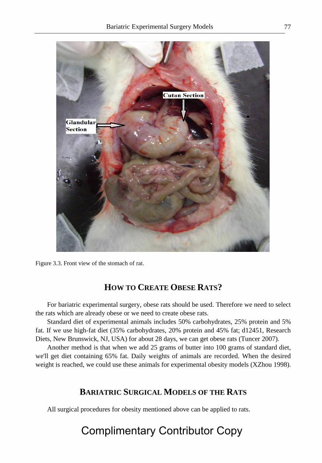
Bariatric Experimental Surgery Models 77
Figure 3.3. Front view of the stomach of rat.
HOW TO CREATE OBESE RATS?
For bariatric experimental surgery, obese rats should be used. Therefore we need to select
the rats which are already obese or we need to create obese rats.
Standard diet of experimental animals includes 50% carbohydrates, 25% protein and 5%
fat. If we use high-fat diet (35% carbohydrates, 20% protein and 45% fat; d12451, Research
Diets, New Brunswick, NJ, USA) for about 28 days, we can get obese rats (Tuncer 2007).
Another method is that when we add 25 grams of butter into 100 grams of standard diet,
we'll get diet containing 65% fat. Daily weights of animals are recorded. When the desired
weight is reached, we could use these animals for experimental obesity models (XZhou 1998).
BARIATRIC SURGICAL MODELS OF THE RATS
All surgical procedures for obesity mentioned above can be applied to rats.
Complimentary Contributor Copy
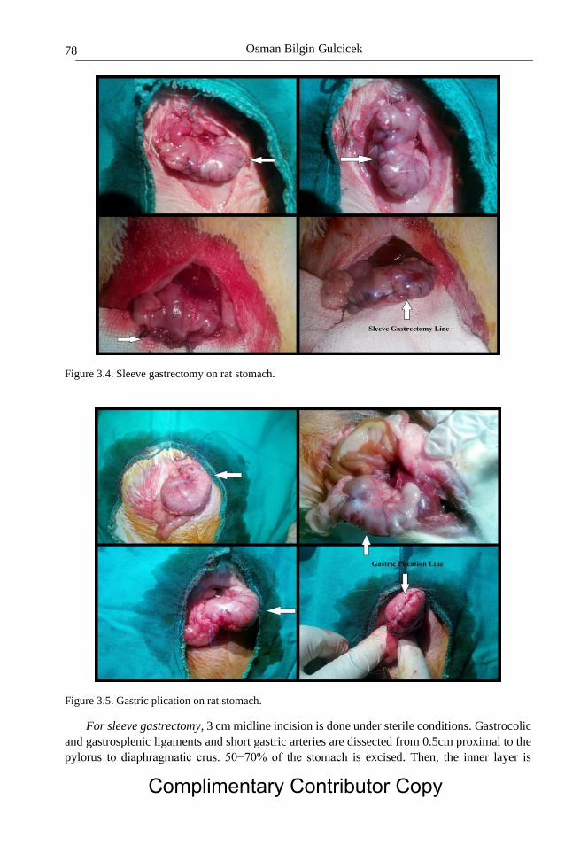
Osman Bilgin Gulcicek 78
Figure 3.4. Sleeve gastrectomy on rat stomach.
Figure 3.5. Gastric plication on rat stomach.
For sleeve gastrectomy, 3 cm midline incision is done under sterile conditions. Gastrocolic
and gastrosplenic ligaments and short gastric arteries are dissected from 0.5cm proximal to the
pylorus to diaphragmatic crus. 50−70% of the stomach is excised. Then, the inner layer is
Complimentary Contributor Copy
Bariatric Experimental Surgery Models 79
closed by continuous 5/0 polypropylene sutures and the outer layer is closed with continuous
extramucosal sutures by 5/0 polypropylene (Figure 3.4) (Gulcicek 2016).
For gastric plication, the same steps are done. Stomach is not excised. After the dissection
of ligaments, plication of the greater curvature is done with two layered extramucosal
continuous sutures by 5/0 polypropylene (Figure 3.5) (Gulcicek 2016).
After the operation, abdomen layers are closed duly and after 5 hours, all rats are fed ad
libitum.
CONCLUSION
All surgical procedures for obesity mentioned above for humans can be applied on rats.
However, gastric plication and sleeve gastrectomy are the most popular bariatric surgical
methods used in recent years.
Technically, it is easier to apply these methods on small experimental animals and
complication rates are much less than the other methods. The short operation time and optimal
use of anesthetic agents increase the success rate of the experiments.
REFERENCES
Barlow, Z., Cairl, B. (2009) Rat Anatomy.
Benaiges, D., Goday, A., Pedro-Botet, J., Ma´s, A., Chillaro´n, J. J., Flores-Le Roux, J. A.
Bariatric surgery: to whom and when? Minerva Endocrinol, 2015; 40(2): 119−28.
Benjamin, S. B., Maher, K. A., Cattau, E. L. Jr., Collen, M. J., Fleischer, D. E., Lewis, J. H.
(1988) Double-blind controlled trial of the Garren- Edwards gastric bubble: an adjunctive
treatment for exogenous obesity. Gastroenterology, 95: 581−588.
Blackburn, G. L., Kanders, B. S. Medical evaluation and treatment of the obese patients with
cardiovascular disease. Am. J. Cardiol., 1987; 60: 55−8.
Dagogo, J. S., Fanelli, C., Paramore, D., Brothers, J., Landt, M. Plasma leptin and insulin
relationships in obese and nonobese humans (abstract). Diabetes, 1996; 45: 695−8.
Davis, J. C. The relation between the pancreatic alpha cells and certain cells in the gastric
mucosa. J. Pathol. Bacteriol., 1954; 67: 237−40.
Ewing, B. T., Thompson, M. A., Wachtel, M. S., Frezza, E. E. A cost-benefit analysis of
bariatric surgery on the South Plains region of Texas. Obes. Surg., 2011; 21: 644−9.
Gastrointestinal surgery for severe obesity: National Institutes of Health Consensus
Development Conference Statement. Am. J. Clin. Nutr., 1992; 55 (2 Suppl.):615S–619S.
Gulcicek, O. B., Ozdogan, K., Solmaz, A., Yigitbas, H., Altınay, S., Gunes, A., Celik, D. S.,
Yavuz, E., Celik, A., Celebi, F. Metabolic and histopathological effects of sleeve
gastrectomy and gastric plication: an experimental rodent model. Food and Nutrition
Research, 2016, 60: 30888.
Kojima, M., Hosoda, H., Date, Y. Ghreline: discovery of natural endogenous ligand for the
growth hormone secretgogue receptor. Trends Endocrinol. Metab., 2001; 12: 118−22.
Kuzmak, L. I., Rickert, R. R. Pathologic Changes in the Stomach at the Site of Silicone Gastric
Banding. Obes. Surg., 1991;1: 63−68. Complimentary Contributor Copy

Osman Bilgin Gulcicek 80
Lo´pez-NavaBrevie`re, G., Bautista-Castan˜o, I., Jimenez, A., de Grado, T., Fernandez-
Corbelle, J. P. The Primary Obesity Surgery Endolumenal (POSE) procedure: one-year
patient weight loss and safety outcomes. Surg. Obes. Relat. Dis., 2015; 11(4): 861−5.
Marceau, P., Hould, F. S., Simard, S., Lebel, S., Bourque, R. A., Potvin, M., Biron, S.
Biliopancreatic diversion with duodenal switch. World J. Surg., 1998; 22: 947−54.
Mahawar, K. Bariatric Surgery: The Past, the Present, and the Future. Webmed Central,
Bariatric and Metabolic Surgery, 2012; 3(7): WMC003610.
Mason, E. E., Ito, C. Gastric bypass in obesity. Surg. Clin. North. Am., 1967; 47: 1345−1351.
National Institutes of Health (2010) NIH consensus statement Gastrointestinal surgery for
severe obesity.
Rutledge, R. The mini-gastric bypass: experience with the first 1,274 cases. Obes. Surg.,
2001;11: 276−280.
Shen, D., Ye, H., Wang, Y., Ji, Y., Zhan, X., Zhu, J., Li, W. Comparison of short-term outcomes
between laparoscopic greater curvature plication and laparoscopic sleeve gastrectomy.
Surg. Endosc., 2013; 27(8): 2768−74.
Saber, A. A., Elgamal, M. H., McLeod, M. K. Bariatric Surgery: The Past, Present, and Future.
Obes. Surg., 2008; 18(1):121−128.
Tadokoro, S., Ide, S., Tokuyama, R., Umeki, H., Tatehara, S., Kataoka, S., Satomura, K. Leptin
promotes wound healing in the skin. PLoS One, 2015; 10(3): 1−16.
Talebpourand, M., Amoli, B. S. Laparoscopic total gastric vertical plication in morbid obesity.
J. Laparoendosc. Adv. Surg. Tech. A, 2007; 17(6): 793−8.
Tuncer, B. B., Barıs, E. et al. Evaluation Of Tooth And Surrounding Tissues In Experimental
Induced Obese Rats With High Fat İntake A Pilot Study. GU Diş Hek Fak Derg., 2007;
24(3):167−171.
Weiner, R. A., Weiner, S., Pomhoff, I., Jacobi, C., Makarewicz, W., Weigand, G. Laparoscopic
sleeve gastrectomy influence of sleevesize and resected gastric volume. Obes. Surg., 2007;
17: 1297−1305.
Zhou, X., De Schepper, J., De Craemer, D., Delhase, M., Gys, G., Smitz, J., and Hooghe-Peters,
E. L. (1998) Pituitary growth hormone release and gene expression in cafeteria-diet-
induced obese rats. Journal of Endocrinology, 1998; 159: 165–72.
Complimentary Contributor Copy
In: Advances in Experimental Surgery. Volume 2 ISBN: 978-1-53612-773-7
Editors: Huifang Chen and Paulo N. Martins © 2018 Nova Science Publishers, Inc.
Chapter 4
ANIMAL MODELS OF HEART FAILURE
Mouer Wang*, MD Cardiovascular Institute, Stanford University, Stanford, California, US
ABSTRACT
Cardiovascular diseases (CVD) often lead to heart failure (HF). HF prevalenceis
continuously rising and represents one of the leading causes ofdeath and an economic
burden in the western societies. The study of potential novel therapeutic options and
interventions requires reliable animal models to evaluate myocardial progressive structural
and functional changes. Indeed, during the past 40 years, basic and translational scientists
have used small animal models to understand the pathophysiology of HF and improve
prevention and treatment of patients suffering from congestive HF (CHF). Each species
and animal model has advantages and disadvantages and the choice of one model over
another should take them into account for a good experimental design.
The aim of this chapter is to describe and highlight the features of some commonly
used small animal (rats and mouse) surgical models of cardiovascular diseases leading to
HF.
Keywords: heart failure, rat, mouse, left anterior descending (LAD) artery ligation, aortic
banding, aorto-caval fistula (ACF)
ABBREVIATIONS
ACF: aorto-caval fistula
CVD: cardiovascular diseases
CHF: chronic heart failure
HF: heart failure
LAD: left anterior descending
LV: left ventricle
*Correspondingauthor: Mouer Wang, M.D. E-mail: [email protected]. Complimentary Contributor Copy
Mouer Wang 82
INTRODUCTION
Heart failure is a clinical syndrome characterized by cardiac dysfunction and myocardial
structural abnormality (e.g., hypertrophy and dilated cardiomyopathy) resulting in inability of
the heart to eject sufficient blood to the metabolizing tissues. Heart failure is a leading cause of
morbidity and mortality, posing a major health and economic burden in the western world.
Nearly 5 million Americans and 6.5 million Europeans suffer from heart failure, which
accounts for 20% of all hospitalizations among patients over the age of 65, and its related
complications result in one million admissions with an annual mortality rate of 8–10%.
Moreover, heart failure has enormous economic impact because of the high costs of the
treatment, frequent hospitalizations, and poor quality of life. Therefore, understanding the basic
mechanisms leading to the development of heart failure and its complications, as well as the
discovery of innovative adequate treatment for heart failure are crucial in order to improve the
outcome of this devastating disease.
Humans present with heart failure of varying severity and of uncertain duration, produced
by a wide variety of causes. This variability, and confounding effects of treatment, complicates
the study of the pathophysiology and progression of the heart failure.
The ideal model should be able to reproduce each of the aspects of the progression of
clinical heart failure. However, none of the models available is able to entirely reproduce heart
failure. While some models reproduce neuroendocrine changes, others better reproduce the
remodeling that occurs during chronic heart failure. Nevertheless, the progress that has been
made so far and the expected future achievements in this field would not have been possible
without the continuous development of experimental models of heart failure and hypertrophy
in small and large animals, where each one has its unique advantages and limitations. There are
three main surgical models of heart failure in rodents: coronary artery ligation to induce
myocardial infarction; transverse aortic constriction to create a model of pressure overload; and
aortocaval fistula to increate a model of volume overload.
RAT LEFT ANTERIOR DESCENDING (LAD) ARTERY LIGATION
The surgical method, first developed by Pfeffer and coworkers, consists of ligating the left
coronary artery (Pfeffer 1979). In this procedure, left thoracotomy is performed on the
anesthetized rat, and the heart is rapidly exteriorized by gentle pressure on the right side of the
thorax. The left coronary artery is either ligated or heat cauterized between the pulmonary artery
outflow tract and the left atrium. The heart is then returned to its normal position and the thorax
immediately closed. Several modifications have been introduced to improve performance and
to reduce animal mortality, and left coronary artery ligation is the most common method used
to induce acute myocardial damage in rat and other animal models. If the left coronary artery
is not completely ligated, heart failure may occur as a consequence of chronic myocardial
ischemia. Complete occlusion of the left coronary artery results in myocardial infarction of
variable sizes with occurrence of overt heart failure after 3–6 weeks in a subset of animals with
large infarcts. The impairment of left ventricular function is related to the loss of myocardium.
Failure is associated with left ventricular dilatation, reduced systolic function and increased
filling pressures. The progression of left ventricular dysfunction and myocardial failure is Complimentary Contributor Copy
Animal Models of Heart Failure 83
associated with neurohumoral activation similar to that seen in patients with chronic heart
failure (CHF). In particular, it was shown that ACE (angiotensin-converting-enzyme) activity
in the left ventricle correlated inversely with left ventricular function and that ACE activity in
the kidney was only increased late after the induction of heart failure. Depressed myocardial
function is associated with altered calcium transients. The density of L-type calcium channels,
as evaluated by antagonist binding was shown to be decreased in moderate to severe stages of
congestive heart failure. Furthermore, it was shown that after 4, 8 and 16 weeks following
coronary artery ligation, SR (sarcoplasmic reticulum)-Ca2+ -ATPase mRNA and protein levels
decrease continuously with increasing severity of congestive heart failure. Interestingly, SR-
Ca2+ -ATPase activity was found to be more depressed than expected from the reduction in
protein levels. Although a high initial mortality and induction of mild failure in most cases may
be a disadvantage of this model, it seems to be very useful for long-term studies of
pharmacological interventions on the neurohumoral activation.
One important modification is temporary occlusion followed by reperfusion, allowing flow
recovery through the previously occluded coronary artery bed. Left coronary artery ligation can
thus be used to evaluate diverse parameters resulting from either permanent ischemia or
ischemia/reperfusion.
Preparation of the Operation
The operating surface is first prepared by disinfecting the area with 70% ethanol.
All surgical instruments are to be sterilized with a hot bead sterilizer before surgery (and
in between individual rat surgeries). These instruments include: surgical scissors (2), forceps
(1), curved forceps (1), needle holder (2), and a chest retractor.
Adjust the temperature of a homeothermic blanket system (heating pad), to be used to
maintain the body temperature of the animal at 37 ± 1°C, in order to prevent a dramatic fall in
body temperature; the size of an infarct is dependent on the duration of occlusion as well as
body temperature. Prior to the induction of anesthesia, animals will be examined to identify
any pre-existing conditions that may complicate surgical outcome; physical examination will
include visual inspection and measurement of heart rate, respiratory rate, and body temperature
and body weight. Prior to anesthesia, each animal will receive a dosage of buprenorphine
(0.1−2.5 mg/kg SQ).
Operative Steps
1. Rats are then anesthetized with 80 mg/kg of pentobarbital; the correct level of
anesthesia is verified through the limb withdrawal response by applying pressure of
the rat nail bed (toe-pinch reflex) and periodically throughout the procedure.
2. An electric shaver is then used to shave the fur form the neck and chest areas.
3. The shaved areas are to be scrubbed and disinfected with beta dinesolution followed
by wiping the area with 70% alcohol; this betadine and alcohol scrub and disinfection
procedure is to be repeated three times (Figure 4.1). (
Complimentary Contributor Copy

Mouer Wang 84
(fing
Figure 4.1. Disinfection of surgical field.
4. The anesthetized animal is then placed in a supine position on a homeothermic blanket
system (heating pad) in order to maintain body temperature.
5. Intubation is then performed by insertion of the endotracheal tube (polyethylene size
90; PE 90) where the tubing is beveled on the edge for ease of entrance through the
larynx.
Figure 4.2. Open the thorax.
Complimentary Contributor Copy
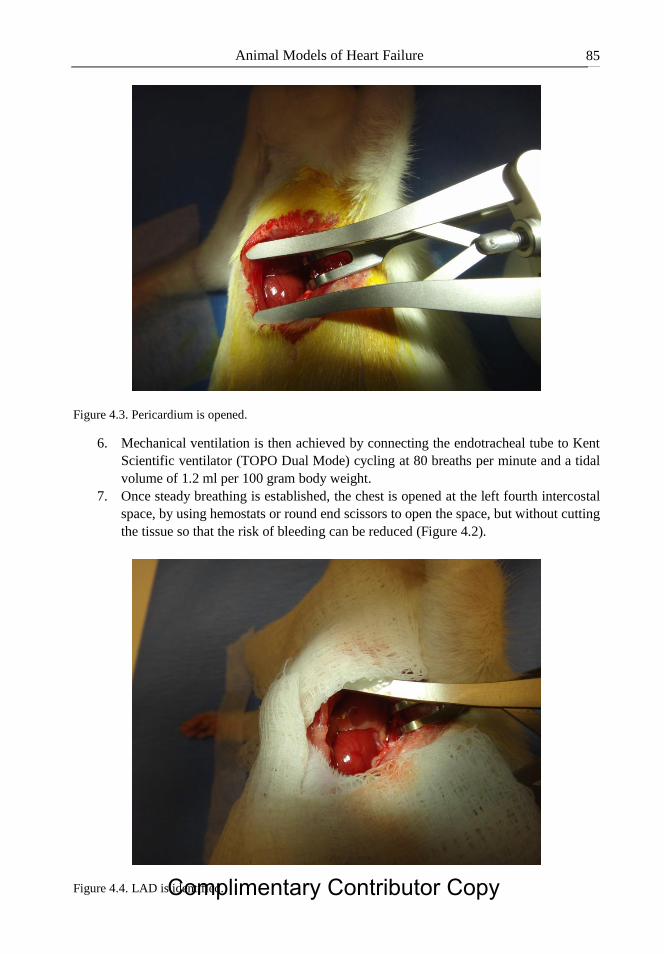
Animal Models of Heart Failure 85
Figure 4.3. Pericardium is opened.
6. Mechanical ventilation is then achieved by connecting the endotracheal tube to Kent
Scientific ventilator (TOPO Dual Mode) cycling at 80 breaths per minute and a tidal
volume of 1.2 ml per 100 gram body weight.
7. Once steady breathing is established, the chest is opened at the left fourth intercostal
space, by using hemostats or round end scissors to open the space, but without cutting
the tissue so that the risk of bleeding can be reduced (Figure 4.2).
Figure 4.4. LAD is identified. Complimentary Contributor Copy

Mouer Wang 86
Figure 4.5. LAD is ligated using a 6-0 proline suture.
8. A chest retractor is then positioned within the fourth intercostal space in order to spread
the ribs so that the left ventricle (LV) is exposed, taking maximal care not to damage
the lung.
9. The pericardium is then opened, but left in place if possible (Figure 4.3).
Figure 4.6. Thorax cavity is closed.
Complimentary Contributor Copy
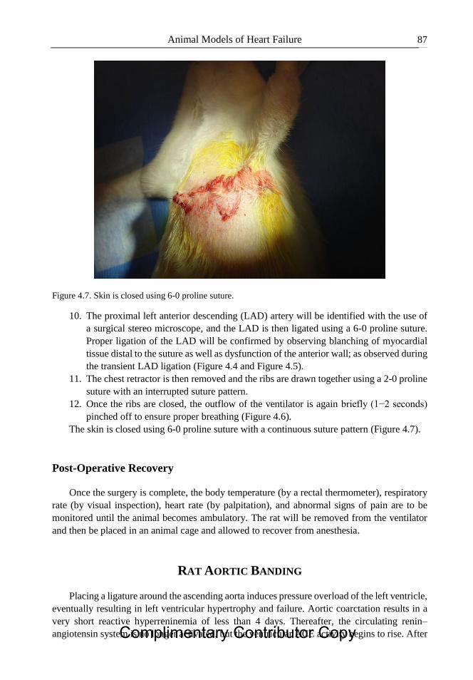
Animal Models of Heart Failure 87
Figure 4.7. Skin is closed using 6-0 proline suture.
10. The proximal left anterior descending (LAD) artery will be identified with the use of
a surgical stereo microscope, and the LAD is then ligated using a 6-0 proline suture.
Proper ligation of the LAD will be confirmed by observing blanching of myocardial
tissue distal to the suture as well as dysfunction of the anterior wall; as observed during
the transient LAD ligation (Figure 4.4 and Figure 4.5).
11. The chest retractor is then removed and the ribs are drawn together using a 2-0 proline
suture with an interrupted suture pattern.
12. Once the ribs are closed, the outflow of the ventilator is again briefly (1−2 seconds)
pinched off to ensure proper breathing (Figure 4.6).
The skin is closed using 6-0 proline suture with a continuous suture pattern (Figure 4.7).
Post-Operative Recovery
Once the surgery is complete, the body temperature (by a rectal thermometer), respiratory
rate (by visual inspection), heart rate (by palpitation), and abnormal signs of pain are to be
monitored until the animal becomes ambulatory. The rat will be removed from the ventilator
and then be placed in an animal cage and allowed to recover from anesthesia.
RAT AORTIC BANDING
Placing a ligature around the ascending aorta induces pressure overload of the left ventricle,
eventually resulting in left ventricular hypertrophy and failure. Aortic coarctation results in a
very short reactive hyperreninemia of less than 4 days. Thereafter, the circulating renin–
angiotensin system is no longer activated, but the ventricular ACE activity begins to rise. After Complimentary Contributor Copy

Mouer Wang 88
a period of several weeks, ventricular ACE activity may decrease again to normal values which
may be related to normalization of wall stress with increasing hypertrophy. Numerous studies
have been performed using aortic banding in rats to evaluate different aspects of left ventricular
hypertrophy. Furthermore, after several months, a subset of animals goes into failure. In a
recent study, chronic experimental aortic constriction imposed by banding of the ascending
aorta in weanlings resulted in compensated left ventricular hypertrophy of the adult rats for
several weeks. After 20 weeks of aortic banding, two distinct groups could be identified: rats
without change in LV systolic pressure development and those with a significant reduction in
left ventricular systolic pressure. The latter group exhibited increased left ventricular volumes,
reduced ejection fraction and clinical signs of overt heart failure. Left ventricular hypertrophy
and failure was associated with increased beta-myosin heavy chain mRNA and atrial natriuretic
factor mRNA. Interestingly, a decrease in SR-Ca-ATPase mRNA levels by the polymerase
chain reaction occurred in left ventricular myocardium from failing animals after 20 weeks of
banding but not in nonfailing hypertrophied hearts. From this data, it was suggested that the
decrease in SR-Ca-ATPase mRNA levels may be a marker of the transition from compensatory
hypertrophy to failure in these animals. During compensated hypertrophy, while catecholamine
levels are normal, there is activation of the local myocardial renin-angiotensin system, which
may be important for the development of myocardial failure. With the development of heart
failure, plasma catecholamine levels can increase.
This model seems to be well suited for studying the transition from hypertrophy to failure
at the level of the myocardium. Nevertheless, one should keep in mind that considerable
differences in the function of subcellular systems exist between rat and human myocardium.
Preparation of the Operation
Keep rat (approximately 200 g body weight) under antibiotic umbrella 24 hrbefore surgery
by administering amoxicillin orally (dose: 500 mg/L of water). Sterilize all surgical tools (such
as scissors, forceps, clip applicator, suture needles, and needle holder) by autoclaving at 121°C
for 15 min. Use a heating pad to maintain the animal body temperature around 37°C to avoid a
rapid decrease in heart rate. Keep the rat on top of a temperature controlled heating pad in the
supine position.
Remove fur from the neckline and left chest region of the rat (preferably using an electric
shaver).Disinfect surgical site with povidone-iodine solution and then with 70% ethyl alcohol.
Use a sterile drape to expose only the surgical site during the operative procedure.
Note: As an additional option for the animal care, 0.25% Bupivicaine ID (2.5 mg/kg) can be
incorporated prior to incising at each incision site for local anesthesia/analgesia.
Insert a 16 G I.V. Catheter into the rat's trachea through mouth. Connect the tracheal
cannula to a rodent ventilator for maintaining a respiratory rate of 50 breaths/min, tidal volume
of 1.70 ml and inspiration time of 0.60 sec (for rat with 200 g body weight).
Operative Steps
1. After skin incision, the upper half of the sternum was divided in the midline using
scissors. Complimentary Contributor Copy

Animal Models of Heart Failure 89
2. The thymus was removed and aortic arch was carefully dissected free of the
surrounding tissues (Figure 4.8 and Figure 4.9).
Figure 4.8 Upper half of the sternum was divided.
Figure 4.9. Thymus was removed and aortic arch exposed.
Complimentary Contributor Copy
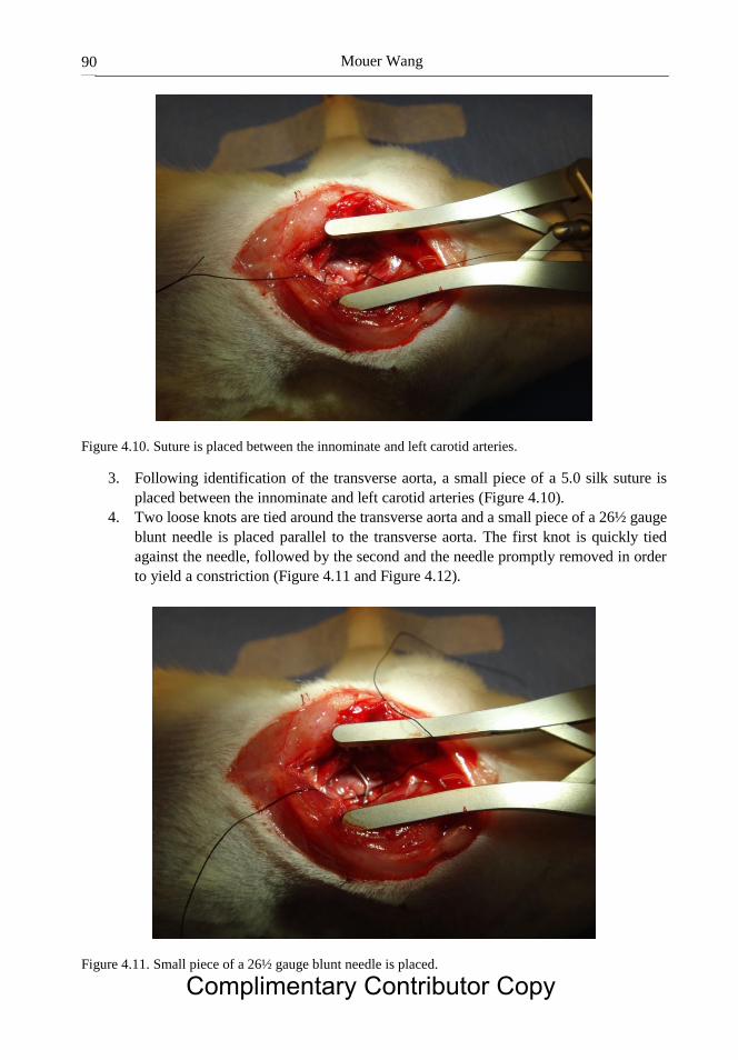
Mouer Wang 90
Figure 4.10. Suture is placed between the innominate and left carotid arteries.
3. Following identification of the transverse aorta, a small piece of a 5.0 silk suture is
placed between the innominate and left carotid arteries (Figure 4.10).
4. Two loose knots are tied around the transverse aorta and a small piece of a 26½ gauge
blunt needle is placed parallel to the transverse aorta. The first knot is quickly tied
against the needle, followed by the second and the needle promptly removed in order
to yield a constriction (Figure 4.11 and Figure 4.12).
Figure 4.11. Small piece of a 26½ gauge blunt needle is placed.
Complimentary Contributor Copy
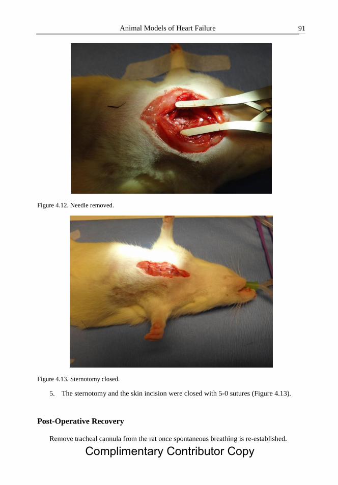
Animal Models of Heart Failure 91
Figure 4.12. Needle removed.
Figure 4.13. Sternotomy closed.
5. The sternotomy and the skin incision were closed with 5-0 sutures (Figure 4.13).
Post-Operative Recovery
Remove tracheal cannula from the rat once spontaneous breathing is re-established.
Complimentary Contributor Copy
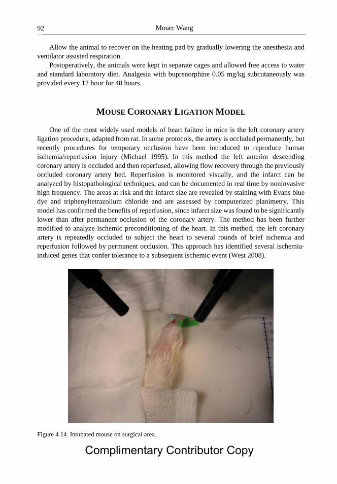
Mouer Wang 92
Allow the animal to recover on the heating pad by gradually lowering the anesthesia and
ventilator assisted respiration.
Postoperatively, the animals were kept in separate cages and allowed free access to water
and standard laboratory diet. Analgesia with buprenorphine 0.05 mg/kg subcutaneously was
provided every 12 hour for 48 hours.
MOUSE CORONARY LIGATION MODEL
One of the most widely used models of heart failure in mice is the left coronary artery
ligation procedure, adapted from rat. In some protocols, the artery is occluded permanently, but
recently procedures for temporary occlusion have been introduced to reproduce human
ischemia/reperfusion injury (Michael 1995). In this method the left anterior descending
coronary artery is occluded and then reperfused, allowing flow recovery through the previously
occluded coronary artery bed. Reperfusion is monitored visually, and the infarct can be
analyzed by histopathological techniques, and can be documented in real time by noninvasive
high frequency. The areas at risk and the infarct size are revealed by staining with Evans blue
dye and triphenyltetrazolium chloride and are assessed by computerized planimetry. This
model has confirmed the benefits of reperfusion, since infarct size was found to be significantly
lower than after permanent occlusion of the coronary artery. The method has been further
modified to analyze ischemic preconditioning of the heart. In this method, the left coronary
artery is repeatedly occluded to subject the heart to several rounds of brief ischemia and
reperfusion followed by permanent occlusion. This approach has identified several ischemia-
induced genes that confer tolerance to a subsequent ischemic event (West 2008).
Figure 4.14. Intubated mouse on surgical area.
Complimentary Contributor Copy
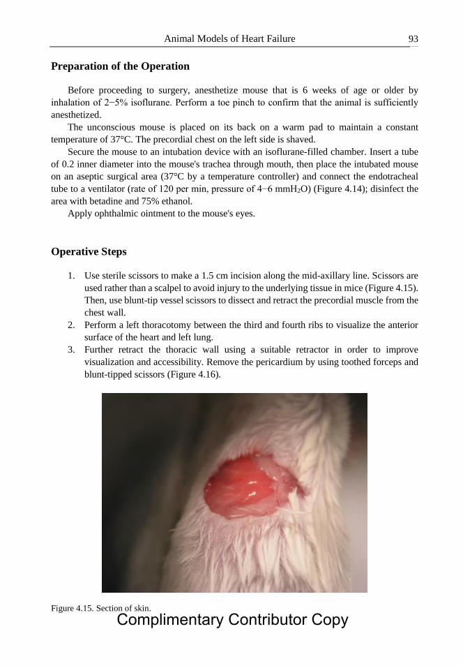
Animal Models of Heart Failure 93
Preparation of the Operation
Before proceeding to surgery, anesthetize mouse that is 6 weeks of age or older by
inhalation of 2−5% isoflurane. Perform a toe pinch to confirm that the animal is sufficiently
anesthetized.
The unconscious mouse is placed on its back on a warm pad to maintain a constant
temperature of 37°C. The precordial chest on the left side is shaved.
Secure the mouse to an intubation device with an isoflurane-filled chamber. Insert a tube
of 0.2 inner diameter into the mouse's trachea through mouth, then place the intubated mouse
on an aseptic surgical area (37°C by a temperature controller) and connect the endotracheal
tube to a ventilator (rate of 120 per min, pressure of 4−6 mmH2O) (Figure 4.14); disinfect the
area with betadine and 75% ethanol.
Apply ophthalmic ointment to the mouse's eyes.
Operative Steps
1. Use sterile scissors to make a 1.5 cm incision along the mid-axillary line. Scissors are
used rather than a scalpel to avoid injury to the underlying tissue in mice (Figure 4.15).
Then, use blunt-tip vessel scissors to dissect and retract the precordial muscle from the
chest wall.
2. Perform a left thoracotomy between the third and fourth ribs to visualize the anterior
surface of the heart and left lung.
3. Further retract the thoracic wall using a suitable retractor in order to improve
visualization and accessibility. Remove the pericardium by using toothed forceps and
blunt-tipped scissors (Figure 4.16).
Figure 4.15. Section of skin.
Complimentary Contributor Copy
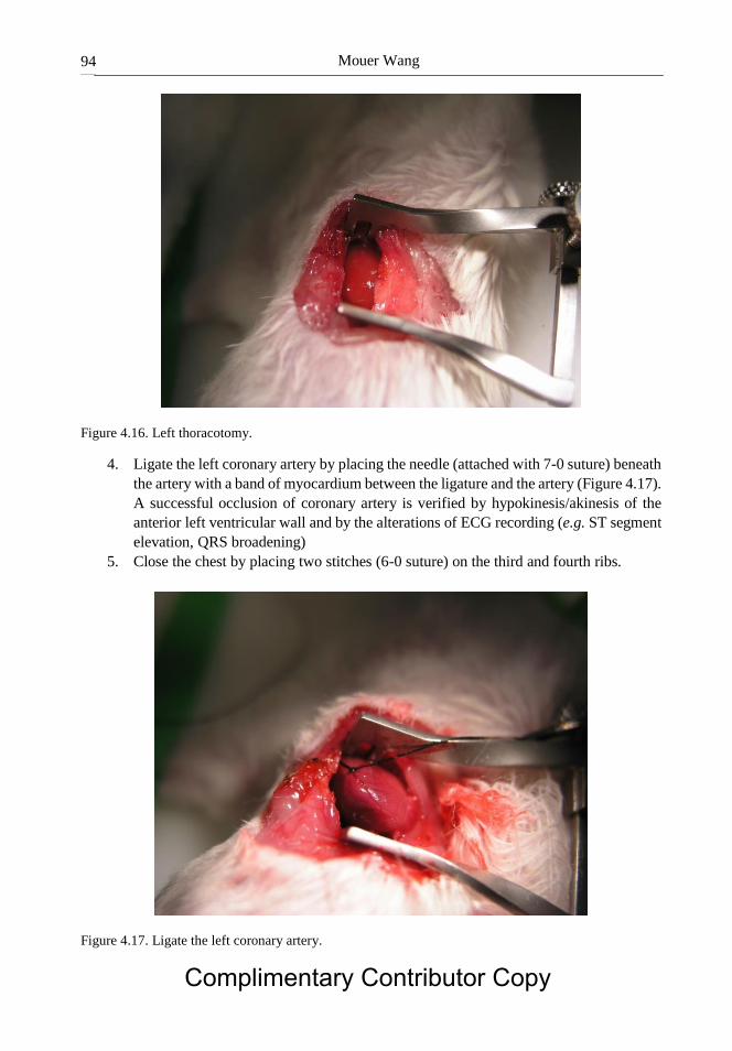
Mouer Wang 94
Figure 4.16. Left thoracotomy.
4. Ligate the left coronary artery by placing the needle (attached with 7-0 suture) beneath
the artery with a band of myocardium between the ligature and the artery (Figure 4.17).
A successful occlusion of coronary artery is verified by hypokinesis/akinesis of the
anterior left ventricular wall and by the alterations of ECG recording (e.g. ST segment
elevation, QRS broadening)
5. Close the chest by placing two stitches (6-0 suture) on the third and fourth ribs.
Figure 4.17. Ligate the left coronary artery.
Complimentary Contributor Copy
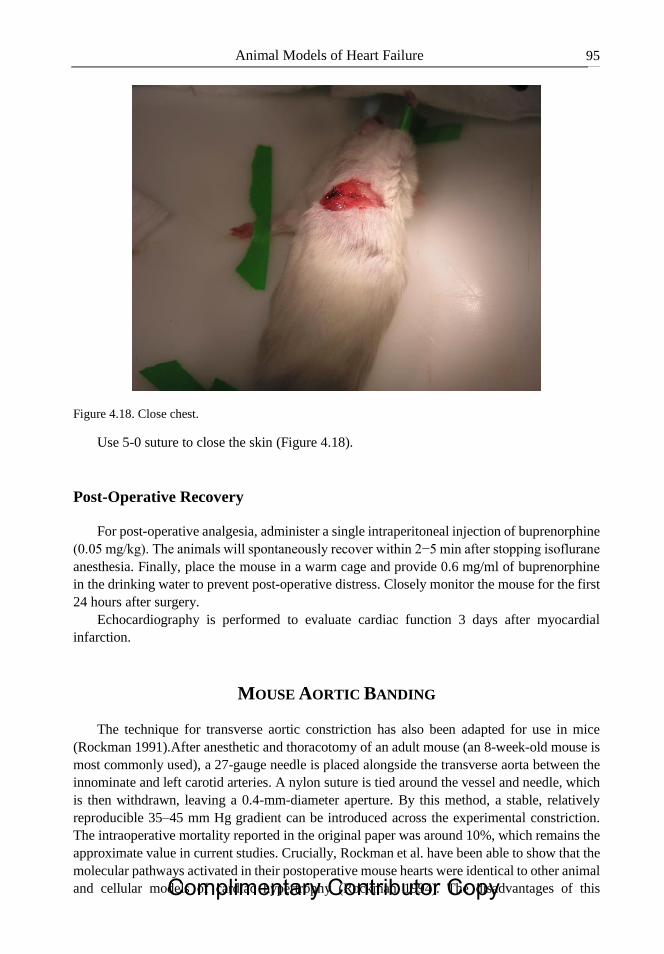
Animal Models of Heart Failure 95
Figure 4.18. Close chest.
Use 5-0 suture to close the skin (Figure 4.18).
Post-Operative Recovery
For post-operative analgesia, administer a single intraperitoneal injection of buprenorphine
(0.05 mg/kg). The animals will spontaneously recover within 2−5 min after stopping isoflurane
anesthesia. Finally, place the mouse in a warm cage and provide 0.6 mg/ml of buprenorphine
in the drinking water to prevent post-operative distress. Closely monitor the mouse for the first
24 hours after surgery.
Echocardiography is performed to evaluate cardiac function 3 days after myocardial
infarction.
MOUSE AORTIC BANDING
The technique for transverse aortic constriction has also been adapted for use in mice
(Rockman 1991).After anesthetic and thoracotomy of an adult mouse (an 8-week-old mouse is
most commonly used), a 27-gauge needle is placed alongside the transverse aorta between the
innominate and left carotid arteries. A nylon suture is tied around the vessel and needle, which
is then withdrawn, leaving a 0.4-mm-diameter aperture. By this method, a stable, relatively
reproducible 35–45 mm Hg gradient can be introduced across the experimental constriction.
The intraoperative mortality reported in the original paper was around 10%, which remains the
approximate value in current studies. Crucially, Rockman et al. have been able to show that the
molecular pathways activated in their postoperative mouse hearts were identical to other animal
and cellular models of cardiac hypertrophy (Rockman 1994). The disadvantages of this Complimentary Contributor Copy
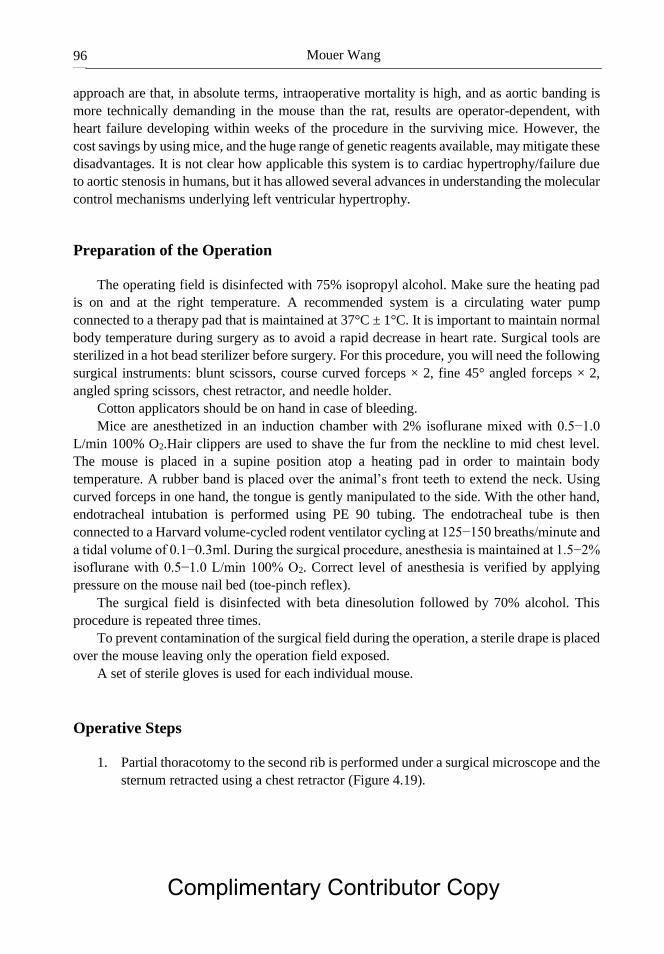
Mouer Wang 96
approach are that, in absolute terms, intraoperative mortality is high, and as aortic banding is
more technically demanding in the mouse than the rat, results are operator-dependent, with
heart failure developing within weeks of the procedure in the surviving mice. However, the
cost savings by using mice, and the huge range of genetic reagents available, may mitigate these
disadvantages. It is not clear how applicable this system is to cardiac hypertrophy/failure due
to aortic stenosis in humans, but it has allowed several advances in understanding the molecular
control mechanisms underlying left ventricular hypertrophy.
Preparation of the Operation
The operating field is disinfected with 75% isopropyl alcohol. Make sure the heating pad
is on and at the right temperature. A recommended system is a circulating water pump
connected to a therapy pad that is maintained at 37°C ± 1°C. It is important to maintain normal
body temperature during surgery as to avoid a rapid decrease in heart rate. Surgical tools are
sterilized in a hot bead sterilizer before surgery. For this procedure, you will need the following
surgical instruments: blunt scissors, course curved forceps × 2, fine 45° angled forceps × 2,
angled spring scissors, chest retractor, and needle holder.
Cotton applicators should be on hand in case of bleeding.
Mice are anesthetized in an induction chamber with 2% isoflurane mixed with 0.5−1.0
L/min 100% O2.Hair clippers are used to shave the fur from the neckline to mid chest level.
The mouse is placed in a supine position atop a heating pad in order to maintain body
temperature. A rubber band is placed over the animal’s front teeth to extend the neck. Using
curved forceps in one hand, the tongue is gently manipulated to the side. With the other hand,
endotracheal intubation is performed using PE 90 tubing. The endotracheal tube is then
connected to a Harvard volume-cycled rodent ventilator cycling at 125−150 breaths/minute and
a tidal volume of 0.1−0.3ml. During the surgical procedure, anesthesia is maintained at 1.5−2%
isoflurane with 0.5−1.0 L/min 100% O2. Correct level of anesthesia is verified by applying
pressure on the mouse nail bed (toe-pinch reflex).
The surgical field is disinfected with beta dinesolution followed by 70% alcohol. This
procedure is repeated three times.
To prevent contamination of the surgical field during the operation, a sterile drape is placed
over the mouse leaving only the operation field exposed.
A set of sterile gloves is used for each individual mouse.
Operative Steps
1. Partial thoracotomy to the second rib is performed under a surgical microscope and the
sternum retracted using a chest retractor (Figure 4.19).
Complimentary Contributor Copy
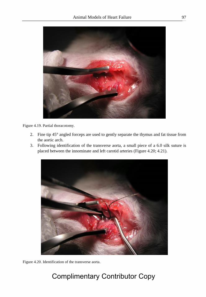
Animal Models of Heart Failure 97
Figure 4.19. Partial thoracotomy.
2. Fine tip 45° angled forceps are used to gently separate the thymus and fat tissue from
the aortic arch.
3. Following identification of the transverse aorta, a small piece of a 6.0 silk suture is
placed between the innominate and left carotid arteries (Figure 4.20; 4.21).
Figure 4.20. Identification of the transverse aorta.
Complimentary Contributor Copy
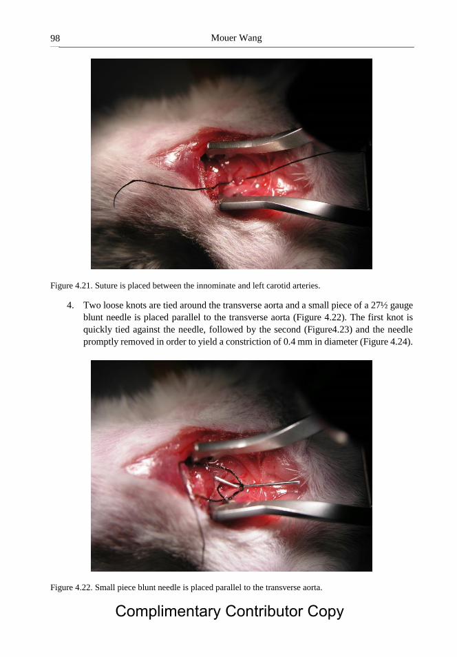
Mouer Wang 98
Figure 4.21. Suture is placed between the innominate and left carotid arteries.
4. Two loose knots are tied around the transverse aorta and a small piece of a 27½ gauge
blunt needle is placed parallel to the transverse aorta (Figure 4.22). The first knot is
quickly tied against the needle, followed by the second (Figure4.23) and the needle
promptly removed in order to yield a constriction of 0.4 mm in diameter (Figure 4.24).
Figure 4.22. Small piece blunt needle is placed parallel to the transverse aorta.
Complimentary Contributor Copy
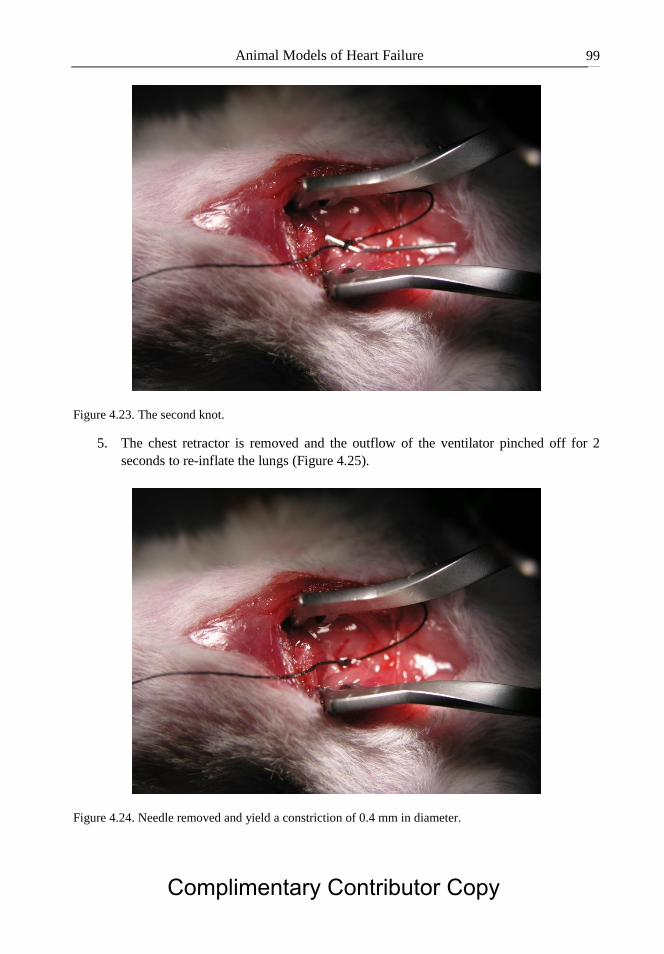
Animal Models of Heart Failure 99
Figure 4.23. The second knot.
5. The chest retractor is removed and the outflow of the ventilator pinched off for 2
seconds to re-inflate the lungs (Figure 4.25).
Figure 4.24. Needle removed and yield a constriction of 0.4 mm in diameter.
Complimentary Contributor Copy
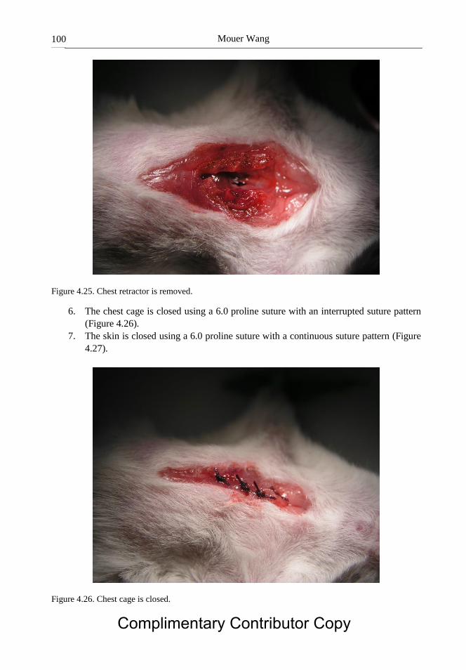
Mouer Wang 100
Figure 4.25. Chest retractor is removed.
6. The chest cage is closed using a 6.0 proline suture with an interrupted suture pattern
(Figure 4.26).
7. The skin is closed using a 6.0 proline suture with a continuous suture pattern (Figure
4.27).
Figure 4.26. Chest cage is closed.
Complimentary Contributor Copy
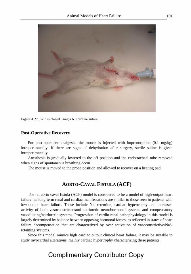
Animal Models of Heart Failure 101
Figure 4.27. Skin is closed using a 6.0 proline suture.
Post-Operative Recovery
For post-operative analgesia, the mouse is injected with buprenorphine (0.1 mg/kg)
intraperitoneally. If there are signs of dehydration after surgery, sterile saline is given
intraperitoneally.
Anesthesia is gradually lowered to the off position and the endotracheal tube removed
when signs of spontaneous breathing occur.
The mouse is moved to the prone position and allowed to recover on a heating pad.
AORTO-CAVAL FISTULA (ACF)
The rat aorto caval fistula (ACF) model is considered to be a model of high-output heart
failure, its long-term renal and cardiac manifestations are similar to those seen in patients with
low-output heart failure. These include Na+-retention, cardiac hypertrophy and increased
activity of both vasoconstrictor/anti-natriuretic neurohormonal systems and compensatory
vasodilating/natriuretic systems. Progression of cardio renal pathophysiology in this model is
largely determined by balance between opposing hormonal forces, as reflected in states of heart
failure decompensation that are characterized by over activation of vasoconstrictive/Na+-
retaining systems.
Since this model mimics high cardiac output clinical heart failure, it may be suitable to
study myocardial alterations, mainly cardiac hypertrophy characterizing these patients.
Complimentary Contributor Copy
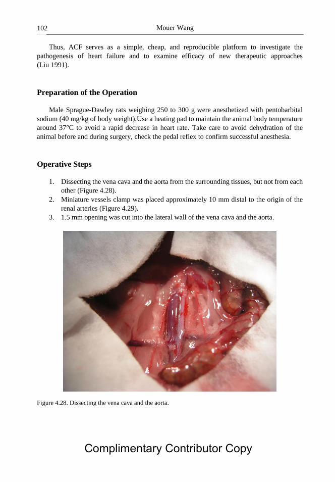
Mouer Wang 102
Thus, ACF serves as a simple, cheap, and reproducible platform to investigate the
pathogenesis of heart failure and to examine efficacy of new therapeutic approaches
(Liu 1991).
Preparation of the Operation
Male Sprague-Dawley rats weighing 250 to 300 g were anesthetized with pentobarbital
sodium (40 mg/kg of body weight).Use a heating pad to maintain the animal body temperature
around 37°C to avoid a rapid decrease in heart rate. Take care to avoid dehydration of the
animal before and during surgery, check the pedal reflex to confirm successful anesthesia.
Operative Steps
1. Dissecting the vena cava and the aorta from the surrounding tissues, but not from each
other (Figure 4.28).
2. Miniature vessels clamp was placed approximately 10 mm distal to the origin of the
renal arteries (Figure 4.29).
3. 1.5 mm opening was cut into the lateral wall of the vena cava and the aorta.
Figure 4.28. Dissecting the vena cava and the aorta.
Complimentary Contributor Copy
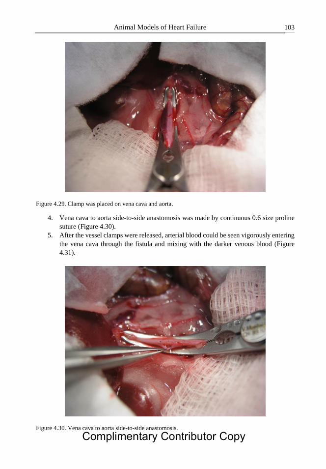
Animal Models of Heart Failure 103
Figure 4.29. Clamp was placed on vena cava and aorta.
4. Vena cava to aorta side-to-side anastomosis was made by continuous 0.6 size proline
suture (Figure 4.30).
5. After the vessel clamps were released, arterial blood could be seen vigorously entering
the vena cava through the fistula and mixing with the darker venous blood (Figure
4.31).
Figure 4.30. Vena cava to aorta side-to-side anastomosis.
Complimentary Contributor Copy

Mouer Wang 104
Figure 4.31. Vessel clamps are released.
6. Time of occlusion of both vessels did not exceed 10 minutes.
POST-OPERATIVE RECOVERY
For post-operative analgesia, administer a single intraperitoneal injection of buprenorphine
(0.05 mg/kg). Place the rat in a warm cage and closely monitor the rat for the first 24 hours
after surgery.
CONCLUSION
The use of animal models has proven to be an extremely valuable tool in understanding the
pathophysiology of complex cardiovascular disease mimicking congestive HF. The ideal
animal model of cardiovascular disease will mimic the human subject metabolically and
pathophysiologically, will be large enough to permit physiological and metabolic studies, and
will develop end-stage disease comparable to those in humans. Given the complex
multifactorial nature of CVD, no species or animal model will be similar to the human disease
and thus the models should be chosen according to the study aim. Animal models may also be
relevant to study the effects of new pharmacological interventions on hemodynamics,
neurohumoral activation and survival under preclinical conditions. At present, transgenic
models of congestive HF are essential for understanding the molecular alterations underlying
the development of the disease, as they allow the identification of genes that are causative for
HF and to characterize molecular mechanisms responsible for the development and progression
of the disease. Animal models which mimic distinct features of human HF will play an
Complimentary Contributor Copy

Animal Models of Heart Failure 105
important role in unravelling the consequences of gene transfer and molecular techniques to
correct disturbed subcellular processes in the failing heart. These experiments are indispensable
and these rodent models will continue to held an important role, not only in expanding our
knowledge about the mechanisms underlying HF, but also in developing novel therapeutic
strategies for this syndrome.
REFERENCES
Michael LH, Entman MI, Hartley CJ, Youker KA, Zhu J, Hall SR, Hawkins HK, Berens K,
Ballantyne CM. Myocardial ischemia and reperfusion: a murine model. American Journal
of Physiology 1995; 269(6):H2147–H2154.
Liu Z, Hilbelink DR, Crockett WB, Gerdes AM. Regional changes in hemodynamics and
cardiac myocyte size in rats with aortocaval fistulas. Developing and established
hypertrophy.Circ. Res. 1991; 69: 52–58.
Pfeffer MA, Pfeffer JM and Fishbein MC. Myocardial infarct size and ventricular function in
rats. Circulation Research 1979; 44 (4):503–512.
Rockman HA, Ross RS, Harris AN, Knowlton KU, Steinhelper ME, Field LJ, et al. Segregation
of atrial-specific and inducible expression of an atrial natriuretic factor transgene inan in
vivo murine model of cardiac hypertrophy. Proc. Natl. Acad. Sci. USA 1991; 88(18):8277–
81.
Rockman HA, Wachhorst SP, Mao L, Ross Jr J. ANG II receptor blockade prevents ventricular
hypertrophy and ANF gene expression with pressure overload in mice. Am. J. Physiol.
1994; 266(6 Pt 2):H2468–75.
West MB, Rokosh G, Obal D, et al. Cardiac myocyte specific expression of inducible nitric
oxide synthase protects against ischemia/reperfusion injury by preventing mitochondrial
permeability transition, Circulation 2008; 118(19):1970–1978.
Complimentary Contributor Copy

Complimentary Contributor Copy
In: Advances in Experimental Surgery. Volume 2 ISBN: 978-1-53612-773-7
Editors: Huifang Chen and Paulo N. Martins © 2018 Nova Science Publishers, Inc.
Chapter 5
HEPATOPANCREATOBILIARY SURGERY
EXPERIMENTAL MODELS
Paulo N. Martins MD, PhD *, Natalie Bath MD
and Adel Bozorgzadeh MD Department of Surgery, Division of Organ Transplantation,
UMass Memorial Medical Center, University Campus, Worcester, MA
ABSTRACT
Well-characterized animal models of human diseases are fundamental for the analysis
of pathophysiologic mechanisms and the development of new treatment strategies. A better
understanding of the underlying pathophysiology of hepatopancreatobiliary (HPB) disease
may lead to more targeted therapeutic options, potentially leading to improved survival.
We will focus this chapter on rodent HPB models. Owing to the low cost of experiments,
easily handling, and availability of a variety of genetically modified strains, rodents have
become a popular model organism for such experiments.
Despite some limitations, the use of rodent models in HPB surgery provides essential
investigative tools to study many important phenomena in HPB research. Due to the
availability of large varieties of genetically modified species and monoclonal antibodies,
more relevant and more specific questions can be answered utilizing rodent models. Most
of these procedures only require basic surgical skills, and they have been performed with
high reproducibility, which has been tolerated very well with minimal operative mortality.
However, some procedures require microsurgical skills to be performed accurately and
reproducibly. Increased knowledge of the rat and mouse liver surgical anatomy and more
advanced microsurgical skills permit individualized dissection, and individual ligature of
vascular and biliary branches.
Here we will review the HPB anatomy; techniques for hepatectomy, bile duct ligation,
and portal systemic shunts; models of cirrhosis; models of acute liver failure; models of
hepatocellular carcinoma (HCC) and cholangiocarcinoma (CCC); model of hepatic
metastases of colon carcinoma; pancreatitis; and common bile duct (CBD) stricture.
* Corresponding author: Paulo N. Martins MD., PhD., FAST, FACS., Assistant Professor of Surgery, Department of
Surgery, Division of Organ Transplantation, UMass Memorial Medical Center, University Campus, Worcester,
MA, Fax: 508-856-1102, Email: [email protected]. Complimentary Contributor Copy

Paulo N. Martins, Natalie Bath and Adel Bozorgzadeh 108
Keywords: hepatopancreatobiliary surgery, hepatectomy technique, cirrhosis, acute liver
failure
ABBREVIATIONS
CBD common bile duct
CBDL common bile duct ligation
CCC cholangiocarcinoma
CCL4 carbon tetrachloride
CDL closed duodenal loop
CL caudate lobe
CP caudate process
DEN diethylnitrosamine
DMNA dimethylnitrosamine
FL falciform ligament
GB gallbladder
HCC hepatocelular carcinoma
HCV hepatitis C virus
HPB hepatopacreaticobiliary
IRL inferior right lobe
LLL left lateral lobe
LML left middle lobe
ML middle lobe
PVL portal vein ligation
PB phenobarbital
PH partial hepatectomy
RLL right lower lobe
RML right middle lobe
RL right lobe
SRL superior right lobe
TAA thioacetamide
LIVER ANATOMY
Introduction
The rat is the most used experimental model in hepatopancreatobiliary (HPB) surgical
research because of many factors, in particular because it is easy to handle and inexpensive
(Rodriguez 1999; Martins 2003). In hepatic regeneration, liver metastasis, transplantation
immunology and hepatic disease studies, the rat is by far the most frequently used model. On
the other hand, the majority of studies that led to advances in techniques of liver resection and
transplantation have used the porcine and canine model. Virtually all procedures in liver
surgery can be performed in the rat including segmental liver resections, porto-systemic shunts, Complimentary Contributor Copy
Hepatopancreatobiliary Surgery Experimental Models 109
transplantation of full and partial organs, and vascular embolizations. As a result of the great
anatomical variability of liver anatomy such as its extrahepatic biliary tract and vascularization,
the knowledge of anatomy is advantageous because it permits individualized dissection,
parenchymal section, ligature of specific vascular and biliary branches, and avoids damage of
other structures.
The objective of this study was to systematically describe and illustrate the surgical
anatomy of the rat liver to facilitate the planning and performance of HPB surgery studies in
this animal.
Liver Anatomy
The rat and mouse livers are multi-lobulated as in other mammals (Lorente 1995; Flower
1872; Martins 2007). In rats, the liver mass represents approximately 5% of the total body
weight compared to 2.5% in adult humans.
Surfaces
With the rat placed in decubitus position, the rat liver has three surfaces: superior, inferior
and posterior. A sharp, well-defined margin divides the inferior from the superior surface.
Unique from the human liver, the other margins of the liver are also sharp. Although the rat
liver is lobulated, it has rather uniform surfaces since the lobes lie flat against each other as
seen in Figure 5.1A. The only exception to this is the posterior caudate lobe, which is separated
from the remainder liver by the stomach.
The superior (parietal) surface, convex as a whole, comprises a part of the left lateral and
medial lobes and fits under the vault of the diaphragm. It is completely covered by peritoneum
except along the line of attachment of the falciform ligament. The line of attachment of the
falciform ligament divides the liver into two parts, termed the right and left lobes. In
comparison to the human liver in which the right lobe is much larger than the left, the rat left
and right liver have approximately the same volume.
The inferior (visceral) surface is uneven, concave, and it is in close proximity to the
stomach, duodenum, right colic flexure, the superior part of the pancreas, the right kidney and
suprarenal gland. The rat liver inferior surface does not have the fossae in the shape of the letter
“H” as in humans. This surface is almost completely invested by peritoneum. Through the porta
(transverse fissure) travels the portal vein, the hepatic artery and nerves, the hepatic duct and
lymphatics. Liver impressions (colic, renal, duodenal, and suprarenal) are not as evident as in
human livers.
The posterior surface it is not covered by peritoneum over some part of its extent, and it is
in direct contact with the diaphragm. It extends obliquely between the caudate lobe and the bare
area of the liver. The inferior vena cava is completely intrahepatic.
Complimentary Contributor Copy

Paulo N. Martins, Natalie Bath and Adel Bozorgzadeh 110
Abbreviations: AC: anterior caudate lobe, CBD common bile duct, Panc pancreas, CT celiac trunk, PHA
proper hepatic artery, SA splenic artery, A aorta, LGA left gastric artery, Ly lymph node, S stomach,
D duodenum, RK right kidney, SHVC supra-hepatic vena cava, GB gallbladder, FL falciform
ligament, P portal vein, IVC inferior vena cava, RLL right lower lobe, ML middle lobe, RML right
middle lobe, LML left middle lobe, CL caudate lobe, LLL left lateral lobe, RL right lobe.
Figure 5.1. Rat liver “in-situ”. (A) Anterior view, (B) Anterior view after separating the lobes, (C) Porta
hepatis, (D) Portal vein and common bile duct descending in the pancreatic tissue along the duodenum
(the point of insertion of the CBD in the duodenum is marked with a X), (E) Celiac trunk and its
branches: proper hepatic, left gastric and splenic arteries. (F) Magnification of the portal triade: portal
vein, CBD, proper hepatic artery and a hilar lymph node.
Ligaments
Similar to the human liver, the rat liver is connected to the under surface of the diaphragm
and to the anterior wall of the abdomen by five ligaments: the falciform, the coronary, two
lateral which are peritoneal folds, and the round ligament − a fibrous cord which represents the
obliterated umbilical vein. The liver is also attached to the lesser curvature of the stomach by
the hepatogastric ligament and to the duodenum by the hepatoduodenal ligament.
Liver Lobes
The rat’s liver lobes, like the human liver, are named after the portal branches that supply
them due to the fact that the portal system is the most constant anatomical reference among
mammals (Flower 1872; Lorente 1995; Martins 2007) (See Figures 5.1−5.6).
The middle or median lobe is the largest lobe, accounting for approximately 38% of the
liver weight. It is fixed between the diaphragm and abdominal wall by the falciform ligament.
It is in continuity with the left lateral lobe and is subdivided by a vertical fissure (main fissure
or umbilical fissure) into a large right medial lobe (2/3 of the volume of the medial lobe) and a Complimentary Contributor Copy

Hepatopancreatobiliary Surgery Experimental Models 111
smaller left medial lobe (1/3 of the volume). The right medial lobe has both left and right hepatic
vascular components. (Lorente 1995; Kogure 1999; Martins 2007).
Abbreviations: GB: gallbladder, FL: falciform ligament.
Figure 5.2. A, B. Anterior view of the mouse liver showing the location of the gallbladder and falciform
ligament. B) Visceral view of the mouse liver.
Abbreviations: the same as in Figure 5.1 and 5.2.
Figure 5.3. Rat liver “ex situ”. (A) Anterior view, (B) Inferior (visceral) view, (C) Anterior after
separating the lobes, (D) posterior view, (E) Superior view with the lobes in normal position, (F)
Superior view with lobes flattened and separated from each other (note here a millimetric scale on the
bottom). When the liver is ex situ, the anterior and posterior caudate lobes are on the same plane (see in
E), and the anterior lobe lies superiorly. Note in D that the inferior vena cava is completely intrahepatic,
a plastic catheter is inside the inferior vena cava.
The right lobe, located to the right of the vena cava and posteriorly in the right
hypochondrium, is almost completely covered by the medial lobe. It comprises approximately Complimentary Contributor Copy
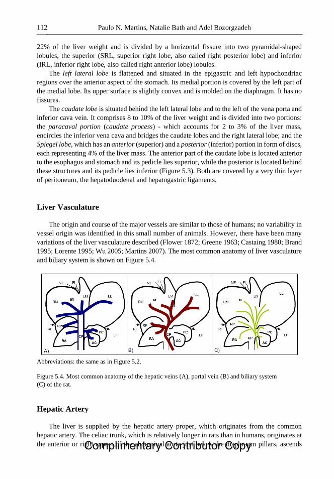
Paulo N. Martins, Natalie Bath and Adel Bozorgzadeh 112
22% of the liver weight and is divided by a horizontal fissure into two pyramidal-shaped
lobules, the superior (SRL, superior right lobe, also called right posterior lobe) and inferior
(IRL, inferior right lobe, also called right anterior lobe) lobules.
The left lateral lobe is flattened and situated in the epigastric and left hypochondriac
regions over the anterior aspect of the stomach. Its medial portion is covered by the left part of
the medial lobe. Its upper surface is slightly convex and is molded on the diaphragm. It has no
fissures.
The caudate lobe is situated behind the left lateral lobe and to the left of the vena porta and
inferior cava vein. It comprises 8 to 10% of the liver weight and is divided into two portions:
the paracaval portion (caudate process) - which accounts for 2 to 3% of the liver mass,
encircles the inferior vena cava and bridges the caudate lobes and the right lateral lobe; and the
Spiegel lobe, which has an anterior (superior) and a posterior (inferior) portion in form of discs,
each representing 4% of the liver mass. The anterior part of the caudate lobe is located anterior
to the esophagus and stomach and its pedicle lies superior, while the posterior is located behind
these structures and its pedicle lies inferior (Figure 5.3). Both are covered by a very thin layer
of peritoneum, the hepatoduodenal and hepatogastric ligaments.
Liver Vasculature
The origin and course of the major vessels are similar to those of humans; no variability in
vessel origin was identified in this small number of animals. However, there have been many
variations of the liver vasculature described (Flower 1872; Greene 1963; Castaing 1980; Brand
1995; Lorente 1995; Wu 2005; Martins 2007). The most common anatomy of liver vasculature
and biliary system is shown on Figure 5.4.
Abbreviations: the same as in Figure 5.2.
Figure 5.4. Most common anatomy of the hepatic veins (A), portal vein (B) and biliary system
(C) of the rat.
Hepatic Artery
The liver is supplied by the hepatic artery proper, which originates from the common
hepatic artery. The celiac trunk, which is relatively longer in rats than in humans, originates at
the anterior or right aspect of the abdominal aorta just below the diaphragm pillars, ascends Complimentary Contributor Copy
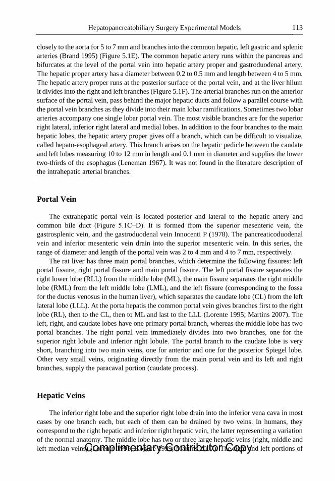
Hepatopancreatobiliary Surgery Experimental Models 113
closely to the aorta for 5 to 7 mm and branches into the common hepatic, left gastric and splenic
arteries (Brand 1995) (Figure 5.1E). The common hepatic artery runs within the pancreas and
bifurcates at the level of the portal vein into hepatic artery proper and gastroduodenal artery.
The hepatic proper artery has a diameter between 0.2 to 0.5 mm and length between 4 to 5 mm.
The hepatic artery proper runs at the posterior surface of the portal vein, and at the liver hilum
it divides into the right and left branches (Figure 5.1F). The arterial branches run on the anterior
surface of the portal vein, pass behind the major hepatic ducts and follow a parallel course with
the portal vein branches as they divide into their main lobar ramifications. Sometimes two lobar
arteries accompany one single lobar portal vein. The most visible branches are for the superior
right lateral, inferior right lateral and medial lobes. In addition to the four branches to the main
hepatic lobes, the hepatic artery proper gives off a branch, which can be difficult to visualize,
called hepato-esophageal artery. This branch arises on the hepatic pedicle between the caudate
and left lobes measuring 10 to 12 mm in length and 0.1 mm in diameter and supplies the lower
two-thirds of the esophagus (Leneman 1967). It was not found in the literature description of
the intrahepatic arterial branches.
Portal Vein
The extrahepatic portal vein is located posterior and lateral to the hepatic artery and
common bile duct (Figure 5.1C−D). It is formed from the superior mesenteric vein, the
gastrosplenic vein, and the gastroduodenal vein Innocenti P (1978). The pancreaticoduodenal
vein and inferior mesenteric vein drain into the superior mesenteric vein. In this series, the
range of diameter and length of the portal vein was 2 to 4 mm and 4 to 7 mm, respectively.
The rat liver has three main portal branches, which determine the following fissures: left
portal fissure, right portal fissure and main portal fissure. The left portal fissure separates the
right lower lobe (RLL) from the middle lobe (ML), the main fissure separates the right middle
lobe (RML) from the left middle lobe (LML), and the left fissure (corresponding to the fossa
for the ductus venosus in the human liver), which separates the caudate lobe (CL) from the left
lateral lobe (LLL). At the porta hepatis the common portal vein gives branches first to the right
lobe (RL), then to the CL, then to ML and last to the LLL (Lorente 1995; Martins 2007). The
left, right, and caudate lobes have one primary portal branch, whereas the middle lobe has two
portal branches. The right portal vein immediately divides into two branches, one for the
superior right lobule and inferior right lobule. The portal branch to the caudate lobe is very
short, branching into two main veins, one for anterior and one for the posterior Spiegel lobe.
Other very small veins, originating directly from the main portal vein and its left and right
branches, supply the paracaval portion (caudate process).
Hepatic Veins
The inferior right lobe and the superior right lobe drain into the inferior vena cava in most
cases by one branch each, but each of them can be drained by two veins. In humans, they
correspond to the right hepatic and inferior right hepatic vein, the latter representing a variation
of the normal anatomy. The middle lobe has two or three large hepatic veins (right, middle and
left median veins) (Lorente 1995; Kogure 1999; Martins 2007). The right and left portions of Complimentary Contributor Copy
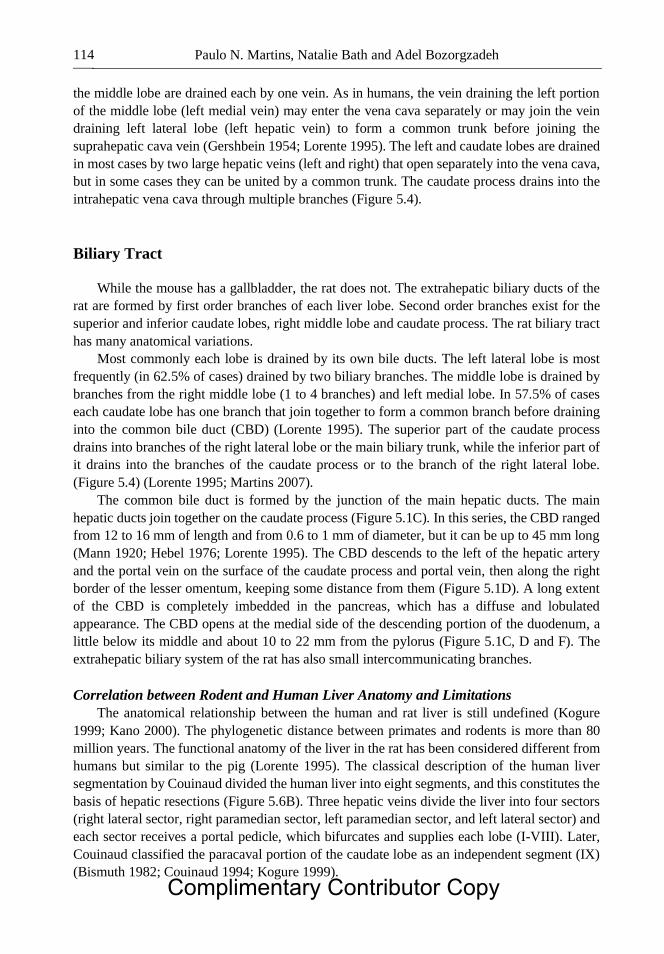
Paulo N. Martins, Natalie Bath and Adel Bozorgzadeh 114
the middle lobe are drained each by one vein. As in humans, the vein draining the left portion
of the middle lobe (left medial vein) may enter the vena cava separately or may join the vein
draining left lateral lobe (left hepatic vein) to form a common trunk before joining the
suprahepatic cava vein (Gershbein 1954; Lorente 1995). The left and caudate lobes are drained
in most cases by two large hepatic veins (left and right) that open separately into the vena cava,
but in some cases they can be united by a common trunk. The caudate process drains into the
intrahepatic vena cava through multiple branches (Figure 5.4).
Biliary Tract
While the mouse has a gallbladder, the rat does not. The extrahepatic biliary ducts of the
rat are formed by first order branches of each liver lobe. Second order branches exist for the
superior and inferior caudate lobes, right middle lobe and caudate process. The rat biliary tract
has many anatomical variations.
Most commonly each lobe is drained by its own bile ducts. The left lateral lobe is most
frequently (in 62.5% of cases) drained by two biliary branches. The middle lobe is drained by
branches from the right middle lobe (1 to 4 branches) and left medial lobe. In 57.5% of cases
each caudate lobe has one branch that join together to form a common branch before draining
into the common bile duct (CBD) (Lorente 1995). The superior part of the caudate process
drains into branches of the right lateral lobe or the main biliary trunk, while the inferior part of
it drains into the branches of the caudate process or to the branch of the right lateral lobe.
(Figure 5.4) (Lorente 1995; Martins 2007).
The common bile duct is formed by the junction of the main hepatic ducts. The main
hepatic ducts join together on the caudate process (Figure 5.1C). In this series, the CBD ranged
from 12 to 16 mm of length and from 0.6 to 1 mm of diameter, but it can be up to 45 mm long
(Mann 1920; Hebel 1976; Lorente 1995). The CBD descends to the left of the hepatic artery
and the portal vein on the surface of the caudate process and portal vein, then along the right
border of the lesser omentum, keeping some distance from them (Figure 5.1D). A long extent
of the CBD is completely imbedded in the pancreas, which has a diffuse and lobulated
appearance. The CBD opens at the medial side of the descending portion of the duodenum, a
little below its middle and about 10 to 22 mm from the pylorus (Figure 5.1C, D and F). The
extrahepatic biliary system of the rat has also small intercommunicating branches.
Correlation between Rodent and Human Liver Anatomy and Limitations
The anatomical relationship between the human and rat liver is still undefined (Kogure
1999; Kano 2000). The phylogenetic distance between primates and rodents is more than 80
million years. The functional anatomy of the liver in the rat has been considered different from
humans but similar to the pig (Lorente 1995). The classical description of the human liver
segmentation by Couinaud divided the human liver into eight segments, and this constitutes the
basis of hepatic resections (Figure 5.6B). Three hepatic veins divide the liver into four sectors
(right lateral sector, right paramedian sector, left paramedian sector, and left lateral sector) and
each sector receives a portal pedicle, which bifurcates and supplies each lobe (I-VIII). Later,
Couinaud classified the paracaval portion of the caudate lobe as an independent segment (IX)
(Bismuth 1982; Couinaud 1994; Kogure 1999).
Complimentary Contributor Copy
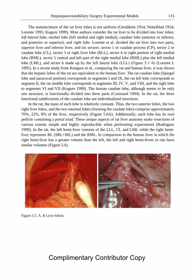
Hepatopancreatobiliary Surgery Experimental Models 115
The nomenclature of the rat liver lobes is not uniform (Gershbein 1954; Nettelblad 1954;
Lorente 1995; Kogure 1999). Most authors consider the rat liver to be divided into four lobes:
left lateral lobe, medial lobe (left medial and right medial), caudate lobe (anterior or inferior,
and posterior or superior), and right lobe. Lorente et al. divided the rat liver into two parts,
superior liver and inferior liver, and six sectors: sector 1 or caudate process (CP), sector 2 or
caudate lobe (CL), sector 3 or right liver lobe (RLL), sector 4 or right portion of right medial
lobe (RML), sector 5 central and left part of the right medial lobe (RML) plus the left medial
lobe (LML), and sector 6 made up by the left lateral lobe (LLL) (Figure 5.1−6) (Lorente L
1995). In a recent study from Kongure et al., comparing the rat and human liver, it was shown
that the hepatic lobes of the rat are equivalent to the human liver. The rat caudate lobe (Spiegel
lobe and paracaval portion) corresponds to segments I and IX, the rat left lobe corresponds to
segment II, the rat middle lobe corresponds to segments III, IV, V, and VIII, and the right lobe
to segments VI and VII (Kogure 1999). The human caudate lobe, although seems to be only
one structure, is functionally divided into three parts (Couinaud 1994). In the rat, the three
functional subdivisions of the caudate lobe are individualized structures.
In the rat, the mass of each lobe is relatively constant. Thus, the two anterior lobes, the two
right liver lobes, and the two omental lobes (forming the caudate lobe) comprise approximately
70%, 22%, 8% of the liver, respectively (Figure 5.6A). Additionally, each lobe has its own
pedicle containing a portal triad. These unique aspects of rat liver anatomy make resections of
various extents simple and highly reproducible when performing experiments (Rodriguez
1999). In the rat, the left hemi-liver consists of the LLL, CL and LML while the right hemi-
liver represents RL (SRL+IRL) and the RML. In comparison to the human liver in which the
right hemi-liver has a greater volume than the left, the left and right hemi-livers in rats have
similar volumes (Figure 5.6).
Figure 5.5. A, B Liver hilum.
Complimentary Contributor Copy
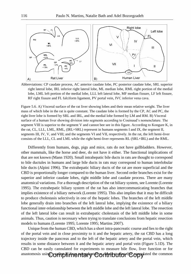
Paulo N. Martins, Natalie Bath and Adel Bozorgzadeh 116
Abbreviations: CP caudate process, AC anterior caudate lobe, PC posterior caudate lobe, SRL superior
right lateral lobe, IRL inferior right lateral lobe, ML median lobe, RML right portion of the medial
lobe, LML left portion of the medial lobe, LLL left lateral lobe, MF median fissure, LF left fissure,
RF right fissure and FL falciform ligament, PV portal vein, IVC inferior vena cava.
Figure 5.6. A) Visceral surface of the rat liver showing lobes and their mean relative weight. The liver
mass of which lobe in the rat is quite constant. The caudate lobe is formed by the CP, AC and PC, the
right liver lobe is formed by SRL and IRL, and the medial lobe formed by LM and RM. B) Visceral
surface of a human liver showing division into segments according to Couinaud’s nomenclature. The
segment VIII is superior to the segment V and cannot bee see in this figure. According to Kongure K, in
the rat, CL, LLL, LML, RML, (IRL+SRL) represent in humans segments I and IX, the segment II,
segments III, IV, V, and VIII; and the segments VI and VII, respectively. In the rat, the left hemi-liver
consists of the LLL, CL and LML while the right hemi-liver represents RL (SRL+IRL) and the RML.
Differently from humans, dogs, pigs and mice, rats do not have gallbladders. However,
other mammals, like the horse and deer, do not have it either. The functional implications of
that are not known (Mann 1920). Small intrahepatic bile ducts in rats are thought to correspond
to bile ductules in humans and large bile ducts in rats may correspond to human interlobular
bile ducts (Alpini 1996). The extrahepatic biliary ducts of the rat are more superficial and the
CBD is proportionally longer compared to the human liver. Second order branches exist for the
superior and inferior caudate lobes, right middle lobe and caudate process. There are many
anatomical variations. For a thorough description of the rat biliary system, see Lorente (Lorente
1995). The extrahepatic biliary system of the rat has also intercommunicating branches that
implies existence of a biliary network (Lorente 1995). This also implies that it may be difficult
to produce cholestasis selectively in one of the hepatic lobes. The branches of the left middle
lobe generally drain into branches of the left lateral lobe, implying the existence of a biliary
functional inter-relationship between the left middle lobe and the left lateral lobe. The resection
of the left lateral lobe can result in extrahepatic cholestasis of the left middle lobe in some
animals. Thus, caution is necessary when trying to translate conclusions from hepatic resection
models to humans (Lorente 1995; Lorente 1995; Martins 2007).
Unique from the human CBD, which has a short intra-pancreatic course and lies to the right
of the portal vein and in close proximity to it and the hepatic artery, the rat CBD has a long
trajectory inside the pancreas and to the left of the hepatic artery and the portal vein, which
results in some distance between it and the hepatic artery and portal vein (Figure 5.1D). The
CBD can be easily cannulated for experiments to measure bile flow, liver function or for
anastomosis using a splint in liver transplantation. Most studies that cannulated the common Complimentary Contributor Copy

Hepatopancreatobiliary Surgery Experimental Models 117
bile duct used a polyethylene tubing (PE-10, inner diameter of 0.28 mm, outer diameter of 0.61
mm). However, a 24 gauge catheter can also be inserted into the CBD.
The origin and course of the major extrahepatic vessels are similar to humans. However,
the branching pattern of the rat portal vein is different from humans. In the rat, the portal vein
trifurcates, while in humans it bifurcates (Kogure 1999). Due to the fact that the rat liver is
lobulated, there is no interdigitation between portal and venous pedicles; however, each one of
the six sectors receives a portal pedicle and has its own venous pedicle (Lorente 1995).
However, at microscopic level interdigitations may occur (Gershbein 1954). The inferior vena
cava, differently from humans, is completely intrahepatic. The hepato-esophageal artery
provides the liver with supra and infra-diaphragmatic systems of collateral circulation, once its
ascending branches anastomose with branches of the mediastinal arteries and its descending
branches communicate with the cardiac branches of the gastric artery. Thus, in experiments
designed to provide arterial supply deprivation to the liver, the hepato-esophageal artery as a
collateral route should be considered and previously ligated (Delong 1953).
Pancreas Anatomy
Rats are commonly used as experimental models for partial and total pancreatectomy. It is
important to note that the biliary system in rats has numerous anatomical variations. After
introducing latex dye into the biliopancreatic duct, portal vein and arteries, pancreas anatomy
present in rats was further delineated as seen in a study by ME Kara, et al. (Kara 2005). The
pancreas was subsequently divided into three parts following latex injection: biliary, duodenal,
and gastrosplenic. The biliopancreatic and pancreatic ducts, biliary, and duodenal portions of
the pancreas can be visualized with relative ease by retracting the duodenum caudally. The
gastrosplenic portion of the pancreas was seen between the descending duodenum and
transverse colon on the right side just caudal to the liver. In order to visualize the pancreas in
its entirety, it was necessary to remove the stomach and duodenum. This maneuver also
permitted visualization of the pancreaticoduodenal, right gastroepiploic and splenic vessels,
and portal vein.
In the study by Kara, et al., the biliopancreatic duct diameter and length of ducts was found
to be 1.01 0.03 and 28.86 0.59 mm, respectively. The anterior pancreatic duct originated
from the biliopancreatic duct on different sides and was found to drain the entire gastrosplenic
portion and part of the duodenal portion of the pancreas. The posterior pancreatic duct opened
into the biliopancreatic duct and was found to do this at the level of the papilla in more than 1/3
of specimens, which is an important consideration when ligating the biliopancreatic duct at the
level of the duodenal opening. The posterior pancreatic duct also carries the exocrine secretion
from the duodenal portion of the pancreas to the biliopancreatic duct. Numerous minor ducts
were also noted that drained all three portions of the pancreas directly into the biliopancreatic
duct (Kara 2005). A thorough understanding of the anatomy found in the rat pancreas is
imperative when using this model to study pancreatic transplantation and regeneration,
treatment of pancreatitis, and metabolic or endocrine studies (Figures 5.7−5.11).
Complimentary Contributor Copy

Paulo N. Martins, Natalie Bath and Adel Bozorgzadeh 118
Figure 5.7 Rat pancreas.
Figure 5.8. Rat pancreas.
Complimentary Contributor Copy
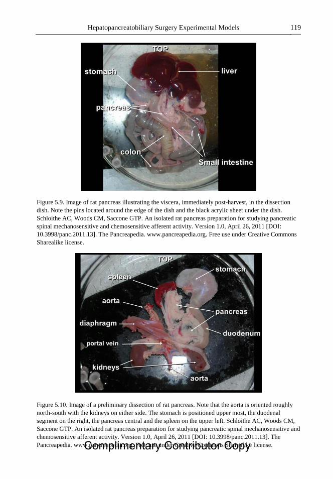
Hepatopancreatobiliary Surgery Experimental Models 119
Figure 5.9. Image of rat pancreas illustrating the viscera, immediately post-harvest, in the dissection
dish. Note the pins located around the edge of the dish and the black acrylic sheet under the dish.
Schloithe AC, Woods CM, Saccone GTP. An isolated rat pancreas preparation for studying pancreatic
spinal mechanosensitive and chemosensitive afferent activity. Version 1.0, April 26, 2011 [DOI:
10.3998/panc.2011.13]. The Pancreapedia. www.pancreapedia.org. Free use under Creative Commons
Sharealike license.
Figure 5.10. Image of a preliminary dissection of rat pancreas. Note that the aorta is oriented roughly
north-south with the kidneys on either side. The stomach is positioned upper most, the duodenal
segment on the right, the pancreas central and the spleen on the upper left. Schloithe AC, Woods CM,
Saccone GTP. An isolated rat pancreas preparation for studying pancreatic spinal mechanosensitive and
chemosensitive afferent activity. Version 1.0, April 26, 2011 [DOI: 10.3998/panc.2011.13]. The
Pancreapedia. www.pancreapedia.org. Free use under Creative Commons Sharealike license. Complimentary Contributor Copy

Paulo N. Martins, Natalie Bath and Adel Bozorgzadeh 120
Figure 5.11. Image of the final preparation of rat pancreas (with duodenal segment retained for
orientation) pinned in the Krebs-bath. Note that the duodenal segment is normally removed. The spleen
is usually retained as it provides a means of maintaining the pancreas in a stretched state without the
direct insertion of pins in the pancreas. The nerve trunks with teased bundles and fibers are visible in
the nerve-bath. A scale bar is included in the image (1 division = 1 mm). Schloithe AC, Woods CM,
Saccone GTP. An isolated rat pancreas preparation for studying pancreatic spinal mechanosensitive and
chemosensitive afferent activity. Version 1.0, April 26, 2011 [DOI: 10.3998/panc.2011.13]. The
Pancreapedia. www.pancreapedia.org. Free use under Creative Commons Sharealike license.
HEPATECTOMY MODELS
Introduction
Hepatic resections in rodents are performed to study liver regeneration, acute liver failure,
tumor dormancy of hepatic metastasis, hepatic function, and response to stress and trauma
(Higgins 1931; Bucher 1964; Weinbren 1965; Aleksandrowicz 1981; Rozga 1986; Lin 1990;
Panis 1990; de Jong 1995; de Jong 1998; Lu 1999; Fausto 2005). Each lobe has its own pedicle
containing a portal triad, and as a result hepatic resections of various extents in rodents are
simple and highly reproducible. Hepatectomies both in the rat and mouse require basic surgical
skills and have been performed with high success rates (Higgins 1931; Weinbren 1965; Pinto
1987; Lorente 1995; Rodriguez 1999; Fausto 2005; Madrahimov 2006; Martins 2007). In this
review, we outline various models, different techniques and limitations of hepatic resections.
Models of Liver Resection
The classic rat model of partial hepatectomy in rats is based on the seminal experiments by
Higgins and Anderson from 1931 (Higgins 1931). These investigations demonstrated that
resection of the two anterior lobes (median and left lateral lobes) is easy to perform and creates
a highly standardized liver reduction of approximately 70%. The classical 70% hepatectomy
model in rats is the most popular because it has been extensively studied and can be Complimentary Contributor Copy
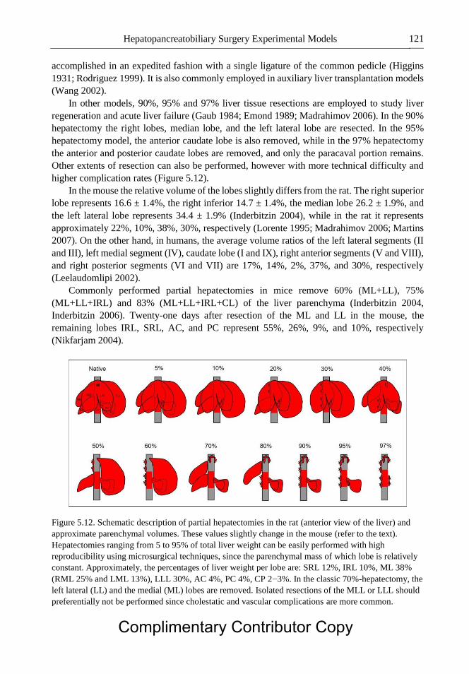
Hepatopancreatobiliary Surgery Experimental Models 121
accomplished in an expedited fashion with a single ligature of the common pedicle (Higgins
1931; Rodriguez 1999). It is also commonly employed in auxiliary liver transplantation models
(Wang 2002).
In other models, 90%, 95% and 97% liver tissue resections are employed to study liver
regeneration and acute liver failure (Gaub 1984; Emond 1989; Madrahimov 2006). In the 90%
hepatectomy the right lobes, median lobe, and the left lateral lobe are resected. In the 95%
hepatectomy model, the anterior caudate lobe is also removed, while in the 97% hepatectomy
the anterior and posterior caudate lobes are removed, and only the paracaval portion remains.
Other extents of resection can also be performed, however with more technical difficulty and
higher complication rates (Figure 5.12).
In the mouse the relative volume of the lobes slightly differs from the rat. The right superior
lobe represents 16.6 ± 1.4%, the right inferior 14.7 ± 1.4%, the median lobe 26.2 ± 1.9%, and
the left lateral lobe represents 34.4 ± 1.9% (Inderbitzin 2004), while in the rat it represents
approximately 22%, 10%, 38%, 30%, respectively (Lorente 1995; Madrahimov 2006; Martins
2007). On the other hand, in humans, the average volume ratios of the left lateral segments (II
and III), left medial segment (IV), caudate lobe (I and IX), right anterior segments (V and VIII),
and right posterior segments (VI and VII) are 17%, 14%, 2%, 37%, and 30%, respectively
(Leelaudomlipi 2002).
Commonly performed partial hepatectomies in mice remove 60% (ML+LL), 75%
(ML+LL+IRL) and 83% (ML+LL+IRL+CL) of the liver parenchyma (Inderbitzin 2004,
Inderbitzin 2006). Twenty-one days after resection of the ML and LL in the mouse, the
remaining lobes IRL, SRL, AC, and PC represent 55%, 26%, 9%, and 10%, respectively
(Nikfarjam 2004).
Figure 5.12. Schematic description of partial hepatectomies in the rat (anterior view of the liver) and
approximate parenchymal volumes. These values slightly change in the mouse (refer to the text).
Hepatectomies ranging from 5 to 95% of total liver weight can be easily performed with high
reproducibility using microsurgical techniques, since the parenchymal mass of which lobe is relatively
constant. Approximately, the percentages of liver weight per lobe are: SRL 12%, IRL 10%, ML 38%
(RML 25% and LML 13%), LLL 30%, AC 4%, PC 4%, CP 2−3%. In the classic 70%-hepatectomy, the
left lateral (LL) and the medial (ML) lobes are removed. Isolated resections of the MLL or LLL should
preferentially not be performed since cholestatic and vascular complications are more common.
Complimentary Contributor Copy
Paulo N. Martins, Natalie Bath and Adel Bozorgzadeh 122
The Liver Regeneration Model
The process of hepatic regeneration in rodents and humans are similar, and the results
obtained from rodents are applicable to the human liver (Fausto 2001). The rat 70%
hepatectomy is the most valuable and most extensively studied animal model for liver
regeneration (Higgins 1931; Rodriguez 1999; Fausto 2005).
The hepatic regeneration process is triggered promptly after injury, and in the rat, the DNA
replication begins as early as 16 hours after resection. After a 70%-hepatectomy in the rat, the
weight of the liver remnant at 24 and 72 hours is 45% and 70% of the original liver weight,
respectively. Between 7 and 14 days, the liver volume is 93%, and by day 20 it completely
recovers its original volume by hyperplasia of the remaining lobes (Higgins 1931). Also in
humans, the regeneration process occurs quickly. The liver mass doubled at 7 days in the donor
liver and 14 days in the recipient after right-lobe transplantation. Donor and recipient livers
reached their original weight by 60 days after surgery. However, despite recovery of
parenchymal weight the regeneration and remodeling processes continue.
The speed of liver regeneration is proportional to the amount of hepatic tissue resected.
(Marcos 2000). Interestingly, it has also been shown that the regenerative capacity of individual
remnant lobes is significantly different (Inderbitzin 2006). In addition, too large (> 85%) or too
small (< 30%) resections may result in slow cell proliferation (Fausto 2001).
Models for Assessment of Hepatic Metastasis
Models of hepatectomy have been used in studies of liver metastasis after direct hepatic
inoculation, or after intraportal or intra-splenic injection of tumor cells. The induction of tumor
by carcinogens in the rat with spontaneous liver metastases closely mimics the natural history
of colon cancer. However, only a low yield of primary cancer, e.g., colonic cancer (in less than
50% of cases) and liver metastases (approx. 25%) is obtained after 6 months of latency. Direct
intraportal injection of colon carcinoma cells is the most frequently used model. Although it
bypasses the natural evolution of colon cancer, this simple model produces liver metastases 6
weeks after injection of tumor cells in up to 100% of cases. This model has been used to study
tumor biology, tumor induced angiogenesis, surgical and adjuvant therapy for liver metastasis,
as well as the influence of hepatic regeneration on reactivation of dormant metastases and tumor
growth (Panis 1990).
In this model, 8 weeks after injection of colon carcinoma cells (DHD K12 cells) into the
portal vein of BD IX rats, 60% of animals did not show apparent liver metastases. However,
after a 70% hepatectomy, 12 weeks after the injection of colon carcinoma cells, 62% of these
animals developed macroscopic metastases compared to 20% in sham operated controls. This
means that liver micrometastases may have been present at 8 weeks and had not developed
until stimulation by liver regeneration after hepatectomy (Panis 1990; Panis 1992). In cases of
random spread of hepatic metastases, tumorectomy alone, non-anatomical resections (wedge
resection), and segmentectomies may be employed.
Complimentary Contributor Copy
Hepatopancreatobiliary Surgery Experimental Models 123
Models of Acute Liver Failure
Models of acute liver failure are important to investigate the underlying molecular
mechanisms and to test liver-support strategies. These models are divided into: toxic-induced,
surgically induced (partial hepatectomy, clamping) or mixed. Surgical models of acute liver
failure are divided into: total hepatectomy, partial hepatectomy, and partial or total liver
devascularization (Rahman 2000).
Small-for-Size Syndrome
Models of hepatic resection and partial liver transplantation have also been used to study
small for size syndrome. One study showed that immediately after classical 70% hepatectomy
the hepatic arterial blood flow decreased (from 0.4 ± 0.12 to 0.33 ± 0.03 ml/min/g liver), the
portal venous inflow increased (from 0.90 ± 0.30 to 2.20 ± 0.26 ml/min/g liver), the total hepatic
blood flow increased (from 1.30 ± 0.39 to 2.53 ± 0.26 ml/min/g liver), and the portal pressure
elevated (from 8.80 ± 0.7 to 11.9 ± 1.7 cm H2O) (Lin 1990).
Portal hyperperfusion in small-for-size livers, in cases of major hepatic resections or
transplantation, may seriously impair postoperative liver regeneration. The altered physiologic
state results in increased portal blood flow, which induces shear stress and damage to sinusoidal
endothelial cells and Kupffer cell activation which contributes to acute liver failure (Glanemann
2005). In these models, a major hepatic resection (e.g., 90%) or a total hepatectomy followed
by small-for-size graft (25 to 30% of original volume) liver transplantation are performed
(Panis 1997; Glanemann 2005; Yao 2007; Zhong 2007). When transplantation is used as a
model, a classical 70% hepatectomy is performed in the donor animal before harvesting the
organ.
Following a 70% resection, nearly 100% of rats survive (Emond 1989). Larger resections
result in hyperperfusion of the remnant liver and ischemia/reperfusion injury (small-for-size
syndrome) with subsequent massive fatty change, congestion, and centrilobular necrosis (Panis
1997). Panis and colleagues showed increased necrosis following progressive increase in the
hepatectomy extent (Panis 1997). Following a 75%, > 85%, and > 90% hepatectomy, survival
rates were 100%, 18% and 0%, respectively. The increased mortality associated with large
resections can be reduced by administration of glucose solution (Gaub 1984; Roger 1996). The
survival rate of 90%-hepatectomy could be increased from 5% to 60% by addition of glucose
20% in drinking water (Gaub 1984, Emond 1989). Roger introduced the 95% hepatectomy with
survival rate of only 20%, even with addition of glucose (Roger 1996). Madrahinov reported a
1-week survival rate of 100% and 66% following a 90% and 95%, respectively. However, in
this study, all animals died of liver failure within 4 days after a 97% hepatectomy (Madrahimov
2006).
Eguchi et al. described a model of resection-ligation (Eguchi 1997). In this model, the left
lateral and median lobes (68%) were resected, and the ligated right liver lobes (24%) were left
in place; the caudal lobes (8%) were left intact. They reported a 90% mortality rate at 48hr after
surgery, proceeded by grade III encephalopathy beginning at 22−24hr, and associated with
deterioration of liver function tests, and increased ammonia and lactate levels.
Complimentary Contributor Copy
Paulo N. Martins, Natalie Bath and Adel Bozorgzadeh 124
In the mouse, Inderbitzin et al. showed that liver regeneration was excellent in up to 75%
partial hepatectomy. On the other hand, 83% resection resulted in animal death after 2−4 days
(Inderbitzin 2006). However, in this experiment only one subcutaneous dose (1 ml) of glucose
1% was given postoperatively. One other study showed that all of the 70%-hepatectomized
mice were alive at 1 week, but the 90%-hepatectomized mice all died within 24 hours after
hepatectomy despite free access to glucose 10% (Makino 2005).
Techniques of Liver Resection
Liver resections in rodents are very well tolerated with minimal operative mortality. So far,
four techniques for hepatic resection in rodents have been described:
1. The classical technique (Ligature en-bloc at the base of the lobe). Although this is the
most commonly used technique, it carries the highest risk for injuries because the mass
ligature may compromise elements of other pedicles. The risk of vena cava stenosis
and liver congestion is particularly high when only one ligature for both the median
and left lateral lobes’ pedicles is performed, due to compression of the vein after mass
ligation. This procedure can be performed through conventional (open), or
laparoscopic technique (Higgins 1931; Ralli 1951; Krahenbuhl 1998). However, the
laparoscopic approach does not seem to provide any advantage over other techniques.
It is time-consuming and requires specialized and costly equipment.
2. The hemostatic clip technique. This is a slight modification of the classical technique,
where titanium clips are applied to the pedicle instead of using sutures. This procedure
is fast, but is associated with similar complications of the mass ligature technique. In
addition, concerns with interference on the regeneration process have been raised,
although this has not been confirmed in subsequent investigations (Schaeffer 1994;
Nikfarjam 2004).
3. The vessel-oriented parenchyma-preserving technique. This technique reported by
Madrahimov requires no prior ligation at the liver hilum (Madrahimov 2006).
Sequential piercing sutures, proximal to clamp, are positioned according to
topographic vascular anatomy. Although this technique is faster than the microsurgical
technique (described below), it may cause injury of other vascular branches since
vessels are not individually visualized. Therefore, it is not recommended for resection
of the right or left segments of the median lobe.
4. The vessel-oriented microsurgical technique. This technique, first reported by Kubota
et al., is similar to the technique for clinical hepatectomies (Kubota 1997). The portal
vein and hepatic artery branches are ligated prior to the resection of the lobe, and the
hepatic veins are ligated within the lobe during parenchyma resection (in a similar
fashion of the parenchyma preserving technique). The advantages of this technique are
reduced risk of bleeding from the stump and reduced risk of vena cava constriction. It
is also more appropriate for segmental hepatectomies, e.g., resection of the right or left
portion of the median lobe, after delineation of the ischemic line following the ligature.
The disadvantages include requirement of microsurgical skills and equipment, risk of
portal vein injury during dissection, and its more time-consuming nature (Kubota
1997). Complimentary Contributor Copy
Hepatopancreatobiliary Surgery Experimental Models 125
Liver Resections Using Microsurgical Techniques
Hepatic resection may be improved by microsurgical techniques because they permit
individualized dissection and ligature of small vascular and biliary branches of the median and
left lateral liver lobes, while avoiding damage to the remaining lobes and other complications
(tear of extra-hepatic biliary tree and biliary fistula, vena cava stenosis). This contributes to
reduce complications and make results more homogenous.
To our knowledge, the first description of the use of microsurgery to perform hepatectomy
in rodents was from Holmin in 1982. However, he used a microscope (10x magnification) not
to perform the parenchymal dissection and resection but to perform a portacaval shunt before
a total hepatectomy in a rat model of acute liver failure (Holmin 1982). In fact, Rodriguez in
1999 was the first to report the use of a microscope to perform vascular isolation of individual
pedicles for partial hepatectomies in the rat (Rodriguez 1999). In a study from Higgins, utilizing
the classical technique without optical magnification, the postoperative mortality rate following
a 70% hepatectomy was 25% (n = 220), Rodriguez reported no morbidity and 0% mortality (n
= 20) utilizing the microscopic approach (Higgins 1931, Rodriguez 1999). Another study
published by Greene et al. obtained 96% survival rate after 70% hepatectomy in the mouse
model using magnifying operative loupes (Greene 2003).
Non-Conventional Anatomical Resections
Microsurgical techniques permit anatomical segmentectomies. Isolated resection of the left
lateral lobe or median lobe as well as segmental resections of the left or right portion of the
median lobe represents a technical challenge. Although the left lateral lobe is easily resectable,
care should be taken to avoid ligature of the branches to the median lobe, which will lead to
ischemia of the lateral aspect of this lobe. In addition, some biliary ducts of the left median lobe
may drain into branches of the left lateral lobe, implying the existence of a biliary functional
interrelationship between the left medial lobe and the left lateral lobe. Thus, the isolated
resection of either the left lateral lobe or median lobe may sometimes result in cholestasis of
the adjacent lobe (Lorente 1995; Lorente 1995).
Isolated resection of the right or left sectors of the median lobe and resection of the superior
or inferior aspect of the left lateral lobe requires delicate dissection of the hilar region and
intrahepatic ligation of individual vascular and biliary branches. For resection of the right
portion of the median lobe (which represents segments IV, V and VIII in humans according to
Kongure), the inferior pedicle of the median lobe (which is the first portal branch to the median
lobe together with the arterial branch) should be ligated en-bloc and divided (Kogure 1999).
The superior pedicle of the median lobe lies close to the superior aspect of the superior right
lobe. For resection of the left portion of the median lobe (which represents segments III in
humans), the second portal branch (superior pedicle) to this lobe should be ligated and divided
(Kogure 1999). It lies in close proximity to the hepatic vein draining the left portion of this lobe
and can be easily visualized after removal of the overlying peritoneum (Figure 5.13).
Complimentary Contributor Copy

Paulo N. Martins, Natalie Bath and Adel Bozorgzadeh 126
Figure 5.13. Ligature of the inferior pedicle of the median lobe of the rat liver. This pedicle (shown here
suture ligated) supplies the right portion of the right median lobe (RML). The superior pedicle of the
median lobe is depicted here by the asterisk (*). C and D) Ligation of the superior pedicle of the median
lobe (in Figure C, magnification 20X). The superior pedicle of the median lobe supplies the left portion
(LML) and is shown here suture ligated (arrow). The location of inferior pedicle of the median lobe is
marked here by the asterisk (*). Shortly after these ligations, an ischemic line (dotted line) on the
medial lobe ML (B and D) equivalent to the Cantlie’s line can be seen on the falciform ligament
insertion line, from the suprahepatic vena cava to the main liver fissure (umbilical fissure). The
resection of the RML (equivalent in humans to segments IV, V and VIII) and the LML (equivalent to
segment III in humans) can only be performed using microsurgical techniques. Dissection and ligation
should be very careful to avoid injuries of the vascular and biliary system of the adjacent segment.
Scale bar 0.6 cm.
The left lateral lobe also has two vascular pedicles (inferior and superior) and two
independent venous drainages, which permits anatomical segmentectomies in this lobe
(Lorente 1995; Madrahimov 2006; Martins 2007). The first vascular pedicle, inferior to part of
the LLL, lies on the bottom of this lobe while the second is located superior-posterior and close
to the left hepatic vein (Figure 5.14). In either one of these segmentectomies (left medial or
right medial), after ligating the vascular pedicle, the ischemic line can be visualized on both
surfaces of the liver. Then, the parenchyma can be resected 2 mm away from the perfused area.
These segmentectomies are more difficult to perform in the mouse because the structures are
much smaller and more delicate (the mouse is in average 20 times smaller than a rat).
Complimentary Contributor Copy
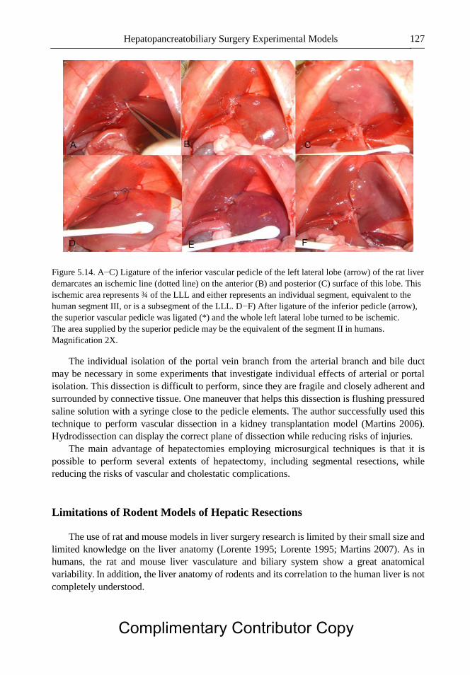
Hepatopancreatobiliary Surgery Experimental Models 127
Figure 5.14. A−C) Ligature of the inferior vascular pedicle of the left lateral lobe (arrow) of the rat liver
demarcates an ischemic line (dotted line) on the anterior (B) and posterior (C) surface of this lobe. This
ischemic area represents ¾ of the LLL and either represents an individual segment, equivalent to the
human segment III, or is a subsegment of the LLL. D−F) After ligature of the inferior pedicle (arrow),
the superior vascular pedicle was ligated (*) and the whole left lateral lobe turned to be ischemic.
The area supplied by the superior pedicle may be the equivalent of the segment II in humans.
Magnification 2X.
The individual isolation of the portal vein branch from the arterial branch and bile duct
may be necessary in some experiments that investigate individual effects of arterial or portal
isolation. This dissection is difficult to perform, since they are fragile and closely adherent and
surrounded by connective tissue. One maneuver that helps this dissection is flushing pressured
saline solution with a syringe close to the pedicle elements. The author successfully used this
technique to perform vascular dissection in a kidney transplantation model (Martins 2006).
Hydrodissection can display the correct plane of dissection while reducing risks of injuries.
The main advantage of hepatectomies employing microsurgical techniques is that it is
possible to perform several extents of hepatectomy, including segmental resections, while
reducing the risks of vascular and cholestatic complications.
Limitations of Rodent Models of Hepatic Resections
The use of rat and mouse models in liver surgery research is limited by their small size and
limited knowledge on the liver anatomy (Lorente 1995; Lorente 1995; Martins 2007). As in
humans, the rat and mouse liver vasculature and biliary system show a great anatomical
variability. In addition, the liver anatomy of rodents and its correlation to the human liver is not
completely understood.
Complimentary Contributor Copy
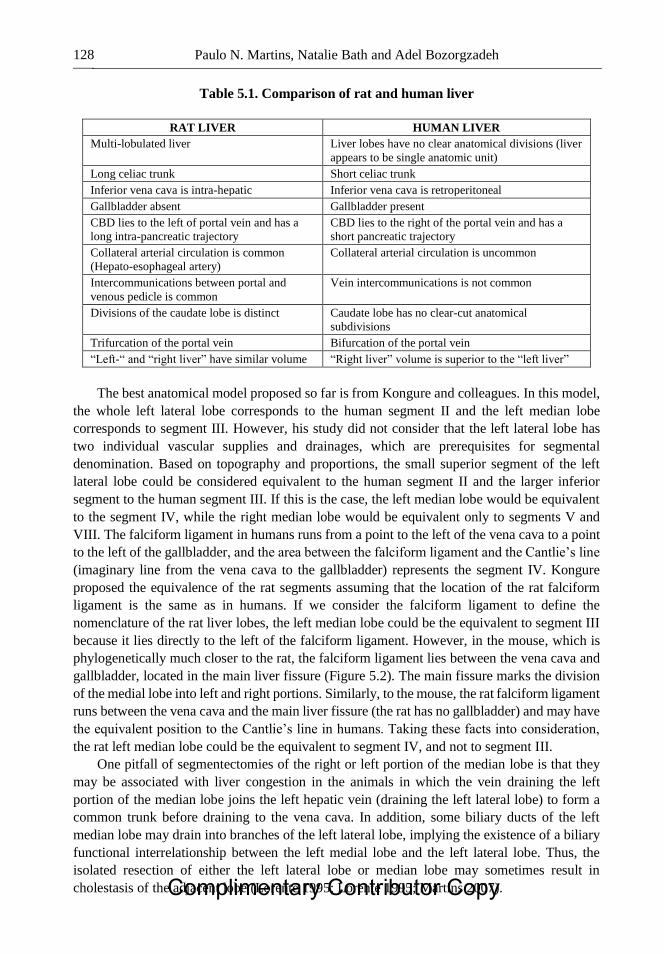
Paulo N. Martins, Natalie Bath and Adel Bozorgzadeh 128
Table 5.1. Comparison of rat and human liver
RAT LIVER HUMAN LIVER
Multi-lobulated liver Liver lobes have no clear anatomical divisions (liver
appears to be single anatomic unit)
Long celiac trunk Short celiac trunk
Inferior vena cava is intra-hepatic Inferior vena cava is retroperitoneal
Gallbladder absent Gallbladder present
CBD lies to the left of portal vein and has a
long intra-pancreatic trajectory
CBD lies to the right of the portal vein and has a
short pancreatic trajectory
Collateral arterial circulation is common
(Hepato-esophageal artery)
Collateral arterial circulation is uncommon
Intercommunications between portal and
venous pedicle is common
Vein intercommunications is not common
Divisions of the caudate lobe is distinct Caudate lobe has no clear-cut anatomical
subdivisions
Trifurcation of the portal vein Bifurcation of the portal vein
“Left-“ and “right liver” have similar volume “Right liver” volume is superior to the “left liver”
The best anatomical model proposed so far is from Kongure and colleagues. In this model,
the whole left lateral lobe corresponds to the human segment II and the left median lobe
corresponds to segment III. However, his study did not consider that the left lateral lobe has
two individual vascular supplies and drainages, which are prerequisites for segmental
denomination. Based on topography and proportions, the small superior segment of the left
lateral lobe could be considered equivalent to the human segment II and the larger inferior
segment to the human segment III. If this is the case, the left median lobe would be equivalent
to the segment IV, while the right median lobe would be equivalent only to segments V and
VIII. The falciform ligament in humans runs from a point to the left of the vena cava to a point
to the left of the gallbladder, and the area between the falciform ligament and the Cantlie’s line
(imaginary line from the vena cava to the gallbladder) represents the segment IV. Kongure
proposed the equivalence of the rat segments assuming that the location of the rat falciform
ligament is the same as in humans. If we consider the falciform ligament to define the
nomenclature of the rat liver lobes, the left median lobe could be the equivalent to segment III
because it lies directly to the left of the falciform ligament. However, in the mouse, which is
phylogenetically much closer to the rat, the falciform ligament lies between the vena cava and
gallbladder, located in the main liver fissure (Figure 5.2). The main fissure marks the division
of the medial lobe into left and right portions. Similarly, to the mouse, the rat falciform ligament
runs between the vena cava and the main liver fissure (the rat has no gallbladder) and may have
the equivalent position to the Cantlie’s line in humans. Taking these facts into consideration,
the rat left median lobe could be the equivalent to segment IV, and not to segment III.
One pitfall of segmentectomies of the right or left portion of the median lobe is that they
may be associated with liver congestion in the animals in which the vein draining the left
portion of the median lobe joins the left hepatic vein (draining the left lateral lobe) to form a
common trunk before draining to the vena cava. In addition, some biliary ducts of the left
median lobe may drain into branches of the left lateral lobe, implying the existence of a biliary
functional interrelationship between the left medial lobe and the left lateral lobe. Thus, the
isolated resection of either the left lateral lobe or median lobe may sometimes result in
cholestasis of the adjacent lobe (Lorente 1995; Lorente 1995; Martins 2007). Complimentary Contributor Copy

Hepatopancreatobiliary Surgery Experimental Models 129
Results obtained in rodent models cannot be fully translated to humans because the kinetics
of liver regeneration in different species is not the same. The amount of remnant hepatic tissue
compatible with life in rodents seems to be less than in humans. While in humans the maximum
amount of liver that can be tolerated is 70−80% of the original volume, rodents can survive
even after a 90−95% hepatectomy (Shirabe 1999; Fan 2000; Renz 2000; Leelaudomlipi 2002;
Schindl 2005; Madrahimov 2006). Table 5.1 compares rat to human livers.
Models of acute liver failure based only on reduction of liver mass (hepatectomy) are in
general not reversible and not associated with the same level of tissue damage and necrosis
commonly seen in clinical setting (caused by toxic and viral agents). In order to overcome this
problem one can subsequently add liver stress after liver resection with tissue ischemia
(clamping) or toxic agents. Another model proposed a combination of major liver resection
with hepatocyte transplantation as a possible approach to recover liver function (Roger 1996).
CIRRHOSIS, PORTAL HYPERTENSION MODELS, AND
PORTACAVAL SHUNTS
Portal hypertension models are important to the study of the pathophysiology of liver
disturbances, liver regeneration, complications and treatment approaches. The models can be
categorized as toxic (e.g., carbon tetrachloride (CCl4), alcohol), nutritional (feeding choline
deficient or methionine deficient diet), surgical (partial portal vein ligation, bile duct ligation),
immunological (heterologous serum, schistosomiasis), and genetic (Rhino mice) (Tsukamoto
1990; Orloff 2001; Abraldes 2006; Abraldes 2006) (Table 5.2). Other toxic agents that have
been used in diverse animal models to produce cirrhosis, include carbon tetrachloride,
dimethylnitrosamine, thioacetamine, monocrotaline, and alcohol (Groszmann 1982; Proctor
1984; Tsukamoto 1985; Dashti 1989; Perazzo 1999). Rodents are the most utilized models
because of low cost and easy handling. The most common model of cirrhosis is the CCL4
model. It can be given via injection, inhalation, or orally. Liver cirrhosis is expedited by giving
phenobarbital added to the drinking water (cirrhosis obtained in 4 to 6 weeks).
Acute administration of carbon tetrachloride induces acute hepatitis. Continuous
administration induces chronic liver injury that leads to cirrhosis. Route of administration varies
among laboratories, but the most effective are oral, intraperitoneal or inhalatory routes. Twelve
to 15 weeks after CCl4 administration, the rats develop micronodular cirrhosis (Proctora 1982;
Kobayashi 2000; Hernandez-Munoz 2001; Constandinou 2005; Abraldes 2006).
Cirrhosis induced by thioacetamide (TAA) is another widely used model of toxic cirrhosis.
The toxin affects both perivenular and periportal areas. It has been used in rats and mice (Li
2002, Okuyama 2005). TAA can be administered in drinking water or by intraperitoneal
injection (Li 2002; Luo 2004; Popov 2006). Intraperitoneal injection offers much more
consistent results (Popov 2006). This model develops macronodular cirrhosis with portal
hypertension in 12 weeks (Abraldes 2006).
Complimentary Contributor Copy

Paulo N. Martins, Natalie Bath and Adel Bozorgzadeh 130
Table 5.2. Experimental Models of Hepatic Fibrosis/Cirrhosis. Adapted from
Tsukamoto H, et al.
Species Method Fibrosis Cirrhosis
Toxic
Carbon tetrachloride
(CCl4)
Dimethylnitrosamine
(DMNA)
Thioacetamide
(TAA)
Rats
Rats
Rats
Dogs
Rats
Subcutaneous injection, 0.1-
0.2ml/100g of body weight twice
weekly
Inhalation twice weekly plus
sodium phenobarbital in drinking
water
Intraperitoneal injection 3 days
weekly
Oral administration 2 days weekly
TAA in drinking water (300mg/L)
>6 weeks
>1-
2weeks
>4 weeks
>3-
4weeks
>2-3
months
>12
weeks
>4 weeks
>13
weeks
>13
weeks
>3months
Nutritional Rats/mice High-fat/low-choline, low-protein
diet
>6weeks >12-24
weeks
Immunologic
Heterologous serum
Bacterial cell wall
Marine
schistosomiasis
Endotoxin
Rats
Rats
Rats
Mice
Rabbits
Low-protein/low-methionine diet
plus ethionine (0.5%)
Swine serum, intraperitoneal
injection twice weekly
Streptococcal cell wall, single
intraperitoneal injection (20mg/g
of body weight)
Subcutaneous injection, 50
cercariae of Schistosoma mansoni
E. coli endotoxin injection into the
common bile duct (0.2/mg)
followed 24 h later by 0.1/mg via
marginal ear vein
>4weeks
>5 weeks
>6weeks
>6weeks
--
>12weeks
>10
weeks
--
--
>9 days
Biliary
Rats
Dogs
Monkeys
Common bile duct ligation
Common bile duct ligation
Common bile duct ligation
>4 weeks
>4 weeks
>2months
--
--
>6
months
Alcoholic
Baboons
Rats
Ethanol-containing liquid diet, ad
lib
Continuous intragastric infusion of
ethanol and a high-fat diet
>6 months
>3 months
>24
months
--
Genetic
Rhino
mice
Mutant with the anatomical
recessive gene
>6 months
--
Dimethilnitrosamine induced cirrhosis (DMNA) DMNA is another hepatotoxin that
induces hepatocellular necrosis. After continuous administration (generally intraperitoneal) the
rats develop fibrosis with portal hypertension, which may be present as early as 5 weeks despite
the fact that animals do not have cirrhosis nor features of hyperdynamic circulation (Veal 2000).
Overt cirrhosis with ascites develops in 13 weeks (Jenkins 1985; Tsukamoto 1990). Complimentary Contributor Copy
Hepatopancreatobiliary Surgery Experimental Models 131
Diet induced cirrhosis is seen in diets deficient in choline and methionin, or a diet with low
protein and choline and enriched with fat, induces liver steatosis associated with marked
oxidative stress that induces inflammation and fibrosis (Tsukamoto 1990; Nanji 2004).
Cirrhosis develops after 12 to 24 weeks.
Partial Portal Vein Ligation
Partial portal vein ligation model (PVL) has been widely used in the study of the
pathophysiology of portal hypertension. This model has been developed in rats, mice, and
rabbits (Vorobioff 1983; Colombato 1992; Castaneda 2000; Iwakiri 2002; Fernandez 2004).
The portal vein is freed from surrounding tissue after a midline abdominal incision. A ligature
(silk 3-0) is placed around a blunt-tipped needle lying along the portal vein. Subsequent
removal of the needle yields a calibrated stenosis of the portal vein that has the diameter of the
needle. In the conventional rat PVL model a 20G needle is used (0.889 mm diameter)
(Vorobioff 1983, Colombato 1992, Castaneda 2000). By using needles of greater caliber, less
severe stenosis and thus less severe degrees of portal hypertension are induced (Lozeva 2004,
Abraldes 2006). The diameters of the needles and resulting levels of stenosis are as follows:
16G: 1.651 mm, 18G: 1.270 mm, 20G: 0.889 mm. The conventional needles for mice and
rabbits are 27G (Iwakiri 2002; Fernandez 2004).
Bile Duct Ligation
Cholestasis is a common pathological condition that can be reproduced in rodents by
surgical ligation of the common bile duct. Common bile duct ligation (CBDL) is a model of
secondary biliary cirrhosis. Evaluation of the various time-related changes after BDL is
paramount for the interpretation of available data as well as for the design of future studies.
A Systematic Chronological Evaluation of Hepatocellular Injury and
Proliferation, Hepatic Inflammation or the Development of Liver Fibrosis
Following BDL
The intervention consists of the isolation of the common bile duct followed by a double
ligature. The first ligature is made below the junction of the hepatic ducts and the other above
the pancreatic duct. The portion of the bile duct between the two ligatures can be resected to
avoid recannulation. Mortality is high after the fifth week (20%). The use of prophylactic
antibiotics (Ampicilin 100 mg/kg s.c. or similar) before surgery and weekly administration of
vitamin K (50 mcg subcutaneous) notably improve the survival of CBDL rats. At 2 weeks rats
develop mild portal hypertension and at 4 weeks severe portal hypertension, hyperdynamic
circulation and portal-systemic shunting of 30−60% (Franco 1979; Beck 1995).
Serum levels of ALT and biliary infarcts increased after BDL, peaking at days 2 and 3
respectively. Following acute or chronic tissue loss, hepatocytes mount a proliferative response.
BDL induced a distinct peak of hepatocellular proliferation at day 5, approximately 48 hours
Complimentary Contributor Copy
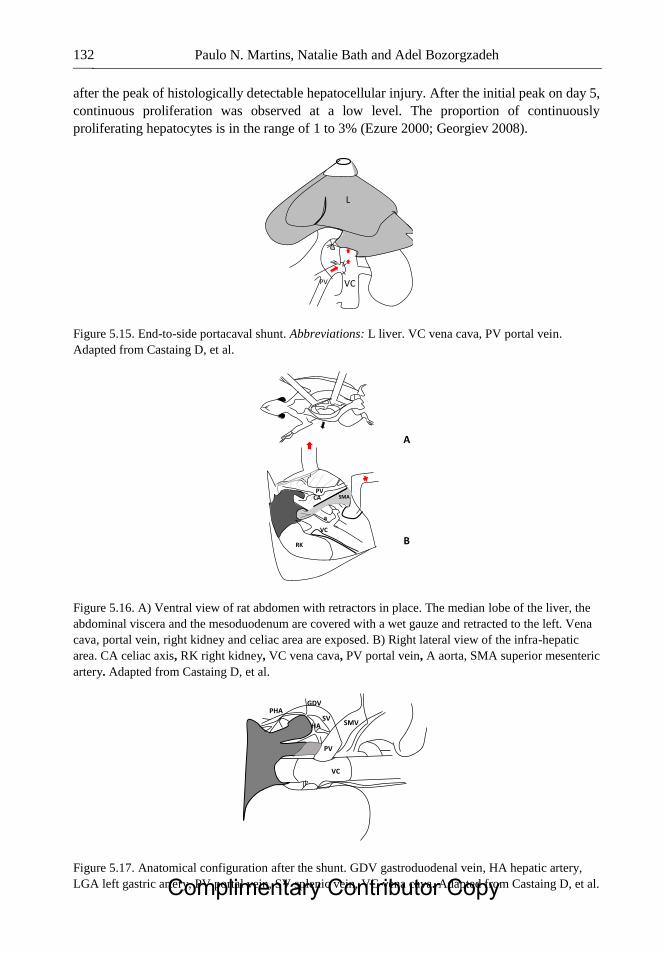
Paulo N. Martins, Natalie Bath and Adel Bozorgzadeh 132
after the peak of histologically detectable hepatocellular injury. After the initial peak on day 5,
continuous proliferation was observed at a low level. The proportion of continuously
proliferating hepatocytes is in the range of 1 to 3% (Ezure 2000; Georgiev 2008).
Figure 5.15. End-to-side portacaval shunt. Abbreviations: L liver. VC vena cava, PV portal vein.
Adapted from Castaing D, et al.
Figure 5.16. A) Ventral view of rat abdomen with retractors in place. The median lobe of the liver, the
abdominal viscera and the mesoduodenum are covered with a wet gauze and retracted to the left. Vena
cava, portal vein, right kidney and celiac area are exposed. B) Right lateral view of the infra-hepatic
area. CA celiac axis, RK right kidney, VC vena cava, PV portal vein, A aorta, SMA superior mesenteric
artery. Adapted from Castaing D, et al.
Figure 5.17. Anatomical configuration after the shunt. GDV gastroduodenal vein, HA hepatic artery,
LGA left gastric artery, PV portal vein, SV splenic vein, VC vena cava. Adapted from Castaing D, et al.
L
VC PV
A
B RK VC
a
PV CA SMA
VC
PV
SMV HA
PHA GDV
SV
Complimentary Contributor Copy

Hepatopancreatobiliary Surgery Experimental Models 133
Figure 5.18. Liver injury was determined by A) serum alanine aminotransferase (ALT) levels. Serum
levels of A) conjugated and unconjugated bilirubin and B) alkaline phosphatase. Percentage of liver
surface stained with Sirius red. Values are mean (s.e.m.) for five to six individual animals per time
point. *P < 0.050 versus sham-operated control (two-tailed Mann–Whitney test). Adapted from
Georgiev, et al.
Serum levels of alkaline phosphatase and bilirubin are classical markers of obstructive
cholestasis in the clinical setting (Pratt 2005). Raised serum levels of alkaline phosphatase are
due to de novo synthesis in the liver (Kaplan 1970). Interestingly, serum levels of alkaline
phosphatase were not found to increase progressively after BDL. A first peak during the acute
phase was followed by decreasing levels from day 3 to day 7, indicating that the acute injury
triggers a massive production and release of this enzyme.
BDL results in an increase in serum markers of cholestasis such as bilirubin and alkaline
phosphatase. Conjugated and unconjugated bilirubin levels increased steadily and in parallel
until day 7 and remained high thereafter (Figure 5.18). Alkaline phosphatase production is
induced after BDL in rats and its release from the canalicular membrane of hepatocytes is
modulated by bile acids. Serum levels of alkaline phosphatase showed a biphasic course,
peaking on day 2 and then increasing steadily from day 7 onwards; cell proliferation reached a
steady state in the biliary epithelial cells (BEL) compartment by day 1.
Neutrophils contribute to cholestatic liver injury after BDL for 3 days, whereas Kupffer
cells are believed to abrogate acute cholestatic liver injury through the release of interleukin 6
(Gujral 2003; Gujral 2004; Gehring 2006). Little attention has been paid to the role of
lymphocytes, which are known to modulate liver fibrosis in other models of liver disease
(Wynn 2004; Novobrantseva 2005). The histological analysis indicated that both B and T cells
were present in the portal tracts of mice after day 5 of BDL. Interestingly, both cell types
accumulated mainly during the phase of collagen deposition and thus may also be involved in
the process of fibrogenesis following BDL (Gujral 2003; Georgiev 2008).
Complimentary Contributor Copy

Paulo N. Martins, Natalie Bath and Adel Bozorgzadeh 134
Figure 5.19. Overview of dynamic changes following bile duct ligation in mice. BD, bile duct; BEC,
biliary epithelial cell; HC, hepatocyte; PMN, polymorphonuclear leucocyte.
This model develops biliary fibrosis-cirrhosis in 4 to 6 weeks. Histology shows marked
cholangiolar proliferation and expansive portal fibrosis (Figure 5.19), but the architectural
disturbances typical of cirrhosis are seldom found (Kountouras 1984). See Figures 5.18−5.19.
PORTACAVAL SHUNTS
In 1877, Dr. Eck described experiments using portacaval shunt in dogs (Eck fistula) (Rocko
1985). Lee and Fisher in 1961 perfected the technique of portacaval shunt in the rat so that it
became a reproducible model (Lee 1961). Before this technique was established, others have
tried to interpose prosthetic tubes between the portal vein and IVC with high mortality rates
(Whitaker 1946; Bernstein 1954).
Technique
The peritoneum over the inferior IVC is cut. Then, with a curved forceps the vena cava is
mobilized from the inferior aspect of the liver to the right renal vein. Next, the peritoneum
covering the portal vein is open, the portal vein is mobilized, and the gastroduodenal vein is
doubly ligated and cut. A small vascular spoon clamp is placed on the IVC between the liver
and the renal veins, partially obstructing its flow. A 4−5 mm incision is made on the IVC and
flushed with heparinized saline. Next, the distal portal vein is ligated with 6-0 silk tie flush to
the liver. The proximal portal vein is clamped with a small vascular clip, and the portal vein is
cut above the stump of the gastroduodenal vein. The portal vein lumen is flushed with saline
and two 7-0 prolene sutures are placed on the corners. The anastomosis is completed on a
running fashion. First the posterior wall, followed by the anterior wall. After completion of the
anastomosis the caval clamp is released and hemostasis accomplished with gentle compression
with cotton swabs. The portal clamping time is of critical importance for the success of the
procedure and it should be less than 30 minutes (see Figures 5.15−5.17) (Castaing 1980). Complimentary Contributor Copy
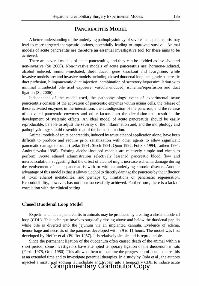
Hepatopancreatobiliary Surgery Experimental Models 135
PANCREATITIS MODEL
A better understanding of the underlying pathophysiology of severe acute pancreatitis may
lead to more targeted therapeutic options, potentially leading to improved survival. Animal
models of acute pancreatitis are therefore an essential investigative tool for these aims to be
achieved.
There are several models of acute pancreatitis, and they can be divided as invasive and
non-invasive (Su 2006). Non-invasive models of acute pancreatitis are: hormone-induced,
alcohol induced, immune-mediated, diet-induced, gene knockout and L-arginine; while
invasive models are: and invasive models including closed duodenal loop, antegrade pancreatic
duct perfusion, biliopancreatic duct injection, combination of secretory hyperstimulation with
minimal intraductal bile acid exposure, vascular-induced, ischemia/reperfusion and duct
ligation (Su 2006).
Independent of the model used, the pathophysiology event of experimental acute
pancreatitis consists of the activation of pancreatic enzymes within acinar cells, the release of
these activated enzymes in the interstitium, the autodigestion of the pancreas, and the release
of activated pancreatic enzymes and other factors into the circulation that result in the
development of systemic effects. An ideal model of acute pancreatitis should be easily
reproducible, be able to adjust the severity of the inflammation and, and the morphology and
pathophysiology should resemble that of the human situation.
Animal models of acute pancreatitis, induced by acute ethanol application alone, have been
difficult to produce and require prior sensitization with other agents to allow significant
pancreatic damage to occur (Letko 1991; Siech 1991; Quon 1992; Foitzik 1994; Luthen 1994;
Andrzejewska 1998). Existing alcohol-induced models are relatively simple and cheap to
perform. Acute ethanol administration selectively lessened pancreatic blood flow and
microcirculation, suggesting that the effect of alcohol might increase ischemia damage during
the evolvement of acute pancreatitis with or without underlying chronic disease. Another
advantage of this model is that it allows alcohol to directly damage the pancreas by the influence
of toxic ethanol metabolites, and perhaps by limitations of pancreatic regeneration.
Reproducibility, however, has not been successfully achieved. Furthermore, there is a lack of
correlation with the clinical setting.
Closed Duodenal Loop Model
Experimental acute pancreatitis in animals may be produced by creating a closed duodenal
loop (CDL). This technique involves surgically closing above and below the duodenal papilla
while bile is diverted into the jejunum via an implanted cannula. Evidence of edema,
hemorrhage and necrosis of the pancreas developed within 9 to 11 hours. The model was first
developed by Pfeffer et al. (Pfeffer 1957). It is relatively simple and is reproducible.
Since the permanent ligation of the duodenum often caused death of the animal within a
short period, some investigators have attempted temporary ligation of the duodenum in rats
(Ferrie 1978, Orda 1980). This allowed them to examine the progression of acute pancreatitis
at an extended time and to investigate potential therapies. In a study by Orda et al., the authors
injected a mixture of sodium taurocholate and trypsin into a temporary CDL to induce acute Complimentary Contributor Copy

Paulo N. Martins, Natalie Bath and Adel Bozorgzadeh 136
pancreatitis in rats (Orda 1980). They found that the acute pancreatitis induced was mild,
associated with a mortality rate of 45%.
Antegrade Pancreatic Duct Perfusion Model
The main pancreatic duct can be made permeable by the perfusion of glycodeoxycholic
acid (GDOC) along the main pancreatic duct. Other methods include the administration of
intragastric ethanol, the stimulation of pancreatic secretion into an obstructed duct, or causing
acute hypercalcemia (Farmer 1983; Wedgwood 1986; Widdison 1992).
The advantages of the pancreatic duct perfusion model are its reliability and
reproducibility. The severity of the acute pancreatitis may be varied within well-defined time
intervals according to the volumes and perfusion pressures. The etiology and the morphological
changes closely resemble the human situation. This model is suitable for studying the early
pathophysiology and subsequent progression of the disease. It is the ideal model to assess
potential therapeutic agents. The major disadvantage of this model is its complexity (ductal
cannulation and perfusion). Other disadvantages include the need to use large animals.
Biliopancreatic Duct Injection Model
Various compounds have been infused into the pancreatic duct to induce acute pancreatitis
(EL 1901; Beck 1964; Papp 1966; Sum 1970; Aho 1980). Following a duodenotomy, a
retrograde injection of bile salts (with or without activated pancreatic enzymes) into the
pancreatic duct at the ampulla leads to severe acute pancreatitis. The severity of the disease can
be manipulated by changing either the pressure or the concentration of bile salt used. Acute
severe pancreatitis develops within 2 to 24 hours and is characterized by edema, necrosis and
hemorrhage. Almost any form of detergent injected into the pancreatic duct under pressure will
cause acute pancreatitis in laboratory animals such as dogs and rats (Musa 1976; Aho 1980;
Aho 1980; Aho 1983). One of the best standardized compounds to use to develop acute
pancreatitis is sodium taurocholate. Infusion of 0.2 ml/kg of 3%, 4.5% or 5% solution induced
acute hemorrhagic pancreatitis with 72 hour mortality rates of 24%, 71% and 100%,
respectively (Aho 1980; Aho 1980; Aho 1983). This model is appropriate for studies of
systemic issues.
The retrograde injection of bile salts into the pancreatic duct of animals is an easy, effective
and reproducible model for creating a severe, rapidly evolving and lethal variety of acute
hemorrhagic pancreatitis.
It is also technically challenging to control constant pressure recordings and hence produce
a standard degree of injury. Another disadvantage of this technique is the injection pressure
with which the solution is applied to cause severe acute pancreatitis (Arendt 1993).
Complimentary Contributor Copy
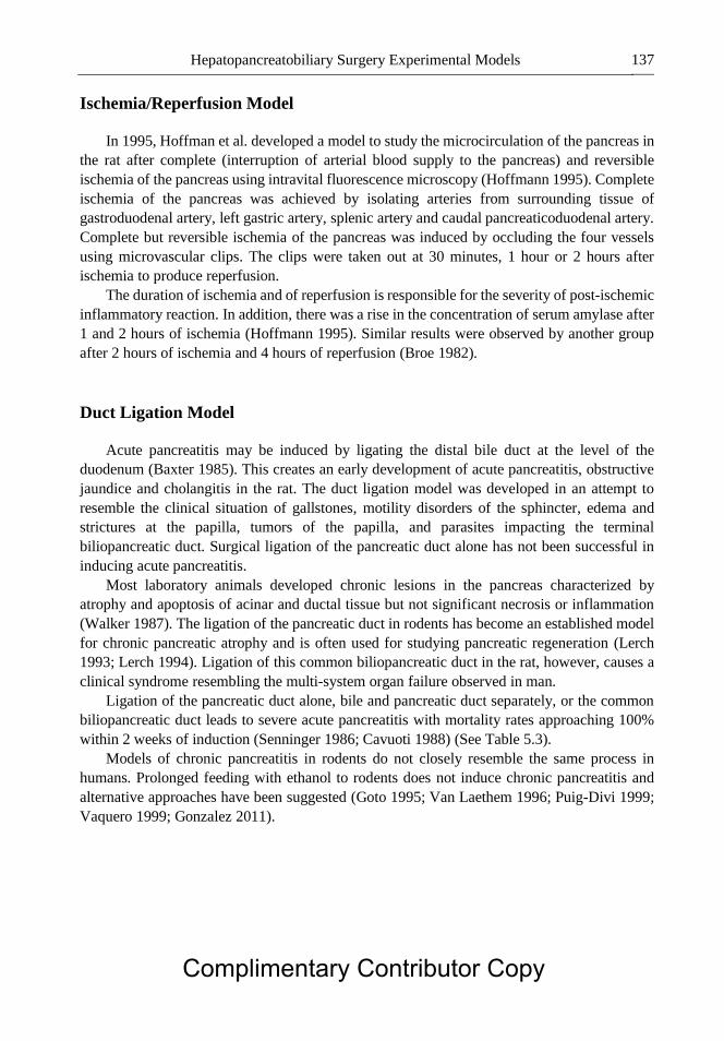
Hepatopancreatobiliary Surgery Experimental Models 137
Ischemia/Reperfusion Model
In 1995, Hoffman et al. developed a model to study the microcirculation of the pancreas in
the rat after complete (interruption of arterial blood supply to the pancreas) and reversible
ischemia of the pancreas using intravital fluorescence microscopy (Hoffmann 1995). Complete
ischemia of the pancreas was achieved by isolating arteries from surrounding tissue of
gastroduodenal artery, left gastric artery, splenic artery and caudal pancreaticoduodenal artery.
Complete but reversible ischemia of the pancreas was induced by occluding the four vessels
using microvascular clips. The clips were taken out at 30 minutes, 1 hour or 2 hours after
ischemia to produce reperfusion.
The duration of ischemia and of reperfusion is responsible for the severity of post-ischemic
inflammatory reaction. In addition, there was a rise in the concentration of serum amylase after
1 and 2 hours of ischemia (Hoffmann 1995). Similar results were observed by another group
after 2 hours of ischemia and 4 hours of reperfusion (Broe 1982).
Duct Ligation Model
Acute pancreatitis may be induced by ligating the distal bile duct at the level of the
duodenum (Baxter 1985). This creates an early development of acute pancreatitis, obstructive
jaundice and cholangitis in the rat. The duct ligation model was developed in an attempt to
resemble the clinical situation of gallstones, motility disorders of the sphincter, edema and
strictures at the papilla, tumors of the papilla, and parasites impacting the terminal
biliopancreatic duct. Surgical ligation of the pancreatic duct alone has not been successful in
inducing acute pancreatitis.
Most laboratory animals developed chronic lesions in the pancreas characterized by
atrophy and apoptosis of acinar and ductal tissue but not significant necrosis or inflammation
(Walker 1987). The ligation of the pancreatic duct in rodents has become an established model
for chronic pancreatic atrophy and is often used for studying pancreatic regeneration (Lerch
1993; Lerch 1994). Ligation of this common biliopancreatic duct in the rat, however, causes a
clinical syndrome resembling the multi-system organ failure observed in man.
Ligation of the pancreatic duct alone, bile and pancreatic duct separately, or the common
biliopancreatic duct leads to severe acute pancreatitis with mortality rates approaching 100%
within 2 weeks of induction (Senninger 1986; Cavuoti 1988) (See Table 5.3).
Models of chronic pancreatitis in rodents do not closely resemble the same process in
humans. Prolonged feeding with ethanol to rodents does not induce chronic pancreatitis and
alternative approaches have been suggested (Goto 1995; Van Laethem 1996; Puig-Divi 1999;
Vaquero 1999; Gonzalez 2011).
Complimentary Contributor Copy
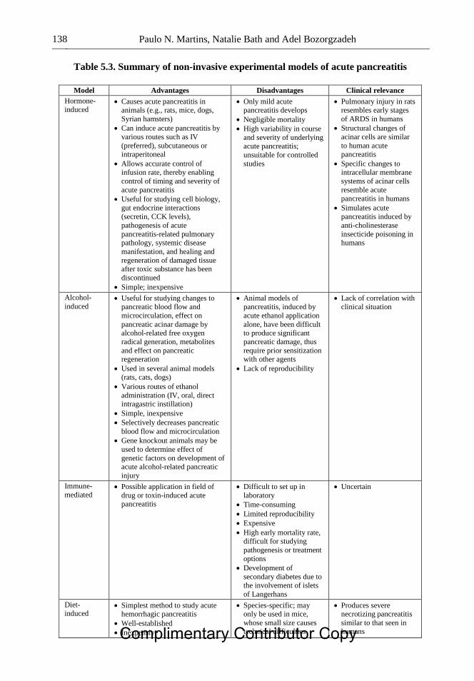
Paulo N. Martins, Natalie Bath and Adel Bozorgzadeh 138
Table 5.3. Summary of non-invasive experimental models of acute pancreatitis
Model Advantages Disadvantages Clinical relevance
Hormone-induced
Causes acute pancreatitis in animals (e.g., rats, mice, dogs,
Syrian hamsters)
Can induce acute pancreatitis by
various routes such as IV
(preferred), subcutaneous or intraperitoneal
Allows accurate control of infusion rate, thereby enabling
control of timing and severity of
acute pancreatitis
Useful for studying cell biology,
gut endocrine interactions
(secretin, CCK levels),
pathogenesis of acute
pancreatitis-related pulmonary pathology, systemic disease
manifestation, and healing and
regeneration of damaged tissue after toxic substance has been
discontinued
Simple; inexpensive
Only mild acute pancreatitis develops
Negligible mortality
High variability in course
and severity of underlying acute pancreatitis;
unsuitable for controlled
studies
Pulmonary injury in rats resembles early stages
of ARDS in humans
Structural changes of
acinar cells are similar
to human acute pancreatitis
Specific changes to intracellular membrane
systems of acinar cells
resemble acute pancreatitis in humans
Simulates acute
pancreatitis induced by
anti-cholinesterase
insecticide poisoning in humans
Alcohol-
induced Useful for studying changes to
pancreatic blood flow and
microcirculation, effect on
pancreatic acinar damage by alcohol-related free oxygen
radical generation, metabolites
and effect on pancreatic regeneration
Used in several animal models (rats, cats, dogs)
Various routes of ethanol
administration (IV, oral, direct intragastric instillation)
Simple, inexpensive
Selectively decreases pancreatic
blood flow and microcirculation
Gene knockout animals may be
used to determine effect of
genetic factors on development of acute alcohol-related pancreatic
injury
Animal models of pancreatitis, induced by
acute ethanol application
alone, have been difficult to produce significant
pancreatic damage, thus
require prior sensitization with other agents
Lack of reproducibility
Lack of correlation with clinical situation
Immune-
mediated Possible application in field of
drug or toxin-induced acute pancreatitis
Difficult to set up in
laboratory
Time-consuming
Limited reproducibility
Expensive
High early mortality rate, difficult for studying
pathogenesis or treatment
options
Development of
secondary diabetes due to the involvement of islets
of Langerhans
Uncertain
Diet-induced
Simplest method to study acute hemorrhagic pancreatitis
Well-established
Inexpensive
Species-specific; may only be used in mice,
whose small size causes technical difficulties
Produces severe necrotizing pancreatitis
similar to that seen in humans Complimentary Contributor Copy
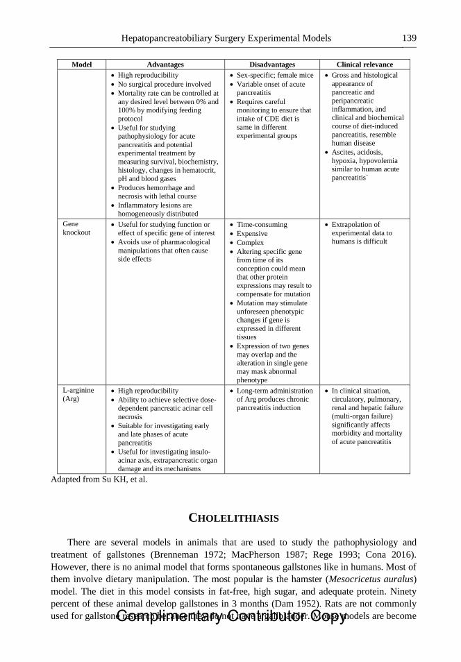
Hepatopancreatobiliary Surgery Experimental Models 139
Model Advantages Disadvantages Clinical relevance
High reproducibility
No surgical procedure involved
Mortality rate can be controlled at
any desired level between 0% and 100% by modifying feeding
protocol
Useful for studying pathophysiology for acute
pancreatitis and potential experimental treatment by
measuring survival, biochemistry,
histology, changes in hematocrit, pH and blood gases
Produces hemorrhage and
necrosis with lethal course
Inflammatory lesions are
homogeneously distributed
Sex-specific; female mice
Variable onset of acute pancreatitis
Requires careful monitoring to ensure that
intake of CDE diet is
same in different experimental groups
Gross and histological
appearance of
pancreatic and peripancreatic
inflammation, and clinical and biochemical
course of diet-induced
pancreatitis, resemble human disease
Ascites, acidosis,
hypoxia, hypovolemia similar to human acute
pancreatitis`
Gene
knockout Useful for studying function or
effect of specific gene of interest
Avoids use of pharmacological
manipulations that often cause side effects
Time-consuming
Expensive
Complex
Altering specific gene from time of its
conception could mean that other protein
expressions may result to
compensate for mutation
Mutation may stimulate
unforeseen phenotypic changes if gene is
expressed in different
tissues
Expression of two genes
may overlap and the alteration in single gene
may mask abnormal
phenotype
Extrapolation of
experimental data to humans is difficult
L-arginine
(Arg) High reproducibility
Ability to achieve selective dose-dependent pancreatic acinar cell
necrosis
Suitable for investigating early
and late phases of acute
pancreatitis
Useful for investigating insulo-
acinar axis, extrapancreatic organ damage and its mechanisms
Long-term administration
of Arg produces chronic pancreatitis induction
In clinical situation,
circulatory, pulmonary, renal and hepatic failure
(multi-organ failure)
significantly affects morbidity and mortality
of acute pancreatitis
Adapted from Su KH, et al.
CHOLELITHIASIS
There are several models in animals that are used to study the pathophysiology and
treatment of gallstones (Brenneman 1972; MacPherson 1987; Rege 1993; Cona 2016).
However, there is no animal model that forms spontaneous gallstones like in humans. Most of
them involve dietary manipulation. The most popular is the hamster (Mesocricetus auralus)
model. The diet in this model consists in fat-free, high sugar, and adequate protein. Ninety
percent of these animal develop gallstones in 3 months (Dam 1952). Rats are not commonly
used for gallstone research because they do not have a gallbladder. Mouse models are become Complimentary Contributor Copy

Paulo N. Martins, Natalie Bath and Adel Bozorgzadeh 140
more popular because the extensive genetic characterization, the availability of knockout,
transgenic and inbred strains. The incidence of gallstones in mice treated with lithogenic diet
is very variable. While only 14% of C57BL/6J mice fed with diet consisting of 15% dairy fat,
50% sucrose, 20% casein, 1% cholesterol, 0.5% cholic acid, cellulose, vitamins and mineral
for 18 weeks developed gallstones, 100% of C57L/J and A/J mice developed gallstones after
the same time (Paigen 1995).
HEPATOCELLULAR CARCINOMA (HCC) MODELS
Hepatocellular carcinoma (HCC) is the most common primary malignancy of the liver.
Establishing animal models of HCC is essential to study the pathophysiology of carcinogenesis,
perform drug screening, and conduct various therapeutic experiments. A wide range of HCC
animal models are currently available: chemically induced models, xenograft models, and
genetically modified models in mice (Schotman 1998; Heindryckx 2009; Li 2011). No model,
however, is ideal for all purposes. This review analyses several mouse models useful for HCC
research and points out their advantages and weaknesses. Chemically induced HCC mice
models mimic the injury-fibrosis-malignancy cycle by administration of a genotoxic compound
alone or, if necessary, followed by a promoting agent.
Chemically Induced
Several chemical agents can promote tumor formation when administered in high doses
and long duration. The advantage of chemically induced models is the similarity with the
injury-fibrosis-malignancy cycle seen in humans. This makes them the favorite models for
HCC research.
Diethylnitrosamine
N-nitrosodiethylamine (DEN) is often used as a carcinogenic reagent. Mouse tumors
induced by DEN harbour activating mutations in the H-ras proto-oncogene. Administration of
a high dose induces HCC after a period of latency. The time needed after a single DEN-injection
to develop HCC does not only depend on the administered dose, but also on sex, age and strain
of mice. A dose-dependent formation of carcinomas after a single injection (5–90 mg/kg) of
DEN in 15-day-old mice is observed after 45–104 weeks. When B6C3F1 mice are exposed to
a single dose of 5.0 mg/kg DEN, it takes approximately 64 weeks to develop HCC (Heindryckx
2009).
A two-stage model in which the initiation by a genotoxic compound is followed by a
promotion phase is often used for inducing HCC. DEN can be used as an initiator and
phenobarbital (PB) as a promoting agent. Several mechanisms might be responsible for the
tumour promoting effect of PB. First, PB can increase the expression of cytochrome P-450 a
100-fold, leading to an enhanced effect of DEN. Another two-step carcinogenesis model is
known as the Solt-Farber protocol (Farber 1977). In this model, initiation by a
hepatocarcinogenic compound is followed by a partial hepatectomy (PH). Partial hepatectomy
Complimentary Contributor Copy
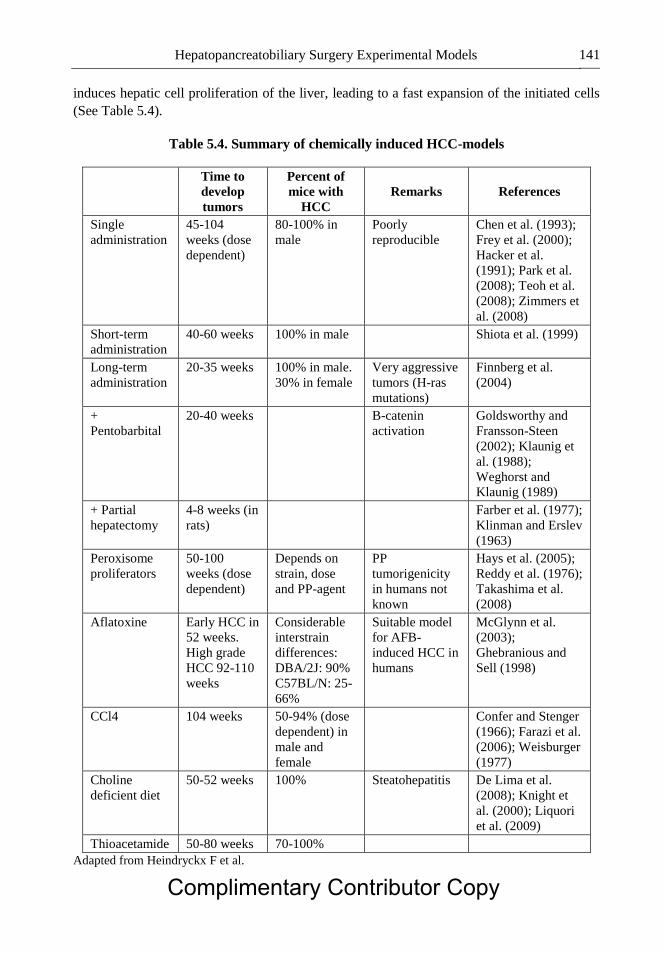
Hepatopancreatobiliary Surgery Experimental Models 141
induces hepatic cell proliferation of the liver, leading to a fast expansion of the initiated cells
(See Table 5.4).
Table 5.4. Summary of chemically induced HCC-models
Time to
develop
tumors
Percent of
mice with
HCC
Remarks References
Single
administration
45-104
weeks (dose
dependent)
80-100% in
male
Poorly
reproducible
Chen et al. (1993);
Frey et al. (2000);
Hacker et al.
(1991); Park et al.
(2008); Teoh et al.
(2008); Zimmers et
al. (2008)
Short-term
administration
40-60 weeks 100% in male Shiota et al. (1999)
Long-term
administration
20-35 weeks 100% in male.
30% in female
Very aggressive
tumors (H-ras
mutations)
Finnberg et al.
(2004)
+
Pentobarbital
20-40 weeks B-catenin
activation
Goldsworthy and
Fransson-Steen
(2002); Klaunig et
al. (1988);
Weghorst and
Klaunig (1989)
+ Partial
hepatectomy
4-8 weeks (in
rats)
Farber et al. (1977);
Klinman and Erslev
(1963)
Peroxisome
proliferators
50-100
weeks (dose
dependent)
Depends on
strain, dose
and PP-agent
PP
tumorigenicity
in humans not
known
Hays et al. (2005);
Reddy et al. (1976);
Takashima et al.
(2008)
Aflatoxine Early HCC in
52 weeks.
High grade
HCC 92-110
weeks
Considerable
interstrain
differences:
DBA/2J: 90%
C57BL/N: 25-
66%
Suitable model
for AFB-
induced HCC in
humans
McGlynn et al.
(2003);
Ghebranious and
Sell (1998)
CCl4 104 weeks 50-94% (dose
dependent) in
male and
female
Confer and Stenger
(1966); Farazi et al.
(2006); Weisburger
(1977)
Choline
deficient diet
50-52 weeks 100% Steatohepatitis De Lima et al.
(2008); Knight et
al. (2000); Liquori
et al. (2009)
Thioacetamide 50-80 weeks 70-100%
Adapted from Heindryckx F et al.
Complimentary Contributor Copy

Paulo N. Martins, Natalie Bath and Adel Bozorgzadeh 142
Peroxisome Proliferators
Peroxisome proliferators (ethyl clofenapate ciprofibrate, fenofibrate, clofibrate) induce
hepatomegaly, peroxisome proliferation in hepatocytes and induction of several hepatic
enzymes.
Aflatoxin B1
The hepatotoxin aflatoxin B1 (AFB), produced by certain fungi of the Aspergillus genus
such as Asparagillus flavum, is known to be a hepatic carcinogen. When 7-day-old mice are
exposed to 6 mg/kg body weight of AFB, in a bolus injection, HCC is developed after 52 weeks
(McGlynn 2003). In 90% of the DBA/2J mice (susceptible strain for HCC) HCC occurs after
AFB exposure, while only 25% of the C57BL/N mice (relatively resistant strain for HCC)
develop HCC after AFB exposure.
Xenograft Models
In xenograft models, the tumors are formed by injecting human cancer cells from a lab
culture in immune deficient mice. Athymic (nude) or severe combined immune deficient
(SCID) mice are often used as hosts. Tumor xenografts can be established either by direct
implantation of biopsy material or by inoculation of human tumor cell lines. First, the ectopic
model, in which human tumor cells are injected subcutaneously in the flank of mice. Second,
the orthotopic model, where tumor cells are injected intrahepatically into the mice. The
advantage of xenograft mouse models is the short time span needed for the development of
tumors. However, the resemblance between xenograft tumors and human tumors is rather poor.
Thus, most researchers prefer to use models in which tumors arise in a background that
resembles the natural history of HCC (Heindryckx 2009).
Transgenic Mouse Models
Genetically modified mouse models are engineered to reproduce pathophysiological and
molecular features of human HCC (by simulating hepatitis B or C, over-expressing growth
factors, or induction of oncogenes) (Heindryckx 2009). It is a unique model for assessing the
effects of oncogenes either alone or in combination with other oncogenes or carcinogenic
agents. The transgenic mouse models are the ones that most resemble the human HCC.
Transgenic mouse models of HBV or HCV infection have provided reliable experimental proof
that viral genes could initiate or promote liver carcinogenesis. Most HBV transgenic mouse
models focus on the HBx gene, which encodes HBV X protein (HBx)−a transcriptional
transactivator that stimulates expression of a broad range of proto-oncogenes, including c-fos,
c-myc and c-jun (Li 2011).
Complimentary Contributor Copy
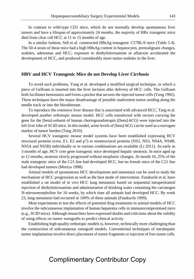
Hepatopancreatobiliary Surgery Experimental Models 143
In contrast to wild-type CD1 mice, which do not normally develop spontaneous liver
tumors and have a lifespan of approximately 24 months, the majority of HBx transgenic mice
died from clear cell HCC at 11 to 15 months of age.
In a similar fashion, Sell et al. constructed HBsAg transgenic C57BL/6 mice (Table 5.4).
The 50-4 strain of these mice had a high HBsAg content in hepatocytes, premalignant changes,
nodules, adenomas and HCC; exposure to diethylnitrosamine or aflatoxin accelerated the
development of HCC, and produced considerably more tumor nodules in the liver.
HBV and HCV Transgenic Mice do not Develop Liver Cirrhosis
To avoid such problems, Yang et al. developed a modified surgical technique, in which a
piece of Gelfoam is inserted into the liver incision after delivery of HCC cells. The Gelfoam
both facilitates hemostasis and forms a pocket that secures the injected tumor cells (Yang 1992).
These techniques have the major disadvantage of possible inadvertent tumor seeding along the
needle track or into the bloodstream.
To reproduce the extensive liver disease that is associated with advanced HCC, Tang et al.
developed another orthotopic mouse model. HCC cells transfected with vectors carrying the
gene for the [beta]-subunit of human choriogonadotropin ([beta]-hCG) were injected into the
left liver lobe of SCID mice. In this model, urine levels of [beta]-hCG can be used as a surrogate
marker of tumor burden (Tang 2010).
Several HCV transgenic mouse model systems have been established expressing HCV
structural proteins (core, E1, E2 and p7) or nonstructural proteins (NS2, NS3, NS4A, NS4B,
NS5A and NS5B) individually or in various combinations are available (Li 2011). As early as
3 months of age, HCV core gene transgenic mice developed hepatic steatosis. In mice aged up
to 12 months, steatosis slowly progressed without neoplastic changes. At month 16, 25% of the
male transgenic mice of the C21 line had developed HCC, but no female mice of the C21 line
had developed tumors (Moriya 1998).
Animal models of spontaneous HCC development and metastasis can be used to study the
mechanism of HCC progression as well as the best mode of intervention. Futakuchi et al. have
established a rat model of in vivo HCC lung metastasis based on sequential intraperitoneal
injection of diethylnitrosamine and administration of drinking water containing the carcinogen
N-nitrosomorpholine for 16 weeks, by which time all animals had developed HCC. By week
23, lung metastasis had occurred in 100% of these animals (Futakuchi 1999).
Most experiments to test the effects of potential drug treatments in animal models of HCC
involve the subcutaneous implantation of human hepatoma cells in immunocompromised mice
(e.g., SCID mice). Although researchers have expressed doubts and criticisms about the validity
of using effects on tumor xenografts to predict clinical activity
Establishing high-quality orthotopic models is, however, technically more challenging than
the construction of subcutaneous xenograft models. Conventional techniques of intrahepatic
tumor implantation involve direct placement of tumor fragments or injection of free tumor cells.
Complimentary Contributor Copy

Paulo N. Martins, Natalie Bath and Adel Bozorgzadeh 144
CHOLANGIOCARCINOMA MODELS
Several carcinogenesis models of cholangiocarcinoma (CCC) have been established in
animals (Thamavit 1978; Elmore 1993; Cheifetz 1996; Sirica 1996; Lee 1997; Lai 1999; Al-
Bader 2000; Yeh 2004; Sirica 2008; Yang 2011; Ko 2013; Ikenoue 2016). There are several
CCC animal models: xenograft and orthotopic models, carcinogen-induced CCC model, and
genetically engineered mouse model for CCA. Recent advancements in cell and molecular
biology make it possible to mimic the pathogenicity of human cholangiocarcinoma using
various animal models.
One model obtained CCC using Syrian hamsters being treated with Clonorchis sinensis or
Opisthorchis viverrini followed by dimethylnitrosamine (DMN) (Thamavit 1978; Flavell 1983;
Lee 1997). In another model Syrian hamsters were treated with DMN followed by bile duct
ligation also develop CCC (Cheifetz 1996). In these models, CCC develops about 24 weeks
after the initial insult, with a yield rate of only 10%. One of the better characterized rat models
of CCC has been the furan model described by Sirica et al., which leads to the development of
intestinal-type CCC in the caudate lobe of the liver (Elmore 1993; Sirica 1996).
Oral administration of thioacetamide (TAA) in drinking water to male Sprangue-Dawley
rats provides a reproducible animal model for development of CCC with a high yield rate.
Drinking water with TAA 300 mg/l was administered orally. Multifocal bile ductular
proliferation with intestinal metaplasia (presence of goblet cells) and increasing histologic
atypia (biliary dysplasia) was observed by the 9th week of TAA administration. Biliary
cytokeratin (CK19)-expressing invasive intestinal-type CCC with stromal desmoplasia was
evident at the 16th week, and by the 22nd week the yield rate for CCCs had increased to 100%.
The progression from normal cholangioles to biliary dysplasia to invasive CCC was
accompanied by up-regulation of the proto-oncogenes c-met and c-erbB-2, tyrosine kinase
receptors over-expressed in human CCCs. In the first 6 months of follow-up, no systemic
metastases or tumor-related deaths were observed in this model. (Yeh 2004).
Yamada and coworkers created a mouse CCC model that transfected the biliary epithelium
directly with plasmids containing oncogenes. The common bile duct located below the liver
was clamped with a small animal surgical clip to prevent the injected material from rapidly
flowing into the duodenum. The bile duct draining the left lateral liver lobe was identified, and
a ligature (6-0 silk) was placed loosely around the duct. Another ligature was placed around the
base of the gallbladder where it meets the cystic duct. Ectopic oncogene expression in the
biliary tract was achieved by the SB transposon transfection system with transduction of murine
constitutively active AKT and human YAP. The solution was injected into the gallbladder with
enough pressure to allow the solution to distend the biliary system and reach the whole the
intrahepatic biliary tree. The ligature around the left lateral liver lobe duct was then tied off so
as not to allow the plasmid solution to flow from this bile duct to the common bile duct.
Following plasmid injection and the left lateral bile duct ligation, the common bile duct was
unclamped. The ligature around the base of the gallbladder was then tied and a cholecystectomy
performed. Then, each animal was injected with 1 g of IL-33 for 3 days (Leake 2015; Yamada
2015).
Complimentary Contributor Copy

Hepatopancreatobiliary Surgery Experimental Models 145
COLON CARCINOMA METASTASES
Tumor models have a key role in oncology. They are powerful tools guiding clinical
research and practice (Demicheli 2001). Most transplantable tumor therapy experiments utilize
ectopic (usually subcutaneous) injection of tumor cells. Ectopically transplanted tumors do not
necessarily reproduce the biology of orthotopically grown tumors. The response of the primary
tumor to therapy is usually what is evaluated in most tumor models. There is a need to place
more emphasis on tumor models in which metastases are the primary target to therapy.
The limitations of animal models are that they usually respond better to anti-cancer therapy
than in the clinical settings.
Most transplantable tumors are very fast growing (doubling times measured in days) while
human tumors (with doubling times measured in months). Experimental tumors tend to be
highly responsive to cytotoxic anti-cancer drugs, since this drugs target rapidly dividing cells.
Most transplantable tumors are highly immunogenic because it is difficult to obtain truly
syngeneic tumors. These tumors therefore become especially vulnerable to various host
immune effector mechanisms.
Figure 5.20. Technique of portal injection to inoculate colon cancer cells DKDK12 (CAMR, Salisburg-
Withshire, UK) in the rat liver. This reproduces the metastatic process of colon cancer. A 30G needle is
used to minimize bleeding.
Complimentary Contributor Copy

Paulo N. Martins, Natalie Bath and Adel Bozorgzadeh 146
Figure 5.21. Isolated liver tumor of colon cancer cells DKDK12 (CAMR, Salisburg-Withshire, UK)
injected in the rat portal vein 2 weeks prior.
Figure 5.22. Metastatic tumor of colon cancer cells DKDK12 (CAMR, Salisburg-Withshire, UK)
injected in the rat portal vein 4 weeks prior.
Over recent years, the interest in the development of experimental models for colorectal
liver metastases has increased due to the need for new adjuvant therapies to improve the
treatment of both colorectal cancer and liver metastases. The induction of colon cancer by
carcinogens in the rat with spontaneous liver metastases closely mimics the natural history of
colon cancer but only a low yield of both colonic cancer (less than 50%) and liver metastases
(approx. 25%) is obtained after 6 months of latency. Direct intraportal injection of colon
carcinoma cells is the most used model. Although it bypasses the natural history of colon
cancer, this simple model produces up to 100% of liver metastases 6 weeks after injection of
cells. This model has been used to study morphology, neovascularization, surgical and adjuvant
therapy (Panis 1991). See Figures 5.20−5.22.
Complimentary Contributor Copy
Hepatopancreatobiliary Surgery Experimental Models 147
In using animal models for preclinical assessment of anti-metastatic agents, it is necessary
that appropriate cell lines, routes of administration and target organs used, be appropriately
matched for the information sought (Chambers 1999).
REFERENCES
Abraldes, JG; Iwakiri, Y; Loureiro-Silva, M; Haq, WC; Sessa, Groszmann RJ. Mild increases
in portal pressure upregulate vascular endothelial growth factor and endothelial nitric oxide
synthase in the intestinal microcirculatory bed: leading to a hyperdynamic state. Am. J.
Physiol. Gastrointest. Liver Physiol., 2006, 290(5), G980−987.
Abraldes, JG; Pasarin, M; Garcia-Pagan, JC. Animal models of portal hypertension. World J.
Gastroenterol., 2006, 12(41), 6577−6584.
Aho, HJ; Koskensalo, SM; Nevalainen, TJ. Experimental pancreatitis in the rat. Sodium
taurocholate-induced acute haemorrhagic pancreatitis. Scand. J. Gastroenterol., 1980,
15(4), 411−416.
Aho, HJ; Nevalainen, YJ. Experimental pancreatitis in the rat. Ultrastructure of sodium
taurocholate-induced pancreatic lesions. Scand. J. Gastroenterol., 1980, 15(4), 417−424.
Aho, HJ; Nevalainen, YJ; Aho, AJ. Experimental pancreatitis in the rat. Development of
pancreatic necrosis; ischemia and edema after intraductal sodium taurocholate injection.
Eur. Surg. Res., 1983, 15(1), 28−36.
Al-Bader, A; Mathew, TC; Abul, H; Al-Sayer, H; Singal, PK; Dashti, HM.
Cholangiocarcinoma and liver cirrhosis in relation to changes due to thioacetamide. Mol.
Cell. Biochem., 2000, 208(1−2), 1−10.
Aleksandrowicz, R; Gawlik, Z; Wisniewska, IE; Tarlowska, H. Physiological aspects of the
partial hepatectomy in rats. Acta. Physiol. Pol., 1981, 32(6), 681−692.
Alpini, G; Roberts, S; Kuntz, SM; Ueno, Y; Gubba, S; Podila, PV; LeSage, G; LaRusso, NF.
Morphological, molecular, and functional heterogeneity of cholangiocytes from normal rat
liver. Gastroenterology, 1996, 110(5), 1636−1643.
Andrzejewska, A; Dlugosz, JW; Jurkowska, G. The effect of antecedent acute ethanol ingestion
on the pancreas ultrastructure in taurocholate pancreatitis in rats. Exp. Mol. Pathol., 1998,
65(2), 64−77.
Arendt, T. Bile-induced acute pancreatitis in cats. Roles of bile, bacteria, and pancreatic duct
pressure. Dig. Dis. Sci., 1993, 38(1), 39−44.
Baxter, JN; Jenkins, SA; Day, DW; Roberts, NB; Cowell, DC; Mackie, CR; Shields, R. Effects
of somatostatin and a long-acting somatostatin analogue on the prevention and treatment
of experimentally induced acute pancreatitis in the rat. Br. J. Surg., 1985, 72(5), 382−385.
Beck, IT; Kahn, DS; Solymar, J; McKenna, RD; Zylberszac, B. The role of pancreatic enzymes
in the pathogenesis of acute pancreatitis. III. Comparison of the the pathological and
biochemical changes in the canine pancreas to intraductal injection with bile and trypsin.
Gastroenterology, 1964, 46, 531−542.
Beck, PL; Lee, SS. Vitamin K1 improves survival in bile-duct-ligated rats with cirrhosis. J.
Hepatol., 1995, 23(2), 235.
Bernstein, DECS. Simple technique for portacaval shunt in the rat. J. Appl. Physiol., 1954, 14,
469.
Complimentary Contributor Copy
Paulo N. Martins, Natalie Bath and Adel Bozorgzadeh 148
Bismuth, H. Surgical anatomy and anatomical surgery of the liver. 1982; World J. Surg., 6(1),
3−9.
Brand, MI; Kononov, A; Vladisavljevic, A; Milsom, JW. Surgical anatomy of the celiac artery
and portal vein of the rat. Lab. Anim. Sci., 1995, 45(1), 76−80.
Brenneman, DE; Connor, WE; Forker, EL; DenBesten, L. The formation of abnormal bile and
cholesterol gallstones from dietary cholesterol in the prairie dog. J. Clin. Invest., 1972,
51(6), 1495−1503.
Broe, PJ; Zuidema, GD; Cameron, JL. The role of ischemia in acute pancreatitis: studies with
an isolated perfused canine pancreas. Surgery, 1982, 91(4), 377−382.
Bucher, NL; Swaffield, MN. The rate of incorporation of labelled thymidine into the
deoxyribonucleic acid of regenerating rat liver in relation to the amount of liver excised.
Cancer. Res., 1964, 24, 1611−1625.
Castaing, DHD; Bismuth, D (1980). Anatomy of the liver and portal system of the rat. Paris,
Masson Co.
Castaneda, B; Debernardi-Venon, W; Bandi, JC; Andreu, V; Perez-del-Pulgar, S; Moitinho, E;
Pizcueta, P; Bosch, J. The role of portal pressure in the severity of bleeding in portal
hypertensive rats. Hepatology, 2000, 31(3), 581−586.
Cavuoti, OP; Moody, FG; Martinez, G. Role of pancreatic duct occlusion with prolamine
(Ethibloc) in necrotizing pancreatitis. Surgery, 1988, 103(3), 361−366.
Chambers, AF. The metastatic process: basic research and clinical implications. Oncol. Res.,
1999, 11(4), 161−168.
Cheifetz, RE; Davis, NL; Owen, DA. An animal model of benign bile-duct stricture, sclerosing
cholangitis and cholangiocarcinoma and the role of epidermal growth factor receptor in
ductal proliferation. Can. J. Surg., 1996, 39(3), 193−197.
Colombato, LA; Albillos, A; Groszmann, RJ. Temporal relationship of peripheral
vasodilatation, plasma volume expansion and the hyperdynamic circulatory state in portal-
hypertensive rats. Hepatology, 15(2), 323−328.
Cona, MM; Liu, Y; Yin, T; Feng, Y; Chen, F; Mulier, S; Li, Y; Zhang, J; Oyen, R; Ni, Y. Rat
model of cholelithiasis with human gallstones implanted in cholestasis-induced virtual
gallbladder. World J. Methodol., 2016, 6(2), 154−162.
Constandinou, C; Henderson, N; Iredale, JP. Modeling liver fibrosis in rodents. Methods Mol.
Med., 2005, 117, 237−250.
Couinaud, C. The paracaval segments of the liver. Journal of Hepato-Biliary-Pancreatic
Surgery, 1994, 1(2), 145−151.
Dam, H; Christensen, F. Alimentary production of gallstones in hamsters. Acta. Pathol.
Microbiol. Scand., 1952, 30(2), 236−242.
Dashti, H; Jeppsson, B; Hagerstrand, I; Hultberg, B; Srinivas, U; Abdulla, M; Bengmark, S.
Thioacetamide- and carbon tetrachloride-induced liver cirrhosis. Eur. Surg. Res., 1989,
21(2), 83−91.
de Jong, KP; Brouwers, MA; Huls, GA; Dam, A; Bun, JC; Wubbena, AS; Nieuwenhuis, P;
Slooff, MJ. Liver cell proliferation after partial hepatectomy in rats with liver metastases.
Anal. Quant. Cytol. Histol., 1998, 20(1), 59−68.
de Jong, KP; Lont, HE; Bijma, AM; Brouwers, MA; de Vries, EG; van Veen, ML; Marquet,
RL; Slooff, MJ; Terpstra, OT. The effect of partial hepatectomy on tumor growth in rats:
in vivo and in vitro studies. Hepatology, 1995, 22(4 Pt 1), 1263−1272.
Complimentary Contributor Copy
Hepatopancreatobiliary Surgery Experimental Models 149
Delong, RP. Revascularization of the rat liver following interruption of its arterial supply. Surg.
Forum, 1953, 4, 388−392.
Demicheli, R. Tumour dormancy: findings and hypotheses from clinical research on breast
cancer. Semin. Cancer Biol., 2001, 11(4), 297−306.
Eguchi, S; Lilja, H; Hewitt, WR; Middleton, Y; Demetriou, AA; Rozga, J. Loss and recovery
of liver regeneration in rats with fulminant hepatic failure. J. Surg. Res., 1997, 72(2),
112−122.
EL, O. The etiology of acute hemorrhagic pancreatitis. Johns Hopkins Hosp. Bull., 1901, 12,
182−190.
Elmore, LW; Sirica, AE. Intestinal-type” of adenocarcinoma preferentially induced in
right/caudate liver lobes of rats treated with furan. Cancer Res., 1993, 53(2), 254−259.
Emond, J; Capron-Laudereau, M; Meriggi, F; Bernuau, J; Reynes, M; Houssin, D. Extent of
hepatectomy in the rat. Evaluation of basal conditions and effect of therapy. Eur. Surg.
Res., 1989, 21(5), 251−259.
Ezure, T; Sakamoto, T; Tsuji, H; Lunz, 3rd JG; Murase, N; Fung, JJ; Demetris, AJ. The
development and compensation of biliary cirrhosis in interleukin-6-deficient mice. Am. J.
Pathol., 2000, 156(5), 1627−1639.
Fan, ST; Lo, CM; Liu, CL; Yong, BH; Chan, JK; Ng, IO. Safety of donors in live donor liver
transplantation using right lobe grafts. Arch. Surg., 2000, 135(3), 336−340.
Farber, E; Solt, D; Cameron, R; Laishes, B; Ogawa, K; Medline, A. Newer insights into the
pathogenesis of liver cancer. Am. J. Pathol., 1977, 89(2), 477−482.
Farmer, RCMS; Reber, HA. Acute pancreatitis: the role of duct permeability. Surg. Forum,
1983, 34, 224−227.
Fausto, N. Liver regeneration: from laboratory to clinic. Liver Transpl., 2001, 7(10), 835−844.
Fausto, N; Riehle, KJ. Mechanisms of liver regeneration and their clinical implications. J.
Hepatobiliary Pancreat. Surg., 2005, 12(3), 181−189.
Fernandez, M; Vizzutti, F; Garcia-Pagan, JC; Rodes, J; Bosch, J. Anti-VEGF receptor-2
monoclonal antibody prevents portal-systemic collateral vessel formation in portal
hypertensive mice. Gastroenterology, 2004, 126(3), 886−894.
Ferrie, MM; O’Hare, R; Joffe, SN. Acute and chronic pancreatitis in the rat caused by a closed
duodenal loop. Digestion, 1978, 18(3−4), 280−285.
Flavell, DJ; Lucas, SB. Promotion of N-nitrosodimethylamine-initiated bile duct
carcinogenesis in the hamster by the human liver fluke, Opisthorchis viverrini.
Carcinogenesis, 1983, 4(7), 927−930.
Flower, MW. Sur la disposition et la nomenclature des lobes du foi chez les mammifères. [On
the disposition and nomenclature of the lobes of faith in mammals] J. de Zool., 1872, 1,
420−425.
Foitzik, T; Lewandrowski, KB; Fernandez-del Castillo, C; Rattner, DW; Klar, E; Warshaw,
AL. Exocrine hyperstimulation but not pancreatic duct obstruction increases the
susceptibility to alcohol-related pancreatic injury. Arch. Surg., 1994, 129(10), 1081−1085.
Franco, D; Gigou, M; Szekely, AM; Bismuth, H. Portal hypertension after bile duct obstruction:
effect of bile diversion on portal pressure in the rat. Arch. Surg., 1979, 114(9), 1064−1067.
Futakuchi, M; Hirose, M; Ogiso, T; Kato, K; Sano, M; Ogawa, K; Shirai, T. Establishment of
an in vivo highly metastatic rat hepatocellular carcinoma model. Jpn. J. Cancer Res., 1999,
90(11), 1196−1202.
Complimentary Contributor Copy
Paulo N. Martins, Natalie Bath and Adel Bozorgzadeh 150
Gaub, J; Iversen, J. Rat liver regeneration after 90% partial hepatectomy. Hepatology, 1984,
4(5), 902−904.
Gehring, S; Dickson, EM; San Martin, ME; van Rooijen, N; Papa, EF; Harty, MW; Tracy, Jr.
TF; Gregory, SH. Kupffer cells abrogate cholestatic liver injury in mice. Gastroenterology,
2006, 130(3), 810−822.
Georgiev, P; Jochum, W; Heinrich, S; Jang, JH; Nocito, A; Dahm, F; Clavien, PA.
Characterization of time-related changes after experimental bile duct ligation. Br. J. Surg.,
2008, 95(5), 646−656.
Gershbein, LL; Elias, H. Observations on the anatomy of the rat liver. Anat. Rec., 1954, 120(1),
85−98.
Glanemann, M; Eipel, C; Nussler, AK; Vollmar, B; Neuhaus, P. Hyperperfusion syndrome in
small-for-size livers. Eur. Surg. Res., 2005, 37(6), 335−341.
Gonzalez, AM; Garcia, T; Samper, E; Rickmann, M; Vaquero, EC; Molero, X. Assessment of
the protective effects of oral tocotrienols in arginine chronic-like pancreatitis. Am. J.
Physiol. Gastrointest. Liver Physiol., 2011, 301(5), G846−855.
Goto, M; Nakano, I; Kimura, T; Miyahara, T; Kinjo, M; Nawata, H. New chronic pancreatitis
model with diabetes induced by cerulein plus stress in rats. Dig. Dis. Sci., 1995, 40(11),
2356−2363.
Greene, AK; Puder, M. Partial hepatectomy in the mouse: technique and perioperative
management. J. Invest. Surg., 2003, 16(2), 99−102.
Greene, E. C. (1963). Anatomy of the rat. New York, Hafner Publishing Company.
Groszmann, RJ; Kravetz, D; Bosch, J; Glickman, M; Bruix, J; Bredfeldt, J; Conn, HO; Rodes,
J; Storer, EH. Nitroglycerin improves the hemodynamic response to vasopressin in portal
hypertension. Hepatology, 1982, 2(6), 757−762.
Gujral, JS; Farhood, A; Bajt, ML; Jaeschke, H. Neutrophils aggravate acute liver injury during
obstructive cholestasis in bile duct-ligated mice. Hepatology, 2003, 38(2), 355−363.
Gujral, JS; Liu Farhood, A; Hinson, JA; Jaeschke, H. Functional importance of ICAM-1 in the
mechanism of neutrophil-induced liver injury in bile duct-ligated mice. Am. J. Physiol.
Gastrointest. Liver Physiol., 2004, 286(3), G499−507.
Hebel, RSM. (1976). Anatomy of the Laboratory Rat. Baltimore, Williams and Wilkins Co.
Heindryckx, F; Colle, I; Van Vlierberghe, H. Experimental mouse models for hepatocellular
carcinoma research. Int. J. Exp. Pathol., 2009, 90(4), 367−386.
Hernandez-Munoz, R; Diaz-Munoz, M; Suarez-Cuenca, JA; Trejo-Solis, C; Lopez, V;
Sanchez-Sevilla, L; Yanez, L; De Sanchez, VC. Adenosine reverses a preestablished CCl4-
induced micronodular cirrhosis through enhancing collagenolytic activity and stimulating
hepatocyte cell proliferation in rats. Hepatology, 2001, 34 (4 Pt 1), 677−687.
Higgins, GMAR. Restoration of the liver of the white rat following partial surgical removal.
Arch. Path. Lab. Med., 1931, 12, 186−202.
Hoffmann, TF; Leiderer, R; Waldner, H; Arbogast, S; Messmer, K. Ischemia reperfusion of the
pancreas: a new in vivo model for acute pancreatitis in rats. Res. Exp. Med. (Berl) 1995,
195(3), 125−144.
Holmin, T; Alinder, G; Herlin, P. A microsurgical method for total hepatectomy in the rat. Eur.
Surg. Res., 1982, 14(6), 420−427.
Ikenoue, T; Terakado, Y; Nakagawa, H; Hikiba, Y; Fujii, T; Matsubara, D; Noguchi, R; Zhu,
C; Yamamoto, K; Kudo, Y; Asaoka, Y; Yamaguchi, K; Ijichi, H; Tateishi, K; Fukushima,
N; Maeda, S; Koike, K; Furukawa, Y. A novel mouse model of intrahepatic Complimentary Contributor Copy
Hepatopancreatobiliary Surgery Experimental Models 151
cholangiocarcinoma induced by liver-specific Kras activation and Pten deletion. Sci. Rep.,
2016, 6, 23899.
Inderbitzin, D; Gass, M; Beldi, G; Ayouni, E; Nordin, A; Sidler, D; Gloor, B; Candinas, D;
Stoupis, C. Magnetic resonance imaging provides accurate and precise volume
determination of the regenerating mouse liver. J. Gastrointest. Surg., 2004, 8(7), 806−811.
Inderbitzin, D; Studer, P; Sidler, D; Beldi, G; Djonov, V; Keogh, A; Candinas, D. Regenerative
capacity of individual liver lobes in the microsurgical mouse model. Microsurgery, 2006,
26(6), 465−469.
Innocenti, PCR; Falcone, A; Gargano, E; Piattelli, E. Anatomia chirurgica del sistema portale
nel rato. Boll. Soc. It. Biol. Sper., 1978, 54, 2421−2425.
Iwakiri, Y; Cadelina, G; Sessa, WC; Groszmann, RJ. Mice with targeted deletion of eNOS
develop hyperdynamic circulation associated with portal hypertension. Am. J. Physiol.
Gastrointest. Liver Physiol., 2002, 283(5), G1074−1081.
Jenkins, SA; Grandison, A; Baxter, JN; Day, DW; Taylor, I; Shields, R. A
dimethylnitrosamine-induced model of cirrhosis and portal hypertension in the rat. J.
Hepatol., 1985, 1(5), 489−499.
Grisham, JW. A morphologic study of deoxyribonucleic acid synthesis and cell proliferation in
regenerating rat liver: autoradiography with thymidine-H3. Cancer Res., 1996, 22,
842−849.
Kanno, N; LeSage, G; Glaser, S; Alvaro, D; Alpini, G. Functional heterogeneity of the
intrahepatic biliary epithelium. Hepatology, 2000, 31(3), 555−561.
Kaplan, MM; Righetti, A. Induction of rat liver alkaline phosphatase: the mechanism of the
serum elevation in bile duct obstruction. J. Clin. Invest., 1970, 49(3), 508−516.
Kara, ME. The anatomical study on the rat pancreas and its ducts with emphasis on the surgical
approach. Ann. Anat., 2005, 187(2), 105−112.
Ko, KS; Peng, J; Yang, H. Animal models of cholangiocarcinoma. Curr. Opin. Gastroenterol.,
2013, 29(3), 312−318.
Kobayashi, N; Ito, M; Nakamura, J; Cai, J; Gao, C; Hammel, JM; Fox, IJ. Hepatocyte
transplantation in rats with decompensated cirrhosis. Hepatology, 2000, 31(4), 851−857.
Kogure, K; Ishizaki, M; Nemoto, M; Kuwano, H; Makuuchi, M. A comparative study of the
anatomy of rat and human livers. J. Hepatobiliary Pancreat. Surg., 1999, 6(2), 171−175.
Kountouras, J; Billing, BH; Scheuer, PJ. Prolonged bile duct obstruction: a new experimental
model for cirrhosis in the rat. Br. J. Exp. Pathol., 1984, 65(3), 305−311.
Krahenbuhl, L; Feodorovici, M; Renzulli, P; Schafer, M; Abou-Shady, M; Baer, HU.
Laparoscopic partial hepatectomy in the rat: a new resectional technique. Dig. Surg., 1998,
15(2), 140−144.
Kubota, T; Takabe, K; Yang, M; Sekido, H; Endo, I; Ichikawa, Y; Togo, S; Shimada, H.
Minimum sizes for remnant and transplanted livers in rats. Journal of Hepato-Biliary-
Pancreatic Surgery, 1997, 4(4), 398−404.
Lai, GH; Sirica, AE. Establishment of a novel rat cholangiocarcinoma cell culture model.
Carcinogenesis, 1999, 20(12), 2335−2340.
Leake, I. Biliary tract. A new mouse model that closely resembles human cholangiocarcinoma.
Nat. Rev. Gastroenterol. Hepatol., 2015, 12(3), 122.
Lee, JH; Rim, HJ; Sell, S. Heterogeneity of the “oval-cell” response in the hamster liver during
cholangiocarcinogenesis following Clonorchis sinensis infection and dimethylnitrosamine
treatment. J. Hepatol., 1997, 26(6), 1313−1323. Complimentary Contributor Copy
Paulo N. Martins, Natalie Bath and Adel Bozorgzadeh 152
Lee, SH; Fisher, B. Portacaval shunt in the rat. Surgery, 1961, 50, 668−672.
Leelaudomlipi, S; Sugawara, Y; Kaneko, J; Matsui, Y; Ohkubo, T; Makuuchi, M. Volumetric
analysis of liver segments in 155 living donors. Liver Transpl., 2002, 8(7), 612−614.
Leneman, F; Burton, S. The hepato-esophageal artery of the rat. Brief report. Acta. Anat.
(Basel), 1967, 68(3), 334−343.
Lerch, MM; Adler, G. Experimental pancreatitis. Current Opinion in Gastroenterology, 1993,
9(5), 752−759.
Lerch, MM; Adler, G. Experimental animal models of acute pancreatitis. Int. J. Pancreatol.,
1994, 15(3), 159−170.
Letko, G; Nosofsky, T; Lessel, W; Siech, M. Transition of rat pancreatic juice edema into acute
pancreatitis by single ethanol administration. Pathol. Res. Pract., 1991, 187(2−3),
247−250.
Li, X; Benjamin, IS; Alexander, B. Reproducible production of thioacetamide-induced
macronodular cirrhosis in the rat with no mortality. J. Hepatol., 2002, 36(4), 488−493.
Li, Y; Tang, ZY; Hou, JX. Hepatocellular carcinoma: insight from animal models. Nat. Rev.
Gastroenterol. Hepatol., 2011, 9(1), 32−43.
Lin, PW. Hemodynamic changes after hepatectomy in rats studied with radioactive
microspheres. J. Formos. Med. Assoc., 1990, 89(3), 177−181.
Lorente, LAM; Duran, HJ; Cejalvo, D; Lloris, JM; Arias, J. Extrahepatic biliary anatomy in
wistar rats. Surg. Res. Comm., 1995, 17, 31−38.
Lorente, LAM; Rodriguez, J; Duran, MC; Duran, HJ; Alonso, S; Arias, J. Surgical Anatomy of
the liver in Wistar rats. Surg. Res. Comm., 1995, 17, 113−121.
Lozeva, V; Montgomery, JA; Tuomisto, L; Rocheleau, B; Pannunzio, M; Huet, PM;
Butterworth, RF. Increased brain serotonin turnover correlates with the degree of shunting
and hyperammonemia in rats following variable portal vein stenosis. J. Hepatol., 2004,
40(5), 742−748.
Lu, MD; Chen, JW; Xie, XY; Liang, LJ; Huang, JF. Portal vein embolization by fine needle
ethanol injection, experimental and clinical studies. World J. Gastroenterol., 1999, 5(6),
506−510.
Luo, B; Liu, L; Tang, L; Zhang, J; Ling, Y; Fallon, MB. ET-1 and TNF-alpha in HPS, analysis
in prehepatic portal hypertension and biliary and nonbiliary cirrhosis in rats. Am. J. Physiol.
Gastrointest. Liver Physiol., 2004, 286(2), G294−303.
Luthen, RE; Niederau, C; Grendell, JH. Glutathione and ATP levels, subcellular distribution of
enzymes; and permeability of duct system in rabbit pancreas following intravenous
administration of alcohol and cerulein. Dig. Dis. Sci., 1994, 39(4), 871−879.
MacPherson, BR; Pemsingh, RS; Scott, GW. Experimental cholelithiasis in the ground squirrel.
Lab. Invest., 1987, 56(2), 138−145.
Madrahimov, N; Dirsch, O; Broelsch, C; Dahmen, U. Marginal hepatectomy in the rat: from
anatomy to surgery. Ann. Surg., 2006, 244(1), 89−98.
Makino, H; Togo, S; Kubota, T; Morioka, D; Morita, T; Kobayashi, T; Tanaka, K; Shimizu, T;
Matsuo, K; Nagashima, Y; Shimada, H. A good model of hepatic failure after excessive
hepatectomy in mice. J. Surg. Res., 2005, 127(2), 171−176.
Mann, FCBS; Forster, JP. The extrahepatic biliary tract in common domestic and laboratory
animals. Anat. Rec., 1920, 18, 47−66.
Marcos, A; Fisher, RA; Ham, JM; Shiffman, ML; Sanyal, AJ; Luketic, VA; Sterling, RK;
Fulcher, AS; Posner, MP. Liver regeneration and function in donor and recipient after right Complimentary Contributor Copy
Hepatopancreatobiliary Surgery Experimental Models 153
lobe adult to adult living donor liver transplantation. Transplantation, 2000, 69(7),
1375−1379.
Martins, PN. Kidney transplantation in the rat: a modified technique using hydrodissection.
Microsurgery, 2006, 26(7), 543−546.
Martins, PN; Neuhaus, P. Surgical anatomy of the liver, hepatic vasculature and bile ducts in
the rat. Liver Int., 2007, 27(3), 384−392.
Martins, PNA; Filatenkov, A. Microsurgical techniques for experimental kidney
transplantation and general guidelines to establish studies about transplantation
immunology. Acta Cirurgica Brasileira, 2003, 18, 355−380.
McGlynn, KA; Hunter, K; LeVoyer, T; Roush, J; Wise, P; Michielli, RA; Shen, FM; Evans,
AA; London, WT; Buetow, KH. Susceptibility to Aflatoxin B, sub>1,/sub>-related
Primary Hepatocellular Carcinoma in Mice and Humans. Cancer Research, 2003, 63(15),
4594−4601.
Moriya, K; Fujie, H; Shintani, Y; Yotsuyanagi, H; Tsutsumi, T; Ishibashi, K; Matsuura, Y;
Kimura, S; Miyamura, T; Koike, K. The core protein of hepatitis C virus induces
hepatocellular carcinoma in transgenic mice. Nat. Med., 1998, 4(9), 1065−1067.
Musa, BE; Nelson, AW; Gillette, EL; Ferguson, HL; Lumb, WV. A model to study acute
pancreatitis in the dog. J. Surg. Res., 1976, 21(1), 51−56.
Nanji, AA. Animal models of nonalcoholic fatty liver disease and steatohepatitis. Clin. Liver
Dis., 2004, 8(3), 559−574, ix.
Nettelblad, SC. (1954). Die Lobierung und innere Topographie der Säugerleber; nebst
Beiträgen zur Kenntnis der Leberentwicklung beim Goldhamster [The Lobung and inner
topography of the mammalian liver; in addition to contributions to the knowledge of the
liver development in the golden hamster] (Cricetus auratus), Karger.
Nikfarjam, M; Malcontenti-Wilson, C; Fanartzis, M; Daruwalla, J; Christophi, C. A model of
partial hepatectomy in mice. J. Invest. Surg., 2004, 17(5), 291−294.
Novobrantseva, TI; Majeau, GR; Amatucci, A; Kogan, S; Brenner, I; Casola, S; Shlomchik,
MJ; Koteliansky, V; Hochman, PS; Ibraghimov, A. Attenuated liver fibrosis in the absence
of B cells. J. Clin. Invest., 2005, 115(11), 3072−3082.
Okuyama, H; Nakamura, H; Shimahara, Y; Uyama, N; Kwon, YW; Kawada, N; Yamaoka, Y
Yodoi, J. Overexpression of thioredoxin prevents thioacetamide-induced hepatic fibrosis
in mice. J. Hepatol., 2005, 42(1), 117−123.
Orda, R; Hadas, N; Orda, S; Wiznitzer, T. Experimental acute pancreatitis. Inducement by
taurocholate sodium-trypsin injection into a temporarily closed duodenal loop in the rat.
Arch. Surg., 1980, 115(3), 327−329.
Orloff, WD; in Souba, WW. Portal hypertension and portacaval shunt. Surgical Research..
Academic Press. San Diego, 2001.
Paigen, B. Genetics of responsiveness to high-fat and high-cholesterol diets in the mouse. Am.
J. Clin. Nutr., 1995, 62(2), 458s−462s.
Panis, Y; McMullan, DM; Emond, JC. Progressive necrosis after hepatectomy and the
pathophysiology of liver failure after massive resection. Surgery, 1997, 121(2), 142−149.
Panis, Y; Nordlinger, B. Experimental models for hepatic metastases from colorectal tumors.
Ann. Chir., 1991, 45(3), 222−228.
Panis, Y; Nordlinger, B; Delelo, R; Herve, JP; Infante, J; Kuhnle, M; Ballet, F. Experimental
colorectal liver metastases. Influence of sex, immunological status and liver regeneration.
J. Hepatol., 1990, 11(1), 53−57. Complimentary Contributor Copy
Paulo N. Martins, Natalie Bath and Adel Bozorgzadeh 154
Panis, Y; Ribeiro, J; Chretien, Y; Nordlinger, B. Dormant liver metastases: an experimental
study. Br. J. Surg., 1992, 79(3), 221−223.
Papp, M; Makara, GB; Hajtman, B; Csaki, L. A quantitative study of pancreatic blood flow in
experimental pancreatitis. Gastroenterology 1966, 51(4), 524−528.
Perazzo, J; Eizayaga, F; Romay, S; Bengochea, L; Pavese, A; Lemberg, A. An experimental
model of liver damage and portal hypertension induced by a single dose of monocrotaline.
Hepatogastroenterology, 1999, 46(25), 432−435.
Pfeffer, RB; Stasior, O; Hinton, JW. The clinical picture of the sequential development of acute
hemorrhagic pancreatitis in the dog. Surg. Forum, 1957, 8, 248−251.
Pinto, M; Herzberg, H; Barnea, A; Shenberg, E. Effects of partial hepatectomy on the immune
responses in mice. Clin. Immunol. Immunopathol., 1987, 42(1), 123−132.
Popov, Y; Patsenker, E; Bauer, M; Niedobitek, E; Schulze-Krebs, A; Schuppan, D.
Halofuginone induces matrix metalloproteinases in rat hepatic stellate cells via activation
of p38 and NFkappaB. J. Biol. Chem., 2006, 281(22), 15090−15098.
Pratt, DS. Harrison’s Principles of Internal Medicine. F. A. Kasper DL, Longo DL, Braunwald
E, Hauser SL, Jameson JL. New York, McGraw-Hill, 1813−1816. 2005
Proctor, E; Chatamra, K. High yield micronodular cirrhosis in the rat. Gastroenterology, 1982,
83(6), 1183−1190.
Proctor, E; Chatamra, K. Standardized micronodular cirrhosis in the rat. Eur. Surg. Res., 1984,
16(3), 182−186.
Puig-Divi, V; Molero, X; Vaquero, E; Salas, A; Guarner, F; Malagelada, J. Ethanol feeding
aggravates morphological and biochemical parameters in experimental chronic
pancreatitis. Digestion, 1999, 60(2), 166−174.
Quon, MG; Kugelmas, M; Wisner, JR; Chandrasoma, P; Valenzuela, JE. Chronic alcohol
consumption intensifies caerulein-induced acute pancreatitis in the rat. Int. J. Pancreatol.,
1992, 12(1), 31−39.
Rahman, TM; and Hodgson, HJ. Animal models of acute hepatic failure. Int. J. Exp. Pathol.,
2000, 81(2), 145−157.
Ralli, EPDM. Simplified technique of partial hepatectomy in the rat with fat liver. Proc. Soc.
Exp. Biol., 1951, 77, 188−190.
Rege, RV; Dawes, LG; Ostrow, JD. Animal models of pigment gallstones. Adv. Vet. Sci. Comp.
Med., 1993, 37, 257−287.
Renz, JF; Busuttil, RW. Adult-to-adult living-donor liver transplantation: a critical analysis.
Semin. Liver Dis., 2000, 20(4), 411-424.
Rocko, JM; Swan, KG. The Eck-Pavlov connection. Am. Surg., 1985, 51(11), 641−644.
Rodriguez, G; Lorente, L; Duran, HJ; Aller, MA; Arias, J. A 70% hepatectomy in the rat using
a microsurgical technique. Int. Surg., 1999, 84(2), 135−138.
Roger, V; Balladur, P; Honiger, J; Delelo, R; Baudrimont, M; Robert, A; Calmus, Y; Capeau,
J; Nordlinger, B. A good model of acute hepatic failure: 95% hepatectomy. Treatment by
transplantation of hepatocytes. Chirurgie, 1996, 121(6), 470−473.
Rozga, J; Jeppsson, JB; Bengmark, S. Portal branch ligation in the rat. Reevaluation of a model.
Am. J. Pathol., 1986, 125(2), 300−308.
Schaeffer, DO; Hosgood, G; Oakes, MG; St Amant, LG; Koon, CE. An alternative technique
for partial hepatectomy in mice. Lab. Anim. Sci., 1994, 44(2), 189−190.
Complimentary Contributor Copy
Hepatopancreatobiliary Surgery Experimental Models 155
Schindl, MJ; Redhead, DN; Fearon, KC; Garden, OJ; Wigmore, SJ. The value of residual liver
volume as a predictor of hepatic dysfunction and infection after major liver resection. Gut,
2005, 54(2), 289−296.
Schotman, SN; Schraa, EO; Marquet, RL; Zondervan, PE; Ijzermans, JN. Hepatocellular
carcinoma and liver transplantation: an animal model. Transpl. Int., 1998, 11 Suppl 1,
S201−205.
Senninger, N; Moody, FG; Coelho, JC; Van Buren, DH. The role of biliary obstruction in the
pathogenesis of acute pancreatitis in the opossum. Surgery, 1986, 99(6), 688-693.
Shirabe, K; Shimada, M; Gion, T; Hasegawa, H; Takenaka, K; Utsunomiya, T; Sugimachi, K.
Postoperative liver failure after major hepatic resection for hepatocellular carcinoma in the
modern era with special reference to remnant liver volume. 1999; J. Am. Coll. Surg.,
188(3), 304−309.
Siech, M; Heinrich, P; Letko, G. Development of acute pancreatitis in rats after single ethanol
administration and induction of a pancreatic juice edema. Int. J. Pancreatol., 1991, 8(2),
169−175.
Sirica, AE. Biliary proliferation and adaptation in furan-induced rat liver injury and
carcinogenesis. Toxicol. Pathol., 1996, 24(1), 90−99.
Sirica, AE; Zhang, Z; Lai, GH; Asano, T; Shen, XN; Ward, DJ; Mahatme, A; Dewitt, JL. A
novel “patient-like” model of cholangiocarcinoma progression based on bile duct
inoculation of tumorigenic rat cholangiocyte cell lines. Hepatology, 2008, 47(4),
1178−1190.
Su, KH; Cuthbertson, C; Christophi, C. Review of experimental animal models of acute
pancreatitis. HPB (Oxford), 2006, 8(4), 264−286.
Sum, PT; Bencosme, SA; Beck, IT. Pathogenesis of bile-induced acute pancreatitis in the dog.
Experiments with detergents. Am. J. Dig. Dis., 1970, 15(7), 637−646.
Tang, TC; Man, S; Lee, CR; Xu, P; Kerbel, RS. Impact of metronomic UFT/cyclophosphamide
chemotherapy and antiangiogenic drug assessed in a new preclinical model of locally
advanced orthotopic hepatocellular carcinoma. Neoplasia, 2010, 12(3), 264−274.
Thamavit, W; Bhamarapravati, N; Sahaphong, S; Vajrasthira, S; Angsubhakorn, S. Effects of
dimethylnitrosamine on induction of cholangiocarcinoma in Opisthorchis viverrini-
infected Syrian golden hamsters. Cancer Res., 1978, 38(12), 4634−4639.
Tsukamoto, H; French, SW; Benson, N; Delgado, G; Rao, GA; Larkin, EC; Largman, C. Severe
and progressive steatosis and focal necrosis in rat liver induced by continuous intragastric
infusion of ethanol and low fat diet. Hepatology, 1985, 5(2), 224−232.
Tsukamoto, H; Matsuoka, M; French, SW. Experimental models of hepatic fibrosis: a review.
Semin. Liver Dis., 1990, 10(1), 56−65.
Van Laethem, JL; Robberecht, P; Resibois, A; Deviere, J. Transforming growth factor beta
promotes development of fibrosis after repeated courses of acute pancreatitis in mice.
Gastroenterology, 1996, 110(2), 576−582.
Vaquero, E; Molero, X; Tian, X; Salas, A; Malagelada, JR. Myofibroblast proliferation,
fibrosis, and defective pancreatic repair induced by cyclosporin in rats. Gut, 1999, 45(2),
269−277.
Veal, N; Oberti, F; Moal, F; Vuillemin, E; Fort, J; Kaassis, M; Pilette, C; Cales, P. Spleno-renal
shunt blood flow is an accurate index of collateral circulation in different models of portal
hypertension and after pharmacological changes in rats. J. Hepatol., 2000, 32(3), 434−440.
Complimentary Contributor Copy
Paulo N. Martins, Natalie Bath and Adel Bozorgzadeh 156
Vorobioff, J; Bredfeldt, JE; Groszmann, RJ. Hyperdynamic circulation in portal-hypertensive
rat model: a primary factor for maintenance of chronic portal hypertension. Am. J. Physiol.,
1983, 244(1), G52−57.
Walker, NI. Ultrastructure of the rat pancreas after experimental duct ligation. I. The role of
apoptosis and intraepithelial macrophages in acinar cell deletion. Am. J. Pathol., 1987,
126(3), 439−451.
Wang, J; Tahara, K; Hakamata, Y; Mutoh, H; Murakami, T; Takahashi, M; Kusama, M;
Kobayashi, E. Auxiliary partial liver grafting in rats: effect of host hepatectomy on graft
regeneration, and review of literature on surgical technique. Microsurgery, 2002, 22(8),
371−377.
Wedgwood, KR; Farmer, RC; Reber, HA. A model of hemorrhagic pancreatitis in cats−role of
16,16-dimethyl prostaglandin E2. Gastroenterology, 1986, 90(1), 32−39.
Weinbren, K; Taghizadeh, A. The mitotic response after subtotal hepatectomy in the rat. Br. J.
Exp. Pathol., 1965, 46(4), 413−417.
Whitaker, WL. Portal vein ligation and the Eck fistula in the rat. Proc. Soc. Exp. Biol. Med.,
1946, 61, 420−423.
Widdison, AL; Alvarez, C; Reber, HA. The low-pressure duct perfusion model of acute
pancreatitis. Eur. Surg. Res., 1992, 24 Suppl 1, 55−61.
Wu, SH; Xu, YX; Yin, T; Song, XH; Wang, JJ; Li, R. Fabrication and data harvesting of casting
sample of rat liver blood vessels. Hepatobiliary Pancreat. Dis. Int., 2005, 4(4), 582−584.
Wynn, TA. Fibrotic disease and the T(H)1/T(H)2 paradigm. Nat. Rev. Immunol., 2005, 4(8),
583−594.
Yamada, D; Rizvi, S; Razumilava, N; Bronk, SF; Davila, JI; Champion, MD; Borad, MJ;
Bezerra, JA; Chen, X; Gores, GJ. IL-33 facilitates oncogene-induced cholangiocarcinoma
in mice by an interleukin-6-sensitive mechanism. Hepatology, 2005, 61(5), 1627−1642.
Yang, H; Li, TW; Peng, J; Tang, X; Ko, KS; Xia, M; Aller, MA. A mouse model of cholestasis-
associated cholangiocarcinoma and transcription factors involved in progression.
Gastroenterology, 2011, 141(1), 378−388, 388.e371−374.
Yang, R; Rescorla, FJ; Reilly, CR; Faught, PR; Sanghvi, NT; Lumeng, L; Franklin, TD; Jr.
Grosfeld, JL. A reproducible rat liver cancer model for experimental therapy: introducing
a technique of intrahepatic tumor implantation. J. Surg. Res., 1992, 52(3), 193−198.
Yao, A; Li, X; Pu, L; Zhong, J; Liu, X; Yu, Y; Zhang, F; Kong, L; Sun, B; Wang, X. Impaired
hepatic regeneration by ischemic preconditioning in a rat model of small-for-size liver
transplantation. Transpl. Immunol., 2007, 18(1), 37−43.
Yeh, CN; Maitra, A; Lee, KF; Jan, YY; Chen, MF. Thioacetamide-induced intestinal-type
cholangiocarcinoma in rat: an animal model recapitulating the multi-stage progression of
human cholangiocarcinoma. Carcinogenesis, 2004, 25(4), 631−636.
Zhong, Z; Theruvath, TP; Currin, RT; Waldmeier, PC; Lemasters, JJ. NIM811, a mitochondrial
permeability transition inhibitor, prevents mitochondrial depolarization in small-for-size
rat liver grafts. Am. J. Transplant., 2007, 7(5), 1103−1111.
Complimentary Contributor Copy

SECTION II. OTHER EXPERIMENTAL MODELS
Complimentary Contributor Copy

Complimentary Contributor Copy

In: Advances in Experimental Surgery. Volume 2 ISBN: 978-1-53612-773-7
Editors: Huifang Chen and Paulo N. Martins © 2018 Nova Science Publishers, Inc.
Chapter 6
ANIMAL MODELS OF ACUTE GRAFT-VERSUS-HOST
AND HOST-VERSUS-GRAFT RESPONSES
AND DISEASE
Abraham Matar1 and Raimon Duran-Struuck2,
1Department of Surgery, Emory University School of Medicine, Atlanta, GA, US 2Department of Pathobiology, University of Pennsylvania School of Veterinary
Medicine. Philadelphia, PA, US
ABSTRACT
This chapter aims to provide insight into the strengths and weaknesses of animal
models for the study of host-versus-graft (HVG) and graft-versus-host (GVH) responses
and disease. As with any complex biological process in which animal models are used,
careful assessment of their strengths and weaknesses is paramount. There is no one
Universal or “best” animal model. Large animal models such as the dog, pig, or non-human
primate, approximate the clinical scenario with much higher fidelity than rodents.
However, rodents can be genetically manipulated with ease for the understanding of
mechanistic processes that are difficult to investigate in large animals. This chapter will
address both large and small animal models used to examine GVH and HVG
responses/disease. We will also discuss how these approaches have reached impacted the
bedside after being tested at the bench side and cage side.
Keywords: host-versus-graft (HVG), graft-versus-host (GVH) responses, mouse, dog, pig,
nonhuman primate, bone marrow transplantation, liver, spleen, small bowel transplantation
Corresponding author: Raimon Duran-Struuck D.V.M. Ph.D. University of Pennsylvania School of Veterinary
Medicine, 3800 Spruce Street, Old Veterinary Quadrangle, Suite 177E, (215) 573-3625,
E. Mail: [email protected]. Complimentary Contributor Copy

Abraham Matar and Raimon Duran-Struuck 160
ABBREVIATIONS
ALS anti-lymphocyte serum
BMT bone marrow transplantation
CyA cyclosporine A
DLA Dog leukocyte antigen
GVHR Graft-versus-host responses
GVHD Graft-versus-host disease
HSCT/HCT hematopoietic stem cell transpolantation/hematopoietic cell transplantation
H2 histocompatibility system
HVG Host-versus-graft
KI knockin
KO knockout
miHA minor histocompatibility antigens
MTX methotrexate
MMF mycophenolate mofetil
MHC Major histocompatibility complex
PBCs Peripheral blood cells
SBT Small bowel transplantation
SOT Solid organ transplantation
TBI total body irradiation
INTRODUCTION
“It should be noted that marrow grafting could not have reached clinical application
without animal research, first in inbred rodents and then in outbred species.” E. Donnall
Thomas, was the Nobel Prizes winner in 1990.
Animal research, as mentioned by Dr. E. Donnall Thomas in 1990 during his Nobel Prize
award, has been crucial for the understanding of bone marrow transplantation (BMT) as a
clinical therapy (Ladiges 1990). His studies extensively used both mice and dogs as animal
models. Other important models used in BMT research are rats, cats, pigs and non-human
primates. The mouse is an excellent animal model because of the similarities in physiological
and pathological traits that it shares with other animals and humans. The field of BMT research
has taken full advantage of these qualities, and mice have become one of the most commonly
used BMT animal models.
Transplantation (organ and hematopoietic) began in the early 20th century. Several studies
published between 1949−1953 (Owen 1945; Billingham 1953) documented that organs
removed and immediately transplanted back into the same individual (autotransplants) were
not rejected. However, organs from genetically different animals but from the same species
(allografts) would eventually fail several days after transplantation. After the identification of
the antigens that comprise the major histocompatibility complexes (MHC) in the 1960’s,
transplantation between genetically different individuals became feasible (Copelan 2006). In
the veterinary medical field, it was observed that freemartin cattle (a cow with masculinized
behavior and non-functioning ovaries that is genetically a female, but is sterilized in utero by Complimentary Contributor Copy

Animal Models of Acute Graft-Versus-Host and Host-Versus-Graft Responses … 161
the hormones from a male twin) did not exhibit similar rejection patterns as observed in
allogeneic transplantation (1945). This led to studies that hypothesized the presence of
“tolerance” in utero which was lost soon after birth. Years later (1956−1959), the immune
system was identified as the responsible mechanism for differentiating between “self” and
“non-self.”
GVHD AFTER BONE MARROW TRANSPLANTATION
Mice and other rodents were first used as animal models for BMT research when scientists
and physicians began investigating irradiation-induced bone marrow injury after the World
War II. It was observed that after lethal total body irradiation (TBI), restoring bone marrow
would rescue the animal’s immune system (Thomas 1975a; Thomas 1975b; Jacobson 1951).
With the development of inbred strains of mice, many discoveries in the field of immunology,
immunogenetics, and radiation oncology have taken place. Important early transplant studies
by Van Bekkum and de Vries were summarized by E. Donnall Thomas and Rainer Storb
(Thomas 1975a; Thomas 1975b). They discovered that: (1) the delivery of bone marrow
intravenously was efficient in rescuing the immunoablated recipient animal; (2) donor bone
marrow would cause a “secondary disease” or “runt disease” in mice from which the recipient
animals would die unless they were genetically identical [This disease was later identified as
GVHD]; (3) genetic factors dictated the severity of GVHD; and (4) the use of
immunosuppressive agents (such as methotrexate) was able to ameliorate the GVHD reaction
in allogeneic transplants, implicating the immune system as the culprit in GVHD (Thomas
1975a; Thomas 1975b).
Hematopoietic stem cell transplantation (HSCT) is most commonly used for the treatment
of hematologic and lymphoid neoplasias and less commonly for non-neoplastic diseases. Mice
are the premier animal model for researching novel therapies for the cure of many of these
neoplastic and non-neoplastic diseases. Furthermore, mice are used extensively for the study
of BMT-related complications such as GVHD. Acute GVHD and leukemic relapse remain the
two major obstacles to successful outcomes after allogeneic bone marrow transplantation. We
will concentrate on common mouse models that are used to understand the major factors
governing graft versus host responses (GVHR) and its side effect, GVHD. These effects are
closely intertwined with GVL effects.
Although HSCT is a lifesaving treatment for many patients, the side effects limit its success
and wider application. Graft versus host disease (GVHD) is the most common complication
and can affect up to 75% of patients. First described in 1962, GVHD was later classically
characterized by Billingham as a syndrome in which a graft from an immunocompetent donor
recognizes non-self antigens and attacks the immunocompromised cells of the allogeneic
recipient (Shlomchik 2007). GVHD can present both acutely and chronically. Acute GVHD
(aGVHD) is most often categorized as disease onset within 100 days of transplant, and is a
rapidly progressive illness with a strong inflammatory component. In addition to the significant
morbidity and mortality of aGVHD, it has also been found to be a powerful predictor of chronic
GVHD risk (Copelan 2006), which displays more autoimmune and fibrotic features (Martin
2004). Chronic GVHD was categorized when the disease developed 100 days post-BMT.
Clearly this method of categorization of GVHD has had to be modified with the much-
Complimentary Contributor Copy
Abraham Matar and Raimon Duran-Struuck 162
improved post-transplant pharmacological approaches where acute GVHD has been observed
beyond 100 days. Acute and chronic GVHD is being divided nowadays based on their
immunological profiles. While acute GVHD is a CD8 T cell driven, chronic GVHD has been
dominated by the generation of allo-antibody and resembles more an antibody-mediated auto-
immune condition.
ACUTE GVHD
The induction of acute GVHD is a stepwise process divided into an afferent and an efferent
phase (Ferrara 2009). Multiple well-written reviews have been published (Copelan 2006;
Shlomchik 2007; Duran-Struuck 2008a). The afferent phase is characterized by donor T cell
activation which can be broken down into three steps. First, an inflammatory environment is
propagated as a direct result of the pre-transplant conditioning protocol (Ferrara 2009). Next,
MHC-peptide complexes are presented on the surface of host antigen presenting cells (APCs)
and recognized by mature T cells through the T cell receptor (TCR). Once engaged with the
TCR, host APCs produce co-stimulatory signals which activate the T cells. Activated T cells
then produce further cytokines, such as IL-2, which act in an autocrine manner to induce clonal
expansion. In the efferent phase, these activated T cells produce inflammatory cytokines, recruit
additional effector cells, induce the expression of HLA proteins and focus the attack on target
organs. This is a self-perpetuating disease state that is difficult to control. 15% of annual HSCT
patients die from aGVHD, and this mortality rate can reach 50% for chronic GVHD (Copelan
2006; Ferrara 2009). For these reasons, it is imperative that we develop a better understanding
of the mechanism of induction and the factors underlying the severity of disease.
Crucial insights into the pathophysiology of GVHD have been provided by both small and
large animal models of GVHD. These insights have in turn, led to improved clinical outcomes
of patients suffering from GVHD.
There are multiple approaches to mitigate GVHD. These combine the use of
pharmacological and biologicals (Table 6.1). In general, all of these are aimed at targeting T
cells but often impact more than one immunological cell subset rendering the animal (or
patient) significantly immunossupressed.
MURINE MODELS OF BONE MARROW TRANSPLANTATION
AND ACUTE GVHD
Murine animal models have been crucial for the elucidation of immunobiology questions
related to bone marrow transplantation. When compared to other species, mice have several
advantages that have facilitated these advances. Mouse models are attractive because of their
small mass, large litter size, short pregnancy period, the availability of diverse stocks and strains
as well as the use of transgenic, knockout (KO) and knock-in (KI) mice have made them the
most valuable and versatile experimental animal model for biomedical research. Additionally,
mice are relatively inexpensive. Large populations of mice can be maintained in facilities
designed specifically for rodents. Several inbred strains are available and have been well
characterized; inbred strains have allowed for large experiments where all individuals are Complimentary Contributor Copy

Animal Models of Acute Graft-Versus-Host and Host-Versus-Graft Responses … 163
genetically identical (as compared to outbred stocks). Knockout and transgenic technology has
enabled the study of specific immunological mechanisms by providing a tool that keeps most
genetic factors constant. Of all animals, the murine immune system is the best defined. In fact,
the major histocompatibility factor (H2) system, was discovered in mice in the 1940’s (Snell
1980). Besides man, mice have the highest number of available biological reagents. Some of
these reagents include, but are not limited to, monoclonal antibodies, cytokines and growth
factors.
Table 6.1. GVHD treatment/sparing strategies
Biologicals
a)Cells
CELLULAR
TARGETS
EFFECT
Regulatory T cells
(Tregs)
T cells, B cells and
innate cells
Direct cell:cell contact
immunosuppression/kill and
cytokine mediated
Mesechymal stromal
cells
T cells and innate
cells
Cytokine mediated and
through indirect increases of
Tregs
Chimeric antigen
Receptors on T cells
(CARs)
Tumor targets Direct cytotoxicity and kill of
tumors
b)Antibodies
Anti-thymocyte
globulin (ATG)
T-cell Apoptosis
Anti-CD52 T cell & B cell Apoptosis
Anti-CD20
(Rituximab)
B-cell Apoptosis
Anti-CD2, CD3, CD5
monoclonal antibodies
T-cell Apoptosis
Infliximab
(monoclonal antibody)
TNF-a Anti-TNF
Non-pharmacological Reduced intensity
conditioning
Decreased pre-
transplant organ
damage
Decreased TNF-α, IL-1 and
LPS translocation
Extra-corporeal
photopheresis
T-cell apoptosis 8-MOP T cell sensitization
Pharmacological Glucocorticoids (1st
line)
Lymphocytes,
monocytes
Anti-inflammatory and
apoptosis
Mycophenolate
mofetil (MMF)
T-cell Inhibition of monophosphate
dehydrogenase
Rapamycin
(Sirolimus)
T-cell Inhibits (G1S) phase
transition
Cyclosporine T-cell Calcineurin inhibitor through
cyclophilin
Tacrolimus T-cell Calcineurin inhibitor through
FK506 binding protein
Etanercept (Enbrel) TNF-α Soluble TNF-α receptor
Mouse models of GVHD can be separated into animals in which GVHD is directed to
either MHC class I or class II, both, or isolated to multiple minor antigens (miHAs). Although
multiple miHA mismatches are present across major MHC mismatches, their impact is usually Complimentary Contributor Copy

Abraham Matar and Raimon Duran-Struuck 164
limited relative to that induced by full MHC disparities (Reddy 2008). The GVHD that
develops in response to a full (class I and II) MHC disparity is dependent on CD4 T cells and
CD8 T cells. These systems result in an inflammatory “cytokine storm,” capable of inducing
GVHD in target tissues without the requirement for cognate T cell interaction with MHC on
tissue (Teshima 2002). In contrast to CD4-dependent GVHD, CD8 T cells induce GVHD
primarily by using their cytolytic machinery, which requires the TCR to engage MHC on target
tissue (Duran-Struuck 2008a). The induction of GVHD to multiple miHAs results in a process
that involves either CD8 T cells, CD4 T cells, or both, depending on the strain combination
(Table 6.2).
Table 6.2. H2 haplotypes of common used mouse strains
Strain H-2 complex
MHC-I MHC-II
K D A E
Common strains
AKR/J k k k k
C3H/HeJ k k k k
BALB/c d d d d
C57BL/6 b b b b
CBA/J k k k k
DBA/2 d d d d
Congenic strains
BALB.B b b b b
BALB.K k k k k
C3H.SW b b b b
B6.C-H2 BM1 bm1 b b b
B6.C-H2 BM12 b b bm12 b
Recombinant strains
A d k k k
B10a k d k k
These mouse models have helped dissect and shape the complex immunopathology of
GVHD. It is critical to understand that no single mouse model can be considered the most
appropriate for clinical BMT, because clinical BMT recipients that are MHC matched with the
donor also have multiple miHA disparaties. In the laboratory, both MHC and miHA disparate
systems can induce aspects of GVHD which are clinically relevant. These permit the isolation
of immunological pathways for understanding the mechanisms of GVHD.
Radiation, as a form of myeloablation, is most commonly utilized in mice. Inbred mouse
strains (including congenics) demonstrate variable sensitivity to radiation (Duran-Struuck
2008b). The higher the TBI dose, the more intense the inflammatory arm (cytokine storm) of
GVHD will be. BMT models utilizing low TBI doses and high donor T cell doses result in
GVHD dominated by later onset T cell-dependent pathology (Reddy 2008). Other forms of
chemotherapy conditioning with cyclophosphamide, fludarabine, and busulfan can also be
delivered in mouse systems, however, these are more commonly used in large animal studies.
Like many animal models, mice have limitations and major differences. First, mouse BMTs
are usually supplemented with T cells from the spleen in order to induce GVHD. This is because Complimentary Contributor Copy
Animal Models of Acute Graft-Versus-Host and Host-Versus-Graft Responses … 165
BM itself, even across MHC barriers, does not cause GVHD. In larger species, such as humans
and pigs, BM-derived T cells are sufficient for inducing GVHD (Duran-Struuck 2015; Duran-
Struuck 2016; Duran-Struuck 2017). Currently, most of the BMTs in humans are performed
with peripheral blood cells (PBCs) only. PBC transplants in mice are rarely used because of
size limitations and blood volume limitations. Second, mouse pre and peri-transplant
preparatory regimens do not follow the protocols used in humans, who are gradually rendered
immunosuppressed over a number of days. Mice are generally given a myeloablative irradiation
dose 2−24 hours prior to the intravenous delivery of the bone marrow graft.
BMT and its applications in mice have been used in a plethora of research fields. Here we
will concentrate on the use of these animal models for BMT biomedical research and
immunobiology. More specifically, we discuss the use of BMT for the study of GVHD and for
its anti-leukemia (GVL) effects, since leukemia relapse and GVHD, as mentioned before,
remain the two major obstacles for successful BMT.
Many of the pivotal concepts in BMT have been elucidated by studying the donor: recipient
combinations in mice. As described by Shlomchik (Shlomchik 2007), these can be divided into:
(a) MHC identical, minor histocompatibility antigen (b) MHC disparate in different
background strains (c) MHC disparate in identical background strains (d) the use of strains
where single MHC-I or MHC-II alleles differ by a small number of peptides (e) Parent to F1
models.
These combinations have been very important in addressing the involvement of specific
cell subsets causing GVHD. T cells have been identified to be important factors for the
development of GVHD (Ferrara 1999). As an example, different transplant combinations have
induced immune responses mediated by CD4+ T cells, CD8+ T cells, or a combination of both.
It should be noted that besides effector T cells, other cell lines such as B cells (Rowe 2006),
natural Killer cells (NKCs) (Sentman 1989), and T regulatory cells (Tregs) (Nguyen 2006) have
been identified to have an impact in the development or protection of the GVH response.
Therefore, the use of mouse models has aided in polarizing the immune system in an effort to
dissect the involvement of each one of the multiple factors involved in GVHD. Such “clean”
studies, most likely, would have not been able in larger, outbred species because of the genetic
variability between animals.
In summary, mouse models, including KI and KO systems, and the use of phenotyping
markers (monoclonal antibodies), availability of agonist/antagonists of chemokines, cytokines,
co-stimulatory molecules, have proven to be crucial for the elucidation of BMT engraftment
and GVHD pathophysiological processes. Clinical applications of many of the concepts
discovered in mice have proven to be necessary in pre-clinical large animal models. In our case,
we used the pig.
LARGE ANIMAL MODELS OF GVHD
1. Canine Models
Progress in experimental bone marrow transplantation in canines has provided for the
translation of dog experimental findings to the clinic. The therapeutic application of marrow
grafting in dog has been applied to a variety of human diseases. Dog models of total body Complimentary Contributor Copy
Abraham Matar and Raimon Duran-Struuck 166
irradiation, engraftment and graft-versus-host disease have been (and continue to be) used to
address existing clinical problems of hematopoietic cell transplantation and GVHD. Domestic
dogs with spontaneously occurring lymphomas and other cancers are used for cellular therapies
(as an example, at the University of Pennsylvania (Panjwani 2016) novel chimeric antigen
receptor (CARs) T cell approaches are being studied in domestic dogs). Clinical parameters
necessary for implementing safe autotransplant approaches in conjunction with high dose
radiation and/or chemotherapy have been studied in dogs. Chemicals, radiation, antisera and
monoclonal antibodies have been (and continue to be) developed in laboratory bred dogs as a
model for human transplantation. Interestingly, the demand for enhanced clinical medical care
in domestic dogs has pushed this model beyond experimental use. As a GVHD model, the
refinement of approaches that suppress the immune system either nonspecifically using
radio/chemo ablation, or directed to specific immune cells continues to be an area of research
in which the dog continues to play a big part.
Much of the early work in large animals studying GVHD was at first performed in canines.
The Seattle group led the way with early experiments demonstrating the importance of HLA
matching in preventing GVHD after BMT. Crucial to these efforts was the development of a
reproducible model of lethal acute GVHD in which dogs receiving between 8.5 − 9.2 Gy TBI
and dog leukocyte antigens (DLA)-nonidentical unrelated marrow grafts without any
immunosuppression died shortly after transplant due to effects of GVHD (Atkinson 1982).
Using this model as a foundation, a major contribution to the understanding of GVHD by the
Seattle group was studying the effects of different immune suppression regimens on GVHD.
Many of these early immunosuppressants tested in the canine model are the same ones used
today in clinical BMT, including cyclosporine A (CyA), mycophenolate mofetil (MMF), and
methotrexate (MTX) (Table 6.1).
In the early 1980s, E. Donald Thomas and colleagues reported GVHD-free survival in
12/13 animals receiving treatment with methotrexate following supralethal levels of TBI and
hematopoietic grafts from DLA matched littermates. This was compared to animals who did
not receive methotrexate, in which 16/28 animals developed and died of GVHD. Thomas went
on to examine the effect of combination Cyclosporin A and methotrexate (MTX) as a
prophylaxis against GVHD (Deeg 1984). They showed that combination MTX and CyA was
superior to MTX alone in prophylaxing against GVHD in recipients conditioned with 9 Gy of
TBI and receiving DLA-haploidentical transplants (Deeg 1984). This initial work sparked
controlled clinical trials assessing the efficacy of combination MTX and CyA for GVHD
prophylaxis. Yu et al. demonstrated the synergistic effect of mycophenolate mofetil (MMF)
and cyclosporine for the prevention of GVHD in this model (Yu 1998). Dogs receiving either
MMF or CyA alone after transplant had increased survival compared to control dogs not
receiving any immunosuppression, but all dogs eventually died secondary to GVHD. Groups
of animals receiving combination MMF and CyA had greater than 50% survival. Kuhr et al.
tested RDP58, a novel anti-inflammatory peptide derived from the HLA class I heavy chain, in
prophylaxing against GVHD in this model (Kuhr 2006). Unfortunately, in five dogs that
engrafted after transplant, all five developed acute GVHD and were euthanized at 20 days after
HCT, not significantly prolonging survival compared to control animals. Similarly, FTY720,
also known as Fingolimod, which is the first oral disease modifying drug for the treatment of
multiple sclerosis, did not significantly increase survival after the development of GVHD in
this model (Lee 2003). Denileukin diftitox, also known as Ontak, an anti-IL2 receptor
immunotoxin was unable to mitigate GVHD or increase survival (Mielcarek 2006). The Complimentary Contributor Copy
Animal Models of Acute Graft-Versus-Host and Host-Versus-Graft Responses … 167
addition of glucocorticoids to tacrolimus or tacrolimus/MTX combination did not show a
synergistic effect with respect to the prevention of GVHD or increasing survival compared to
tacrolimus or tacrolimus/MTX combination alone (Yu 1997).
More recently, Zorn et al. showed the reduced incidence of GVHD in a canine model of
DLA-homozygous donor and DLA-heterozygous recipient transplant using CD6-depleted bone
marrow (Zorn 2009). In a control group of 7 canines treated with 10 Gy TBI and receiving
unmanipulated bone marrow, all animals died of GVHD within 1 month of transplantation.
Animals treated with the same preparatory regimen but receiving CD6-depleted bone marrow
using a mouse-anti-human antibody which cross reacts with the canine CD6 antigen, had a
significantly reduced incidence of GVHD without jeopardizing the engraftment.
2. Primate Models
Non-human primates (NHP) have been used in transplantation for several decades.
Leukaphoresis can be performed in NHPs (Pathiraja 2013) as an alternative to BMT. Not until
relatively recently have been MHC-characterized colonies made available for experimental
purposes (to mimic allogeneic BMT has been performed like in mice or humans). Two NHP
models, one in cynomolgus macaques (cynos) and one in rhesus macaques (rhesus) have
genotyped and characterized their MHC to better design transplant studies. A population of
cynos has been naturally isolated on the small island of Mauritius. The Mauritian Islands have
been naturally separated from the main land Asia for hundreds of thousands of years. Over five
hundred years ago Asian cynos were brought by settlers to the islands from Asia as few founder
animals. This geographic isolation has allowed for some natural inbreeding to occur, narrowing
their genetic MHC diversity to 6 different haplotypes (PHS 1996; O'Connor 2007). This makes
the Mauritius cyno a particularly attractive model for immunological studies. No studies
focusing in GVHD have yet been reported using these animals, but have been recently made
available commercially and will be an invaluable tool for studies of GVHD biology. There is a
recent study which extensively describes the leukocyte populations within the
lymphohematopoietic organs (bone marrow, peripheral blood, thymus, lymph nodes and
spleen) from these animals providing a platform upon which to build from (Duran-Struuck
2017; Zitsman 2016). No other study exists to date that describes in such careful detail all these
cell subsets in GVHD target organs. It is a matter of time that these are further utilized for
studies of GVHD.
Under a closely managed breeding program at Yerkes National Primate Research Center
and the direction of Kean and colleagues, the NHP GVHD model has been established. This
new model is currently being used as a valuable tool to pre-clinically screen novel checkpoint
blockade immunotherapies (Kaliyaperumal 2014; Miller 2010). As an example, studies from
Miller et al. reported that 100% of animals conditioned with 8 – Gy TBI on day 0 developed
rapid-onset of severe GVHD involving the skin, GI system, and liver (Miller 2010). Two of
three animals receiving leukapheresis-derived grafts succumbed within 7 days, and 1 animal
who received a BM-derived graft succumbed at day 22. Clinical disease correlated closely with
CD8-predominant lymphocyte expansion and activation. In these animals, expanding CD4+ and
CD8+ lymphocytes expressed a memory phenotype as indicated by CD95+ cells. Interestingly,
the majority of expanding CD4+ cells expressed a central memory phenotype (CD28+/CD95+)
compared to expanding CD8+ cells which predominantly expressed an effector memory Complimentary Contributor Copy

Abraham Matar and Raimon Duran-Struuck 168
phenotype (CD28-/CD95+). Regardless, both expanding CD4+ and CD8+ lymphocytes
significantly upregulated expression of Ki67, an intra-nuclear marker of proliferation, and
downregulated expression of BCl-2, an antiapoptotic protein. Furthermore, untreated animals
developing GVHD accumulated high levels of IL-1RA, IL-18, IFNy, and CCL4. Alternatively,
five animals treated with 8 Gy TBI in addition to costimulation blockade (CTLA4Ig and anti-
CD40L monoclonal antibody) and sirolimus were initially protected from the severe GVHD
observed in the untreated recipients and showed 100% survival at day 30, compared with 0%
30 day survival among the untreated animals. T cell activation in treated animals was
significantly reduced, and treated animals retained a cohort of naïve CD4 and CD8 T cells
(CD95-) despite alloantigen exposure that was essentially absent in untreated animals. Despite
early protection from GVHD, all treated animals did eventually succumb to GVHD. This was
hypothesized to be due to a subset of CD8+ T cells (CD28-/CD95+) exhibiting breakthrough
immune activation despite treatment with costimulation blockade and sirolimus. This subset of
CD8+ T cells exhibited an activated phenotype as evidenced by Ki67 upregulation and BCl-2
downregulation.
3. Swine Models
Studies and descriptions of GVHD in swine have mostly been performed by Sachs and
colleagues. The pig is arguably one of the best models for the study GVHD, in particular skin
GVHD (Duran-Struuck 2016). To date, the most extensive swine studies of HSCT and GVHD
come from the Massachusetts General Hospital (MGH). Over a period of 40 years, David Sachs
has developed a herd of partially inbred, MHC-defined miniature swine that have allowed for
the study of different clinical HSCT scenarios including minor antigen mismatch,
haploidentical match, and full MHC mismatch (Cina 2006). The development of acute GVHD
in swine involves the same organ systems as in humans, including skin, liver, gastrointestinal
system, and bone marrow (Duran-Struuck 2015). Skin GVHD is the earliest and most common
manifestation in swine. Lesions usually involve the neck, back and abdomen of animals, often
becoming confluent and ulcerative in nature. Histologically, a differentiating feature of swine
GVHD is a denser infiltrate of neutrophils into the skin. This is seen in addition to the classic
lymphocytic infiltrate seen in human. An extensive review of swine as a model for GVHD has
recently been published (Duran-Struuck 2015).
Early studies from MGH showed that in a haploidentical model (parent to F1), 100% of
recipients (n = 18) irradiated with 900 cGY of TBI successfully engrafted after allogeneic BMT.
All but one recipient developed varying intensities of skin GVHD, which directly correlated
with the degree of T cell depletion in the infused marrow. Similarly to humans, attempts at
preventing GVHD by depleting the graft of T cells led to graft failure (Pennington 1988; Popitz-
Bergez 1988; Sakamoto 1988).
Table 6.3. Major histocompatibility complexes in mice, pigs and men
Species MHC-I MHC-II
Mouse H2-K, D, L H2- A, E, O
Human HLA- A, B, C HLA- DR, DQ, DP
Complimentary Contributor Copy

Animal Models of Acute Graft-Versus-Host and Host-Versus-Graft Responses … 169
In mice, the administration of IL-2 post BMT between HLA-mismatched recipient/donor
pairs reduces the incidence of GHVD. The effect of high dose IL-2 therapy on GHVD in swine
was studied in a series of single haplotype mismatch and full haplotype mismatch transplants
(Kozlowski 2000). In fully mismatched bone marrow transplants, high dose IL-2 therapy had
no effect on the development of GVHD, and all swine eventually succumb to severe GVHD.
However, in the context of single haplotype (class II) mismatched BMT, high dose IL-2
significantly reduced the incidence and severity of GVHD, which translated into increased
survival.
Over time, the MGH group has developed a model of haploidentical hematopoietic cell
transplantation using a novel reduced intensity conditioning regimen consisting of low dose
total body irradiation, T cell depletion using a CD3 immunotoxin, and a short course of
cyclosporine. This regimen results in stable multilineage chimerism without significant GVHD,
and can induce immunological tolerance to solid organ grafts including kidney, lung, and
vascularized composite allografts (Cina 2006; Hettiaratchy 2004).
GVHD AFTER SOLD ORGAN TRANSPLANTATION
The first description of GVHD after solid organ transplantation (SOT) in the literature was
by Starzl et al. in 1984, who described GVHD in a patient after undergoing a combined pancreas
and splenic transplant (Starzl 1984). Since then, there have been a number of case series
retrospectively analyzing GVHD after SOT, specifically liver and small bowel allografts.
However, due to the relatively rare incidence of GVHD after SOT, prospective studies of
GVHD in humans are impractical. To date, no dedicated large animal model of acute GVHD
after SOT exists, most likely related to its rare clinical incidence. Several rat models of GVHD
after liver transplantation exist, and we will review those here.
1. Liver
1.1. Experimental
Xue et al. established a reproducible model of acute GVHD after liver transplantation and
donor splenocyte infusion in Lewis rats. In this model, the level of chimerism after liver
transplantation was shown to correlate with the development of acute GVHD (Xue 2009).
Another subsequent study by the same group assessed the effect of tacrolimus vs. rapamycin
on GVHD in this model (Xu 2010). They found that rapamycin significantly increased survival
compared to tacrolimus, and this survival was associated with higher percentages of
CD4+CD25+FoxP3+ T regulatory cells in the circulation and tissues. Finally, the infusion of
either donor or recipient derived mesenchymal stem cells (MSCs) prior to the development of
aGVHD symptoms (days 0–6) led to the abrogation of typical aGVHD symptoms seen in this
model and led to significantly increased survival (Xia 2012). Interestingly, MSCs infused
between days 8–14, after the typical symptoms of aGVHD started, had no effect on GVHD or
survival. The group hypothesized that the mechanism of MSC protection against aGVHD was
via the induction of Tregs, as MSC-infused rats had higher amounts of Tregs in the blood and
intestines. Complimentary Contributor Copy

Abraham Matar and Raimon Duran-Struuck 170
Another recent model of aGVHD after liver transplantation in rats involved
preconditioning recipients with sublethal irradiation plus treatment with anti-CD8a mAB to
deplete radioresistant NK cells (Yu 2016). Subsequent transplantation of liver allografts alone
without a cellular graft consistently resulted in lethal aGVHD. Interestingly, in an attempt to
abrogate aGVHD in this model, donor livers were perfused ex vivo with a media containing a
TCRab mAB, and then transplanted using the same preparatory regimen. Ex vivo T cell
depletion of liver grafts prevented aGVHD without any accompanying effect on graft function.
This strategy may be a viable clinical option in high risk donor and recipient pairs undergoing
liver transplantation.
1.2. Clinical
It has been almost 30 years since a case of acute GVHD after orthotopic liver transplant
(OLT) was first described in the literature by Burdick et al. (Burdick 1988). Since that sentinel
report in 1988, there have been numerous case reports describing GVHD after OLT, as well as
several large series reporting on incidence and outcomes (Taylor 2004a; Taylor 2004b; Smith
2003). The three prerequisites that Billingham set forth in his initial description of GVHD after
bone marrow transplant hold true for the development of GVHD after solid organ transplant.
It is estimated that a donor liver graft retains between 1×10⁹ and 1×10¹⁰ passenger leukocytes
within its parenchyma and portal tracts even after aggressive flushing with cold preservative
solution. This is equivalent or greater to the number of donor lymphocytes transplanted during
an HSCT. In the first few weeks following OLT, these donor leukocytes can often be detected
in the recipient blood.
As expected, compared to cell transplantation, GVHD after OLT has a much lower
incidence, estimated at 1−1.5% (Akbulut 2012). Despite its low incidence, GVHD after OLT
carries a dismal prognosis, with a mortality of 85−90% (Smith 2003). GVHD after OLT
manifests anywhere between 1−8 weeks following transplantation and produces a similar
clinical picture of GVHD after HSCT with symptoms including rash, fevers, diarrhea, and
pancytopenia. Marked neutropenia and thrombocytopenia can often precipitate life-threatening
infection or hemorrhage. One notable difference is the absence of hepatic and biliary
dysfunction as GVHD is restricted to host derived tissues.
Several retrospective analyses have identified risk factors for the development of GVHD
after OLT. The most important risk factor appears to be the sharing of HLA antigens between
recipient and donor, specifically the use of an HLA homozygous donor. This results in complete
one way HLA antigen match in the GVH direction, predisposing to GVHD. Kamei et al.
analyzed 8 cases of fatal GVHD after OLT in Japan, and found that all eight cases had one-way
HLA matching in the GVH direction in the three loci, HLA-A, HLA-B, and HLA-DR (Kamei
2006). The authors concluded that the risk of fatal GVHD after OLT was likely associated with
the number of loci (with the one-way HLA matching) and those with mismatching at all 3 loci
(HLA-A, -B, -DR) were at highest risk. In another retrospective series of 12 patients who
developed GVHD after OLT by Smith et al. identified several risk factors including close
matching at HLA antigens, recipient age >65, and age difference >40 years between donor and
recipient (Smith 2003).
Due to the rare incidence of GVHD after OLT and its generally vague presenting symptoms
of fever, rash, and diarrhea, the diagnosis of GVHD is not always made immediately. Often
times, drug reactions and infectious processes, such as cytomegalovirus (CMV) are
investigated first as these are much more common. The strongest supportive evidence for a Complimentary Contributor Copy
Animal Models of Acute Graft-Versus-Host and Host-Versus-Graft Responses … 171
diagnosis of GVHD, short of a biopsy, is a large number of circulating donor derived T cells in
the peripheral blood, referred to as chimerism. These donor cells may be identified by flow
cytometry, serologic typing, or PCR-based analysis. In the case of sex differences between
donor and recipient, fluorescence in situ hybridization (FISH) analysis can distinguish donor
and recipient lymphocytes (Kanehira 2009). It is important to remember that the majority of
liver transplant recipients will have detectable donor-derived lymphocytes in the peripheral
blood for the first two weeks, but usually taper off by that time (Schlitt 1993). Therefore, the
presence of large numbers of donor-derived lymphocytes several weeks after transplant in the
setting of fever, rash, diarrhea, etc. should prompt immediate investigation.
Due to the rare incidence of GVHD after OLT and variable patient scenarios, a
standardized and proven treatment modality is lacking. Instead, the literature contains various
case reports reporting on different treatment strategies, often in a small cohort of patients.
Nevertheless, the majority of these reports revolve around several different therapeutic
approaches including the use of corticosteroids, increasing/reducing the dose of
immunosuppression, and the use of antibody preparations such as antithymocyte globulin
(ATG) or OKT3 to eliminate donor lymphocytes. Each has had variable success, and as such,
not one agreed regimen yet exists.
The use of corticosteroids for the treatment of GVHD is based on the HSCT experience, in
which steroids are the mainstay of treatment and have largely been successful. Although the
exact mechanism by which corticosteroids dampen the GVH reaction is not completely
understood, it is thought to be a combination of their lymphocytic and anti-inflammatory
properties. The use of corticosteroids appears to be the first line approach by many clinicians
based on a review of the literature. In the most comprehensive literature review to date, Akbulut
et al. reviewed 87 cases of GVHD after OLT (Akbulut 2012). They reported the use of
corticosteroids in 61 of 87. Of those 61 patients, 43 died. In the majority of patients who had a
suboptimal response to corticosteroids, a variety of other agents were added to attempt to
control GVHD. Of these agents, ATG and basiliximab were the most commonly used adjuncts.
Regardless, mortality rates did not significantly differ based on the treatment regimen used and
remained high.
Another approach is to reduce the level of immunosuppression, thereby allowing the host
immune system to reconstitute and mount an immune response to eliminate the donor
lymphocytes mediating the GVHD response. The theoretical downside to this approach is the
risk of liver rejection in the absence of host immunosuppression. A prospective series of three
patients by Chinnakolta et al. suggested that later onset of GVHD (>8 weeks) and lower levels
of donor chimerism (<20%) may be predictive factors of favorable GVHD response to
withdrawal of immunosuppression (Chinnakotla 2007). In that report, two patients with late
onset GVHD (10 weeks, 18 weeks) and low levels of peripheral blood donor chimerism (7%,
11%) underwent withdrawal of immunosuppression and experienced reduction of donor
chimerism levels and rapid resolution of symptoms. Both patients did experience mild cases of
acute cellular rejection in response to immunosuppression withdrawal, but these were
successfully treated with a short course of steroids. A third patient who developed symptoms
of GVHD at 2 weeks and had 26.5% donor chimerism in the peripheral blood did not
immediately respond to withdrawal of immunosuppression and had worsening of clinical
symptoms, eventually dying of uncontrolled sepsis on day 63.
Complimentary Contributor Copy

Abraham Matar and Raimon Duran-Struuck 172
2. Intestine
2.1. Experimental
The first published report of intestinal transplantation came in 1959 by Richard Lillehei,
who reported an experimental model for isolated intestinal transplantation in canines (Lillehei
1959). With the introduction of cyclosporine and later tacrolimus, intestinal transplantation has
now become a viable clinical option for patients with short bowel syndrome (SBS) who are
total parenteral nutrition dependent. Early studies by Lillehei demonstrated that some dogs who
died early after intestinal transplantation had relatively normal graft histology, but did have
enlarged mesenteric lymph nodes, suggesting that GVHD could develop after intestinal
transplantation (Lillehei 1963). Later, a heterotopic small bowel transplant model was
developed in rats by Monchik at el, in which GVHD reproducibly led to recipient death within
12–20 days (Monchik 1971). That model was used as a foundation for further studies which
showed that immunocompetent T cells in the intestinal grafts were necessary for the induction
of GVHD (Kirkman 1984). Donor T cells adoptively transferred from recipient spleens of
intestinal transplants were able to induce a graft vs. host reaction in animals syngeneic to the
recipient (Pomposelli 1985). Deltz et al. also showed that the extent of GVHD correlated
closely with the amount of lymphoid tissue contained within the donor intestinal graft (Deltz
1981). Interestingly Shaffer et al. attempted to abrogate the development of GVHD in this
model and were able to demonstrate the effectiveness of pretreatment of donor rats with
antilymphocyte serum (ALS) prior to small bowl transplantation (SBT) (Shaffer 1988). ALS
prior to SBT prevented GVHD in recipient rats without adversely affecting graft function in
both heterotopic and orthotopic allograft models.
There are few large animal models of SBT, likely due to the cost and technical difficulty
of the operation. Hale et al. developed a model of heterotopic small bowel allotransplantation
in non-human primates (rhesus macaque and baboons) that led to long-term graft survival using
an immunosuppressive regimen of Cyclosporine 40 mg/kg/day, Solu-Medrol 2 mg/kg/day, and
Azathioprine 5 mg/kg/day (Hale 1991). Graft survival using this immunosuppressive regimen
was 75.3 days, and three long-term survivors were euthanized for presumed sepsis with viable
grafts. Importantly, there was no incidence of GVHD.
Miura et al. compared the efficacy of heterotopic vs. orthotopic small bowel transplantation
in MHC inbred miniature swine and found that orthotopic SBT was superior (Miura 2016). All
heterotopic grafts underwent ischemic changes soon after transplantation, presumably due to a
compartment-syndrome type process in the limited abdominal space. However, when the native
small bowel was removed and orthotopic small bowel allografts were transplanted into swine,
all grafts were viable on autopsy and showed no signs of ischemia. Furthermore, compared to
recipients of orthotopic small bowel allografts, those swine receiving heterotopic allografts had
higher elevation of serum inflammatory cytokines and had a significantly faster progression to
lethal metabolic acidosis. In both heterotopic and orthotopic SBT, there was no incidence of
GVHD.
2.2. Clinical
Clinically, GVHD following small bowel transplant (SBT) is more common than after
other solid organ transplants due to the significant amount of lymphoid tissue contained within
the small bowel, particularly the ileum. Several case series reporting on GVHD after SBT have
Complimentary Contributor Copy
Animal Models of Acute Graft-Versus-Host and Host-Versus-Graft Responses … 173
described the incidence between 5−10% (Andres 2010; Mazariegos 2004; Wu 2011). However,
because SBT is a much more recent endeavor compared to liver transplantation, there is less
published literature regarding GVHD after SBT. Therefore, we will focus on three large
retrospective analyses describing the incidence of GVHD after intestinal transplantation (ITx).
In 2004, Mazariegos et al. reviewed 250 adult and pediatric patients receiving ITx between
1990 and 2003 at a single transplant center (Mazariegos 2004). There was a mix of isolated
small bowel transplants (44.8%), combined liver and small bowel transplants (36.8%),
multivisceral transplants (12.8%), and modified multivisceral transplants without liver (5.6%).
Immunosuppression regimens were also quite variable and evolved overtime from a baseline
tacrolimus and steroid therapy to include cyclophosphamide, daclizumab, or antibody
preconditioning with either thymoglobulin or Campath.
Of the 250 patients included in the study, GVHD was clinically suspected in 23 (9.2%) (6
adults, 17 children) patients based on clinical presentation including skin rash, diarrhea,
lymphadenopathy, liver dysfunction, or oral mucosal ulceration. The median onset of GVHD
was 1.2 months, which is consistent with acute GVHD. Risk factors for the development of
GVHD included multivisceral transplantation and crossmatch negative patients. Multivisceral
transplant recipients were twice as likely to develop GVHD as those receiving isolated small
bowel transplants, most likely related to the increased lymphoid tissues in multivisceral grafts.
Crossmatch negative patients were also more likely to develop GVHD as would be
hypothesized, as recipient neutralizing antibodies would have a deleterious effect on donor graft
T cells.
Of the 14 histopathological confirmed cases of GVHD, seven patients had increased donor-
cell chimerism supportive of the GVHD diagnosis. Donor cell chimerism in those 7 patients
was a median of 7.9%, and ranged from 0−35.9%. The majority of confirmed cases of GVHD
were mild (Grade I-II) and confined to the skin, although several included the GI tract. There
was only 1 case of chronic GVHD. Fortunately, of the 23 cases of suspected GVHD, there were
only 2 deaths and neither directly attributed to GVHD. GVHD was resolved in the other 21
patients. 4 patients resolved spontaneously without intervention while the others resolved after
either steroid boluses or a methylprednisolone taper.
In 2010, Andres et al. reviewed 46 pediatric patients who underwent SBT between 1999
and 2009 (Andres 2010). Of the 46 patients, 5 (10.8%) developed GVHD as determined by
clinical diagnosis. Similarly to the previous study, those patients receiving multivisceral
transplants were more likely to develop GVHD as two of the five patients received combined
liver-intestinal transplants and three patients underwent multivisceral transplants. There was no
GVHD observed after isolated SBT. The median time to GVHD was 47 days (16−333 days).
All five GVHD patients had skin manifestation, two had concomitant GI symptoms and three
interestingly had respiratory symptoms. Histopathological diagnosis of GVHD was made in
four of the five patients, and donor chimerism levels were supportive of the diagnosis in two
patients. In contrast to the study by Mazariegos et al, the majority of GVHD observed in this
study was severe (Grade III-IV). All five patients were initially treated with high dose steroids
(HDS), and four of the five patients had a partial response to therapy defined as a decrease in
stage of GVHD. Of the five patients who developed GVHD in this study, three died due to
complications of GVHD within four months of diagnosis, a stark contrast to previous studies.
In 2011, Wu et al. reviewed 241 adult and pediatric patients who underwent intestinal
transplantation during a 13 year time span between 1994 and 2007 (Wu 2011). The incidence
of GVHD was 9.1% (22/241) as determined by clinical diagnosis and confirmed by Complimentary Contributor Copy
Abraham Matar and Raimon Duran-Struuck 174
histopathological evidence. Of the 10 patients with available flow cytometric analysis, four had
confirmed macrochimerism (>1%) and the remaining six had detectable microchimerism
(<1%). The median onset was twice that of the previous study by Mazariegos et al. (2.5 months
vs. 1.2 months). Risk factors associated with the development of GVHD included age,
diagnosis, type of graft, and the presence of splenectomy. Children less than 5 years of age
were more likely to develop GVHD than were adults (13.2% vs. 4.7%). Interestingly, patients
requiring transplant for intestinal atresia were more likely to develop GVHD than those with
other diagnoses including gastroschisis, necrotizing enterocolitis, or Hirschsprung’s disease
(22.2% vs. 2.6%). Consistent with previous studies, recipients of multivisceral transplants were
at higher risk of GVHD than those recipients of isolated SBT (12.4% vs. 4.6%). Finally, the
addition of recipient splenectomy was also associated with a higher rate of GVHD (13.6 vs.
6.8%). Crossmatch status was not associated with GVHD in this study. Of the 22 cases of
GVHD, all involved the skin and all but three were considered mild (grade I-II). Despite the
majority of GVHD cases being grade I-II, 17/22 patients died in follow-up, with infection being
the most common cause of mortality. The authors concluded that treatment with corticosteroids
and increased immunosuppression was inadequate to control GVHD in this series.
Although younger age appears to be associated with a higher chance of developing GVHD
and a higher GVHD-associated mortality, it is unclear in these studies whether age is an
independent risk factor or whether this association is affected by other variables. In Wu et al.
children < 5 were more likely to receive a multivisceral transplant which may have been a
factor. Alternatively, one hypothesis to the association between younger age and GVHD could
be the underdeveloped immune system of children and their inability to mount an immune
response. Similarly, the inability to mount an immune response could be the mechanism by
which recipients of recipient splenectomy seem to be more prone to GVHD as in the study by
Wu et al.
The most glaring difference between the results of Mazariegos et al. and Wu et al. were the
responses to treatment after development of GVHD as well as the GVHD-associated mortality.
The vast majority of patients reviewed by Mazariegos et al. that were suspected of having
GVHD (n=23), either resolved spontaneously or responded promptly to corticosteroids and
adjustment of tacrolimus doses. On the other hand, patients treated with corticosteroids and
increased immunosuppression in the series by Wu et al. did not respond to conventional therapy
and 77% of patients died of GVHD-related complications according to the authors.
SPLEEN
Small and large animal models of spleen transplantation are uncommon due to the fact that
clinical splenic transplantation is rare. The spleen is not a vital organ for survival, and thus
justifying long term immunosuppression for splenic transplantation is difficult. However, there
have been several studies in large animals assessing the use of splenic transplantation to induce
mixed hematopoietic chimerism and immunological tolerance. The rationale is that the spleen
is a relatively rich source of hematopoietic progenitor cells. Dor et al. showed that spleen
allografts can be accepted across full MHC mismatch barriers in miniature swine (Dor 2005).
The acceptance of splenic allografts and continued viability was associated with multilineage
chimerism in thymus, bone marrow and blood. Importantly, this multilineage chimerism was
Complimentary Contributor Copy

Animal Models of Acute Graft-Versus-Host and Host-Versus-Graft Responses … 175
associated with donor-specific hyporesponsiveness and did not cause GVHD. Interestingly, two
swine that were tolerant of splenic grafts were then transplanted with donor-MHC matched
kidney allografts without immunosuppression which were both accepted. The authors
concluded that splenic transplantation in miniature swine could result in hematopoietic cell
engraftment in the absence of GVHD.
HOST-VERSUS-GRAFT (HVG) RESPONSES
Very few studies have investigated the causality of host-versus-graft responses after BMT.
In general, graft loss has always been considered an unwelcomed outcome of BMT, especially
when the anti-leukemia graft-versus-host responses sought were not harnessed. Host-versus-
graft responses are known as rejection (in solid organ transplantation) or marrow failure/loss
(in BMT patients). Little is known about the immunological processes involved in loss of donor
bone marrow. The use of reduced intensity conditioning (RIC) regimens in BMT has facilitated
the transplantation of patients who previously would have not been considered suitable
candidates due to the toxicity of the myeloablative preparatory regimens (Li 2012). Although
the risk of complications due to conditioning is decreased with non-myeloablative conditioning,
the risk of graft loss is increased. Graft loss can be caused by several factors; i) rejection, ii) the
inability of the stem cells (SCs) to engraft due to the lack of “space” (Salomon 1990) or iii)
poor donor graft quality (Zeng 1997). These causes may lead to different immunological
responses. Rejection of the donor graft implies an active immunological process whereby donor
cells sensitize the host (through cellular and/or humoral mechanisms). Conversely, if the loss
of donor cells is not immunological, but due to a deficiency in stem cell “fitness” or stem cell
quality, there may not be immunological consequences (e.g., sensitization). Some factors
related to graft loss include donor/recipient MHC mis/match, the degree of host myeloablation,
the level of immunosuppression post-HCT, the degree of host immune competence related to
immediate preparatory regimens, the level of T cell depletion of the donor graft and pre-
sensitization to donor antigens as is seen in aplastic anemia patients. Clinical studies assessing
immune responses of patients following graft loss and after re-exposure of donor antigen have
not been extensively investigated. A recent study investigated the immunological responses
after graft loss in the MGH MHC defined miniature swine (Duran-Struuck 2012). In this study,
recipients underwent reduced intensity conditioning and received cytokine mobilized
peripheral blood mononuclear cells (PBMCs) which were haplo-mismatched at both MHC I
and II. The RIC regimen consisted of CD3-immunotoxin, 100cGy of TBI and 45 days of
cyclosporine A. Cellular and humoral anti-donor responses were studied before and after re-
exposure to donor antigen and anti-donor (HVG) immune responses among transplanted and
naïve animals exposed to donor antigen were compared. The data this study presented
highlighted two important findings. First, peri-transplant conditioning could modify how the
immune system responded to a graft and that not all graft rejection was the same. Second,
absence of in vitro evidence of sensitization by the means utilized (MLRs and antibody binding)
did not correlate with absence of a sensitized response leading to the loss of the donor graft.
Animals that lost their HCT graft following conditioning regained normal cellular proliferative
and cytotoxic allo-responses to donors. These responses were similar in quality to a naïve
animal and without any detectable anti-donor antibody responses. Interestingly and to the
Complimentary Contributor Copy
Abraham Matar and Raimon Duran-Struuck 176
authors’ surprise, whereas animals that lost chimerism might have been expected to have
immune responses to donor antigen comparable to naïve animals, this was incorrect. Only after
a second donor antigen exposure (without immunosuppression and in the form of a donor
leukocyte infusion) in animals which lost chimerism, eventually induced a sensitized cellular
immune response that differed from naïve animals. The studies suggested that the initial
exposure to donor cells induced an immune response that may have contributed to graft loss
but were not detectable with the immunological tools commonly used.
Immunologically, HCT recipients which presumably rejected their grafts, had a much
higher host CD8+/CD4+ Tc ratio when measured by CFSE assays. Sensitized animals at a
cellular level had very low CD4+ T cell proliferation. It was postulated that lack of antibody
production in animals that rejected their graft was that CD8+ T cell responses may have
overwhelmed the ability of CD4+ T cells to provide sufficient B cell help (also known as
immune deviation). Similar to the findings reported in the swine studies, immunological
responses with a dominant CD8+ T cell population have been observed in patients that rejected
their grafts (Kraus 2003).
In conclusion, and of clinical importance, a remarkable finding was the observation that
allo-antibodies were never induced after graft loss or after leukocyte infusions. This does have
important clinical implications since allo-antibodies are generally tested in the clinic to assess
for sensitization (Gandini 2000). Hence, the data supporting allo-antibodies following graft loss
may not be a reliable indicator for lack of sensitization and re-transplantation decisions based
on their presence may be misleading. More studies are needed to provide further insight into
the immunological responses after graft loss which may serve as a guide for modifications of
preparatory regimens when subsequent re-transplantation of immune-sensitized hosts is
considered.
CONCLUSION
We hope that this chapter provides insight of the strengths and weaknesses of animal
models to study HVG and GVH responses and disease. There is no one best animal model.
They all have their advantages and disadvantages. Clearly, large animal models (dog, pig, NHP)
approximate the clinical scenario with more fidelity than rodents do for the reasons discussed.
The most important factor for the researcher will remain to identify the pre-clinical model that
can best answer the experimental question. Often, more than one model may be necessary prior
to translation to the bedside and begin clinical trials.
REFERENCES
Akbulut S, Yilmaz M, Yilmaz S. Graft-versus-host disease after liver transplantation: a
comprehensive literature review. World Journal of Gastroenterology 2012; 18(37):5240–
5248.
Andres AM, Santamaria ML, Ramos E, Sarriá J, Molina M, Hernandez F, Encinas JL, Larrauri
J, Prieto G, Tovar JA. Graft-vs-host disease after small bowel transplantation in children.
J. Pediatr. Surg. 2010; 45(2):330–336; discussion 336. Complimentary Contributor Copy

Animal Models of Acute Graft-Versus-Host and Host-Versus-Graft Responses … 177
Atkinson K, Shulman HM, Deeg HJ, Weiden PL, Graham TC, Thomas ED, Storb R. Acute and
chronic graft-versus-host disease in dogs given hemopoietic grafts from DLA-nonidentical
littermates. Two distinct syndromes. The American Journal of Pathology 1982;
108(2):196–205.
Billingham RE, Brent L, Medawar PB. Actively acquired tolerance to foreign cells. Nature
1953; 172:603–606.
Burdick JF, Vogelsang GB, Smith WJ, Farmer ER, Bias WB, Kaufmann SH, Horn J,
Colombani PM, Pitt HA, Perler BA, Merritt WT, Williams GM, Boitnott JK, Herlong HF.
Severe graft-versus-host disease in a liver-transplant recipient. The New England Journal
of Medicine 1988; 318(11):689–691.
Chinnakotla S, Smith DM, Domiati-Saad R, Agura ED, Watkins DL, Netto G, Uemura T,
Sanchez EQ, Levy MF, Klintmalm GB. Acute graft-versus-host disease after liver
transplantation: role of withdrawal of immunosuppression in therapeutic management.
Liver transplantation: official publication of the American Association for the Study of
Liver Diseases and the International Liver Transplantation Society. 2007; 13(1):157–161.
Cina RA, Wikiel KJ, Lee PW, Cameron AM, Hettiarachy S, Rowland H, Goodrich J, Colby C,
Spitzer TR, Neville DM Jr, Huang CA. Stable multilineage chimerism without graft versus
host disease following nonmyeloablative haploidentical hematopoietic cell transplantation.
Transplantation 2006; 81(12):1677–1685.
Copelan EA. Hematopoietic stem-cell transplantation. N Engl J Med. 2006; 354(17):1813–
1826.
Deeg HJ, Storb R, Appelbaum FR, Kennedy MS, Graham TC, Thomas ED. Combined
immunosuppression with cyclosporine and methotrexate in dogs given bone marrow grafts
from DLA-haploidentical littermates. Transplantation 1984; 37(1):62–65.
Deltz E, Muller-Hermelink HK, Ulrichs K, Thiede A, Muller-Ruchholtz W. Development of
graft-versus-host reaction in various target organs after small intestine transplantation.
Transplantation Proceedings 1981; 13(1 Pt 2):1215–1216.
Dor FJ, Tseng YL, Kuwaki K, Gollackner B, Ramirez ML, Prabharasuth DD, Cina RA,
Knosalla C, Nuhn MG, Houser SL, Huang CA, Ko DS, Cooper DK. Immunological
unresponsiveness in chimeric miniature swine following MHC-mismatched spleen
transplantation. Transplantation 2005; 80(12):1791–1804.
Duran-Struuck R, Hartigan A, Clouthier SG, Dyson MC, Lowler K, Gatza E, Tawara I, Toubai
T, Weisiger E, Hugunin K, Reddy P, Wilkinson JE. Differential susceptibility of
C57BL/6NCr and B6.Cg-Ptprca mice to commensal bacteria after whole body irradiation
in translational bone marrow transplant studies. J. Transl. Med. 2008b; 6:10.
Duran-Struuck R, Huang CA, Orf K, Bronson RT, Sachs DH, Spitzer TR. Miniature Swine as
a Clinically Relevant Model of Graft-Versus-Host Disease. Comp. Med. 2015; 65(5):429–
443.
Duran-Struuck R, Matar A, Crepeau R, Gusha A, Schenk M, Hanekamp I, Pathiraja V, Spitzer
TR, Sachs DH, Huang CA. Lack of antidonor alloantibody does not indicate lack of
immune sensitization: studies of graft loss in a haploidentical hematopoietic cell
transplantation Swine model. Biol. Blood Marrow Transplant 2012; 18(11):1629–1637.
Duran-Struuck R, Matar AJ, Crepeau RL, Teague AG, Horner BM, Pathiraja V, Spitzer TR,
Fishman JA, Bronson RT, Sachs DH, Huang CA. Donor Lymphocyte Infusion-Mediated
Graft-versus-Host Responses in a Preclinical Swine Model of Haploidentical
Complimentary Contributor Copy
Abraham Matar and Raimon Duran-Struuck 178
Hematopoietic Cell Transplantation. Biol. Blood Marrow Transplant. 2016; 22(11):1953–
1960.
Duran-Struuck R, Reddy P. Biological advances in acute graft-versus-host disease after
allogeneic hematopoietic stem cell transplantation. Transplantation 2008a; 85(3):303–308.
Duran-Struuck R, Sondermeijer HP, Buhler L, Alonso-Guallart P, Zitsman J, Kato Y, Wu A,
McMurchy AN, Woodland D, Griesemer A, Martinez M, Boskovic S, Kawai T, Cosimi
AB, Wuu CS, Slate A, Mapara MY, Baker S, Tokarz R, DʼAgati V, Hammer S, Pereira M,
Lipkin WI, Wekerle T, Levings MK, Sykes M. Effect of Ex Vivo-Expanded Recipient
Regulatory T Cells on Hematopoietic Chimerism and Kidney Allograft Tolerance Across
MHC Barriers in Cynomolgus Macaques. Transplantation 2017; 101(2):274–283.
Ferrara JL, Levine JE, Reddy P, Holler E. Graft-versus-host disease. Lancet 2009;
373(9674):1550–1561.
Ferrara JL, Levy R, Chao NJ. Pathophysiologic mechanisms of acute graft-vs.-host disease.
Biol Blood Marrow Transplant. 1999; 5(6):347–356.
Gandini G, Franchini M, de Gironcoli M, Vassanelli A, Benedetti F, Turrini A, Benini F, Aprili
G. Detection of an anti-RhD antibody 2 years after sensitization in a patient who had
undergone an allogeneic BMT. Bone Marrow Transplant. 2000; 25(4):457–459.
Hale DA, Waldorf KA, Kleinschmidt J, Pearl RH, Seyfer AE. Small intestinal transplantation
in nonhuman primates. Journal of Pediatric Surgery. 1991; 26(8):914–920.
Hettiaratchy S, Melendy E, Randolph MA, Coburn RC, Neville DM Jr, Sachs DH, Huang CA,
Lee WP. Tolerance to composite tissue allografts across a major histocompatibility barrier
in miniature swine. Transplantation 2004; 77(4):514–521.
Jacobson LO, Simmons EL, Marks EK, Eldredge JH. Recovery from radiation injury. Science.
1951; 113 (2940):510–511.
Kaliyaperumal S, Watkins B, Sharma P, Furlan S, Ramakrishnan S, Giver C, Garcia A,
Courtney C, Knight H, Strobert E, Elder E, Crenshaw T, Blazar BR, Waller EK,
Westmoreland S, Kean LS. CD8-predominant T-cell CNS infiltration accompanies GVHD
in primates and is improved with immunoprophylaxis. Blood 2014; 123(12):1967–1969.
Kamei H, Oike F, Fujimoto Y, Yamamoto H, Tanaka K, Kiuchi T. Fatal graft-versus-host
disease after living donor liver transplantation: differential impact of donor-dominant one-
way HLA matching. Liver transplantation: official publication of the American
Association for the Study of Liver Diseases and the International Liver Transplantation
Society. 2006; 12(1):140–145.
Kanehira K, Riegert-Johnson DL, Chen D, Gibson LE, Grinnell SD, Velgaleti GV. FISH
diagnosis of acute graft-versus-host disease following living-related liver transplant. The
Journal of Molecular Diagnostics: JMD 2009; 11(4):355–358.
Kirkman RL, Lear PA, Madara JL, Tilney NL. Small intestine transplantation in the rat--
immunology and function. Surgery 1984; 96(2):280–287.
Kozlowski T, Sablinski T, Basker M, Kitamura H, Spitzer TR, Fishman J, Sykes M, Cooper
DK, Sachs DH. Decreased graft-versus-host disease after haplotype mismatched bone
marrow allografts in miniature swine following interleukin-2 treatment. Bone Marrow
Transplantation 2000; 25(1):47–52.
Kraus AB, Shaffer J, Toh HC, Preffer F, Dombkowski D, Saidman S, Colby C, George R,
McAfee S, Sackstein R, Dey B, Spitzer TR, Sykes M. Early host CD8 T-cell recovery and
sensitized anti-donor interleukin-2-producing and cytotoxic T-cell responses associated
Complimentary Contributor Copy
Animal Models of Acute Graft-Versus-Host and Host-Versus-Graft Responses … 179
with marrow graft rejection following nonmyeloablative allogeneic bone marrow
transplantation. Exp. Hematol. 2003; 31(7):609–621.
Kuhr CS, Lupu M, Little MT, Zellmer E, Sale GE, Storb R. RDP58 does not prevent graft-
versus-host disease after dog leukocyte antigen-nonidentical canine hematopoietic cell
transplantation. Transplantation 2006; 81(10):1460–1462.
Ladiges WC, Storb R, Thomas ED. Canine models of bone marrow transplantation. Lab. Anim.
Sci. 1990; 40:11–15.
Lee RS, Kuhr CS, Sale GE, Zellmer E, Hogan WJ, Storb R, Little MT. FTY720 does not
abrogate acute graft-versus-host disease in the dog leukocyte antigen-nonidentical
unrelated canine model. Transplantation 2003; 76(8):1155–1158.
Li HW, Sykes M. Emerging concepts in haematopoietic cell transplantation. Nat. Rev.
Immunol. 2012; 12(6):403–416.
Lillehei RC, Goldberg S, Goott B, Longerbeam JK. The present status of intestinal
transplantation. American Journal of Surgery 1963; 105:58–72.
Lillehei RC, Goott B, Miller FA. The physiological response of the small bowel of the dog to
ischemia including prolonged in vitro preservation of the bowel with successful
replacement and survival. Annals of Surgery 1959; 150:543–560.
Martin PJ, Carpenter PA, Sanders JE, Flowers ME. Diagnosis and clinical management of
chronic graft-versus-host disease. Int. J. Hematol. 2004; 79(3):221–228.
Mazariegos GV, Abu-Elmagd K, Jaffe R, Bond G, Sindhi R, Martin L, Macedo C, Peters J,
Girnita A, Reyes J. Graft versus host disease in intestinal transplantation. Am. J.
Transplant. 2004; 4(9):1459–1465.
Mielcarek M, Georges GE, Storb R. Denileukin diftitox as prophylaxis against graft-versus-
host disease in the canine hematopoietic cell transplantation model. Biol. Blood Marrow
Transplant. 2006; 12(9):899–904.
Miller WP, Srinivasan S, Panoskaltsis-Mortari A, Singh K, Sen S, Hamby K, Deane T,
Stempora L, Beus J, Turner A, Wheeler C, Anderson DC, Sharma P, Garcia A, Strobert E,
Elder E, Crocker I, Crenshaw T, Penedo MC, Ward T, Song M, Horan J, Larsen CP, Blazar
BR, Kean LS. GVHD after haploidentical transplantation: a novel, MHC-defined rhesus
macaque model identifies CD28- CD8+ T cells as a reservoir of breakthrough T-cell
proliferation during costimulation blockade and sirolimus-based immunosuppression.
Blood 2010; 116(24):5403–5418.
Miura K, Sahara H, Waki S, Kawai A, Sekijima M, Kobayashi T, Zhang Z, Wakai T, Shimizu
A, Yamada K. Development of the Intestinal Transplantation Model With Major
Histocompatibility Complex Inbred CLAWN Miniature Swine. Transplant. Proc. 2016;
48(4):1315–1319.
Monchik GJ, Russell PS. Transplantation of small bowel in the rat: technical and
immunological considerations. Surgery 1971; 70(5):693–702.
Nguyen VH, Zeiser R, Negrin RS. Role of naturally arising regulatory T cells in hematopoietic
cell transplantation. Biol. Blood Marrow Transplant. 2006; 12(10):995–1009.
O'Connor SL, Blasky AJ, Pendley CJ, Becker EA, Wiseman RW, Karl JA, Hughes AL,
O'Connor DH. Comprehensive characterization of MHC class II haplotypes in Mauritian
cynomolgus macaques. Immunogenetics 2007; 59(6):449–462.
Owen RD. Immunogenetic consequences of vascular anastomoses between bovine twins.
Science 1945; 102:400–401.
Complimentary Contributor Copy
Abraham Matar and Raimon Duran-Struuck 180
Panjwani MK, Smith JB, Schutsky K, Gnanandarajah J, O'Connor CM, Powell DJ Jr, Mason
NJ. Feasibility and Safety of RNA-transfected CD20-specific Chimeric Antigen Receptor
T Cells in Dogs with Spontaneous B Cell Lymphoma. Mol. Ther. 2016; 24(9):1602–1614.
Pathiraja V, Matar AJ, Gusha A, Huang CA, Duran-Struuck R. Leukapheresis protocol for
nonhuman primates weighing less than 10 kg. J Am Assoc Lab Anim Sci. 2013 Jan;
52(1):70-7.
Pennington LR, Sakamoto K, Popitz-Bergez F, Pescovitz MD, McDonough MA, MacVittie TJ,
Gress RE, Sachs DH. Bone marrow transplantation in miniature swine. I. Development of
the model. Transplantation 1988; 45:21–26.
PHS. Public Health Service. Draft Public Health Service Guideline on Infectious Disease Issues
in Xenotransplantation, Fed. Regist. 1996; 61(185): 49920–49932.
Pomposelli F, Maki T, Kiyoizumi T, Gaber L, Balogh K, Monaco AP. Induction of graft-
versus-host disease by small intestinal allotransplantation in rats. Transplantation 1985;
40(4):343–347.
Popitz-Bergez FA, Sakamoto K, Pennington LR, Pescovitz MD, McDonough MA, MacVittie
TJ, Gress RE, Sachs DH. Bone marrow transplantation in miniature swine. II. Effect of
selective genetic differences on marrow engraftment and recipient survival.
Transplantation 1988; 45:27–31.
Reddy P, Negrin R, Hill GR. Mouse models of bone marrow transplantation. Biol. Blood
Marrow Transplant 2008; 14(1 Suppl 1):129–135.
Rowe V, Banovic T, Macdonald KP, Kuns R, Don AL, Morris ES, Burman AC, Bofinger HM,
Clouston AD, Hill GR. Host B cells produce IL-10 following TBI and attenuate acute
GVHD after allogeneic bone marrow transplantation. Blood 2006; 108(7):2485–2492.
Sakamoto K, Sachs DH, Shimada S, Popitz-Bergez FA, Pennington LR, Pescovitz MD,
McDonough MA, MacVittie TJ, Katz SI, Gress RE. Bone marrow transplantation in
miniature swine. III Graft-versus-host disease and the effect of T cell depletion of marrow.
Transplantation 1988; 45:869–875.
Salomon O, Lapidot T, Terenzi A, Lubin I, Rabi I, Reisner Y. Induction of donor-type
chimerism in murine recipients of bone marrow allografts by different radiation regimens
currently used in treatment of leukemia patients. Blood 1990; 76(9):1872–1878.
Schlitt HJ, Kanehiro H, Raddatz G, Steinhoff G, Richter N, Nashan B, Ringe B, Wonigeit K,
Pichlmayr R. Persistence of donor lymphocytes in liver allograft recipients.
Transplantation 1993; 56(4):1001–1007.
Sentman CL, Kumar V, Koo G, Bennett M. Effector cell expression of NK1.1, a murine natural
killer cell-specific molecule, and ability of mice to reject bone marrow allografts. J.
Immunol. 1989; 142:1847–1853.
Shaffer D, Maki T, DeMichele SJ, Karlstad MD, Bistrian BR, Balogh K, Monaco AP. Studies
in small bowel transplantation. Prevention of graft-versus-host disease with preservation
of allograft function by donor pretreatment with antilymphocyte serum. Transplantation
1988; 45(2):262–269.
Shlomchik WD. Graft-versus-host disease. Nat. Rev. Immunol. 2007; 7(5):340–352.
Smith DM, Agura E, Netto G, Collins R, Levy M, Goldstein R, Christensen L, Baker J,
Altrabulsi B, Osowski L, McCormack J, Fichtel L, Dawson DB, Domiati-Saad R, Stone
M, Klintmalm G. Liver transplant-associated graft-versus-host disease. Transplantation
2003; 75(1):118–126.
Complimentary Contributor Copy
Animal Models of Acute Graft-Versus-Host and Host-Versus-Graft Responses … 181
Snell GD. The major histocompatibility complex: its evolution and involvement in cellular
immunity. Harvey Lect. 1980; 74:49–80.
Starzl TE, Iwatsuki S, Shaw BW, Jr., Greene DA, Van Thiel DH, Nalesnik MA, Nusbacher J,
Diliz-Perez H, Hakala TR. Pancreaticoduodenal transplantation in humans. Surg Gynecol
Obstet. 1984; 159(3):265–272.
Taylor AL, Gibbs P, Bradley JA. Acute graft versus host disease following liver
transplantation: the enemy within. American journal of transplantation: official journal of
the American Society of Transplantation and the American Society of Transplant Surgeons.
2004a; 4(4):466–474.
Taylor AL, Gibbs P, Sudhindran S, Key T, Goodman RS, Morgan CH, Watson CJ, Delriviere
L, Alexander GJ, Jamieson NV, Bradley JA, Taylor CJ. Monitoring systemic donor
lymphocyte macrochimerism to aid the diagnosis of graft-versus-host disease after liver
transplantation. Transplantation 2004b; 77(3):441–446.
Teshima T, Ordemann R, Reddy P, Gagin S, Liu C, Cooke KR, Ferrara JL. Acute graft-versus-
host disease does not require alloantigen expression on host epithelium. Nat. Med. 2002;
8(6):575–581.
Thomas ED, Storb R, Clift RA, Fefer A, Johnson FL, Neiman PE, Lerner KG, Glucksberg H,
Buckner CD. Bone marrow transplantation. N. Engl. J. Med. 1975a; 292:832–843.
Thomas ED, Storb R, Clift RA, Fefer A, Johnson L, Neiman PE, Lerner KG, Glucksberg H,
Buckner CD. Bone-marrow transplantation (second of two parts). N. Engl. J. Med. 1975b;
292(17):895–902.
Wu G, Selvaggi G, Nishida S, Moon J, Island E, Ruiz P, Tzakis AG. Graft-versus-host disease
after intestinal and multivisceral transplantation. Transplantation 2011; 91(2):219–224.
Xia X, Chen W, Ma T, Xu G, Liu H, Liang C, Bai X, Zhang Y, He Y, Liang T. Mesenchymal
stem cells administered after liver transplantation prevent acute graft-versus-host disease
in rats. Liver Transpl. 2012; 18(6):696–706.
Xu G, Wang L, Chen W, Xue F, Bai X, Liang L, Shen X, Zhang M, Xia D, Liang T. Rapamycin
and tacrolimus differentially modulate acute graft-versus-host disease in rats after liver
transplantation. Liver Transpl. 2010; 16(3):357–363.
Xue F, Chen W, Wang XG, Liang L, Bai XL, Wang LY, Wang HP, Liang TB. Establishment
of an acute graft-versus-host disease model following liver transplantation in donor-
dominant one-way major histocompatibility complex matching rats. Transplant. Proc.
2009; 41(5):1914–1920.
Yu C, Seidel K, Fitzsimmons WE, Sale G, Storb R. Glucocorticoids fail to enhance the effect
of FK506 and methotrexate in prevention of graft-versus-host disease after DLA-
nonidentical, unrelated marrow transplantation. Bone Marrow Transplantation 1997;
20(2):137–141.
Yu C, Seidel K, Nash RA, Deeg HJ, Sandmaier BM, Barsoukov A, Santos E, Storb R.
Synergism between mycophenolate mofetil and cyclosporine in preventing graft-versus-
host disease among lethally irradiated dogs given DLA-nonidentical unrelated marrow
grafts. Blood 1998; 91(7):2581–2587.
Yu E, Ueta H, Kimura H, Kitazawa Y, Sawanobori Y, Matsuno K. Graft-Versus-Host Disease
Following Liver Transplantation: Development of a High-Incidence Rat Model and a
Selective Prevention Method. Am. J. Transplant. 2017; 17(4):979–991.
Complimentary Contributor Copy

Abraham Matar and Raimon Duran-Struuck 182
Zeng D, Dejbakhsh-Jones S, Strober S. Granulocyte colony-stimulating factor reduces the
capacity of blood mononuclear cells to induce graft-versus-host disease: impact on blood
progenitor cell transplantation. Blood 1997; 90(1):453–463.
Zitsman JS, Alonso-Guallart P, Ovanez C, Kato Y, Rosen JF, Weiner JI, Duran-Struuck R.
Distinctive Leukocyte Subpopulations According to Organ Type in Cynomolgus
Macaques. Comp. Med. 2016; 66(4):308–323.
Zorn J, Herber M, Schwamberger S, Panzer W, Adler H, Kolb HJ. Tolerance in DLA-
haploidentical canine littermates following CD6-depleted marrow transplantation and
donor lymphocyte transfusion. Exp. Hematol. 2009; 37(8):998–1006.
Complimentary Contributor Copy
In: Advances in Experimental Surgery. Volume 2 ISBN: 978-1-53612-773-7
Editors: Huifang Chen and Paulo N. Martins © 2018 Nova Science Publishers, Inc.
Chapter 7
MICROFLUIDIC DEVICES AND MICRO-DISSECTED
TISSUE TO PREDICT THERAPEUTIC RESPONSE IN
PATIENTS WITH PROSTATE CANCER
Robin Guay-Lord*, Muhammad Abdul Lateef*, PhD,
Kayla Simeone*, Benjamin Péant, PhD,
Jennifer Kendall-Dupont, Anne-Marie Mes-Masson, PhD,
Thomas Gervais, PhD and Fred Saad†, MD CRCHUM, Departments of Molecular Biology, Surgery and Medicine,
Faculty of Medicine, University of Montreal, Montreal, Quebec, Canada
ABSTRACT
Given the genetic and epigenetic diversity of prostate tumors and their variable
response to chemotherapy, therapeutic strategies against prostate cancer have experienced
a framework shift from broad cytotoxic drugs to a more personalized level of medicine.
This personalized approach not only results in superior medical care by improving
effectiveness while diminishing toxicities, but it also directly impacts health economics
and patient quality of life. In this scope, we present a novel model that allows empirical
testing of therapeutic agents on ex vivo patient-derived tissues, cultured in a non-perfused
microfluidic device. The proposed model relies on the micro-dissection of tumor biopsies
and samples obtained through surgical resection of the prostate (radical prostatectomy or
transurethral resection of the prostate) to produce micro-dissected tissue (MDT) samples,
which are trapped in a microfluidic device and exposed to various combinations of drugs.
Confocal microscopy and flow cytometry analysis are used to determine the viability of
the drug-treated MDTs. These analytical methods are a promising way to determine
therapeutic response or resistance and to help identify the optimal treatment plan for an
individual patient in less than two weeks after tissue procurement. The presented model is
simple to operate, cost effective, offers high-throughput potential through multiplexing,
* Each author contributed equally to this project. † Corresponding Author Email: [email protected]. Complimentary Contributor Copy

Robin Guay-Lord, Abdul Lateef Muhammed, Kayla Simeone et al. 184
and is clinically relevant to predict in vivo therapeutic response, thus making it a new tool
with the potential to significantly improve the current treatment of prostate cancer.
Keywords: microfluidics, micro-dissected tissue, prostate cancer, therapeutic response, lab-
on-a-chip, organotypic tissue
ABBREVIATIONS
LOC Lab-on-a-Chip
MEMS Microelectromechanical System
PDMS Poly (dimethylsiloxane)
CSRA Chemosensitivity and Resistance Assay
2D two-dimensional
3D three-dimensional
ECM Extracellular Matrix
PDX Patient-Derived Xenograft
MDT Micro-Dissected Tissue
PMMA Poly (methylmethacrylate)
CNC Computer Numerical Control
TURP Transurethral Resection of the Prostate
BPH Benign Prostate Hyperplasia
SOP Standard Operating Procedure
HBSS Hanks’ Balanced Salt Solution
HBSS complete HBSS, 10% FBS, 55 mg/L gentamicin and 0.6 mg/L amphotericin
HBSS antibio HBSS, 55 mg/L gentamicin and 0.6 mg/L amphotericin
PBS Phosphate-Buffered Saline
FBS Fetal Bovine Serum
RPMI Roswell Park Memorial Institute
CTO Cell Tracker Orange CMTMR
SG Sytox Green
INTRODUCTION
Despite the impressive stride forward in early diagnostics, prostate cancer remains the
second leading cause of death by cancer in western countries. Given the genetic and epigenetic
diversity of prostate tumors and their variable response to different agents, therapeutic
treatment plan efficacy has become a challenge. Recently, therapeutic strategies against
prostate cancer have experienced a framework shift from broad cytotoxic drugs to a more
personalized level of medicine. This personalized approach not only results in superior medical
care by enhancing effectiveness while minimizing drug-induced toxicities, but it also directly
impacts health economics and potentially improve patient quality of life. To attain this goal,
much effort has focused on predictive biomarkers that provide a statistical probability of
Complimentary Contributor Copy
Microfluidic Devices and Micro-Dissected Tissue to Predict Therapeutic Response … 185
response, however this approach has proven to be unpredictable and not always straightforward
(Ginsburg 2001).
In this context, direct testing of therapeutic agents on the biopsied tumor mass, or on cell
cultures derived from tumor tissues, has been proposed as a viable complementary method to
predict patient response. So far, preclinical models used to address therapeutic efficacy and
specificity have largely relied on monolayer (2D) cultures, either of cell lines or tumor explants.
These often provide an imperfect measure of what is observed in xenografts or patients, largely
because they do not take into account the tumor heterogeneity and the complexity of the tumor
microenvironment (Justice 2009). Considering the limited amount of tissue available from
biopsies and the long assay time to execute multiplexed drug resistance tests, the direct testing
of treatment on biopsied tumor mass has not been feasible until recently.
In this chapter, we present a new model that relies on the application of microfluidic
technologies coupled to micro-dissected tissue (MDT)-based sampling to assess the
chemosensitivity profile of tumor tissues in a patient-specific manner. We first review the
history and the advantages of microfluidics for drug screening applications in oncology. We
then describe the design and fabrication of the microfluidic platform along with the
methodology to produce the MDT samples and trap them into the microfluidic device for
further culturing and maintenance. Finally, the MDTs can be treated with various therapeutic
agents on-chip to allow confocal microscopy and flow cytometry analysis of MDT response to
treatment.
EVOLUTIONARY HISTORY OF MICROFLUIDIC TECHNOLOGY
First introduced in 1990 in the seminal work of Manz et al., the concept of lab-on-a-chip
(LOC) today has deeply influenced modern analytical chemistry and biology (Manz 1990). The
idea behind LOC technologies is to miniaturize and integrate all the necessary steps for a
particular assay in a low-cost and portable format, with fast analysis time and digital-like
capabilities for efficient processing of biological samples. These LOC systems offer multiple
benefits over their macroscale counterparts, including improved throughput and automation,
which will be further detailed in this chapter. The history of LOCs is closely related to
microfluidics where the science and technologies involve controlling small volumes of fluid at
the sub-millimeter scale (Whitesides 2006). Some of the earliest applications integrating the
concept of microfluidics and LOCs were used for analytical chemistry techniques: gas-phase
chromatography (de Mello 2002; Terry 1979), high-pressure liquid chromatography (de Mello
2002; Manz 1990) and capillary electrophoresis (Kim 1996; Mathies 1992; Woolley 1994).
Since then, a great number of biological applications ranging from genomics to cellular assays
have been miniaturized and integrated into LOC devices. For instance, in fundamental research,
LOC devices, which reproduce organ functionality, have been developed to study human
physiology in an organ-specific context (Huh 2011). Often named “organs-on-chips,” various
new in vitro models have been reported to study organ-specific functions of brain (Park 2009),
liver (Kane 2006; Lee 2007), kidney (Jang 2010), gut (Kimura 2008), breast (Song 2009), and
bone (You 2008). In a clinical context, LOCs have been used to provide a high-throughput
platform to manipulate and probe tissue samples for drug screening (Astolfi 2016; Pak 2015).
Complimentary Contributor Copy
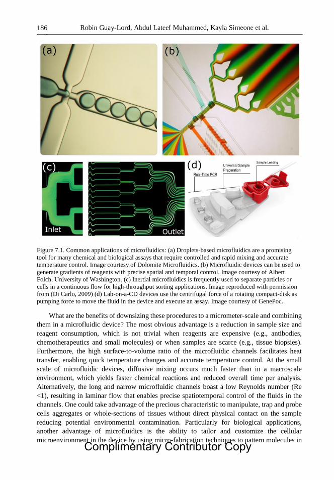
Robin Guay-Lord, Abdul Lateef Muhammed, Kayla Simeone et al. 186
Figure 7.1. Common applications of microfluidics: (a) Droplets-based microfluidics are a promising
tool for many chemical and biological assays that require controlled and rapid mixing and accurate
temperature control. Image courtesy of Dolomite Microfluidics. (b) Microfluidic devices can be used to
generate gradients of reagents with precise spatial and temporal control. Image courtesy of Albert
Folch, University of Washington. (c) Inertial microfluidics is frequently used to separate particles or
cells in a continuous flow for high-throughput sorting applications. Image reproduced with permission
from (Di Carlo, 2009) (d) Lab-on-a-CD devices use the centrifugal force of a rotating compact-disk as
pumping force to move the fluid in the device and execute an assay. Image courtesy of GenePoc.
What are the benefits of downsizing these procedures to a micrometer-scale and combining
them in a microfluidic device? The most obvious advantage is a reduction in sample size and
reagent consumption, which is not trivial when reagents are expensive (e.g., antibodies,
chemotherapeutics and small molecules) or when samples are scarce (e.g., tissue biopsies).
Furthermore, the high surface-to-volume ratio of the microfluidic channels facilitates heat
transfer, enabling quick temperature changes and accurate temperature control. At the small
scale of microfluidic devices, diffusive mixing occurs much faster than in a macroscale
environment, which yields faster chemical reactions and reduced overall time per analysis.
Alternatively, the long and narrow microfluidic channels boast a low Reynolds number (Re
<1), resulting in laminar flow that enables precise spatiotemporal control of the fluids in the
channels. One could take advantage of the precious characteristic to manipulate, trap and probe
cells aggregates or whole-sections of tissues without direct physical contact on the sample
reducing potential environmental contamination. Particularly for biological applications,
another advantage of microfluidics is the ability to tailor and customize the cellular
microenvironment in the device by using micro-fabrication techniques to pattern molecules in
Complimentary Contributor Copy
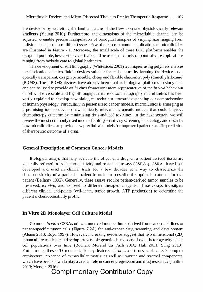
Microfluidic Devices and Micro-Dissected Tissue to Predict Therapeutic Response … 187
the device or by exploiting the laminar nature of the flow to create physiologically relevant
gradients (Young 2010). Furthermore, the dimensions of the microfluidic channel can be
adjusted to enable precise manipulation of biological samples of varying size ranging from
individual cells to sub-milliliter tissues. Few of the most common applications of microfluidics
are illustrated in Figure 7.1. Moreover, the small scale of these LOC platforms enables the
design of portable, low-cost devices that could be used in a variety of point-of-care applications
ranging from bedside care to global healthcare.
The development of soft lithography (Whitesides 2001) techniques using polymers enables
the fabrication of microfluidic devices suitable for cell culture by forming the device in an
optically transparent, oxygen permeable, cheap and flexible elastomer: poly (dimethylsiloxane)
(PDMS). These PDMS devices have already been used as biological platforms to study cells
and can be used to provide an in vitro framework more representative of the in vivo behaviour
of cells. The versatile and high-throughput nature of soft lithography microfluidics has been
vastly exploited to develop new biological techniques towards expanding our comprehension
of human physiology. Particularly in personalized cancer models, microfluidics is emerging as
a promising tool to develop new clinically relevant therapeutic models that could improve
chemotherapy outcome by minimizing drug-induced toxicities. In the next section, we will
review the most commonly used models for drug sensitivity screening in oncology and describe
how microfluidics can provide new preclinical models for improved patient-specific prediction
of therapeutic outcome of a drug.
General Description of Common Cancer Models
Biological assays that help evaluate the effect of a drug on a patient-derived tissue are
generally referred to as chemosensitivity and resistance assays (CSRAs). CSRAs have been
developed and used in clinical trials for a few decades as a way to characterize the
chemosensitivity of a particular patient in order to prescribe the optimal treatment for that
patient (Bellamy 1992). Generally, these assays require patient-derived tumor samples to be
preserved, ex vivo, and exposed to different therapeutic agents. These assays investigate
different clinical end-points (cell-death, tumor growth, ATP production) to determine the
patient’s chemosensitivity profile.
In Vitro 2D Monolayer Cell Culture Model
Common in vitro CSRAs utilize tumor cell monocultures derived from cancer cell lines or
patient-specific tumor cells (Figure 7.2A) for anti-cancer drug screening and development
(Abaan 2013; Boyd 1997). However, increasing evidence suggest that two dimensional (2D)
monoculture models can develop irreversible genetic changes and loss of heterogeneity of the
cell populations over time (Bounaix Morand du Puch 2016; Huh 2011; Sung 2013).
Furthermore, these 2D models lack key features of in vivo tissues such as 3D complex
architecture, presence of extracellular matrix as well as immune and stromal components,
which have been shown to play a crucial role in cancer progression and drug resistance (Junttila
2013; Morgan 2016).
Complimentary Contributor Copy
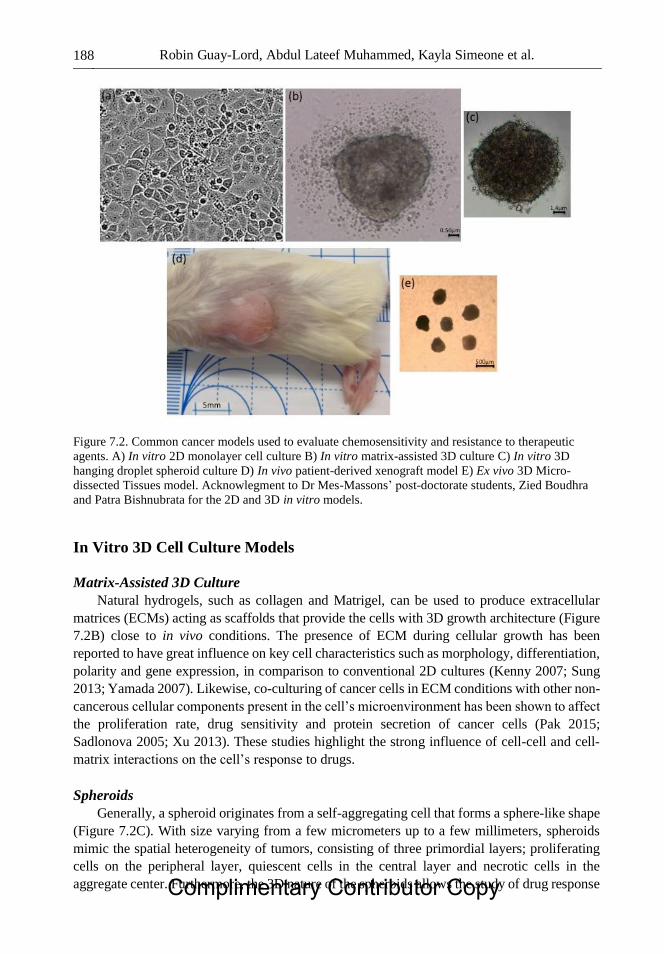
Robin Guay-Lord, Abdul Lateef Muhammed, Kayla Simeone et al. 188
Figure 7.2. Common cancer models used to evaluate chemosensitivity and resistance to therapeutic
agents. A) In vitro 2D monolayer cell culture B) In vitro matrix-assisted 3D culture C) In vitro 3D
hanging droplet spheroid culture D) In vivo patient-derived xenograft model E) Ex vivo 3D Micro-
dissected Tissues model. Acknowlegment to Dr Mes-Massons’ post-doctorate students, Zied Boudhra
and Patra Bishnubrata for the 2D and 3D in vitro models.
In Vitro 3D Cell Culture Models
Matrix-Assisted 3D Culture
Natural hydrogels, such as collagen and Matrigel, can be used to produce extracellular
matrices (ECMs) acting as scaffolds that provide the cells with 3D growth architecture (Figure
7.2B) close to in vivo conditions. The presence of ECM during cellular growth has been
reported to have great influence on key cell characteristics such as morphology, differentiation,
polarity and gene expression, in comparison to conventional 2D cultures (Kenny 2007; Sung
2013; Yamada 2007). Likewise, co-culturing of cancer cells in ECM conditions with other non-
cancerous cellular components present in the cell’s microenvironment has been shown to affect
the proliferation rate, drug sensitivity and protein secretion of cancer cells (Pak 2015;
Sadlonova 2005; Xu 2013). These studies highlight the strong influence of cell-cell and cell-
matrix interactions on the cell’s response to drugs.
Spheroids
Generally, a spheroid originates from a self-aggregating cell that forms a sphere-like shape
(Figure 7.2C). With size varying from a few micrometers up to a few millimeters, spheroids
mimic the spatial heterogeneity of tumors, consisting of three primordial layers; proliferating
cells on the peripheral layer, quiescent cells in the central layer and necrotic cells in the
aggregate center. Furthermore, the 3D nature of the spheroids allows the study of drug response Complimentary Contributor Copy

Microfluidic Devices and Micro-Dissected Tissue to Predict Therapeutic Response … 189
and the effect of hypoxia through its ability to transport natural gradients of oxygen nutrients
and metabolic waste.
For those reasons, spheroids have been generally adopted as a new therapeutic model and
a number of microfluidic devices have been designed to allow the formation of spheroids as
well as to study the chemosensitivity of these spheroids on-chip (Hirschhaeuser 2010; Shield
2009; Weiswald 2015). For instance, Torisawa et al. have reported a microfluidic device that
allows on-chip generation of multicellular spheroids and long-term analysis of cellular viability
following treatment of the spheroids in the chip (Torisawa 2007). While spheroids provide a
3D structure that better reflects in vivo tumor architecture than 2D monocultures, these models
still fail to include components of tumor microenvironment that highly influence tumor cell
behaviour.
Organoid Culture
Organoid culture models have emerged in effort to include both the in vivo
microenvironment and the complex architecture of tumors. Organoids are derived from the
stem cells that are grown in ECM, which allow the cells to differentiate and migrate to recreate
the specific architecture and function of an organ (Morgan 2016). Fully-grown organoids are
composed of multiple cell types and exhibit intra- and inter-cellular communication networks
as well as cell signalling pathways that reflect the physiological in vivo behaviour of organs.
Furthermore, the organoids can be cultured for a long period of time while maintaining
histological, immunohistological and genetic properties of the tissue, making them ideal for
drug sensitivity assays, clearly observed by Santo et al. through the generation gut-specific
crypt-villus structures originating from a single intestinal stem cell (Sato 2009).
In Vivo Patient-Derived Xenograft Model
Patient-Derived Xenograft
At the other end of the experimental continuum, in vivo CSRAs are most commonly carried
out on patient-derived xenografts (PDXs). To generate a PDX, a small portion of a patient’s
tumor tissue is engrafted subcutaneously in an immune-compromised mouse, where it grows
in vivo (Figure 7.2D) in conditions that maintain the local human tumor microenvironment.
Such models have the advantage of retaining key characteristics of the original tumor tissue
such as tumor heterogeneity, complex 3D architecture, and genetic makeup. Furthermore,
genetic analysis revealed that the PDX tumors share the same genetic mutations in the stromal
cells and immune cells, with some minor exception. These exceptions may be attributed to the
fact that PDX tumors are generated in immune-compromised mice and that tumor stromal cells
can be infiltrated by host stromal cells (De Wever 2003; Marangoni 2007; Reyal 2012).
Recently published reports suggest that the genetic expression profile of the donor and the PDX
tumor were similar but those of the cell lines generated from the same donor tumor showed a
very different profile (Bertotti 2011; Fichtner 2008; Julien 2012; Zhang 2013). While PDXs
are an interesting model for drug screening and studying treatment effects on cancer
progression, these models still present limitations that need to be addressed. For instance, it
normally takes 3−4 months to generate a PDX tumor model that corresponds to a donor tumor,
making it inappropriate to determine the chemosensitivity of a patient before the first treatment
Complimentary Contributor Copy

Robin Guay-Lord, Abdul Lateef Muhammed, Kayla Simeone et al. 190
is initiated. Additionally, as these mice lack a functional immune system, their use in immune
drug screening is impractical. Moreover, these models require a lot of resources and are
extremely costly to maintain (Ellis 2010). In effort to bridge the gap between 2D/3D
monocultures and PDXs, a wide selection of in vitro microfluidics-based assays of varying
complexity has been developed to improve our understanding of therapeutic response.
Ex vivo 3D Models
Organotypic Tissue Slices
Although the biological models presented thus far help to understand how the 3D
architecture and cell-matrix contacts influence the therapeutic outcome of a drug, matching the
in vitro complexity of in vivo tumors is still an unmet challenge. Instead of artificially recreating
the complex microenvironment of a tissue, one step towards this goal is to produce organotypic
cultures that preserves both in vitro tissue viability and the original structure of the tissue in an
ex vivo environment. These models generally require the tumor tissue to be extracted by surgery
and sectioned in smaller pieces to allow diffusion of oxygen and nutrients to the center of the
tissue. As it preserves some of the tissue’s original in vivo architecture and maintains cell-
matrix interactions from the primary tissue, organotypic cultures offer the potential of
predicting in vivo behaviour of the tissue. It is an outstanding tool used to characterize the
therapeutic outcome of a drug for a specific patient tissue. Black first described this tool in
1951 where epithelial carcinomas (breast, colon, kidney, ovary, etc.) are extracted by surgery
and sliced free hand to a size of approximately 0.5 cm by 0.5 cm and a thickness of 1 mm
(Black 1951). The therapeutic response is evaluated by monitoring dehydrogenase activity of
the tissues fragments exposed to various chemotherapy treatments or enzyme inhibitors.
A few years later, Thomlinson and Gray used histological analyses to study the mass
transport limitation in human lung tumors and showed that large tumors exhibit a hypoxic core
in their center (Thomlinson 1955). Based on the diffusive properties of metabolites and the
general intake of nutrients by the tissue, they determined a critical diameter that a tumor core
should not exceed: if one dimension of the cultured tissue is inferior to this critical diameter,
the diffusion of metabolites through the tissue is enough to prevent depletion of nutrients. In
this scope, different research groups began to slice tissue biopsies in thin slices (300–400 μm)
before cultivating them to prevent diffusion-limited hypoxia (Figure 7.3). For instance, van
Midwoud et al. reported that it is possible to study the metabolism of a precision-cut rat liver
slice culture on-chip for a minimum period of 24 hours under continuous perfusion (van
Midwoud 2010). Later, Chang et al. introduced a microfluidic device compatible with a 96-
well plate allowing long-term culture of mouse brain slices and parallel chemosensitivity
testing on the individual slices (Chang 2014).
Complimentary Contributor Copy

Microfluidic Devices and Micro-Dissected Tissue to Predict Therapeutic Response … 191
Figure 7.3. Organotypic models published previously in the literature. (a) Microfluidic chip that was
used by Hattersley et al. to culture liver tissue biopsies. The device comprises a large tissue chamber
where the tissue is perfused and can remain viable for over 70h. Image reproduced with permission
from (Hattersley, 2008). (b) Microfluidic device reported by Chang et al. that allows organotypic
culture of liver slices and parallel chemosensitivity screening on single liver slices. The multi-layered
device is compatible with a 96-wells plate and enables the treatment of the sliced tissue with multiple
compounds for high-throughput screening assays. Image reproduced with permission from (Chang,
2014). (c) Microfluidic platform developed by van Midwoud et al. to culture rat liver slices under
continuous perfusion to study the metabolism and toxicology of the liver slices. Image reproduced with
permission from (van Midwoud, 2010).
The main shortcoming of organotypic slice culture relies on the manipulation of large tissue
slices without damaging the structure of the tissue, making such a model inappropriate for high-
throughput drug screening on a large number of tissue slices. Moreover, long-term culture of
large tissue slices usually requires continuous perfusion of the system to prevent hypoxia and
nutrient starvation. Such perfusion and associated fluidic connections are cumbersome thus
reducing the number of tissue slices that can be multiplexed in a single device, once again
affecting the high-throughput screening capabilities of the model. Complimentary Contributor Copy

Robin Guay-Lord, Abdul Lateef Muhammed, Kayla Simeone et al. 192
Micro-Dissected Tissue Culture
In effort to address these shortcomings, our group developed a new model for high-
throughput in vitro study of therapeutic drug response on ex vivo patient-derived tissues (Figure
7.4). Briefly, tumor samples are micro-dissected and loaded in a microfluidic device where they
are trapped and cultured. The micro-dissected tissue (MDT) samples are exposed to various
therapeutic agents in the device and therapeutic response is quantified to determine the optimal
treatment for that particular patient. The novelty of the presented model relies on the micro-
dissection of the patient’s tumor biopsy to enable precise spatiotemporal control of MDTs in a
microfluidic device. The device consists of parallel fluidic channels to help trap and cultivate
multiple MDTs thus assessing chemosensitivity of the tissue in a simple, high-throughput and
multiplexed manner. Moreover, the micro-dissection of the tumor sample in sub-milliliter
samples eliminates the need for continuous perfusion. The small diameter of the MDTs allows
simple diffusion of oxygen through the PDMS matrix, enough to prevent hypoxia in the center.
This novel biological model for drug screening combines the physiological relevance of the
sliced tissue culture with the simple manipulations of small spheroid-sized tissue sample. The
following sections of this chapter will allow in-depth understanding of this new MDT-based
organotypic model, highlighting its clinically relevant chemosensitivity screening potential.
Figure 7.4. Presentation of the proposed personalized treatment model. 1) A tumor biopsy is extracted
through biopsy or surgery. 2) Micro-dissection of the tumor biopsy to produce micro-dissected tissues
(MDTs). 3) Loading and trapping of the MDTs in the microfluidic device, followed by treatment with
different therapeutic agents directly in the device. 4) Evaluation of the therapeutic response with
confocal microscopy and fluorescence activated cell sorting. 5) Results are interpreted and used to
select the most effective treatment for that patient.
Complimentary Contributor Copy

Microfluidic Devices and Micro-Dissected Tissue to Predict Therapeutic Response … 193
MICROFLUIDIC PLATFORM
Design and Fabrication of the Microfluidic Platform
Microfluidic devices dedicated to cell culture have certain requirements that distinguish
them from micro-scale devices used commonly for analytical chemistry or physics. In the case
of a microfluidic device that traps and preserves ex vivo tissues, design considerations of
particular importance are: (1) the choice of material for the device, (2) choice of trapping
mechanism, and (3) geometry and dimensions of the channels. We will review each of these
considerations and present the important design characteristics of the microfluidic device. We
will then describe the fabrication process to produce the microfluidic chip.
Description of Polymer Used for Microfluidic Design
The development of soft lithography by Whitesides and his group in the late 1990s opened
new avenues for microfluidic-based cellular applications (Whitesides 2001). Soft lithography
comprises a set fabrication technique similar to photolithography, but uses silicon rubber
instead due to low-cost and easy fabrication than conventional silicon-based materials. PDMS
has several unique properties that make it an ideal choice of material for cell culture
applications. First and foremost, unlike silicon or glass, PDMS has a high gas permeability that
ensures sufficient oxygen supply to the cells in the device, eliminating the need for separate
oxygen chambers. This property is particularly important when cultivating cells that have high
metabolic demands like cancer cells. Another advantage of PDMS is its optical transparency
and low autofluorescence, both in the visible and infrared regions. This property has been vastly
exploited to couple microfluidic devices with imaging modalities that allow assessing the
viability of the tissue directly on-chip. Moreover, PDMS is fairly cheap and easy to mold,
making it ideal for rapid prototyping and mass production of microfluidic devices. Finally, to
form enclosed micro-channels, PDMS can be bonded to different materials (e.g., PDMS, glass,
polystyrene) using simple methods such as oxygen plasma treatment.
Sample Trapping Mechanism
As mentioned previously, there are numerous advantages provided by microfluidic
devices, particularly when working with biological samples. These devices have the ability to
enclose a biological sample in a chamber allowing for easy perfusion and use of smaller
volumes of reagents than conventional macro-scale techniques (i.e., 96-wells plate).
Microfluidic devices are transparent such that samples can be imaged directly on-chip,
eliminating the need to manipulate the sample once it is loaded in the device. For all those
reasons, research groups have used microfluidic chips to trap and enclose various types of tissue
samples of different sizes ranging from single-cells to tissue slices. For specific sub-microliter
particle trapping, such as spheroids, cell aggregates and MDTs, numerous trapping mechanisms
have been reported. These mechanisms include acoustic (Liu 2007), mechanical (Khoury
2010), resistive (Ruppen 2014) and sedimentation trapping (Ruppen 2015). The latter Complimentary Contributor Copy
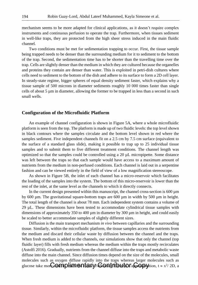
Robin Guay-Lord, Abdul Lateef Muhammed, Kayla Simeone et al. 194
mechanism seems to be more adapted for clinical applications, as it doesn’t require complex
instruments and continuous perfusion to operate the trap. Furthermore, when tissues sediment
in well-like traps, they are protected from the high sheer stress induced in the main fluidic
channel.
Two conditions must be met for sedimentation trapping to occur. First, the tissue sample
being trapped needs to be denser than the surrounding medium for it to sediment to the bottom
of the trap. Second, the sedimentation time has to be shorter than the travelling time over the
trap. Cells are slightly denser than the medium in which they are cultured because the organelles
and proteins they contain are denser than water. This is exploited in petri-dish cultures where
cells need to sediment to the bottom of the dish and adhere to its surface to form a 2D cell layer.
In steady-state regime, bigger spheres of equal density sediment faster, which explains why a
tissue sample of 500 microns in diameter sediments roughly 10 000 times faster than single
cells of about 5 µm in diameter, allowing the former to be trapped in less than a second in such
small wells.
Configuration of the Microfluidic Platform
An example of channel configuration is shown in Figure 5A, where a whole microfluidic
platform is seen from the top. The platform is made up of two fluidic levels: the top level shown
in black contours where the samples circulate and the bottom level shown in red where the
samples sediment. Five independent channels fit on a 2.5 cm by 7.5 cm surface (equivalent to
the surface of a standard glass slide), making it possible to trap up to 25 individual tissue
samples and to submit them to five different treatment conditions. The channel length was
optimized so that the samples could be controlled using a 20 µL micropipette. Some distance
was left between the traps so that each sample would have access to a maximum amount of
nutrients from the medium in non-perfused conditions. Each channel is laid out in a serpentine
fashion and can be viewed entirely in the field of view of a low magnification stereoscope.
As shown in Figure 5B, the inlet of each channel has a micro-reservoir which facilitates
the loading of the samples into the system. The bottom of this micro-reservoir is lower than the
rest of the inlet, at the same level as the channels to which it directly connects.
In the current design presented within this manuscript, the channel cross-section is 600 µm
by 600 µm. The gravitational square-bottom traps are 600 µm in width by 500 µm in height.
The total length of the channel is about 78 mm. Each independent system contains a volume of
29 µL. These dimensions have been tested to accommodate cylindrical tissue samples with
dimensions of approximately 350 to 400 µm in diameter by 300 µm in height, and could easily
be scaled to better accommodate samples of slightly different sizes.
Diffusion is the main transport mechanism in vivo between capillaries and the surrounding
tissue. Similarly, within the microfluidic platform, the tissue samples access the nutrients from
the medium and discard their cellular waste by diffusion between the channel and the traps.
When fresh medium is added to the channels, our simulations show that only the channel (top
fluidic layer) fills with fresh medium whereas the medium within the traps mostly recirculates
(Astolfi 2016). Gradually, nutrients from the channel diffuse into the traps and metabolic waste
diffuse into the main channel. Since diffusion times depend on the size of the molecules, small
molecules such as oxygen diffuse rapidly into the traps whereas larger molecules such as
glucose take more time to reach the tissue. Following Einstein’s approximation, t x2/ 2D, a Complimentary Contributor Copy
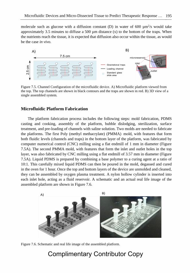
Microfluidic Devices and Micro-Dissected Tissue to Predict Therapeutic Response … 195
molecule such as glucose with a diffusion constant (D) in water of 600 µm2/s would take
approximately 3.5 minutes to diffuse a 500 µm distance (x) to the bottom of the traps. When
the nutrients reach the tissue, it is expected that diffusion also occur within the tissue, as would
be the case in vivo.
Figure 7.5. Channel Configuration of the microfluidic device. A) Microfluidic platform viewed from
the top. The top channels are shown in black contours and the traps are shown in red. B) 3D view of a
single assembled system.
Microfluidic Platform Fabrication
The platform fabrication process includes the following steps: mold fabrication, PDMS
casting and cooking, assembly of the platform, bubble dislodging, sterilization, surface
treatment, and pre-loading of channels with saline solution. Two molds are needed to fabricate
the platforms. The first Poly (methyl methacrylate) (PMMA) mold, with features that form
both fluidic levels (channels and traps) in the bottom layer of the platform, was fabricated by
computer numerical control (CNC) milling using a flat endmill of 1 mm in diameter (Figure
7.5A). The second PMMA mold, with features that form the inlet and outlet holes in the top
layer, was also fabricated by CNC milling using a flat endmill of 3.57 mm in diameter (Figure
7.5A). Liquid PDMS is prepared by combining a base polymer to a curing agent at a ratio of
10:1. This carefully mixed liquid PDMS can then be poured in the mold, degassed and cured
in the oven for 1 hour. Once the top and bottom layers of the device are unmolded and cleaned,
they can be assembled by oxygen plasma treatment. A nylon hollow cylinder is inserted into
each inlet hole, acting as a fluid reservoir. A schematic and an actual real life image of the
assembled platform are shown in Figure 7.6.
Figure 7.6. Schematic and real life image of the assembled platform.
Complimentary Contributor Copy
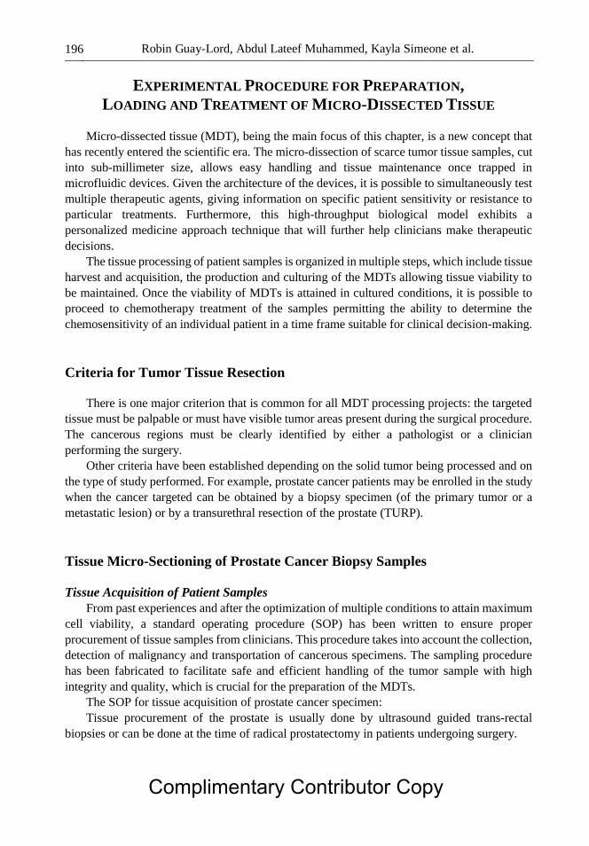
Robin Guay-Lord, Abdul Lateef Muhammed, Kayla Simeone et al. 196
EXPERIMENTAL PROCEDURE FOR PREPARATION,
LOADING AND TREATMENT OF MICRO-DISSECTED TISSUE
Micro-dissected tissue (MDT), being the main focus of this chapter, is a new concept that
has recently entered the scientific era. The micro-dissection of scarce tumor tissue samples, cut
into sub-millimeter size, allows easy handling and tissue maintenance once trapped in
microfluidic devices. Given the architecture of the devices, it is possible to simultaneously test
multiple therapeutic agents, giving information on specific patient sensitivity or resistance to
particular treatments. Furthermore, this high-throughput biological model exhibits a
personalized medicine approach technique that will further help clinicians make therapeutic
decisions.
The tissue processing of patient samples is organized in multiple steps, which include tissue
harvest and acquisition, the production and culturing of the MDTs allowing tissue viability to
be maintained. Once the viability of MDTs is attained in cultured conditions, it is possible to
proceed to chemotherapy treatment of the samples permitting the ability to determine the
chemosensitivity of an individual patient in a time frame suitable for clinical decision-making.
Criteria for Tumor Tissue Resection
There is one major criterion that is common for all MDT processing projects: the targeted
tissue must be palpable or must have visible tumor areas present during the surgical procedure.
The cancerous regions must be clearly identified by either a pathologist or a clinician
performing the surgery.
Other criteria have been established depending on the solid tumor being processed and on
the type of study performed. For example, prostate cancer patients may be enrolled in the study
when the cancer targeted can be obtained by a biopsy specimen (of the primary tumor or a
metastatic lesion) or by a transurethral resection of the prostate (TURP).
Tissue Micro-Sectioning of Prostate Cancer Biopsy Samples
Tissue Acquisition of Patient Samples
From past experiences and after the optimization of multiple conditions to attain maximum
cell viability, a standard operating procedure (SOP) has been written to ensure proper
procurement of tissue samples from clinicians. This procedure takes into account the collection,
detection of malignancy and transportation of cancerous specimens. The sampling procedure
has been fabricated to facilitate safe and efficient handling of the tumor sample with high
integrity and quality, which is crucial for the preparation of the MDTs.
The SOP for tissue acquisition of prostate cancer specimen:
Tissue procurement of the prostate is usually done by ultrasound guided trans-rectal
biopsies or can be done at the time of radical prostatectomy in patients undergoing surgery.
Complimentary Contributor Copy

Microfluidic Devices and Micro-Dissected Tissue to Predict Therapeutic Response … 197
1) By using true-cut biopsy needles one can obtain specimens of 2 mm in diameter ×
20 mm in length.
2) 5–6 biopsies of the suspected prostate region with palpable or visible tumor should
be obtained.
3) Biopsy specimens should be kept in Hanks' Balanced Salt Solution (HBSS, #311-
516-CL, Wisent Inc., Saint-Bruno-de-Montarville, Canada) supplemented with
10% fetal bovine serum (FBS, BD Biosicences) and 55 mg/L gentamicin (Wisent)
and 0.6 mg/L amphotericin (Wisent) (HBSScomplete) and further transported on ice
in aseptic conditions.
HBSS is a balanced salt solution that helps maintain tissue pH and osmotic balance. This
solution thus provides water and essential inorganic ions such as calcium and magnesium to
support vital cellular activity. Additionally, HBSS contains glucose, a main nutrient allowing
viability of tissue biopsy to be sustained for a longer period of time. Recent studies have
compared HBSS to regular phosphate-buffered saline (PBS) in terms of tissue viability and
have shown that HBSS better supports whole tissue maintenance compared to PBS thus making
it tissue specific (Astolfi 2016).
For solid tumors that require the identification of cancerous regions by a pathologist, here
is the SOP for tissue processing and sectioning by the pathologist:
1) A sterile scalpel is used to cut the tumor where there is suspicion of tumor cells.
2) Freeze a fresh, unfixed tissue sample, up to 2.0 cm in diameter, at optimal cutting
temperature (OCT) in a suitable tissue mold onto the specialized metal grids that
fit onto the cryostat. NOTE: Approximate freezing times at -24 oC using a cold
chuck ranges between 20–60 seconds depending on tissue depth.
3) Cut sections 5−15 µm-thick in the cryostat at -20°C. If necessary, adjust the
temperature of the cutting chamber ± 5°C, according to the tissue under study. A
hairbrush is useful to help guide the emerging section over the knife blade.
4) Within the first minute of cutting the tissue section, transfer it directly onto a
microscope slide at room temperature. By letting the slide touch the tissue, the
tissue section will melt onto the slide. This must be accomplished within the first
minute of cutting the section to avoid freeze-drying of the tissue. The slide is then
coated with Poly-L-lysine to improve adherence of the section to the slide.
5) To evaluate tissue preservation and orientation, stain the first couple of slides with
toluidine blue (1−2% w/v in H2O), hematoxylin, and eosin, or any aqueous stain.
6) The pathologist examines the slides and determines the approximate probability of
tumor cell in the sample.
7) Using a clean and sterile blade, the mirror section of the tumor sample is cut and
placed in HBSScomplete.
8) The sample is picked up by a technician and transported on ice to the processing
laboratory
9) The tumor tissue is either processed right away or stored at 4 oC for no more than
24 hours in HBSScomplete. Complimentary Contributor Copy

Robin Guay-Lord, Abdul Lateef Muhammed, Kayla Simeone et al. 198
Microfluidic Chip Preparation
The microfluidic devices must be prepared one day prior to micro-dissection of the patient
sample. It is important that the microfluidic chips go through several decontamination and
sterilization procedures. Firstly, the devices are treated with 100% ethanol to decontaminate
the chips followed by 70% ethanol incubation at room temperature for 15 minutes in to allow
sterilization. Next, they are treated overnight in a humidified cell culture incubator (37°C, 5%
CO2, 95% ambient air) with a 10 mg/mL triblock copolymer, Pluronic F-108 (Sigma-Aldrich,
St-Louis, USA) to reduce cell adhesion on the PDMS surfaces. The following day, the devices
are re-sterilized through 70% ethanol incubation for 15 minutes at room temperature. Lastly,
they are thoroughly rinsed using sterile HBSS supplemented with 55 mg/L gentamicin and 0.6
mg/L amphotericin (HBSSantibio) and placed at 4˚C until the MDTs are produced and ready to
be loaded in the devices.
Generation of Micro-Dissected Tumor Tissue
Detail of Micro-Dissection Protocol
Prostate cancer biopsy samples are approximately 2 cm in length and 1 mm in diameter.
These fragments are cut into 1 to 2 mm long fragments and immediately embedded in low
melting-point agarose supplemented with 10% FBS, which is kept in a liquid form at 400C. The
agarose solidifies on ice to obtain a supporting structure around the tissue. The excess agarose
is then removed and the fragments are repositioned vertically and glued to the stage for slicing
into 300 µm thick slices using a traditional vibratome. These slices were further punched, using
a 400 µm biopsy punch, into disk-like tumor micro-sections, MDTs. On average, a single
prostate cancer biopsy can produce 80 MDTs, thus with 5–6 biopsies, we can easily obtain
between 400 to 480 MDTs from one single patient sample. The MDTs obtained from each
biopsy are pooled together in a petri plate, submerged in HBSSantibio, to ensure randomized
selection of the MDTs allowing drug testing of heterogeneous tissues. The MDTs obtained are
then carefully loaded in the customized microfluidic chips under a stereomicroscope and
cultured in specific culture medium, Roswell Park Memorial Institute (RPMI 1640) (#350-045-
CL, Wisent Inc.) medium used for prostate cancer sample culturing. For the loading procedure,
the hollow nylon cylinders that form the inlet reservoirs are removed and the microfluidic
device is placed under a stereomicroscope. For each individual channel, 5 MDTs are collected
using a P20 micropipette and introduced in the inlet, where they sediment to the bottom of the
channel. By aspirating fluid from the outlet of the device, flow is induced in the microfluidic
channels allowing the MDTs to travel in the channel to attain the wells. Once the MDTs are
above the wells the flow is stopped for approximately 1 to 2 seconds allowing them to sediment
to the bottom of the traps. The same procedure is repeated to fully load the 5 channels of the
device, resulting in a total number of 25 MDTs per device (for visual comprehension, the whole
process is illustrated in Figure 7.7). Furthermore, This RPMIcomplete medium used does not
contain phenol but is supplemented with L-glutamine, 10% FBS, 55 mg/L gentamicin and 0.6
mg/L amphotericin and allows whole cell overall survival of MDTs in our microfluidic devices
to be between 60 to 95% (Astoli, 2016).
Complimentary Contributor Copy
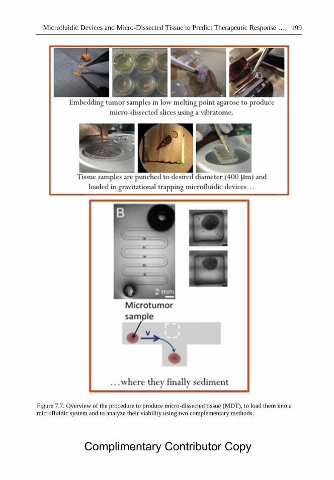
Microfluidic Devices and Micro-Dissected Tissue to Predict Therapeutic Response … 199
Figure 7.7. Overview of the procedure to produce micro-dissected tissue (MDT), to load them into a
microfluidic system and to analyze their viability using two complementary methods.
Complimentary Contributor Copy

Robin Guay-Lord, Abdul Lateef Muhammed, Kayla Simeone et al. 200
Tissue Sectioning Reproducibility
The distribution of size of the MDTs produced was determined by confocal microscopy
analysis. Using maximum projection imaging, the diameter of each MDT was calculated as the
average of two perpendicular diameter measurements. Upon analysis, the diameter of the
MDTs remained relatively stable with time and slight variability was detected. This variability
was further explained by differences in mechanical properties of the tumor tissues, especially
their elasticity, by wear and tear of the biopsy punch and slight differences in sectioning
procedure between users. It was clearly noted that tissue with higher elasticity could easily be
stretched out during sectioning due to compression force applied by the biopsy punch and can
later be brought back into its original configuration.
Irrespective of the tumor type, the diameters of 247 produced MDTs seem to follow a
normal distribution with an average (µ) of 380 µm and a standard deviation (σ) of 43 µm
(Figure 7.8) (Astolfi 2016). The ratio σ/µ being less than 12% indicates that the sectioning
procedure is highly reproducible.
Figure 7.8. Histogram (blue bars) with normal fit (red curve) representing the diameter of microsections
of xenograft models of various cell lines.
Culturing of Micro-Dissected Tissues and Chemotherapy Treatments
To ensure cell viability and tumor survival of the MDTs at harvest day, HBSSantibio was
replaced by fresh RPMIcomplete medium. The MDTs are continuously incubated at 370C, 5%
CO2 for the remaining period. The medium is changed 24 hours subsequent to harvest day to
ensure proper circulation of nutrients and avoid glucose deprivation, followed by 24 to 48 hours
of recovery period to acclimatize the MDTs within the systems before further treatment with
different therapeutic agents.
The concentration of the chemotherapy agents used to treat the MDTs was determined by
the IC50 obtained by monolayer cell line cultures that are generally used to generate xenografts.
Recent studies have demonstrated that at a concentration of 10 times the IC50 of chemotherapy Complimentary Contributor Copy
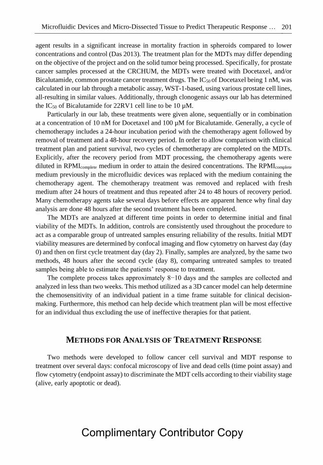
Microfluidic Devices and Micro-Dissected Tissue to Predict Therapeutic Response … 201
agent results in a significant increase in mortality fraction in spheroids compared to lower
concentrations and control (Das 2013). The treatment plan for the MDTs may differ depending
on the objective of the project and on the solid tumor being processed. Specifically, for prostate
cancer samples processed at the CRCHUM, the MDTs were treated with Docetaxel, and/or
Bicalutamide, common prostate cancer treatment drugs. The IC50 of Docetaxel being 1 nM, was
calculated in our lab through a metabolic assay, WST-1-based, using various prostate cell lines,
all-resulting in similar values. Additionally, through clonogenic assays our lab has determined
the IC50 of Bicalutamide for 22RV1 cell line to be 10 µM.
Particularly in our lab, these treatments were given alone, sequentially or in combination
at a concentration of 10 nM for Docetaxel and 100 µM for Bicalutamide. Generally, a cycle of
chemotherapy includes a 24-hour incubation period with the chemotherapy agent followed by
removal of treatment and a 48-hour recovery period. In order to allow comparison with clinical
treatment plan and patient survival, two cycles of chemotherapy are completed on the MDTs.
Explicitly, after the recovery period from MDT processing, the chemotherapy agents were
diluted in RPMIcomplete medium in order to attain the desired concentrations. The RPMIcomplete
medium previously in the microfluidic devices was replaced with the medium containing the
chemotherapy agent. The chemotherapy treatment was removed and replaced with fresh
medium after 24 hours of treatment and thus repeated after 24 to 48 hours of recovery period.
Many chemotherapy agents take several days before effects are apparent hence why final day
analysis are done 48 hours after the second treatment has been completed.
The MDTs are analyzed at different time points in order to determine initial and final
viability of the MDTs. In addition, controls are consistently used throughout the procedure to
act as a comparable group of untreated samples ensuring reliability of the results. Initial MDT
viability measures are determined by confocal imaging and flow cytometry on harvest day (day
0) and then on first cycle treatment day (day 2). Finally, samples are analyzed, by the same two
methods, 48 hours after the second cycle (day 8), comparing untreated samples to treated
samples being able to estimate the patients’ response to treatment.
The complete process takes approximately 8−10 days and the samples are collected and
analyzed in less than two weeks. This method utilized as a 3D cancer model can help determine
the chemosensitivity of an individual patient in a time frame suitable for clinical decision-
making. Furthermore, this method can help decide which treatment plan will be most effective
for an individual thus excluding the use of ineffective therapies for that patient.
METHODS FOR ANALYSIS OF TREATMENT RESPONSE
Two methods were developed to follow cancer cell survival and MDT response to
treatment over several days: confocal microscopy of live and dead cells (time point assay) and
flow cytometry (endpoint assay) to discriminate the MDT cells according to their viability stage
(alive, early apoptotic or dead).
Complimentary Contributor Copy
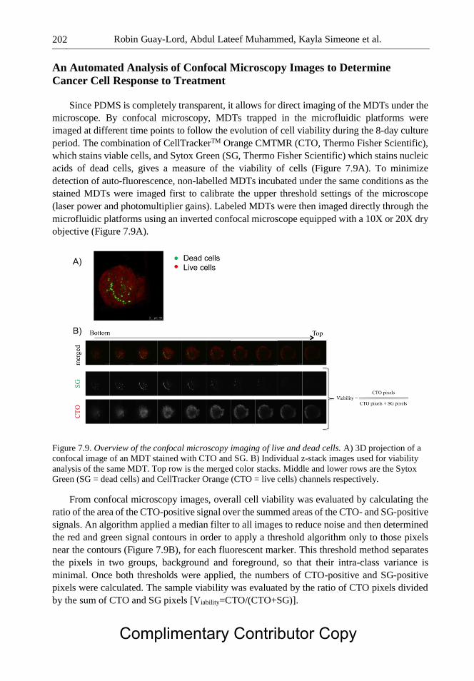
Robin Guay-Lord, Abdul Lateef Muhammed, Kayla Simeone et al. 202
An Automated Analysis of Confocal Microscopy Images to Determine
Cancer Cell Response to Treatment
Since PDMS is completely transparent, it allows for direct imaging of the MDTs under the
microscope. By confocal microscopy, MDTs trapped in the microfluidic platforms were
imaged at different time points to follow the evolution of cell viability during the 8-day culture
period. The combination of CellTrackerTM Orange CMTMR (CTO, Thermo Fisher Scientific),
which stains viable cells, and Sytox Green (SG, Thermo Fisher Scientific) which stains nucleic
acids of dead cells, gives a measure of the viability of cells (Figure 7.9A). To minimize
detection of auto-fluorescence, non-labelled MDTs incubated under the same conditions as the
stained MDTs were imaged first to calibrate the upper threshold settings of the microscope
(laser power and photomultiplier gains). Labeled MDTs were then imaged directly through the
microfluidic platforms using an inverted confocal microscope equipped with a 10X or 20X dry
objective (Figure 7.9A).
Figure 7.9. Overview of the confocal microscopy imaging of live and dead cells. A) 3D projection of a
confocal image of an MDT stained with CTO and SG. B) Individual z-stack images used for viability
analysis of the same MDT. Top row is the merged color stacks. Middle and lower rows are the Sytox
Green (SG = dead cells) and CellTracker Orange (CTO = live cells) channels respectively.
From confocal microscopy images, overall cell viability was evaluated by calculating the
ratio of the area of the CTO-positive signal over the summed areas of the CTO- and SG-positive
signals. An algorithm applied a median filter to all images to reduce noise and then determined
the red and green signal contours in order to apply a threshold algorithm only to those pixels
near the contours (Figure 7.9B), for each fluorescent marker. This threshold method separates
the pixels in two groups, background and foreground, so that their intra-class variance is
minimal. Once both thresholds were applied, the numbers of CTO-positive and SG-positive
pixels were calculated. The sample viability was evaluated by the ratio of CTO pixels divided
by the sum of CTO and SG pixels [Viability=CTO/(CTO+SG)].
Complimentary Contributor Copy
Microfluidic Devices and Micro-Dissected Tissue to Predict Therapeutic Response … 203
Characterization of Treatment Response of Each Cell Type Using Flow
Cytometry Assays
Flow cytometry was employed as an endpoint assay to analyze the cell population
(epithelial, immune or stromal cells) and their viability before and after the MDTs had been
exposed to different chemotherapy agents. MDTs were analyzed at different time points to
reflect the evolution of cell death over the 8-day incubation period. All fifteen MDTs from the
three channels were pooled in the same tube for analysis. A single cell suspension of MDTs
was obtained through enzymatic digestion using a combination of crude and type 1A
collagenase and mechanical action of gentle repeated pipetting. The resulting suspension is
analyzed by flow cytometry. All medium fractions collected from the three channels during the
medium changes and stored at 4°C were also pooled together with the digested MDT
suspension. This step was added to take into account all the live and dead cells that had been
washed out of the systems during medium changes in the microfluidic platform. Single cells
from the homogenous cell suspension were then stained with the apoptotic fluorescent dyes
Annexin V for apoptosis and DRAQ7TM for dead cells, unstained cells were considered as
viable cells for flow cytometry analyses. The cell survival was gated in forward and side scatter
graphs, as shown in Figure 7.10. A survival state was associated for each cell according to its
dye staining (live, early apoptotic or dead).
Figure 7.10. Typical representation of flow cytometry analysis. Each cell (represented by a blue dot)
obtained from the MDT was associated to one of three viability categories according to its fluorescence
intensity in both DRAQ7 and Annexin V detection channels: early apoptotic cells (Annexin V-stained
only), late apoptotic / dead cells (DRAQ7-stained), and live cells (non stained).
Preliminary Analysis of Treatment Response
Patient A was a TURP sample that had developed a castration-resistant prostate cancer
(CRPC). The MDTs prepared from this patient sample were cultured for 8 days in the
microfluidic platforms and received two complete treatment cycles (for details, see section
Culturing of Micro-Dissected Tissues and Chemotherapy Treatments). Confocal 3D projection
Complimentary Contributor Copy
Robin Guay-Lord, Abdul Lateef Muhammed, Kayla Simeone et al. 204
imaging of treated MDTs allowed for quantitative evaluation and effect of two common
prostate cancer treatment drugs. The viability of prostate cancer MDTs is shown below with
the live cells in red (CTO) and dead cells in green (SG). The percent viability of the control
samples at 95.8% 3.1% was compared with the 2 treatment conditions, Drug A at 90.5%
2.4% and Drug B at 83.1% 2.8% (Figure 7.11), indicating a decrease in viability in the Drug
B treated samples with respect to the control. In parallel, flow cytometry technique was used to
confirm MDT viability and drug sensitivity. The percent viability of the control MDT samples
at 62.4% 10.1% was compared with the 2 treatment conditions, Drug A at 63.1% 4.8% and
Drug B at 35.2% 2.3% (Figure 7.12). The preliminary results from both techniques show an
increase in Drug B sensibility compared to Drug A. However, the viability results of flow
cytometry were quite inferior to those obtained by confocal microscopy. Differences between
these two techniques were explained by the identification of early apoptotic cells in the flow
cytometry analysis (Figure 7.12), which might still appear CTO-positive in the dual-stain
confocal microscopy analysis.
Figure 7.11. 3D projections of confocal z-stack images for prostate
patient A taken 8 days after surgery. Treatment applied for a 24 hr period 2 days and 6 days after
surgery. 1) control, 2) Drug A, and 3) Drug B. Complimentary Contributor Copy

Microfluidic Devices and Micro-Dissected Tissue to Predict Therapeutic Response … 205
Figure 7.12. Flow cytometry survival analysis of MDTs from patient A. MDTs were treated with Drug
A or Drug B. Each treatment was applied for a 24 hr period 2 days and 6 days after surgery.
Advantages and Disadvantages of Each Viability Analysis Technique
To compare the potential of confocal microscopy and flow cytometry to evaluate the cancer
cell sensitivity to different treatments, it is necessary to analyze advantages and disadvantages
of each technique.
Confocal Microscopy
Confocal microscopy gives information about the spatial distribution and the morphology
of MDTs. Since samples are imaged on-chip, this technique is fast and makes it possible to
easily follow the treatment effect at different time points and the survival evolution of a single
MDT in response to one treatment or to a sequence of treatments. On the other hand, mortality
is generally underestimated due to the washing out of dead cells during medium changes and
staining protocols as well as the staining of early apoptotic cells by “live” dyes. The limited
imaging depth of the confocal microscopy (around 100 μm) creates a “black” unanalyzed
region in the center of the MDT. This being a strong limitation to estimate the effect of drugs
on the survival of cancer cells in the middle of the MDTs.
Flow Cytometry
Flow cytometry is a better quantitative technique to evaluate the overall survival of cells
and to allow the discrimination of early apoptotic cells. These early apoptotic cells are the
starting point of cell death processes that are accounted as dead cells for the purpose of Complimentary Contributor Copy
Robin Guay-Lord, Abdul Lateef Muhammed, Kayla Simeone et al. 206
evaluating cell survivability fraction. However, this technique is uniquely an endpoint analysis
as it necessitates the digestion of MDTs, thus preventing the ability to monitor sample viability
over time. This technique also needs a high quantity of cells (at least 15 MDTs) and gives an
average reading of the whole microfluidic platforms which reduces the ability to perform
statistical analyses.
Overall, these two techniques are complementary to each other and were adapted to
measure the overall survival of MDTs within the microfluidic platforms.
CONCLUSION
Strategies toward the management of cancer have changed significantly over the last
decade and focused on a personalized medical approach. The major challenge of this approach
is to identify treatments that will significantly increase patient survival. One solution proposed
is the direct testing of patient tumor material that should allow empirical evaluation of
therapeutic responses to a wide concentration and variety of agents. This idea led to the
development of a novel high-throughput and low-cost tool for monitoring drug sensitivity of
patient tissue. This tool associates a novel technique of micro-dissection, to produce sub-
microliter cancer tissue samples, and microfluidic technologies, to generate a biocompatible,
gas-permeable and optically transparent platform suitable for cell biology and imaging
application. The new ex vivo micro-dissected cancer tissue model, which retains the original
3D tumor architecture, should closely mimic the type of responses expected in patients. This
rapid and direct approach would influence clinical decision-making and allow the tailoring of
therapeutic strategies for individual patients within an acceptable time frame. This would
reduce not only the economic burden on the health care system but also the inconvenience and
risk of exposing patients to drugs with little chance of response as well as the optimization of
therapies in patients more likely to respond.
REFERENCES
Abaan OD, Polley EC, Davis SR, Zhu YJ, Bilke S, Walker RL, Pineda M, Gindin Y, Jiang Y,
Reinhold WC, Holbeck SL, Simon RM, Doroshow JH, Pommier Y, Meltzer PS. The
exomes of the NCI-60 panel: a genomic resource for cancer biology and systems
pharmacology. Cancer Res. 2013; 73(14):4372–82.
Astolfi M, Péant B, Lateef MA, Rousset N, Kendall-Dupont J, Carmona E, Monet F, Saad F,
Provencher D, Mes-Masson A-M, Gervais T. Micro-dissected tumor tissues on chip: an ex
vivo method for drug testing and personalized therapy. Lab. Chip. 2016; 16:312–325.
Bellamy WT. Prediction of response to drug therapy of cancer. A review of in vitro assays.
Drugs 1992; 44(5):690–708.
Bertotti A, Migliardi G, Galimi F, Sassi F, Torti D, Isella C, Corà D, Di Nicolantonio F,
Buscarino M, Petti C, Ribero D, Russolillo N, Muratore A, Massucco P, Pisacane A,
Molinaro L, Valtorta E, Sartore-Bianchi A, Risio M, Capussotti L, Gambacorta M, Siena
S, Medico E, Sapino A, Marsoni S, Comoglio PM, Bardelli A, Trusolino L. A molecularly
annotated platform of patient-derived xenografts identifies HER2 as an effective Complimentary Contributor Copy
Microfluidic Devices and Micro-Dissected Tissue to Predict Therapeutic Response … 207
therapeutic target in cetuximab-resistant colorectal cancer. Cancer Discov. 2011;
1(6):508–23.
Black MM, Kleiner IS, Speer FD. Effects of enzyme inhibitors on in vitro dehydrogenase
activity of cancer tissue slices. Proc. Soc. Exp. Biol. Med. 1951; 77(4):611–5.
Black MM, Speer FD. Effects of cancer chemotherapeutic agents on dehydrogenase activity of
human cancer tissue in vitro. Am. J. Clin. Pathol. 1953; 23(3):218–27.
Bounaix Morand du Puch C, Nouaille M, Giraud S, Labrunie A, Luce S, Preux P-M, Labrousse
F, Gainant A, Tubiana-Mathieu N, Le Brun-Ly V, Valleix D, Guillaudeau A, Mesturoux
L, Coulibaly B, Lautrette C, Mathonnet M. Chemotherapy outcome predictive
effectiveness by the Oncogramme: pilot trial on stage-IV colorectal cancer. J. Transl. Med.
2016; 14(1):10.
Boyd MR. The NCI In Vitro Anticancer Drug Discovery Screen. In Anticancer Drug
Development Guide (pp. 23–42). Totowa, NJ: Humana Press; 1997.
Chang TC, Mikheev AM, Huynh W, Monnat RJ, Rostomily RC, Folch A, Folch A. Parallel
microfluidic chemosensitivity testing on individual slice cultures. Lab. Chip. 2014;
14(23):4540–51.
de Mello A. On-chip chromatography: the last twenty years. Lab. Chip. 2002; 2(3):48N–54N.
De Wever O, Mareel M. Role of tissue stroma in cancer cell invasion. J. Pathol. 2003;
200(4):429–47.
Ellis LM, Fidler IJ. Finding the tumor copycat: Therapy fails, patients don’t. Nat. Med. 2010;
16(9):974–975.
Fichtner I, Rolff J, Soong R, Hoffmann J, Hammer S, Sommer A, Becker M, Merk J.
Establishment of patient-derived non-small cell lung cancer xenografts as models for the
identification of predictive biomarkers. Clin. Cancer Res. 2008; 14(20):6456–68.
Ginsburg GS, McCarthy JJ. Personalized medicine: revolutionizing drug discovery and patient
care. Trends Biotechnol. 2001; 19(12):491–496.
Hirschhaeuser F, Menne H, Dittfeld C, West J, Mueller-Klieser W, Kunz-Schughart LA.
Multicellular tumor spheroids: An underestimated tool is catching up again. J. Biotechnol.
2010; 148(1):3–15.
Huh D, Hamilton GA, Ingber DE. From 3D cell culture to organs-on-chips. Trends Cell Biol.
2011; 21(12):745–754.
Jang K-J, Suh K-Y. A multi-layer microfluidic device for efficient culture and analysis of renal
tubular cells. Lab. Chip. 2010; 10(1):36–42.
Johnson JI, Decker S, Zaharevitz D, Rubinstein L V, Venditti JM, Schepartz S, Kalyandrug S,
Christian M, Arbuck S, Hollingshead M, Sausville EA. Relationships between drug
activity in NCI preclinical in vitro and in vivo models and early clinical trials. Br. J. Cancer
2001; 84(10):1424–31.
Julien S, Merino-Trigo A, Lacroix L, Pocard M, Goéré D, Mariani P, Landron S, Bigot L,
Nemati F, Dartigues P, Weiswald L-B, Lantuas D, Morgand L, Pham E, Gonin P, Dangles-
Marie V, Job B, Dessen P, Bruno A, Pierré A, De Thé H, Soliman H, Nunes M, Lardier G,
Calvet L, Demers B, Prévost G, Vrignaud P, Roman-Roman S, Duchamp O, Berthet C.
Characterization of a Large Panel of Patient-Derived Tumor Xenografts Representing the
Clinical Heterogeneity of Human Colorectal Cancer. Clin. Cancer Res. 2012;
18(19):5314−28.
Junttila MR, de Sauvage FJ. Influence of tumour micro-environment heterogeneity on
therapeutic response. Nature 2013; 501(7467):346–354. Complimentary Contributor Copy
Robin Guay-Lord, Abdul Lateef Muhammed, Kayla Simeone et al. 208
Justice BA, Badr NA, Felder RA. 3D cell culture opens new dimensions in cell-based assays.
Drug Discov. Today 2009; 14(1–2):102–7.
Kane BJ, Zinner MJ, Yarmush ML, Toner M. Liver-specific functional studies in a microfluidic
array of primary mammalian hepatocytes. Anal. Chem. 2006; 78(13):4291–8.
Kenny PA, Lee GY, Myers CA, Neve RM, Semeiks JR, Spellman PT, Lorenz K, Lee EH,
Barcellos-Hoff MH, Petersen OW, Gray JW, Bissell MJ. The morphologies of breast
cancer cell lines in three-dimensional assays correlate with their profiles of gene
expression. Mol. Oncol. 2007; 1(1):84–96.
Keysar SB, Astling DP, Anderson RT, Vogler BW, Bowles DW, Morton JJ, Paylor JJ,
Glogowska MJ, Le PN, Eagles-Soukup JR, Kako SL, Takimoto SM, Sehrt DB, Umpierrez
A, Pittman MA, Macfadden SM, Helber RM, Peterson S, Hausman DF, Said S, Leem TH,
Goddard JA, Arcaroli JJ, Messersmith WA, Robinson WA, Hirsch FR, Varella-Garcia M,
Raben D, Wang XJ, Song JI, Tan AC, Jimeno A. A patient tumor transplant model of
squamous cell cancer identifies PI3K inhibitors as candidate therapeutics in defined
molecular bins. Mol. Oncol. 2013; 7(4):776–90.
Khoury M, Bransky A, Korin N, Konak LC, Enikolopov G, Tzchori I, Levenberg S. A
microfluidic traps system supporting prolonged culture of human embryonic stem cells
aggregates. Biomed. Microdevices 2010; 12(6):1001–1008.
Kim E, Xia Y, Whitesides GM. Micromolding in Capillaries: Applications in Materials
Science. J. Am. Chem. Soc. 1996; 118(24):5722–5731.
Kimura H, Yamamoto T, Sakai H, Sakai Y, Fujii T. An integrated microfluidic system for long-
term perfusion culture and on-line monitoring of intestinal tissue models. Lab. Chip. 2008;
8(5):741–6.
Lee PJ, Hung PJ, Lee LP. An artificial liver sinusoid with a microfluidic endothelial-like barrier
for primary hepatocyte culture. Biotechnol. Bioeng. 2007; 97(5):1340–6.
Liu J, Kuznetsova LA, Edwards GO, Xu J, Ma M, Purcell WM, Jackson SK, Coakley WT.
Functional three-dimensional HepG2 aggregate cultures generated from an ultrasound trap:
Comparison with HepG2 spheroids. J. Cell. Biochem. 2007; 102(5):1180–1189.
Manz A, Graber N, Widmer HM. Miniaturized total chemical analysis systems: A novel
concept for chemical sensing. Sensors Actuators B Chem. 1990; 1(1–6):244–248.
Manz A, Miyahara Y, Miura J, Watanabe Y, Miyagi H, Sato K. Design of an open-tubular
column liquid chromatograph using silicon chip technology. Sensors Actuators B Chem.
1990; 1(1–6):249–255.
Marangoni E, Vincent-Salomon A, Auger N, Degeorges A, Assayag F, de Cremoux P, de Plater
L, Guyader C, De Pinieux G, Judde J-G, Rebucci M, Tran-Perennou C, Sastre-Garau X,
Sigal-Zafrani B, Delattre O, Diéras V, Poupon M-F. A new model of patient tumor-derived
breast cancer xenografts for preclinical assays. Clin. Cancer Res. 2007; 13(13):3989–98.
Mathies RA, Huang XC. Capillary array electrophoresis: an approach to high-speed, high-
throughput DNA sequencing. Nature 1992; 359(6391):167–169.
Morgan MM, Johnson BP, Livingston MK, Schuler LA, Alarid ET, Sung KE, Beebe DJ.
Personalized in vitro cancer models to predict therapeutic response: Challenges and a
framework for improvement. Pharmacol. Ther. 2016;165:79-92.
Pak C, Callander NS, Young EWK, Titz B, Kim K, Saha S, Chng K, Asimakopoulos F, Beebe
DJ, Miyamoto S. MicroC 3 : an ex vivo microfluidic cis-coculture assay to test
chemosensitivity and resistance of patient multiple myeloma cells. Integr. Biol. 2015;
7(6):643–654. Complimentary Contributor Copy

Microfluidic Devices and Micro-Dissected Tissue to Predict Therapeutic Response … 209
Pampaloni F, Reynaud EG, Stelzer EHK. The third dimension bridges the gap between cell
culture and live tissue. Nat. Rev. Mol. Cell Biol. 2007; 8(10):839–845.
Park J, Koito H, Li J, Han A. Microfluidic compartmentalized co-culture platform for CNS
axon myelination research. Biomed. Microdevices 2009; 11(6):1145–53.
Reyal F, Guyader C, Decraene C, Lucchesi C, Auger N, Assayag F, De Plater L, Gentien D,
Poupon M-F, Cottu P, De Cremoux P, Gestraud P, Vincent-Salomon A, Fontaine J-J,
Roman-Roman S, Delattre O, Decaudin D, Marangoni E. Molecular profiling of patient-
derived breast cancer xenografts. Breast Cancer Res. 2012; 14(1):R11.
Ruppen J, Cortes-Dericks L, Marconi E, Karoubi G, Schmid RA, Peng R, Marti TM, Guenat
OT. A microfluidic platform for chemoresistive testing of multicellular pleural cancer
spheroids. Lab Chip 2014; 14(6):1198–1205.
Ruppen J, Wildhaber FD, Strub C, Hall SRR, Schmid RA, Geiser T, Guenat OT. Towards
personalized medicine: chemosensitivity assays of patient lung cancer cell spheroids in a
perfused microfluidic platform. Lab. Chip. 2015; 15(14):3076–3085.
Sadlonova A, Novak Z, Johnson MR, Bowe DB, Gault SR, Page GP, Thottassery J V, Welch
DR, Frost AR. Breast fibroblasts modulate epithelial cell proliferation in three-dimensional
in vitro co-culture. Breast Cancer Res. 2005; 7(7):R46.
Sato T, Vries RG, Snippert HJ, van de Wetering M, Barker N, Stange DE, van Es JH, Abo A,
Kujala P, Peters PJ, Clevers H. Single Lgr5 stem cells build crypt–villus structures in vitro
without a mesenchymal niche. Nature 2009; 459(7244):262–265.
Shield K, Ackland ML, Ahmed N, Rice GE. Multicellular spheroids in ovarian cancer
metastases: Biology and pathology. Gynecol. Oncol. 2009; 113(1):143–148.
Song JW, Cavnar SP, Walker AC, Luker KE, Gupta M, Tung Y-C, Luker GD, Takayama S.
Microfluidic Endothelium for Studying the Intravascular Adhesion of Metastatic Breast
Cancer Cells. PLoS One 2009; 4(6):e5756.
Sung KE, Su X, Berthier E, Pehlke C, Friedl A, Beebe DJ. Understanding the Impact of 2D and
3D Fibroblast Cultures on In Vitro Breast Cancer Models. PLoS One 2013; 8(10):e76373.
Terry SC, Jerman JH, Angell JB. A gas chromatographic air analyzer fabricated on a silicon
wafer. IEEE Trans. Electron Devices 1979; 26(12):1880–1886.
Thomlinson RH, Gray LH. The histological structure of some human lung cancers and the
possible implications for radiotherapy. Br. J. Cancer 1955; 9(4):539–49.
Torisawa Y, Takagi A, Nashimoto Y, Yasukawa T, Shiku H, Matsue T. A multicellular
spheroid array to realize spheroid formation, culture, and viability assay on a chip.
Biomaterials 2007; 28(3):559–66.
van Midwoud PM, Groothuis GMM, Merema MT, Verpoorte E. Microfluidic biochip for the
perifusion of precision-cut rat liver slices for metabolism and toxicology studies.
Biotechnol. Bioeng. 2010; 105(1):184–194.
Venditti JM, Wesley RA, Plowman J. Current NCI Preclinical Antitumor Screening in Vivo:
Results of Tumor Panel Screening, 1976–1982, and Future Directions. Adv. Pharmacol.
1984; 20:1–20.
Weiswald L-B, Bellet D, Dangles-Marie V. Spherical Cancer Models in Tumor Biology.
Neoplasia 2015; 17(1):1–15.
Whitesides GM. The origins and the future of microfluidics. Nature 2006; 442(7101):368–373.
Whitesides GM, Ostuni E, Takayama S, Jiang X, Ingber DE. Soft Lithography in Biology and
Biochemistry. Annu. Rev. Biomed. Eng. 2001; 3(1):335–373.
Complimentary Contributor Copy

Robin Guay-Lord, Abdul Lateef Muhammed, Kayla Simeone et al. 210
Woolley AT, Mathies RA. Ultra-high-speed DNA fragment separations using microfabricated
capillary array electrophoresis chips. Proc. Natl. Acad. Sci. U. S. A. 1994; 91(24):11348–
52.
Xu Z, Gao Y, Hao Y, Li E, Wang Y, Zhang J, Wang W, Gao Z, Wang Q. Application of a
microfluidic chip-based 3D co-culture to test drug sensitivity for individualized treatment
of lung cancer. Biomaterials 2013; 34(16):4109–4117.
Yamada KM, Cukierman E. Modeling Tissue Morphogenesis and Cancer in 3D. Cell 2007;
130(4):601–610.
You L, Temiyasathit S, Lee P, Kim CH, Tummala P, Yao W, Kingery W, Malone AM, Kwon
RY, Jacobs CR. Osteocytes as mechanosensors in the inhibition of bone resorption due to
mechanical loading. Bone 2008; 42(1):172–9.
Young EWK, Beebe DJ. Fundamentals of microfluidic cell culture in controlled
microenvironments. Chem. Soc. Rev. 2010; 39(3):1036–48.
Zhang X, Claerhout S, Prat A, Dobrolecki LE, Petrovic I, Lai Q, Landis MD, Wiechmann L,
Schiff R, Giuliano M, Wong H, Fuqua SW, Contreras A, Gutierrez C, Huang J, Mao S,
Pavlick AC, Froehlich AM, Wu MF, Tsimelzon A, Hilsenbeck SG, Chen ES, Zuloaga P,
Shaw CA, Rimawi MF, Perou CM, Mills GB, Chang JC, Lewis MT. A renewable tissue
resource of phenotypically stable, biologically and ethnically diverse, patient-derived
human breast cancer xenograft models. Cancer Res. 2013; 73(15):4885–97.
Complimentary Contributor Copy

In: Advances in Experimental Surgery. Volume 2 ISBN: 978-1-53612-773-7
Editors: Huifang Chen and Paulo N. Martins © 2018 Nova Science Publishers, Inc.
Chapter 8
OF MICE AND RATS: ANIMAL MODELS FOR USE
IN LAPAROSCOPY AND LAPAROTOMY WITH
HUMAN OVARIAN CANCER CELL LINE
INTRAPERITONEAL XENOGRAFTS
Philippe Sauthier1,2,3,, MD, Anne-Marie Mes-Masson1,2,4, PhD,
Louise Champoux1,2, Michèle Bally1, PhD
and Diane M. Provencher1,2,3, MD 1Centre de recherche du Centre hospitalier de l’Université de Montréal (CRCHUM),
Montreal, Canada 2Institut du Cancer de Montréal, Montreal, Canada
3Department of Obstetrics and Gynecology, Division of Gynecologic Oncology,
Université de Montréal, Montreal, Canada 4Department of Medicine, Université de Montréal, Montreal, Canada
ABSTRACT
Experimental models of human diseases are crucial not only for understanding the
biological and genetic factors that influence the phenotypic characteristics of the disease
but also for developing rational intervention strategies. These models often require trial
and error adaptation for which limited guidance exists; for example in epithelial ovarian
cancer (EOC), a human disease characterized by intraperitoneal dissemination. In this
chapter, we provide a detailed stepwise methodology that facilitates laparoscopic or
laparotomy approaches in rodent models with EOC xenograft and report outcomes of our
experiments.
We compared two immunocompromised rodent intraperitoneal xenograft models for
their suitability for laparoscopic or laparotomy techniques. Intraperitoneal xenografts were
obtained by injecting nude rats (HSD: RH−rnu) with one of the three human EOC cell lines
Corresponding author: Philippe Sauthier, MD. Department of Obstetrics and Gynecology, Gynecologic Oncology
Unit 1560 Sherbrooke E., H2L 4M1, Montreal, Quebec, Canada, E-mail: [email protected], Tel.:
(514) 890−8000 ext 27244, Fax: (514) 412−7604. Complimentary Contributor Copy

Philippe Sauthier, Anne-Marie Mes-Masson, Louise Champoux et al. 212
(TOV-112D, TOV-21G, SKOV-3) and nude mice (SWISS-CD-1) with TOV-112D.
Laparoscopy or laparotomy was performed using a customized operating table. Rats and
mice developed intraperitoneal macroscopic tumors within 2 to 3 months after injection.
In some animals, these were associated with hemorrhagic ascites. Anesthesia, when
measured by the number of injections, was more difficult in mouse than in rat laparoscopies
and, in both animals, an incisional hernia was sometimes observed. The rat tolerated and
survived surgical laparoscopy or laparotomy. In contrast, the mouse was more fragile
perioperatively, the number of surgical acts was more limited and survival rate was lower.
While the nude mouse was most cost-effective, requiring less operative time and smaller
volumes of anesthetic. Our results suggest that the nude rat is better suited to oncological
experimentation that involves laparotomy or laparoscopy. Further studies are needed to
confirm whether the biological characteristics of the disease are similar in rat and mouse
models.
Keywords: ovarian cancer, xenograft, rodent models, laparoscopy, laparotomy
ABBREVIATIONS
CIPA Comité Institutionel de la Protection des Animaux
EOC Epithelial ovarian cancer
MMP2 Matrix metalloproteinases
VEGF Vascular endothelial growth factor
INTRODUCTION
Laparoscopic surgery has been applied in oncology for several years. It has remained
controversial in animal models as in clinical practice (Dargent 2000; Hopkins 2000; Canis
2001), although in the last two decades it has gained acceptance in defined clinical settings
(Barakat 2013). A number of studies have demonstrated the feasibility of this technique,
particularly in human epithelial ovarian cancer (EOC) (Deffieux 2006; Nezhat 2013).
Laparoscopy can offer highly satisfactory surgical staging of cancer, including human EOC
(Pomel 1995; Pratt 2000). The main concerns with laparoscopy have been the implantation of
tumors in trocar puncture sites (Childers 1994), peritoneal carcinomatosis (Fondrinier 2002;
Ramirez 2003), modification of tumor growth parameters, and the influence of the laparoscopic
environment (CO2) (LeMoine 1998; Shen 2008). A low rate (1%) of parietal implantation was
found in early work (Childers 1994) but further studies found variable rates ranging from 1%
up to 18% (Ramirez 2003; Curet 2004; Vergote 2005). Of note, over 100 cases of parietal
metastases have been reported in the literature after laparoscopic cholecystectomy (Martinez
1995). In gynecological oncology, such metastases have been documented with invasive
ovarian cancer (Hopkins 2000), borderline ovarian tumors (Morice 2004), cancer of the cervix
(Agostini 2003), cancer of the endometrium (Sanjuan 2005) and even after laparoscopic surgery
for benign disease (benign implants) (Carlson 2002). Nevertheless, some would argue that the
presence of parietal metastases after laparoscopy or after paracentesis for ovarian cancer in
human seems to have no significant impact on survival (Kruitwagen 1996; Vergote 2005).
In an ovarian cancer animal model, the surgical wound resulting from laparoscopy was
suggested to induce little surgical stress on enhancing tumor growth compared with laparotomy, Complimentary Contributor Copy
Of Mice and Rats 213
possibly because it minimally influences the secretion of VEGF (Vascular Endothelial Growth
Factor) and MMP2 (Matrix Metalloproteinases) (Lee 2013). Moreover, in a rat ovarian cancer
model, peritoneal closure is reported to reduce port site metastases (Agostini 2002).
Numerous hypotheses have been put forward for explaining wound or port site implants
which implicate immune responses, pneumoperitoneum (Lecuru 2002), carbon dioxide (CO2),
tumoral laceration, smoke, water vapor and contaminated instruments (Brundell 2002; Ramirez
2003; Curet 2004). However tumor implantation was not affected by CO2, helium, or nitrous
oxide gas in an animal model. Thus, CO2 insufflation does not appear to increase tumor spread
compared with other gasses (Agostini 2002; Hopkins 2002). Further characterization of this
phenomenon can only be carried out with an animal model (Canis 1998; Agostini 2001; Lecuru
2001; Bourdel 2008). Use of experimental models for laparoscopy has been reviewed (Canis
2000). The models that are traditionally used are large, (Volz 1996) thereby reproducing
conditions of human surgery, including instruments. However these models are expensive and
immune-competent and therefore they may reject human tumoral transplants (Reymond 2000).
Several immune-incompetent animal models have been developed, such as the nude mouse, the
nude rat and the nude rabbit (Hajduch 1992). From an oncological viewpoint, the nude mouse
is well characterized, whereas the rat is far less so and the rabbit not at all. Laparoscopy in the
rat has been studied for colic resection (Allendorf 1997), rectal tumor resection (Braumann
2008), retroperitoneal dissection (Sandoval 1996) and cecum ligature (Berguer 1997). The
metabolic effect of this type of surgery has been described in detail in the rat (Bouvy 1996). In
summary, the rat has become an interesting surgical model, primarily owing to its lower cost
than that of large animals.
A few animal models develop ovarian tumors spontaneously (Vanderhyden 2003).
Intraperitoneal or sub-cutaneous xenograft models have been described for a mouse
adenocarcinoma in the previously immune-incompetent rat (Rose 1996), an ovarian
adenocarcinoma in a rat of the same species (Sugiyama 1990) and for ovarian tumors of human
origin (Sawada 1982; Lecuru 2001; Rubin 2003). The influence of laparoscopy on the tumoral
intra-abdominal behavior in the rat was studied after the implantation of mammary cancer
(Mathew 1996) and adenocarcinoma of the colon (Jacobi 1997). Likewise, an intraperitoneal
xenograft model in the mouse that mimics the dynamic progression of human ovarian cancer
might provide a valuable surrogate for studying the biological properties of ovarian cancer as
well as for testing new therapeutic strategies in preclinical trials (Lin 2007). In human EOC, a
large number of cell lines have been sourced from patient samples, although only a small
number is derived solely from chemotherapy- and radiation therapy-naïve women. Our
laboratory has previously described such cell lines, which were obtained from solid tumors and
readily formed tumors in mice (Provencher 2000). We report details of our procedure for
conducing laparoscopy or laparotomy in xenograft rodent models with EOC as well as our
findings.
METHODS
Animals
Following approval by the local Institutional Review Board and the Comité Institutionel
de la Protection des Animaux (CIPA) animal care guidelines, rodents such as female (Swiss-Complimentary Contributor Copy
Philippe Sauthier, Anne-Marie Mes-Masson, Louise Champoux et al. 214
CD-1) nude mice (Charles Rivers Laboratories) and female (Hsd:RH-rnu) nude rats (Harlan
Sprague Dawley Laboratories) were maintained on site at the Hôpital Notre-Dame animal
facilities. These rats have a non-functional cystic thymus and accept allografts and xenografts.
Mice were housed in groups of five and rats in groups of four. All animals were
acclimatized to a sterile environment controlled for temperature and light cycle. They had
access to adequate food and water ad libitum. Tumor cell injections were performed when
attained weight was 30 grams in miceand 60 grams in rats. At the time of their surgery, mice
weighed approximately 50 grams whereas rats weighed about 200 grams. An ear punch allowed
identification and surveillance of each animal.
All animals were clinically evaluated twice weekly and observed for 12 weeks or until
there were obvious clinical signs of a tumor. Euthanasia was practiced on animals at 12 weeks
or when tumors were larger than 1 cm on clinical examination. During autopsy, the tumors, as
well as the trocar and laparotomy scar tissues were excised for pathological analysis (Figure
8.1a, 8.1b).
Human EOC Cell Lines Preparation and Injection
We grew and prepared TOV-21G and TOV-112D EOC cell lines obtained from
chemotherapy- and radiotherapy-naïve patients, as previously reported (Provencher 2000).
Briefly, TOV-112D was derived from an aggressive FIGO Stage IIIc grade 3 endometrioid
adenocarcinoma. TOV-21G was established from a less aggressive FIGO IIIc clear cell
carcinoma. The SKOV-3 EOC cell line was derived from ascitic cells of a patient with an
ovarian adenocarcinoma previously treated with Thiotepa and obtained from American Type
Culture Collection. Before their injection in rodents, tumor cells were diluted in phosphate-
buffered saline with a pH of 7.4 containing Mg+ but without Ca++ in a volume of 1 ml for the
rats and 0.5 ml for the mice. A total of 14 mice were injected at passage (p66) with 106 cells
from the TOV-21G cell line as described earlier (Provencher 2000). Rats were injected with
one of the three tumoral cell lines. Either 106 or 107 cells were injected in order to determine
optimum seeding of tumors in the rat model. Six (6) rats were injected with TOV-112D cells
at passage (p74), 6 were injected with TOV-21G at passage (p56), and 2 were injected with
SKOV-3. Intraperitoneal tumor growth was assessed by clinical observation of rodent
abdominal distension, which guided the appropriate time for surgery.
EQUIPMENT
Operating Table
We custom built a laparoscopic table for small animals. It is adapted to the size of the
animal, its height can be modified, and unlike commercially available laparoscopic tables, it
allows for a variable Trendelenburg position (Figure 8.2a, 8.2b and 8.2c). Furthermore, this
laparoscopic table is equipped with an optical system that can be fixed to reduce the interference
caused by hand shaking movements and it enables one sole operator to work in a sterile
environment.
Complimentary Contributor Copy

Of Mice and Rats 215
Figure 8.1a. Undifferentiated carcinoma in the scar tissue (low-magnification).
Figure 8.1b. Undifferentiated carcinoma in the scar tissue (high-magnification).
Figure 8.2a. Operating table in horizontal position. Complimentary Contributor Copy

Philippe Sauthier, Anne-Marie Mes-Masson, Louise Champoux et al. 216
Figure 8.2b. Operating table in Trendelenburg position.
Figure 8.2c. Operating table in action.
Laparoscopy
Surgical laparoscopy was carried out with a conventional system. This consisted of a
monitor (Trinitron™, Sony™), a cold light source (Xenon 611™, Karl Storz™), a CO2 insufflator
(Cabot Medical™), a camera (Autoexposure™, Karl Storz™), and an optical system with a
diameter of 5 mm angled at 30 (Karl Storz™). Alternatively we also tested optical systems of 2
mm and 5 mm in diameter (Origin System). In addition, the instruments for laparoscopic
surgery comprised a pair of scissors, prehensile and biopsy forceps, 1.7 mm in diameter (Origin
System). We also used #15 scalpel blades, 3-0 absorbable sutures (Vicryl, Ethicon) and non-
absorbable sutures (Surgilon™, Davis + Geck™), cutaneous sutures (Steristrips Skin Closures™,
3M™), 22G catheters (Jelco™, Critikon™), compresses, and 1 ml syringes with 26G needles.
Complimentary Contributor Copy
Of Mice and Rats 217
PROCEDURE
Anaesthesia
All surgeries were done in a sterile environment. Slow anesthesia was performed on the
animals over 15 to 30 minutes. For rats, Pentobarbital 50 mg/kg was given intraperitoneally for
induction, and additional doses of 25 mg/kg were injected as necessary. For mice, a
combination of Ketamine 100 mg/kg, Xylazine 10 mg/kg and Acepromazine 0.1 mg/kg was
administered by intraperitoneal injection, repeating the injection with half the dose as
maintenance. Control of the anesthesia was carried out by pinching a hind paw, then observing
the animal reactions and monitoring respiratory movements. As soon as the anesthesia was
deemed sufficient, the animal was laid out on its back with its forelegs splayed out at right
angles and attached with cutaneous sutures (Steristrips™). This procedure was intended to
verify for perioperatory respiratory failure.
Laparoscopy
The initial pneumoperitoneum was induced by means of a 22G catheter in the abdominal
right lower quadrant of the animal with a CO2 air flow controlled at 3 to 5 mm Hg. A bursa, 4
mm in diameter and 1 cm below the xiphoid appendix, was created, supported with a 3-0
absorbable suture. The abdominal wall was then incised with a #15 scalpel blade at the centre
of the bursa and the optical system was rapidly introduced. The bursa was tightened with a
simple knot to ensure that there was no leak. The insufflation system was then fixed to the
optical system allowing the surgical instruments to be introduced under direct vision, without
trocar, laterally in the left and right iliac fossae. They were made impermeable by an identical
bursa to that of the optical system. After the surgery, the instruments were withdrawn while
still under direct vision and the bursae were closed. An analgesic (Buprenorphine 0.1 mg/kg)
was injected subcutaneously immediately postoperatively and 24 hours later for pain control.
Laparotomy
Laparotomies were carried out with the same operative table under the same anesthesia
protocol. In each case, a xyphopubic median incision was made with a cold knife #15 blade.
The wound was closed with a continuous plane sunk suture with a 3-0 non-absorbable suture.
Surgery
Surgery, whether by laparotomy or laparoscopy, entailed the observation of the intra-
abdominal tumor spread status and the tumor was manipulated and resected when possible. The
extent of the tumoral invasion was evaluated according to the peritoneal cancer indexes
described by Chauffert (PCIC) (Chauffert 1992) and Eggermont (PICE) (Eggermont 1988)
(Table 8.2). Complimentary Contributor Copy
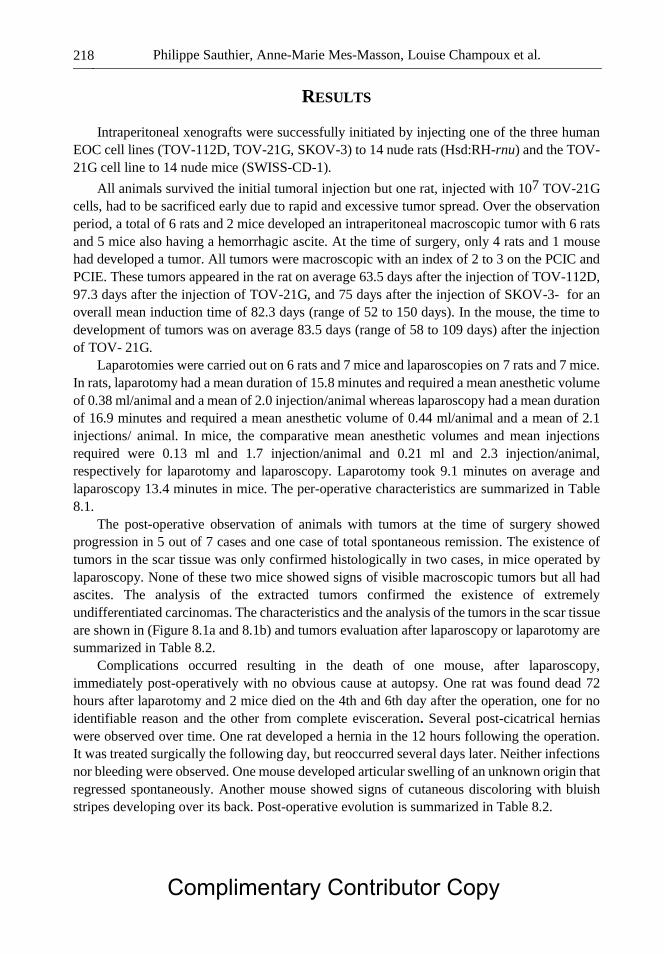
Philippe Sauthier, Anne-Marie Mes-Masson, Louise Champoux et al. 218
RESULTS
Intraperitoneal xenografts were successfully initiated by injecting one of the three human
EOC cell lines (TOV-112D, TOV-21G, SKOV-3) to 14 nude rats (Hsd:RH-rnu) and the TOV-
21G cell line to 14 nude mice (SWISS-CD-1).
All animals survived the initial tumoral injection but one rat, injected with 107 TOV-21G
cells, had to be sacrificed early due to rapid and excessive tumor spread. Over the observation
period, a total of 6 rats and 2 mice developed an intraperitoneal macroscopic tumor with 6 rats
and 5 mice also having a hemorrhagic ascite. At the time of surgery, only 4 rats and 1 mouse
had developed a tumor. All tumors were macroscopic with an index of 2 to 3 on the PCIC and
PCIE. These tumors appeared in the rat on average 63.5 days after the injection of TOV-112D,
97.3 days after the injection of TOV-21G, and 75 days after the injection of SKOV-3- for an
overall mean induction time of 82.3 days (range of 52 to 150 days). In the mouse, the time to
development of tumors was on average 83.5 days (range of 58 to 109 days) after the injection
of TOV- 21G.
Laparotomies were carried out on 6 rats and 7 mice and laparoscopies on 7 rats and 7 mice.
In rats, laparotomy had a mean duration of 15.8 minutes and required a mean anesthetic volume
of 0.38 ml/animal and a mean of 2.0 injection/animal whereas laparoscopy had a mean duration
of 16.9 minutes and required a mean anesthetic volume of 0.44 ml/animal and a mean of 2.1
injections/ animal. In mice, the comparative mean anesthetic volumes and mean injections
required were 0.13 ml and 1.7 injection/animal and 0.21 ml and 2.3 injection/animal,
respectively for laparotomy and laparoscopy. Laparotomy took 9.1 minutes on average and
laparoscopy 13.4 minutes in mice. The per-operative characteristics are summarized in Table
8.1.
The post-operative observation of animals with tumors at the time of surgery showed
progression in 5 out of 7 cases and one case of total spontaneous remission. The existence of
tumors in the scar tissue was only confirmed histologically in two cases, in mice operated by
laparoscopy. None of these two mice showed signs of visible macroscopic tumors but all had
ascites. The analysis of the extracted tumors confirmed the existence of extremely
undifferentiated carcinomas. The characteristics and the analysis of the tumors in the scar tissue
are shown in (Figure 8.1a and 8.1b) and tumors evaluation after laparoscopy or laparotomy are
summarized in Table 8.2.
Complications occurred resulting in the death of one mouse, after laparoscopy,
immediately post-operatively with no obvious cause at autopsy. One rat was found dead 72
hours after laparotomy and 2 mice died on the 4th and 6th day after the operation, one for no
identifiable reason and the other from complete evisceration. Several post-cicatrical hernias
were observed over time. One rat developed a hernia in the 12 hours following the operation.
It was treated surgically the following day, but reoccurred several days later. Neither infections
nor bleeding were observed. One mouse developed articular swelling of an unknown origin that
regressed spontaneously. Another mouse showed signs of cutaneous discoloring with bluish
stripes developing over its back. Post-operative evolution is summarized in Table 8.2.
Complimentary Contributor Copy
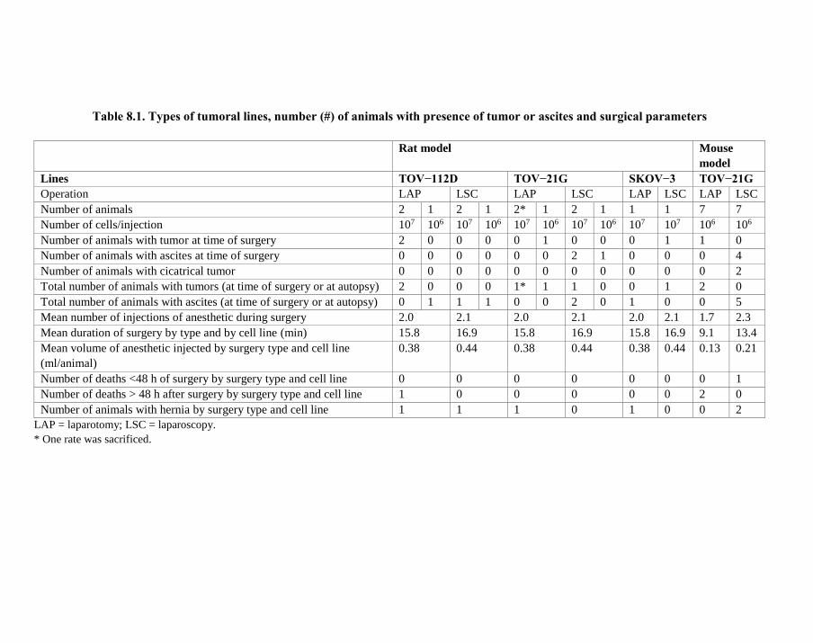
Table 8.1. Types of tumoral lines, number (#) of animals with presence of tumor or ascites and surgical parameters
Rat model Mouse
model
Lines TOV−112D TOV−21G SKOV−3 TOV−21G
Operation LAP LSC LAP LSC LAP LSC LAP LSC
Number of animals 2 1 2 1 2* 1 2 1 1 1 7 7
Number of cells/injection 107 106 107 106 107 106 107 106 107 107 106 106
Number of animals with tumor at time of surgery 2 0 0 0 0 1 0 0 0 1 1 0
Number of animals with ascites at time of surgery 0 0 0 0 0 0 2 1 0 0 0 4
Number of animals with cicatrical tumor 0 0 0 0 0 0 0 0 0 0 0 2
Total number of animals with tumors (at time of surgery or at autopsy) 2 0 0 0 1* 1 1 0 0 1 2 0
Total number of animals with ascites (at time of surgery or at autopsy) 0 1 1 1 0 0 2 0 1 0 0 5
Mean number of injections of anesthetic during surgery 2.0 2.1 2.0 2.1 2.0 2.1 1.7 2.3
Mean duration of surgery by type and by cell line (min) 15.8 16.9 15.8 16.9 15.8 16.9 9.1 13.4
Mean volume of anesthetic injected by surgery type and cell line
(ml/animal)
0.38 0.44 0.38 0.44 0.38 0.44 0.13 0.21
Number of deaths <48 h of surgery by surgery type and cell line 0 0 0 0 0 0 0 1
Number of deaths > 48 h after surgery by surgery type and cell line 1 0 0 0 0 0 2 0
Number of animals with hernia by surgery type and cell line 1 1 1 0 1 0 0 2
LAP = laparotomy; LSC = laparoscopy.
* One rate was sacrificed.
Complimentary Contributor Copy
Table 8.2. Evolution of the models and characteristics of the tumors
Animal # Lines No PCIC1 PCIE2 Time [days] Evolution Death Time of post− operative death
Rat #3 TOV−112D 107 2 2 75 Unknown Spontaneous 48 h
Rat #5 TOV−112D 107 2 2 52 Remission Autopsy 12th week
Rat #9 TOV−21G 107 3 3 67 Progression Autopsy No operation
Rat #12 TOV−21G 107 2 2 150 Progression Autopsy 12th week
Rat #7 TOV−21G 106 1 1 75 Progression Autopsy 12th week
Rat #14 SKOV−3 107 3 3 75 Progression Autopsy 2nd week
Mouse #2 TOV−21G 106 1 1 58 Progression Autopsy 4th week
Mouse #3 TOV−21G 106 2 2 109 Progression Autopsy 8th week 1 Peritoneal Cancer Index of Chauffert (PCIC): 2 Peritoneal Cancer Index of Eggermont (PCIE): 0 = No tumour 1 = Tumoral nodule < 1 mm. 2 = Tumoral nodule > 1 mm 3 = Diffuse carcinomatosis with ascite. 0 = No tumour. 1 = Nodule of between 1 and 3 mm 2 = Moderate tumour 3 = Abundant tumour replacing the cavity.
Complimentary Contributor Copy
Of Mice and Rats 221
DISCUSSION
We described our methods for successful EOC xenograft models amenable to either
laparotomy or laparoscopy in rodents. We encountered no problems for injecting animals with
tumoral lines extracted from untreated solid ovarian human tumors and animals withstood cell
line injection well. The observation of cutaneous discoloring with bluish stripes in one of the
mice remains unexplained, however this phenomenon was also observed in an earlier study
(Provencher 2000). General anesthesia by subcutaneous injection is preferred by others to avoid
any kind of interference with tumor development (Lecuru 2001). Anesthesia posed no particular
problem in the rat, neither in laparoscopy nor in laparotomy. The total quantity of anesthetic
and number of injections per rat were comparable. In the mouse, the anesthesia was particularly
difficult when performing laparoscopy, as evidenced by a high number of re-injections and
mean anesthetic volume that was almost twice as high compared with that needed for
laparotomy. Nonetheless, mice required smaller anesthetic volumes than rats overall.
As the dimensions of the abdominal cavity in the mouse are small, the number of acts and
manipulations remains very limited despite using instruments of 1.7 mm in diameter. We tested
an optical system 2 mm in diameter, which frequently had to be repositioned due to its restricted
visual field. Nonetheless, this optical system can be useful in diagnostic laparoscopy. The use
of a 5 mm optical system and thus vision that extends over the entire pelvic cavity is possible
in the rat.
Coagulation attempts with monopolar instruments were carried out and were feasible as
long as an isolated 2 mm trocar was used (Veress needle).
Postoperative complications are more serious in mice than in rats. Our results in the rat
were similar to those reported by Canis (Canis 1998). The frequently observed incisional
hernias are probably explained by the fact that the animals are able to access their abdominal
sutures. They can chew and tear off their sutures or their clips. For this reason, it would be
preferable to close the wounds without suture material (sutures or clips), possibly using
biological glue.
The tumors developed after the injection of 107 cells and it appears that this concentration
is a minimal concentration required to induce tumors in this animal model. In his study, Rubin
(1993) injected 4 × 10⁷ to 2.5 × 10⁹ cells per animal of the SKOV-3 and SKOV-7 lines and
Lecuru (2001) injected 27 × 106 IGR-OV1 and 36 × 106 NIH: OVCAR-3 cells per animal. As
the volume injected is already 1 ml (maximum injectable intraperitoneal volume tolerated by
the rat), we felt that the viscousness would be too high if the number of cells were to exceed
107. It would seem preferable to inject 107cells and to repeat the injection 48 hours later. This
warrants further consideration.
All the tumors in the rats were sizeable as confirmed by the intraperitoneal cancer indexes,
PCIC and PCIE (Table 8.2). They appeared after a relatively long time after injection (mean of
82.3 days in the rats and 83.5 days in the mice). This is significantly longer than in the IGR-
OV1 model (7−21 days) and even in the NIH: OVCAR-3 model (14−21 days) (Lecuru 2001).
This parameter is important to consider when planning studies of this kind. The rat, contrary to
the mouse, tolerates the growth of intraperitoneal tumors well. This enabled us to stagger the
surgery, sometimes by a few days, which may be of practical use. In contrast, for mice,
abdominal distension quickly progressed to serious deterioration of the animal condition
translating into a need for emergency surgery. In rodents the distribution of intraperitoneal Complimentary Contributor Copy
Philippe Sauthier, Anne-Marie Mes-Masson, Louise Champoux et al. 222
tumors was similar to that observed in women with EOC. It should be noted that surgery
consisted of a close examination of the cavity and that maximal cytoreduction could only be
carried out in a few cases. The examination of the abdominal cavity was more precise with
laparoscopy.
Tumoral implants were sometimes found in the laparoscopy incision scars in the mice
(Figure 8.1a and 8.1b). These mice showed no signs of visible tumors but had ascites. No rats
developed tumors in any incision sites in our pilot study. The number of animals studied is too
small to draw conclusions but the presence of tumoral ascite has already been evoked as a factor
that favors parietal tumoral implants (Kruitwagen 1996).
The nude rat therefore seems more robust as a model of human ovarian cancer and could
enable an in-depth study of the influence of laparoscopy on the in vivo and ex vivo behavior of
tumoral lines. Using rats involves a greater infrastructure in terms of animal husbandry and a
higher daily cost. These factors need to be taken into consideration as the animals are generally
housed in small sterile units. Furthermore, the acquisition cost of nude rats is higher than that
of nude mice.
The operating table we custom built enabled us to carry out all required laparoscopic acts
we wanted in a sterile environment. Its size is well adapted to the height of the animals and the
adjustable Trendelenburg position was extremely useful allowing leverage for manipulation of
the instruments (Figure 8.1a, 8.1b and 8.1c). The camera being fixed, enables stability while
providing gentle rotation to visualize the specific operating field. In this respect, our custom
table may offer an advantage over the existing commercial operating table (Kaps Co.,
Frankfurt, Germany™) (Berguer 1997).
CONCLUSION
The use of laparoscopy in oncology today raises certain concerns and requires more
detailed investigations. Our experience suggests that the nude rat is a good animal model of
human epithelial ovarian cancer. Animals tolerate laparoscopy and laparotomy well and have
a high survival rate. Although it is better characterized from an oncological point of view, the
nude mouse has proved to be less reliable in terms of tumor growth in our study. Most
importantly, it is more fragile perioperatively and its overall survival is not as good as that of
the rat. Furthermore, the restricted size of this animal greatly limits the surgical acts as surgical
instruments are not very well adapted for this type of animal despite recent development of
operating tables for laparoscopy on small animals. The nude rat, therefore, currently appears to
be the ideal model for studying the concept of laparoscopy in oncology. Nevertheless, more in-
depth studies are needed to fully characterize the biology of xenograft EOC tumorigenesis in
the rat.
ACKNOWLEDGMENTS
We are grateful for the support of Jocelyne Gauvin research fund for this project.
Complimentary Contributor Copy

Of Mice and Rats 223
REFERENCES
Agostini A, Robin F, Aggerbeck M, Jais JP, Blanc B, Lecuru F. Influence of peritoneal factors
on port− site metastases in a xenograft ovarian cancer model. BJOG 2001;
108(8):809−812.
Agostini A, Robin F, Jais JP, Aggerbeck M, Vildé F, Blanc B, Lécuru F. Impact of different
gases and pneumoperitoneum pressures on tumor growth during laparoscopy in a rat
model. Surg. Endosc. 2002; 16(3):529−532.
Agostini A, Robin F, Jais JP, Aggerbeck M, Vildé F, Blanc B, Lécuru F. Peritoneal closure
reduces port site metastases: results of an experiment in a rat ovarian cancer model. Surg.
Endosc. 2002; 16(2): 289−291.
Agostini A, Carcopino X, Franchi F, Cravello L, Lecuru F, Blanc B. Port site metastasis after
laparoscopy for uterine cervical carcinoma. Surg. Endosc. 2003; 17(10):1663−1665.
Allendorf JD, Bessler M, Whelan RL. A murine model of laparoscopic−assisted intervention.
Surg. Endosc. 1997; 11(6):622−624.
Barakat RR, Berchuck A, Markman M, Randall ME. Principles and Practice of Gynecologic
Oncology. 6th edition. Philadelphia: Williams and Wilkins. 2013.
Berguer R, Alarcon A, Feng S, Gutt C. Laparoscopic cecal ligation and puncture in the rat.
Surgical technique and preliminary results. Surg. Endosc. 1997; 11(12):1206−1208.
Bourdel N, Matsuzaki S, Bazin JE, Darcha C, Pouly JL, Mage G, Canis M. Postoperative
peritoneal dissemination of ovarian cancer cells is not promoted by carbon−dioxide
pneumoperitoneum at low intraperitoneal pressure in a syngenic mouse laparoscopic model
with controlled respiratory support: a pilot study. J. Minim. Invasive. Gynecol. 2008;
15(3):321−326.
Bouvy ND, Marquet RL, Hamming JF, Jeekel J, Bonjer HJ. Laparoscopic surgery in the rat.
Beneficial effect on body weight and tumor take. Surg. Endosc. 1996; 10(5):490−494.
Braumann C, Balague C, Guenther N, Menenakos C, Jacobi CA. Application of a solid tumor
model to evaluate tumor recurrence after an open or laparoscopic rectal resection in rats.
Surg. Laparosc. Endosc. Percutan. Tech. 2008; 18(4):348−352.
Brundell SM, Tucker K, Texler M, Brown B, Chatterton B, Hewett PJ. Variables in the spread
of tumor cells to trocars and port sites during operative laparoscopy. Surg. Endosc. 2002;
16(10):1413−1419.
Canis M, Botchorishvili R, Wattiez A, Mage G, Pouly JL, Bruhat MA. Tumor growth and
dissemination after laparotomy and CO2 pneumoperitoneum: a rat ovarian cancer model.
Obstet. Gynecol. 1998; 92(1):104−108.
Canis M, Botchorishvili R, Wattiez A, Pouly JL, Mage G, Manhes H, et al. Cancer and
laparoscopy, experimental studies: a review. Eur. J. Obstet. Gynecol. Reprod. Biol. 2000;
91(1):1−9.
Canis M, Rabischong B, Botchorishvili R, Tamburro S, Wattiez A, Mage G, Pouly JL, Bruhat
MA. Risk of spread of ovarian cancer after laparoscopic surgery. Curr. Opin. Obstet.
Gynecol. 2001; 13(1): 9−14.
Carlson NL, Krivak TC, Winter WE, 3rd, Macri CI. Port site metastasis of ovarian carcinoma
remote from laparoscopic surgery for benign disease. Gynecol Oncol 2002;85(3):529−531.
Complimentary Contributor Copy

Philippe Sauthier, Anne-Marie Mes-Masson, Louise Champoux et al. 224
Chauffert B, Dimanche−Boitrel MT, Genne P, Petit JM, Onier N, Jeannin JF. [Experimental
chemotherapy of peritoneal carcinomatosis of colonic origin in rats]. Gastroenterol. Clin.
Biol. 1992; 16(3):215−219.
Childers JM, Aqua KA, Surwit EA, Hallum AV, Hatch KD. Abdominal−wall tumor
implantation after laparoscopy for malignant conditions. Obstet. Gynecol. 1994;
84(5):765−769.
Curet MJ. Port site metastases. Am. J. Surg. 2004; 187(6):705−712.
Dargent D. [Laparoscopic surgery in gynecologic oncology]. J. Gynecol. Obstet. Biol. Reprod.
(Paris) 2000; 29(3):282−284.
Deffieux X, Castaigne D, Pomel C. Role of laparoscopy to evaluate candidates for complete
cytoreduction in advanced stages of epithelial ovarian cancer. Int. J. Gynecol. Cancer.
2006; 16 Suppl 1:35−40.
Eggermont AM, Steller EP, Marquet RL, Jeekel J, Sugarbaker PH. Local regional promotion
of tumor growth after abdominal surgery is dominant over immunotherapy with
interleukin−2 and lymphokine activated killer cells. Cancer Detect. Prev. 1988;
12(1−6):421−429.
Fagotti A, Ferrandina G, Fanfani F, Garganese G, Vizzielli G, Carone V, Salerno MG, Scambia
G. Prospective validation of a laparoscopic predictive model for optimal cytoreduction in
advanced ovarian carcinoma. Am. J. Obstet. Gynecol. 2008; 199(6):642.e1−6.
Fondrinier E, Descamps P, Arnaud JP, Pezet D. [Carbon dioxide pneumoperitoneum and
peritoneal carcinosis: review]. J. Gynecol. Obstet. Biol. Reprod. (Paris) 2002; 31(1):11−27.
Hajduch M, Kod'ousek R. "Nude rabbit" with aplasia of B−lymphoid structures. Acta. Univ.
Palacki. Olomuc. Fac. Med. 1992; 134:51−54.
Hopkins MP, von Gruenigen V, Gaich S. Laparoscopic port site implantation with ovarian
cancer. Am. J. Obstet. Gynecol. 2000; 182(3):735−736.
Hopkins MP. The myths of laparoscopic surgery. Am. J. Obstet. Gynecol. 2000; 183(1):1−5.
Hopkins MP, von Gruenigen V, Haller NA, Holda S. The effect of various insufflation gases
on tumor implantation in an animal model. Am. J. Obstet. Gynecol. 2002; 187(4):994−996.
Jacobi CA, Ordemann J, Bohm B, Zieren HU, Liebenthal C, Volk HD. The influence of
laparotomy and laparoscopy on tumor growth in a rat model. Surg. Endosc. 1997;
11(6):618−621
Kadar N. Laparoscopic management of gynecological malignancies. Curr. Opin. Obstet.
Gynecol. 1997; 9(4):247−255.
Kruitwagen RF, Swinkels BM, Keyser KG, Doesburg WH, Schijf CP. Incidence and effect on
survival of abdominal wall metastases at trocar or puncture sites following laparoscopy or
paracentesis in women with ovarian cancer. Gynecol. Oncol. 1996; 60(2):233−237.
Le Moine MC, Navarro F, Burgel JS, Pellegrin A, Khiari AR, Pourquier D, et al. Experimental
assessment of the risk of tumor recurrence after laparoscopic surgery. Surgery 1998;
123(4):427−431.
Lecuru F, Agostini A, Camatte S, Robin F, Aggerbeck M, Jaiss JP, et al. Impact of
pneumoperitoneum on visceral metastasis rate and survival. Results in two ovarian cancer
models in rats. BJOG 2001; 108(7):733−737.
Lecuru F, Guilbaud N, Agostini A, Augereau C, Vilde F, Taurelle R. Description of two new
human ovarian carcinoma models in nude rats suitable for laparoscopic experimentation.
Surg. Endosc. 2001; 15(11):1346−1352.
Complimentary Contributor Copy

Of Mice and Rats 225
Lécuru F, Agostini A, Camatte S, Robin F, Aggerbeck M, Jaïs JP, Vilde F, Taurelle R. Impact
of pneumoperitoneum on tumor growth. Surg. Endosc. 2002; 16(8):1170−1174.
Lee JW, Park YA, Cho YJ, Kang KH, Choi JJ, Lee YY, Kim TJ, Choi CH, Kim BG, Bae DS.
The effect of surgical wound on ovarian carcinoma growth in an animal model. Anticancer
Res. 2013; (8):3177−3184.
Lin XJ, Chen XC, Wang L, Wei YQ, Kan B, Wen YJ, He X, Zhao X. Dynamic progression of
an intraperitoneal xenograft model of human ovarian cancer and its potential for preclinical
trials. J. Exp. Clin. Cancer Res. 2007; 26(4):467−474.
Martinez J, Targarona EM, Balague C, Pera M, Trias M. Port site metastasis. An unresolved
problem in laparoscopic surgery. A review. Int. Surg. 1995; 80(4):315−321.
Mathew G, Watson DI, Rofe AM, Baigrie CF, Ellis T, Jamieson GG. Wound metastases
following laparoscopic and open surgery for abdominal cancer in a rat model. Br. J. Surg.
1996; 83(8):1087−1090.
Morice P, Camatte S, Larregain−Fournier D, Thoury A, Duvillard P, Castaigne D. Port−site
implantation after laparoscopic treatment of borderline ovarian tumors. Obstet. Gynecol.
2004; 104(5 Pt 2):1167−1170.
Nezhat FR, Pejovic T, Finger TN, Khalil SS.Role of minimally invasive surgery in ovarian
cancer. J. Minim. Invasive Gynecol. 2013 Nov−Dec; 20(6):754−765.
Pomel C, Provencher D, Dauplat J, Gauthier P, Le Bouedec G, Drouin P, Audet−Lapointe P,
Dubuc− Lissoir J.Gynecol Oncol. 1995 Sep; 58(3):301−306.
Pratt BL, Greene FL. Role of laparoscopy in the staging of malignant disease. Surg. Clin. North
Am. 2000; 80(4):1111−1126.
Provencher DM, Lounis H, Champoux L, Tetrault M, Manderson EN, Wang JC, et al.
Characterization of four novel epithelial ovarian cancer cell lines. In Vitro Cell Dev. Biol.
Anim. 2000; 36(6):357−361.
Ramirez PT, Wolf JK, Levenback C. Laparoscopic port−site metastases: etiology and
prevention. Gynecol. Oncol. 2003; 91(1):179−189.
Reymond MA, Tannapfel A, Schneider C, Scheidbach H, Kover S, Jung A. Description of an
intraperitoneal tumour xenograft survival model in the pig. Eur. J. Surg. Oncol. 2000;
26(4):393−397.
Rose GS, Tocco LM, Granger GA, DiSaia PJ, Hamilton TC, Santin AD, et al. Development
and characterization of a clinically useful animal model of epithelial ovarian cancer in the
Fischer 344 rat. Am. J. Obstet. Gynecol. 1996; 175(3 Pt 1):593−599.
Rubin SC, Kairemo KJ, Brownell AL, Daghighian F, Federici MG, Pentlow KS, et al.
High−resolution positron emission tomography of human ovarian cancer in nude rats using
124I−labeled monoclonal antibodies. Gynecol. Oncol. 1993; 48(1):61−67.
Sandoval BA, Sulaiman TT, Robinson AV, Stellato TA. Laparoscopic surgery in a small animal
model. A simplified technique of retroperitoneal dissection in the rat. Surg. Endosc. 1996;
10(9):925−927.
Sanjuan A, Hernandez S, Pahisa J, Ayuso JR, Torne A, Martinez Roman S. Port-site metastasis
after laparoscopic surgery for endometrial carcinoma: two case reports. Gynecol. Oncol.
2005; 96(2): 539−542.
Sawada M, Matsui Y, Hayakawa K, Nishiura H, Okudaira Y, Taki I. Human gynecologic
cancers heterotransplanted into athymic nude rats. Gynecol. Oncol. 1982; 13(2):220−228.
Complimentary Contributor Copy
Philippe Sauthier, Anne-Marie Mes-Masson, Louise Champoux et al. 226
Shen MY, Huang IP, Chen WS, Chang JT, Lin JK. Influence of pneumoperitoneum on tumor
growth and pattern of intra−abdominal tumor spreading: in vivo study of a murine model.
Hepatogastroenterology 2008; 55(84):947−951.
Sugiyama T, Nishida T, Yokota D, Ushijima K, Imaishi K, Hirakawa N, et al. An experimental
model for advanced ovarian cancer. Kurume Med. J. 1990; 37(1):15−21.
Vanderhyden BC, Shaw TJ, Ethier JF. Animal models of ovarian cancer. Reprod. Biol.
Endocrinol. 2003; 1:67−82.
Vergote I, Marquette S, Amant F, Berteloot P, Neven P. Port−site metastases after open
laparoscopy: a study in 173 patients with advanced ovarian carcinoma. Int. J. Gynecol.
Cancer. 2005; 15(5):776−779.
Volz J, Koster S, Weiss M, Schmidt R, Urbaschek R, Melchert F. Pathophysiologic features of
a pneumoperitoneum at laparoscopy: a swine model. Am. J. Obstet. Gynecol. 1996; 174(1
Pt 1):132−140.
Complimentary Contributor Copy

SECTION III. ADVANCE TECHNIQUES IN
EXPERIMENTAL SURGERY
Complimentary Contributor Copy

Complimentary Contributor Copy

In: Advances in Experimental Surgery. Volume 2 ISBN: 978-1-53612-773-7
Editors: Huifang Chen and Paulo N. Martins © 2018 Nova Science Publishers, Inc.
Chapter 9
ORGAN BIOENGINEERING
THROUGH DECELLULARIZATION
AND RECELLULARIZATION APPROACHES
Maria Jaramillo, PhD and Basak E. Uygun, PhD Center for Engineering in Medicine, Massachusetts General Hospital.
Department of Surgery, Harvard Medical School,
Massachusetts General Hospital, Boston, Massachusetts, US
ABSTRACT
Organ transplantation has been used for over a century for the treatment of end organ
failure. Despite vast advances in the field, the main challenge remains the availability of
good quality donor organs. Scientists around the world are working on the development of
alternative options for donor organs available for transplantation, including development
of decellularization-based artificial organs, which is the focus of this chapter.
Organ and tissue decellularization has a number of advantages, including preservation
of the extracellular matrix architecture and composition, which provide signals necessary
for cell function and survival. The preserved vascular architecture in the scaffolds allows
for connecting the bioengineered organ to the blood torrent upon transplantation. In
addition, ECM scaffolds have been found to elicit low to negligible immune response
which is a major advantage, eliminating the need for long-lasting use of potentially harmful
immunosuppression therapy in transplant patients.
While very promising to increase the pool of donor organs, the
decellularization/recellularization approach for bioengineered organs has a number of
challenges. These include methods for decellularization with minimal damage to the
extracellular matrix composition and architecture, choice of cells type and source for
replacing the functions of organ in question and achieving long-term viability of the
engineered organ in vivo. Here, we discuss each of these challenges and present current
progress in the field for different organs including liver, heart, kidney and lungs.
Corresponding author: Basak E. Uygun, PhD. Center for Engineering in Medicine, Massachusetts General Hospital,
Department of Surgery, Harvard Medical School, 51 Blossom St., Boston, MA, Tel: 617-726-3474, E-mail:
[email protected]. Complimentary Contributor Copy
Maria Jaramillo and Basak E. Uygun 230
Keywords: decellularization, recellularization, organ engineering
ABBREVIATIONS
ECM Extracellular Matrix
SDS Sodium dodecyl sulfate
hiPSC human induced pluripotent stem cells
EPC endothelial progenitor cells
HUVEC Human umbilical vein endothelial cells
MSC Mesenchymal Stem cells
INTRODUCTION
According to the United Network for Organ Sharing (UNOS), there are currently over
120,000 people in need of organ transplants in the United States, from which over 75,000 are
on an active waiting list. However, it is estimated that only 30,000 to 35,000 transplants will
be performed by the end of this year. This highlights the need for strategies to increase the pool
of available organs. Several approaches have been developed to this end, including the
formation of organizations devoted to education regarding organ donation to increase donor
participation and research in areas including organ preservation, organ rehabilitation and organ
engineering.
Organ engineering refers to the development of biological substitutes to replace or aid
damaged and diseased tissues, thus maintaining physiological homeostasis in the patient. This
involves the use of biomaterials, cells and other microenvironmental signals to mimic the niche
of native organs such that their functions are recapitulated and maintained. Several strategies
have been adopted for these purposes, including encapsulation of donor cells in biomaterials
that provide immunogenic barriers while promoting cell survival and allowing proper cell
function. Another approach that is quickly gaining interest is the concept of 3D printing,
however, one of the most promising approaches and our main focus for this chapter is the
development of artificial organs by decellularization and recellularization of native tissues and
whole organs.
Organ decellularization consists of the complete removal of cells and cellular components
from native tissues. This offers a number of advantages, including perhaps the closest
recapitulation of physical and chemicals microenvironmental signals of tissues as well as low
biomaterial immunogenicity. In addition, this offers a potentially abundant source of scaffolds,
as, in contrast to organ transplantation, organs used for decellularization do not require such
strict parameters such as minimal ischemia time, and preservation of the ECM can be achieved
more easily than preservation of cell functions. Organs are then decellularized and repopulated
with healthy cells, which can be derived from different sources such as allogeneic primary cells,
expanded autologous cells, cells differentiated from different sources of stem cells (embryonic,
induced pluripotent, adult) amongst others.
In the next sections, we will talk about different strategies used for tissue decellularization,
including methods used to evaluate successful removal of cellular components. We will discuss Complimentary Contributor Copy

Organ Bioengineering through Decellularization and Recellularization Approaches 231
several approaches for recellularization and will review some of the most notable advances in
the field. Because each organ has a different function, organ engineering approaches have to
be tailored to satisfy the physical and chemical requirements, therefore we will discuss research
work in different organs. Finally, we will review other aspects of decellularization and
recellularization organ engineering like introduction of non-parenchymal cells such as
endothelial cells for proper vascularization and function of these replacement organs and their
immunogenicity.
DECELLULARIZATION
Organ decellularization is typically achieved by subjecting tissues/organs to treatments that
disrupt cell integrity, while preserving structure and composition of the extracellular matrix.
Most commonly, these treatments are chemical agents, although several physical treatments
have been successfully used alone or in combination with chemical treatment to ensure
appropriate removal of cellular components from tissues and organs. Ultimately, all methods
have a number of advantages and disadvantages, which have to be taken into consideration
along with the tissue-specific requirements to come up with the best possible specific strategies
for decellularization.
CHEMICAL METHODS
Detergents
Perhaps the most commonly used method for organ and tissue decellularization is the use
of detergents that emulsify oils, disrupting the lipid bilayer of the cellular membrane, hence
promoting cell lysis. Addition of fluid flow through whole organ perfusion or tissue agitation
ensures efficient removal of remaining cellular components. Detergents can be classified into
ionic, non-ionic or Zwitterionic detergents.
Ionic detergents have a charged hydrophilic group. They disrupt lipids and proteins, hence
strongly disturbing the cellular structures, making them more effective at removing cellular
components. Because of the fact that they can interact and denature proteins, they are
considered the most destructive and they have a greater effect on the chemical composition of
ECM. Additionally, they are harder to “rinse” than other types of detergents, which presents an
obstacle to subsequent recellularization of the tissues due to its cytotoxic effects. The most
commonly used ionic detergent is sodium dodecyl sulfate (SDS). It is routinely used for
decellularization of tissues such as heart valves (Korossis 2002), tubular structures such as
trachea (Hung 2016) and blood vessels (Schaner 2004), and whole organs including heart
(Oberwallner 2014), liver (Uygun 2010) and kidneys (Wang 2015).
Non-ionic detergents are neutral and hydrophilic. They are highly capable of breaking
lipid-lipid interactions, but due to their limited ability to interact with proteins, are unable to
break protein-protein interactions and have reduced ability to break lipid-protein interactions.
Because of this, they have weaker decellularization potential, but are better at maintaining the
chemical composition of the ECM. The most commonly used non-ionic detergent is Triton Complimentary Contributor Copy

Maria Jaramillo and Basak E. Uygun 232
X-100, which is typically used in addition to SDS. In a few cases, it has been reported to
promote appropriate decellularization alone in tissues such as ligaments (ACL) (Vavken 2009;
Xu 2014) and annulus fibrosus (Xu 2014), suggesting to be more appropriate for
decellularization of tissues with relatively low cellularity.
Finally, Zwitterionic detergents are considered to be “in between” ionic and non-ionic
detergents. They have a net neutral charge, but are able to disturb protein interactions, without
altering their native charge, hence are less damaging to the ECM composition than non-ionic
detergents. 3-[(3-cholamidopropyl)dimethylammonio]-1-propanesulfonate (CHAPS) is the
most widely used Zwitterionic detergent in decellularization approaches, and has been reported
to efficiently decellularize tissues such as lung (Tsuchiya 2016). When compared to
decellularization with SDS, it has been proven to preserve composition of components such as
collagen and elastin, which have a strong effect in mechanical integrity, suggesting that CHAPS
may be a better choice for highly cellular tissues whose function is highly dependent on its
mechanical properties.
Osmotic Decellularization
Osmotic decellularization, consisting of alternation of hypertonic and hypotonic solution
for cell disruption and removal, has been used alone and in combination with other methods
such as detergent-based decellularization. Typically, TRIS-based buffers and Phosphate
Buffered Saline (PBS) solutions are used. Hypotonic solutions force movement of water into
the cell, causing cells to burst, which is followed by use of hypertonic solutions that promote
non-histone protein dissociation from DNA. This method is gentler than the use of detergents,
and it is considered to lead to better preservation of ECM, so it is typically used for tissue with
low cellularity such as heart valves (Meyer 2006), cartilage (Schneider 2016) and cornea (Yam
2016).
Enzymes
Enzymatic decellularization consists of removing cellular components with the aid of
enzymes that break down cellular components; some of such enzymes are trypsin, nucleases
and dispase. Nucleases are enzymes that break down nucleic acids (DNA and RNA), but
usually do not act on other cellular components, therefore are typically used in conjunction with
other treatments that aid penetration of nucleases into the cells. There are other enzymes such
as trypsin that promote breakdown of serine-containing proteins, which are very efficient at
breaking down cells; however, because they are non-specific, they also interact with ECM
proteins. Lipases are a good candidate for decellularization as they specifically disrupt the lipid
bilayer of the cell membranes without disrupting ECM proteins, however, rather limited
success at complete cellular removal has been achieved by the use of lipases alone
(Crapo 2011).
Complimentary Contributor Copy
Organ Bioengineering through Decellularization and Recellularization Approaches 233
PHYSICAL METHODS
Sonication
Sonication has been used mostly in combination with other methods for decellularization
of tissues such as larynx (Hung 2013), trachea (Hung 2016), cardiovascular tissues (Azhim
2011), cartilage (Azhim 2013) and small intestine (Oliveira 2013). It has been shown to be
advantageous in cases when perfusion decellularization is not possible due to the limited
diffusion of chemical agents. In such cases, sonication has shown to enhance the degree of
removal of cellular components through passive diffusion of decellularizing agents. It has been
observed that sonication greatly speeds up the decellularization process, decreasing exposure
time to chemical agents which typically show higher disruption of chemical composition.
Freeze/Thaw
This method is rarely used alone, but typically used as a critical step of decellularization in
combination with other methods discussed here. Freezing promotes ice crystal formation of
water, which, unlike most other materials, increases volume in its frozen form. This increase in
water volume damages cell membrane, leading to break down of the cells. Typically, ice crystal
formation can be reduced by cooling at low rates; hence, decellularization by thawing is better
achieved by rapid freezing of tissue.
Irreversible Electroporation
Irreversible electroporation (IRE) is a novel technique that is currently being explored. It
consists of applying non-thermal pulsed electric fields to disrupt the cellular membrane while
maintaining the integrity of the ECM. This method has been explored as an in vivo
decellularized method (Golberg 2016), where the scaffolds are decellularized in live animals,
however, it has also been used in ex vivo perfusion models (Sano 2010).
Evaluation and Success Parameters
Biochemical evaluation of decellularization is typically performed by quantifying cellular
components such as DNA and actin to estimate the amount of cellular material removed. In
addition, preservation of ECM composition is evaluated by estimating concentration of ECM
proteins such as laminin, elastin, collagen and fibronectin, or other components such as
glycosaminoglycans (GAG). Quantification of these is typically done by standard assays such
as ELISA, PicoGreen and immunostaining, however, due to the destructive nature of these
methods, recent studies have highlighted the need to develop new methods that do not require
damage to the decellularized tissue, thus promoting maintenance of its integrity for subsequent
use. Some of the suggested technique include analysis of perfusate (Jörn 2015), but also more
Complimentary Contributor Copy

Maria Jaramillo and Basak E. Uygun 234
sophisticated methods such as computer tomography, which has been used for monitoring DNA
content in real time (Geerts 2016).
Another parameter that is typically evaluated in most applications is the maintenance of
the microstructural integrity of the scaffolds. For most tissues, the maintenance of a capillary
network is essential, and this can be investigated by corrosion casting and a number of
microscopy techniques including angiography and scanning electron microscopy. Furthermore,
perfusion of dyes that can be removed by additional perfusion can be used to visualize capillary
integrity after decellularization. In addition to capillary network integrity, other parameters
such as topography have been evaluated through technique such as atomic force microscopy;
the significance of it being that material characteristics such as roughness can alter cell
behavior.
Additionally, for tissues that serve a mechanical function or tissues that need to be able to
withstand mechanical loads, bulk physical parameters need to be evaluated. This is the case for
tissues such as bone, tendon, cartilage, cardiac valves, lungs, muscle, blood vessels and many
others. Depending on the tissue requirements, stress, strain, tensile strength, elasticity and
compliance need to be evaluated.
Overall, there is no standardized set of parameters to define success in decellularization
except for DNA content which is defined as 1) less than 50 ng of dsDNA per mg of dry weight;
2) DNA fragments less than 200 bp in length; and 3) no visible nuclear material in histologic
analysis with DAPI or H&E, according to Faulk et al. (Faulk 2015). When performing such
evaluation, it is important to keep in mind that every tissue serves a different function and
hence, preservation of certain characteristics may be more important. For instance, in the case
of liver engineering, the conservation of biochemical composition may be of higher importance,
as hepatocyte function and viability is highly dependent on the chemical microenvironment.
Alternatively, tissues such as cartilage are relatively acellular and the biggest functional
requirement is the ability to withstand mechanical loads, hence, higher importance is given to
the maintenance of physical characteristics. This highlights the need for a comprehensive
compilation of data to define tissue-specific parameters and tolerance levels that will allow
optimizing decellularization procedures based on specific requirements.
RECELLULARIZATION
After successful decellularization of organs and tissues, the next step is to repopulate the
decellularized scaffolds with cells that will carry out vital functions of the organ. The main
challenge in this step is the identification of a reliable and abundant cell source that will replace
the functions of the native organ. There are a number of constraints in this step which include
development of strategies for cell delivery, retention and maintenance. However, perhaps the
biggest challenge is the establishment of an abundant cell source, which we will discuss in the
following sections.
In Vitro Expansion and Maintenance of Primary Cells
Most cells in the body have a limited ability to expand and maintain a functional phenotype
in an in vitro setting. This phenomenon is not well understood but its investigation could lead Complimentary Contributor Copy
Organ Bioengineering through Decellularization and Recellularization Approaches 235
to the development of strategies to generate an abundance of functional cells to be used in
cell/tissue engineering applications. Unfortunately, despite the great promise of this field for
regenerative medicine applications, very limited advances have been made in this area.
A recent study describes genetic modification of primary human hepatocytes to express
Human Papilloma Virus (HPV) genes, which mediate expression of receptors-associated
hepatocyte proliferation in vivo. Exposure of the genetically modified hepatocytes to
proliferation-inducing cytokines promotes their expansion amounting to up to a trillion cells
per single hepatocyte. Removal of proliferation signals leads to polarization and induction of
function in the hepatocytes (Levy 2015). Genetic engineering strategies has also been exploited
for the expansion and maintenance of beta-cells using Cre/loxP-based reversible
immortalization (Kobayashi 2006), however, these approaches rely on use of viral vectors,
which could cause activation of oncogenes, hence, tumorigenicity. Other studies have
concentrated on manipulation of the in vivo microenvironment rather than the cells, including
the development of scaffolds with appropriate physical and chemical cues that have been found
to promote limited expansion and functionality of cells such as alveolar cells and hepatocytes
(Shamis 2011).
A number of tissues rely on progenitor cells with proliferation potential for repair of
damaged tissue and cell turnover. Isolation of these cells, with subsequent expansion and
induction of terminally differentiated phenotype, constitutes another strategy for organ
engineering relying on primary human cells. The difficulty of this approach is isolation and
identification of these cells from live patients; however, some advances have been made with
the use of progenitor cells derived from stem cells, which will be further discussion in a later
section.
To conclude this section, it is important to note that use of primary cells would require
immunological matching of cells to patient, and potentially immunosuppression to avoid
rejection of the transplanted graft. Recent studies have also explored the possibility of silencing
genes involved in immune rejection (Figueiredo 2013), which in conjunction with successful
strategies for expansion and maintenance of primary cells, would make the possibility of the
creation of universal cell banking for off-shelve regenerative medicine applications a reality.
Cell Recovery from Discarded Organs
A new strategy that has emerged as an alternative approach is to recover cells from
marginal organs deemed unacceptable for transplantation. While very little work has been done
in this field, a few recent studies have shown that a number of cells including beta cells
(Matsumoto 2007) and hepatocytes (Izamis 2013; Izamis 2015; Uygun 2016) can be recovered
from organs exposed to warm ischemia for up to one hour by perfusion with preservation
solutions that restore ionic concentrations of the cells and ATP generation. The resulting cells
have been isolated at yields and levels of functionality comparable to those of primary cells.
Stem Cell Differentiation
Perhaps one of the most explored options is differentiation of functional cells from a
number of different stem cell sources. Depending on their origin, cells can be pluripotent, Complimentary Contributor Copy
Maria Jaramillo and Basak E. Uygun 236
multipotent, or have the ability to become a very limited number of cells. Stem cells are most
commonly categorized into pluripotent, mesenchymal or tissue-specific stem cells.
Pluripotent stem cells include embryonic stem cells derived from the inner mass of the
embryonic blastocyst and induced pluripotent stem cells, which are somatic cells that have been
genetically engineered to express a number of genes characteristic of embryonic stem cells,
giving these cells their pluripotency characteristics. Both cell types have two main features:
their ability to self-renew indefinitely (making them a potentially unlimited cell source) and to
differentiate into cells derived from the three germ layers of embryonic development
(mesoderm, ectoderm and endoderm). Differentiation programs are based on developmental
studies that identify specific signals that determine their fate, which are then recapitulated in an
in vitro setting. Most commonly, these signals are chemical cues such as cytokines and small
molecules, but can also be in the form of physical cues, such as substrates with varying
mechanical properties. Despite their enormous potential, success of pluripotent stem cell
differentiation has been limited partly due to the fact that they rely on developmental studies
using non-human models; hence translation of these observations into development of human
systems is imprecise. Challenges in this field remain, including low functionality and maturity
of the resulting cells. In terms of complex cell types, such as hepatocytes, that carry a large
number of diverse functions, a challenge has been the recapitulation of all the functions. This
last requirement can be bypassed by the alternative strategy of creating auxiliary grafts that
instead of replacing the entire organ will aid it by providing the function that is aberrated in the
patient. This strategy could be used in deficiency diseases, such as Alpha-1 antitrypsin (AAT)
deficiency. Hepatocytes in AAT-deficient patients often carry out other vital functions such as
albumin production, lipid metabolism, carbohydrate metabolism and drug metabolism. In such
cases, creation of a graft with differentiated cells that produce AAT and be transplanted in the
patients, auxiliary to their native liver to aid rather than replace the marginal organ.
Mesenchymal stem cells are naturally occurring adult cells that are not terminally
differentiated hence maintain the ability to differentiate into a limited number of tissues within
the mesodermal lineage (bone, cartilage, muscle, adipose tissue and blood; although some
studies report limited differentiation into ectoderm and endoderm lineages). Their sources
include cells derived from adipose tissue, bone marrow, tooth bud and umbilical cord which
are readily accessible and can be patient-specific.
Finally, some tissues have fast cell turnover and rely on resident stem cells to continually
replace aged or damaged cells. Some of these include oval cells in the liver, epithelial lining of
the stomach and intestine and skin. These cells typically can differentiate into just one or two
cell types, and can be isolated using cell-specific markers.
ADVANCES
In the following sections, we will discuss and summarize some of the most prominent
advances in the field of decellularized tissue-based organ engineering of a number of specific
systems.
Complimentary Contributor Copy
Organ Bioengineering through Decellularization and Recellularization Approaches 237
Liver
First reports of whole liver decellularization date back to 2009 (Baptista 2009). Being a
highly cellular organ, and because the lipid content can be higher than in most other tissues,
liver decellularization is most often achieved by the use of detergent, which preserves a
significant amount of the chemical composition and the architectural integrity of the liver
including its vascular network. Because of the highly vascular content, decellularization is
typically done by perfusion through one of the main vessels (typically portal vein).
Report of recellularization first appeared in 2010 (Uygun 2010) when rat livers were
recellularized using freshly isolated primary rat hepatocytes, which performed hepatic
functions (albumin and urea secretion, and drug metabolism protein expression) comparable to
those found when hepatocytes were maintained under static collagen sandwich culture
conditions, which to date, remains the gold-standard for culture of human hepatocytes. In
addition, the recellularized grafts were transplanted into rats that underwent unilateral
nephrectomy to prepare a viable site for auxiliary liver graft transplantation. The renal vein and
artery were used as ports to create blood source. Efflux was established within 5 min of
unclamping and the recellularized graft was kept for 8 h before evaluating damage to the
hepatocytes due to the arterial blood flow and the resulting shear stress, which was found to be
minimal. A later study showed that the recipient survival could be extended to 24 h by the
addition of a heparin layer on recellularized liver grafts (Bruinsma 2015). However,
thrombogenicity of the grafts still remains a problem.
Subsequent studies have been conducted in larger animal models including rabbits, sheep,
and porcine livers (Kajbafzadeh 2012; Nari 2013; Wang 2015). One study using porcine livers
(Barakat 2012) evaluated recellularization using human fetal hepatocytes and stellate cells, and
the recellularized grafts were kept under perfusion culture for up to two weeks, during which
they showed metabolic activity including oxygen and glucose consumption, lactate and urea
production, and albumin secretion.
Several cells have been investigated as potential sources for recellularization. A number of
studies have investigated the use of MSC (He 2013; Jiang 2014) and have reported
differentiation of these cells within the scaffolds (with addition of soluble chemical cues) to be
superior to differentiation of the same cells under 2D culture conditions. However, neither
report compared hepatic functions of differentiated cells to those found in primary hepatocytes,
making it hard to determine whether maturation is achieved to physiologically-relevant levels.
Other studies have used MSC for recellularization in conjunction with primary hepatocytes,
and have shown that this co-culture configuration improves engraftment and function of the
primary hepatocytes (Kadota 2014).
Finally, there are two alternative approaches to both decellularization and recellularization
that are worth discussing. First, there have been a few reports of efficient liver decellularization
using IRE (Golberg 2016; Sano 2010). This method has been used for decellularization of livers
under ex vivo perfusion, which can be subsequently recellularized, however, due to the
regenerative potential of liver, has also been explored as a method for
in vivo decellularization and recellularization, as it has been demonstrated that after IRE
treatment, liver regeneration occurs in the absence of scar formation. Other methods for
in vivo decellularization and recellularization have been developed, including detergent-based
decellularization of a single lobe in vivo through surgical by-pass circulation with a perfusion
chamber system (Pan 2016). In this study, decellularized lobe was subsequently recellularized Complimentary Contributor Copy
Maria Jaramillo and Basak E. Uygun 238
in vivo with primary hepatocytes from donor rats. Alternatively, the idea of transplanting
decellularized livers to allow for “spontaneous” in vivo recellularization has also been explored,
but the resulting grafts were inferior when compared to in vitro recellularized grafts (Sabetkish
2015).
Lung
Lung decellularization was first reported in 2010 in studies in mice and rat lungs (Ott 2010;
Price 2010) and using a combination of methods, including hypertonic/hypotonic solutions,
detergents and enzymes. Because of the function of the lung, preservation of its mechanical
integrity is of utmost importance, hence, decellularization protocols have been developed
avoiding or minimizing use of ionic detergents, which have been shown to lead to greater loss
of elastin and collagen that confer lungs their mechanical properties, including tensile strength
and elastic behavior (Petersen 2012). A number of strategies have been developed to maximize
cell removal while promoting maximal retention of ECM components, and in turn mechanical
properties. These include: use of zwitterionic detergents such as sulfobetaine-10 (Nagao 2013),
or non-ionic detergent such as Triton X-100 or CHAPS. Alternative ionic detergents such as
ethoxylated sodium lauryl ether sulfate (SLES), have been developed solely for
decellularization purposes. SLES is weaker than traditionally used SDS, and because of its
ethoxylation, the lauryl alcohol content is reduced, hence exhibiting lower protein denaturation
(Kawasaki 2015). Alternative approaches have focused on modification of delivery of
decellularization reagents. For instance, it has been suggested that perfusion route has an effect
on efficiency of decellularization, with intratracheal route being suggested as the preferred
method (Maghsoudlou 2013; Tsuchiya 2016; Wang 2016). In addition, some studies focus on
optimization of physical parameters such as flow rate and pressure, which can be increased to
speed up the decellularization process, hence, minimize exposure (da Palma 2015; Khalpey
2015). Despite, or partly due to the large number of studies evaluating decellularization
methods in lungs, there is little consensus about which method leads to best decellularization.
Because these studies are often performed in different animal systems, there is very little real
time monitoring of the progression of decellularization, and so much variability in terms of
concentrations, physical parameters, and timing used, reports often come to contradicting
conclusions, highlighting the need for a comprehensive evaluation of these methods in a
systematic way that would allow for standardization of lung decellularization protocols.
Recellularization of lungs has been attempted using adult and fetal alveolar cells (Price
2010; Song 2011), airway epithelial cells (Gilpin 2014), lung cancer cells (Mishra 2012), MSC
(Bonvillain 2013), human iPSC-derived alveolar epithelial cells (Ghaedi 2013), hESC
(Nakayama 2013), basal epithelial stem cells isolated and expanded from human lungs (Gilpin
2016) and fibroblasts (Sun 2014). In general, these studies have demonstrated that
decellularized scaffolds are capable of supporting cell engraftment and in some cases,
differentiation. It has also been suggested that when recellularizing lungs with progenitor cells
(either fetal or stem cell-derived), the addition of mechanical forces such as stress and strain
are important as these provide differentiation signals to the cells (Bonvillain 2013).
Interestingly, decellularized rhesus monkey lungs have been used as a model to demonstrate
decellularized matrix specificity to cell type by performing differentiation studies of hESC
showing that differentiation of these cells in lung scaffolds showed better epithelial airway Complimentary Contributor Copy
Organ Bioengineering through Decellularization and Recellularization Approaches 239
differentiation than if the cells were seeded on scaffolds derived from other tissues (Nakayama
2013).
Finally, there have been a few reported studies of evaluation of function of the
decellularization-based engineered lungs in an in vivo setting. This was first attempted in 2010
(Ott 2010) when rat lungs were recellularized with lung endothelial and epithelial cells and
transplanted into recipient rats after 5 days of ex vivo culture. The rats first underwent
pneumonectomy (left lung) and the grafts were transplanted by anastomosis of the graft to
pulmonary artery, pulmonary vein and left main bronchus of the recipient. No bleeding, edema
or leakage was observed within one hour of transplant, and blood gas levels showed
improvement when compared to the levels after pneumonectomy. The grafts were maintained
for up to 6 hours, however, complications were observed at this time-point, including edema,
which was probably because of pulmonary secretions.
Heart
Heart valves were amongst the first tissues explored for decellularization/recellularization
tissue engineering approaches. Despite this, whole heart decellularization was not explored
until 2008 (Ott 2008) in a seminal study that produced decellularized hearts from rats via
coronary perfusion of SDS. Since then, a number of protocols have been developed exploring
different chemical agents (Triton X-100, EDTA, deoxycholic acid, Sodium Azide, Soponin,
Glycerol), perfusion routes (pulmonary, aortic or coronary) and physical parameters, however,
there is little consensus regarding the best method for whole-heart decellularization.
Decellularization success parameters are typically retention of chemical components,
especially, elastin and GAG. Biomechanical characterization often involves stress-relaxation
which analyzes elastic moduli and viscosity, and tensile-failure tests which also can be used to
estimate elastic modality and viscosity in addition to load limit (Bronshtein 2013).
Recellularization of heart tissue has been explored using a number of cell types, including
myoblast cell lines (Akhyari 2011), adult and fetal cardiomyocytes from different sources
(Wainwright 2010), as well as supporting cell types present in the heart. Cardiomyocytes
recellularization has been attempted using primary rat cardiomyocytes. They have been shown
to gain myocardial organization and express cardiac markers, and exhibit synchronized beating
(Eitan 2009). A different study using neonatal cardiomyocytes from mouse to recellularize
porcine hearts showed maintenance of cell viability and measurable electrical activity within
five days of recellularization (Weymann 2014). Similarly, recellularization of human hearts
with human cardiomyocytes has shown that the cells align in a muscle-like fiber manner,
exhibit calcium dynamics and action potential propagation (Sanchez 2015). Pluripotent stem
cells and MSC (Sarig 2012) have been seeded onto decellularized hearts both to test the
cytocompatibility of the scaffolds with these cells, but also for their ability to differentiate
within the scaffolds. In the absence of differentiation signals, it was shown that hiPSC were
able to engrafts and survive for up to seven days, however, it was also observed that by the end
of the culture, there was a significant loss of pluripotency markers (Carvalho 2012). In addition,
hiPSC-derived cardiomyocytes have been used to recellularize human heart ventricular
scaffolds resulting in increased expression of ion channels and increase in electrophysiological
responses (Garreta 2016).
Complimentary Contributor Copy

Maria Jaramillo and Basak E. Uygun 240
Recellularization with fibroblasts was shown to regain some of the lost components during
decellularization process such as GAGs, which enabled to gain strength (Eitan 2009). Finally,
it has been suggested that recellularization success is dependent on the source of the scaffolding
tissue. A recent study compared recellularization success in hearts prepared from adult and fetal
rats and found that fetal scaffolds provide better engraftment and proliferation of neonatal
cardiomyocytes and adult cardiac progenitor cell (Silva 2016).
Very few translational studies involving transplantation of decellularized matrix-based
engineered hearts have been performed. The first reported of such studies involved
transplantation of porcine scaffolds using ascending aorta and the superior vena cava as an inlet
and outlet of the blood stream, respectively, with closure of vena cava, pulmonary veins and
pulmonary artery circulation. The results of this study showed that the scaffold integrity was
adequate to endure surgical procedure, and artery perfusion was achieved. Additional
procedures were performed with scaffold pre-recellularized with MSC, however, blood flow in
this model was not achieved. Alternatively, MSC were injected into the scaffold immediately
prior to transplantation, which allowed for establishment of blood flow and some degree of
engraftment of the cells was confirmed after 3 days (Kitahara 2016).
Finally, several groups have attempted to engineer bioreactors for cardiac heart engineering
in order to ensure small variability in decellularization/recellularization process, but also to
efficiently deliver a variety of signals that will promote cell engraftment, viability, proper
function and/or differentiation, and in some cases, even provide real time monitoring of the
grafts. One example is a modular bioreactor developed to maintain recellularized hearts under
controlled perfusion and medium conditioning (oxygenation, temperature and pH control),
which also provides myocardial stretching, electrophysiological stimulation and/or monitoring
(Hülsmann 2013).
Kidney
Kidney decellularization was first achieved in rat models (Ross 2009) and shortly after
tested in large animal models (Nakayama 2010; Vishwakarma 2014). Decellularization of
kidneys is typically achieved by detergent-based methods, with SDS being deemed the most
successful (Wang 2015), hence the most commonly used. Transplantation studies with SDS-
decellularized porcine kidneys have been performed which showed maintenance of vascular
structure as suggested by lack of blood extravasation (Orlando 2012), intact microarchitecture
of the glomerulal structures, and when seeded with primary human cells, promoted their
survival and viability (Sullivan 2012). Furthermore, SDS decellularization has been
successfully attempted in human kidneys (Orlando 2013), which exhibit preserved vasculature,
microstructure and maintenance of growth factors within the matrix (Peloso 2015).
Decellularized kidney recellularization has been performed using murine stem cells
(Bonandrini 2014; Guan 2015), iPSC-derived tubular epithelial cells (Caralt 2015), renal
epithelial cells (Uzarski 2015) and adipose-derived MSC (Rafighdoust 2015). Addition of
chemical signals, such as chondroitin sulfate has been used to improve seeding and
differentiation of stem cells into renal epithelial cells (Rafighdoust 2015). In terms of stem
cells, it has been found that seeding and culture in decellularized scaffolds promotes a decrease
in pluripotency and increase in mesoendodermal markers (Bonandrini 2014). Finally, it has
been suggested that recellularization with different cell types can be achieved by perfusion Complimentary Contributor Copy

Organ Bioengineering through Decellularization and Recellularization Approaches 241
seeding of epithelial cells through the ureter, and non-parenchymal cells through arterial route
(Willenberg 2015).
Pancreas
Pancreas is perhaps one of the most complex organs in terms of decellularization, which is
probably reflected by the very small number of published studies in the field. It has been
suggested that success has been hampered by the complex architecture of pancreas and very
delicate vascular structure, which is easily disrupted by common decellularization methods
(Willenberg 2015).
Decellularization of rat pancreas was first reported in 2013 (Goh 2013). In this study,
decellularization was achieved by perfusion with SDS at low concentration, and the
decellularized scaffolds were subsequently seeded with acinar and beta cell lines, which
showed an increase in functionality. A very recent study successfully achieved human pancreas
recellularization with triton X-100 and recellularized with primary human beta cells and
endothelial cells, showing viability and insulin secretion after 4 days in perfusion culture
(Peloso 2016).
VASCULARIZATION/ENDOTHELIALIZATION
Every tissue in the body relies in some degree of vascularization for its survival and proper
functioning. Vascular structures are in charge of ensuring delivery of nutrients and oxygen to
each cell to guarantee its survival, but in many cases, cell signals are also delivered from the
cells into the blood-stream through the same capillary structures, for systemic delivery and
homeostasis. Liver and pancreas are two organs that are highly vascularized, both due to the
high metabolic requirements of the cells and the delivery of cell-products, which include
albumin and insulin respectively. This highlights the need for decellularization protocols that
maintain the integrity of capillary network.
Blood vessels, regardless of size, are lined by a specialized epithelium that performs a
number of functions, from providing a selective barrier between blood and tissues to production
of signals that aid functioning of parenchymal cells. Importantly, endothelial cells prevent
blood coagulation and fibrinolysis by a number of mechanisms, including the production of
prostacyclin and nitric oxide that prevent platelet aggregation, activation of anticoagulation
pathways and thrombin inhibition through heparin-rich domains in their cell surface.
Transplantation studies have shown that in the absence of endothelium, decellularized scaffolds
fail due to blood clot formation. For this and more reasons, in addition to maintenance of
capillary structure, it is important to develop approaches for appropriate endothelial coverage
of the vasculature of decellularized tissues.
Challenges
Success of decellularization-based artificial organs is highly dependent on the achievement
of adequate re-endothelialization of the vascular networks, which remains one of the greatest Complimentary Contributor Copy

Maria Jaramillo and Basak E. Uygun 242
obstacles in this field. A number of factors contribute to the difficulty of achieving adequate
endothelial coverage, including challenges in delivering endothelial cells exclusively to
capillary networks, poor engraftment, insufficient spreading of endothelial cells, poor
formation of tight junctions and other functional morphological characteristics of endothelial
cells and identification of highly available cell sources.
At the initial stages of development of this field, it was hypothesized that decellularized
matrix possessed a sort of “postal code” that would allow specific cells to attach to the
appropriate locations within the scaffolds (Bonvillain 2012). However, a number of studies
found that when a mixed population of cells was seeded onto decellularized organ scaffolds,
cell engraftment was heterogeneous. Some studies have nevertheless found that maintenance
of phenotype and differentiation into specific cell types is improved when cells are seeded onto
their appropriate site (Petersen 2010). Approaches for cell delivery into specific sites vary
greatly depending on the organs, with some organs having several routes for cell delivery that
can be used to orchestrate the spatial organization of cells. Improved engraftment of endothelial
cells can be promoted by a number of approaches including use of capture antibodies that will
recruit endothelial cells through surface markers, preparation of coatings that improve
endothelial cell attachment, functionalization of matrix with angiogenic factors and signals that
promote endothelial cell proliferation and spreading. In addition, it is also important to provide
signals that will induce organization of cells into functional structures that will provide barrier
functions, including tight junctions, adherent junctions and adhesive structures that regulate
specific permeability of cells. Finally, like with other aspects of organ engineering, a significant
challenge is the identification of an abundant cell source. In terms of reendothelialization, some
of the candidate cell sources include: primary cells from cadaveric tissues, stem cell-derived
endothelial cells and progenitor endothelial cells from patient circulation.
Advances
As previously mentioned, one strategy for spatial control of cell organization consists of
using specific routes for cell delivery. This can more easily be used in organs with separate
luminal networks such as lung that contains circulatory and airway networks. Specifically, re-
endothelialization is achieved by perfusion seeding on endothelial cells through pulmonary
vein and/or artery (Ren 2015) while alveolar epithelial cells are to be seeded via the trachea
(Ott 2010). In cases where multiple routes are not an option, spatial organization was attempted
through injection of parenchymal cells into the parenchymal space, in combination with
perfusion seeding of endothelial cells through the vasculature (Zhou 2016). Sequential seeding
can also be exploited so that parenchymal cells are first introduced through vascular perfusion
filling the parenchymal space, followed by perfusion of endothelial cells (Kadota 2014).
Finally, it has been suggested that simply adjusting the posture of the decellularized organ
during cell-seeding increased cell homogeneity and cell retention (Stabler 2016).
In terms of improved endothelial cell capture, spreading and survival, some strategies have
been developed based on previous studies for endothelialization of engineered vascular grafts,
valves and devices such as cardiovascular stents. These include CD133 or CD31 antibody
conjugation to the decellularized scaffolds to increase capture of endothelial cells through
surface marker capture (Jackson 2014; Ko 2015; Vossler 2015). This is achieved by activation
of carboxylic groups in the ECM to induce amide bond formation with primary amines in Complimentary Contributor Copy

Organ Bioengineering through Decellularization and Recellularization Approaches 243
antibodies, which was found to increase endothelial cell adhesion under sheer conditions.
Similarly, conjugation of other factors used for improvement of endothelial cell attachment
include fibronectin (Ren 2015) and heparin-gelatin mixtures that both increased attachment and
migration of endothelial cells, while also providing an antithrombotic coating to reduce blot
clot formation (Bao 2015; Hussein 2016). The aforementioned study highlights an alternative
approach to overcome the challenge of blood clot formation that relies on reduction of
thrombogenicity independent of endothelial cells. One example is a recent study that explored
layer-by-layer heparinization of decellularized organ matrices, resulting in decreased blood clot
formation (Bruinsma 2015).
In terms of selection of cell source, the same alternatives previously mentioned for
parenchymal cell source have been explored. In this section, we will focus on endothelial
progenitor cells due to their numerous unique advantages. Endothelial progenitor cells are
naturally occurring cells that participate in vessel formation, remodeling and repair. They can
be isolated from a number of sources including peripheral blood, bone marrow and umbilical
source, and selected by surface marker labeling methods. Finally, certain subsets of EPC have
been found to be highly proliferative making them a potentially abundant source. Several
groups have explored their use for endothelialization approaches, including a recent study
where EPC were isolated and expanded from bone marrow and seeded into liver scaffolds
through portal vein perfusion. The resulting scaffolds exhibited coverage of the lining of tubular
structures by endothelial cell marker expressing cells, suggesting at the very least, maintenance
of endothelial cells markers, however, further analysis is required to assess their maturation
(Zhou 2016). EPC from cord blood have also been utilized for recellularization of
decellularized rat lungs, although their viability after perfusion culture was lower compared to
recellularization using other endothelial cell sources such as HUVEC (Ren 2015).
OTHER CONSIDERATIONS
Monitoring Decellularization
As previously mentioned, current methods for evaluating completion of decellurization
consist of destructive methods that render the scaffolds unusable for further applications. This
is acceptable in animal studies where homogeneity of the organs is higher, hence, the results
obtained in one experiments are typically representative of subsequent experiments. However,
due to variability of lifestyles in human subjects, variability of the organs is greater, and the
possibility of developing a “one-size fits all” protocol is unfeasible. A great way to exemplify
this is by mentioning dietary effects on variation of lipid contents in liver, which may increase
difficulty of liver decellularization in humans, requiring the use of additional components that,
while helping with removal of excess fat from fatty livers, may induce greater loss of important
components in non-fatty organs. This highlights the need for creation of non-destructive and
real-time monitoring methods that allow for the monitoring of decellularization progress, so
that protocols can be modified to optimize decellularization in real time.
A limited number of studies have shown promise in using non-destructive techniques for
monitoring various attributes of decellularized matrix scaffolds. This includes evaluation of
perfusates, which have been used to determine appropriate removal of detergent from matrix Complimentary Contributor Copy

Maria Jaramillo and Basak E. Uygun 244
through biochemical assays (Zvarova 2016). This has also been used to determine degree of
removal of cellular components through rheological measurements (Jörn 2015). More
sophisticated techniques such as phase-based X-ray tomography (Hagen 2015) which have
been used to acquire high resolution images of the scaffolds, hence allowing for evaluation of
their architectural integrity. Similarly, it has been shown that methods such as computer
tomography can be used to estimate certain parameters such as Hounsfield unit of tissue, which
was found to be correlated with DNA content in tissue, hence, in combination with other
methods, such as perfusate analysis, could be an adequate method for non-destructive
monitoring of decellularization (Geerts 2016).
Monitoring Recellularization
Equally important to the development of non-destructive methods for monitoring
decellularization, it is essential to develop approaches to evaluate status of the engineered
organs after recellularization. A number of these methods are in existence, as they rely on the
same principles used to evaluate function or organs in vivo, such as measurement of electrical
properties of heart, biochemical secretions from liver and pancreas that can be measured from
perfusates (albumin, insulin, glucose levels). However, these are end points that are typically
measured days after cell seeding to determine the function of the organs before implantation.
Evaluation of additional parameter earlier in the process may prove beneficial, as they would
provide information that can be used to change culture parameters that could aid optimize
conditions for survival of the cells within the scaffolds.
Some novel techniques for monitoring recellularization include the evaluation of dynamic
changes in physical parameters, such as hydrodynamic pressure, which increases with
appropriate endothelialization (Uzarski 2015). Furthermore, methods such as perfusion with
Resazurin has been found to be a non-cytotoxic method to assess metabolic activity of the
grafts, hence can be used to determine cell engraftment, proliferation and survival
(Ren 2015).
Immunogenicity
The ultimate goal of organ engineering is to develop fully functional tissue substitutes that
will not elicit a host immune reaction, hence, bypassing the need for immunosuppression,
which leaves patients vulnerable to infections and long-term complications such as
cardiovascular diseases and increased risk for cancer. In addition to incomplete removal of
cellular component, which has been extensively discussed in previous sections, immune
response can be elicited both by the scaffold itself or cells used for recellularization. These
issues have been addressed by a number of groups, and will be discussed in the following
paragraphs.
The main activators of immune response from xenogeneic sources are DNA and Galα1-
3Galβ1-4GlcNAc-R (alpha-Gal), a membrane epitope present in most mammals except
primates (including humans). Theoretically, complete removal of cell and cellular components
should render decellularized scaffold from other species usable for organ engineering
applications for human use, however, other aspects should be considered such as differences in Complimentary Contributor Copy
Organ Bioengineering through Decellularization and Recellularization Approaches 245
size and architecture of organ/tissues, and removal of pathogens. The possibility of using
decellularized tissues from xenogeneic sources has most extensively been evaluated in heart
valves, and it has been established that collagen and elastin from bovine and porcine sources
does not elicit a response from human dendritic cells, B cells, or T cells, suggesting
immunocompatibility (Bayrak 2013). Despite this, availability of human organs from cadaveric
donors is abundant, hence, still remains the most desirable source for decellularized scaffolds.
As discussed earlier, cells from xenogeneic sources express a number of epitopes that elicit
responses from the immune system of humans, making human sources the most viable
alternative. Cells recovered from cadaveric donors would require HLA typing and possibly
blood matching and cross matching and despite this, transplantation of these cells may still
require immune therapy. From the sources mentioned earlier, iPS-derived cells, and some
MSC-derived cells are perhaps the most non-immunogenic, as they can be patient-specific. A
number of other stem cells, including ESC and some of the resident stem cells present in a
number of tissues have been found to have little to no expression of HLA and elicit no immune
responses, suggesting immune privileged status of these cells.
CONCLUSION
Engineering of functional organs through decellularization/recellularization of cadaveric
organs is a field with great promise for the alleviation of current limitations to organ
transplantation, including limited availability of donor organs. Decellularization can be
achieved by a number of physical and chemical methods, all with several advantages and
disadvantages, as well as success rates that depend on the specific characteristics of the organs
themselves. Adequate recellularization is dictated by a number of parameters, including
availability of a reliable and abundant cell source that needs to be successfully delivered into
the scaffold, where it should remain both viable and functional. Recellularization also requires
addition of non-parenchymal cells that support proper functioning of the organs. Particularly
important is inclusion of endothelial cells that in addition to their supportive function, provide
a functional vasculature, which is fundamental to the survival of the grafts. Other interactions
yet to be investigated, include the incorporation of innervation into the engineered organs,
however, most visceral organs function well without a nerve supply, and even in traditional
transplantation approaches, innervations of the donor organ is not performed (Sanatani 2004).
Finally, studies with several tissues including a recent work using decellularized liver scaffolds
have shown some degree of inflammatory response in rats implanted with porcine liver matrices
(Wang 2016). Crosslinking has been suggested as a possible method to overcome
immunogenicity associated with use of xenogeneic scaffolds (Boer 2015, Wang 2016) and has
led to promising results.
REFERENCES
Akhyari P, Aubin H, Gwanmesia P, Barth M, Hoffmann S, Huelsmann J, Preuss K, Lichtenberg
A. The Quest for an Optimized Protocol for Whole-Heart Decellularization: A Comparison
of Three Popular and a Novel Decellularization Technique and Their Diverse Effects on Complimentary Contributor Copy

Maria Jaramillo and Basak E. Uygun 246
Crucial Extracellular Matrix Qualities. Tissue Eng. Part C: Methods 2011; 17 (9):
915−926.
Azhim A, Ono T, Fukui Y, Morimoto Y, Furukawa K, Ushida T. Preparation of decellularized
meniscal scaffolds using sonication treatment for tissue engineering. Conf. Proc. IEEE
Eng. Med. Biol. Soc. 2013; 2013 6953−6.
Azhim A, Yamagami K, Muramatsu K, Morimoto Y, Tanaka M. The use of sonication
treatment to completely decellularize blood arteries: a pilot study. Conf. Proc. IEEE Eng.
Med. Biol. Soc. 2011; 2011: 2468−71.
Bao J, Wu Q, Sun J, Zhou Y, Wang Y, Jiang X, Li L, Shi Y, Bu H. Hemocompatibility
improvement of perfusion-decellularized clinical-scale liver scaffold through heparin
immobilization. Sci. Rep. 2015; 5: 10756.
Baptista PM, Orlando G, Mirmalek-Sani SH, Siddiqui M, Atala A, Soker S. Whole organ
decellularization − a tool for bioscaffold fabrication and organ bioengineering. Conf. Proc.
IEEE Eng. Med. Biol. Soc. 3−6 Sept. 2009; 2009: 6526−6529.
Barakat O, Abbasi S, Rodriguez G, Rios J, Wood RP, Ozaki C, Holley LS, Gauthier PK. Use
of Decellularized Porcine Liver for Engineering Humanized Liver Organ. J. Surg. Res.
2012; 173 (1): e11−e25.
Bayrak A, Pruger P, Stock UA, Seifert M. Absence of immune responses with xenogeneic
collagen and elastin. Tissue Eng. Part A. 2013; 19 (13−14): 1592−600.
Boer U, Schridde A, Anssar M, Klingenberg M, Sarikouch S, Dellmann A, Harringer W,
Haverich A, Wilhelmi M. The immune response to crosslinked tissue is reduced in
decellularized xenogeneic and absent in decellularized allogeneic heart valves. Int. J. Artif.
Organs. 2015; 38 (4): 199−209.
Bonandrini B, Figliuzzi M, Papadimou E, Morigi M, Perico N, Casiraghi F, Dipl C, Sangalli F,
Conti S, Benigni A, Remuzzi A, Remuzzi G. Recellularization of well-preserved acellular
kidney scaffold using embryonic stem cells. Tissue Eng. Part A. 2014; 20 (9−10): 1486−98.
Bonandrini B, Figliuzzi M, Papadimou E, Morigi M, Perico N, Casiraghi F, Sangalli F, Conti
S, Benigni A, Remuzzi A,Remuzzi G. Recellularization of Well-Preserved Acellular
Kidney Scaffold Using Embryonic Stem Cells. Tissue Eng. Part A. 2014; 20 (9−10):
1486−1498.
Bonvillain RW, Danchuk S, Sullivan DE, Betancourt AM, Semon JA, Eagle ME, Mayeux JP,
Gregory AN, Wang G, Townley IK, Borg ZD, Weiss DJ, Bunnell BA. A Nonhuman
Primate Model of Lung Regeneration: Detergent-Mediated Decellularization and Initial In
Vitro Recellularization with Mesenchymal Stem Cells. Tissue Engineering. Part A. 2012;
18 (23−24): 2437−2452.
Bonvillain RW, Scarritt ME, Pashos NC, Mayeux JP, Meshberger CL, Betancourt AM,
Sullivan DE, Bunnell BA. Nonhuman Primate Lung Decellularization and
Recellularization Using a Specialized Large-organ Bioreactor. J. Vis. Exp. 2013; (82):
50825.
Bronshtein T, Au-Yeung GCT, Sarig U, Nguyen EB-V, Mhaisalkar PS, Boey FYC,
Venkatraman SS, Machluf M. A Mathematical Model for Analyzing the Elasticity,
Viscosity, and Failure of Soft Tissue: Comparison of Native and Decellularized Porcine
Cardiac Extracellular Matrix for Tissue Engineering. Tissue Eng. Part A. 2013; 19 (8):
620−630.
Complimentary Contributor Copy
Organ Bioengineering through Decellularization and Recellularization Approaches 247
Bruinsma BG, Kim Y, Berendsen TA, Ozer S, Yarmush ML, Uygun BE. Layer-by-layer
heparinization of decellularized liver matrices to reduce thrombogenicity of tissue
engineered grafts. J. Clin. Transl. Res. 2015; 1 (1): 04.
Caralt M, Uzarski JS, Iacob S, Obergfell KP, Berg N, Bijonowski BM, Kiefer KM, Ward HH,
Wandinger-Ness A, Miller WM, Zhang ZJ, Abecassis MM, Wertheim JA. Optimization
and critical evaluation of decellularization strategies to develop renal extracellular matrix
scaffolds as biological templates for organ engineering and transplantation. Am. J.
Transplant 2015; 15 (1): 64−75.
Carvalho JL, de Carvalho PH, Gomes DA, Goes AM. Characterization of Decellularized Heart
Matrices as Biomaterials for Regular and Whole Organ Tissue Engineering and Initial In-
vitro Recellularization with Ips Cells. J. Tissue Sci. Eng. 2012; Suppl 11 002.
Crapo PM, Gilbert TW, Badylak SF. An overview of tissue and whole organ decellularization
processes. Biomaterials 2011; 32 (12): 3233−3243.
Da Palma RK, Campillo N, Uriarte JJ, Oliveira LVF, Navajas D, Farré R. Pressure- and flow-
controlled media perfusion differently modify vascular mechanics in lung
decellularization. J. Mech. Behav. Biomed. Mater. 2015; 49 69−79.
Eitan Y, Sarig U, Dahan N, Machluf M. Acellular Cardiac Extracellular Matrix as a Scaffold
for Tissue Engineering: In Vitro Cell Support, Remodeling, and Biocompatibility. Tissue
Eng. Part C: Methods. 2009; 16 (4): 671−683.
Faulk DM, Wildemann JD, Badylak SF. Decellularization and Cell Seeding of Whole Liver
Biologic Scaffolds Composed of Extracellular Matrix. J. Clin. Exp. Hepatol. 2015; 5 (1):
69−80.
Figueiredo C, Wedekind D, Müller T, Vahlsing S, Horn PA, Seltsam A, Blasczyk R. MHC
Universal Cells Survive in an Allogeneic Environment after Incompatible Transplantation.
Biomed. Res. Int. 2013; 2013 796046.
Garreta E, de Oñate L, Fernández-Santos ME, Oria R, Tarantino C, Climent AM, Marco A,
Samitier M, Martínez E, Valls-Margarit M, Matesanz R, Taylor DA, Fernández-Avilés F,
Izpisua Belmonte JC, Montserrat N. Myocardial commitment from human pluripotent stem
cells: Rapid production of human heart grafts. Biomaterials 2016; 98 64−78.
Geerts S, Ozer S, Jaramillo M, Yarmush ML, Uygun BE. Nondestructive Methods for
Monitoring Cell Removal During Rat Liver Decellularization. Tissue Eng Part C:
Methods. 2016; 22 (7): 671−678.
Ghaedi M, Calle EA, Mendez JJ, Gard AL, Balestrini J, Booth A, Bove PF, Gui L, White ES,
Niklason LE. Human iPS cell–derived alveolar epithelium repopulates lung extracellular
matrix. J. Clin. Invest. 2013; 123 (11): 4950−4962.
Gilpin SE, Charest JM, Ren X, Tapias LF, Wu T, Evangelista-Leite D, Mathisen DJ, Ott HC.
Regenerative potential of human airway stem cells in lung epithelial engineering.
Biomaterials 2016; 108 111−119.
Gilpin SE, Guyette JP, Gonzalez G, Ren X, Asara JM, Mathisen DJ, Vacanti JP, Ott HC.
Perfusion decellularization of human and porcine lungs: Bringing the matrix to clinical
scale. J. Heart Lung Transplant. 2014; 33 (3): 298−308.
Goh SK, Bertera S, Olsen P, Candiello JE, Halfter W, Uechi G, Balasubramani M, Johnson
SA, Sicari BM, Kollar E, Badylak SF, Banerjee I. Perfusion-decellularized pancreas as a
natural 3D scaffold for pancreatic tissue and whole organ engineering. Biomaterials 2013;
34 (28): 6760−72.
Complimentary Contributor Copy

Maria Jaramillo and Basak E. Uygun 248
Golberg A, Bruinsma BG, Jaramillo M, Yarmush ML, Uygun BE. Rat liver regeneration
following ablation with irreversible electroporation. Peer J. 2016; 4 e1571.
Guan Y, Liu S, Sun C, Cheng G, Kong F, Luan Y, Xie X, Zhao S, Zhang D, Wang J, Li K, Liu
Y. The effective bioengineering method of implantation decellularized renal extracellular
matrix scaffolds. Oncotarget 2015; 6 (34): 36126−38.
Hagen CK, Maghsoudlou P, Totonelli G, Diemoz PC, Endrizzi M, Rigon L, Menk RH, Arfelli
F, Dreossi D, Brun E, Coan P, Bravin A, De Coppi P, Olivo A. High contrast
microstructural visualization of natural acellular matrices by means of phase-based x-ray
tomography. Sci. Rep. 2015; 5: 18156.
He H, Liu X, Peng L, Gao Z, Ye Y, Su Y, Zhao Q, Wang K, Gong Y, He F. Promotion of
Hepatic Differentiation of Bone Marrow Mesenchymal Stem Cells on Decellularized Cell-
Deposited Extracellular Matrix. Biomed. Res. Int. 2013; 2013: 406871.
Hülsmann J, Aubin H, Kranz A, Godehardt E, Munakata H, Kamiya H, Barth M, Lichtenberg
A, Akhyari P. A novel customizable modular bioreactor system for whole-heart cultivation
under controlled 3D biomechanical stimulation. J. Artif. Organs. 2013; 16 (3): 294−304.
Hung SH, Su CH, Lee FP, Tseng H. Larynx decellularization: combining freeze-drying and
sonication as an effective method. J. Voice. 2013; 27 (3): 289−94.
Hung SH, Su CH, Lin SE, Tseng H. Preliminary experiences in trachea scaffold tissue
engineering with segmental organ decellularization. Laryngoscope 2016; 126 (11):
2520−2527.
Hussein KH, Park KM, Kang KS, Woo HM. Heparin-gelatin mixture improves vascular
reconstruction efficiency and hepatic function in bioengineered livers. Acta Biomater.
2016; 38 82−93.
Izamis ML, Calhoun C, Uygun BE, Guzzardi MA, Price G, Luitje M, Saeidi N, Yarmush ML,
Uygun K. Simple Machine Perfusion Significantly Enhances Hepatocyte Yields of
Ischemic and Fresh Rat Livers. Cell Med. 2013; 4 (3): 109−123.
Izamis M-L, Perk S, Calhoun C, Uygun K, Yarmush ML, Berthiaume F. Machine Perfusion
Enhances Hepatocyte Isolation Yields From Ischemic Livers. Cryobiology 2015; 71 (2):
244−255.
Jackson JR, Ko IK, Abolbashari M, Huling J, Zambon J, Kim C, Orlando G, Yoo J,
Aboushwareb T, Atala A. CD31 Antibody Conjugation Improves Re-Endothelialization of
Acellular Kidney Scaffolds for Whole Organ Engineering. J. Am. Coll. Surg. 2014; 219
(3): S141.
Jiang WC, Cheng YH, Yen MH, Chang Y, Yang VW, Lee OK. Cryo-chemical decellularization
of the whole liver for mesenchymal stem cells-based functional hepatic tissue engineering.
Biomaterials 2014; 35 (11): 3607−3617.
Jörn H, Hug A, Shahbaz TB, Alexander K, Volker RS, Artur L, Payam A. Rheology of
perfusates and fluid dynamical effects during whole organ decellularization: A perspective
to individualize decellularization protocols for single organs. Biofabrication 2015; 7 (3):
035008.
Kadota Y, Yagi H, Inomata K, Matsubara K, Hibi T, Abe Y, Kitago M, Shinoda M, Obara H,
Itano O, Kitagawa Y. Mesenchymal stem cells support hepatocyte function in engineered
liver grafts. Organogenesis 2014; 10 (2): 268−277.
Kajbafzadeh AM, Javan-Farazmand N, Monajemzadeh M, Baghayee A. Determining the
Optimal Decellularization and Sterilization Protocol for Preparing a Tissue Scaffold of a
Human-Sized Liver Tissue. Tissue Eng Part C: Methods 2012; 19 (8): 642−651. Complimentary Contributor Copy
Organ Bioengineering through Decellularization and Recellularization Approaches 249
Kawasaki T, Kirita Y, Kami D, Kitani T, Ozaki C, Itakura Y, Toyoda M, Gojo S. Novel
detergent for whole organ tissue engineering. J. Biomed. Mater. Res. A. 2015; 103 (10):
3364−3373.
Khalpey Z, Qu N, Hemphill C, Louis AV, Ferng AS, Son TG, Stavoe K, Penick K, Tran PL,
Konhilas J, Lagrand DS, Garcia JGN. Rapid Porcine Lung Decellularization Using a Novel
Organ Regenerative Control Acquisition Bioreactor. ASAIO J. 2015; 61 (1): 71−77.
Kitahara H, Yagi H, Tajima K, Okamoto K, Yoshitake A, Aeba R, Kudo M, Kashima I,
Kawaguchi S, Hirano A, Kasai M, Akamatsu Y, Oka H, Kitagawa Y, Shimizu H.
Heterotopic transplantation of a decellularized and recellularized whole porcine heart.
Interact. Cardiovasc. Thorac. Surg. 2016; 22 (5): 571−579.
Ko IK, Peng L, Peloso A, Smith CJ, Dhal A, Deegan DB, Zimmerman C, Clouse C, Zhao W,
Shupe TD, Soker S, Yoo JJ, Atala A. Bioengineered transplantable porcine livers with re-
endothelialized vasculature. Biomaterials 2015; 40 72−79.
Kobayashi N. Cell Therapy for Diabetes Mellitus. Cell Transplant. 2006; 15 (10): 849-854.
Korossis SA, Booth C, Wilcox HE, Watterson KG, Kearney JN, Fisher J, Ingham E. Tissue
engineering of cardiac valve prostheses II: biomechanical characterization of
decellularized porcine aortic heart valves. J. Heart Valve Dis. 2002; 11 (4): 463−71.
Levy G, Bomze D, Heinz S, Ramachandran SD, Noerenberg A, Cohen M, Shibolet O, Sklan
E, Braspenning J, Nahmias Y. Long-term culture and expansion of primary human
hepatocytes. Nat. Biotech. 2015; 33 (12): 1264−1271.
Maghsoudlou P, Georgiades F, Tyraskis A, Totonelli G, Loukogeorgakis SP, Orlando G,
Shangaris P, Lange P, Delalande JM, Burns AJ, Cenedese A, Sebire NJ, Turmaine M,
Guest BN, Alcorn JF, Atala A, Birchall MA, Elliott MJ, Eaton S, Pierro A, Gilbert TW,
De Coppi P. Preservation of micro-architecture and angiogenic potential in a pulmonary
acellular matrix obtained using intermittent intra-tracheal flow of detergent enzymatic
treatment. Biomaterials 2013; 34 (28): 6638−6648.
Matsumoto S, Noguchi H, Naziruddin B, Onaca N, Jackson A, Nobuyo H, Teru O, Naoya K,
Klintmalm G,Levy M. Improvement of pancreatic islet cell isolation for transplantation.
Proc. (Bayl. Univ. Med. Cent.). 2007; 20 (4): 357−362.
Meyer SR, Chiu B, Churchill TA, Zhu L, Lakey JR, Ross DB. Comparison of aortic valve
allograft decellularization techniques in the rat. J. Biomed. Mater. Res A. 2006; 79 (2):
254−62.
Mishra DK, Thrall MJ, Baird BN, Ott HC, Blackmon SH, Kurie JM, Kim MP. Human Lung
Cancer Cells Grown on Acellular Rat Lung Matrix Create Perfusable Tumor Nodules. Ann.
Thorac. Surg. 2012; 93 (4): 1075−1081.
Nagao RJ, Ouyang Y, Keller R, Lee C, Suggs LJ, Schmidt CE. Preservation of Capillary-beds
in Rat Lung Tissue Using Optimized Chemical Decellularization. J. Mater. Chem. B Mater.
Biol. Med. 2013; 1 (37): 4801−4808.
Nakayama KH, Batchelder CA, Lee CI, Tarantal AF. Decellularized Rhesus Monkey Kidney
as a Three-Dimensional Scaffold for Renal Tissue Engineering. Tissue Eng. Part A. 2010;
16 (7): 2207−2216.
Nakayama KH, Lee CCI, Batchelder CA, Tarantal AF. Tissue Specificity of Decellularized
Rhesus Monkey Kidney and Lung Scaffolds. PLoS One 2013; 8 (5): e64134.
Nari G, Cid M, Comín R, Reyna L, Juri G, Taborda R,Salvatierra N. Preparation of a three-
dimensional extracellular matrix by decellularization of rabbit livers. Rev. Esp. Enferm.
Dig. 2013; 105. (3): 6. Complimentary Contributor Copy
Maria Jaramillo and Basak E. Uygun 250
Oberwallner B, Brodarac A, Choi YH, Saric T, Anic P, Morawietz L, Stamm C. Preparation of
cardiac extracellular matrix scaffolds by decellularization of human myocardium. J.
Biomed. Mater. Res. A. 2014; 102 (9): 3263−72.
Oliveira AC, Garzon I, Ionescu AM, Carriel V, Cardona Jde L, Gonzalez-Andrades M, Perez
Mdel M, Alaminos M, Campos A. Evaluation of small intestine grafts decellularization
methods for corneal tissue engineering. PLoS One 2013; 8 (6): e66538.
Orlando G, Booth C, Wang Z, Totonelli G, Ross CL, Moran E, Salvatori M, Maghsoudlou P,
Turmaine M, Delario G, Al-Shraideh Y, Farooq U, Farney AC, Rogers J, Iskandar SS,
Burns A, Marini FC, De Coppi P, Stratta RJ, Soker S. Discarded human kidneys as a source
of ECM scaffold for kidney regeneration technologies. Biomaterials 2013; 34 (24):
5915−25.
Orlando G, Farney AC, Iskandar SS, Mirmalek-Sani SH, Sullivan DC, Moran E, AbouShwareb
T, De Coppi P, Wood KJ, Stratta RJ, Atala A, Yoo JJ, Soker S. Production and implantation
of renal extracellular matrix scaffolds from porcine kidneys as a platform for renal
bioengineering investigations. Ann. Surg. 2012; 256 (2): 363−70.
Ott HC, Clippinger B, Conrad C, Schuetz C, Pomerantseva I, Ikonomou L, Kotton D, Vacanti
JP. Regeneration and orthotopic transplantation of a bioartificial lung. Nat. Med. 2010; 16
(8): 927−933.
Ott HC, Matthiesen TS, Goh S-K, Black LD, Kren SM, Netoff TI, Taylor DA. Perfusion-
decellularized matrix: using nature's platform to engineer a bioartificial heart. Nat. Med.
2008; 14 (2): 213−221.
Pan J, Yan S, Gao JJ, Wang YY, Lu ZJ, Cui CW, Zhang YH, Wang Y, Meng XQ, Zhou L, Xie
HY, Zheng J, Zheng MH, Zheng SS. In-vivo organ engineering: Perfusion of hepatocytes
in a single liver lobe scaffold of living rats. Int. J. Biochem. Cell Biol. 2016; 80 124−131.
Peloso A, Petrosyan A, Da Sacco S, Booth C, Zambon JP, O’Brien T, Aardema C, Robertson
J, De Filippo RE, Soker S, Stratta RJ, Perin L, Orlando G. Renal Extracellular Matrix
Scaffolds From Discarded Kidneys Maintain Glomerular Morphometry and Vascular
Resilience and Retains Critical Growth Factors. Transplantation 2015; 99 (9): 1807−16.
Peloso A, Urbani L, Cravedi P, Katari R, Maghsoudlou P, Fallas ME, Sordi V, Citro A, Purroy
C, Niu G, McQuilling JP, Sittadjody S, Farney AC, Iskandar SS, Zambon JP, Rogers J,
Stratta RJ, Opara EC, Piemonti L, Furdui CM, Soker S, De Coppi P, Orlando G. The
Human Pancreas as a Source of Protolerogenic Extracellular Matrix Scaffold for a New-
generation Bioartificial Endocrine Pancreas. Ann. Surg. 2016; 264 (1): 169−79.
Petersen TH, Calle EA, Colehour MB, Niklason LE. Matrix Composition and Mechanics of
Decellularized Lung Scaffolds. Cells Tissues Organs 2012; 195 (3): 222−231.
Petersen TH, Calle EA, Zhao L, Lee EJ, Gui L, Raredon MB, Gavrilov K, Yi T, Zhuang ZW,
Breuer C, Herzog E, Niklason LE. Tissue-engineered lungs for in vivo implantation.
Science 2010; 329 (5991): 538−41.
Price AP, England KA, Matson AM, Blazar BR, Panoskaltsis-Mortari A. Development of a
Decellularized Lung Bioreactor System for Bioengineering the Lung: The Matrix
Reloaded. Tissue Eng. Part A. 2010; 16 (8): 2581−2591.
Rafighdoust A, Shahri NM, Baharara J. Decellularized kidney in the presence of chondroitin
sulfate as a natural 3D scaffold for stem cells. Iran. J. Basic Med. Sci. 2015; 18 (8): 788−98.
Ren X, Moser PT, Gilpin SE, Okamoto T, Wu T, Tapias LF, Mercier FE, Xiong L, Ghawi R,
Scadden DT, Mathisen DJ, Ott HC. Engineering pulmonary vasculature in decellularized
rat and human lungs. Nat. Biotech. 2015; 33 (10): 1097−1102. Complimentary Contributor Copy
Organ Bioengineering through Decellularization and Recellularization Approaches 251
Ren X, Tapias LF, Jank BJ, Mathisen DJ, Lanuti M, Ott HC. Ex vivo Non-invasive Assessment
of Cell Viability and Proliferation in Bio-engineered Whole Organ Constructs.
Biomaterials 2015; 52 103−112.
Ross EA, Williams MJ, Hamazaki T, Terada N, Clapp WL, Adin C, Ellison GW, Jorgensen M,
Batich CD. Embryonic Stem Cells Proliferate and Differentiate when Seeded into Kidney
Scaffolds. J. Am. Soc. Nephrol. 2009; 20 (11): 2338−2347.
Sabetkish S, Kajbafzadeh A-M, Sabetkish N, Khorramirouz R, Akbarzadeh A, Seyedian SL,
Pasalar P, Orangian S, Beigi RSH, Aryan Z, Akbari H, Tavangar SM. Whole-organ tissue
engineering: Decellularization and recellularization of three-dimensional matrix liver
scaffolds. J. Biomed. Mater. Res. A. 2015; 103 (4): 1498−1508.
Sanatani S, Chiu C, Nykanen D, Coles J, West L, Hamilton R. Evolution of heart rate control
after transplantation: conduction versus autonomic innervation. Pediatr. Cardiol. 2004; 25
(2): 113−8.
Sanchez PL, Fernandez-Santos ME, Costanza S, Climent AM, Moscoso I, Gonzalez-Nicolas
MA, Sanz-Ruiz R, Rodriguez H, Kren SM, Garrido G, Escalante JL, Bermejo J, Elizaga J,
Menarguez J, Yotti R, Perez del Villar C, Espinosa MA, Guillem MS, Willerson JT, Bernad
A, Matesanz R, Taylor DA, Fernandez-Aviles F. Acellular human heart matrix: A critical
step toward whole heart grafts. Biomaterials 2015; 61 279−89.
Sano MB, Neal RE, Garcia PA, Gerber D, Robertson J, Davalos RV. Towards the creation of
decellularized organ constructs using irreversible electroporation and active mechanical
perfusion. Biomed. Eng. Online 2010; 9 (1): 83.
Sarig U, Au-Yeung GCT, Wang Y, Bronshtein T, Dahan N, Boey FYC, Venkatraman SS,
Machluf M. Thick Acellular Heart Extracellular Matrix with Inherent Vasculature: A
Potential Platform for Myocardial Tissue Regeneration. Tissue Eng. Part A. 2012; 18
(19−20): 2125−2137.
Schaner PJ, Martin ND, Tulenko TN, Shapiro IM, Tarola NA, Leichter RF, Carabasi RA,
Dimuzio PJ. Decellularized vein as a potential scaffold for vascular tissue engineering. J.
Vasc. Surg. 2004; 40 (1): 146−53.
Schneider C, Lehmann J, van Osch GP, Hildner F, Teuschl AH, Monforte X, Miosga D, Heimel
P, Priglinger E, Redl H, Wolbank S, Nurnberger S. Systematic comparison of protocols for
the preparation of human articular cartilage for use as scaffold material in cartilage tissue
engineering. Tissue engineering. Part C, Methods. 2016.
Shamis Y, Hasson E, Soroker A, Bassat E, Shimoni Y, Ziv T, Sionov RV, Mitrani E. Organ-
Specific Scaffolds for In Vitro Expansion, Differentiation, and Organization of Primary
Lung Cells. Tissue Eng Part C: Methods. 2011; 17 (8): 861−870.
Silva AC, Rodrigues SC, Caldeira J, Nunes AM, Sampaio-Pinto V, Resende TP, Oliveira MJ,
Barbosa MA, Thorsteinsdóttir S, Nascimento DS, Pinto-do-Ó P. Three-dimensional
scaffolds of fetal decellularized hearts exhibit enhanced potential to support cardiac cells
in comparison to the adult. Biomaterials 2016; 104 52−64.
Song JJ, Kim SS, Liu Z, Madsen JC, Mathisen DJ, Vacanti JP, Ott HC. Enhanced In Vivo
Function of Bioartificial Lungs in Rats. Ann. Thorac. Surg. 2011; 92 (3): 998−1006.
Stabler CT, Caires LC, Mondrinos MJ, Marcinkiewicz C, Lazarovici P, Wolfson MR, Lelkes
PI. Enhanced Re-Endothelialization of Decellularized Rat Lungs. Tissue Eng. Part C:
Methods. 2016; 22 (5): 439−450.
Complimentary Contributor Copy

Maria Jaramillo and Basak E. Uygun 252
Sullivan DC, Mirmalek-Sani S-H, Deegan DB, Baptista PM, Aboushwareb T, Atala A, Yoo JJ.
Decellularization methods of porcine kidneys for whole organ engineering using a high-
throughput system. Biomaterials 2012; 33 (31): 7756−7764.
Sun H, Calle E, Chen X, Mathur A, Zhu Y, Mendez J, Zhao L, Niklason L, Peng X, Peng H,
Herzog EL. Fibroblast engraftment in the decellularized mouse lung occurs via a β1-
integrin-dependent, FAK-dependent pathway that is mediated by ERK and opposed by
AKT. Am. J. Physiol. Lung Cell Mol. Physiol. 2014; 306 (6): L463−L475.
Tsuchiya T, Mendez J, Calle EA, Hatachi G, Doi R, Zhao L, Suematsu T, Nagayasu T, Niklason
LE. Ventilation-Based Decellularization System of the Lung. Biores. Open Access. 2016;
5 (1): 118−26.
Tsuchiya T, Mendez J, Calle EA, Hatachi G, Doi R, Zhao L, Suematsu T, Nagayasu T, Niklason
LE. Ventilation-Based Decellularization System of the Lung. Biores. Open Access. 2016;
5 (1): 118−126.
Uygun BE, Izamis M-L, Jaramillo M, Chen Y, Price G, Ozer S, Yarmush ML. Discarded Livers
Find a New Life: Engineered Liver Grafts Using Hepatocytes Recovered From Marginal
Livers. Artif. Organs. 2016; n/a-n/a.
Uygun BE, Soto-Gutierrez A, Yagi H, Izamis M-L, Guzzardi MA, Shulman C, Milwid J,
Kobayashi N, Tilles A, Berthiaume F, Hertl M, Nahmias Y, Yarmush ML, Uygun K. Organ
reengineering through development of a transplantable recellularized liver graft using
decellularized liver matrix. Nature Med. 2010; 16 (7): 814−820.
Uzarski JS, Bijonowski BM, Wang B, Ward HH, Wandinger-Ness A, Miller WM, Wertheim
JA. Dual-Purpose Bioreactors to Monitor Noninvasive Physical and Biochemical Markers
of Kidney and Liver Scaffold Recellularization. Tissue Eng Part C: Methods. 2015; 21
(10): 1032−1043.
Uzarski JS, Su J, Xie Y, Zhang ZJ, Ward HH, Wandinger-Ness A, Miller WM, Wertheim JA.
Epithelial Cell Repopulation and Preparation of Rodent Extracellular Matrix Scaffolds for
Renal Tissue Development. J. Vis. Exp. 2015; (102): e53271.
Vavken P, Joshi S, Murray MM. TRITON-X is most effective among three decellularization
agents for ACL tissue engineering. J. Orthop. Res. 2009; 27 (12): 1612−8.
Vishwakarma SK, Bhavani PG, Bardia A, Abkari A, Murthy GS, Venkateshwarulu J, Khan
AA. Preparation of natural three-dimensional goat kidney scaffold for the development of
bioartificial organ. Indian J. Nephrol. 2014; 24 (6): 372−5.
Vossler JD, Ju YM, Williams JK, Goldstein S, Hamlin J, Lee SJ, Yoo JJ, Atala A. CD133
antibody conjugation to decellularized human heart valves intended for circulating cell
capture. Biomedical materials 2015; 10 (5): 055001−055001.
Wainwright JM, Czajka CA, Patel UB, Freytes DO, Tobita K, Gilbert TW, Badylak SF.
Preparation of Cardiac Extracellular Matrix from an Intact Porcine Heart. Tissue Eng. Part
C: Methods. 2010; 16 (3): 525−532.
Wang Y, Bao J, Wu Q, Zhou Y, Li Y, Wu X, Shi Y, Li L, Bu H. Method for perfusion
decellularization of porcine whole liver and kidney for use as a scaffold for clinical-scale
bioengineering engrafts. Xenotransplantation. 2015; 22 (1): 48−61.
Wang Z, Wang Z, Yu Q, Xi H, Weng J, Du X, Chen D, Ma J, Mei J, Chen C. Comparative
study of two perfusion routes with different flow in decellularization to harvest an optimal
pulmonary scaffold for recellularization. J. Biomed. Mater. Res. A. 2016; 104 (10):
2567−2575.
Complimentary Contributor Copy

Organ Bioengineering through Decellularization and Recellularization Approaches 253
Weymann A, Patil NP, Sabashnikov A, Jungebluth P, Korkmaz S, Li S, Veres G, Soos P, Ishtok
R, Chaimow N, Pätzold I, Czerny N, Schies C, Schmack B, Popov AF, Simon AR, Karck
M,Szabo G. Bioartificial Heart: A Human-Sized Porcine Model – The Way Ahead. PLoS
One 2014; 9 (11): e111591.
Willenberg BJ, Oca-Cossio J, Cai Y, Brown AR, Clapp WL, Abrahamson DR, Terada N,
Ellison GW, Mathews CE, Batich CD, Ross EA. Repurposed biological scaffolds: kidney
to pancreas. Organogenesis 2015; 11 (2): 47−57.
Xu H, Xu B, Yang Q, Li X, Ma X, Xia Q, Zhang Y, Zhang C, Wu Y, Zhang Y. Comparison of
decellularization protocols for preparing a decellularized porcine annulus fibrosus scaffold.
PLoS One 2014; 9 (1): e86723.
Yam GH-F, Yusoff NZBM, Goh T-W, Setiawan M, Lee XW, Liu YC, Mehta JS.
Decellularization of human stromal refractive lenticules for corneal tissue engineering. Sci.
Rep. 2016; 6 26339.
Zhou P, Huang Y, Guo Y, Wang L, Ling C, Guo Q, Wang Y, Zhu S, Fan X, Zhu M, Huang H,
Lu Y, Wang Z. Decellularization and Recellularization of Rat Livers With Hepatocytes
and Endothelial Progenitor Cells. Artif. Organs 2016; 40 (3): E25−E38.
Zvarova B, Uhl FE, Uriarte JJ, Borg ZD, Coffey AL, Bonenfant NR, Weiss DJ, Wagner DE.
Residual Detergent Detection Method for Nondestructive Cytocompatibility Evaluation of
Decellularized Whole Lung Scaffolds. Tissue Eng. Part C: Methods 2016; 22 (5):
418−428.
Complimentary Contributor Copy

Complimentary Contributor Copy

In: Advances in Experimental Surgery. Volume 2 ISBN: 978-1-53612-773-7
Editors: Huifang Chen and Paulo N. Martins © 2018 Nova Science Publishers, Inc.
Chapter 10
WOUND HEALING AND SCARRING
A. Samandar Dowlatshahi*, MD Plastic and Reconstructive Surgery,
Harvard Medical School, Boston, MA, US
ABSTRACT
Wounding is ubiquitous in clinical as well as experimental surgery. It is defined as any
violation of tissue integrity in a live subject. The success of an experimental model in
surgical research depends on control and predictability of the wounding as well as wound
healing processes. Wounds typically are thought of in the context of skin wounds. In
reality, even procedures deep within the torso, such as a model to study solid organ
transplantation, rely on wound healing at every level: from the microvascular anastomosis,
to the fascial layers that were obligatorily violated and to the skin at the site of the incision.
In some circumstances, wound healing itself is the subject of study, such as in hernia or
wound healing research. In experimental models, where it is not the subject matter in itself,
any impaired tissue healing can contribute to morbidity and experiment failure. It is
therefore critical for the surgical scientist to have a basic understanding of wound
physiology and mechanisms of tissue regeneration and scarring, as well as animal-specific
differences.
Keywords: wound healing, wound physiology, wounding, experimental wound models,
surgical tissue handling, atraumatic technique, scarring, skin, tissue regeneration
ABBREVIATIONS
TF Tissue Factor
ECM Extracellular Matrix
* Corresponding author: A. Samandar Dowlatshahi, M.D., Plastic and Reconstructive Surgery, Harvard Medical
School, Boston, MA161 S. Huntington Ave, #236, Jamaica Plain, MA 02130, [email protected], Cell: +1
617 901 8131. Complimentary Contributor Copy
A. Samandar Dowlatshahi 256
CD Cluster of Differentiation
FGF Fibroblast Growth Factor
VEGF Vascular Endothelial Growth Factor
CCL C-C Motif Chemokine Ligand
PDGF Platelet Derived Growth Factor
IL Interleukin
TGF Transforming Growth Factor
MIP Macrophage Infectivity Potentiator
MMP Matrix Metalloproteinase
RNA Ribonucleic Acid
DNA Deoxyribonucleic Acid
siRNA Small Interfering RNA
miRNA Micro RNA
INTRODUCTION
From the beginning of recorded history, wound healing has been the subject of awe and
fascination to healers, scientists, physicians, and even the layman in the form of “home
remedies,” throughout all cultures. The principles of primary and secondary intention healing
were laid out by Galen in the first century A.D. Major advances occurred throughout the 19th
and 20th centuries in particular with the advent of large-scale geopolitical conflicts, improving
our understanding of the physiologic processes involved. The impetus underlying these
advances was first and foremost the desire to save the lives of wounded warriors and civilian
casualties, and decrease human suffering and physical morbidity.
Our advanced understanding of the physiologic processes involved has not only impacted
clinical care but it also allowed for the development of more elaborate and sophisticated
experimental models.
Every type of live tissue has, embedded within its genetic coding, mechanisms for tissue
repair secondary to injury and wounding. Wounding does not only imply mechanical injury,
but also includes tissue injury in a broader sense, such as chemical injury, radiation and
reperfusion injury. It is critical to understand the difference between repair and regeneration.
Regeneration refers to the reconstitution, or re-creation of the injured cells to result in an organ
or tissue with its original integrity and architecture. It is a remarkable biochemical and
physiologic pathway unique to select organs and animals and is the subject of regenerative
medicine. Wound healing, on the other hand, leads to creation of repair tissue, which differs in
varying degrees from the original tissue. In terms of skin, the resulting tissue is referred to as
scar, which has a different cellular and extracellular composition as compared to native skin.
In cartilage healing, the substrate is known as fibrocartilage. Virtually all types of human and
animal tissue have a genetically programmed response to wounding.
Complimentary Contributor Copy
Wound Healing and Scarring 257
PHYSIOLOGY OF WOUND HEALING
Three Integrated Phases
Tissue wounding results in a series of physiologic processes that occur both consecutively
and also simultaneously, traditionally defined as the phases of hemostasis and inflammation,
proliferation, and remodeling (Childs 2017). They can also be referred to as the wound healing
cascade, resulting in the formation of repair tissue, restoring the wounded organ’s integrity and
function.
Hemostasis and Inflammation
Immediately at wounding, the process of hemostasis begins. The key step which sets this
process in motion is the exposure of blood components to the sub-endothelial layers of the
vessel wall. This leads to vasoconstriction, as well as formation of a blood clot which in turn
serves as a barrier against microbial invasion, at the same time minimizing blood loss. Our
current understanding is that the blood clot is the epicenter that orchestrates wound chemotaxis,
triggering the subsequent wound healing phases of inflammation and proliferation by attracting
progenitor cells. It contains several growth factors and cytokines (Midwood 2004; Huntington
2005).
Hemostasis is initiated by the extrinsic pathway whereby Tissue Factor (TF) which is
expressed by sub-endothelial tissues (vascular smooth muscle cells, pericytes and adventitial
fibroblasts), comes in contact with circulating factor VII and creates the FVIIa-TF complex
(Schecter 2000; Mackman 2007). This, activates FIX and FX. FXa and its cofactor FVa form
the prothrombinase complex which leads to the activation of prothrombin to thrombin. The
latter then converts the soluble plasma fibrinogen to fibrin which, in turn, is insoluble and leads
to thrombocyte entrapment in the nascent clot. Thrombin also activates factors V and VIII,
which both feed back into the cycle of factor X activation. Thrombin itself is also pro-
inflammatory, leading to vasodilatation, edema, and a release of cytokines such as C-C Motif
chemokine Ligand 2 (CCL2), Interleukin 8 (IL-8) and IL-6 (Levy 2016).
The role of fibrin in wound healing is substantial. It promotes stromal cell proliferation,
binds to integrin CD11b/CD18 on monocytes and neutrophils, Fibroblast Growth Factor 2
(FGF-2) as well as Vascular Endothelial Growth Factor (VEGF) (Sahni 1998).
Degranulating platelets also release chemoattractants and growth factors such as
Transforming Growth Factor β (TGF-β), Platelet Derived Growth Factor (PDGF), VEGF,
CCL5, monocyte chemoattractant protein-1, CCL2, CCL3, Macrophage Inflammatory Protein
1⍺ (MIP1⍺), TGF-⍺, C5a, C3a and nerve growth factor. (Maraganore 1993; Frank 2000).
Interferon-γ, IL-1β and TNF-⍺ promote the transmigration of neutrophils across the
capillary wall by means of adhesion and diapedesis. Current research indicates that
macrophages within the wound require the guidance of neutrophils in the process of wound
repair (Peters 2005). In the presence of infectious agents, as well as devitalized tissue, the
neutrophil response appears to be enhanced. This is another reason why atraumatic technique
is critical in controlling the amount of inflammation in the postoperative period; traumatic
tissue handling can lead to tissue injury, necrosis, and inflammation. Complimentary Contributor Copy
A. Samandar Dowlatshahi 258
Mast cells and macrophages also contribute to the inflammatory phase of wound healing.
Once the monocyte leaves the blood vessel, it differentiates into a macrophage, now functioning
as antigen presenting cell and phagocyte. It also secretes numerous growth factors such as TGF-
β, TGF-⍺, PDGF, basic FGF (bFGF) and VEGF. Mast cells hereby modulate the inflammatory
response and their influence extends throughout the entirety of the wound healing cascade.
They have even been implicated in the pathogenesis of hypertrophic scarring as well as delayed
healing (Noli 2001).
Proliferation
This phase has classically been described as taking place between two and 21 days post-
injury. It overlaps with the inflammatory phase and begins with the gradual degradation of the
blood clot which functions as a provisional matrix. During this phase, fibroblasts deposit
extracellular matrix and proliferate. Within three to five days, they become the predominant
cell type in the wound. They deposit collagen onto a fibronectin and glycosaminoglycan matrix
resulting in scar tissue. In the skin, this collagen is initially histologically disorganized and is
primarily of type III; it will eventually undergo remodeling and the ratio of type I to type III
collagen reaches a ratio of 4:1, similar to native skin.
Granulation tissue refers to the beefy, dark red colored tissue which appears within days in
open wounds. It consists of fibroblasts, macrophages, as well as sprouting blood vessels which
explains its tendency to bleed, similar to a pyogenic granuloma. It is not only seen in cutaneous
wounds. Even areas affected by chronic inflammation such as tendinitis, can often have an
appearance and histopathologic similarity to granulation tissue.
In the case of skin healing, the proliferative phase is accompanied by epithelial resurfacing.
This is clinically one of the most visible elements of wound healing and can be followed by
visual inspection of the wound on a day-to-day basis. Within hours of injury, the keratinocytes
at the margin of the wound undergo morphologic changes and migrate across the wound. Once
the wound is resurfaced, a keratinocyte layering process takes place, increasing the thickness
of the restored epidermal layer. This process is also visible when a thin skin graft thickens over
time.
In conjunction with fibroblast and keratinocyte proliferation and migration of the latter,
wound contraction can also contribute to wound closure. It is mediated by the presence of
myofibroblasts, which are fibroblasts at the wound margin that differentiate into
protomyofibroblasts and subsequently to myofibroblasts, when subjected to mechanical tension
and PDGF. (Hermanns-Le 2015) The extent of wound contraction for non-cutaneous wounds
remains to be determined. Interestingly, keratinocytes also appear to have a cytoskeleton
allowing for contractility and may also contribute to skin wound contraction (Gniadecki 2001).
Remodeling
During this phase, the structural elements of the skin, and in particular the ECM, undergo
changes. This is a balanced act of extracellular matrix (ECM) degradation and synthesis, partly
modulated by matrix metalloproteinases (MMP). Collagen cross-linking also takes place,
increasing wound tensile strength. Lysyl oxidase is the primary cross-linking enzyme. Complimentary Contributor Copy
Wound Healing and Scarring 259
(Kobayashi 1994) This phase of healing lasts at least one year and results in a mature scar. The
initial erythema associated with ongoing inflammation and scar maturation eventually resolves
and corresponds to regression of the dense capillary network.
Despite this length of time, the end-result of wound healing consists of repair tissue, and is
different from unwounded skin. Its tensile strength can only reach a maximum of 80% of
unwounded skin and the orientation and organization of sub-epithelial collagen is dissimilar to
the reticular pattern in native dermis. In fetal wound healing on the other hand, the end-result
is scarless tissue regeneration, with a restoration of normal epidermal and dermal architecture.
(Beanes 2002, Buchanan 2009) It was once thought that scarless skin healing was associated
with factors present in the amniotic fluid, but this hypothesis has been refuted (Buchanan 2009).
MODIFYING FACTORS IN WOUND HEALING
Colonization and Infection
Colonized wounds show no immune response, in contrast with infected tissue. Infection,
in contrast to colonization, indicates that a threshold number of bacteria are present in the
wound and have overcome host resistance, leading to slowing down fibroblast proliferation and
ECM synthesis. Bacterial loads greater than 105 organisms per gram of tissue is the historically
accepted threshold. (Robson 1990) In our clinical experience, however, this number is no
longer of absolute relevance. Depending on the vascularity of the wound bed, the presence of
ionizing radiation, level of oxygenation as well as tissue edema, the state of the immune system
and the anatomical location, varying degrees of bacterial burden are tolerated and may or may
not lead to an overwhelming immune response as seen in infected wounds. In consequence, the
clinical presentation of an infected wound can vary, traditionally described as the presence of
pus with associated tissue inflammation (tumor, calor, rubor, functio laesa), and be as subtle as
increased drainage, or discolored appearing granulation tissue with increased friability. The
virulence of the offending organism is another variable which is difficult to quantify.
The mechanisms by which bacteria affect the wound microenvironment are multiple. The
organism’s attempt at neutralizing the offending agent leads to release of proinflammatory
cytokines, leading to inflammation and pain. Pain in itself has been shown to delay the immune
response. (White 2009) Biofilm represents an accumulation of microorganisms within a matrix
of extracellular material and has prompted interest in recent years because of its ability to shield
itself from this host inflammatory response (Nouvong 2016).
Vascularization: Angiogenesis and Vasculogenesis
Wound healing requires blood supply, enabling tissue oxygenation, delivery of nutrients,
and removal of waste products. Small wounds can heal by means of diffusion, as seen in the
initial stages of free skin graft healing. Larger wounds require neovascularization. This process
is divided into vasculogenesis and angiogenesis. The former refers to the formation of new
blood vessels from marrow-derived stem cells, the latter is described as the sprouting of
capillaries from already present blood vessels at the wound margins. In reality, these processes Complimentary Contributor Copy

A. Samandar Dowlatshahi 260
overlap. Their regulation is sophisticated and is primarily driven by proangiogenetic factors
and cytokines originating from platelets, macrophages, leucocytes as well as local
circumstances such as hypoxia. It is important to note that most of these key players are located
within the hemostatic plug that eventually undergoes ECM and cellular changes during the
proliferative and remodeling phases of healing.
Platelets induce migration of endothelial cells to the wound site. Macrophages secrete
TGF-β, TGF-⍺, PDGF, bFGF and VEGF which drive angiogenesis. (Okizaki 2016) VEGF-A
and IL-8 are proangiogenetic factors which originate from leucocytes. (Labler 2009) The VEGF
mechanism of action consists of ligation with endothelial tyrosine kinase receptors, in particular
VEGFR-2 and VEGFR-3. Many of the endothelial signal transmission pathways in
vascularization involve tyrosine kinase receptors. The endothelial cell receptors Tie-1 and Tie-
2, and VEGFR are the only known endothelial tyrosine kinases (Singh 2009; Savant 2015).
Once activated, endothelial cells proliferate and migrate. The ECM also contributes to this
process of vascularization. (Sottile 2004) A significant element of the ECM, the basement
membrane, is required in order to stabilize the tubular configuration of endothelial cell clusters.
In addition to a structural and stabilizing role, the vessel basement membrane induces
polarization of the endothelial cells.
The Role of Ischemia and Tissue Oxygenation
Ischemia and hypoxia designate different processes. Ischemia relates to hypoperfusion.
Hypoxia will often be present in hypoperfused, ischemic tissues, but itself is defined by a lack
of oxygen supply relative to oxygen demand. In consequence, tissue hypoxia can be induced
by either increasing tissue oxygen demand (e.g., wound healing, wound infection with oxygen
consumption to create reactive oxygen species such as oxygen free radicals), decreasing arterial
oxygen supply (e.g., pulmonary fibrosis, anemia inefficient cardiac circulation, altitude), or
increasing the resistance to oxygen diffusion (e.g., tissue fibrosis, radiation, edema). Tissue
hypoxia will amplify angiogenetic pathways which, in turn, are linked into all three phases of
wound healing. This has several implications for experimental surgery. One must keep in mind
that within a wound, there may be local pockets of extreme hypoxia leading to focal apoptotic
cell nests with a significantly different micro-environment and resulting cell behavior.
The Role of Micro RNA
In order to go from protein-coding gene to protein, the deoxyribonucleic acid (DNA) is
transcribed to messenger ribonucleic acid (RNA), which is translated to protein. Micro RNAs
(miRNAs) are 19−22 nucleotides long and are noncoding RNA which, in conjunction with
small interfering RNAs (siRNAs) modulate both transcription and translation. 1048 miRNAs
are encoded in the human genome. The role of miRNAs in wound healing is established, but
poorly understood (Shilo 2007).
Endothelial cell capillary sprouting for instance can be inhibited by knockdown of the
miRNAs Dicer and Drosha. (Kuehbacher 2007; Kuehbacher 2008) On the other hand, there
exist antiangiogenic miRNAs such as miR-92a, miR-17, and miR-320, just to name a few.
Complimentary Contributor Copy

Wound Healing and Scarring 261
Other proangiogenic miRNAs are miR-17-92, miR-126, miR-130a, miR-210, miR-296 and
miR378 (Banerjee 2011).
miRNAs also play a role in the proliferative phase of wound healing (miR-184, miR-205,
miR-210) as well as the inflammatory phase (miR-105 targets TLR2, miR-140 targets the
PDGF receptor, miR-146a and miR-125b target TNF-⍺) (Banerjee 2011).
The purpose here is not to provide an exhaustive list, but to demonstrate that known
chemokines have now been shown to have another layer of regulatory factors as seen in the
miRNAs which adds another level of fine-control and complexity. It also offers a potential
therapeutic strategy; modulating a group of functionally related genes in a pathway by targeting
a single miRNA, as opposed to targeting a single gene at a time, as in conventional gene
therapy.
In wound healing research, the most fascinating phenomenon is scarless wound healing as
observed in mammalian fetal skin. (Beanes 2002) miRNA may explain some of these findings,
as a number of these (miRNA-29b, miRNA-29c, miRNA-192) have been found to be
differentially induced during different phases of gestation and may play a role in scarless
cutaneous healing (Cheng 2010).
PRACTICAL IMPLICATIONS FOR EXPERIMENTAL SURGERY
Animal Models for Wound Healing
Role and Limitations of in Vitro Models
In vitro studies allow to study the effect of agents on specific cell types in a controlled
environment. In particular, the presence or absence of confounding cell lines and chemokines
can be controlled for, which is of particular interest in wound healing research because of the
numerous cell-to-cell and cell-to-ECM interactions involved. They are however unable to
completely reproduce biological conditions relevant to clinical wound healing. Furthermore,
unlike many other physiologic processes, wound healing always involves biomechanical
factors such as wound contraction as well as periodic changes in repair-tissue tensile strength
that cannot be studied in an ex vivo construct, or even by computerized simulation. The animal
study is therefore the logical next step. It should be noted that that the number of articles in
Pubmed pertaining to animal wound healing models has exponentially increased since 1980.
Small Mammal Wound Healing Models
Rodents and small mammals find widespread use in wound healing models. They are
inexpensive and require minimal space, food and water. They also exhibit accelerated wound
healing as compared to humans, which shortens the duration of experiments. Small mammal
anesthesia is relatively facile, and inexpensive. Due to their size, facilities with limited space
are not an issue and animal transportation is easily accomplished, especially in studies with
larger numbers. Wound healing experiments are typically carried out on the back of the animal,
where partial and full thickness wounding can be reproducibly accomplished. Lastly, the
Complimentary Contributor Copy

A. Samandar Dowlatshahi 262
availability of numerous knock out rodents allows the animal model to be more refined and
specific to the needs of the study in question.
Limitations include the difficulty in securing dressings, wound care, a different hair growth
cycle as compared with humans, the extremely thin skin (50 μm) and the presence of the
subcutaneous panniculus carnosus muscle which aids in wound contraction. (Davidson 1998;
Wong 2011). Furthermore, certain studies requiring larger numbers can be successfully
accomplished with fewer animals of larger size. Murine models have been preferentially used
for burn research but it has been shown that they have a physiologic response to injury that is
dissimilar to what is seen in humans. (Seok 2013) Lastly, mice can only tolerate 30% Total
Body Surface Area Burns, which in humans will only begin to induce a systemic inflammatory
response (Pereira 2005).
Porcine Wound Healing Models
Of all animals, porcine skin is structurally as well as histologically the most similar to
humans. (Lindblad 2008) Porcine collagen, cutaneous blood supply and follicular hair structure
is similar to humans, as well as a similar physiologic response to wounding. The skin turnover
time is 30 days, similar to human epidermis. The greatest downside is the cost compared to
smaller animals, in terms of animal acquisition, as well as feeding, space requirement, dedicated
vivarium and more complex anesthesia monitoring modalities. Miniature breeds are now
available, weighing 12−45 kg as compared to the >100 kg domestic pig.
On the other hand, the swine model is much more convenient to use: partial thickness
defects can be easily created using dermatomes or hydrodissection (salt split technique),
ischemic wound models are easier to achieve by elevation of a skin flap and interposition of a
barrier. Serial wound biopsies are easier to obtain because of the larger size and surface area.
The thickness and architecture of the subcutaneous fat is also more similar to humans, whereas
in a rodent model, a full thickness skin wound can often extend straight to muscle fascia. Lastly,
wound healing is greatly dependent on wound hygiene as well as dressings utilized in wound
care. Poorly applied dressings lead to contamination, shear, desiccation or maceration, and can
affect the study outcome. Wound care is greatly facilitated in larger animal models.
With the increasing incidence of diabetes and diabetic wounds, studying the altered wound
healing in the setting of diabetes is of particular interest. Streptozotocin can be utilized to induce
a diabetic state in swine. (Sheets 2016) Hypertrophic scar models can be studied using Red
Duroc pigs (Zhu 2003; Gallant 2004; Zhu 2004). Chronic, non-ischemic wounds can be created
by irradiation and wounding. (Bernatchez 1998) Infected wound models are created by using
different pathogens, most commonly Staph Aureus (Breuing 2003; Hirsch 2008).
Tissue Handling and Atraumatic Technique
As experimental models increase in complexity, it becomes more important to pay attention
to adequate tissue handling, as this correlates directly with the ability of the tissues to heal. This
is relevant for skin wound healing research, but also when the abdominal cavity or chest wall
is entered and procedures on internal organs carried out. For microsurgeons this is obvious, as
they have seen a vessel readily thrombose following a poorly placed stitch or aggressive Complimentary Contributor Copy
Wound Healing and Scarring 263
grasping of the vessel wall during microvascular anastomosis. Meticulous technique is required
when handling other tissues as well, and will determine outcome.
Instruments and Tissue Handling
All too often, instruments of poor quality, or even damaged instruments are made available
to the experimental surgeon. Not only does this violate the core ethical values pertaining to
animal research, in which animals are to be treated as living beings and respected as one would
respect a human being (poor instruments are never acceptable in clinical surgery), but also, due
to the more difficult circumstances in a laboratory, as well as smaller size of the anatomical
structures, it becomes even more important to use clean and precisely engineered instruments
that are in good shape.
Grasping instruments such as forceps and retractors should be utilized only when
necessary, and for the shortest time possible. The most atraumatic instrument used for retraction
is a sharp hook or double hook. Crushing of tissues must be prevented at all cost, as it will lead
to obligatory tissue ischemia and injury. Therefore, when using forceps, preference is usually
given to toothed forceps, except when handling nerves and blood vessels. Here, every effort is
made to not grab the structure itself, but to use the vessel adventitia or the epineurium as a
handle. Aggressive tissue handling will lead to perineurial scar formation, vessel thrombosis,
skin injury and resulting wound dehiscence or delayed healing. In tendon surgery, careful
handling will prevent adhesion formation and possibly failure of the experiment. Brutalization
of bowel loops leads to bowel adhesions and possible serosal injury.
Bleeding and Hemostasis
The use of cautery and other hemostatic agents is fortunately less common today in the
animal operating theatre than in the clinical setting where the monopolar electrocautery appears
to be the only instrument able to cut tissue. The degree of thermal trauma caused by cautery
has been well documented, and linked to poor wound healing, as well as adjacent organ injury.
Most tissues will stop bleeding spontaneously or with gentle pressure. Visible vessels should
be precisely cauterized or ligated. Thermal methods of cautery include the monopolar, bipolar
and the handheld battery-powered cautery. The latter is the least traumatic and can be used as
precisely as a knife. The only downside is the limited battery life. Surgifoam has been found to
be very useful immediately post-anastomosis in microsurgical cases where it can be applied in
a thinly cut sheet to cease bleeding from the anastomotic suture line.
Dissection and Desiccation
The most atraumatic dissection is carried out sharply with scissors or knife. The scalpel
blade is the cheapest instrument in the operating room and fresh blades should therefore be
used liberally. Atraumatic dissection is a largely acquired skill but obeys a few guiding
principles. Tissue planes are always present and should be followed when possible. This allows
for a rapid and precise dissection, following the naturally present tissue planes. When this plane Complimentary Contributor Copy
A. Samandar Dowlatshahi 264
is entered, it opens up and can be easily followed with good traction and counter-traction. Blood
staining makes the identification of tissues more difficult and must be prevented. Blunt
dissection can be considered an atraumatic technique when following the tissue planes.
However, uncoordinated and aggressive spreading with scissors or a vascular clamp is usually
indicative of lack of anatomical knowledge on the part of the surgeon, and leads to more
bleeding and crush-avulsive tissue trauma.
Tissue desiccation must be prevented at all cost. Dry tissue is dead tissue. The operating
room lights expedite this process because they act as a heat source. Lengthy procedures are at
higher risk for tissue desiccation. Certain tissues such as tendon, nerves, blood vessels and bone
are also at higher risk. The paratenon dries within seconds of air exposure. It is critical to
moisten the tissues intermittently. The more practical solution for lengthy cases is the
placement of saline-moistened sponges onto the tissues. This cannot be overemphasized.
Skin Closure
In animal surgery, skin closure is as important as it is in clinical surgery. If the skin edges
are not precisely aligned at the end of the procedure, delayed wound healing can occur, with
the high likelihood of wound separation, colonization and infection. Wound margin eversion
will ensure uninterrupted healing and can be simply achieved with careful technique.
Planning
A well-conceived operation will ensure ease of execution. This pertains to all aspects of
the procedure, including good lighting (which is often lacking), and operating theatre
ergonomics (surgeon fatigue is directly correlated with the quality of the work delivered).
Probably the most important point is not to compromise with any of the steps of the operation.
When one compromises with the lighting for instance, then one compromises with a dull blade,
poor instruments, uncomfortable posture, one thing after the next leading to fatigue, frustration
and failure. Furthermore, in animal studies, often the same procedure is performed a number of
times in repetition. Meticulous and standardized technique will provide reproducible and more
consistent data.
CONCLUSION
Wound healing is a necessary prerequisite in all experimental surgery, whether or not it is
wound healing itself that is being studied. Wound healing is broken down into various phases
but in reality they overlap: hemostasis, inflammation, proliferation and remodeling. Each phase
is characterized by up and down regulation of specific processes, in turn driven by chemokines
that originate both from the repair tissue itself as well as the adjacent wound bed.
Vascularization and oxygenation are rate-limiting factors which are at the core of all wound
healing. Despite certain advantages of small mammalian animal models, the swine model is
best suited for wound healing in vivo studies, albeit at higher cost and logistical effort.
Complimentary Contributor Copy

Wound Healing and Scarring 265
Atraumatic tissue handling will ensure minimal tissue injury and uninterrupted healing in all
experimental surgery procedures.
REFERENCES
Banerjee J, Chan YC, and Sen CK. MicroRNAs in skin and wound healing. Physiol. Genomics
2011; 43(10): 543−556.
Beanes SR, Hu FY, Soo C, Dang CM, Urata M, Ting K, Atkinson JB, Benhaim P, Hedrick
MH, and Lorenz HP. Confocal microscopic analysis of scarless repair in the fetal rat:
defining the transition. Plast. Reconstr. Surg. 2002; 109(1): 160−170.
Bernatchez SF, Parks PJ, Grussing DM, S. L. Matalas SL, and Nelson GS. Histological
characterization of a delayed wound healing model in pig. Wound Repair Regen. 1998;
6(3): 223−233.
Breuing K, Kaplan S, Liu P, Onderdonk AB, and Eriksson E. Wound fluid bacterial levels
exceed tissue bacterial counts in controlled porcine partial−thickness burn infections. Plast.
Reconstr. Surg. 2003; 111(2): 781−788.
Buchanan EP, Longaker MT and Lorenz HP. Fetal skin wound healing. Adv. Clin. Chem. 2009;
48: 137−161.
Cheng J, Yu H, Deng S, and Shen G. MicroRNA profiling in mid- and late-gestational fetal
skin: implication for scarless wound healing. Tohoku. J. Exp. Med. 2010; 221(3): 203−209.
Childs DR, and Murthy AS. Overview of Wound Healing and Management. Surg. Clin. North
Am. 2017; 97(1): 189−207.
Davidson JM. Animal models for wound repair. Arch. Dermatol. Res. 1998; 290 Suppl: S1−11.
Frank S, Kampfer H, Wetzler C, Stallmeyer B, and Pfeilschifter J. Large induction of the
chemotactic cytokine RANTES during cutaneous wound repair: a regulatory role for nitric
oxide in keratinocyte-derived RANTES expression. Biochem. J. 2000, 347 Pt 1: 265−273.
Gallant CL, Olson ME, and Hart DA. Molecular, histologic, and gross phenotype of skin wound
healing in red Duroc pigs reveals an abnormal healing phenotype of hypercontracted,
hyperpigmented scarring. Wound Repair Regen. 2004; 12(3): 305−319.
Gniadecki R, Olszewska H, and Gajkowska B. Changes in the ultrastructure of cytoskeleton
and nuclear matrix during HaCaT keratinocyte differentiation. Exp. Dermatol. 2001; 10(2):
71−79.
Hermanns−Le, T, Pierard GE, Jennes S, and Pierard−Franchimont C. Protomyofibroblast
Pathway in Early Thermal Burn Healing. Skin Pharmacol. Physiol. 2015; 28(5): 250−254.
Hirsch T, Spielmann M, Zuhaili B, Koehler T, Fossum M, Steinau HU, Yao F, Steinstraesser
L, Onderdonk AB, and Eriksson E. Enhanced susceptibility to infections in a diabetic
wound healing model. BMC Surg. 2008; 8: 5.
Huntington J, Molecular A. recognition mechanisms of thrombin. J. Thromb. Haemost. 2005;
3(8): 1861−1872.
Kobayashi H, Ishii M, Chanoki M, Yashiro N, Fushida H, Fukai K, Kono T, Hamada T,
Wakasaki H, and Ooshima A. Immunohistochemical localization of lysyl oxidase in
normal human skin. Br. J. Dermatol. 1994; 131(3): 325−330.
Kuehbacher A, Urbich C, and Dimmeler S. Targeting microRNA expression to regulate
angiogenesis. Trends. Pharmacol. Sci. 2008; 29(1): 12−15.
Complimentary Contributor Copy
A. Samandar Dowlatshahi 266
Kuehbacher A, Urbich C, Zeiher AM, and Dimmeler S. Role of Dicer and Drosha for
endothelial microRNA expression and angiogenesis. Circ. Res. 2007; 101(1): 59−68.
Labler L, Rancan M, Mic L, Harter L, Mihic-Probst D, and Keel M. Vacuum−assisted closure
therapy increases local interleukin-8 and vascular endothelial growth factor levels in
traumatic wounds. J. Trauma. 2009; 66(3): 749−757.
Levy JH, Sniecinski RM, Welsby IJ, and Levi M. Antithrombin: anti-inflammatory properties
and clinical applications. Thromb. 2016; 115(4): 712−728.
Lindblad WJ. Considerations for selecting the correct animal model for dermal wound-healing
studies. J. Biomater. Sci. Polym. Ed. 2008; 19(8): 1087−1096.
Mackman N, Tilley RE, and Key NS. Role of the extrinsic pathway of blood coagulation in
hemostasis and thrombosis. Arterioscler. Thromb. Vasc. Biol. 2007; 27(8): 1687−1693.
Maraganore JM. Thrombin, thrombin inhibitors, and the arterial thrombotic process. Thromb.
Haemost. 1993; 70(1): 208−211.
Midwood KS, Williams LV, and Schwarzbauer JE. Tissue repair and the dynamics of the
extracellular matrix. Int. J. Biochem. Cell Biol. 2004; 36(6): 1031−1037.
Noli C, and Miolo A. The mast cell in wound healing. Vet. Dermatol. 2001; 12(6): 303−313.
Nouvong A, Ambrus AM, Zhang FR, L. Hultman L, and Coller HA. Reactive oxygen species
and bacterial biofilms in diabetic wound healing. Physiol Genomics: physiol. Genomics
00066 02016. 2016; 48: 889−896.
Okizaki S, Ito Y, Hosono K, Oba K, Ohkubo H, Kojo K, Nishizawa N, Shibuya M, Shichiri M,
and Majima M. Vascular Endothelial Growth Factor Receptor Type 1 Signaling Prevents
Delayed Wound Healing in Diabetes by Attenuating the Production of IL-1beta by
Recruited Macrophages. Am. J. Pathol. 2016; 186(6): 1481−1498.
Pereira CT, and Herndon DN. The pharmacologic modulation of the hypermetabolic response
to burns. Adv. Surg. 2005; 39: 245−261.
Peters T, Sindrilaru A, Hinz B, Hinrichs R, Menke A, Al-Azzeh EA, Holzwarth K, Oreshkova
T, Wang H, Kess D, Walzog B, Sulyok S, Sunderkotter C, Friedrich W, Wlaschek M, Krieg
T, and Scharffetter-Kochanek K. Wound-healing defect of CD18(-/-) mice due to a
decrease in TGF-beta1 and myofibroblast differentiation. EMBO J. 2005; 24(19):
3400−3410.
Robson MC, Stenberg BD, and Heggers JP. Wound healing alterations caused by infection.
Clin. Plast. Surg. 1990; 17(3): 485−492.
Sahni A, Odrljin T, and Francis CW. Binding of basic fibroblast growth factor to fibrinogen
and fibrin. J. Biol. Chem. 1998; 273(13): 7554−7559.
Savant S, La Porta S, Budnik A, Busch K, Hu J, Tisch N, Korn C, Valls AF, Benest AV,
Terhardt D, Qu X, Adams RH, Baldwin HS, Ruiz de Almodovar C, Rodewald HR, and
Augustin HG. The Orphan Receptor Tie1 Controls Angiogenesis and Vascular
Remodeling by Differentially Regulating Tie2 in Tip and Stalk Cells. Cell. Rep. 2015;
12(11): 1761−1773.
Schecter AD, Spirn B, Rossikhina M, Giesen PL, Bogdanov V, Fallon JT, Fisher EA, Schnapp
LM, Y. Nemerson Y, and Taubman MB. Release of active tissue factor by human arterial
smooth muscle cells. Circ. Res. 2000; 87(2): 126−132.
Seok J, Warren HS, Cuenca AG, Mindrinos MN, Baker HV, Xu W, Richards DR, McDonald-
Smith GP, Gao H, Hennessy L, Finnerty CC, Lopez CM, Honari S, Moore EE, Minei JP,
Cuschieri J, Bankey PE, Johnson JL, Sperry J, Nathens AB, Billiar TR, West MA, Jeschke
MG, Klein MB, Gamelli RL, Gibran NS, Brownstein BH, Miller-Graziano C, Calvano SE, Complimentary Contributor Copy
Wound Healing and Scarring 267
Mason PH, Cobb JP, Rahme LG, Lowry SF, Maier RV, Moldawer LL, Herndon DN, Davis
RW, Xiao W, Tompkins RG. Inflammation and L. S. C. R. P. Host Response to Injury.
Genomic responses in mouse models poorly mimic human inflammatory diseases. Proc.
Natl. Acad. Sci. USA 2013; 110(9): 3507−3512.
Sheets AR, Massey CJ, Cronk SM, Iafrati MD, and Herman IM. Matrix- and plasma-derived
peptides promote tissue-specific injury responses and wound healing in diabetic swine. J.
Transl. Med. 2016; 14(1): 197.
Shilo S, Roy S, Khanna S, and Sen CK. MicroRNA in cutaneous wound healing: a new
paradigm. DNA Cell Biol. 2007; 26(4): 227−237.
Singh HCS, Milner CS, Aguilar MM, Hernandez NP, and Brindle NP. Vascular endothelial
growth factor activates the Tie family of receptor tyrosine kinases. Cell Signal. 2009; 21(8):
1346−1350.
Sottile J. Regulation of angiogenesis by extracellular matrix. Biochim. Biophys. Acta. 2004;
1654(1): 13−22.
White RJ. Wound infection-associated pain. J. Wound Care 2009; 18(6): 245−249.
Wong VW, Sorkin M, Glotzbach JP, Longaker MT, and Gurtner GC. Surgical approaches to
create murine models of human wound healing. J. Biomed. Biotechnol. 2011: 969618.
Zhu KQ, Engrav LH, Gibran NS, Cole JK, Matsumura H, Piepkorn M, Isik FF, Carrougher GJ,
Muangman PM, Yunusov MY, and Yang TM. The female, red Duroc pig as an animal
model of hypertrophic scarring and the potential role of the cones of skin. Burns 2003;
29(7): 649−664.
Zhu KQ, Engrav LH, Tamura RN, Cole JA, Muangman P, Carrougher GJ, and Gibran NS.
Further similarities between cutaneous scarring in the female, red Duroc pig and human
hypertrophic scarring. Burns 2004; 30(6): 518−530.
Complimentary Contributor Copy

Complimentary Contributor Copy

In: Advances in Experimental Surgery. Volume 2 ISBN: 978-1-53612-773-7
Editors: Huifang Chen and Paulo N. Martins © 2018 Nova Science Publishers, Inc.
Chapter 11
ORGAN PRESERVATION
Cheng Yang1, MD, PhD, Jiawei Li1, MD
and Ruiming Rong1,2,*, MD, PhD
1Department of Urology, Zhongshan Hospital, Fudan University,
Shanghai Key Laboratory of Organ Transplantation; Shanghai, China 2Department of Transfusion, Zhongshan Hospital,
Fudan University, Shanghai, China
ABSTRACT
Organ transplantation has become a preferred clinical therapy for end-stage organ
disease. Since the first kidney transplant was successfully performed in 1954, organ
transplantation developed rapidly and more methods for transplantation have quickly
emerged. Therefore, based on this background, the method with which to preserve organs
for a long time and the ideal method for organ preservation could become meaningful
problems that require discussion by specialists and doctors. In this chapter, we discuss the
addition of materials in preservation solutions, list the main preservation solutions, and
describe gene therapy in organ preservation and clinical trials. Some classic preservation
solutions are still used in clinical works, such as University of Wisconsin solution and
Histidine-tryptophan-ketoglutarate solution; however, others are mainly based on these
classic solutions with the addition of some materials. Recent studies have found that new
materials such as gas molecules added into a solution can provide better protection for
organ preservation, and we are glad to see more studies about how to better preserve donor
organs even while machine preservation appears to be most prominent.
Keywords: organ preservation, kidney transplantation, liver transplantation,
heart transplantation, lung transplantation, pancreatic transplantation, RNA Interference
* Corresponding Author: Professor Ruiming Rong, Department of Urology, Zhongshan Hospital, Fudan University;
Shanghai Key Laboratory of Organ Transplantation, Department of Transfusion, Zhongshan Hospital, Fudan
University, 180 Fenglin Road, Shanghai, 200032, China. Tel.: +86-21-64041990, Fax: +86-21-64037269, E-
mail: [email protected]. Complimentary Contributor Copy
Cheng Yang, Jiawei Li and Ruiming Rong 270
ABBREVIATIONS
ROS reactive oxygen species
UW solution University of Wisconsin solution
HTK Histidine-tryptophan-ketoglutarate
IGL-1 Institute George-Lopez
EC Euro-Collins
HOC Hypertonic citrate
PBS Phosphate-buffered sucrose
BNP-1 bovine neutrophil peptide-1
SP substance P
NGF nerve growth factor
EGF epidermal growth factor
IGF-1 insulin-like growth factor-1
DGF delayed graft function
siRNA small interfering RNA
PEG polyethylene glycol
CO-RMs carbon monoxide release molecules
MDA malondialdehyde
HES hydroxy-ethyl starch
SCOT Solution de conservation des organes et des tissus
SMO Shanghai multi-organ preservation
HC-A hyperosmolar citrate adenine
PGD primary graft dysfunction
LPD low-potassium dextran
PRRs pattern recognition receptors
RIG1 retinoic acid inducible gene 1
PKR protein kinase.
INTRODUCTION
With the development of organ transplantation, solid organ preservation has become a
significant focus in the transplantation field. Considerable attention is given to the ex vivo
period as this segment represents a vulnerable timeframe whereby organs are susceptible to
ongoing cellular damage that is further compounded by reperfusion injury upon re-anastomosis
(Korte 2016). In the late 1960s, two important studies demonstrated that kidneys could be safely
preserved for 30 hours by cold storage and for as long as 72 hours by continuous perfusion.
With this evidence, some experts considered hypothermia, which can be utilized to decrease
the metabolic activity of donor organs during the ex vivo period. However, hypothermia is
unable to abolish all cellular damage, as metabolism persists at approximately 5−10% of normal
levels. In addition, hypothermia can lead to Na+/K+-ATPase alterations, ATP depletion, the
dysregulation of Ca2+ homeostasis, mitochondrial perturbations, xanthine oxidase
accumulation, and increased levels of reactive oxygen species (ROS), which may have
deleterious effects on cellular viability. Therefore, preservation solutions have been Complimentary Contributor Copy
Organ Preservation 271
implemented in conjunction with hypothermia for additional cellular protection. In 1988,
Belzer et al. presented the opinions that proper solid organ storage solution should obey the
following principles: (1) minimize hypothermic-induced cell swelling; (2) prevent intracellular
acidosis; (3) prevent the expansion of the interstitial space during the flush-out period; (4)
prevent injury from oxygen-free radicals (especially during reperfusion); and (5) provide
substrates for regenerating high-energy phosphate compounds during reperfusion (Belzer and
Southard 1988). According to these principles, numerous solutions are commercially available
while others remain institutionally derived.
1. HISTORY OF PRESERVATION SOLUTIONS
Kidney transplantation remains limited by four problems: (1) a high rate of delayed graft
function; (2) early loss of kidneys from chronic rejection; (3) donor kidney shortages; and (4)
the need for immunomodulation treatments. However, improved cold storage methods can
significantly impact the first 3 of these 4 major problems, so the development of new types of
organ preservation solutions can be a new strategy for organ transplantation. After the
introduction of static Collins solution by Collins in 1969, prolonged kidney preservation
became feasible. Currently, the principle solutions for kidney preservation are University of
Wisconsin (UW) solution, Histidine-tryptophan-ketoglutarate (HTK) solution, Celsior
solution, Institute George-Lopez (IGL-1) solution, Polysol solution, Euro-Collins solution,
hypertonic citrate (HOC) or Marshall solution, and Phosphate-buffered sucrose (PBS).
Considering their electrolyte composition, these solutions are divided into intracellular-type
and extracellular-type. Intracellular solutions are characterized by high potassium and low
sodium concentrations, which is necessary to prevent cell swelling. Because the Na+/K+-
ATPase is inactive during cold storage, a solution in the extracellular space with a Na+/K+
balance equal to the intracellular compartment reaches Donnan equilibrium, preventing sodium
and chloride from entering the cell. Intracellular-type solutions such as UW are considered
important to preserve cell viability. The extracellular-type of solution, such HTK, has low
potassium and high sodium concentrations. Recent studies have suggested that this composition
is equal to, or even better than, the intracellular type, as potassium can induce vasospasm.
2. THE IMPROVEMENT OF PRESERVATION SOLUTIONS
BY ADDING MATERIALS
Growth Factors
Growth factors are cytokines that can stimulate cell growth. MeAnuhy et al. found that
when a variety of growth factors were mixed into the UW solution, such as an antimicrobial
peptide (bovine neutrophil peptide-1, BNP-1, also referred to as bactenecin), a neuropeptide
(substance P, SP), a neurotrophin (nerve growth factor, NGF), and two polypeptide growth
factors (epidermal growth factor (EGF) and insulin-like growth factor-1, IGF-1), dog kidneys
were successfully preserved for 6 days. The serum creatinine level was lower in the normal
UW solution group than in the normal group 3 days after storage for 4 days, and the canine Complimentary Contributor Copy

Cheng Yang, Jiawei Li and Ruiming Rong 272
kidney function for 6 days was the same as that of the normal group (McAnulty 2002). Another
animal experiment showed that the addition of growth factor and FRl67653 (a P38 mitogen-
activated protein kinase inhibitor) to the UW solution significantly improved the preservation
of a pig kidney, reduced ischemia-reperfusion injury, and reduced the incidence of long-term
renal fibrosis and infection (Desurmont 2011). These studies suggest that the addition of growth
factors to the organ preservation solution can improve the effect of simple cryopreservation of
organs; further research is needed to explore potential applications.
Trimetazidine
Trimetazidine can enhance myocardial oxygen supplementation, maintain mitochondrial
function and reduce vascular resistance and is mainly used in the clinical treatment of angina.
In a study of acute coronary ischemia, trimetazidine was shown to protect mitochondrial
structures and improve mitochondrial function via enhanced oxidative phosphorylation and a
reduction in the production of reactive oxygen species. Faure et al. evaluated the mitochondrial
protective effects of trimetazidine added to UW liquid and Celsior solution and showed that
pig kidneys were saved at low temperature for more than 72 hours (Faure 2003). Belous et al.
also found that hypothermia, especially combined with an elevated extracellular calcium
concentration, would significantly increase mitochondrial calcium intake, and the increased
calcium concentration would eventually lead to the irreversible damage of mitochondrial
structure and function (Belous 2003). Mitochondrial damage is closely related to acute
rejection, which is induced by ischemia reperfusion and could cause delayed graft function
(DGF) and chronic graft rejection. Thus, the protective effect of trimetazidine in reducing
mitochondrial damage might be the main mechanism by which ischemia reperfusion injuries
are decreased. However, some studies found opposite results, suggesting that the role of
trimetazidine in reducing ischemia reperfusion injury is not obvious (Abreu Lde 2011).
Therefore, the use of trimetazidine in organ preservation solution still needs further study.
Thrombin Inhibitors
Recently, studies have shown that adding Melagatran, a type of thrombin inhibitor, to the
UW solution can improve the recovery of renal function after ischemia reperfusion in porcine
kidneys. Compared with the control group, the survival rate of the experimental group was
significantly improved at 7 days after the operation, and kidney damage and the inflammatory
response were significantly reduced (Giraud 2009). Moreover, the addition of a thrombin
inhibitor to the preservation solution can prevent the occurrence of chronic diseases such as
renal interstitial fibrosis, tubular atrophy and infection.
Small Interfering RNA (siRNA)
RNA interference is a way to down-regulate specific proteins by limiting gene expression
and siRNA is one of the common tools. It has been demonstrated that cysteine-aspartate 3
(caspase-3) siRNA can be directly added to HOC solution for the preservation of porcine Complimentary Contributor Copy

Organ Preservation 273
kidney, which can reduce the level of caspase-3 and cell death, improve kidney oxygenation
and acid-base balance, thereby reducing ischemia-reperfusion injury (Yang 2011; Hosgood
2011; Nicholson 2011). The main challenge of applying siRNA to preservation solution is its
safety, which includes the choice of vector, ectopic expression of the transgene, and the adverse
effects of gene expression.
Polyethylene Glycol
Polyethylene glycol (PEG) was able to reduce the hypoxic ischemic injury of renal tubular
cells. Hauet et al. added 30 g/L polyethylene glycol (20000 U) to UW solution and high sodium
UW solution and found that the renal tubules in the PEG group were significantly smaller than
renal tubules that had been maintained in the UW solution alone in porcine autologous kidney
transplantation model 7 days after the operation (Hauet 2002).
Calcium Channel Blockers
Organ preservation can be regarded as a mitochondrial function protectant. Mitochondria
produce ATP and maintain the intracellular calcium balance, and mitochondrial calcium
overload is a characteristic change during ischemia reperfusion injury. Calcium channel
blockers such as nitrendipine can inhibit ischemia reperfusion injury and improve the effect of
cryopreservation (Sasaki 1999).
Fluorocarbon (Peflanca)
The capacity of Fluorocarbon dissolved oxygen is 20 to 25 times higher in the plasma, and
the oxygen saturation curve also shows that low temperature is conducive to the release of
oxygen. Studies found that the addition of fluorocarbon to UW solution can increase organ
oxygen supplementation and improve the level of oxidative phosphorylation during
cryopreservation. During pancreas preservation, the fully oxidized fluorocarbon can maintain
the preservation solution at approximately 85% oxygen saturation for more than 18 hours.
Compared with the UW solution, this method can increase the recovery of islet function and
improve the effect of pancreas transplantation.
Gas Molecules
Recently, gas molecules have become a focus in organ preservation. In addition to
traditional oxygen, the effects of gasses such as hydrogen, nitric oxide, hydrogen sulfide and
carbon monoxide, on reducing ischemia reperfusion injury and the potential mechanisms
involved have been explored. Among these findings, carbon monoxide release molecules (CO-
RMs) have received more attention. The molecule is a transition metal carbonyl compound that
can control the release of carbon monoxide into tissues. A recent study confirmed that the
Complimentary Contributor Copy
Cheng Yang, Jiawei Li and Ruiming Rong 274
addition of CO-RMs to the preservation solutions can significantly reduce renal, liver and heart
ischemia reperfusion injury. Gas molecules will have broad clinical prospects for the protection
of transplanted organs (Wei 2010; Soni 2012; Jain 2012; Mehta 2012).
Chinese Medicine Ingredients
Recently, Chinese researchers added some traditional Chinese medicine ingredients to
organ preservation solution and achieved encouraging results. Wang et al. used Salvia
miltiorrhiza irrigation in a rat pancreas-duodenal kidney transplantation model and found that
the levels of serum amylase, serum creatinine and serum malondialdehyde (MDA) in the
treatment group were significantly lower than in the control group, and the pathological damage
of the pancreas and kidney was also reduced after transplantation. That is because Salvia
miltiorrhiza scavenges oxygen free radicals and protects against ischemia-reperfusion injury.
Zhou et al. used ginseng extract in the HC-A kidney preservation solution for cryopreserved
isolated canine kidneys, and the results showed that, compared with the control group,
ultrastructural tissue damage was significantly reduced in the treatment group, and serum
creatinine was significantly decreased, suggesting that ginseng extract can help reduce renal
ischemia reperfusion injury. Another study also confirmed that Astragalus, dodder, oxymatrine
and other traditional Chinese medicines can reduce of ischemia- reperfusion injury, suggesting
that the addition of some traditional Chinese medicine ingredients to the organ preservation
solution can improve the effect of existing preservation solutions. Chinese medicine has already
shown advantages and is expected to become a field of study for organ preservation materials.
3. MAIN PRESERVATION SOLUTIONS
General without Organ Specificity
University of Wisconsin Solution
In 1988, Belzer et al. developed an excellent organ preservation solution at Wisconsin
University named UW solution (University of Wisconsin), which has become the gold standard
among organ preservation solutions (Belzer and Southard 1988). UW solution combines
metabolically inert substrates, such as lactobionate and raffinose, with hydroxy-ethyl starch
(HES) as a colloid, an ATP precursor (adenosine) and oxygen radical scavengers (glutathione
and allopurinol). The principal benefit of this solution is that it is associated with good
outcomes of short and long term kidney preservation. Its disadvantages are due to the high
viscosity, which compromises the microcirculation, and the high potassium concentration that
can produce vasoconstriction with hyperaggregation of HES. Two kinds of UW solution have
been utilized for HMP and CS. In CS, UW-CSS is the main solution, while UW-G is used for
HMP. Lactobionate is replaced by gluconate and the potassium level is reduced in UW-G,
resulting in a consistency that more closely resembles an extracellular solution (Catena 2013).
Complimentary Contributor Copy

Organ Preservation 275
Histidine-Tryptophan-Ketoglutarate Solution
HTK was introduced in 1980 by Bretschneider and was originally used as a cardioplegic
solution (Yuan 2010). HTK contains histidine and tryptophan as membrane stabilizers as well
as antioxidants and ketoglutarate for anaerobic metabolism. HTK studies have shown
conflicting results. However, its low viscosity may improve microperfusion. Because of this
characteristic, HTK is recommended in HMP at low pressures and high perfusion volumes
(Yuan 2010; Catena 2013).
Celsior Solution
Celsior solution is a colloid that was developed in 1994 and was originally used in heart
transplantation before being extended to other abdominal organs. It combines the osmotic
efficacy of UW (lactobionate and mannitol) with the potent buffering ability of HTK (histidine)
(Yuan 2010). Colloid solutions contain added macromolecules that do not pass the cellular
membrane and thus prevent tissue edema caused by the hydrostatic pressure of HMP
(Catena 2013).
Institute-George-Lopez
IGL-1, a fairly new preservation solution developed in France (Faure 2003), combines the
advantages of UW and Celsior (Yuan 2010). Polyethylene glycol is bound to cell and tissue
surfaces, thus stabilizing them from adverse interactions. Studies suggest that this feature
modifies the inherent immunogenicity of donor tissue as a consequence of ischemia-
reperfusion injury (Eugene 2004). Badet et al. demonstrated a reduced incidence of delayed
graft function (DGF) compared with kidneys preserved with UW. However, a recently
published multi-center study showed no significant difference in DGF when IGL-1 was
compared with UW (Badet 2005; Catena 2013; Codas 2009).
Polysol
Polysol was recently introduced with the goal of facilitating the transplantation of
ischemically damaged organs (Yuan 2010). Therefore, amino acids, vitamins, potent buffers,
and antioxidants have been added to support metabolism under hypothermic conditions. The
solution has also been tested during HMP (Bessems 2005). A case study by Schreinemachers
et al. demonstrated superior graft survival with Polysol compared with UW in a porcine renal
autotransplant model (Schreinemachers 2009; Doorschodt 2009; Florquin 2009; van den Bergh
2009; Weerman 2009). Further reports by the same group have shown beneficial effects of
Polysol for kidneys damaged by warm ischemia (Schreinemachers, Doorschodt, Florquin, Idu,
et al. 2009). Polysol is currently used in experimental trials only. While recent reports on
Polysol appear to be promising, more clinical data are necessary to evaluate its efficacy and
benefits (Bessems 2005; Catena 2013).
Hypertonic Citrate
HOC, or Marshall’s solution, an intracellular-mimicking medium, is extensively used for
clinical transplantation in the United Kingdom and Australia (Wilson 2007). HOC uses
mannitol as the impermeable solute and citrate as its buffer to aid calcium extension from the
cell while maintaining a physiologic pH. As a hypertonic solution, HOC prevents the entry of
fluid into cells. Experimental trials using canine kidneys have demonstrated it to be effective
for preservation periods of up to 72 hours (Kay 2009; Catena 2013). Complimentary Contributor Copy
Cheng Yang, Jiawei Li and Ruiming Rong 276
Lifor Solution
Lifor solution is a type of anti-extracellular organ preservation solution developed on the
basis of the comprehensive analysis of UW solution. It has low viscosity, low osmotic pressure,
is rich in ATP and other nutrients, and the formula is simple and inexpensive. In 2007, Stowe
et al. reported for the first time that Lifor preservation solution was used to preserve an isolated
pig heart (Stowe 2007). Recently, a series of studies have shown that Lifor solution is superior
to UW solution in reducing renal ischemia-reperfusion injury, alleviating cell apoptosis and
improving renal blood flow and renal vascular resistance (Gage 2009; Regner 2010). In
preserving small intestine, the effect is similar to UW solution, but the cost is low. However,
Lifor solution is still in the experimental stage, and its effects need to be further evaluated.
Kyoto Solution
Kyoto solution is a type of extracorporeal multi-organ preservation solution that was
developed by Kyoto University in Japan; it was initially used as a lung preservation solution
(Chen 2004; Nakamura 2004; Wada 2004). The preservation solution uses trehalose and
gluconic acid as non-permeable molecules, effectively reducing cell edema, which can replace
the kapok and lactose acid in UW solution, but at a lower cost. Until now, the preservation
solution has been used for clinical lung and kidney preservation and has shown benefits over
UW solution (Chen 2004; Yoshida 2002). However, the use of Kyoto solution in liver, pancreas
and small intestine preservation is still under research (Zhao 2008).
Solution De Conservation Des Organes Et Des Tissus
Solution de conservation des organes et des tissus (SCOT) is a multi-organ preservation
solution developed by the University of Poitiers, France, with high sodium ion and low
potassium ion concentration, which uses PEG 20 kDa (19 842 U, 30 g/L) as a colloidal
component of the preservation solution. Animal experiments showed that the preservation
effect in the kidney, liver and islet transplantation are better than with UW solution; therefore,
it has been used in clinical kidney preservation (Billault 2006).
Shanghai Multi-Organ Preservation Solution
With the rapid development of organ transplantation in China, multiple organ
transplantation has become increasingly common. Although simple solutions are still
advantageous for domestic transplantations, high prices have become a huge problem. Some
researchers invented a new kind of solution named shanghai multi-organ preservation (SMO)
solution, which is based on UW solution. SMO solution uses Xylitol as energy and ligustrazine
as an oxygen scavenger. In animal models, SMO showed significant protective function in the
liver and kidney compared to HTK, and the effect was the same as UW.
Kidney Preservation Solutions
Collins Solution
Collins solution was designed in the 1960s and was considered the preservation solution
of choice for more than 15 years until organ preservation was revolutionized by the introduction
of UW solution in 1988 (Latchana 2014). Collins solution is a high potassium, high magnesium, Complimentary Contributor Copy
Organ Preservation 277
low sodium intracellular liquid type solution. By maintaining glucose concentration at 420
mol/L, Collins used this liquid to successfully save the kidneys for 30 hours at 4°C, and after
transplantation, the kidneys showed good function. In 1976, the European Transplantation
Organization improved Collins solution and removed the negative effects of magnesium ions,
forming the Euro-Collins (EC) solution. They recommended the solution as a standard
preservation solution for clinical kidney transplantation. Thus, EC solution was widely used in
Europe (de Boer 1999).
Hyperosmolar Citrate Adenine Solution
Hyperosmolar citrate adenine (HC-A) solution, also known as hypertonic citrate adenine
solution, was jointly developed by the Second Military Medical University Changzheng
Hospital and Shanghai Blood Center in 1979. Its main components are potassium, sodium,
magnesium sulfate, citrate, mannitol and adenine. HC-A solution is the main irrigation and
cryopreservation solution for organ transplantation in China. It has a simple preparation, good
stability, and low price. Currently, the utility of HC-A solution has been widely affirmed after
20 years of use.
HCA-II Solution
HC-A solution has been used for more than 20 years. With the further study of preservation,
the inadequacies in this solution have gradually appeared. In recent years, combined with the
latest organ preservation theory, the HC-A preservation formula has undergone comprehensive
improvement. In accordance with the preservation of the composition of the elements, a
phosphate buffer system has been added, and a cell membrane protective agent and the
antioxidant ligustrazine have been incorporated, which increased energy substrate content and
osmotic pressure. HCA-II organ preservation solution was developed after animal experiments
and a number of clinical applications in the transplant center confirmed that HCA-II
preservation solution was significantly better than HC-A liquid for kidney preservation.
Compared with the HTK solution, kidneys could be saved for 36 h, 48 h and 72 h. After 72 h,
the apoptotic index was significantly lower in kidney tissue that had been stored in HCA-II
solution compared to kidney tissue that had been stored in HTK solution. The results of an
animal model of canine kidney transplantation showed that the preservation effect was better
than that of HTK and was equivalent to that of UW preservation solution. HC is the upgraded
liquid product. And the product has a national patent and is expected to officially enter
production in 2007.
Heart Preservation Solutions
According to the systematic review of Demmy et al. as many as 167 HPSs were used for
heart preservation in the USA in the middle of the 1990s. At present, the histidine-tryptophan-
ketoglutarate (HTK) solution, the University of Wisconsin (UW) solution and the Celsior
solution are most commonly used for heart preservation.
HTK (or Custodiol) was introduced by Bretschneider in the 1970s as a cardioplegic
solution and has subsequently been used in organ preservation for the heart, liver and pancreas.
Complimentary Contributor Copy
Cheng Yang, Jiawei Li and Ruiming Rong 278
Its usage has been increasingly popular in cardiac surgery because a single administration of
cold HTK in the coronary vascular bed provides reliable protection from IRI for at least 2 h.
The Euro-Collins solution is an intracellular HPS with high concentrations of glucose (194
mmol/l) and K+ (115 mmol/l) that includes bicarbonate and phosphate buffer systems. The
Stanford solution is similar to Euro-Collins with the exception of an even higher glucose level
(250 mmol/l) and a lower K+ concentration (27 mmol/l).
The UW solution, developed by Belzer et al., typically has a low Na+ concentration,
although an extracellular modification of this HPS that includes 125 mmol/l Na+ also exists.
Along with intracellular-type HPSs, there are several widely accepted extracellular solutions
with high Na+ and low K+ concentrations.
The most common extracellular HPS is Celsior. Celsior is enriched with the reactive
oxygen species scavengers lactobionate and glutathione, which ensures the prevention of
oxidative stress during reperfusion. In contrast to other HPSs, Celsior contains 20 mmol/l
glutamate, which has anti-ischemic activity and exerts cardio-protective effects. In addition,
Celsior, HTK and the Stanford solution contain the polyatomic alcohol mannitol, which can
prevent edema and quench reactive oxygen species (Minasian 2015).
However, the high molecular weight compounds within UW, such as hydroxyethyl starch
(HES), result in a highly viscous solution that has been implicated, in part, in organ dysfunction,
thereby supporting the development of less viscous alternatives, including Celsior (CEL) and
histidine- tryptophan-ketoglutarate (HTK).
Many targeted approaches to cardiac organ preservation have been attempted, including
the development of Plegisol, which arose from the initial St. Thomas solution used for
cardioplegia, albeit with slight modifications, including the addition of a buffering system. In
contrast to the aforementioned acellular approaches, Papworth solution was centered on the
inclusion of donor blood in its composition. The different metabolic demand and physiology
of the lung supported the construction of pulmonary specific solutions, including Perfadex
(PER), which has been authorized to be used in pulmonary transplantations by the Food and
Drug Administration (FDA) in the United States (Latchana 2014).
Liver Preservation Solutions
Preservation solutions for donor liver are required to preserve homeostasis, support liver
metabolism and delay cell damage. Several solutions are currently available: University of
Wisconsin (UW), Histidine-Tryptophan-Ketoglutarate (HTK), Celsior (CS) and the most
recent Institute Georges Lopez-1 (IGL-1). They all differ in terms of composition, ion balance,
viscosity, osmolality and cost.
UW is a high viscosity colloid solution due to the presence of hydroxyethyl starch (HES),
high potassium and low sodium concentrations (125 mmol/l and 27 mmol/l, respectively).
Despite its widespread use, UW carries some side effects: high viscosity might result in
impaired flushing with microcirculatory disturbances; high potassium concentration might
provoke cardiac arrest in the recipient upon liver reperfusion; and finally, ischemic-type biliary
lesions are not rare.
HTK represents an alternative to UW and possesses advantages that can be summarized as
lower viscosity, lower potassium content (10 mmol/l, thus avoiding the risk of hyperkalemia)
and better buffering properties. HTK was believed equivalent to UW for SCS but less Complimentary Contributor Copy
Organ Preservation 279
expensive; however, recent findings highlighted HTK-associated disadvantages. In a large
single-center series and a retrospective analysis of the UNOS database, the survival of grafts
from standard or ECD donors did not differ when grafts were preserved with HTK or UW;
however, in subgroup analyses, HTK-preserved grafts achieved significantly inferior survival
after exposure to long-lasting cold ischemia or procurement from DCD donors. Furthermore,
two different clinical trials from Meine et al. and Gulsen et al. reported relevant increases in
the incidence of NAS in patients receiving grafts from DCD donors preserved with HTK. The
reason that HTK is associated with worse outcomes after LT is not fully understood; the lack
of antioxidants and oncotic agents might be an explanation. To summarize, the available
evidence points towards the avoidance of HTK as a preservation solution for livers.
Celsior solution has low viscosity, high sodium and low potassium concentration (100
mmol/l and 15 mmol/l, respectively) and combines the best aspects of both UW and HTK. It
retains the buffering properties of HTK (histidine) and the presence of impermeants as in UW
(lactobionic acid).
Sodium and potassium concentrations in IGL-1 are switched compared to UW ([Na+] =
120 mmol/l, [K+] = 25 mmol/l) and HES is replaced by polyethylene glycol, resulting in lower
viscosity. These properties could improve liver wash-out during procurement and reduce IRI.
IGL-1 has been previously tested in a rat model of isolated liver perfusion and in a pig model
of auto-transplantation, showing better preservation of cell integrity. Dondero et al., in a recent
randomized trial that included fatty livers, failed to demonstrate the superiority of IGL-1 and
found that it is at least as effective as UW and is less expensive. However, the outcome of
steatotic donor grafts was not the primary endpoint of this study, which was probably
underpowered to draw solid conclusions. Randomized clinical trials comparing IGL-1 to other
preservation solutions are still missing, but the recent large series analysis from Adam et al.
confirmed good outcomes and postulated possible advantages for IGL-1. Further investigation
is therefore needed to confirm this hypothesis.
Identifying the best preservation solution is a difficult task since randomized trials have
shown conflicting results; however, some conclusions can be drawn from large studies or
clustering data from different trials. In a recent meta-analysis, Zuluaga et al. reported no
differences in LT outcomes when UW was compared to CS or HTK. In particular, no
differences were observed in terms of EAD, 1-year patient survival and the occurrence of NAS.
Adam et al. partly confirmed this observation in their recent retrospective analysis of the
European Liver Transplant Registry that included more than 48,000 LTs. Grafts preserved with
UW, IGL-1 or CS showed similar 3-year graft survival; however, IGL-1 was associated with a
better preservation of partial liver grafts as shown by higher graft survival in this group.
Notably, grafts preserved with HTK exhibited significantly inferior survival three years post-
LT when compared to grafts preserved with other preservation solutions, and based on
multivariate analysis, HTK was an independent risk factor for graft loss.
All available preservation solutions were developed with the purpose of preventing
hepatocyte death during the ischemic storage phase. Other cell populations, including
cholangiocytes, are probably not so well protected from IRI. Two recent studies that focused
on the pathological aspects of the common bile duct have revealed that the bile duct epithelium
is seriously injured at the end of SCS in up to 88% of cases. These findings indicate that the
bile duct of almost all grafts suffers relevant injury, which might be responsible for NAS
development. These complications add relevant morbidity and significantly reduce long-term
graft survival, representing a rising concern due to the increased transplantation of DCD grafts, Complimentary Contributor Copy
Cheng Yang, Jiawei Li and Ruiming Rong 280
which is associated with markedly higher rates of biliary complications. Improved bile duct
preservation might be offered by MP, which has been associated with better preservation of the
bile duct in experimental settings (Gilbo 2016).
Pancreas Preservation Solutions
Islets are most often infused into the patient with an open laparotomy incision immediately
after processing while the patient is still in the operating room. Alternatively, islets may be
cultured and infused postoperatively via a percutaneous approach. Islets are infused into the
portal system using a stump of the splenic vein, via direct puncture of the portal, or by
cannulation of the umbilical vein. With a significant tissue volume infused into the portal vein,
the elevation of portal pressure may lead to a reduction in blood flow and portal vein
thrombosis. Because of this risk, heparin anticoagulation is recommended. Portal pressure is
monitored closely during islet infusion and should not exceed 25–27 cm H2O (18.4–20 mm
Hg). When pellet volume is < 20 mL and the liver is healthy, portal pressure usually does not
increase substantially. If portal pressure does increase during the infusion of a large volume of
unpurified islets, portal infusion should be discontinued and the remaining islets should be
dispersed into the peritoneal cavity or injected into the leaves of the mesentery or omentum,
bowel subserosa, gastric submucosa, or intra-muscular space. Although islets transplanted into
these alternative sites have been shown to survive in animal models, whether this approach is
effective in the clinical setting is unknown (Witkowski 2014; Savari 2014; Matthews 2014).
Only a few studies have adequately evaluated lower temperature conditions in islet
culture/preservation before our report. Frankel et al. reported the comparison of 8°C storage
with 22°C storage or 37°C culture for 1 week using islets from obese (ob/ob) mice. They
showed that the glucose-stimulated insulin release was better maintained by storage at 8°C in
tissue culture medium with a high concentration (18 mM) of glucose and an additional pre-
incubation period of 4 h at 37°C than by storage at 22°C or 37°C. In addition, 8°C storage in a
high-potassium “intracellular medium” resulted in a better subsequent glucose-stimulated
insulin release and islet morphology in comparison to a 37°C culture in traditional culture
medium. Korbutt et al. reported the survival of rat pancreatic b-cells after 96 h of storage at
4°C. After 4°C storage in Collins solution with albumin and benzamidine, b-cells exhibited a
higher insulin content than after culture in HAM’s F10 at 20°C or 37°C, although their capacity
for subsequent insulin synthesis and release was comparable. It was demonstrated that storage
with UW solution plus pefabloc at 4°C was superior to culture at 20°C or 37°C; however, the
experiments used unpurified islets (Noguchi 2015).
The first step after surgical dissection is to flush the extracted pancreas with a chilled
preservation solution. This is usually done with University of Wisconsin (UW) solution. UW
solution gives equivalent or superior results to other perfusations in many but not all studies by
preventing the loss of amylase and inducing a slight shrinkage of the acinar cells, which in turn
improves the density separation of islets and the viability of the cells. Other preservation
solutions include Celsior, HTK, IGL-1 and others. The flushing step cools the pancreas and
removes debris and thrombi.
Cold storage and transport of the pancreas is vital to islet yield and viability; warm ischemia
has been found to damage tissue; however, cold ischemia time beyond 8 h also results in
reduced yields and a lower quality of human islets. In most studies, shorter cold ischemic time Complimentary Contributor Copy
Organ Preservation 281
is associated with better islet yields. Transport using a two-layer method where the pancreas
rests on a substrate in the top layer of a perfluorocarbon “bath” appears to give slightly
improved outcomes and showed promising results in pancreas tissues from humans (Deters
2011; Stokes 2011; Gunton 2011).
Lung Preservation Solutions
The procurement of lungs for transplantation is a challenging task since lung tissue is at a
high risk of ischemic and hypoxic injuries, which can result in life-threatening primary graft
dysfunction (PGD). During the first successful LTx surgeries, cooling was the only technique
of preservation. The application of various organ preservation solutions emerged over the
following decades. To extend the cold ischemic time, intracellular-type solutions such as Euro-
Collins or University of Wisconsin solution were used when available. As understanding of the
pathological and pharmaceutical pathways of lung preservation developed, practice shifted
towards extracellular-type preservation solutions, primarily the low-potassium dextran (LPD)
solution. The LPD solution is understood to work via the following two mechanisms: the low-
potassium concentration maintains a low pulmonary artery (PA) pressure during perfusion and
the dextran acts as an oncotic agent, increasing the intravascular pressure to prevent interstitial
lung edema.
There are distinct advantages to using an extracellular-type LPD solution in lung
procurement. LPD is the accepted standard pulmoplegic solution and shows superior
preservation effects. In addition to potassium and dextran, other pharmaceutical agents are
currently used in cold ischemic lung procurement, such as prostaglandins and/or
methylprednisolone to prevent vasoconstriction (due to the cold flush) and inflammatory
responses (Andreasson 2014; Dark 2014; Fisher 2014).
4. GENE THERAPY IN ORGAN PRESERVATION
RNA interference (RNAi) is a highly conserved biological phenomenon in all eukaryotes,
including renal cells. In the late 1990s, due to the development of molecular biology and
genetics, the biological understanding of RNA evolved from simply an intermediate between
DNA and protein to a dynamic and versatile regulator that functions in genes and cells in all
living organisms. In 1998, as a milestone, Fire et al. injected a few molecules of double-
stranded RNA (dsRNA) into Caenorhabditis elegans and found that dsRNAs could specifically
interfere with the protein expression of an endogenous gene. This molecule was named small
interfering RNA (siRNA) and mediates RNAi. siRNA is able to recognize and degrade
homologous host mRNA. Therefore, the gene from which the mRNA is transcribed is silenced,
which is referred to post-transcriptional gene silencing.
Although RNAi exists naturally, synthetic artificial siRNA exerts similar effects as natural
endogenous micro-RNA (miRNA). Both sense and antisense strands of siRNA can be
synthesized separately and annealed to form double-stranded siRNA duplexes in vitro. After
the siRNA is delivered into the cytoplasm, the artificial siRNA silences the target gene using
similar biological processes as endogenous miRNA. Since the introduction of 21-nucleotide
artificial siRNAs that can trigger gene silencing in mammalian cells, synthetic siRNA has Complimentary Contributor Copy
Cheng Yang, Jiawei Li and Ruiming Rong 282
generated much interest in biomedical research, including in the kidney. siRNA as a strategic
molecule has been expected to impact the field of innovative therapy. Because siRNA is highly
efficient at gene silencing, it is possible to develop specific siRNA-based drugs that could target
any genes, including those that have no known pharmacological antagonists or inhibitors.
Different types of synthetic siRNA have been tested for their efficacy in various disease models,
including cancer, autoimmune disorders, cardiovascular injuries, and organ transplantation,
including native and transplanted organ injuries.
In addition to the in vitro delivery of siRNA, ex vivo/in vivo siRNA delivery to target
organs is an indispensable step before its clinical application. If it was directly delivered into
the kidneys, siRNA could obtain higher local concentrations, which would result in improved
gene silencing efficacy. During kidney transplantation, ex vivo local delivery of siRNA into the
donor kidney is feasible because it could be facilitated by the unique structure of the kidney
and the characteristics of kidney transplantation. Recently, we utilized an ex vivo isolated
porcine kidney reperfusion system to assess the efficacy of naked caspase-3 siRNA. The
caspase-3 siRNA was directly infused into the renal artery (locally) and autologous blood
perfusate (mimic systemic delivery) before 24-hour cold storage (CS), followed by an
additional reperfusion for 3 hours. The results demonstrated that the caspase-3 siRNA improved
ischemic re-perfusion (IR) injury with reduced caspase-3 expression and apoptosis, better renal
oxygenation and acid–base homeostasis (Yang 2013; Jia 2013). These promising proof-of-
principle observations provide valuable guidance for the further development of siRNA in
clinical practice.
Cold ischemia injury activates multiple signaling pathways and upregulates several genes,
subsequently causing cell apoptosis and death and resulting in organ damage. In addition to
single siRNA delivery, siRNA cocktails have been used for better efficacy to silence multiple
genes. Recently, Min’s group demonstrated that the perfusion and preservation of donor hearts
with siRNA solution can attenuate cold I/R injury during heart transplantation, and an siRNA
cocktail that contains 3 siRNAs that target C3, RelB, and TNFα showed a synergistic protective
effect compared with single siRNAs (Zheng et al. 2009). Based on their previous study, the
authors further reported that treatment with siRNA targeting the C3, Fas, and RelB genes
successfully attenuated I/R injury and improved renal graft function in a murine kidney
transplantation model (Zheng 2016). MMPs also play an important role in injury to the
transplanted kidney. MMP expression is increased in patients with chronic antibody-mediated
rejection, and because of the role of MMPs in fibrotic renal diseases, MMPs have been
suggested as a possible common pathway for chronic allograft nephropathy in the transplanted
kidney (Yan 2012). In a rat kidney cold perfusion model, the inhibition of MMP-2 during
perfusion via MMP-2 siRNA led to a decrease in MMP-2 levels in the perfusate, as well as a
decrease in LDH, NGAL, and CcO release after 22 hours of perfusion (Moser 2016).
Long-term pancreatic cold ischemia contributes to decreased islet number and viability
after isolation and culture, leading to poor islet transplantation outcomes in patients with type
1 diabetes. Silencing Bbc3 by transfecting Bbc3 siRNA into islets in vitro prior to cold
preservation improved post-preservation mitochondrial viability (Omori 2016). In a lung
isolated preservation model, isolated lungs were exposed to 6 hours of cold ischemia (4°C),
followed by 2 hours of warm (37°C) reperfusion with a solution that contained 10% of fresh
whole blood and mechanical ventilation with constant low driving pressure. siRNA that
targeted Fas was administered intratracheally, as was control small interfering RNA. The
results revealed that silencing Fas gene expression reduced edema formation (bronchoalveolar Complimentary Contributor Copy
Organ Preservation 283
lavage protein concentration and lung histology) and improved lung compliance
(Del Sorbo 2016).
Although siRNA therapy for organ preservation is effective and promising, off-target
effects are one of the major obstacles for further clinical translation. The induction of various
side effects may be caused by unexpected perturbations between RNAi molecules and cellular
components. The off-target effects of siRNA were first reported by Jackson and colleagues in
2003 (Baan 2016). Broadly speaking, off-target effects can be siRNA specific or non-specific.
The former is caused by limited siRNA complementarity to non-targeted mRNAs. The latter,
resulting in immune- and toxicity-related responses, is due to the construction of the siRNA
sequence, its modification or the modification of the delivery vehicle. RNA-sensing pattern
recognition receptors (PRRs), localized in endosomes, are the most important components of
the innate immune system. The responses of PRRs to siRNAs are either TLR-mediated or non-
TLR-mediated. The PRR responses are also associated with siRNA sequence specific side
effects and have recently attracted lots of attention from researchers (Kabilova 2012). RNA-
sensing TLRs (TLR3 and TLR7) are predominantly located intracellularly and recognize
nucleic acids released from invading pathogens. The non-TLR-mediated innate immune
responses triggered by siRNA binding are linked to the RNA-regulated expression of protein
kinase (PKR) and retinoic acid inducible gene 1 (RIG1), which further induce caspase-3 and
NF-κB expression, respectively. The activation of PRRs generates excessive cytokine release
and subsequent inflammation (Robbins 2009; Judge 2009; MacLachlan 2009). Based on this
second type of off-target RNAi effects, our group further investigated the mechanism of how
short-acting caspase-3 siRNA impaired post-transplanted kidneys. The results suggested that
the amplified inflammatory responses in caspase-3 siRNA preserved auto-transplant kidneys
were associated with TLR3, TLR7 and PKR activation, which may be due to systemic
compensative responses, although persistent actions initiated by short-acting caspase-3 siRNA
cannot be completely excluded (Yang 2013; Li 2013). In the future, better siRNA designs will
decrease these off-target effects to a minimum and acceptable level.
5. CURRENT CLINICAL TRIALS OF ORGAN PRESERVATION
Twenty-five current clinical trials of organ preservation solutions have already been
registered on clinicaltrials.gov; 19 of them focus on solid organ transplantation. At the time of
writing, 6 trials have been completed and relevant articles were published online. These trials
have not focused on changing the recipes or inventing a kind of new solution but have addressed
the use of new machines for the preservation of organs, especially marginal organs, which can
be more complex. Thus, clinicaltrials.gov has published many trials mainly based on machine
organ preservation, and several of them are still recruiting participants.
The Clinical Trial of Hydrogen-Rich Celsior Solution Applied in Aging DBD
Liver/Kidney Transplantation (HRCSDBD) was sponsored by Renji Hospital, Shanghai,
China, in 2015. This prospective, randomized clinical trial proposes to investigate whether
hydrogen-rich Celsior solution improves the quality of aging grafts in liver/kidney
transplantation. The study is based on the findings of the protective effects of hydrogen gas,
which has been reported to display antioxidant properties and protective effects against organ
dysfunction induced by various ischemia reperfusion injuries (Abe 2012; Buchholz 2011).
Complimentary Contributor Copy

Cheng Yang, Jiawei Li and Ruiming Rong 284
The Evaluation of a Marine OXYgen Carrier: HEMO2Life® for hypOthermic Kidney
Graft preservation, Before Transplantation (OXYOP) study was received on December 22,
2015, provided by University Hospital, Brest. It was an interventional study that aimed to
evaluate the use of an oxygen carrier HEMO2Life® as an additive in organ preservation solution
in kidney transplantation for patients with end-stage renal disease.
The University of Bologna sponsored a trial about hypothermic oxygenated perfusion
versus static cold storage for marginal graft (PIO) in 2017, and it is still recruiting participants.
This trial aims to assess the impact of hypothermic oxygenated perfusion (PIO) of marginal
human kidney and liver compared with static cold storage (SCS).
6. PERSPECTIVE
Although organ preservation solutions have become quite common, the method with which
to protect donor organs can still be a valuable research topic in basic and clinical studies. To
date, donor organs are different from those retrieved two decades ago, and new solutions and
techniques have developed rapidly. However, due to the shortage of donor organs, just the
consideration of new techniques is inadequate, and effective strategies to extend the donor pool
are advocated.
DISCLOSURES
The authors declare that no conflicts of interest exist.
ACKNOWLEDGMENTS
This study was supported by the National Natural Science Foundation of China (81270832,
81400752).
REFERENCES
Abe T, Li XK, Yazawa K, Hatayama N, Xie L, Sato B, Kakuta Y, Tsutahara K, Okumi M,
Tsuda H, Kaimori JY, Isaka Y, Natori M, Takahara S, Nonomura N. Hydrogen-rich
University of Wisconsin solution attenuates renal cold ischemia-reperfusion injury.
Transplantation 2012; 94(1): 14−21.
Abreu Lde A, Kawano PR, Yamamoto H, Damiao R, Fugita OE. Comparative study between
trimetazidine and ice slush hypothermia in protection against renal ischemia/reperfusion
injury in a porcine model. Int. Braz. J. Urol. 2011; 37(5): 649−656.
Andreasson AS, Dark JH, Fisher AJ. Ex vivo lung perfusion in clinical lung transplantation--
state of the art. Eur. J. Cardiothorac Surg. 2014; 46(5): 779−788.
Baan CC. Basic sciences in development: What changes will we see in transplantation in the
next five years? Transplantation 2016; 100(12): 2507−2511. Complimentary Contributor Copy
Organ Preservation 285
Badet L, Petruzzo P, Lefrancois N, McGregor B, Espa M, Berthillot C, Danjou F, Contu P,
Aissa AH, Virieux SR, Colpart JJ, Martin X. Kidney preservation with IGL-1 solution: a
preliminary report. Transplant Proc. 2005; 37(1): 308−311.
Belous A, Knox C, Nicoud IB, Pierce J, Anderson C, Pinson CW, Chari RS. Altered ATP-
dependent mitochondrial Ca2+ uptake in cold ischemia is attenuated by ruthenium red. J.
Surg. Res. 2003; 111(2): 284−289.
Belzer FO, Southard JH. Principles of solid-organ preservation by cold storage.
Transplantation 1988; 45(4): 673−676.
Bessems M, Doorschodt BM, van Vliet AK, van Gulik TM. Machine perfusion preservation of
the non-heart-beating donor rat livers using polysol, a new preservation solution.
Transplant Proc. 2005; 37(1): 326−328.
Billault C, Vaessen C, Van Glabeke E, Rolland E, Ourahma S, Dimitru L, Richard F, Eugene
M, Barrou B. Use of the SCOT solution in kidney transplantation: preliminary report.
Transplant Proc. 2006; 38(7): 2281−2282.
Buchholz BM, Masutani K, Kawamura T, Peng X, Toyoda Y, Billiar TR, Bauer AJ, Nakao A.
Hydrogen-enriched preservation protects the isogeneic intestinal graft and amends
recipient gastric function during transplantation. Transplantation 2011; 92(9): 985−992.
Catena F, Coccolini F, Montori G, Vallicelli C, Amaduzzi A, Ercolani G, Ravaioli M, Del
Gaudio M, Schiavina R, Brunocilla E, Liviano G, Feliciangeli G, Pinna AD. Kidney
preservation: review of present and future perspective. Transplant Proc. 2013; 45(9):
3170−3177.
Chen F, Fukuse T, Hasegawa S, Bando T, Hanaoka N, Kawashima M, Sakai H, Hamakawa H,
Fujinaga T, Nakamura T, Wada H. Effective application of ET-Kyoto solution for clinical
lung transplantation. Transplant Proc. 2004; 36(9): 2812−2815.
Chen F, Nakamura T, Wada H. Development of new organ preservation solutions in Kyoto
University. Yonsei Med. J. 2004; 45(6): 1107−1114.
Codas R, Petruzzo P, Morelon E, Lefrancois N, Danjou F, Berthillot C, Contu P, Espa M,
Martin X, Badet L. IGL-1 solution in kidney transplantation: first multi-center study. Clin.
Transplant. 2009; 23(3): 337−342.
de Boer J, De Meester J, Smits JM, Groenewoud AF, Bok A, van der Velde O, Doxiadis, II,
Persijn GG. Eurotransplant randomized multicenter kidney graft preservation study
comparing HTK with UW and Euro-Collins. Transpl. Int. 1999; 12(6): 447−453.
Del Sorbo L, Costamagna A, Muraca G, Rotondo G, Civiletti F, Vizio B, Bosco O, Martin
Conte EL, Frati G, Delsedime L, Lupia E, Fanelli V, Ranieri VM. Intratracheal
Administration of Small Interfering RNA Targeting Fas Reduces Lung Ischemia-
Reperfusion Injury. Crit. Care Med. 2016; 44(8): e604−613.
Desurmont T, Giraud S, Cau J, Goujon JM, Scepi M, Roumy J, Chatauret N, Thuillier R, Hauet
T. Trophic factor and FR167653 supplementation during cold storage rescue chronic renal
injury. J. Urol. 2011; 185(3): 1139−1146.
Deters NA, Stokes RA, Gunton JE. Islet transplantation: factors in short-term islet survival.
Arch. Immunol. Ther. Exp. (Warsz). 2011; 59(6): 421−429.
Eugene M. Polyethyleneglycols and immunocamouflage of the cells tissues and organs for
transplantation. Cell Mol. Biol. (Noisy-le-grand). 2004; 50(3): 209−215.
Faure JP, Baumert H, Han Z, Goujon JM, Favreau F, Dutheil D, Petit I, Barriere M, Tallineau
C, Tillement JP, Carretier M, Mauco G, Papadopoulos V, Hauet T. Evidence for a
Complimentary Contributor Copy
Cheng Yang, Jiawei Li and Ruiming Rong 286
protective role of trimetazidine during cold ischemia: targeting inflammation and nephron
mass. Biochem. Pharmacol. 2003; 66(11): 2241−2250.
Gage F, Leeser DB, Porterfield NK, Graybill JC, Gillern S, Hawksworth JS, Jindal RM, Thai
N, Falta EM, Tadaki DK, Brown TS, Elster EA. Room temperature pulsatile perfusion of
renal allografts with Lifor compared with hypothermic machine pump solution. Transplant
Proc. 2009; 41(9): 3571−3574.
Gilbo N, Catalano G, Salizzoni M, Romagnoli R. Liver graft preconditioning, preservation and
reconditioning. Dig. Liver Dis. 2016; 48(11): 1265−1274.
Giraud S, Thuillier R, Belliard A, Hebrard W, Nadeau C, Milin S, Goujon JM, Manguy E,
Mauco G, Hauet T, Macchi L. Direct thrombin inhibitor prevents delayed graft function in
a porcine model of renal transplantation. Transplantation 2009; 87(11): 1636−1644.
Hauet T, Goujon JM, Baumert H, Petit I, Carretier M, Eugene M, Vandewalle A. Polyethylene
glycol reduces the inflammatory injury due to cold ischemia/reperfusion in
autotransplanted pig kidneys. Kidney Int. 2002; 62(2): 654−667.
Kabilova TO, Meschaninova MI, Venyaminova AG, Nikolin VP, Zenkova MA, Vlassov VV,
Chernolovskaya EL. Short double-stranded RNA with immunostimulatory activity:
sequence dependence. Nucleic. Acid Ther. 2012; 22(3): 196−204.
Kay MD, Hosgood SA, Bagul A, Nicholson ML. Comparison of preservation solutions in an
experimental model of organ cooling in kidney transplantation. Br. J. Surg. 2009; 96(10):
1215−1221.
Korte C, Garber JL, Descourouez JL, Richards KR, Hardinger K. Pharmacists' guide to the
management of organ donors after brain death. Am. J. Health Syst. Pharm. 2016; 73(22):
1829−1839.
Latchana N, Peck JR, Whitson B, Black SM. Preservation solutions for cardiac and pulmonary
donor grafts: a review of the current literature. J. Thorac. Dis. 2014; 6(8): 1143−1149.
McAnulty JF, Reid TW, Waller KR, Murphy CJ. Successful six-day kidney preservation using
trophic factor supplemented media and simple cold storage. Am. J. Transplant. 2002; 2(8):
712−718.
Minasian SM, Galagudza MM, Dmitriev YV, Karpov AA, Vlasov TD. Preservation of the
donor heart: from basic science to clinical studies. Interact. Cardiovasc. Thorac. Surg.
2015; 20(4): 510−519.
Moser MA, Arcand S, Lin HB, Wojnarowicz C, Sawicka J, Banerjee T, Luo Y, Beck GR, Luke
PP, Sawicki G. Protection of the Transplant Kidney from Preservation Injury by Inhibition
of Matrix Metalloproteinases. PLoS One. 2016; 11(6): e0157508.
Noguchi H, Miyagi-Shiohira C, Kurima K, Kobayashi N, Saitoh I, Watanabe M, Noguchi Y,
Matsushita M. Islet Culture/Preservation Before Islet Transplantation. Cell Medicine 2015;
8(1): 25−29.
Omori K, Kobayashi E, Komatsu H, Rawson J, Agrawal G, Parimi M, Oancea AR, Valiente L,
Ferreri K, Al-Abdullah IH, Kandeel F, Takahashi M, Mullen Y. Involvement of a
proapoptotic gene (BBC3) in islet injury mediated by cold preservation and rewarming.
Am. J. Physiol. Endocrinol. Metab. 2016; 310(11): E1016−1026.
Regner KR, Nilakantan V, Ryan RP, Mortensen J, White SM, Shames BD, Roman RJ.
Protective effect of Lifor solution in experimental renal ischemia-reperfusion injury. J.
Surg. Res. 2010; 164(2): e291−297.
Robbins M, Judge A, MacLachlan I. siRNA and innate immunity. Oligonucleotides. 2009;
19(2): 89−102. Complimentary Contributor Copy
Organ Preservation 287
Sasaki S, Yasuda K, McCully JD, LoCicero J, 3rd. Calcium channel blocker enhances lung
preservation. J. Heart Lung Transplant. 1999; 18(2): 127−132.
Schreinemachers MC, Doorschodt BM, Florquin S, Idu MM, Tolba RH, van Gulik TM.
Improved renal function of warm ischemically damaged kidneys using Polysol. Transplant
Proc. 2009; 41(1): 32−35.
Schreinemachers MC, Doorschodt BM, Florquin S, van den Bergh Weerman MA, Reitsma JB,
Lai W, Sitzia M, Minor TM, Tolba RH, van Gulik TM. Improved preservation and
microcirculation with POLYSOL after transplantation in a porcine kidney
autotransplantation model. Nephrol. Dial. Transplant. 2009; 24(3): 816−824.
Soni HM, Jain MR, Mehta AA. Mechanism(s) Involved in Carbon Monoxide-releasing
Molecule-2-mediated Cardioprotection During Ischaemia-reperfusion Injury in Isolated
Rat Heart. Indian J. Pharm. Sci. 2012; 74(4): 281−291.
Stowe DF, Camara AK, Heisner JS, Aldakkak M, Harder DR. Ten-hour preservation of guinea
pig isolated hearts perfused at low flow with air-saturated Lifor solution at 26{degrees}C:
comparison to ViaSpan solution. Am. J. Physiol. Heart Circ. Physiol. 2007; 293(1): H895-
901.
Wei Y, Chen P, de Bruyn M, Zhang W, Bremer E, Helfrich W. Carbon monoxide-releasing
molecule-2 (CORM-2) attenuates acute hepatic ischemia reperfusion injury in rats. BMC
Gastroenterol. 2010; 10: 42−50.
Wilson CH, Asher JF, Gupta A, Vijayanand D, Wyrley-Birch H, Stamp S, Rix DA, Soomro N,
Manas DM, Jaques BC, Peaston R, Talbot D. Comparison of HTK and hypertonic citrate
to intraarterial cooling in human non-heart-beating kidney donors. Transplant Proc. 2007;
39(2): 351−352.
Witkowski P, Savari O, Matthews JB. Islet autotransplantation and total pancreatectomy. Adv.
Surg. 2014; 48: 223−233.
Yan Q, Sui W, Wang B, Zou H, Zou G, Luo H. Expression of MMP-2 and TIMP-1 in renal
tissue of patients with chronic active antibody-mediated renal graft rejection. Diagn.
Pathol. 2012; 7: 141−146.
Yang B, Hosgood SA, Nicholson ML. Naked small interfering RNA of caspase-3 in
preservation solution and autologous blood perfusate protects isolated ischemic porcine
kidneys. Transplantation 2011; 91(5): 501−507.
Yang C, Jia Y, Zhao T, Xue Y, Zhao Z, Zhang J, Wang J, Wang X, Qiu Y, Lin M, Zhu D, Qi
G, Qiu Y, Tang Q, Rong R, Xu M, Ni S, Lai B, Nicholson ML, Zhu T, Yang B. Naked
caspase 3 small interfering RNA is effective in cold preservation but not in
autotransplantation of porcine kidneys. J. Surg. Res. 2013; 181(2): 342−354.
Yang C, Li L, Xue Y, Zhao Z, Zhao T, Jia Y, Rong R, Xu M, Nicholson ML, Zhu T, Yang B.
Innate immunity activation involved in unprotected porcine auto-transplant kidneys
preserved by naked caspase-3 siRNA. J. Transl. Med. 2013; 11: 210−220.
Yoshida H, Okuno H, Kamoto T, Habuchi T, Toda Y, Hasegawa S, Nakamura T, Wada H,
Ogawa O, Yamamoto S. Comparison of the effectiveness of ET-Kyoto with Euro-Collins
and University of Wisconsin solutions in cold renal storage. Transplantation 2002; 74(9):
1231−1236.
Yuan X, Theruvath AJ, Ge X, Floerchinger B, Jurisch A, Garcia-Cardena G, Tullius SG.
Machine perfusion or cold storage in organ transplantation: indication, mechanisms, and
future perspectives. Transpl. Int. 2010; 23(6): 561−570.
Complimentary Contributor Copy

Cheng Yang, Jiawei Li and Ruiming Rong 288
Zhao X, Koshiba T, Nakamura T, Tsuruyama T, Li Y, Bando T, Wada H, Tanaka K. ET-Kyoto
solution plus dibutyryl cyclic adenosine monophosphate is superior to University of
Wisconsin solution in rat liver preservation. Cell Transplant. 2008; 17(1−2): 99−109.
Zheng X, Lian D, Wong A, Bygrave M, Ichim TE, Khoshniat M, Zhang X, Sun H, De Zordo
T, Lacefield JC, Garcia B, Jevnikar AM, Min WP. Novel small interfering RNA-containing
solution protecting donor organs in heart transplantation. Circulation 2009; 120(12):
1099−1107, 1091 p following 1107.
Zheng X, Zang G, Jiang J, He W, Johnston NJ, Ling H, Chen R, Zhang X, Liu Y, Haig A, Luke
P, Jevnikar AM, Min WP. Attenuating Ischemia-Reperfusion Injury in Kidney
Transplantation by Perfusing Donor Organs With siRNA Cocktail Solution.
Transplantation 2016; 100(4): 743−752.
Complimentary Contributor Copy
In: Advances in Experimental Surgery. Volume 2 ISBN: 978-1-53612-773-7
Editors: Huifang Chen and Paulo N. Martins © 2018 Nova Science Publishers, Inc.
Chapter 12
MACHINE ORGAN PRESERVATION
Hazel L. Marecki, MD, Isabel Brüggenwirth
and Paulo N. Martins, MD, PhD
Department of Surgery, Division of Organ Transplantation
University of Massachusetts Medical School,
Worcester, Massachusetts, US
ABSTRACT
Ex-vivo machine perfusion is a promising way to better preserve organs prior to
transplantation. Since the 1930s, researchers have explored machine perfusion as
preservation method, but only recently has interest been renewed in this technology as a
means to resuscitate extended criteria donor organs. Hypothermic machine perfusion has
become the standard in kidney preservation and has been shown to be superior to simple
cold storage in resuscitation of inferior quality organs. Now, new results show that
normothermic machine perfusion of the kidney may be a yet more powerful tool in donor
organ resuscitation. In livers as well, making machine perfusion technology clinically
available holds the promise of expanding the donor pool through more effective
preservation of extended criteria donor livers, showing potential in decreasing delayed
graft function, primary non-function and biliary strictures, all common failure modes of
transplanted extended criteria donor livers. However, the precise settings and clinical role
for liver machine perfusion has not yet been established. In research, there are two schools
of thought regarding both liver and heart perfusion: normothermic machine perfusion,
closely mimicking physiologic conditions, and hypothermic machine perfusion, as a means
of resuscitative cold storage. A new compromise has been introduced for liver preservation
(and applied to kidney preservation as well) called subnormothermic or rewarming
machine perfusion as a method to minimize the ischemia reperfusion injury following cold
storage but also replenish energy stores prior to transplantation. In hearts, recent preclinical
trials show hypothermic perfusion superior to simple cold storage, but normothermic
perfusion has been used to extend the storage time and shows promise in resuscitating
hearts donated after cardiac death (DCD). In lung allograft transplantation as well,
Corresponding Author: Paulo Martins, MD, PhD, FAST, FACS, University of Massachusetts, Dept. of Surgery,
Transplant Division. 55 Lake Ave North, Worcester, MA 01655, Tel: (508) 334-2023, Email:
[email protected]. Complimentary Contributor Copy
Hazel L. Marecki, Isabel Brüggenwirth and Paulo N. Martins 290
normothermic machine perfusion offers to increase function of extended criteria donor
grafts and provides a modality for pretransplantation assessment of function. The pancreas
shows the highest rate of discard due to unsuitability of any transplanted organ and
therefore perhaps stands the most to gain from experimentation in normothermic perfusion
as means of extended criteria organ resuscitation.
Keywords: machine perfusion, liver transplantation, heart transplantation, kidney
transplantation, lung transplantation, pancreatic transplantation, preservation
ABBREVIATIONS
ECD extended criteria donor
NMP Normothermic Machine Perfusion
HMP Hypothermic Machine Perfusion
SNMP Sub-Normothermic Machine perfusion
RMP Rearming Machine Perfusion
IRI Ischemia Reperfusion Injury
DGF Delayed graft function
PNF Primary non-function
DCD donation after cardiac death
CS cold storage
SCS simple cold storage
ITBL ischemic type biliary lesions
HOPE hypothermic oxygenated perfusion
RV right ventricle
WI warm ischemia
LDH lactate dehydrogenase
ATP adenosine triphosphate
INTRODUCTION
The shortage of organ transplantation donors is a worldwide problem. For example, 1,410
patients on the liver transplant waiting list died in 2015. Because of the exponential discrepancy
between organ demand and supply, more organs of inferior quality are used every year.
However, many of these organs are still discarded. The use of extended criteria donors (ECD)
is a promising way to expand the available donor pool. In particular, donation after cardiac
death (DCD) liver grafts has become increasingly important as they have become a larger
proportion of the donor pool; over 18% of the total donor pool in 2015 (UNOS 2016). DCD
kidney grafts are used successfully for clinical transplantation, with outcomes very similar to
brain-dead donor grafts, however poorer outcomes have delayed the use of other DCD organs.
Due to warm ischemic damage, DCD liver grafts, carry higher risks for delayed graft function
(DGF), primary non-function, and biliary complications following transplantation (Saidi 2013).
Biliary complications in DCD organs are a major source of morbidity, graft loss, and even
mortality long-term after liver transplantation. These complications are much more common in
DCD grafts (20−40% compared to 5% in grafts from brain-dead donors) (Jay 2011). The most Complimentary Contributor Copy

Machine Organ Preservation 291
troublesome biliary complication is the ischemic-type biliary lesions (ITBL), also called
ischemic cholangiopathy.
The risk of ischemic cholangiopathy in grafts from DCD donors is 10 times higher than for
brain dead donors (Jay 2011). Eventually, up to 50−65% of the patients with ITBL will require
a retransplantation or may die (Merion 2006; Verdonk 2006). Most of DCD organs are
discarded because of prolonged warm ischemia time, with most centers declining when longer
than 30min. Because of poor results with DCD liver grafts after conventional cold storage
preservation, interest in liver machine preservation was renewed (Guarrera 2010). For similar
reasoning, interest in lung, heart, and pancreatic machine perfusion has been renewed as well,
with aim to expand the donor pool through the resuscitation of extended criteria donor grafts.
Machine perfusion allows the opportunity to repair the damage of warm ischemia and cold
storage, reawakens metabolic functions and measures the viability of a graft ex-vivo prior to
transplantation. However, the simplicity and low cost of cold storage (CS) have kept it the
standard of care for transplantation centers. Growing data supports the use of liver machine
perfusion preservation, but the variability of systems, techniques, settings, and costs have
interfered with standardization and global use. Likewise, heart, lung and pancreatic machine
perfusion preservation have a growing body of data demonstrating potential superiority, but the
technology has not yet been widely commercialized (Tolboom 2016; Karcz 2010).
The main categorization of machine perfusion preservation has traditionally been by
perfusion temperature. Hypothermic machine perfusion (HMP) has been an attractive option to
avoid further warm ischemic time and minimize graft metabolic requirements for ease of
transport, while still providing oxygenation and metabolic support. Contrarily, much research
supports the use of normothermic machine preservation (NMP) to most closely mimic the
physiologic environment with oxygenation, nutrition, and full metabolic support. In addition,
NMP has the advantage of more thoroughly testing organ functionality (for example bile
production and lactate clearance in liver) prior to transplantation, which cannot be tested during
hypothermic perfusion preservation. Other researchers have advocated for temperature profiles
somewhere in between. Subnormothermic (SNMP) or rewarming machine preservation (RMP)
represent some of the newest approaches to both liver and now kidney perfusion in their
attempts to maximally increase graft metabolism while minimizing reperfusion injury resulting
after cold storage. Optimal temperature, mode of oxygenation, ideal perfusate, perfusion
cannulation, and pressure and flow settings are debated, but with little consensus as to ideal
perfusion settings, even in kidney where the technology is clinically available. This chapter
compiles a summary of the history of machine organ preservation, its most promising research,
successful clinical applications, and what the future may hold for kidney, liver, heart, lung and
pancreatic machine perfusion preservation.
KIDNEY MACHINE PERFUSION PRESERVATION
History and Introduction
The stilted progression of machine preservation is perhaps best explained by the history of
kidney machine preservation. In the 1960s, Belzer et al. started experiments studying
hypothermic machine perfusion (HMP) in canine kidneys and the first successful human
transplantation of a hypothermically machine perfused kidney took place in 1968 (Belzer Complimentary Contributor Copy
Hazel L. Marecki, Isabel Brüggenwirth and Paulo N. Martins 292
1967). The perfusion preservation of kidneys first became part of clinical practice in the late
1960s using a perfusate of blood and cryoprecipitated plasma. By the 1970s, HMP was used to
preserve and transport kidneys, extending the time grafts could be stored prior to subsequent
successful transplantation.
However, after the definition of irreversible coma in 1968, the term brain death became
accepted in the early 1970s. Consequently, centers shifted to procure organs from heart beating
instead of non-heart-beating (NHB) donors, with increased success rates. The majority of
transplant centers abandoned the practice of donation after circulatory death (DCD) due to
associated higher complication and failure rates. Additionally, in 1980, the development of UW
Solution allowed for 72 hour kidney preservation by simple CS, and use of MP was largely
discontinued. Only young trauma patients were considered suitable candidates for DCD
donation and these pristine organs were preserved equally well by either static cold storage
(SCS) or HMP (Belzer 1980).
Over the last few decades, more marginal donors, as well as DCD donors are accepted to
overcome organ shortage which has re-sparked interest in machine perfusion worldwide.
Substantial experimental work on machine organ preservation contributed to improved clinical
results in extended criteria donor kidney preservation. An international multicenter trial of 672
paired kidneys found that HMP reduced the risk of DGF, and decreased serum creatinine
(Moers 2009). Therefore, machine perfusion became the method of choice to obtain good long-
term preservation and for the preservation of marginal kidneys.
By 2008, nearly half of U.S. kidney transplants from extended criteria donors (ECD) and
70% of those from DCD donors were machine perfused (Catena 2013). During perfusion, a
recirculating perfusate is continuously pumped through the organ vasculature, which can be
non-oxygenated or oxygenated. A heat exchanger regulates perfusate temperature at
hypothermic (0−4ºC), sub-normothermic (20−30ºC) or normothermic (37ºC) temperatures.
The machine itself generally consists of a circulatory pump (which perfuses the organ ex
vivo, after procurement) and renal artery cannulation (Jochmans 2016). Continuous perfusion
(from procurement to implantation) and pre-implantation perfusion (after a period of SCS and
just before transplantation) are the two most commonly used modalities. During perfusion, the
kidney is supplied with nutrients and oxygen, has waste products removed, and can be treated
with therapeutic agents. Historical challenges which persist today include understanding the
metabolic requirement of the ex vivo perfused organ, understanding the effect of simple cold
storage and the capacity to alleviate reperfusion injury, the development of ex vivo viability
biomarkers, maintenance of endothelial cell integrity, and the engineering the ideal perfusate
delivery system and settings (Belzer 1980).
Hypothermic Kidney Perfusion
Our discussion begins with HMP modalities as these are currently the only widely
clinically available machine perfusion technique. HMP places kidney grafts under continuous
circulation of a cold perfusate (0−4ºC) through the cannulated renal artery immediately after
procurement. Circulation of the perfusate is achieved through a device that generates either a
continuous or pulsatile flow by a roller pump. HMP maintains hemodynamic stimulation of the
vasculature, known to play a critical role in renal function under normal physiologic conditions.
HMP may reduce ischemia-reperfusion (I/R) injury as was suggested by decreased levels of Complimentary Contributor Copy
Machine Organ Preservation 293
genes that are correlated with I/R injury in HMP kidneys compared to SCS-treated kidneys
(Wszola 2014). Zhang et al. observed increased apoptosis in cold stored kidneys versus HMP
kidneys in a canine DCD model, with phospho-AKT and ezrin expression induced in the HMP
group, implying that cell proliferation, turnover, and renewal are positively influenced by HMP
(Zhang 2016). A meta-analysis conducted between 1971 and 2001 showed HMP to be
associated with a decreased relative risk of DGF of 0.80 (95% CI = 0.67−0.96) compared to
SCS (Jochmans 2015). In 2009, a large randomized controlled trial was conducted comparing
continuous HMP with SCS of 336 kidney pairs using the LifePort, which showed an overall
reduced risk of DGF in HMP kidneys compared to SCS kidneys (20.8% vs. 26.5%, p = 0.05)
with increased one-year graft survival from 90% in SCS kidneys to 94% in HMP kidneys (p =
0.04) (Moers 2009).
HMP is thought to be of particular benefit in more marginal ECD grafts, which are
particularly prone to IRI, DGF and PNF. A study compared ECD kidneys that were subjected
to CS followed by HMP to kidneys that were treated with SCS throughout and HMP-treated
organs showed markedly reduced expression of cytokines and soluble intracellular adhesion
molecular type 1 (Tozzi 2013). In a study on 91 randomized ECD kidney pairs, HMP compared
to SCS significantly reduced the risk of DGF (adjusted odds ratio (OR) = 0.46; 95% CI =
0.21−0.99), reduced PNF (3% vs. 12%, p = 0.04) and increased 1-year graft survival (92.3%
vs. 80.2%, p = 0.02) (Treckmann 2011).
One category of ECD donors is the DCD donor (previously referred to as non-heart-
beating) whose cardiac arrest occurs before organ procurement, exposing kidneys to an
unavoidable extra period of warm ischemia (WI). Warm ischemia makes DCD organs
particularly prone to the development of DGF. The literature reveals conflicting data on HMP
in DCD kidneys. Many studies suggest that HMP of DCD kidneys leads to better outcomes,
but other studies are not able to show a significant difference in DGF or graft survival. In the
multi-center Eurotransplant trial on 82 DCD kidney pairs, HMP showed a reduced risk of DGF
compared to SCS kidneys (53.7% vs. 69.5% respectively, p = 0.025) (Jochmans 2010). In 2010,
Watson et al. published results on a RCT comparing HMP with SCS for 45 DCD kidney pairs
which showed no beneficial effect on 1- or 3-year graft survival or DGF for hypothermic
machine perfused kidneys (Watson 2010). These contradicting outcomes of the world’s
currently largest RCTs may be related to timing of HMP throughout procurement and
transplantation. Hosgood et al. showed in a porcine kidney transplant model that the beneficial
effect of MP disappears when kidneys are not pumped immediately after procurement
(Hosgood 2011). In the Eurotransplant trial, kidneys were pumped immediately after retrieval
at the donor center, whereas in the U.K. trial, kidneys were cold stored during transfer and HMP
was started subsequently. New data suggests that alternative temperature profiles besides
hypothermic may be more clearly beneficial in DCD kidneys, particularly those treated with an
unavoidable period of SCS (Bagul 2008; Hosgood 2013; Hosgood 2011).
HMP Preservation Solutions
Countless preservation solutions have been created and trialed, all with different
compositions aimed to prevent IRI and maximize graft survival and function. Early
preservation solutions consisted of diluted blood and ringers lactate solution. Currently, the
most commonly used preservation solutions are University of Wisconsin (UW), histidine-Complimentary Contributor Copy
Hazel L. Marecki, Isabel Brüggenwirth and Paulo N. Martins 294
tryptophan-ketoglurate (HTK), Celsior, Institute George-Lopez (IGL-1), Polysol, EuroCollins,
hypertonic citrate or Marshall solution (HOC), and phosphate-buffered sucrose (PBS).
In 1987, Belzer and colleagues developed the University of Wisconsin (UW) solution (also
known as KPS-1 or Belzer’s solution) and this became the gold standard among organ
preservation solutions. In the European Multicenter Trial, the use of UW versus EuroCollins
solution significantly reduced the rate of DGF (10% vs. 23%) (Ploeg 1992). UW shows good
outcome of short- and long-term kidney preservation, but the main disadvantage is its high
viscosity mainly due to hydroxyl ethyl starch (HES) (Catena 2013). For HMP, UW-G is used,
in which a more extracellular solution is achieved through replacing lactobionate by gluconate
and reducing the potassium level (Catena 2013).
Some institutions favor low-viscosity HTK solution to preserve DCD kidneys. HTK was
introduced in 1980 by Bretschneider and is recommended in HMP at low pressures and in high
perfusion volumes. More recently, Rauen and De Groot developed Custodiol-N solution as
improved modification of the former HTK solution with iron chelators to inhibit cold-induced
free radical-mediated injury, shown to be efficacious in protecting rat livers during HMP
(Stegemann 2010). Developed in 1994, Celsior solution is a colloid and combines the osmotic
efficacy of UW (lactobionate and mannitol) with the potent buffering ability of HTK
(histidine), with the benefit of reduced tissue edema from hydrostatic pressure since Celsior
contains macromolecules that do not pass the cellular membrane (Catena 2013).
The fairly new preservation solution IGL-1 combines the advantages of UW and Celsior
with high sodium concentration and polyethylene glycol (PEG, 35 kDa), substituting HES. A
non-randomized prospective multi-center study showed the same effectiveness for IGL-1
versus UW solution, but lower costs favor the former (Codas 2009). Polysol, a new low-
viscosity preservation solution, is introduced to facilitate transplantation of ischemically
damaged organs. Amino acids, vitamins, potent buffers, and anti-oxidants have been added to
support metabolism during HMP (Bessems 2005). One of the main components is PEG 35, a
low-molecular-weight colloid that does not increase viscosity as seen with HES-containing
solutions. Polysol is currently used in experimental settings only, but recent reports seem
promising. A study in a porcine auto-transplant model reports better preservation of
microcirculation and renal function after 20-h SCS preservation compared to UW
(Schreinemachers 2009).
HOC, or Marshall’s solution is extensively used for clinical transplantation in the U.K. and
Australia. HOC is an intracellular solution with high potassium content, mannitol as the
impermanent ion, and citrate as its buffer. The hypertonic character of HOC prevents entry of
fluid into the cells and has shown to be effective for 72-h canine kidney
preservation (Kay 2009).
Oxygenation during HMP
In HMP, oxygenation plays a central role of importance for reconditioning organs after
ischemic time. Lack of oxygen leads to ATP decrease, which can be as large as 80% for kidneys
(Jassem 2004). Continuous oxygenation during perfusion is thought to be useful to support the
metabolic demand of the organ. However, high concentrations of oxygen are believed to favor
the production of oxygen free radicals and promote adverse effects on tissue during long-term
oxygenated preservation (Stegemann 2010; Rauen 2004). Many studies strongly suggest Complimentary Contributor Copy
Machine Organ Preservation 295
oxygen to be a constitutive adjunct in HMP, improving functional outcome of the graft. Hoyer
and colleagues showed that HMP with 100% oxygen results in higher flow and lower renal
resistance (RR) during perfusion and improved graft function as compared to oxygen-free HMP
(Hoyer 2014). Positive results for oxygenated HMP were confirmed with a 3 month post-
transplant follow-up by showing lower serum and peak creatinine levels and injury markers,
which correlated with improved longer term outcomes of lower serum creatinine, less
proteinuria and less chronic kidney fibrosis (Thuillier 2013). Kron et al. investigated the effects
of 1 hour hypothermic oxygenated perfusion (HOPE) following a period of CS and immediately
prior to transplantation, with dramatic improvements in graft function after transplantation as
shown by decreased nuclear injury, macrophage activation, endothelium activation, tubular
damage, and improved graft function (Kron 2016). Grafts exposed to warm ischemia,
simulating DCD, seem to particularly benefit from oxygenated perfusion with restored ATP
stores in DCD porcine kidneys, but no added benefit of oxygenation was found in kidneys
without injury (Buchs 2011).
Commercial Hypothermic Machine Perfusion Devices
As HMP is gaining recognition, more perfusion machines are becoming available. (For
more details see the chapter Isolated Kidney Perfusion Models). The most commonly used
device in Europe and the USA is the pressure-driven LifePort Kidney Transporter (Organ
Recovery Systems, Itasca, IL, USA). Another commercially available system is the flow-driven
Waters Medical RM3 (Birmingham, AL, USA). In both systems, the operator can modify the
systolic perfusion pressure, with flow and resistance recorded over time. Relevant differences
between the systems are related to the way both devices generate and control flow and pressure.
The RM3 uses a flow-driven system, which requires 50% higher systolic pressures (45 vs. 30
mm Hg) to obtain similar flow rates. A comparative study on both systems demonstrated
minimal differences in early kidney recovery, but significantly higher levels of post-transplant
fibrosis in kidneys preserved by RM3 compared to LifePort (Wszola 2013). Another study
showed favorable results for the LifePort device compared to RM3 with shorter duration of
DGF after transplant, lower incidences of interstitial fibrosis and tubular atrophy in biopsies
taken within 1-year post transplant, and lower renal resistance during perfusion (Wszola 2013).
Sub-Normothermic Kidney Perfusion
The interest in sub-normothermic machine perfusion (SNMP) increased as an attempt to
alleviate or prevent organ injury caused by a sometimes unavoidable period of SCS
preservation, which seems to be especially damaging to DCD organs. In a porcine model of
renal I/R injury, oxygenated SNMP demonstrated better protection of the organ compared to
cold storage. Moreover, SNMP grafts showed nearly twofold better values of creatinine
clearances compared to oxygenated HMP. SNMP also permits higher pressure perfusion with
reduced risk of endothelial and vascular impairment, as higher temperatures increase vascular
compliance (Hoyer 2014). Higher temperatures also lead to higher metabolic demands
requiring active oxygenation and sufficient perfusion flow. The use of SNMP in clinical kidney
transplantation has not yet been reported. Complimentary Contributor Copy

Hazel L. Marecki, Isabel Brüggenwirth and Paulo N. Martins 296
Gradual Rewarming
Another experimental machine perfusion temperature profile is gradual rewarming based
on the hypothesis that ischemic damage results from the abrupt temperature shift from hypo-
to normothermia, leading to mitochondrial dysfunction and proapoptotic signal transduction
(Hoyer 2016). Recent studies have investigated the effect of the controlled oxygenated
rewarming (COR), a concept that originated in liver perfusion due to logistical necessities of a
period of SCS prior to machine perfusion. Schopp et al. reported a nearly twofold increase in
renal clearances of creatinine and urea after COR of pig kidneys compared to controls. This
was accompanied by significantly improved renal oxygen consumption and less cellular
apoptosis (Schopp 2015). Similarly, Mahboub et al. demonstrated lower levels of
transaminases, and heat shock protein 70, ICAM-1, VCAM-1 mRNA expressions were
decreased in gradually rewarmed kidneys. Kidneys that underwent gradual rewarming suffered
less renal parenchymal tubular injury and showed better endothelial preservation (Mahboub
2015). These data suggest that a gradual increase in temperature results in better energetic
recovery, less cellular injury, and ultimately superior function of kidneys subjected to COR,
however, there have been no clinical applications in kidney perfusion to date.
Normothermic Kidney Perfusion
In contrast to cold preservation, the concept of normothermic machine perfusion (NMP) is
to closely replicate the physiological environment and maintain cellular metabolism during
preservation. With aerobic metabolism restored, the kidney grafts regain function and minimize
cold ischemic insult, thought to be preferred in the resuscitation of DCD and ECD kidneys.
NMP has been shown to replenish ATP stores and up-regulate repair mechanisms such as heat
shock protein 70, which aids repair due to oxidative damage (Hosgood 2013). However, the act
of rewarming may aggravate inflammatory processes and increase graft injury, particularly
without optimal perfusion settings, nutrients and oxygenation profiles. Proinflammatory
cytokines, such as IL-6, are upregulated in response to ischemic injury and were released in the
kidney during reperfusion after NMP (Hosgood 2013). Alternatively, IL-6 can also have anti-
inflammatory properties in resolving tissue damage after an initial inflammatory response
(Nechemia-Arbely 2008).
Ex vivo NMP should most ideally be used throughout the preservation interval, which is
logistically difficult due to tenuous perfusion requirements. Alternatively, NMP can be
implemented for a shorter period prior to transplantation as a form of graft resuscitation. One
practical proposed approach is to use 1−2 hours of NMP as a restorative period prior to
transplantation to allow graft function and replenish ATP stores (Bagul 2008; Hosgood 2013;
Hosgood 2011). NMP-resuscitated kidneys had less tubular injury, improved blood flow and
oxygenation and were metabolically more stable as compared to the cold stored kidneys. In
2011, Hosgood et al. reported the first case of successful kidney transplantation in man after
the organ was cold stored for 11h, followed by 35 min of ex vivo NMP. The 55-year old
recipient had slow graft function, but ultimately remained dialysis independent (Hosgood
2011). A pilot clinical study compared 18 ECD kidneys that received 1h of pre-implantation
blood-based NMP with 47 matched SCS controls. Significantly lower DGF rates were observed
with pre-implantation NMP (5.6% vs. 36.2%, p = 0.014) (Hosgood 2013). Complimentary Contributor Copy

Machine Organ Preservation 297
Since DCD and ECD organs poorly tolerate the effects of hypothermia in SCS, a more
novel approach of NMP totally avoids storage in hypothermic conditions. A study by Kaths et
al. demonstrated that NMP with an erythrocyte-based solution could replace hypothermic
storage techniques. NMP kidneys showed similar serum creatinine levels and creatinine
clearance post-transplant compared to SCS grafts. After 10 days follow-up, NMP animals had
serum creatinine and blood urea nitrogen values comparable to basal levels, whereas SCS grafts
showed elevated values (Kaths 2016). Recently, Hosgood et al. reported the successful
transplantation of a pair of human kidneys that were deemed unsuitable for transplantation due
to inadequate perfusion, but subsequently were transplanted after perfusion and assessment
using ex vivo NMP (Hosgood 2016).
Normothermic Preservation Solutions
Continuous perfusion with an oxygenated solution to support aerobic metabolism is key
under normothermic conditions. During NMP, the perfusate can be based on either a natural
oxygen carrier such as blood or an artificial-based medium. Early preservation solutions
consisted of diluted blood and Ringers lactate solution. From 1985 until 1993 Collins or Euro-
Collins solution was the commonly used solution, and from 1993 onwards, this shifted to UW,
which is still the gold standard for abdominal organ preservation today (Wight 2003).
Brasile et al. were the first to develop an acellular normothermic solution based on
perfluorocarbon (PFC) emulsion and enriched tissue culture-like medium containing essential
and non-essential amino acids, lipids and carbohydrates (Brasile 1994). PFCs are inert solutions
that have a high capacity for dissolving oxygen. New, more stable, PFCs are now being
developed mainly as blood substitutes. Humphreys et al. recently used a commercially made
PFC to provide oxygenation and reduce ischemic injury to the kidney during warm ischemia
by retrograde infusion through the urinary system (Humphreys 2006). These new generation
PFCs could potentially be developed as normothermic preservation solutions. However, due to
complexity of manufacturing, they are very expensive. A more stable pyridoxalated
haemoglobin polyoxyethylene (PHP) solution has proven to be more successful and Brasile et
al. have since replaced the PFC with this solution (Stubenitsky 2000). Other solutions such as
Lifor (an artificial preservation medium containing a non-protein oxygen carrier, nutrients and
growth factors) have also been used during NMP. Some studies have shown higher flow rates
in Lifor perfused kidneys compared to UW (Gage 2009; Olschewski 2010). The most newly
reported acellular solution is Hemarina-M101, extracellular hemoglobin derived from a marine
invertebrate. It has been formulated into an oxygen carrier mainly as a blood substitute. M101
has been used in SCS to deliver oxygen with favorable results and functions over a wide range
of temperatures (4−37ºC) (Thuillier 2011).
A blood-based solution during NMP was previously considered limited due to hemolysis,
platelet activation, deterioration of red blood cell oxygen-carrying capacity, high intra-renal
resistance and tissue edema (Thiara 2007). However, results of an isolated perfusion system
with 1 unit of compatible packed red blood cells mixed with a priming solution and added
protective agents are promising. In a series of 18 ECD kidneys, DGF was 5.6% compared to
36% in similarly matched recipients of a kidney undergoing SCS (Nicholson 2013).
Complimentary Contributor Copy
Hazel L. Marecki, Isabel Brüggenwirth and Paulo N. Martins 298
Normothermic Machine Perfusion Viability Assessment
In contrast to HMP, kidney function can be evaluated during NMP by directly assessing
functional measures such as blood perfusion, renal blood flow and urine output. Recently,
Hosgood et al. used ex vivo NMP to develop a novel scoring system for pre-transplant
assessment of marginal kidneys (Hosgood 2015). Based on assessment scores from their
clinical series, it became clear that kidneys with a score of 1 to 4 could be transplanted
successfully. Kidneys with a score of 5 would be considered very high-risk and unlikely to be
suitable for transplantation. In their study on 36 kidneys, grafts with an assessment score of 3
had a significantly higher incidence of DGF (38%) compared to kidneys with a score of 1 (6%)
or 2 (0%) (p = 0.024). The ability to assess organs adequately prior to transplantation is an
especially important benefit of ex vivo machine perfusion and is an especially powerful use for
NMP.
LIVER MACHINE PRESERVATION
History and Introduction
In 1935, Alexis Carrel and Charles Lindbergh designed the first liver perfusion machine
using pressure-controlled, oxygenated normothermic perfusate in a glass device (Dutkowski
2008). In spite of significant technical advances, no system has been proven to clinically
significantly extend the length of ex vivo organ viability as compared to CS in preservation
solutions such as University of Wisconsin (UW), Histidine-Tryptophane-Ketoglutarate (HTK)
and Celsior. After the first unsuccessful attempts of hypothermic perfusion of human livers by
Starzl et al. in 1967, the clinical use and development of human liver perfusion devices did not
move forward (Starzl 1967). Because of the success of cold storage preservation in UW
solution, there was no need to use machine preservation until the use of marginal livers became
necessary due to increased discrepancy between demand and supply. Several clinical trials now
support the preferred use of machine perfusion in grafts which have been subjected to warm
ischemic time, but the ideal machine perfusion set-up and settings are still very much debated.
In 2010, Guarrera et al. reported the first successful human liver transplantations following
hypothermic machine perfusion preservation (Guarrera 2010). More recently, Dutkowski has
examined matched case analyses of HMP vs. cold storage with marked decrease of intrahepatic
cholangiopathy, biliary complications and improved 1 year graft survival (Dutkowski 2015).
In 2016, Bral et al. published the first clinical normothermic machine perfusion transplantation
series, demonstrating feasibility and safety, but bringing to light the questions of ideal
methodology (Bral 2016).
Liver Perfusion Route
Given the unique physiology of liver perfusion in vivo, several perfusion routes have been
advocated for machine liver perfusion. Portal vein only perfusion is commonly used in rat liver
perfusion for simplicity reasons, but is generally believed inferior for larger animal models and Complimentary Contributor Copy

Machine Organ Preservation 299
humans. Arguments for hepatic artery perfusion include perhaps superior oxygen supply to the
peribiliary vascular plexus (Bruggenwirth 2016). Although it has been reported that the biliary
ducts also have blood supply from the portal vein (Slieker 2012), the biliary tract is mainly
supplied with arterial blood through the Peribiliary vascular plexus (PVP) and therefore, any
injury to the PVP may contribute to the ischemic death of the biliary epithelial cells after
transplantation (Nishida 2006). The importance of hepatic artery perfusion is illustrated by the
high incidence of biliary necrosis and strictures after hepatic artery thrombosis (HAT) in
patients after liver transplantation (Mourad 2014). Additionally, during early reperfusion, the
O2 delivery is heavily dependent on arterial flow and as a result, dual perfusion is superior to
portal perfusion in bile production as well as output of phospholipid, cholesterol and bilirubin
(Foley 1999, 2003). Combined arterial and portal machine perfusion of DCD porcine livers
also results in a better preservation of the PVP and less arteriolonecrosis (Op den Dries 2014).
Hepatic artery perfusion alone seems to be less beneficial than portal vein or dual perfusion
(Compagnon 2001). All trials of NMP, SNMP or rewarming machine perfusion (RMP) utilized
dual perfusion circuits, as these methods attempt to approximate physiologic conditions and
maximize oxygenation. Most HMP studies also utilized dual perfusion circuits with few studies
demonstrating portal vein perfusion only. Portal vein only studies state goals such as
simplifying machine perfusion or improving machine portability. No study argues portal vein
only perfusion to be superior or even comparable to the benefits of a dual perfusion system
(with the exception of Schlegel et al. in the setting of HMP (Shlegel 2015). Dual porcine
machine perfusion through hepatic artery (HA) and portal vein (PV) appears to be more
effective at alleviating reperfusion injury than either portal vein or hepatic artery alone (Foley
1999).
The physiology of liver perfusion is notably complex, and is an additional reason for dual
perfusion that it most closely mimics in vivo mechanics. For example, one study showed the
principle of ex vivo flow competition: arterial pressure alters portal flow (Monbaliu 2012). For
reasons of functional rejuvenation of liver tissue as well as providing consistent physiology-
mimicking perfusion, most researchers choose dual perfusion in porcine machine liver
perfusion models, and this practice has transferred over to the first trials in human livers.
Normothermic Liver Perfusion
The tenant of NMP has long been the replication of physiologic parameters to minimize
ischemia and maximize ex vivo metabolism. Hence, most NMP trials utilize modified
heparinized blood solutions or Steen solution plus erythrocytes as the perfusate (Monbaliu
2012; St Peter 2002; Reddy 2005; Xu 2012; Knaak 2014; Liu 2014; Schon 2001). Other studies
have utilized Celsior (Gringeri 2012), Steen (Boehnert 2013), Custodiol (Minor 2013), or UW
gluconate (Obara 2013) or Modified nutritive Williams’ medium E (Izamis 2014) as perfusate.
Liu et al. directly compared Steen Solution vs. Steen with erythrocytes vs. whole blood with
the result that aspartate aminotransferase (AST), alanine aminotransferase (ALT) and LDH
production was decreased and bile production was increased with improved histology in the
groups with erythrocytes or whole blood, emphasizing the importance of an oxygen carrier in
NMP. All NMP studies utilized dual perfusion routes, mimicking physiology and perfused for
a minimum of 3 hours up to as long as 24 hours (Liu 2014). Early studies were aimed to
lengthen liver storage up to 72 hours (Butler 2002). Other proponents of NMP claimed that it Complimentary Contributor Copy

Hazel L. Marecki, Isabel Brüggenwirth and Paulo N. Martins 300
decreased ischemia reperfusion injury and resulted in superior bile production (Imber 2002; St
Peter 2002). All NMP studies utilize oxygenators. Perfusion was controlled by either pressure
of flow with flow being less popular at 5 out of 16 studies and a few studies using flow for
hepatic artery (HA) and pressure for portal vein (PV) or vice versa. Goal pressures ranged from
50−100 mm Hg for the hepatic artery and 3−10 mm Hg for the PV. Flows ranged from 150−700
mL/min for HA and 950−1750 mL/min for the PV, with a few studies reporting flow as a
function of liver weight at HA 0.16−0.25 mL/min/g and PV as 0.22−0.8 mL/min/g.
The benefits of NMP are nearly undisputed in the literature, but the addition of NMP into
clinical practice is logistically complicated. Now that CS is the established standard
preservation, and most centers lack perfusion machines, some period of CS in clinical practice
is unavoidable. Four hours of CS prior to NMP results in statistically significant reduction in
bile and Factor V production, reduction of glucose stores, increased release of AST and ALT,
and base excess and endothelial injury, as shown through increased hyaluronic acid as well as
necrosis in histology, and none of these parameters were normalized if NMP was performed
following CS (St Peter 2002).
Subnormothermic and Rewarming Liver Perfusion
The unavoidable period of CS has brought upon newer variations of NMP where the liver
is perfused either at subnormothermic temperatures or the perfusate is gradually rewarmed from
cold storage to normothermic temperatures. Initial perfusion at a cold temperature enables
replenishment of cellular energy stores in order to reduce ischemia reperfusion injury upon
rewarming. Subnormothermic Machine perfusion (SNMP) was shown to improve tissue
energetics, peroxidation and increased bile production (Minor 2013) as well as better recover
homeostasis, oxygen consumption, bile production and histology (Knaak 2014) However,
others have found decreased bile production, increased AST/ALT and worse histology using
SNMP (21C) versus NMP (38.5) (Nassar 2016). Temperature controlled rewarming has been
found to result in decreased release of LDH and superior mechanistic properties of the livers
such as pressure curves (Obara 2013). Gradually warming warm-ischemic treated livers to
normothermic perfusion or subnormothermic perfusion was also shown to lower liver enzyme
release, increase ATP stores, improve bile production and improve histology as compared to
hypothermic or subnormothernic perfusions (Minor 2013; Furukori 2016). Most recently,
rewarming perfusion was shown to improve international normalized ratio (INR) (marker of
coagulation factor production), bile production as well as minimize sinusoidal endothelial cell
injury and activation of Kupffer cells. However, extending the rewarming phase to 60 or 120
min rather than 20−30 min decreased synthetic liver functions perhaps by increasing anaerobic
respiration time (Banan 2016). Preliminary studies also suggest a period of in situ
subnormothermic extracorporeal membrane oxygenation (ECMO) prior to rewarming
compounds the benefits of subnormothermic machine perfusion (SNMP) on resuscitation of
livers after (warm ischemic time) WIT (Hagiwara 2016). Others found that gradual increase of
perfusate flow and oxygen throughout the rewarming period minimized AST/ALT release
(Morito 2016). Controlled oxygenated rewarming perfusion (COR) was also shown to be
superior in recovering energy stores and minimizing hepatocellular and mitochondrial damage
after 18 hours of CS as compared to NMP (Hoyer 2016). COR also resulted in lower
mitochondrial caspase-9-activity. Significantly lower enzyme leakage and higher bile Complimentary Contributor Copy
Machine Organ Preservation 301
production were also observed during reperfusion. The first clinical application of COR has
recently been successfully applied in liver transplantation (Hoyer 2016).
Hypothermic Liver Perfusion
In contrast to NMP settings, HMP settings are by definition, not particularly physiologic,
with perfusate temperature most commonly defined as 4 C. Many porcine HMP studies used
KPS-1 Solution (Jain 2004, Monbaliu 2007, Vekemans 2007), while many others used UW
Solution or Belzer MP (Obara 2012; Schlegel 2013). Bessems at al. was unique in comparing
Celsior perfusate versus Polysol with the result that Celsior solution was more damaging than
Polysol, increasing AST/ALT and intravascular resistance to a greater extent (Bessems 2006).
(For more detailed discussion of perfusion solutions see above where we discussed kidney
perfusion). Most studies, besides the exceptions noted in section “perfusion routes,” used a dual
circuit (portal vein and hepatic artery perfusion) and most recent studies seem to agree that dual
circuit HMP seems more beneficial. In many studies, the lengths of perfusion for HMP were
much longer than NMP, up to 24 hours, as HMP is looked at as a potential means to extend the
storage time of donated livers. Most studies since 2013 have focused on recovery of donor
livers from warm ischemia time and have shorter perfusion times ranging from 1 to 4 hours.
Most studies used pressure-controlled circuits with hepatic pressures 20−30 mm Hg (exception
Monbaliu with increasing pressures to 55 then 70) and portal pressures 3−13mm Hg with the
majority limited to less than 7 mm Hg.
Studies in 2005 through 2007 focused on defining the capabilities of HMP. HMP was
shown to maintain superior oxygen consumption and decrease ALT when compared to cold
storage (Jain 2004). A portable perfusion machine was shown to maintain a liver for 24 hours
with no significant histological damage (van der Plaats 2006). The pattern of decreasing HA
vascular resistance over the first 6 hours of perfusion was first noted by Monbaliu et al. Also
noted in this study was the introduction of anoxic vacuoles by HMP, likely due to lack of
oxygenation in this study, and inability of their HMP livers to produce bile ex vivo. Another
study showed increased ATP stores after HMP, indicating better preservation of energy stores
as well as use of an oxygenator directly protecting against anoxic vacuolization
(Vekemans 2007).
Predicting Pretransplantation Viability
A strong argument for the use of machine preservation is the added ability to assess
viability of donor liver grafts prior to transplantation. Proposed outcome measures include flow
and pressure characteristics and perfusate enzymes such as AST and LDH. The assessment of
liver grafts is complicated by its unique dual blood supply, and the wide ranged and low flow
portal system. Obara et al. found that hepatic arterial pressure, AST and LDH correlated well
with warm ischemia time (WIT) during HMP (Obara 2012). Logically, oxygen consumption,
complement/factor V production, bile production, and hyaluronic acid (marker of endothelial
injury) have been used as outcomes and their levels correlated to damage found in histology.
Other recent studies suggest the use of microRNA profiles of perfusate samples as indicators
of WI injury and prediction of PNF. One miRNA, miR-22, was correlated to transplantation Complimentary Contributor Copy
Hazel L. Marecki, Isabel Brüggenwirth and Paulo N. Martins 302
survival outcome, but is believed to have low diagnostic potential (Khorsandi 2015). Further
work must be done to establish the best ex vivo markers of WI injury and reperfusion injury to
determine the best predictors of good transplantation outcomes.
When focusing on histologic changes caused by warm ischemia, it is important to perform
reperfusion with whole blood because it magnifies ischemia reperfusion injury (IRI). Whole
blood has all the key players of ischemia reperfusion injury, namely the complement system,
white blood cells, and platelets. One cannot expect to see all the changes associated with IRI if
the liver is not perfused with blood experimentally. Likewise, clinically it is difficult to assess
the degree of IRI during HMP as the liver becomes metabolically less active and NMP allows
for improved functional assessment ex vivo prior to transplantation. Despite many failed
attempts to quantify in an experimental setting the impact of HMP using whole blood
reperfusion, Op Den Dries et al. showed after 30 minutes WI and no CS, that HA flow and ATP
content increased after HMP, while AST and LDH decreased significantly. HMP-treated livers
also showed improved histology and lower caspase-3 levels. This study was also significant for
the use of miRNA and the establishment of miRNA profiles in the porcine model which may
be helpful in profiling livers damaged by WI (Op den Dries 2013). After 60 minutes WI and 5
hours CS, Schlegel et al. found that the effects of 1 hour of hypothermic oxygenated portal
perfusion on NAD+, CO2, Cytochrome C, AST, HMGB1, histology, TNFα resulted in
decreased cytochrome C, HMGB1, AST and better histology with reduced Kupffer cell
activiation (decreased TNFα). This study also compared low perfusion pressures to high
perfusion pressures, showing that low pressure perfusion (3 mm Hg) was superior to high
(8 mm Hg) (Schlegel 2013). Liu et al. studied pH, AST, L-fatty acid binding protein (L-FABP),
ATP and redox-activated iron after 0−120 minutes WI and no CS, attempting to create a
damage index predictive of ischemia reperfusion injury. AST level and pH in the perfusate
proved the most valuable predictors studied (Liu 2014).
Shifting attention to potential ex vivo assessment, a recent review by Verhoeven et al.
showed that bile production during NMP correlated well with early graft survival, as did
bilirubin and phospholipid concentration, which are increased by NMP (Verhoeven 2014).
Impaired secretion of phospholipids after CS may result in a high bile salt/phospholipid ratio
known to be toxic to cholangiocytes and associated with the development of ITBL. Hepatocyte
injury is generally measured by release of AST, ALT, and LDH into the perfusate, and is
correlated with both warm ischemia time and poorer survival post transplantation.
Traditionally, energy status is recorded by ATP levels, hyaluronic acid and thrombomodulin
have become established markers for endothelial injury. Inflammatory markers such as TNF-α
levels are shown to correlate positively on histology with Kupffer cell activation. Peribiliary
epithelial and vascular injury is represented by arteriolosclerosis and changes in the luminal
size of the portal vein branch, but these changes were only present after reperfusion. Lastly, a
particular profile of miRNA in the bile is associated with later development of ischemic type
biliary injury (Khorsandi 2015).
Future of Clinical Liver Machine Perfusion
Nearly a century of experimental ex vivo machine perfusion of livers has left modern
medical science in much the same place it started: What is the clinical role for ex vivo liver
perfusion and how can it best be implemented? Recent breakthroughs finally may show what Complimentary Contributor Copy
Machine Organ Preservation 303
clinical liver machine perfusion may look like. Simple cold storage in UW Solution has become
the gold standard worldwide such that any proposed perfusion must accept some period of
interim cold storage prior to perfusion and that the perfusion must add some benefit beyond
simple cold storage. At one point, the goal was to increase the length of the organ’s viability as
compared to cold storage, but with modern transportation and the organ donation system,
machine perfusion has not been rigorously shown to be effective in this regard. Modern goals
of machine perfusion look towards restoring organ damage, for example extending donor
criteria by alleviating damaging effects of warm ischemia in DCD livers. Additionally, modern
perfusion systems look to test viability and predict dysfunction of donated livers prior to
transplantation. Reviewing the successes and pitfalls of various machine perfusion techniques
can show us where the greatest successes have been and where future studies should focus their
energies.
As early as 1970, Belzer et al. showed the promise of NMP in maintaining livers ex-vivo
by showing equivalent outcomes after 8−10 hours NMP as compared to immediate
transplantation. Schon et al. showed that 4 hours of NMP could reverse histology, bile
production, alpha GST and AST levels after WI injury, and prevent the PNF seen within 24
hours of non-perfused livers (Schon 2001). However, many of these studies included no CS
time. Brockmann et al. also showed improved survival with 5 or 20 hours of NMP, particularly
in organs that had been severely ischemic (after 40 minutes of WI injury) (Brockmann 2009).
Of the porcine liver hypothermic machine preservation (HMP) studies, results have shown
promising resuscitation of WI damaged livers, but the aspect of reperfusion injury remains
largely untested. Obara et al. showed promise with the HA normalized pressure recovery to
baseline with 2−3 hours of HMP. Li et al. were only able to show slightly delayed morphologic
change, apoptosis and release of AST/ALT with cold storage as compared to CS (Li 2015). De
Rougemont et al. showed HMP increased glutathione stores and arterial pressure and HMP
liver recipients survived whereas CS recipients did not (de Rougemont 2009).
In trials of transplantation after HMP, no benefit is seen as compared to cold storage
(Guarrera 2004) except in the resuscitation of unsalvageable livers after 60 min warm ischemia
(de Rougemont 2009) where transplants were viable only after HMP, and had improved GSH
stores and arterial pressure. Many pig liver transplant studies showed survival rates after WI
and HMP as low as 33% (Vekemans 2009) and 20% (Fondevila 2011), speaking both to the
technical difficulty of the porcine transplantation model and the livers sensitivity to warm
ischemia. Most promising were livers with only 30 min warm ischemia for which HMP
improved survival from 0% in CS to 75% in HMP with lower AST and LDH (Shigeta 2012).
While histologically and biochemically HMP-treated porcine livers were better preserved as
compared to CS, the liver viability is still less than excellent in these preclinical trials.
The clearest side by side demonstration of the effects of NMP, HMP, and rewarming
machine perfusion (RMP) comes from a study by Shigeta et al. who studied porcine livers after
60 min WI and 2 hours CS and compared n = 3 orthotopic liver transplantations after 2 hours
of either NMP, HMP, or RMP. In 24 hours, the study found that all NMP and HMP livers
suffered PNF with 0% survival, with 75% survival and improved histology in the RMP group
(Shigeta 2013). Others have been able to show 100% survival after no warm ischemia and
preservation at 21°C RMP with total alleviation of post reperfusion syndrome, lower AST/ALT
and less centrilobular necrosis. RMP transplants were perfused in UW gluconate solution with
added branched chain amino acids, with the SNP livers perfused using cell-free bovine-derived
hemoglobin with hetastarch colloid (Fontes 2015). Complimentary Contributor Copy
Hazel L. Marecki, Isabel Brüggenwirth and Paulo N. Martins 304
The porcine model has provided a stepping stone and a wealth of iterative learning about
ex vivo liver machine perfusion preservation. With physiological, anatomical and
immunological properties similar to humans, porcine livers have allowed the modeling of warm
ischemia and ischemia reperfusion injury as well as establishing markers predictive of post-
transplantation outcomes. With SNP and RMP, studies are showing clinically applicable
methodology which is improving post-transplantation outcomes with modeled extended criteria
donors. The potentials of SNP and RMP perfusions are clear to see, as more and more
researchers are publishing results preferring these temperature settings. It has not been fully
defined how slowly livers should be rewarmed, the critical temperature the liver must reach,
and the length of warm perfusion needed to maximize metabolic parameters. Finally,
researchers are producing clinically viable solutions to extending liver donation criteria to
include DCD livers. Future studies should focus on essential requirements for limits of
recoverable warm ischemic time, limits for length of CS prior to perfusion, in addition to
perfusion settings, and upon ideal perfusion time to maximize liver viability. Additionally,
studies which follow porcine perfusion transplantation models for three to six month would
expand our understanding of the development of ischemic cholangiopathy and consequent
biliary stenosis, and potentially protective role of machine perfusions. Before clinical
implementation of machine perfusion preservation, ex-vivo markers which reliably predict
major transplantation outcomes, such as primary non-function and early allograft dysfunction,
must be established.
Lastly, we turn our attention towards machine preservation which has been implemented
in human trials. Implementation of HMP after a period of CS for ECD human livers was shown
to improve ATP stores and result in increased bile, bilirubin and bicarbonate production, but
was not able to reduce hepatobiliary injury as measured by AST, ALT, LDH and gamma-
glutamyl transferase as tested with NMP simulated transplantation (Westerkamp 2016).
However, in a matched graft study of two European hospitals, DCD livers treated with
oxygenated HMP prior to transplantation had reduced ALT, cholangiopathy, biliary
complications and improved 1-year graft survival compared to SCS-treated DCD grafts, and
decreased necessity of retransplantation (18% SCS to 0% HMP) (Dutowski 2015). The first
ECD human liver was transplanted after resuscitation with NMP in 2015 (Perera 2016) with
subsequent studies summarizing their results (Mergental 2016). Recently, a single-center study
in Canada showed the feasibility of ten human DCD livers treated with NMP for a mean of
11.5 hours prior to transplantation, with a small decrease in six-month graft survival in NMP-
treated livers (80%) versus SCS livers (100%), demonstrating the technical difficulty of
implementing liver NMP clinically (Bral 2016). Additionally, the superiority of COR and
SNMP in porcine livers sparked the first successful application of COR in liver transplantation
(Hoyer 2016). Rewarming therapies, the newest methodology in machine preservation, may
become the ultimate compromise with their ability to capture advantages of relative
convenience as compared to NMP, longevity of storage comparable to HMP or SCS, optimal
resuscitation in predictive animal models, and ex vivo assessment prior to transplantation. After
nearly 60 years of experimental liver machine perfusion, we may soon see its role develop in
clinical medicine. Now that trials are honing in on the particulars of a protocol for superior liver
machine preservation, trials need to be thorough about reporting their perfusion results to allow
for comparison and pooled analysis among clinical trials (Karangwa 2016).
Complimentary Contributor Copy
Machine Organ Preservation 305
Heart Machine Preservation
As with the liver, technical difficulty in cannulation and determination of perfusion settings
has limited clinical applicability of machine perfusion. However, the heart is unique in that
current preservation techniques and transplantation guidelines severely limit the number of
usable grafts which can be matched with a recipient. Nearly 50% of discarded heart donations
could have been used if a suitable recipient was available within the time constraints of modern
heart transplantation (DiMaio 2013). Additionally, DCD heart donations are critically limited
due to the unavoidable period of warm ischemia. HMP has produced encouraging results in
porcine hearts subjected to warm ischemia, increasing energy stores, decreasing lactate levels
and increasing contractility as compared to CS in simulated transplantation (Van Caenegem
2016). HMP has the capability to better preserve ultrastructure compared to CS as seen by
electron microscopy with decreased muscle fiber and endothelial cell damage and less
apoptosis (assessed by TUNEL) as compared to cold storage (Michel 2015). Most perfusion
attempts have focused on perfusion via the aorta, which is limited by aortic valve incompetence.
One recent study showed promising results of canine hearts treated with hypothermic
retrograde perfusion via the coronary sinus, allowing for 14 hour preservation, increased pre-
load recruitable stroke work and decreased creatinine kinase release as compared to SCS hearts,
but unfortunately showing no differences in histological assessment of cell death (TUNEL)
(Brant 2016.) Retrograde perfusion raises concerns over inferior right ventricle (RV) perfusion,
and canine hearts did show decreased RV perfusion, with no RV dysfunction noted upon
transplantation. The same retrograde system as applied to human hearts showed preservation
of oxidative metabolism and low lactate-to-alanine ratios, suggesting superior RV tissue
protection (Cobert 2014). Another concern with regards to machine perfusion of the heart in
general is the resultant interstitial edema which results in graft weight increase prior to
transplantation, but this edema has not been clearly proven to diminish graft function (Brant
2016).
Normothermic perfusion has also been trialed in heart resuscitation prior to transplantation,
as a reconditioning method following cold storage and prior to transplantation. As found in the
liver, NMP more closely mimics human physiology resulting in superior ex vivo assessment of
organ functionality. Phase I Trials of the prospective multicenter European Trial to evaluate the
safety and performance of the Organ Care System for heart transplants (PROTECT) and
effectiveness evaluation (PROCEED) both showed promising results for heart NMP with
whole blood perfusate, with 100% and 93% survival at 30 days, comparable to CS (McCurry
2008). It has since been found that functional parameters such as stroke work, ejection fraction
and ventricular relaxation measured during NMP better predict myocardial performance as
compared to metabolic parameters (White 2015). One study found rat hearts subjected to WI
and CS time had improved contractility after normothermic reconditioning (Tolboom 2015).
The same group recently compared normothermic resuscitation to subnormothermic (SNMP)
at 20ºC with superior preservation in the SNMP group with decreased troponin-t levels and
higher ATP stores (Tolboom 2016). As has been found in liver perfusion, there may be a role
for SNMP or rewarming perfusion as a compromise between the longevity benefits of
hypothermic perfusion and the resuscitation and assessment capabilities of normothermic
perfusion. As researchers learn more about heart machine preservation, many hope that clinical
implementation of machine perfusion will increase the quality of DCD and ECD grafts and
enable distant procurement for dramatic increase of the heart donor pool. Complimentary Contributor Copy

Hazel L. Marecki, Isabel Brüggenwirth and Paulo N. Martins 306
Lung Machine Preservation
As with DCD hearts, DCD lungs are fragile and often critically damaged by warm
ischemia. Indeed, 80% of potential brain death donor lungs are injured in the intensive care unit
and deemed unusable for transplantation (Cypel 2009.) Therefore, machine perfusion capable
of reconditioning and repairing damaged grafts possesses the potential to drastically increase
the donor pool. Lung machine preservation is unique among organ machine perfusion in that
evaluation of high-risk graft function is a necessity rather than an improvement, and machine
perfusion could additionally be used to treat and repair lung grafts. Preclinical trials have thus
far focused on hyperoncotic perfusates to intentionally draw fluid from the intravascular space
to decrease lung edema. A few early trials showed promising results with HMP. HMP was used
as a delivery system for oxygen free radical scavengers in canine heart-lungs with improved
histology and lower resulting creatinine phosphokinase in the treated heart-lungs (Hajjar 1986).
HMP was also shown to ameliorate IRI in rat lungs subjected to WI, with replenished energy
stores, decreased vascular resistance, increase pulmonary compliance and improved pulmonary
oxygenation (Nakajima 2011). The same group showed similar promising results in canine
hearts with 2 hour HMP (Steen perfusate) following WI and 12 hour SCS time with elimination
of microthrombi histologically, improved energy stores and improved physiologic lung
functions (Nakajima 2012).
Although only a small number of medical centers have begun to use lung machine
preservation, early results show benefit in conditioning and resuscitation of lung grafts thought
to be unsuitable for transplantation. In a study aimed to assess clinical feasibility, high risk
lungs were treated with 4 hours of normothermic perfusion and then 20 of 23 high-risk lungs
were transplanted, with no difference between these high risk grafts and regular lung grafts
transplanted in the same time period (Cypel 2011). NMP was therefore useful in restoration of
high risk graft function and a useful tool in the elimination of truly unsuitable grafts.
Additionally, lung NMP has been found an effective delivery system for antibiotics and gene
therapies to better optimize the fragile grafts for transplantation (Nakajima 2016). One anti-
inflammatory cytokine, IL-10 was upregulated by an adenoviral vector delivered through 12
hours of NMP in discarded lung grafts, which resulted in superior arterial oxygen pressure and
decreased pulmonary vascular resistance, suggestive of successful lung injury repair (Cypel
2009). Still in its early days, there is a long and promising road ahead for lung machine
preservation, reconditioning, and therapeutics.
Pancreatic Machine Preservation
As in the organs mentioned previously, machine perfusion of the pancreas holds promise
for decreasing IRI and expanding the donor pool through superior resuscitation of DCD and
ECD organs. Historically, HMP of the pancreas has been shown to extend storage times to 24
to 48 hours as compared to SCS (Tersigni 1975). However, machine perfusion of the pancreas,
particularly at normal to high pressures, has been associated with edema, congestion and
ultimately early venous thrombosis and graft failure, due to the delicacy of the pancreatic
vessels (Wright 1990; Karcz 2010). Further research delineated that tight control over arterial
pressure in the range of 15−30 mm Hg with UW eliminated endothelial damage and improved
graft function (Florack 1983). Difficulty with determining proper settings for pancreatic Complimentary Contributor Copy
Machine Organ Preservation 307
machine perfusion has been due to research optimizing the wrong endpoints: islet cell yield
instead of whole-organ preservation (Barlow 2013). For example, high pressure perfusion
resulting in endothelial damage and tissue edema actually improves islet cell yield (Taylor
2010), without improving transplantation outcomes. In addition to sensitivity to perfusion
settings, pancreatic perfusion also faces a question of arterial cannulation: splenic or superior
mesenteric separately or together.
Recently, pancreatic perfusions preservation has shifted interest from HMP to NMP due to
promise in more thorough resuscitation of energy stores, warm ischemic damage, and
pancreatic function prior to transplantation. Pancreatic NMP models began as early as 1926 by
Babkin and Starling to assess exocrine and endocrine responses (Babkin 1926), and then later
as a research model for transplantation following HMP preservation (Pegg 1982). Recent
efforts have focused on NMP as a concept for preservation of DCD and ECD livers. However,
despite potential benefits of pancreatic MP, the pancreas has proved to be a difficult organ to
machine perfuse. Some researchers have attempted ex vivo co-perfusion of kidney and pancreas
with the kidney functioning as a dialysis organ, hoping for tighter pH control and decreased
pancreatic damage, but with limited success (Kuan 2016). The pancreas is particularly
susceptible to ischemic damage, both increasing the necessity and relevance of an optimal
preservation of DCD livers, but increasing the complexity and physiologic requirements of an
ex vivo perfusion system. Insulin production and vascular resistance during NMP have both
been shown to correlate well to pancreatic injury. While there are no current clinical
applications, and relatively few animal studies in pancreatic machine perfusion, the technical
limitations will likely be overcome in the future in order to expand the number of usable
pancreatic allografts.
Conflict of Interest Statement
The authors of this chapter have no conflicts of interest to disclose.
REFERENCES
Babkin BP, Starling EH. A method for the study of the perfused pancreas. J. Physiol. 1926; 61:
245.
Bagul A, Hosgood SA, Kaushik M, Kay MD, Waller HL, Nicholson ML. Experimental renal
preservation by normothermic resuscitation perfusion with autologous blood. Br. J. Surg.
2008; 95(1): 111–118.
Banan B, Xiao Z, Watson R, Xu M, Jia J, Upadhya GA, Mohanakumar T, Lin Y, Chapman W.
Novel strategy to decrease reperfusion injuries and improve function of cold-preserved
livers using normothermic ex vivo liver perfusion machine. Liver Transpl. 2016;22(3):333–
343.
Barlow AD, Hosgood SA, Nicholson ML. Current state of pancreas preservation and
implications for DCD pancreas transplantation. Transplantation 2013; 95(12):1419–1424.
Belzer FO & Southard JH. The future of kidney preservation. Transplantation 1980;
30(3):161–165. Complimentary Contributor Copy
Hazel L. Marecki, Isabel Brüggenwirth and Paulo N. Martins 308
Belzer FO, Ashby BS & Dunphy JE. 24-hour and 72-hour preservation of canine kidneys.
Lancet 1967; 2(7515):536–538.
Bessems M, Doorschodt BM, Dinant S, de Graaf W, van Gulik TM. Machine perfusion
preservation of the pig liver using a new preservation solution, polysol. Transplant Proc.
2006; 38(5):1238–1242.
Boehnert MU, Yeung JC, Bazerbachi F, Knaak JM, Selzner N, McGilvray ID, Rotstein OD,
Adeyi OA, Kandel SM, Rogalla P, Yip PM, Levy GA, Keshavjee S, Grant DR, Selzner M.
Normothermic acellular ex vivo liver perfusion reduces liver and bile duct injury of pig
livers retrieved after cardiac death. Am. J. Transplant. 2013;13(6):1441–1449.
Bral M, Gala-Lopez B, Bigam D, Kneteman N, Malcom A, Livingstone S, Andres A,
Emamaullee J, Russell L, Coussios C, West LJ, Friend PJ, Shapiro AMJ. Preliminary
Single Centre Canadian Experience of Human Normothermic Ex Vivo Liver Perfusion:
Results of a Clinical Trial. Am. J. Transplant. 2016; 17(4):1071–1080.
Brant S, Holmes C, Cobert M, Powell L, Shelton J, Jessen M, Peltz M. Successful
transplantation in canines after long-term coronary sinus machine perfusion preservation
of donor hearts. J. Heart Lung Transplant. 2016;35(8):1031–1036.
Brasile L, Delvecchio P, Amyot K, Haisch, Clarke J. Organ preservation without extreme
hypothermia using an Oxygen supplemented perfusate. ABB. 1994; 22(4):1463–1468.
Brockmann J, Reddy S, Coussios C, Pigott D, Guirriero D, Hughes D, Morovat A, Debabrata
R, Winter L, Friend PJ. Normothermic perfusion: a new paradigm for organ preservation.
Ann. Surg. 2009; 250(1):1–6.
Brüggenwirth IMA, Burlage LC, Porte RJ, Martins PN. Is single portal vein perfusion the best
approach for machine preservation of liver grafts? J. Hepatol. 2016; 64(5):1194–1195.
Buchs J, Lazeyras F, Ruttimann R, Nastasi A, Morel P. Oxygenated hypothermic pulsatile
perfusion versus cold static storage for kidneys from non heart-beating donors tested by in-
line ATP resynthesis to establish a strategy of preservation. Perfusion 2011; 26(2):159–
165.
Butler AJ, Rees MA, Wight DG, Casey ND, Alexander G, White DJ, Friend PJ. Successful
extracorporeal porcine liver perfusion for 72 hr. Transplantation 2002; 73(8):1212–1218.
Catena F, Coccolini F, Montori G, Vallicelli C, Amaduzzi A, Ercolani G, Ravaioli M, Gaudio
MD, Schiavina R, Brunocilla E, Liviano G, Feliciangeli G, Pinna AD. Kidney preservation:
review of present and future perspective. Transplant Proc. 2013;45(9):3170–3177.
Cobert ML, Merritt ME, West LSM, Ayers C, Jessen ME, Peltz M. Metabolic characteristics
of human hearts preserved for 12 hours by static storage, antegrade perfusion, or retrograde
coronary sinus perfusion. J. Thorac. Cardiovasc. 2014; 148(15): 2310–2315.
Codas, R. Petruzzo P, Morelon A, Lefrancois N, Danjou F, Berthillot C, Contu P, Espa M,
Martin X, Badet L. IGL-1 solution in kidney transplantation: first multi-center study. Clin.
Transplant. 2009;23(3):337–342.
Compagnon P, Clément B, Campion JP, Boudjema K. Effects of hypothermic machine
perfusion on rat liver function depending on the route of perfusion. Transplantation 2001;
72(4):606-614.
Cypel M, Liu M, Rubacha M, Yeung J, Hirayama S, Ankaru M, Sato M, Medin J, Davidson B,
Perrot M, Waddell T, Slutsky A, Keshavjee S. Functional repair of human donor lungs by
IL-10 gene therapy. Sci. Transl. Med. 2009;1(4):4ra9.
Cypel M, Yeung JC, Liu M, Anraku M, Chen A, Karolak W, Sato M, Laratta J, Azad S,
Madonik M, Chow CW, Chaparro C, Hutcheon M, Singer LG, Slutsky AS, Yasufuku K, Complimentary Contributor Copy
Machine Organ Preservation 309
Perrot M, Pierre AF, Waddell TK, Keshavjee S. Normothermic ex vivo lung perfusion in
clinical lung transplantation. N. Engl. J. Med. 2011; 364(15):1431–1440.
de Rougemont O, Breitenstein S, Leskosek B, Weber A, Graf R, Clavien PA, Dutowski P. One
hour hypothermic oxygenated perfusion (HOPE) protects nonviable liver allografts
donated after cardiac death. Ann. Surg. 2009; 250(5):674–683.
DiMaio JM, Morse M, Teeter WA, Cobert ML, West LM, Jessen ME, Peltz M. Donor hearts
not offered or rejected for transplantation—a lost opportunity? J. Heart Lung Transplant.
2013;3: S156.
Dutkowski P, Polak WG, Muiesan P, Schlegel A, Verhoeven C, Scalera I, DeOliveira M, Kron
P, Clavien PA. First Comparison of Hypothermic Oxygenated Perfusion Versus Static
Cold Storage of Human Donation After Cardiac Death Liver Transplants: An International-
matched Case Analysis. Ann. Surg. 2015; 262(5):764–771.
Dutkowski P, de Rougemont O, Clavien PA. Alexis Carrel: genius, innovator and ideologist.
Am. J. Transplant. 2008; 8(10):1998–2003.
Florack G, Sutherland DE, Heil J, Squifflet JP, Najarian JS. Preservation of canine segmental
pancreatic autografts: cold storage versus pulsatile machine perfusion. J. Surg. Res.
1983;34(5):493–504.
Foley DP, Ricciardi R, Traylor AN, McLaughlin TJ, Donohue SE, Wheeler SM, Meyers WC,
Quarfordt SH. Effect of hepatic artery flow on bile secretory function after cold ischemia.
Am. J. Transplant. 2003;3(2):148–155.
Foley DP, Vittimberga FJ, Quarfordt SH, Donohue SE, Traylor AN, MacPhee J, McLaughlin
T, Ricciardi R, Callery MP, Meyers WC. Biliary secretion of extracorporeal porcine livers
with single and dual vessel perfusion. Transplantation 1999; 68(3):362–368.
Fondevila C, Hessheimer AJ, Maathuis M-HJ, Munoz J, Taura P, Calatayud D, Leuvenik H,
Rimola A, Ploeg RJ, Garcia-Valdecasas JC. Superior preservation of DCD livers with
continuous normothermic perfusion. Ann. Surg. 2011; 254(6):1000–1007.
Fontes P, Lopez R, van der Plaats A, Vodovotz Y, Minervini M, Scott V, Soltys K, Shiva S,
Paranjpe S, Sadowsky D, Barclay D, Zamora R, Stolz D, Demetris A, Michalopoulos G,
Marsh JW. Liver preservation with machine perfusion and a newly developed cell-free
oxygen carrier solution under subnormothermic conditions. Am. J. Transplant. 2015;
15(2):381–394.
Furukori M, Matsuno N, Meng LT, Shonaka T, Nishikawa Y, Imai K, Obara H, Furukawa H.
Subnormothermic Machine Perfusion Preservation With Rewarming for Donation After
Cardiac Death Liver Grafts in Pigs. Transplant Proc. 2016;48(4):1239–1243.
Gage F, Leeser DB, Porterfield NK, Graybill JC, Gillern JS, Hawksworth JS, Kindal RM, Thai
N, Falta EM, Tadaki DK, Brown TS, Elster EA. Room temperature pulsatile perfusion of
renal allografts with Lifor compared with hypothermic machine pump solution. Transplant
proc. 2009; 41(9):3571–3574.
Gringeri E, Bonsignore P, Bassi D, D’Amico FE, Mescoli C, Polacco M, Buggio M, Luisetto
R, Boetto R, Noaro G, Ferrigno A, Boncompagni E, Freitas I, Vairetti MP, Carraro A, Neri
D, Cillo U. Subnormothermic machine perfusion for non-heart-beating donor liver grafts
preservation in a Swine model: a new strategy to increase the donor pool? Transplant Proc.
2012; 44(7):2026–2028.
Guarrera JV, Estevez J, Boykin J, Boyce R, Rashid J, Sun S, Arrington B. Hypothermic
machine perfusion of liver grafts for transplantation: technical development in human
discard and miniature swine models. Transplant Proc. 2004:37(1):323–325. Complimentary Contributor Copy
Hazel L. Marecki, Isabel Brüggenwirth and Paulo N. Martins 310
Guarrera JV, Henry SD, Samstein B, Odeh-Ramadan R, Kinkhabwala M, Goldstein MJ, Ratner
LE, Renz JF, Lee HT, Brown RS Jr., Emond JC. Hypothermic machine preservation in
human liver transplantation: the first clinical series. Am. J. Transplant. 2010; 10(2):372–
381.
Hagiwara M, Matsuno N, Meng LT, Furukori M, Watanabe K, Shonaka T, Imai K, Obara H,
Nishikawa Y, Furukawa H. Applicability of Combined Use of Extracorporeal Support and
Temperature-Controlled Machine Perfusion Preservation for Liver Procurement of Donors
After Cardiac Death in Pigs. Transplant Proc. 2016; 48(4):1234–1238.
Hajjar G, Toledo-Pereyra LH, Mackenzie GH. Effect of 24-hour preservation with oxygen free
radical scavengers on isolated-perfused canine heart-lungs. P. R. Health Sci. J. 1986;
5(1):19–25.
Hosgood SA & Nicholson ML. First in man renal transplantation after ex vivo normothermic
perfusion. Transplantation 2011; 92(7):735–738.
Hosgood SA, Mohamed IH, Bagul A, Nicholson ML. Hypothermic machine perfusion after
static cold storage does not improve the preservation condition in an experimental porcine
kidney model. Br. J. Surg. 2011; 98(7): 943–950.
Hosgood SA, Barlow AD, Hunter JP, Nicholson ML. Ex vivo normothermic perfusion for
quality assessment of marginal donor kidney transplants. Br. J. Surg. 2015;102(11):1433–
1440.
Hosgood SA, Saeb-Parsy K, Hamed MO, Nicholson ML. Successful Transplantation of Human
Kidneys Deemed Untransplantable but Resuscitated by Ex Vivo Normothermic Machine
Perfusion. Am. J. Transplant. 2016; 16(11):3282–3285
Hosgood SA, Patel M. & Nicholson ML. The conditioning effect of ex vivo normothermic
perfusion in an experimental kidney model. J. Surg. Res. 2013; 182(1):153–160.
Hoyer DP, Paul A, Luer S, Reis H, Efferz P, Minor T. End-ischemic reconditioning of liver
allografts: Controlling the rewarming. Liver Transpl. 2016; 22(9):1223–1230.
Hoyer DP, Mathe Z, Gallinat A, Canbay A, Treckmann J, Rauen U, Paul A, Minor T.
Controlled Oxygenated Rewarming of Cold Stored Livers Prior to Transplantation: First
Clinical Application of a New Concept. Transplantation 2016; 100(1):147–152.
Hoyer DP, Gallinat A, Swoboda S, Wohlschläger J, Rauen U, Paul A, Minor T.
Subnormothermic machine perfusion for preservation of porcine kidneys in a donation
after circulatory death model. Transpl. Int. 2014; 27(10):1097–106.
Humphreys MR, Ereth MH, Sebo TJ, Slezak JM, Dong Y, Blute ML, Gettman MT. Can the
kidney function as a lung? Systemic oxygenation and renal preservation during retrograde
perfusion of the ischaemic kidney in rabbits. BJU Int. 2006;98(3):674–679.
Imber CJ, St Peter SD, Lopez de Cenarruzabeitia I, Pigott D, James T, Taylor R McGuire J,
Hughes D, Butler A, Rees M, Friend PJ. Advantages of normothermic perfusion over cold
storage in liver preservation. Transplantation 2002; 73:701– 709.
Izamis ML, Efstathiades A, Keravnou C, Georgiadou S, Martins PN, Averkiou MA. Effects of
Air Embolism Size and Location on Porcine Hepatic Microcirculation in Machine
Perfusion. Liver Transplant. 2014; 20:601– 611.
Jain S, Lee CY, Baicu S, Duncan H, Jones JW Jr., Clemens MG, Brassil J, Taylor MJ,
Brockbank KGM. Hepatic function in hypothermically stored porcine livers: comparison
of hypothermic machine perfusion vs cold storage. Transplant Proc. 2004; 37(1):340– 341.
Jassem W & Heaton ND. The role of mitochondria in ischemia/reperfusion injury in organ
transplantation. Kidney Int. 2004;66(2):514–7. Complimentary Contributor Copy

Machine Organ Preservation 311
Jay CL, Lyuksemburg V, Ladner DP, Wang E, Caicedo J, Holl JL, Abecassis MM, Skaro AI.
Ischemic cholangiopathy after controlled donation after cardiac death liver transplantation:
a meta-analysis. Ann. Surg. 2011; 253(2):259– 264.
Jochmans I, Moers C, Smits J, Leuvenik HGD, Treckmann J, Paul A, Rahmel A, Squifflet J-P,
van Heurn E, Monbaliu D, Ploeg RJ, Pirenne J. Machine perfusion versus cold storage for
the preservation of kidneys donated after cardiac death: a multicenter, randomized,
controlled trial. Ann. Surg. 2010; 252(5):756–64.
Jochmans I, O’Callaghan JM, Pirenne J, Ploeg RJ. Hypothermic machine perfusion of kidneys
retrieved from standard and high-risk donors. Transplant Int. 2015; 28(6):665–676.
Jochmans I, Akhtar MZ, Nasralla D, Kocabayoglu P, Boffa C, Kaisar M, Brat A, O’Callaghan
J, Pengel LHM, Knight S, Ploeg RJ. Past, present and future of dynamic kidney and liver
preservation and resuscitation. Am. J. Transplant. 2016; 16(9)2545–2555.
Karangwa SA, Dutkowski P, Fontes P, Friend PJ, Guarrera JV, Markmann JF, Mergental H,
Minot T, Quintini C, Selzner M, Uygun K, Watson CJ, Porte RJ. Machine Perfusion of
Donor Livers for Transplantation: A Proposal for Standardized Nomenclature and
Reporting Guidelines. Am. J. Transplant. 2016; 16(10) 2932–2942.
Karcz M, Cook HT, Sibbons P, Gray C, Dorling A, Papalois V. An ex-vivo model for
hypothermic pulsatile perfusion of porcine pancreata: hemodynamic and morphologic
characteristics. Exp. Clin. Transplant. 2010; 8(1):55– 60.
Kaths JM, Echeverri J, Goldaracena N, Louis KS, Chun Y-M, Linares I, Weibe A, Foltys DB,
Yip PM, John R, Mucsi I, Ghaneker A, Bagli D, Grant DR, Robinson LA, Selzner M.
Eight-Hour Continuous Normothermic Ex Vivo Kidney Perfusion Is a Safe Preservation
Technique for Kidney Transplantation: A New Opportunity for the Storage, Assessment,
and Repair of Kidney Grafts. Transplantation 2016; 100(9):1862–1870.
Kay MD, Hosgood SA, Bagul A, Nicholson ML. Comparison of preservation solutions in an
experimental model of organ cooling in kidney transplantation. Br. J. Surg. 2009;
96(10):1215–21.
Khorsandi SE, Quaglia A, Salehi S, Jassem W, Vilca-Melendez H, Prachalias A, Srinivasan P,
Heaton N. The microRNA Expression Profile in Donation after Cardiac Death (DCD)
Livers and Its Ability to Identify Primary Non Function. PLoS One. 2015; 10(5):e0127073.
Knaak JM, Spetzler VN, Goldaracena N, Louis KS, Selzner N, Selzner M. Technique of
subnormothermic ex vivo liver perfusion for the storage, assessment, and repair of marginal
liver grafts. J. Vis. Exp. 2014(90):e51419.
Kron P, Schlegel A, de Rougemont O, Oberkofler CE, Clavien P-A, Dutowski P. Short, Cool,
and Well Oxygenated - HOPE for Kidney Transplantation in a Rodent Model. Ann. Surg.
2016;264(5):815– 822.
Kuan KG, Wee MN, Chung WY Kumar R, Torge Mees S, Dennison A, Maddern G, Markus
T. A Study of Normothermic Hemoperfusion of the Porcine Pancreas and Kidney. Artif
Organs. 2017;41(5):490–495.
Li P, Liu Y-F, Yang L. Advantages of dual hypothermic oxygenated machine perfusion over
simple cold storage in the preservation of liver from porcine donors after cardiac death.
Clin. Transplant. 2015; 29(9):820–828.
Liu Q, Nassar A, Farias K, Buccini L, Baldwin W, Mangino M, Bennett A, O’Rourke C,
Okamoto T, Diago Uso T, Fung J, Abu-Elmagd K, Miller C, Quintini C. Sanguineous
normothermic machine perfusion improves hemodynamics and biliary epithelial
Complimentary Contributor Copy
Hazel L. Marecki, Isabel Brüggenwirth and Paulo N. Martins 312
regeneration in donation after cardiac death porcine livers. Liver Transpl. 2014; 20(8):987–
999.
Liu Q, Vekemans K, Iania L, Komuta M, Parkkinen J, Heedfeld V, Wylin T. Assessing warm
ischemic injury of pig livers at hypothermic machine perfusion. J. Surg. Res.
2014;186(1):379−389.
Mahboub P, Ottens P, Seelen M, Hart NT, Goor HV, Ploeg R, Martins PN, Leuvenink H.
Gradual Rewarming with Gradual Increase in Pressure during Machine Perfusion after
Cold Static Preservation Reduces Kidney Ischemia Reperfusion Injury. PloS One.
2015;10(12):e0143859.
McCurry K, Jeevanandam V, Mihaljevic T, Couper G, Elanwar M, Saleh H, Ardehali A. The
1-year follow-up results of the PROTECT patient population using the organ care system.
J. Heart Lung Transplant. 2008; 27 (2 Supp):S166.
Mergental H, Perera MTPR, Laing RW, Muisan P, Issac JR, Smith A, Stephenson BTF, Cilliers
H, Neil DAH, Hubscher. Transplantation SG, Afford SC, Mirza DF. Transplantation of
Declined Liver Allografts Following Normothermic Ex-Situ Evaluation. Am. J.
Transplant. 2016; 16(11):3235– 3245
Merion RM, Pelletier SJ, Goodrich N, Englesbe MJ, Delmonico FL. Donation after cardiac
death as a strategy to increase deceased donor liver availability. Ann. Surg. 2006;
244(4):555– 562.
Michel S, La Muraglia GM II, Madariaga MLL, Titus JS, Selig MK, Farkash EA, Allan JS,
Anderson LM, Madsen JC. Twelve-Hour Hypothermic Machine Perfusion for Donor Heart
Preservation Leads to Improved Ultrastructural Characteristics Compared to Conventional
Cold Storage. Ann. Transplant. 2015; 20:461– 468.
Minor T, Efferz P, Fox M, Wohlschlaeger J, Lüer B. Controlled oxygenated rewarming of cold
stored liver grafts by thermally graduated machine perfusion prior to reperfusion. Am. J.
Transplant. 2013; 13(6):1450–1460.
Moers C, Smits JM, Maathuis M-HJ, Treckmann J, van Gelder F, Napieralski BP, van
Kasterop-Kutz M, Homan van der Heide JJ, Squifflet J-P, van Heurn E, Kirste GR, Rahmel
A, Leuvenink HGD, Paul A, Pirenne J, Ploeg RJ. Machine perfusion or cold storage in
deceased-donor kidney transplantation. N. Engl. J. Med. 2009; 360(1):7– 19.
Monbaliu D, Vekemans K, De Vos R, Brassil J, Heedfeld V, Qiang L, D’hollander M, Roskams
T, Pirenne J. Hemodynamic, biochemical, and morphological characteristics during
preservation of normal porcine livers by hypothermic machine perfusion. Transplant Proc.
2007; 39(8):2652– 2658.
Monbaliu DR, Debbaut C, Hillewaert WJA, Brassil JM, Laleman WJC, Sainz-Barriga M,
Kravitz D, Pirenne J, Segers P. Flow competition between hepatic arterial and portal
venous flow during hypothermic machine perfusion preservation of porcine livers. Int. J.
Artif. Organs. 2012; 35(2):119– 131.
Morito N, Obara H, Matsuno N, Enosawa S, Watanabe K, Furukori M, Furukawa H. Regulated
Oxygenation of Rewarming Machine Perfusion for Porcine Donation After Cardiac Death
Liver Transplantation. Transplant Proc. 2016;48(4):1244– 1246.
Mourad MM, Liossis C, Gunson BK, Mergental H, Isaac J, Muisan P, Mirza D, Perera TPR,
Bramhall SR. Etiology and management of hepatic artery thrombosis after adult liver
transplantation. Liver Transpl. 2014;20(6):713– 723.
Nakajima D, Chen F, Okita K, Motoyama H, Hijiya K, Ohsumi A, Sakamoto J, Yamada T,
Sato M, Aoyama A, Bando T, Date H. Reconditioning lungs donated after cardiac death Complimentary Contributor Copy
Machine Organ Preservation 313
using short-term hypothermic machine perfusion. Transplantation 2012; 94(10):999–
1004.
Nakajima D, Chen F, Yamada T, Sakamoto J, Osumi A, Fujinaga T, Shoji T, Hiroaki S, Toru
B, Date H. Hypothermic machine perfusion ameliorates ischemia-reperfusion injury in rat
lungs from non-heart-beating donors. Transplantation 2011; 92(8):858–863.
Nakajima D, Cypel M, Bonato R, Muachuca TN, Iskender I, Hashimoto K, Linacre V, Chen
M, Coutinho R, Azad S, Martinu T, Waddell TK, Hwang DM, Husain S, Liu M, Keshavjee
S. Ex Vivo Perfusion Treatment of Infection in Human Donor Lungs. Am. J. Transplant.
2016; 16(4):1229–1237.
Nassar A, Liu Q, Farias K, D’Amico G, Tom C, Grady P, Bennett A, Diago Uso T, Eghtesad
B, Kelly D, Fung J, Abu-Elmagd K, Miller C, Quintini C. Ex vivo normothermic machine
perfusion is safe, simple, and reliable: results from a large animal model. Surg. Innov. 2015;
22(1):61– 69.
Nassar A, Liu Q, Farias K, Buccini L, Baldwin W, Bennet A, Mangino M, Irefin S, Cywinski
J, Okamoto T, Diago Uso T, Iuppa G, Soliman B, Miller C, Quintini C. Impact of
Temperature on Porcine Liver Machine Perfusion From Donors After Cardiac Death. Artif.
Organs. 2016; 40(10):999–1008.
Nechemia-Arbely Y, Barkan D, Pizov G, Shriki A, Rose-John S, Galun E, Axelrod JH. IL-
6/IL-6R axis plays a critical role in acute kidney injury. J. Am. Soc. Nephrol.
2008;19(6):1106–1115.
Nishida S, Nakamura N, Kadono J, Komokata T, Sakata R, Madariaga JR, Tzakis AG.
Intrahepatic biliary strictures after liver transplantation. J. Hepatobiliary Pancreat. Surg.
2006;13(6):511– 516.
Obara H, Matsuno N, Enosawa S, Shigeta T, Huai-Che H, Hirano T, Muto M, Kasahara M,
Uemoto S, Mizunuma H. Pretransplant screening and evaluation of liver graft viability
using machine perfusion preservation in porcine transplantation. Transplant Proc. 2012;
44(4):959– 961.
Obara H, Matsuno N, Shigeta T, Hirano T, Enosawa S, Mizunuma H. Temperature controlled
machine perfusion system for liver. Transplant Proc. 2013; 45(5):1690–1692.
Olschewski P, GaB P, Ariyakhagorn V, Jasse K, Hunold G, Mensel M, Schoning W, Schmitz
V, Neuhaus P, Puhl G. The influence of storage temperature during machine perfusion on
preservation quality of marginal donor livers. Cryobiology. 2010; 60(3):337–343.
Op den Dries S, Sutton ME, Karimian N, de Boer MT, Wiersema-Buist J, Gouw ASH,
Leuvenink HGD, Lisman T, Porte RJ. Hypothermic oxygenated machine perfusion
prevents arteriolonecrosis of the peribiliary plexus in pig livers donated after circulatory
death. PLoS One. 2014;9(2):e88521.
Pegg DE, Klempnauer J, Diaper MP, Taylor MJ. Assessment of hypothermic preservation of
the pancreas in the rat by a normothermic perfusion assay. J. Surg. Res. 1982; 33(3):
194–200.
Perera T, Mergental H, Stephenson B, Roll GR, Cilliers H, Liang R, Angelico R, Hubscher S,
Neil DA, Reynolds G, Isaac J, Adams DA, Afford S, Mirza DF, Muisan P. First human
liver transplantation using a marginal allograft resuscitated by normothermic machine
perfusion. Liver Transpl. 2016; 22(1):120–124.
Ploeg RJ, van Bockel JH, Langendijk PTH, Groenewegen M, van der Woude FJ, Persijn GG,
Thorogood J, Hermans J. Effect of preservation solution on results of cadaveric kidney
transplantation. Lancet 1992;340(8812):129–137. Complimentary Contributor Copy
Hazel L. Marecki, Isabel Brüggenwirth and Paulo N. Martins 314
Rauen U & de Groot H. New insights into the cellular and molecular mechanisms of cold
storage injury. J. Investig. Med. 2004; 52(5):299–309.
Reddy S, Greenwood J, Maniakin N, Bhattacharjya S, Zilvetti M, Brockmann J, James T, Pigott
D, Friend PJ. Non heart-beating donor porcine livers: the adverse effect of cooling. Liver
Transpl. 2005;11: 35–38.
Saidi RF. Utilization of expanded criteria donors in liver transplantation. Int. J. organ
Transplant Med. 2013; 4(2):46–59.
Schlegel A, Rougemont O de, Graf R, Clavien P-A, Dutkowski P. Protective mechanisms of
end-ischemic cold machine perfusion in DCD liver grafts. J Hepatol. 2013; 58(2):278–
286.
Schon MR, Kollmar O, Wolf S, Schrem H, Matthes M, Akkoc N, Schnoy NC, Neuhaus P.
Liver transplantation after organ preservation with normothermic extracorporeal perfusion.
Ann. Surg. 2001; 233:114–123.
Schopp I, Reissberg E, Luer B, Efferz P, Minor T. Controlled Rewarming after Hypothermia:
Adding a New Principle to Renal Preservation. Clini. Transl. Sci. 2015; 8(5):475–478.
Schreinemachers MCJM, Doorschodt BM, Florquin S, Idu MM, Tolba RH, van Gulik TM.
Improved renal function of warm ischemically damaged kidneys using Polysol. Transplant
proc. 2009; 41(1):32–35.
Shigeta T, Matsuno N, Obara H, Mizunuma H, Kanazawa H, Tanaka H, Fukuda A, Sakamoto
S, Kasahara M, Uemoto S, Enosawa S. Functional recovery of donation after cardiac death
liver graft by continuous machine perfusion preservation in pigs. Transplant Proc. 2012;
44(4):946– 947.
Shigeta T, Matsuno N, Obara H, Kanazawa H, Tanaka H, Fukuda A, Sakamoto S, Kasahara M,
Mizunuma H, Enosawa S. Impact of rewarming preservation by continuous machine
perfusion: improved post-transplant recovery in pigs. Transplant Proc. 2013;45(5):1684–
1689.
Slieker JC, Farid WRR, van Eijck CHJ Lange JF, van Bommel J, Metselaar HJ, de Jonge J,
Kazemier G. Significant contribution of the portal vein to blood flow through the common
bile duct. Ann. Surg. 2012;255(3):523–527.
St Peter SD, Imber CJ, Lopez I, Hughes D, Friend PJ. Extended preservation of non-heart-
beating donor livers with normothermic machine perfusion. Br. J. Surg. 2002;89(5):609–
616.
Stegemann J, Hirner A, Rauen U, Minor T. Use of a new modified HTK solution for machine
preservation of marginal liver grafts. J. Surg. Res. 2010; 160(1):155–162.
Stubenitsky BM, Booster M, Brasile L, Araneda D, Haisch C, Kootstra G. Exsanguinous
metabolic support perfusion--a new strategy to improve graft function after kidney
transplantation. Transplantation 2000;70(8):1254–1258.
Taylor MJ, Baicu SC. Current state of hypothermic machine perfusion preservation of organs:
The clinical perspective. Cryobiology 2010;60(3 Suppl):S20–35.
Tersigni R, Toledo-Pereyra LH, Pinkham J, Najarian JS. Pancreaticoduodenal preservation by
hypothermic pulsatile perfusion for twenty-four hours. Ann. Surg. 1975;182(6):743–748.
Thiara APS, Hoel TN, Kristiansen F, Karlsen HM, Fiane AE, Svennevig JL. Evaluation of
oxygenators and centrifugal pumps for long-term pediatric extracorporeal membrane
oxygenation. Perfusion 2007; 22(5):323–326.
Thuillier R, Dutheil D, Trieu MTN, Mallet V, Allain G, Rousselor M, Denizot M, Goujon J-
M, Zal F, Hauet T. Supplementation with a new therapeutic oxygen carrier reduces chronic Complimentary Contributor Copy
Machine Organ Preservation 315
fibrosis and organ dysfunction in kidney static preservation. Am. J. Transplant.
2011;11(9):1845–1860.
Thuillier R, Allain G, Celhay O, Hebrard W, Barrou B, Badet L, Leuvenink HGD, Hauet T.
Benefits of active oxygenation during hypothermic machine perfusion of kidneys in a
preclinical model of deceased after cardiac death donors. Journal Surg. Res.
2013;184(2):1174–1181.
Tolboom H, Makhro A, Rosser BA, Wilhelm MJ, Bogdanova A, Falk V. Recovery of donor
hearts after circulatory death with normothermic extracorporeal machine perfusion. Eur. J.
Cardiothorac. Surg. 2015;47(1):173– 9.
Tolboom H, Olejníčková V, Reser D, Rosser B, Wilhelm MJ, Gassmann M, Bogdanova A,
Falk V. Moderate hypothermia during ex vivo machine perfusion promotes recovery of
hearts donated after cardiocirculatory death. Eur. J. Cardiothorac Surg. 2016; 49(1):25–
31.
Tozzi M, Franchin M, Soldini G, Ietto G, Chiappa C, Maritan E, Villa F, Carcano G, Dionigi
R. Impact of static cold storage VS hypothermic machine preservation on ischemic kidney
graft: inflammatory cytokines and adhesion molecules as markers of ischemia/reperfusion
tissue damage. Our preliminary results. Int. J. Surg. 2013; 11(Suppl 1):S110–114.
Treckmann J, Moers C, Smits JM, Gallinat A, Maathuis M-HJ, van Kasterop-Kutz M,
Jochmans I, Homan van der Heide JJH, Squifflet J-P, van Heurn E, Kirste GR, Rahmel A,
Leuvenink HGD, Pirenne J, Ploeg RJ, Paul A. Machine perfusion versus cold storage for
preservation of kidneys from expanded criteria donors after brain death. Transplant Int.
2011; 24(6):548–554.
United Network for Organ Sharing. Available at: http://www. unos.org/. Accessed March 17,
2016.
van Caenegem O, Beauloye C, Bertrand L, Horman S, Lepropre S, Sparavier G, Vercruysse J,
Bethuyne N, Poncelet AJ, Gianello P, Demuylder P, Legrand E, Beaurin G, Bontemps F,
Jacquet LM, Vanoverschelde J-L. Hypothermic continuous machine perfusion enables
preservation of energy charge and functional recovery of heart grafts in an ex vivo model
of donation following circulatory death. Eur. J. Cardiothorac Surg. 2016;49(5):1348–
1353.
van der Plaats A, Maathuis MHJ, ’t Hart NA, Bellekom AA, Hofker HS, van der Houwen EB,
Verkerke GJ, Leuvenink HGD, Verdonck P, Ploeg RJ, Rakhorst G. The Groningen
hypothermic liver perfusion pump: functional evaluation of a new machine perfusion
system. Ann. Biomed Eng. 2006; 34(12):1924–1934.
Vekemans K, Liu Q, Brassil J, Komuta M, Pirenne J, Monbaliu D. Influence of flow and
addition of oxygen during porcine liver hypothermic machine perfusion. Transplant Proc.
2007; 39(8):2647– 2651.
Vekemans K, Liu Q, Heedfeld V, Van de Val K, Wylin T, Pirenne J, Monbaliu D. Hypothermic
Liver Machine Perfusion With EKPS-1 Solution vs Aqix RS-I Solution: In Vivo Feasibility
Study in a Pig Transplantation Model. Transplant Proc. 2009; 41(2):617– 621.
Verdonk RC, Buis CI, Porte RJ, Haagsma EB. Biliary complications after liver transplantation:
a review. Scand. J. Gastroenterol Suppl. 2006; (243):89–101.
Verhoeven CJ, Farid WRR, de Jonge J, Metselaar HJ, Kazemier G, van der Laan LJW.
Biomarkers to assess graft quality during conventional and machine preservation in liver
transplantation. J. Hepatol. 2014; 61(3):672– 684.
Complimentary Contributor Copy
Hazel L. Marecki, Isabel Brüggenwirth and Paulo N. Martins 316
Watson CJE, Wells AC, Roberts RJ, Akoh JA, Friend PJ, Akyol M, Calder FR, Allen JE, Jones
MN, Collet D, Bradley JA. Cold machine perfusion versus static cold storage of kidneys
donated after cardiac death: a UK multicenter randomized controlled trial. Am. J.
Tranplant. 2010; 10(9):1991–1999.
Westerkamp AC, Karimian N, Matton APM, Mahboub P, van Rijn R, Wiersema-Buist J, de
Boer MT, Leuvenink HGD, Gouw A, Lisman T, Porte RJ. Oxygenated Hypothermic
Machine Perfusion After Static Cold Storage Improves Hepatobiliary Function of
Extended Criteria Donor Livers. Transplantation 2016; 100(4):825–835.
White CW, Ambrose E, Müller A, Li Y, Le H, Hiebert B, Arora R, Lee TW, Dixon I, Tian G,
Nagendran J, Hryshko L, Freed D. Assessment of donor heart viability during ex vivo heart
perfusion. Can J Physiol Pharmacol. 2015; 93(10):893–901.
Wright FH, Wright C, Ames SA, Smith JL, Corry RJ. Pancreatic allograft thrombosis: donor
and retrieval factors and early postperfusion graft function. Transplant Proc. 1990;
22(2):439–441.
Wszola M, Kwiatkowski A, Diuwe P, Domagala P, Gorski L, Kiesek R, Berman A, Perkowska-
Ptasinska A, Durlik M, Paczek L, Chmura A. One-year results of a prospective,
randomized trial comparing two machine perfusion devices used for kidney preservation.
Transplant Int. 2013; 26(11):1088–1096.
Wszola M, Kwiatkowski A, Domagala P, Wirowska A, Bieniasz M, Diuwe P, Kiesek R, Durlik
M, Chmura A. Preservation of kidneys by machine perfusion influences gene expression
and may limit ischemia/reperfusion injury. Prog. Transplant. 2014;24(1):19–26.
Xu H, Berendsen T, Kim K, Soto-Gutierrez A, Bertheium F, Yarmush ML, Hertl M.
Excorporeal normothermic machine perfusion resuscitates pig DCD livers with extended
warm ischemia. J. Surg. Res. 2012; 173(2):e83–88.
Zhang Y, Fu Z, Zhong Z, Wang R, Hu L, Xiong Y, Wang Y, Ye Q. Hypothermic Machine
Perfusion Decreases Renal Cell Apoptosis During Ischemia/Reperfusion Injury via the
Ezrin/AKT Pathway. Artificial organs. 2016; 40(2):129–135.
Complimentary Contributor Copy

In: Advances in Experimental Surgery. Volume 2 ISBN: 978-1-53612-773-7
Editors: Huifang Chen and Paulo N. Martins © 2018 Nova Science Publishers, Inc.
Chapter 13
MICROCIRCULATION
Norbert Nemeth1,* and Andrea Szabo2 1Department of Operative Techniques and Surgical Research, Faculty of Medicine,
University of Debrecen, Debrecen, Hungary 2Institute of Surgical Research, Faculty of Medicine,
University of Szeged, Szeged, Hungary
ABSTRACT
In this chapter, we give a brief overview of the structural and functional aspects of the
microcirculation with particular interest in the rheology of microcirculation. A summary
of the up-to-date methodology for dynamic observation and quantification of
microcirculatory characteristics and hemorheological changes is also given. A special
emphasis is put on the applicability of the diverse methodological approaches in different
experimental models and human diseases. Finally, we provide highlights of the key
findings of vasoregulatory and inflammatory microcirculatory changes as well as
rheological alterations seen in local and systemic circulatory disorders in clinically-
relevant animal models of human diseases.
Keywords: microcirculatory perfusion, microcirculatory inflammation, hemorheology,
methods, key findings
ABBREVIATIONS
CT computed tomography
DIC disseminated intravascular coagulation
EC endothelial cells
EI elongation index
* Corresponding author: Norbert Nemeth M.D., Ph.D., Department of Operative Techniques and Surgical Research,
Faculty of Medicine, University of Debrecen, 22 Moricz Zs. krt., H-4032 Debrecen, Hungary, Tel: +36 52 416
915, E-mail: [email protected]. Complimentary Contributor Copy

Norbert Nemeth and Andrea Szabo 318
ESR erythrocyte sedimentation rate
FCD functional capillary density
Htc hematocrit
IDF incident dark field
IVM intravital microscopy
LORCA laser-assisted optical rotational cell analyzer
MCHC mean cell hemoglobin concentration
MCV mean corpuscular volume
MFI microvascular flow index
Micro-CT Micro-computed tomography
MRI magnetic resonance imaging
NIRS near-infrared spectroscopy
NO nitric oxide
OPS orthogonal polarization spectral imaging
PMNs polymorphonuclear neutrophil leukocytes
PPV proportion of perfused vessels
SDF sidestream dark-field imaging
INTRODUCTION
It is noteworthy that most of the cardinal clinical symptoms and signs of inflammation
(redness/rubor, swelling/tumor, increased heat/calor, pain/dolor, and loss of function/functio
laesa) can be linked to changes in microcirculation. Due to recent methodological
improvements, assessment of the underlying contributory microcirculatory mechanisms has
greatly improved in the last decades. Microcirculatory examinations provide not only excellent
diagnostic opportunities, but have also been shown to predict clinical outcome and
quantitatively express the therapeutic benefits of experimental and clinical interventions.
Microcirculation research is an extremely broad field in the aspects of physiology,
pathophysiology, clinical medicine and methodology. Therefore it is challenging attempt to
give a detailed outline of these topics. In this chapter, we aimed to overview briefly the
morphological and functional aspects of microcirculation, including micro-rheology and a
summary of the up-to-date methodology. We also depict some of the major findings and
technical aspects of experimental surgical research.
A BRIEF OVERVIEW OF THE STRUCTURE
AND FUNCTION OF MICROCIRCULATION
Structure and Function
The microcirculation represents the highest vascular surface of the cardiovascular system
consisting of blood vessels with a diameter of <150 µm. The main function of the
microcirculation is to deliver oxygen to the cells and maintain tissue oxygenation. Small
arteries can give rise to several arterioles as its diameter decreases toward the periphery. Complimentary Contributor Copy

Microcirculation 319
Arterioles have an external muscular coat with circumferentially arranged smooth muscle cells
while the terminal (precapillary) arterioles (having an internal diameter of 15 to 20 μm) are
surrounded by only one layer of smooth muscle cells. Arterioles show active changes in
diameter, and account for a majority of total vascular resistance, therefore the smallest arterioles
play a dominant role in the regulation of blood flow. At the level of vessels with the lowest
diameter, parenchymal cell lay in close proximity to the capillaries, so that passive diffusion
ensures an efficient supply of oxygen to the tissues. Because of the high permeability of the
arteriolar and capillary wall to oxygen, the release of oxygen starts from oxyhemoglobin in the
red blood cells, a further passaging across the capillary wall and the interstitium reaching the
mitochondria where it is consumed by its reaction with cytochrome c oxidase. As the
endothelial barrier is highly permeable to lipid-soluble and small water-soluble molecules, it
ensures the exchange of not only gases, but also nutrients and waste products. In certain tissues
such as the mesentery, precapillary sphincters (smooth muscle surrounding the entrance of a
capillary) regulate the opening state of the capillary network according to the needs of the tissue
segment. Capillaries drain into larger vessels, the postcapillary venules which are the most
reactive sites of inflammation within the microvasculature and these veins contain intercellular
endothelial junctions that allow transvascular protein exchange and trafficking of circulating
cells including leukocytes into the interstitium. Indeed, venules represent the major site of
leukocyte trafficking (leukocyte–endothelial cell adhesion). Larger venules that are draining
the postcapillary venules, also possess smooth muscle in their media layer (Pries 2003; Pries
2008).
Regulatory Mechanisms in the Microvasculature
Arterioles show dramatic active changes in their diameter, and therefore arterioles account
for a majority of total vascular resistance (capillaries and venules accounting for less than 25%
of vascular resistance), therefore, the smallest arterioles are mostly liable for the regulation of
blood flow under resting conditions. During intense vasodilation, however, this role is shifted
toward the larger arterioles (Granger 1988).
Intact microcirculation is characterized by a high proportion of perfused capillaries with
low level of flow heterogeneity. Open and closed states of the capillaries generally meet the
metabolic needs of the surrounding tissues. Modulation of precapillary sphincters is partly
under the influence of systemic factors (i.e., sympathetic stimulation, circulating factors and
metabolites), being also dynamically regulated by local factors. In general, regional blood flow
is regulated by myogenic, metabolic, and neurohumoral mechanisms. Myogenic autoregulation
is the intrinsic ability of a blood vessel to constrict or dilate in response to a change in
intraluminal pressure which is further modulated by shear stress-induced release of nitric oxide
(NO) (Davis 2008; Davis 2012). According to the metabolic theory of autoregulation, local
capillary blood flow meets the metabolic needs of the underlying tissue (Granger 1975).
It appears that there is an important interplay between vascular endothelial cells (EC) and
smooth muscle cells. For instance during the phenomenon of endothelium-dependent
vasodilation, ECs control the tone of adjacent vascular smooth muscle cells through the
production and liberation of vasoactive substances (e.g., NO, prostaglandins, endothelin, …
etc.) and the tone of the blood vessel wall has certain retroactive effect on the activity of ECs
(Granger 2010). Complimentary Contributor Copy
Norbert Nemeth and Andrea Szabo 320
Apart from being a selective barrier to fluids and solutes, EC regulate vasomotor tone and
exhibit various anti-inflammatory and anti-thrombogenic properties. Intact EC functions are
also secured by the endothelial glycocalyx layer which not only separates blood cells from the
EC membrane, but also influences local hematocrit, flow, oxygen transport, and interactions
between blood cells and the vessel wall (Reitsma 2007). EC activation can be evoked by a
variety of chemical stimuli (e.g., cytokines) and physical/shear stress. ECs also play an active
role in mediating an inflammatory response. Likewise, an endothelium-mediated vasodilation
occurs in response to hypoxia which is mediated by the release of vasodilating prostaglandins
and NO (Jackson 1983; Vallet 1994; Vallet 1998; Vallet 2002; Michiels 1993). Similar
vasodilatory response is evoked by the release of metabolites (adenosine, lactate, H+, and K+)
from the affected tissues (Nakhostine 1993). Neurohumoral modulation depends on vessel size
and the distribution of adrenergic receptors within the particular tissue type (Watts 2008).
During inflammation, EC activation leads to characteristic alterations in microvascular function
(e.g., impaired vasomotor function, thrombus formation, leukocyte adhesion and increased
microvascular permeability). During this process, reduction in endothelial glycocalyx thickness
and mobilization of endothelium-derived P-selectin from Weibel-Palade bodies occur, causing
a rapid initiation of leukocyte rolling, which is followed by a later release of E-selectin (Ley
2007; Langer 2009). These changes contribute to the propagation of the inflammatory response
(Aird 2003). A balance between the production of reactive oxygen (superoxide) and nitrogen
(NO) species has been implicated in the EC response to inflammation by influencing activating
nuclear transcription factors for both pro-inflammatory and anti-inflammatory proteins.
Rheology in Microcirculation
Briefly, the main parameters determining blood viscosity are the plasma viscosity (as the
characteristic of suspension fluid), the hematocrit (as the quantity of suspended corpuscle), the
red blood cell deformability and aggregation. Main determinants of plasma viscosity are the
state of hydration, concentration of large plasma proteins with elongated configuration
(especially fibrinogen) and triglyceride level. Hematocrit derives from red blood cell count and
mean corpuscular volume (MCV). Factors forming blood cell deformability are absolute cell
volume, the surface-volume ratio, cell morphology, intracellular viscosity, cell membrane
viscosity and elastic characteristics. Determinants of red blood cell aggregation include
composition of plasma as suspension medium macromolecule (mainly fibrinogen
concentration), surface features of red blood cells (glycocalyx composition), morphological
and mechanical characteristics of the cell and shear rate (Chien 1975; Meiselman 1981; Cokelet
2007a, b; Mohandas 2008; Baskurt 2009; Saldanha 2013).
It is well known that the wall shear rate and the shear stress is the highest in the territory
of the microcirculation (Table 13.1) (McDonald 1974; Lipowsky 1995; Lipowsky 2005;
Chandran 2012).
Complimentary Contributor Copy
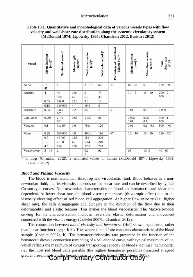
Microcirculation 321
Table 13.1. Quantitative and morphological data of various vessels types with flow
velocity and wall shear rate distribution along the systemic circulatory system
(McDonald 1974; Lipowsky 1995; Chandran 2012, Baskurt 2012)
Vessel
Mea
n d
iam
ete
r
[mm
]*
Nu
mb
er o
f v
ess
els
*
Mea
n l
en
gth
[mm
]*
To
tal
cro
ss s
ecti
on
[cm
2]*
To
tal
vo
lum
e [
mL
]*
Perce
nta
ge
of
tota
l b
loo
d
volu
me [
%]*
Mea
n d
iam
ete
r
[mm
] #
Mea
n f
low
velo
city
[cm
/s]
#
Wa
ll
shea
r ra
te
[s-1
] #
Aorta 19 –
45
1 2 – 16 60 11 16 – 32 6 150 – 300
Arteries
4 40 150 5 75 0.1 – 6 15 – 50 200 – 2
000 1.3 500 45 6.6 30
0.45 6 000 13.5 9.5 13
0.15 110 000 4 19.4 8
Arterioles 0.05 2.8 x
106
1.2 55 7 5 0.04 0.5 1 000
Capillaries 0.008 2.7 x 10⁹
0.65 1 357 88 0.005 – 0.01
0.05 – 0.1
400 – 1 600
Venules 0.1 1 x 10⁷ 1.6 785.4 126 0.02 – 0.1
0.2 – 0.5 800 – 400
Veins
0.28 660 000 4.8 406.4 196 67 0.2 – 10 15 – 20 120 – 320
0.7 40 000 13.5 154 208
1.8 2 100 45 53.4 240
4.5 110 150 17.5 263
Venae cavae 5 – 14 2 0.2 –
1.5
92 20 10.15 40 – 60
* in dogs (Chandran 2012), # estimated values in human (McDonald 1974; Lipowsky 1995;
Baskurt 2012)
Blood and Plasma Viscosity
The blood is non-newtonian, thixotrop and viscoelastic fluid. Blood behaves as a non-
newtonian fluid, i.e., its viscosity depends on the shear rate, and can be described by typical
Casson-type curves. Non-newtonian characteristics of blood are hematocrit and shear rate
dependent. At lower shear rate, the blood viscosity increases (thixotropic effect) due to the
viscosity elevating effect of red blood cell aggregation. At higher flow velocity (i.e., higher
shear rate), the cells disaggregate and elongate in the direction of the flow due to their
deformability and elastic features. This makes the blood viscoelastic. The Maxwell-model
serving for its characterization includes reversible elastic deformation and movement
connected with the viscous energy (Cokelet 2007b; Chandran 2012).
The connection between blood viscosity and hematocrit (Htc) shows exponential rather
than linear function (logη = k + k’Htc, where k and k’ are constants characteristic of the blood
sample (Cokelet 2007a, b). The hematocrit/viscosity rate presented in the function of the
hematocrit shows a connection reminding of a bell-shaped curve, with typical maximum value,
which reflects the maximum of oxygen transporting capacity of blood (“optimal” hematocrit),
i.e., the most red blood cells possible (the highest hematocrit possible) measured at speed
gradient resulting from the lowest viscosity possible (Ernst 1983; Bogar 2005). Complimentary Contributor Copy

Norbert Nemeth and Andrea Szabo 322
Main determining factors of plasma viscosity are its water content and the macromolecules
with elongated configuration, as the fibrinogen concentration, globulin fractions and
triglyceride level are important factors (Harkness 1971; Cokelet 2007b; Kesmarky 2008;
Kwaan 2010). It is important to note, that while in several stages of circulation, the apparent
viscosity of blood is not constant due to the intravascular interactions and distribution of
different shear rate profile and the formed elements, mainly the red blood cells, the
intravascular plasma viscosity can be considered constant. So the plasma viscosity maintains
shear stress on the endothelium at the cell-poor or sometimes even cell-free zone in the direct
vicinity of the ECs (Poiseuille-zone) due to the axial flow profile in vessels with diameter
approximately under 300 μm. Moreover, the plasma viscosity provides the shear force in case
of the so-called plasma-skimming, when at a certain point and at a certain site, there is no
formed element and only plasma in the capillary (Cokelet 2007b; Kesmarky 2008).
Red Blood Cell Deformability
Red blood cell deformability is an ability of passive transformation appearing under the
influence of shear stress, and it depends on the absolute volume, surface/volume ratio of the
cells, morphological characteristics, viscoelastic properties of the cell membrane and
intracellular viscosity (hemoglobin content) (Chien 1975; Meiselman 1981; Cokelet 2007a,b;
de Oliveira 2010).
Mature red blood cells of mammals and humans are without nucleus, so the intracellular
(cytoplasm) viscosity is mainly determined by the properties of hemoglobin solution. When the
mean cell hemoglobin concentration (MCHC) increases above 37 g/dl, the intracellular
viscosity increases precipitously, affecting the deformability of the whole cell, and thus also
the tissue perfusion and the oxygenation due to impeded passage in the microcirculation.
MCHC is regulated within a relatively narrow range (30−35 g/dl), where ATP-dependent Na+,
K+ transporters participating in the cell volume regulation play the key roles (Chien 1975;
Cokelet 2007b; Mohandas 2008; Baskurt 2012a).
Viscoelastic characteristics of red blood cells are complex (Thurston 2007). Viscosity
property derives from membrane viscosity and cytoplasm viscosity. The elasticity feature
consists of surface expansion, shearing and bending elements (Thurston 2007; Kim 2012).
Surface area expansion (or compression) modulus (K) reflects the stored elastic energy, which
develops through isotropic surface expansion or shrinkage of the membrane: Tt = K (ΔA/A0),
where Tt is the stress force, ΔA is the surface change, A0 is the original surface. The shear
module (μ) expresses the elastic energy which derives from membrane extension at same
surface: Ts = (μ(λ2 - λ-2))/2, where Ts is the shear stress, λ is the extension rate. Bending
(fluxural) modulus (B) expresses the energy which is required for bending the membrane from
one curve into another curve: M = B (C1 + C2 – C0), where M is the momentum of bending, C1
and C2 are the curves, C0 is the curve of stress free state (Kim 2012).
Motion of red blood cells can be described in a complex way, depending on the flow
conditions; rolling, rotation, swinging, elongation, elastic deformation due to collisions, the
combination of all the above, in their hardly modellable complexity. When suspending the cells
(such as in plasma) in a low viscosity medium, their motion is similar to that of solids. As the
flow rate increases, most of the cells flow along the same path (C = 0), where they rotate around
their symmetrical axis (perpendicular to the shear level) (Bitbol 1986). If the cells are
suspended in a higher viscosity medium over a critical speed-gradient, their behavior is similar
to that of fluid drops. The membrane transmits the momentum towards the cytoplasm, while Complimentary Contributor Copy
Microcirculation 323
maintaining the orientation of flow. At low and moderate speed gradient, the biconcave shape
of cells remains stable, but at higher values they elongate and gain an ellipsoid shape. Under
the effect of pulsatility and in the course of oscillation flow, the inclination angle of the cells is
not equal and therefore it becomes chaotic. In smaller veins, especially in the capillaries, the
friction has an increased role not only between the cells, but also in the relationship of
endothelium and blood cells. The erythrocytes flow in a formation reminding of doughnuts
flattened in their middle, where they are the thinnest (Goldsmith 1972; Pries 2003, 2008; Popel
2005; Dupire 2012).
Therefore, the deformability participates to a different extent in the different stages of
circulation. Worsening deformability, i.e., the enhanced rigidity of red blood cells, in the mass
flow zone of circulation (vessel diameter >300 μm) may lead to elevated blood viscosity. Red
blood cells with reduced deformability can cause the most significant problem in the
microcirculation (vessel diameter <100 μm), more precisely in the zone of so-called individual
flow. Here, capillaries with diameter of 3–5 μm may also occur, not to mention the fenestration
through the red pulp of the spleen to gaps of even smaller diameter. For passing through these
capillaries, the appropriate deformability is essential.
Red Blood Cell Aggregation
Erythrocyte aggregation means the reversible connection of cells which happens at low
shear rate or during stasis. Red blood cells initially arrange next to each other in a rouleaux
formation, resembling a stack of coins. It takes place within few seconds: the simpler rouleaux
form in 1–5 seconds. Over a period of further 10 to 60 seconds arranging in a two- then three-
dimensional shape until the effects maintaining the aggregation are present (Baskurt 2012a). In
anticoagulated blood samples, the aggregates coagulate into larger balls, then due to the gravity,
start to sink. This provides the background of the erythrocyte sedimentation rate (ESR), since
if the aggregation is fast and/or extensive, the erythrocyte sedimentation will also be more
intensive (Hardeman 2007).
The process of red blood cell aggregation is not yet completely clear. At present there are
two model theories to explain the aggregation development. (1) The aggregation of red blood
cells approaching each other happens via non-covalent cross-connections of macromolecules
(large proteins with elongated structure, such as fibrinogen, or in vitro with different polymers)
(bridging model). This weak linkage dissolves as the disaggregation forces such as the
electrostatic repulsion (due to negative charge, zeta potential: -15.7 mV), membrane tension,
deformation and mechanical shear stress prevail (the aggregates can be completely dispersed
at above 20–40 s-1 shear rate). This model supposes that the protein or polymer concentration
is high on the cell surface. Absorbing macromolecules non-specifically binding to cell surface
structures through their size and quantity may compensate the electrostatic repulsion,
facilitating the aggregation. (2) According to the other explanation, the macromolecules are not
able to penetrate as far as the membrane just because of the glycocalyx structure of cell surface
(5–50 nm), therefore close to the cell surface, a layer deficient in macromolecules or depletion
zone develops (depletion model). As two red blood cells approach each other, due to the
osmotic gradient between the macromolecule concentration being in plasma phase and the
depletion zone, a “pulling” force may develop, which makes the aggregation. The higher is the
macromolecule concentration, the more intensive may this gradient be, causing enhanced
aggregation (Baskurt 2012a; Saldanha 2013).
Complimentary Contributor Copy
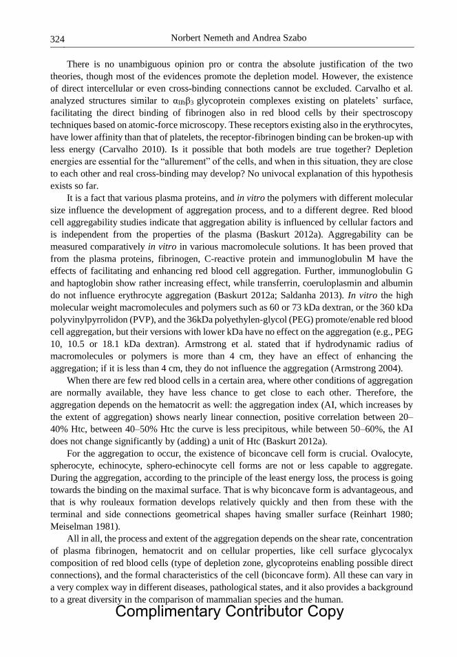
Norbert Nemeth and Andrea Szabo 324
There is no unambiguous opinion pro or contra the absolute justification of the two
theories, though most of the evidences promote the depletion model. However, the existence
of direct intercellular or even cross-binding connections cannot be excluded. Carvalho et al.
analyzed structures similar to αIIbβ3 glycoprotein complexes existing on platelets’ surface,
facilitating the direct binding of fibrinogen also in red blood cells by their spectroscopy
techniques based on atomic-force microscopy. These receptors existing also in the erythrocytes,
have lower affinity than that of platelets, the receptor-fibrinogen binding can be broken-up with
less energy (Carvalho 2010). Is it possible that both models are true together? Depletion
energies are essential for the “allurement” of the cells, and when in this situation, they are close
to each other and real cross-binding may develop? No univocal explanation of this hypothesis
exists so far.
It is a fact that various plasma proteins, and in vitro the polymers with different molecular
size influence the development of aggregation process, and to a different degree. Red blood
cell aggregability studies indicate that aggregation ability is influenced by cellular factors and
is independent from the properties of the plasma (Baskurt 2012a). Aggregability can be
measured comparatively in vitro in various macromolecule solutions. It has been proved that
from the plasma proteins, fibrinogen, C-reactive protein and immunoglobulin M have the
effects of facilitating and enhancing red blood cell aggregation. Further, immunoglobulin G
and haptoglobin show rather increasing effect, while transferrin, coeruloplasmin and albumin
do not influence erythrocyte aggregation (Baskurt 2012a; Saldanha 2013). In vitro the high
molecular weight macromolecules and polymers such as 60 or 73 kDa dextran, or the 360 kDa
polyvinylpyrrolidon (PVP), and the 36kDa polyethylen-glycol (PEG) promote/enable red blood
cell aggregation, but their versions with lower kDa have no effect on the aggregation (e.g., PEG
10, 10.5 or 18.1 kDa dextran). Armstrong et al. stated that if hydrodynamic radius of
macromolecules or polymers is more than 4 cm, they have an effect of enhancing the
aggregation; if it is less than 4 cm, they do not influence the aggregation (Armstrong 2004).
When there are few red blood cells in a certain area, where other conditions of aggregation
are normally available, they have less chance to get close to each other. Therefore, the
aggregation depends on the hematocrit as well: the aggregation index (AI, which increases by
the extent of aggregation) shows nearly linear connection, positive correlation between 20–
40% Htc, between 40–50% Htc the curve is less precipitous, while between 50–60%, the AI
does not change significantly by (adding) a unit of Htc (Baskurt 2012a).
For the aggregation to occur, the existence of biconcave cell form is crucial. Ovalocyte,
spherocyte, echinocyte, sphero-echinocyte cell forms are not or less capable to aggregate.
During the aggregation, according to the principle of the least energy loss, the process is going
towards the binding on the maximal surface. That is why biconcave form is advantageous, and
that is why rouleaux formation develops relatively quickly and then from these with the
terminal and side connections geometrical shapes having smaller surface (Reinhart 1980;
Meiselman 1981).
All in all, the process and extent of the aggregation depends on the shear rate, concentration
of plasma fibrinogen, hematocrit and on cellular properties, like cell surface glycocalyx
composition of red blood cells (type of depletion zone, glycoproteins enabling possible direct
connections), and the formal characteristics of the cell (biconcave form). All these can vary in
a very complex way in different diseases, pathological states, and it also provides a background
to a great diversity in the comparison of mammalian species and the human.
Complimentary Contributor Copy
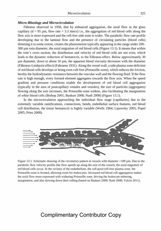
Microcirculation 325
Micro-Rheology and Microcirculation
Fåhræus observed in 1958, that by enhanced aggregation, the axial flow in the glass
capillary (d = 95 µm, flow rate = 3.3 mm/s) i.e., the aggregation of red blood cells along the
flow axis is more expressed and the cell-free side zone is wider. The parabolic flow rate profile
developing due to the laminal flow and the presence of circulating particles (blood cells)
dimming it to some extent, creates the phenomenon typically appearing in the range under 200–
300 µm vein diameter, the axial migration of red blood cells (Figure 13.1). It means that within
the vein’s cross section, the distribution and velocity of red blood cells are not even, which
leads to the dynamic reduction of hematocrit, to the Fåhræus-effect. Below approximately 30
µm diameter, down to about 10 µm, the apparent blood viscosity decreases with the diameter
(Fåhræus-Lindqvist effect) (Fahraeus 1931). Along the vessel wall, a side plasma zone deficient
of red blood cells develops or being even cell-free (Poiseuille-zone), which reduces the friction,
hereby the hydrodynamic resistance between the vascular wall and the flowing fluid. If the flow
rate is high enough, every formed element aggregates towards the flow axis. When the speed
gradient and pressure conditions enable the development of red blood cell aggregation
(typically in the area of postcapillary venules and venules), the size of particles (aggregates)
flowing along the axis increases, the Poiseuille-zone widens, also facilitating the margination
of white blood cells (Bishop 2001; Baskurt 2008; Nash 2008) (Figure 13.1).
In the microcirculation approaching the individual flow stage (capillaries) due to the
extremely variable ramifications, connections, bends, endothelial surface features, red blood
cell distribution, the tissue hematocrit is highly variable (Wells 1964; Lipowsky 2005; Popel
2005; Pries 2008).
Figure 13.1. Schematic drawing of the circulatory pattern in vessels with diameter <100 µm. Due to the
parabolic flow velocity profile (the flow speeds up along the axis of the vessel), the axial migration of
red blood cells occur. In the vicinity of the endothelium, the cell-poor/cell-free plasma zone, the
Poiseuille-zone is formed, allowing room for leukocytes. Increased red blood cell aggregation makes
the axial flow more expressed with widening Poiseuille zone, driving the leukocyte tethering,
margination, and also slowing down their rolling (based on Baskurt 2008; Nash 2008; Yalcin 2011).
Complimentary Contributor Copy
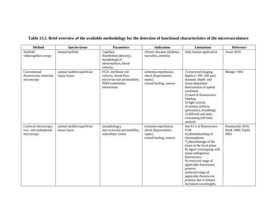
Table 13.2. Brief overview of the available methodology for the detection of functional characteristics of the microvasculature
Method Species/tissue Parameters Indications Limitations Reference
Nailfold
videocapillaro-scopy
human/nailfold Capillary
distribution (density),
morphological
abnormalities, blood
velocity,
chronic diseases (diabetes,
vasculitis, arteritis)
Only human application Awan 2010
Conventional
fluorescence intravital
microscopy
animal models/superficial
tissue layers
FCD, red blood cell
velocity, blood flow,
microvascular permeability,
PMN-endothelial
interactions
ischemia-reperfusion,
shock (hypovolemic,
septic)
wound healing, tumors
1) restricted imaging
depths (~100–200 µm)
dramatic depth- and
tissue-dependent
deterioration of spatial
resolution
2) need of fluorescence
labeling
3) light toxicity
4) motion artifacts
(peristalsys, breathing)
5) difficult and time-
consuming (off-line)
analysis
Menger 1992
Confocal microscopy,
two- and multiphoton
microscopy
animal models/superficial
tissue layers
(morphology),
microvascular permeability,
subcellular events
ischemia-reperfusion,
shock (hypovolemic,
septic)
wound healing, tumors
See #1-5 at fluorescence
IVM
6) photobleaching of
chromophores
7) photodamage of the
tissue at the focal plane
8) signal overlapping with
tissue endogenous
fluorescence
9) restricted range of
applicable fluorescent
proteins
restricted range of
applicable fluorescent
proteins due to limited
excitation wavelengths
Paramsothy 2010;
Denk 1990; Zipfel
2003
Complimentary Contributor Copy
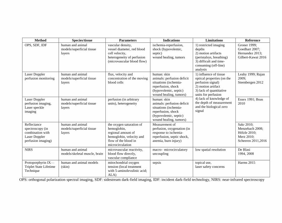
Method Species/tissue Parameters Indications Limitations Reference
OPS, SDF, IDF
human and animal
models/superficial tissue
layers
vascular density,
vessel diameter, red blood
cell velocity,
heterogeneity of perfusion
(microvascular blood flow)
ischemia-reperfusion,
shock (hypovolemic,
septic)
wound healing, tumors
1) restricted imaging
depths
2) motion artifacts
(peristalsys, breathing)
3) difficult and time-
consuming (off-line)
analysis
Groner 1999;
Goedhart 2007;
Hernandez 2013;
Gilbert-Kawai 2016
Laser Doppler
perfusion monitoring
human and animal
models/superficial tissue
layers
flux, velocity and
concentration of the moving
blood cells
human: skin
animals: perfusion deficit
situations (ischemia-
reperfusion, shock
(hypovolemic, septic)
wound healing, tumors)
1) influence of tissue
optical properties (on the
perfusion signal)
2) motion artifact
3) lack of quantitative
units for perfusion
4) lack of knowledge of
the depth of measurement
and the biological zero
signal
Leahy 1999; Rajan
2009;
Steenbergen 2012
Laser Doppler
perfusion imaging,
Laser speckle
imaging
human and animal
models/superficial tissue
layers
perfusion (in arbitrary
units), heterogeneity
human: skin
animals: perfusion deficit
situations (ischemia-
reperfusion, shock
(hypovolemic, septic)
wound healing, tumors)
Essex 1991; Boas
2010
Reflectance
spectroscopy (in
combination with
Laser Doppler
perfusion imaging)
human and animal
models/superficial tissue
layers
the oxygen saturation of
hemoglobin,
regional amount of
hemoglobin, velocity and
flow of the blood in
microcirculation
Measurement of
perfusion, oxygenation (in
response to ischemia-
reperfusion, septic shock,
anemia, burn injury)
Sakr 2010;
Menzebach 2008;
Hölzle 2010;
Merz 2010;
Scheeren 2011,2016
NIRS human and animal
models/skeletal muscle, brain
microvascular reactivity,
blood flow directly,
vascular compliance
macro– microcirculatory
uncoupling
low spatial resolution De Blasi
1994, 2008
Protoporphyrin IX—
Triplet State Lifetime
Technique
human and animal models
(skin)
mitochondrial oxygen
tension (local treatment
with 5-aminolevulinic acid;
ALA)
sepsis topical use,
laser safety concerns
Harms 2015
OPS: orthogonal polarization spectral imaging, SDF: sidestream dark-field imaging, IDF: incident dark-field technology, NIRS: near-infrared spectroscopy
Complimentary Contributor Copy

Norbert Nemeth and Andrea Szabo 328
Several other factors should be considered to better understand circulation conditions in
the field of microcirculation such as the extremely variable microvascular network geometry.
The vesselcross section can almost never be described as a regular circle. Elasticity of vessel
wall, hemodynamic features, flow profile and parameters (e.g., Reynolds number, Womersley
number) have to be taken into consideration as well (Pries 2008; Chandran 2012). Thus, in vivo
models have a great significance in further studying the connection of hemorheological factors
and microcirculatory characteristics, which can provide simultaneous data on most of the above
parameters.
So, the microcirculation possesses characteristic micro-hemodynamic features. Due to the
decreasing size of the microvessels, the major pressure drop occurs in the first-order arterioles
(based on the Hagen-Poiseuille equation), therefore constriction status of these vessels has the
highest influence on the regional flow. Local capillary flow, however, is controlled by third-
order, small-sized terminal arterioles. Efficacy of perfusion can be termed by a parameter called
the “functional capillary density” (FCD) which reflects the length of perfused capillaries within
a given area (Honig 1982). Reduced FCD results in a reduced surface area for capillary
exchange and an increase in diffusion distance (Hepple 2000). Recruitment of capillaries
depends on morphological features of vessels/endothelium of the given region and also on
blood rheology factors (i.e., hematocrit, viscosity, blood cell deformability and aggregation),
cellular factors, immune function, and coagulation function). Deterioration of microcirculatory
perfusion is marker of severity for many forms of circulatory disorders such as hemorrhagic,
cardiogenic, and septic shock, also reflecting the outcome of the disease (Sakr 2004; Fang 2006;
den Uil 2010). Hence, microcirculation might be considered an important prognostic indicator
in different clinical scenarios.
METHODS FOR DYNAMIC EXAMINATION OF
MICROCIRCULATORY FUNCTIONS
There are numerous invasive and non-invasive methods for investigating the
microcirculation. The limitation of this chapter does not allow to overview all the methods and
their developments in details. Table 13.2 gives a brief overview of the currently available
methodology for the detection of functional characteristics of the microvasculature.
Videomicroscopic Techniques
Direct visualization of the microcirculation with different types of in vivo microscopic
methods (IVM) enables not only examination of the architecture of arterioles, capillaries and
venules, but also provides an opportunity for continuous detection and quantification of
dynamic functional changes. Apart from nailfold capillaroscopy, most organs can be reached
via surgical exposure of the tissues for IVM examinations. Using meticulous microsurgical
techniques, an access of the microscope objectives and the required tissue depth can be
achieved by maintaining the proper physiological conditions without causing any disturbance
Complimentary Contributor Copy
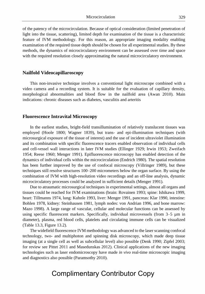
Microcirculation 329
of the patency of the microcirculation. Because of optical consideration (limited penetration of
light into the tissue, scattering), limited depth for examination of the tissue is a characteristic
feature of IVM methodology. For this reason, an appropriate imaging modality enabling
examination of the required tissue depth should be chosen for all experimental studies. By these
methods, the dynamics of microcirculatory environment can be assessed over time and space
with the required resolution closely approximating the natural microcirculatory environment.
Nailfold Videocapillaroscopy
This non-invasive technique involves a conventional light microscope combined with a
video camera and a recording system. It is suitable for the evaluation of capillary density,
morphological abnormalities and blood flow in the nailfold area (Awan 2010). Main
indications: chronic diseases such as diabetes, vasculitis and arteritis
Fluorescence Intravital Microscopy
In the earliest studies, bright-field transillumination of relatively translucent tissues was
employed (Hoole 1800; Wagner 1839), but trans- and epi-illumination techniques (with
microsurgical exposure of the tissue of interest) and the use of incident ultraviolet illumination
and its combination with specific fluorescence tracers enabled observation of individual cells
and cell-vessel wall interactions in later IVM studies (Ellinger 1929; Irwin 1953; Zweifach
1954; Reese 1960; Menger 1991). Epifluorescence microscopy has enabled detection of the
dynamics of individual cells within the microcirculation (Endrich 1980). The spatial resolution
has been further improved by the use of confocal microscopy (Villringer 1989), but these
techniques still resolve structures 100–200 micrometers below the organ surface. By using the
combination of IVM with high-resolution video recordings and an off-line analysis, dynamic
microcirculatory processes could be analyzed in sufficient details (Menger 1991).
Due to atraumatic microsurgical techniques in experimental settings, almost all organs and
tissues could be reached for IVM examinations (brain: Rovainen 1993; spine: Ishikawa 1999,
heart: Tillmanns 1974, lung: Kuhnle 1993, liver: Menger 1991, pancreas: Klar 1990, intestine:
Bohlen 1978, kidney: Steinhausen 1981, lymph nodes: von Andrian 1996, and bone marrow:
Mazo 1998). A large range of vascular, cellular and molecular functions can be assessed by
using specific fluorescent markers. Specifically, individual microvessels (from 3–5 µm in
diameter), plasma, red blood cells, platelets and circulating immune cells can be visualized
(Table 13.3, Figure 13.2).
The widefield fluorescence IVM methodology was advanced to the laser scanning confocal
technology, two- and multiphoton and spinning disk microscopy, which made deep tissue
imaging (at a single cell as well as subcellular level) also possible (Denk 1990; Zipfel 2003;
for review see Pittet 2011 and Masedunskas 2012). Clinical applications of the new imaging
technologies such as laser endomicroscopy have made in vivo real-time microscopic imaging
and diagnostics also possible (Paramsothy 2010).
Complimentary Contributor Copy
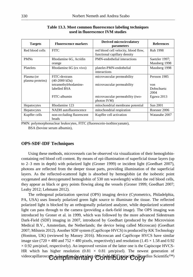
Norbert Nemeth and Andrea Szabo 330
Table 13.3. Most common fluorescence labeling techniques
used in fluorescence IVM studies
Targets Fluorescence markers Derived microcirculatory
parameters References
Red blood cells FITC red blood cell velocity, blood flow,
functional capillary density
Ruh 1998
PMNs Rhodamine 6G, Acridin
orange
PMN-endothelial interactions Saetzler 1997;
Massberg 1998
Platelets Rhodamine 6G (ex vivo) platelet-PMN-endothelial
interactions
Massberg 1998
Plasma (or
plasma proteins)
FITC-dextrans
(40-2000 kDa)
tetramethylrhodamine-
labelled BSA
FITC-albumin
microvascular permeability
microvascular permeability
microvascular permeability (two-
photon IVM)
Persson 1985
von
Dobschuetz
2004
Egawa 2013
Hepatocytes Rhodamine 123 mitochondrial membrane potential Sun 2001
Hepatocytes NADH autofluorescence mitochondrial respiration Roesner 2006
Kupffer cells non-occluding fluorescent
beads
Kupffer cell activation Watanabe 2007
PMN: polymorphonuclear leukocytes, FITC (fluorescein-isothiocyanate),
BSA (bovine serum albumin),
OPS-SDF-IDF Techniques
Using these methods, microvessels can be observed via visualization of their hemoglobin-
containing red blood cell content. By means of epi-illumination of superficial tissue layers (up
to 2–3 mm in depth) with polarized light (Groner 1999) or incident light (Goedhart 2007),
photons are reflected from the deeper tissue parts, providing illumination of the superficial
layers. As the reflected-scattered light is absorbed by hemoglobin (at the isobestic point
oxygenated and deoxygenated hemoglobin of 530 nm wavelength) within the red blood cells,
they appear as black or grey points flowing along the vessels (Groner 1999; Goedhart 2007;
Leahy 2012; Lehmann 2012).
The orthogonal polarization spectral (OPS) imaging device (Cytometrics, Philadelphia,
PA, USA) uses linearly polarized green light source to illuminate the tissue. The reflected
polarized light is blocked by an orthogonally polarized analyzer, while depolarized scattered
light can pass through to the camera (providing a dark-field image). The OPS imaging was
introduced by Groner et al. in 1999, which was followed by the more advanced Sidestream
Dark-Field (SDF) imaging in 2007, introduced by Goedhart (produced by the Microvision
Medical B.V., Amsterdam, the Netherlands; the device being called Microscan) (Goedhart
2007; Milstein 2012). Another SDF system (CapiScope HVCS) is produced by KK Technology
(Honiton, UK) (reviewed by Massey 2016). Microscan and CapiScope HVCS have similar
image size (720 × 480 and 752 × 480 pixels, respectively) and resolution (1.45 × 1.58 and 0.92
× 0.92 µm/pixel, respectively). An improved version of the latter one is the Capiscope HVCS-
HR which has higher resolution (0.81 × 0.81 µm/pixel). The newest generation of
videocapillaroscopy is based on the incident dark field (IDF) principle (Braedius Scientific™) Complimentary Contributor Copy
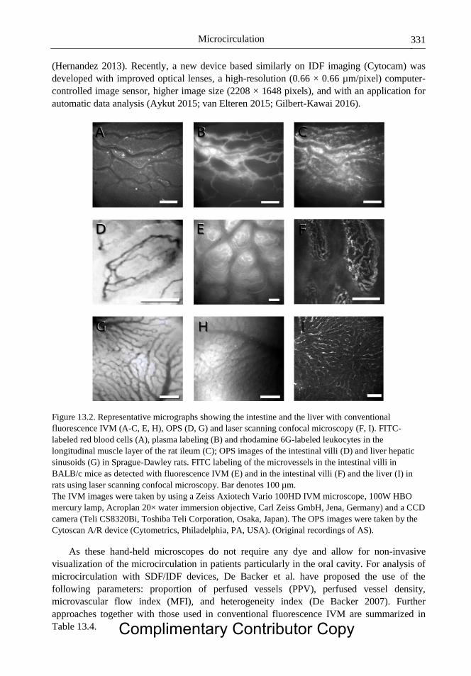
Microcirculation 331
(Hernandez 2013). Recently, a new device based similarly on IDF imaging (Cytocam) was
developed with improved optical lenses, a high-resolution (0.66 × 0.66 µm/pixel) computer-
controlled image sensor, higher image size (2208 × 1648 pixels), and with an application for
automatic data analysis (Aykut 2015; van Elteren 2015; Gilbert-Kawai 2016).
Figure 13.2. Representative micrographs showing the intestine and the liver with conventional
fluorescence IVM (A-C, E, H), OPS (D, G) and laser scanning confocal microscopy (F, I). FITC-
labeled red blood cells (A), plasma labeling (B) and rhodamine 6G-labeled leukocytes in the
longitudinal muscle layer of the rat ileum (C); OPS images of the intestinal villi (D) and liver hepatic
sinusoids (G) in Sprague-Dawley rats. FITC labeling of the microvessels in the intestinal villi in
BALB/c mice as detected with fluorescence IVM (E) and in the intestinal villi (F) and the liver (I) in
rats using laser scanning confocal microscopy. Bar denotes 100 µm.
The IVM images were taken by using a Zeiss Axiotech Vario 100HD IVM microscope, 100W HBO
mercury lamp, Acroplan 20× water immersion objective, Carl Zeiss GmbH, Jena, Germany) and a CCD
camera (Teli CS8320Bi, Toshiba Teli Corporation, Osaka, Japan). The OPS images were taken by the
Cytoscan A/R device (Cytometrics, Philadelphia, PA, USA). (Original recordings of AS).
As these hand-held microscopes do not require any dye and allow for non-invasive
visualization of the microcirculation in patients particularly in the oral cavity. For analysis of
microcirculation with SDF/IDF devices, De Backer et al. have proposed the use of the
following parameters: proportion of perfused vessels (PPV), perfused vessel density,
microvascular flow index (MFI), and heterogeneity index (De Backer 2007). Further
approaches together with those used in conventional fluorescence IVM are summarized in
Table 13.4. Complimentary Contributor Copy
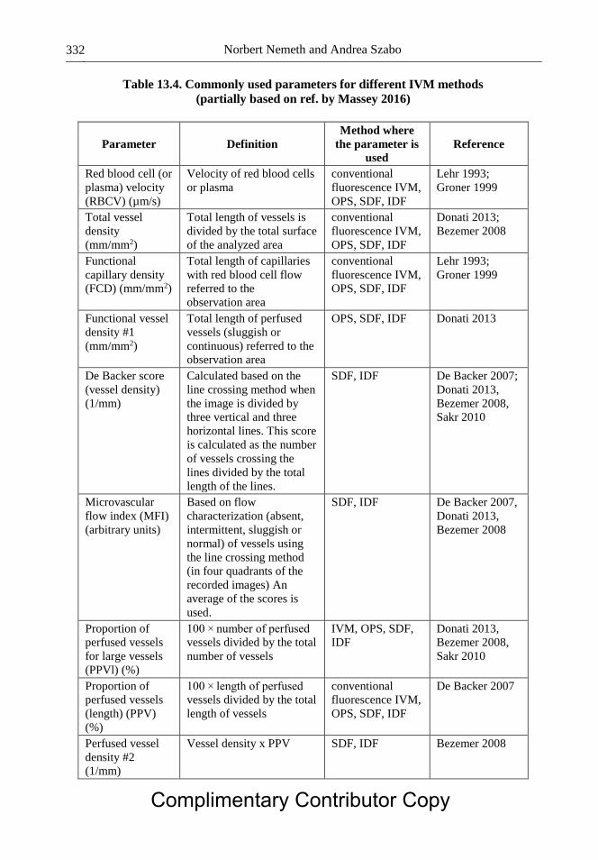
Norbert Nemeth and Andrea Szabo 332
Table 13.4. Commonly used parameters for different IVM methods
(partially based on ref. by Massey 2016)
Parameter Definition
Method where
the parameter is
used
Reference
Red blood cell (or
plasma) velocity
(RBCV) (µm/s)
Velocity of red blood cells
or plasma
conventional
fluorescence IVM,
OPS, SDF, IDF
Lehr 1993;
Groner 1999
Total vessel
density
(mm/mm2)
Total length of vessels is
divided by the total surface
of the analyzed area
conventional
fluorescence IVM,
OPS, SDF, IDF
Donati 2013;
Bezemer 2008
Functional
capillary density
(FCD) (mm/mm2)
Total length of capillaries
with red blood cell flow
referred to the
observation area
conventional
fluorescence IVM,
OPS, SDF, IDF
Lehr 1993;
Groner 1999
Functional vessel
density #1
(mm/mm2)
Total length of perfused
vessels (sluggish or
continuous) referred to the
observation area
OPS, SDF, IDF Donati 2013
De Backer score
(vessel density)
(1/mm)
Calculated based on the
line crossing method when
the image is divided by
three vertical and three
horizontal lines. This score
is calculated as the number
of vessels crossing the
lines divided by the total
length of the lines.
SDF, IDF De Backer 2007;
Donati 2013,
Bezemer 2008,
Sakr 2010
Microvascular
flow index (MFI)
(arbitrary units)
Based on flow
characterization (absent,
intermittent, sluggish or
normal) of vessels using
the line crossing method
(in four quadrants of the
recorded images) An
average of the scores is
used.
SDF, IDF De Backer 2007,
Donati 2013,
Bezemer 2008
Proportion of
perfused vessels
for large vessels
(PPVl) (%)
100 × number of perfused
vessels divided by the total
number of vessels
IVM, OPS, SDF,
IDF
Donati 2013,
Bezemer 2008,
Sakr 2010
Proportion of
perfused vessels
(length) (PPV)
(%)
100 × length of perfused
vessels divided by the total
length of vessels
conventional
fluorescence IVM,
OPS, SDF, IDF
De Backer 2007
Perfused vessel
density #2
(1/mm)
Vessel density x PPV SDF, IDF Bezemer 2008
Complimentary Contributor Copy

Microcirculation 333
Parameter Definition
Method where
the parameter is
used
Reference
Flow
heterogeneity
index
The difference between
highest and the lowest
MFI is divided by the
mean flow velocity
SDF, IDF De Backer 2007,
Donati 2013
PMN rolling
(defined as
rodamine 6G-
labaled cells
moving at a
velocity less than
40% of that of the
erythrocytes in
the centerline of
the microvessel)
(1/mm/s or % of
total PMNs)
The number of rolling
cells devided by the vessel
circumference
conventional
fluorescence IVM
Lehr 1993,
Bajory 2002
PMN adhesion
(cells that did not
move or detach
from the
endothelial lining
within an
observation
period of 30 s)
(1/mm2)
The number of cells
devided by mm2 of
endothelial surface
conventional
fluorescence IVM
Lehr 1993
Laser Doppler Flowmetry
The background of laser Doppler monitoring is the “Doppler shift” phenomenon (Doppler
1942). When a monochromatic laser light enters the tissues and meets moving blood cells, it
undergoes changes in wavelength (Doppler shift), but remains unchanged if the beam of light
hits static tissue structures. Magnitude and frequency changes of wavelength correlate with the
number of moving blood cells (Stern 1975). The Doppler technique provides “perfusion units”
which cannot be transformed in terms of flow (e.g., ml/min), but rather a flux unit without
dimension, since it is an integral over the velocity and the number of moving red blood cells in
the tissue territory laying under the probe (Obeid 1990; Leahy 1999, 2012; Friedriksson 2012;).
There are two major approaches of perfusion measurements, the laser Doppler perfusion
monitoring and laser Doppler perfusion imaging (Rajan 2009; Friedriksson 2012;
Leahy 2012).
Laser Doppler Perfusion Monitoring
In this one-point measurement method, a fiber-optic probe is used to deliver the laser light
to the tissue, and another fiber to detect the back-scattered photons recording the integrated Complimentary Contributor Copy
Norbert Nemeth and Andrea Szabo 334
perfusion in a sampling volume in real time. The measurement depth and sampling volume
depend on the wavelength (usually 630, 780 and 830 nm are used) and the fiber separation
used. In the skin, (using 780 nm wavelength), the measuring depth is 0.5–1 mm and the
measurement volume is approximately 1 mm3. The main manufacturers of laser Doppler
instruments are Perimed AB (Stockholm, Sweden), Moor Instruments Ltd. (Axminster, UK),
Transonic Systems Inc. (Ithaca, New York, USA), Oxford Optronix Ltd (Oxford, UK) and LEA
Medizintechnik (Giessen, Germany).
Laser Doppler tissue flowmetry method can be widely applied both in the clinical and
research work to study local or systemic effect of heat, reflex vasodilation, microcirculatory
changes following occlusion, or the effects of vasoactive drugs, as well as endothelial reactivity
using provocation tests (ischemia, heat) (Obeid 1990; Neviere 1996; Rajan 2009; Friedriksson
2012; Leahy 2012).
However, numerous factors may influence the results. This method considerably and
sensitively indicates the changes occurring in microcirculation, but at the same time, several
factors may influence the measurements. These factors on one hand include the thickness of
the examined tissue, its anatomical structure, optical properties (pigmentation), possible
morphological alteration and vascularity. On the other hand, from the patient’s side, the gender,
age, hormonal and neurological effects and drugs may affect the results of the measurement.
From the side of the device, the displacement and contamination of the probe, kinking of fiber
optics, distance of emission and receiving optical fibers, wavelength of laser light and
biological zero signal may also be considered as influencing factors. Disturbing artifacts or
“noises” may develop through the movement of optic fiber or from the affected tissue (e.g.,
breathing, intestinal peristalsis). External factors such as temperature, humidity, air movement
also influence the measurement; therefore, it is very important to ensure standard
circumstances. However, performing comparative studies (e.g., before and after intervention)
is difficult, because to ensure the same conditions in all parameters is almost impossible (Obeid
1990; Neviere 1996; Nemeth 2003; Rajan 2009; Friedriksson 2012; Leahy 2012).
In pathophysiological processes, such as sepsis or ischemia-reperfusion, associated with
acute changes of tissue microcirculation, laser Doppler technique is highly justified. In
experimental models, probes available in various versions can be applied in several ways
(Nemeth 2003; Rajan 2009; Friedriksson 2012; Leahy 2012):
probe touched directly to the surface of skin or in open surgery of the organ (manual
or preferred trestle fixing)
probe fixed on the surface of the organ (stitches)
in laparoscopy introduced via trocar and touched to the surface of examined
intraperitoneal organ or the peritoneum
introduced in endoscopy and touched to the surface of mucosa (multichannel
endoscopy, fiber optic probe)
implanted between tissue layers or bone mortise for monitoring
Depending on the wavelength of the applied laser, the depth of the tissue area that can be
examined may range from 0.1 to 0.8 mm, e.g., by applying laser with 780 mm wavelength and
probe with 0.5 mm diameter, this tissue depth is 0.73 mm in muscles, 0.52 mm in the liver and
0.5 mm in the skin. The duration of the examination and positioning of probe should be taken
Complimentary Contributor Copy
Microcirculation 335
into consideration. The 0.5 mW energy derives from laser light, and in the examined tissue, it
may also influence the state of capillaries after some time (Friedriksson 2012).
The probe stabilization is essential. Overpressure of the tissues may cause local
microcirculatory disturbance; if too loose, displacing or fixing may lead to inaccurate
measurements and several artifacts.
Several solutions are known for the right interpretation of the recorded signal in the
literature. For instance, the native values measured and assessed at a certain interval by the
device can be used. Within this interval (e.g., data of stable signal over 10 or 20 seconds), the
values can be averaged. Relative changes compared to initial values, moving average, Fourier
transformation and various timeline analysis methods can also be used for analyzing laser
Doppler recordings (Obeid 1990; Nemeth 2003, 2014b; Rajan 2009; Friedriksson 2012; Leahy
2012).
An organ may show different laser Doppler signals with different recorded patterns
depending on the functional and morphological state of microvasculature. Therefore, it is
important to observe the characteristics of the recorded signal and decide how to analyze and
interpret the data. Signals recorded in skin, liver, kidney and muscles vary within a relatively
small range, but they are never consistent. In other organs such as bowel, omentum or pancreas,
the signals oscillate between wider ranges, so the averaging is difficult in these cases; it may
even be misleading if we analyze a data set of t an inadequate period with their extremes
(Nemeth 2003, 2014b).
During a prolonged monitoring, when the probe of the device is fixed in a steady,
permanent position and the microcirculation is being monitored for half an hour or even longer,
the extent and dynamics of changes can be analyzed separately. This point of view is of
particular importance in examinations which aim to follow organic microcirculatory effects of
different materials and surgical maneuvers.
Regarding the several factors which influence the measured values, examining the changes
in correlation with the baseline values is an extremely useful data analyzing method. Thus it
can be expressed as a percentage to what extent a certain tissue area changes during a surgery.
It is a very important point to indicate baseline value, as it was mentioned, intensity of laser
Doppler signal is not constant in any of the tissue areas, more or less irregularities, periodicity
and artifacts appear. Should we use any data analysis and interpretation method, it has to
beconsistent and standardized.
Laser Doppler Perfusion Imaging (Laser Doppler Scanner Technology)
A laser Doppler perfusion imaging technology maps the perfusion from a larger area by
scanning the laser beam over the area of interest without having any direct contact with the
tissues (Essex 1991; Fredriksson 2012; Steenbergen 2012 a, b). These instruments provide
perfusion map of an area with a (normally) heterogeneous perfusion and also gives the average
of perfusion of the examined area in a single measurement. The main manufacturers of laser
Doppler perfusion imagers are Perimed AB and Moor Instruments Ltd. Using stepwise changes
in the illuminating laser beam wavelength or different wavelength simultaneously, the spatial
resolution can be increased up to 0.1 mm.
Complimentary Contributor Copy
Norbert Nemeth and Andrea Szabo 336
Laser Doppler Flowmetry Combined with Reflectance Spectrometry
The more recent technology (the O2C device, Lea, Giessen, Germany) combines the laser
Doppler technique and reflection spectroscopy. Using specific probes, white light (wavelengths
500–800 nm) and laser light (wavelength 830 nm) are introduced simultaneously. The absorbed
portion of the light spectrum is absorbed on erythrocytes assuming the color of the hemoglobin
as measured by oxygen saturation, while laser light reflects perfusion (based on the Doppler
shift). The sum of all erythrocytes and their velocity provides a measure of blood flow (volume
flow). The following parameters can be measured simultaneously with this technology: oxygen
saturation of hemoglobin at the venous end of the capillaries, regional amount of hemoglobin
as a measure of blood volume and capillary density, velocity and flow of the blood within the
microcirculation. This method has been employed to assess the microvascular response in
patients with septic shock (Sakr 2004; Sakr 2010) and in anemia (Menzebach 2008), to estimate
flap viability (Hölzle 2010) and wound healing in burn injury (Merz 2010).
Laser Speckle Imaging
A speckle image is created when an object is illuminated by laser light. The backscattered
light will forms a random interference pattern consisting of dark and bright areas (speckle
pattern). In case of a moving object (e.g., red blood cells) in the underlying tissue, the speckle
pattern changes over time depending on the extent of movements within the examined area. As
a result, the level of blurring differs resulting in various extent of speckle contrast (Briers 1999;
Briers 2012; Boas 2010; Steenbergen 2012b). Consequently, changes in microcirculatory
perfusion characteristics under a surface (within a typical depth range of 1–5 mm, max. 15 mm)
can be analyzed. The resolution depends on the characteristics of detector as well as the size of
examined surface and the distance of the detector. Using this method, preclinical and clinical
detection of the skin (Choi 2004; Rege 2011; Sandker 2014) and even cerebral blood flow
(Parthasarathy 2010; Qin 2012) could be performed.
It was recently demonstrated that the distribution of blood flow within tissue beds can
noninvasively be visualized also by the motion-contrast laser speckle imaging which leads to
enhanced visibility of distinct vascular beds, including capillaries (Liu 2013).
Near-Infrared Spectroscopy
Near-infrared spectroscopy (NIRS) is a non-invasive technique that assesses the redox state
of microcirculatory hemoglobin by measuring oxyhemoglobin, deoxyhemoglobin, total
hemoglobin and other chromophores (myoglobin, cytochrome aa3) in mostly the venous
system (De Blasi 1994; Pareznik 2006). Vascular occlusion tests allow quantification of the
rate of decay of microcirculatory oxygen saturation thus representing the metabolic status of
the underlying tissue (muscle) (De Blasi 2008).
Complimentary Contributor Copy
Microcirculation 337
Measurements of O2 Tension and Saturation
Conventional oximetry (hemoglobin oxygen saturation measurement) is widely used in the
clinical practice and is based on the assessment of differences in the absorption spectra of oxy-
and deoxy-hemoglobin. Interestingly, it was first applied in vivo within the microcirculation to
obtain oxygen saturation in single vessels using IVM (Pittman 1975; Pittman 2013).
Transcutaneous arterial oxygen pressures can simultaneously be measured using standard
pulsoximetry, whereas transcutaneous venous carbon monoxide tensions with CO2-sensitive
electrodes (Fromy 1997).
Phosphorescence quenching microscopy is based on the collisional quenching
phenomenon which occurs when excited phosphor decays back to the ground state by emitting
a photon (phosphorescence) or by transferring energy to nearby oxygen molecules. This enables
measurements of the O2 tensions (Vinogradov 2002). The energy transfer to oxygen results in
the formation of the reactive species singlet oxygen, which can then react with nearby organic
molecules, thereby resulting in the consumption of the original oxygen molecule.
Oxygen tensions in the microcirculation can be measured using oxygen-sensitive
polarographic microcathodes (Buerk 2004). Oxygen saturation can also be non-invasively
measured using the Resonance Raman microspectroscopy (Ward 2006). The phosphorescence
quenching approach (Vinogradov 2002) and oxygen electrodes (Buerk 2004) can also measure
oxygen tensions in the interstitium.
In Vivo Measurements of Mitochondrial Functions and Oxygen Tensions
The “Protoporphyrin IX–Triplet State Lifetime Technique” is a new approach developed
for the measurement of mitochondrial function and oxygen tension (mitoPO2) in vivo (Mik
2006, Harms 2015). A recently developed non-invasive technique is able to measure a decline
in mitoPO2 after pressure-induced cessation of blood supply (called oxygen disappearance
rate), also providing estimates of mitochondrial oxygen consumption. The oxygenation data
was validated in a sepsis study in rats with the use of reflectance spectroscopy (combined laser
Doppler) in the skin. They also demonstrated the feasibility of the non-invasive technology to
assess mitochondrial function in the human skin.
Further Methods
Due to the limitations of the chapter, further special methods are just briefly summarized,
referring to detailed handbooks (e.g., Leahy 2012).
Subsurface Spectroscopy (Tissue Viability Imaging)
For tissue viability imaging (TiVi), further possibilities are based on wideband
spectroscopy of backscattered light (subsurface spectroscopy) for mapping local red blood cell
concentration. The sequential photo recordings are being analyzed by a software package,
Complimentary Contributor Copy
Norbert Nemeth and Andrea Szabo 338
allowing studies of dynamic skin reactions (using agent or effects that cause vasodilatation or
vasoconstriction) (O’Doherty 2012).
Optical Microangiography
Because of the high spatiotemporal resolution and the small dimension, microvasculature
imaging is technologically challenging. Optical microangiography (OMAG) is capable of
resolving three-dimensional distribution of dynamic blood perfusion by imaging the
endogenous light scattering from moving red blood cells. The technique is based on Fourier
domain optical coherence tomography (FDOCT) (Wang 2012).
Photoacoustic Tomography
Photoacoustic tomography (PAT) detects endogenous or exogenous optical absorption
contrast acoustically. When red blood cells are irradiated by a short-pulsed laser beam, a
transient thermoelastic expansion occurs that induces high-frequency ultrasonic
(photoacoustic) waves. The device encodes the red blood cell distribution and makes a
reconstruction of a three-dimensional microvascular image. Using multi-wavelength
measurements, differentiation of oxyhemoglobin and deoxyhemoglobin is also possible. Based
on PAT, photoacoustic microscopy (PAM) and photoacoustic computed tomography (PACT)
methods are known (Hu 2012).
High Frequency Ultrasound Imaging
Maximal imaging frequency of clinical ultrasound systems are typically 12−15 MHz,
providing resolution on structures of ˃ 300 µm. The micro-ultrasound devices (preclinical
imaging) have been developed for the range of 15–80 MHz (Foster 2012).
Micro-CT, Nano-SPECT-CT, NMR
Micro-computed tomography (Micro-CT) is a powerful tool to study small animals,
corrosion specimens or biopsies. The resolution of Micro-CT is about 1–100 µm in rotating
state, ~50–100 µm in rotating scanner in plane. Micro-SPECT and micro-PET resolution is
about 1–2 mm, while micro-magnetic resonance imaging (MRI) provides a resolution about
100–200 µm in plane and ~0.5–10 mm slice thickness (Kline 2012). Boundaries of the nuclear
magnetic response imaging (NMRI) techniques and applications (MR angiography, MR
perfusion imaging, functional MRI) widen, providing more possibilities to investigate
structural and functional state of the vasculature as well (Kerskens 2012).
Complimentary Contributor Copy
Microcirculation 339
BRIEF OVERVIEW ON METHODS FOR ASSESSMENT
OF RHEOLOGICAL PARAMETERS
Methods for Measuring Blood and Plasma Viscosity
For the determination of blood viscosity, only those measuring devices are suitable, which
can determine viscosity at different shear rates (Cokelet 2007a, b). The shearing tension or
speed gradient profile may be generated under the effect of gravitational force (e.g., in capillary
viscometers), or through rotational motion with a certain angular velocity, produced by a built-
in mechanism (rotational viscometers) (Baskurt 2009).
Most of the viscometers were the capillary type until the 1960s, rotational viscometers
appeared later. In the capillary-type ones, the flow rate profile can be determined from the
position-time data of fluid column, and the shearing tension can be calculated from the
geometry of tube. The geometry of rotational viscometers can be various: cone-plate, cone-in-
cone or coaxial cylinder (or Couette-system) in speed-gradient controlled or shearing tension
controlled form. The fluid to be measured can be filled in between the two elements (e.g., ‘bob’
and ‘cup’). One element can be rotated by adjusted angular velocity. The torque relocated to
the non-rotated element to an extent is depending on the viscosity of the fluid. The torque is
tested by laser reflection, and the viscosity can be calculated at a certain shear rate-gradient.
Oscillation flow viscometers are also known, which are suitable for the investigation of viscous
and elastic components. The viscous component can be calculated from the amplitude of speed
gradient, and the elastic component is calculated from phase delay of shearing tension in
relation to the speed gradient (Rosencranz 2006; Hardeman 2007).
For the measurement of plasma viscosity-based on the newtonian characteristics–
viscometers, whose method does not depend on shear rate gradient, are suitable. The dynamic
viscosity of the fluid can be calculated from fluid flow rate in a system with a certain geometry,
from position-time data (U-tube or capillary viscometers: e.g., Ostwald, Coulter-Harkness,
Rheomed, Luckham viscometers), or in the falling ball viscometers (e.g., Haake-type) from the
effect of drag force applying the Navier-Stokes equation: F = 6πrηv, where F is the frictional
force, r is the radius of the spherical object, η is the fluid viscosity, v is the particle’s velocity.
With a simpler method: η = k(ρ1-ρ2)t, where k is constant, ρ1 is the density of the particles, ρ2
is the density of the fluid, t is the duration of ball falling (Rosencranz 2006; Hardeman 2007;
Baskurt 2009).
Methods for Testing red Blood Cell Deformability
For the determination of red blood cell deformability, several methods and various
approaches are known. The best-known and still in use techniques are based on the filtration
method, the micropipette aspiration technique, microchannel methods and the ektacytometry
(Hardeman 2007; Baskurt 2009).
Filtration technique has been used since the 1960s: measuring the passage of blood cell
suspension through a filter with a certain pore diameter/size (~200 000 pores), the cells’
deformability can be concluded; due to the clogging of pores, the permeability of the filtering
membrane gradually decreases. The initially used classical paper filters were replaced by Complimentary Contributor Copy

Norbert Nemeth and Andrea Szabo 340
polycarbonate filter membranes containing cylindrical channels (diameter: 3, 5 or 8 μm, length:
10 μm). The Reid-Dormandy type filtration method utilized the filtration of red blood cell
aggregation through the filter with 5 μm in diameter. Improving the principle, Dormandy and
his colleagues prepared the St. George’s filtrometer. The oligopore (23–30 pores) and the single
pore technique (Single Erythrocyte Rigidometer, SER) are more sophisticated methods; here
mainly the time of change in conductivity between the two sides of the filter correlate with
presence of red blood cells just passing through the pore, where the process is analyzed by the
attached computer according to the pores transit time histogram (Cell Transit Analyzer, CTA)
(Hardeman 2007; Baskurt 2009; Musielak 2009).
With the micropipette aspiration method, a single cell can be examined, which is wholly
or partly aspirated into a glass capillary with 1–5 μm diameter. From the measurement of the
negative pressure and the length of the aspirated membrane phase provides shearing elastic
module, while the aspiration of the whole cell gives the deformability. It requires serious
technical skills (Hardeman 2007; Baskurt 2009; Musielak 2009).
The basic principle of the microchannel method is that the cell suspension is flowing
through one or two parallel channels, or through an artificial microchannel network, while the
movement of red blood cells, the transit time, and position of the cells can be visually analyzed
(Baskurt 2009; Musielak 2009; Reinhart 2015).
The ektacytometry is based on the diffraction analysis of laser beam (wavelength: 670 nm)
reflected from red blood cells suspended in high-viscosity (preferably ~30 mPas)
macromolecule solution (e.g., 70 kDa dextran or 350 kPa polyvinylpyrrolidone isotonic
solution) and elongated due to the shear stress, providing the elongation index (EI) in the
function of shear stress (Chien 1975; Chasis 1986; Hardeman 2007). The shear stress on the
sample is generated by the motor or vacuum device, while the laser diffraction pattern is
captured by a CCD camera and projected on a screen. The EI can be calculated from the
analyzed picture by the software diffractrogram: EI = (L-W) / (L + W), where L is the length
and W is the width of the diffractogram. Different shearing geometries are used in
ektacytometer devices: plane-plane rotational system in the Rheodyn device, Couette-system
in laser-assisted optical rotational cell analyzer (LORCA), or vacuum-driven slit-flow
technique in the Rheoscan device (Hardeman 2007; Baskurt 2009; Musielak 2009). Besides the
classical deformability measurements, some modern ektacytometers are capable of testing
osmotic gradient deformability (i.e., deformability test at constant shear stress over an osmotic
gradient range) and membrane mechanical stability (deformability test before and after a period
pf mechanical shearing at definitive magnitude and duration) (Hardeman 2007; Baskurt 2009).
Further deformability measurement methods are the rheoscope technique (a combination
of ektacytometry and microscopy), atomic-force microscopy, the laser optical tweezers and
magnetic twisting cytometry (Hardeman 2007; Musielak 2009).
Methods for Testing Red Blood Cell Aggregation
Several indirect and direct methods are known for the determination of red blood cell
aggregation. The classical and modern methods (Westergren, Seditainer, Automatic ESR)
based on the already mentioned measurement of erythrocyte sedimentation rate (ESR) are
widely used and well standardized methods. Also known is the determination of zeta
sedimentation rate (by centrifugation) and the determination of indirect aggregation parameters Complimentary Contributor Copy
Microcirculation 341
(aggregation index = (ηlow – ηhigh) / ηhigh, where η is the blood viscosity determined at a certain
low and a higher speed gradient) from the results of blood viscosity measured at low speed
gradient (e.g., 1 s-1). By microscopy techniques or rheoscope the morphology of aggregates can
also be examined and their size and red blood cell count per aggregate can be determined. By
this method, the heteroaggregates (white blood cell, platelet, red blood cell) can also be
separated. Ultrasound techniques enable non-invasive in vivo examinations (mainly B-mode
echography) of aggregation developing in a certain vascular region (Hardeman 2007; Baskurt
2009, 2012b).
In clinical and experimental hemorheological studies, the most commonly used methods
are based on the principles of photometry. The presence of aggregates increases the light
transmission of blood sample related to a certain wavelength, as in this case the width of plasma
phase (or other suspending medium) between the particles increases. In the course of
disaggregation, the cells dispersed on a larger surface decreasing the plasma phase width and
so the light-transmittance. When cells start to aggregate during stasis or at low shear rate, the
plasma phase width and the light-transmittance increase. Furthermore, red blood cell
aggregates reflect light differently compared to the individual cells. Based on these principles,
different devices are available to analyze the light transmission (e.g., Myrenne MA-1
erythrocyte aggregometer), or the reflection of light (laser) from the aggregating cells
(syllectometry, e.g., LORCA). Using these methods, both the extent and the dynamics of
aggregation can be examined (Hardeman 2007; Baskurt 2009, 2012b).
Thoughts on the Link between Microcirculatory and
Micro-Rheological Investigations
The examination of rheological conditions of microcirculation and their interaction cannot
be simplified to the correlation of Poiseuille’s law related to the rigid-walled, straight regular
pipes. The large region of microcirculation is not even homogenous from rheological point of
view. In addition to the vein diameter and vascular geometry, the viscosity cannot be considered
constant due to axial migration of red blood cells (Fåhræus and Fåhræus-Lindqvist effect),
phase separation, very diverse and changeable, unsteady distribution of tissue hematocrit
(microvascular hematocrit), and the particularities of the zone of individual flow (Fahreus 1931,
1958; Wells 1964; Lipowsky 2005; Pries 2008).
Therefore, in the isolated organs, in perfusion ex vivo models, the solutions ensuring the
stability of vessel diameter (maximal dilatation or maximal constriction) could only show a
thorough picture if we hypothesized and presumed that the viscosity is constant at every stage
of the circulation. This is however, obviously not true. That is why a more detailed investigation
of the role of micro-rheological parameters in the microcirculation is required (Wells 1964;
Lipowsky 2005; Popel 2005).
Parallel investigations of hemodynamic, microcirculatory, hemorheological, acid-base
parameters, hematological and hemostaseological factors may provide enough information to
evaluate and understand the dynamics of these changes and their interactions (Baskurt 2007;
Nemeth 2014b).
Complimentary Contributor Copy

Norbert Nemeth and Andrea Szabo 342
MAIN MICROCIRCULATORY AND MICRO-RHEOLOGICAL FINDINGS
IN VARIOUS PATHOPHYSIOLOGICAL PROCESSES
Ischemia/Reperfusion
Ischemia/reperfusion injury causes all of the microcirculatory symptoms which can
develop during local or systemic circulatory disorders (Ames 1968; Menger 1992; Anaya-
Prado 2002; Eltzschig 2011). It appears that the ECs are primary targets during the pathogenesis
of ischemia/reperfusion-induced reactions (including inflammation). Being a major source of
the xanthine oxidase-derived radicals (Granger 1981; Granger 1986; McCord 1985), a
microvascular dysfunction (increased capillary permeability) develops. The xanthine oxidase-
derived radicals further attract neutrophils (PMNs), thus augmenting tissue injury (Hernandez
1987). The endothelium-derived oxidants promote neutrophil adhesion to ECs via endothelial
production of chemotactic factors (Yoshida 1992) as well as by activation of transcription
factors (e.g., NFκB), causing an increased expression of adhesion molecules on ECs (Ichikawa
1997). Bioavailability of NO (an endogenous inhibitor of PMN adhesion) is reduced via its
interaction with superoxide (Grisham 1998). This is accompanied by an activation of interstitial
immune cells (macrophages and mast cells) with resultant generation of inflammatory
mediators causing a chemotactic gradient which facilitates neutrophil emigration into the
interstitium (Kanwar 1998). Lymphocytes may also be involved in the modulation of PMN-
related reactions by releasing soluble factors, such as TNF–α or INF–γ (Osman 2009).
Micro-rheological parameters can be altered during ischemia and reperfusion by several
pathways (Baskurt 2007). Impaired red blood cell deformability may contribute to an increased
blood viscosity and cause perfusion problems in the capillaries (Wells 1964; Maeda 1996;
Lipowsky 2005; Popel 2005). The enhanced red blood cell aggregation elevates blood viscosity
and increases the flow resistance in the microcirculation (Baskurt 2007; Baskurt 2008). When
erythrocyte aggregation is enhanced, the axial migration of the red blood cells in the vessel
becomes more expressed, resulting in a widening cell-poor zone at the vicinity of the
endothelium (Poiseuille-zone) (Fahraeus 1958). Widening Poiseuille-zone facilitates leukocyte
tethering and margination; furthermore, the wide cell-poor zone slows down the rolling (Nash
2008; Yalcin 2011) (Figure 13.1). Additionally, altered blood rheology has an impact on the
shear stress profile on the endothelial surface, thus modulating numerous vascular functions
(Gori 2007, 2011; Pober 2008; Chandran 2012).
During ischemia, the local metabolic changes such as decrease in pH, increase of H+ and
lactate, affects blood cells (Weed 1969; Johnson 1985; Reinhart 2002). The pH of stagnant
blood decreases in the area excluded from the circulation during the ischemia, which turns the
red bloods cells’ discocyte shape into a stomacyte or sphero-stomacyte form (Reinhart 1980).
The echinocyte and sphero-echinocyte forms may appear when the ATP depletion and calcium
accumulation are dominant (Reinhart 1980). Both morphological transformations result in
worsened micro-rheological characteristics: impairment of red blood cells’ deformability and
disturbed aggregation (Meiselman 1981; Mohandas 2008).
During stasis, hematocrit increases locally and the altered fluid distribution results in
elevated protein concentration, leading to increased plasma viscosity. These factors together
with the less deformable erythrocytes and aggregates formation result in an increased local
blood viscosity (Koppensteiner 1996; Baskurt 2007; Nemeth 2014a). Furthermore, the Complimentary Contributor Copy
Microcirculation 343
mechanical trauma to red blood cells has to be taken into consideration (Kameneva 2007). The
hypoxia leads to impaired EC barrier function (Gori 2007, 2011). By revascularization, the
metabolites flushing into the systemic circulation together with the damaged erythrocytes may
cause further changes in the microcirculation and even in remote organs (Anaya-Prado 2002;
Eltzschig 2011; Nemeth 2015b).
During reperfusion, the nascent oxygen-centered free radicals initiate harmful chain
reactions in the erythrocytes: damaging the cell membrane (lipid peroxidation), the
transmembrane and structural proteins (receptors, ion pumps) by sulfhydryl cross-links and
transforming the hemoglobin molecules (methemoglobin, Heinz-body formation) (McCord
1985; Baskurt 1997, 2007). Red blood cells are rich in iron, which catalyzes the free radical
reactions through the Fenton-reaction; making these cells highly sensitive to oxidative stress
(Baskurt 1997; Kayar 2001). The “second” assault of free-radicals happens by the activated
PMNs during the inflammatory process that is triggered by an ischemic injury of the tissues.
NO plays an important role in the local flow regulation over the complex hemodynamic
changes during ischemia-reperfusion (Anaya-Prado 2002; Eltzschig 2011). The NO has a
beneficial effect on red blood cells, improving their deformability, and erythrocytes also act as
an enzymatic source of NO (Bor-Kucukatay 2003; Baskurt 2007). However, in the presence of
NO and O2-, peroxynitrite anion formation can jeopardize the red blood cells (Baskurt 2007).
The macro-hemorheological changes are also non-specific and are related to the acute
phase reactions: increase of plasma viscosity by elevated fibrinogen concentration and α2-
macroglobulin, increase in immunoglobulin levels, decrease in albumin level, rise in leukocyte
count, increase or decrease of platelet count, as well as hemoconcentration and erythrocytes’
micro-rheological changes (Koppensteiner1996; Baskurt 2007; Nemeth 2014a).
In the microcirculatory bed, the “no-reflow” phenomenon is characteristic for tissue
ischemia-reperfusion. Despite of restarting the circulation in large-caliber vessels, there is a
significant slow-down or total arrest in the microcirculatory flow (Ames 1968). In the
background of this paradoxical phenomenon, numerous factors can be listed: microvascular
spasm, swelling of ECs, bleb formation on the endothelial surface, increased capillary
permeability, interstitial edema, micro-thrombi, neutrophil adhesion and plugging, and local
acidosis (Menger 1992; Reffelmann 2002; Granger 2010; Vollmar 2011). The presence of red
blood cells with impaired deformability and enhanced aggregation contributes further to the
microcirculatory disturbance (Pries 2003, 2008; Lipowsky 2005; Popel 2005).
Systemic Circulatory Disorders
Hypovolemia-induced microcirculatory (arteriolar) vasoconstriction develops on the basis
of an increased sympathetic activation causing A1 vasoconstriction and inverse A4 vasodilation
with resultant reduction of capillary cross-sectional area and endothelial swelling (Mazzoni
1992; Szopinski 2011). Furthermore, an arteriolar hypo-responsiveness develops via up-
regulation of inducible NO synthase and endothelin-1 (Liu 2005; van Meurs 2008). Widening
gap junctions of the capillary endothelium is typical reaction seen also at sepsis causing tissue
infiltration of PMNs (Czabanka 2007), which creates a positive feedback loop of
microcirculatory derangement and obstruction of the capillary lumen (Ivanov 2006).
Furthermore, red blood cell deformability is diminished (Baskurt 1998; Baskurt 2007). As a
Complimentary Contributor Copy
Norbert Nemeth and Andrea Szabo 344
result, FCD is reduced and the oxygen delivery and removal of waste products are impaired.
Similar reactions are seen in cardiogenic shock (Mazzoni 1992; Moore 2015).
Sepsis
The pathophysiology of sepsis includes several points which link it to the rheology of
blood, since it is a disorder of the microcirculation (Levy 2003; Spronk 2004; Ince 2005;
Trzeciak 2007, 2008; Lundy 2009; Nduka 2011; Hernandez 2013). The microcirculatory
deterioration in sepsis is multifactorial. Septic process affects the entire microcirculation,
disturbing micro-vascular blood flow and vascular resistance. The amount of blood entering
the capillaries is affected by hemodynamic changes, redistribution of organ flow, vasodilatation
and vasoplegia, as well as by opening arterio-venous shunts. Rigid blood cells may easily plug
the capillaries. As result of dysfunction of the huge endothelial surface along the
microcirculation, the perfusion further decreases due to increased adhesiveness for platelets,
leukocytes and even erythrocytes. Microvascular thrombosis (as part of DIC) increased
capillary permeability, edema and enhanced red blood cell aggregation also contributes to
perfusion problems (Spronk 2004; Ince 2005; Moutzouri 2007; Trzeciak 2007; Lundy 2009;
Ostergaard 2015). In a developed manifest sepsis, fibrinogen concentration increases (Baskurt
1997; Baskurt 2007; Reggiori 2009). However, in early stage, due to the fibrinogen
consumption and other direct effects on blood cells, a decrease in erythrocyte aggregation might
be also observed (Nemeth 2015a). Since several endothelial functions are controlled by the
shear stress via mechanotransducers, any changes in the rheology of circulating blood, and
consequently in shear forces and flow profile on the vascular wall, may influence the
endothelial functions (Gori 2007; Chiu 2011; Chandran 2012). Altered rheology can cause
further progression in the pathophysiological process that may result in rheological changes
again, as a part of a vicious circle (Baskurt 2007).
EXPERIMENTAL SURGICAL CONSIDERATIONS INVESTIGATING
MICROCIRCULATION AND MICRO-RHEOLOGY
Most of the surgical investigations, by their nature, involve animal models. Using the well-
defined, animal protection and laboratory animal science considerations (van Zutphen 2001),
correctly designed animal experimentation is still important nowadays. Several questions exist
that cannot be examined by in vitro methods or in tissue cultures: sufficient information can
only be obtained by complex investigation in organ systems or in organs in vivo.
Extrapolatability of the questions, i.e., their extrapolation to the original clinical issue in human
references, is a further determining factor.
In view of factors determining the blood and plasma viscosity, red blood cell deformability
and aggregation, and microcirculation, it is clear, that with respect to laboratory animal species,
we have to expect significant interspecies and gender differences (Usami 1969; Chien 1971;
Chen 1994; Popel 1994; Windberger 2003, 2007; Szabo 2006; Nemeth 2010). We have to
carefully consider all these aspects when deciding on the necessity of adaptation and
methodological standardization of measuring methods. Complimentary Contributor Copy
Microcirculation 345
For humans the currently known hemorheological measuring methods or a measurement
technique guideline is available (Baskurt 2009). This guideline covers the sampling and sample
handling instructions and recommendations beside the special measurement technique details.
With respect to animal experimentation, no such guideline is available; therefore, in addition
to exploring the interspecies and gender hemorheological differences, we have to examine the
handling and preparation details of blood samples required during the experiments, where
considerable interspecies differences can also be anticipated. They can seriously influence the
results, their evaluation, assessment and extrapolatability.
Another question is the way and location of blood sampling. In addition to the animal
welfare considerations, such as the quantity of blood sample and the repeatability (van Zutphen
2001), we have to consider the possible arterio-venous micro-rheological differences (Nemeth
2014b).
In the detectability of the alterations, the different sensitivity and specifications of
hemorheological measuring instruments and devices, such as cell size/pore size ratio during
filtrometry or the orientation of cells and the viscosity of suspending medium in the
ektacytometry examinations of EI need to be considered (Hardeman 2007; Baskurt 2009;
Nemeth 2014a). A detailed analysis of these issues, the standardization and comparative
hemorheological tests may contribute to the safe planning, performing of the experimental
surgical models and to the evaluability of the obtained results with objective data.
In microcirculatory and micro-rheological investigations of animal experiments, several
issues have to be taken into consideration (Nemeth 2014a):
Concerns a priori: planning of experiments and techniques; counting on inter-species
and gender differences (additionally: the estrus cycle of animals); careful planning of
blood sampling and handling (site of sampling, required blood sample volume versus
available sample volume, anticoagulant, storage); sample preparation protocols
(centrifugation, buffers, etc.); standardized measurement conditions depending on the
method/device.
Concerns a posteriori: extrapolation, reliability.
All of these issues are important for critical revision and correct evaluation of the results,
so they could be comparable and may provide useful information for the clinical medicine.
CONCLUSION
Adequate microcirculation and restored micro-rheology strikingly influence tissue
oxygenation and overall tissue survival during experimental and clinical surgical interventions.
Dynamic assessment of the microcirculatory functions can be conducted by numerous methods.
The required spatial resolution and tissue depth as well as the type of tissue/organ of interest
critically necessitate the use of appropriate microcirculatory methodology when visualization
is concerned. Vascular and circulatory (cellular and micro-rheological) elements of the
microcirculation have dynamic interactions and these usually show parallel changes in different
surgical models. By using appropriate and up-to-date methodology, these changes can be
dynamically assessed and the effect of alleviating therapies be accurately judged by using Complimentary Contributor Copy
Norbert Nemeth and Andrea Szabo 346
proper quantification. These findings provide a rational basis for tailoring future life-saving
interventions.
REFERENCES
Aird, WC. The role of the endothelium in severe sepsis and multiple organ dysfunction
syndrome. Blood, 2003, 101(10), 3765–3777.
Ames, A; 3rd, Wright, RL; Kowada, M; Thurston, JM; Majno, G. Cerebral ischemia. II. The
no reflow phenomenon. Am. J. Pathol., 1968, 52(2), 437–453.
Anaya-Prado R, Toledo-Pereyra LH, Lentsch AB, Ward PA. Ischemia/reperfusion injury. J.
Surg. Res., 2002, 105(2), 248–258.
Armstrong, JK; Wenby, RB; Meiselman, HJ; Fisher, TC. The hydrodynamic radii of
macromolecules and their effect on red blood cell aggregation. Biophys. J., 2004, 87(6),
4259–4270.
Awan, ZA; Wester, T; Kvernebo, K. Human microvascular imaging, a review of skin and
tongue videomicroscopy techniques and analysing variables. Clin. Physiol. Funct.
Imaging, 2010, 30(2), 79−88.
Aykut, G; Veenstra, G; Scorcella, C; Ince, C; Boerma, C. Cytocam-IDF (incident dark field
illumination) imaging for bedside monitoring of the microcirculation. Intensive Care Med.
Exp., 2015, 3(1), 4. http: //icm-experimental.springeropen.com/articles/ 10.1186/s40635-
015-0040-7.
Bajory, Z; Hutter, J; Krombach, F; Messmer, K. The role of endothelin-1 in ischemia-
reperfusion induced acute inflammation of the bladder in rats. J. Urol., 2002, 168(3),
1222−1225.
Baskurt, OK; Boynard, M; Cokelet, GC; Connes, P; Cooke, BM; Forconi, S; Hardeman, MR;
Jung, F; Liao, F; Meiselman, HJ; Nash, G; Nemeth, N; Neu, B; Sandhagen, B; Shin, S;
Thurston, G; Wautier, JL. International Expert Panel for Standardization of
Hemorheological Methods, New guidelines for hemorheological laboratory techniques.
Clin. Hemorheol. Microcirc., 2009, 42(2), 75–97.
Baskurt, OK; Gelmont, D; Meiselman, HJ. Red blood cell deformability in sepsis. Am. J.
Respir. Crit. Care Med., 1998, 157(2), 421–427.
Baskurt, OK; Neu, B; Meiselman, HJ. Determinants of red blood cell aggregation. In, Baskurt
OK, Neu B, Meiselman HJ, editors. Red Blood Cell Aggregation. Boca Raton, CRC Press,
2012. pp. 9–29.
Baskurt, OK; Neu, B; Meiselman, HJ. Measurement of red blood cell aggregation. In, Baskurt
OK, Neu B, Meiselman HJ, editors. Red Blood Cell Aggregation. Boca Raton, CRC Press,
2012. pp. 63–132.
Baskurt, OK; Temiz, A; Meiselman, HJ. Red blood cell aggregation in experimental sepsis. J.
Lab. Clin. Med., 1997, 130(2), 183–190.
Baskurt, OK. In vivo correlates of altered blood rheology. Biorheology, 2008, 45(6), 629–638.
Baskurt, OK. Mechanisms of blood rheology alterations. In: Baskurt OK, Hardeman MR,
Rampling MW, Meiselman HJ, editors. Handbook of Hemorheology and Hemodynamics.
Amsterdam, IOS Press, 2007. pp. 170–190.
Complimentary Contributor Copy
Microcirculation 347
Bezemer, R; Khalilzada, M; Ince, C. Recent advancements in microcirculatory image
acquisition and analysis. In, Yearbook of intensive care and emergency medicine. Berlin,
Springer, 2008. p. 677–690.
Bishop, JJ; Popel, AS; Intaglietta, M; Johnson, PC. Rheological effects of red blood cell
aggregation in the venous network, a review of recent studies. Biorheology, 2001, 38, 263–
274.
Bitbol, M. Red blood cell orientation in orbit C = 0. Biophys. J., 1986, 49(5), 1055–1068.
Boas, DA; Dunn, AK. Laser speckle contrast imaging in biomedical optics. J. Biomed. Opt.,
2010, 15(1), 011109.
Bogar, L; Juricskay, I; Kesmarky, G; Kenyeres, P; Toth, K. Erythrocyte transport efficacy of
human blood, a rheological point of view. Eur. J. Clin. Invest., 2005, 35(11), 687–690.
Bohlen, HG; Henrich, H; Gore, RW; Johnson PC. Intestinal muscle and mucosal blood flow
during direct sympathetic stimulation. Am. J. Physiol., 1978, 235, H40–H45.
Bor-Kucukatay, M; Wenby, RB; Meiselman, HJ; Baskurt, OK. Effects of nitric oxide on red
blood cell deformability. Am. J. Physiol. Heart Circ. Physiol., 2003, 284(5), H1577–
H1584.
Briers, JD; McNamara, PM; O’Connell, ML; Leahy MJ. Laser Speckle Contrast Analysis
(LASCA) for measuring blood flow. In: Leahy MJ, editor. Microcirculation imaging.
Weinheim, Wiley-Blackwell, 2012. pp. 147−164.
Briers, JD; Richards, G; He XW. Capillary Blood Flow Monitoring Using Laser Speckle
Contrast Analysis (LASCA). J. Biomed. Opt., 1999, 4(1), 164−175.
Buerk, DG. Measuring tissue PO2 with microelectrodes. Methods Enzymol., 2004, 381,
665−690.
Carvalho, FA; Connell, S; Miltenberger-Miltenyi, G; Pereira, SV; Tavares, A; Ariëns, RA;
Santos NC. Atomic force microscopy-based molecular recognition of a fibrinogen receptor
on human erythrocytes. ACS Nano, 2010, 4(8), 4609–4620.
Chandran, KB; Rittgers, SE; Yoganathan AP. Rheology of blood and vascular mechanics. In,
Chandran KB, Rittgers SE, Yoganathan AP, editors. Biofluid Mechanics. CRC Boca
Raton, Press, 2012. pp. 109–154.
Chasis, JA; Mohandas N. Erythrocyte membrane deformability and stability, two distinct
membrane properties that are independently regulated by skeletal protein associations. J.
Cell Biol., 1986, 103(2), 343–350.
Chen, D; Kaul DK. Rheologic and hemodynamic characteristics of red cells of mouse, rat and
human. Biorheology, 1994, 31(1), 103–113.
Chien, S; Usami, S; Dellenback, RJ; Bryant CA. Comparative hemorheology – hematological
implications of species differences in blood viscosity. Biorheology, 1971, 8(1), 35–57.
Chien, S. Biophysical behavior of red cells in suspension. In: Surgenor DM, editor. The red
blood cell. New York, Academic Press, 1975. pp. 1031–1133.
Chiu, JJ; Chen S. Effects of disturbed flow on vascular endothelium, pathophysiological basis
and clinical perspectives. Physiol. Rev., 2011, 91(1), 327–387.
Choi, B; Kang, NM; Nelson JS. Laser speckle imaging for monitoring blood flow dynamics in
the in vivo rodent dorsal skin fold model. Microvasc. Res., 2004, 68(2), 143−146.
Cokelet, GR; Meiselman HJ. Basic aspects of hemorheology. In: Baskurt OK, Hardeman MR,
Rampling MW, Meiselman HJ, editors. Handbook of Hemorheology and Hemodynamics.
Amsterdam, IOS Press, 2007a pp. 21–33.
Complimentary Contributor Copy
Norbert Nemeth and Andrea Szabo 348
Cokelet, GR; Meiselman HJ. Macro- and micro-rheological properties of blood. In, Baskurt
OK, Hardeman MR, Rampling MW, Meiselman HJ, editors. Handbook of Hemorheology
and Hemodynamics. Amsterdam, IOS Press, 2007b pp. 45–71.
Czabanka, M; Peter, C; Martin, E; Walther A. Microcirculatory endothelial dysfunction during
endotoxemia –insights into pathophysiology, pathologic mechanisms and clinical
relevance. Curr. Vasc. Pharmacol., 2007, 5(4), 266–275.
Davis, MJ; Hill, MA; Kuo L. Local regulation of microvascular perfusion. In, Tuma RF, Duran
WN, Ley K, editors. Handbook of Physiology, Microcirculation. 2nd edition. Amsterdam,
Elsevier Academic Press, 2008. pp. 161−284.
Davis, MJ. Perspective, physiological role(s) of the vascular myogenic response.
Microcirculation, 2012, 19(2), 99−114.
De, Backer, D; Hollenberg, S; Boerma, C; Goedhart, P; Büchele, G; Ospina-Tascon, G; Dobbe,
I; Ince C. How to evaluate the microcirculation, report of a round table conference. Crit.
Care. 2007, 11(5), R101. http, //ccforum.com/content/11/5/R101.
De, Blasi, RA; Ferrari, M; Natali, A; Conti, G; Mega, A; Gasparetto A. Noninvasive
measurement of forearm blood flow and oxygen consumption by near-infrared
spectroscopy. J. Appl. Physiol., (1985) 1994, 76(3), 1388−1393.
De Blasi, RA. Is muscle StO2 an appropriate variable for investigating early compensatory
tissue mechanisms under physiological and pathological conditions? Intensive Care Med.,
2008, 34(9), 1557−1559.
de Oliveira, S; Saldanha, C. An overview about erythrocyte membrane. Clin. Hemorheol.
Microcirc., 2010, 44(1), 63–74.
den Uil, CA; Lagrand, WK; van der Ent, M; Jewbali, LS; Cheng, JM; Spronk, PE; Simoons,
ML. Impaired microcirculation predicts poor outcome of patients with acute myocardial
infarction complicated by cardiogenic shock. Eur. Heart J., 2010, 31(24), 3032–3039.
Denk, W; Strickler, JH; Webb WW. Two-photon laser scanning fluorescence microscopy.
Science, 1990, 248(4951), 73−76.
Donati, A; Domizi, R; Damiani, E; Adrario, E; Pelaia, P; Ince C. From macrohemodynamic to
the microcirculation. Crit. Care. Res. Pract., 2013, 2013, 892710.
Doppler, JC. Uber das farbige Licht der Doppelsterne und einiger anderer Gestirne des
Himmels. [About the colored light of the double stars and some other stars of the sky.] In:
Versuch einer das Bradley’sche berrations-theorem als integrirrenden Theil in sich
schlissenden allgemeineren Theorie. Prag, In Commission bei Borrosch & Andre, 1842.
Dupire, J; Socol, M; Viallat A. Full dynamics of a red blood cell in shear flow. Proc. Natl.
Acad. Sci. USA, 2012, 109(51), 20808–20813.
Egawa, G; Nakamizo, S; Natsuaki, Y; Doi, H; Miyachi, Y; Kabashima K. Intravital analysis of
vascular permeability in mice using two-photon microscopy. Sci. Rep., 2013, 3, 1932.
Ellinger, P; Hirt A. Mikroscopische Untersuchungen an lebenden Organen. [Mikroscopische
Untersuchungen an lebenden Organen. I. Mitt. Metodik, Intravitalmikroskopie.] I. Mitt.
Metodik, Intravitalmikroskopie. Zeitschrift für Anatomie und Entwicklungs-Geschichte,
1929, 80, 791−802.
Eltzschig, H; Eckle T. Ischemia and reperfusion – from mechanism to translation. Nature
Medicine, 2011, 17(11), 1391–1401.
Endrich, B; Asaishi, K; Götz, A; Messmer K. Technical report--a new chamber technique for
microvascular studies in unanesthetized hamsters. Res. Exp. Med. (Berl)., 1980, 177(2),
125−134. Complimentary Contributor Copy
Microcirculation 349
Ernst, E; Matrai A. Zum Thema ‘optimaler Hämatocrit’ – Rationale der Hämodilutionstherapie.
[On the subject of 'optimal hematocrit' - Rationale of hemodilution therapy.] Herz
Kreislauf., 1983, 9, 409–415.
Essex, TJ; Byrne PO. A laser Doppler scanner for imaging blood flow in skin. J. Biomed. Eng.,
1991, 13(3), 189−194.
Fåhraeus, R; Lindqvist T, The viscosity of the blood in narrow capillary tubes. Am. J. Physiol.
1931, 96, 562–568.
Fåhraeus, R. The influence of the rouleau formation of the erythrocytes on the rheology of the
blood. Acta Med. Scand., 1958, 161(2), 151–165.
Fang, X; TangW; Sun, S; Huang, L; Chang, YT; Castillo, C; Weil MH. Comparison of buccal
microcirculation between septic and hemorrhagic shock. Crit. Care Med., 2006, 34 (12
Suppl), S447–S453.
Foster, FS. High frequency ultrasound for the visualization and quantification of the
microcirculation. In, Leahy MJ, editor. Microcirculation imaging. Weinheim, Wiley-
Blackwell, 2012. pp. 293−312.
Fredriksson, I; Larsson, M; Strömberg T. Laser Doppler flowmetry. In, Leahy MJ, editor.
Microcirculation imaging. Weinheim, Wiley-Blackwell, 2012. pp. 67−86.
Fromy, B; Legrand, MS; Abraham, P; Leftheriotis, G; Cales, P; Saumet JL. Effects of positive
pressure on both femoral venous and arterial blood velocities and the cutaneous
microcirculation of the forefoot. Cardiovasc. Res., 1997, 36(3), 372−376.
Gilbert-Kawai, E; Coppel, J; Bountziouka, V; Ince, C; Martin D. A comparison of the quality
of image acquisition between the incident dark field and sidestream dark field video-
microscopes. BMC Med. Imaging 2016, 16, 10.http, //bmcmedimaging.
biomedcentral.com/articles/10.1186/ s12880-015-0078-8
Goedhart, PT; Khalilzada, M; Bezemer, R; Merza, J; Ince C. Sidestream Dark Field (SDF)
imaging, a novel stroboscopic LED ring-based imaging modality for clinical assessment of
the microcirculation. Opt. Express., 2007, 15(23), 15101–15114.
Goldsmith, HL; Marlow J. Flow behavior of erythrocytes, I. Rotation and deformation in dilute
suspensions. Proc. R. Soc. Lond., B 1972, 182, 351–384.
Gori, T; Forconi S. Endothelium and hemorheology. In: Baskurt OK, Hardeman MR, Rampling
MW, Meiselman HJ, editors. Handbook of Hemorheology and Hemodynamics.
Amsterdam, IOS Press, 2007. pp. 339–350.
Gori, T; Münzel T. Oxidative stress and endothelial dysfunction, therapeutic implications. Ann.
Med., 2011, 43(4), 259–272.
Granger, DN; Höllwarth, ME; Parks DA. Ischemia-reperfusion injury, role of oxygen-derived
free radicals. Acta Physiol. Scand Suppl., 1986, 548, 47–63.
Granger, DN; Rutili, G; McCord JM. Superoxide radicals in feline intestinal ischemia.
Gastroenterology, 1981, 81(1), 22−29.
Granger, DN; Senchenkova E. Inflammation and the Microcirculation. San Rafael, Morgan &
Claypool Life Sciences, 2010.
Granger, DN. Physiology and pathophysiology of the microcirculation. Dialogues Cardiovasc
Med., 1988, 3, 123–140.
Granger, HJ; Goodman, AH; Cook BH. Metabolic models of microcirculatory regulation. Fed.
Proc., 1975, 34(11), 2025−2030.
Complimentary Contributor Copy
Norbert Nemeth and Andrea Szabo 350
Grisham, MB; Granger, DN; Lefer DJ. Modulation of leukocyte–endothelial interactions by
reactive metabolites of oxygen and nitrogen, relevance to ischemic heart disease. Free
Radic. Biol. Med., 1998, 25(4-5), 404−433.
Groner, W; Winkelman, JW; Harris, AG; Ince, C; Bouma, GJ; Messmer, K; Nadeau RG.
Orthogonal polarization spectral imaging, a new method for study of the microcirculation.
Nat. Med., 1999, 5(10), 1209−1212.
Hardeman, MR; Goedhart, PT; Shin S. Methods in hemorheology. In, Baskurt OK, Hardeman
MR, Rampling MW, Meiselman HJ, editors. Handbook of Hemorheology and
Hemodynamics. Amsterdam, IOS Press, 2007. pp. 242–266.
Harkness, J. The viscosity of human blood plasma, its measurement in health and disease.
Biorheology, 1971, 8(3), 171–193.
Harms, FA; Bodmer, SI; Raat, NJ; Mik EG. Cutaneous mitochondrial respirometry, non-
invasive monitoring of mitochondrial function. J. Clin. Monit. Comput., 2015, 29(4),
509−519.
Hepple, RT; Hogan, MC; Stary, C; Bebout, DE; Mathieu-Costello, O; Wagner PD. Structural
basis of muscle O2 diffusing capacity, evidence from muscle function in situ. J. Appl.
Physiol., 2000, 88(2), 560–566.
Hernandez, G; Bruhn, A; Ince C. Microcirculation in sepsis, new perspectives. Curr. Vasc.
Pharmacol., 2013, 11(2), 161−169.
Hernandez, LA; Grisham, MB; Twohig, B; Arfors, KE; Harlan, JM; Granger DN. Role of
neutrophils in ischemia–reperfusion-induced microvascular injury. Am. J. Physiol., 1987,
253(3 Pt 2), H699−H703.
Hölzle, F; Rau, A; Loeffelbein, DJ; Mücke, T; Kesting, MR; Wolff KD. Results of monitoring
fasciocutaneous, myocutaneous, osteocutaneous and perforator flaps, 4-year experience
with 166 cases. Int. J. Oral Maxillofac. Surg., 2010, 39(1), 21−28.
Honig, CR; Odoroff, CL; Frierson JL. Active and passive capillary control in red muscle at rest
and in exercise. Am. J. Physiol., 1982, 243(2), H196–H206.
Hoole, S. The select works of Antony van Leeuwenhoek, containing his microscopical
discoveries in many of the works of nature. Fleet-Street, Black-Horse Court, 1800.
Hu, S; Wang LV. Photoacustic tomography of microcirculation. In, Leahy MJ, editor.
Microcirculation imaging. Weinheim, Wiley-Blackwell, 2012. pp. 259−278.
Ichikawa, H; Flores, S; Kvietys, PR; Wolf, RE; Yoshikawa, T; Granger, DN; Aw TY.
Molecular mechanisms of anoxia/reoxygenation-induced neutrophil adherence to cultured
endothelial cells. Circ. Res., 1997, 81(6), 922−931.
Ince, C. The microcirculation is the motor of sepsis. Crit. Care., 2005, 9(Suppl 4), S13–S19.
Irwin, JW; Macdonald, J; 3rd. Microscopic observations of the intrahepatic circulation of living
guinea pigs. Anat. Rec., 1953, 117(1), 1−15.
Ishikawa, M; Sekizuka, E; Sato, S; Yamaguchi, N; Shimizu, K; Kobayashi, K; Bertalanffy, H;
Kawase T. In vivo rat closed spinal window for spinal microcirculation, observation of pial
vessels, leukocyte adhesion, and red blood cell velocity. Neurosurgery., 1999, 44, 156–
161.
Ivanov, KP; Mel’nikova NN. Leukocytes as a cause of microcirculatory dysfunction. Bull. Exp.
Biol. Med., 2006, 141(6), 666–668.
Jackson, WF; Duling BR. The oxygen sensitivity of hamster cheek pouch arterioles. In vitro
and in situ studies. Circ. Res., 1983, 53(4), 515−525.
Johnson, RM. pH effects on red blood cell deformability. Blood Cells, 1985, 11(2), 317–321. Complimentary Contributor Copy

Microcirculation 351
Kameneva, MV; Antaki JF. Mechanical trauma to blood. In, Baskurt OK, Hardeman MR,
Rampling MW, Meiselman HJ, editors. Handbook of Hemorheology and Hemodynamics.
Amsterdam, IOS Press, 2007. pp. 206–227.
Kanwar, S; Hickey, MJ; Kubes P. Postischemic inflammation, a role for mast cells in intestine
but not in skeletal muscle. Am. J. Physiol., 1998, 275(2 Pt 1), G212−G218.
Kayar, E; Mat, F; Meiselman, HJ; Baskurt OK. Red blood cell rheological alterations in a rat
model of ischemia-reperfusion injury. Biorheology, 2001, 38(5-6), 405–414.
Kerskens, CM; Piech, RM; Meaney JFM. Imaging blood circulation using Nuclear Magnetic
Resonance. In: Leahy MJ, editor. Microcirculation imaging. Weinheim, Wiley-Blackwell,
2012. pp. 349−385.
Kesmarky, G; Kenyeres, P; Rabai, M; Toth K. Plasma viscosity, a forgotten variable. Clin.
Hemorheol. Microcirc., 2008, 39(1-4), 243–246.
Kim, Y; Kim, K; Park YK. Measurement techniques for red blood cell deformability, recent
advances. In, Blood Cell – An Overview of Studies in Hematology. Moschandreou TE, ed.
InTech, 2012. pp. 167–194.
Klar, E; Endrich, B; Messmer K. Microcirculation of the pancreas. A quantitative study of
physiology and changes in pancreatitis. Int. J. Microcirc. Clin. Exp., 1990, 9, 85–101.
Kline, TL; Ritman EL. Studying microcirculation with Micro-CT. In, Leahy MJ, editor.
Microcirculation imaging. Weinheim, Wiley-Blackwell, 2012. pp. 313−348.
Koppensteiner, R. Blood rheology in emergency medicine. Semin. Thromb. Hemost., 1996,
22(1), 89–91.
Kuhnle, GE; Leipfinger, FH; Goetz AE. Measurement of microhemodynamics in the ventilated
rabbit lung by intravital fluorescence microscopy. J. Appl. Physiol., 1993, 74, 1462–1471.
Kvietys, PR. The Gastrointestinal Circulation. San Rafael, Morgan & Claypool Life Sciences,
2010.
Kwaan, HC. Role of plasma proteins in whole blood viscosity, a brief clinical review. Clin.
Hemorheol. Microcirc., 2010, 44(3), 167–176.
Langer, HF; Chavakis T. Leukocyte-endothelial interactions in inflammation. J Cell Mol Med.,
2009, 13(7), 1211–1220.
Leahy, MJ; de, Mul, FF; Nilsson, GE; Maniewski R. Principles and practice of the laser-
Doppler perfusion technique. Technol. Health Care, 1999, 7(2-3), 143−162.
Leahy, MJ. editor. Microcirculation imaging. Weinheim, Wiley-Blackwell, 2012.
Lehmann, C. editor. Intravital microcirculation imaging. Lengerich, Pabst Science Publishers,
2012.
Lehr, HA; Leunig, M; Menger, MD; Nolte, D; Messmer K. Dorsal skinfold chamber technique
for intravital microscopy in nude mice. Am. J. Pathol., 1993, 143(4), 1055−1062.
Levy, MM; Fink, MP; Marshall, JC; Abraham, E; Angus, D; Cook, D; Cohen, J; Opal, SM;
Vincent, JL; Ramsay G, International Sepsis Definitions Conference. 2001
SCCM/ESICM/ACCP/ATS/SIS International Sepsis Definitions Conference. Intensive
Care Med., 2003, 29(4), 530–538.
Ley, K; Laudanna, C; Cybulsky, MI; Nourshargh S. Getting to the site of inflammation, the
leukocyte adhesion cascade updated. Nat Rev Immunol., 2007, 7(9), 678–689.
Lipowsky, HH. Microvascular rheology and hemodynamics. Microcirculation, 2005,12(1), 5–
15.
Complimentary Contributor Copy

Norbert Nemeth and Andrea Szabo 352
Lipowsky, HH. Shear stress in the circulation. In, Bevan J, Kaley G, editors. Flow Dependent
Regulation of Vascular Function. Clinical Physiology Series. New York, Oxford
University Press, 1995. pp. 28–45.
Liu, LM; Dubick MA. Hemorrhagic shock-induced vascular hyporeactivity in the rat,
relationship to gene expression of nitric oxide synthase, endothelin-1, and select cytokines
in corresponding organs. J. Surg. Res., 2005, 125(2), 128–136.
Liu, R; Qin, J; Wang RK. Motion-contrast laser speckle imaging of microcirculation within
tissue beds in vivo. J. Biomed. Opt., 2013, 18(6), 060508.
Lundy, DJ; Trzeciak S. Microcirculatory dysfunction in sepsis. Crit. Care Clin., 2009, 25(4),
721–731.
Maeda, N. Erythrocyte rheology in microcirculation. Jpn. J. Physiol., 1996,46(1), 1–14.
Masedunskas, A; Milberg, O; Porat-Shliom, N; Sramkova, M; Wigand, T; Amornphimoltham,
P; Weigert R. Intravital microscopy, a practical guide on imaging intracellular structures
in live animals. Bioarchitecture, 2012, 2(5), 143−157.
Massberg, S; Enders, G; Leiderer, R; Eisenmenger, S; Vestweber, D; Krombach, F; Messmer
K. Platelet-endothelial cell interactions during ischemia/reperfusion, the role of P-selectin.
Blood, 1998, 92, 507–515.
Massey, MJ; Shapiro NI. A guide to human in vivo microcirculatory flow image analysis. Crit.
Care, 2016, 20, 35. http: //ccforum.biomedcentral.com/articles/10.1186/s13054-016-1213-
9
Mazo, IB; Gutierrez-Ramos, JC; Frenette, PS; Hynes, RO; Wagner, DD; von, Andrian UH.
Hematopoietic progenitor cell rolling in bone marrow microvessels, parallel contributions
by endothelial selectins and vascular cell adhesion molecule 1. J. Exp. Med., 1998, 188,
465–474.
Mazzoni, MC; Intaglietta, M; Cragoe, EJ; Jr. Arfors, KE. Amiloride-sensitive Na+ pathways
in capillary endothelial cell swelling during hemorrhagic shock. J. Appl. Physiol., 1992,
73(4), 1467–1473.
McCord, JM; Roy RS; Schaffer SW. Free radicals and myocardial ischemia. The role of
xanthine oxidase. Adv. Myocardiol., 1985, 5, 183–189.
McCord, JM. Oxygen-derived free radicals in post-ischemic tissue injury. N. Engl. J. Med.,
1985, 312, 159–163.
McDonald. Blood Flow in Arteries. Baltimore, The Williams and Wilksin Co, 1974. pp. 356–
378.
Meiselman, HJ. Morphological determinants of red blood cell deformability. Scand. J. Clin.
Lab. Invest., 1981, 41(Suppl. 156), 27–34.
Menger, MD; Marzi, I; Messmer K. In vivo fluorescence microscopy for quantitative analysis
of the hepatic microcirculation in hamsters and rats. Eur. Surg. Res., 1991, 23, 158–169.
Menger, MD; Steiner, D; Messmer K. Microvascular ischemia-reperfusion injury in striated
muscle, significance of ’no-reflow’. Am. J. Physiol., 1992, 263(6 Pt 2), H1892–H1900.
Menzebach, A; Mutz, C; Scheeren TW. Microcirculatory monitoring of a Jehovah's Witness
suffering from haemorrhagic shock. Eur. J. Anaesthesiol., 2008, 25(1), 81−83.
Merz, KM; Pfau, M; Blumenstock, G; Tenenhaus, M; Schaller HE, Rennekampff HO.
Cutaneous microcirculatory assessment of the burn wound is associated with depth of
injury and predicts healing time. Burns., 2010, 36(4), 477−482.
Complimentary Contributor Copy
Microcirculation 353
Michiels, C; Arnould, T; Knott, I; Dieu, M; Remacle, J. Stimulation of prostaglandin synthesis
by human endothelial cells exposed to hypoxia. Am. J. Physiol. Cell Physiol., 1993, 264(4
Pt 1), C866–C874.
Mik, EG; Stap, J; Sinaasappel, M; Beek, JF; Aten, JA; van, Leeuwen, TG; Ince C.
Mitochondrial PO2 measured by delayed fluorescence of endogenous protoporphyrin IX.
Nat. Methods, 2006, 3(11), 939−945.
Milstein, DMJ; Bezemer, R; Ince C. Sidestream Dark-Filed (SDF) video microscopy for
clinical imaging of the microcirculation. In, Leahy MJ, editor. Microcirculation imaging.
Weinheim, Wiley-Blackwell, 2012. pp. 37−52.
Mohandas, N; Gallagher PG. Red cell membrane, past, present, and future. Blood, 2008,
112(10), 3939–3948.
Moore, JP; Dyson, A; Singer, M; Fraser J. Microcirculatory dysfunction and resuscitation, why,
when, and how. Br. J. Anaesth., 2015, 115(3), 366−375.
Moutzouri, AG; Skoutelis, AT; Gogos, CA; Missirlis, YF; Athanassiou GM. Red blood cell
deformability in patients with sepsis, a marker for prognosis and monitoring of severity.
Clin. Hemorheol. Microcirc., 2007, 36(4), 291–299.
Musielak, M. Red blood cell-deformability measurement, review of techniques. Clin.
Hemorheol. Microcirc., 2009, 42(1), 47–64.
Nakhostine, N; Lamontagne D. Adenosine contributes to hypoxia-induced vasodilation through
ATP-sensitive K+ channel activation. Am. J. Physiol., 1993, 265(4 Pt 2), H1289–H1293.
Nash, GB; Watts, T; Thornton, C; Barigou M. Red cell aggregation as a factor influencing
margination and adhesion of leukocytes and platelets. Clin. Hemorheol. Microcirc., 2008,
39(1-4), 303–310.
Nduka, OO; Parrillo JE. The pathophysiology of septic shock. Crit. Care Nurs. Clin. North
Am., 2011, 23(1), 41–66.
Nemeth, N; Berhes, M; Kiss, F; Hajdu, E; Deak, A; Molnar, A; Szabo, J; Fulesdi B. Early
hemorheological changes in a porcine model of intravenously given E. coli induced
fulminant sepsis. Clin. Hemorheol. Microcirc., 2015a, 61(3), 479–496.
Nemeth, N; Furka, I; Miko I. Hemorheological changes in ischemia-reperfusion, an overview
on our experimental surgical data. Clin. Hemorheol. Microcirc., 2014a, 57(3), 215−225.
Nemeth, N; Kiss, F; Furka, I; Miko I. Gender differences of blood rheological parameters in
laboratory animals. Clin. Hemorheol. Microcirc., 2010, 45(2-4), 263−272.
Nemeth, N; Kiss, F; Klárik, Z; Tóth, E; Mester, A; Furka, I; Miko I. Simultaneous investigation
of hemodynamic, microcirculatory and arterio-venous micro-rheological parameters in
infrarenal or suprarenal aortic cross-campling model in the rat. Clin. Hemorheol.
Microcirc., 2014b, 57(4), 339−353.
Nemeth, N; Lesznyak, T; Brath, E; Acs, G; Nagy, A; Pap-Szekeres, J; Furka, I; Miko I. Changes
in microcirculation after ischemic process in rat skeletal muscle. Microsurgery, 2003,
23(5), 419−423.
Nemeth, N; Toth, E; Nemes B. Agents targeting ischemia-reperfusion injury. In, Huifang C,
Shiguang Q, editors. Current Immunosuppressive Therapy in Organ Transplantation.
Hauppauge, New York, Nova Science Publishers, 2015b pp. 487–533.
Neviere, R; Mathieu, D; Chagnon, JL; Lebleu, N; Millien, JP; Wattel, F. Skeletal muscle
microvascular blood flow and oxygen transport in patients with severe sepsis. Am. J.
Respir. Crit. Care Med., 1996, 153(1), 191–195.
Complimentary Contributor Copy
Norbert Nemeth and Andrea Szabo 354
O’Doherty, J; Leahy, MJ; Nilsson GE. Tissue viability imaging. In, Leahy MJ, editor.
Microcirculation imaging. Weinheim, Wiley-Blackwell, 2012. pp. 165−196.
Obeid, AN; Barnett, NJ; Dougherty, G; Ward G. A critical review of laser Doppler flowmetry.
J. Med. Eng. Technol., 1990, 14(5), 178–181. 37, 43–69.
Pries, AR; Secomb TW. Blood flow in microvascular networks. In, Tuma RF, Duran WN, Ley
K, editors. Handbook of Physiology, Microcirculation. 2nd edition. Amsterdam, Elsevier
Academic Press, 2008. pp. 3−36.
Pries, AR; Secomb TW. Rheology of the microcirculation. Clin. Hemorheol. Microcirc., 2003,
29(3-4), 143–148.
Qin, J; Shi, L; Dziennis, S; Reif, R; Wang RK. Fast synchronized dual-wavelength laser speckle
imaging system for monitoring hemodynamic changes in a stroke mouse model. Opt. Lett.,
2012, 37(19), 4005−4007.
Rajan, V; Varghese, B; van, Leeuwen, TG; Steenbergen W. Review of methodological
developments in laser Doppler flowmetry. Lasers Med. Sci., 2009, 24(2), 269–283.
Reese, AJ. The effect of hypoxia on liver secretion studied by intravital fluorescence
microscopy. Br. J. Exp. Pathol., 1960, 41, 527−535.
Reffelmann, T; Kloner RA. The “no-reflow” phenomenon, basic science and clinical correlates.
Heart, 2002, 87(2), 162–168.
Rege, A; Murari, K; Seifert, A; Pathak, AP; Thakor NV. Multiexposure laser speckle contrast
imaging of the angiogenic microenvironment. J. Biomed. Opt., 2011, 16(5), 056006.
Reggiori, G; Occhipinti, G; De, Gasperi, A; Vincent, JL; Piagnerelli M. Early alterations of red
blood cell rheology in critically ill patients. Crit. Care Med., 2009, 37(12), 3041–3046.
Reinhart, WH; Chien S. Red cell rheology in stomatocyte-echinocyte transformation, roles of
cell geometry and cell shape. Blood, 1980, 67(4), 1110–1118.
Reinhart, WH; Gaudenz, R; Walter R. Acidosis induced by lactate, pyruvate, or HCl increases
blood viscosity. J. Crit. Care, 2002, 17(1), 38–42.
Reinhart, WH; Piety, NZ; Goede, JS; Shevkoplyas SS. Effect of osmolality on erythrocyte
rheology and perfusion of an artificial microvascular network. Microvasc. Res., 2015, 98,
102–107.
Reitsma, S; Slaaf, DW; Vink, H; van, Zandvoort, MA; oude, Egbrink MG. The endothelial
glycocalyx, composition, functions, and visualization. Pflugers Arch., 2007, 454(3), 345–
359.
Roesner, JP; Vagts, DA; Iber, T; Eipel, C; Vollmar, B; Nöldge-Schomburg GF. Protective
effects of PARP inhibition on liver microcirculation and function after haemorrhagic shock
and resuscitation in male rats. Intensive Care Med., 2006, 32(10), 1649−1657.
Rosencranz, R; Bogen SA. Clinical laboratory measurement of serum, plasma, and blood
viscosity. Am. J. Clin. Pathol., 2006, 125(Suppl), S78 –S86.
Rovainen, CM; Woolsey, TA; Blocher, NC; Wang, DB; Robinson OF. Blood flow in single
surface arterioles and venules on the mouse somatosensory cortex measured with
videomicroscopy, fluorescent dextrans, nonoccluding fluorescent beads, and computer-
assisted image analysis. J. Cereb. Blood Flow. Metab., 1993, 13(3), 359–371.
Ruh, J; Ryschich, E; Secchi, A; Gebhard, MM; Glaser, F; Klar, E; Herfarth C. Measurement of
blood flow in the main arteriole of the villi in rat small intestine with FITC-labeled
erythrocytes. Microvasc. Res., 1998, 56(1), 62−69.
Complimentary Contributor Copy
Microcirculation 355
Saetzler, RK; Jallo, J; Lehr, HA; Philips, CM; Vasthare, U; Arfors, KE; Tuma RF. Intravital
fluorescence microscopy, impact of light-induced phototoxicity on adhesion of
fluorescently labeled leukocytes. J. Histochem. Cytochem., 1997, 45, 505–513.
Sakr, Y; Dubois, MJ; De, Backer, D; Creteur, J; Vincent JL. Persistent microcirculatory
alterations are associated with organ failure and death in patients with septic shock. Crit.
Care Med., 2004, 32(9), 1825–1831.
Sakr, Y; Gath, V; Oishi, J; Klinzing, S; Simon, TP; Reinhart, K; Marx G. Characterization of
buccal microvascular response in patients with septic shock. Eur. J. Anaesthesiol., 2010,
27(4), 388−394.
Saldanha, C. Fibrinogen interaction with the red blood cell membrane. Clin. Hemorheol.
Microcirc., 2013, 53(1-2), 39–44.
Sandker, SC; Hondebrink, E; Grandjean, JG; Steenbergen W. Laser speckle contrast analysis
for quantifying the Allen test, a feasibility study. Lasers Surg. Med., 2014, 46(3), 186−192.
Scheeren, TW. Journal of Clinical Monitoring and Computing 2015 end of year summary,
tissue oxygenation and microcirculation. J. Clin. Monit. Comput., 2016, 30(2), 141−146.
Scheeren, TW. Monitoring the microcirculation in the critically ill patient, reflectance
spectroscopy. Intensive Care Med., 2011, 37(6), 1045−1046.
Spronk, PE; Zandstra, DF; Ince C. Bench-to-bedside review, Sepsis is a disease of the
microcirculation. Crit. Care, 2004, 8(6), 462–468.
Steenbergen, W. Fast Full-Field Laser Doppler Perfusion Imaging. In, Leahy MJ, editor.
Microcirculation imaging. Weinheim, Wiley-Blackwell, 2012. pp. 113−134.
Steenbergen, W. Speckle effects in Laser Doppler Perfusion Imaging. In, Leahy MJ, editor.
Microcirculation imaging. Weinheim, Wiley-Blackwell, 2012. pp. 135−146.
Steinhausen, M; Zimmerhackl, B; Thederan, H; Dussel, R; Parekh, N; Esslinger, HU; von,
Hagens, G; Komitowski, D; Dallenbach FD. Intraglomerular microcirculation,
measurements of single glomerular loop flow in rats. Kidney Int., 1981, 20, 230–239.
Stern, MD. In vivo evaluation of microcirculation by coherent light scattering. Nature, 1975,
254(5495), 56–58.
Sun, CK; Zhang, XY; Zimmermann, A; Davis, G; Wheatley AM. Effect of ischemia-
reperfusion injury on the microcirculation of the steatotic liver of the Zucker rat.
Transplantation., 2001, 72(10), 1625−1631.
Szabo, A; Vollmar, B; Boros, M; Menger MD. Gender differences in ischemia-reperfusion-
induced microcirculatory and epithelial dysfunction in the small intestine. Life Sci., 2006,
78(26), 3058–3065.
Szopinski, J; Kusza, K; Semionow M. Microcirculatory responses to hypovolemic shock. J.
Trauma, 2011, 71(6), 1779–1788.
Thurston, GB; Henderson NM. Viscoelasticity of human blood. In, Baskurt OK, Hardeman
MR, Rampling MW, Meiselman HJ, editors. Handbook of Hemorheology and
Hemodynamics. Amsterdam, IOS Press, 2007. pp. 72–90.
Tillmanns, H; Ikeda, S; Bing RJ. The effects of nicotine on the coronary microcirculation in
the cat heart. J. Clin. Pharmacol., 1974, 14, 426–433.
Trzeciak, S; Cinel, I; Dellinger, PR; Shapiro, NI; Arnold, RC; Parrillo, JE; Hollenberg SM.
Microcirculatory Alterations in Resuscitation and Shock (MARS) Investigators.
Resuscitating the microcirculation in sepsis, the central role of nitric oxide, emerging
concepts for novel therapies, and challenges for clinical trials. Acad. Emerg. Med., 2008,
15(5), 399–413. Complimentary Contributor Copy
Norbert Nemeth and Andrea Szabo 356
Trzeciak, S; Dellinger, RP; Parrillo, JE; Guglielmi, M; Bajaj, J; Abate, NL; Arnold, RC; Colilla,
S; Zanotti, S; Hollenberg SM. Early microcirculatory perfusion derangements in patients
with severe sepsis and septic shock, relationship to hemodynamics, oxygen transport, and
survival. Ann. Emerg. Med., 2007, 49(1), 88–98.
Usami, S; Chien, S; Gregersen MI. Viscometric characteristic of blood of the elephant, man,
dog, sheep, and goat. Am. J. Physiol., 1969, 217(3), 884–890.
Vallet, B; Winn, MJ; Asante, NK; Cain SM. Influence of oxygen on endothelium-derived
relaxing factor/nitric oxide and K(+)-dependent regulation of vascular tone. J. Cardiovasc.
Pharmacol., 1994, 24(4), 595–602.
Vallet, B. Endothelial cell dysfunction and abnormal tissue perfusion. Crit. Care Med., 2002,
30(5 Suppl), S229−S234.
Vallet, B. Vascular reactivity and tissue oxygenation. Intensive Care Med., 1998, 24(1), 3−11.
van, Elteren, HA; Ince, C; Tibboel, D; Reiss IK; de Jonge, RC. Cutaneous microcirculation in
preterm neonates, comparison between sidestream dark field (SDF) and incident dark field
(IDF) imaging. J. Clin. Monit. Comput., 2015, 29(5), 543−548.
van Meurs, M; Wulfert, FM; Knol, AJ; De Haes, A; Houwertjes, M; Aarts, LP; Molema, G.
Early organ-specific endothelial activation during hemorrhagic shock and resuscitation.
Shock, 2008, 29(2), 291–299.
van Zutphen, LFM; Baumans, V; Beynen, AC; editors. Principles of Laboratory Animal
Science. Amsterdam, Elsevier, 2001.
Villringer, A; Haberl, RL; Dirnagl, U; Anneser, F; Verst, M; Einhäupl KM. Confocal laser
microscopy to study microcirculation on the rat brain surface in vivo. Brain Res., 1989,
504(1), 159−160.
Vinogradov, SA; Fernandez-Seara, MA; Dupan, BW; Wilson DF. A method for measuring
oxygen distributions in tissue using frequency domain phosphorometry. Comp. Biochem.
Physiol. A Mol. Integr. Physiol., 2002, 132(1), 147−152.
Vollmar, B; Menger M. Intestinal ischemia/reperfusion, microcirculatory pathology and
functional consequences. Langenbecks Arch. Surg., 2011, 396(1), 13–29.
von Andrian, UH. Intravital microscopy of the peripheral lymph node microcirculation in mice.
Microcirculation, 1996, 3(3), 287–300.
von Dobschuetz, E; Pahernik, S; Hoffmann, T; Kiefmann, R; Heckel, K; Messmer, K; Mueller-
Hoecker, J; Dellian M. Dynamic intravital fluorescence microscopy--a novel method for
the assessment of microvascular permeability in acute pancreatitis. Microvasc. Res., 2004,
67(1), 55−63.
Wagner, Z. Erlauterungstaflen zur Physiologie und Entwicklungsgeschichte. Leipzig, Leopold
Voss, 1839.
Wang, RK; Subhash HM. Optical microangiography, theory and application. In, Leahy MJ,
editor. Microcirculation imaging. Weinheim, Wiley-Blackwell, 2012. pp. 197−258.
Ward, KR; Torres, Filho, I; Barbee, RW; Torres, L; Tiba, MH; Reynolds, PS; Pittman, RN;
Ivatury, RR; Terner J. Resonance Raman spectroscopy, a new technology for tissue
oxygenation monitoring. Crit. Care Med., 2006, 34(3), 792−799.
Watanabe, R; Munemasa, T; Matsumura, M; Fujimaki M. Fluorescent liposomes for intravital
staining of Kupffer cells to aid in vivo microscopy in rats. Methods Find. Exp. Clin.
Pharmacol., 2007, 29(5), 321−327.
Complimentary Contributor Copy

Microcirculation 357
Watts, SW; Kanagy, NL; Lombard JH. Receptor-mediated events in the microcirculation. In,
Tuma RF, Duran WN, Ley K, editors. Handbook of Physiology, Microcirculation. 2nd
edition. Amsterdam, Elsevier Academic Press, 2008. pp. 285−348.
Weed, RI; LaCelle, PL; Merrill EW. Metabolic dependence of red cell deformability. J. Clin.
Invest., 1969, 48(5), 795–809.
Wells, RE. Jr. Rheology of blood in the microvasculature. N. Engl. J. Med., 1964, 270, 832–
839.
Windberger, U; Bartholovitsch, A; Plasenzotti, R; Korak, KJ; Heinze G. Whole blood viscosity,
plasma viscosity and erythrocyte aggregation in nine mammalian species, reference values
and comparison of data. Exp. Physiol., 2003, 88(3), 431–440.
Windberger, U; Baskurt OK. Comparative hemorheology. In, Baskurt OK, Hardeman MR,
Rampling MW, Meiselman HJ, editors. Handbook of Hemorheology and Hemodynamics.
Amsterdam, IOS Press, 2007. pp. 267–285.
Yalcin, O; Wang, Q; Johnson, PC; Palmer, AE, Cabrales P. Plasma expander viscosity effects
on red cell-free layer thickness after moderate hemodilution. Biorheology, 2011, 48(5),
277–291.
Yoshida, N; Granger, DN; Anderson, DC; Rothlein, R; Lane, C; Kvietys PR.
Anoxia/reoxygenation-induced neutrophil adherence to cultured endothelial cells. Am. J.
Physiol., 1992,262(6 Pt 2), H1891−H1898.
Zipfel, WR; Williams, RM; Webb WW. Nonlinear magic, multiphoton microscopy in the
biosciences. Nat. Biotechnol., 2003, 21(11), 1369−1377.
Zweifach, BW. Direct observation of the mesenteric circulation in experimental animals. Anat.
Rec., 1954, 120(1), 277−291.
Complimentary Contributor Copy

Complimentary Contributor Copy
In: Advances in Experimental Surgery. Volume 2 ISBN: 978-1-53612-773-7
Editors: Huifang Chen and Paulo N. Martins © 2018 Nova Science Publishers, Inc.
Chapter 14
EXPERIMENTAL GENE THERAPY USING
NAKED DNA
Eiji Kobayashi*, MD, PhD Department of Organ Fabrication, Keio University School of Medicine, Tokyo, Japan
ABSTRACT
Experimental surgery using animal models is indispensable for applying new science
developments to human clinical surgery. In recent years, attempts have been made to apply
gene transfer technology in the surgical field as well as various naked genes.
In this chapter, we first introduced non-viral in vivo gene transfer techniques that we
have applied thus far. In addition to describing the historical background of the
development of the gene gun, we showed cancer treatment research and organ transplant
research models implemented in small animals. We also introduced the hydrodynamic
approach, which involves using catheter techniques via the arteries and veins of the target
organ. We used a rat model to demonstrate that gene transfer can be effectively performed
on the liver and muscles due to the characteristics of this organ and tissue. This approach
was then extended to pig models. Finally, we introduced DVA vaccine in pigs via
electroporation, which is a technique that is already clinically used in the veterinary field.
These advanced surgical technologies and models are expected to contribute to future
research.
Keywords: gene therapy, naked DNA, pig, gene gun, hydrodynamic method, DNA vaccine
ABBREVIATIONS
AAV Adeno-associated vector
ALT Auxiliary liver transplantation
* Corresponding author: Eiji Kobayashi, M.D., Ph.D., Department of Organ Fabrication, Keio University School of
Medicine, 35 Shinanomachi, Shinjuku-ku, Tokyo, Japan, TEL: +81-3-5315-4090, FAX: +81-3-5315-4089,
Email: [email protected]. Complimentary Contributor Copy
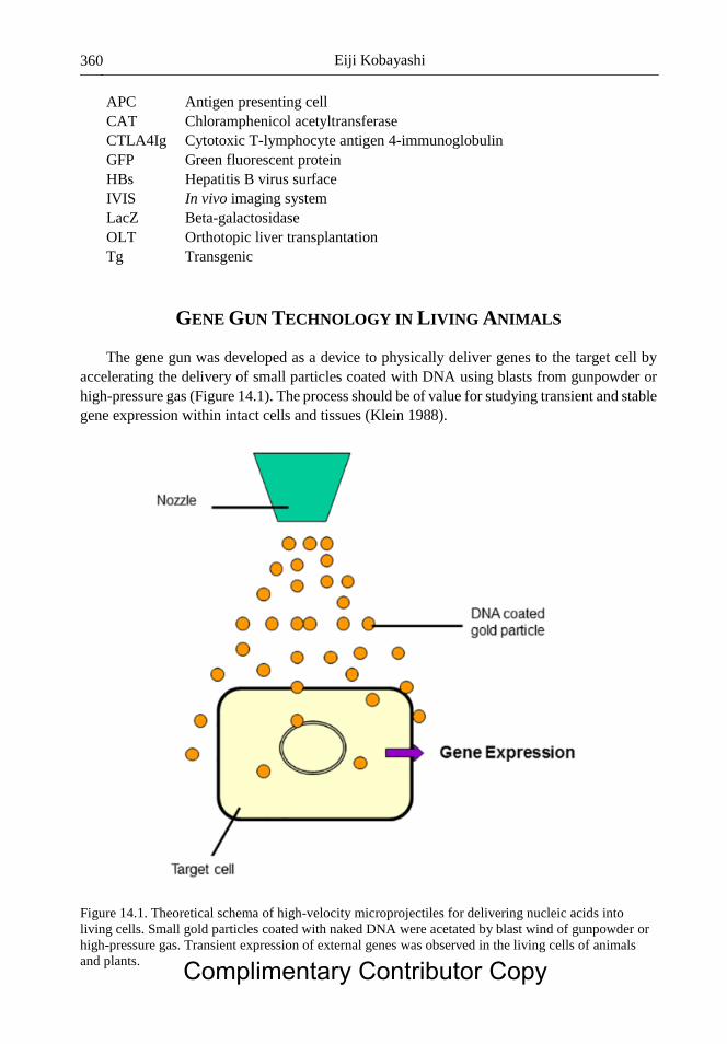
Eiji Kobayashi 360
APC Antigen presenting cell
CAT Chloramphenicol acetyltransferase
CTLA4Ig Cytotoxic T-lymphocyte antigen 4-immunoglobulin
GFP Green fluorescent protein
HBs Hepatitis B virus surface
IVIS In vivo imaging system
LacZ Beta-galactosidase
OLT Orthotopic liver transplantation
Tg Transgenic
GENE GUN TECHNOLOGY IN LIVING ANIMALS
The gene gun was developed as a device to physically deliver genes to the target cell by
accelerating the delivery of small particles coated with DNA using blasts from gunpowder or
high-pressure gas (Figure 14.1). The process should be of value for studying transient and stable
gene expression within intact cells and tissues (Klein 1988).
Figure 14.1. Theoretical schema of high-velocity microprojectiles for delivering nucleic acids into
living cells. Small gold particles coated with naked DNA were acetated by blast wind of gunpowder or
high-pressure gas. Transient expression of external genes was observed in the living cells of animals
and plants. Complimentary Contributor Copy

Experimental Gene Therapy Using Naked DNA 361
The historical background of this technological development has been summarized
previously (Belyantseva 2016). A process was developed for delivering foreign genes into
maize cells that did not require the removal of cell walls and was capable of delivering DNA
into embryogenic and nonembryogenic tissues. A plasmid harboring a chloramphenicol
acetyltransferase (CAT) gene was adsorbed into the surface of microscopic tungsten particles
(microprojectiles). These microprojectiles were then accelerated to velocities sufficient for
penetrating the cell walls and membranes of maize cells in a suspension culture. High levels of
CAT activity were consistently observed after the bombardment of cell cultures into the
cultivars of black Mexican sweet corn, and these levels were comparable to the CAT levels
observed after the electroporation of protoplasts. Measurable increases in CAT levels were also
observed in two embryogenic cell lines after bombardment. Because this process circumvents
the difficulties associated with regenerating whole plants from protoplasts, the particle
bombardment process provides significant advantages over existing DNA delivery methods for
the production of transgenic maize plants.
In the 1900s, modifications were made to substitute gunpowder with high-pressure helium
gas for injection and to allow its delivery while holding the gene gun in the hand instead of
within a chamber. The gene gun is now commercialized and is commonly used in experiments.
The following is also a summary of the Helios® Gene Gun, which is a representative gene gun
(Belyantseva 2016). The mechanical energy of compressed helium gas is used for the biolistic
delivery of exogenous DNA, mRNA, or siRNA to bombard tissue with micron- or submicron-
sized DNA- or RNA-coated gold particles, which can penetrate and transfect cells in vitro or
in vivo. Helios Gene Gun-mediated transfection has several advantages: (1) It is simple and can
be learned in a short time; (2) it is designed to overcome cell barriers, even those as tough as
the plant cell membrane or stratum corneum in the epidermis; (3) it can transfect cells deep
inside a tissue such as specific neurons within a brain slice; (4) it can deliver practically of any
size of mRNA, siRNA, or DNA; and (5) it works well with various cell types, including non-
dividing, terminally differentiated cells that are difficult to transfect such as neurons or
mammalian inner ear sensory hair cells, which is particularly important for inner ear research.
We have made attempts at in vivo gene transfer (Yoshida 1997). For the first time anywhere
in the world, we attempted to introduce marker genes in vivo into the liver of a rat that was
directly exposed under general anesthesia. DNA-coated Au particles were accelerated by
pressurized He gas to supersonic velocities to introduce a gene into the cells. Experimental and
theoretical analyses both revealed a heterogeneous distribution of the particles in each shot (1
mg Au = 2.4 × 10⁷ particles with 2 µg [32P] DNA = 2.5 × 10¹¹ moles). To introduce genes into
the liver of living rats, the best results were obtained with a newly developed handheld gene
delivery system. The beta-galactosidase gene was introduced into the rat liver with Au particles
using He at 250 psi and was expressed (1.2 µunits/µg protein) in a limited area of the liver
surface (8 × 8 mm, depth 0.5 mm). When the same gene gun was used on a monolayer of
cultured COS7 cells (about 5 µm thick), the cells in the central area of heavy bombardment
were killed. Cell death caused by the influx of Ca2+ was prevented by the use of a cytosol-type
culture medium. This method is extremely useful because genes can be easily introduced into
living plants and animals without using viral vectors (Figure 14.2). We introduced experimental
gene therapies that we have performed to date using mice, rats (Figure 14.3), and hamsters.
Later, the possible use of this technique for the in vivo topical introduction of anticarcinogenic
cytokines, among other genes, was examined, and the research area of this technique was
expanded (Yang 1995). Complimentary Contributor Copy
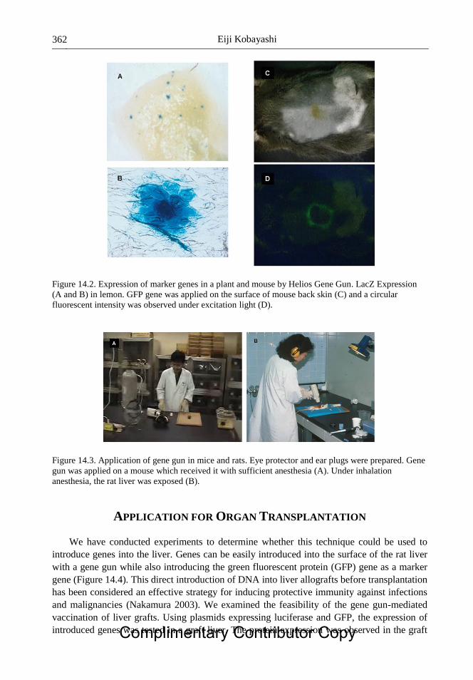
Eiji Kobayashi 362
Figure 14.2. Expression of marker genes in a plant and mouse by Helios Gene Gun. LacZ Expression
(A and B) in lemon. GFP gene was applied on the surface of mouse back skin (C) and a circular
fluorescent intensity was observed under excitation light (D).
Figure 14.3. Application of gene gun in mice and rats. Eye protector and ear plugs were prepared. Gene
gun was applied on a mouse which received it with sufficient anesthesia (A). Under inhalation
anesthesia, the rat liver was exposed (B).
APPLICATION FOR ORGAN TRANSPLANTATION
We have conducted experiments to determine whether this technique could be used to
introduce genes into the liver. Genes can be easily introduced into the surface of the rat liver
with a gene gun while also introducing the green fluorescent protein (GFP) gene as a marker
gene (Figure 14.4). This direct introduction of DNA into liver allografts before transplantation
has been considered an effective strategy for inducing protective immunity against infections
and malignancies (Nakamura 2003). We examined the feasibility of the gene gun-mediated
vaccination of liver grafts. Using plasmids expressing luciferase and GFP, the expression of
introduced genes was tested in a graft liver. The protein expression was observed in the graft Complimentary Contributor Copy
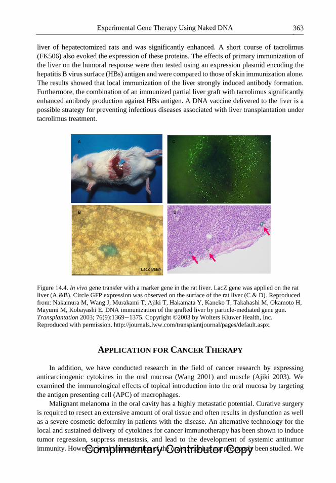
Experimental Gene Therapy Using Naked DNA 363
liver of hepatectomized rats and was significantly enhanced. A short course of tacrolimus
(FK506) also evoked the expression of these proteins. The effects of primary immunization of
the liver on the humoral response were then tested using an expression plasmid encoding the
hepatitis B virus surface (HBs) antigen and were compared to those of skin immunization alone.
The results showed that local immunization of the liver strongly induced antibody formation.
Furthermore, the combination of an immunized partial liver graft with tacrolimus significantly
enhanced antibody production against HBs antigen. A DNA vaccine delivered to the liver is a
possible strategy for preventing infectious diseases associated with liver transplantation under
tacrolimus treatment.
Figure 14.4. In vivo gene transfer with a marker gene in the rat liver. LacZ gene was applied on the rat
liver (A &B). Circle GFP expression was observed on the surface of the rat liver (C & D). Reproduced
from: Nakamura M, Wang J, Murakami T, Ajiki T, Hakamata Y, Kaneko T, Takahashi M, Okamoto H,
Mayumi M, Kobayashi E. DNA immunization of the grafted liver by particle-mediated gene gun.
Transplantation 2003; 76(9):1369−1375. Copyright ©2003 by Wolters Kluwer Health, Inc.
Reproduced with permission. http://journals.lww.com/transplantjournal/pages/default.aspx.
APPLICATION FOR CANCER THERAPY
In addition, we have conducted research in the field of cancer research by expressing
anticarcinogenic cytokines in the oral mucosa (Wang 2001) and muscle (Ajiki 2003). We
examined the immunological effects of topical introduction into the oral mucosa by targeting
the antigen presenting cell (APC) of macrophages.
Malignant melanoma in the oral cavity has a highly metastatic potential. Curative surgery
is required to resect an extensive amount of oral tissue and often results in dysfunction as well
as a severe cosmetic deformity in patients with the disease. An alternative technology for the
local and sustained delivery of cytokines for cancer immunotherapy has been shown to induce
tumor regression, suppress metastasis, and lead to the development of systemic antitumor
immunity. However, local immunization of the oral cavity has not previously been studied. We Complimentary Contributor Copy
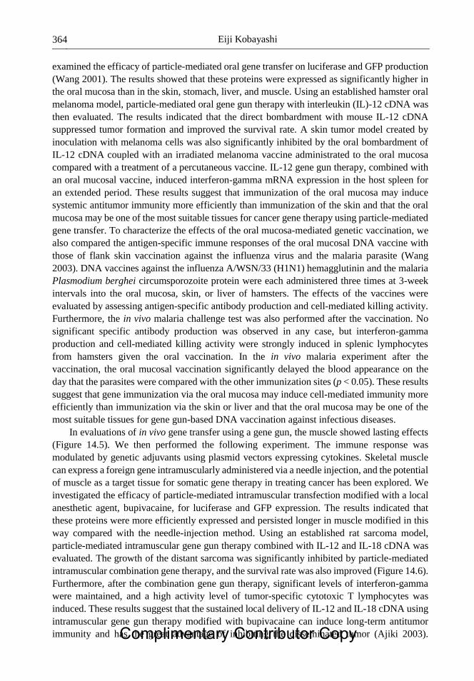
Eiji Kobayashi 364
examined the efficacy of particle-mediated oral gene transfer on luciferase and GFP production
(Wang 2001). The results showed that these proteins were expressed as significantly higher in
the oral mucosa than in the skin, stomach, liver, and muscle. Using an established hamster oral
melanoma model, particle-mediated oral gene gun therapy with interleukin (IL)-12 cDNA was
then evaluated. The results indicated that the direct bombardment with mouse IL-12 cDNA
suppressed tumor formation and improved the survival rate. A skin tumor model created by
inoculation with melanoma cells was also significantly inhibited by the oral bombardment of
IL-12 cDNA coupled with an irradiated melanoma vaccine administrated to the oral mucosa
compared with a treatment of a percutaneous vaccine. IL-12 gene gun therapy, combined with
an oral mucosal vaccine, induced interferon-gamma mRNA expression in the host spleen for
an extended period. These results suggest that immunization of the oral mucosa may induce
systemic antitumor immunity more efficiently than immunization of the skin and that the oral
mucosa may be one of the most suitable tissues for cancer gene therapy using particle-mediated
gene transfer. To characterize the effects of the oral mucosa-mediated genetic vaccination, we
also compared the antigen-specific immune responses of the oral mucosal DNA vaccine with
those of flank skin vaccination against the influenza virus and the malaria parasite (Wang
2003). DNA vaccines against the influenza A/WSN/33 (H1N1) hemagglutinin and the malaria
Plasmodium berghei circumsporozoite protein were each administered three times at 3-week
intervals into the oral mucosa, skin, or liver of hamsters. The effects of the vaccines were
evaluated by assessing antigen-specific antibody production and cell-mediated killing activity.
Furthermore, the in vivo malaria challenge test was also performed after the vaccination. No
significant specific antibody production was observed in any case, but interferon-gamma
production and cell-mediated killing activity were strongly induced in splenic lymphocytes
from hamsters given the oral vaccination. In the in vivo malaria experiment after the
vaccination, the oral mucosal vaccination significantly delayed the blood appearance on the
day that the parasites were compared with the other immunization sites (p < 0.05). These results
suggest that gene immunization via the oral mucosa may induce cell-mediated immunity more
efficiently than immunization via the skin or liver and that the oral mucosa may be one of the
most suitable tissues for gene gun-based DNA vaccination against infectious diseases.
In evaluations of in vivo gene transfer using a gene gun, the muscle showed lasting effects
(Figure 14.5). We then performed the following experiment. The immune response was
modulated by genetic adjuvants using plasmid vectors expressing cytokines. Skeletal muscle
can express a foreign gene intramuscularly administered via a needle injection, and the potential
of muscle as a target tissue for somatic gene therapy in treating cancer has been explored. We
investigated the efficacy of particle-mediated intramuscular transfection modified with a local
anesthetic agent, bupivacaine, for luciferase and GFP expression. The results indicated that
these proteins were more efficiently expressed and persisted longer in muscle modified in this
way compared with the needle-injection method. Using an established rat sarcoma model,
particle-mediated intramuscular gene gun therapy combined with IL-12 and IL-18 cDNA was
evaluated. The growth of the distant sarcoma was significantly inhibited by particle-mediated
intramuscular combination gene therapy, and the survival rate was also improved (Figure 14.6).
Furthermore, after the combination gene gun therapy, significant levels of interferon-gamma
were maintained, and a high activity level of tumor-specific cytotoxic T lymphocytes was
induced. These results suggest that the sustained local delivery of IL-12 and IL-18 cDNA using
intramuscular gene gun therapy modified with bupivacaine can induce long-term antitumor
immunity and has the great advantage of inhibiting the disseminated tumor (Ajiki 2003). Complimentary Contributor Copy
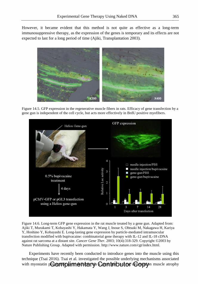
Experimental Gene Therapy Using Naked DNA 365
However, it became evident that this method is not quite as effective as a long-term
immunosuppressive therapy, as the expression of the genes is temporary and its effects are not
expected to last for a long period of time (Ajiki, Transplantation 2003).
Figure 14.5. GFP expression in the regenerative muscle fibers in rats. Efficacy of gene transfection by a
gene gun is independent of the cell cycle, but acts more effectively in BrdU positive myofibers.
Figure 14.6. Long-term GFP gene expression in the rat muscle treated by a gene gun. Adapted from:
Ajiki T, Murakami T, Kobayashi Y, Hakamata Y, Wang J, Inoue S, Ohtsuki M, Nakagawa H, Kariya
Y, Hoshino Y, Kobayashi E. Long-lasting gene expression by particle-mediated intramuscular
transfection modified with bupivacaine: combinatorial gene therapy with IL-12 and IL-18 cDNA
against rat sarcoma at a distant site. Cancer Gene Ther. 2003; 10(4):318-329. Copyright ©2003 by
Nature Publishing Group. Adapted with permission. http://www.nature.com/cgt/index.html.
Experiments have recently been conducted to introduce genes into the muscle using this
technique (Tsai 2016). Tsai et al. investigated the possible underlying mechanisms associated
with myostatin propeptide gene delivery via a gene gun in a rat denervation muscle atrophy Complimentary Contributor Copy
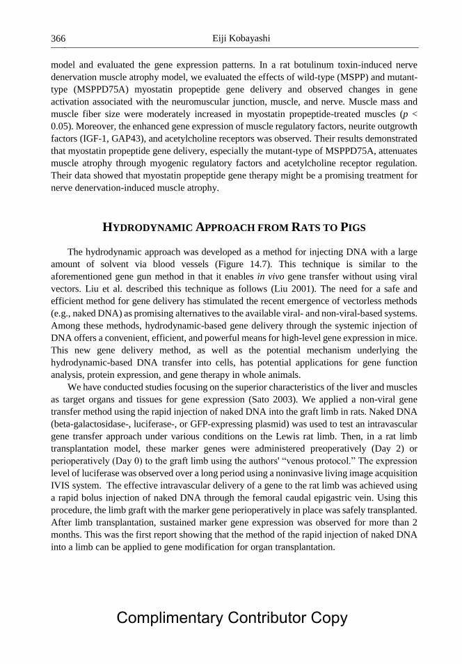
Eiji Kobayashi 366
model and evaluated the gene expression patterns. In a rat botulinum toxin-induced nerve
denervation muscle atrophy model, we evaluated the effects of wild-type (MSPP) and mutant-
type (MSPPD75A) myostatin propeptide gene delivery and observed changes in gene
activation associated with the neuromuscular junction, muscle, and nerve. Muscle mass and
muscle fiber size were moderately increased in myostatin propeptide-treated muscles (p <
0.05). Moreover, the enhanced gene expression of muscle regulatory factors, neurite outgrowth
factors (IGF-1, GAP43), and acetylcholine receptors was observed. Their results demonstrated
that myostatin propeptide gene delivery, especially the mutant-type of MSPPD75A, attenuates
muscle atrophy through myogenic regulatory factors and acetylcholine receptor regulation.
Their data showed that myostatin propeptide gene therapy might be a promising treatment for
nerve denervation-induced muscle atrophy.
HYDRODYNAMIC APPROACH FROM RATS TO PIGS
The hydrodynamic approach was developed as a method for injecting DNA with a large
amount of solvent via blood vessels (Figure 14.7). This technique is similar to the
aforementioned gene gun method in that it enables in vivo gene transfer without using viral
vectors. Liu et al. described this technique as follows (Liu 2001). The need for a safe and
efficient method for gene delivery has stimulated the recent emergence of vectorless methods
(e.g., naked DNA) as promising alternatives to the available viral- and non-viral-based systems.
Among these methods, hydrodynamic-based gene delivery through the systemic injection of
DNA offers a convenient, efficient, and powerful means for high-level gene expression in mice.
This new gene delivery method, as well as the potential mechanism underlying the
hydrodynamic-based DNA transfer into cells, has potential applications for gene function
analysis, protein expression, and gene therapy in whole animals.
We have conducted studies focusing on the superior characteristics of the liver and muscles
as target organs and tissues for gene expression (Sato 2003). We applied a non-viral gene
transfer method using the rapid injection of naked DNA into the graft limb in rats. Naked DNA
(beta-galactosidase-, luciferase-, or GFP-expressing plasmid) was used to test an intravascular
gene transfer approach under various conditions on the Lewis rat limb. Then, in a rat limb
transplantation model, these marker genes were administered preoperatively (Day 2) or
perioperatively (Day 0) to the graft limb using the authors' “venous protocol.” The expression
level of luciferase was observed over a long period using a noninvasive living image acquisition
IVIS system. The effective intravascular delivery of a gene to the rat limb was achieved using
a rapid bolus injection of naked DNA through the femoral caudal epigastric vein. Using this
procedure, the limb graft with the marker gene perioperatively in place was safely transplanted.
After limb transplantation, sustained marker gene expression was observed for more than 2
months. This was the first report showing that the method of the rapid injection of naked DNA
into a limb can be applied to gene modification for organ transplantation.
Complimentary Contributor Copy
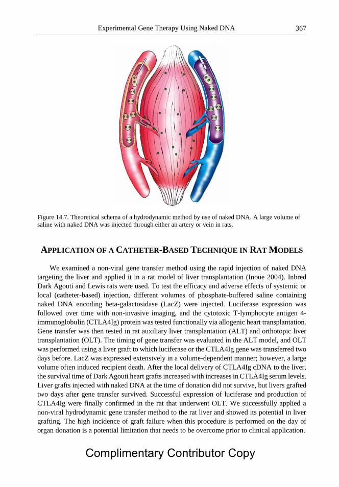
Experimental Gene Therapy Using Naked DNA 367
Figure 14.7. Theoretical schema of a hydrodynamic method by use of naked DNA. A large volume of
saline with naked DNA was injected through either an artery or vein in rats.
APPLICATION OF A CATHETER-BASED TECHNIQUE IN RAT MODELS
We examined a non-viral gene transfer method using the rapid injection of naked DNA
targeting the liver and applied it in a rat model of liver transplantation (Inoue 2004). Inbred
Dark Agouti and Lewis rats were used. To test the efficacy and adverse effects of systemic or
local (catheter-based) injection, different volumes of phosphate-buffered saline containing
naked DNA encoding beta-galactosidase (LacZ) were injected. Luciferase expression was
followed over time with non-invasive imaging, and the cytotoxic T-lymphocyte antigen 4-
immunoglobulin (CTLA4Ig) protein was tested functionally via allogenic heart transplantation.
Gene transfer was then tested in rat auxiliary liver transplantation (ALT) and orthotopic liver
transplantation (OLT). The timing of gene transfer was evaluated in the ALT model, and OLT
was performed using a liver graft to which luciferase or the CTLA4Ig gene was transferred two
days before. LacZ was expressed extensively in a volume-dependent manner; however, a large
volume often induced recipient death. After the local delivery of CTLA4Ig cDNA to the liver,
the survival time of Dark Agouti heart grafts increased with increases in CTLA4Ig serum levels.
Liver grafts injected with naked DNA at the time of donation did not survive, but livers grafted
two days after gene transfer survived. Successful expression of luciferase and production of
CTLA4Ig were finally confirmed in the rat that underwent OLT. We successfully applied a
non-viral hydrodynamic gene transfer method to the rat liver and showed its potential in liver
grafting. The high incidence of graft failure when this procedure is performed on the day of
organ donation is a potential limitation that needs to be overcome prior to clinical application.
Complimentary Contributor Copy
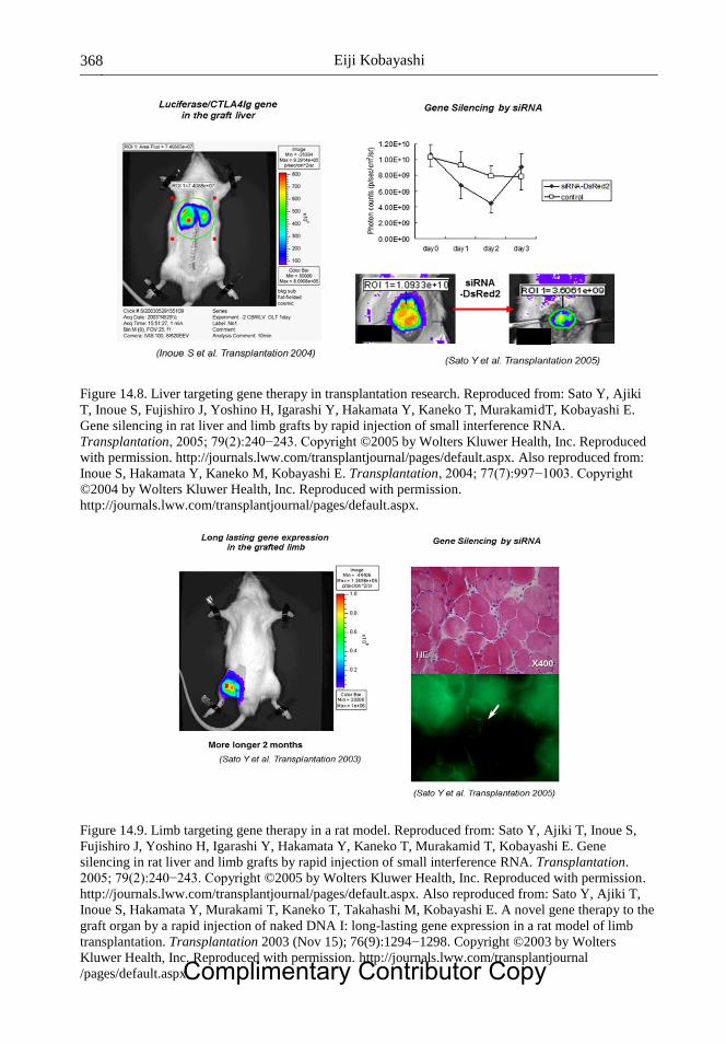
Eiji Kobayashi 368
Figure 14.8. Liver targeting gene therapy in transplantation research. Reproduced from: Sato Y, Ajiki
T, Inoue S, Fujishiro J, Yoshino H, Igarashi Y, Hakamata Y, Kaneko T, MurakamidT, Kobayashi E.
Gene silencing in rat liver and limb grafts by rapid injection of small interference RNA.
Transplantation, 2005; 79(2):240−243. Copyright ©2005 by Wolters Kluwer Health, Inc. Reproduced
with permission. http://journals.lww.com/transplantjournal/pages/default.aspx. Also reproduced from:
Inoue S, Hakamata Y, Kaneko M, Kobayashi E. Transplantation, 2004; 77(7):997−1003. Copyright
©2004 by Wolters Kluwer Health, Inc. Reproduced with permission.
http://journals.lww.com/transplantjournal/pages/default.aspx.
Figure 14.9. Limb targeting gene therapy in a rat model. Reproduced from: Sato Y, Ajiki T, Inoue S,
Fujishiro J, Yoshino H, Igarashi Y, Hakamata Y, Kaneko T, Murakamid T, Kobayashi E. Gene
silencing in rat liver and limb grafts by rapid injection of small interference RNA. Transplantation.
2005; 79(2):240−243. Copyright ©2005 by Wolters Kluwer Health, Inc. Reproduced with permission.
http://journals.lww.com/transplantjournal/pages/default.aspx. Also reproduced from: Sato Y, Ajiki T,
Inoue S, Hakamata Y, Murakami T, Kaneko T, Takahashi M, Kobayashi E. A novel gene therapy to the
graft organ by a rapid injection of naked DNA I: long-lasting gene expression in a rat model of limb
transplantation. Transplantation 2003 (Nov 15); 76(9):1294−1298. Copyright ©2003 by Wolters
Kluwer Health, Inc. Reproduced with permission. http://journals.lww.com/transplantjournal
/pages/default.aspx. Complimentary Contributor Copy
Experimental Gene Therapy Using Naked DNA 369
We introduced a non-viral method of gene transfer to donor grafts using an organ-selective
injection technique to up-regulate gene expression. Based on the attractive methodology of
RNA interference for silencing the expression of a particular gene, we also applied our catheter-
based injection method to transfer small interference RNA (siRNA)-GFP into the liver and limb
grafts (Figures 14.8 and 14.9) (Sato 2005). We quantified the interfering activity after the
systemic delivery of siRNA into the liver of Alb-DsRed2 transgenic (Tg) rats using an in vivo
bioimaging system. Then, using GFP Tg Lewis rats as donors, the preoperative rapid injection
of siRNA resulted in the transient down regulation of GFP expression in both the liver and limb
transplantation models. Genetic modification by siRNA may provide new therapeutic options
for the down regulation of endogenous antigenicity.
APPLICATION IN LARGE ANIMAL MODELS
For the first time worldwide, we successfully performed gene transferance with a pig liver
as the target using this technique (Yoshino 2006). We attempted to transfect naked plasmid
DNA into the porcine liver using a modified hydrodynamic method. We transferred plasmid
DNA to a part of the liver using an angio-catheter to reduce damage to the liver. To discern the
injection conditions, naked plasmid DNA encoding GFP was transferred for use as a marker
gene. The GFP gene was markedly expressed in the gene-transferred pig livers (Figure 14.10).
In large animals, in addition to the naked gene quantity, the solution volume containing the
plasmid DNA, and the injection speed, the additional treatments for the portal vein and the
preparation of the hepatic artery are crucial. We found that the following injection conditions
were needed: 3 mg of plasmid DNA, a solution volume of 150 ml, and an injection speed of 5
ml/s. The portal vein and the hepatic artery were clamped during gene delivery, and the blood
flow of the portal vein was flushed out using normal saline. The CTLA4-Ig gene was used to
test for the secretory protein. The CTLA4-Ig gene was injected with a large volume of solution
via the hepatic vein specifically into the left outer lobe of the liver. CTLA4-Ig was detected in
the blood of the pigs at a maximum serum level of 161.7 ng/ml 1 day after gene transfer, and
CTLA4-Ig was detected for several weeks. This new technique of inserting a catheter into only
a selected portion of the liver reduces liver toxicity and increased gene transfer efficiency.
With this method, gene transfer efficiency is increased by washing out the blood in the
liver using a balloon catheter. This catheter technique was also applied to monkeys using AAV
vector therapy (Mimuro 2013). This method results in high transfer efficiency in the liver, and
studies using rats (Kobayashi 2016) and pigs (Sendra 2016) are currently underway to develop
the technique for use in clinical practice.
DVA VACCINATION BY ELECTROPORATION IN PIGS
The development of a safe DNA vaccination strategy is underway following advances
concerning the in vivo electroporation technique (Liu 2011). Advances have been made in
research using pigs in particular for preclinical application (Ramanathan 2009, Luo 2016). We
also evaluated universal influenza vaccines on pigs using the electroporation technique
(Figure 14.11). A conduction needle was inserted into the medial thigh area of a pig under Complimentary Contributor Copy

Eiji Kobayashi 370
general anesthesia, and the naked target DNA was injected into the central region. Gene transfer
was completed by immediate conduction.
This technique has already been used in clinical practice and has been tested on hepatitis
B (Yang 2017) and HIV (Huang 2017), and its effects are being revealed.
Figure 14.10. Naked plasmid DNA transfer to the porcine liver. Reproduced from: Yoshino H,
Hashizume K, Kobayashi E. Naked plasmid DNA transfer to the porcine liver using rapid injection with
large volume. Gene Ther. 2006; 13(24):1696−1702. Copyright ©2006 by Nature Publishing Group.
http://www.nature.com/gt/index.html.
Figure 14.11. DNA vaccine by electroporation in pigs.
Complimentary Contributor Copy

Experimental Gene Therapy Using Naked DNA 371
CONCLUSION
We have described the gene gun, catheter-based hydrodynamic approaches, and the
electroporation technique as experimental in vivo gene transfer methods that do not use viral
vectors while also presenting the data from our experiments via a historical background.
REFERENCES
Ajiki T, Murakami T, Kobayashi Y, Hakamata Y, Wang J, Inoue S, Ohtsuki M, Nakagawa H,
Kariya Y, Hoshino Y, Kobayashi E. Long-lasting gene expression by particle-mediated
intramuscular transfection modified with bupivacaine: combinatorial gene therapy with IL-
12 and IL-18 cDNA against rat sarcoma at a distant site. Cancer Gene Ther. 2003; 10(4):
318−329.
Ajiki T, Takahashi M, Hakamata Y, Murakami T, Kariya Y, Hoshino Y, Kobayashi E.
Difficulty of achieving long-term graft survival of MHC-disparate composite graft using
CTLA4IG. Transplantation 2003; 76(2): 438; author reply 438−439. No abstract available.
Belyantseva IA. Helios (®) Gene Gun-Mediated Transfection of the Inner Ear Sensory
Epithelium: Recent Updates. Methods Mol. Biol. 2016; 1427: 3−26.
Huang Y, Zhang L, Janes H, Frahm N, Isaacs A, Kim JH, Montefiori D, McElrath MJ, Tomaras
GD, Gilbert PB Predictors of durable immune responses six months after the last
vaccination in preventive HIV vaccine trials. Vaccine 2017 Jan 25. pii: S0264-
410X(16)30879-9. doi: 10.1016/j.vaccine.2016.09.053. [Epub ahead of print].
Inoue S, Hakamata Y, Kaneko M, Kobayashi E. Gene therapy for organ grafts using rapid
injection of naked DNA: application to the rat liver. Transplantation 2004; 77(7):
997−1003.
Klein TM, Fromm M, Weissinger A, Tomes D, Schaaf S, Sletten M, Sanford JC. Transfer of
foreign genes into intact maize cells with high-velocity microprojectiles. Proc. Natl. Acad.
Sci. U S A. 1988; 85(12): 4305−4309.
Kobayashi Y, Kamimura K, Abe H, Yokoo T, Ogawa K, Shinagawa-Kobayashi Y, Goto R,
Inoue R, Ohtsuka M, Miura H, Kanefuji T, Suda T, Tsuchida M, Aoyagi Y, Zhang G, Liu
D, Terai S. Effects of fibrotic tissue on liver-targeted hydrodynamic gene delivery. Mol.
Ther. Nucleic Acids. 2016; 5:e359. doi: 10.1038/mtna.2016.63.
Liu D, Knapp JE. Hydrodynamics-based gene delivery. Curr. Opin. Mol. Ther. 2001; 3(2):
192−197. Review.
Liu MA. DNA vaccines: an historical perspective and view to the future. Immunol. Rev. 2011;
239(1): 62−84. doi: 10.1111/j.1600-065X.2010.00980.x. Review.
Luo X, Liang X, Li J, Shi J, Zhang W, Chai W, Wu J, Guo S, Fang G, Zhou X, Zhang J, Xu K,
Zeng J, Niu L. The effects of irreversible electroporation on the colon in a porcine model.
PLoS One 2016; 11(12):e0167275. doi: 10.1371/journal.pone.0167275.
Mimuro J, Mizukami H, Hishikawa S, Ikemoto T, Ishiwata A, Sakata A, Ohmori T, Madoiwa
S, Ono F, Ozawa K, Sakata Y. Minimizing the inhibitory effect of neutralizing antibody
for efficient gene expression in the liver with adeno-associated virus 8 vectors. Mol. Ther.
2013; 21(2): 318−323. doi: 10.1038/mt.2012.258.
Complimentary Contributor Copy

Eiji Kobayashi 372
Nakamura M, Wang J, Murakami T, Ajiki T, Hakamata Y, Kaneko T, Takahashi M, Okamoto
H, Mayumi M, Kobayashi E. DNA immunization of the grafted liver by particle-mediated
gene gun. Transplantation 2003; 76(9): 1369−1375.
Ramanathan MP, Kuo YC, Selling BH, Li Q, Sardesai NY, Kim JJ, Weiner DB. Development
of a novel DNA SynCon tetravalent dengue vaccine that elicits immune responses against
four serotypes. Vaccine 2009; 27(46): 6444−53. doi: 10.1016/j.vaccine.2009.06.061.
Sato Y, Ajiki T, Inoue S, Fujishiro J, Yoshino H, Igarashi Y, Hakamata Y, Kaneko T,
Murakamid T, Kobayashi E. Gene silencing in rat-liver and limb grafts by rapid injection
of small interference RNA. Transplantation 2005; 79(2): 240−243.
Sato Y, Ajiki T, Inoue S, Hakamata Y, Murakami T, Kaneko T, Takahashi M, Kobayashi E. A
novel gene therapy to the graft organ by a rapid injection of naked DNA I: long-lasting
gene expression in a rat model of limb transplantation. Transplantation 2003; 76(9):
1294−1298.
Sendra L, Miguel A, Pérez-Enguix D, Herrero MJ, Montalvá E, García-Gimeno MA, Noguera
I, Díaz A, Pérez J, Sanz P, López-Andújar R, Martí-Bonmatí L, Aliño SF. Studying closed
hydrodynamic models of “in vivo” DNA perfusion in pig liver for gene therapy translation
to humans. PLoS One 2016 3; 11(10): e0163898. doi: 10.1371/ journal.pone.0163898.
Tsai SW, Tung YT, Chen HL, Yang SH, Liu CY, Lu M, Pai HJ, Lin CC, Chen CM. Myostatin
propeptide gene delivery by gene gun ameliorates muscle atrophy in a rat model of
botulinum toxin-induced nerve denervation. Life Sci. 2016; 146: 15−23. doi:
10.1016/j.lfs.2015.12.056.
Wang J, Murakami T, Hakamata Y, Ajiki T, Jinbu Y, Akasaka Y, Ohtsuki M, Nakagawa H,
Kobayashi E. Gene gun-mediated oral mucosal transfer of interleukin 12 cDNA coupled
with an irradiated melanoma vaccine in a hamster model: successful treatment of oral
melanoma and distant skin lesion. Cancer Gene Ther. 2001; 8(10): 705−712.
Wang J, Murakami T, Yoshida S, Matsuoka H, Ishii A, Tanaka T, Tobita K, Ohtsuki M,
Nakagawa H, Kusama M, Kobayashi E. Predominant cell-mediated immunity in the oral
mucosa: gene gun-based vaccination against infectious diseases. J. Dermatol. Sci. 2003;
31(3): 203−210.
Yang FQ, Rao GR, Wang GQ, Li YQ, Xie Y, Zhang ZQ, Deng CL, Mao Q, Li J, Zhao W,
Wang MR, Han T, Chen SJ, Pan C, Tan DM, Shang J, Zhang MX, Zhang YX, Yang JM,
Chen GM. Phase IIb trial of in vivo electroporation mediated dual-plasmid hepatitis B virus
DNA vaccine in chronic hepatitis B patients under lamivudine therapy. World J.
Gastroenterol. 2017; 23(2): 306−317. doi: 10.3748/wjg.v23.i2.306.
Yang NS, Sun WH. Gene gun and other non-viral approaches for cancer gene therapy. Nat.
Med. 1995; 1(5): 481−483. No abstract available.
Yoshida Y, Kobayashi E, Endo H, Hamamoto T, Yamanaka T, Fujimura A, Kagawa Y.
Introduction of DNA into rat liver with a hand-held gene gun: distribution of the expressed
enzyme, [32P] DNA, and Ca2+ flux. Biochem. Biophys Res. Commun. 1997; 234(3):
695−700.
Yoshino H, Hashizume K, Kobayashi E. Naked plasmid DNA transfer to the porcine liver using
rapid injection with large volume. Gene Ther. 2006; 13(24):1696−1702.
Complimentary Contributor Copy

In: Advances in Experimental Surgery. Volume 2 ISBN: 978-1-53612-773-7
Editors: Huifang Chen and Paulo N. Martins © 2018 Nova Science Publishers, Inc.
Chapter 15
ROBOTIC SURGERY IN EXPERIMENTAL MEDICINE
Sandra S. Y. Kim, B.Sc., David Ian Harriman, MD
and Christopher Nguan*, MD, FRCPSC Department of Urologic Sciences
University of British Columbia, Vancouver, British Columbia
ABSTRACT
Robotic assisted surgery has become a vitally important surgical modality developed
through experimentation, innovation and collaboration between engineers, scientists, and
surgeons. Digitalization of the surgeon-patient interaction allows for previously unheard
of feats of engineering in procedural medicine including increased degrees of freedom of
dexterity beyond human capability, telesurgery across great distances, and augmented
reality. Animal models for training and testing these robotic platforms have been developed
along the way which have helped the field progress. There remains a lack of randomized
control trials comparing robot-assisted surgery versus other surgical modalities, however
preliminary studies are promising. Despite initial upfront costs, the appeal of robot-assisted
surgery continues to increase and its use will continue to expand as knowledge of how best
to implement the technology continues to advance.
Keywords: robotic surgery, telesurgery, daVinci, Zeus
ABBREVIATIONS
BMI Body Mass Index
CI Confidence Interval
FDA Federal Drug and Administration
* Corresponding author: Christopher Nguan M.D., FRCPSC. Department of Urological Sciences, Gordon & Leslie
Diamond Health Care Centre, Level 6, 2775 Laurel Street, Vancouver, BC Canada V5Z 1M9, Tel: 604-875-
5003. E-mail: [email protected]. Complimentary Contributor Copy
Sandra S. Y. Kim, David Ian Harriman and Christopher Nguan 374
LOS Length of Stay
MIS Minimally invasive surgery
NASA National Aeronautics and Space Administration
HISTORY OF ROBOTIC SURGERY
Robotic technology was originally developed so manufacturing industries could perform
precise, repetitive and potentially dangerous jobs with intent on improving quality, efficiency
and safety. The progression of robotics into the operating theatre seemed pre-ordained as there
are many parallels in the ongoing challenges to improve accuracy and precision of surgical
techniques to optimize patient outcomes.
Minimally invasive surgery (MIS) has a long history, as Georg Kelling first developed and
performed laparoscopic surgery using a Nitze cystoscope in a dog in 1901 Germany (Hatzinger
2006). Hans Christian Jacobaeus from Sweden went on to perform the first clinical laparoscopic
operation and since then, MIS adoption has steadily increased with both hand-assisted and pure
laparoscopic techniques having been validated and successfully incorporated into many
surgical fields around the world (Hatzinger 2006). The advantages of laparoscopic surgery over
open surgery are well described and include: smaller incisions, shorter postoperative hospital
stays, quicker return to normal function, decreased risk of wound infection and better cosmesis
(Lanfranco 2004; Kim 2002; Fuchs 2002). Drawbacks of MIS are also recognized, specifically:
loss of 3D vision, reduction of tactile feedback, amplification of tremor through rigid
laparoscopic instruments, range of motion limitation, and poor ergonomics that put additional
stress on the operating surgeon. In fact, these issues in MIS are thought to contribute to the
steep learning curve to master complex MIS procedures (Lanfranco 2004; Prasad 2001).
Robotic surgical systems were envisioned to overcome the limitations of standard laparoscopy
and to further bolster the penetration of MIS procedures in more complex and restrictive
scenarios.
The first surgical robots used computer interfaces and an effector arm to carry out surgical
procedures. In 1985, the PUMA 560 (Programmable Universal Manipulation Arm), an
industrial robot, became one of the earliest robots to be used in a clinical setting for stereotactic
CT-guided brain biopsy (Kwoh 1988). ROBODOC (Integrated Surgical Systems) is comprised
of a preoperative planning computer workstation and a five-axis robotic arm with a high speed
milling device to more precisely core out the femur for improved placement of orthopedic
implants (Bargar 1998).
In the early 1990s, NASA, the US military and researchers at Stanford combined efforts to
develop telepresence robotic systems (Satava 2002). The initial impetus of telepresence robotic
surgery was to provide surgical care to people over great distances per a battlefield situation,
or in remote locations such as in space or deep sea scenarios (Lanfranco 2004). The first robot
to incorporate telepresence technology was AESOP (Automated Endoscopic System for
Optimal Positioning; Computer Motion Inc.) and approved by the FDA in December 1993. The
Zeus robotic system complemented the AESOP robot and further extended its capabilities,
representing the first ‘master-slave’ system with a separate surgeon console and robotic system
(Lanfranco 2004). In a master-slave configuration, a human operator manipulates control
interfaces which translate those movements to instruments manipulating the patient’s anatomy;
Complimentary Contributor Copy
Robotic Surgery in Experimental Medicine 375
no autonomous motions are made by the robot itself. Intuitive Surgical Systems developed their
own telepresence robot during the late 1990s, the daVinci robotic surgical system. These rival
companies eventually elected to merge together to promote the daVinci system, resulting in the
discontinuation of the Zeus platform.
The daVinci surgical system has become the de facto standard in medical robotics, and is
currently the dominant surgical robotic system in clinical practice. The coincidental
miniaturization and evolution of stereoscopic imaging, instrumentation, hardware and software
engineering combined to make surgical robotics possible. Operations that were previously
thought to be impossible without resorting to traditional open methods of surgery can now often
be tackled in a minimally invasive fashion with robotic assistance. Robotic surgery remains at
the frontier of surgical innovation and continues to advance the limits of conventional thinking
in minimally invasive surgery.
TYPES OF TELEPRESENCE ROBOTS
There has been rapid progress in surgical robotics and their applications, and robotic MIS
systems are now widely available with uses ranging from endoluminal, laparoscopic, single
port to natural orifice based procedures (Barbash 2010; Lanfranco 2004; Davies 2000). The
development and technology of each telesurgical system is reviewed here.
AESOP: Automated Endoscopic System for Optimal Positioning
AESOP was the first telesurgical robotic device. It was originally developed from a
research grant from NASA for the development of a robotic arm to be used in the US space
program (Ballantye 2002). The current iteration of AESOP is voice controlled and commands
a robotic endoscope holder with 7 degrees of freedom. Urologists at John Hopkins
demonstrated the success of AESOP as a surgical assistant in 17 different urological procedures
(Partin 1995). The success of AESOP led to further developments in surgical robots and an
influx of interest from medical technology companies.
ARTEMIS: Advanced Robotics and Telemanipulator System for Minimally
Invasive Surgery
ARTEMIS was developed by scientists experienced in the nuclear and industrial
applications fields, at the Karlsruhe Research Centre (Rininsland 1993). ARTEMIS is a master-
slave robotic system comprised of 2 robotic arms that are controlled by a surgeon through a
control console and a 3-D endoscopic system (Schurr 2000). ARTEMIS also gave the surgeon
six degrees of freedom with a flexible distal robotic arm, allowing for precise motion in surgery
(Rininsland 1999).
Complimentary Contributor Copy

Sandra S. Y. Kim, David Ian Harriman and Christopher Nguan 376
Zeus Surgical System
The Zeus robot was developed in 1999 and is a comprehensive surgical robotic system in
which the surgeon controls two robotic arms attached to the bed rails of the operating table.
The third arm is an AESOP for visualization, which is voice controlled. The surgeon sits upright
in a cockpit-like console facing a 2-dimensional viewing system of the operating field (Maseo
2010).
daVinci Surgical System
The daVinci Surgical System consists of 3 separate parts; 1) a master console consisting
of a work station, computer and 3D display system; 2) a control tower which houses the 3D
video capture equipment and synchronizers; and 3) a patient side cart comprised of a motorized
wheeled platform and a vertical post with four robotic arms suspended from it. The master
console translates a full 7 degrees of freedom to the instruments of the patient side cart, while
the high definition stereoendoscopic cameras are mixed to provide true 3D stereoscopic views
of the operative field back to the surgeon console. By converting the standard “analog”
interaction of the surgeon with the patient to a digital process, the system allows for adjunctive
benefits to the operator: tremor filtration, motion scaling, swapping of controls and arms,
control of additional robotic arms greater than the number of master controllers through hot
swapping, and all the while providing a seated, ergonomic approach to surgery which promotes
the efficient and effective conduct of a potentially prolonged operation. Finally, research
conducted at Intuitive and academic centers worldwide can provide surgical platform “plugins”
to add novel functionality to the standard robotic system.
Typically, major operations require the assistance of an additional person at the bedside,
but with the latest versions of the daVinci robot, the patient side cart’s four arms are able to be
controlled by two concurrent master controller units controlled by one primary surgeon and one
assistant. Although telesurgery across distances is possible with the daVinci surgical system,
in North America, the FDA does not allow the use of this functionality and requires the patient
and surgeon always be in the same room (Maseo 2010).
The daVinci system has evolved over time where in 1999, the original daVinci system
(“Standard”) had a 2-dimensional viewing screen and 3 robotic arms, a 4th arm was added in
2003. In 2006, the daVinci S was released that had 3-dimensional high definition vision. In
2009, the daVinci Si was produced with a dual console option to aid with operative teaching
and assistance, the Firefly Fluorescence Imaging System to aid in real-time visualization of
tissue perfusion, the option for single site surgery, system integration with complex imaging
modalities and advanced instrumentation for finer tasks. The most recent model was produced
in 2014 and is termed the daVinci Xi. This model is the first major overhaul since 1999 and has
redesigned instrument arm architecture to facilitate anatomical access from any position,
increased flexibility of visualization through repositioning of the endoscope through any port,
and a smaller footprint.
Complimentary Contributor Copy
Robotic Surgery in Experimental Medicine 377
SPORT Surgical System
Titan Medical Incorporation based in Toronto, Ontario is currently developing the Single
Port Orifice Robotic Technology (SPORT) surgical system, unveiled in early 2016, with the
aim to expand the field of robotics surgery by lowering the cost barrier to allow for pervasive
use in all surgical specialties. The SPORT is a single-incision surgical system with multi-
articulated instruments, camera with 3D high definition view of the surgical field, ergonomic
work station, single-arm mobile patient cart, with single-use replaceable tips. This surgical
system is not yet available for sale.
CURRENT ROBOTIC APPLICATIONS
Cardiac Surgery (Table 15.1)
MIS cardiac surgery became reality with advances in perfusion strategies, myocardial
protection and access techniques (Bush 2013). Robotic cardiac valve and bypass surgery have
become a mainstay of the discipline with use cases including: atrial septal defect closure on a
beating heart, atrial fibrillation ablation and cardiac tumor resections (Gao 2010; Schilling
2012). Advantages of robotic assistance include improved cosmesis, shorter recovery and
comparable outcomes to traditional techniques with the main disadvantage being prolonged
operative times (Bush 2013).
Mitral valve surgery is the most common robotic assisted cardiac procedure performed, the
first case performed in 1998 with an early prototype of the daVinci surgical system (Carpentier
1998). Within a week after the first robot-assisted mitral valve repair, Mohr et al. performed 5
more robotic mitral valve repairs as well as a coronary revascularization procedure (Mohr
1999). Between 2006 and 2009, Mihaljevic et al. compared key outcomes grouped by surgical
technique for 759 patients undergoing mitral valve repair (Mihaljevic 2011). On average, the
robot-assisted procedures took 42 minutes longer than complete sternotomy, 39 minutes longer
than partial sternotomy, and 11 minutes longer than right mini-anterolateral thoracotomy (p <
0.05). The increased robot operative time was offset by decreased blood transfusion rates as
well as lower rates of pleural effusion and atrial fibrillation compared to standard open or MIS
methods, leading to significantly reduced hospital stay (median 4.2 days vs. 5.2, 5.8 and 5.1
days respectively; p < 0.05).
As briefly alluded to above, robotic assisted surgery has also been employed for total
endoscopic coronary artery bypass grafting (TECAB). A multicenter, prospective study by
Argenziano et al. assessed the safety and efficacy of TECAB surgery (Argenziano 2006). Of
85 patients undergoing TECAB surgery, 5 were converted to open procedures, 1 required
reintervention for bleeding and 1 experienced myocardial infarction. Mean hospital length of
stay was 5.1 ± 3.4 days. Angiographic success, defined as patient anastomosis, was 91% and
the authors concluded non-inferiority to traditional techniques. More recently, robotic surgical
techniques in the absence of cardiac bypass have been performed with encouraging results
(Bush 2013). From these studies, it has been demonstrated that the use of robot-assisted CABG
can be successful in properly chosen operative candidates.
Complimentary Contributor Copy
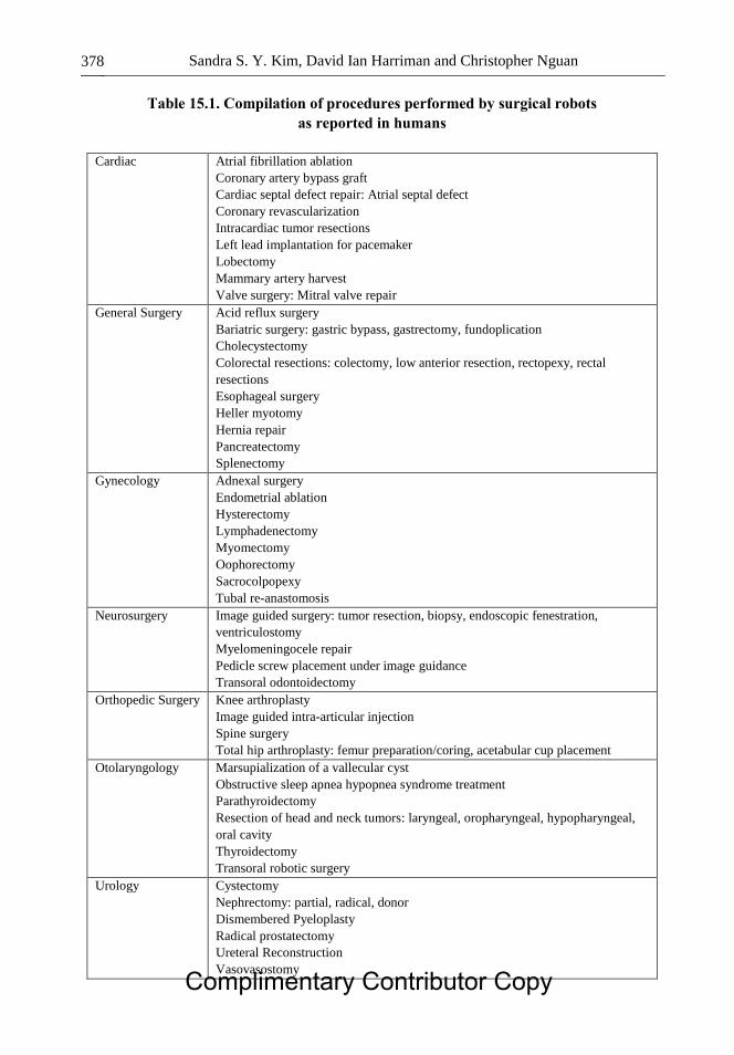
Sandra S. Y. Kim, David Ian Harriman and Christopher Nguan 378
Table 15.1. Compilation of procedures performed by surgical robots
as reported in humans
Cardiac Atrial fibrillation ablation
Coronary artery bypass graft
Cardiac septal defect repair: Atrial septal defect
Coronary revascularization
Intracardiac tumor resections
Left lead implantation for pacemaker
Lobectomy
Mammary artery harvest
Valve surgery: Mitral valve repair
General Surgery Acid reflux surgery
Bariatric surgery: gastric bypass, gastrectomy, fundoplication
Cholecystectomy
Colorectal resections: colectomy, low anterior resection, rectopexy, rectal
resections
Esophageal surgery
Heller myotomy
Hernia repair
Pancreatectomy
Splenectomy
Gynecology Adnexal surgery
Endometrial ablation
Hysterectomy
Lymphadenectomy
Myomectomy
Oophorectomy
Sacrocolpopexy
Tubal re-anastomosis
Neurosurgery Image guided surgery: tumor resection, biopsy, endoscopic fenestration,
ventriculostomy
Myelomeningocele repair
Pedicle screw placement under image guidance
Transoral odontoidectomy
Orthopedic Surgery Knee arthroplasty
Image guided intra-articular injection
Spine surgery
Total hip arthroplasty: femur preparation/coring, acetabular cup placement
Otolaryngology Marsupialization of a vallecular cyst
Obstructive sleep apnea hypopnea syndrome treatment
Parathyroidectomy
Resection of head and neck tumors: laryngeal, oropharyngeal, hypopharyngeal,
oral cavity
Thyroidectomy
Transoral robotic surgery
Urology Cystectomy
Nephrectomy: partial, radical, donor
Dismembered Pyeloplasty
Radical prostatectomy
Ureteral Reconstruction
Vasovasostomy Complimentary Contributor Copy

Robotic Surgery in Experimental Medicine 379
General Surgery
Contemporary results in the field of General Surgery show modest benefit of surgical
robotics over traditional laparoscopic methods across a range of operations (Maseo 2010).
Additionally, it has been estimated that per-procedure cost increases of up to $1500 are incurred
as a result of robot assistance (Barbash 2010). As such, adoption of robotic laparoscopic
techniques in General Surgery have been variable and limited with an eye towards gaining
further evidence and ongoing cost containment.
A large study out of the University of Illinois at Chicago assessed outcomes for 676 robotic
cholecystectomies compared to 289 laparoscopic cholecystectomies (Gangemi 2016).
Statistically significant differences were identified favoring robotic cholecystectomy with
decreased minor biliary injuries (p = 0.049), decreased open conversion (p < 0.001) and reduced
blood loss (p < 0.001). Mean hospital stay and number of biliary anomalies identified
intraoperatively were comparable across groups. Overall, this study indicates benefit of robotic
versus laparoscopic cholecystectomy. Robotic multi-port (n = 4) and single site
cholecystectomy (n = 5) has been safely performed in a pediatric population (mean age 14)
(Ahn 2015). Median console time was 47 minutes (range, 44−58) for multi-port
cholecystectomy and 69 minutes (range, 66−86) for single site cholecystectomy. Eight of 9
patients were discharged home on day 0 with no complications. Both techniques were deemed
safe in a pediatric population with possibly increased anesthetic time with single-site surgery
for the trade-off of potential improved cosmesis and functional recovery.
Robotic colorectal surgery has also garnered increasing interest and a recent meta-analysis
composed of data from 2 randomized controlled trials and 22 non-randomized controlled trials
(5 prospective and 17 retrospective) representing 3318 patients (robotic n = 1852; laparoscopic
n = 1466) compared outcomes for robotic assisted versus laparoscopic colorectal surgery
(Zhang 2016). Length of hospital stay, conversion rates and blood loss were significantly
reduced in the robotic group (p < 0.05) while the operation times (SMD = 0.78, 95% CI 0.54 –
1.02) and total costs (SMD = 1.23, 95% CI 1.01−1.45) trended in favor of laparoscopic surgery
while not reaching statistical significance between groups. A separate study by Shiomi et al.
assessed the advantages of robotic assisted surgery (ntot = 127; nobese = 52) compared to
laparoscopic surgery (ntot = 109; nobese = 30) for lower rectal cancer and visceral obesity
cases, defined as visceral fat area ≥ 130 cm2 (Shiomi 2016). Robotic assisted surgery had lower
overall complication rate (9.4% vs. 23.9%; p = 0.007), less blood loss (p = 0.007), shorter length
of stay (p < 0.01) with comparable operative time and pathological results. Visceral obesity
cases displayed decreased blood loss, length of stay and complication rate in favor of the robotic
group (all p < 0.05). These results suggest that robotic assisted surgery may have advantages
for challenging surgical locations and patient factors.
Gynecology
Multiple studies have compared the daVinci surgical system to standard laparoscopic
myomectomy. These are technically challenging operations and many surgeons prefer open
surgical approaches compared to MIS for this reason. Advincula et al., demonstrated that the
use of surgical robots was associated with fewer intraoperative complications and decreased
Complimentary Contributor Copy
Sandra S. Y. Kim, David Ian Harriman and Christopher Nguan 380
blood loss with a mean of 196 mL 95% CI [50, 700] compared to 365 mL 95% CI [75, 1550]
(p < 0.05), increased operative time with a mean of 231 minutes 95% CI [199, 263] for the
robot-assisted surgery compared to mean of 154 minutes 95% CI [138,170] (p < 0.05),
decreased mean hospital stay (1.5 days 95% CR [1, 3] compared to 3.6 days 95% CR [3, 8]
(p < 0.05), and increased cost compared to traditional laparoscopic methods. The largest study
comparing the daVinci surgical system to laparoscopic and open myomectomy approaches
demonstrated the ability to resect larger masses with the robot (223g 95% CI [85.2, 391.5])
compared to laparoscopy (96.65 g 95% CI [49.5, 227.3]) (p < 0.001). Additionally, robotic
surgical time was 181 minutes 95% CI [151, 265] compared to laparoscopy (115 minutes 95%
CI [98, 200]) compared to open procedure time of 126 minutes 95% CI [95, 177] (p < 0.010)
(Barakat 2011). Patients undergoing the open procedure had increased LOS of 3 days, 1 day
for laparoscopic, compared to 1 day for robot-assisted surgery (p < 0.001) (Barakat 2011). The
findings of these studies suggest that the use of surgical robots may benefit patients with
complex, multiple myomas who would traditionally not be considered candidates for a
minimally invasive approach.
Uterine cancer is one of the most common cancers in women, and surgical treatment
consists of combination of a hysterectomy, bilateral salpingo-oophorectomy, and pelvic and
para-aortic lymphadenectomy (Weinberg 2011). The largest study to date comparing 377 robot-
assisted surgeries to historical data of 131 patients undergoing open surgery for staging of
endometrial cancer demonstrated that the conversion rate from robotic surgery to open surgery
was 2.9% (Paley 2011). The robotic surgery group had a decreased blood loss of 46.9 mL
compared to 197.6 mL (p < 0.0001) for open procedures, decreased intraoperative and
postoperative complications (6.4% compared to 20.6% p < 0.0001), a higher lymph node count
of 15.5 compared to 13.1 (p = 0.007) as well as a shorter LOS (1.4 days compared to 5.3 days
for open procedure, (p < 0.0001) (Paley 2011).
The utility of the daVinci robot treating early stage cervical cancer with radical
hysterectomy has been evaluated, given the complexity of performing laparoscopic radical
hysterectomy. A study done by Nam et al. compared 32 cases each of robot-assisted as well as
open hysterectomy with similar patient and tumor demographics. There was no significant
difference between the operative time between the robotic and open procedures (218.8 ± 78.3
minutes, 209.9 ± 87.8 minutes respectively, p = 0.654), with similar number of lymph nodes
sampled (20.2 ± 9.4, 24.2 ± 11.1 respectively, p = 0.121), and decreased blood loss (220.0 ±
134.7 mL, 531.5 ± 435.5 mL respectively, p = 0.002) (Nam 2010).
Ovarian cancer requires operative staging including: exploratory laparotomy with
hysterectomy, adenectomy, abdominal-pelvic washing, omentectomy, pelvic and paraaortic
lymphadenectomy, and peritoneal biopsies, as well as debulking of resectable metastatic tumor
(Weinberg 2011). One study done by Magrina et al., compared 25 cases of daVinci-assisted
robotic surgery for staging of endothelial ovarian cancer compared to 27 laparoscopic surgery
and 119 open surgery cases over 3 years (Magrina 2011). Despite confounding variables in this
study, the authors demonstrated that the robotic, laparoscopic and open treatment arms were
associated with: mean operative times of 314.8 minutes, 253.8 minutes, and 260.7 minutes
respectively (p < 0.05), decreased blood loss of 164.0 mL, 266.7 mL, and 1307.0 mL
respectively (p = 0.001), and mean length of hospital stay of 4.2, 3.2, and 9.2 days respectively
(p = 0.08) (Magrina 2011).
Currently there are no substantial randomized trials with long-term outcomes comparing
robotic surgery efficacy and safety in Gynecology compared to traditional surgical approaches Complimentary Contributor Copy

Robotic Surgery in Experimental Medicine 381
to definitively change pattern of practice, but studies remain ongoing and robotics in
Gynecology remains an emerging field.
Neurosurgery
The field of neurosurgery has undergone monumental changes as a result of revolutions in
stereotactic and minimally invasive surgery (Nathoo 2005) NOT IN LIST. MIS Neurosurgery
has directly led to improved patient outcomes and new surgical options for tumors that were
previously thought to be unresectable, all the while improving safety for patients in these
critical operations.
The initial types of robots helped surgeons perform basic stereotactic tasks, the first one of
which was the PUMA 560 used to predetermine the trajectory of a brain biopsy procedure
(Kwoh 1988). NeuroMate (Integrated Surgical Systems, 1987, Sacramento, California) was the
first surgical robot to be approved by FDA comprises a system with 6 degrees of freedom that
can be used for biopsy procedures, stereoencephalography, endoscopy, as well as deep brain
stimulation with its ability to properly and accurately orient, position, hold, and manipulate
tools in a 3D space with high precision and accuracy (Nathoo 2005; Haegelen 2010).
NeuroArm, developed at the University of Calgary in 2001, is an ambidextrous robot designed
to work within an MRI which allows the neurosurgeon to perform operations while leveraging
real-time MRI scans throughout the procedure (Sutherland 2013). NeuroArm was used in 2008
to remove a brain tumor from a 21 year old patient, and since then has shown positive clinical
results in other cranial neoplasia cases (Sutherland 2013; Pandya 2009).
Spine surgery saw the development of SpineAssist (Mazor Robotics, Caeserea, Israel), the
predecessor to the Renaissance surgical system from the same company, which was FDA
approved in 2011. Both of these surgical systems consist of a miniature robot securely attached
to the operating table coordinated with real-time fluoroscopy to assist in accurate placement of
spinal pedicle screws (Devito 2010). A retrospective study comparing the use of SpineAssist
showed that 3204 of 3271 (97.9%) pedicle screws were appropriately placed on targeted
positions by the robot (Devito 2010).
The daVinci surgical system has also been used in Neurosurgery for spinal schwannoma
removal, transoral odontoidectomy, and intrauterine repair of myelomeningocele, with
inconclusive outcome reports comparing robot-assisted to conventional open methodologies
(Perez-Cruet 2012). However, daVinci is limited in this capacity due to reduced
instrumentation availability and need for multiple access ports (Doulgeris 2015).
Current studies of robotics in Neurosurgery draw inconclusive results due to lack of good
quality clinical trials and current evidence is comprised primarily of case reports or on in vivo
models. However the increasing number of case reports from institutions illustrates the desire
of surgeons to investigate this emerging technology in the context of Neurosurgery.
Orthopedics
Although the field of Orthopedic surgery pioneered surgical robotics, their use has not
gained the widespread popularity and growth as seen in other surgical specialties. The external
fixation of limbs in optimal position presents fewer challenges to accessing the operative field Complimentary Contributor Copy
Sandra S. Y. Kim, David Ian Harriman and Christopher Nguan 382
and providing adequate exposure compared to other surgical specialties which work with
mobile viscera and soft tissues (Yen 2010).
Traditional knee replacement surgery (TKR) involves intraoperative sizing guides to assist
the surgeon determine the appropriate implant size to ensure a tight fit between the bone and
prosthetic. Intraoperative cutting errors and resulting gaps can lead to delays in bone growth
and increased use of bone cement in such cases. Many surgical robots were developed for the
purposes of knee arthroplasty, with only few of them having clinical success throughout the
world. These robots include the CASPAR system (URS Ortho Rastatt, Germany), RGA system
(formerly known as ACROBOT), ROBODOC system (Curexo Technology Corporation,
Fremont, California, USA), and the Robotic Arm Interactive Orthopedic System (RIO; MAKO
Surgical Corporation, Fort Lauderdale, Florida, USA), with the most commonly used
orthopedic surgical robots being the ACROBOT and ROBODOC systems (Netravali 2013).
The Active Constraint ROBOT (ACROBOT) was first developed in Imperial College in
London, United Kingdom (Jakopec 2003) and is now known as the Stanmore Sculptor Robotic
Guidance Arm (RGA) system (Stanmore Implants, Elstree, UK) (Netravali 2013). Its main
parts include a high-speed cutter mounted on a robotic device that uses 3D reconstruction and
visual guidance to precisely target and core out long bones for implants (Cobb 2006). The
ACROBOT allows free movement inside a predetermined safe region, however, motion at the
boundaries becomes progressively more stiff, preventing inadvertent errors (Jakopec 2003). A
prospective double-blind randomized control trial by Cobb et al., compared conventional and
robot-assisted unicompartmental knee arthroplasty in 27 patients using the ACROBOT system
showed increased operating time but significantly better alignment that achieved near normal
coronal plane alignment within 2 degrees of the targeted position with the ACROBOT, while
only 40% (6/15) of the conventional method resulted in this degree of accuracy (Cobb 2006).
Likewise, the difference between tibiofemoral angles planned and achieved favored
ACROBOT assistance (0.65 ± 0.59 degrees vs. -0.84 ± 2.75 degrees respectively, p = 0.001).
Three other studies done by Coon, Pearle et al., and Plate et al., also evaluated the use of robot-
assisted unicompartmental knee arthroplasty, which all demonstrated more accurate implant
placement, improvements in restoration of natural knee kinematics, and positive outcomes for
implant survival as a result of accurate placement of implants (Coon 2009; Pearle 2010;
Plate 2013).
ROBODOC is one of the first clinically successful robots, and is the first robot to be
approved by the FDA to be used in orthopedic surgery (Karthik 2015).
Robot-assisted total hip arthroplasty (THR) was originally performed with ROBODOC in
1992 and has been used in over 24,000 surgeries worldwide with data showing improved patient
outcomes, with more precise implant positioning and elimination of intraoperative femoral
fractures (Bargar 1998). A study done by Harris et al., compared the surgical outcomes of hip
replacements performed in three different centers with 69 ROBODOC-assisted total hip
replacement compared to 65 control cases. As with knee replacement surgery, the robot-
assisted procedure was associated with longer operative times (258 minutes, which was later
reduced to 90 minutes with surgeon experience, compared to 134 minutes for the control group,
p < 0.0001) (Bargar 1998). Blood loss was reported to be higher in the robot-assisted group
(1189 mL compared to 644 mL compared to the conventional open surgical method,
p < 0.0001), and average length of stay was not significantly different between the two groups
(8.2 days for the ROBODOC and 7.5 days vs. control) (Barger 1998). Postoperative
complications including intraoperative fractures, dislocations, partial sciatic nerve palsy, deep Complimentary Contributor Copy
Robotic Surgery in Experimental Medicine 383
vein thrombosis and pulmonary embolism were evaluated with no significant differences in
complication rates between the two treatment groups, other than no intraoperative femoral
fractures in the ROBODOC group compared to three in the control group (p < 0.01) (Barger
1998). The modified Harris hip scores postoperatively showed no difference in the first year
favoring the robotic group (89.4 for the ROBODOC vs. 86.6 for the control group), and two
years (88.5 for the ROBODOC vs. 91.1 for the control group) (Bargar 1998).
The innovative motion (INNOMOTION, Innomedic, Herxheim & FZK Karlsruhe
Germany & TH Gelsenkir, Germany) allows for robot-guided intraarticular injections into the
many joint articulations of the foot under CT or MRI guidance, including: calcaneocuboid,
subtalar, tarsometatarsal, talonavicular, and tibiotalar joints. INNOMOTION was successful in
infiltrating all 16 joints of patients (n = 16) with no intra or postinterventional complications.
Small case studies to date have shown that patients had relief of pain following injections with
a preinterventional pain measurement visual analogue scale (VAS) 5.3 ± 2.33 to a score of 1.1
± 1.45 postinterventionally (Wiewiorski 2009).
Surgical robotics in Orthopedic surgery has a long and storied history, and remains an
active area of research and development. Given the current widespread use of mechanical
instruments in the field of Orthopedic surgery, lends the specialty especially well transition into
the world of robotics surgery.
Otolaryngology
The field of Otolaryngology has traditionally performed procedures involving large
incisions and extensive surgical dissections to adequately perform head, neck and airway
surgeries to minimize damage to vital structures (Oliveira 2012). Thus the nature of these
surgical procedures can lead to significant tissue damage, extended time to functional recovery,
decreased cosmesis, and decreased quality of life (Garg 2010). Robotic assisted surgery is well
poised to positively affect these negative patient outcomes (Oliveira 2012).
The daVinci robot is currently used for Transoral Robotic Surgery (TORS) and was FDA
approved in 2009 (Moore 2009). TORS surgery involves the use of the oral cavity as an entry
site, and was first carried out by McLeod and Melder for marsupialization of a vallecular cyst
in 2005 (McLeod 2005). This technique was further adapted and developed by Weinstein and
O’Malley in 2006 on three human patients with tongue base cancers (O’Malley 2006).
There are few studies to date looking into the functional surgical outcomes associated with
patients undergoing TORS for oncologic resection in the head and neck. Case reports of a
variety of different surgical procedures performed in head and neck regions from the
oropharynx, laryngopharynx, oral cavity, skull base surgery (infratemporal and parapharyngeal
fossae), thyroid glands, and in pediatric airways have been documented (Garg 2010). The most
comprehensive study thus far was done by Boudreaux et al. in 2009 with 36 patients with
laryngeal, oropharyngeal, hypopharyngeal, and oral cavity tumors. Although 36 patients were
recruited for the study, only 29 (81%) underwent successful robotic resections with negative
surgical margins reported in all 29 patients (Boudreaux 2009). Of the 7 patients who failed to
undergo successful robotic resection of their tumors, the biggest factor was adequate and
appropriate exposure of the surgical site, extensive infiltrative tumor requiring free-flap
reconstruction, or patients opting for chemotherapy or chemoradiotherapy treatments instead
(Boudreaux 2009). The mean robotic operative time was 99 ± 32.6 minutes, mean estimated Complimentary Contributor Copy
Sandra S. Y. Kim, David Ian Harriman and Christopher Nguan 384
blood loss was 51 ± 42 mL and no transfusion was required for any cases (Boudreaux 2009).
The mean hospital stay was 2.9 ± 2.9 days, and 16 of the 29 patients (55%) were able to return
to oral nutrition prior to discharge. Postoperative complications were limited to bleeding in 4
patients, one whom required cautery and another as a result of coumadin therapy several days
postoperatively. 21 patients (21/29, 72%) undergoing TORS were safely extubated prior to
exiting the operating room, and 7 patients required postoperative intubation with only one
patient requiring a planned short term tracheotomy tube placement. This study demonstrated
that TORS is a safe surgical alternative for patients with suitable airway anatomy and limited
tumor disease pathology, however, further studies comparing TORS to conventional open
procedures will need to be carried out to further elucidate the benefits of using the daVinci
robot in these procedures.
The use of TORS has also been extended to non-malignant lesions in the head and neck.
Vicini et al., in 2012 assessed the utility of TORS in treating obstructive sleep apnea hypopnea
syndrome in 20 patients. The mean operative time was 42.5 ± 15.2 minutes, with a mean setup
time of 31.2 ± 18.2 minutes and estimated blood loss was at 27.7 ± 13.6 mL (Vicini 2012).
There were no serious intraoperative or postoperative complications in this study other than
minor bleeding managed non-surgically in three patients (Vicini 2012). Evaluation of the
surgical outcome was done through the apnea hypopnea index (AHI) score and Epworth
sleepiness scale (ESS) with improvements of 24.6 ± 22.2 for AHI and 5.9 ± 4.4 for ESS, and
using cutoff values of AHI, ESS, and oxygen saturation levels postoperatively, 12 (60%) of the
patients were considered to be cured (Vicini 2012).
The use of the daVinci has been evaluated more extensively in the case of thyroidectomies,
comparing it to the conventional open method. The first case was performed by Lobe et al., in
2005 who used a transaxillary robotic technique to resect a thyroid nodule in one patient and
placement of a vagal nerve stimulator for intractable seizures in another (Lobe 2005). A recent
systematic review and meta-analysis looked at 11 articles comparing robot-assisted
thyroidectomy to conventional open procedures. The mean operative time for robot-assisted
thyroidectomy was increased compared to conventional open methods by 48.1 minutes 95% CI
[33.6, 62.7] in total thyroidectomies and by 37.3 minutes 95% CI [12, 62.5] in subtotal
thyroidectomies (Sun 2014). Conversion from robot-assisted to open cases was required in 2
out of 243 cases (0.8% cases). Length of hospital stay was decreased in the robot-assisted
thyroidectomy group by -0.006 days 95% CI [-0.25, 0.24]. There was no significant difference
between the two patient groups in terms of postoperative complications including hematoma
or seroma formation, recurrent laryngeal nerve injury, hypocalcemia, or chyle leak, (Sun 2014).
Postoperatively, the conventionally open procedure treated patients had increased severity of
voice symptoms at postoperative day 1 (p = 0.008), first month postoperatively (p = 0.049), 3
months postoperatively (p = 0.043), but not at 6 months (Tae 2012).
The feasibility of skull base surgery was first demonstrated by O’Malley and Weinstein in
humans in a patient who underwent parapharyngeal cystic neoplasm resection in the
infratemporal fossa with no adverse surgical events such as damage to many vital structures
(carotid artery, jugular vein, cranial nerves) (Oliveira 2012). Further development of
appropriate instruments for Otolaryngology in terms of size, function, and flexibility will be
needed prior to execution of more robot-assisted skull base surgery in Otolaryngology patients
(Oliveira 2012).
Complimentary Contributor Copy
Robotic Surgery in Experimental Medicine 385
Urology
The field of Urology has been a frontrunner in exploring surgical robots use in a spectrum
of operations. The first reports of the use of the daVinci robot in surgery were described by
Partin et al. in 1995 who utilized robotic assistance as surgical assistant substitutes during 17
laparoscopic procedures (Burch bladder suspension (n = 2), nephrectomy (n = 4), nephropexy
(n = 1), orchiopexy (n = 1), pelvic lymph node dissection (n = 1), pyeloplasty (n = 3),
retroperitoneal lymph node sampling (n = 2), varix ligation (n = 2), and ureterolysis (n = 1)
(Partin 1995). The study reported no increase in total operative time when the robotic arms
were used, and no difference between the set up and take down times for the robot-assisted
surgeries compared to the conventional laparoscopic surgical assists (p > 0.05) (Partin 1995).
DaVinci robot-assisted prostatectomy (DVP) is the most frequently performed robotic
procedure worldwide, and has supplanted both laparoscopic and open approaches at many
centers (Barbash 2010). There remains insufficient data to determine if robotic prostatectomy
can decrease the long term rates of impotence and incontinence which are potential
complications of this operation. However, introduction of robotic Urology has changed the
landscape of radical prostatectomy operations as a whole, as it has leveled the steep learning
curve of laparoscopic prostatectomy and allowed an entire population of MIS-naive surgeons
seemingly the ability to offer an MIS approach to this procedure overnight (Barbash 2010).
The literature supports the fact that DVP leads to significantly less blood loss secondary to
improved visualization of the dorsal venous complex coupled with the tamponade effect of the
pneumoperitoneum compared to conventional open or laparoscopic methods (Jain 2015;
Skolarus 2010; Krambeck 2009).
Comparing robot-assisted radical prostatectomy to traditional open procedures, the current
studies show longer operative time for robot-assisted laparoscopic prostatectomy (2.3 hours for
open and 4.8 hours for robot-assisted, p < 0.001) in the early phases of the learning curve with
decreased operative time with increased robot-assisted surgical cases (N = 200) (163 minutes
for open and 160 minutes for robot-assisted) (Menon 2002; Tewari 2003). Transfusion rates
were lower in the robot-assisted group compared to the open surgical treatment group with a
relative ratio of 4.51 95% CI [1.35, 15.03] (p = 0.01) (Ficarra 2009). Only a single study by
Tewari et al., looked into mean catheterization time which demonstrated a lower catheterization
time in robot-assisted treatment group of 15.8 days for open compared to 7 days (p < 0.05)
(Tewari 2003). The current studies comparing the various surgical modalities show similar rates
of complication in prostatectomies in the different treatment groups, with the exception of the
study done by Tewari et al., which demonstrated a 20% intraoperative complication rate for
open surgical group (N = 100) compared to 5% in the robot-assisted surgical group (n = 200)
(p < 0.05) (Tewari 2003).
Comparison of laparoscopic prostatectomy to robot-assisted prostatectomy showed similar
mean operative time after 20 robot cases (Ficarra 2007). The cumulative weighted mean
difference for the mean operative time were overlapping in laparoscopic compared to robot-
assisted treatment groups with a faster operative time in the laparoscopic group of -19.39
minutes 95% CI [-49.34, 88.13] (p = 0.58) and an increased weighted mean difference blood
loss in the laparoscopic group of 19.45 mL 95% CI [-112.53, 106.73] compared to robot-
assisted prostatectomy (Ficarra 2009). Robot-assisted prostatectomy did not have significant
differences with regards to transfusion rate, in-hospital stay, or catheterization time, with
conflicting results with regards to overall complication rates (Ficarra 2009). The cumulative Complimentary Contributor Copy
Sandra S. Y. Kim, David Ian Harriman and Christopher Nguan 386
analysis of very limited data looking into urinary incontinence after robot-assisted or
laparoscopic prostatectomy shows a similar relative risk of 0.87 95% CI [0.54, 1.39] (p = 0.56)
for the two groups, and one study looking into recovery of erectile function demonstrated a
median time to erectile function recovery of 440 days for open prostatectomy compared to 180
days for robot-assisted prostatectomy (p < 0.5) and median time to intercourse of 700 days for
open compared to 340 days for robot-assisted procedures (p < 0.05) (Ficarra 2009;
Tewari 2003).
The oncologic outcomes following DVP (biochemical recurrence, positive surgical
margins) appear similar between robot-assisted and conventional open radical prostatectomy
(Masterson 2013; Barocas 2010). A cumulative analysis of studies found similar reports of
positive surgical margins amongst robot-assisted and laparoscopic prostatectomy with a
relative risk of 1.08 95% CI [0.97, 1.19] (p = 0.17) (Ficarra 2009). Recent systematic review
and meta-analysis looking into the current studies of oncological outcomes associated with
robot-assisted prostatectomy showed positive surgical margins with a mean of 15% in all cases
and 9% in localized cancers (Novara 2012). Only very few studies reported follow up over
5 years with a 7 year biochemical recurrence-free survival being 80% for robot-assisted
prostatectomy (Novara 2012). Cumulative analysis comparing robot-assisted prostatectomy to
open prostatectomy showed similar positive surgical margins with an odds ratio of 1.25
(p = 0.19) and similar biochemical recurrence survival estimate hazard ratio of 0.9 (p = 0.526)
(Novara 2012). Looking into robot-assisted prostatectomy compared to laparoscopic
prostatectomy, the studies also demonstrate similar positive surgical margins with an odds ratio
of 1.12 (p = 0.47) and similar biochemical recurrence survival estimate hazard ratio of 0.5
(p = 0.141) (Novara 2012).
The promising results of DVP and the demonstration of technical success of the platform
in a restricted space performing complex reconstruction, paved the way for expansion of
daVinci applications to other urological procedures. MIS radical nephrectomy procedures for
renal cell carcinoma is a prime example of the benefits of minimally invasive surgery as there
are well documented reductions in (LOS), pain, improved cosmesis and earlier return to
activity. However, MIS partial nephrectomy, in which only a portion of the kidney is removed
which harbors a tumor, followed by reconstruction of the remaining kidney, is a much more
technically challenging operation and one for which daVinci has proven effective (Fergany
2000). The operation is often stressful as the renal hilum is clamped, depriving the kidney of
blood during the resection, but also instituting a time limit to complete the procedure within 30
minutes. A retrospective review of daVinci partial nephrectomies performed at four large
centers in Europe and United States by Benway et al. (n = 183), reported a mean ischemic time
of 23.9 minutes (range: 10−51 minutes), a mean total operative time of 210 minutes (range:
86−370 minutes) with the robot, and a mean estimated blood loss of 131.5 mL (range: 10−900
mL), the tumor size was positively correlated with both the operative time (p = 0.003) and
warm ischemic time (p = 0.005), with a mean tumor size of 2.87 cm (range: 1.0−7.9 cm). Two
cases (1%) were converted to open partial nephrectomies due to failure to progress (extensive
perinephric scarring in one patient and another failure to identify tumor in intraoperative
ultrasounds). A total of 18 (9.8%) complications were reported in the study with 15 (8.2%)
being major in nature, such as two postoperative bleeds requiring transfusion, two postoperative
urine leaks (stent placement and drainage), hepatic laceration in one patient and splenic
laceration in another, two chylous leaks, three pseudoaneurysms requiring embolization, one
subscapsular renal hematoma, and two patients suffering from postoperative stroke and one Complimentary Contributor Copy
Robotic Surgery in Experimental Medicine 387
myocardial infarction, with no fatalities. There were 3 (1.6%) minor complications such as
incisional cellulitis and self-resolving gross hematuria, with no reported disease-specific
mortalities. However, continued investigation of the efficacy of daVinci partial nephrectomy is
required to document long term outcomes (Wu 2014).
The current gold standard treatment for muscle invasive bladder cancer is radical
cystectomy. Laparoscopic radical cystectomy leads to lower morbidity and comparable
oncological outcomes compared to open radical cystectomy (Haber 2007). The familiar
benefits and MIS approach including: decreased hospital stay and blood loss, and early
recovery from ileus are also reported benefits of daVinci robot-assisted radical cystectomy (Nix
2010). Staging pelvic lymph node dissection during these operations can be challenging via
laparoscopy, but robot approaches yield numbers similar to that of the open procedure
(Hellenthal 2011). It should also be noted that currently the preference is to complete the
urinary diversion step of radical cystectomy via extracorporeal technique, although methods
have recently been developed to facilitate a completely intracorporeal reconstruction (Pruthi
2010). A study evaluating the learning curve for robot-assisted radical cystectomy was
appraised at 14 different institutions (N = 496) (Hayn 2010). Mean operative time for daVinci
cystectomy was 441 minutes, 368 minutes, and 307 minutes for surgeons who had
performed < 30, 30−50, or > 50 cases respectively (p < 0.0001) The median lymph node yield
increased by 73% amongst surgeons who had done < 30 or > 50 cases (p < 0.0001), with lymph
node yields of 13, 18, or 20 for surgeons who had performed < 30, 30−50, or > 50 cases
respectively (p < 0.0001) The mean estimated blood loss was 477 mL (n = 476), 283 mL (n =
193), and 451 mL (n = 419) with surgeons who had performed < 30, 30−50, or > 50 cases
respectively (p < 0.0001). The mean length of hospital stay was found to be 12 days, 10 days,
and 12 days in the groups with surgeons who had performed < 30, 30−50, or > 50 cases
respectively (p = 0.4889) and 21%, 27%, and 26% of cases performed by surgeons who had
performed < 30, 30−50, or > 50 cases respectively required transfusions (p = 0.2923). Positive
margins were found in 12%, 10%, and 6% of cases performed by surgeons who had performed
< 30, 30−50, or > 50 cases respectively (p = 0.6054).
Benefits
Surgical robotics originally had foundations in the US military’s desire to deliver
specialized care in forward hospitals without risking these specialized human assets’ lives.
Following the advent of telesurgical robotics, refinement in the platform followed parallel
developments in the fields of computational power and complexity, hardware miniaturization,
and videoendoscopy. While traditional open surgery allows for wide operative field
visualization and manipulation and the recruitment of surgical assistants, traditional
laparoscopy suddenly handcuffed the surgeon, limiting their range of motion, degrees of
freedom and taking away depth perception during conduct of the operation. For these reasons,
surgeons wishing to implement laparoscopy into their practice must undertake specific training
in MIS techniques as there is a steep learning curve to relearn how to conduct procedures under
such restrictive conditions. Surgeons were willing to continue to push MIS procedures over
traditional open procedures despite the difficulties inherent in the surgical modality as there
quickly became obvious benefits to patient postoperative recovery and return to normal
function. At this point, surgical robotics fortuitously matured into a viable entity and quickly Complimentary Contributor Copy

Sandra S. Y. Kim, David Ian Harriman and Christopher Nguan 388
gained popularity as it severely flattened the learning curve to deliver MIS style surgical
approaches. The robot added back increased dexterity and range of motion, improved
visualization and depth perception, motion scaling and tremor filtration, and did away with the
fulcrum effect; all factors which made minimally invasive surgery distinctly foreign, now made
entirely intuitive (Prajapati 2012; Lanfranco 2004; Davies 2000).
Incorporation of robotic MIS has promoted the undertaking of surgical procedures
previously thought to be too technically challenging or complex using laparoscopic methods.
The development of “endowrist” technology which places an additional joint next to the tip of
the instrument allows for fine, dextrous movements and a full 6 degrees of freedom, further
increased the impression of deploying and controlling miniaturized virtual “hands” in the
operative field. To many, it was essentially inconceivable to perform complex MIS procedures
on the brain or spine, or pelvis, and the field of robotics has shown that these are not only
feasible, but also often with improved patient outcomes.
Limitations
Despite its accelerating popularity and traction in the health care delivery landscape, there
remain some disadvantages to surgical robotics, and there are currently no definitive high
quality clinical trials comparing robotics to other MIS procedures. Furthermore, compared to
other surgical methods such as their laparoscopic counterparts, there are very little long-term
follow-up studies on patients of robotics-assisted surgery (Lafranco 2004; Maseo 2010;
Weinberg 2011; Karthik 2015).
With any new technology comes additional cost, and robotic surgery is no exception. The
capital cost of acquiring the daVinci surgical system is on the order of $3−5 million dollars
(Turchetti 2012; Barbash 2010). The robotic instruments are the principal driver in incremental
cost as they are disposable consumables which may be used a maximum of 10 cases before
being decommissioned through an internal mechanism (Maseo 2010). A study published in the
New England Journal of Medicine by Barbash and Sherry showed that on average across 20
different types of surgeries, the incorporation of robots into the procedure adds about $1600,
or 6% to the cost (Barbash 2010). Intuitive Surgical is the sole provider of commercially
available surgical robots and enjoys a monopoly in the market to date. The prohibitive cost of
implementing a robotic surgery program can prevent more widespread adoption outside of
large, academic centers (Kim 2002). Contemporary surgical training programs in North
America particularly in the USA often teach trainees operations via robotics approaches first,
and thus this generation of surgeons and those following are destined to accept these
technologies as just a part of the standard armamentarium in the delivery of patient care. Even
still, it is important to recognize that like any surgical procedure, it is estimated that one would
need to perform 150 to 250 procedures prior to becoming proficient, and anywhere between 8
to 150 cases to reach a plateau on the learning curve (Barbash 2010; Jain 2015).
Another limitation of surgical robots is their physical presence in the operating rooms
(Lafranco 2004). These machines are large, and take up valuable operating room real estate,
particularly in already crowded operating rooms with other essential equipment and personnel.
Questions have been raised as to safe conduct of procedures in these cramped quarters,
however, these concerns may be circumvented with future development of smaller robots, or
larger operating rooms built with these technologies in mind (Savata 2001). Already, between Complimentary Contributor Copy

Robotic Surgery in Experimental Medicine 389
the daVinci Standard and the most recently released version, the daVinci Xi, there has been a
significant reduction in footprint and resources required for the robotics platform in the OR.
The primary limitation in preventing widespread use of surgical robots is the prohibitive
cost of purchasing and maintaining the robot, prolonged operative time which can further
prolong surgical wait lists, and the need for specially trained operating staff and surgeons. It is
predicted that with time, these limitations will be overcome; some through market competition,
and some through “network effects” in that the more robots are in use, the more robotic
operations are integrated, the more robots are purchased, and so on.
Training Models
Intuitive surgical has developed the daVinci Skills SimulatorTM which consists of various
modules designed to gain comfort in basic robotic skills such as endowrist manipulation,
camera and clutching, fourth arm integration, system setting, needle and control driving and
energy and dissection (Intuitive Surgical 2016). Cho et al. prospectively assessed if this trainer
could improve performance (Cho 2013). 12 subjects were randomized into 2 groups, virtual
reality plus standard training (n = 6) versus standard training alone (n = 6), to assess needle
control and suturing. Participants were evaluated across two time points with the intervention
group undergoing virtual reality training in between. Those subjects in the virtual reality group
displayed significantly improved performance skills (p = 0.028) and suturing time (7.1 ± 1.54
minutes versus 10.55 ± 1.93 minutes, respectively; P = .018). Improved performance following
robotic virtual reality simulation training has also been verified by other investigators
(Foell 2013).
Robotic training on inanimate objects has gained popularity. Finan et al. created a dry lab
in which the procedure for robotic hysterectomy was broken down into critical steps of the
operation (i.e., identification of ureter, suturing vaginal cuff, etc.) (Finan 2010). They used
objects such as balloons, rubber bands, nails, a ‘beer huggie’, foley catheters and plywood to
model female anatomy. 16 residents were put through the dry lab training and graduated to live
surgery if they showed proficiency at the critical steps as judged subjectively by an expert. 10
of 16 residents were granted permission to move onto live surgery performing key parts or the
entire robotic hysterectomy with no complications to date.
Animal models are another mainstay of robotic surgical training. Mehrabi et al. used a
small (rat) and large (porcine) animal model to evaluate performance improvement over time
for visceral and vascular robotic surgical procedures (Mehrabi 2006). 4 trainees with different
experience level underwent a baseline evaluation in a porcine model for cholecystectomy,
gastrotomy, anastomosis of small intestine and aorta. Following this, training on a rat model
performing 4 procedures each of gastrotomy and anastomosis of colon, aorta and small intestine
was performed. Finally, re-evaluation on the porcine procedures occurred to conclude the study.
Operative times significantly improved for everything except cholecystectomy (11 min and
8 min for cholecystectomies; p = 0.24, 18 and 10 min for gastrotomies; p = 0.002, 28 and 18
min for anastomosis of small intestine; p = 0.007, and 25 and 12 min for anastomosis of the
aorta; p = 0.049). Also, complication rates and performance quality significantly improved for
all procedures across time points as gauged by expert evaluators (p < 0.05). In the field of
Urology, a novel robot-assisted partial nephrectomy model consisting of an ex vivo porcine
kidney with an implanted Styrofoam ball mimicking the tumor was developed and tested by Complimentary Contributor Copy

Sandra S. Y. Kim, David Ian Harriman and Christopher Nguan 390
Hung et al. (Hung 2012). This study divided participants into 3 groups based on number of
robotic cases completed: expert (>100 cases; n = 13), intermediate (1−100 cases; n = 9), and
novice (0 cases; n = 24). Partial nephrectomy using the daVinci Si was performed by each
group, with each procedures being judged by 3 blinded expert reviewers using the validated
Global Operative Assessment of Laparoscopic Skills tool (Vassiliou 2005). Expert surgeons
outperformed the other groups in all performance metrics (p < 0.05) and felt the model may be
beneficial for residents and fellows, but they were unsure how useful it would be for more
experienced surgeons.
Hung et al. designed a study to gauge the relative performance of three standardized robotic
training methods: virtual reality using the da Vinci Skills SimulatorTM, structured inanimate
tasks and a live porcine model (Hung 2013). Two groups were identified consisting of urology
residents (novice) with median (range) of 0 (0−20) robotic cases (n = 38) and faculty surgeons
(expert) with median (range) of 300 (30−2000) robotic cases (n = 11). Blinded experts graded
performance using the Global Evaluative Assessment of Robotic Skills tool (Goh 2012). As
expected, experts received higher scores than novices over all three training methods (p <
0.001). Performance on all three methods was found to be strongly correlated (p < 0.001)
indicating cross method validity, thus signifying the training value of each of these training
methods.
Telesurgery
The master-slave configuration of the daVinci robotic surgical system has the primary
surgeon sitting at a console that is connected to robotic effector arms via a series of cables. A
logical extension of this set up is to increase the distance between the console and the robot to
allow greater access to expert robotic surgical therapy. Telesurgery over great distances has the
potential to deliver quality surgical care to people of remote areas without the added costs of
travel and logistics. Additional benefits are quite intuitive, as clearly identified by two of the
original collaborators of the technology; NASA and the USA army. Both of these groups hope
to be able to deliver surgical care to their personnel either in space or on the battlefield (Satava
2002). Additional advantages include surgeon safety, as there are reports of surgeon morbidity
when travel is required (Cazac 2014), and telementoring whereby novice surgeons can receive
active feedback from expert surgeons across the world (Ballantyne 2002).
Pre-surgical telesurgery trials have been performed with encouraging results. Nguan et al.
used an Internet-based virtual private network (VPN) with data transfer speeds of 17 Mb/s to
assess robot-assisted laparoscopic pyeloplasty in a porcine model over a distance of 2848 km
with the daVinci system (Nguan 2008). Surgical anastomotic times for the 6 porcine
pyeloplasties were evaluated. Despite connection latency of 370 ms, the procedure was feasible
with average anastomosis of 20.7 ± 4.7 min. In a separate study, Nguan et al. performed 18
total robotic-assisted pyeloplasties with the Zeus system on a porcine model using real-time (n
= 6), Internet-based VPN (n = 6) and satellite network connections (n = 6) (Nguan 2008).
Latency and operative time did not reach statistical significance between groups and were as
follows: real time (0 ms; 41.3 ± 15.0 min), VPN (66.3 ms; 47.0 ± 24.1 min) and satellite (560.7
ms; 51.8 ± 4.7 min). Both the landline and satellite groups had 2 of 6 anastomoses display mild
leak with pressure test of 100 cmH2O whereas the real time procedures had no such leak.
Finally, despite no statistically significant results between groups, surgeons subjectively Complimentary Contributor Copy

Robotic Surgery in Experimental Medicine 391
reported that pyeloplasty was more difficult with delays between actions and response in the
landline and satellite groups.
Innovative communication systems are necessary to achieve more widespread accessibility
to telesurgery while also minimizing latency of data transfer. Sterbis et al. have shown that
telesurgery is feasible over public Internet (Sterbis 2008). Using a combination of
telementoring and telesurgical approaches, 4 robotic assisted porcine nephrectomies were
carried out with the primary surgeon greater than 1200 miles away. Data transfer delays were
between 450 to 900 ms, however blood loss was minimal, there were no inter-operative
complications and all surgeries were successfully performed. A group from the University of
Washington, in collaboration with the US military is attempting to develop a fully mobile,
wireless robotic telesurgery system (Lum 2007). This group implemented a study in which a
surgical robot was deployed in the Simi Valley desert of California for experiments on an
inanimate model with communication facilitated via an Unmanned Aerial Vehicle in the
HAPs/MRT (High Altitude Platform/Mobile Robotic Telesurgery) project. Time delay between
console and robot was 200 ms for video stream and 20 ms for robotic control signals with no
loss of signal. This communication system, along with the development of deployable surgical
robot systems such as “The Trauma Pod” with the purpose of being a rapidly deployable robotic
system with no human personnel that can be telesurgically controlled to perform critical life-
saving procedures on wounded soldiers on the battlefield, could be used to perform complex
care to people in disaster areas, on the battlefield or in other remote areas (Garcia 2009).
In 2001, a surgeon in New York performed the first long distance telesurgical operation by
performing a laparoscopic cholecystectomy on a patient in Strasbourg, France over 14000 km
away (Marescaux 2001). This was accomplished with the Zeus robotic system and a dedicated
fiber optic line, known as Asynchronous Transfer Mode (ATM), with a rate of data transfer of
10 Mb/s. The 68 year old patient who was operated on recovered well from the surgery without
complication. In Canada, telesurgery has been practiced between McMaster University in
Hamilton, Ontario and North Bay, Ontario located 400 km apart, also with the Zeus robotic
system (Anvari 2005). Between 2003 and 2005, Dr. Anvari and colleagues were able to perform
21 robot-assisted laparoscopic telesurgeries using an internet protocol virtual private network
with 15 Mb/s of bandwidth which included 13 fundoplications, 3 sigmoid resections, 2 right
hemicolectomies, 2 inguinal hernia repairs and 1 anterior resection. Latency experienced by the
telerobotic surgeon was 135−140 ms. Surgeries were performed in a comparable time to on-
site laparoscopic surgery with no significant surgical complications or increases in post-
operative stay.
Despite the excitement, there are several concerns regarding telesurgery that has limited
widespread acceptance. First, remote locations that may benefit from telesurgery may not
currently have skilled individuals to set up the robot platform required for surgery or take
control of the operation should a complication arise. Second, hospital and communication
infrastructure in non-urban centers may be insufficient to accommodate the data quality and
bandwidth required in a mission critical telesurgery setup where unreliable data transfer could
be catastrophic (Korte 2014). Third, the financial cost of robotic surgery is prohibitive in many
remote locations. Finally, latency in communication speeds does present technical challenges.
Perez et al. used the robotic daVinci Trainer to determine impact of communication latency on
telesurgical performance (Perez 2016). As latency of data transfer increases, this appears to
have an expanding effect on performance. Deterioration in performance was initially identified
with delays in data transfer of 300 ms with many surgeons being unable to perform complex Complimentary Contributor Copy
Sandra S. Y. Kim, David Ian Harriman and Christopher Nguan 392
tasks with delays greater than 700 ms. Ultimately, a dedicated, reliable, rapid connection with
appropriate resources and personnel on the other end is required to ensure a safe procedure.
CONCLUSION AND FUTURE DEVELOPMENTS
Robotic surgery has become widely available throughout major cities globally. Initial costs
of acquiring, setting up and training personnel in a surgical robotics program remain prohibitive
in many areas of the world. Despite this, robotic assisted laparoscopic surgical platforms will
continue to evolve and further penetrate surgical specialties, driven by a desire to conduct ever
more complex operations at no incremental morbidity to the patient.
Surgical robots are unique in that they have the capacity to integrate and interface between
other technologies widely used in the operating rooms. Preoperative imaging results (computed
tomography or magnetic resonance imaging) can be integrated with the daVinci surgeon
console to help surgeons more accurately target pathologies. Simulated complex procedures
could also potentially be practiced prior to live surgery to improve outcomes (Lanfranco 2004).
Furthermore, the original goal of long-distance surgeries might eventually become possible to
bring care to remote areas with limited access to skilled surgeons.
The continued growth and development of new technological advances in medicine,
coupled with the efforts of surgeons and researchers to further improve patient care will
continue to fuel the development of new approaches to tackle procedures thought to be
inaccessible. The expansion of the surgical robotics market with additional manufacturers will
help lower the cost of entry for many institutions. Robotic surgery has demonstrated promising
results thus far with respect to patient outcomes despite formal clinical trials and is likely to
take its place in the standard armamentarium of patient care.
REFERENCES
Advincula AP, Xu X, Goudeau S 4th, Ransom SB. Robot-assisted laparoscopic myomectomy
versus abdominal myomectomy: a comparison of short-term surgical outcomes and
immediate costs. J. Minim. Invasive Gynecol. 2007; 14(6):698−705.
Ahn N, Signor G, Singh TP, Stain S, Whyte C.Robotic single- and Multisite Cholecystectomy
in Children. J. Laparendosc. Adv. Surg. Tech. A 2015; 25:1033−1035.
Amr AN, Giese A, Kantelhardt SR. Navigation and robot-aided surgery in the spine: historical
review and state of the art. Dove Press 2014; 2014(1):19−26.
Anvari M, McKinley C, Stein H. Establishment of the world’s first telerobotic remote surgical
service for provision of advanced laparoscopic surgery in a rural community. Ann. Surg.
2005; 241(3):460−464.
Anvari M, Birch D, Bamehriz F, Gryfe, R, Chapman T. Robotic-assisted laparoscopic
colorectal surgery. Surg. Laparosc. Endosoc. Percutan. Tech. 2004; 14(6):311−315.
Argenziano M, Katz M, Bonatti J, Srivastava S, Murphy D, Poirier R, Loulmet D, Siwek L,
Kreaden U, Ligon D; TECABResults of the prospective multicenter trial of robotically
assisted totally endoscopic coronary artery bypass grafting. Ann. Thorac. Surg. 2006;
81(5):1666−74. Complimentary Contributor Copy

Robotic Surgery in Experimental Medicine 393
Ballantyne, GH. Robotic surgery, telerobotic surgery, telepresence, and telementoring. Review
of early clinical results. Surg. Endosc. 2012; 16(10):1389−1402.
Barbash GI and Glied SA. New technology and health care costs - the case of robot-assisted
surgery. N. Engl. J. Med. 2010; 363(8): 701−704.
Bargar WL, Bauer A, Börner M. Primary and revision total hip replacement using the Robodoc
system. Clin. Orthop. Relat. Res. 1998(354); 354:82−91.
Barakat EE, Bedaiwy MA, Zimberg S, Nutter B, Nosseir M, Falcone T. Robot-assisted,
laparoscopic, and abdominal myomectomy a comparison of surgical outcomes. Obstet.
Gynecol. 2011; 117(2 pt 1):256−265.
Barocas DA, Salem S, Kordan Y, Herrell SD, Chang SS, Clark PE, David R, Baumgartner R,
Phillips S, Cookson MS, Smith JA Jr. Robotic assisted laparoscopic prostatectomy versus
radical retropubic prostatectomy for clinically localized prostate cancer: Comparison of
short-term biochemical recurrence-free survival. J. Urol. 2010; 183(3):990−996.
Benway BM, Bhayani SB, Rogers CG, Porter JR, Buffi NM, Figenshau RS, Mottrie A. Robot-
assisted partial nephrectomy: an international experience. Eur. Urol. 2010; 57(5):815−820.
Bodner J, Kafka-Ritsch R, Lucciarini P, Fish JH, Schmid T. A critical comparison of robotic
versus conventional laparoscopic splenectomies. World J. Surg. 2005; 29(8):982−985.
Bodner J, Prommegger R, Profanter C, Schmid T. Thoracoscopic resection of mediastinal
parathyroids: current status and future perspectives. Minim. Invasive Ther. Allied. Technol.
2004; 13(3):199−204.
Bozkurt M, Apaydin N, Isik C, Bilgetekin YG, Acar HI, Elhan A. Robotic arthroscopic surgery:
a new challenge in arthroscopic surgery part-I: robotic shoulder arthroscopy: a cadaveric
feasibility study. Int. J. Med. Robot. 2011; 7(4):496−500.
Bush B, Nifong WL, Chitwood, RW. Robotics in Cardiac Surgery: Past, Present, and Future.
Rambam. Maimonides Med. J. 2013; 4(3):1−8.
Cadierre GB, Himpens J. Feasibility of robotic laparoscopic surgery: 146 cases. World J. Surg.
2001; 25(11):1467−1477.
Carpentier A, Loulmet D, Aupecle B, Kieffer JP, Tournay D, Guibort P, Fiemeyer A, Méléard
D, Richomme P, Cardon C. Computer assisted open heart surgery. First case operated on
with success. C. R. Acad. Sci. III. 1998; 321(5):427−442.
Cazac C and Radu G. Telesurgery - an efficient interdisciplinary approach used to improve the
health care system. J. Med. Life. 2014; 7(3):137−141.
Chammas MF Jr, Kim FJ, Barbarino A, Hubert N, Feuiliu B, Coissard A, Hubert J.
Asymptomatic rectal and bladder endometriosis: a case for robotic-assisted surgery. Can.
J. Urol. 2008; 15(3):4097−4100.
Chitwood WR Jr, Nifong LW, Elbeery JE, Chapman WH, Albrecht R, Kim V, Young JA.
Robotic mitral valve repair: trapezoidal resection and prosthetic annuloplasty with the da
vinci surgical system. J. Thorac. Cardiovasc. Surg. 2000; 120(6):1171−1172.
Cho JS, Hahn KY, Kwak JM, Kim J, Baek SJ, Shin JW, Kim SH. Virtual reality training
improves da Vinci performance: a prospective trial. J. Laparoendosc. Adv. Surg. Tech. A
2013; 23(12):992−998.
Cobb J, Henckel J, Gomes P, Harris S, Jakopec M, Rodriguez F, Barrett A, Davies B. Hands-
on robotic unicompartmental knee replacement: a prospective, randomised controlled
study of the acrobot system. J. Bone. Joint. Surg. Br. 2006; 88(2):188−197.
Coon TM. Integrating robotic technology into the operating room. Am. J. Orthop. 2009;
38(2):7−9. Complimentary Contributor Copy

Sandra S. Y. Kim, David Ian Harriman and Christopher Nguan 394
Davies B. A review of robotics in surgery. Proc. Inst. Mech. Eng. 2000; 214(1):129−140.
Deane LA, Lee JH, Box GN, Melamud O, Yee DS, Abraham JB, Finley DS, Borin JF,
McDougall EM, Clayman RV, Ornstein DK. Robotic versus standard laparoscopic
partial/wedge nephrectomy: a comparision of intraoperative and perioperative results from
a single institution. J. Endourol. 2008; 22(5):947−952.
Devito DP, Kaplan L, Dietl R, Pfeiffer M, Horne D, Siberstein B, Hardenbrook M, Kiriyanthan
G, Barzilay Y, Bruskin A, Sackerer D, Alexandrovsky V, Stüter C, Burger R, Maeurer J,
Donald GD, Schoenmayr R, Friedlander A, Knoller N, Schmieder K, Pechlivanis I, Kim
IS, Meyer B, Shoham M. Clinical acceptance and accuracy assessment of spinal implants
guided with SpineAssist surgical robot: retrospective study. Spine. (Phila Pa 1976). 1010;
35(24):2109−2015.
Dharia P SP, Steinkampf MP, Witten SJ, Malizia BA. Robotic tubal anastomosis: surgical
technique and cost effectiveness. Fertil. Steril. 2008; 90(4):1175−1179.
Doulgeris JJ, Gonzalez-Blohm SA, Filis AK, Shea ™, Aghayev K, Vironis FD. Robotics in
Neurosurgery: Evolution, Current Challenges, and Compromises. Cancer Control. 2015;
22(3):352−359.
Gao C, Yang M, Wang G, Wang J, Xiao C, Wu Y, Li J. Totally endoscopic robotic atrial septal
defect repair on the beating heart. Heart Surg. Forum 2010; 13(3):e155−158.
Gao C, Yang M, Wang G, Wang J, Xiao C, Wu Y, Li J. Excision of atrial myxoma using robotic
technology. J. Thorac. Cardiovasc. Surg. 2010; 139(5):1282−1285.
Gangemi A, Danilkowicz R, et al. Could ICG-aided robotic cholecystectomy reduce the rate of
open conversion reported with laparoscopic approach? A head to head comparison of the
largest single institution studies. J. Robot. Surg. 2016; Epub; PMID: 27435700.
Ganpule A, Chhabra JS, Desai M. Chicken and porcine models for training in laparoscopy and
robotics. Curr. Opin. Urol. 2015; 25(2):158−162.
Garcia P, Rosen J, Kapoor C, Noakes M, Elbert G, Treat M, Ganous T, Hanson M, Manak J,
Hasser C, Rohler D, Satava R. Trauma Pod: a semi-automated telerobtoic surgical system.
Int. J. Med. Robot. 2009; 5(2):136−146.
Geller EJ, Siddiqui NY, Wu JM, Visco AG. Short-term outcomes of robotic sacrocolpopexy
compared with abdominal sacrocolpopexy. Obstet. Gynecol. 2008; 112(6):1201−1206.
Giep BN, Giep HN, Hubert HB. Comparison of minimally invasive surgical approaches for
hysterectomy at a community hospital: robotic-assisted laparoscopic hysterectomy,
laparoscopic-assisted vaginal hysterectomy and laparoscopic supracervical hysterectomy.
J. Robot. Surg. 2010; 4(3):167−175.
Fergany AF, Hafez KS, Novick AC. Long-term results of nephron sparing surgery for localized
renal cell carcinoma: 10-year followup. J. Urol. 2000; 163(2):442−445.
Ficarra V, Cavalleri S, Novara G, Aragona M, Artibani W. Evidence from robot-assisted
laparoscopic radical prostatectomy: a systematic review. Eur. Urol. 2007; 51(1): 9−11.
Ficarra V, Novara G, Artibani W, Cestari A, Galfano A, Graefen M, Guazzoni G, Guillonneau
B, Menon M, Montorsi F, Patel V, Rassweiler J, Van Poppel H. Retropubic, laparoscopic,
and robot-assisted radical prostatectomy: a systematic review and cumulative analysis of
comparative studies. Eur. Urol. 2009; 55(5): 1037−1063.
Finan MA, Clark ME, Rocconi RP. A novel method for training residents in robotic
hysterectomy. J. Robot. Surg. 2010; 4:33−39.
Complimentary Contributor Copy
Robotic Surgery in Experimental Medicine 395
Foell K, Finelli A, Yasufuki K, Bernardini MQ, Waddell TK, Pace KT, Honey RJ, Lee JY.
Robotic surgery basic skills training: Evaluation of a pilot multidisciplinary simulation-
based curriculum. Can. Urol. Assoc. J. 2013; 7(11−12):430−434.
Fuchs KH. Minimally invasive surgery. Endoscopy. 2002; 34:154−159.
Ganpule A, Chhabra JS, Desai M. Chicken and porcine models for training in laparoscopy and
robotics. Curr. Opin. Urol. 2015; 25(2):158−162.
Garg A, Dwivedi RC, Sayed S, Katna R, Komorowski A, Pathak KA, Rhys-Evans P, Kazi R.
Robotic surgery in head and neck cancer: a review. Oral Oncol. 2010; 46(8):571−576.
Goh AC, Goldfarb DW, Sander JC, et al. Global evaluative assessment of robotic skills:
validation of a clinical assessment tool to measure robotic surgical skills. J. Urol. 2012;
187:247−252.
Haber GP, Crouzet S, Kamoi K, Berger A, Aron M, Goel R, Desai M, Gill IS, Kaouk JH.
Robotic NOTES (Natural Orifice Translumenal Endoscopic Surgery) in reconstructive
urology: initial laboratory experience. Urology 2008; 71(6): 996−1000.
Haber GP and Gill IS. Laparoscopic radical cystectomy for cancer: oncological outcomes at up
to 5 years. BJU Int. 2007; 100(1):137−142.
Haegelen C, Touzet G, Reyns N, Maurage CA, Ayachi M, Blond S. Stereotactic robot-guided
biopsies of brain stem lesions: Experience with 15 cases. Neurochirurgie 2010;
56(5):363−367.
Hanley EJ, Marohn MR, Bachman SL, Talamini MA, Hacker SO, Howard RS, Schenkman NS.
Multiservice laparoscopic surgical training using the daVinci surgical system. Am. J. Surg.
2004; 187(2):309−315.
Hanna EY, Holsinger C, DeMonte F, Kupferman M. Robotic endoscopic surgery of the skull
base: a novel surgical approach. Arch Otolaryngol. Head Neck Surg. 2007;
133(12):1209−1214.
Hatzinger M, Häcker A, Langbein S, Kwon S, Hoang-Böhm J, Alken P. Hans-Christian
Jacobaeus (1879-1937): The inventor of human laparoscopy and thoracoscopy. Urologe A
2006; 45(9):1184−1186.
Hayn MH, Hussain A, Mansour AM, Andrews PE, Carpentier P, Castle E, Dasgupta P,
Rimington P, Thomas R, Khan S, Kibel A, Kim H, Manoharan M, Menon M, Mottrie A,
Omstein D, Peabody J, Pruthi R, Palou Redorta J, Richstone L, Schanne F, Stricker H,
Wiklund P, Chandrasekhar R, Wilding GE, Guru KA. The learning curve of robot-assisted
radical cystectomy: results from the international robotic cystectomy consortium. Eur.
Urol. 2010; 58(2):197−202.
Heemskerk J, de Hoog DE, van Gemert WG, Baeten CG, Greve JW, Bouvy ND. Robot-assisted
vs. conventional laparoscopic rectopexy for rectal prolapse: a comparative study on costs
and time. Dis. Colon Rectum. 2007; 50(11):1825−1830.
Hellenthal NJ, Hussain A, Andrews PE, Carpentier P, Castle E, Dasgupta P, Kaouk J, Khan S,
Kibel A, Kim H, Manoharan M, Menon M, Mottrie A, Omstein D, Palou J, Peabody J,
Pruthi R, Richstone L, Schanne F, Stricker H, Thomas R, Wiklund P, Wilding G, Guru
KA. Lymphadenectomy at the time of robot-assisted radical cystectomy: results from the
international robotic cystectomy consortium. BJU Int. 2011; 107(4):642−646.
Honl M, Dierk O, Gauck C, Carrero V, Lampe F, Dries S, Quante M, Schwieger K, Hille E,
Morlock MM. Comparison of robotic-assisted and manual implantation of a primary total
hip replacement. A prospective study. J. Bone Joint Surg. 2003; 85-A (8):1470−1478.
Complimentary Contributor Copy
Sandra S. Y. Kim, David Ian Harriman and Christopher Nguan 396
Hung AJ, Jayaratna IS, Teruya K,Desai MM, Gill IS, Goh AC. Comparative assessment of
three standardized robotic surgery training methods. BJU Int. 2013; 112:864−871.
Hung AJ, Ng CK, Patil MB, Zehnder P, Huang E, Aron M, Gill IS, Desai MM. Validation of
novel robotic-assisted partial nephrectomy surgical training model. BJU Int. 2012;
110(6):870−874.
Intuitive Surgical [Internet]. California; 2016 [cited 2016 Sept 26]. Available from:
http://www.intuitivesurgical.com/products/
Jain S, and Gautam G. Robotics in urologic oncology. J. Minim. Access. Surg. 2015;
11(4):40−44.
Jakopec M, Rodriguez y Baena F, Harris SH, Gomes P, Cobb J, Davies BL. The hands-on
Orthopaedic robot “Acrobot”: Early clinical trials of total knee replacement surgery. IEEE
Trans. Rob. Autom. 2003; 19(5):902−911.
Kakeji Y, Konishi K, Leiri S, Yasunaga T, Nakamoto M, Tanoue K, Baba H, Maehara Y,
Hashizume M. Robotic laparoscopic distal gastrectomy: a comparison of the da Vinci and
Zeus systems. Int. J. Med. Robot. 2006; 2(4):299−304.
Kamath GS, Balaram S, Choi A, Kuteyeva O, Garikipati NV, Steinberg JS, Mittal S. Long-
term outcome of leads and patients following robotic epicardial left ventricular lead
placement for cardiac resynchronization therapy. Pacing. Clin. Electrophysiol. 2011;
34(2):235−240.
Karthik K, Colegate-Stone T, Dasgupta P, Tavakkolizadeh A, Sinha J. Robotic surgery in
trauma and orthopaedics: a systematic review. Bone Joint J. 2015; 97-B (3):292−299.
Kerr B, O’Leary JP. The training of the surgeon: Dr. Halsted’s greatest legacy. Am. Surg. 1999;
65:1101−1102.
Kim VB, Chapman WH, Albrecht RJ, Bailey BM, Young JA, Nifong LW, Chitwood WR Jr.
Early experience with telemanipulative robot-assisted laparoscopic cholecystectomy using
Da Vinci. Surg. Laparosc. Endosc. Percutan. Tech. 2002; 12(1):34−40.
Korte C, Nair SS, Nistor V, Low TP, Doarn Cr, Schaffner G. Determining the threshold of
time-delay for teleoperation accuracy and efficiency in relation to telesurgery. Telemed. J.
E. Health. 2014; 20(12):1078−1086.
Krambeck AE, DiMarco DS, Rangel LJ, Bergstralh EJ, Myers RP, Blute ML, Gettman MT.
Radical prostatectomy for prostatic adenocarcinoma: a matched comparison of open
retropubic and robot-assisted techniques. BJU Int. 2009; 103(4):448−453.
Kwoh YS, Hou J, Jonckheere EA, Hayati S. A robot with improved absolute positioning
accuracy for CT guided stereotactic brain surgery. IEEE Trans. Biomed. Eng. 1988;
35(2):153−161.
Lafranco AR, Castellanos AE, Desai JP, Meyers, WC. Robotics surgery: a current perspective.
Ann. Surg. 2004; 239(1):14−21.
Le CQ, Lightner DJ, VanderLei L, Segura JW, Gettman MT. The current role of medical
simulation in American urological residency training programs: an assessment by program
directors. J. Urol. 2007; 177(1):288−291.
Lee J, Nah KY, Kim RM, Ahn YH, Soh EY, Chung WU. Differences in postoperative
outcomes, function, and cosmesis: open versus robotic thyroidectomy. Surg. Endosc. 2010;
24(12):3186−3194.
Lee JY, O’Malley BW, Newman JG, Weinstein GS, Lega B, Diaz J, Grady MS. Transoral
robotic surgery of craniocervical junction and atlantoaxial spine: a cadaveric study. J.
Neurosurg. Spine 2010; 12(1):13−18. Complimentary Contributor Copy

Robotic Surgery in Experimental Medicine 397
Liu C, Peresic D, Samadi D, Nezhat F. Robotic assisted laparoscopic partial bladder resection
for the treatment of infiltrating endometriosis. J. Minim. Invasive Gynecol. 2008;
15(6):145−148.
Lobe TE, Wright SK, Irish MS. Novel uses of surgical robotics in head and neck surgery. J.
Laparoendosc. Adv. Surg. Tech. A 2005; 15(6):647−652.
Loulmet D, Carpentier A, D’Attellis N, Berrebi A, Cardon C, Ponzio O, Aupecle B, Relland
JY. Endoscopic coronary artery bypass grafting with the aid of robotic assisted instruments.
J. Thorac. Cardiovasc. Surg. 1999; 118(1):4−10.
Lum MJ, Rosen J, King H, Friedman DC, Donlin G, Sankaranarayanan G, Harnett B, Huffman
L, Doarn C, Broderick T, Hannaford B. Telesurgery via Unmanned Aerial Vehicle (UAV)
with a field deployable surgical robot. Stud. Health Technol. Inform. 2007; 125:313−315.
Maeso S, Reza M, Mayol JA, Blasco JA, Guerra M, Andradas E, Plana MN. Efficacy of the
Da Vinci surgical system in abdominal surgery compared with that of laparoscopy. Ann.
Surg. 2010; 252(2):254−262.
Magrina JF, Espada M, Munoz R, Noble BN, Kho RM. Robotic adnexectomy compared with
laparoscopy for adnexal mass. Obstet. Gynecol. Obstet. Gynecol. 2009; 114(3):581−584.
Magrina JF, Zanagnolo V, Noble BN, Kho RM, Magtibay P. Robotic approach for ovarian
cancer: perioperative and survival results and comparison with laparoscopy and
laparotomy. Gynecol. Oncol. 2011; 121(1):100−105.
Marescaux J, Leroy J, Gagner M, Rubino F, Mutter D, Vix M, Butner SE, Smith ML.
Transatlantic robot-assisted telesurgery. Nature 2001; 413(6854):379−380.
Masterson TA, Cheng L, Boris RS, Koch KO. Open vs. robotic-assisted radical prostatectomy:
a single surgeon and pathologist comparison of pathologic and oncologic outcomes. Urol.
Oncol. 2013; 31(7):1043−1048.
Matthews CA, Reid N, Ramakrishnan V, Hull K, Cohen S. Evaluation of the introduction of
robotic technology on route of hysterectomy and complications in the first year of use. Am.
J. Obstet. Gynecol. 2010; 203(5):499e1−499e5.
McCool RR, Warren FM, Wiggins RH 3rd, Hunt JP. Robotic surgery of the infratemporal fossa
utilizing novel suprahyoid port. Laryngoscope 2010; 120(9):1738−1743.
McLeod IK, Melder PC. Da Vinci robot-assisted excision of a vallecular cyst: a case report.
Ear Nose Throat. J. 2005; 84(3):170−172.
Mehrabi A, Yetimoglu CL, Nickkholgh A, Kashfi A, Kienle P, Kostan-tinides L et al.
Development and evaluation of a training module for the clinical introduction of the da
Vinci robotic system in visceral and vascular surgery. Surg. Endosc. 2006; 20:1376−1382.
Menon M, Tewari A, Baize B, Guillonneau B, Vallancien G. Prospective comparison of radical
retropubic prostatectomy and robot-assisted anatomic prostatectomy: The Vattikuti
urology institute experience. J. Urol. 2002; 60(5):864−868.
Mihaljevic T, Jarrett CM, Gillinov AM, Williams SJ, DeVilliers PA, Stewart WJ, Svensson
LG, Sabik JF 3rd, Blackstone EH. Robotic repair of posterior mitral valve prolapse versus
conventional approaches: potential realized. J. Thorac. Cardiovasc. Surg. 2011;
141(1):72−80.
Mohr FW, Falk V, Diegeler A, Autschback R. Computer-enhanced coronary artery bypass
surgery. J. Thorac. Cardiovasc. Surg. 1999; 117(6):1212−1214.
Moore EJ, Olsen KD, Kasperbauer JL. Transoral robotic surgery for oropharyngeal squamous
cell carcinoma: a prospective study of feasibility and functional outcomes. Laryngoscope
2009; 119(11):2156−2164. Complimentary Contributor Copy
Sandra S. Y. Kim, David Ian Harriman and Christopher Nguan 398
Mottrie A, Wilson TG. Systemic review and meta-analysis of perioperative outcomes and
complications after robot-assisted radical prostatectomy. Eur. Urol. 2012; 62(3):431−452.
Muhlmann G, Klaus A, Kirchmayr W, Wykypiel H, Unger A, Holler E, Nehoda H, Aigner F,
Weiss HG. DaVinci robot-assisted laparoscopic bariatric surgery: is it justified in a routine
setting? Obes. Surg. 2003; 13(6):848−854.
Nakamura N, Sugano N, Nishii T, Kakimoto A, Miki H. A comparison between robotic-
assisted and manual implantation of cementless total hip arthroplasty. Clin. Orthop. Relat.
Res. 2010; 468(4):1072−1081.
Nam EJ, Kim SW, Kim S, Kim JH, Jung YW, Paek JH, Lee SH, Kim JW, Kim YT. A case-
control study of robotic radical hysterectomy and pelvic lymphadenectomy using 3 robotic
arms compared with abdominal radical hysterectomy in cervical cancer. Int. J. Gynecol.
Cancer 2010; 20(7):1284−1289.
Nathoo N, Cavusoglu MC, Vogelbaum MA, Barnett GH. In touch with robotics: neurosurgery
for the future.Neurosurgery 2005; 56(3): 421−33.
Netravali NA, Shen F, Park Y, Bargar WL. A perspective on robot assistance for knee
arthroplasty. Adv. Orthop. 2013; 970703.
Nguan CY, Kwan K, Al Omar M, Beasley KA, Luke PP. Robotic pyeloplasty: experience with
three robotic platforms. Can. J. Urol. 2007; 14(3):3571−3576.
Nguan CY, Morady R, Wang C, Harrison D, Browning D, Rayman R, Luke PP. Robotic
pyeloplasty using internet protocol and satellite network- based telesurgery. Int. J. Med.
Robot. 2008; 4(1):10−14.
Nguan CY, Miller B, Patel R, Luke PP, Schlacha CM. Pre-clinical remote telesurgery trial of
da Vinci telesurgery prototype. Int. J. Med. Robot. 2008; 4(4):304−309.
Nifong LW, Rodriquez E, Chitwood WR Jr. 540 consecutive robotic mitral valve repairs
including concomitant atrial fibrillation cryoablation. Ann. Thorac. Surg. 2012;
94(1):38−42.
Nix J, Smith A, Kurpad R, Nielsen ME, Wallen EM, Pruthi RS. Prospective randomized
controlled trial of robotic versus open radical cystectomy for bladder cancer: perioperative
and pathologic results. Eur. Urol. 2010; 57(2):196−201.
Novara G, Ficarra V, Mocellin S, Ahlering TE, Carroll PR, Graefen M, Guazzoni G, Menon
M, Patel VR, Shariat SF, Tewari AK, Van Poppel H, Zattoni F, Montorsi F, Mottrie A,
Rosen RC, Wilson TG. Systemic review and meta-analysis of studies reporting oncologic
outcome after robot-assisted radical prostatectomy. Eur. Urol. 2012; 62(3):382−404.
Novara G, Ficarra V, Rosen RC, Artibani W, Costello A, Eastham JA, Graefen M, Guazzoni
G, Shariat SF, Stolzenburg JU, Van Poppel H, Zattoni F, Montorsi F, Mottrie A, Wilson
TG. Systematic review and meta-analysis of perioperative outcomes and complications
after robot assisted radical prostatectomy. Eur. Urol. 2012; 62(3): 431−52.
Oliveira CM, Nguyen HT, Ferraz AR, Watters K, Rosman B, Rahbar, R. Robotic surgery in
Otolaryngology and head and neck surgery: a review. Minim Invasive Surg. 2012;
2012:286563.
O’Malley BW Jr, Weinstein GS, Snyder W, Hockstein NG. Transoral robotic surgery (TORS)
for base of tongue neoplasms. Laryngoscope 2006; 116(8):1465−1472.
Paley PJ, Veljovich DS, Shah CA, Everett EN, Bondurant AE, Drescher CW, Peters WA 3rd.
Surgical outcomes in gynecologic oncology in the era of robotics: analysis of first 1000
cases. Am. J. Obstet. Gynecol. 2011; 204(6):551e1−9.
Complimentary Contributor Copy

Robotic Surgery in Experimental Medicine 399
Pandya S, Motkoski JW, Serrano-Almeida C, Greer AD, Latour I, Sutherland GR. Advancing
neurosurgery with image-guided robotics. J. Neurosurg. 2009; 111(6):1141−1149.
Park SE and Lee CT. Comparison of robotic-assisted and conventional manual implantation of
a primary total knee arthroplasty. J. Arthroplasty 2007; 22:1054−1059.
Partin AW, Adams JB, Moore RG, Kavoussi LR. Complete robot-assisted laparoscopic
urologic surgery: a preliminary report. J. Am. Coll. Surg. 1995; 181(6):552−557.
Paul HA, Bargar WL, Mittlestadt B, Musits B, Taylor RH, Kazanzides P, Zuhars J, Williamson
B, Hanson W. Development of a surgical robot for cementless total hip arthroplasty. Clin.
Orthop. Relat. Res. 1992; 285:57−66.
Payne TN, Dauterive FR. A comparison of total laparoscopic hysterectomy to robotically
assisted hysterectomy: surgical outcomes in a community practice. J. Minim. Invasive
Gynecol. 2008; 15(3):286−291.
Pearle AD, O’Loughlin PF, Kendoff DO. Robot assisted unicompartmental knee arthroplasty.
J. Arthroplasty 2010; 25:230−237.
Perez M, Xu S, Chauhan S, Tanaka A, Simpson K, Abdul-Muhsin H, Smith R. Impact of delay
on telesurgical performance: study on the robotic simulator dV-Trainer. Int. J. Comput.
Assist. Radiol. Surg. 2016; 11(4):581−587.
Perez-Cruet MJ, Welsh RJ, Hussain NS, Begun EM, Lin J, Park P. Use of the da Vinci
minimally invasive robotic system for resection of a complicated paraspinal schwannoma
with thoracic extension: case report. J. Neurosurg. 2012; 72(1):e209−214.
Plate JF, Mofidi A, Mannava S, Smith BP, Lang JE, Poehling GG, Conditt MA, Jinnah RH.
Achieving accurate ligament balancing using robotic-assisted unicompartmental knee
arthroplasty. Adv. Orthop. 2013; 2013:837167.
Prajapati PM, Solanki AS, Sen DJ. Robotic surgery: A new hope in medical science. Int. J.
Pharm. 2012; 3(1):10−12.
Prasad SM, Ducko CT, Stephenson ER, Chambers CE, Damiano RJ Jr. Prospective clinical
trial of robotically assisted endoscopic coronary grafting with 1-year follow up. Ann. Surg.
2001; 233(6):725−732.
Pruitt JC, Lazzara RR, Dworkin GH, Badhwar V, Kuma C, Ebra G. Totally endoscopic ablation
of lone atrial fibrillation: initial clinical experience. Ann. Thorac. Surg. 2006;
81(4):1325−1331.
Pruthi RS, Nix J, McRackan D, Hickerson A, Nielsen ME, Raynor M, Wallen EM. Robotic-
assisted laparoscopic intracorporeal urinary diversion. Eur. Urol. 2010; 57(6):1013−1021.
Rasmus M. Preliminary clinical results with the MRI-compatible guiding system
INNOMITON. Int. J. Cars. S. 2007; 2(s1):138−145.
Rassweiler J, Henkel TO, Potempa DM, Coptocat MJ, Miller K, Preminger GM, Alken P.
Transperitoneal laparoscopic nephrectomy: training, technique, and results. J. Endourol.
1993; 7(6):505−516.
Rassweiler JJ, Scheitlin W, Heidenreich A, Laguna MP, Janetschek G, Laparoscopic
retroperitoneal lymph node dissection: Does it still have a role in the management of
clinical stage I nonseminomatous testis cancer? A European perspective. Eur. Urol. 2008;
54(5):1004−1015.
Rininsland H. ARTEMIS. A telemanipulator for cardiac surgery. Eur. J. Cardiothorac. Surg.
1999; 16:S106−111.
Rininsland H. Basics of robotics and manipulators in endoscopic surgery. Endosoc. Surg.
Allied. Technol. 1993; 1(3):154−159. Complimentary Contributor Copy

Sandra S. Y. Kim, David Ian Harriman and Christopher Nguan 400
Royal College of Physicians and Surgeons of Canada. Competence by Design (CBD): Moving
towards competency-based medical education. 2016. http://www.royalcollege.ca/
portal/page/portal/rc/resources/cbme.
Satava RM. Surgical robotics: the early chronicles: a personal historical perspective. Surg.
Laparosc. Endosc. Percutan. Tech. 2002; 12(1):6−16.
Schilling J, Engel AM, Hassan M, Smith JM. Robotic excision of atrial myxoma. J. Card. Surg.
2012; 27(4):423−426.
Schizas C, Thein E, Kwiatkowski B, Kulik G. Pedicle screw insertion: robotic assistance versus
conventional C-arm fluoroscopy. Acta. Orthop. Belg. 2012; 78(2):240−245.
Schulz AP, Seide K, Queitsch C, von Haugwitz A, Meiners J, Kienast B, Tarabolsi M, Kammal
M, Jugens C. Results of total hip replacement using the Robodoc surgical assistant system:
clinical outcome and evaluation of complications for 97 procedures. Int. J. Med. Robot.
2007; 3(4):301−306.
Schurr MO, Buess G, Neisius B, Vogues U. Robotics and telemanipulation technologies for
endoscopic surgery. A review of the ARTEMIS project. Advanced Robotic
Telemanipulator for Minimally Invasive Surgery. Surg. Endosc. 2000; 14(4):375−381.
Scott DJ, Young WN, Tesfay ST, Frawley WH, Rege RV, Jones DB. Laparoscopic skills
training. Am. J. Surg. 2001; 182(2):137−42.
Shiomi A, Kinugasa Y, et al. Robot-assisted versus laparoscopic surgery for lower rectal
cancer: the impact of visceral obesity on surgical outcomes. Int. J. colorectal Dis. 2016;
31(10):1701−1710.
Skolarus TA, Zhang Y, Hollenbeck BK. Robotic surgery in urologic oncology: Gathering the
evidence. Expert. Rev. Pharmacoecon. Outcomes. Res. 2010; 10(4):421−432.
Song EK, Seon JK, Park SJ, Jung WB, Park HW, Lee GW. Simultaneous bilateral total knee
arthroplasty with robotic and conventional techniques: a prospective, randomized study.
Knee Surg. Sports Traumatol. Arthrosc. 2011; 19(7):1069−1076.
Sterbis JR, Hanly EJ, Herman BC, Marohn MR, Broderick TJ, Shih SP, Harnett B, Doarn C,
Schenkman NS. Transcontinental telesurgical nephrectomy using the da Vinci robot in a
porcine model. Urology 2008; 71(5):971−973.
Sukovich W, Brink-Danan S, Hardenbrook M. Miniature robotic guidance for pedicle screw
placement in posterior spinal fusion: early clinical experience with the SpineAssist. Int. J.
Med. Robot. 2006; 2(2):114−122.
Sutherland GR, Lama S, Gan LS, Wolfsberger S, Zareinia K. Merging machines with
microsurgery: clinical experience with neuroArm. J. Neurosurg. 2013; 118(3):521−529.
Tae K, Ji YB, Cho SH, Lee SH, Kim DS, Kim TW. Early surgical outcomes of robotic
thyroidectomy by a gasless unilateral axillo-breast or axillary approach for papillary
thyroid carcinoma: 2 years’ experience. Head Neck 2012; 34(5):617−625.
Tae K, Ji YB, Jeong JH, Lee SH, Jeong MA, Park CW. Robotic thyroidectomy by a gasless
unilateral axillo-breast or axillary approach: our early surgical experiences: Surg. Endosc.
2011; 25(1):221−228.
Tae K, Kim KY, Yun BR, Ji YB, Park CW, Kim DS, Kim TW. Functional voice and
swallowing outcomes after robotic thyroidectomy by a gasless unilateral axillo-breast
approach: comparison with open thyroidectomy. Surg. Endosc. 2012; 26(7):1871−1877.
Takasuna H, Goto T, Kakizawa Y, Miyahara T, Joyama J, Tanaka Y, Kawaii T, Hongo K. Use
of a micromanipulator system (NeuRobot) in endoscopic neurosurgery. J. Clin. Neurosci.
2012; 19(11):1553−1557. Complimentary Contributor Copy
Robotic Surgery in Experimental Medicine 401
Tewari A, Srivasatava A, Menon M, Members of the VIP Team. A prospective comparison of
radical retrpubic and robot-assisted prostatectomy: experience in one institution. BJU Int.
2003; 92(3): 205−210.
Titan Medical Inc [Internet]. Ontario; 2016 [cited 2016 Sept 26]. Available from:
http://www.titanmedicalinc.com/technology/
Tolley N, Arora A, Palazzo F, Garas G, Dhawan R, Cox J, Darzi A. Robotic-assisted
parathyroidectomy: a feasibility study. Otolaryngol. Head Neck Surg. 2011;
144(6):859−866.
Turchetti G, Palla I, Pierotti F, Cuschieri A. Economic evaluation of da Vinci-assisted robotic
surgery: a systematic review. Surg. Endosc. 2012; 26(3):598−606.
Wang W, Li J, Wang S, Su H, Jiang X. System design and animal experiment study of a novel
minimally invasive surgical robot. Int. J.Med. Robot. 2016; 12(1):73−84.
Weinberg L, Rao S, Escobar PF. Robotic surgery in Gynecology: An updated systemic review.
Int. J. Gynecol. Obstet. 2011; 2011:852061.
Wiewiorski M, Valderrabano V, Kretzschmar M, Rasch H, Markus T, Dziergwa S, Kos S,
Bilecen D, Jacob AL. CT-guided robotically-assisted infiltration of foot and ankle joints.
Minim. Invasive Ther. Allied Technol. 2009; 18(5):291−296.
Woo Y and Nacke EA. Robotic minimally invasive mitral valve reconstruction yields less
blood product transfusion and shorter length of stay. Surgery 2006; 140:263−267.
Wu Z, Li M, Liu B, Cai C, Ye H, Lv C, Yang Q, Sheng J, Song S, Qu L, Xiao L, Sun Y, Wang
L. Robotic versus open partial nephrectomy: A systematic review and meta-analysis. PLoS
One 2014; 9(4):e94878.
Yen PL and Davies BL. Active constraint control for image-guided robotic surgery. Proc. Inst.
Mech. Eng. H. 2010; 224(5):623−631.
Yoshiki H, Tadano K, Ban D, Ohuchi K, Tanabe M, Kawashima K. Surgical energy device
using steam jet for robotic assisted surgery. Conf. Proc. IEEE Eng. Med. Biol. Soc. 2015;
2015:6872−6875.
Complimentary Contributor Copy

Complimentary Contributor Copy

ABOUT THE EDITORS
Dr. Huifang Chen, MD., PhD. Professor of Surgery
Laboratory of Experimental Surgery, Research Center, CHUM
10th floor, R10. 480, University of Montreal
900 rue Saint Denis, Montreal, Quebec, Canada H2X 0A9
E. Mail: [email protected]
Dr. Paulo N. Martins, MD., PhD, FAST, FACS Department of Surgery
Division of Transplantation ,UMass Memorial Medical Center,
University of Massachusetts, Worcester, MA, USA
E. Mail: [email protected]
Complimentary Contributor Copy

Complimentary Contributor Copy
INDEX
#
2D (two-dimensional), 3, 6, 20, 28, 35, 184, 185,
187, 188, 189, 194, 209, 237
3D (three-dimensional), 3, 6, 22, 26, 28, 184, 187,
188, 189, 190, 195, 201, 202, 203, 204, 206, 207,
208, 209, 210, 230, 247, 248, 249, 250, 251, 252,
323, 338, 374, 376, 377, 381, 382
3D laparoscopy, 29
A
AAV (Adeno-Associated Vector), 359, 369
absorption spectra, 337
acetylcholine, 366
ACF (aorto-caval fistula), 81, 101, 102, 105
acidosis, 139, 271, 343
ACL, 232, 252
action potential, 239
active feedback, 390
active oxygen, 295, 315
activity level, 364
acute liver failure, 108, 120, 121, 123, 125, 129
acute lung injury, 55, 58, 59
acute rejection, 272
acute respiratory distress syndrome, 38
adenectomy, 380
adenine, 270, 277
adenocarcinoma, 149, 213, 214, 396
adenosine, 38, 274, 288, 290, 320
adenosine triphosphate, 38, 290
adipose, 236, 240
adipose tissue, 236
adjustable gastric banding, 74
adventitia, 263
adverse effects, 273, 294, 367
AESOP, 374, 375, 376
aflatoxin, 142, 143
agar, 51
aggregation, 44, 63, 68, 241, 320, 321, 323, 324,
325, 328, 340, 341, 342, 343, 344, 346, 347, 353,
357
aggregation process, 324
airway epithelial cells, 238
alanine, 133, 299, 305
alanine aminotransferase, 133, 299
albumin, 45, 47, 236, 237, 241, 244, 280, 324, 330,
343
alcohol consumption, 154
alkaline phosphatase, 133, 151
ALS (anti-lymphocyte serum), 160, 172
ALT (Auxiliary Liver Transplantation), 121, 131,
133, 299, 300, 301, 302, 303, 304, 359, 367
alveoli, 46
ambient air, 198
amino acids, 275, 297, 303
ammonia, 123
amniotic fluid, 259
ampulla, 136
amylase, 137, 274, 280
analgesic, 61, 217
anastomosis, 16, 18, 32, 34, 52, 64, 74, 103, 116,
134, 239, 255, 263, 270, 377, 378, 389, 390, 394
anatomy, 11, 12, 16, 21, 22, 25, 58, 107, 109, 112,
113, 114, 115, 117, 124, 127, 148, 150, 151, 152,
153, 374, 384, 389
anemia, 260, 327, 336
anesthesia, 18, 22, 25, 26, 50, 51, 60, 61, 62, 63, 83,
87, 88, 92, 95, 96, 101, 102, 212, 217, 221, 261,
262, 361, 362, 370
angina, 272
angiogenesis, 122, 259, 260, 265, 266, 267
angiography, 234, 338 Complimentary Contributor Copy

Index 406
animal husbandry, 222
animal models, v, vi, 3, 4, 11, 12, 16, 17, 18, 22, 23,
24, 25, 26, 30, 32, 34, 35, 37, 39, 40, 53, 63, 65,
66, 67, 68, 69, 81, 82, 104, 107, 122, 129, 138,
139, 140, 143, 144, 145, 147, 148, 152, 155, 156,
159, 160, 161, 162, 164, 165, 169, 172, 174, 176,
211, 212, 213, 221, 222, 224, 225, 237, 240, 261,
262, 264, 266, 267, 276, 277, 280, 298, 304, 313,
317, 326, 327, 344, 359, 369, 389
animal welfare, 345
anoxia, 350
antibiotic, 50, 70, 88
antibody, 65, 162, 167, 171, 173, 175, 176, 178,
242, 252, 282, 287, 363, 364, 371
anti-cancer, 145, 187
anticoagulant, 345
anticoagulation, 241, 280
antigen, 160, 162, 163, 165, 166, 167, 168, 170, 175,
179, 258, 363, 364, 367
antigenicity, 369
antioxidant, 277, 283
antitumor immunity, 363, 364
antrum, 75
anuria, 46
anus, 27
aorta, 87, 90, 95, 97, 98, 102, 103, 110, 112, 119,
132, 240, 305, 389
aortic, 81, 88, 89, 96, 97, 239, 249, 305, 353, 380
banding, 81, 87, 88, 95, 96
stenosis, 96
valve, 249, 305
aortocaval fistula, 82
APC (Antigen Presenting Cell), 162, 360, 363
aplasia, 224
aplastic anemia, 175
apnea, 384
apoplexy, 47
apoptosis, 137, 156, 163, 203, 276, 282, 293, 296,
303, 305
appendectomy, 4, 5, 7, 16, 28, 31, 33, 67
appendicitis, 50, 67
ARDS (acute respiratory distress syndrome), 38, 58,
138
arginine, 63, 150
ARs, 163
ARTEMIS, 375, 399, 400
arterioles, 44, 59, 318, 319, 328, 350, 354
arteritis, 326, 329
artery, 52, 59, 60, 61, 81, 82, 83, 87, 92, 94, 109,
110, 112, 113, 114, 116, 117, 124, 128, 132, 137,
148, 152, 237, 240, 242, 282, 292, 299, 300, 301,
309, 312, 367, 369, 378, 384, 397
arthroplasty, 378, 382
arthroscopy, 393
articular cartilage, 251
ascending colon, 51
ascites, 130, 212, 218, 219, 222
ascitic cells, 214
aseptic, 93, 197
aspartate, 38, 272, 299
atelectasis, 46
atherosclerosis, 62
atomic force, 234
ATP, 38, 43, 152, 187, 235, 270, 273, 274, 276, 285,
290, 294, 296, 300, 301, 302, 304, 305, 308, 322,
342, 353
atraumatic technique, 255, 257, 264
atrial fibrillation, 46, 377, 398, 399
atrial myxoma, 394, 400
atrial septal defect, 377, 394
atrophy, 137, 272, 295, 366
attachment, 109, 242, 243
automation, 185
autopsy, 172, 214, 218, 219
Azathioprine, 172
B
bacteremia, 47, 68
bacteria, 44, 47, 48, 49, 50, 51, 52, 53, 54, 55, 56,
57, 59, 60, 147, 177, 259
bariatric experimental surgery, 17, 77
bariatric experimental surgery models, 73
bariatric surgery, 6, 17, 34, 74, 79, 80, 398
bariatric surgical methods, 73
barium, 51
basement membrane, 260
basic research, 148
B-catenin, 141
beryllium, 55
betadine, 83, 93
bicarbonate, 278, 304
bilateral, 380, 400
bile, 13, 74, 107, 114, 116, 127, 129, 130, 131, 133,
134, 135, 136, 137, 144, 147, 148, 149, 150, 151,
153, 155, 279, 291, 299, 300, 301, 302, 303, 304,
308, 309
bile acids, 133
bile duct, 13, 107, 114, 116, 127, 129, 130, 131,
134, 137, 144, 149, 150, 151, 153, 155, 279, 308
bile duct ligation, 107, 129, 130, 131, 134, 144, 150
biliary cirrhosis, 131, 149
biliary obstruction, 155
biliary stricture, 289, 313
biliary tract, 109, 114, 144, 152, 299
biliopancreatic diversion, 74, 75, 80 Complimentary Contributor Copy
Index 407
biliopancreatic diversion with duodenal switch, 74,
80
bilirubin, 41, 47, 133, 299, 302, 304
biocompatibility, 17, 34, 247
biological processes, 281
biological samples, 185, 187, 193
biomarkers, 64, 184, 207, 292
biopsy, 142, 171, 192, 196, 197, 198, 200, 216, 374,
378, 381
biopsy needle, 197
biosciences, 357
bladder cancer, 387, 398
bleeding, 85, 96, 124, 145, 148, 218, 239, 263, 264,
377, 384
blood, 26, 41, 43, 44, 45, 46, 51, 54, 58, 59, 60, 61,
63, 66, 67, 68, 69, 71, 75, 82, 103, 123, 135, 137,
138, 139, 154, 155, 156, 160, 165, 169, 170, 171,
174, 182, 229, 231, 234, 236, 237, 239, 240, 241,
243, 245, 246, 257, 258, 259, 262, 263, 264, 266,
276,278, 280, 282, 287, 292, 293, 296, 297, 298,
299, 301, 302, 305, 307, 314, 318, 319, 320, 321,
322, 323, 324, 325, 326, 327, 328, 329, 330, 332,
333, 336, 337, 338, 339, 340, 341, 342, 343, 344,
345, 346, 347, 348, 349, 350, 351, 352, 353, 354,
355, 356, 357, 364, 366, 369, 377, 379, 380, 384,
385, 386, 387, 391, 401
blood circulation, 351
blood clot, 241, 243, 257, 258
blood flow, 26, 44, 45, 51, 59, 67, 71, 123, 135, 138,
154, 155, 237, 240, 276, 280, 296, 298, 314, 319,
326, 327, 329, 330, 336, 344, 347, 348, 349, 353,
354, 369
blood plasma, 350
blood pressure, 41, 45, 46
blood stream, 240
blood supply, 137, 259, 262, 299, 301, 337
blood transfusion, 377
blood urea nitrogen, 297
blood vessels, 156, 231, 234, 258, 259, 263, 264,
318, 366
blood viscosity, 320, 321, 323, 325, 339, 341, 342,
347, 351, 354, 357
bloodstream, 43, 143
body fat, 75
Body Mass Index (BMI), 74, 75, 373
body weight, 23, 83, 85, 102, 109, 130, 142, 223
bone, 159, 160, 161, 162, 165, 167, 168, 169, 170,
174, 175, 177, 178, 179, 180, 185, 210, 234, 236,
243, 264, 329, 334, 352, 382
bone growth, 382
bone marrow, 159, 160, 161, 162, 165, 167, 168,
169, 170, 174, 175, 177, 178, 179, 180, 236, 243,
329, 352
bone marrow transplant, 159, 160, 161, 162, 165,
169, 170, 177, 179, 180
bone marrow transplantation, 159, 160, 161, 162,
165, 179, 180
bone resorption, 210
bowel, 18, 26, 41, 47, 50, 52, 74, 159, 160, 169, 172,
173, 176, 179, 180, 263, 280, 335
bowel sounds, 41
box trainer, 12, 19, 20
simulators, 12, 19
BPH (Benign Prostate Hyperplasia), 184
brain, 152, 185, 190, 286, 290, 291, 292, 306, 315,
327, 329, 356, 361, 374, 381, 388, 395, 396
brain stem, 395
brain tumor, 381
breast cancer, 149, 208, 209, 210
bronchus, 60, 239
bursa, 217
bypass graft, 378
C
C reactive protein, 41
Ca2+, 83, 270, 285, 361, 372
cadaver, 11, 18, 19, 34
calcium, 83, 197, 239, 272, 273, 275, 342
caliber, 131, 343
cancer, 62, 122, 142, 145, 146, 184, 187, 188, 189,
193, 196, 198, 201, 204, 205, 206, 207, 208, 209,
212, 213, 217, 221, 225, 244, 282, 359, 363, 364,
372, 379, 380, 395, 399, 400
cancer cells, 142, 188, 193, 205
cancer progression, 187, 189
capillary, 41, 42, 43, 44, 45, 46, 185, 210, 234, 241,
242, 257, 259, 260, 318, 319, 322, 325, 328, 329,
330, 332, 336, 339, 340, 342, 343, 344, 349, 350,
352
capillary refill, 41
carbohydrate metabolism, 236
carbohydrates, 77, 297
carbon dioxide, 23, 213
carbon monoxide, 270, 273, 337
carbon tetrachloride, 49, 55, 108, 129, 148
carboxylic groups, 242
carcinogen, 142, 143, 144
carcinogenesis, 140, 142, 144, 149, 155
carcinoma, 107, 108, 122, 140, 146, 152, 155, 214,
215, 223, 224, 225, 226, 400
cardiac arrest, 278, 293
cardiac output, 51, 59, 101
cardiac surgery, 26, 278, 377, 393, 399
cardiogenic shock, 344, 348
cardioplegia, 278 Complimentary Contributor Copy

Index 408
cardiovascular disease(s), 79, 81, 104, 244
cardiovascular system, 46, 73, 318
carotid arteries, 90, 95, 97, 98
cartilage, 232, 233, 234, 236, 251, 256
casein, 140
CASP (colon ascendens stent peritonitis model), 38
castration, 203
CAT (Chloramphenicol acetyltransferase), 360, 361
catheter, 51, 60, 74, 111, 117, 217, 359, 367, 369,
371
CBD (common bile duct), 13, 31, 107, 108, 110,
113, 114, 116, 128, 130, 131, 144, 279, 314, 400
CBDL (common bile duct ligation), 108, 131
C-C, 256, 257
CCA, 144
CCC (cholangiocarcinoma), 107, 108, 144, 148,
151, 155, 156
CCL (C-C Motif Chemokine Ligand), 256
CCL4 (carbon tetrachloride), 49, 55, 108, 129, 148,
168
CD (Cluster of Differentiation), 67, 71, 181, 186,
212, 214, 218, 251, 253, 256, 292, 328
CD8+, 165, 167, 176, 179
CD95, 167
CDL (closed duodenal loop), 108, 135, 149, 153
cDNA, 364, 365, 367, 371, 372
cecum, 50, 51, 70, 213
cell biology, 138, 206
cell culture, 151, 185, 187, 188, 193, 198, 207, 208,
209, 210, 361
cell cycle, 365
cell death, 203, 205, 273, 305
cell invasion, 207
cell line, 142, 147, 155, 165, 185, 187, 189, 200,
208, 211, 213, 214, 218, 219, 221, 225, 239, 241,
261, 361
cell membranes, 232
cell organization, 242
cell size, 345
cell surface, 44, 241, 323, 324
cellular energy, 45, 300
cellular immunity, 181
cellular properties, 324
cellulitis, 387
cellulose, 140
central nervous system, 58
cerebral blood flow, 336
cervical cancer, 380, 398
cervix, 212
challenges, 6, 25, 28, 65, 70, 229, 242, 292, 355,
374, 381, 391
channel blocker, 273, 287
chemical reactions, 186
chemokines, 165, 261, 264
chemotaxis, 257
chemotherapeutic agent, 207
chemotherapy, 155, 164, 166, 183, 187, 190, 196,
200, 201, 203, 213, 214, 224, 383
CHF (chronic heart failure), 81, 82, 83
Chinese medicine, 274
cholangiocarcinoma, 107, 108, 144, 148, 151, 155,
156
cholangitis, 137, 148
cholecystectomy, 5, 16, 18, 27, 28, 30, 32, 34, 35,
144, 379, 389, 394
cholecystitis, 34, 47, 52
cholelithiasis, 5, 12, 148, 152
cholestasis, 67, 116, 125, 128, 133, 148, 150, 156
cholesterol, 140, 148, 153, 299
cholic acid, 140
choline, 129, 130, 131
cholinesterase, 138
chondroitin sulfate, 240, 250
chromatography, 185, 207
chronic diseases, 272, 326, 329
chronic heart failure, 81, 82, 83
chronic heart failure (CHF), 83
chronic rejection, 271
chyle, 384
circulation, 65, 68, 117, 128, 130, 131, 135, 151,
155, 156, 169, 200, 237, 240, 242, 260, 292, 322,
323, 328, 341, 342, 343, 350, 352, 357
cirrhosis, 62, 107, 108, 129, 130, 131, 134, 147, 148,
149, 150, 151, 152, 154
CL (caudate lobe), 30, 64, 67, 108, 109, 110, 111,
112, 113, 114, 115, 116, 121, 128, 144, 149, 180,
197, 198, 246, 250, 265, 311, 350, 372, 397
clinical application, 160, 194, 266, 277, 282, 291,
296, 301, 307, 367
clinical assessment, 349, 395
clinical diagnosis, 173
clinical examination, 214
clinical interventions, 318
clinical judgment, 9
clinical presentation, 173, 259
clinical problems, 166
clinical symptoms, 171, 318
clinical syndrome, 37, 82, 137
clinical trials, vii, 37, 53, 62, 70, 166, 176, 187, 207,
269, 279, 283, 298, 304, 355, 381, 388, 392, 396
clinical versus experimental sepsis, 37
CNS, 178, 209
CO2, 198, 200, 212, 213, 216, 217, 223, 302, 337
coarctation, 87
cognitive abilities, 10, 11, 32
cognitive load, 28 Complimentary Contributor Copy
Index 409
cognitive psychology, 32
colectomy, 378
colic, 109, 213
collaboration, 373, 391
collagen, 133, 188, 232, 233, 237, 238, 245, 246,
258, 259, 262
collateral, 117, 149, 155
collisions, 322
colon, 16, 32, 38, 51, 56, 67, 71, 76, 107, 122, 145,
146, 190, 213, 371, 389
colon cancer, 122, 145, 146
colonization, 259, 264
colonoscopy, 33
colorectal cancer, 146, 207
coma, 46, 292
common bile duct, 13, 31, 107, 108, 110, 113, 114,
117, 130, 131, 144, 279, 314
common bile duct (CBD) stricture, 107
communication systems, 391
complementarity, 283
complexity, 6, 136, 185, 190, 261, 262, 297, 307,
322, 380, 387
comprehension, 187, 198
computed tomography, 317, 318, 338, 392
confocal microscopy, 183, 185, 192, 200, 201, 202,
204, 205, 326, 329, 331
confounding variables, 380
congestive heart failure, 83
congestive HF, 81, 104
conjugation, 242, 252
connective tissue, 127
control group, 167, 272, 274, 382
controlled studies, 35, 138
controlled trials, 4, 5, 33, 379
conversion rate, 27, 379, 380
copolymer, 49, 198
cornea, 232
coronary artery bypass graft, 377, 392, 397
coronary artery ligation, 82, 83
coronary ligation, 92
corticosteroids, 171, 174
cranial nerve, 384
creatinine, 46, 271, 274, 292, 295, 296, 297, 305,
306
cryopreservation, 272, 273, 277
CT scan, 22
CTA, 340
CTO, 184, 202, 204
cultivars, 361
cultivation, 248
culture, 53, 142, 188, 189, 190, 191, 192, 198, 202,
207, 208, 209, 210, 237, 239, 240, 241, 243, 244,
249, 280, 282, 297, 361
culture conditions, 237
culture medium, 198, 280, 361
current limit, 245
Cutan Section, 76
CVD, 81, 104
cyclophosphamide, 155, 164, 173
cyclosporine, 160, 166, 169, 172, 175, 177, 181
cyst, 378, 383, 397
cystectomy, 387, 395
cysteine, 272
cystic duct, 13, 144
cytochrome, 45, 140, 302, 319, 336
cytocompatibility, 239
cytokines, 42, 49, 162, 163, 165, 172, 235, 236, 257,
259, 260, 271, 293, 296, 315, 320, 352, 361, 363,
364
cytomegalovirus, 170
cytometry, 171, 183, 185, 201, 203, 204, 205, 340
cytoplasm, 281, 322
cytoskeleton, 258, 265
cytotoxicity, 163
D
data analysis, 331, 335
data set, 335
data transfer, 390, 391
daVinci, 373, 375, 376, 377, 379, 380, 381, 383,
384, 385, 386, 387, 388, 389, 390, 391, 392, 395
skills simulator, 21
surgical system, 375, 376, 377, 379, 381, 388,
395
death rate, 51
deaths, 38, 144, 173, 219
decellularization, 229, 230, 231, 232, 233, 234, 237,
238, 239, 240, 241, 243, 244, 245, 246, 247, 248,
249, 250, 252, 253
decontamination, 198
deep brain stimulation, 381
deformability, 43, 44, 68, 69, 320, 321, 322, 323,
328, 339, 340, 342, 343, 344, 346, 347, 350, 351,
352, 353, 357
dendritic cell, 245
dengue, 372
deoxyribonucleic acid, 148, 151, 260
depolarization, 156
depth perception, 7, 28, 387
dermis, 259
descending colon, 30
desiccation, 262, 264
detectable, 132, 171, 174, 175
detergents, 155, 231, 232, 238
Complimentary Contributor Copy
Index 410
diabetes, 34, 41, 62, 73, 79, 138, 150, 249, 262, 266,
326, 329
dialysis, 296, 307
diapedesis, 257
diaphragm, 13, 75, 109, 110, 112
diaphragmatic hernia, 18
diarrhea, 170, 173
DIC, 38, 43, 44, 56, 317, 344
didactic teaching, 7
diffusion time, 194
digestive enzymes, 74
dilated cardiomyopathy, 82
disaster area, 391
discrimination, 205
discs, 112
disease model, 23, 181, 282
diseases, ix, 73, 81, 107, 161, 165, 211, 236, 282,
317, 324, 363, 364, 372
disinfection, 83
disseminated intravascular coagulation, 38, 63, 317
dissolved oxygen, 273
diverticulitis, 50, 64
dizziness, 28
DNA, 122, 208, 210, 232, 233, 234, 244, 256, 260,
267, 281, 359, 360, 361, 362, 363, 364, 366, 367,
368, 369, 370, 371, 372
sequencing, 208
DNA vaccine(s), 359, 363, 364, 370, 371, 372
domain optical coherence tomography, 338
donors, 27, 149, 152, 175, 245, 279, 286, 287, 290,
291, 292, 293, 304, 308, 311, 313, 314, 315, 369
drug discovery, 207
drug metabolism, 236, 237
drug reactions, 170
drug resistance, 185, 187
drug testing, 198, 206
drug therapy, 206
drug treatment, 143
duodenum, 74, 76, 109, 110, 114, 117, 135, 137,
144
dynamic viscosity, 339
dysplasia, 144
E
E. coli, 51, 52, 56, 59, 60, 64, 65, 68, 70, 130, 353
ECG, 94
ECM, 184, 188, 189, 229, 230, 231, 232, 233, 238,
242, 250, 255, 258, 259, 260, 261
economics, 183, 184
ECs, 319, 320, 322, 342, 343
ectoderm, 236
edema, 41, 44, 45, 55, 135, 136, 137, 147, 152, 155,
239, 257, 259, 260, 275, 276, 278, 281, 282, 294,
297, 305, 306, 343, 344
EEG, 46
elastic deformation, 321, 322
elastin, 232, 233, 238, 239, 245, 246
electric field, 233
electrical properties, 244
electrocautery, 26, 263
electrodes, 337
electrolyte, 271
electron microscopy, 305
electrophoresis, 185, 208, 210
electroporation, 233, 248, 251, 359, 361, 369, 370,
371, 372
ELISA, 233
emboli, 152, 386
embolization, 152, 386
embryonic stem cells, 208, 236, 246
encephalopathy, 46, 71, 123
endocrine, 117, 138, 307
endoderm, 236
endometrial carcinoma, 225
endometriosis, 393, 397
endoscope, 27, 28, 375, 376
endoscopic-laparoscopic interdisciplinary training
entity (ELITE), 22
endoscopy, 334, 381
endothelial cells, 123, 230, 231, 241, 242, 243, 245,
260, 317, 319, 350, 353, 357
endothelial dysfunction, 44, 348, 349
endothelium, 44, 241, 295, 319, 320, 322, 323, 325,
328, 342, 343, 346, 347, 356
endothelium-derived P-selectin, 320
EndoTower, 20
endotoxemia, 48, 49, 59, 65, 66, 68, 71, 348
endotracheal intubation, 96
end-stage renal disease, 284
energy, 25, 26, 45, 271, 276, 277, 289, 300, 301,
302, 305, 306, 307, 315, 321, 322, 324, 335, 337,
361, 389, 401
energy transfer, 337
environment control, 214
environmental contamination, 186
environmental factors, 59
enzyme(s), 45, 83, 133, 135, 136, 142, 147, 152,
190, 207, 232, 238, 258, 300 301, 372
enzyme inhibitors, 190, 207
EPC, 230, 243
epidermis, 262, 361
epinephrine, 42
epithelial cells, 133, 238, 239, 240, 242, 299
epithelial ovarian cancer, 211, 212, 222, 224, 225 Complimentary Contributor Copy
Index 411
epithelium, 76, 144, 151, 181, 241, 247, 279
epitopes, 245
ergonomics, 264, 374
Erythrocyte aggregation, 323
erythrocyte sedimentation rate, 318, 323, 340
erythrocytes, 44, 299, 323, 324, 333, 336, 342, 343,
344, 347, 349, 354
esophagus, 5, 18, 74, 76, 112, 113
ESR, 318, 323, 340
ethanol, 62, 83, 93, 130, 135, 136, 137, 138, 147,
152, 155, 198
ethical implications, 25
ethyl alcohol, 88
Eurotransplant, 285, 293
ex vivo, 16, 26, 32, 170, 183, 187, 188, 190, 192,
193, 206, 208, 222, 239, 251, 270, 282, 284, 292,
296, 297, 298, 299, 301, 302, 307, 308, 309, 310,
311, 313, 315, 316, 330, 341, 389
patient-derived, 183, 192
excessive fat tissue, 73
excision, 16, 397, 400
experimental
design, 81
laparoscopy, 4
models, 69, 153, 155, 211
wound models, 255
exposure, 6, 51, 135, 142, 143, 168, 175, 233, 238,
257, 264, 279, 328, 329, 382, 383
external fixation, 381
extracellular matrix, 187, 229, 231, 247, 248, 249,
250, 258, 266, 267
extraction, 13
extravasation, 240
exudate, 46
ex-vivo
machine perfusion, 289
F
fabrication, 156, 185, 186, 187, 193, 195, 246, 359
failing heart, 105
fascia, 53, 262
fasting, 50
fat, 16, 18, 73, 77, 97, 130, 131, 139, 153, 154, 243,
262, 379
feces, 52, 53, 56
Federal Drug and Administration (FDA), 278, 373,
374, 376, 381, 382, 383
femur, 374, 378
fiber optics, 334
fibrillation, 378
fibrin, 52, 56, 257, 266
fibrinogen, 44, 257, 266, 320, 322, 323, 324, 343,
344, 347
fibroblast growth factor, 266
fibroblast proliferation, 259
fibroblasts, 209, 238, 240, 257, 258
fibrogenesis, 133
fibrosis, 130, 131, 133, 134, 140, 148, 153, 155,
260, 272, 295, 315
fidelity, 19, 159, 176
FIGO, 214
filtration, 66, 339, 340, 376, 388
fistulas, 105
flexibility, 376, 384
flow cytometry, 171, 183, 185, 201, 203, 204, 205
FLS (Fundamentals of Laparoscopic Surgery), 4, 6,
36
fluid balance, 41
fluorescence, 27, 137, 171, 192, 202, 203, 326, 329,
330, 331, 332, 333, 348, 351, 352, 353, 354, 355,
356
Food and Drug Administration, 278
food intake, 17
Fourier, 335, 338
fractures, 382
free radicals, 260, 271, 274, 294, 343, 349, 352
friction, 323, 325
functional capillary density, 318, 328, 330
functional changes, 328
functional MRI, 338
functional organs, 245
functionalization, 242
fundamentals of laparoscopic surgery, 4, 6, 36
fungi, 49, 54, 142
furan, 144, 149, 155
future laparoscopy, 4
future of clinical liver machine perfusion, 302
G
gallbladder, 110, 111, 114, 128, 139, 144, 148
gallstones, 137, 139, 148, 154
gastrectomy, 17, 73, 74, 75, 78, 79, 80, 378, 396
gastric mucosa, 79
Gastric Plication, 73, 74, 75, 76, 78, 79
gastroenterostomy, 74
gastroesophageal reflux, 5
gastroschisis, 174
GB (Gallbladder), 108, 128
GCS (Glasgow Coma Scale), 38
gender differences, 344, 345
gene expression, 80, 105, 188, 208, 272, 282, 316,
352, 360, 365, 366, 368, 369, 371, 372
Complimentary Contributor Copy
Index 412
Gene Gun, 359, 360, 361, 362, 363, 364, 365, 366,
371, 372
gene silencing, 281, 282
gene therapy, vii, ix, 261, 269, 308, 359, 364, 365,
366, 368, 371, 372
gene transfer, 105, 359, 361, 363, 364, 366, 367,
369, 371
general anesthesia, 26, 361, 370
general surgeon, 12
general surgery, 3, 6, 17, 73, 378, 379
genes, 92, 104, 139, 235, 236, 261, 281, 282, 293,
359, 360, 361, 362, 365, 371
genetic factors, 138, 161, 163, 211
genetic mutations, 189
genetics, 281
genitals, 16
genomics, 185
geometry, 193, 328, 339, 341, 354
germ layer, 236
gestation, 261
Ghrelin, 17, 73, 74, 75, 76
ginseng, 274
glandular section, 76
glucocorticoids, 167
glucose, 41, 61, 123, 124, 194, 197, 200, 237, 244,
277, 278, 280, 300
glutamate, 278
glutamine, 198
glutathione, 274, 278, 303
glycol, 270, 273, 275, 279, 286, 294, 324
glycoproteins, 324
glycosaminoglycans, 233
goblet cells, 144
Gori, 342, 343, 344, 349
graft dysfunction, 270, 281
graft healing, 259
gravitational force, 28, 339
growth factor, 142, 148, 155, 163, 240, 257, 258,
267, 270, 271, 297
growth hormone, 79, 80
guiding principles, 263
gunpowder, 360, 361
GVH (graft-versus-host), 159
GVHR (Graft-versus-host responses), 159
gynecology, 211, 378, 379, 380, 401
H
hair cells, 361
haplotypes, 164, 167, 179
haptoglobin, 324
HBV, 142, 143
HCC, 107, 108, 140, 141, 142, 143
head and neck cancer, 395
healing, 138, 255, 256, 258, 259, 260, 261, 263, 264,
265, 266, 352
health, 35, 73, 82, 183, 184, 206, 350, 388, 393
health care, 206, 388, 393
health care costs, 393
health care system, 206, 393
heart disease, 350
heart failure, 67, 81, 82, 88, 92, 96, 101, 102
Heart Machine Preservation, 305
heart rate, 38, 83, 87, 88, 96, 102, 251
heart transplantation, 269, 275, 282, 288, 290, 305,
367
heart valves, 231, 232, 245, 246, 249, 252
heat shock protein, 296
heat transfer, 186
helium, 213, 361
hematocrit, 139, 318, 320, 321, 324, 325, 328, 341,
342
hematoma, 384, 386
hematuria, 387
hemoglobin, 52, 297, 303, 318, 322, 327, 330, 336,
337, 343
hemorheology, 317, 347, 349, 350, 357
hemorrhage, 44, 63, 64, 135, 136, 139, 170
hemorrhagic, 62, 136, 138, 149, 154, 156, 212, 218,
328, 349, 352, 356
hemostasis, 11, 134, 143, 257, 264, 266
hepatectomy, 13, 14, 15, 27, 31, 36, 107, 108, 120,
121, 122, 123, 124, 125, 127, 129, 140, 141, 147,
148, 149, 150, 151, 152, 153, 154, 156
technique, 108
hepatic failure, 139, 149, 152, 154
hepatic fibrosis, 153, 155
hepatic stellate cells, 154
hepatitis, 108, 129, 142, 153, 363, 370, 372
hepatocellular carcinoma, 107, 149, 150, 153, 155
hepatocytes, 131, 133, 142, 143, 154, 208, 235, 236,
237, 238, 249, 250
hepatoma, 143
hepatomegaly, 142
hepatopancreatobiliary surgery, 108
experimental models, v, 107
hernia, 17, 218, 219, 255
hernia repair, 17
heterogeneity, 40, 147, 151, 185, 187, 188, 189, 207,
319, 327, 331, 333
HF (heart failure), 36, 67, 81, 82, 88, 92, 96, 101,
102, 104, 177, 351
hiatal hernia, 18
hip arthroplasty, 378, 382, 398, 399
hip replacement, 382, 393, 395, 400
histidine, 275, 277, 278, 279, 293, 294 Complimentary Contributor Copy
Index 413
histocompatibility antigens, 160
histogram, 340
histological examination, 25
histology, 139, 172, 283, 299, 300, 301, 302, 303,
306
histone, 232
historical data, 380
HIV, 370, 371
HLA, 162, 166, 168, 169, 170, 178, 245
HLA antigens, 170
homeostasis, 230, 241, 270, 278, 282, 300
homogeneity, 242, 243
hormones, 17, 62, 74, 161
host-versus-graft (HVG), 159
HPB anatomy, 107
HPB models, 107
HPB research, 107
HPV, 235
HSCT, 160, 161, 162, 168, 170, 171
human body, 22
human diseases, 107, 165, 211, 317
human genome, 260
human skin, 265, 337
human subjects, 243
humidity, 334
hybrid, 19, 21, 28, 35
hydrodynamic method, 359, 367, 369
hydrogels, 188
hydrogen, 66, 273, 283
hydrogen gas, 283
hydrogen sulfide, 273
hydroxyl, 294
hygiene, 262
hypercalcemia, 136
hyperkalemia, 278
hyperplasia, 122
hypersensitivity, 65, 66
hypertension, 58, 62, 129, 131, 149, 153
hypertrophy, 82, 87, 88, 95, 101, 105
hypoglycemia, 49
hypotension, 40, 41, 59
hypotensive, 56
hypothermia, 49, 270, 272, 284, 297, 308, 315
hypothermic kidney perfusion, 292
hypothermic liver perfusion, 301
hypovolemia, 139, 343
hypovolemic shock, 355
hypoxemia, 41
hypoxia, 45, 66, 139, 189, 190, 191, 192, 260, 320,
343, 353, 354
hysterectomy, 380, 389, 394, 397, 398, 399
I
ICAM, 150, 296
icterus, 47
IFN, 55
IFN-β, 55
IL-8, 257, 260
ileostomy, 64
ileum, 52, 172, 331
IMA, 308
image analysis, 352, 354
imaging modalities, 193, 376
immune activation, 55, 168
immune function, 62, 328
immune reaction, 244
immune response, 63, 154, 165, 171, 174, 175, 213,
229, 244, 245, 246, 259, 364, 371, 372
immune system, 23, 54, 55, 65, 161, 163, 165, 166,
171, 174, 175, 190, 245, 259, 283
immunobiology, 162, 165
immunocompromised, 143, 161, 211
immunogenetics, 161
immunogenicity, 230, 231, 245, 275
immunoglobulin, 324, 343, 367
immunomodulation, 271
immunostimulatory, 286
immunosuppression, 54, 163, 166, 171, 174, 175,
177, 179, 229, 235, 244
immunosuppressive agent, 161
immunotherapy, 224, 363
implant placement, 382
implants, 212, 213, 222, 374, 382, 394
improvements, 4, 6, 17, 295, 318, 382, 384
in situ hybridization, 171
in utero, 160
inbreeding, 167
incisional hernia, 212, 221
incubation period, 201, 203, 280
incubator, 198
induction, 48, 54, 60, 61, 62, 83, 96, 122, 137, 139,
142, 146, 155, 162, 164, 169, 172, 217, 218, 235,
265, 283
induction methods, 60
induction time, 218
INF, 342
infection, 37, 40, 41, 48, 50, 54, 56, 57, 59, 62, 65,
71, 142, 151, 155, 170, 174, 264, 266, 267, 272
infectious agents, 257
inferior vena cava, 13, 109, 110, 111, 112, 113, 116,
117
inflammation, 49, 55, 64, 65, 66, 67, 71, 131, 135,
137, 139, 257, 258, 259, 264, 283, 286, 317, 318,
319, 320, 342, 346, 351 Complimentary Contributor Copy
Index 414
inflammatory disease, 267
inflammatory mediators, 342
inflammatory response, 25, 38, 40, 54, 55, 56, 57,
58, 64, 69, 245, 258, 259, 262, 272, 281, 283,
296, 320
influenza, 364, 369
influenza vaccine, 369
influenza virus, 364
infrared spectroscopy, 318, 327, 336, 348
infrastructure, 222, 391
infusion model, 49
inguinal hernia, 18, 31, 33, 391
injections, 53, 57, 212, 214, 218, 219, 221, 383
innate immune response, 283
innate immunity, 286
inner ear, 361
innominate, 90, 95, 97, 98
innovator, 309
inoculation, 51, 52, 53, 57, 61, 122, 142, 155, 364
insecticide, 138
insulin, 64, 79, 241, 244, 270, 271, 280
integrity, 45, 196, 231, 232, 233, 234, 237, 238, 240,
241, 244, 255, 256, 257, 279, 292
intensive care unit, 38, 306
intercourse, 386
interference, 124, 214, 221, 272, 281, 336, 368, 369,
372
interferon, 66, 71, 364
interleukin-8, 266
intervention strategies, 211
intestinal villi, 331
intestine, 23, 178, 236, 329, 331, 351, 389
intra-abdominal abscess, 68
intracellular calcium, 273
intragastric balloon, 74
intravenously, 48, 60, 68, 161, 353
investigative tools, 107
ion channels, 239
ionizing radiation, 259
IRI, 278, 279, 290, 293, 302, 306
iron, 66, 294, 302, 343
irradiation, 160, 161, 165, 166, 169, 170, 177, 262
irrigation, 274, 277
IRS, 336
ischemia, ix, 47, 52, 83, 92, 105, 123, 125, 129, 135,
137, 147, 148, 172, 179, 230, 235, 263, 272, 273,
274, 275, 276, 279, 280, 282, 283, 284, 285, 286,
287, 289, 290, 291, 292, 293, 295, 297, 299, 300,
301, 302, 303, 304, 305, 306, 309, 310, 313, 315,
316, 326,327, 334, 342, 343, 346, 349, 350, 351,
352, 353, 355, 356
ischemia reperfusion injury, 272, 273, 274, 287,
289, 300, 302, 304, 313, 342, 346, 351, 352, 353,
355
J
jaundice, 137
jejunum, 74, 135
joints, 383, 401
K
K+, 270, 278, 279, 320, 322, 353
keratinocyte, 258, 265
key findings, 317
kidney, 23, 46, 65, 83, 109, 110, 127, 132, 153, 169,
175, 185, 190, 229, 240, 246, 250, 252, 253, 269,
271, 272, 273, 274, 276, 277, 282, 283, 284, 285,
286, 287, 289, 290, 291, 292, 293, 294, 295, 296,
297, 298, 301, 307, 308, 310, 311, 312, 313, 314,
315, 316, 329, 335, 386, 389
kidney machine perfusion preservation, 291
kidney transplantation, 127, 153, 269, 273, 274, 277,
282, 283, 284, 285, 286, 290, 295, 296, 308, 311,
312, 313, 314
killer cells, 224
knee arthroplasty, 382, 398, 399, 400
L
labeling, 243, 326, 330, 331
lab-on-a-chip, 184, 185
laceration, 213, 386
lactate dehydrogenase, 290
lactate level, 41, 123, 305
lactose, 276
laparoscope, 14
laparoscopic cholecystectomy, 5, 13, 20, 26, 27,
212, 379, 391, 396
laparoscopic hepatobiliary surgery, 12
laparoscopic nephrectomy, 16, 22, 399
laparoscopic simulator, 3, 4, 10, 11, 19, 31, 32, 33,
35
laparoscopic surgery, 3, 4, 5, 6, 9, 10, 12, 19, 21, 22,
26, 27, 28, 29, 30, 32, 33, 34, 35, 212, 216, 223,
224, 225, 374, 379, 380, 391, 392, 393, 400
learning curve, 6
laparoscopic suturing, 7, 8, 19, 30
laparoscopic table, 214
laparoscopic training, 4, 5, 17, 18, 19, 22, 25, 30, 32,
34, 35
Complimentary Contributor Copy
Index 415
laparoscopy, 3, 4, 5, 6, 9, 10, 11, 12, 16, 17, 21, 23,
26, 27, 28, 29, 30, 32, 33, 35, 212, 213, 216, 217,
218, 219, 221, 222, 223, 224, 225, 226, 334, 374,
380, 387, 394, 395, 397
laparotomy, 61, 211, 212, 213, 214, 217, 218, 219,
221, 222, 223, 224, 280, 380, 397
LapMentor II, 20
LapMentor/, 20
LapSim, 20
L-arginine, 65, 135, 139
larynx, 84, 233
Laser Doppler perfusion imaging technology, 335
Laser Doppler tissue flowmetry, 334
Laser Speckle Imaging, 336
Leahy, 327, 330, 333, 334, 335, 337, 347, 349, 350,
351, 353, 354, 355, 356
LED, 349
Left Anterior Descending (LAD) ArteryLigation, 81
left atrium, 82
left coronary artery ligation, 82, 83, 92
left ventricle, 81, 83, 86, 87
legislation, 25
leptin(s), 73, 74, 75, 79, 80
lesions, 137, 139, 147, 278, 290, 291, 384, 395
leucocyte, 134
leukemia, 165, 175, 180
leukocytes, 44, 68, 170, 318, 319, 325, 330, 331,
344, 353, 355
leukocytosis, 49
leukopenia, 49
ligament, 74, 108, 109, 110, 111, 116, 126, 128, 399
ligand, 79
light cycle, 214
light scattering, 338, 355
light transmission, 341
linear function, 321
lipases, 232
lipid metabolism, 236
lipid peroxidation, 343
liposomes, 356
liquid chromatography, 185
lithography, 187, 193
Liver,
anatomy, 108, 109, 114
engineering, 234
transplantation, 116, 123, 149, 153, 154, 155,
156, 169, 170, 173, 176, 177, 178, 181, 269,
290, 298, 299, 301, 303, 304, 310, 311, 312,
313, 314, 315, 363, 367
liver cancer, 149, 156
liver cirrhosis, 147, 148
liver damage, 154
liver disease, 13, 133, 143, 153
liver failure, 107, 108, 120, 121, 123, 125, 129, 153,
155
liver function tests, 123
liver metastases, 122, 146, 148, 153, 154
liver transplant, 116, 121, 123, 149, 153, 154, 155,
156, 169, 170, 171, 173, 176, 177, 178, 181, 269,
290, 298, 299, 301, 303, 304, 310, 311, 312, 313,
314, 315, 363, 367
local anesthesia, 88
local anesthetic, 364
loci, 170
logistics, 18, 390
longevity, 304, 305
(LOS) Length of Stay, 373, 377, 379, 382, 401
low fat diet, 155
LTx, 281
luciferase, 362, 364, 366, 367
lumen, 16, 28, 51, 76, 134, 343
lung cancer, 207, 209, 210, 238
lung function, 306
lung machine preservation, 306
lung transplantation, 23, 269, 284, 285, 290, 309
lymph node, 110, 167, 172, 329, 356, 380, 385, 387,
399
lymphadenopathy, 173
lymphocytes, 133, 167, 170, 171, 180, 364
lymphoid, 161, 172, 173, 224
lymphoid tissue, 172, 173
lysine, 197
M
machine organ preservation, vi, 289
machine perfusion, 289, 290, 291, 292, 295, 296,
298, 299, 300, 302, 303, 304, 305, 306, 307, 308,
309, 310, 311, 312, 313, 314, 315, 316
macro-hemorheological changes, 343
macromolecules, 275, 294, 322, 323, 324, 346
macrophages, 43, 59, 156, 257, 258, 260, 342, 363
magnesium, 197, 276, 277
magnetic resonance, 318, 338, 392
magnetic resonance imaging, 318, 338, 392
major histocompatibility complex, 160, 179, 181
malabsorption, 17
malaria, 364
mammalian cells, 281
mannitol, 275, 277, 278, 294
mapping, 337
marker genes, 361, 362, 366
marrow, 160, 161, 165, 166, 167, 168, 175, 179,
180, 181, 182, 243, 259
marsupialization, 383
mast cells, 342, 351 Complimentary Contributor Copy

Index 416
matrix metalloproteinase, 154, 258
mean arterial pressure, 38, 41
mechanical properties, 200, 232, 236, 238
mechanical ventilation, 45, 60, 67, 282
medical, 7, 22, 23, 25, 26, 35, 53, 74, 160, 166, 183,
184, 206, 302, 306, 375, 396, 399, 400
medical care, 166, 183, 184
medical science, 302, 399
medicine, vii, 31, 34, 35, 183, 184, 196, 207, 209,
274, 304, 318, 345, 347, 351, 373, 392
melanoma, 363, 372
mellitus, 62, 73
MEMS, 184
meningitis, 48, 53, 68, 70
mercury, 331
mesenchymal stem cells, 169, 248
mesentery, 280, 319
mesoderm, 236
messenger ribonucleic acid, 260
meta-analysis, 19, 27, 30, 33, 35, 279, 293, 311,
379, 384, 386, 398, 401
metabolic acidosis, 172
metabolic change, 342
metabolic responses, 65
metabolic syndrome, 73
metabolism, 17, 64, 190, 191, 209, 270, 275, 278,
291, 294, 296, 297, 299, 305
metabolites, 135, 138, 190, 319, 320, 343, 350
metabolizing, 82
metastasis, 108, 120, 122, 143, 223, 224, 225, 363
methodology, 185, 211, 298, 304, 317, 318, 326,
328, 329, 345, 369
methyl methacrylate, 195
methylprednisolone, 53, 173, 281
MFI, 318, 331, 332, 333
MHC, 160, 162, 163, 164, 165, 167, 168, 172, 174,
175, 177, 178, 179, 247, 371
microcirculation, 43, 66, 68, 70, 71, 135, 137, 138,
274, 287, 294, 317, 318, 319, 320, 322, 323, 325,
327, 328, 329, 331, 334, 335, 336, 337, 341, 342,
343, 344, 345, 346, 348, 349, 350, 351, 352, 353,
354, 355, 356, 357
microcirculatory inflammation, 317
microcirculatory perfusion, 70, 317, 328, 336, 356
micro-dissected tissue, v, 183, 184, 185, 192, 196,
199, 200, 203
microenvironments, 210
microfluidic device, 183, 185, 186, 187, 189, 190,
192, 193, 195, 196, 198, 201, 207
microfluidics, 184, 185, 186, 187, 190, 209
micro-hemodynamic features, 328
micrometer, 186
microorganism(s), 62, 259
micro-rheology, 318, 345
microRNA, 265, 266, 301, 311
microscope, 87, 96, 125, 197, 202, 328, 329, 331
microscopy, 137, 183, 185, 192, 200, 201, 202, 204,
205, 234, 318, 324, 326, 329, 331, 337, 338, 340,
341, 347, 348, 351, 352, 354, 355, 356, 357
microspheres, 152
microstructure, 240
micro-ultrasound devices, 338
microvasculature, 319
migration, 5, 17, 243, 258, 260, 325, 341, 342
mini gastric bypass, 74
miniaturization, 375, 387
Minimally Invasive Surgery (MIS), 3, 5, 9, 10, 18,
21, 27, 30, 225, 374, 375, 381, 386, 388, 395
Minimally Invasive Surgical Trainer – Virtual
Reality (MIST-VR), 19, 20
MISTELS/FLS trainer, 21
mitochondria, 310, 319
mitochondrial damage, 272, 300
mitogen, 272
mitral valve, 377, 393, 397, 398, 401
mitral valve prolapse, 397
MMP, 256, 258, 282, 287
MMP-2, 282, 287
MMPs, 282
model of hepatic metastases of colon carcinoma,
107
models of acute liver failure, 107, 123
models of cirrhosis, 107
models of hepatocellular carcinoma, 107
Models of Liver Resection, 120
modifications, 58, 82, 176, 278, 361
modules, 389
modulus, 322
mold, 193, 195, 197
molecular biology, 144, 281
molecular weight, 278, 324
molecules, 165, 186, 194, 236, 269, 270, 273, 276,
281, 283, 315, 319, 337, 342, 343
momentum, 322
monoclonal antibody, 149, 163, 168
monocyte chemoattractant protein, 257
monolayer, 185, 188, 200, 361
morbidity, 37, 59, 82, 125, 139, 161, 255, 256, 279,
290, 387, 390, 392
morphine, 60
morphological abnormalities, 326, 329
morphology, 13, 44, 135, 146, 188, 205, 280, 320,
326, 341
mortality, 5, 37, 41, 47, 50, 51, 52, 55, 59, 68, 69,
70, 71, 82, 95, 107, 123, 124, 125, 134, 136, 137,
Complimentary Contributor Copy
Index 417
138, 139, 152, 161, 162, 170, 171, 174, 201, 205,
290
mortality rate, 50, 52, 55, 82, 123, 125, 134, 136,
137, 138, 162, 171
mouse, 48, 49, 55, 57, 58, 63, 68, 71, 81, 92, 93, 95,
96, 101, 107, 109, 111, 114, 120, 121, 124, 125,
126, 127, 128, 140, 142, 143, 144, 150, 151, 153,
156, 159, 160, 161, 164, 165, 167, 189, 190, 212,
213, 218, 221, 222, 223, 239, 252, 267, 347, 354,
362, 364
MRI, 318, 338, 381, 383, 399
mRNA, 83, 88, 281, 283, 296, 361, 364
mucin, 52, 53
mucosa, 334, 363, 364, 372
multiple factors, 6, 9, 165
multiple myeloma, 208
multiple sclerosis, 166
multivariate analysis, 279
muscle atrophy, 365, 372
muscles, 334, 335, 359, 366
musculoskeletal, 22, 73
mutation(s), 139, 140, 141
myelomeningocele, 381
myocardial infarction, 82, 95, 348, 377, 387
myocardial ischemia, 82, 352
myocardium, 46, 82, 88, 94, 250
myocyte, 105
myofibroblasts, 258
myoglobin, 336
myosin, 88
N
Na+, 101, 270, 278, 279, 322, 352
NAD, 302
NADH, 330
naked DNA, vi, 359, 360, 366, 367, 368, 371, 372
NAS, 279
National Aeronautics and Space Administration
(NASA), 374, 375, 390
National Institutes of Health, 74, 79, 80
natural evolution, 122
natural killer cell, 180
natural orifice translumenal endoscopic surgery
(NOTES), 3, 4, 16, 17, 22, 26, 27, 28, 29, 30, 31,
35, 395
Near-infrared spectroscopy, 336
necrosis, 46, 69, 123, 129, 130, 135, 136, 137, 139,
147, 153, 155, 257, 299, 300, 303
negative effects, 277
neonates, 18, 62, 71, 356
neoplasm, 384
neovascularization, 146, 259
nephrectomy, 16, 18, 22, 26, 31, 32, 33, 34, 237,
385, 386, 389, 393, 394, 396, 399, 400, 401
nephron, 286, 394
nephropathy, 282
nerve, 75, 120, 245, 257, 270, 271, 366, 372, 382,
384
nerve growth factor, 257, 270, 271
nervous system, 42
neurons, 361
neurosurgery, 350, 381, 394, 398, 399, 400
neutropenia, 170
neutrophils, 44, 168, 257, 342, 350
NFκB (nuclear factor kappa B), 38, 342
NGF (nerve growth factor), 257, 270, 271
nicotine, 355
NIRS (near-infrared spectroscopy), 318, 327, 336,
348
Nissen fundoplication, 5
nitric oxide, 38, 65, 68, 70, 71, 105, 147, 241, 265,
273, 318, 319, 347, 352, 355, 356
nitric oxide synthase, 71, 105, 147, 352
nitrite, 45
nitrogen, 45, 320, 350
nitrous oxide, 213
NK cells, 170
NMP (Normothermic Machine Perfusion), 290, 291,
295, 296, 297, 298, 299, 300, 301, 302, 303, 304,
305, 306, 307, 310
NMR, 338
Nobel Prize, ix, 160
NOD (nucleotide-binding oligomerization domain-
like (receptor)), 38, 42
nodules, 143
nonhuman primate, vii, 58, 159, 178, 180
non-neoplastic diseases, 161
norepinephrine, 42
normal distribution, 200
normothermic liver perfusion, 299
NOTES technique, 16, 17, 28
Nuclear Magnetic Resonance, 351
nucleic acid, 202, 232, 283, 360
nucleotides, 260
nutrition, 291, 384
O
obesity, 17, 31, 73, 74, 77, 79, 80, 379, 400
obstructive sleep apnea, 384
olfactory nerve, 53
oligomerization, 38, 42
omentum, 22, 75, 114, 280, 335
oncogenes, 142, 144, 235
one dimension, 190 Complimentary Contributor Copy

Index 418
open heart surgery, 393
Open-source Heidelberg laparoscopy phantom
(OpenHELP), 21
operating table, 212, 222, 376, 381
operations, 3, 12, 17, 19, 26, 75, 376, 379, 381, 385,
387, 388, 389, 392
optical fiber, 334
optical properties, 327, 334
optical systems, 216
optimization, 196, 206, 238
oral cavity, 331, 363, 378, 383
orbit, 347
organ engineering, 230, 231, 235, 236, 242, 244,
247, 250, 252
organ preservation, ix, 230, 269, 270, 271, 272, 273,
274, 276, 277, 278, 281, 283, 284, 285, 291, 292,
294, 297, 307, 308, 314
organelles, 194
organotypic tissue, 184, 190
Orthopedics, 381
osmolality, 278, 354
osmotic pressure, 276, 277
Otolaryngology, 378, 383, 384, 398
ovarian cancer, 209, 211, 212, 213, 222, 223, 224,
225, 226, 380, 397
ovarian tumor, 212, 213, 225
ovaries, 160
ox, 284, 290, 292, 294, 295, 296, 297, 298, 300,
302, 304, 309, 311, 312, 313, 330
oxidative damage, 296
oxidative stress, 23, 131, 278, 343
oxygen, 41, 43, 45, 59, 70, 71, 138, 187, 189, 190,
192, 193, 194, 195, 237, 241, 260, 266, 271, 272,
273, 274, 276, 278, 284, 292, 294, 296, 297, 299,
300, 301, 306, 309, 310, 314, 315, 318, 320, 321,
327, 336, 337, 343, 344, 348, 349, 350, 353, 356,
384
tensions, 337
oxygen consumption, 59, 260, 296, 300, 301, 337,
348
oxygen plasma, 193, 195
oxygenation, 43, 46, 68, 69, 240, 259, 264, 273, 282,
291, 294, 295, 296, 297, 299, 300, 301, 306, 310,
314, 315, 318, 322, 327, 337, 345, 355, 356
oxyhemoglobin, 319, 336, 338
P
pacemaker, 378
pain, 5, 27, 28, 87, 217, 259, 267, 318, 383, 386
pancreas, 23, 47, 109, 110, 113, 114, 116, 117, 118,
119, 120, 135, 137, 147, 148, 150, 151, 152, 156,
169, 241, 244, 247, 253, 273, 274, 276, 277, 280,
290, 306, 307, 313, 329, 335, 351
pancreas anatomy, 117
pancreas transplant, 273, 307
pancreatic acinar cell, 139
pancreatic machine preservation, 306
pancreatic transplantation, 117, 269, 290
pancreatitis, 107, 117, 135, 136, 137, 138, 139, 147,
148, 149, 150, 152, 153, 154, 155, 156, 351, 356
paracentesis, 212, 224
paradigm shift, 4
parasites, 54, 137, 364
parathyroidectomy, 401
parenchyma, 25, 121, 124, 126, 170
parenchymal cell, 231, 241, 242, 243, 245, 319
partial portal vein ligation, 131
partial thromboplastin time, 41
participants, 7, 12, 283, 284, 390
particle bombardment, 361
pathogenesis, 72, 102, 138, 147, 149, 155, 258, 342
pathogens, 42, 45, 67, 245, 262, 283
pathologist, 196, 197, 397
pathology, 138, 164, 209, 356, 384
pathophysiological, 42, 46, 48, 63, 142, 165, 334,
344, 347
pathophysiology, 37, 38, 39, 40, 50, 53, 54, 58, 62,
63, 64, 68, 81, 82, 101, 104, 107, 129, 131, 135,
136, 139, 140, 153, 162, 318, 344, 348, 349, 353
patient care, 9, 207, 388, 392
pattern recognition, 270, 283
PCR, 151, 171
pediatric surgery training, 18, 23
pelvic floor, 16
pelvis, 388
pepsin, 76
peptide(s), 17, 162, 165, 166, 267, 270, 271
peptides
perforation, 5, 50
perfusion, 25, 41, 44, 45, 70, 135, 136, 156, 190,
191, 192, 193, 208, 231, 233, 234, 235, 237, 238,
239, 240, 241, 242, 243, 244, 246, 247, 251, 252,
270, 275, 279, 281, 282, 284, 285, 286, 287, 289,
290, 291, 292, 294, 295, 296, 297, 298, 299, 300,
301, 302, 303,304, 305, 306, 307, 308, 309, 310,
311, 312, 313, 314, 315, 316, 317, 327, 328, 333,
334, 335, 336, 338, 341, 342, 344, 348, 351, 354,
356, 372, 377
pericardium, 86, 93
pericytes, 257
periodicity, 335
peripheral blood, 165, 167, 171, 175, 243
peripheral blood mononuclear cell, 175
peristalsis, 334 Complimentary Contributor Copy
Index 419
peritoneal carcinomatosis, 212, 224
peritoneal cavity, 16, 50, 52, 61, 280
peritoneum, 16, 23, 109, 112, 125, 134, 334
peritonitis, 38, 45, 48, 50, 51, 52, 55, 56, 57, 61, 64,
65, 66, 67, 71
permeability, 44, 59, 66, 105, 149, 152, 156, 193,
242, 319, 320, 326, 330, 339, 342, 343, 344, 348,
356
permit, 104, 107, 125, 164
peroxidation, 300
peroxynitrite, 343
personalized approach, 183, 184
PET, 338
PGD, 270, 281
pH, 43, 139, 197, 214, 240, 275, 302, 307, 342, 350
phagocyte, 258
pharmacokinetics, 65
pharmacological agents, 53
pharmacology, 206
phenol, 198
phenotype, 139, 167, 234, 235, 242, 265
phosphate, 49, 55, 197, 214, 271, 277, 278, 294, 367
Phosphate-Buffered Saline (PBS), 184
Phosphate-buffered sucrose (PBS), 270, 271
phospholipids, 302
phosphorescence, 337
phosphorylation, 272, 273
photoacoustic tomography, 338
photobleaching, 326
photolithography, 193
photons, 330, 333
phototoxicity, 355
physical characteristics, 234
physiology, 25, 58, 63, 185, 187, 255, 278, 298, 299,
305, 318, 351
PI3K, 208
pig(s), 3, 4, 11, 13, 14, 16, 17, 18, 22, 23, 24, 26, 32,
33, 34, 35, 40, 45, 48, 49, 52, 55, 56, 57, 58, 59,
60, 63, 64, 66, 68, 70, 114, 116, 159, 160, 165,
168, 176, 225, 262, 265, 267, 272, 276, 279, 286,
287, 296, 303, 308, 309, 310, 312, 313, 314, 315,
316, 350, 359, 366, 369, 370, 372
pigmentation, 334
pilot study, 222, 223, 246
plasma levels, 48
plasma proteins, 320, 324, 330, 351
plasma viscosity, 44, 320, 322, 339, 342, 343, 344,
357
plasmid, 144, 361, 363, 364, 366, 369, 370, 372
plasmid DNA, 369, 370, 372
platelet aggregation, 241
platelet count, 41, 343
platelets, 68, 257, 260, 302, 324, 329, 344, 353
platform, 102, 167, 185, 191, 194, 195, 203, 206,
209, 250, 375, 376, 386, 387, 389, 391
pleural effusion, 377
plexus, 299, 313
PMMA, 184, 195
PNA, 153
pneumonectomy, 239
pneumonia, 48
polarity, 188
polarization, 235, 260, 318, 327, 330, 350
polycarbonate, 340
polymer, 195, 323
polymerase, 88
polymerase chain reaction, 88
polymers, 187, 323, 324
polypeptide, 271
polypropylene, 17, 34, 49, 79
polysaccharide, 49
polystyrene, 193
population, 16, 18, 23, 167, 176, 203, 242, 312, 379,
385
Porcine model(s), 11, 12, 16, 17, 18, 25, 32, 44, 45,
65, 68, 284, 286, 295, 302, 304, 353, 371, 389,
390, 394, 395, 400
portability, 299
portal hypertension, 129, 130, 131, 147, 150, 151,
152, 154, 155, 156
portal systemic shunt, 107
portal vein, 14, 25, 45, 108, 109, 110, 112, 113, 114,
116, 117, 122, 124, 127, 128, 129, 131, 132, 134,
146, 148, 152, 237, 243, 280, 299, 300, 301, 302,
308, 314, 369
positive correlation, 324
positive feedback, 343
positron, 225
positron emission tomography, 225
postoperative complications, 221, 382, 384
postoperative outcome, 27, 396
post-transplant, 162, 283, 295, 297, 304, 314
potassium, 270, 271, 274, 276, 277, 278, 279, 280,
281, 294
pregnancy, 162
preservation, ix, 179, 180, 197, 229, 230, 232, 233,
234, 235, 238, 269, 270, 271, 272, 273, 274, 275,
276, 277, 278, 279, 280, 281, 282, 283, 284, 285,
286, 287, 288, 289, 290, 291, 292, 293, 294, 295,
296, 297, 298, 299, 300, 301, 303, 304, 305, 306,
307, 308, 309, 310, 311, 312, 313, 314, 315, 316
primary cells, 230, 235, 242
primary tumor, 145, 196
probability, 184, 197
probe, 45, 185, 186, 333, 334, 335
procurement, 183, 196, 279, 281, 292, 293, 305 Complimentary Contributor Copy
Index 420
progenitor cell, 174, 182, 230, 235, 238, 240, 243,
257, 352
prognosis, 68, 170, 353
pro-inflammatory, 48, 49, 54, 257, 320
proliferation, 122, 131, 133, 134, 141, 142, 144,
148, 150, 151, 155, 168, 176, 179, 188, 209, 235,
240, 242, 244, 257, 258, 264, 293
proline, 86, 87, 100, 101, 103
propagation, 239, 320
prophylactic, 131
prophylaxis, 166, 179
prostaglandin(s), 156, 281, 319, 320, 353
prostate, v, 183, 184, 196, 197, 198, 201, 203, 204,
393
cancer, 183, 184, 196, 198, 201, 203, 393
prostatectomy, 16, 26, 183, 196, 378, 385, 386, 393,
394, 396, 397, 398, 401
prostheses, 249
protein kinases, 42
protein-protein interactions, 231
proteins, 58, 143, 162, 194, 231, 232, 233, 272, 320,
323, 324, 326, 363, 364
proteinuria, 295
prothrombin, 257
proto-oncogene, 140, 142, 144
prototype, 28, 377, 398
Pseudomonas aeruginosa, 49, 52, 53, 71
psychomotor ability, 7
pulmonary artery, 60, 61, 82, 239, 240, 281
pulmonary embolism, 383
pulmonary hypertension, 59
pulmonary vascular resistance, 306
pulp, 323
PUMA, 374, 381
PVP, 299, 324
pylorus, 78, 114
pyogenic, 258
Q
quality of life, 82, 183, 184, 383
quantitative technique, 205
R
rabbit model, 18, 23, 33
radiation, 161, 164, 166, 178, 180, 213, 256, 260
radiation therapy, 213
radical cystectomy, 387, 395, 398
radical hysterectomy, 380, 398
radical reactions, 343
radicals, 342, 343, 349, 352
radio, 166
radiotherapy, 209, 214
radius, 324, 339
Ramadan, 310
Raman spectroscopy, 356
RANTES, 265
rash, 170, 173
Rat,
models, 16, 144, 169, 240
stomach, 76, 78
reactions, 49, 54, 217, 338, 342, 343, 344
reactive oxygen, 43, 260, 270, 272, 278, 320
reactive sites, 319
reagents, 58, 96, 163, 186, 193, 238
recellularization, 229, 230, 231, 237, 239, 240, 241,
243, 244, 245, 251, 252
receptor(s), 38, 42, 65, 67, 69, 79, 105, 148, 149,
163, 166, 261, 267, 270, 283, 320, 324, 347, 366
reconstruction, 16, 248, 338, 382, 383, 386, 387,
401
recovery, 3, 4, 5, 27, 28, 51, 83, 92, 122, 149, 178,
200, 201, 272, 273, 295, 296, 301, 303, 314, 315,
377, 379, 383, 386, 387
rectal prolapse, 395
red blood cell count, 320, 341
red blood cell deformability, 43, 68, 322, 346, 353
red blood cells, 297, 319, 320, 321, 322, 323, 324,
325, 329, 330, 331, 332, 333, 336, 338, 340, 341,
342, 343
regeneration, ix, 13, 108, 117, 120, 121, 122, 123,
124, 129, 135, 137, 138, 149, 150, 152, 153, 156,
237, 248, 250, 255, 256, 259, 312
regenerative capacity, 122
regenerative medicine, 235, 256
remission, 218
renal cell carcinoma, 386, 394
renin, 87
resection, 15, 30, 64, 74, 75, 108, 116, 120, 121,
122, 123, 124, 125, 126, 128, 129, 153, 155, 213,
223, 378, 383, 384, 386, 391, 393, 397, 399
resistance, 46, 48, 51, 59, 183, 187, 188, 196, 208,
259, 260, 272, 276, 295, 297, 301, 306, 307, 319,
325, 342, 344
respiratory failure, 217
respiratory rate, 83, 87, 88
response, 25, 37, 38, 40, 41, 48, 49, 51, 54, 55, 56,
57, 58, 59, 64, 65, 66, 69, 71, 83, 120, 131, 145,
150, 151, 156, 164, 165, 171, 173, 174, 175, 179,
183, 184, 185, 188, 190, 192, 201, 205, 206, 207,
208, 245, 256, 257, 258, 259, 262, 266, 272, 296,
319, 320,327, 336, 338, 348, 355, 363, 391
responsiveness, 153, 343
restoration, 259, 306, 382 Complimentary Contributor Copy

Index 421
reticulum, 83
rheology, 44, 63, 64, 68, 69, 317, 318, 328, 342,
344, 345, 346, 349, 351, 352, 354
right ventricle, 290, 305
risk factors, 170
risk management, 5
RNA, 180, 232, 256, 260, 269, 270, 272, 281, 282,
283, 285, 286, 287, 288, 361, 368, 369, 372
RNA Interference, 269
RNAi, 281, 283
RNAs, 260
robotic surgery, vii, ix, 3, 21, 26, 30, 373, 374, 375,
378, 380, 388, 391, 392, 393, 395, 396, 397, 398,
399, 400, 401
robotics, 374, 375, 377, 379, 381, 383, 387, 388,
389, 392, 394, 395, 397, 398, 399, 400
rodent models, 105, 107, 129, 211, 212, 213
rodents, 38, 40, 48, 49, 52, 54, 55, 56, 57, 59, 62, 82,
107, 114, 120, 122, 124, 125, 127, 129, 131, 137,
148, 159, 160, 161, 162, 176, 213, 214, 221, 262
rotating camera, 20
Roux-en-Y gastric bypass, 17, 74
rubber, 96, 193, 389
ruthenium, 285
S
safety, 5, 16, 19, 80, 273, 298, 305, 327, 374, 377,
380, 381, 390
Salmonella, 59
salpingo-oophorectomy, 380
salts, 136
saturation, 41, 273, 327, 336, 337, 384
scaling, 376, 388
scanning electron microscopy, 234
scar tissue, 214, 215, 218, 258
scarring, ix, 255, 258, 265, 267, 386
scatter, 203
scavengers, 274, 278, 306, 310
schistosomiasis, 129, 130
SDS, 230, 232, 238, 239, 240, 241
secretin, 138
sensitivity, 48, 49, 55, 63, 164, 187, 188, 189, 196,
204, 205, 206, 210, 303, 307, 345, 350
sensitization, 135, 138, 163, 175, 176, 177, 178
sepsis, 37, 38, 39, 40, 41, 42, 43, 44, 45, 46, 47, 48,
49, 50, 52, 53, 54, 56, 57, 58, 59, 60, 61, 62, 63,
64, 65, 66, 67, 68, 69, 70, 71, 72, 171, 172, 327,
334, 337, 343, 344, 346, 350, 352, 353, 355, 356
septic shock, 41, 44, 46, 63, 64, 65, 66, 67, 68, 69,
70, 71, 327, 328, 336, 353, 355, 356
serine, 232
serotonin, 152
serum, 41, 46, 129, 130, 133, 137, 151, 160, 172,
180, 197, 271, 274, 292, 295, 297, 330, 354, 367,
369
serum albumin, 330
sex differences, 171
sex hormones, 62
shear rates, 339
sheep, 16, 40, 48, 49, 55, 56, 58, 59, 65, 66, 69, 70,
237, 356
sheep models, 16
signal transduction, 296
signaling pathway, 282
signs of dehydration, 101
silicon, 22, 193, 208, 209
silk, 90, 97, 131, 134, 144
SIMENDO VR, 21
simulation-based training, 18, 30
Single Incision Laparoscopic Surgery (SILS), 3, 26,
27
siRNA, 256, 270, 272, 281, 282, 283, 286, 287, 288,
361, 369
skeletal muscle, 327, 351, 353
skills training, 4, 6, 34, 395, 400
sleep apnea, 378
sleeve gastrectomy, 17, 73, 74, 75, 78, 79, 80
small animal models, 22, 63, 81, 159
small bowel transplantation, 159, 172, 176, 180
small intestine, 74, 76, 177, 233, 250, 276, 354, 355,
389
small-for-size syndrome, 123
smooth muscle cells, 257, 266, 319
Society of American Gastrointestinal and
Endoscopic Surgeons (SAGES), 6
sodium dodecyl sulfate (SDS), 231
soft lithography, 187, 193
solid tumors, 197, 213
somatic cell, 236
somnolence, 46
spatial ability, 9, 10, 33, 35
spatial memory, 10, 11
spectroscopy, 324, 327, 336, 337, 355
sphincter, 74, 137
spinal fusion, 400
spine, 329, 388, 392, 396
spleen, 76, 119, 120, 159, 164, 167, 174, 177, 323,
364
splint, 116
Sprague-Dawley rats, 102, 331
squamous cell, 208, 397
squamous cell carcinoma, 397
starch, 270, 274, 278, 294
stasis, 45, 323, 341, 342
stem cell differentiation, 236 Complimentary Contributor Copy
Index 422
stem cells, 175, 181, 189, 209, 230, 235, 236, 238,
239, 240, 245, 247, 248, 250, 259
stenosis, 124, 125, 131, 152, 304
stent, 35, 38, 51, 52, 56, 67, 71, 386
stereomicroscope, 198
sterile, 55, 60, 78, 88, 93, 96, 101, 197, 198, 214,
217, 222
sternum, 88, 89, 96
steroids, 171, 173
stimulation, 122, 136, 240, 248, 292, 319, 347
stimulus, 48
stomach, 74, 75, 76, 77, 78, 109, 110, 112, 117, 119,
236, 364
stress, 27, 31, 88, 120, 123, 129, 150, 194, 212, 234,
237, 238, 239, 319, 320, 322, 323, 340, 342, 344,
349, 352, 374
strictures, 137, 299
stroke, 55, 305, 354, 386
stroke volume, 55
stroma, 207
stromal cells, 163, 189, 203
structural protein, 143, 343
subcutaneous injection, 51, 53, 221
subcutaneous tissue, 53
submucosa, 280
subnormothermic and rewarming liver perfusion,
300
substrate(s), 236, 256, 271, 274, 277, 281
sucrose, 140, 270, 271, 294
sulfate, 51, 230, 238, 277
superior vena cava, 240
surface structure, 323
surface treatment, 195
surgical intervention, 345
surgical removal, 150
surgical resection, 183
surgical technique, vii, 3, 5, 17, 31, 143, 156, 374,
377, 394
surgical tissue handling, 255
survival, 45, 50, 51, 66, 70, 71, 104, 107, 123, 125,
131, 135, 139, 147, 166, 168, 169, 172, 174, 179,
180, 198, 200, 201, 203, 205, 206, 212, 222, 224,
225, 229, 230, 237, 240, 241, 242, 244, 245, 272,
275, 279, 280, 285, 293, 298, 302, 303, 304, 305,
345, 356, 364, 367, 371, 382, 386, 393, 397
survival rate, 51, 66, 123, 125, 212, 222, 272, 303,
364
suture, 51, 53, 57, 86, 87, 88, 90, 94, 95, 97, 100,
101, 103, 126, 217, 221, 263
swelling, 218, 271, 318, 343, 352
syndrome, 38, 40, 65, 71, 105, 123, 150, 161, 172,
303, 346, 378, 384
synergistic effect, 166
synthesis, 65, 133, 151, 258, 259, 280, 353
systolic blood pressure, 41
systolic pressure, 88, 295
T
T cell, 133, 162, 163, 164, 165, 166, 168, 169, 170,
171, 172, 173, 175, 176, 179, 180, 245
T cell receptor (TCR), 162, 164
T lymphocytes, 364
T regulatory cells, 165, 169
target organs, 147, 162, 167, 177, 282, 366
Task Trainer, 20
TBI, 160, 161, 164, 166, 167, 168, 175, 180
techniques of liver resection, 124
telesurgery, 373, 376, 390, 391, 393, 396, 397, 398
tendinitis, 258
tendon, 234, 263, 264
tensile strength, 234, 238, 258, 259, 261
testis, 399
TGF, 256, 257, 258, 260, 266
the liver regeneration model, 122
therapeutic agents, 53, 136, 183, 185, 187, 188, 192,
196, 200, 292
therapeutic approaches, 37, 102, 171
therapeutic benefits, 318
therapeutic response, 183, 184, 190, 192, 206, 207,
208
thermal energy, 25
third dimension, 209
thoracoscopy, 395
thoracotomy, 82, 93, 94, 95, 96, 97, 377
thorax, 82, 84
three-dimensional laparoscopy, 26
three-dimensional model, 22
three-dimensional space, 3, 6
thrombin, 241, 257, 265, 266, 272, 286
thrombocyte, 257
thrombocytopenia, 170
thrombomodulin, 302
thrombosis, 263, 266, 280, 299, 306, 312, 316, 344,
383
thrombus, 320
thymus, 89, 97, 167, 174, 214
thyroid, 383, 384, 400
thyroid gland, 383
time constraints, 305
time frame, 196, 201, 206
time periods, 7
TIMP, 287
TIMP-1, 287
tissue engineering, vii, ix, 235, 239, 246, 248, 249,
250, 251, 252, 253 Complimentary Contributor Copy
Index 423
tissue perfusion, 25, 45, 51, 59, 322, 356, 376
tissue regeneration, 255, 259
tissue viability imaging, 337
titanium, 14, 124
TLR, 38, 48, 55, 283
TLR2, 261
TLR3, 283
TNF, 38, 48, 49, 55, 56, 57, 59, 152, 163, 257, 261,
302, 342
TNF-alpha, 152
TNF-α, 38, 48, 49, 55, 56, 57, 59, 163, 302
Togo, 151, 152
total costs, 379
total parenteral nutrition, 172
toxicity, 175, 283, 326, 369
toxicology, 191, 209
toxicology studies, 209
toxin, 62, 129, 138, 366, 372
trachea, 57, 88, 93, 231, 233, 242, 248
tracheostomy, 60
training programs, 12, 19, 388, 396
transaminases, 296
transcription factors, 156, 320, 342
transduction, 144
transection, 74
transfection, 144, 361, 364, 365, 371
transferrin, 324
transfusion, 182, 384, 385, 386, 401
transgene, 105, 273
transition metal, 273
translocation, 163
transplant, 29, 161, 162, 163, 165, 166, 167, 169,
170, 171, 172, 173, 174, 175, 177, 180, 208, 229,
239, 269, 277, 283, 287, 292, 293, 294, 295, 298,
359
transplant recipients, 173
transplantation, vii, ix, 33, 108, 117, 122, 123, 129,
151, 153, 154, 159, 160, 161, 166, 167, 169, 170,
172, 173, 174, 175, 176, 177, 178, 179, 180, 181,
182, 229, 230, 235, 237, 240, 245, 247, 249, 250,
251, 255, 269, 270, 271, 274, 275, 276, 277, 279,
281, 282, 283, 284, 285, 286, 287, 289, 290, 291,
292, 293, 294, 295, 296, 297, 298, 299, 301, 302,
303, 304, 305, 306, 307, 308, 309, 310, 313, 314,
362, 366, 367, 368, 369, 372, 403
transurethral resection, 183, 196
transverse aortic constriction, 82, 95
transverse colon, 117
trauma, 3, 4, 27, 28, 40, 43, 62, 63, 65, 66, 68, 120,
263, 264, 292, 343, 351, 396
tremor, 26, 374, 376, 388
trypsin, 135, 147, 153, 232
tryptophan, 269, 270, 271, 275, 277, 278, 294
tumor, 32, 38, 65, 68, 70, 120, 122, 140, 142, 143,
144, 145, 146, 148, 156, 183, 185, 187, 189, 190,
192, 196, 197, 198, 200, 201, 206, 207, 208, 212,
213, 214, 217, 218, 219, 221, 222, 223, 224, 225,
226, 259, 318, 363, 364, 377, 378, 380, 383, 386,
389
tumor cells, 122, 142, 143, 145, 187, 197, 214, 223
tumor development, 221
tumor growth, 122, 148, 187, 212, 214, 222, 223,
224, 225, 226
tumor necrosis factor, 38, 65, 68, 70
tumoral injection, 218
tumorigenesis, 222
tungsten, 361
turnover, 152, 235, 236, 262, 293
TURP (Transurethral Resection of the Prostate),
183, 184, 196, 203
type 1 diabetes, 282
type 2 diabetes, 17
typhoid fever, 59
tyrosine, 144, 260, 267
U
ultrastructure, 147, 265, 305
umbilical cord, 236
unconjugated bilirubin, 133
underlying mechanisms, 365
urea, 237, 296
ureter, 16, 241, 389
urinary bladder, 60
urinary tract, 54
urine, 41, 46, 143, 298, 386
URO Mentor, 21
urologic laparoscopic training, 16
urology, 16, 32, 34, 35, 269, 378, 385, 389, 395, 400
V
vaccine, 359, 363, 364, 370, 371, 372
variations, 17, 47, 112, 114, 116, 117, 300
vascular cell adhesion molecule, 352
vascular surgery, vii, 397
vascular wall, 325, 344
vascularization, 109, 231, 241, 260
vasculature, 21, 112, 127, 153, 240, 241, 242, 245,
249, 250, 292, 338
vasculitis, 326, 329
vasoconstriction, 257, 274, 281, 338, 343
vasodilation, 44, 319, 320, 334, 343, 353
vasomotor, 320
vasopressin, 63, 150 Complimentary Contributor Copy

Index 424
vasopressor, 44
vasospasm, 271
VCAM, 296
VEGF (Vascular Endothelial Growth Factor), 147,
149, 212, 213, 256, 257, 258, 260, 266
VEGFR, 260
vein, 13, 60, 61, 110, 112, 113, 114, 116, 117, 124,
125, 126, 128, 130, 131, 132, 134, 152, 156, 230,
237, 239, 242, 251, 280, 298, 299, 325, 341, 366,
367, 369, 383, 384
velocity, 321, 325, 326, 327, 330, 332, 333, 336,
339, 350, 360, 371
ventilation, 60, 62, 85
venules, 44, 319, 325, 328, 354
vessels, 44, 45, 69, 102, 104, 112, 117, 124, 137,
237, 241, 259, 263, 306, 318, 319, 321, 322, 325,
328, 330, 331, 332, 337, 343, 350
video microscopy, 353
video trainers, 16, 19
villus, 189, 209
viral gene, 142, 366, 367
viral vectors, 235, 361, 366, 371
viscera, 119, 132, 382
viscoelastic properties, 322
viscosity, 44, 239, 274, 275, 276, 278, 279, 294,
320, 321, 322, 323, 325, 328, 339, 340, 341, 342,
343, 344, 345, 347, 349, 350, 351, 354, 357
visual field, 11, 221
visualization, 23, 26, 93, 117, 248, 328, 330, 331,
345, 349, 354, 376, 385, 387
vitamin K, 47, 131
vitamins, 140, 275, 294
VR (Virtual Reality), 11, 12, 19, 21, 28, 30, 31, 33,
35, 389, 390
models, 11
trainer, 9
W
water, 38, 46, 60, 88, 92, 96, 144, 194, 195, 197,
213, 214, 232, 233, 261, 319, 322, 331
water vapor, 213
weight loss, 17, 73, 74, 75, 80
white blood cell count, 41
white blood cells, 302, 325
World War I, 161
wound dehiscence, 263
wound healing, ix, 80, 255, 256, 257, 258, 259, 260,
261, 262, 263, 264, 265, 266, 267, 326, 327, 336
wound infection, 5, 260, 374
wound physiology, 255
wounding, 255, 256, 257, 261, 262
X
xenograft(s), 140, 142, 143, 144, 188, 200, 206, 207,
208, 209, 210, 211, 212, 213, 214, 218, 221, 222,
223, 225
models, 140, 142, 143, 200, 210, 211, 213, 221
Z
Zeus, 373, 374, 376, 390, 391, 396
Complimentary Contributor Copy


