Embed Size (px)
Citation preview

1
Comparative Topography and Ultrastructure of the Tegument in a
polyopisthocotylean parasite (Metamicrocotyla cephalus) and a
monopisthocotylean parasite (Dactylogyrus extensus)
Ola A. Abu Samak and Ashraf E. Said
Zoology Department, Faculty of Sciences, Damietta Branch, Mansoura University
Key words: Tegument – Metamicrocotyla cephalus – Polyopisthocotylea -
Dactylogyrus extensus – Monopisthocotylea.
Abstract
In the present study, comparative topography and ultrastructure have been
made of the tegument architecture between a polyopisthocotylean parasite
(Metamicrocotyla cephalus) and a monopisthocotylean parasite (Dactylogyrus
extensus) for the first time. This comparison revealed that the tegument of
Metamicrocotyla cephalus is characterized by many foldings producing closely
packed annular corrugations superimposed with a complex configuration of
many robust papillae bearing microvillus-like projections, thin terminal web just
underneath the apical plasma membrane, membranous projections from the
basal plasma membrane inside the syncytium, one type of tegumental cell
producing two tegumental secretory bodies inside the syncytium (electron-lucent
bodies "ts1" and electron-dense bodies "ts2") and the syncytium is thick (1.3-
3µm). In contrast, the tegument of Dactylogyrus extensus was characterized by
weak annulations and many minute folds like microvilli, some invaginations in
the apical plasma membrane form small vacuoles, eruption of some secretory
bodies through the apical plasma membrane, three kinds of secretory bodies
inside the syncytium from two different tegumental cells (small electron-dense
granules "td1" and large fusiform moderately electron-dense bodies containing
fibrous threads "td2" from one cell type and thin dumbbell-shaped electron-
dense bodies "td3" from other cell type) and the syncytium is thin (1.4-1.5 µm).
The possible functions of the distinctive features of the tegument whether
presence or absence have been discussed, especially the proposed role played
during the life of the parasite and the possibility of making them as
characteristic features of the polyopisthocotyleans than monopisthocotyleans.
Introduction
Monogeneans are the most common and abundant ectoparasites of fish.
They have been divided into two main taxonomic groups, the
monopisthocotyleans which are distinguished by a single undivided posterior
attachment organ and are probably "nearer" to ancestral type of monogenean
than the other group, polyopisthocotyleans which have a subdivided posterior
attachment organ.

2
There are some solitary works concerning the function of the
characteristic tegumental structures that are specific for each monogenean
species. However, there were no comprehensive structural studies discus these
different compartments and also, to show weather these structures are differ in
their appearance or function or if there is another feature like the "terminal
web", could used as a taxonomical characteristic feature distinguishing
polyopisthocotyleans from monopisthocotyleans. The main purpose of the
previous literatures that deal with the study of topography and ultrastructural
studies of the monogenean tegument is to investigate the tegument of individual
species inside these two main groups or to elucidate the phylogenetic isolation
of class Monogenea from the other parasitic classes of Platyhelminths (see
Smyth and Halton, 1983; Tyler and Tyler, 1997; Cohen et al., 2001; Xylander,
2001; Tyler and Hooge, 2004). However, few literatures have been deal with a
comparison between of two monopisthocotyleans (see Lyons, 1970) or of two
polyopisthocotyleans (see Rohde, 1980; Ramasamy and Hana, 1989).
The present unique revise of representative work aims to use one model
of polyopisthocotylean (Metamicrocotyla cephalus) and another representative
of monopisthocotylean (Dactylogyrus extensus) in study the tegumental
structures to explore the similarity and dissimilarity between these two
monogenean groups and to use as specific taxonomical features for each group.
Moreover it will be try to prove if the similarity or dissimilarity in the taxonomic
group of the parasite or in the host or host environment must be followed by
similarity or dissimilarity in the ultrastructural compartment or in its suggested
function. Also, it will be look weather the tegument which is the first organ that
directly contact with the host tissue or host environment of the different
parasites belonging to different taxonomic groups, may play different role
during the parasite life and this idea may more valid or pronounced.
Materials and Methods
Parasites
Gills of Mugil cephalus (the mugilid fish) infested with Metamicrocotyla
cephalus (Polyopisthocotylea) were obtained from Ras El-Bar on the
Mediterranean Sea, Damietta, Egypt. Gills of Cyprinus carpio (the common
carp) infested with Dactylogyrus extensus (Monopisthocotylea) were obtained
from lake El-Manzala near Damietta City, Egypt.
Electron microscopy studies
The monogenean parasites were washed in several changes of cold
distilled water to free them of gill mucus.

3
i-Scanning electron microscopy (SEM)
The parasites were fixed in 1% osmium tetroxide solution (OsO4) in
sucrose-containing sodium cacodylate buffer pH (7.2 – 7.4) for 1h at 4°C then
were post-fixed for 2-3h at 4°C in a 2.5% glutaraldehyde buffered solution. The
specimens were washed gently with the cold buffer and dehydrated specimens
were critical point dried using liquid CO2, mounted on aluminium stubs using
double adhesive tape and finally coated with gold in a Jeol-JEC-1100 ion –
sputtering device. Examination was done with a 5300 Jeol SEM operating at
20kV.
i-Transmission electron microscopy (TEM)
The parasites were fixed in 2.5% buffered glutaraldehyde solution for 2-
3h at 4°C and post-fixed in 1% buffered OsO4 for 1h at 4°C. The specimens
were washed in several changes through the cold buffer then were dehydrated in
graded ethanol to propylene oxide and embedded in Epon-Araldite resin.
Ultrathin sections were collected on 200 mesh grids, contrasted with uranyl
acetate and lead citrate, and examined under a JEOL 100CX TEM operating at
80kM.
Results
Surface Topography of tegument
i- Metamicrocotyla cephalus
There are major foldings of the body surface, producing closely packed
annular corrugations. Superimposed on these major folds is a complex
configuration of many robust papillae, bearing short microvillius-like
projections (Figs.1&2). This structure is nearly reduced from the regions in
between the foldings (Fig. 2). Uniciliated dome-shaped ornamented papillae,
presumed sensory structures occur on dorsal and ventral body surfaces (Fig. 1).
ii-Dactylogyrus extensus
Numerous minute folds like microvilli appear covering the tegument of
the body surfaces (Fig. 3). There are weak annular corrugations of the tegument
surfaces. These structures are completely disappeared on the head lobes while
the uniciliated structures project from pit-like depressions are pronounced (Fig.
4).

4
Ultrastructure of the tegument
i- Metamicrocotyla cephalus
The body tegument is composed of an external syncytial layer and an
inner layer containing musculature and tegumental cells. The external syncytial
layer is 1.3 -3µm and delimited by apical and basal plasma membranes (Fig. 5).
The apical plasma membrane has irregular pattern of intervals forming folds and
bears irregular evaginations similar to microvilli (Figs. 6). A thin fibrous
"terminal web" appears to be present directly under the apical plasma membrane
(Fig. 5). The basal plasma membrane presents thin membranous projections
which extend into the syncytial layer, continuous with the basal lamina which is
just beneath (Figs. 5&6). The basal plasma membrane and basal lamina undulate
forming zigzag-like pattern. The syncytial layer contains two kinds of secretory
bodies and small mitochondria. The mitochondria are present in the proximal
region of the syncytium in between the projections and beneath the apical
plasma membrane (Fig. 5).
Discontinuous layers of outer circular, inner longitudinal and deeply
inner diagonal myofibers lie in the interstitial layer of connective tissue beneath
the external syncytial layer (Fig. 6). The circular muscle layer is composed of
separate bundles, one lying beneath each elevation of the basal plasma
membrane/basal lamina complex (Fig. 5) and supports the external tegument.
The longitudinal muscle layer is also composed of separate bundles, deeply
lying in between the tegumental cell reservoirs-like ducts (Fig. 6), which support
the tegumental cell ducts or reservoirs while the bundles of diagonal muscle
layer lie deeply in the region of the tegumental cell bodies (Fig. 7).
Single type of tegumental cell bodies lies at the distance 7.6µm far from
the external syncytial layer. Each cell produces a cytoplasmic connection like
reservoir or duct joining itself to the external syncytium. The cytoplasm of these
connections is denser than that of the area around the nucleus. These
connections are rich with ribosome and two kinds of secretory bodies similar to
those in the syncytium. However, the area around the nucleus contains large
number of spherical to oval mitochondria with several short cristae, granular
endoplasmic reticulum (GER), free ribosomes and few number of secretory
bodies (Fig. 7). The nucleus is large, oval in shape, irregular in outline and
contains central and peripheral patches of chromatin bodies (Fig. 7) and lateral
nucleolus.
The secretory bodies that fill the tegumental reservoirs and characteristic
of the external syncytial layer are roughly spherical, membrane-bound, electron-
lucent bodies (ts1) and spherical to oval electron-dense bodies (ts2). These

5
secretory granules are varied in their abundance throughout the syncytium. The
electron-lucent bodies (ts1) are more abundant to consist most of the syncytial
content, than the dense electron bodies (ts2) which are concentrated at the apical
region of the syncytium just beneath the apical plasma membrane and also fill
the tips of the processes like microvilli (Figs. 5&6).
ii-Dactylogyrus extensus
The basic structure of the tegument is of a similar appearance to that of
the above parasite, in being the nucleated secretory regions of tegument lie in
the parenchyma beneath the tegumental muscle fibers and are connected to a
superficial syncytial covering layer. The latter is not microvillus but is
irregularly folded (Figs. 8). Cytoplasm of the syncytium is electron-dense and
can be recognized three kinds of secretory bodies; small spherical electron-dense
bodies (td1), fusiform, membrane-bound bodies containing parallel fibrous
threads embedded in a moderately electron-dense ground substance (td2) and
thin dumbbell-shaped, electron dense-bodies embedding in an electron-lucent
matrix (td3) (Fig. 8). Also, numerous small mitochondria were detected (Fig. 8)
but neither Golgi bodies nor GER were observed in the syncytium. The
syncytium measures 1.4-1.5µm in depth and is bounded by apical and basal
plasma membranes. Sometimes, the apical plasma membrane is invaginated
forming vacuoles or some secretory bodies that are apparently eventually erupt
through the membrane as dilute vacuoles (Figs. 9). The basal plasma membrane
is thrown into irregular folds. This membrane is in intimate contact with the
basal lamina. These membranes are composed of fairly constant thickness (0.2
µm) of moderately electron-dense fibrous material (Fig. 8). It merges proximally
to invest the tegumental muscles where the circular muscle fibers lie beneath its
elevations and follows by the inner continuous layer of longitudinal muscle
fibers (Fig. 8). The tegumental nucleated cells lie in the parenchyma underneath
the muscles. There are two kinds of these cells. It can be differentiated between
these two cell kinds, not only by the presence of secretory bodies but also by the
electron density of the cytoplasm. The td1 and td2 bodies are produced from the
cell having pale cytoplasm while the td3 bodies are produced from the cell
having highly electron-dense cytoplasm (Figs. 10&11). The cell body producing
td1 and td2 bodies is larger than the cell producing the td3 and its cytoplasm
extends to house the body of the other cell kind. The cytoplasm of the two
tegumental cell kinds shows regional differentiation. The region around the
nucleus is more highly electron-dense and is filled with granular endoplasmic
reticulum. The later is with dilated cisternae in the first cell kind. The region far
away the nucleus in both cell kinds is filled with the secretory bodies and with
much paler cytoplasm (Figs. 10&11). The first cell kind contains ovoid nucleus
with prominent and lateral nucleolus (Fig. 11). Also, thin chromatin patches lie
on the nuclear envelope. The nucleus of the second cell kind is irregular in shape
with large nucleolus and thin peripheral chromatin bodies (Fig. 10). Free

6
ribosomes are common between the reticular membranes and in the general
cytoplasm of both cell kinds. Also, fairly mitochondria and Golgi bodies are
present (Figs. 10&11). Each cell body may produce many cytoplasmic
connections carrying the secretory inclusions and joining itself to the outer
syncytial layer.
Discussion
The present ultrastructural comparative study of the tegument of one
model of polyopisthocotyleans (Metamicrocotyla cephalus) and another model
of monopisthocotyleans (Dactylogyrus extensus) has revealed that these two
models possess the same basic tegumental structure like other previously studied
monogeneans, in being a syncytial cytoplasmic layer connected to tegumental
cells (cytons) lying beneath the tegumental muscle layers. In spit of this great
similarity, there are some unique structures that are dissimilar and serve as
different taxonomical criteria.
The first unique structure is the presence of "terminal web" just beneath
the apical plasma membrane in the present polyopisthocotylean (M. cephalus)
and in all previously studied members of this group, while it did not
observed in the present monopisthocotylean, D. extensus and in the other
previously studied members of this group. Careful revision by the authors has
found that the thickness of this web is directly proportion with the thickness of
syncytium. It is thin in the species having thin syncytial layer as in M. cephalus
(0.07 µm / 1.3 – 3 µm) and increase as syncytium increases as in Rajonchocotyle
emarginata (0.4 µm / 5-6 µm) (Lyons, 1972). Besides, its basic structure of
fibrous material, this criterion may have a role in the performance of parasite
through its life. This role may be protective from the foreign forces that fall
down on its tegument. These forces may exert from the movement of gill
filament that accommodate such parasite during host normal respiration.
Consequently, this characteristic tegument resists the successive shocks creating
during the host normal breathing. Furthermore, this suggestion may gain support
from our finding that the longer polyopisthocotyleans (2mm to 14mm) have this
unique structure (terminal web) rather than shorter monopisthocotyleans (less
than 0.5mm up to 2mm) which did not have such structure. In addition to this,
the longer polyopisthocotyleans have haptor attached to secondary gill lamellae
with their projecting body between primary gill lamellae whereas, the shorter
monopisthocotyleans have haptor accommodated between two successive
secondary gill lamellae and their body still shielded in this place. This
accommodation makes the longer polyopisthocotyleans more receive to
ventilation forces and consequent shocks of primary gill lamellae however,
shorter monopisthocotyleans gain more shielding effect from its place. In
general, the absence of this specific specialization of the tegument indeed leads
to the fag of its function which should be compensated either by another similar

7
specialization or by the loss of this function. Previously, Lyons (1972), Rhode
(1975), Ramasamy et al. (1987) suggested other functions of this terminal web
as supportive, skeletal or transmitted structure. Its only finding in the
polyopisthocotyleans let' s workers to suggested that it could has a specific
criterion of polyopisthocotyleans as reported by Lyons (1973) and has a
phylogenetic value as stated by Justine (1992).
The second unique structure is in the form of thin membranous
projections cross the syncytial layer arising from the basal plasma membrane in
M. cephalus (a polyopisthocotylean) rather than D. extensus (a
monopisthocotylean). This structure may be rudiments of the embryonic
extracellular spaces. This may be used as osmoregulation of intra and extra body
fluids as well as the movement of endogenous or exogenous molecules through
the syncytial layer of the tegument. The later function may improve the
physiological impact work of the tegument. Also, we can go deep to say that this
may be a strong evidence of the sharing propriety of tegument specially haptoral
tegument in feeding or survival of the whole parasite. This may be by allowing
the host blood fluids to inter the body of the parasite in away rather than mouth.
Moreover, this membranous structure may have a supporting mechanism. This
supporting mechanism may help the parasite to tolerate the repeating movement
of adjacent primary gill lamellae in the case of polyopisthocotyleans having long
body length. This characteristic structure is mostly prominent in
polyopisthocotyleans and has not been previously observed in
monopisthocotyleans with some exceptions. The authors have noticed that this
unique structure in previously mentioned in the work by Lyons (1970) without
referring to it in its paper text. Also, in some other polyopisthocotyleans;
Vallisia indica (Ramasamy et al., 1987) and Allodiscocotyla diacanthi
(Ramasamy et al., 1995), we didn't have noticed this structure, however, it is
found in the other polyopisthocotyleans. This makes the generalization of this
taxonomical character in distinguishing between these two groups difficult.
Consequently, it is generally recommended that the tegument of those species
should be reviewed carefully to enable us or not to use this character as general
taxonomical feature.
The third unique finding is the variation in surface topography of
tegument from the simplicity in a monopisthocotylean parasite to complexity in
a polyopisthocotylean parasite. The tegument of D. extensus in the present study
is characterized by weak annulations and many minute folds like microvilli,
however M. cephalus is characterized by many foldings producing closely
packed annular corrugations superimposed with a complex configuration of
many robust papillae bearing microvillus-like projections. The simplicity in
other monopisthocotyleans is constricted in the presence of microvilli,
transverse annulations, folds and /or finger-like invaginations (Shaw, 1980; El-
Naggar et al., 1991; Khidr, 1996; Ramasamy and Brennan, 2000; El-Naggar and

8
Cable, 2007). In contrast, the complexity in other polyopisthocotyleans is varied
widely between annular corrugations, tegumental ridges, microvilli, tegumental
protrusions forming reticulum, bosses, folds, lamellate-like reticulum, honey
comb-like array, deep invaginations, serration and shallow invaginations
(Ramasamy and Hanna, 1986a, b; Williams and Mckenzie, 1995). Considering
the difference in length of parasitic monopisthocotyleans in compare to the
polyopisthocotyleans, this length difference give a very obvious distinguish in
the presence and shape of tegumental structures. These structures in
polyopisthocotyleans may provide protection and supporting mechanism
(forces) and firm attachment to antagonize respiratory current and movement of
primary gill lamellae. On the other hand, the tegumental topography of
monopisthocotyleans is devoid of the richness and variation of specific
structures that appear clearly in polyopisthocotyleans. At is also noteworthy
mentioning that, this complex structure may help the parasite as mechanical
barrier against the hits of antibodies and immunological cells of the host blood
since polyopisthocotylean parasite is facing by host blood due to its feeding
habit rather than mucus feeding monopisthocotylean parasite. In general, the
authors are wondering if this variation in surface topography from this
simplicity to complexity may be used as a taxonomical key to differentiate
between monopisthocotyleans from polyopisthocotyleans.
In the present study, there are two kinds of secretion in the syncytium of
the polyopisthocotylean parasite namely M. cephalus, one is electron-lucent
which constitute the most cytoplasmic components of the syncytium, however,
the other is electron-dense constituting only few of the cytoplasmic components.
These two secretory bodies are produced from one kind of tegumental cell. In
contrast, the syncytium of D. extensus was characterized by the presence of
three kinds of electron-dense secretory bodies produced from two kinds of
cytotic cells. One of these cells produce two kinds of secretory bodies (small
electron-dense bodies "td1" and large fusiform moderately electron-dense bodies
containing fibrous threads "td2") and the other cell kind produce only one
secretory kind (thin dumbbell-shaped electron-dense bodies "td3"). These
secretions were randomly distributed within the cytoplasm (as in Fig. 8).
Concerning monopisthocotyleans, it is most common that each secretion is
produced only from a specific kind of secretory cell, but it was rarely that one
secretory cell can produce two kinds of secretion and electron-lucent secretion is
common than other kinds. However in polyopisthocotyleans, it is most common
that one kind of secretory cell can produce two kind of secretions and electron-
lucent secretion also is common than the other kind, i.e., the secretions produce
by monopisthocotyleans are more specific and have a wide variety of secretory
cells while that of polyopisthocotyleans are not. These findings are similar to the
polyopisthocotylean of the present work, M. cephalus and with some difference
with the monopisthocotylean of the present study, D. extensus.

9
The present study shows clearly that the tegument of the
monopisthocotylean, D. extensus, exhibit exo/endocytotic movement of
secretory bodies while this feature couldn't be confirmly observed for the
polyopisthocotylean, M. cephalus. However, the previous studies (Rohde, 1975;
Kritsky and Kruidenier, 1976; Ramasamy et al., 1987; 1995; El-Naggar et al.,
1991; El-Naggar and Cable, 2007) have shown that for general sense the
tegument of monogenean parasites could exhibit exo/endocytotic movement of
tegumental secretory bodies freely with uncertainty for the kind of these
secretory bodies or their functional roles. It is greatly suggested that further deep
biological studies were needed to elucidate the chemical, physical and
immunological nature of these secretions.
Finally it can said that all the previous literatures concerning the
tegumental studies in Monogenea either singular or in a comparative subject, did
not clearly listed an exactly differential criteria that could used for distinguished
between the monopisthocotyleans and polyopisthocotyleans. Consequently, we
could advice that the recent studies should take in concentration a certain major
criteria in studying ultrastructure of the tegument. This will unable us to
differentiate between the two major monogenean groups taxonomically or this
will establish some basic features and re-arrange for a review article to enclose
all characteristic features and fill the gaps about the essay of tegument.
Legend of Figures
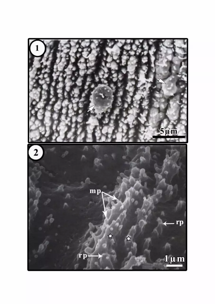
Figure 1. Scanning electron micrograph of tegument of Metamicrocotyla
cephalus, showing many foldings producing closely packed annular
corrugations superimposed with a complex configuration of many robust
papillae bearing microvillus-like projections and few uniciliated dome-
shaped ornamented papillae (arrows), presumed sensory structures.
Figure 2. Scanning electron micrograph of tegument of M. cephalus, showing
some foldings (stars) superimposed with a complex configuration of many
robust papillae (rp) bearing microvillus-like projections (mp).
Figure 3. Scanning electron micrograph of tegument of Dactylogyrus extensus,
showing weak annulations and many minute folds like microvilli (mf).
Figure 4. Scanning electron micrograph of D. extensus tegument covering the
head lobes, showing uniciliated structures (head arrows) projected from pit-
like depressions.
Figure 5. Transmission electron micrograph of the body tegument of M.
cephalus, showing the deep infoldings of both apical and basal membranes
that bounded the outer syncytium and thin membranous structures (arrows).

10
cmf, circular muscle fibers; m, mitochondrion; ts1, electron-lucent bodies;
ts2, electron-dense bodies and tw, terminal web.
Figure 6. Transmission electron micrograph of the body tegument of M.
cephalus, showing thin membranous structure (arrows). cmf, circular muscle
fibers; f, folds; lmf, longitudinal muscle fibers; mp, microvilli-like
projections; rp, robust papilla and tr, tegumental reservoir.
Figure 7. Transmission electron micrograph of the body tegument of M.
cephalus, showing the tegumental cell bodies. ch, chromatin body; dmf,
diagonal muscle fibers; GER, granular endoplasmic reticulum; m,
mitochondria; n, nucleus; nu, nucleolus; td, tegumental duct and ts2,
electron-dense bodies.
Figure 8. Transmission electron micrograph of the body tegument of D.
extensus, showing the irregularly weak folded syncytium containing small
electron-dense bodies (td1), fusiform, membrane-bound bodies containing
parallel longitudinal fibrous threads (td2) and thin dumbbell-shaped, electron
dense-bodies embedding in electron-lucent bodies (td3). bl, basal plasma
membrane/basal lamina complex; cmf, circular muscle fibers and lmf,
longitudinal muscle fibers.
Figure 9. Transmission electron micrograph of the body tegument of D.
extensus, showing the exocytotic (stars) and endocytotic (head arrows)
movements throughout the apical plasma membrane.
Figure 10. Transmission electron micrograph of the body tegument of D.
extensus, showing the tegumental cell bodies producing thin dumbbell-
shaped, electron dense-bodies (td3). GER, granular endoplasmic reticulum;
n, nucleus and nu, nucleolus.
Figure 11. Transmission electron micrograph of the body tegument of D.
extensus, showing the tegumental cell bodies producing small electron-dense
bodies (td1) and fusiform, membrane-bound bodies containing parallel
longitudinal fibrous threads (td2). GER, granular endoplasmic reticulum; n,
nucleus and nu, nucleolus.
References
Cohen, S.C.; Kohn, A. and Baptista-Farias, M.F.D. (2001). Scanning and
transmission electron microscopy of the tegument of Paranaella luquei
Kohn, Baptista-Farias & Cohen, 2000 (Microcotylidae, Monogenea),
Parasite of a Brazilian Catfish, Hypostomus regani. Mem. Inst. Oswaldo.
Cruz, Rio de Janeiro, 96(4): 555-560.

11
El-Naggar, M.M. and Cable, J. (2007). Ultrastructural observations on the
elusive subtegumental cells of the viviparous gill monogenean,
Macrogyrodactylus clarii. Parasitol. Res., 101: 9-17.
El-Naggar, M.M.; Khider, A.A. and Kearn, G.C. (1991). Ultrastructural
observations on the tegument and associated structures of the monogenean
Cichlidogyrus halli typicus (Price & Kirk, 1967) Paperna, 1979.
International Journal for Parasitology, 21: 707-713.
Justine, J.L. (1992). Ultrastructure of spermiogenesis, spermatozoa and the
tegument in Atriaster sp. (Platyhelminthes, Monogenea,
Polyopisthocotylea, Microcotylidae) Zool. Scripta., 21: 231-238.
Khidr, A.A. (1996). Scanning and transmission electron microscope studies on
the tegument of the monogenean stomach parasite Enterogyrus cichlidarum
Paperna, 1963 (Ancyrocephalidae: Monogenea). Proc. Zool. Soc. A.R.
Egypt, 27: 161-170.
Kritsky, D.C. and Kruidenier, F. J. (1976). Fine structure and development of
the body wall in the monogenean, Gyrodactylus eucaliae Ikezaki and
Hoffman, 1957. Proc. Helminthol. Soc. Wash., 43: 47-58.
Lyons, K.M. (1970). The fine structure and function of the adult epidermis of
two skin parasitic monogeneans, Entobdella soleae and Acanthocotyle
elegans. Parasitology, 60: 39-52.
Lyons, K.M. (1972). Ultrastructure observations on the epidermis of the
polyopisthocotylean monogeneans Rajonchocotyle emarginata and
Plectanocotyle gurnardi. Z. Parasitenk., 40: 87-100.
Lyons, K.M. (1973). The epidermis and sense organs of the Monogenea and
some related groups. Adv. Parasitol., 11: 193-232.
Ramasamy, P. and Brennan, G.P. (2000). Ultrastructure of the surface structures
and haptor of Empleurosoma pyriforme (Ancyroceohalinae;
Monopisthocotylea: Monogenea) from the gills of the teleost fish Therapon
jarbua. Parasitol. Res., 86: 129-139.
Ramasamy, P. and Hanna, R.E.B. (1986a). The surface topography of Bicotyle
vellavoli (Monogenea) from the gills of Pampus chinensis . International
Journal for Parasitology, 16: 591-594.
Ramasamy, P. and Hanna, R.E.B. (1986b). The surface topography of a
monogenean Heterapta chorinemi from the gills of Scomberoides
commersonianus. International Journal for Parasitology, 16: 595-600.

12
Ramasamy, P. and Hanna, R.E.B. (1989). The surface topography of Gotocotyla
secunda and Gotocotyla bivaginalis (Monogenea, Polyopisthocotylea) from
Scomberomorus commerson. International Journal for Parasitology, 19: 63-
69.
Ramasamy, P.; Brennan, G.P. and Halton, D.W. (1995). Ultrastructure of the
surface structures of Allodiscocotyla diacanthi (Polyopisthocotylea:
Monogenea) from the gills of the marine teleost fish, Scomberoides tol.
International Journal for Parasitology, 25: 43-54.
Ramasamy, P.; Hanna, R.E.B. and Threadgold, L.T. (1987). Scanning and
transmission electron microscopic studies of the surface of Vallisia indica
(Monogenea, Polyopisthocotylea). International Journal for Parasitology,
17: 1187-1195.
Rohde, K. (1975). Fine structure of the Monogenea, especially Polystomoides
Ward. Advances in Parasitology, 13: 1-33.
Rohde, K. (1980). Some aspects of the ultrastructure of Gotocotyla secunda and
Hexostoma euthynni. Angew. Parasitol., 21: 32-48.
Shaw, M.K. (1980). The ultrastructure of the epidermis of Diplectanum aequans
(Monogenea). Parasitology, 80: 9-25.
Smyth, J.D. and Halton, D.W. (1983). The physiology of trematodes. 2nd
edn.
Cambridge University Press, Cambridge.
Tyler, S. and Hooge, M. (2004). Comparative morphology of the body wall in
flatworms (Platyhelminthes). Can. J. Zool., 82: 194-210.
Tyler, S. and Tyler, M.S. (1997). Origin of the epidermis in parasitic
Platyhelminths. Int. J. Parasitol., 715-738.
Williams, J.B. and McKenzie, J. (1995). Scanning electron microscopy of
Polystoma integerrimum (Monogenea, Polystomatidae). International
Journal for Parasitology, 25(3): 335-342.
Xylander, W.E.R. (2001). The Gyrocotylidae, Amphilinidae and the early
evolution of the Cestoda. In "Interrelationships of the Platyhelminthes".
Edited by D.T.J. Littlewood and R.A. Bray. Taylor and Francis, London.
pp. 103-111.

مقارنة السمات السطحية والتركيب الدقيق إليهاب طفيلى بولىبيثوكوتيلين
( اكستنسس داكتيلوجيرس)وطفيلى مونوبيثوكوتيلين (سيفالس ميتاميكروكوتيل)
عال عبد الحليم أبو سمك و أشرف المتولى سعيد مصر - جامعة المنصورة – فرع دمياط – كلية العلوم –قسم علم الحيوان
تم فى الدراسة الحالية ألول مرة مقارنة السمات السطحية و التركيب الدقيق إليهاب
وطفيلى من المونوبيثوكوتيليدات (سيفالس ميتاميكروكوتيل)طفيلى من البولىبيثوكوتيليدات
ميتاميكروكوتيلوقد كشفت هذه المقارنة عن تميز إيهاب الطفيلى . (اكستنسس داكتيلوجيرس)
بالعديد من الطيات التى شكلت تموجات حلقية متقاربة على نحو وثيق تحمل تشكيل سيفالس
معقد من الحلمات الغليظة التى تنتهى بنتوءات تشبه الخمالت الدقيقة، ووجود غشاء طرفى
رقيق يقع مباشرة تحت الغشاء البالزمى القمى ، ووجود امتدادات من الغشاء البالزمى
القاعدى داخل المدمج، إضافة لوجود نوع واحد من الخاليا الجلدية التى تنتج نوعان من
وتراوح (أجسام المعة الكترونيا و أجسام أخرى داكنة الكترونيا)اإلفرازات داخل المدمج وهما
داكتيلوجيرسوفى المقابل تميز إيهاب الطفيلى .ميكرون3الى 1.3سمك المدمج بين
بحلقات ضعيفة وطيات تشبه الخمالت الدقيقة، ووجود بعض االنغمادات فى الغشاء اكستنسس
لبعض ءالبالزمى القمى مكونة فجوات صغيرة وأيضا ظهور تفجر افرازى من خالل هذا الغشا
ينتج النوع األول أجسام : األجسام اإلفرازية، وتميز أيضا بوجود نوعان من الخاليا الجلدية
صغيرة داكنة الكترونيا و أجسام أخرى مغزلية كبيرة ذات كثافة متوسطة الكترونيا بينما النوع
1.5-1.4)الثانى أجسام إفرازية داكنة الكترونيا تشبه الدمبل وقد كان المدمج أكثر رقة
وقد تم مناقشة الوظائف المحتملة للسمات المميزة لغطاء جسم الطفيليان سواء . (ميكرون
بالوجود أو بالغياب وخصوصا الدور المقترح الذى تضطلع به أثناء حياة الطفيليات ومدى
إمكانية جعلها صفات مميزة لمجموعة البولىبيثوكوتيليدات عن مجموعة
. المونوبيثوكوتيليدات