Embed Size (px)
Citation preview

Protein Expression and PuriWcation 39 (2005) 199–208
www.elsevier.com/locate/yprep
Comparative evaluation of two puriWcation methodsof anti-CD19-c-myc-His6-Cys scFv
Dipankar Dasa, Theresa M. Allena, Mavanur R. Sureshb,¤
a Department of Pharmacology, University of Alberta, Edmonton, AB, Canada T6G 2H7b Faculty of Pharmacy and Pharmaceutical Sciences, University of Alberta, Edmonton, AB, Canada T6G 2N8
Received 29 July 2004, and in revised form 6 October 2004Available online 8 December 2004
Abstract
DiVerent chromatographic methods have been used to purify bacterially expressed single chain antibodies in soluble or insolubleform. Here, we compared two methods for puriWcation of anti-CD19-c-myc-His6-Cys scFv expressed in Escherichia coli as solubleprotein. The protein-L–agarose puriWcation method is a one step puriWcation method that yielded signiWcant amounts of pure pro-tein compared to the two-step Ni–NTA–agarose plus Resource 15S puriWcation method. However, the protein-L puriWcationmethod exhibited an additional lower molecular weight protein contaminant. Based on results from in vitro gel digestion, mass spec-trometry and database search results, we conWrmed that the lower molecular weight protein contaminant, which could not be puri-Wed by Ni–NTA–agarose and 15S column method, is a degraded product of the full length scFv construct. 2004 Elsevier Inc. All rights reserved.
Keywords: CD19; scFv; IMAC; Protein-L
Tumor-associated surface antigens play a crucial role Most malignant B-cells express the CD19, CD20, and
in antibody-directed imaging and therapy. Antibody-based tumor targeting has found utility for the speciWcdelivery of a variety of agents such as drugs, radioiso-topes, lymphokines, and enzymes for imaging andimmunotherapy of tumor tissue. New coupling technol-ogies for attaching antibodies or recombinant antibodyfragments and ligands via covalent bonds to the surfaceof liposomes provide opportunities for further increas-ing the selective targeting of liposomal anticancer drugs[1,2]. We are interested in developing simple, eYcientmethods for the puriWcation of scFv fragments thatcould be used to target liposomal anticancer drugs todisease cells, such as malignant B cells.1046-5928/$ - see front matter 2004 Elsevier Inc. All rights reserved.doi:10.1016/j.pep.2004.10.007
* Corresponding author. Fax: +1 780 4921217.E-mail address: [email protected] (M.R. Suresh).
1 Abbreviations used: mAb, monoclonal antibody; scFv, single chain va0.5% NaCl, pH 7.5; Ni–NTA, nickel–nitrilotriacetic acid; IMAC, immobilizer; HRPO, horseradish peroxidase; IPTG, isopropyl �-D-1-thiogalactopyranthiothreitol; SDS–PAGE, sodium dodecyl sulfate–polyacrylamide gel elEDTA, ethylene diamine tetraacetic acid; MES, (2-[N-morpholino]ethanesu
CD22 antigens on their cell surface. The CD19 antigen isa diVerentiation marker that is widely expressed on Blymphocytes [3], including B-lineage malignancies suchas acute lymphoblastic leukemia and non-Hodgkin’slymphoma [4]. Moreover, this antigen is not shed and isabsent from hemopoietic stem cells, plasma cells, T cells,and other tissues. Antibodies and antibody fragmentsagainst CD19 have proven to be useful targeting agentsfor the development of immunoconjugates or immuno-liposomes [5–9].
The potential value of monoclonal antibodies (mAb)1
against CD19 has been demonstrated in animal modelsof B cell malignancies [10,11]. For the treatment of
riable fragment antibody; 2£ YT, 1.6% tryptone, 1% yeast extract, anded metal aYnity chromatography; FACS, Xuorescence activated cell sort-oside; ECL, enhanced chemiluminescence; OD, optical density; DTT, di-
ectrophoresis; GAM-HRPO, goat anti-mouse horseradish peroxidase;lfonic acid); LC/MS, liquid chromatography/mass spectrometry.
200 D. Das et al. / Protein Expression and PuriWcation 39 (2005) 199–208
leukaemia and lymphoma, mAbs against CD19 [12], incombination with IL-2 [13], or conjugated to toxin[14,15] have been used in clinical trials. In addition,diVerent constructs of anti-CD19 single chain variableantibody fragments (scFv, mono and bispeciWc) havealso been described, allowing the development of fusionprotein strategies [16–21] for the treatment of B cell leu-kaemias.
ScFvs are smaller antibody fragments composed ofimmunoglobulin heavy (VH) and light chain variable(VL) regions with a Xexible peptide linker designed toconnect the two chains such that the antigen binding siteis retained in a single co-linear molecule [22,23]. The useof phage display technology to select scFvs with speci-Wed binding properties and aYnities oVers a new oppor-tunity for tumor-targeted therapy [24]. ScFvs have thepotential to be very useful designer nanoprobes for thetargeted delivery of drugs, toxins or radioisotopes tospeciWc tumor cells. In comparison to the much largerFab�, F(ab�)2, and IgG forms of the monoclonal anti-bodies from which they are derived, scFv have lowerretention times in non-target tissues, more rapid bloodclearance, better tumor penetration and reduced immu-nogenicity [25–28] making them attractive for therapeu-tic applications.
Many scFvs contain poly His and c-myc tags that areused in their puriWcation and identiWcation in vitro. Ourobjective was to develop simple and eYcient methods forthe puriWcation of an anti-CD19-c-myc-His6-Cys scFvfragment, which can be covalently attached to the sur-face of drug-containing liposomes via its unpaired termi-nal Cys. It has been reported in the literature that scFvswith C-terminal Cys retain binding activity [27,29,30].Another objective was to develop a puriWcation methodthat could be applied to scFvs that lack the c-myc andHis6 tags and to compare the two methods. It is possibleto engineer scFv fragments that lack tags, but this cancomplicate their puriWcation. Targeting moieties withoutextraneous tags may be desirable for human clinical use,since the possibility of side eVects resulting from thepresence of tags such as c-myc (a protooncogene) maydelay or prevent regulatory approval.
In this report, we evaluated two diVerent puriWcationmethods for the preparation of anti-CD19-c-myc-His6-Cys scFv expressed in Escherichia coli. Prokaryoticexpression systems have several advantages, whichinclude cost-eVective production and the ability to pro-duce large amounts of recombinant protein in short peri-ods of time. A two-step puriWcation method, involvingimmobilized metal aYnity chromatography (IMAC)plus Resource 15S, generated a signiWcant amount ofpuriWed protein, which retained its cell-speciWc bindingin FACS analysis. This method is only applicable toscFvs containing the poly His tag. The second method,which is applicable to scFvs lacking a poly His tag, is aone-step puriWcation method (protein-L–agarose) that
gave highly pure scFv along with a second lower molec-ular weight protein which could not be puriWed by theIMAC plus Resource 15S column method. The secondlower molecular weight protein was identiWed by in vitroin gel digestion, mass spectrometry, and databasesearching. Here, we have demonstrated that the secondprotein is the degraded product of anti-CD19-c-myc-His6-Cys scFv and its degradation is not speciWc to theE. coli strain. The degraded anti-CD19-c-myc-His6-CysscFv product could only be puriWed by protein-L–aga-rose and was detected in Western blots probed with pro-tein-L–HRPO.
Materials and methods
Bacterial strain and chemicals
The plasmid pSKK, containing the anti-CD19-c-myc-His6-Cys scFv originally generated from HD37 hybrid-oma cells by AYmed Therapeutics [31] and was kindlyprovided by INEX Pharmaceuticals, Burnaby, Vancou-ver, Canada. The E. coli RV308 strain and the 9E10 anti-c-myc hybridoma cell line were purchased from ATCC(Rockville, USA). The anti-His6 mAb was purchasedfrom Novagen (Madison, USA). IPTG was purchasedfrom Gibco-BRL (Burlington, Canada). Acrylamide:bisacrylamide, prestained low range protein molecularweight markers and Protein Assay reagent were pur-chased from Bio-Rad (Mississauga, Canada). Ni–NTA–agarose resin was obtained from Qiagen (Mississauga,Canada). Resource 15S ion exchange resin, PD10 col-umns, Hybond ECL nitrocellulose membranes, and theECL Western blotting reagent were purchased fromAmersham–Pharmacia Biotech (BaiedUrfe, Quebec,Canada). Biomax-10 Ultrafree-15 centrifugal Wlterdevice was purchased from Millipore (Bedford, USA).Imidazole, MES, protein-L–agarose, and protein-L–HRPO were purchased from Sigma (Oakville, Canada).
Analysis of recombinant functional clone of anti-CD19-c-myc-His6-Cys scFv
A recombinant plasmid containing the correctly ori-ented anti-CD19-c-myc-His6-Cys scFv gene was used totransform E. coli RV308 by a heat shock method forrecombinant protein expression [32]. E. coli RV308transformants, carrying recombinant plasmid, werepropagated in 10 ml 2£ YT containing 100 �g/ml ofampicillin and were incubated at 26 °C with shaking at250 rpm until an optical density at 600 nm (OD600) ofapproximately 0.8 was reached. The bacterial culturewas induced with 1 mM IPTG and allowed to grow foranother 16 h at 24 °C. The bacterial culture was har-vested by centrifugation at 5000g for 10 min and thetotal cell lysate was prepared by addition of sample

D. Das et al. / Protein Expression and PuriWcation 39 (2005) 199–208 201
buVer (50 mM Tris–HCl, pH 6.8, 100 mM DTT, 2% SDS,0.1% bromophenol blue, and 10% glycerol) to the pelletand heated at 95 °C for 5 min. Total cell protein was ana-lyzed by SDS–PAGE using 10% polyacrylamide gel per-formed according to Laemmli [33] with a Mini ProteanII apparatus (Bio-Rad). The protein gel was stained with0.25% (w/v) Coomassie brilliant blue R-250 in 10% ace-tic acid and 45% methanol and destained with 10% ace-tic acid and 30% methanol.
Large scale protein expression and puriWcation
A fresh single colony of E. coli RV308 was inoculatedin 2£ YT medium containing 100 �g/ml ampicillin andallowed to grow overnight at 26 °C in an incubatorshaker. The overnight culture was diluted to 1/40th vol-ume in fresh 2£ YT medium containing 100 �g/ml ofampicillin and allowed to grow at 26 °C with vigorousshaking until an OD600 of 0.8–0.9 was reached. Inductionwas started by directly dissolving 0.4 M sucrose in 2£YT medium and IPTG to a Wnal concentration of0.2 mM, which is a modiWcation of a method describedearlier [19]. The bacterial culture was incubated for 16 hwith vigorous shaking at 22–24 °C. Bacterial pellets werecollected by centrifugation at 5000g for 20 min at 4 °C.Total cell protein from induced and uninduced culturewas analyzed by SDS–PAGE and by Western blotprobed with anti-His6 mAb, followed by GAM-HRPOand enhanced chemiluminescence.
Isolation of scFv in soluble form
Bacterial pellets were resuspended in 5% of the origi-nal culture volume of periplasmic extraction buVer[200 mM Tris–HCl, 20 % (w/v) sucrose, and 1 mMEDTA, pH 8.0] and incubated on ice for 1 h with occa-sional shaking. The cell extract was centrifuged at15,000g for 30 min at 4 °C and the supernatant was col-lected as a soluble periplasmic extract. The periplasmicextract was thoroughly dialyzed against 50 mM Tris–HCl, 1 M NaCl, pH 7.0 at 4 °C with three changes. At theend of the dialysis, the dialyzed extract was clariWed bycentrifugation at 15,000g for 30 min at 4 °C.
IMAC puriWcation
Periplasmic extract was incubated with Ni–NTA–agarose and rocked overnight at 4 °C prior to puriWca-tion. An IMAC column (5 ml) was prepared by loadingthe sample on a column equilibrated with 10 bed vol-umes of 50 mM Tris–HCl, 1 M NaCl, pH 7.0, followedby 20 bed volumes of washing buVer (50 mM Tris–HCl,1 M NaCl, and 20 mM imidazole, pH 7.0). Bound scFvwas eluted with 50 mM Tris–HCl, 1 M NaCl, and300 mM imidazole, pH 7.0, collecting 20 fractions of 1 mleach. Unbound periplasmic extract was collected and
rocked for the second time with Ni–NTA–agarose andpuriWcation was repeated as above. All the eluted frac-tions were analyzed by SDS–PAGE.
Resource 15S puriWcation
IMAC fractions contained some impurities alongwith the target scFv protein, hence an attempt was madeto purify the target protein by passing through an ionexchange resin (Resource 15S). Selected IMAC fractionswere pooled and concentrated by passing through a Bio-max-10 Ultrafree-15 centrifugal Wlter device. The buVerwas exchanged in the concentrated protein sample on aPD10 column equilibrated in 25 ml of 50 mM MESbuVer, pH 5.5. The protein sample was eluted with 3.5 mlof 50 mM MES buVer, pH 5.5 and any turbidity wasremoved by centrifugation at 15,000g for 15 min at 4 °C.A 5 ml 15S column was prepared according to manufac-turer’s instruction and equilibrated with 50 mM MESbuVer, pH 5.5. The sample was loaded on the column bya peristaltic pump and the column was washed with 20bed volumes of 50 mM MES buVer, pH 5.5. The boundscFv was eluted using a linear 0–1 M NaCl gradient in50 mM MES buVer, pH 5.5, collecting 1 ml fractions.Eluted fractions were analyzed by SDS–PAGE. PurescFv fractions were pooled and dialyzed against phos-phate buVer saline (PBS) with 50 mM imidazole, pH 7.3.
ScFv puriWcation on protein-L–agarose columns
Periplasmic soluble scFv was isolated as describedearlier and dialyzed against 10 mM PBS, pH 7.4, at 4 °C.The dialyzed sample was clariWed by centrifugation at15,000g for 30 min at 4 °C and the clear supernatant wascollected. The protein sample was loaded on a protein-L–agarose column with a peristaltic pump at a rate of20 ml/h. Unbound protein sample was washed with 10–15 volumes of PBS. Bound scFv was eluted from the col-umn with elution buVer (0.1 M glycine, pH 2.0). Theeluted scFv was neutralized with 1 M Tris, pH 9.0, toachieve a pH of 7.4. The purity of the eluted fractionswas checked by SDS–PAGE and Western blot.
Western blot analysis
Total cell proteins or puriWed anti-CD19-c-myc-His6-Cys scFv fractions were electrophoresed on SDS–PAGEusing 10% polyacrylamide gel and then electroblottedonto Hybond ECL nitrocellulose membranes [34] with aTransblot apparatus (Bio-Rad) following manufac-turer’s instructions. The membrane was blocked with 5%skim milk in PBST (0.1% Tween 20 in 1£ PBS, pH 7.3)for 1 h. The membrane was washed four times withPBST and incubated for 1 h with either anti-His6 mAb,anti-c-myc mAb (9E10) or protein-L–HRPO. Afterwashing four times with PBST, the membrane (with the

202 D. Das et al. / Protein Expression and PuriWcation 39 (2005) 199–208
exception of the membrane reacted with protein-L–HRPO) was incubated with GAM-HRPO for 1 h.Finally, the membrane was washed with PBST fourtimes and ECL-based detection was performed accord-ing to manufacturer’s instructions.
Expression of scFv in diVerent E. coli strains
The recombinant plasmid containing the correctlyoriented anti-CD19-c-myc-His6-Cys scFv gene was usedto transform several diVerent E. coli strains (RV308, XL-1 Blue, MC1061, HB101, JM83, and RR1) by heat shockmethods for recombinant protein expression. The diVer-ent E. coli strains carrying recombinant plasmid werepropagated in 10 ml 2£ YT medium containing 100 �g/ml of ampicillin and incubated at 26 °C with shaking at250 rpm until the OD600 reached 0.8. The bacterial cul-tures were induced with 1 mM IPTG and incubated forovernight at 24 °C. The bacterial cultures were harvestedby centrifugation at 5000g for 10 min and total cell lysatewas isolated by addition of sample buVer and heating at95 °C for 5 min. Total cell proteins were separated onSDS–PAGE using 10% polyacrylamide gel, stained, anddestained as described above.
In gel digestion
Protein identiWcation was performed at the Institutefor Biomolecular Design, University of Alberta, Edmon-ton, Alberta, Canada. BrieXy, stained bands (spots) wereexcised and an automated in-gel tryptic digestion wasperformed on a Mass Prep Station (Micromass, UK).The gel pieces were destained, reduced with DTT, alkyl-ated with iodoacetamide, then digested with trypsin(Promega Sequencing Grade ModiWed) and the resultingpeptides were extracted from the gel and analyzed viaLC/MS.
LC/MS was performed on a CapLC HPLC (Waters,USA) coupled with a Q-ToF-2 mass spectrometer(Micromass, UK). Tryptic peptides were separated usinga linear water/acetonitrile gradient (0.2% formic acid) ona Picofrit reversed-phase capillary column (5 �m BioBa-sic C18, 300 Å pore size, 75 �m ID £ 10 cm, and 15 �mtip) (New Objectives, MA, USA) with an in-line PepMapcolumn (C18, 300�m ID £ 5 mm) (LC Packings, CA,USA) used as a loading/desalting column.
Results
ScFv expression and puriWcation
The plasmid pSKK, containing the anti-CD19-c-myc-His6-Cys scFv, was generated from HD37 hybridomacells [31]. The scFv is arranged in the VH–VL orientationwith a YOL linker (a tubulin epitope recognized by mAb
YOL 1/34) [35]. The pSKK plasmid containing anti-CD19-c-myc-His6-Cys scFv was transformed into an E.coli expression host RV308 and the clone expressing theanti-CD19-c-myc-His6-Cys scFv, with a predicted bandaround 34 kDa, was picked (Fig. 1A). ScFv-producingclones were analyzed by Western blot. Blotted proteinswere detected using an anti-His6 mAb (Fig. 1B) and itwas demonstrated that these clones expressed theexpected scFv band at 34 kDa.
The anti-CD19-c-myc-His6-Cys scFv fragment wasproduced in E. coli by secretion into the bacterial peri-plasm. The soluble antibody fragments were isolated fromthe periplasmic extracts and puriWed by two chromatogra-phy methods. IMAC column puriWcation on a Ni–NTA–agarose column resulted in a typical yield of 500–800�g/Lof bacterial shake Xask culture. SDS–PAGE gels showedthat the scFv was present as a major peak; several minorcontaminant proteins of bacterial origin were alsodetected after metal aYnity chromatography (Fig. 2A).Proteins eluted from the IMAC column were then passeddown a Resource 15S ion exchange column (Fig. 2B),which removed the bacterial proteins. Proteins from theResource 15S column were separated by SDS–PAGE andshowed a single band with an approximate molecularweight of 34 kDa and a purity greater than 90% (Fig. 2B).This step provided approximately 100–300�g of puriWedmaterial per 1 L of bacterial shake Xask culture. This rep-resents approximately 20–40% recovery by this two-stepprocedure. SDS–PAGE and Western blot of the puriWedscFv under reducing and non-reducing conditions showedonly one band of approximately 34 kDa under reducingcondition, corresponding to monomeric scFv (Figs. 3Aand B, lane 1) and two bands under non-reducing condi-tions with approximate molecular weights of 68 and34 kDa, corresponding to the dimer and the monomerform of scFv (Figs. 3A and B, lane 2). These results sug-gest that a signiWcant amount of dimer is secreted into theperiplasm, which is generated by the formation of S–Sbonds between C-terminal Cys groups.
Fig. 1. Total cell protein of induced and uninduced bacterial culturesseparated on SDS–PAGE, stained with Coomassie brilliant blue (A)and Western blot probed with anti-His6 mAb (B). Lane M, standardprestained molecular weight markers; lanes 1–4, induced culture offour individual clones; and lane 5, uninduced culture.

D. Das et al. / Protein Expression and PuriWcation 39 (2005) 199–208 203
In the second puriWcation method, anti-CD19-c-myc-His6-Cys scFv fragments were puriWed by protein-L–agarose and analyzed by SDS–PAGE (Fig. 4). Theone-step protein-L–agarose puriWcation yieldedapproximately 400–500 �g/L of shake Xask culture.Two protein bands were present, one with an approxi-mate molecular weight of 34 kDa and one with a lowermolecular weight. To maximize protein recovery, theunbound protein fractions were reloaded for furthertwo cycles of puriWcation onto the protein-L column,
Fig. 2. SDS–PAGE analysis of IMAC (A) and 15S puriWed (B) anti-CD19-c-myc-His6-Cys scFv. (A) Lane M, standard prestained molecu-lar weight markers and lanes 1–9, diVerent eluted fractions fromIMAC column. (B) Lane M, standard prestained molecular weightmarkers and lanes 1–8, diVerent eluted fractions from 0 to 1 M NaClgradient.
Fig. 3. SDS–PAGE (A) and Western blot analysis probed with anti-His6 mAb (B) (reducing/non-reducing conditions) of 15S puriWed anti-CD19-c-myc-His6-Cys scFv. Lane M, standard prestained molecularweight markers; lane 1, puriWed scFv under reducing condition; andlane 2, puriWed scFv under non-reducing condition.
which resulted complete puriWcation of the scFv (datanot shown). The pooled bands were analyzed by SDS–PAGE (Fig. 5A) and Western blot, probed with threediVerent reagents. Both anti-His6 and anti-c-myc mAbsbound only to the upper protein band (»34 kDa),which corresponded to the anti-CD19-c-myc-His6-CysscFv monomer (Figs. 5B and C). Surprisingly, protein-L–HRPO bound to both the upper (»34 kDa) andlower protein bands (Fig. 5D), suggesting that lowerprotein may be the degraded product of the upperprotein.
Fig. 4. SDS–PAGE analysis of protein-L–agarose puriWed anti-CD19-c-myc-His6-Cys scFv. Lane M, standard prestained molecular weightmarkers and lanes 1–7, diVerent eluted fractions by glycine–HCl.
Fig. 5. SDS–PAGE (A) and Western blot analysis probed with anti-His6 mAb (B), anti-c-myc mAb (C), and protein-L–HRPO (D) of pro-tein-L–agarose puriWed anti-CD19-c-myc-His6-Cys scFv. Lane M,standard prestained molecular weight markers and lane 1, puriWedanti-CD19-c-myc-His6-Cys scFv.

204 D. Das et al. / Protein Expression and PuriWcation 39 (2005) 199–208
Expression of anti-CD19-c-myc-His6-Cys scFv in diVerent E. coli hosts
To determine if the presumed degradation product ofscFv was dependent of the E. coli strain, we expressedthe anti-CD19-c-myc-His6-Cys scFv in several diVerentE. coli strains. The transformed cells were induced with1 mM IPTG, then total cell proteins were isolated andanalyzed by SDS–PAGE and Western blot, probed withanti-His6 mAb and protein-L–HRPO. The expressionlevels of scFv diVered between the diVerent E. colistrains, but all of them had a detectable band around»34 kDa (Fig. 6A). In the Western blots, anti-His6 mAbbound only to the protein band (»34 kDa) that corre-sponded to the anti-CD19-c-myc-His6-Cys scFv mono-mer (Fig. 6B) and protein-L–HRPO bound to both the»34 kDa protein and the lower molecular weight protein(Fig. 6C). These data correspond to the previous West-ern blot data of anti-CD19-c-myc-His6-Cys scFv puri-Wed by protein-L–agarose.
In gel digestion
Protein identiWcation from the generated LC/MS dataof the fragments from the upper and lower protein bands(Fig. 4) was done by searching the NCBI (National Centerfor Biotechnology Information) non-redundant database[Database: NCBInr 20021030, (1226480 sequences;
390314779 residues)] using Mascot Daemon search meth-odology (Matrix Science, UK, http://www.matrixscience.com). Search parameters included carbamidomethylationof cysteine, possible oxidation of methionine and onemissed cleavage per peptide. Mascot search results areincluded in Table 1 and both the upper and lower proteinbands (Fig. 4) showed signiWcant hits for the anti-CD19antibody light chain variable region (VL) and the immuno-globulin heavy chain variable domain (VH), suggesting thatboth proteins were anti-CD19 scFv and that both wereeYciently puriWed by protein-L–agarose. We conclude thatthe lower protein band was the anti-CD19 scFv withoutthe c-myc-His6-Cys tag and the upper protein band was thefull scFv construct, anti-CD19-c-myc-His6-Cys.
Discussion
The development of new therapeutic reagents anddrug delivery systems is in demand for the treatment of avariety of diseases [36]. One approach to developingthese therapies has been to chemically conjugate mAbsto toxins or other therapeutic molecules or particulatecarriers [10,37–40]. However, there are several disadvan-tages to using whole antibodies, including prematureclearance via Fc recognition, generation of immune reac-tions, and limited tissue penetration due to the large sizeof the molecules [26,40,41]. ScFv and other small anti-
Fig. 6. Expression analysis of anti-CD19-c-myc-His6-Cys scFv in diVerent E. coli strains. Total cell protein of induced bacterial culture separated onSDS–PAGE, stained with Coomassie brilliant blue (A) and Western blot probed with anti-His6 mAb (B), and protein-L–HRPO (C). Lane M, stan-dard prestained molecular weight markers; lane 1, E. coli RV 308; lane 2, E. coli XL-1 Blue; lane 3, E. coli MC1061; lane 4, E. coli HB101; lane 5, E.coli JM83; and lane 6: E. coli RR1.

D. Das et al. / Protein Expression and PuriWcation 39 (2005) 199–208 205
body fragments have been developed to overcome theproblems associated with whole antibodies.
The expression level of foreign genes is usually veryhigh in prokaryotic systems in comparison to eukaryoticsystems. Foreign genes can be expressed as soluble pro-teins, as well as in inclusion bodies [42–45]. Expression ininclusion bodies is at a higher level and can be advanta-geous for purifying large amounts of protein. However,many scFvs that are expressed in bacteria as inclusionbodies [46,47] can be diYcult to refold and their abilityto retain binding speciWcity and aYnity for the antigenof interest is highly variable.
In the present study, we have evaluated two puriWca-tion methods for anti-CD19–c-myc-His6-Cys scFvexpressed in E. coli as soluble protein. The Wnal yield ofpuriWed protein was approximately 100–300 �g by thecombined IMAC and 15S method and approximately400–500 �g by the one-step protein-L method per 1 L ofbacterial culture. The purity of protein was judged to behigh by SDS–PAGE and Western blot for both methods.Not only did the protein-L column provides a higheryield of puriWed scFv, but it can be applied to the puriW-cation of scFv where the tags have been removed, so it ismore versatile.
SuYciently pure material for most applications couldbe obtained in one-step via IMAC puriWcation of His-tagged protein [48,49]. However, in our study, the pro-tein of interest was present as only at low concentrationsin the soluble extract. Several contaminating proteins,presumably bacterial, were also bound to the IMAC col-umn and co-eluted under the puriWcation conditionsemployed. This has also reported by another laboratory[50]. Since our IMAC puriWcation did not yield a pureprotein as judged by SDS–PAGE, we attempted furtherpuriWcation to get rid of the contaminating proteins.
Many laboratories have reported diVerent puriWca-tion methods for antibody fragments from IMAC-elutedmaterial, which include Ag aYnity chromatography [29],thiophilic adsorption chromotography [51,52] orimmunoaYnity puriWcation, using immobilized anti-His-tag-mAbs [50]. Here, we used a simple alternativechromatography method, based on the separation of
proteins by ion-exchange chromatography (Resource15S). IMAC-puriWed scFv fragments could be eYcientlypuriWed to greater than 90% by a Resource 15S column,which yielded a single band of 34 kDa (Fig. 2B).
Many gram-positive pathogenic bacteria express sur-face proteins such as protein-A [53], protein-G [54,55],and protein-L [56] that bind immunoglobulins. Proper-ties and interactions of these proteins with immunoglob-ulins have been well described in the literature [57–60].Protein-A and protein-LA also have binding aYnity forscFv fragments and have been successfully used for one-step puriWcation of scFv [61–63].
In this study, we used an alternative method for puriW-cation of anti-CD19-c-myc-His6-Cys scFv based on pro-tein-L–agarose. This one-step aYnity puriWcation methodyielded a signiWcantly pure protein as shown by SDS–PAGE. To the best of our knowledge this is the Wrstreport of anti-CD19 scFv puriWcation by protein-L–aga-rose. However, an additional protein in the lower molecu-lar weight region co-eluted from the protein-L–agarosegel. We initially thought that, under these puriWcationconditions, the anti-CD19-c-myc-His6-Cys scFv could notbe separated from a major bacterial contaminant, presum-ably acidic 27 kDa WHP proteins [64]. DiVerent laborato-ries reported that a host-encoded protein was observed asthe major contaminant during the puriWcation of severalrecombinant proteins from E. coli by IMAC [65,66]. Butthere is no report in the literature that bacterial WHP pro-tein or any other metal-binding protein has aYnity forProtein-L or other Ig binding proteins.
Our Western blot data probed with protein-L–HRPO(Fig. 5D) suggested that the lower protein band may be adegraded product of anti-CD19-c-myc-His6-Cys scFv thatwas capable of binding to protein-L–HRPO. The lowerprotein band could not be detected by either anti-His6 oranti-c-myc mAbs in Western blots (Figs. 5B and C), sug-gesting that the anti-CD19-c-myc-His6-Cys might bedegraded to anti-CD19 scFv (Fig. 7). This being the case,the degraded product, anti-CD19 scFv, would not be ableto bind to Ni–NTA–agarose and or be puriWed by 15Scolumn chromatography (Fig. 2B), as can be seen fromthe SDS–PAGE and Western blot data (Figs. 3A and B).
Table 1Mascot search results
Protein SigniWcant hits
Upper band gi�999108 (S78338) anti-CD19 antibody light chain variable region [Homo sapiens]gi�2208941 (Y12778) scFv8E5 recombinant antibody fragment [synthetic construct]gi�1435159 (X99230) immunoglobulin heavy chain variable domain [Mus musculus]gi�20534979 (XM_094001) similar to Kelch-like protein 2 (actin-binding protein Mayven) [Homo sapiens]
Lower band gi�999108 (S78338) anti-CD19 antibody light chain variable region [Homo sapiens]gi�1435159 (X99230) immunoglobulin heavy chain variable domain [Mus musculus]gi�2208941 (Y12778) scFv8E5 recombinant antibody fragment [synthetic construct]gi�229902 Chain H, Fab fragment from a monoclonal anti-arsonate antibody, R19.9 (Igg2b, kappa)gi�539894 Ig heavy chain V-D-J region (419.1)—mouse (fragment)gi�20534979 (XM_094001) similar to Kelch-like protein 2 (actin-binding protein Mayven) [Homo sapiens]

206 D. Das et al. / Protein Expression and PuriWcation 39 (2005) 199–208
The identity of degraded protein was conWrmed by invitro gel digestion, mass spectrometry, and NCBI non-redundant database searching. Both of the proteins puri-Wed from protein-L–agarose (Fig. 4) showed signiWcanthits for the anti-CD19 antibody VL region and theimmunoglobulin VH domain (Table 1), suggesting thatboth the proteins are anti-CD19 scFv and both wereeYciently puriWed by the protein-L–agarose columns.These data conWrmed that lower protein band was thedegradation product of anti-CD19-c-myc-His6-CysscFv.
Further expression analysis of the anti-CD19-c-myc-His6-Cys scFv was done in diVerent E. coli strains todemonstrate whether the degradation was E. coli strainspeciWc. Our SDS–PAGE data (Fig. 6A) suggest that theE. coli strains tested in this report all expressed the anti-CD19-c-myc-His6-Cys scFv, but at diVerent levels. InWestern blots, only one protein band could be detectedusing anti-His6 mAb, which conWrmed the presence ofanti-CD19-c-myc-His6-Cys scFv, and no degraded pro-tein was detected. In contrast, two protein bands weredetected in Western blots using protein-L–HRPO, whichconWrmed the presence of both anti-CD19-c-myc-His6-Cys and anti-CD19 scFv, without the tag. Hence, thedegradation of the anti-CD19-c-myc-His6-Cys scFv isnot E. coli strain speciWc. All of the experimental datareported here suggest that protein degradation occurseither during protein expression or protein extractionfrom bacterial culture. This could signiWcantly reducethe protein yield, a hidden anomaly that could not bedetected with anti-His6 mAbs in Western blots.
In conclusion, the protein-L–agarose puriWcation is aone-step puriWcation method for anti-CD19-c-myc-His6-Cys scFv compared to the two-step IMAC and 15Smethod. Protein-L puriWcation of scFv is a simple, fast,and easy strategy for preparation of scFv. This aYnityprocedure can also eliminate the need for incorporatingextraneous tags since the method could also be used forthe puriWcation of tagless scFv fragments. The develop-ment of puriWcation methods for scFvs without extrane-ous tags may be useful from a regulatory perspective,
especially when such targeting moieties are designed forclinical uses.
Acknowledgments
This work was supported by research grant fromCanadian Institute for Health Research (MOP-9127).The protein identiWcation was performed at the Institutefor Biomolecular Design, University of Alberta, Edmon-ton, Alberta, Canada.
References
[1] T.M. Allen, C.B. Hansen, S. Zalipsky, Antibody-targeted Stealth®liposomes, in: D.D. Lasic, F. Martin (Eds.), Stealth Liposomes,CRC Press, Boca Raton, FL, 1995, pp. 193–202.
[2] P. Sapra, T.M. Allen, Ligand-targeted liposomal anticancer drugs,Prog. Lipid Res. 42 (2003) 439–462.
[3] A.N. Barclay, A.D. Beyers, M.L. Birkeland, M.H. Brown, S.J.Davis, C. Somoza, A.F. Willimas, The Leukocyte Antigen FactsBook, Academic Press, London, 1994 p. 142.
[4] K.C. Anderson, M.P. Bates, B.L. Slaughenhoupt, G.S. Pinkus, S.F.Schlossman, L.M. Nadler, Expression of human B cell-associatedantigens on leukemias and lymphomas: a model of human B celldiVerentiation, Blood 63 (1984) 1424–1433.
[5] R.D. May, E.S. Vitetta, G. Moldenhauer, B. Dorken, Selective kill-ing of normal and neoplastic human B cells with anti-CD19- andanti-CD22-ricin A chain immunotoxins, Cancer Drug Deliv. 3(1986) 261–272.
[6] F.M. Uckun, Regulation of human B-cell ontogeny, Blood 76(1990) 1908–1923.
[7] M.A. Ghetie, H. Bright, E.S. Vitetta, Homodimers but not mono-mers of Rituxan (chimeric anti-CD20) induce apoptosis in humanB-lymphoma cells and synergize with a chemotherapeutic agentand an immunotoxin, Blood 97 (2001) 1392–1398.
[8] P. Sapra, T.M. Allen, Internalizing antibodies are necessary forimproved therapeutic eYcacy of antibody-targeted liposomaldrugs, Cancer Res. 62 (2002) 7190–7194.
[9] P. Sapra, T.M. Allen, Improved outcome when B-cell lymphoma istreated with combinations of immunoliposomal anticancer drugstargeted to both the CD19 and CD20 epitopes, Clin. Cancer Res.10 (2004) 2530–2537.
[10] B. Jansen, F.M. Uckun, W.B. Jaszcz, J.H. Kersey, Establishmentof a human t(4; 11) leukemia in severe combined immunodeW-cient mice and successful treatment using anti-CD19 (B43)-
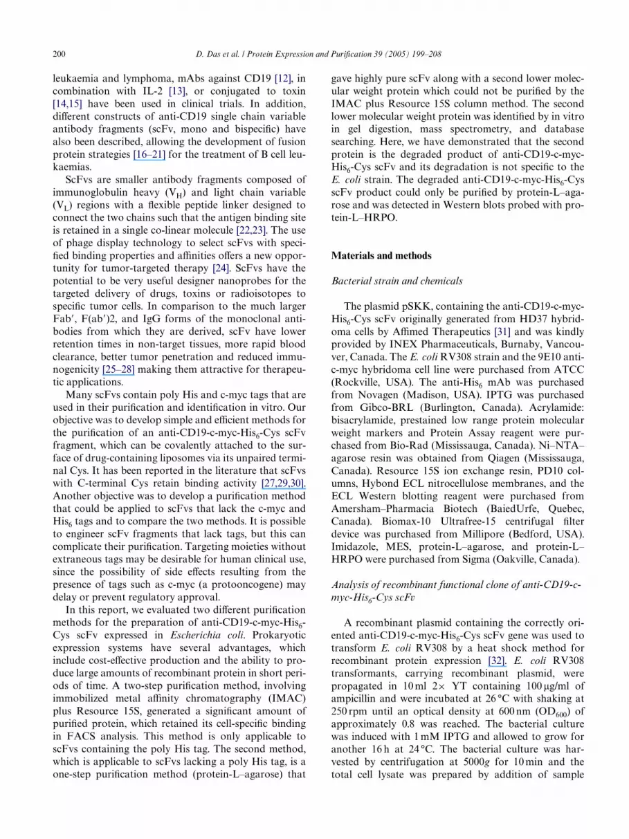
Fig. 7. Schematic diagram of anti-CD19-c-myc-His6-Cys scFv.

D. Das et al. / Protein Expression and PuriWcation 39 (2005) 199–208 207
pokeweed antiviral protein immunotoxin, Cancer Res. 52 (1992)406–412.
[11] G.A. Pietersz, L. Wenjun, V.R. Sutton, J. Burgess, I.F. McKenzie,H. Zola, J.A. Trapani, In vitro and in vivo antitumor activity of achimeric anti-CD19 antibody, Cancer Immunol. Immunother. 1(1995) 53–60.
[12] A. Hekman, A. Honselaar, W.M. Vuist, J.J. Sein, S. Rodenhuis,W.W. ten Bokkel Huinink, R. Somers, P. Rumke, C.J. Melief, Ini-tial experience with treatment of human B cell lymphoma withanti-CD19 monoclonal antibody, Cancer Immunol. Immunother.32 (1991) 364–372.
[13] L.T. Vlasveld, A. Hekman, F.A. Vyth-Dreese, C.J. Melief, J.J. Sein,A.C. Voordouw, T.A. Dellemijn, E.M. Rankin, Treatment of low-grade non-Hodgkin’s lymphoma with continuous infusion of low-dose recombinant interleukin-2 in combination with the B-cell-speciWc monoclonal antibody CLB-CD19, Cancer Immunol.Immunother. 40 (1995) 37–47.
[14] M.L. Grossbard, J.M. Lambert, V.S. Goldmacher, N.L. Spector, J.Kinsella, L. Eliseo, F. Coral, J.A. Taylor, W.A. Blattler, C.L.Epstein, et al., Anti-B4-blocked ricin: a phase I trial of 7-day con-tinuous infusion in patients with B-cell neoplasms, J. Clin. Oncol.11 (1993) 726–737.
[15] M.J. Stone, E.A. Sausville, J.W. Fay, D. Headlee, R.H. Collins,W.D. Figg, M. Stetler-Stevenson, V. Jain, E.S. JaVe, D. Solomon,R.M. Lush, A. Senderowicz, V. Ghetie, J. Schindler, J.W. Uhr, E.S.Vitetta, A phase I study of bolus versus continuous infusion of theanti-CD19 immunotoxin, IgG-HD37-dgA, in patients with B-celllymphoma, Blood 88 (1996) 1188–1197.
[16] B.E. Bejcek, D. Wang, E. Berven, C.A. Pennell, S.C. Peiper, S.Poppema, F.M. Uckun, J.H. Kersey, Development and character-ization of three recombinant single chain antibody fragments(scFvs) directed against the CD19 antigen, Cancer Res. 55 (1995)2346–2351.
[17] I.C. Nicholson, K.A. Lenton, D.J. Little, T. Decorso, F.T. Lee,A.M. Scott, H. Zola, A.W. Hohmann, Construction and charac-terisation of a functional CD19 speciWc single chain Fv fragmentfor immunotherapy of B lineage leukaemia and lymphoma, Mol.Immunol. 34 (1997) 1157–1165.
[18] F. Le Gall, S.M. Kipriyanov, G. Moldenhauer, M. Little, Di-, tri-and tetrameric single chain Fv antibody fragments against humanCD19: eVect of valency on cell binding, FEBS Lett. 453 (1999)164–168.
[19] S.M. Kipriyanov, G. Moldenhauer, J. Schuhmacher, B. Cochlo-vius, C.-W. Von der Lieth, E.R. Matys, M. Little, BispeciWc tan-dem diabody for tumor therapy with improved antigen bindingand pharmacokinetics, J. Mol. Biol. 293 (1999) 41–56.
[20] B. Cochlovius, S.M. Kipriyanov, M.J.J.G. Stassar, O. Christ, J.Schuhmacher, G. Strauss, G. Moldenhauer, M. Little, Treatmentof human B cell lymphoma xenografts with a CD3 £ CD19 dia-body and T cells, J. Immunol. 165 (2000) 888–895.
[21] U. Reusch, F. Le Gall, M. Hensel, G. Moldenhauer, A.D. Ho, M.Little, S.M. Kipriyanov, EVect of tetravalent bispeciWc CD19 £ CD3recombinant antibody construct and CD28 costimulation on lysisof malignant B cells from patients with chronic lymphocytic leuke-mia by autologous T cells, Int. J. Cancer 112 (2004) 509–518.
[22] J.S. Huston, D. Levinson, M. Mudgett-Hunter, M.S. Tai, J. Nov-otny, M.N. Margolies, R.J. Ridge, R.E. Bruccoleri, E. Haber, R.Crea, H. Oppermann, Protein engineering of antibody bindingsites: recovery of speciWc activity in an anti-digoxin single-chainFv analogue produced in Escherichia coli, Proc. Natl. Acad. Sci.USA 85 (1988) 5879–5883.
[23] R.E. Bird, K.D. Hardman, J.W. Jacobson, S. Johnson, B.M. Kauf-man, S.M. Lee, T. Lee, S.H. Pope, G.S. Riordan, M. Whitlow, Sin-gle-chain antigen-binding proteins, Science 242 (1988) 423–426.
[24] G. Winter, A.D. GriYths, R.E. Hawkins, H.R. Hoogenboom,Making antibodies by phage display technology, Annu. Rev.Immunol. 12 (1994) 433–455.
[25] D.E. Milenic, T. Yokota, D.R. Filpula, M.A. Finkelman, S.W.Dodd, J.F. Wood, M. Whitlow, P. Snoy, J. Schlom, Construction,binding properties, metabolism, and tumor targeting of a single-chain Fv derived from the pancarcinoma monoclonal antibodyCC49, Cancer Res. 51 (1991) 6363–6371.
[26] T. Yokota, D.E. Milenic, M. Whitlow, J. Schlom, Rapid tumorpenetration of a single-chain Fv and comparison with otherimmunoglobulin forms, Cancer Res. 52 (1992) 3402–3408.
[27] G.P. Adams, J.E. McCartney, M.S. Tai, H. Oppermann, J.S.Huston, W.F. StaVord III, M.A. Bookman, I. Fand, L.L. Houston,L.M. Weiner, Highly speciWc in vivo tumor targeting by monova-lent and divalent forms of 741F8 anti-c-erbB-2 single-chain Fv,Cancer Res. 53 (1993) 4026–4034.
[28] P.J. Hudson, C. Souriau, Engineered antibodies, Nat. Med. 9(2003) 129–134.
[29] S.M. Kipriyanov, S. Dubel, F. Breitling, R.E. Kontermann, M. Lit-tle, Recombinant single-chain Fv fragments carrying C-terminalcysteine residues: production of bivalent and biotinylated minian-tibodies, Mol. Immunol. 31 (1994) 1047–1058.
[30] D. Wang, E. Berven, Q. Li, F. Uckun, J.H. Kersey, Optimization ofconditions for formation and analysis of anti-CD19 FVS191 sin-gle-chain Fv homodimer (scFv’)2, Bioconjug. Chem. 8 (1997) 64–70.
[31] S.M. Kipriyanov, O.A. Kupriyanova, M. Little, G. Moldenhauer,Rapid detection of recombinant antibody fragments directedagainst cell-surface antigens by Xow cytometry, J. Immunol.Methods 196 (1996) 51–62.
[32] J. Sambrook, E.F. Fritsch, T. Maniatis, Molecular Cloning: ALaboratory Manual, Cold Spring Harbor Laboratory Press, NewYork, 1989.
[33] U.K. Laemmli, Cleavage of structural proteins during the assem-bly of the head of bacteriophageT4, Nature (London) 277 (1970)680–685.
[34] H. Towbin, T. Staehelin, J. Gordon, Electrophoretic transfer ofproteins from polyacrylamide gels to nitrocellulose sheets, Proc.Natl. Acad. Sci. USA 76 (1979) 4350–4354.
[35] H.J. Rode, M. Little, P. Fuchs, H. Dorsam, H. Schooltink, C. deInes, S. Dubel, F. Breitling, Cell surface display of a single-chainantibody for attaching polypeptides, Biotechniques 21 (1996) 650See also pp. 652–653, 655–656, 658.
[36] T.M. Allen, P.R. Cullis, Drug delivery systems: entering the main-stream, Science 303 (2004) 1818–1822.
[37] M.A. Ghetie, R.D. May, M. Till, J.W. Uhr, V. Ghetie, P.P. Know-les, M. Relf, A. Brown, P.M. Wallace, G. Janossy, et al., Evaluationof ricin A chain-containing immunotoxins directed against CD19and CD22 antigens on normal and malignant human B-cells aspotential reagents for in vivo therapy, Cancer Res. 48 (1988) 2610–2617.
[38] F.M. Uckun, K.J. Gajl-Peczalska, J.H. Kersey, L.L. Houston, D.A.Vallera, Use of a novel colony assay to evaluate the cytotoxicity ofan immunotoxin containing pokeweed antiviral protein againstblast progenitor cells freshly obtained from patients with commonB-lineage acute lymphoblastic leukemia, J. Exp. Med. 163 (1986)347–368.
[39] D.E. Myers, J.D. Irvin, R.S. Smith, V.M. Kuebelbeck, F.M. Uckun,Production of a pokeweed antiviral protein (PAP)-containingimmunotoxin, B43-PAP, directed against the CD19 human B line-age lymphoid diVerentiation antigen in highly puriWed form forhuman clinical trials, J. Immunol. Methods 136 (1991) 221–237.
[40] T.M. Allen, Ligand-targeted therapeutics in anticancer therapy,Nat. Rev. Cancer 2 (2002) 750–763.
[41] I. Pastan, D. FitzGerald, Recombinant toxins for cancer treat-ment, Science 254 (1991) 1173–1177.
[42] H. Albrecht, P.A. Burke, A. Natarajan, C.Y. Xiong, M. Kalicinsky,G.L. DeNardo, S.J. DeNardo, Production of soluble ScFvs withC-terminal-free thiol for site-speciWc conjugation or stable dimericScFvs on demand, Bioconjug. Chem. 15 (2004) 16–26.

208 D. Das et al. / Protein Expression and PuriWcation 39 (2005) 199–208
[43] J.Q. Guo, S.Y. You, L. Li, Y.Z. Zhang, J.N. Huang, C.Y. Zhang,Construction and high-level expression of a single-chain Fv anti-body fragment speciWc for acidic isoferritin in Escherichia coli, J.Biotechnol. 102 (2003) 177–189.
[44] I. Kurucz, J.A. Titus, C.R. Jost, D.M. Segal, Correct disulWde pair-ing and eYcient refolding of detergent-solublilized single chain Fvproteins from bacterial inclusion bodies, Mol. Immunol. 32 (1995)1443–1452.
[45] L. Sanchez, M. Ayala, F. Freyre, I. Pedroso, H. Bell, V. Falcon, J.V.Gavilondo, High cytoplasmic expression in E. coli, puriWcation, andin vitro refolding of a single chain Fv antibody fragment against thehepatitis B surface antigen, J. Biotechnol. 72 (1999) 13–20.
[46] Q. Li, W. Hudson, D. Wang, E. Berven, F.M. Uckun, J.H. Kersey,Pharmacokinetics and biodistribution of radioimmunoconjugatesof anti-CD19 antibody and single-chain Fv for treatment ofhuman B-cell malignancy, Cancer Immunol. Immunother. 47(1998) 121–130.
[47] D. Das, J. Kriangkum, L.P. Nagata, R.E. Fulton, M.R. Suresh,Development of a biotin mimic tagged ScFv antibody againstwestern equine encephalitis virus: bacterial expression and refold-ing, J. Virol. Methods 117 (2004) 169–177.
[48] J.L. Casey, P.A. Keep, K.A. Chester, L. Robson, R.E. Hawkins,R.H. Begent, PuriWcation of bacterially expressed single chain Fvantibodies for clinical applications using metal chelate chroma-tography, J. Immunol. Methods 179 (1995) 105–116.
[49] S.M. Kipriyanov, G. Moldenhauer, G. Strauss, M. Little, Bispec-iWc CD3 £ CD19 diabody for T cell-mediated lysis of malignanthuman B cells, Int. J. Cancer 77 (1998) 763–772.
[50] K.M. Muller, K.M. Arndt, K. Bauer, A. Pluckthun, Tandemimmobilized metal-ion aYnity chromatography/immunoaYnitypuriWcation of His-tagged proteins—evaluation of two anti-His-tag monoclonal antibodies, Anal. Biochem. 259 (1998) 54–61.
[51] R.A. Schulze, R.E. Kontermann, I. Queitsch, S. Dubel, E.K. Bautz,Thiophilic adsorption chromatography of recombinant single-chain antibody fragments, Anal. Biochem. 220 (1994) 212–214.
[52] K.M. Muller, K.M. Arndt, A. Pluckthun, A dimeric bispeciWc min-iantibody combines two speciWcities with avidity, FEBS Lett. 432(1998) 45–49.
[53] A. Forsgren, J. Sjoquist, “Protein A” from S. aureus. I. Pseudo-immune reaction with human gamma-globulin, J. Immunol. 97(1966) 822–827.
[54] L. Bjorck, G. Kronvall, PuriWcation and some properties of strep-tococcal protein G, a novel IgG-binding reagent, J. Immunol. 133(1984) 969–974.
[55] K.J. Reis, E.M. Ayoub, M.D. Boyle, Streptococcal Fc receptors. I.Isolation and partial characterization of the receptor from agroup C streptococcus, J. Immunol. 132 (1984) 3091–3097.
[56] L. Bjorck, Protein L. A novel bacterial cell wall protein with aYn-ity for Ig L chains, J. Immunol. 140 (1988) 1194–1197.
[57] W. Kastern, U. Sjobring, L. Bjorck, Structure of peptostreptococ-cal protein L and identiWcation of a repeated immunoglobulinlight chain-binding domain, J. Biol. Chem. 267 (1992) 12820–12825.
[58] B.H. Nilson, A. Solomon, L. Bjorck, B. Akerstrom, Protein L fromPeptostreptococcus magnus binds to the kappa light chain variabledomain, J. Biol. Chem. 267 (1992) 2234–2239.
[59] B. Akerstrom, B.H. Nilson, H.R. Hoogenboom, L. Bjorck, On theinteraction between single chain Fv antibodies and bacterialimmunoglobulin-binding proteins, J. Immunol. Methods 177(1994) 151–163.
[60] J. Enokizono, M. Wikstrom, U. Sjobring, L. Bjorck, S. Forsen, Y.Arata, K. Kato, I. Shimada, NMR analysis of the interactionbetween protein L and Ig light chains, J. Mol. Biol. 270 (1997) 8–13.
[61] S.L. Li, S.J. Liang, N. Guo, A.M. Wu, Y. Fujita-Yamaguchi, Sin-gle-chain antibodies against human insulin-like growth factor Ireceptor: expression, puriWcation, and eVect on tumor growth,Cancer Immunol. Immunother. 49 (2000) 243–252.
[62] U.B. Nielsen, D.B. Kirpotin, E.M. Pickering, K. Hong, J.W. Park,M. Refaat Shalaby, Y. Shao, C.C. Benz, J.D. Marks, TherapeuticeYcacy of anti-ErbB2 immunoliposomes targeted by a phage anti-body selected for cellular endocytosis, Biochim. Biophys. Acta1591 (2002) 109–118.
[63] H.G. Svensson, H.R. Hoogenboom, U. Sjobring, Protein LA, anovel hybrid protein with unique single-chain Fv antibody-and Fab-binding properties, Eur. J. Biochem. 258 (1998) 890–896.
[64] C. WulWng, J. Lombardero, A. Pluckthun, An Escherichia coli pro-tein consisting of a domain homologous to FK506-binding pro-teins (FKBP) and a new metal binding motif, J. Biol. Chem. 269(1994) 2895–2901.
[65] F.H. Arnold, Metal-aYnity separations: a new dimension in pro-tein processing, Biotechnology (NY) 9 (1991) 151–156.
[66] P. Lindner, B. Guth, C. WulWng, K. Krebber, B. Steipe, F.Muller, A. Pluckthun, PuriWcation of native proteins from thecytoplasm and periplasm of Escherichia coli using IMAC andhistidine tails: a comparison of proteins and protocols, Methods4 (1992) 41–56.


