Embed Size (px)
Citation preview

ORIGINAL ARTICLES
Combined Resection of the Liver and Inferior Vena Cava forHepatic Malignancy
Alan W. Hemming, MD, MSc, Alan I. Reed, MD, Max R. Langham, Jr, MD, Shiro Fujita, MD, PhD,and Richard J. Howard, MD, PhD
Objective: The objective of this paper is to review the results ofcombined resection of the liver and inferior vena cava for hepaticmalignancy. The morbidity and mortality along with preliminarysurvival data are assessed in order to determine the utility of thisaggressive approach to otherwise unresectable tumors.Summary Background Data: Involvement of the inferior venacava has traditionally been considered a contraindication to resec-tion for advanced tumors of the liver because the surgical risks arehigh and the long-term prognosis is poor. Progress in liver surgeryallows resection in some cases.Methods: Twenty-two patients undergoing hepatic resection from1997 to 2003, that also required resection and reconstruction of theinferior vena cava (IVC), were reviewed. The median age was 49years (range 2 to 68 years). Resections were carried out for:hepatocellular carcinoma (n � 6), colorectal metastases (n � 6),cholangiocarcinoma (n � 5), gastrointestinal stromal tumor (n � 2),hepatoblastoma (n � 2), and squamous cell carcinoma in 1 patient.Liver resections performed included 13 right trisegmentectomies, 6right lobectomies extended to include the caudate lobe, and 3 lefttrisegmentectomies. Complex ex vivo procedures were performed in2 cases using venovenous bypass while the other 20 cases wereperformed using varying degrees of vascular isolation. In situ coldperfusion of the liver was used in 1 case. The IVC was reconstructedwith ringed Gore-Tex tube graft (n � 14), primarily (n � 6), or withGore-Tex patches (n � 2).Results: There were 2 perioperative deaths (9%). One cirrhoticpatient died of liver failure 3 weeks post operatively and 1 patientwith cholangiocarcinoma died of pulmonary hemorrhage secondaryto a cavitating pulmonary infection after aspiration pneumonia 6weeks after resection. Six patients had evidence of postoperativeliver failure that resolved with supportive management and 2 pa-tients required temporary dialysis. All vascular reconstructions werepatent at last follow-up. With median follow-up of 26 months, 5patients have died of recurrent malignancy at 44, 40, 32, 26, and 24
months, while an additional patient is alive with disease at 31months. Actuarial 1-, 3-, and 5-year survivals were 85%, 60%, and33%, respectively.Conclusions: IVC involvement by hepatic malignancy does notnecessarily preclude resection. Liver resection with reconstructionof the inferior vena cava can be performed in selected cases. Theincreased risk associated with the procedure appears to be balancedby the possible benefits, particularly when the lack of alternativecurative approaches is considered.
(Ann Surg 2004;239: 712–721)
In recent years, improved operative technique and a betterunderstanding of the segmental anatomy of the liver have
enabled surgeons to perform hepatic resections previouslythought to be unresectable with a concomitant decrease inmorbidity and mortality. Perioperative mortality has beenreduced to less than 5% in most series and 5-year survival isreported from 30% to 50% after liver resection for primaryhepatic malignancies, metastatic colorectal cancer, and othernoncolorectal cancers metastatic to the liver.1–5 In the past,patients with involvement of the inferior vena cava (IVC) byhepatic malignancy were considered poor candidates forsurgical management. Untreated patients, however, have amedian survival of less than 12 months6 and chemotherapydoes not really offer a curative option7 with few 5-yearsurvivors reported. The development of innovative surgicaltechniques, such as total hepatic vascular exclusion (HVE),8
venovenous bypass,9 and ex vivo hepatic resection,10–12 hasmade a curative surgical approach to tumors involving boththe liver and IVC possible. The resected IVC can be repairedprimarily if the segment of IVC resected is small.13,14 Largerresections of the IVC that cannot be repaired primarily can bereconstructed with synthetic or autogenous grafts.15–17 Thispaper reports our experience with combined hepatic and IVCresection for malignant tumors.
PATIENTSFrom January 1997 to December 2003, a total of 22
patients required resection of the IVS along with a portion of
From the Department of Surgery, Center for Hepatobiliary Diseases, Uni-versity of Florida, Gainesville, FL.
Reprints: Alan W. Hemming, MD, MSc, FRCSC, University of FloridaCenter for Hepatobiliary Diseases, P.O. Box 100286, Gainesville, FL32610–0286. E-mail: [email protected].
Copyright © 2004 by Lippincott Williams & WilkinsISSN: 0003-4932/04/23905-0712DOI: 10.1097/01.sla.0000124387.87757.eb
Annals of Surgery • Volume 239, Number 5, May 2004712

the liver for malignant disease. This number represents ap-proximately 4% of liver resections that were performedduring that time period. Eleven of these patients have beenpreviously reported.18 There were 15 male patients and 7female patients. Their ages ranged from 2 to 68 years with amedian of 49 years. Median follow-up after discharge fromhospital was 26 months (range 1–75 months). Resectionswere carried out for: hepatocellular carcinoma (HCC) (n �6), colorectal metastases (n � 6), cholangiocarcinoma (n �5), gastrointestinal stromal tumor (GIST, n � 2), hepatoblas-toma (n � 2), and squamous cell carcinoma in 1 patient.Liver resections performed included 13 right trisegmentecto-mies, 6 right lobectomies extended to include the caudatelobe, and 3 left trisegmentectomies. Complex ex vivo proce-dures were performed in 2 cases using venovenous bypasswhile the other 19 cases were performed using varyingdegrees of vascular isolation. In situ cold perfusion of theliver was used in 1 case. The IVC was reconstructed withringed Gore-Tex tube graft (n � 14), primarily (n � 6), orwith Gore-Tex patches (n � 2). Two of the patients withHCC underwent arterial chemoembolization of tumor prior toresection. One patient with HCC had cirrhosis with theremainder of cases in this series were performed in patientswith noncirrhotic livers. Fifty percent of the patients withcolorectal metastases had previously received adjuvant 5-flu-orouracil (5-FU) � leucovorin. Two of the 6 patients withcolorectal cancer (CRC) metastases also had oxaliplatin priorto liver resection, and 1 patient received 5-FU plus irinotecanchemotherapy for bulky liver disease with minimal responseprior to the surgery. One of the 2 patients with GIST meta-static to the liver had undergone an attempted resection 6months earlier by different surgeons, which was aborted dueto chest wall involvement. The patient then received localradiation directed at the chest wall 2 months prior to hersurgery.
All patients underwent contrast-enhanced computedtomography (CT) of abdomen (Fig. 1) and chest to assess forextrahepatic disease. Magnetic resonance imaging (MRI) wasalso performed in 9 of the patients and was thought to haveprovided additional information about the relation of thetumor to the hepatic veins and vena cava. The use of MRIdecreased in the last half of the study period when goodquality triphasic three-dimensional CT reconstructions be-came available. Patients with metastatic colorectal cancer hadcolonoscopy performed prior to surgery. Fifteen patients,including both patients who required ex vivo procedures, hadstaging laparoscopy to assess for extrahepatic disease. Pa-tients older than 50 years were evaluated with either a stressECG or dobutamine stress echocardiogram.
Preoperative portal vein embolization was used in only2 of the first 11 patients but was subsequently used in 7 of thelast 11 cases.19 Remnant liver volume increased approxi-
mately 28% in patients that had preoperative portal veinembolization.
Intraoperative ultrasound was performed in all patientsto assess the number of lesions as well as to assess therelation of tumor to vascular structures. The number oflesions was 1 in 13 patients, 2 in 5 patients, 3 in 2 patients,and 4 in 2 patients (Table 1). Liver lesions involved all 3hepatic veins in 4 cases, the right and middle hepatic veins in12 cases, the middle and left hepatic veins in 3 cases, and theright hepatic vein only in 3 cases.
Surgical Approach to ResectionSurgery was performed through a bilateral subcostal
incision with midline extension added if additional exposurewas required. After mobilization of the liver, intraoperativeultrasound was performed. As much mobilization of the liveroff of the vena cava was performed as possible withoutencroaching tumor planes prior to hepatic parenchymal tran-section. In 7 cases, however, the bulky nature of the tumorinhibited our ability to rotate the liver safely, and a primaryanterior approach to the IVC was taken with little or nomobilization of the liver off of the IVC.20 The hepaticparenchyma was divided using the Cavipulse Ultrasonic Sur-gical Aspirator (CUSA).
The approach to vena caval resection depended on theextent and location of tumor involvement. If the portion ofvena cava involved with tumor was below the hepatic veins,then the parenchyma of the liver was divided exposing theretrohepatic IVC. During the earlier portion of the studyperiod, the parenchymal transection was performed withinflow occlusion (Pringle maneuver); however, in the last few
FIGURE 1. Venous phase CT demonstrating involvement ofthe inferior vena cava, right and middle hepatic veins bytumor.
Annals of Surgery • Volume 239, Number 5, May 2004 Combined Resection of Liver and Inferior Vena Cava
© 2004 Lippincott Williams & Wilkins 713
years of the study period, inflow occlusion was used lessfrequently. Central venous pressure was kept at or below 5cm H2O during parenchymal transection to minimize bloodloss. Once the IVC was exposed, portal inflow occlusion wasreleased if used, the patient volume loaded, and clamps
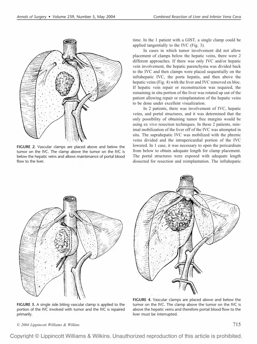
placed above and below the area of tumor involvement (Fig.2). The portion of liver and involved IVC was then removed,allowing improved access for reconstruction of the IVC. Theplacing of clamps on the IVC below the hepatic veins allowedperfusion of the liver and minimized the hepatic ischemic
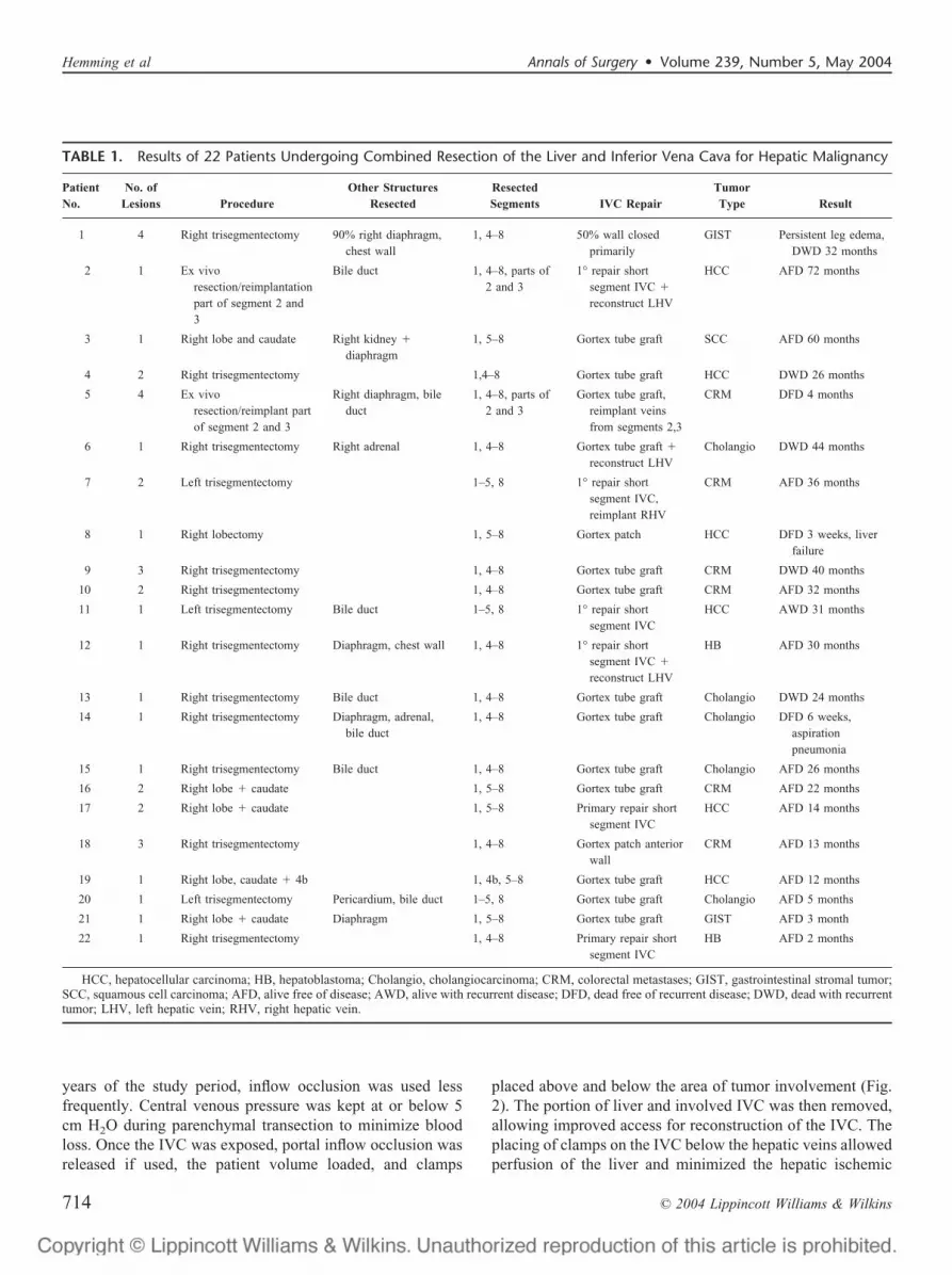
TABLE 1. Results of 22 Patients Undergoing Combined Resection of the Liver and Inferior Vena Cava for Hepatic Malignancy
PatientNo.
No. ofLesions Procedure
Other StructuresResected
ResectedSegments IVC Repair
TumorType Result
1 4 Right trisegmentectomy 90% right diaphragm,chest wall
1, 4–8 50% wall closedprimarily
GIST Persistent leg edema,DWD 32 months
2 1 Ex vivoresection/reimplantationpart of segment 2 and3
Bile duct 1, 4–8, parts of2 and 3
1° repair shortsegment IVC �
reconstruct LHV
HCC AFD 72 months
3 1 Right lobe and caudate Right kidney �
diaphragm1, 5–8 Gortex tube graft SCC AFD 60 months
4 2 Right trisegmentectomy 1,4–8 Gortex tube graft HCC DWD 26 months
5 4 Ex vivoresection/reimplant partof segment 2 and 3
Right diaphragm, bileduct
1, 4–8, parts of2 and 3
Gortex tube graft,reimplant veinsfrom segments 2,3
CRM DFD 4 months
6 1 Right trisegmentectomy Right adrenal 1, 4–8 Gortex tube graft �
reconstruct LHVCholangio DWD 44 months
7 2 Left trisegmentectomy 1–5, 8 1° repair shortsegment IVC,reimplant RHV
CRM AFD 36 months
8 1 Right lobectomy 1, 5–8 Gortex patch HCC DFD 3 weeks, liverfailure
9 3 Right trisegmentectomy 1, 4–8 Gortex tube graft CRM DWD 40 months
10 2 Right trisegmentectomy 1, 4–8 Gortex tube graft CRM AFD 32 months
11 1 Left trisegmentectomy Bile duct 1–5, 8 1° repair shortsegment IVC
HCC AWD 31 months
12 1 Right trisegmentectomy Diaphragm, chest wall 1, 4–8 1° repair shortsegment IVC �
reconstruct LHV
HB AFD 30 months
13 1 Right trisegmentectomy Bile duct 1, 4–8 Gortex tube graft Cholangio DWD 24 months
14 1 Right trisegmentectomy Diaphragm, adrenal,bile duct
1, 4–8 Gortex tube graft Cholangio DFD 6 weeks,aspirationpneumonia
15 1 Right trisegmentectomy Bile duct 1, 4–8 Gortex tube graft Cholangio AFD 26 months
16 2 Right lobe � caudate 1, 5–8 Gortex tube graft CRM AFD 22 months
17 2 Right lobe � caudate 1, 5–8 Primary repair shortsegment IVC
HCC AFD 14 months
18 3 Right trisegmentectomy 1, 4–8 Gortex patch anteriorwall
CRM AFD 13 months
19 1 Right lobe, caudate � 4b 1, 4b, 5–8 Gortex tube graft HCC AFD 12 months
20 1 Left trisegmentectomy Pericardium, bile duct 1–5, 8 Gortex tube graft Cholangio AFD 5 months
21 1 Right lobe � caudate Diaphragm 1, 5–8 Gortex tube graft GIST AFD 3 month
22 1 Right trisegmentectomy 1, 4–8 Primary repair shortsegment IVC
HB AFD 2 months
HCC, hepatocellular carcinoma; HB, hepatoblastoma; Cholangio, cholangiocarcinoma; CRM, colorectal metastases; GIST, gastrointestinal stromal tumor;SCC, squamous cell carcinoma; AFD, alive free of disease; AWD, alive with recurrent disease; DFD, dead free of recurrent disease; DWD, dead with recurrenttumor; LHV, left hepatic vein; RHV, right hepatic vein.
Hemming et al Annals of Surgery • Volume 239, Number 5, May 2004
© 2004 Lippincott Williams & Wilkins714
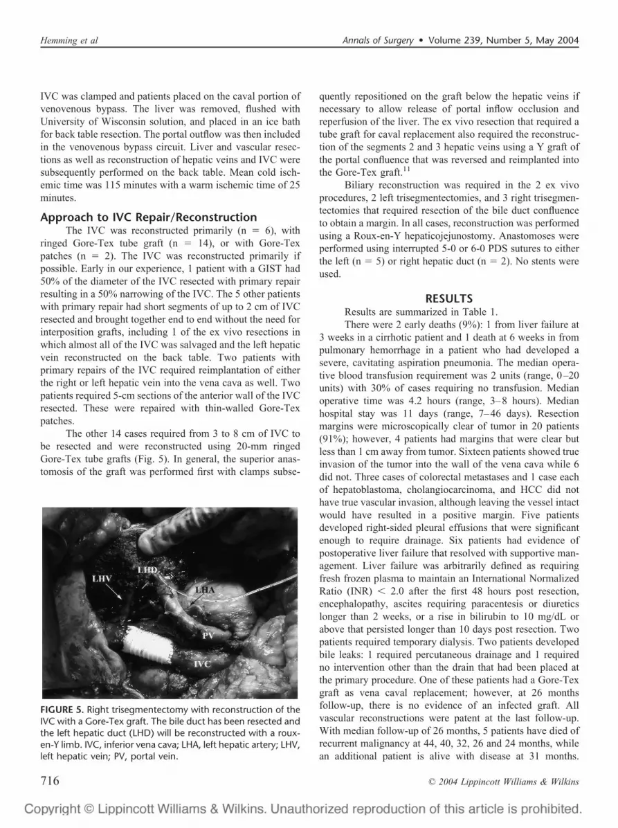
time. In the 1 patient with a GIST, a single clamp could beapplied tangentially to the IVC (Fig. 3).
In cases in which tumor involvement did not allowplacement of clamps below the hepatic veins, there were 2different approaches. If there was only IVC and/or hepaticvein involvement, the hepatic parenchyma was divided backto the IVC and then clamps were placed sequentially on theinfrahepatic IVC, the porta hepatis, and then above thehepatic veins (Fig. 4) with the liver and IVC removed en bloc.If hepatic vein repair or reconstruction was required, theremaining in situ portion of the liver was rotated up out of thepatient allowing repair or reimplantation of the hepatic veinsto be done under excellent visualization.
In 2 patients, there was involvement of IVC, hepaticveins, and portal structures, and it was determined that theonly possibility of obtaining tumor free margins would beusing ex vivo resection techniques. In these 2 patients, min-imal mobilization of the liver off of the IVC was attempted insitu. The suprahepatic IVC was mobilized with the phrenicveins divided and the intrapericardial portion of the IVClowered. In 1 case, it was necessary to open the pericardiumfrom below to obtain adequate length for clamp placement.The portal structures were exposed with adequate lengthdissected for resection and reimplantation. The infrahepatic
FIGURE 2. Vascular clamps are placed above and below thetumor on the IVC. The clamp above the tumor on the IVC isbelow the hepatic veins and allows maintenance of portal bloodflow to the liver.
FIGURE 3. A single side biting vascular clamp is applied to theportion of the IVC involved with tumor and the IVC is repairedprimarily.
FIGURE 4. Vascular clamps are placed above and below thetumor on the IVC. The clamp above the tumor on the IVC isabove the hepatic veins and therefore portal blood flow to theliver must be interrupted.
Annals of Surgery • Volume 239, Number 5, May 2004 Combined Resection of Liver and Inferior Vena Cava
© 2004 Lippincott Williams & Wilkins 715
IVC was clamped and patients placed on the caval portion ofvenovenous bypass. The liver was removed, flushed withUniversity of Wisconsin solution, and placed in an ice bathfor back table resection. The portal outflow was then includedin the venovenous bypass circuit. Liver and vascular resec-tions as well as reconstruction of hepatic veins and IVC weresubsequently performed on the back table. Mean cold isch-emic time was 115 minutes with a warm ischemic time of 25minutes.
Approach to IVC Repair/ReconstructionThe IVC was reconstructed primarily (n � 6), with
ringed Gore-Tex tube graft (n � 14), or with Gore-Texpatches (n � 2). The IVC was reconstructed primarily ifpossible. Early in our experience, 1 patient with a GIST had50% of the diameter of the IVC resected with primary repairresulting in a 50% narrowing of the IVC. The 5 other patientswith primary repair had short segments of up to 2 cm of IVCresected and brought together end to end without the need forinterposition grafts, including 1 of the ex vivo resections inwhich almost all of the IVC was salvaged and the left hepaticvein reconstructed on the back table. Two patients withprimary repairs of the IVC required reimplantation of eitherthe right or left hepatic vein into the vena cava as well. Twopatients required 5-cm sections of the anterior wall of the IVCresected. These were repaired with thin-walled Gore-Texpatches.
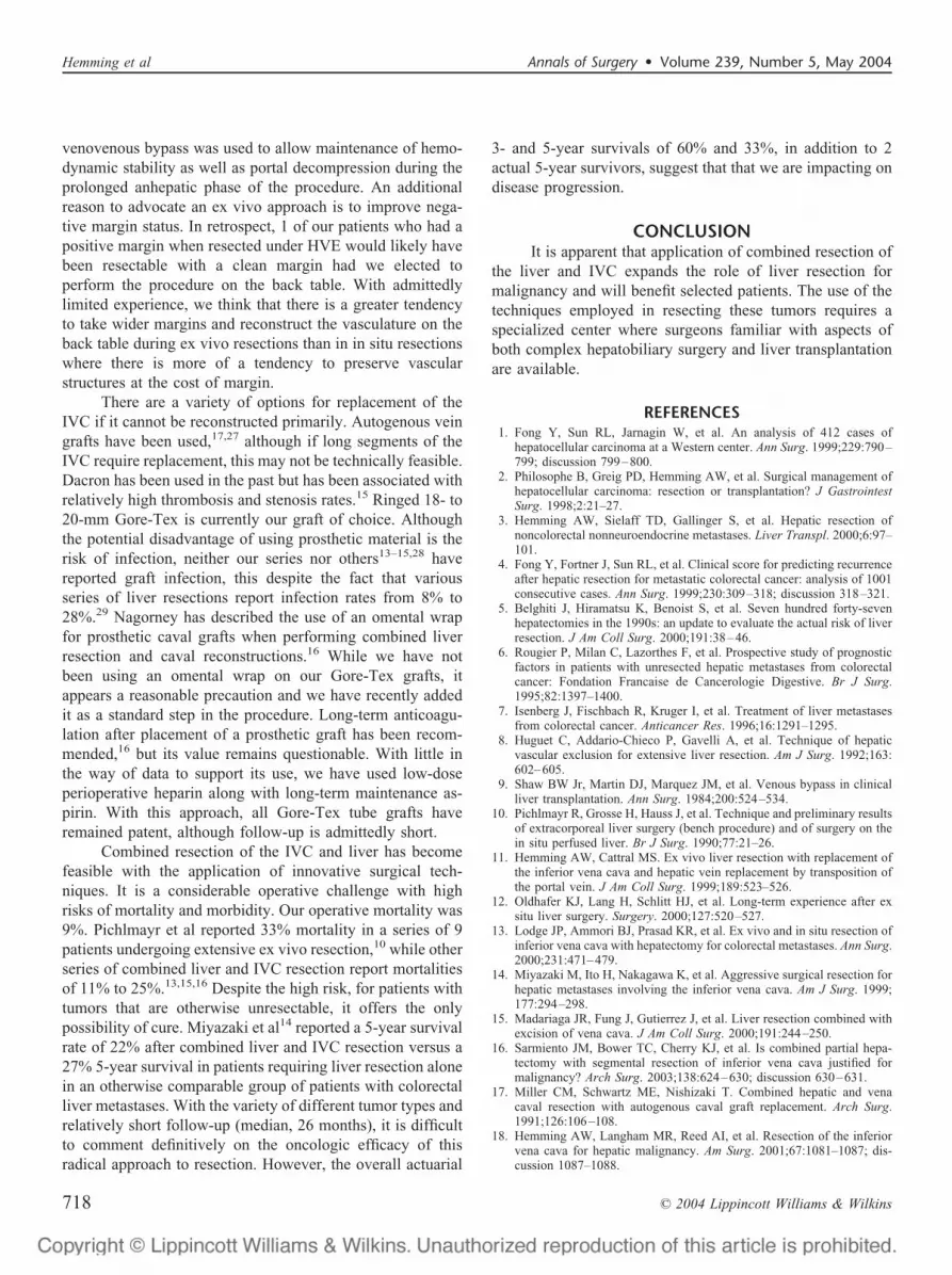
The other 14 cases required from 3 to 8 cm of IVC tobe resected and were reconstructed using 20-mm ringedGore-Tex tube grafts (Fig. 5). In general, the superior anas-tomosis of the graft was performed first with clamps subse-
quently repositioned on the graft below the hepatic veins ifnecessary to allow release of portal inflow occlusion andreperfusion of the liver. The ex vivo resection that required atube graft for caval replacement also required the reconstruc-tion of the segments 2 and 3 hepatic veins using a Y graft ofthe portal confluence that was reversed and reimplanted intothe Gore-Tex graft.11
Biliary reconstruction was required in the 2 ex vivoprocedures, 2 left trisegmentectomies, and 3 right trisegmen-tectomies that required resection of the bile duct confluenceto obtain a margin. In all cases, reconstruction was performedusing a Roux-en-Y hepaticojejunostomy. Anastomoses wereperformed using interrupted 5-0 or 6-0 PDS sutures to eitherthe left (n � 5) or right hepatic duct (n � 2). No stents wereused.
RESULTSResults are summarized in Table 1.There were 2 early deaths (9%): 1 from liver failure at
3 weeks in a cirrhotic patient and 1 death at 6 weeks in frompulmonary hemorrhage in a patient who had developed asevere, cavitating aspiration pneumonia. The median opera-tive blood transfusion requirement was 2 units (range, 0–20units) with 30% of cases requiring no transfusion. Medianoperative time was 4.2 hours (range, 3–8 hours). Medianhospital stay was 11 days (range, 7–46 days). Resectionmargins were microscopically clear of tumor in 20 patients(91%); however, 4 patients had margins that were clear butless than 1 cm away from tumor. Sixteen patients showed trueinvasion of the tumor into the wall of the vena cava while 6did not. Three cases of colorectal metastases and 1 case eachof hepatoblastoma, cholangiocarcinoma, and HCC did nothave true vascular invasion, although leaving the vessel intactwould have resulted in a positive margin. Five patientsdeveloped right-sided pleural effusions that were significantenough to require drainage. Six patients had evidence ofpostoperative liver failure that resolved with supportive man-agement. Liver failure was arbitrarily defined as requiringfresh frozen plasma to maintain an International NormalizedRatio (INR) � 2.0 after the first 48 hours post resection,encephalopathy, ascites requiring paracentesis or diureticslonger than 2 weeks, or a rise in bilirubin to 10 mg/dL orabove that persisted longer than 10 days post resection. Twopatients required temporary dialysis. Two patients developedbile leaks: 1 required percutaneous drainage and 1 requiredno intervention other than the drain that had been placed atthe primary procedure. One of these patients had a Gore-Texgraft as vena caval replacement; however, at 26 monthsfollow-up, there is no evidence of an infected graft. Allvascular reconstructions were patent at the last follow-up.With median follow-up of 26 months, 5 patients have died ofrecurrent malignancy at 44, 40, 32, 26 and 24 months, whilean additional patient is alive with disease at 31 months.
FIGURE 5. Right trisegmentectomy with reconstruction of theIVC with a Gore-Tex graft. The bile duct has been resected andthe left hepatic duct (LHD) will be reconstructed with a roux-en-Y limb. IVC, inferior vena cava; LHA, left hepatic artery; LHV,left hepatic vein; PV, portal vein.
Hemming et al Annals of Surgery • Volume 239, Number 5, May 2004
© 2004 Lippincott Williams & Wilkins716

Actuarial 1-, 3-, and 5-year survivals were 85%, 60%, and33%, respectively (Fig. 6). The 1 patient with a GIST that hada 50% resection of the diameter of the IVC developedmoderate lower limb edema that eventually resolved 6months post procedure. Imaging studies demonstrated apatent but narrowed IVC. All other patients had patent IVC atthe last follow-up. All patients were placed on low-doseheparin perioperatively, and patients with Gore-Tex graftswere maintained long-term on a single aspirin daily.
DISCUSSIONResection of liver tumors that involve the vena cava has
become possible with lessons learned from liver transplanta-tion. Tumors in the central or posterior segments of the livermay extend to involve the vena cava or hepatic veins thatmake resection using standard techniques impossible. If thetumor involvement of the IVC is small, control of the IVCcan be achieved simply by placing a vascular clamp tangen-tial to the vena cava, as was done in patient 1 in the series.Although the IVC can then be repaired primarily with alateral venorrhaphy, this is feasible in only a small number ofpatients and care must be taken not to narrow the IVCexcessively. In our 1 patient who had this performed, the IVCwas narrowed by 50% and the patient developed persistentmoderate leg edema, although the IVC remained patent. Insubsequent cases, we have elected to completely transect theIVC along with resection and bring the IVC together end toend, eliminating the possibility of tangential narrowing.
Larger resections of the IVC require interruption ofIVC flow. When involvement of the IVC is below the hepaticveins and there is sufficient room to place a vascular clampabove the tumor but below the hepatic veins, then liver bloodflow can be maintained while resecting and reconstructing the
IVC. Although we prefer to divide the liver parenchyma firstwith subsequent placement of clamps on the IVC, an alter-native approach by Madariaga et al15 describes replacing theIVC prior to dividing the liver parenchyma. Either approachminimizes the time that portal inflow occlusion is required.Normal livers can tolerate 60 to 90 minutes of warm isch-emia21,22; however, it would seem prudent to minimize isch-emic time if possible. In our initial report,18 we used portalinflow occlusion during the parenchymal transection of theliver for the majority of cases. Experience gained in livedonor liver transplantation has altered our practice. Currently,we perform the parenchymal transection in a similar con-trolled fashion as during live donor liver surgery whilehepatic perfusion is maintained. While this increases the timeof the procedure, it minimizes the ischemic injury to whichthe remnant liver is exposed. In addition, we have beenapplying the principle of ischemic preconditioning23 to ourliver resections in an attempt to ameliorate the effects ofwarm ischemia if portal inflow occlusion is required.
If clamps must be placed above the hepatic veins on theIVC then complete hepatic vascular exclusion (HVE) must beused.24 HVE can result in a fair degree of hemodynamicinstability and requires volume loading to maintain cardiacoutput.25 Although all 11 patients who required HVE toler-ated the procedure, we were prepared to use venovenousbypass if it had been required and the availability of veno-venous bypass would seem essential from other reports.13,15
In general, we mobilize as much of the liver off of the venacava as possible prior to starting the transection of the hepaticparenchyma. However, occasionally a large bulky tumormakes the mobilization of the liver off of the vena cavadifficult or even hazardous, in which case the liver parenchy-mal transection can be performed first, allowing exposure ofthe IVC without excessive rotation and traction on the liver.20
The need for ex vivo resection should be rare since themajority of tumors can be resected with different, less tech-nically demanding techniques. The 2 cases that required exvivo resection had involvement of all 3 hepatic veins, the IVCas well as portal structures. If only the hepatic veins and IVCare involved, the portal structures can be left intact (thoughclamped), and the vena cava divided above and below thetumor, allowing the liver to be rotated up to the surface of theoperative field. This permits improved access for reconstruc-tion of the hepatic veins or reimplantation of the hepatic veinsinto the vena cava. Hannoun et al have described a techniquein which the liver, with portal structures intact, can be flushedvia a branch of the portal vein with cold University ofWisconsin solution to extend the ischemic time tolerated bythe liver.26 When complete ex vivo resection is used and theliver is flushed with preservation solution, the transection ofthe liver parenchyma and the reconstruction of vascularstructures take place in a bloodless field and can be donewithout time pressure. In both cases of ex vivo resection,
FIGURE 6. Actuarial survival of 22 patients undergoing com-bined resection of the liver and IVC for hepatic malignancy.
Annals of Surgery • Volume 239, Number 5, May 2004 Combined Resection of Liver and Inferior Vena Cava
© 2004 Lippincott Williams & Wilkins 717
venovenous bypass was used to allow maintenance of hemo-dynamic stability as well as portal decompression during theprolonged anhepatic phase of the procedure. An additionalreason to advocate an ex vivo approach is to improve nega-tive margin status. In retrospect, 1 of our patients who had apositive margin when resected under HVE would likely havebeen resectable with a clean margin had we elected toperform the procedure on the back table. With admittedlylimited experience, we think that there is a greater tendencyto take wider margins and reconstruct the vasculature on theback table during ex vivo resections than in in situ resectionswhere there is more of a tendency to preserve vascularstructures at the cost of margin.
There are a variety of options for replacement of theIVC if it cannot be reconstructed primarily. Autogenous veingrafts have been used,17,27 although if long segments of theIVC require replacement, this may not be technically feasible.Dacron has been used in the past but has been associated withrelatively high thrombosis and stenosis rates.15 Ringed 18- to20-mm Gore-Tex is currently our graft of choice. Althoughthe potential disadvantage of using prosthetic material is therisk of infection, neither our series nor others13–15,28 havereported graft infection, this despite the fact that variousseries of liver resections report infection rates from 8% to28%.29 Nagorney has described the use of an omental wrapfor prosthetic caval grafts when performing combined liverresection and caval reconstructions.16 While we have notbeen using an omental wrap on our Gore-Tex grafts, itappears a reasonable precaution and we have recently addedit as a standard step in the procedure. Long-term anticoagu-lation after placement of a prosthetic graft has been recom-mended,16 but its value remains questionable. With little inthe way of data to support its use, we have used low-doseperioperative heparin along with long-term maintenance as-pirin. With this approach, all Gore-Tex tube grafts haveremained patent, although follow-up is admittedly short.
Combined resection of the IVC and liver has becomefeasible with the application of innovative surgical tech-niques. It is a considerable operative challenge with highrisks of mortality and morbidity. Our operative mortality was9%. Pichlmayr et al reported 33% mortality in a series of 9patients undergoing extensive ex vivo resection,10 while otherseries of combined liver and IVC resection report mortalitiesof 11% to 25%.13,15,16 Despite the high risk, for patients withtumors that are otherwise unresectable, it offers the onlypossibility of cure. Miyazaki et al14 reported a 5-year survivalrate of 22% after combined liver and IVC resection versus a27% 5-year survival in patients requiring liver resection alonein an otherwise comparable group of patients with colorectalliver metastases. With the variety of different tumor types andrelatively short follow-up (median, 26 months), it is difficultto comment definitively on the oncologic efficacy of thisradical approach to resection. However, the overall actuarial
3- and 5-year survivals of 60% and 33%, in addition to 2actual 5-year survivors, suggest that that we are impacting ondisease progression.
CONCLUSIONIt is apparent that application of combined resection of
the liver and IVC expands the role of liver resection formalignancy and will benefit selected patients. The use of thetechniques employed in resecting these tumors requires aspecialized center where surgeons familiar with aspects ofboth complex hepatobiliary surgery and liver transplantationare available.
REFERENCES1. Fong Y, Sun RL, Jarnagin W, et al. An analysis of 412 cases of
hepatocellular carcinoma at a Western center. Ann Surg. 1999;229:790–799; discussion 799–800.
2. Philosophe B, Greig PD, Hemming AW, et al. Surgical management ofhepatocellular carcinoma: resection or transplantation? J GastrointestSurg. 1998;2:21–27.
3. Hemming AW, Sielaff TD, Gallinger S, et al. Hepatic resection ofnoncolorectal nonneuroendocrine metastases. Liver Transpl. 2000;6:97–101.
4. Fong Y, Fortner J, Sun RL, et al. Clinical score for predicting recurrenceafter hepatic resection for metastatic colorectal cancer: analysis of 1001consecutive cases. Ann Surg. 1999;230:309–318; discussion 318–321.
5. Belghiti J, Hiramatsu K, Benoist S, et al. Seven hundred forty-sevenhepatectomies in the 1990s: an update to evaluate the actual risk of liverresection. J Am Coll Surg. 2000;191:38–46.
6. Rougier P, Milan C, Lazorthes F, et al. Prospective study of prognosticfactors in patients with unresected hepatic metastases from colorectalcancer: Fondation Francaise de Cancerologie Digestive. Br J Surg.1995;82:1397–1400.
7. Isenberg J, Fischbach R, Kruger I, et al. Treatment of liver metastasesfrom colorectal cancer. Anticancer Res. 1996;16:1291–1295.
8. Huguet C, Addario-Chieco P, Gavelli A, et al. Technique of hepaticvascular exclusion for extensive liver resection. Am J Surg. 1992;163:602–605.
9. Shaw BW Jr, Martin DJ, Marquez JM, et al. Venous bypass in clinicalliver transplantation. Ann Surg. 1984;200:524–534.
10. Pichlmayr R, Grosse H, Hauss J, et al. Technique and preliminary resultsof extracorporeal liver surgery (bench procedure) and of surgery on thein situ perfused liver. Br J Surg. 1990;77:21–26.
11. Hemming AW, Cattral MS. Ex vivo liver resection with replacement ofthe inferior vena cava and hepatic vein replacement by transposition ofthe portal vein. J Am Coll Surg. 1999;189:523–526.
12. Oldhafer KJ, Lang H, Schlitt HJ, et al. Long-term experience after exsitu liver surgery. Surgery. 2000;127:520–527.
13. Lodge JP, Ammori BJ, Prasad KR, et al. Ex vivo and in situ resection ofinferior vena cava with hepatectomy for colorectal metastases. Ann Surg.2000;231:471–479.
14. Miyazaki M, Ito H, Nakagawa K, et al. Aggressive surgical resection forhepatic metastases involving the inferior vena cava. Am J Surg. 1999;177:294–298.
15. Madariaga JR, Fung J, Gutierrez J, et al. Liver resection combined withexcision of vena cava. J Am Coll Surg. 2000;191:244–250.
16. Sarmiento JM, Bower TC, Cherry KJ, et al. Is combined partial hepa-tectomy with segmental resection of inferior vena cava justified formalignancy? Arch Surg. 2003;138:624–630; discussion 630–631.
17. Miller CM, Schwartz ME, Nishizaki T. Combined hepatic and venacaval resection with autogenous caval graft replacement. Arch Surg.1991;126:106–108.
18. Hemming AW, Langham MR, Reed AI, et al. Resection of the inferiorvena cava for hepatic malignancy. Am Surg. 2001;67:1081–1087; dis-cussion 1087–1088.
Hemming et al Annals of Surgery • Volume 239, Number 5, May 2004
© 2004 Lippincott Williams & Wilkins718
19. Hemming AW, Reed AI, Howard RJ, et al. Preoperative portal veinembolization for extended hepatectomy. Ann Surg. 2003;237:686–691;discussion 691–693.
20. Liu CL, Fan ST, Lo CM, et al. Anterior approach for major right hepaticresection for large hepatocellular carcinoma. Ann Surg. 2000;232:25–31.
21. Huguet C, Gavelli A, Bona S. Hepatic resection with ischemia of theliver exceeding one hour. J Am Coll Surg. 1994;178:454–458.
22. Delva E, Camus Y, Nordlinger B, et al. Vascular occlusions for liverresections. Operative management and tolerance to hepatic ischemia:142 cases. Ann Surg. 1989;209:211–218.
23. Clavien PA, Yadav S, Sindram D, et al. Protective effects of ischemicpreconditioning for liver resection performed under inflow occlusion inhumans. Ann Surg. 2000;232:155–162.
24. Huguet C, Gavelli A. Total vascular exclusion for liver resection.Hepatogastroenterology. 1998;45:368–369.
25. Delva E, Barberousse JP, Nordlinger B, et al. Hemodynamic andbiochemical monitoring during major liver resection with use of hepaticvascular exclusion. Surgery. 1984;95:309–318.
26. Hannoun L, Delriviere L, Gibbs P, et al. Major extended hepaticresections in diseased livers using hypothermic protection: preliminaryresults from the first 12 patients treated with this new technique. J AmColl Surg. 1996;183:597–605.
27. Yamamoto H, Hayakawa N, Ogawa A, et al. Segmental resection andreconstruction of the inferior vena cava with an autogenous vein graft.Br J Surg. 1997;84:51.
28. Arii S, Teramoto K, Kawamura T, et al. Significance of hepatic resectioncombined with inferior vena cava resection and its reconstruction withexpanded polytetrafluoroethylene for treatment of liver tumors. J AmColl Surg. 2003;196:243–249.
29. Andersson R, Saarela A, Tranberg KG, et al. Intraabdominal abscessformation after major liver resection. Acta Chir Scand. 1990;156:707–710.
DiscussionsDR. YUMAN FONG (New York, New York): Liver resec-
tion has come a long way in the last 2 decades and lobecto-mies and trisegmentectomies are now commonly performedin many hospitals throughout this world. In this regard Icongratulate Dr. Hemming and his colleagues for pushing theenvelope further in their report indicating that combined liverand vena caval resection can be performed safely and withgood mid- and long-term results.
The marked recent improvement in outcome for pa-tients after liver resection however are due to a combinationof improved patient selection and technical advances. So myquestions are therefore separated into 2 categories: thoserelated to how the authors chose the patients for surgery andthose related to the technical aspects of doing the surgery.
With regard to patient selection, I note in this series allpatients had fewer than 5 tumors. My first question thereforeis: Were all patients with tumors completely resectable byliver and caval resection subjected to resection or were thereother biological selection criteria for choosing these patients?In particular, were patients that have the greatest chance forlong-term survival chosen for these extensive operations?
In my own practice, I am much more enthusiastic aboutsubjecting someone to an extensive procedure who is re-sponding to neoadjuvant chemotherapy. Was neoadjuvant
therapy part of the overall strategy in any of the patients in theseries?
I note that there were 6 patients with hepatocellularcarcinoma in the series and at least one of them had cirrhosis.How many others had cirrhosis? How do the authors choosepatients with cirrhosis for these extensive procedures andwhat are the technical differences in performing vena cavalreconstruction in these cirrhotic patients?
Finally, I note with surprise that there was a singlepatient with squamous cell carcinoma and the fact that he iscurrently a long-term survivor. Was this a patient with pri-mary squamous cell carcinoma of the gallbladder? If not,what squamous cell carcinoma metastasized to the liver in asingle spot and how was this patient chosen for surgery?
As for the technical issues, I have found that bovinepericardium actually works pretty well as a patching materialfor vena cava. Do the authors have any experience with this?
In the manuscript the authors emphasize that they nolonger use inflow occlusion during these resections in order todecrease ischemic damage to the liver. Do the authors havedata supporting this as an improvement?
Finally, the authors state that 7 of the last 11 patients inthe series were also subjected to a portal vein embolizationbefore surgery. Is this because of the planned caval resectionor are all patients at the University of Florida being consid-ered for major liver resection now being subjected to portalvein embolization?
DR. JEAN-NICOLAS VAUTHEY (Houston, Texas): I rise tocongratulate Dr. Hemming and his colleagues at the Univer-sity of Florida for an effort to expand the option of surgicalresection for a group of patients with advanced hepatobiliarymalignancies with otherwise limited life expectancy. Theresults are excellent.
The authors used 14 ringed Gore-Text grafts withoutthrombosis after a median follow-up of 28 months. Five ofthese resections were combined with bile duct resection in acontaminated field and no graft infection was reported.
The negative margin rate is 91%, and this translates ina survival at 3 years of 63%. And there is not early drop in thesurvival curve, witnessing good patient selection. I have 4questions for the authors.
The first question relates to the anticoagulation. Youmention in your manuscript that you use low dose heparinpostoperatively in these patients followed by aspirin. How doyou manage these patients intraoperatively? Do you reverseyour heparinization? Also, how do you adjust your low doseheparin? Since in the first 2 days following major liverresection there is always a rise in INR.
The second question is about hepatic vascular exclu-sion. I note that you have only used veno-venous bypass in 2patients who had ex-vivo resection. That means in 11 patientsyou used total vascular exclusion without veno-venous by-
Annals of Surgery • Volume 239, Number 5, May 2004 Combined Resection of Liver and Inferior Vena Cava
© 2004 Lippincott Williams & Wilkins 719

pass while accepting the attendant hemodynamic shifts.There is a randomized study on major hepatic resection withand without hepatic vascular exclusing showing an increasein complication and transfusions in the absence of veno-venous bypass. Do you think a comparison of total vascularexclusion with or without venous bypass is indicated and thatyou might have had less transfusion requirements and lessinfusions by using veno-venous bypass?
The third question relates to the involvement of thevena cava. Although we would agree that greater than 50%involvement of the vena cava requires resection of the venacava, how do you decide to resect the vena cava in patientswho have less than 50% involvement? You mention 3-Dvolumetric CT reconstruction in the evaluation of your pa-tients. How does this compare to thin-cut CTs without 3-Dreconstruction?
Finally, you had several hepatocellular carcinomas inthis series. This tumor is notorious for invading the hepaticvein and the vena cava as tumor thrombose without adhesionto the hepatic vein wall or vena cava wall. Have you used thetechnique recently described as reduction of the tumor throm-bose and resection via cavatomy without vascular resection inthese patients? Did your patients have pathological involve-ment of the vena cava?
DR. ROBERT MARTIN (Louisville, Kentucky): I congrat-ulate the authors on a very impressive series. I have just 2simple questions. What was the median and range time forclamps with both techniques? Did any of these patients haveto undergo a partial aortic occlusion in order to maintainblood pressure?
DR. J. ALEX HALLER, JR. (Baltimore, Maryland): I havejust one additional question to extend one of the discussant’squestions. This has to do with the use of veno-venous bypass.It seems to me that this would become a standardized ap-proach to the management of all of these tumors. I realize thatit requires heparinization to do that, but what is the reason forso many fantastic, innovative technical maneuvers that wereused rather than adopting a straightforward standard perfu-sion technique for all of these patients?
DR. REID ADAMS (Charlottesville, Virginia): A very niceseries demonstrating again the extension of vascular tech-niques along with liver resection to improve the survival forthese patients. In determining your operative candidates,could you elaborate a little bit again on what Dr. Fongmentioned? That is, how do you decide who you are going toembolize? Secondly, how are you determining who you aregoing to resect? Are you doing volumetrics as your primaryendpoint for liver remnant?
DR. ALAN W. HEMMING (Gainesville, Florida): Dr.Fong, you asked regarding the criteria for patient selection,how did we choose which patients we were going to beaggressive on? We clearly don’t subject every patient whohas caval involvement to this operation. If you look at ourmedian age, it was 49, although we had a few patients whowe resected out to age 68. These are by and large youngerpatients with longer disease-free intervals, at least with colo-rectal metastases, and with what at least appears to us to beless aggressive or at least localized disease.
The question about whether we use neoadjuvant ther-apy. Over the time course of the study, from ’97 to currently,we did not use neoadjuvant therapy from about 1997–2001.In the last 2 years we have begun using neoadjuvant chemo-therapy. However, in this series only 3 patients had neoad-juvant therapy. We didn’t particularly use a response totherapy to dictate whether or not we were going to proceedwith resection. There was 1 patient who had a response thatwe resected, but we would have resected him if he had notresponded simply because there is no other curative optionavailable.
I think it makes me happier when I see patients respondto the neoadjuvant therapy, since big surgery does not beatthe biology of the disease all the time. We need somethingelse. So if we have additional chemotherapy that we have ahint is going to work, it makes me happier about performingan extensive procedure such as this.
How do we choose the cirrhotics to do this on? Ingeneral, I don’t plan on doing very many of these in cirrhot-ics. I think there is only one cirrhotic in the series. And I cantell you I wasn’t planning on taking his vena cava out at thetime, but it became obvious during the procedure that itwould be required. That patient needed a right lobectomy, sohe had a fairly large future remnant liver volume. In generalI wouldn’t plan vascular reconstructions on a cirrhotic with-out a few other additional maneuvers such as portal veinembolization, assessing their volumetrics, and assessing theirICG clearance.
The squamous cell carcinoma. I was called into theoperating room in the middle of that one. A kidney and partof the liver was already in the process of being removed whenit became evident that the inferior vena cava needed to comeout. So we took out the liver, the kidney, and the vena cava.We have no idea where it came from. It wasn’t gallbladdercancer. The patient was still alive at last follow-up with noevidence of disease. Our urologists had actually preopera-tively beta blocked and alpha blocked the patient thinking itwas a pheochromocytoma. If you have ever tried to take avena cava out on somebody who is alpha blocked and betablocked, it is not easy to maintain their blood pressure withclamps on the vena cava. Bovine pericardium certainly can beused to patch vena cavas. I haven’t used it particularly. Thereis no reason not to use it however.
Hemming et al Annals of Surgery • Volume 239, Number 5, May 2004
© 2004 Lippincott Williams & Wilkins720
Inflow occlusion. I wouldn’t say that we don’t useinflow occlusion, because we do. We use it if we need it. Ifwe are having ongoing blood loss as we come through theliver, I have no problems using inflow occlusion. I think itreduces blood loss and makes the technical aspects of theoperation frequently more easy. But as we gain more expe-rience with live donor transplantation the ability to comethrough a liver under complete control and have no blood lossallows us to save any ischemic injury we might give the liveruntil we need it for vascular reconstruction therefore mini-mizing ischemic time.
Do we do portal vein embolization on all patients? No,we do not. We use a similar cut-off as most groups. In fact,we presented a portal vein embolization paper here at theSouthern Surgical last year.
Our criteria are: If we think that the future liver remnantis going to be less than 25% of total liver volume based onpreoperative 3-D CT, then we will portal vein embolize theside that is coming out so that we can get growth on the sidethat is staying in. Once you are talking about caval recon-struction and other vascular reconstruction, I think you aregiving a bigger potential injury to the liver. In cases where weplan vascular reconstructions, if we are going to leave lessthan 40% of the liver behind, then we use portal veinembolization.
Dr. Vauthey asked about the anticoagulation. Low-doseheparin just means we were giving routine perioperativeheparin. We weren’t monitoring the PTT. We didn’t eitherheparinize patients or reverse them intraoperatively, and theyreceived aspirin starting on about postoperative day 3.
The question as to whether if we use veno-venousbypass would we have less blood loss than total hepaticvascular exclusion. If you have used veno-venous bypass,you know you have a certain amount of blood loss just toprime the circuit. In fact, I think many times we wouldactually have lost more blood in the circuit than we wouldhave by just going ahead with total vascular exclusion. Theexclusion is done at the tail end of the procedure, so we havealready divided the liver. Most of the potential bleeding hasalready been dealt with. The liver is divided under low CVPconditions and complete vascular exclusion is only donewhen we are resecting the cava. So to try and compareprevious series where you are doing the whole liver resectionunder total vascular exclusion to this is like comparing applesand oranges.
How do we decide on who needs a caval resection?That is a tough one. Because 3 dimensional CT or thin cut CTfrequently show you compression or involvement of the cava
but many times when you actually take the vena cava out youwon’t see actual invasion into the wall of the vena cava. Inthis series about two-thirds of patients had some evidence ofinvolvement right through into the wall of the vessel. Two or3 patients had invasion through right into the endothelium.About a third of patients had no real involvement with thevena cava. We would have cracked into the tumor plane hadwe tried to get the tumor off of the vena cava but the tumorwasn’t necessarily involving the cava.
Preoperative imaging is not great for deciding exactlywho has caval involvement, unless you see tumor within thelumen of the vena cava. Patients who have tumor within thelumen of the vena cava are not good candidates for thisprocedure. And that goes to one of the other questions aboutvena cava resections for HCC with tumor thrombus. By andlarge we don’t resect vena cava. We just open the vena cava,pull the tumor thrombus out and repair the vena cava. Thistype of patient is not included in this series.
Dr. Martin asked about clamp times. I am not sureexactly what you mean, whether you mean on the veno-venous bypass, on the ex-vivo resections or on the totalvascular exclusion cases. Total vascular exclusion cases wewould clamp for between 15 to 20 minutes, really not verylong. The 2 patients that were on veno-venous bypass, I thinkthe cold ischemic time was around 110 minutes and anadditional warm ischemic time of about 20 minutes. On thepatients with total vascular isolation, we don’t clamp theaorta. We have found we haven’t needed it. You do need tovolume load these patients. Let your anesthesiologist knowthat you are about to clamp the vena cava and that they needto volume load before you put your clamps on. There shouldbe no need to clamp the aorta.
The last question was about veno-venous bypass andheparinization. Actually, when you are on veno-venous by-pass you are not heparinized. It is a heparin-bonded circuit.The whole idea behind veno-venous bypass is to not beheparinized, so that you avoid full heparinization in standardcardiopulmonary bypass.
Dr. Adams asked about who gets embolized. I think weanswered that a bit. The patients who I am going to dovascular resections on who have a volume of future liverremnant of 40% or less we try and embolize. And we do do3-D CT volumetry beforehand. There are scattered within thisseries patients that we didn’t plan on doing the vascularreconstructions on, and so you end up doing vascular recon-struction without pre-op embolization. So we don’t alwaysfollow what sound like strict protocols.
Annals of Surgery • Volume 239, Number 5, May 2004 Combined Resection of Liver and Inferior Vena Cava
© 2004 Lippincott Williams & Wilkins 721


