Embed Size (px)
Citation preview
Int. J. Mol. Sci. 2014, 15, 17644-17666; doi:10.3390/ijms151017644
International Journal of
Molecular Sciences ISSN 1422-0067
www.mdpi.com/journal/ijms
Review
Circulating RNA Molecules as Biomarkers in Liver Disease
Liviu S. Enache 1,2,3,*, Elena L. Enache 2, Christophe Ramière 1,4,5,6,7,8, Olivier Diaz 1,4,5,6,7,
Ligia Bancu 2,3, Anca Sin 2,3 and Patrice André 1,4,5,6,7,8
1 Université de Lyon, Université Lyon 1, Lyon F-69008, France;
E-Mails: [email protected] (C.R.); [email protected] (O.D.);
[email protected] (P.A.) 2 University of Medicine and Pharmacy Tirgu Mures, 38 Gh. Marinescu st., Tirgu Mures 540142,
Romania; E-Mails: [email protected] (E.L.E.); [email protected] (L.B.);
[email protected] (A.S.) 3 Emergency County Clinical Hospital, 50 Gh. Marinescu st.,
Tirgu Mures 540136, Romania 4 Inserm U1111, 21 Avenue Tony Garnier, Lyon F-69007, France 5 CIRI, Centre International de Recherche en Infectiologie, Université de Lyon,
21 Avenue Tony Garnier, 69365 Lyon Cedex 07, France 6 Ecole Normale Supérieure de Lyon, 15 parvis René Descartes, BP 7000 69342 Lyon Cedex 07, France 7 CNRS, UMR5308, 21 avenue Tony Garnier, 69365 Lyon Cedex 07, France 8 Hospices Civils de Lyon, Hôpital de la Croix Rousse, Laboratoire de Virologie, Lyon F-69004, France
* Author to whom correspondence should be addressed; E-Mail: [email protected];
Tel.: +40-772-074-967; Fax: +40-265-217-425.
External editor: Camile S. Farah
Received: 23 August 2014; in revised form: 4 September 2014 / Accepted: 17 September 2014 /
Published: 30 September 2014
Abstract: Liver disease is a major cause of morbidity and mortality worldwide. As in other
fields of medicine, there is a stringent need for non-invasive markers to improve patient
diagnostics, monitoring and prognostic ability in liver pathology. Cell-free circulating RNA
molecules have been recently acknowledged as an important source of potential medical
biomarkers. However, many aspects related to the biology of these molecules remain to be
elucidated. In this review, we summarize current concepts related to the origin, transportation
and possible functions of cell-free RNA. We outline current development of extracellular
RNA-based biomarkers in the main forms of non-inherited liver disease: chronic viral
OPEN ACCESS
Int. J. Mol. Sci. 2014, 15 17645
hepatitis, hepatocellular carcinoma, non-alcoholic fatty liver, hepato-toxicity, and liver
transplantation. Despite recent technological advances, the lack of standardization in the
assessment of these markers makes their adoption into clinical practice difficult. We thus
finally review the main factors influencing quantification of circulating RNA. These factors
should be considered in the reporting and interpretation of current findings, as well as in the
proper planning of future studies, to improve reliability and reproducibility of results.
Keywords: liver disease; biomarker; cell-free RNA; miRNA; diagnostic; preanalytical
variable
1. Introduction
Liver disease is a significant burden for the public health system worldwide. For instance, hepatitis
B virus (HBV) infection has affected approximately one third of the world’s population, and, at
present, there are up to 400 million HBV surface antigen carriers worldwide [1]. There are also
approximately 160 million persons chronically infected with hepatitis C virus (HCV) [2]. Both forms
of chronic viral hepatitis are associated with development of liver cirrhosis and hepatocellular
carcinoma (HCC). Indeed, approximately 80% of cases of hepatocellular carcinoma are associated
with chronic HBV or HCV infections [3]. Liver cancer is the sixth most common cancer and the third
cause of cancer-related death [4]. Due to difficulties in the management of these conditions, there is a
stringent need for informative markers, that can facilitate early diagnostic, accurate prognostic and
treatment monitoring in liver disease.
The presence of cell-free nucleic acids in plasma and serum has been acknowledged since the
late 1940s. Later, fetal mRNA was found to be detectable in plasma of pregnant women. Specific
circulating mRNA sequences have also been described in patients with cancer, cell and organ
transplantation, coronary heart disease, stroke, sepsis, burns, and in several other medical fields [5].
Recent technical advances have enabled detection of hundreds of RNA sequences in the extracellular
environment of healthy individuals [6].
In the context of an ever-increasing need for noninvasive molecular markers in medicine, circulating
RNA molecules have become appealing biomarker candidates in liver disease. However, many aspects
related to their origin and biological significance, remain to be clarified. Moreover, a long-acknowledged
heterogeneity of technical methods employed in the assessment and interpretation of circulating RNA
levels imposes a more comprehensive standardization and harmonization of biological assays.
The aim of this review is to highlight recent advances in the study of circulating RNA molecules as
biomarkers in liver pathology, and to provide a short overview on the biological properties of these
molecules and the analytical challenges in their assessment.
2. Circulating RNA: Sources and Transportation
The presence of nucleic acids, particularly RNA, in body fluids was rather surprising, given the
high amounts of nucleases in the extracellular environment. Whereas the addition of purified RNA to
Int. J. Mol. Sci. 2014, 15 17646
blood or plasma results in its immediate degradation [7], endogenous RNA is stable for several
hours in plasma at room temperature. A hybridization of circulating RNA with DNA molecules was
proposed as an explanation for these observations. However, the addition of RNase-H to plasma does
not affect RNA recovery, thus excluding the RNA-DNA hybrid hypothesis. Instead, an association of
cell-free RNA with lipids, either in the form of vesicles or lipoproteins, has been suggested. Indeed,
the degradation of endogenous RNA after detergent addition to plasma samples and the retention of
most of the circulating mRNA by 0.22 µm filters, support this theory [8].
2.1. Sources of Circulating RNA
Multiple mechanisms are involved in the release of RNA from cells. Among passive processes,
RNA leakage during cellular necrosis has been cited [9]. Following cell death, RNA can reach
extracellular environment bound to and protected by sub-cellular structures. RNA release via apoptotic
bodies and microvesicles has been described among active processes.
Apoptosis is a highly organized process culminating with the ordered disposal of cell structures.
During apoptosis, cellular RNA is packaged into granules and subsequently into apoptotic bodies,
separately from the DNA [10]. Although the uptake of apoptotic bodies by neighboring cells is
facilitated by exposure of phosphatidylserine on the outer leaflet of the membrane, a minority of these
vesicles reaches the circulation [9].
Viable cells are also able to release microvesicles in the extracellular environment, both in vivo and
in vitro. Microvesicles include a heterogeneous population of particles released as shedding vesicles and
exosomes, and are now acknowledged as a constitutive part of the intercellular environment. Shedding
vesicles are spherical structures with a diameter up to 200 nm, formed by the direct budding of the plasma
membrane which entraps a portion of the cytosolic content. The release of shedding vesicles is mostly
regulated and depends on the activation state of the source cells. Exosomes are microvesicles of
30–100 nm in diameter whose biogenesis begins with the endocytosis process, followed by the inward
budding of the endosome membrane, fission and segregation of vesicles inside the multivesicular bodies.
The fate of these structures via lysosomal degradation or exocytosis is dictated by specific processes.
Indeed, exosomes are released both in a constitutive and a regulated fashion [11].
Cells seem to differentially release mRNA into vesicles, depending on environmental conditions [12].
It is not clear yet how certain RNA sequences are specifically enriched in membrane-derived
microvesicles before secretion. Recently, Bolukbasi et al. [13] discovered a common pattern in the
structure of several microvesicle-enriched mRNAs. A stem-loop forming sequence of 25 nt, containing
a binding site for miR-1289 and a CTGCC core sequence, was common to enriched mRNAs secreted by
glioblastoma cells. Also, the enrichment of a reporter mRNA into secreted microvesicles depended on
miR-1289 expression in those cells.
2.2. Transport of Extracellular RNA Molecules
In addition to microvesicles, other forms of RNA transportation outside the cells have been
described. In fact, most of miRNAs in plasma are associated with proteins [14]. The main protein
transporter of plasma miRNA was identified as argonaute 2 (AGO2), the key effector protein of
miRNA-mediated silencing machinery [14,15]. AGO2-bound miRNAs show remarkable stability in
Int. J. Mol. Sci. 2014, 15 17647
the extracellular space, being detectable in culture media up to two months after cell death [15]. It is
thus possible that at least some of the AGO2-associated plasma miRNA originates from dead cells.
Other members of the AGO family, such as AGO1, AGO3 and AGO4, with different tissue
specificities, also seem to be associated with extracellular miRNA [15]. The lack of correlation
between extracellular miRNAs bound to AGO1 or AGO2 proteins suggests that these miRNAs have
different tissue origins [16].
MicroRNAs have a variate distribution in plasma, some of them being associated with proteins, others
with exosomes, and others present in both compartments. This distribution may reflect the heterogeneity
of the type and functionality of cells from which miRNAs originated. For example, the liver-specific
miR-122 was detected only in protein-associated fractions, suggesting a protein carrier-related mechanism
of release. On the other hand, miRNAs mainly associated with vesicles may be exported by cells adapted
to vesicle secretion, such as reticulocytes and platelets [14]. However, various types of tissue injury
differentially alter the abundance of miRNA molecules in circulatory compartments. For example,
in alcoholic liver disease and in inflammatory liver injury, miR-122 and miR-155 are mainly associated
with exosomes, whereas in drug-induced liver injury, these miRNAs predominate in the protein-rich
fraction. This suggests that miRNA distribution in circulatory compartments may provide further
specificity to the identification of mechanisms of liver pathology [17].
Other miRNAs, such as miR-223, are transported in plasma by high-density lipoproteins (HDL),
and their delivery to recipient cells depends on scavenger receptor class B type I (SR-BI) [18]. This
mechanism of transfer proved to be functional, directly altering gene expression in target cells.
SR-BI-mediated transfer may serve to direct HDL-bound miRNAs into the cytoplasm and avoid their
lysosomal degradation, thus increasing the chances that the message is delivered [18].
2.3. Circulating RNA: “Message in a Bottle”?
There is increasing evidence that RNA-carrying microvesicles produced by several cell types convey
specific messages to recipient cells. In vivo, exosomes can be taken up by macrophages and other cells, and
their RNA content is partially shuttled to the nuclei of recipient cells [19]. The active uptake of exosomes
from body fluids by target cells suggests the in vivo relevance of exosome-mediated transfer of RNA [20].
Exosome-borne mRNA was found to be translatable [20]. Moreover, the mRNA cargo of
microvesicles contains a specific subset of transcripts, rather than a random sample of the cellular
RNA content [21]. The interaction between microvesicles and target cells, and the consequent transfer
of genetic information, seems to be cell-specific. Thus, exosomes derived from MC/9 liver mast cells
are able to transfer RNA to other mast cells, but not to CD4 cells [20]. Microvesicles derived from
endothelial progenitor cells are functional both in vivo and in vitro, being able to induce an angiogenic
program to human endothelial cells, via horizontal transfer of mRNA [21]. Exosomes secreted by
cardiomyocytes were found to contain more than 1500 mRNA sequences. These vesicles can be taken
up by fibroblasts and induce expression changes in hundreds of genes [22].
Hepatocytes can be both a source and a target for microvesicle-mediated intercellular communication.
Hep3B hepatocarcinoma cells secrete microvesicles that differ in both RNA and protein content from
the producing cells, and these vesicles can transmit a functional transgene to other cells. Hepatocarcinoma
cells are able to reduce, via exosome-mediated transfer of miRNA, the expression of transforming
Int. J. Mol. Sci. 2014, 15 17648
growth factor beta activated kinase-1 (TAK1) in other cells. Since TAK1 is an essential inhibitor of
hepatocarcinogenesis, its downregulation may promote tumor progression [23]. On the other hand,
hepatoma cells can be targeted by microvesicles originating from human liver stem cells.
The CD29-mediated uptake of these microvesicles by hepatoma cells results in significant inhibition of
tumor cell growth and stimulation of their apoptosis both in vitro and in vivo. The anti-tumor effect of
stem cell-derived microvesicles appears to depend on the horizontal delivery of a specific set of
miRNAs. Since these miRNAs modulate signaling pathways differentially activated in cancer
compared with normal cells, it was supposed that the effect of vesicle-dependent miRNA delivery may
be specific to the functional state of the target cell rather than the gene expression of source cells [24].
Besides miRNA, other small RNA species can be exchanged between cells in a contact-independent
manner. As a possible therapeutic application, the ability of intercellular exchange of genetic material
to interfere with viral replication was tested in a HCV infection model [25]. Human and mouse liver
cells, as well as primary human B lymphocytes, appeared able to deliver small silencing RNA
targeting the HCV genome and the HCV receptor CD81. This transmission of siRNA was partially
mediated by exosomes [25].
Although RNA-mediated non-contact intercellular communication is an exciting emerging concept,
many aspects related to the physiopathological conditions leading to release of RNA from cells and the
signaling properties of these molecules are still to be described.
3. Circulating RNA as Biomarkers of Liver Injury
Ideally, circulating biomarkers of tissue injury should be expressed at high levels preferentially or
exclusively in the tissue of interest. They should also have low circulating levels in healthy individuals,
whereas important changes (usually increases) in their blood concentrations should be detectable upon
tissue injury [26]. Additionally, they should allow rapid, accurate and inexpensive detection, be invariant
to unrelated conditions and easily translatable from pre-clinical to clinical observations. Tissue-specific
transcripts possess several of these requisites [27]. Although rigorous validation is still necessary,
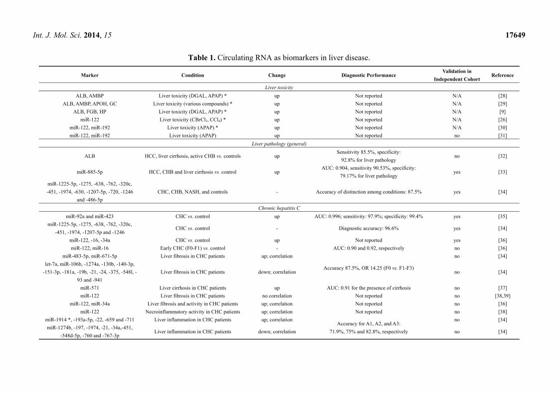
circulating RNAs appear as a promising source of biomarkers in various forms of liver disease (Table 1).
3.1. Liver Toxicity
Hepatocyte-specific RNA sequences including miR-122, albumin (ALB), microglobulin/bikunin
precursor, haptoglobin (HP), fibrinogen B β-polypeptide, apolipoprotein H and vitamin D binding
protein, were assessed as potential biomarkers in various animal models of liver toxicity [9,26,28–30].
Several classes of hepatotoxic compounds were tested, including CCl4 and CBrCl3, D-galactosamine,
and acetaminophen. A common finding of these studies is the increase of circulating liver-derived
RNAs in treated animals. Peak levels of specific RNAs are correlated with classical liver injury
markers, such as serum transaminase activities. Circulating RNAs rise in a time and dose-dependent
manner in response to liver injury. They also constitute a more sensitive marker of tissue damage, and
can be detected earlier than changes in alanine aminotransferase (ALT) levels, even before the onset of
observable histological modifications. These circulating RNAs are also more specific than serum
transaminases, since their levels are not influenced by injury affecting other tissues, including skeletal
muscle and brain.
Int. J. Mol. Sci. 2014, 15 17649
Table 1. Circulating RNA as biomarkers in liver disease.
Marker Condition Change Diagnostic Performance Validation in
Independent Cohort Reference
Liver toxicity
ALB, AMBP Liver toxicity (DGAL, APAP) * up Not reported N/A [28]
ALB, AMBP, APOH, GC Liver toxicity (various compounds) * up Not reported N/A [29]
ALB, FGB, HP Liver toxicity (DGAL, APAP) * up Not reported N/A [9]
miR-122 Liver toxicity (CBrCl3, CCl4) * up Not reported N/A [26]
miR-122, miR-192 Liver toxicity (APAP) * up Not reported N/A [30]
miR-122, miR-192 Liver toxicity (APAP) up Not reported no [31]
Liver pathology (general)
ALB HCC, liver cirrhosis, active CHB vs. controls up Sensitivity 85.5%, specificity:
92.8% for liver pathology no [32]
miR-885-5p HCC, CHB and liver cirrhosis vs. control up AUC: 0.904, sensitivity 90.53%, specificity:
79.17% for liver pathology yes [33]
miR-1225-5p, -1275, -638, -762, -320c,
-451, -1974, -630, -1207-5p, -720, -1246
and -486-5p
CHC, CHB, NASH, and controls - Accuracy of distinction among conditions: 87.5% yes [34]
Chronic hepatitis C
miR-92a and miR-423 CHC vs. control up AUC: 0.996; sensitivity: 97.9%; specificity: 99.4% yes [35]
miR-1225-5p, -1275, -638, -762, -320c,
-451, -1974, -1207-5p and -1246 CHC vs. control - Diagnostic accuracy: 96.6% yes [34]
miR-122, -16, -34a CHC vs. control up Not reported yes [36]
miR-122, miR-16 Early CHC (F0-F1) vs. control - AUC: 0.90 and 0.92, respectively no [36]
miR-483-5p, miR-671-5p Liver fibrosis in CHC patients up; correlation
Accuracy 87.5%, OR 14.25 (F0 vs. F1-F3)
no [34]
let-7a, miR-106b, -1274a, -130b, -140-3p,
-151-3p, -181a, -19b, -21, -24, -375, -548l, -
93 and -941
Liver fibrosis in CHC patients down; correlation no [34]
miR-571 Liver cirrhosis in CHC patients up AUC: 0.91 for the presence of cirrhosis no [37]
miR-122 Liver fibrosis in CHC patients no correlation Not reported no [38,39]
miR-122, miR-34a Liver fibrosis and activity in CHC patients up; correlation Not reported no [36]
miR-122 Necroinflammatory activity in CHC patients up; correlation Not reported no [38]
miR-1914 *, -193a-5p, -22, -659 and -711 Liver inflammation in CHC patients up; correlation Accuracy for A1, A2, and A3:
71.9%, 75% and 82.8%, respectively
no [34]
miR-1274b, -197, -1974, -21, -34a,-451,
-548d-5p, -760 and -767-3p Liver inflammation in CHC patients down; correlation no [34]

Int. J. Mol. Sci. 2014, 15 17650
Table 1. Cont.
Marker Condition Change Diagnostic Performance Validation in
Independent Cohort Reference
Chronic hepatitis B
miR-375, -92a, -10a, -223, -423, -23b/a,
-342-3p, -99a, -122a, -125b, -150 and let-7c CHB vs. control up Not reported yes [35]
miR-375, -10a, -223 and -423 CHB vs. control up AUC: 0.999; sensitivity: 99.3%; specificity: 98.8% yes [35]
miR-122 CHB vs. control up AUC: 0.989 yes [40]
miR-122 CHB (active) vs. control up AUC: 0.762 no [41]
ALB, APOA2, HP, CYP2E1 CHB (active) vs. control up AUC: 0.945, 0.909, 0.834, 0.801, respectively no [41]
miR-122, -638, -575, -572 and -744 CHB vs. control - CHB vs. healthy: AUC: 0.98, 1.00,
0.91, 0.95 and 0.95, respectively no [42]
miR-21, -122 and -223 CHB vs. control up CHB vs. healthy: AUC: 0.91,
0.93, and 0.88, respectively no [43]
miR-99a, -100, -122, -122 *, -125b, -192,
-192 *, -193b, -194, -215, -365, -455-5p,
-455-3p, -483-3p, -885-5p and -1247
CHB: HBeAg positive, HBeAg negative and
healthy children
up; HBeAg positive >
HBeAg negative >
healthy
Not reported no [44]
miR-99a-5p, -100-5p, -122-5p, -122-3p,
-125b-5p, 192-5p, -192-3p, -193b-3p,
-194-5p, -215, -365a-3p, -455-5p, -483-3p
and -855-5p
CHB: immunological phases of HBV
infection in children
down; immune-tolerant >
immune-active >
immune-inactive
Not reported no [45]
miR-10a and miR-125b CHB vs. HBV-positive HCC up AUC: 0.992; sensitivity: 98.5%; specificity: 98.5 yes [35]
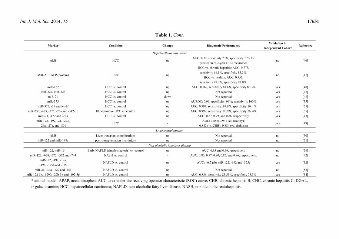
Int. J. Mol. Sci. 2014, 15 17651
Table 1. Cont.
Marker Condition Change Diagnostic Performance Validation in
Independent Cohort Reference
Hepatocellular carcinoma
ALB HCC up AUC: 0.72, sensitivity 73%, specificity 70% for
prediction of 2-year HCC recurrence no [46]
MiR-21 + AFP (protein) HCC up
HCC vs. chronic hepatitis: AUC: 0.773,
sensitivity 61.1%, specificity 83.3%.
HCC vs. healthy: AUC: 0.953,
sensitivity 87.3%, specificity 92.0%.
no [47]
miR-122 HCC vs. control up AUC: 0.869, sensitivity 81.6%, specificity 83.3% yes [48]
miR-222, miR-223 HCC vs. control up Not reported yes [48]
miR-21 HCC vs. control down Not reported yes [48]
miR-375 HCC vs. control up AUROC: 0.96, specificity: 96%; sensitivity: 100% yes [35]
miR-375, -25 and let-7f HCC vs. control up AUC: 0.997; sensitivity: 97.9%; specificity: 99.1% yes [35]
miR-23b, -423, -375, -23a and -342-3p HBV-positive HCC vs. control up AUC: 0.999; sensitivity: 96.9%; specificity: 99.4% yes [35]
miR-21, -122 and -223 HCC vs. control up AUC: 0.87, 0.79, and 0.86, respectively. yes [43]
miR-122, -192, -21, -223,
-26a, -27a, and -801 HCC -
AUC: 0.888; 0.941 (vs. healthy);
0.842 (vs. CHB); 0.884 (vs. cirrhosis) yes [49]
Liver transplantation
ALB Liver transplant complications up Not reported no [50]
miR-122 and miR-148a post-transplantation liver injury up Not reported no [51]
Non-alcoholic fatty liver disease
miR-122, miR-16 Early NAFLD (simple steatosis) vs. control up AUC: 0.93 and 0.96, respectively no [36]
miR-122, -638, -575, -572 and -744 NASH vs. control - AUC: 0.80, 0.97, 0.90, 0.85, and 0.96, respectively. no [42]
miR-122, -192, -19a,
-19b, -125b and -375 NAFLD vs. control up AUC: ~0.7 (for miR-122, -192 and -375) yes [52]
miR-21, -34a, -122 and -451 NAFLD vs. control up Not reported no [53]
miR-122-5p, -1290, -27b-3p and -192-5p NAFLD vs. control up AUC: 0.856, sensitivity 85.55%, specificity 73.3% yes [54]
* animal model; APAP, acetaminophen; AUC, area under the receiving operator characteristic (ROC) curve; CHB, chronic hepatitis B; CHC, chronic hepatitis C; DGAL,
D-galactosamine; HCC, hepatocellular carcinoma; NAFLD, non-alcoholic fatty liver disease; NASH, non-alcoholic seatohepatitis.
Int. J. Mol. Sci. 2014, 15 17652
Interestingly, liver-derived mRNAs and the reference transcripts are also present in plasma vesicles
from non-treated animals, suggesting a physiological, active secretion of microvesicles, independent of
tissue injury. In the case of treated animals, liver mRNAs are present in both microvesicles and cellular
debris isolated from plasma. Their circulating levels are elevated despite a reduced expression in liver
tissue. This suggests a shift in the secretion mechanism from an active one, in the absence of injury,
to both active and passive, at cytotoxic doses of chemicals [9].
Similar changes were reported in humans. In patients with acetaminophen poisoning, serum
miR-122 was significantly correlated with peak serum ALT activity. Also, miR-122 levels returned to
baseline earlier than ALT, suggesting a shorter circulatory half-life of the microRNA [31]. Another
study identified 11 circulating miRNAs, including miR-122, that were able to distinguish patients with
acetaminophen poisoning from healthy controls and patients with ischemic hepatitis, another form of
severe liver injury [55]. The levels of these miRNAs in plasma and serum increased earlier than ALT
after acetaminophen overdosage, and also returned to normal more rapidly than ALT, in response to
N-acetyl cysteine therapy, suggesting their potential use in treatment monitoring in these patients.
3.2. Chronic Viral Hepatitis
HBV infection is another condition associated with altered levels of plasma miRNAs. Thirteen
circulating miRNAs, including miR-375, miR-92a, miR-10a, miR-223, miR-423, miR-23b/a, miR-342-3p,
miR-99a, miR-122a, miR-125b, miR-150, and let-7c, were found to be upregulated in chronic HBV
carriers compared with healthy controls [35]. This panel could separate HBV cases from controls and
HCV carriers, and also HBV-positive HCC from controls, HBV cases, and HCV cases [35]. Four other
serum miRNAs (miR-572, miR-575, miR-638 and miR-744), together with miR-122 were reported to
distinguish chronic hepatitis B (CHB), non-alcoholic steatohepatitis (NASH) and control individuals [42].
Similarly to CHC, the increase of miR-122 in HBV infection positively correlates with plasma
ALT [40,41]. However, increases in plasma miR-122 coincide with histologic alterations in the same
individuals, even in cases with normal levels of ALT. Plasma miR-122 seems to be superior to ALT in
the accuracy of detecting HBV-induced liver damage [40].
Serum HBeAg is regarded as a surrogate marker for active viral replication in CHB patients.
Interestingly, the replication status of HBV translates into different circulating miRNA profiles [44].
A panel of 16 miRNAs, related to signaling or cancer pathways, had different expression patterns in
plasma of HBeAg positive, HBeAg negative and healthy children. A strong correlation was observed
between the circulating levels of these miRNAs and HBV DNA [44]. Concentrations of several plasma
miRNAs also seem to change among the immunological phases of HBV infection in children [45]. Four
circulating miRNAs, namely miR-99a-5p, -122-5p, -122-3p and -125b-5p, decreased in immune-tolerant
and immune-active children, whereas their levels were stable in immune-inactive children [45].
Among these miRNAs, miR-122 negatively regulates HBV gene expression and replication by binding
to highly conserved regions of the viral polymerase and the 3' untranslated region (UTR) of the core
protein mRNA [56].
Interestingly, several plasma miRNAs correlate with HBs antigenemia in HBV-infected persons [45].
Zhang et al. [41] found miR-122 to correlate more with HBsAg titre than with serum ALT. These
observations are consistent with the earlier finding that HBsAg particles carry hepatocellular miRNAs
Int. J. Mol. Sci. 2014, 15 17653
and the associated AGO2 protein [57]. Apparently, liver-specific mRNA sequences can also be
entrapped along with miRNAs, during the process of HBs particle assembly and secretion [41].
In chronic hepatitis C (CHC), serum miRNAs correlate with the stage of liver injury. miR-571 is
upregulated in the serum of CHC patients with liver cirrhosis, reflecting a concordant regulation in the
liver tissue in response to the pro-fibrogenic cytokine transforming growth factor beta (TGF-β) [37].
Similarly, a good correlation in gene transcription was observed between liver and serum in CHC
patients. This correlation involved a variety of transcripts, including genes for immune markers, or
related to inflammation, apoptosis and matrix turnover [58].
Serum miRNA profile partially correlates with liver fibrosis stage and inflammation grade in CHC
patients. One study reported a high accuracy of discrimination between fibrosis stage F0 and F1–F3 in
CHC patients, through assessment of serum miRNAs. The same set of miRNAs could also separate
CHC from CHB and NASH [34].
miR-122 was reported as a positive regulator of HCV replication and particle production [59,60].
miR-122 promotes HCV replication in experimental models, possibly by facilitating the folding of
viral RNA and its sequestration in active replication sites [61]. miR-122 also activates HCV translation
by binding to its 5' UTR, in a process mediated by AGO proteins [62]. However, HCV viral load is not
correlated with liver miR-122 expression in infected patients [63], and plasma miR-122 does not
correlate with HCV viral load either [36]. Instead, circulating miR-122 correlates with serum transaminases
and with necroinflammatory activity in CHC, whereas intrahepatic miR-122 shows a negative
correlation with the extent of liver damage [38]. The lack of parallelism between cellular and
circulating miR-122 may be explained by an increased release of miRNA from affected cells, despite
the downregulation of its expression.
There are conflicting reports regarding the relationship between circulating miR-122 and liver
fibrosis. In one study investigating approximately 50 patients, plasma miR-122, as well as miR-34a,
correlated with liver fibrosis severity, and was suggested as a suitable marker [36]. In contrast,
two other studies including 68 and 164 patients respectively, did not find a significant correlation
between circulating miR-122 and fibrosis [38,39]. Unfortunately, the lack of details regarding the
technical assessment of miRNAs in these studies, especially the preanalytical variables, makes the
reported results difficult to interpret and compare. As discussed below, a simple factor such as sample
storage time at room temperature before centrifugation, can bias the results and should be accounted
for [64].
3.3. Hepatocellular Carcinoma
MicroRNAs play key roles in the development of hepatocellular carcinoma, by interfering with
crucial cancer-associated pathways [65]. miR-21 is highly overexpressed in HCC tissue and contributes to
tumor growth and spread by modulating the expression of phosphatase and tensin homolog (PTEN)
tumor suppressor [66]. Inhibition of miR-21 in cultured HCC cells impairs tumor cell proliferation,
migration and invasion, whereas enhanced expression of miR-21 has the opposite effect [66]. Upregulation
of miR-222 and downregulation of miR-223 were observed in primary HCC as compared with
adjacent normal liver tissue [67]. Restoration of miR-223 expression severely impairs viability of HCC
cell lines by targeting Stathmin 1 [67]. miR-122 is a powerful tumor suppressor, by inhibiting
Int. J. Mol. Sci. 2014, 15 17654
angiogenesis, tumorigenesis and HCC intrahepatic metastasis, partially by targeting ADAM
metallopeptidase domain 17 (ADAM17) [68]. Accordingly, miR-122 is strongly downregulated in
HCC [68].
Interestingly, modified circulating levels of these RNAs were repeatedly found in HCC patients.
Plasma miR-21 was higher in HCC patients than in chronic hepatitis and healthy volunteers, and its
levels correlated with expression in tumor tissue [47]. Its diagnostic value was better than that of alpha
fetoprotein (AFP), and improved for the combination of the two markers. Diagnostic sensitivity,
specificity and accuracy of the combination of miR-21 and AFP for the diagnosis of HCC were higher
than 90%, suggesting that circulating RNAs are a useful addition to classical tumor markers.
Chronic HBV infection is a major cause of HCC. By comparison of circulating miRNA profiles in
healthy subjects and in patients with various stages of HBV infection, seven miRNAs were identified
as potential markers of HCC [49]. The panel, comprising miR-122, miR-192, miR-21, miR-223, miR-26a,
miR-27a, and miR-801, was developed on large groups of participants (407 and 390 participants in the
training and validation groups, respectively) and demonstrated a high diagnostic accuracy for HCC,
with an area under the receiving operator characteristic curve (AUC) of 0.888 in the validation set.
The 7 miRNA panel could also differentiate HCC from healthy (AUC 0.941), chronic hepatitis B
(AUC 0.842), and cirrhosis (AUC 0.884), respectively. Among the seven miRNAs of the panel, tumor
expression of miR-26a and miR-192 was associated with early recurrence of HCC [69]. In an
independent study, Xu et al. [43] showed that miR-21, miR-122, and miR-223 had higher serum levels
in HCC and CHB patients than in healthy controls. Serum levels of two miRNAs, miR-10a and miR-125b,
were shown to be lower in HBV-positive HCC than in CHB patients, and the combination of the two
markers could accurately distinguish between these pathological conditions [35]. Accordingly, miR-125b is
underexpressed in HCC tissue and was recently identified as a potential tumor suppressor [70].
A set of three miRNAs, miR-375, miR-25 and let-7f could separate HCC from healthy subjects
with a sensitivity and specificity of 97.9% and 99.1%, respectively [35]. When considered alone,
miR-375 had 100% sensitivity and 96% specificity in predicting HCC [35].
Gui et al. [33] identified a single serum miRNA that could differentiate patients with liver
pathologies from healthy controls with more than 90% sensitivity and 79% specificity. miR-885-5p
was significantly more abundant in serum from patients with HCC, CHB and liver cirrhosis.
miRNAs are not the only circulating RNA molecules deregulated in patients with liver cancer.
A study reported that more than 90% of the investigated HCC patients had increased plasma ALB
mRNA, whereas less than 50% had a high level of circulating AFP mRNA [32].
Besides HCC, higher levels of plasma ALB mRNA were found in cirrhosis and active (but not
inactive) hepatitis B patients than in healthy controls. Plasma ALB mRNA had a diagnostic sensitivity
of more than 85% and a specificity over 90% for the detection of these pathologies [32]. It was
debated whether the detected ALB mRNA originated from hepatocytes or from an illegitimate
transcription process in other cells. It is known that peripheral blood mononuclear cells transcribe ALB
in almost one third of healthy individuals and in about 90% of patients with chronic hepatitis [71].
Recently, it was confirmed that ALB mRNA detected in whole blood comprised a mixture of
molecules released by blood cells and hepatocytes. However, using a RNA single nucleotide
polymorphism approach to genotype ALB mRNA in the plasma of liver and bone marrow
transplantation recipients, it was shown that cell-free ALB transcripts are only of hepatic origin [32].
Int. J. Mol. Sci. 2014, 15 17655
3.4. Non-Alcoholic Fatty Liver Disease
Non-alcoholic fatty liver disease (NAFLD) is the most common cause of chronic liver disease
in America [72], and includes a spectrum of histological hepatic changes ranging from simple steatosis
to NASH. Depending on their severity, these pathological changes alter the expression of several
hepatic miRNAs. Of the 474 investigated miRNAs, Cheung et al. [73] found 46 that were
differentially expressed in the liver of NASH patients compared with controls with normal liver
histology. Hepatic miR-122 is significantly downregulated in NASH. Accordingly, liver mRNA and
protein levels of sterol response element binding protein 1c (SREBP-1c), fatty acid synthase,
and 3-hydroxy-3-methylglutaryl-coenzyme A reductase, some of the miR-122 targets involved in lipid
biosynthesis, are increased in NASH patients [73]. A decrease of liver miR-122 levels in NASH
patients was also confirmed by in situ hybridization, in an independent study [52]. Interestingly,
miR-122 is preferentially localized in lipid-laden hepatocytes, near the cell membrane, as if ready
to be secreted. Accordingly, circulating miR-122 levels are more than 7-fold increased in NASH
patients compared with controls [52]. Serum miR-122 correlates with ALT levels and liver fibrosis in
NAFLD patients. It also performs slightly better in the diagnosis of NAFLD severity than classical
liver disease markers, including aspartate aminotransferase (AST), ALT and plasma caspase generated
cytokeratin-18 fragments. Besides miR-122, Pirola et al. [52] found two other miRNAs, miR-192
(upregulated by TGFβ1) and miR-375 (a key regulator of glucose homeostasis), with lower liver
expression and higher serum levels in NASH compared with simple steatosis. These changes were
subsequently validated in an independent cohort. Similarly, Yamada et al. [53] found increased levels
of serum miR-21, miR-34a, miR-122 and miR-451 in participants with NAFLD from a large group of
non-selected Japanese people attending health examinations. Among these markers, only miR-122
correlated with the severity of liver steatosis.
A panel with high diagnostic accuracy for NAFLD was recently described [54]. The panel, consisting of
four miRNAs (miR-122-5p, miR-1290, miR-27b-3p and miR-192-5p), was developed and validated
on two large independent cohorts and seems to have better sensitivity and specificity for NAFLD than
ALT and the non-invasive score FIB-4. Moreover, the performance of the miRNA panel was not
influenced by disease severity. Of note, miR-27b, a member of this panel, promotes fat accumulation
in hepatocytes via repression of peroxisome proliferator-activated receptor-α and angiopoietin-like
protein 3 [74], and was previously found upregulated in the liver of NASH patients [73].
3.5. Liver Transplantation
In liver transplant recipients, serial assessment of circulating ALB mRNA can be used to detect the
progression of hepatic complications [50]. Hepatic injury and rejection after liver transplantation can
also be monitored using hepatic-specific miRNAs [51]. Accordingly, serum miR-122 and miR-148a
were elevated in patients with post-transplantation liver injury and correlated with serum transaminases.
During episodes of acute rejection, their levels increased up to 20-fold, and their peak occurred earlier
than in the case of transaminases [51]. It was also speculated that liver-derived mRNA levels could be
used as an indicator of the quality of transplanted grafts or as an aid in the choice of proper
Int. J. Mol. Sci. 2014, 15 17656
immunosuppresive therapy in transplant recipients. However, further research is needed for the proper
validation of circulating mRNA as biomarkers in this field [75].
3.6. Prognostic Value of Circulating RNAs in Liver Disease
Although the diagnostic value of circulating RNAs in liver disease has been assessed in many
comparative studies, their prognostic relevance in longitudinal designs was addressed to a much
lower extent.
The prognostic value of serum miR-122 in advanced liver disease was recently evaluated.
Waidmann et al. [76] observed that patients with hepatic decompensation had lower circulating
miR-122 than those with compensated liver disease. In their cohort, low serum miR-122 was
associated with complications such as ascites, spontaneous bacterial peritonitis and hepatorenal
syndrome in cirrhotic patients. Multivariate Cox analysis revealed that miR-122 was an independent
predictive factor for survival in patients with cirrhosis. The authors validated their observations in a
second independent cohort. However, the threshold for serum miR-122 was chosen relative to their
study group, dividing the cohorts into the third of patients with highest serum miR-122 levels and the
other two thirds with lower miR-122. Since the general recommendation in clinical chemistry practice
is that each laboratory should establish its own reference ranges for the analytes it assesses, such a
threshold choice is acceptable. However, the lack of a quantitative value (e.g., in copies/microliter)
makes their results difficult to translate into other laboratories. High serum miR-122, as well as high
miR-1, also predicts longer overall survival in HCC patients [77].
Plasma ALB mRNA is detectable in the majority of HCC patients [46]. Preoperative levels of
plasma ALB mRNA predict survival and HCC recurrence. The predictive ability of the transcript is
higher than the size and number of tumors (the usual preoperative radiological criteria) and is similar
to the presence of vascular invasion (pathological criterion). Cheung et al. [46] detected ALB mRNA
in almost all HCC, in contrast with AFP mRNA, which was only detectable in some of the cases.
4. Pre-Analytical and Analytical Challenges in the Assessment of Circulating RNA
Real-time PCR is the preferred format for the quantification of cell-free circulating RNA. Despite
the high sensitivity and specificity attainable in RNA quantification with the available technology,
conflicting results have been reported in different studies on circulating RNA as biomarkers. It was
rapidly observed that a multitude of factors were able to influence circulating RNA quantification at
both pre-analytical and analytical levels. In consequence, several studies were performed to assess the
pre-analytical variables affecting the quantification of circulating RNA, especially miRNA. In contrast,
mRNAs were less studied from this point of view.
Low level haemolysis often occurs during collection of blood samples. This phenomenon can
increase the levels of red blood cell-related miRNAs in plasma, thus affecting any potential miRNA
biomarker that is also present within erythrocytes [78]. miR-16, which is expressed at high levels in
circulating cells, is greatly affected by haemolysis [78,79]. Other blood cells can also serve as a source
of circulating miRNAs. Variations in blood cell counts and haemolysis can alter plasma miRNA levels
by up to 50-fold [80]. It is not clear yet to what extent haemolysis affects other extracellular RNA
species. RNAs not expressed in blood cells, such as miR-122, are not increased upon sample
Int. J. Mol. Sci. 2014, 15 17657
haemolysis [64]. Short noncoding RNA and mRNA are less stable in human plasma than miRNA [15]
and thus, one can expect their levels to be less influenced by haemolysis. However, haemolysis
entrains a release of RNase inhibitors from lysed cells, thus reducing degradation of certain circulating
RNAs, such as miR-122 [64]. The general recommendation is that free hemoglobin in samples be
assessed by spectrophotometry and haemolyzed samples be removed from analysis, unless it is
rigorously demonstrated that the RNA sequences of interest, including reference genes, are not
affected by haemolysis [78,79]. Alternatively, if only extracted material is available, putative
haemolyzed samples can be identified by the relative expression of the erythrocyte-specific miR-451
and the stable miR-23a [81].
The choice of sample type also influences results. Although serum and plasma levels of several
miRNAs, including miR-15b, miR-16, and miR-24, are strongly correlated [82], these molecules are
present at higher concentrations in plasma than in serum [79]. Differences between serum and plasma
miRNA profiles showed a certain association with miRNA from platelets, suggesting that coagulation
may distort extracellular miRNA concentrations in samples [83]. In the case of plasma, miRNA
quantification is affected by the type of anticoagulant. For example, the fluoride/oxalate mixture
enables higher sensitivity in miRNA detection than citrate and ethylenediaminetetraacetic acid
(EDTA) [84].
Sample pre-processing steps affect subsequent quantification results. Most of extracellular RNA is
removed by filtration through 0.2 µm filters [8]. Also, total RNA content of plasma samples decreases
as higher centrifugation forces are applied [85]. Exosome enrichment can improve detection of
certain RNA targets, but can significantly distort expression profiles compared with plasma. This can
be due to the different concentrations of targets in exosomes vs. exosome-free fluids [86] and to the
unbalanced distribution of various RNA sequences inside and outside microvesicles [14].
Delays in sample processing seem to decrease the amounts of detectable mRNA [87] and
miRNA [88] in plasma. Serum storage at room temperature biases miRNA profiles, increasing the
proportion of vesicle-associated miRNAs as compared to protein-associated miRNAs [64]. It seems
that microvesicle-borne miRNAs are more resistant to RNase activity than are miRNAs complexed
with proteins. Relative expression profiles of certain plasma miRNAs were reportedly unaffected by
transportation times of up to 48 h prior to centrifugation, as compared to samples processed within
four hours of collection [44,45]. However, in order to limit pre-analytical variability, sample storage at
room temperature should be minimized, and a temperature of −70 °C or below is recommended for
long-term storage of plasma [89]. Although a certain loss of miRNAs can be observed, successful
quantification is possible in samples stored for up to 12 years at −80 °C, suggesting the utility of
long-term archived samples in retrospective studies [88]. It was found that a single freeze-thaw cycle
does not significantly degrade plasma mRNA [7]. However, in a direct comparison, serum miRNAs
were found to be more stable than high molecular weight RNAs (including GAPDH and β-actin
mRNAs) when samples were subjected to multiple freeze-thaw cycles [90].
The yield and reproducibility of RNA extraction from body fluids are method-dependent.
The column-based mirVana PARIS kit was repeatedly reported among the best performing kits for
the isolation of circulating miRNA, giving the highest yields of purified nucleic acids [79,89].
McDonald et al. [79] found that kits not employing a phenol-chloroform isolation step, such as Roche
High Pure miRNA Isolation Kit, performed better in terms of reproducibility. There are conflicting
Int. J. Mol. Sci. 2014, 15 17658
reports regarding miRNA isolation using liquid-liquid extraction methods. Whereas some researchers
found TRIzol-LS extraction equal or better than column-based kits for serum miRNAs [79], others
arrived at the opposite conclusion [89]. Plasma RNA recovery and detection do not necessarily
parallel the sample input volume, as several blood-borne inhibitors may co-purify with nucleic acids.
The addition of small amounts of inhibitor-resistant polymerase to common real-time PCR reaction
mixes seems to improve detection of circulating miRNAs [84]. Storage time of purified nucleic acids
in elution buffer prior to assessment by real-time PCR may also be an important factor, since
long-term stability of RNA may depend on the nature of the storage solution. Thus, some researchers
found that long-time storage of purified miRNAs isolated from clinical samples increased quantification
variability compared with long-time storage of unextracted samples [79]. Conversely, others reported
very good stability of purified miRNAs during prolonged storage [89].
Finally, target detection and normalization methods fundamentally contribute to quantification
results. In a direct comparison of real-time PCR low-density arrays for plasma miRNA quantification,
locked nucleic acid-based miRCURY platform performed better than the TaqMan counterpart in the
case of low expression targets. At higher concentrations of miRNA, the performance of the two
platforms was similar [91].
It is important that all these technical details be considered and kept as constant as possible within a
study, in order to improve the consistency of the results and minimize the risk of bias.
Many studies on miRNA expression utilize U6, RNU6B or 18S as internal normalizers. However,
this approach is controversial, since these targets are not miRNAs, thus their origin, stability, and
efficiency of their extraction, reverse transcription and amplification may be different from those of
miRNAs. If the reference gene method is used for normalization of miRNA expression data, reference
miRNAs should be used instead. For example, the combination of miR-26a, miR-221 and miR-22*,
was found as the most stable set of reference genes for serum miRNA assessment in HBV carriers vs.
healthy controls [92].
Reference targets should be properly validated in each study, since they may be influenced by
several physiological or demographic conditions affecting the studied groups of patients [93].
A number of miRNAs were shown to have gender-related circulating concentrations. Thus, plasma
miR-130b and miR-18b are higher in males [83], whereas miR-548-3p, miR-1323, miR-940 and
miR-1292 are upregulated in females [94]. Pathophysiological changes in sample matrix, related to the
studied condition or disease may also entrain changes in the levels of detection inhibitors, and thus
become a source of bias.
Due to the multiple variables affecting the results, reports on circulating RNAs should include
relevant details regarding the analytical workflow [95,96]. Guidelines for reporting results of real-time
PCR experiments have been issued [97] and constitute a useful aid in the standardization of reports
regarding development of biomarkers based on circulating nucleic acids. Such a standardization would
allow more accurate comparisons of data obtained in different laboratories and lead to a better
understanding of results.
Int. J. Mol. Sci. 2014, 15 17659
5. Conclusions and Perspectives
Despite a wealth of evidence for the presence of RNA molecules in body fluids, the origin and,
more importantly, the function of these RNAs in the extracellular environment remains poorly
understood. An intriguing possibility is that these molecules are related to the process of intercellular
communication. Given that certain RNAs are selectively expressed in specific organs, it may be
possible to monitor the physiopathological conditions of organs by the levels of circulating
organ-specific RNAs. This possibility opens up new perspectives in the development of non-invasive
markers of liver disease.
Finding of informative biomarkers is not only critical for understanding of disease-related
physiopathological processes, but is also important for therapeutic development. Current technology
for the assessment of circulating RNAs in liver disease has several advantages over the detection of
protein-based markers, including higher sensitivity and wider dynamic range, and its use has already
led to very encouraging results. Undoubtedly, extracellular RNAs will constitute an excellent addition
to classical noninvasive markers of liver pathology. However, standardization of sample preparation,
quality assessment and quantification of circulating RNA molecules is urgently needed. It is also a key
prerequisite for the adoption of cell-free RNA markers into clinical practice.
Conflicts of Interest
The authors declare no conflict of interest.
References
1. European Association for the Study of the Liver. EASL clinical practice guidelines: Management
of chronic hepatitis B virus infection. J. Hepatol. 2012, 57, 167–185.
2. EASL Recommendations on Treatment of Hepatitis C. Available online: http://files.easl.eu/
easl-recommendations-on-treatment-of-hepatitis-C.pdf (accessed on 22 August 2014).
3. El-Serag, H.B. Epidemiology of viral hepatitis and hepatocellular carcinoma. Gastroenterology
2012, 142, 1264–1273.e1.
4. European Association for the Study of the Liver; European Organisation for Research and
Treatment of Cancer. EASL–EORTC Clinical Practice Guidelines: management of hepatocellular
carcinoma. J. Hepatol. 2012, 56, 908–943.
5. Tsang, J.C.H.; Lo, Y.M.D. Circulating nucleic acids in plasma/serum. Pathology 2007, 39,
197–207.
6. Weber, J.A.; Baxter, D.H.; Zhang, S.; Huang, D.Y.; Huang, K.H.; Lee, M.J.; Galas, D.J.; Wang, K.
The microRNA spectrum in 12 body fluids. Clin. Chem. 2010, 56, 1733–1741.
7. Tsui, N.B.Y.; Ng, E.K.O.; Lo, Y.M.D. Stability of endogenous and added RNA in blood
specimens, serum, and plasma. Clin. Chem. 2002, 48, 1647–1653.
8. El-Hefnawy, T.; Raja, S.; Kelly, L.; Bigbee, W.L.; Kirkwood, J.M.; Luketich, J.D.; Godfrey, T.E.
Characterization of amplifiable, circulating RNA in plasma and its potential as a tool for cancer
diagnostics. Clin. Chem. 2004, 50, 564–573.
Int. J. Mol. Sci. 2014, 15 17660
9. Wetmore, B.A.; Brees, D.J.; Singh, R.; Watkins, P.B.; Andersen, M.E.; Loy, J.; Thomas, R.S.
Quantitative analyses and transcriptomic profiling of circulating messenger RNAs as biomarkers
of rat liver injury. Hepatol. Baltim. Md. 2010, 51, 2127–2139.
10. Halicka, H.D.; Bedner, E.; Darzynkiewicz, Z. Segregation of RNA and separate packaging of
DNA and RNA in apoptotic bodies during apoptosis. Exp. Cell Res. 2000, 260, 248–256.
11. Cocucci, E.; Racchetti, G.; Meldolesi, J. Shedding microvesicles: Artefacts no more. Trends Cell Biol.
2009, 19, 43–51.
12. García, J.M.; García, V.; Peña, C.; Domínguez, G.; Silva, J.; Diaz, R.; Espinosa, P.; Citores, M.J.;
Collado, M.; Bonilla, F. Extracellular plasma RNA from colon cancer patients is confined in a
vesicle-like structure and is mRNA-enriched. RNA 2008, 14, 1424–1432.
13. Bolukbasi, M.F.; Mizrak, A.; Ozdener, G.B.; Madlener, S.; Ströbel, T.; Erkan, E.P.; Fan, J.-B.;
Breakefield, X.O.; Saydam, O. miR-1289 and “Zipcode”-like sequence enrich mRNAs in
microvesicles. Mol. Ther. Nucleic Acids 2012, 1, doi:10.1038/mtna.2011.2.
14. Arroyo, J.D.; Chevillet, J.R.; Kroh, E.M.; Ruf, I.K.; Pritchard, C.C.; Gibson, D.F.; Mitchell, P.S.;
Bennett, C.F.; Pogosova-Agadjanyan, E.L.; Stirewalt, D.L.; et al. Argonaute2 complexes carry a
population of circulating microRNAs independent of vesicles in human plasma. Proc. Natl. Acad.
Sci. USA 2011, 108, 5003–5008.
15. Turchinovich, A.; Weiz, L.; Langheinz, A.; Burwinkel, B. Characterization of extracellular
circulating microRNA. Nucleic Acids Res. 2011, 39, 7223–7233.
16. Turchinovich, A.; Burwinkel, B. Distinct AGO1 and AGO2 associated miRNA profiles in human
cells and blood plasma. RNA Biol. 2012, 9, 1066–1075.
17. Bala, S.; Petrasek, J.; Mundkur, S.; Catalano, D.; Levin, I.; Ward, J.; Alao, H.; Kodys, K.; Szabo, G.
Circulating microRNAs in exosomes indicate hepatocyte injury and inflammation in alcoholic,
drug-induced, and inflammatory liver diseases. Hepatol. Baltim. Md. 2012, 56, 1946–1957.
18. Vickers, K.C.; Palmisano, B.T.; Shoucri, B.M.; Shamburek, R.D.; Remaley, A.T. MicroRNAs are
transported in plasma and delivered to recipient cells by high-density lipoproteins. Nat. Cell Biol.
2011, 13, 423–433.
19. Lässer, C.; Alikhani, V.S.; Ekström, K.; Eldh, M.; Paredes, P.T.; Bossios, A.; Sjöstrand, M.;
Gabrielsson, S.; Lötvall, J.; Valadi, H. Human saliva, plasma and breast milk exosomes contain
RNA: Uptake by macrophages. J. Transl. Med. 2011, 9, doi:10.1186/1479-5876-9-9.
20. Valadi, H.; Ekström, K.; Bossios, A.; Sjöstrand, M.; Lee, J.J.; Lötvall, J.O. Exosome-Mediated
transfer of mRNAs and microRNAs is a novel mechanism of genetic exchange between cells.
Nat. Cell Biol. 2007, 9, 654–659.
21. Deregibus, M.C.; Cantaluppi, V.; Calogero, R.; Iacono, M.L.; Tetta, C.; Biancone, L.; Bruno, S.;
Bussolati, B.; Camussi, G. Endothelial progenitor cell–derived microvesicles activate an
angiogenic program in endothelial cells by a horizontal transfer of mRNA. Blood 2007, 110,
2440–2448.
22. Waldenström, A.; Gennebäck, N.; Hellman, U.; Ronquist, G. Cardiomyocyte microvesicles
contain DNA/RNA and convey biological messages to target cells. PLoS One 2012, 7, e34653.
23. Kogure, T.; Lin, W.-L.; Yan, I.K.; Braconi, C.; Patel, T. Intercellular nanovesicle-mediated
microRNA transfer: A mechanism of environmental modulation of hepatocellular cancer cell
growth. Hepatology 2011, 54, 1237–1248.
Int. J. Mol. Sci. 2014, 15 17661
24. Fonsato, V.; Collino, F.; Herrera, M.B.; Cavallari, C.; Deregibus, M.C.; Cisterna, B.; Bruno, S.;
Romagnoli, R.; Salizzoni, M.; Tetta, C.; et al. Human liver stem cell-derived microvesicles inhibit
hepatoma growth in SCID mice by delivering antitumor MicroRNAs. Stem Cells 2012, 30,
1985–1998.
25. Pan, Q.; Ramakrishnaiah, V.; Henry, S.; Fouraschen, S.; de Ruiter, P.E.; Kwekkeboom, J.;
Tilanus, H.W.; Janssen, H.L.A.; van der Laan, L.J.W. Hepatic cell-to-cell transmission of small
silencing RNA can extend the therapeutic reach of RNA interference (RNAi). Gut 2012, 61,
1330–1339.
26. Laterza, O.F.; Lim, L.; Garrett-Engele, P.W.; Vlasakova, K.; Muniappa, N.; Tanaka, W.K.;
Johnson, J.M.; Sina, J.F.; Fare, T.L.; Sistare, F.D.; et al. Plasma MicroRNAs as sensitive and
specific biomarkers of tissue injury. Clin. Chem. 2009, 55, 1977–1983.
27. Etheridge, A.; Lee, I.; Hood, L.; Galas, D.; Wang, K. Extracellular microRNA: A new source of
biomarkers. Mutat. Res. 2011, 717, 85–90.
28. Miyamoto, M.; Yanai, M.; Ookubo, S.; Awasaki, N.; Takami, K.; Imai, R. Detection of cell-free,
liver-specific mRNAs in peripheral blood from rats with hepatotoxicity: A potential toxicological
biomarker for safety evaluation. Toxicol. Sci. 2008, 106, 538–545.
29. Okubo, S.; Miyamoto, M.; Takami, K.; Kanki, M.; Ono, A.; Nakatsu, N.; Yamada, H.; Ohno, Y.;
Urushidani, T. Identification of novel liver-specific mRNAs in plasma for biomarkers of
drug-induced liver injury and quantitative evaluation in rats treated with various hepatotoxic
compounds. Toxicol. Sci. 2013, 132, 21–31.
30. Wang, K.; Zhang, S.; Marzolf, B.; Troisch, P.; Brightman, A.; Hu, Z.; Hood, L.E.; Galas, D.J.
Circulating microRNAs, potential biomarkers for drug-induced liver injury. Proc. Natl. Acad.
Sci. USA 2009, 106, 4402–4407.
31. Starkey Lewis, P.J.; Dear, J.; Platt, V.; Simpson, K.J.; Craig, D.G.N.; Antoine, D.J.; French, N.S.;
Dhaun, N.; Webb, D.J.; Costello, E.M.; et al. Circulating microRNAs as potential markers of
human drug-induced liver injury. Hepatology 2011, 54, 1767–1776.
32. Chan, R.W.Y.; Wong, J.; Chan, H.L.Y.; Mok, T.S.K.; Lo, W.Y.W.; Lee, V.; To, K.F.; Lai, P.B.S.;
Rainer, T.H.; Lo, Y.M.D.; et al. Aberrant concentrations of liver-derived plasma albumin mRNA
in liver pathologies. Clin. Chem. 2010, 56, 82–89.
33. Gui, J.; Tian, Y.; Wen, X.; Zhang, W.; Zhang, P.; Gao, J.; Run, W.; Tian, L.; Jia, X.; Gao, Y.
Serum microRNA characterization identifies miR-885–5p as a potential marker for detecting liver
pathologies. Clin. Sci. Lond. Engl. 1979 2011, 120, 183–193.
34. Murakami, Y.; Toyoda, H.; Tanahashi, T.; Tanaka, J.; Kumada, T.; Yoshioka, Y.; Kosaka, N.;
Ochiya, T.; Taguchi, Y. Comprehensive miRNA expression analysis in peripheral blood can
diagnose liver disease. PLoS One 2012, 7, e48366.
35. Li, L.-M.; Hu, Z.-B.; Zhou, Z.-X.; Chen, X.; Liu, F.-Y.; Zhang, J.-F.; Shen, H.-B.; Zhang, C.-Y.;
Zen, K. Serum microRNA profiles serve as novel biomarkers for HBV infection and diagnosis of
HBV-positive hepatocarcinoma. Cancer Res. 2010, 70, 9798–9807.
36. Cermelli, S.; Ruggieri, A.; Marrero, J.A.; Ioannou, G.N.; Beretta, L. Circulating microRNAs in
patients with chronic hepatitis C and non-alcoholic fatty liver disease. PLoS One 2011, 6, e23937.
Int. J. Mol. Sci. 2014, 15 17662
37. Roderburg, C.; Mollnow, T.; Bongaerts, B.; Elfimova, N.; Vargas Cardenas, D.; Berger, K.;
Zimmermann, H.; Koch, A.; Vucur, M.; Luedde, M.; et al. Micro-RNA profiling in human serum
reveals compartment-specific roles of miR-571 and miR-652 in liver cirrhosis. PLoS One 2012,
7, e32999.
38. Bihrer, V.; Friedrich-Rust, M.; Kronenberger, B.; Forestier, N.; Haupenthal, J.; Shi, Y.;
Peveling-Oberhag, J.; Radeke, H.H.; Sarrazin, C.; Herrmann, E.; et al. Serum miR-122 as
a biomarker of necroinflammation in patients with chronic hepatitis C virus infection.
Am. J. Gastroenterol. 2011, 106, 1663–1669.
39. Trebicka, J.; Anadol, E.; Elfimova, N.; Strack, I.; Roggendorf, M.; Viazov, S.; Wedemeyer, I.;
Drebber, U.; Rockstroh, J.; Sauerbruch, T.; et al. Hepatic and serum levels of miR-122 after
chronic HCV induced fibrosis. J. Hepatol. 2012, 58, 234–239.
40. Zhang, Y.; Jia, Y.; Zheng, R.; Guo, Y.; Wang, Y.; Guo, H.; Fei, M.; Sun, S. Plasma microRNA-122
as a biomarker for viral-, alcohol-, and chemical-related hepatic diseases. Clin. Chem. 2010, 56,
1830–1838.
41. Zhang, X.; Zhang, Z.; Dai, F.; Shi, B.; Chen, L.; Zhang, X.; Zang, G.; Zhang, J.; Chen, X.;
Qian, F.; et al. Comparison of circulating, hepatocyte specific messenger RNA and microRNA as
biomarkers for chronic hepatitis B and C. PLoS One 2014, 9, e92112.
42. Zhang, H.; Li, Q.-Y.; Guo, Z.-Z.; Guan, Y.; Du, J.; Lu, Y.-Y.; Hu, Y.-Y.; Liu, P.; Huang, S.;
Su, S.-B. Serum levels of microRNAs can specifically predict liver injury of chronic hepatitis B.
World J. Gastroenterol. WJG 2012, 18, 5188–5196.
43. Xu, J.; Wu, C.; Che, X.; Wang, L.; Yu, D.; Zhang, T.; Huang, L.; Li, H.; Tan, W.; Wang, C.; et al.
Circulating microRNAs, miR-21, miR-122, and miR-223, in patients with hepatocellular carcinoma
or chronic hepatitis. Mol. Carcinog. 2011, 50, 136–142.
44. Winther, T.N.; Bang-Berthelsen, C.H.; Heiberg, I.L.; Pociot, F.; Hogh, B. Differential plasma
microRNA profiles in HBeAg positive and HBeAg negative children with chronic hepatitis B.
PLoS One 2013, 8, e58236.
45. Winther, T.N.; Heiberg, I.L.; Bang-Berthelsen, C.H.; Pociot, F.; Hogh, B. Hepatitis B surface
antigen quantity positively correlates with plasma levels of microRNAs differentially expressed in
immunological phases of chronic hepatitis B in children. PLoS One 2013, 8, e80384.
46. Cheung, S.T.; Fan, S.T.; Lee, Y.T.; Chow, J.P.; Ng, I.O.; Fong, D.Y.; Lo, C.M. Albumin mRNA
in plasma predicts post-transplant recurrence of patients with hepatocellular carcinoma.
Transplant 2008, 85, 81–87.
47. Tomimaru, Y.; Eguchi, H.; Nagano, H.; Wada, H.; Kobayashi, S.; Marubashi, S.; Tanemura, M.;
Tomokuni, A.; Takemasa, I.; Umeshita, K.; et al. Circulating microRNA-21 as a novel biomarker
for hepatocellular carcinoma. J. Hepatol. 2012, 56, 167–175.
48. Qi, P.; Cheng, S.; Wang, H.; Li, N.; Chen, Y.; Gao, C. Serum MicroRNAs as biomarkers for
hepatocellular carcinoma in chinese patients with chronic hepatitis B virus infection. PLoS One
2011, 6, e28486.
49. Zhou, J.; Yu, L.; Gao, X.; Hu, J.; Wang, J.; Dai, Z.; Wang, J.-F.; Zhang, Z.; Lu, S.; Huang, X.;
et al. Plasma MicroRNA panel to diagnose hepatitis B virus-related hepatocellular carcinoma.
J. Clin. Oncol. 2011, 29, 4781–4788.
Int. J. Mol. Sci. 2014, 15 17663
50. Chan, R.W.Y.; Wong, J.; Lai, P.B.S.; Lo, Y.M.D.; Chiu, R.W.K. The potential clinical utility of
serial plasma albumin mRNA monitoring for the post-liver transplantation management.
Clin. Biochem. 2013, 46, 1313–1319.
51. Farid, W.R.R.; Pan, Q.; van der Meer, A.J.P.; de Ruiter, P.E.; Ramakrishnaiah, V.; de Jonge, J.;
Kwekkeboom, J.; Janssen, H.L.A.; Metselaar, H.J.; Tilanus, H.W.; et al. Hepatocyte-Derived
microRNAs as serum biomarkers of hepatic injury and rejection after liver transplantation.
Liver Transpl. 2012, 18, 290–297.
52. Pirola, C.J.; Gianotti, T.F.; Castaño, G.O.; Mallardi, P.; Martino, J.S.; Ledesma, M.M.G.L.;
Flichman, D.; Mirshahi, F.; Sanyal, A.J.; Sookoian, S. Circulating microRNA signature in
non-alcoholic fatty liver disease: From serum non-coding RNAs to liver histology and disease
pathogenesis. Gut 2014, doi:10.1136/gutjnl-2014-306996.
53. Yamada, H.; Suzuki, K.; Ichino, N.; Ando, Y.; Sawada, A.; Osakabe, K.; Sugimoto, K.; Ohashi, K.;
Teradaira, R.; et al. Associations between circulating microRNAs (miR-21, miR-34a, miR-122
and miR-451) and non-alcoholic fatty liver. Clin. Chim. Acta 2013, 424, 99–103.
54. Tan, Y.; Ge, G.; Pan, T.; Wen, D.; Gan, J. A pilot study of serum MicroRNAs panel as potential
biomarkers for diagnosis of nonalcoholic fatty liver disease. PLoS One 2014, 9, e105192.
55. Ward, J.; Kanchagar, C.; Veksler-Lublinsky, I.; Lee, R.C.; McGill, M.R.; Jaeschke, H.; Curry, S.C.;
Ambros, V.R. Circulating microRNA profiles in human patients with acetaminophen hepatotoxicity
or ischemic hepatitis. Proc. Natl. Acad. Sci. USA 2014, 111, 12169–12174.
56. Chen, Y.; Shen, A.; Rider, P.J.; Yu, Y.; Wu, K.; Mu, Y.; Hao, Q.; Liu, Y.; Gong, H.; Zhu, Y.;
et al. A liver-specific microRNA binds to a highly conserved RNA sequence of hepatitis B virus
and negatively regulates viral gene expression and replication. FASEB J. 2011, 25, 4511–4521.
57. Novellino, L.; Rossi, R.L.; Bonino, F.; Cavallone, D.; Abrignani, S.; Pagani, M.; Brunetto, M.R.
Circulating hepatitis B surface antigen particles carry hepatocellular microRNAs. PLoS One
2012, 7, e31952.
58. Carpentier, A.; Conti, F.; Carrière, M.; Aoudjehane, L.; Miroux, C.; Moralès, O.; Calmus, Y.;
Groux, H.; Auriault, C.; Pancré, V.; et al. Analysis of gene transcription in sera during chronic
hepatitis C infection. J. Med. Virol. 2009, 81, 473–480.
59. Randall, G.; Panis, M.; Cooper, J.D.; Tellinghuisen, T.L.; Sukhodolets, K.E.; Pfeffer, S.;
Landthaler, M.; Landgraf, P.; Kan, S.; Lindenbach, B.D.; et al. Cellular cofactors affecting
hepatitis C virus infection and replication. Proc. Natl. Acad. Sci. USA 2007, 104, 12884–12889.
60. Jangra, R.K.; Yi, M.; Lemon, S.M. Regulation of hepatitis C virus translation and infectious virus
production by the MicroRNA miR-122. J. Virol. 2010, 84, 6615–6625.
61. Jopling, C.L.; Yi, M.; Lancaster, A.M.; Lemon, S.M.; Sarnow, P. Modulation of hepatitis C virus
RNA abundance by a liver-specific MicroRNA. Science 2005, 309, 1577–1581.
62. Roberts, A.P.E.; Lewis, A.P.; Jopling, C.L. miR-122 activates hepatitis C virus translation
by a specialized mechanism requiring particular RNA components. Nucleic Acids Res. 2011,
doi:10.1093/nar/gkr426.
63. Sarasin-Filipowicz, M.; Krol, J.; Markiewicz, I.; Heim, M.H.; Filipowicz, W. Decreased levels of
microRNA miR-122 in individuals with hepatitis C responding poorly to interferon therapy.
Nat. Med. 2009, 15, 31–33.
Int. J. Mol. Sci. 2014, 15 17664
64. Köberle, V.; Pleli, T.; Schmithals, C.; Augusto Alonso, E.; Haupenthal, J.; Bönig, H.;
Peveling-Oberhag, J.; Biondi, R.M.; Zeuzem, S.; Kronenberger, B.; et al. Differential stability of
cell-free circulating microRNAs: Implications for their utilization as biomarkers. PLoS One 2013,
8, e75184.
65. Callegari, E.; Gramantieri, L.; Domenicali, M.; D’Abundo, L.; Sabbioni, S.; Negrini, M. MicroRNAs
in liver cancer: A model for investigating pathogenesis and novel therapeutic approaches.
Cell Death Differ. 2014, doi:10.1038/cdd.2014.136.
66. Meng, F.; Henson, R.; Wehbe–Janek, H.; Ghoshal, K.; Jacob, S.T.; Patel, T. MicroRNA-21
regulates expression of the PTEN tumor suppressor gene in human hepatocellular cancer.
Gastroenterology 2007, 133, 647–658.
67. Wong, Q.W.-L.; Lung, R.W.-M.; Law, P.T.-Y.; Lai, P.B.-S.; Chan, K.Y.-Y.; To, K.; Wong, N.
MicroRNA-223 is commonly repressed in hepatocellular carcinoma and potentiates expression of
Stathmin1. Gastroenterology 2008, 135, 257–269.
68. Tsai, W.-C.; Hsu, P.W.-C.; Lai, T.-C.; Chau, G.-Y.; Lin, C.-W.; Chen, C.-M.; Lin, C.-D.;
Liao, Y.-L.; Wang, J.-L.; Chau, Y.-P.; et al. MicroRNA-122, a tumor suppressor microRNA that
regulates intrahepatic metastasis of hepatocellular carcinoma. Hepatology 2009, 49, 1571–1582.
69. Yang, Z.; Miao, R.; Li, G.; Wu, Y.; Robson, S.C.; Yang, X.; Zhao, Y.; Zhao, H.; Zhong, Y.
Identification of recurrence related microRNAs in hepatocellular carcinoma after surgical
resection. Int. J. Mol. Sci. 2013, 14, 1105–1118.
70. Jia, H.-Y.; Wang, Y.-X.; Yan, W.-T.; Li, H.-Y.; Tian, Y.-Z.; Wang, S.-M.; Zhao, H.-L.
MicroRNA-125b functions as a tumor suppressor in hepatocellular carcinoma cells. Int. J. Mol. Sci.
2012, 13, 8762–8774.
71. Muller, C.; Petermann, D.; Pfeffel, F.; Oesterreicher, C.; Fugger, R. Lack of specificity of
albumin-mRNA–positive cells as a marker of circulating hepatoma cells. Hepatology 1997, 25,
896–899.
72. Tiniakos, D.G.; Vos, M.B.; Brunt, E.M. Nonalcoholic fatty liver disease: Pathology and
pathogenesis. Annu. Rev. Pathol. Mech. Dis. 2010, 5, 145–171.
73. Cheung, O.; Puri, P.; Eicken, C.; Contos, M.J.; Mirshahi, F.; Maher, J.W.; Kellum, J.M.; Min, H.;
Luketic, V.A.; Sanyal, A.J. Nonalcoholic steatohepatitis is associated with altered hepatic
MicroRNA expression. Hepatology 2008, 48, 1810–1820.
74. Singaravelu, R.; Chen, R.; Lyn, R.K.; Jones, D.M.; O’Hara, S.; Rouleau, Y.; Cheng, J.;
Srinivasan, P.; Nasheri, N.; Russell, R.S.; et al. Hepatitis C virus induced up-regulation of
microRNA-27: A novel mechanism for hepatic steatosis. Hepatology 2014, 59, 98–108.
75. Selzner, N.; Levy, G. Measurements of serial plasma levels of albumin mRNA for management of
patients post transplant: Does it add value? Clin. Biochem. 2013, 46, 1311–1312.
76. Waidmann, O.; Köberle, V.; Brunner, F.; Zeuzem, S.; Piiper, A.; Kronenberger, B. Serum
MicroRNA-122 predicts survival in patients with liver cirrhosis. PLoS One 2012, 7, e45652.
77. Köberle, V.; Kronenberger, B.; Pleli, T.; Trojan, J.; Imelmann, E.; Peveling-Oberhag, J.;
Welker, M.-W.; Elhendawy, M.; Zeuzem, S.; Piiper, A.; et al. Serum microRNA-1 and
microRNA-122 are prognostic markers in patients with hepatocellular carcinoma. Eur. J. Cancer
2013, 49, 3442–3449.
Int. J. Mol. Sci. 2014, 15 17665
78. Kirschner, M.B.; Kao, S.C.; Edelman, J.J.; Armstrong, N.J.; Vallely, M.P.; van Zandwijk, N.;
Reid, G. Haemolysis during sample preparation alters microRNA content of plasma. PLoS One
2011, 6, e24145.
79. McDonald, J.S.; Milosevic, D.; Reddi, H.V.; Grebe, S.K.; Algeciras-Schimnich, A. Analysis of
circulating microRNA: Preanalytical and analytical challenges. Clin. Chem. 2011, 57, 833–840.
80. Pritchard, C.C.; Kroh, E.; Wood, B.; Arroyo, J.D.; Dougherty, K.J.; Miyaji, M.M.; Tait, J.F.;
Tewari, M. Blood cell origin of circulating microRNAs: A cautionary note for cancer biomarker
studies. Cancer Prev. Res. 2012, 5, 492–497.
81. Blondal, T.; Jensby Nielsen, S.; Baker, A.; Andreasen, D.; Mouritzen, P.; Wrang Teilum, M.;
Dahlsveen, I.K. Assessing sample and miRNA profile quality in serum and plasma or other
biofluids. Methods 2013, 59, 164–169.
82. Mitchell, P.S.; Parkin, R.K.; Kroh, E.M.; Fritz, B.R.; Wyman, S.K.; Pogosova-Agadjanyan, E.L.;
Peterson, A.; Noteboom, J.; O’Briant, K.C.; Allen, A.; et al. Circulating microRNAs as stable
blood-based markers for cancer detection. Proc. Natl. Acad. Sci. USA 2008, 105, 10513–10518.
83. Wang, K.; Yuan, Y.; Cho, J.-H.; McClarty, S.; Baxter, D.; Galas, D.J. Comparing the MicroRNA
Spectrum between Serum and Plasma. PLoS One 2012, 7, e41561.
84. Kim, D.-J.; Linnstaedt, S.; Palma, J.; Park, J.C.; Ntrivalas, E.; Kwak-Kim, J.Y.H.; Gilman-Sachs, A.;
Beaman, K.; Hastings, M.L.; Martin, J.N.; et al. Plasma components affect accuracy of circulating
cancer-related microRNA quantitation. J. Mol. Diagn. JMD 2012, 14, 71–80.
85. Wong, S.C.C.; Ma, B.B.Y.; Lai, P.B.S.; Ng, S.S.M.; Lee, J.F.Y.; Hui, E.P.; Lam, M.Y.Y.;
Chan, C.M.L.; Chan, A.T.C. The effect of centrifugation on circulating mRNA quantitation opens
up a new scenario in expression profiling from patients with metastatic colorectal cancer.
Clin. Biochem. 2007, 40, 1277–1284.
86. Schageman, J.; Zeringer, E.; Li, M.; Barta, T.; Lea, K.; Gu, J.; Magdaleno, S.; Setterquist, R.;
Vlassov, A.V. The complete exosome workflow solution: From isolation to characterization of
RNA cargo. BioMed. Res. Int. 2013, 2013, doi:10.1155/2013/253957.
87. Holford, N.C.; Sandhu, H.S.; Thakkar, H.; Butt, A.N.; Swaminathan, R. Stability of β-Actin
mRNA in Plasma. Ann. N. Y. Acad. Sci. 2008, 1137, 108–111.
88. Page, K.; Guttery, D.S.; Zahra, N.; Primrose, L.; Elshaw, S.R.; Pringle, J.H.; Blighe, K.;
Marchese, S.D.; Hills, A.; Woodley, L.; et al. Influence of plasma processing on recovery and
analysis of circulating nucleic acids. PLoS One 2013, 8, e77963.
89. Sourvinou, I.S.; Markou, A.; Lianidou, E.S. Quantification of circulating miRNAs in plasma:
Effect of preanalytical and analytical parameters on their isolation and stability. J. Mol. Diagn.
2013, 15, 827–834.
90. Chen, X.; Ba, Y.; Ma, L.; Cai, X.; Yin, Y.; Wang, K.; Guo, J.; Zhang, Y.; Chen, J.; Guo, X.; et al.
Characterization of microRNAs in serum: A novel class of biomarkers for diagnosis of cancer and
other diseases. Cell Res. 2008, 18, 997–1006.
91. Jensen, S.G.; Lamy, P.; Rasmussen, M.H.; Ostenfeld, M.S.; Dyrskjøt, L.; Orntoft, T.F.;
Andersen, C.L. Evaluation of two commercial global miRNA expression profiling platforms for
detection of less abundant miRNAs. BMC Genomics 2011, 12, doi:10.1186/1471-2164-12-435.
Int. J. Mol. Sci. 2014, 15 17666
92. Zhu, H.-T.; Dong, Q.-Z.; Wang, G.; Zhou, H.-J.; Ren, N.; Jia, H.-L.; Ye, Q.-H.; Qin, L.-X.
Identification of suitable reference genes for qRT-PCR analysis of circulating microRNAs in
hepatitis B virus-infected patients. Mol. Biotechnol. 2011, 50, 49–56.
93. Schwarzenbach, H.; Hoon, D.S.B.; Pantel, K. Cell-Free nucleic acids as biomarkers in cancer
patients. Nat. Rev. Cancer 2011, 11, 426–437.
94. Duttagupta, R.; Jiang, R.; Gollub, J.; Getts, R.C.; Jones, K.W. Impact of cellular miRNAs on
circulating miRNA biomarker signatures. PLoS One 2011, 6, e20769.
95. Kirschner, M.B.; van Zandwijk, N.; Reid, G. Cell-Free microRNAs: Potential biomarkers in need
of standardized reporting. Front. Genet. 2013, 4, doi:10.3389/fgene.2013.00056.
96. Bustin, S.A.; Benes, V.; Garson, J.; Hellemans, J.; Huggett, J.; Kubista, M.; Mueller, R.; Nolan, T.;
Pfaffl, M.W.; Shipley, G.; et al. The need for transparency and good practices in the qPCR
literature. Nat. Methods 2013, 10, 1063–1067.
97. Bustin, S.A.; Benes, V.; Garson, J.A.; Hellemans, J.; Huggett, J.; Kubista, M.; Mueller, R.;
Nolan, T.; Pfaffl, M.W.; Shipley, G.L.; et al. The MIQE guidelines: Minimum information for
publication of quantitative real-time PCR experiments. Clin. Chem. 2009, 55, 611–622.
© 2014 by the authors; licensee MDPI, Basel, Switzerland. This article is an open access article
distributed under the terms and conditions of the Creative Commons Attribution license
(http://creativecommons.org/licenses/by/4.0/).