Embed Size (px)
Citation preview
JPET #116871
1
Title Page
Chronic Activation of PPARα with Fenofibrate Prevents Alterations in
Cardiac Metabolic Phenotype without Changing the Onset of
Decompensation in Pacing-Induced Heart Failure
Volodymyr Labinskyy, Michelle Bellomo, Margaret P. Chandler, Martin E. Young,
Vincenzo Lionetti, Khaled Qanud, Federico Bigazzi, Tiziana Sampietro, William C.
Stanley, Fabio A. Recchia
Department of Physiology, New York Medical College, Valhalla, NY 10595 (V.L., M.B.,
K.Q., F.A.R.); Sector of Medicine, Scuola Superiore Sant’Anna, Pisa, Italy (V.L.,
F.A.R.); Children's Nutrition Research Center, Baylor College of Medicine, Houston,
TX (M.E.Y.); Institute of Clinical Physiology, CNR, Pisa, Italy (V.L., F.B., T.S.);
Department of Physiology and Biophysics, Case Western Reserve University, Cleveland,
OH (M.P.C., W.C.S.)
JPET Fast Forward. Published on January 10, 2007 as DOI:10.1124/jpet.106.116871
Copyright 2007 by the American Society for Pharmacology and Experimental Therapeutics.

JPET #116871
2
Running Title Page Running title: Fenofibrate alters metabolism in the failing heart Corresponding author: Fabio A. Recchia, MD, PhD
Department of Physiology
New York Medical College,
Valhalla, NY 10595
phone: 914-594-4095
fax: 914-594-4018
e-mail: [email protected] Number of text pages: 16 (excluding abstract/figures/table/figure legend/reference pages) Number of tables: 1 Number of figures: 5 Number of references: 28 Number of words in the Abstract: 244 Number of words in Introduction: 499 Number of words in Discussion: 1090 Abbreviations: HF, heart failure; Feno, fenofibrate; FFA, free fatty acid; PPARα, peroxisome proliferators-activated receptor-α; LV, left ventricle; MCAD, medium chain acyl-CoA dehydrogenase.

JPET #116871
3
ABSTRACT Severe heart failure (HF) is characterized by profound alterations in cardiac metabolic
phenotype, with downregulation of the free fatty acid (FFA) oxidative pathway and
marked increase in glucose oxidation. We tested whether fenofibrate, a pharmacological
agonist of PPARα, the nuclear receptor that activates the expression of enzymes involved
in FFA oxidation, can prevent metabolic alterations and modify the progression of HF.
We administered 160 mg/day p.o. of fenofibrate to eight chronically instrumented dogs
over the entire period of high frequency left ventricular pacing (HF+Feno). Eight
additional HF dogs were not treated, and eight normal dogs were used as a control. 3H-
oleate and 14C-glucose were infused intravenously to measure the rate of substrate
oxidation. At 21 days of pacing, left ventricular end-diastolic pressure was significantly
lower in HF+Feno (14.1±1.6 mmHg) compared to HF (18.7±1.3 mmHg), but increased
up to 25±2 mmHg, indicating end-stage failure, in both groups after 29±2 days of pacing.
FFA oxidation was reduced by 40% and glucose oxidation was increased by 150% in HF
compared to control, changes that were prevented by fenofibrate. Consistently, the
activity of myocardial medium chain acyl-CoA dehydrogenase, a marker enzyme of the
FFA β-oxidation pathway, was reduced in HF vs control (1.46±0.25 vs 2.42±0.24
µmol/min/gww, p<0.05), but not in HF+Fenof (1.85±0.18 µmol/min/gww, NS vs
control). Thus preventing changes in myocardial substrate metabolism in the failing heart
causes a modest improvement of cardiac function during the progression of the disease,
with no effects on the onset of decompensation.
JPET #116871
4
Introduction The cardiac metabolic phenotype undergoes profound alterations during heart failure,
including defective energy production, lower mechanical efficiency and a partial shift in
energy substrate utilization (Stanley et al., 2005). Oxidation of free fatty acids (FFA),
which constitutes the preferential energy source for the normal heart, decreases in overt
heart failure, whereas glucose oxidation markedly increases. The mechanisms underlying
this phenomenon are numerous and complex. There is reduced myocardial expression
and activity of key enzymes of the FFA oxidative pathway in different models of human
as well as experimental heart failure (Sack et al., 1996; Osorio et al., 2002). The
expression of these enzymes is under the control of the peroxisome proliferators-
activated receptor-α (PPARα) and retinoid X receptor-α (RXRα), nuclear receptors that
were also found downregulated in the failing heart (Osorio et al., 2002; Karbowska et al.,
2003). Whether such alterations in substrate metabolism play a role in the
pathophysiological progression of heart failure remains an open question, with obvious
implications for new therapeutic strategies based on metabolic modulators (Stanley et al.,
2005). It has been proposed that a reduced FFA oxidative capacity is a detrimental event
in the progression of cardiac pathologies towards failure (Barger et al., 2000), whereas
others have found beneficial effects of pharmacological inhibitors of FFA oxidation
(Rupp and Vetter, 2000; Lionetti et al., 2005; Lee et al., 2005; Fragasso et al., 2006) or
stimulators of glucose uptake (Bersin et al.,1994; Nikolaidis et al, 2004). Moreover, it
has been shown that the reactivation of PPARα by a selective agonist is associated with
contractile dysfunction in pressure overload-induced hypertrophy, suggesting that
PPARα down-regulation is essential for the maintenance of contractile function of the

JPET #116871
5
hypertrophied heart (Young et al, 2001). The negative effects of PPARα stimulation were
not confirmed in a rat model of post-infarction heart failure, in which chronic
administration of the PPARα agonist fenofibrate enhanced left ventricular hypertrophy,
but did not cause greater deterioration of mechanical and hemodynamic parameters
(Morgan et al., 2006). It is possible that different etiologies of HF, such as ischemia,
pathological hypertrophy or idiopathic cardiomyopathy, differentially effect cardiac
substrate metabolism and respond differently to treatment with a PPARα agonist.
Pacing-induced heart failure has been used for many years as an established
model of dilated cardiomyopathy and is characterized by a very predictable time course.
We previously found in this model that the inhibition of FFA oxidation causes a
significant delay of decompensation and prevents other important molecular alterations
(Lionetti et al., 2005), therefore in the present study we tested the hypothesis that a
sustained pharmacological activation of PPARα with the specific agonist fenofibrate
(Forman et al., 1997) can accelerate the development and progression of HF. Large
animal models are particularly advantageous for this type of investigation, in that they
allow withdrawal of blood samples from coronary sinus in conscious animals to measure
cardiac substrate metabolism in vivo. At the end of the experiments, myocardial biopsies
from the beating heart were freeze-clamped to quantify the activity and gene expression
of key metabolic enzymes.

JPET #116871
6
Methods
Surgical instrumentation and hemodynamic measurements
Twenty-four adult, male, mongrel dogs (25-27 kg) were sedated with acepromazine
maleate (1 mg/kg im), anesthetized with pentobarbital sodium (25 mg/kg iv), ventilated
with room air and chronically instrumented as previously described (Recchia et al., 1998;
Lionetti et al., 2005). A thoracotomy was performed in the left fifth intercostal space. One
catheter was placed in the descending thoracic aorta, and a second catheter was placed in
the coronary sinus with the tip leading away from the right atrium. A solid-state pressure
gauge (P6.5; Konigsberg Instruments) was inserted into the left ventricle through the
apex. A Doppler flow transducer (Craig Hartley) was placed around the left circumflex
coronary artery, and a human, screw-type, unipolar myocardial pacing lead was fixed in
the left ventricular (LV) wall. Wires and catheters were run subcutaneously to the
intrascapular region, the chest was closed in layers, and the pneumothorax was reduced.
Antibiotics were given after surgery, and the dogs were allowed to recover fully. After 7-
10 days of recovery from surgery, dogs were trained to lie quietly on the laboratory table.
The protocol was approved by the Institutional Animal Care and Use Committee (IACUC)
of the New York Medical College and conform to the guiding principles for the care and use
of laboratory animals published by the National Institutes of Health.
Experimental protocol
Heart failure (HF) was induced in 16 dogs by pacing the left ventricle at 210 bpm for 3
weeks, and then the pacing rate was increased to 240 bpm. This model has a very
predictable time course characterized by a phase of compensated cardiac dysfunction,

JPET #116871
7
with no major alterations in pulmonary gas exchange and no clinical evidence of failure
and a final phase of decompensation, whose onset is indicated by a LV end-diastolic
pressure of 25 mmHg (Recchia et al., 1998). Eight of the paced dogs received 6.5
mg/kg/day per os of fenofibrate (HF+Feno), a hypolipidemic agent (Adkins et al., 1997)
and direct activator of PPARα (Forman et al., 1997; Staels and Fruchart, 2005).
Fenofibrate administration was started from the first day of pacing and continued until
end-stage failure. Since it was necessary to harvest large cardiac biopsies at the end of the
experiment, we used a separate group of 8 normal dogs for control samples of
myocardium. Experiments were conducted in the morning in conscious dogs placed on
the laboratory table after overnight fasting. Hemodynamic variables were recorded and
echocardiographic measurements were performed at baseline, at 3 weeks, which
corresponds to compensated failure, and in end-stage HF. The experiments were
performed at spontaneous heart rate, with the pacemaker turned off. Dogs were
considered in end-stage HF when left ventricular end-diastolic pressure reached 25
mmHg and clinical signs of severe decompensation were observed (Recchia et al., 1998;
Osorio et al., 2002). Hemodynamics were recorded and the isotopic tracers [9,10-3H]-
oleate (0.7 µCi/min) and [U-14C]-glucose (20 µCi + 0.3 µCi/min) were continuously
infused for the duration of the experiment through a peripheral vein to track the metabolic
fate of FFA and carbohydrates utilized by cardiac muscle as source of energy (Osorio et
al., 2002; Lei et al., 2004). After 40 min of tracer infusion, paired blood samples were
withdrawn from the aorta and coronary sinus. In one HF+Feno dog, isotopes were not
infused, due to the occlusion of the coronary sinus catheter and the consequent inability
to withdraw paired blood samples. At the end of this procedure, the dogs were
JPET #116871
8
anesthetized with 30 mg/kg of sodium pentobarbital i.v., intubated and ventilated. The
fifth intercostal space was rapidly opened to harvest a large transmural biopsy (~10 g)
from the left ventricular anterior free wall while the heart was still beating. The harvested
tissue was immediately freeze-clamped with tongs pre-cooled in liquid nitrogen as
previously described (Osorio et al., 2002; Lei et al., 2004; Lionetti et al., 2005). The
heart was then removed and weighed to determine the heart mass to body mass ratio.
Hemodynamics, echocardiographic recordings and calculated parameters
The aortic catheter was attached to a P23ID strain-gauge transducer for measurement of
aortic pressure. LV pressure was measured using the solid-state pressure gauge. The first
derivative of LV pressure, LV dP/dt, was obtained using an operational amplifier (National
Semiconductor LM 324). Coronary blood flow was measured with a pulsed Doppler
flowmeter (Model 100, Triton Technology). All signals were recorded on an eight-channel
direct-writing oscillograph (Gould RS 3800). The analog signals were also stored in
computer memory through an analog-digital interface (National Instruments), at a sampling
rate of 250 Hz. (Recchia et al., 1998; Osorio et al., 2002). Two-dimensional and M-mode
echocardiography was also performed (Sequoia C256). Images were obtained from a
right parasternal approach at the mid–papillary muscle level, according to the criteria of
the American Society of Echocardiography, as previously described (Lionetti et al.,
2005).

JPET #116871
9
Total and labeled metabolites
Oxygen content and total cardiac substrate concentrations were measured in arterial and
coronary sinus blood samples. Analysis and calculations were performed as previously
described (Osorio et al., 2002; Lei et al., 2004). In brief, blood gases and oxygen content
were measured by using a blood gas analyzer and a hemoglobin analyzer, respectively. FFA
concentration was determined spectrophotometrically in plasma. Glucose and lactate
concentrations were measured in blood deproteinized with ice-cold 1M perchloric acid
(1:2 vol/vol) using spectrophotometric enzymatic assays. The concentrations of labeled
metabolites were determined in arterial and coronary sinus blood samples. In particular,
3H-oleate activity was measured in plasma, whereas 14C-glucose activity was determined
in blood deproteinized with ice-cold 1M perchloric acid (1:2 vol/vol). 3H2O and 14CO2
activities were also measured in plasma and whole blood, respectively. Myocardial
oxygen consumption (MVO2) was calculated by multiplying the arterial-coronary sinus
difference in oxygen content by mean coronary blood flow. The concentration of total
and labeled substrate in arterial and coronary blood samples and mean coronary blood
flow were used to calculate the rates of FFA, lactate and glucose uptake (µmol/min).
Mean coronary blood flow and the specific activities of 3H-oleate and 14C-glucose were
multiplied, respectively, by the arterial-coronary sinus difference in 3H2O and in 14CO2
and by mean coronary blood flow to calculate the rates of FFA and glucose oxidation
(µmol/min). MVO2 and rates of substrate consumption were normalized by cardiac
weight.
Triglycerides and total cholesterol were measured in duplicate in serum by standard
enzymatic techniques (Synchron CX9 Pro, Beckman Coulter, Inc., Fullerton USA).

JPET #116871
10
Enzyme activities and metabolic products in cardiac tissue
We measured the activity of the citric acid cycle enzyme citrate synthase, a marker of
mitochondrial Krebs cycle function, and of medium chain acyl-CoA dehydrogenase
(MCAD), a marker enzyme of the fatty acid β-oxidation pathway whose expression is
under the control of the nuclear receptor PPARα. These activities were measured in
powdered left ventricular tissue as previously described by us (Osorio et al., 2002;
Lionetti et al., 2005).
Gene Expression Analysis
RNA was extracted using standard methods and analyzed using reverse transcription
followed by real-time quantitative polymerase chain reaction (PCR) for the transcripts of
interest, as described previously (Lionetti et al., 2005). Primers and probes for canine
MCAD were: 5’-TGGCACGTTCTGATCCAGAT-3’ (forward primer),
5’CGCTGGCCCATGTTTAATT-3’ (reverse primer) and 5’-FAM
AAAGCCTTTACTGGATTCATTGTGGAAGCA-TAMRA-3’ (probe). Standard RNA
was made for all assays by the T7 polymerase method (Ambion), using total RNA
isolated from the dog heart. Transcript levels are expressed as the number of molecules
of mRNA per ng of total RNA (as measured by UV spectrophotometry).
Statistical analysis
Data are presented as mean ±standard error of the mean (SEM). Statistical analysis was
performed by employing commercially available software (SigmaStat 3.01).

JPET #116871
11
Hemodynamic and metabolic changes at different time points in the same group and
differences between groups were compared by one- and two-way ANOVA followed by
the Tukey post-hoc test. For all the statistical analyses, significance was accepted at
p<0.05.

JPET #116871
12
Results
Hemodynamic Alterations
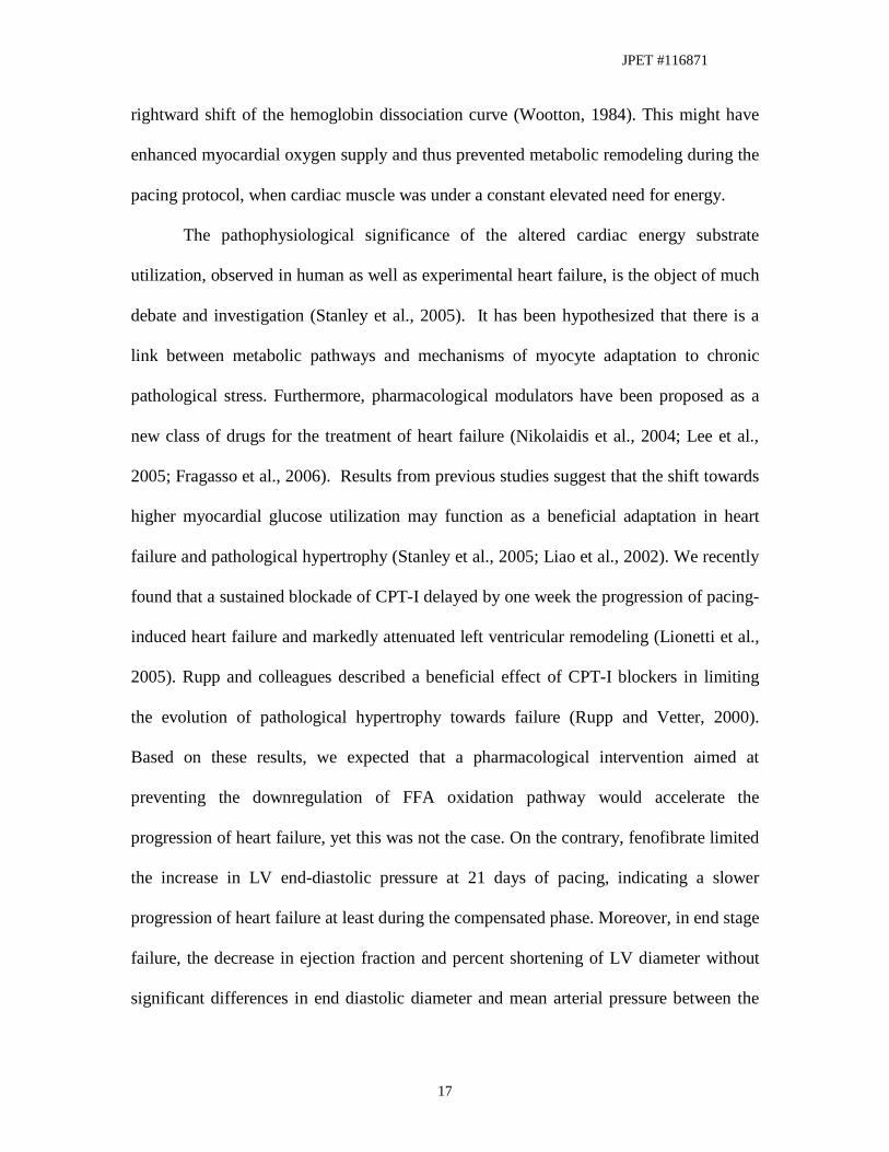
Figure 1, panel A, shows changes in LV end-diastolic pressure (LVEDP). In this model,
we consider a LVEDP of 25 mmHg as a hallmark of end-stage failure (Recchia et al.,
1998). Chronic fenofibrate administration significantly delayed the increase of LV end-
diastolic pressure at three weeks of pacing, however this beneficial effect was only
transient and did not further affect the progression towards end-stage failure that occurred
at the same time point in both groups. Alterations of other key hemodynamic parameters
(panels B-D) followed the typical pattern previously found in this model of failure
(Recchia et al., 1998), with a marked reduction in mean arterial pressure, LV systolic
pressure and dP/dtmax, an index of systolic function. Fenofibrate treatment did not affect
these changes. Left circumflex coronary artery blood flow prior to the initiation of
pacing was 41.6±2.0 mL/min in HF and 39.4±3.5 mL/min in HF+Feno (N.S.) and did not
change over the progression of failure. Spontaneous heart rate increased significantly
within each group from 97.2±4.5 to 133.1±5.8 beats/min in HF and from 94.0±6.2 to 121
±4.9 in HF+Feno, with no difference between groups.
LV Ejection Fraction and Dimensions
In end stage failure, LV ejection fraction was significantly higher in HF+Feno compared
to untreated HF (Figure 2A). LV end-diastolic diameter increased whereas wall
thickness decreased significantly during the progression of dilated cardiomyopathy, with
no significant differences between the two groups (Figure 2B-D). LV end-systolic
diameter was not significantly different between the two groups (data not shown).

JPET #116871
13
Consistent with the changes in ejection fraction, we found that the percent shortening in
LV diameter during systole in HF and HF+Feno was, respectively, 0.35±0.02 and
0.36±0.03 at baseline (N.S.), 0.22±0.02 and 0.20±0.01 at 21 days (N.S.) and 0.16±0.01
and 0.23±0.03 (p<0.05) in end stage failure. The heart weight/body weight ratio was not
different among three groups (8.4±0.2 g/kg in normal dogs, 9.0±0.3 g/kg in HF and
8.5±0.4 g/kg in HF+Feno).
Circulating lipids Circulating triglyceride levels were not affected by the progression of HF, however
treatment with fenofibrate resulted in lower triglyceride concentration at 21 days of
pacing in and also in end stage failure. (Figure 3). On the other hand, circulating
cholesterol was significantly reduced during pacing in the untreated HF dogs, but this
decrease was significantly more pronounced in the fenofibrate treated dogs, with a
progressive fall from 21 days to end stage failure.
Cardiac metabolism
There were no significant differences in arterial concentrations of FFA or lactate among
control, HF, and HF+Feno (Table 1), however plasma glucose was approximately 20%
higher in the untreated HF.
MVO2 was not significantly different among the three experimental groups (Figure 4A),
however FFA oxidation was reduced by approximately 40% in HF compared to control
(Figure 4B), while glucose oxidation was increased by approximately 2.5 fold in HF

JPET #116871
14
compared to control (Figure 4C). Fenofibrate treatment prevented these metabolic
changes. Net lactate uptake was similar among groups (Figure 4D).
Metabolic Enzymes
The activity of the citric acid cycle enzyme citrate synthase was reduced by 33% in HF
compared to control hearts, but there was no significant change versus control in the
HF+Feno group (Figure 5). The activity of medium chain acyl-CoA dehydrogenase
(MCAD) was also significantly decreased in HF compared to control, which was
prevented in the HF+Feno group. Similarly, MCAD gene expression was decreased by
55% in HF compared to normal, but was not significantly reduced in the HF+Feno group
(Figure 5).
JPET #116871
15
Discussion
The present study shows that a sustained activation of PPARα during pacing-
induced heart failure prevents alterations in cardiac FFA and glucose oxidation and the
downregulation of medium chain acyl-CoA dehydrogenase (MCAD), one of the key
enzymes involved in mitochondrial FFA beta-oxidation cycle, without accelerating the
progression towards decompensated heart failure. These results are surprising, since we
have previously found that an opposing intervention, i.e. pharmacological inhibition of
mitochondrial fatty acid oxidation, delays the onset of decompensation in pacing-induced
heart failure (Lionetti et al., 2005). This is the first evidence to suggest that the
downregulation of myocardial fatty acid oxidation and acceleration in glucose oxidation
in advanced heart failure in not a necessary compensatory mechanism to optimize cardiac
energetics, as previously thought.
Fenofibrate is a member of the fibrate class that displays hypolipidemic actions
(Adkins and Faulds, 1997; Staels et al., 1998) and is clinically used in patients with
hyper-triglyceridemia. To a lesser extent, it can also lower plasma cholesterol levels. The
triglyceride-lowering activity of fibrates is attributed to both inhibition of hepatic fatty
acid synthesis and increased catabolism of very low density lipoproteins (Staels et al.,
1998), likely mediated through the interaction with nuclear receptor PPARα (Staels et al.,
1998; Gervois et al., 2000). Once activated by fenofibrate, PPARα forms a complex with
retinoid X receptor-α and with the co-activator PGC-1 and binds sequence-specific target
elements of a number of gene promoters (Vega et al., 2000; Berger and Moller, 2002). In
the present study, we administered fenofibrate at the daily dose that is normally used in
humans, therefore, given the difference in body size, the dogs received approximately

JPET #116871
16
threefold the amount recommended for clinical use. Other studies in animals have
employed a much higher dosage of fenofibrate (Morgan et al., 2006), but we chose to
maintain a regimen closer to the human therapeutical range. The efficacy of the dose
employed in our study is demonstrated by the significant effects on lipid plasma levels
and on cardiac energy substrate metabolism in treated, compared to untreated HF dogs.
After 21 days of pacing, fenofibrate lowered triglyceride and cholesterol by 40% and
15%, respectively, very close to the effects recently found by other authors who
administered 10 mg/kg/day to dogs for 15 days (Serisier et al., 2006). Moreover, in
HF+Feno circulating cholesterol was decreased further in end stage failure compared to
21 days. Although our study was not designed to assess the pharmacokinetics of
fenofibrate, taken together these data indicate that the dose used in the present study was
sufficient to reach pharmacological efficacy over the entire period of pacing.
The main goal of our study was to prevent the downregulation of the FFA
oxidative pathway by activating PPARα. Cardiac mRNA level and activity of MCAD, a
key enzyme of mitochondrial fatty acid β-oxidation, were decreased by heart failure as
previously shown in this model (Osorio et al., 2002; Lionetti et al., 2005) and humans
(Sack et al., 1996), but was not significantly different from control dogs in fenofibrate-
treated HF animals, consistent with a preserved activation state of PPARα. Since there
were modest differences in plasma FFA concentrations among groups, these results
strongly suggest that the greater rate of FFA oxidation in the HF+Feno group compared
to the untreated HF animals was due to direct myocardial effects of fenofibrate. We
cannot exclude, however, other PPARα-independent mechanisms of action accounting
for the observed metabolic effects. For instance, it is known that fibrates can cause a
JPET #116871
17
rightward shift of the hemoglobin dissociation curve (Wootton, 1984). This might have
enhanced myocardial oxygen supply and thus prevented metabolic remodeling during the
pacing protocol, when cardiac muscle was under a constant elevated need for energy.
The pathophysiological significance of the altered cardiac energy substrate
utilization, observed in human as well as experimental heart failure, is the object of much
debate and investigation (Stanley et al., 2005). It has been hypothesized that there is a
link between metabolic pathways and mechanisms of myocyte adaptation to chronic
pathological stress. Furthermore, pharmacological modulators have been proposed as a
new class of drugs for the treatment of heart failure (Nikolaidis et al., 2004; Lee et al.,
2005; Fragasso et al., 2006). Results from previous studies suggest that the shift towards
higher myocardial glucose utilization may function as a beneficial adaptation in heart
failure and pathological hypertrophy (Stanley et al., 2005; Liao et al., 2002). We recently
found that a sustained blockade of CPT-I delayed by one week the progression of pacing-
induced heart failure and markedly attenuated left ventricular remodeling (Lionetti et al.,
2005). Rupp and colleagues described a beneficial effect of CPT-I blockers in limiting
the evolution of pathological hypertrophy towards failure (Rupp and Vetter, 2000).
Based on these results, we expected that a pharmacological intervention aimed at
preventing the downregulation of FFA oxidation pathway would accelerate the
progression of heart failure, yet this was not the case. On the contrary, fenofibrate limited
the increase in LV end-diastolic pressure at 21 days of pacing, indicating a slower
progression of heart failure at least during the compensated phase. Moreover, in end stage
failure, the decrease in ejection fraction and percent shortening of LV diameter without
significant differences in end diastolic diameter and mean arterial pressure between the

JPET #116871
18
two groups indicate that fenofibrate partially preserved cardiac contractility. Despite this
beneficial effect on systolic function, fenofibrate did not delay the onset of overt
congestive failure. One possibility is that PPARα activation does not affect molecular
mechanisms responsible for the development of dilated cardiomyopathy. Perhaps the
beneficial effects previously found with CPT-1 inhibitors are related to the inhibition of
this specific enzyme, rather than to a generic reduction of FFA oxidation. However, our
results are consistent with the lack of effects of fenofibrate on cardiac function and left
ventricular remodeling described in a rat model of post-infarct heart failure (Morgan et
al., 2006). In that study, fenofibrate was administered at the dose of 150 mg/kg/day and,
although the rate of cardiac FFA and carbohydrate oxidation was not determined, the
authors found a more dramatic upregulation of MCAD mRNA and activity in fenofibrate-
treated infarcted hearts, indicative of a potentiation of the fatty acid oxidation pathway.
In conclusion, the PPARα agonist fenofibrate can effectively prevent changes in
myocardial substrate metabolism that occur in pacing-induced heart failure. Such a
remarkable effect on energy substrate selection is accompanied by a modest improvement
of cardiac function during the progression of the disease, whereas PPARα activation does
not affect the time to terminal decompensation. These findings prompt new questions
about the implications of cardiac metabolic alterations in the pathophysiology of dilated
cardiomyopathy.

JPET #116871
19
References
Adkins JC and Faulds D. (1997) Micronised fenofibrate: a review of its
pharmacodynamic properties and clinical efficacy in the management of
dyslipidaemia. Drugs 54:615–33 .
Barger PM and Kelly DP (2000) PPAR signaling in the control of cardiac energy
metabolism. Trends Cardiovasc Med 10:238-45.
Belardinelli R and Purcaro A (2001) Effects of trimetazidine on the contractile response
of chronically dysfunctional myocardium to low-dose dobutamine in ischaemic
cardiomyopathy. Eur Heart J 22: 2164–2170.
Berger J and Moller DE (2002) The mechanisms of action of PPARs. Annu Rev Med 53:
409–435.
Bersin RM, Wolfe C, Kwasman M, Lau D, Klinski C, Tanaka K, Khorrami P, Henderson
GN, de Marco T, and Chatterjee K (1994) Improved hemodynamic function and
mechanical efficiency in congestive heart failure with sodium dichloroacetate. J Am
Coll Cardiol 23: 1617–1624.
JPET #116871
20
Forman B.M., J. Chen and R.M. Evans (1997) Hypolipidemic drugs, polyunsaturated
fatty acids, and eicosanoids are ligands for peroxisome proliferators-activated
receptors α and ß. Proc Natl Acad Sci U S A 94: 4312–4317.
Fragasso G, Palloshi A, Puccetti P, Silipigni C, Rossodivita A, Pala M, Calori G, Alfieri
O, Margonato A (2006) A randomized clinical trial of trimetazidine, a partial free
fatty acid oxidation inhibitor, in patients with heart failure. J Am Coll Cardiol 48:
992-998.
Gervois P, Torra IP, Fruchart JC and Staels B (2000) Regulation of lipid and lipoprotein
metabolism by PPAR activators. Clin Chem Lab Med 38: 3–11.
Karbowska J, Kochan Z, and Smolenski RT (2003) Peroxisome proliferator-activated
receptor alpha is downregulated in the failing human heart. Cell Mol Biol Lett 8: 49–
53.
Lee L, Campbell R, Scheuermann-Freestone M, Taylor R, Gunaruwan P, Williams L,
Ashrafian H, Horowitz J, Fraser AG, Clarke K, Frenneaux M (2005) Metabolic
modulation with perhexiline in chronic heart failure: a randomized, controlled trial of
short-term use of a novel treatment. Circulation 112: 3280-3288.
Lei B, Lionetti V, Young ME, Chandler MP, D' Agostino C, Kang E, Altarejos M,
Matsuo K, Hintze TH, Stanley WC, and Recchia FA (2004) Paradoxical

JPET #116871
21
downregulation of the glucose oxidation pathway despite enhanced flux in severe
heart failure. J Mol Cell Cardiol 36: 567–576.
Liao R, Jain M, Cui L, D'Agostino J, Aiello F, Luptak I, Ngoy S, Mortensen RM and
Tian R (2002) Cardiac-specific overexpression of GLUT1 prevents the development
of heart failure attributable to pressure overload in mice Circulation 15;106:2125-31.
Lionetti V, Linke A, Chandler MP, Young ME, Penn MS, Gupte S, d'Agostino C, Hintze
TH, Stanley WC, and Recchia FA (2005) Carnitine palmitoyl transferase-I inhibition
prevents ventricular remodeling and delays decompensation in pacing-induced heart
failure. Cardiovasc Res 66: 274–281.
Martin MA, Gomez MA, Guillen F, Bornstein B, Campos Y, Rubio JC, de la Calzada
CS, and Arenas J (2000) Myocardial carnitine and carnitine palmitoyltransferase
deficiencies in patients with severe heart failure. Biochim Biophys Acta 1502: 330–
336.
Morgan EE, Rennison JH, Young ME, McElfresh TA, Kung TA, Tserng KY, Hoit BD,
Stanley WC and Chandler MP (2006) Effects of chronic activation of peroxisome
proliferator-activated receptor-alpha or high-fat feeding in a rat infarct model of heart
failure. Am J Physiol Heart Circ Physiol 290: H1899-1904.

JPET #116871
22
Nikolaidis LA, Elahi D, Hentosz T, Doverspike A, Huerbin R, Zourelias L, Stolarski C,
Shen YT and Shannon RP (2004) Recombinant glucagon-like peptide-1 increases
myocardial glucose uptake and improves left ventricular performance in conscious
dogs with pacing-induced dilated cardiomyopathy. Circulation 110: 955-61.
Osorio JC, Stanley WC, Linke A, Castellari M, Diep QN, Panchal AR, Hintze TH,
Lopaschuk GD, and Recchia FA (2002) Impaired myocardial fatty acid oxidation and
reduced protein expression of retinoid X receptor-alpha in pacing-induced heart
failure. Circulation 106: 606–612.
Recchia FA, McConnell PI, Bernstein RD, Vogel TR, Xu X, and Hintze TH (1998)
Reduced nitric oxide production and altered myocardial metabolism during the
decompensation of pacing-induced heart failure in the conscious dog. Circ Res 83:
969–979.
Rosenblatt-Velin N, Montessuit C, Papageorgiou I, Terrand J, and Lerch R (2001)
Postinfarction heart failure in rats is associated with upregulation of GLUT-1 and
downregulation of genes of fatty acid metabolism. Cardiovasc Res 52: 407–416.
Rupp H and Vetter R (2000) Sarcoplasmic reticulum function and carnitine
palmitoyltransferase-1 inhibition during progression of heart failure. Br J Pharmacol
131: 1748–1756.

JPET #116871
23
Sack MN, Rader TA, Park S, Bastin J, McCune SA, and Kelly DP (1996) Fatty acid
oxidation enzyme gene expression is downregulated in the failing heart. Circulation
94: 2837–2842.
Serisier S, Briand F, Ouguerram K, Siliart B, Magot T and Nguyen P (2006) Fenofibrate
lowers lipid parameters in obese dogs. J Nutr 136: 2037S-2040S.
Staels B and Fruchart JC (2005) Therapeutic roles of peroxisome proliferator-activated
receptor agonists. Diabetes 54: 2460-2470.
Staels B, Dallongeville J, Auwerx J, Schoonjans K, Leitersdorf E and Fruchart JC (1998)
Mechanism of action of fibrates on lipid and lipoprotein metabolism. Circulation 98:
2088–93.
Stanley WC, Recchia FA and Lopaschuk GD (2005) Myocardial substrate metabolism in
the normal and failing heart. Physiol Rev 85: 1093-129.
Vega RB, Huss JM, and Kelly DP (2000) The coactivator PGC-1 cooperates with
peroxisome proliferator-activated receptor alpha in transcriptional control of nuclear
genes encoding mitochondrial fatty acid oxidation enzymes. Mol Cell Biol 20: 1868–
1876.
JPET #116871
24
Wootton R (1984) Analysis of the effect of bezafibrate on the oxygen dissociation curve
of human hemoglobin. FEBS Lett 171: 187-191.
Young ME, Laws FA, Goodwin GW, and Taegtmeyer H (2001) Reactivation of
peroxisome proliferator-activated receptor alpha is associated with contractile
dysfunction in hypertrophied rat heart. J Biol Chem 276: 44390–44395.

JPET #116871
25
Footnotes
This study was supported by the National Heart, Lung, and Blood Institute Grant P01-
HL-74237 (F.A. Recchia and W.C. Stanley).

JPET #116871
26
Legends For Figures
Figure 1. Changes in LV end-diastolic pressure (LVEDP, panel A), mean aortic pressure
(MAP, panel B), LV systolic pressure (LVSP, panel C) and dP/dtmax (panel D) during the
progression of pacing induced HF. n= 8 for both groups. Data are mean ±SEM. * p<0.05
vs day 0 (baseline), # p<0.05 between untreated HF and HF+Feno.
Figure 2. Changes in LV ejection fraction (LVEF, panel A), LV end-diastolic diameter
(LVEDD, panel B), end-diastolic thickness of the LV free wall (LVFWT, panel C) and
end-diastolic thickness of the interventricular septum (IVST, panel D) during the
progression of pacing induced HF. n=8 for both groups. Data are mean ±SEM. *P<0.05
vs day 0 (baseline). # p<0.05 between untreated HF and HF+Feno.
Figure 3. Changes in arterial concentration of triglyceride (panel A) and cholesterol
(panel B) during the progression of pacing induced HF. n= 6 for both groups. Data are
mean ±SEM. * p<0.05 vs day 0 (baseline), † p<0.05 vs 21 days of pacing, # p<0.05
between untreated HF and HF+Feno
Figure 4. Changes in MVO2 and cardiac FFA oxidation, glucose oxidation and net
lactate uptake in HF and HF+Feno compared to normal dogs (control). N=8 for control
and HF and n=7 for HF+Feno. Data are mean ±SEM. * p<0.05 vs control; # p<0.05
between untreated HF and HF+Feno.

JPET #116871
27
Figure 5. Activity of citrate synthase (CS) and mRNA expression and activity for
medium chain acyl-CoA dehydrogenase (MCAD). N=8 for control and HF and n=7 for
HF+Feno. Data are mean ±SEM. * p<0.05 vs control.

JPET #116871
28
Table 1. Arterial concentrations of cardiac metabolic substrates and lipids
*p<0.05 vs control.
Control
Heart Failure
Heart Failure +
Fenofibrate
FFA (mM)
0.69 + 0.05
0.45+0.04
0.69+0.15
Glucose (mM)
4.42 + 0.20
5.32 + 0.26*
4.60+0.27
Lactate (mM)
0.83 + 0.08
0.88 + 0.11
0.63+0.08
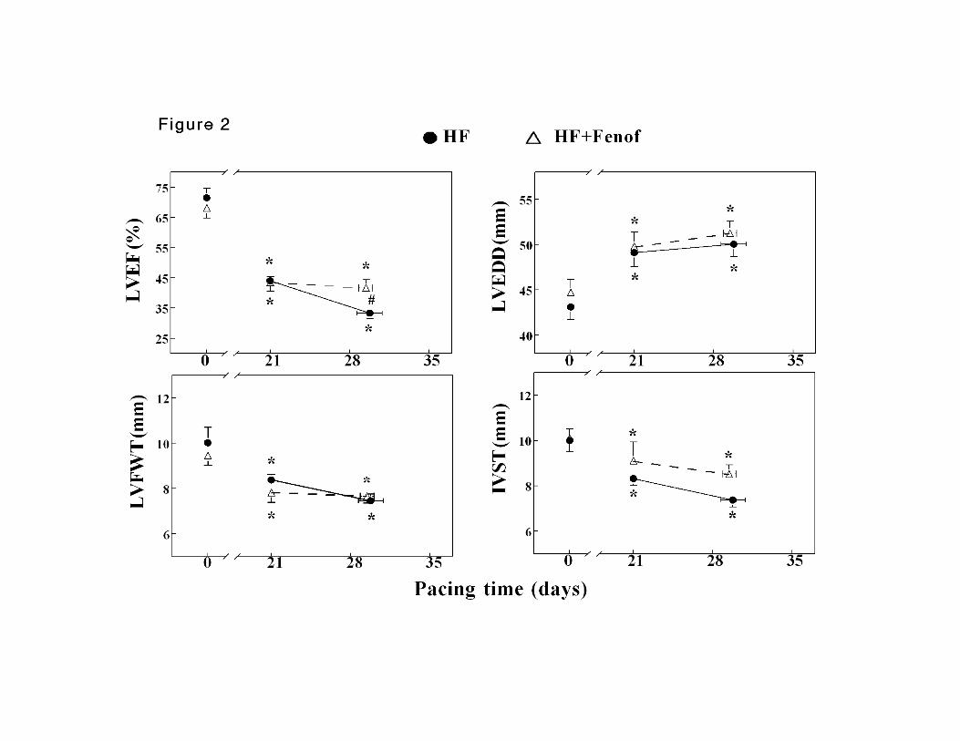


Figure 4

Figure 5


