Embed Size (px)
Citation preview
molecules
Review
Chitin and Chitosan Derivatives as BiomaterialResources for Biological and Biomedical Applications
Saravut Satitsri and Chatchai Muanprasat *
Chakri Naruebodindra Medical Institute, Faculty of Medicine Ramathibodi Hospital, Mahidol University,Bang Phli, Samut Prakarn 10540, Thailand; [email protected]* Correspondence: [email protected]; Tel.: +66-2-839-5158
Academic Editors: Mohamed Samir Mohyeldin, Katarína Valachová and Tamer M TamerReceived: 21 November 2020; Accepted: 10 December 2020; Published: 16 December 2020
�����������������
Abstract: Chitin is a long-chain polymer of N-acetyl-glucosamine, which is regularly found in theexoskeleton of arthropods including insects, shellfish and the cell wall of fungi. It has been knownthat chitin can be used for biological and biomedical applications, especially as a biomaterial for tissuerepairing, encapsulating drug for drug delivery. However, chitin has been postulated as an inducer ofproinflammatory cytokines and certain diseases including asthma. Likewise, chitosan, a long-chainpolymer of N-acetyl-glucosamine and d-glucosamine derived from chitin deacetylation, and chitosanoligosaccharide, a short chain polymer, have been known for their potential therapeutic effects,including anti-inflammatory, antioxidant, antidiarrheal, and anti-Alzheimer effects. This reviewsummarizes potential utilization and limitation of chitin, chitosan and chitosan oligosaccharide in avariety of diseases. Furthermore, future direction of research and development of chitin, chitosan,and chitosan oligosaccharide for biomedical applications is discussed.
Keywords: chitin; chitosan; chitosan oligosaccharide
1. Introduction
1.1. Chitin
Chitin is long-chain polymers of N-acetylglucosamine, presence of which has been experimentallyconfirmed in unicellular (diatoms, protists, fungi) as well as in multicellular (sponges, corals, mollusks,worms and arthropods) organisms [1]. Chitin has been used as supplementary for nutraceuticalfood, pharmaceutical products as well as 3D scaffolds for biomedicine [2–4] and technologicalapplications [5–8]. Chitin contains thermostability which can be synthesized in the high temperatureprocess [9]. Moreover, chitin has a high tolerance to high chemical concentration that generates thedeposition of metals such as copper into chitin via electrochemical procedure at room temperature [10].The linear polymer of chitin contains β-(1,4)-N-acetyl-d-glucosamines, which are linked by glycosidicbond. There are three isoforms of chitin including α-chitin, β-chitin and γ-chitin [11]. The differentforms of chitin depend on the arrangement of side chain of backbone. For instance, the structureof α-chitin is parallel chain arrangement, whereas the structure of β-chitin is antiparallel chainarrangement. α-chitin is frequently found in nature including exoskeleton of arthropods. α-chitin hasbeen frequently applied for tissue engineering [12,13]. In contrast to α-chitin, β-chitin is mostly foundin squid pen. β-chitin has been also applied for biomaterial usage such as wound healing [14,15],preservative agents for methylene blue [16], and biological application such as an enhancer of saltinessperception [17]. γ-chitin has been gathered from cocoon of the moth (Orgyia dubia), and it is foundthat the structure of γ-chitin is similar to α-chitin rather than β-chitin, but the surface morphology ofγ-chitin is consisted of microfibers, whereas α-chitin and β-chitin are composed of nanofibers [18].
Molecules 2020, 25, 5961; doi:10.3390/molecules25245961 www.mdpi.com/journal/molecules
Molecules 2020, 25, 5961 2 of 25
1.2. Chitosan
Chitosan is derived from chitin by deacetylation. The major component of chitosan is themixture between N-acetyl-d-glucosamine and β-(1,4)-linked-d-glucosamine. Chitosan has amounts ofN-acetyl-d-glucosamine less than β-(1,4)-linked-d-glucosamine [19]. Chitosan is generally applied asbiomaterials, especially for drug delivery system and use in combination with other substances forimproving their therapeutic effects [20,21].
1.3. Chitosan Oligosaccharide
Chitosan oligosaccharide or chitooligosaccharide is oligomers of chitosan. Chitosan oligosaccharidehas the degree of polymerization of <55 and molecular weight of <10 kDa [22]. Chitosan oligosaccharideshave a variety of biomedical applications such as drug delivery system [20], functional food [23],as well as the drug against acne vulgaris [24].
In this review, we emphasize the potential application of chitin, chitosan and their oligosaccharidesas therapeutics and useful biomaterials in a variety of human diseases. Furthermore, we discuss thelimitation of chitin and chitosan oligosaccharide, which will be helpful in guiding future direction ofresearch and development based on these molecules.
2. Potential Applications of Chitin and Chitosan Oligosaccharides
2.1. Neurological and Musculoskeletal Diseases
Chitin has been proposed as biomaterials for neural treatment. For instance, chitin was utilizedwith carbon nanotube as a scaffold for neural growth [25]. Chitin as a biological absorbable tubeor catheter was effectively used as the bridge for sural nerve grafts in a rat sciatic nerve defectmodel [26,27]. Chitin hydrogel repaired cartilage injury by protecting chondrocytes from apoptosis andpromoting immunomodulation of macrophage and chondrogenesis [28]. The combination of chitinas 2,2,6,6-Tetramethylpiperidine-1-oxyl (TEMPO)-oxidized sacchachitin nanofibers (TOSCNFs) andchitosan-activated platelet-rich plasma (cPRP) induced healing effect in corneal damage by promotingcell proliferation and cell migration in Statens Seruminstitut rabbit corneal (SIRC) epithelial cells [29].Interestingly, chitin is not only proposed as potential therapy, but also claimed as a molecular markerfor neurological diseases. In Alzheimer’s disease, chitin is elevated and accumulated within the brainand facilitates a scaffolding for amyloid-β deposition [30–32]. Fungal chitin was also detected inbrain tissues from Alzheimer’s disease patients [33]. Moreover, chitin accumulation was found inmultiple sclerosis patients [34]. Interestingly, both microglia and neurons produce N-acetylglucosaminepolymerization, which causes neurotoxicity in Alzheimer’s disease [35]. Furthermore, chitin derivedfrom demosponge Aplysina aerophoba was used as 3D scaffold for human bone marrow-derivedstromal cells, and it promoted cell proliferation, cell bridging formation and metabolic activity with notoxicity [36]. Interestingly, chitin derived from demosponge Ianthella basta was applied as a cryopreservativeagent that effectively retained adipogenic differentiation in human mesenchymal stromal cells [4].
In contrast to chitin, chitosan and chitosan oligosaccharide have been widely studied for thebeneficial effect on neurological diseases, especially Alzheimer’s disease. A water-soluble form ofchitosan inhibited the production of proinflammatory cytokines including TNFα and IL-6 as wellas inducible nitric oxide synthase (iNOS) in human astrocytoma cell line CCF-STTG1 stimulatedwith IL-1β and Aβ fragments (25–35) [37]. It is known that acetylcholinesterase inhibition is thetarget for Alzheimer’s disease therapy. Chitosan oligosaccharide with 90% deacetylation and lowmolecular weight (1 to 5 kDa) inhibited the protein expression of acetylcholinesterase and Aβ fragment(25–35)-induced acetylcholinesterase activity in PC12 cell lines [38]. Furthermore, caffeic acid conjugatedchitosan oligosaccharide effectively inhibitedβ-site amyloid precursor protein-cleaving enzyme activity,which was the rate limiting step of Aβ peptide formation in Alzheimer’s disease [39]. Interestingly,chitosan oligosaccharide at 500 µg/mL inhibited β-site amyloid precursor protein cleaving enzyme 1(BACE1) activity and protein expression in HEK293 APPswe cells [40]. Chitosan oligosaccharide
Molecules 2020, 25, 5961 3 of 25
inhibited αβ aggregation, inhibited Aβ1–42 fibrils formation, and induced fibril destabilization inoligomeric Aβ-induced neurotoxicity and oxidative stress in rat hippocampal neurons [41]. In addition,0.1% of chitosan oligosaccharide injected into the spatium intermusculare around the biceps femorismuscle inhibited scar formation and promoted regeneration of axons, as well as sensory and motorfunction in a mouse model of sciatic nerve injury [42]. Interestingly, ingestion of chitosan oligosaccharide(10 mg/kg/day) alleviated inflammatory signal including COX-2 expression in the synovium of ananterior cruciate ligament (ACL) transection-induced osteoarthritis rabbit model [43]. Ingestion ofchitosan oligosaccharide at least 200 mg/kg/day recovered cognitive deficiency in Aβ1–42-inducedlearning and memory loss rats [44]. Chitosan oligosaccharide also protected hippocampal neuronfrom Aβ peptide [45]. Furthermore, low molecular weight chitosan activated mitogenic response toplatelet-derived growth factor (PDGF) in vascular smooth muscle cells [46].
Chitosan has been applied as biomaterials for neural therapy. For instance, amphiphaticcarboxymethyl-hexanoyl chitosan hydrogel increased cell viability and maintained stem-cell-like geneexpression of induced pluripotent stem cells applied for corneal reconstruction [47]. Chitosan-polylactidefiber was utilized for nerve growth factor in PC12 cell lines [48]. Chitosan oligosaccharide with calciumsilicate and gelatin was applied for implantation of cortical bone repair and bone fracture fixation [49].Chitosan was coated into nanoparticles for delivering antiamyloid antibody as a drug for Alzheimer’sdisease. It was found that chitosan coating improved aqueous dispersibility and stability of vehicleduring lyophilization [50]. Chitosan in the form of chitosan beads effectively interfered with amyloid-βaggregation [51]. Chitosan was utilized as nanocapsules for delivering p38 inhibitor to the brain bynasal administration [52]. Chitosan with polyvinyl alcohol nanofibrous scaffold promoted skeletalmuscle regeneration by increasing cell viability, cell adhesion, cell growth, and cell spread on thescaffold [53]. Chitosan with laminin and poly (lactic-co-glycolic acid) effectively repaired nerve injuryby promoting nerve regeneration [54]. Chitosan combined to hyaluronate regenerated nerve functiondefect in parotidectomy rabbit model by promoting scar formation, increasing nerve fibers, thickeningmyelin sheath, and promoting nerve conduction velocity [55]. Furthermore, chitosan, as chitosan tubesor incorporated to mesenchymal stem cells or keratin, was also utilized for nerve repairing [56–58].
2.2. Cardiovascular and Hematological Diseases
Chitin has been incorporated with other substances for treatment of cardiovascular disease.For instance, chitin with glucan and polyphenols from pomegranate recovered endothelial dysfunctionby reducing inflammatory marker in the liver and adipose tissues and promoting NO synthase inapolipoprotein E deficient mice (apoE−/−) with high fat diet [59]. Furthermore, chitin combined tographene oxide as aerogel beads effectively absorbed excessive bilirubin in the blood [60]. Chitinnanogel with rectorite nanocomposite stopped bleeding within 121 s in rat tail vein, and promotedhigher hemostatic activity compared to chitosan-based hemostatic products [61]. Furthermore, Chitinderived from demosponge Ianthella labyrinthus and chitin derived from spider Caribena versicolor wereeffectively served as a 3D scaffold for culturing induced pluripotent stem-cell-derived cardiomyocytesas well as commercial extracellular matrix [62,63].
It has been known that orally intake of 5% chitosan in the diet reduces serum cholesterol andatherogenesis inhibition in apolipoprotein E-deficient mouse model [64]. Chitosan as a supplementaldiet downregulated the markers involving obesity such as leptin in high fat diet rats [65]. Chitosanoligosaccharide decreased serum cholesterol by promoting accumulation of cholesterol in liver, bile,and feces with reverse cholesterol transport pathway [66]. It is known that chitosan oligosaccharidereduces serum cholesterol. Chitosan oligosaccharide increased cell surface expression of low-densitylipoprotein receptor (LDLR) and increased lipid droplets in HepG2 cells, suggesting that chitosanoligosaccharide effectively reduced serum lipids by facilitating the accumulation of lipids into thecells [67]. Furthermore, chitosan oligosaccharide downregulated mRNA expression of LPS-inducedE-selectin and intercellular adhesion molecule-1, which are a part of the inflammatory responses inendothelial cells via MAPK inhibition [68]. Oral intake of chitosan oligosaccharide also reduced the
Molecules 2020, 25, 5961 4 of 25
marker of atherosclerosis including the lesion area in aorta or plaque area in aortic roots, and greatlyreduced cholesterol and triglyceride in apolipoprotein E deficient mice (apoE−/−) [69]. The combinationof chitosan oligosaccharide ingestion and exercise such as running improved immune system byincreasing spleen to body weight ratio and lung to body weight ratio compared to water gavage onlyin Sprague-Dawley (SD) rats [70]. Interestingly, chitosan improved blood perfusion and promotedneovascularization by modulation of gut microbiota in a mouse hindlimb ischemia model [71].Furthermore, chitosan was used as a vehicle for drug delivery for transportation of doxorubicin toimprove the treatment of blood malignancies [72]. Chitosan in the form of synthetic CD47 antibody-chitosan/hyaluronic acid polyelectrolyte complex inhibited atherosclerotic plaques with downregulatedNLRP3 inflammasome expression in apolipoprotein E deficient mice (apoE−/−) [73].
2.3. Respiratory Diseases
It is well known that penetration of chitin into human bodies causes the production of chitinase,the enzyme that modulates immune response, and chitinase-like proteins YKL-40. YKL-40 wasassociated with increased severity in asthmatic patients [74]. YKL-40 level was also positivelycorrelated with neutrophils, sputum IL-1β, and plasma IL-6 [75]. Moreover, chitin from fungi inducedeosinophilic infiltration in a mouse model [76]. Chitin also promoted proinflammatory response byinducing proinflammatory cytokine release including IL-25 and IL-33 in human bronchial epithelialcells [77]. Chitin has been known as an adjuvant for immune response. It was demonstrated that chitinfrom house dust mite promoted airway hypersensitivity in ovalbumin-induced airway inflammationvia a TLR-2 dependent pathway [78]. Furthermore, it is known that chitin is the major componentof fungi in their cell wall. Chitin exposure induced macrophage activation which upregulates theexpression of chitin degrading enzyme chitotriosidase [79]. Chitotriosidase was also involved in lungdiseases such as tuberculosis, chronic obstructive lung diseases [80]. Apart from chitotriosidase, acuteexposure to the fungal pathogen Aspergillus fumigatus promoted the function of acidic mammalianchitinase, which determined the severity of fungal asthma [81]. Therefore, inhibition of chitotriosidaseand acidic mammalian chitinase is regarded as a drug target for respiratory diseases [82]. However,it has been shown that a chitin analog AVR-25 partially alleviated pulmonary dysregulation ina hyperoxia-induced experimental mouse model of bronchopulmonary dysplasia by suppressinginflammation [83]. These findings indicate that chitin may have both beneficial and detrimental effects.
In contrast to chitin, chitosan oligosaccharides have been demonstrated to have potentialbeneficial effects on respiratory diseases. Oral intake of chitosan oligosaccharide (500 mg/kg) ata single dose alleviated particulate matter (PM) 2.5-induced lung inflammation by decreasing lactatedehydrogenase, IL-8, and TNF-α in PM 2.5-induced rat model [84]. Chitosan oligosaccharide (100 kDaand 90% deacetylation) prevented inflammation, oxidative stress and apoptosis in the lung tissuesof blast injury-induced mice by diminishing protein expression of p38 and ADMA (an inhibitorof endogenous nitric oxide synthase that positively correlated with hypertension) and recoveringdimethylarginine dimethylaminohydrolase 1 (DDAH1), a hydrolase of ADMA [85]. Furthermore,oral intake of 16 mg/kg/day of low molecular weight chitosan oligosaccharide reduced IgE-inducedairway inflammation in mice by downregulating both protein level and mRNA level of proinflammatorycytokines including IL-4, IL-5, IL-13, and TNF-α [86].
2.4. Renal Diseases
There are reports demonstrating the effect of chitin on renal diseases. Oral ingestion of chitin assurface-deacetylated chitin nano-fiber (40 mg/kg/day) reduced uremic toxins including oxidants innephrectomized rats [87]. However, upregulated YKL-40 level in urine and plasma represents thebiomarker of acute kidney injury [88,89]. In contrast, chitosan and chitosan oligosaccharide havepotential therapeutic effects on kidney diseases. The combination of chitosan with gynostemma andmotherwort was used for protecting chronic renal failure by inhibiting inflammation in adenine-inducedrat chronic renal failure [90]. Chitosan incorporated with gallic acid reduced the formation of calcium
Molecules 2020, 25, 5961 5 of 25
oxalate crystal, which was mainly a kidney stone, and had antioxidative effects [91]. Chitosan as acat diet improved kidney function and quality of life in elderly cats with 3 and 4 International RenalInterest Society (IRIS) stages [92]. It was also shown that chitosan oligosaccharide improved kidneyfunction in streptozotocin-induced diabetic rats [93]. It was found that 0.1% chitosan oligosaccharidewith more than 90% deacetylation prevented glycerol-induced acute renal failure in rats by decreasingrenal dipeptidase activity, a diagnostic marker of acute renal failure [94]. Furthermore, chitosanoligosaccharide with carboxymethyl group relieved renal injury induced by doxorubicin and promotedantioxidative effects in rats [95]. Interestingly, chitosan oligosaccharide triggered G2/M phase arrest,promoted endoplasmic reticulum stress pathway, and inhibited tumor growth in human renal carcinomacells and xenograft tumor models [96]. Moreover, chitosan oligosaccharide had a chelating propertywhich detoxified depleted uranium cytotoxicity in human renal proximal tubular epithelial cells [97].Recently, chitosan oligosaccharide at the concentration of 50 and 100 µg/mL was shown to reducerenal cyst growth via CaMKKβ-induced AMPK activation without cytotoxicity [98]. Furthermore,the detoxification property of chitosan has been applied in hemodialysis patients. The ingestion ofchitosan decreased the level of indoxyl sulfate and phosphate by binding to these molecules [99].Chitosan has also been applied as siRNA delivery system targeting kidney. For instance, chitosannanoplex effectively covered siRNA for knocking down PDGF-B and PDGFR-beta [100]. Chitosan wasalso used as biomaterials in combination with collagen for culturing human renal proximal tubularcells [101]. The potential beneficial effects of chitosan oligosaccharide on kidney diseases have beenalso reviewed elsewhere [102].
2.5. Gastrointestinal Diseases and Gut Microbiota
Many previous studies demonstrated the potential effects of chitin and chitosan oligosaccharide ongastrointestinal disease. It was shown that chitin protected intestinal barrier function in DSS-inducedcolitis in a protochordate model [103]. Chitin combined with glucan (chitin-glucan complex) was usedas a prebiotic. This chitin-glucan complex improved the growth of Bifidobacterium, a probiotic, in a ratmodel [104]. Interestingly, intake of 4.5 g/day of chitin-glucan in food for 3 weeks increased beneficialmicrobiota metabolites including butyric, iso-valeric and caproic acids without major changes in gutmicrobiota composition [105]. Chitin has also been applied as surface-deacetylated chitin nanofiber.It was shown that oral intake of 80 mg/kg/day of surface-deacetylated chitin nanofiber decreasedhepatic injury and oxidative stress in a nonalcoholic steatohepatitis model of rats [106].
Chitosan and chitosan oligosaccharide have beneficial effects on gastrointestinal tract, especiallyas nutritional supplements for animals and food supplements for human. In livestock, chitosanoligosaccharide has been used as feed additives in animal diet. It was shown that 100 mg/kg of dietarychitosan oligosaccharide supplementation promoted growth performance, reduction of diarrhea,nutrient digestibility and attenuation of E.coli K88 infection in weaning pigs [107,108]. The combinationof chitosan and zinc at the dose of 100 mg/kg as a feed additive in diet also promoted the activitiesof digestive enzymes such as amylase, reduced diarrhea and improved growth performance inweaning pigs [109]. However, chitosan mixed with probiotic such as Enterococcus faecalis did notsignificantly reduce severity of diarrhea and affect growth performance in E.coli K88-inoculatedweaning pigs [110]. Moreover, high molecular weight chitosan oligosaccharide (20 to 30 kDa) justonly increased ZO-1 expression and decreased the mRNA expression of IL-1β and TNFα in weaningpigs without affecting diarrhea, average dairy gain, gain to feed ratio, and antioxidant capacity [111],suggesting that specific forms of chitosan oligosaccharide gives the therapeutic effects in weaning pig.Furthermore, low molecular weight chitosan oligosaccharide (8 kDa) with 90% deacetylation improvedgut absorption, increased villus length and promoted intestinal cell proliferation in weaning pig [112].Oral intake of 200 mg/kg/day of chitosan oligosaccharide in drinking water also protected gut fromthe modulation of glucose metabolism and gut dysbiosis in diabetic mice [113]. At a cellular level,100 µg/mL of low molecular wight chitosan oligosaccharide (approximately 5 kDa) accelerated tightjunction assembly and inhibited cholera toxin-induced intestinal fluid secretion via CaSR-PLC-IP3
Molecules 2020, 25, 5961 6 of 25
receptor channel-mediated AMPK activation in intestinal epithelial cells [114]. Chitosan oligosaccharidesuppressed mRNA expression of proinflammatory cytokines, and inhibited the downregulation ofPPARγ in palmitic acid-induced HepG2 cells and high fat diet mice [115]. Many reports reveal thatchitosan and chitosan oligosaccharide modulate gut microbiota. Chitosan prevented gut dysbiosisand inhibited the activation of toll-like receptor and nod-like receptor signaling pathway in high fatdiet rats [116]. However, molecular weight and degree of deacetylation of chitosan oligosaccharideare major determinants of effects on human gut microbiota composition as well as therapeuticeffects. For instance, a highly deacetylated chitosan oligosaccharide decreased Bifidobacterium spp.,E. rectale/C. coccoides, C. histolyticum and Bacteroides/Prevotella populations in human gut [117]. Chitosanoligosaccharide with >95% deacetylation reduced Lactobacillus, Bifidobacterium and Desulfovibrio,deleterious bacteria that were correlated with inflammatory bowel disease [118], and increasedabundance of Akkermansia that was a good bacteria [119]. The 3 kDa chitosan oligosaccharidediminished gut dysbiosis and downregulated mRNA expression of proinflammatory cytokines inazoxymethane and dextran sulfate sodium-induced mouse model of colorectal cancer [120]. Moreover,chitosan oligosaccharide ameliorated hepatic steatosis and liver injury, and reduced triglyceride andfree fatty acid in diet-induced obese mice by downregulating inflammatory genes and modulation of gutmicrobiota [121]. Furthermore, chitosan protected liver from ischemia-reperfusion injury via regulatingBcl-2/Bax, TNF-α and TGF-β expression [122], prevented lipid metabolic disorder by combination withGanoderma polysaccharide [123], alleviated menopausal symptoms [124], and protected the gut fromischemic symptoms [71]. High molecular weight chitosan was also prepared as nanoparticles for drugdelivery in gut. For instance, 400 kDa chitosan integrated to insulin loaded chitosan nanoparticlesprolonged bioavailability of insulin release [125]. Interestingly, chitosan nanoparticle was also used asa food supplement for improvement of growth performance and immunity in weaning pigs [126].
Apart from chitin, chitosan, and chitosan oligosaccharide, there are other polysaccharides thathave therapeutic effects on gut. For instance, mannan oligosaccharide (10 µM) promoted intestinalbarrier function in T84 cells via AMPK activation [127,128]. Fructo-oligosaccharide (0.1 mg/mL),a prebiotic, accelerated intestinal tight junction reassembly via AMPK activation in T84 cells [129].
2.6. Endocrinological Diseases and Diabetes Mellitus
Chitin combined with other compounds has been shown to possess therapeutic effects. For instance,oral intake of 4.5 g/day of chitin combined with glucan reduced oxidized low-density lipoproteinin human subjects [130]. Furthermore, chitin has been applied as a biomaterial for prolongingbioavailability. For instance, injectable thermo-sensitive hydrogel based on hydroxypropyl chitin wasincorporated to salmon calcitonin to extend long-term sustained salmon calcitonin release [131].
Likewise, chitosan has been combined with other compounds that possess antidiabetic effects.For instance, 3-O-sulfochitosan was reported to reduce blood glucose in diabetic rats [132]. Chitosancombined with metformin, a type 2 diabetic drug, synergistically enhanced drug efficacy and reducedlethal effects of drug overdose [133]. Furthermore, chitosan oligosaccharide has been applied for drugdelivery system in diabetes. For instance, chitosan-microcapsulated insulin, chitosan-stabilized seleniumnanoparticles, chitosan encapsulated resveratrol, chitosan coating of TiO2 nanotube arrays for metformin,chitosan nanoparticle and chitosan hydrogel were used for improving diabetic therapy [134–142].Interestingly, the development of chitosan oligosaccharide as a drug or supplement for diabetictreatment has been studied for a decade. The proteomic data demonstrated the antidiabetic effectsand anti-obesity of orally intake chitosan oligosaccharide in ob/ob mice [143]. Oral intake of chitosanoligosaccharide (>90% deacetylation; 500 mg/kg) promoted insulin sensitivity in streptozotocin-induceddiabetic rats. Chitosan oligosaccharide (100 mg/L) also promoted cell proliferation in primary cultureislet cells and pancreaticβ-cell lines [144]. Moreover, the low molecular weight chitosan oligosaccharide(~1.2 kDa) promoted cell proliferation in primary culture islet cells and pancreatic β-cell lines as wellas improving insulin sensitivity greater than the high molecular weight chitosan oligosaccharide [145].Moreover, chitosan oligosaccharide with a molecular weight of 1.3 kDa and 55% deacetylation was used
Molecules 2020, 25, 5961 7 of 25
as an oral insulin delivery system that showed the highest effect of glucose reduction [146]. Furthermore,chitosan oligosaccharide has been used as a food supplement for diabetic treatment. For instance,GO2KA1, a commercial chitosan oligosaccharide supplement, had a beneficial effect on glucose controlin subjects with prediabetes by regulating postprandial glucose [147]. GO2KA1 promoted glucoseuptake into intestinal epithelial cells, enhanced adipocyte differentiation, and upregulated PPARγexpression [148]. GO2KA1 has been used in clinical trials. It was found that GO2KA1 effectivelyreduced postprandial blood glucose levels in subjects with impaired glucose tolerance and impairedfasting glucose [149]. However, it remains unclear whether chitosan oligosaccharide has a director indirect antidiabetic effect. It was also shown that chitosan oligosaccharide exerted antidiabeticeffects via gut microbiota modulation [116]. Chitosan oligosaccharide was used as a supplementarydrug for improving the glycemic control of sitagliptin in type 2 diabetes mellitus (T2DM) by reducinginsulin resistance and proinflammatory cytokines, and increasing insulin sensitivity [150]. Chitosanoligosaccharide combined with xanthine derivatives improved liver and kidney functions comparedto pioglitazone, a standard antidiabetic drug [151]. Chitosan oligosaccharide also upregulated theexpression of browning genes in white adipose tissues and thermogenesis of brown adipose tissues,which consequently reduced obesity in obese rats [152]. Chitosan oligosaccharide was applied in theform of tablet that had therapeutic effects on the regulation of serum lipid level and downregulationof cholesterol excretion genes including CYP7A1, LXR, PPAR-α, and LDLR in high fat diet-inducedhyperlipidemic rats [153]. Moreover, chitosan oligosaccharide reduced endoplasmic reticulum stressin HepG2 cell lines [154]. Interestingly, chitosan oligosaccharide did not induce any hepatotoxic effectsor lipid metabolism disorders in normal Sprague-Dawley rats [155].
2.7. Inflammatory Diseases
Inflammation is generally a crucial defense mechanism of human body against pathogens,injuries, or toxins. In addition, there are several diseases related to hyperinflammatory responsessuch as inflammatory bowel disease and systemic lupus erythematosus. Chitin has been applied asa biomaterial for suppressing inflammation. For instance, chitin nanofibril was used for inhibitingskin inflammation in the experimental atopic dermatitis mouse model by suppression of NF-κB [156].However, many reports have demonstrated that chitin is an inflammatory inducer. For instance, chitininduced inflammation in peripheral blood mononuclear cells from obese subjects ex vivo [157]. Chitintriggered inflammatory responses via type 2 innate lymphoid cells and γδ T cell activation [158].Furthermore, chitin enhanced IL-33 secretion and consequently IL-1β secretion by dendritic cells inovalbumin-induced asthmatic mice [159]. However, Wagener el al demonstrated that fungal chitintriggered anti-inflammatory cytokines including IL-10 via NOD2 and TLR-9 activation, indicatingthat chitin exposure triggered inflammatory responses together with anti-inflammatory responses asnegative feedback to regulate the inflammatory process. Interestingly, low size chitin (1-10 µm) inducedsecretion of IL-10, an anti-inflammatory cytokine, at low concentrations, but induced secretion ofTNFα, a proinflammatory cytokine, at high concentrations [160]. However, chitin nanofibrils, nanorodsstructure of chitin, downregulated proinflammatory cytokines including TNF-α, IL-1α, IL-1β, IL-6,and IL-8, and concomitantly upregulated antimicrobial peptide β-defensin 2 in human keratinocytes(HaCaT cells) [161].
Chitosan and chitosan oligosaccharide have been reported as anti-inflammatory agents. Chitosanrecovered intestinal barrier function in DSS-induced colitis by stimulating expression of tight junctionproteins such as claudin-1, occludin, and ZO-1 [162]. Chitosan downregulated chitinase enzyme YKL-40in primary human macrophages [163]. Carboxymethyl chitosan was shown to have anti-inflammatoryeffects in mice [164]. Likewise, it was shown that an oral intake of chitosan oligosaccharide (20 mg/kg/day)alleviated DDS-induced acute and chronic colitis in mice by inhibiting an NF-κB pathway [165].Moreover, chitosan oligosaccharide downregulated NF-κB downstream targets such as COX-2 andupstream targets such as TLR-4 in lipopolysaccharide-induced inflammation in intestinal epithelialcells [166]. Furthermore, 50-200 µg/mL of a highly N-acetylated chitosan oligosaccharide inhibited
Molecules 2020, 25, 5961 8 of 25
protein expression of PI3K/Akt signaling pathway, which was involved in proinflammatory cytokineproduction in RAW 264.7 macrophage cells [167]. The physiochemical properties as well as preparationprocesses of chitosan oligosaccharide influence its anti-inflammatory effect. It was shown that 42%fully deacetylated oligomers plus 54% monoacetylated oligomers of chitosan oligosaccharide alleviatedinflammation, whereas 50% fully deacetylated oligomers plus 27% monoacetylated oligomers promotedinflammation in RAW 264.7 macrophage cells [168]. Furthermore, chitosan oligosaccharide protectedagainst shrimp tropomyosin-induced food allergy by downregulation of IL-4, IL-5, and IL-13 andupregulation of IFN-γ in sensitized mice [169]. Chitosan oligosaccharide (200 mg/kg) prevented heatstress-induced inflammatory responses by decreasing liver IL-1β concentration [170]. Apart from chitinand chitosan oligosaccharide, fructooligosaccharide and yeast polysaccharide had an inhibitory effectson TNF-α-induced GLP-1 secretion in L cells and DSS-induced colitis in mice, respectively [171,172].
2.8. Cancer
Chitin has promise for development as an anti-cancer agent and a vehicle for anticancer drugdelivery. It was shown that chitinase-3 like protein-1 (CHI3L1), which was upregulated and promotedproinflammatory mediators in breast cancer cells, was inhibited by chitin [173,174]. Chitin downregulatedvascular endothelial growth factor C (VEGF-C) synthesis that was related to tumor angiogenesis [175].In addition, chitin has been prepared in various forms that can counteract cancer. For instance, silverembedded chitin nanocomposites promoted cytotoxicity in human breast cancer (MCF-7) cells [176].Chitin-glucan-aldehyde-quercetin conjugation induced cytotoxicity in a macrophage cancer cell line(J774) with no toxic effect on peripheral blood mononuclear cells (PBMCs) [177]. Furthermore, chitin hasbeen used for anticancer drug delivery. For instance, chitin with poly L lactic acid composite nanogelcontaining doxorubicin induced cytotoxicity in liver cancer HepG2 cells and enhanced anticancer drugefficacy [178]. Chitin nanoparticles were loaded with anticancer natural product ellagic acid, whichinhibited breast cancer cell growth [179].
Interestingly, there are several reports demonstrating the potential antitumor effect of chitosanand chitosan oligosaccharide. For instance, chitosan decreased cell proliferation, stimulated apoptoticeffects, and decreased cell adhesion in human melanoma cell lines including SKMEL38 cells, RPMI7951cells, and A375 cells, respectively [180]. Oral intake of 500 mg/kg/day of chitosan oligosaccharideabolished tumor progression in colitis-associated colorectal cancer via NF-κB inhibition and AMPKactivation [181]. Chitosan oligosaccharide modulated cell autophagy that inhibited cell proliferation ofA549 lung cancer cell line [182]. Low molecular weight chitosan oligosaccharide induced cytotoxiceffects, cell cycle arrest and apoptosis in oral squamous cell carcinoma (SCC) cells without any effectson noncancerous keratinocyte (HaCaT) cell lines [183]. Chitosan also had anticancer activity in varioustypes of cancer such as human ovarian cancer, breast cancer and cervical carcinoma [184–186]. Chitosanwas combined with other compounds to enhance anticancer effects. For instance, carboxymethylchitosan inhibited tumor growth in mouse hepatocarcinoma by abolishing tumor angiogenesis [187].Chitosan selenate inhibited cancer cell viability and promoted cancer cell apoptosis in lung cancerA549 cells [188]. Furthermore, 5-fluorouracil-conjugated chitosan oligosaccharide plus vanillin,indomethacin-conjugated chitosan oligosaccharide nanoparticles, and thioguanine-conjugated chitosangraphene oxide were applied for cancer drug delivery systems [189–191].
2.9. Aging
The world population is beginning to age. Antiaging agents have been developed to supportthe aging society. Oxidative stress is a major promoting factor of aging. Antioxidant compoundsare recognized as therapeutic agents for delay aging. Chitin had a scavenging activity to chelate1,1-diphenyl-2-picrylhydrazyl radicals [192]. Chitin was also used as a biomaterial for antioxidant agentcontainer. For instance, chitin nano-crystal complex containing melatonin, vitamin E, and β-glucanreduced wrinkle and yielded a better skin appearance in human subjects [193]. Chitin-glucan-aldehyde-quercetin conjugates also had a potent antioxidant activity [177]. Interestingly, chitin nanofibrils and
Molecules 2020, 25, 5961 9 of 25
nanochitin can mimic the extracellular matrix, so these agents have been applied as cosmeceuticalsagainst aging [194].
Chitosan oligosaccharide has widely been used as antioxidative agents. Chitosan supplementationreduced oxidative stress in the heart tissues and maintained glutathione reductase, glutathioneperoxidase, and reduced glutathione in young and aged rats [195]. Chitosan oligosaccharide restoredredox balance in LPS-induced oxidative stress [196]. Chitosan oligosaccharide with 90% deacetylationpotently inhibited oxidative stress in rats [197]. Several studies confirmed the therapeutic effects ofchitosan and chitosan oligosaccharide on oxidative stress in many types of animal models including agingmice, weaning pig, hydrogen peroxide-induced rats, and heat-stressed rats [198–201]. Interestingly,chitosan oligosaccharide recovered aging-induced liver dysfunction via the upregulation of Nrf2antioxidant signaling [202]. Chitosan-gallic acid, synthetic carboxymethyl chitosan, chitosan-ellagicacid and selenide chitosan sulfate have also been demonstrated to have an antioxidant activity [203–206].
2.10. Infectious Disease
Generally, chitin has no antipathogenic effects [207]. Although, chitin derived fromdemosponges Ianthella flabelliformis was applied as drug delivery material for antibiotic drug includingdecamethoxine [208]. Additionally, many reports have demonstrated the antibacterial effects andantifungal effects of chitosan oligosaccharide. Chitosan oligosaccharide (10 kDa) had the highestantimicrobial effect with minimum inhibitory concentration values of 32–64 µg/mL on Propionibacteriumacnes [24]. In addition, low-molecular-weight chitosan oligosaccharide inhibited bacterial activity, biofilmformation and hemolytic activity of Staphylococcus aureus [209]. In vivo and in vitro models showed thatchitosan reduced Cryptosporidium parvum oocyst viability and Cryptosporidium parvum multiplication,respectively [210,211]. Furthermore, chitosan oligosaccharide killed Candida auris in both nonaggregativeform (NCPF 8973) and aggregative form (NCPF 8978) [212]. Interestingly, chitosan oligosaccharidewith an averaged degree of polymerization of 32 and a fraction of acetylation of 0.15 inhibitedCandida spp. growth [213]. Interestingly, chitosan oligosaccharide had antifungal effects againstCeratocystis fimbriata by promoting fungal apoptotic cascades including ROS accumulation, mitochondrialdysfunction, and caspase activation [214]. Furthermore, N,N,N-trimethyl-O-(ureidopyridinium)acetyl chitosan derivatives, chitosan oligosaccharide functionalized silver nanoparticles, chitosanoligosaccharide-capped gold nanoparticles, and chitosan oligosaccharide-N-chlorokojic acid mannichbase polymer were shown to hold promise for antibacterial application [215–218].
2.11. Trauma/Wound
Chitin has been applied as a major component of biomaterials for wound healing. Chitin wasused for suturing because of its safety and rapid tissue recovery [219]. Chitin derived fromdemosponges Ianthella labyrinthus and Aplysina archeri was applied as alternative gauze fabrics,and it effectively absorbed blood into chitinous microtubes [62,220]. Additionally, several studiesimproved efficacy of chitin for wound healing by integration with other components. For instance,diacetyl chitin, a novel absorbable surgical suture, was ultimately absorbed within 42 days aftersuturing without tissue reaction, and promoted faster skin regeneration in vivo [221]. Chitin hydrogel,acrylamide-modified-β-chitin with alginate dialdehyde, showed promising properties includingbiocompatibility, biodegradability and injectability, and effectively accelerated wound healing [222].In addition, sacchachitin nanofibers accelerated blood clotting times by 30 s and significantly promotedwound healing in streptozotocin-induced diabetic rats [223]. Interestingly, chitin accelerated woundhealing via a MyD88-dependent pathway, followed by a TGF-β/Smad pathway [224]. Furthermore,Pseudomonas aeruginosa-infected wounds in db/db diabetic mice were diminished by cleaning withcleansing agent hypochlorous acid and covering with silver nanoparticle/chitin in the form of nanofibersheet [225]. Chitin-amphiphilic ion/quaternary ammonium salt having antibacterial and antipollutioneffects was also used for wound healing [226]. Chitin-lignin gels, as an extracellular matrix-like scaffold,were applied as wound dressing material that had a property of sustainable drug release especially
Molecules 2020, 25, 5961 10 of 25
antibiotics [227]. Recently, chitin has been applied as a tissue adhesive. Chitin nanowhiskers witha Schiff base crosslinking hydrogel of carboxymethyl chitosan and dextran dialdehyde enhancedtissue adhesive strength with no cytotoxicity and with antibacterial effects [228]. Furthermore, chitinnanofibrils have been also applied as biomaterials. For instance, addition of 0.5% chitin nanofibrilsinto chitosan sponges promoted the stopping of arterial bleeding faster than commercial hemostaticagents [229].
Likewise, chitosan and chitosan oligosaccharide have been demonstrated as a biomaterial forpromoting wound healing. For instance, chitosan oligosaccharide incorporated with silver nanoparticlesaccelerated wound healing by activating TGF-β/Smad pathway [230,231]. Chitosan was further usedas a nanoparticle for carrying drugs including pioglitazone, heparin and bemiparin for wound healingapplication especially for diabetic wounds [232,233]. Chitosan-polyurethane hydrogel membrane incombination with mononuclear bone marrow fraction cells was used for wound healing in a diabetic ratmodel [234]. Furthermore, chitosan-curcumin complex, quaternary ammonium chitosan nanoparticles,carboxymethyl chitosan plus alginate were developed as biomaterials with high potential for woundhealing application [235–237].
3. Limitation
The limitation in utilization of chitin is its potential toxicity in the human body, especially inthe respiratory tract. According to previous studies, chitin exposure in the respiratory tract inducedchitinase production, which was positively correlated to asthma [76]. In addition, chitin exposureto the respiratory tract can trigger innate immune response, especially macrophage and eosinophilactivation. Furthermore, chitin is insoluble in water. However, oral intake of 5% chitin in a diet for13 weeks showed no apparent toxicity in the gastrointestinal tract in rats [238]. Therefore, it appearsthat the adverse effects of chitin depend on the route of exposure.
In contrast to chitin, several studies have reported that oral ingestion of chitosan and chitosanoligosaccharide shown minimal toxicity [239,240]. Furthermore, chitosan oligosaccharide can dissolvein water. Chitosan and chitosan oligosaccharide have been approved as food additives by the AmericanFood and Drug Administration (FDA). The potential systemic toxic effects of chitosan oligosaccharideare minimal.
4. Conclusions and Future Perspectives
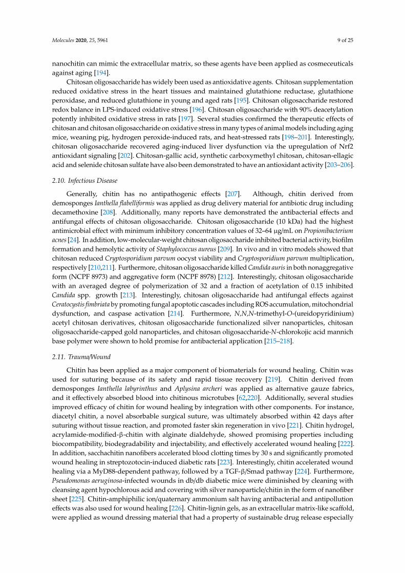
Currently, chitin and chitosan have been used as a biomaterial for wound healing and drugdelivery. Moreover, chitin and chitosan have been integrated with other chemicals to improve efficacyin therapeutic applications. Interestingly, the thermostability of chitin, chemical tolerance of chitin,and accessible natural source of chitin shed light on various procedures for biomaterial generation.Based on these advantageous properties, further development of chitin is needed to provide betterbiomaterials applicable for various human diseases. However, the development of chitin for medicalapplications requires further steps to improve safety and efficacy. A summary of potential applicationsand adverse effects of chitin is shown in Figure 1.
Molecules 2020, 25, 5961 11 of 25
Molecules 2020, 25, x FOR PEER REVIEW 11 of 26
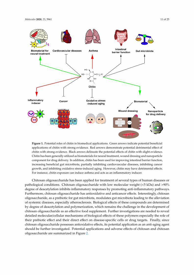
Figure 1. Potential roles of chitin in biomedical applications. Green arrows indicate potential beneficial applications of chitin with strong evidence. Red arrows demonstrate potential detrimental effect of chitin with strong evidence. Black arrows delineate the potential effects of chitin with slight evidence. Chitin has been generally utilized as biomaterials for neural treatment, wound dressing and nanoparticle component for drug delivery. In addition, chitin has been used for improving intestinal barrier function, increasing beneficial gut microbiota, partially inhibiting cardiovascular diseases, inhibiting cancer growth, and inhibiting oxidative stress-induced aging. However, chitin may have detrimental effects. For instance, chitin exposure can induce asthma and acts as an inflammatory inducer.
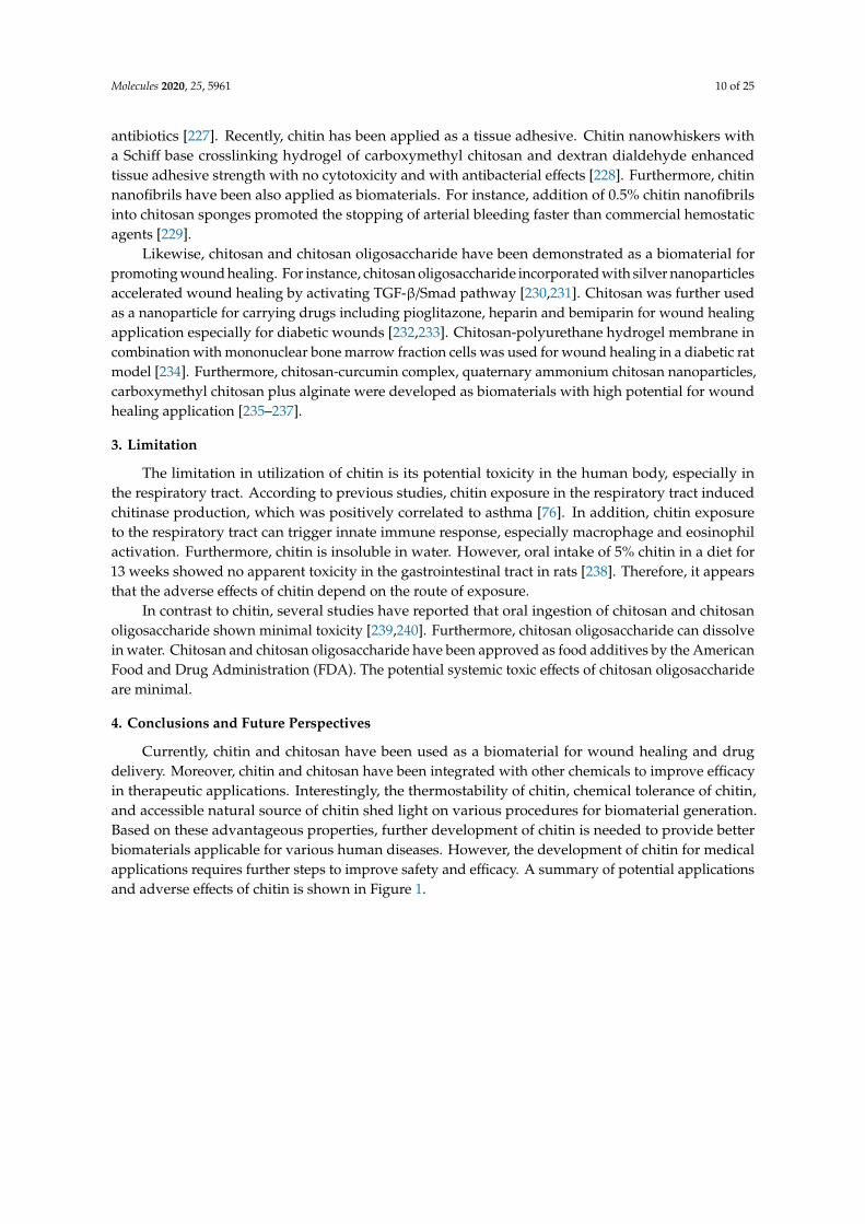
Chitosan oligosaccharide has been applied for treatment of several types of human diseases or pathological conditions. Chitosan oligosaccharide with low molecular weight (<5 kDa) and >90% degree of deacetylation inhibits inflammatory responses by promoting anti-inflammatory pathways. Furthermore, chitosan oligosaccharide has antioxidative and anticancer effects. Interestingly, chitosan oligosaccharide, as a prebiotic for gut microbiota, modulates gut microbiota leading to the alleviation of systemic diseases, especially atherosclerosis. Biological effects of these compounds are determined by degree of deacetylation and polymerization, which remains the challenge in the development of chitosan oligosaccharide as an effective food supplement. Further investigations are needed to reveal detailed molecular/cellular mechanisms of biological effects of these polymers especially the role of their prebiotic effect and their direct effect on disease-specific cells or drug targets. Finally, since chitosan oligosaccharide possesses antioxidative effects, its potential application as an anti-aging agent should be further investigated. Potential applications and adverse effects of chitosan and chitosan oligosaccharide are summarized in Figure 2.
Figure 1. Potential roles of chitin in biomedical applications. Green arrows indicate potential beneficialapplications of chitin with strong evidence. Red arrows demonstrate potential detrimental effect ofchitin with strong evidence. Black arrows delineate the potential effects of chitin with slight evidence.Chitin has been generally utilized as biomaterials for neural treatment, wound dressing and nanoparticlecomponent for drug delivery. In addition, chitin has been used for improving intestinal barrier function,increasing beneficial gut microbiota, partially inhibiting cardiovascular diseases, inhibiting cancergrowth, and inhibiting oxidative stress-induced aging. However, chitin may have detrimental effects.For instance, chitin exposure can induce asthma and acts as an inflammatory inducer.
Chitosan oligosaccharide has been applied for treatment of several types of human diseases orpathological conditions. Chitosan oligosaccharide with low molecular weight (<5 kDa) and >90%degree of deacetylation inhibits inflammatory responses by promoting anti-inflammatory pathways.Furthermore, chitosan oligosaccharide has antioxidative and anticancer effects. Interestingly, chitosanoligosaccharide, as a prebiotic for gut microbiota, modulates gut microbiota leading to the alleviationof systemic diseases, especially atherosclerosis. Biological effects of these compounds are determinedby degree of deacetylation and polymerization, which remains the challenge in the development ofchitosan oligosaccharide as an effective food supplement. Further investigations are needed to revealdetailed molecular/cellular mechanisms of biological effects of these polymers especially the role oftheir prebiotic effect and their direct effect on disease-specific cells or drug targets. Finally, sincechitosan oligosaccharide possesses antioxidative effects, its potential application as an anti-aging agentshould be further investigated. Potential applications and adverse effects of chitosan and chitosanoligosaccharide are summarized in Figure 2.
Molecules 2020, 25, 5961 12 of 25
Molecules 2020, 25, x FOR PEER REVIEW 12 of 26
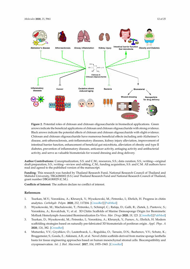
Figure 2. Potential roles of chitosan and chitosan oligosaccharide in biomedical applications. Green arrows indicate the beneficial applications of chitosan and chitosan oligosaccharide with strong evidence. Black arrows indicate the potential effects of chitosan and chitosan oligosaccharide with slight evidence. Chitosan and chitosan oligosaccharide have numerous beneficial effects including anti-Alzheimer’s disease, anti-atherosclerosis, anti-inflammatory diseases, kidney injury alleviation, improvement of intestinal barrier function, enhancement of beneficial gut microbiota, alleviation of obesity and type II diabetes, prevention of inflammatory diseases, anticancer activity, antiaging activity and antibacterial activity, and serve as valuable biomaterials for wound dressing and drug delivery.
Author Contributions: Conceptualization, S.S. and C.M.; resources, S.S.; data curation, S.S.; writing—original draft preparation, S.S.; writing—review and editing, C.M.; funding acquisition, S.S. and C.M. All authors have read and agreed to the published version of the manuscript.
Funding: This research was funded by Thailand Research Fund, National Research Council of Thailand and Mahidol University, TRG6280002 (S.S.) and Thailand Research Fund and National Research Council of Thailand, grant number DBG6180029 (C.M.).
Conflicts of Interest: The authors declare no conflict of interest.
References
1. Tsurkan, M.V.; Voronkina, A.; Khrunyk, Y.; Wysokowski, M.; Petrenko, I.; Ehrlich, H. Progress in chitin analytics. Carbohydr. Polym. 2021, 252, 117204, doi:10.1016/j.carbpol.2020.117204.
2. Wysokowski, M.; Machalowski, T.; Petrenko, I.; Schimpf, C.; Rafaja, D.; Galli, R.; Zietek, J.; Pantovic, S.; Voronkina, A.; Kovalchuk, V. et al. 3D Chitin Scaffolds of Marine Demosponge Origin for Biomimetic Mollusk Hemolymph-Associated Biomineralization Ex-Vivo. Mar. Drugs 2020, 18, doi:10.3390/md18020123.
3. Tsurkan, D.; Wysokowski, M.; Petrenko, I.; Voronkina, A.; Khrunyk, Y.; Fursov, A.; Ehrlich, H. Modern scaffolding strategies based on naturally pre-fabricated 3D biomaterials of poriferan origin. Appl. Phys. A 2020, 126, 382, doi:10.1007/s00339-020-03564-9.
4. Mutsenko, V.V.; Gryshkov, O.; Lauterboeck, L.; Rogulska, O.; Tarusin, D.N.; Bazhenov, V.V.; Schutz, K.; Bruggemeier, S.; Gossla, E.; Akkineni, A.R. et al. Novel chitin scaffolds derived from marine sponge
Figure 2. Potential roles of chitosan and chitosan oligosaccharide in biomedical applications. Greenarrows indicate the beneficial applications of chitosan and chitosan oligosaccharide with strong evidence.Black arrows indicate the potential effects of chitosan and chitosan oligosaccharide with slight evidence.Chitosan and chitosan oligosaccharide have numerous beneficial effects including anti-Alzheimer’sdisease, anti-atherosclerosis, anti-inflammatory diseases, kidney injury alleviation, improvement ofintestinal barrier function, enhancement of beneficial gut microbiota, alleviation of obesity and type IIdiabetes, prevention of inflammatory diseases, anticancer activity, antiaging activity and antibacterialactivity, and serve as valuable biomaterials for wound dressing and drug delivery.
Author Contributions: Conceptualization, S.S. and C.M.; resources, S.S.; data curation, S.S.; writing—originaldraft preparation, S.S.; writing—review and editing, C.M.; funding acquisition, S.S. and C.M. All authors haveread and agreed to the published version of the manuscript.
Funding: This research was funded by Thailand Research Fund, National Research Council of Thailand andMahidol University, TRG6280002 (S.S.) and Thailand Research Fund and National Research Council of Thailand,grant number DBG6180029 (C.M.).
Conflicts of Interest: The authors declare no conflict of interest.
References
1. Tsurkan, M.V.; Voronkina, A.; Khrunyk, Y.; Wysokowski, M.; Petrenko, I.; Ehrlich, H. Progress in chitinanalytics. Carbohydr. Polym. 2020, 252, 117204. [CrossRef] [PubMed]
2. Wysokowski, M.; Machalowski, T.; Petrenko, I.; Schimpf, C.; Rafaja, D.; Galli, R.; Zietek, J.; Pantovic, S.;Voronkina, A.; Kovalchuk, V.; et al. 3D Chitin Scaffolds of Marine Demosponge Origin for BiomimeticMollusk Hemolymph-Associated Biomineralization Ex-Vivo. Mar. Drugs 2020, 18, 123. [CrossRef] [PubMed]
3. Tsurkan, D.; Wysokowski, M.; Petrenko, I.; Voronkina, A.; Khrunyk, Y.; Fursov, A.; Ehrlich, H. Modernscaffolding strategies based on naturally pre-fabricated 3D biomaterials of poriferan origin. Appl. Phys. A2020, 126, 382. [CrossRef]
4. Mutsenko, V.V.; Gryshkov, O.; Lauterboeck, L.; Rogulska, O.; Tarusin, D.N.; Bazhenov, V.V.; Schutz, K.;Bruggemeier, S.; Gossla, E.; Akkineni, A.R.; et al. Novel chitin scaffolds derived from marine sponge Ianthellabasta for tissue engineering approaches based on human mesenchymal stromal cells: Biocompatibility andcryopreservation. Int. J. Biol. Macromol. 2017, 104, 1955–1965. [CrossRef]
Molecules 2020, 25, 5961 13 of 25
5. Petrenko, I.; Khrunyk, Y.; Voronkina, A.; Kovalchuk, V.; Fursov, A.; Tsurkan, D.; Ivanenko, V. Poriferan chitin:3D scaffolds from nano-to macroscale. A review. Lett. Appl. Nanobiosci. 2020, 9, 1004–1014.
6. Machałowski, T.; Wysokowski, M.; Petrenko, I.; Fursov, A.; Rahimi-Nasrabadi, M.; Amro, M.d.M.;Meissner, H.; Joseph, Y.; Fazilov, B.; Ehrlich, H.; et al. Naturally pre-designed biomaterials: Spidermolting cuticle as a functional crude oil sorbent. J. Environ. Manag. 2020, 261, 110218. [CrossRef] [PubMed]
7. Machałowski, T.; Amemiya, C.; Jesionowski, T. Chitin of Araneae origin: Structural features and biomimeticapplications: A review. Appl. Phys. A 2020, 126, 1–17. [CrossRef]
8. Khrunyk, Y.; Lach, S.; Petrenko, I.; Ehrlich, H. Progress in Modern Marine Biomaterials Research. Mar. Drugs2020, 18, 589. [CrossRef]
9. Wysokowski, M.; Petrenko, I.; Stelling, A.L.; Stawski, D.; Jesionowski, T.; Ehrlich, H. Poriferan chitin as aversatile template for extreme biomimetics. Polymers 2015, 7, 235–265. [CrossRef]
10. Petrenko, I.; Bazhenov, V.V.; Galli, R.; Wysokowski, M.; Fromont, J.; Schupp, P.J.; Stelling, A.L.; Niederschlag, E.;Stöker, H.; Kutsova, V.Z.; et al. Chitin of poriferan origin and the bioelectrometallurgy of copper/copperoxide. Int. J. Biol. Macromol. 2017, 104, 1626–1632. [CrossRef]
11. Moussian, B. Chitin: Structure, Chemistry and Biology. Adv. Exp. Med. Biol. 2019, 1142, 5–18. [CrossRef][PubMed]
12. Kumar, P.T.S.; Srinivasan, S.; Lakshmanan, V.K.; Tamura, H.; Nair, S.V.; Jayakumar, R. Synthesis,characterization and cytocompatibility studies of α-chitin hydrogel/nano hydroxyapatite composite scaffolds.Int. J. Biol. Macromol. 2011, 49, 20–31. [CrossRef] [PubMed]
13. Jayakumar, R.; Divya Rani, V.V.; Shalumon, K.T.; Kumar, P.T.S.; Nair, S.V.; Furuike, T.; Tamura, H. Bioactiveand osteoblast cell attachment studies of novel α- and β-chitin membranes for tissue-engineering applications.Int. J. Biol. Macromol. 2009, 45, 260–264. [CrossRef] [PubMed]
14. Song, S.; Zhao, Y.; Yuan, X.; Zhang, J. β-Chitin nanofiber hydrogel as a scaffold to in situ fabricatemonodispersed ultra-small silver nanoparticles. Colloids Surf. A 2019, 574, 36–43. [CrossRef]
15. Jung, H.S.; Kim, M.H.; Shin, J.Y.; Park, S.R.; Jung, J.Y.; Park, W.H. Electrospinning and wound healing activityof β-chitin extracted from cuttlefish bone. Carbohydr. Polym. 2018, 193, 205–211. [CrossRef]
16. Mulongo-Masamba, R.; El Hazzat, M.; El Hamidi, A.; Halim, M.; Arsalane, S. New functional β-chitin/calciumphosphate as promising support of copper nanocatalyst for the reductive degradation of methylene blue.Int. J. Environ. Sci. Technol. 2019, 16, 8117–8128. [CrossRef]
17. Somsak, P.; Sriwattana, S.; Prinyawiwatkul, W. Ultrasonic-assisted chitin nanoparticle and its application assaltiness enhancer. Int. J. Food Sci. Technol. 2020. [CrossRef]
18. Kaya, M.; Mujtaba, M.; Ehrlich, H.; Salaberria, A.M.; Baran, T.; Amemiya, C.T.; Galli, R.; Akyuz, L.; Sargin, I.;Labidi, J. On chemistry of γ-chitin. Carbohydr. Polym. 2017, 176, 177–186. [CrossRef]
19. Qin, Z.; Zhao, L. The History of Chito/Chitin Oligosaccharides and Its Monomer. In Oligosaccharides of Chitinand Chitosan: Bio-manufacture and Applications; Zhao, L., Ed.; Springer: Singapore, 2019; pp. 3–14.
20. Li, J.; Cai, C.; Li, J.; Li, J.; Li, J.; Sun, T.; Wang, L.; Wu, H.; Yu, G. Chitosan-Based Nanomaterials for DrugDelivery. Molecules 2018, 23, 2661. [CrossRef]
21. Jiang, T.; James, R.; Kumbar, S.G.; Laurencin, C.T. Chitosan as a Biomaterial: Structure, Properties,and Applications in Tissue Engineering and Drug Delivery. In Natural and Synthetic Biomedical Polymers;Kumbar, S.G., Laurencin, C.T., Deng, M., Eds.; Elsevier: Oxford, UK, 2014; pp. 91–113.
22. Muanprasat, C.; Chatsudthipong, V. Chitosan oligosaccharide: Biological activities and potential therapeuticapplications. Pharmacol. Ther. 2017, 170, 80–97. [CrossRef]
23. Gallo, M.; Naviglio, D.; Armone Caruso, A.; Ferrara, L. Applications of chitosan as a functional food. In NovelApproaches of Nanotechnology in Food; Grumezescu, A.M., Ed.; Academic Press: London, UK, 2016; pp. 425–464.
24. Kim, S.H.; Eom, S.H.; Yu, D.; Lee, M.S.; Kim, Y.M. Oligochitosan as a potential anti-acne vulgaris agent:Combined antibacterial effects against Propionibacterium acnes. Food Sci. Biotechnol. 2017, 26, 1029–1036.[CrossRef] [PubMed]
25. Singh, N.; Chen, J.; Koziol, K.K.; Hallam, K.R.; Janas, D.; Patil, A.J.; Strachan, A.; Hanley, J.G.; Rahatekar, S.S.Chitin and carbon nanotube composites as biocompatible scaffolds for neuron growth. Nanoscale 2016, 8,8288–8299. [CrossRef] [PubMed]
26. Wang, Z.Y.; Wang, J.W.; Qin, L.H.; Zhang, W.G.; Zhang, P.X.; Jiang, B.G. Chitin biological absorbablecatheters bridging sural nerve grafts transplanted into sciatic nerve defects promote nerve regeneration.CNS Neurosci. Ther. 2018, 24, 483–494. [CrossRef] [PubMed]
Molecules 2020, 25, 5961 14 of 25
27. Wang, Z.; Fan, J.; Yang, X.; Zhang, W.; Zhang, P.; Jiang, B. The neural regeneration effect of chitin biologicalabsorbable tubes bridging sciatic nerve defects with sural nerve grafts. Am. J. Transl. Res. 2018, 10, 2362–2371.[PubMed]
28. Ji, X.; Lei, Z.; Yuan, M.; Zhu, H.; Yuan, X.; Liu, W.; Pu, H.; Jiang, J.; Zhang, Y.; Jiang, X.; et al. Cartilagerepair mediated by thermosensitive photocrosslinkable TGFβ1-loaded GM-HPCH via immunomodulatingmacrophages, recruiting MSCs and promoting chondrogenesis. Theranostics 2020, 10, 2872–2887. [CrossRef]
29. Lin, H.L.; Wu, T.H.; Ho, H.O.; Chao, F.C.; Meng, H.; Liu, W.D.Z.; Chen, L.C.; Sheu, M.T. TEMPO-oxidizedsacchachitin nanofibers (TOSCNFs) combined with platelet-rich plasma (PRP) for management of dry eyesyndrome. Int. J. Nanomed. 2020, 15, 1721–1730. [CrossRef]
30. Lomiguen, C.; Vidal, L.; Kozlowski, P.; Prancan, A.; Stern, R. Possible role of chitin-like proteins in theetiology of Alzheimer’s disease. J. Alzheimer’s Dis. 2018, 66, 439–444. [CrossRef]
31. Castellani, R.J.; Perry, G.; Smith, M.A. The role of novel chitin-like polysaccharides in Alzheimer disease.Neurotox. Res. 2007, 12, 269–274. [CrossRef]
32. Castellani, R.J.; Siedlak, S.L.; Fortino, A.E.; Perry, G.; Ghetti, B.; Smith, M.A. Chitin-like polysaccharides inAlzheimer’s disease brains. Curr. Alzheimer. Res. 2005, 2, 419–423. [CrossRef]
33. Pisa, D.; Alonso, R.; Rábano, A.; Horst, M.N.; Carrasco, L. Fungal enolase, β-tubulin, and chitin are detectedin brain tissue from Alzheimer’s disease patients. Front. Microbiol. 2016, 7. [CrossRef]
34. Sotgiu, S.; Musumeci, S.; Marconi, S.; Gini, B.; Bonetti, B. Different content of chitin-like polysaccharides inmultiple sclerosis and Alzheimer’s disease brains. J. Neuroimmunol. 2008, 197, 70–73. [CrossRef] [PubMed]
35. Turano, E.; Busetto, G.; Marconi, S.; Guzzo, F.; Farinazzo, A.; Commisso, M.; Bistaffa, E.; Angiari, S.;Musumeci, S.; Sotgiu, S.; et al. Neurotoxicity and synaptic plasticity impairment of N-acetylglucosaminepolymers: Implications for Alzheimer’s disease. Neurobiol. Aging 2015, 36, 1780–1791. [CrossRef] [PubMed]
36. Mutsenko, V.V.; Bazhenov, V.V.; Rogulska, O.; Tarusin, D.N.; Schütz, K.; Brüggemeier, S.; Gossla, E.;Akkineni, A.R.; Meißner, H.; Lode, A.; et al. 3D chitinous scaffolds derived from cultivated marinedemosponge Aplysina aerophoba for tissue engineering approaches based on human mesenchymal stromalcells. Int. J. Biol. Macromol. 2017, 104, 1966–1974. [CrossRef] [PubMed]
37. Kim, M.S.; Sung, M.J.; Seo, S.B.; Yoo, S.J.; Lim, W.K.; Kim, H.M. Water-soluble chitosan inhibits theproduction of pro-inflammatory cytokine in human astrocytoma cells activated by amyloid beta peptide andinterleukin-1beta. Neurosci. Lett. 2002, 321, 105–109. [CrossRef]
38. Lee, S.H.; Park, J.S.; Kim, S.K.; Ahn, C.B.; Je, J.Y. Chitooligosaccharides suppress the level of protein expressionand acetylcholinesterase activity induced by Aβ25-35 in PC12 cells. Bioorganic Med. Chem. Lett. 2009, 19,860–862. [CrossRef]
39. Eom, T.K.; Ryu, B.; Lee, J.K.; Byun, H.G.; Park, S.J.; Kim, S.K. β-secretase inhibitory activity of phenolic acidconjugated chitooligosaccharides. J. Enzym. Inhib. Med. Chem. 2013, 28, 214–217. [CrossRef]
40. Dai, X.; Chang, P.; Li, X.; Gao, Z.; Sun, Y. The inhibitory effect of chitosan oligosaccharides on beta-siteamyloid precursor protein cleaving enzyme 1 (BACE1) in HEK293 APPswe cells. Neurosci. Lett. 2018, 665,80–85. [CrossRef]
41. Dai, X.; Hou, W.; Sun, Y.; Gao, Z.; Zhu, S.; Jiang, Z. Chitosan oligosaccharides inhibit/disaggregate fibrils andattenuate amyloid β-mediated neurotoxicity. Int. J. Mol. Sci. 2015, 16, 10526–10536. [CrossRef]
42. Hou, H.; Zhang, L.; Ye, Z.; Li, J.; Lian, Z.; Chen, C.; He, R.; Peng, B.; Xu, Q.; Zhang, G.; et al.Chitooligosaccharide Inhibits Scar Formation and Enhances Functional Recovery in a Mouse Modelof Sciatic Nerve Injury. Mol. Neurobiol. 2016, 53, 2249–2257. [CrossRef]
43. Kunanusornchai, W.; Witoonpanich, B.; Tawonsawatruk, T.; Pichyangkura, R.; Chatsudthipong, V.;Muanprasat, C. Chitosan oligosaccharide suppresses synovial inflammation via AMPK activation: An in vitroand in vivo study. Pharm. Res. 2016, 113, 458–467. [CrossRef]
44. Jia, S.; Lu, Z.; Gao, Z.; An, J.; Wu, X.; Li, X.; Dai, X.; Zheng, Q.; Sun, Y. Chitosan oligosaccharides alleviatecognitive deficits in an amyloid-beta1-42-induced rat model of Alzheimer’s disease. Int. J. Biol. Macromol.2016, 83, 416–425. [CrossRef] [PubMed]
45. Dai, X.; Chang, P.; Zhu, Q.; Liu, W.; Sun, Y.; Zhu, S.; Jiang, Z. Chitosan oligosaccharides protect rat primaryhippocampal neurons from oligomeric beta-amyloid 1-42-induced neurotoxicity. Neurosci. Lett. 2013, 554,64–69. [CrossRef] [PubMed]
Molecules 2020, 25, 5961 15 of 25
46. Inui, H.; Tsujikubo, M.; Hirano, S. Low molecular weight chitosan stimulation of mitogenic response toplatelet-derived growth factor in vascular smooth muscle cells. Biosci. Biotechnol. Biochem. 1995, 59,2111–2114. [CrossRef]
47. Chien, Y.; Liao, Y.W.; Liu, D.M.; Lin, H.L.; Chen, S.J.; Chen, H.L.; Peng, C.H.; Liang, C.M.; Mou, C.Y.; Chiou, S.H.Corneal repair by human corneal keratocyte-reprogrammed iPSCs and amphiphatic carboxymethyl-hexanoylchitosan hydrogel. Biomaterials 2012, 33, 8003–8016. [CrossRef] [PubMed]
48. Wu, H.; Liu, J.; Fang, Q.; Xiao, B.; Wan, Y. Establishment of nerve growth factor gradients on alignedchitosan-polylactide /alginate fibers for neural tissue engineering applications. Colloids Surf. B 2017, 160,598–609. [CrossRef] [PubMed]
49. Wei, C.K.; Ding, S.J. Acid-resistant calcium silicate-based composite implants with high-strength asload-bearing bone graft substitutes and fracture fixation devices. J. Mech. Behav. Biomed. Mater. 2016, 62,366–383. [CrossRef] [PubMed]
50. Jaruszewski, K.M.; Ramakrishnan, S.; Poduslo, J.F.; Kandimalla, K.K. Chitosan enhances the stability andtargeting of immuno-nanovehicles to cerebro-vascular deposits of Alzheimer’s disease amyloid protein.Nanomedicine 2012, 8, 250–260. [CrossRef]
51. Mahl, C.R.A.; Taketa, T.B.; Rocha-Neto, J.B.M.; Almeida, W.P.; Beppu, M.M. Copper Ion Uptake by Chitosanin the Presence of Amyloid-beta and Histidine. Appl. Biochem. Biotechnol. 2020, 190, 949–965. [CrossRef]
52. Casadome-Perales, A.; Matteis, L.; Alleva, M.; Infantes-Rodriguez, C.; Palomares-Perez, I.; Saito, T.; Saido, T.C.;Esteban, J.A.; Nebreda, A.R.; de la Fuente, J.M.; et al. Inhibition of p38 MAPK in the brain through nasaladministration of p38 inhibitor loaded in chitosan nanocapsules. Nanomedicine 2019, 14, 2409–2422. [CrossRef]
53. Kheradmandi, M.; Vasheghani-Farahani, E.; Ghiaseddin, A.; Ganji, F. Skeletal muscle regeneration viaengineered tissue culture over electrospun nanofibrous chitosan/PVA scaffold. J. Biomed. Mater. Res. A 2016,104, 1720–1727. [CrossRef]
54. Li, Y.; Yu, Z.; Men, Y.; Chen, X.; Wang, B. Laminin-chitosan-PLGA conduit co-transplanted with Schwann andneural stem cells to repair the injured recurrent laryngeal nerve. Exp. Med. 2018, 16, 1250–1258. [CrossRef][PubMed]
55. Liu, H.; Huang, H.; Bi, W.; Tan, X.; Li, R.; Wen, W.; Song, W.; Zhang, Y.; Zhang, F.; Hu, M. Effect of chitosancombined with hyaluronate on promoting the recovery of postoperative facial nerve regeneration andfunction in rabbits. Exp. Med. 2018, 16, 739–745. [CrossRef] [PubMed]
56. Crosio, A.; Fornasari, B.E.; Gambarotta, G.; Geuna, S.; Raimondo, S.; Battiston, B.; Tos, P.; Ronchi, G.Chitosan tubes enriched with fresh skeletal muscle fibers for delayed repair of peripheral nerve defects.Neural. Regen. Res. 2019, 14, 1079–1084. [CrossRef] [PubMed]
57. Carvalho, C.R.; Costa, J.B.; Costa, L.; Silva-Correia, J.; Moay, Z.K.; Ng, K.W.; Reis, R.L.; Oliveira, J.M.Enhanced performance of chitosan/keratin membranes with potential application in peripheral nerve repair.Biomater. Sci. 2019, 7, 5451–5466. [CrossRef]
58. Moattari, M.; Kouchesfehani, H.M.; Kaka, G.; Sadraie, S.H.; Naghdi, M.; Mansouri, K. Chitosan-filmassociated with mesenchymal stem cells enhanced regeneration of peripheral nerves: A rat sciatic nervemodel. J. Chem. Neuroanat. 2018, 88, 46–54. [CrossRef]
59. Neyrinck, A.M.; Catry, E.; Taminiau, B.; Cani, P.D.; Bindels, L.B.; Daube, G.; Dessy, C.; Delzenne, N.M.Chitin-glucan and pomegranate polyphenols improve endothelial dysfunction. Sci. Rep. 2019, 9, 14150.[CrossRef]
60. Song, X.; Huang, X.; Li, Z.; Li, Z.; Wu, K.; Jiao, Y.; Zhou, C. Construction of blood compatible chitin/grapheneoxide composite aerogel beads for the adsorption of bilirubin. Carbohydr. Polym. 2019, 207, 704–712. [CrossRef]
61. Zhang, J.; Xue, S.; Zhu, X.; Zhao, Y.; Chen, Y.; Tong, J.; Shi, X.; Du, Y.; Zhong, Z.; Ye, Q. Emerging chitinnanogels/rectorite nanocomposites for safe and effective hemorrhage control. J. Mater. Chem. B 2019, 7,5096–5103. [CrossRef]
62. Schubert, M.; Binnewerg, B.; Voronkina, A.; Muzychka, L.; Wysokowski, M.; Petrenko, I.; Kovalchuk, V.;Tsurkan, M.; Martinovic, R.; Bechmann, N.; et al. Naturally Prefabricated Marine Biomaterials: Isolation andApplications of Flat Chitinous 3D Scaffolds from Ianthella labyrinthus (Demospongiae: Verongiida). Int. J.Mol. Sci. 2019, 20, 5105. [CrossRef]
63. Machałowski, T.; Wysokowski, M.; Zółtowska-Aksamitowska, S.; Bechmann, N.; Binnewerg, B.; Schubert, M.;Guan, K.; Bornstein, S.R.; Czaczyk, K.; Pokrovsky, O.; et al. Spider Chitin. The biomimetic potential andapplications of Caribena versicolor tubular chitin. Carbohydr. Polym. 2019, 226, 115301. [CrossRef]
Molecules 2020, 25, 5961 16 of 25
64. Ormrod, D.J.; Holmes, C.C.; Miller, T.E. Dietary chitosan inhibits hypercholesterolaemia and atherogenesis inthe apolipoprotein E-deficient mouse model of atherosclerosis. Atherosclerosis 1998, 138, 329–334. [CrossRef]
65. Bahijri, S.M.; Alsheikh, L.; Ajabnoor, G.; Borai, A. Effect of Supplementation With Chitosan on Weight,Cardiometabolic, and Other Risk Indices in Wistar Rats Fed Normal and High-Fat/High-Cholesterol DietsAd Libitum. Nutr. Metab. Insights 2017, 10, 1178638817710666. [CrossRef] [PubMed]
66. Zong, C.; Yu, Y.; Song, G.; Luo, T.; Li, L.; Wang, X.; Qin, S. Chitosan oligosaccharides promote reversecholesterol transport and expression of scavenger receptor BI and CYP7A1 in mice. Exp. Biol. Med. (Maywood)2012, 237, 194–200. [CrossRef] [PubMed]
67. Yang, X.; Zhang, J.; Chen, L.; Wu, Q.; Yu, C. Chitosan oligosaccharides enhance lipid droplets viadown-regulation of PCSK9 gene expression in HepG2 cells. Exp. Cell. Res. 2018, 366, 152–160. [CrossRef]
68. Li, Y.; Xu, Q.; Wei, P.; Cheng, L.; Peng, Q.; Li, S.; Yin, H.; Du, Y. Chitosan oligosaccharides downregulate theexpression of E-selectin and ICAM-1 induced by LPS in endothelial cells by inhibiting MAP kinase signaling.Int. J. Mol. Med. 2014, 33, 392–400. [CrossRef]
69. Yu, Y.; Luo, T.; Liu, S.; Song, G.; Han, J.; Wang, Y.; Yao, S.; Feng, L.; Qin, S. Chitosan OligosaccharidesAttenuate Atherosclerosis and Decrease Non-HDL in ApoE-/- Mice. J. Atheroscler. Thromb. 2015, 22, 926–941.[CrossRef]
70. Xiong, Y.; Xiong, M.; Li, Y.; Qian, J.; Li, Y.; Han, X.; Tan, J.; Luo, Y.; Wang, Q.; Qin, C. Chitosan oligosaccharidecombined with running benefited the immune status of rats. Int. Immunopharmacol. 2020, 88, 106915.[CrossRef]
71. Zhu, L.; Hu, B.; Guo, Y.; Yang, H.; Zheng, J.; Yao, X.; Hu, H.; Liu, H. Effect of Chitosan oligosaccharides onischemic symptom and gut microbiota disbalance in mice with hindlimb ischemia. Carbohydr. Polym. 2020,240, 116271. [CrossRef]
72. Termsarasab, U.; Yoon, I.S.; Park, J.H.; Moon, H.T.; Cho, H.J.; Kim, D.D. Polyethylene glycol-modifiedarachidyl chitosan-based nanoparticles for prolonged blood circulation of doxorubicin. Int. J. Pharm. 2014,464, 127–134. [CrossRef]
73. Yu, J.; Ruan, Q.; Nie, X.; Yu, L.; Huang, B. Synthetic CD47 antibody-chitosan/hyaluronic acid polyelectrolytecomplex mediates targeted inhibition of atherosclerotic plaques by exogenous foam-like cells via the NLRP3pathway. J. Biomater. Appl. 2020, 34, 1381–1394. [CrossRef]
74. Tang, H.; Fang, Z.; Sun, Y.; Li, B.; Shi, Z.; Chen, J.; Zhang, T.; Xiu, Q. YKL-40 in asthmatic patients,and its correlations with exacerbation, eosinophils and immunoglobulin E. Eur. Respir. J. 2010, 35, 757–760.[CrossRef] [PubMed]
75. Liu, L.; Zhang, X.; Liu, Y.; Zhang, L.; Zheng, J.; Wang, J.; Hansbro, P.M.; Wang, L.; Wang, G.; Hsu, A.C.Y.Chitinase-like protein YKL-40 correlates with inflammatory phenotypes, anti-asthma responsiveness andfuture exacerbations. Respir. Res. 2019, 20, 95. [CrossRef] [PubMed]
76. Van Dyken, S.J.; Garcia, D.; Porter, P.; Huang, X.; Quinlan, P.J.; Blanc, P.D.; Corry, D.B.; Locksley, R.M. Fungalchitin from asthma-associated home environments induces eosinophilic lung infiltration. J. Immunol. 2011,187, 2261–2267. [CrossRef] [PubMed]
77. Khosravi, A.R.; Erle, D.J. Chitin-Induced Airway Epithelial Cell Innate Immune Responses Are Inhibited byCarvacrol/Thymol. PLoS ONE 2016, 11, e0159459. [CrossRef]
78. Choi, J.P.; Lee, S.M.; Choi, H.I.; Kim, M.H.; Jeon, S.G.; Jang, M.H.; Jee, Y.K.; Yang, S.; Cho, Y.J.;Kim, Y.K. House Dust Mite-Derived Chitin Enhances Th2 Cell Response to Inhaled Allergens, Mainlyvia a TNF-alpha-Dependent Pathway. Allergy Asthma Immunol. Res. 2016, 8, 362–374. [CrossRef]
79. Boot, R.G.; Renkema, G.H.; Strijland, A.; van Zonneveld, A.J.; Aerts, J.M. Cloning of a cDNA encodingchitotriosidase, a human chitinase produced by macrophages. J. Biol. Chem. 1995, 270, 26252–26256. [CrossRef]
80. Cho, S.J.; Weiden, M.D.; Lee, C.G. Chitotriosidase in the pathogenesis of inflammation, interstitial lungdiseases and COPD. Allergy Asthma Immunol. Res. 2014, 7, 14–21. [CrossRef]
81. Garth, J.M.; Mackel, J.J.; Reeder, K.M.; Blackburn, J.P.; Dunaway, C.W.; Yu, Z.; Matalon, S.; Fitz, L.; Steele, C.Acidic Mammalian Chitinase Negatively Affects Immune Responses during Acute and Chronic Aspergillusfumigatus Exposure. Infect. Immun. 2018, 86. [CrossRef]
82. Mazur, M.; Dymek, B.; Koralewski, R.; Sklepkiewicz, P.; Olejniczak, S.; Mazurkiewicz, M.; Piotrowicz, M.;Salamon, M.; Jedrzejczak, K.; Zagozdzon, A.; et al. Development of Dual Chitinase Inhibitors as PotentialNew Treatment for Respiratory System Diseases. J. Med. Chem. 2019, 62, 7126–7145. [CrossRef]
Molecules 2020, 25, 5961 17 of 25
83. Das, P.; Acharya, S.; Shah, D.; Agarwal, B.; Prahaladan, V.; Bhandari, V. Chitin Analog AVR-25 PreventsExperimental Bronchopulmonary Dysplasia. J. Pediatr. Intensive Care 2020, 9, 225–232. [CrossRef]
84. Zhao, Y.; Xu, G.; Wang, S.; Yi, X.; Wu, W. Chitosan oligosaccharides alleviate PM2.5-induced lung inflammationin rats. Environ. Sci. Pollut. Res. Int. 2018, 25, 34221–34227. [CrossRef] [PubMed]
85. Liu, Y.E.; Tong, C.C.; Zhang, Y.B.; Cong, P.F.; Shi, X.Y.; Liu, Y.; Shi, L.; Tong, Z.; Jin, H.X.; Hou, M.X. Chitosanoligosaccharide ameliorates acute lung injury induced by blast injury through the DDAH1/ADMA pathway.PLoS ONE 2018, 13, e0192135. [CrossRef] [PubMed]
86. Chung, M.J.; Park, J.K.; Park, Y.I. Anti-inflammatory effects of low-molecular weight chitosan oligosaccharidesin IgE-antigen complex-stimulated RBL-2H3 cells and asthma model mice. Int. Immunopharmacol. 2012, 12,453–459. [CrossRef] [PubMed]
87. Anraku, M.; Tabuchi, R.; Ifuku, S.; Nagae, T.; Iohara, D.; Tomida, H.; Uekama, K.; Maruyama, T.; Miyamura, S.;Hirayama, F.; et al. An oral absorbent, surface-deacetylated chitin nano-fiber ameliorates renal injury andoxidative stress in 5/6 nephrectomized rats. Carbohydr. Polym. 2017, 161, 21–25. [CrossRef] [PubMed]
88. Conroy, A.L.; Hawkes, M.T.; Elphinstone, R.; Opoka, R.O.; Namasopo, S.; Miller, C.; John, C.C.; Kain, K.C.Chitinase-3-like 1 is a biomarker of acute kidney injury and mortality in paediatric severe malaria. Malar. J.2018, 17, 82. [CrossRef]
89. Schmidt, I.M.; Hall, I.E.; Kale, S.; Lee, S.; He, C.-H.; Lee, Y.; Chupp, G.L.; Moeckel, G.W.; Lee, C.G.; Elias, J.A.;et al. Chitinase-Like Protein Brp-39/YKL-40 Modulates the Renal Response to Ischemic Injury and PredictsDelayed Allograft Function. J. Am. Soc. Nephrol. 2013, 24, 309. [CrossRef]
90. Bai, W.; Wang, S.; An, S.; Guo, M.; Gong, G.; Liu, W.; Ma, S.; Li, X.; Fu, J.; Yao, W. Combination therapy ofchitosan, gynostemma, and motherwort alleviates the progression of experimental rat chronic renal failureby inhibiting STAT1 activation. Oncotarget 2018, 9, 15498–15511. [CrossRef]
91. Queiroz, M.F.; Melo, K.R.T.; Sabry, D.A.; Sassaki, G.L.; Rocha, H.A.O.; Costa, L.S. Gallic Acid-ChitosanConjugate Inhibits the Formation of Calcium Oxalate Crystals. Molecules 2019, 24. [CrossRef]
92. Biasibetti, E.; Martello, E.; Bigliati, M.; Biasato, I.; Cocca, T.; Bruni, N.; Capucchio, M.T. A long term feedsupplementation based on phosphate binders in Feline Chronic Kidney Disease. Vet. Res. Commun. 2018, 42,161–167. [CrossRef]
93. Liu, B.; Qin, Z.K.; Lin, X.M.; Mei, L.; Liu, W.S.; Han, B.Q. Effects of chitooligosaccharides and its derivativeson antioxygenization and renal function in experimental diabetes rats. J. Clin. Rehabil. Tissue Eng. Res. 2008,12, 9651–9654.
94. Yoon, H.J.; Moon, M.E.; Park, H.S.; Kim, H.W.; Im, S.Y.; Lee, J.H.; Kim, Y.H. Effects of chitosan oligosaccharide(COS) on the glycerol-induced acute renal failure in vitro and in vivo. Food Chem. Toxicol. 2008, 46, 710–716.[CrossRef] [PubMed]
95. Qiao, J.; Liu, Y.; Jiang, Z.; Yang, Y.; Liu, W.; Han, B. Preparation and renoprotective effects of carboxymethylchitosan oligosaccharide on adriamycin nephropathy. Carbohydr. Polym. 2018, 201, 347–356. [CrossRef][PubMed]
96. Zhai, X.; Yuan, S.; Yang, X.; Zou, P.; Li, L.; Li, G.; Shao, Y.; Abd El-Aty, A.M.; Hacimuftuoglu, A.;Wang, J. Chitosan Oligosaccharides Induce Apoptosis in Human Renal Carcinoma via Reactive-Oxygen-Species-Dependent Endoplasmic Reticulum Stress. J. Agric. Food Chem. 2019, 67, 1691–1701. [CrossRef][PubMed]
97. Zhang, X.F.; Ding, C.L.; Liu, H.; Liu, L.H.; Zhao, C.Q. Protective effects of ion-imprinted chitooligosaccharidesas uranium-specific chelating agents against the cytotoxicity of depleted uranium in human kidney cells.Toxicology 2011, 286, 75–84. [CrossRef]
98. Pathomthongtaweechai, N.; Soodvilai, S.; Pichyangkura, R.; Muanprasat, C. Novel Potential Applicationof Chitosan Oligosaccharide for Attenuation of Renal Cyst Growth in the Treatment of Polycystic KidneyDisease. Molecules 2020, 25, 5589. [CrossRef]
99. Anraku, M.; Tanaka, M.; Hiraga, A.; Nagumo, K.; Imafuku, T.; Maezaki, Y.; Iohara, D.; Uekama, K.;Watanabe, H.; Hirayama, F.; et al. Effects of chitosan on oxidative stress and related factors in hemodialysispatients. Carbohydr. Polym. 2014, 112, 152–157. [CrossRef]
100. Salva, E.; Turan, S.O.; Akbuga, J. Inhibition of Glomerular Mesangial Cell Proliferation by siPDGF-B- andsiPDGFR-beta-Containing Chitosan Nanoplexes. AAPS PharmSciTech. 2017, 18, 1031–1042. [CrossRef]
101. Chiang, I.N.; Huang, W.C.; Huang, C.Y.; Pu, Y.S.; Young, T.H. Development of a chitosan-basedtissue-engineered renal proximal tubule conduit. J. Biomed. Mater. Res. B 2018, 106, 9–20. [CrossRef]
Molecules 2020, 25, 5961 18 of 25
102. Sutthasupha, P.; Lungkaphin, A. The potential roles of chitosan oligosaccharide in prevention of kidneyinjury in obese and diabetic conditions. Food Funct. 2020, 11, 7371–7388. [CrossRef]
103. Liberti, A.; Zucchetti, I.; Melillo, D.; Skapura, D.; Shibata, Y.; De Santis, R.; Pinto, M.R.; Litman, G.W.;Dishaw, L.J. Chitin protects the gut epithelial barrier in a protochordate model of DSS-induced colitis.Biol. Open 2018, 7. [CrossRef]
104. Alessandri, G.; Milani, C.; Duranti, S.; Mancabelli, L.; Ranjanoro, T.; Modica, S.; Carnevali, L.; Statello, R.;Bottacini, F.; Turroni, F.; et al. Ability of bifidobacteria to metabolize chitin-glucan and its impact on the gutmicrobiota. Sci. Rep. 2019, 9, 5755. [CrossRef] [PubMed]
105. Rodriguez, J.; Neyrinck, A.M.; Zhang, Z.; Seethaler, B.; Nazare, J.A.; Robles Sanchez, C.; Roumain, M.;Muccioli, G.G.; Bindels, L.B.; Cani, P.D.; et al. Metabolite profiling reveals the interaction of chitin-glucanwith the gut microbiota. Gut. Microbes 2020, 12, 1810530. [CrossRef] [PubMed]
106. Goto, M.; Iohara, D.; Michihara, A.; Ifuku, S.; Azuma, K.; Kadowaki, D.; Maruyama, T.; Otagiri, M.;Hirayama, F.; Anraku, M. Effects of surface-deacetylated chitin nanofibers on non-alcoholic steatohepatitismodel rats and their gut microbiota. Int. J. Biol. Macromol. 2020, 164, 659–666. [CrossRef] [PubMed]
107. Liu, P.; Piao, X.S.; Thacker, P.A.; Zeng, Z.K.; Li, P.F.; Wang, D.; Kim, S.W. Chito-oligosaccharide reducesdiarrhea incidence and attenuates the immune response of weaned pigs challenged with Escherichia coliK88. J. Anim. Sci. 2010, 88, 3871–3879. [CrossRef] [PubMed]
108. Liu, P.; Piao, X.S.; Kim, S.W.; Wang, L.; Shen, Y.B.; Lee, H.S.; Li, S.Y. Effects of chito-oligosaccharidesupplementation on the growth performance, nutrient digestibility, intestinal morphology, and fecalshedding of Escherichia coli and Lactobacillus in weaning pigs. J. Anim. Sci. 2008, 86, 2609–2618. [CrossRef][PubMed]
109. Qian, L.; Yue, X.; Hu, L.; Ma, Y.; Han, X. Changes in diarrhea, nutrients apparent digestibility, digestiveenzyme activities of weaned piglets in response to chitosan-zinc chelate. Anim. Sci. J. 2016, 87, 564–569.[CrossRef] [PubMed]
110. Aluko, K.; Velayudhan, D.E.; Khafipour, E.; Li, A.; Yin, Y.; Nyachoti, M. Combined effects of chitosan andmicroencapsulated Enterococcus faecalis CG1.0007 probiotic supplementation on performance and diarrheaincidences in enterotoxigenic Escherichia coli K88(+) challenged piglets. Anim. Nutr. 2017, 3, 366–371.[CrossRef]
111. Hu, S.; Wang, Y.; Wen, X.; Wang, L.; Jiang, Z.; Zheng, C. Effects of low-molecular-weight chitosan on thegrowth performance, intestinal morphology, barrier function, cytokine expression and antioxidant system ofweaned piglets. Bmc. Vet. Res. 2018, 14, 215. [CrossRef]
112. Thongsong, B.; Suthongsa, S.; Pichyangkura, R.; Kalandakanond-Thongsong, S. Effects of chito-oligosaccharidesupplementation with low or medium molecular weight and high degree of deacetylation on growth performance,nutrient digestibility and small intestinal morphology in weaned pigs. Livest. Sci. 2018, 209, 60–66. [CrossRef]
113. Zheng, J.; Yuan, X.; Cheng, G.; Jiao, S.; Feng, C.; Zhao, X.; Yin, H.; Du, Y.; Liu, H. Chitosan oligosaccharidesimprove the disturbance in glucose metabolism and reverse the dysbiosis of gut microbiota in diabetic mice.Carbohydr. Polym. 2018, 190, 77–86. [CrossRef]
114. Muanprasat, C.; Wongkrasant, P.; Satitsri, S.; Moonwiriyakit, A.; Pongkorpsakol, P.; Mattaveewong, T.;Pichyangkura, R.; Chatsudthipong, V. Activation of AMPK by chitosan oligosaccharide in intestinal epithelialcells: Mechanism of action and potential applications in intestinal disorders. Biochem. Pharm. 2015, 96,225–236. [CrossRef] [PubMed]
115. Bai, Y.; Zheng, J.; Yuan, X.; Jiao, S.; Feng, C.; Du, Y.; Liu, H.; Zheng, L. Chitosan oligosaccharides improveglucolipid metabolism disorder in liver by suppression of obesity-related inflammation and restoration ofperoxisome proliferator-activated receptor gamma (PPARγ). Mar. Drugs 2018, 16. [CrossRef] [PubMed]
116. Prajapati, B.; Rajput, P.; Jena, P.K.; Seshadri, S. Investigation of Chitosan for Prevention of Diabetic ProgressionThrough Gut Microbiota Alteration in Sugar Rich Diet Induced Diabetic Rats. Curr. Pharm. Biotechnol. 2015,17, 173–184. [CrossRef] [PubMed]
117. Mateos-Aparicio, I.; Mengibar, M.; Heras, A. Effect of chito-oligosaccharides over human faecal microbiotaduring fermentation in batch cultures. Carbohydr. Polym. 2016, 137, 617–624. [CrossRef] [PubMed]
118. Rowan, F.; Docherty, N.G.; Murphy, M.; Murphy, B.; Calvin Coffey, J.; O’Connell, P.R. Desulfovibrio bacterialspecies are increased in ulcerative colitis. Dis. Colon. Rectum. 2010, 53, 1530–1536. [CrossRef] [PubMed]
119. Zhang, C.; Jiao, S.; Wang, Z.A.; Du, Y. Exploring Effects of Chitosan Oligosaccharides on Mice Gut Microbiotain in vitro Fermentation and Animal Model. Front. Microbiol. 2018, 9, 2388. [CrossRef]
Molecules 2020, 25, 5961 19 of 25
120. Wu, M.; Li, J.; An, Y.; Li, P.; Xiong, W.; Li, J.; Yan, D.; Wang, M.; Zhong, G. Chitooligosaccharides Preventsthe Development of Colitis-Associated Colorectal Cancer by Modulating the Intestinal Microbiota andMycobiota. Front. Microbiol. 2019, 10, 2101. [CrossRef]
121. Qian, M.; Lyu, Q.; Liu, Y.; Hu, H.; Wang, S.; Pan, C.; Duan, X.; Gao, Y.; Qi, L.W.; Liu, W.; et al. ChitosanOligosaccharide Ameliorates Nonalcoholic Fatty Liver Disease (NAFLD) in Diet-Induced Obese Mice.Mar. Drugs 2019, 17. [CrossRef]
122. Saleh, H.; El-Shorbagy, H.M. Chitosan protects liver against ischemia-reperfusion injury via regulatingBcl-2/Bax, TNF-alpha and TGF-beta expression. Int. J. Biol. Macromol. 2020, 164, 1565–1574. [CrossRef]
123. Tong, A.J.; Hu, R.K.; Wu, L.X.; Lv, X.C.; Li, X.; Zhao, L.N.; Liu, B. Ganoderma polysaccharide and chitosansynergistically ameliorate lipid metabolic disorders and modulate gut microbiota composition in high fatdiet-fed golden hamsters. J. Food. Biochem. 2020, 44, e13109. [CrossRef]
124. Wu, X.; Kim, M.J.; Yang, H.J.; Park, S. Chitosan alleviated menopausal symptoms and modulated the gutmicrobiota in estrogen-deficient rats. Eur. J. Nutr. 2020, 1–13. [CrossRef] [PubMed]
125. Wang, J.; Kong, M.; Zhou, Z.; Yan, D.; Yu, X.; Cheng, X.; Feng, C.; Liu, Y.; Chen, X. Mechanism of surfacecharge triggered intestinal epithelial tight junction opening upon chitosan nanoparticles for insulin oraldelivery. Carbohydr. Polym. 2017, 157, 596–602. [CrossRef] [PubMed]
126. Xu, Y.; Mao, H.; Yang, C.; Du, H.; Wang, H.; Tu, J. Effects of chitosan nanoparticle supplementation on growthperformance, humoral immunity, gut microbiota and immune responses after lipopolysaccharide challengein weaned pigs. J. Anim. Physiol. Anim. Nutr. (Berl) 2020, 104, 597–605. [CrossRef] [PubMed]
127. Nopvichai, C.; Pongkorpsakol, P.; Wongkrasant, P.; Wangpaiboon, K.; Charoenwongpaiboon, T.; Ito, K.;Muanprasat, C.; Pichyangkura, R. Galactomannan Pentasaccharide Produced from Copra Meal EnhancesTight Junction Integration of Epithelial Tissue through Activation of AMPK. Biomedicines 2019, 7. [CrossRef][PubMed]
128. Nopvichai, C.; Charoenwongpaiboon, T.; Luengluepunya, N.; Ito, K.; Muanprasat, C.; Pichyangkura, R.Production and purification of mannan oligosaccharide with epithelial tight junction enhancing activity.PeerJ 2019, 7, e7206. [CrossRef] [PubMed]
129. Wongkrasant, P.; Pongkorpsakol, P.; Ariyadamrongkwan, J.; Meesomboon, R.; Satitsri, S.; Pichyangkura, R.;Barrett, K.E.; Muanprasat, C. A prebiotic fructo-oligosaccharide promotes tight junction assembly in intestinalepithelial cells via an AMPK-dependent pathway. Biomed. Pharm. 2020, 129, 110415. [CrossRef] [PubMed]
130. Bays, H.E.; Evans, J.L.; Maki, K.C.; Evans, M.; Maquet, V.; Cooper, R.; Anderson, J.W. Chitin-glucan fibereffects on oxidized low-density lipoprotein: A randomized controlled trial. Eur. J. Clin. Nutr. 2013, 67, 2–7.[CrossRef]
131. Yu, P.; Xie, J.; Chen, Y.; Liu, J.; Liu, Y.; Bi, B.; Luo, J.; Li, S.; Jiang, X.; Li, J. A thermo-sensitive injectablehydroxypropyl chitin hydrogel for sustained salmon calcitonin release with enhanced osteogenesis andhypocalcemic effects. J. Mater. Chem. B 2020, 8, 270–281. [CrossRef]
132. Xing, R.; He, X.; Liu, S.; Yu, H.; Qin, Y.; Chen, X.; Li, K.; Li, R.; Li, P. Antidiabetic activity of differentlyregioselective chitosan sulfates in alloxan-induced diabetic rats. Mar. Drugs 2015, 13, 3072–3090. [CrossRef]
133. Arun, G.; Rajaram, R.; Kaleshkumar, K.; Gayathri, N.; Sivasudha, T.; Kandasamy, S. Synergistic effect of novelchitosan combined metformin drug on streptozotocin-induced diabetes mellitus rat. Int. J. Biol. Macromol.2020, 153, 1335–1349. [CrossRef]
134. Yan, H.; Rong, L.; Xiao, D.; Zhang, M.; Sheikh, S.P.; Sui, X.; Lu, M. Injectable and self-healing hydrogelas a stem cells carrier for treatment of diabetic erectile dysfunction. Mater. Sci. Eng. C 2020, 116, 111214.[CrossRef] [PubMed]
135. Wong, C.Y.; Al-Salami, H.; Dass, C.R. Lyophilisation Improves Bioactivity and Stability of Insulin-LoadedPolymeric-Oligonucleotide Nanoparticles for Diabetes Treatment. Aaps. Pharmscitech. 2020, 21, 108.[CrossRef]
136. Trinh, T.A.; Duy Le, T.M.; Ho, H.G.V.; To, T.C.T.; Nguyen, V.V.L.; Huynh, D.P.; Lee, D.S. A novel injectablepH-temperature sensitive hydrogel containing chitosan-insulin electrosprayed nanosphere composite for aninsulin delivery system in type I diabetes treatment. Biomater. Sci. 2020, 8, 3830–3843. [CrossRef] [PubMed]
137. Hashemi, A.; Ezati, M.; Mohammadnejad, J.; Houshmand, B.; Faghihi, S. Chitosan Coating of TiO2 NanotubeArrays for Improved Metformin Release and Osteoblast Differentiation. Int. J. Nanomed. 2020, 15, 4471–4481.[CrossRef] [PubMed]
Molecules 2020, 25, 5961 20 of 25
138. Du, S.; Lv, Y.; Li, N.; Huang, X.; Liu, X.; Li, H.; Wang, C.; Jia, Y.F. Biological investigations on therapeutic effectof chitosan encapsulated nano resveratrol against gestational diabetes mellitus rats induced by streptozotocin.Drug Deliv. 2020, 27, 953–963. [CrossRef] [PubMed]
139. Bansal, A.; D’Sa, S.; D’Souza, M.J. Biofabrication of microcapsules encapsulating beta-TC-6 cells via scalabledevice and in-vivo evaluation in type 1 diabetic mice. Int. J. Pharm. 2019, 572, 118830. [CrossRef] [PubMed]
140. Al-Nemrawi, N.K.; Alsharif, S.S.M.; Alzoubi, K.H.; Alkhatib, R.Q. Preparation and characterization of insulinchitosan-nanoparticles loaded in buccal films. Pharm. Dev. Technol. 2019, 24, 967–974. [CrossRef]
141. Zeng, S.; Ke, Y.; Liu, Y.; Shen, Y.; Zhang, L.; Li, C.; Liu, A.; Shen, L.; Hu, X.; Wu, H.; et al. Synthesis andantidiabetic properties of chitosan-stabilized selenium nanoparticles. Colloids Surf. B 2018, 170, 115–121.[CrossRef]
142. Xu, J.; Cao, L.; Suo, Y.; Xu, X.; Sun, H.; Xu, S.; Zhu, X.; Yu, H.; Cao, W. Chitosan-microcapsulated insulinalleviates mesenteric microcirculation dysfunction via modulating COX-2 and VCAM-1 expression in ratswith diabetes mellitus. Int. J. Nanomed. 2018, 13, 6829–6837. [CrossRef]
143. Kumar, S.G.; Rahman, M.A.; Lee, S.H.; Hwang, H.S.; Kim, H.A.; Yun, J.W. Plasma proteome analysis foranti-obesity and anti-diabetic potentials of chitosan oligosaccharides in ob/ob mice. Proteomics 2009, 9,2149–2162. [CrossRef]
144. Liu, B.; Liu, W.S.; Han, B.Q.; Sun, Y.Y. Antidiabetic effects of chitooligosaccharides on pancreatic islet cells instreptozotocin-induced diabetic rats. World J. Gastroenterol. 2007, 13, 725–731. [CrossRef] [PubMed]
145. Liu, B.; Qin, Z.K.; Lin, X.M.; Mei, L.; Liu, W.S.; Han, B.Q. Antidiabetic effects of chitooligosaccharides withdifferent molecular weights on pancreatic islet cells in streptozotocin-induced diabetic rats. World Chin.J. Dig. 2009, 17, 36–42. [CrossRef]
146. Qinna, N.A.; Karwi, Q.G.; Al-Jbour, N.; Al-Remawi, M.A.; Alhussainy, T.M.; Al-So’ud, K.A.; Al Omari, M.M.;Badwan, A.A. Influence of molecular weight and degree of deacetylation of low molecular weight chitosanon the bioactivity of oral insulin preparations. Mar. Drugs 2015, 13, 1710–1725. [CrossRef] [PubMed]
147. Kim, H.J.; Ahn, H.Y.; Kwak, J.H.; Shin, D.Y.; Kwon, Y.I.; Oh, C.G.; Lee, J.H. The effects of chitosanoligosaccharide (GO2KA1) supplementation on glucose control in subjects with prediabetes. Food Funct.2014, 5, 2662–2669. [CrossRef] [PubMed]
148. Yu, S.Y.; Kwon, Y.I.; Lee, C.; Apostolidis, E.; Kim, Y.C. Antidiabetic effect of chitosan oligosaccharide (GO2KA1)is mediated via inhibition of intestinal alpha-glucosidase and glucose transporters and PPARgammaexpression. BioFactors 2017, 43, 90–99. [CrossRef] [PubMed]
149. Jeong, S.; Min Cho, J.; Kwon, Y.I.; Kim, S.C.; Yeob Shin, D.; Ho Lee, J. Chitosan oligosaccharide (GO2KA1)improves postprandial glycemic response in subjects with impaired glucose tolerance and impaired fastingglucose and in healthy subjects: A crossover, randomized controlled trial. Nutr. Diabetes 2019, 9, 31.[CrossRef] [PubMed]
150. Zhao, L.; Sun, T.; Wang, L. Chitosan oligosaccharide improves the therapeutic efficacy of sitagliptin for thetherapy of Chinese elderly patients with type 2 diabetes mellitus. Clin. Risk. Manag. 2017, 13, 739–750.[CrossRef]
151. Lupascu, F.G.; Giusca, S.E.; Caruntu, I.D.; Anton, A.; Lupusoru, C.E.; Profire, L. The safety profile of newantidiabetic xanthine derivatives and their chitosan based formulations. Eur. J. Pharm. Sci. 2019, 127, 71–78.[CrossRef]
152. Wang, J.; He, W.; Yang, D.; Cao, H.; Bai, Y.; Guo, J.; Su, Z. Beneficial Metabolic Effects of Chitosan andChitosan Oligosaccharide on Epididymal WAT Browning and Thermogenesis in Obese Rats. Molecules 2019,24. [CrossRef]
153. Yang, D.; Hu, C.; Deng, X.; Bai, Y.; Cao, H.; Guo, J.; Su, Z. Therapeutic Effect of Chitooligosaccharide Tabletson Lipids in High-Fat Diets Induced Hyperlipidemic Rats. Molecules 2019, 24. [CrossRef]
154. Deng, X.; Ye, Z.; Cao, H.; Bai, Y.; Che, Q.; Guo, J.; Su, Z. Chitosan oligosaccharide ameliorated obesityby reducing endoplasmic reticulum stress in diet-induced obese rats. Food. Funct. 2020, 11, 6285–6296.[CrossRef] [PubMed]
155. Liu, S.H.; Chen, R.Y.; Chiang, M.T. Effects of Chitosan Oligosaccharide on Plasma and Hepatic LipidMetabolism and Liver Histomorphology in Normal Sprague-Dawley Rats. Mar. Drugs 2020, 18, 408.[CrossRef]
Molecules 2020, 25, 5961 21 of 25
156. Izumi, R.; Azuma, K.; Izawa, H.; Morimoto, M.; Nagashima, M.; Osaki, T.; Tsuka, T.; Imagawa, T.; Ito, N.;Okamoto, Y.; et al. Chitin nanofibrils suppress skin inflammation in atopic dermatitis-like skin lesions inNC/Nga mice. Carbohydr. Polym. 2016, 146, 320–327. [CrossRef] [PubMed]
157. Huang, C.J.; Beasley, K.N.; Acevedo, E.O.; Franco, R.L.; Jones, T.L.; Mari, D.C.; Shibata, Y. Chitin enhancesobese inflammation ex vivo. Hum. Immunol. 2014, 75, 41–46. [CrossRef]
158. Van Dyken, S.J.; Mohapatra, A.; Nussbaum, J.C.; Molofsky, A.B.; Thornton, E.E.; Ziegler, S.F.; McKenzie, A.N.J.;Krummel, M.F.; Liang, H.E.; Locksley, R.M. Chitin activates parallel immune modules that direct distinctinflammatory responses via innate lymphoid type 2 and γδ T cells. Immunity 2014, 40, 414–424. [CrossRef][PubMed]
159. Arae, K.; Morita, H.; Unno, H.; Motomura, K.; Toyama, S.; Okada, N.; Ohno, T.; Tamari, M.; Orimo, K.;Mishima, Y.; et al. Chitin promotes antigen-specific Th2 cell-mediated murine asthma through induction ofIL-33-mediated IL-1beta production by DCs. Sci. Rep. 2018, 8, 11721. [CrossRef]
160. Wagener, J.; Malireddi, R.K.S.; Lenardon, M.D.; Köberle, M.; Vautier, S.; MacCallum, D.M.; Biedermann, T.;Schaller, M.; Netea, M.G.; Kanneganti, T.D.; et al. Fungal Chitin Dampens Inflammation through IL-10Induction Mediated by NOD2 and TLR9 Activation. PloS Pathog. 2014, 10. [CrossRef]
161. Danti, S.; Trombi, L.; Fusco, A.; Azimi, B.; Lazzeri, A.; Morganti, P.; Coltelli, M.B.; Donnarumma, G. ChitinNanofibrils and Nanolignin as Functional Agents in Skin Regeneration. Int. J. Mol. Sci. 2019, 20, 2669.[CrossRef]
162. Wang, J.; Zhang, C.; Guo, C.; Li, X. Chitosan Ameliorates DSS-Induced Ulcerative Colitis Mice by EnhancingIntestinal Barrier Function and Improving Microflora. Int. J. Mol. Sci. 2019, 20, 5751. [CrossRef]
163. Gudmundsdottir, S.; Lieder, R.; Sigurjonsson, O.E.; Petersen, P.H. Chitosan leads to downregulation ofYKL-40 and inflammasome activation in human macrophages. J. Biomed. Mater. Res. A 2015, 103, 2778–2785.[CrossRef]
164. Liu, Y.; Zong, S.; Li, J. Carboxymethyl chitosan perturbs inflammation profile and colonic microbiota balancein mice. J. Food Drug Anal. 2020, 28, 175–182. [CrossRef]
165. Yousef, M.; Pichyangkura, R.; Soodvilai, S.; Chatsudthipong, V.; Muanprasat, C. Chitosan oligosaccharide aspotential therapy of inflammatory bowel disease: Therapeutic efficacy and possible mechanisms of action.Pharm. Res. 2012, 66, 66–79. [CrossRef] [PubMed]
166. Yang, Y.; Tong, Q.; Luo, H.; Huang, R.; Li, Z. Chitooligosaccharides attenuate lipopolysaccharide-inducedinflammation and apoptosis of intestinal epithelial cells: Possible involvement of TLR4/NF-κB pathway.Indian J. Pharm. Educ. Res. 2016, 50, 109–115. [CrossRef]
167. Xu, Q.; Liu, M.; Liu, Q.; Wang, W.; Du, Y.; Yin, H. The inhibition of LPS-induced inflammation in RAW264.7macrophages via the PI3K/Akt pathway by highly N-acetylated chitooligosaccharide. Carbohydr. Polym.2017, 174, 1138–1143. [CrossRef]
168. Sánchez, Á.; Mengíbar, M.; Fernández, M.; Alemany, S.; Heras, A.; Acosta, N. Influence of preparationmethods of chitooligosaccharides on their physicochemical properties and their anti-inflammatory effects inmice and in RAW264.7 macrophages. Mar. Drugs 2018, 16, 430. [CrossRef] [PubMed]
169. Jiang, T.; Ji, H.; Zhang, L.; Wang, Y.; Zhou, H. Chitosan Oligosaccharide Exerts Anti-Allergic Effect againstShrimp Tropomyosin-Induced Food Allergy by Affecting Th1 and Th2 Cytokines. Int. Arch. Allergy Immunol.2019, 180, 10–16. [CrossRef]
170. Lan, R.; Li, S.; Chang, Q.; Zhao, Z. Chitosan oligosaccharides protect sprague dawley rats from cyclic heatstress by attenuation of oxidative and inflammation stress. Animals 2019, 9, 1074. [CrossRef] [PubMed]
171. Wongkrasant, P.; Pongkorpsakol, P.; Chitwattananont, S.; Satianrapapong, W.; Tuangkijkul, N.; Muanprasat, C.Fructo-oligosaccharides alleviate inflammation-associated apoptosis of GLP-1 secreting L cells via inhibitionof iNOS and cleaved caspase-3 expression. J. Pharm. Sci. 2020, 143, 65–73. [CrossRef]
172. Sun, Y.; Shi, X.; Zheng, X.; Nie, S.; Xu, X. Inhibition of dextran sodium sulfate-induced colitis in mice bybaker’s yeast polysaccharides. Carbohydr. Polym. 2019, 207, 371–381. [CrossRef]
173. Libreros, S.; Garcia-Areas, R.; Iragavarapu-Charyulu, V. CHI3L1 plays a role in cancer through enhancedproduction of pro-inflammatory/pro-tumorigenic and angiogenic factors. Immunol. Res. 2013, 57, 99–105.[CrossRef]
174. Libreros, S.; Garcia-Areas, R.; Shibata, Y.; Carrio, R.; Torroella-Kouri, M.; Iragavarapu-Charyulu, V. Inductionof proinflammatory mediators by CHI3L1 is reduced by chitin treatment: Decreased tumor metastasis in abreast cancer model. Int. J. Cancer 2012, 131, 377–386. [CrossRef] [PubMed]
Molecules 2020, 25, 5961 22 of 25
175. Timoshenko, A.V. Chitin hydrolysate stimulates VEGF-C synthesis by MDAMB-231 breast cancer cells.Cell Biol. Int. 2011, 35, 281–286. [CrossRef] [PubMed]
176. Solairaj, D.; Rameshthangam, P.; Arunachalam, G. Anticancer activity of silver and copper embedded chitinnanocomposites against human breast cancer (MCF-7) cells. Int. J. Biol. Macromol. 2017, 105, 608–619.[CrossRef] [PubMed]
177. Singh, A.; Dutta, P.K.; Kumar, H.; Kureel, A.K.; Rai, A.K. Synthesis of chitin-glucan-aldehyde-quercetinconjugate and evaluation of anticancer and antioxidant activities. Carbohydr. Polym. 2018, 193, 99–107.[CrossRef] [PubMed]
178. Arunraj, T.R.; Sanoj Rejinold, N.; Ashwin Kumar, N.; Jayakumar, R. Bio-responsive chitin-poly(l-lactic acid)composite nanogels for liver cancer. Colloids Surf. B 2014, 113, 394–402. [CrossRef]
179. Pirzadeh-Naeeni, S.; Mozdianfard, M.R.; Shojaosadati, S.A.; Khorasani, A.C.; Saleh, T. A comparative studyon schizophyllan and chitin nanoparticles for ellagic acid delivery in treating breast cancer. Int. J. Biol.Macromol. 2020, 144, 380–388. [CrossRef]
180. Gibot, L.; Chabaud, S.; Bouhout, S.; Bolduc, S.; Auger, F.A.; Moulin, V.J. Anticancer properties of chitosan onhuman melanoma are cell line dependent. Int. J. Biol. Macromol. 2015, 72, 370–379. [CrossRef]
181. Mattaveewong, T.; Wongkrasant, P.; Chanchai, S.; Pichyangkura, R.; Chatsudthipong, V.; Muanprasat, C.Chitosan oligosaccharide suppresses tumor progression in a mouse model of colitis-associated colorectalcancer through AMPK activation and suppression of NF-kappaB and mTOR signaling. Carbohydr. Polym.2016, 145, 30–36. [CrossRef]
182. Li, H.F.; Huang, L.F.; Chen, L.H. Chitooligosaccharides inhibit A549 lung cancer cell line proliferation byregulating cell autophagy. J. Biol. Regul. Homeost. Agents 2019, 33, 1527–1532.
183. Wimardhani, Y.S.; Suniarti, D.F.; Freisleben, H.J.; Wanandi, S.I.; Siregar, N.C.; Ikeda, M.A. Chitosan exertsanticancer activity through induction of apoptosis and cell cycle arrest in oral cancer cells. J. Oral. Sci. 2014,56, 119–126. [CrossRef]
184. Abedian, Z.; Moghadamnia, A.A.; Zabihi, E.; Pourbagher, R.; Ghasemi, M.; Nouri, H.R.; Tashakorian, H.;Jenabian, N. Anticancer properties of chitosan against osteosarcoma, breast cancer and cervical cancer celllines. Casp. J. Intern. Med. 2019, 10, 439–446. [CrossRef]
185. Tan, G.; Kaya, M.; Tevlek, A.; Sargin, I.; Baran, T. Antitumor activity of chitosan from mayfly with comparisonto commercially available low, medium and high molecular weight chitosans. In Vitro Cell Dev. Biol. Anim.2018, 54, 366–374. [CrossRef] [PubMed]
186. Srinivasan, H.; Kanayairam, V.; Ravichandran, R. Chitin and chitosan preparation from shrimp shells Penaeusmonodon and its human ovarian cancer cell line, PA-1. Int. J. Biol. Macromol. 2018, 107, 662–667. [CrossRef][PubMed]
187. Jiang, Z.; Han, B.; Li, H.; Yang, Y.; Liu, W. Carboxymethyl chitosan represses tumor angiogenesis in vitro andin vivo. Carbohydr. Polym. 2015, 129, 1–8. [CrossRef] [PubMed]
188. Gao, J.; Zhao, Y.; Wang, C.; Ji, H.; Yu, J.; Liu, C.; Liu, A. A novel synthetic chitosan selenate (CS) inducesapoptosis in A549 lung cancer cells via the Fas/FasL pathway. Int. J. Biol. Macromol. 2020, 158, 689–697.[CrossRef] [PubMed]
189. Hasanzade, Z.; Raissi, H. Assessment of the chitosan-functionalized graphene oxide as a carrier for loadingthioguanine, an antitumor drug and effect of urea on adsorption process: Combination of DFT computationaland molecular dynamics simulation studies. J. Biomol. Struct. Dyn. 2019, 37, 2487–2497. [CrossRef]
190. Lee, J.Y.; Termsarasab, U.; Lee, M.Y.; Kim, D.H.; Lee, S.Y.; Kim, J.S.; Cho, H.J.; Kim, D.D. Chemosensitizingindomethacin-conjugated chitosan oligosaccharide nanoparticles for tumor-targeted drug delivery.Acta. Biomater. 2017, 57, 262–273. [CrossRef]
191. Li, P.W.; Wang, G.; Yang, Z.M.; Duan, W.; Peng, Z.; Kong, L.X.; Wang, Q.H. Development of drug-loadedchitosan-vanillin nanoparticles and its cytotoxicity against HT-29 cells. Drug Deliv. 2016, 23, 30–35. [CrossRef]
192. Hafsa, J.; Smach, M.A.; Charfeddine, B.; Limem, K.; Majdoub, H.; Rouatbi, S. Antioxidant and antimicrobialproprieties of chitin and chitosan extracted from Parapenaeus Longirostris shrimp shell waste. Ann. Pharm. Fr.2016, 74, 27–33. [CrossRef]
193. Morganti, P.; Fabrizi, G.; Palombo, P.; Palombo, M.; Guarneri, F.; Cardillo, A.; Morganti, G. New chitincomplexes and their anti-aging activity from inside out. J. Nutr. Health Aging 2012, 16, 242–245. [CrossRef]
194. Morganti, P.; Morganti, G.; Colao, C. Biofunctional Textiles for Aging Skin. Biomedicines 2019, 7. [CrossRef][PubMed]
Molecules 2020, 25, 5961 23 of 25
195. Anandan, R.; Ganesan, B.; Obulesu, T.; Mathew, S.; Asha, K.K.; Lakshmanan, P.T.; Zynudheen, A.A. Antiagingeffect of dietary chitosan supplementation on glutathione-dependent antioxidant system in young and agedrats. Cell Stress Chaperones 2013, 18, 121–125. [CrossRef]
196. Qiao, Y.; Bai, X.F.; Du, Y.G. Chitosan oligosaccharides protect mice from LPS challenge by attenuation ofinflammation and oxidative stress. Int. Immunopharmacol. 2011, 11, 121–127. [CrossRef] [PubMed]
197. Zhang, C.M.; Yu, S.H.; Zhang, L.S.; Zhao, Z.Y.; Dong, L.L. Effects of several acetylated chitooligosaccharideson antioxidation, antiglycation and NO generation in erythrocyte. Bioorg. Med. Chem. Lett. 2014, 24,4053–4057. [CrossRef] [PubMed]
198. Wei, L.; Li, Y.; Chang, Q.; Guo, G.; Lan, R. Effects of chitosan oligosaccharides on intestinal oxidative stressand inflammation response in heat stressed rats. Exp. Anim. 2020, 20–0085. [CrossRef] [PubMed]
199. Lan, R.; Chang, Q.; An, L.; Zhao, Z. Dietary Supplementation with Chitosan Oligosaccharides AlleviatesOxidative Stress in Rats Challenged with Hydrogen Peroxide. Animals 2019, 10. [CrossRef]
200. Xu, Y.Q.; Xing, Y.Y.; Wang, Z.Q.; Yan, S.M.; Shi, B.L. Pre-protective effects of dietary chitosan supplementationagainst oxidative stress induced by diquat in weaned piglets. Cell Stress Chaperones 2018, 23, 703–710.[CrossRef]
201. Kong, S.-Z.; Li, J.-C.; Li, S.-D.; Liao, M.-N.; Li, C.-P.; Zheng, P.-J.; Guo, M.-H.; Tan, W.-X.; Zheng, Z.-H.; Hu, Z.Anti-Aging Effect of Chitosan Oligosaccharide on d-Galactose-Induced Subacute Aging in Mice. Mar. Drugs2018, 16, 181. [CrossRef]
202. Wang, Y.; Xiong, Y.; Zhang, A.; Zhao, N.; Zhang, J.; Zhao, D.; Yu, Z.; Xu, N.; Yin, Y.; Luan, X.; et al.Oligosaccharide attenuates aging-related liver dysfunction by activating Nrf2 antioxidant signaling. Food Sci.Nutr. 2020, 8, 3872–3881. [CrossRef]
203. Chen, F.; Hou, L.; Zhu, L.; Chengbo, Y.; Zhu, F.; Qiu, H.; Qin, S. Effects of selenide chitosan sulfate onglutathione system in hepatocytes and specific pathogen-free chickens. Poult. Sci. 2020, 99, 3979–3986.[CrossRef]
204. Chaiwong, N.; Leelapornpisid, P.; Jantanasakulwong, K.; Rachtanapun, P.; Seesuriyachan, P.; Sakdatorn, V.;Leksawasdi, N.; Phimolsiripol, Y. Antioxidant and Moisturizing Properties of Carboxymethyl Chitosan withDifferent Molecular Weights. Polymers 2020, 12, 1445. [CrossRef] [PubMed]
205. Ahlawat, J.; Neupane, R.; Deemer, E.; Sreenivasan, S.T.; Narayan, M. Chitosan-Ellagic Acid Nanohybrid forMitigating Rotenone-induced Oxidative Stress. ACS Appl. Mater. Interfaces 2020, 12, 18964–18977. [CrossRef][PubMed]
206. Park, H.H.; Ko, S.C.; Oh, G.W.; Jang, Y.M.; Kim, Y.M.; Park, W.S.; Choi, I.W.; Jung, W.K. Characterizationand biological activity of PVA hydrogel containing chitooligosaccharides conjugated with gallic acid.Carbohydr. Polym. 2018, 198, 197–205. [CrossRef]
207. Tachaboonyakiat, W.; Sukpaiboon, E.; Pinyakong, O. Development of an antibacterial chitin betainate wounddressing. Polym. J. 2014, 46, 505–510. [CrossRef]
208. Kovalchuk, V.; Voronkina, A.; Binnewerg, B.; Schubert, M.; Muzychka, L.; Wysokowski, M.; Tsurkan, M.V.;Bechmann, N.; Petrenko, I.; Fursov, A.; et al. Naturally Drug-Loaded Chitin: Isolation and Applications.Mar. Drugs 2019, 17, 574. [CrossRef] [PubMed]
209. Khan, F.; Lee, J.W.; Pham, D.T.N.; Kim, Y.M. Chitooligosaccharides as antibacterial, antibiofilm, antihemolyticand anti-virulence agent against staphylococcus aureus. Curr. Pharm. Biotechnol. 2019, 20, 1223–1233.[CrossRef]
210. Ahmed, S.A.; El-Mahallawy, H.S.; Karanis, P. Inhibitory activity of chitosan nanoparticles againstCryptosporidium parvum oocysts. Parasitol. Res. 2019, 118, 2053–2063. [CrossRef]
211. Mammeri, M.; Chevillot, A.; Thomas, M.; Polack, B.; Julien, C.; Marden, J.P.; Auclair, E.; Vallee, I.; Adjou, K.T.Efficacy of chitosan, a natural polysaccharide, against Cryptosporidium parvum in vitro and in vivo inneonatal mice. Exp. Parasitol. 2018, 194, 1–8. [CrossRef]
212. Arias, L.S.; Butcher, M.C.; Short, B.; McKloud, E.; Delaney, C.; Kean, R.; Monteiro, D.R.; Williams, C.;Ramage, G.; Brown, J.L. Chitosan Ameliorates Candida auris Virulence in a Galleria mellonella InfectionModel. Antimicrob. Agents Chemother. 2020, 64. [CrossRef]
213. Ganan, M.; Lorentzen, S.B.; Agger, J.W.; Heyward, C.A.; Bakke, O.; Knutsen, S.H.; Aam, B.B.; Eijsink, V.G.H.;Gaustad, P.; Sørlie, M. Antifungal activity of well-defined chito-oligosaccharide preparations againstmedically relevant yeasts. PLoS ONE 2019, 14, e0210208. [CrossRef]
Molecules 2020, 25, 5961 24 of 25
214. Li, T.; Zhang, Y.; Xu, M.; Liu, Y.; Zhang, C.; Zhang, Y.; Peng, X.; Li, Z.; Qin, S.; Xing, K. Novel antifungalmechanism of oligochitosan by triggering apoptosis through a metacaspase-dependent mitochondrialpathway in Ceratocystis fimbriata. Carbohydr. Polym. 2020, 245, 116574. [CrossRef] [PubMed]
215. Zhang, J.; Tan, W.; Li, Q.; Dong, F.; Guo, Z. Synthesis and Characterization of N,N,N-trimethyl-O-(ureidopyridinium)acetyl Chitosan Derivatives with Antioxidant and Antifungal Activities. Mar. Drugs2020, 18, 163. [CrossRef] [PubMed]
216. Mei, L.; Xu, Z.; Shi, Y.; Lin, C.; Jiao, S.; Zhang, L.; Li, P. Multivalent and synergistic chitosan oligosaccharide-Agnanocomposites for therapy of bacterial infection. Sci. Rep. 2020, 10, 10011. [CrossRef] [PubMed]
217. Khan, F.; Lee, J.W.; Manivasagan, P.; Pham, D.T.N.; Oh, J.; Kim, Y.M. Synthesis and characterization ofchitosan oligosaccharide-capped gold nanoparticles as an effective antibiofilm drug against the Pseudomonasaeruginosa PAO1. Microb. Pathog. 2019, 135. [CrossRef]
218. Liu, X.; Xia, W.; Jiang, Q.; Yu, P.; Yue, L. Chitosan oligosaccharide-N-chlorokojic acid mannich base polymeras a potential antibacterial material. Carbohydr. Polym. 2018, 182, 225–234. [CrossRef]
219. Nakajima, M.; Atsumi, K.; Kifune, K. Chitin is an effective material for sutures. Jpn. J. Surg. 1986, 16, 418–424.[CrossRef]
220. Klinger, C.; Zółtowska-Aksamitowska, S.; Wysokowski, M.; Tsurkan, M.V.; Galli, R.; Petrenko, I.;Machałowski, T.; Ereskovsky, A.; Martinovic, R.; Muzychka, L.; et al. Express Method for Isolationof Ready-to-Use 3D Chitin Scaffolds from Aplysina archeri (Aplysineidae: Verongiida) Demosponge.Mar. Drugs 2019, 17, 131. [CrossRef]
221. Shao, K.; Han, B.; Gao, J.; Jiang, Z.; Liu, W.; Liu, W.; Liang, Y. Fabrication and feasibility study of an absorbablediacetyl chitin surgical suture for wound healing. J. Biomed. Mater. Res. B 2016, 104, 116–125. [CrossRef]
222. Balitaan, J.N.I.; Hsiao, C.D.; Yeh, J.M.; Santiago, K.S. Innovation inspired by nature: Biocompatible self-healinginjectable hydrogels based on modified-β-chitin for wound healing. Int. J. Biol. Macromol. 2020, 162, 723–736.[CrossRef]
223. Chao, F.C.; Wu, M.H.; Chen, L.C.; Lin, H.L.; Liu, D.Z.; Ho, H.O.; Sheu, M.T. Preparation and characterizationof chemically TEMPO-oxidized and mechanically disintegrated sacchachitin nanofibers (SCNF) for enhanceddiabetic wound healing. Carbohydr. Polym. 2020, 229, 115507. [CrossRef]
224. Zhong, Z.; Huang, Y.; Hu, Q.; He, W.; Duan, B.; Yan, X.; Yang, Z.; Liang, W.; Liu, Z.; Peng, Z.; et al. Elucidationof molecular pathways responsible for the accelerated wound healing induced by a novel fibrous chitindressing. Biomater. Sci. 2019, 7, 5247–5257. [CrossRef]
225. Kuwabara, M.; Sato, Y.; Ishihara, M.; Takayama, T.; Nakamura, S.; Fukuda, K.; Murakami, K.; Yokoe, H.;Kiyosawa, T. Healing of Pseudomonas aeruginosa-infected wounds in diabetic db/db mice by weakly acidichypochlorous acid cleansing and silver nanoparticle/chitin-nanofiber sheet covering. Wound Med. 2020, 28.[CrossRef]
226. Zhou, D.; Yang, R.; Yang, T.; Xing, M.; Luo, G. Preparation of chitin-amphipathic anion/ quaternaryammonium salt ecofriendly dressing and its effect on wound healing in mice. Int. J. Nanomed. 2018, 13,4157–4169. [CrossRef] [PubMed]
227. Abudula, T.; Gauthaman, K.; Mostafavi, A.; Alshahrie, A.; Salah, N.; Morganti, P.; Chianese, A.; Tamayol, A.;Memic, A. Sustainable drug release from polycaprolactone coated chitin-lignin gel fibrous scaffolds. Sci. Rep.2020, 10, 20428. [CrossRef] [PubMed]
228. Pang, J.; Bi, S.; Kong, T.; Luo, X.; Zhou, Z.; Qiu, K.; Huang, L.; Chen, X.; Kong, M. Mechanically andfunctionally strengthened tissue adhesive of chitin whisker complexed chitosan/dextran derivatives basedhydrogel. Carbohydr. Polym. 2020, 237, 116138. [CrossRef]
229. Maevskaia, E.N.; Shabunin, A.S.; Dresvyanina, E.N.; Dobrovol’skaya, I.P.; Yudin, V.E.; Paneyah, M.B.;Fediuk, A.M.; Sushchinskii, P.L.; Smirnov, G.P.; Zinoviev, E.V.; et al. Influence of the Introduced ChitinNanofibrils on Biomedical Properties of Chitosan-Based Materials. Nanomaterials 2020, 10, 945. [CrossRef]
230. Li, Z.W.; Li, C.W.; Wang, Q.; Shi, S.J.; Hu, M.; Zhang, Q.; Cui, H.H.; Sun, J.B.; Zhou, M.; Wu, G.L.; et al.The cellular and molecular mechanisms underlying silver nanoparticle/chitosan oligosaccharide/poly(vinylalcohol) nanofiber-mediated wound healing. J. Biomed. Nanotechnol. 2017, 13, 17–34. [CrossRef]
231. Li, C.W.; Wang, Q.; Li, J.; Hu, M.; Shi, S.J.; Li, Z.W.; Wu, G.L.; Cui, H.H.; Li, Y.Y.; Zhang, Q.; et al. Silvernanoparticles/chitosan oligosaccharide/poly(vinyl alcohol) nanofiber promotes wound healing by activatingTGFβ1/Smad signaling pathway. Int. J. Nanomed. 2016, 11, 373–387. [CrossRef]
Molecules 2020, 25, 5961 25 of 25
232. Cifuentes, A.; Gomez-Gil, V.; Ortega, M.A.; Asunsolo, A.; Coca, S.; Roman, J.S.; Alvarez-Mon, M.; Bujan, J.;Garcia-Honduvilla, N. Chitosan hydrogels functionalized with either unfractionated heparin or bemiparinimprove diabetic wound healing. Biomed. Pharm. 2020, 129, 110498. [CrossRef]
233. Natarajan, J.; Sanapalli, B.K.R.; Bano, M.; Singh, S.K.; Gulati, M.; Karri, V. Nanostructured Lipid Carriers ofPioglitazone Loaded Collagen/Chitosan Composite Scaffold for Diabetic Wound Healing. Adv. Wound Care(New Rochelle) 2019, 8, 499–513. [CrossRef]
234. Viezzer, C.; Mazzuca, R.; Machado, D.C.; de Camargo Forte, M.M.; Gomez Ribelles, J.L. A new waterbornechitosan-based polyurethane hydrogel as a vehicle to transplant bone marrow mesenchymal cells improvedwound healing of ulcers in a diabetic rat model. Carbohydr. Polym. 2020, 231, 115734. [CrossRef] [PubMed]
235. Xia, G.; Zhai, D.; Sun, Y.; Hou, L.; Guo, X.; Wang, L.; Li, Z.; Wang, F. Preparation of a novel asymmetricwettable chitosan-based sponge and its role in promoting chronic wound healing. Carbohydr. Polym. 2020,227, 115296. [CrossRef] [PubMed]
236. Nguyen, M.H.; Lee, S.E.; Tran, T.T.; Bui, C.B.; Nguyen, T.H.N.; Vu, N.B.D.; Tran, T.T.; Nguyen, T.H.P.;Nguyen, T.T.; Hadinoto, K. A simple strategy to enhance the in vivo wound-healing activity of curcumin inthe form of self-assembled nanoparticle complex of curcumin and oligochitosan. Mater. Sci. Eng. C Mater.Biol. Appl. 2019, 98, 54–64. [CrossRef] [PubMed]
237. Lv, X.; Liu, Y.; Song, S.; Tong, C.; Shi, X.; Zhao, Y.; Zhang, J.; Hou, M. Influence of chitosan oligosaccharide onthe gelling and wound healing properties of injectable hydrogels based on carboxymethyl chitosan/alginatepolyelectrolyte complexes. Carbohydr. Polym. 2019, 205, 312–321. [CrossRef]
238. Niho, N.; Tamura, T.; Toyoda, K.; Uneyama, C.; Shibutani, M.; Hirose, M. A 13-week subchronic toxicitystudy of chitin in F344 rats. Kokuritsu Iyakuhin Shokuhin Eisei Kenkyusho Hokoku 1999, 117, 129–134.
239. Mohammed, M.A.; Syeda, J.T.M.; Wasan, K.M.; Wasan, E.K. An Overview of Chitosan Nanoparticles and ItsApplication in Non-Parenteral Drug Delivery. Pharmaceutics 2017, 9, 53. [CrossRef]
240. Bellich, B.; D’Agostino, I.; Semeraro, S.; Gamini, A.; Cesàro, A. “The Good, the Bad and the Ugly” ofChitosans. Mar. Drugs 2016, 14, 99. [CrossRef]
Publisher’s Note: MDPI stays neutral with regard to jurisdictional claims in published maps and institutionalaffiliations.
© 2020 by the authors. Licensee MDPI, Basel, Switzerland. This article is an open accessarticle distributed under the terms and conditions of the Creative Commons Attribution(CC BY) license (http://creativecommons.org/licenses/by/4.0/).


