Embed Size (px)
Citation preview

Biochem. J. (2006) 395, 641–652 (Printed in Great Britain) doi:10.1042/BJ20051667 641
Characterization of Medicago truncatula (barrel medic) hydroperoxide lyase(CYP74C3), a water-soluble detergent-free cytochrome P450 monomerwhose biological activity is defined by monomer–micelle associationRichard K. HUGHES*1, Eric J. BELFIELD*, Mylrajan MUTHUSAMAY*, Anuja KHAN†, Arthur ROWE†, Stephen E. HARDING†,Shirley A. FAIRHURST*, Stephen BORNEMANN*, Ruth ASHTON*, Roger N. F. THORNELEY* and Rod CASEY**John Innes Centre, Norwich Research Park, Norwich NR4 7UH, U.K., and †National Centre for Macromolecular Hydrodynamics, School of Biosciences, University of Nottingham,Sutton Bonington, Loughborough LE12 5RD, U.K.
We describe the detailed biochemical characterization ofCYP74C3 (cytochrome P450 subfamily 74C3), a recombinantplant cytochrome P450 enzyme with HPL (hydroperoxide lyase)activity from Medicago truncatula (barrel medic). Steady-statekinetic parameters, substrate and product specificities, RZ (Rein-heitszahl or purity index), molar absorption coefficient, haemcontent, and new ligands for an HPL are reported. We show onthe basis of gel filtration, sedimentation velocity (sedimentationcoefficient distribution) and sedimentation equilibrium (mol-ecular mass) analyses that CYP74C3 has low enzyme activity as adetergent-free, water-soluble, monomer. The enzyme activity canbe completely restored by re-activation with detergent micelles,but not detergent monomers. Corresponding changes in the spinstate equilibrium, and probably co-ordination of the haem iron, arenovel for cytochrome P450 enzymes and suggest that detergentmicelles have a subtle effect on protein conformation, rather thansubstrate presentation, which is sufficient to improve substrate
binding and catalytic-centre activity by an order of magnitude.The kcat/Km of up to 1.6 × 10⁸ M−1 · s−1 is among the highestrecorded, which is remarkable for an enzyme whose reactionmechanism involves the scission of a C–C bond. We carried outboth kinetic and biophysical studies to demonstrate that this effectis a result of the formation of a complex between a protein mono-mer and a single detergent micelle. Association with a deter-gent micelle rather than oligomeric state represents a new mech-anism of activation for membrane-associated cytochrome P450enzymes. Highly concentrated and monodispersed samples ofdetergent-free CYP74C3 protein may be well suited for thepurposes of crystallization and structural resolution of the firstplant cytochrome P450 enzyme.
Key words: cytochrome P450, haem, hydroperoxide, metabolism,micelle, oxylipin.
INTRODUCTION
Cytochrome P450 enzymes are widespread in Nature, but mem-bers of the CYP74 (cytochrome P450 subfamily 74) subfamilyof these enzymes, which are common in plants, have not beenstudied extensively. CYP74 enzymes are very different from othercytochrome P450 enzymes, for example CYP73 plant enzymeslike cinnamate hydroxylases [1], or classical cytochrome P450enzymes of microbial [2] or mammalian [3] origin, in that theyhave an atypical reaction mechanism that requires neither oxygennor an NADPH-reductase [4], and as a consequence have extra-ordinarily high catalytic-centre activity. In this sense, they havemore in common with non-classical mammalian cytochrome P450enzymes like thromboxane synthase [5]. HPL (hydroperoxidelyase), or hemiacetal synthase [6], is a member of this CYP74subfamily and has an important role in oxylipin metabolism,plant defence and the food industry [7,8]. The enzyme cleaveshydroperoxides, formed from the oxygenation of polyunsaturatedfatty acids by the action of LOX (lipoxygenase), into an array ofvolatile and non-volatile products that have both antibacterial andorganoleptic properties [7]. HPL has the same substrate specificityas another class of CYP74 enzymes, AOS (allene oxide synthase),
which has been studied in much greater detail [9]. UnlikeHPL, which cleaves hydroperoxides, AOS transforms them intounstable fatty acid epoxides; the mammalian equivalent of AOSis prostaglandin endoperoxide H synthase [10], but there is noknown mammalian equivalent of HPL. The molecular mech-anisms and primary determinants of this difference in specificityare unknown, primarily because there is no detailed structural andkinetic analysis of any homogeneously purified recombinant HPL.Structural and kinetic analyses of eukaryotic cytochrome P450enzymes are especially problematic because they are: (i) mem-brane- or microsomal-associated with a surface hydrophobicdomain, usually located at the N-terminus of the protein [1,11,12];(ii) highly insoluble in the absence of detergents, and/or form aheterogeneous mixture of higher oligomers [13,14]; (iii) oftenstudied using crude extracts and not as homogeneous, well-characterized, recombinant enzymes; (iv) poorly expressed inEscherichia coli and require engineering at their N-terminus toenhance water solubility which means that the heterologouslyexpressed protein is not always biologically identical with theprotein predicted from the cDNA; some N-terminal truncatedcytochrome P450 enzymes, for example, interacted differentlywith their reductase and exhibited changes in specific activity and
Abbreviations used: AOS, allene oxide synthase; BCA, bicinchoninic acid; BI, benzimidazole; c.m.c., critical micellar concentration; CYP74, cyto-chrome P450 subfamily 74; DMPhP, dimethylphenylphosphine; Emulphogene, polyoxyethylene 10 tridecyl ether; EST, expressed sequence tag; HPL,hydroperoxide lyase; 9-HPODE, 9-S-hydroperoxyoctadeca-10E,12Z-dienoic acid; 13-HPODE, 13(S)-hydroperoxyoctadeca-(9Z,11E)-dienoic acid; 9-HPOTE, 9-S-hydroperoxyoctadeca-10E,12Z,15Z-trienoic acid; 13-HPOTE, 13-S-hydroperoxyoctadeca-9Z,11E,15Z-trienoic acid; IMAC, immobilizedmetal-ion affinity chromatography; ISD, in-source decay; LOX, lipoxygenase; MALDI–TOF, matrix-assisted laser-desorption ionization–time-of-flight; RZ,Reinheitszahl (or purity index); TMS, trimethylsilyl.
1 To whom correspondence should be addressed (email [email protected]).
c© 2006 Biochemical Society

642 R. K. Hughes and others
product specificity; (v) relatively unstable and cannot be storedlong term; and (vi) of uncertain oligomeric status in relation tothe active species in vitro. All HPLs are membrane-associatedand require detergent for extraction and solubilization. It hasbeen difficult to resolve detergent and protein interactions, andconsequently there is some disagreement about the oligomericstate of HPL purified from a number of higher plants, includingguava fruit [15], bell pepper fruits [16,17], sunflower hypocotyls[18], apple fruits [19], tomato leaves [20] and fruits [21], soyabean seedlings [22] and watermelon [23]. The enzyme has beenreported to be either trimeric or tetrameric; the oligomeric stateof various recombinant HPLs: CYP74B1 [24], CYP74B2 [25],CYP74B3 [26], CYP74B4 [13], CYP74B5 [15], CYP74C1 [27]and CYP74C2 [28], and the effects of detergent removal, arehardly ever reported. The effects of detergent on increasing theactivity of HPL are well documented (see [29]) but the molecularmechanism responsible for this activation is unknown.
There are currently no examples of a detailed biochemical char-acterization of any HPL. Homology modelling of HPL is difficult,due to very poor sequence identity with any other cytochromeP450 enzyme whose structure has been solved, unrecognizableprotein folds, and the absence of structures for HPLs, or any otherplant cytochrome P450. The structure of the AOS domain of thecoral AOS–LOX chimaera has recently been solved and shownto be very similar to a catalase [9], but this is highly dissimilar toHPLs that are neither water-soluble nor predicted to have a cata-lase-fold. The structures of a number of water-soluble microbialcytochrome P450 enzymes have been solved [3], but thosefrom eukaryotes – for example CYP2B4 [30], CYP2C5 [3],CYP2C8 [31], CYP2C9 [32], CYP3A4 [33] or CYP2A6 [34] –tend to have surface features for membrane interactions, and,to obtain crystals, required modifications in protein sequence toimprove their water solubility and oligomeric state in the absenceof detergents. All these are also highly dissimilar to any HPL andthere remains a clear requirement for a detailed structural and kin-etic analysis of HPL and of membrane-associated plant cyto-chrome P450 enzymes in general.
We first describe a procedure to obtain milligram quantities ofa recombinant HPL from Medicago truncatula (barrel medic),herein classified as CYP74C3, and carry out a detailed bio-chemical characterization of CYP74C3 when isolated as adetergent-free protein with low activity. Secondly, we report theresults of a comprehensive ligand-binding study. Thirdly, weexamine the effects of detergent or substrate on re-activating thedetergent-free enzyme, and distinguish between effects of sub-strate presentation and protein conformation. Detergent andsubstrate-induced changes in the spin state equilibrium of thehaem iron, and their associated effects on haem co-ordination, arealso reported. Comparisons are then made with other CYP74 andtwo mammalian membrane-associated cytochrome P450 enzymesas a basis to help improve our understanding of the differences inthe regulation of catalysis of CYP74 and classical cytochromeP450 enzymes. Finally, the role of micellar-association andoligomeric status in defining CYP74C3 activity is discussed.
EXPERIMENTAL
Cloning and expression
CYP74C3 cDNA [full-length EST (expressed sequence tag) cloneobtained from the Samuel Roberts Noble Foundation, Ardmore,OK, U.S.A.] was cloned into the destination vector pDEST17using Gateway® technology (Invitrogen, Paisley, Scotland, U.K.)according to the manufacturer’s instructions to give theplasmid pDEST17HPL-F + 7. Expression of CYP74C3 protein
in pDEST17HPL-F + 7 occurred from the ATG start codon inpDEST17, so the encoded protein had a 22-N-terminal-amino-acid extension sequence (including a 6× N-terminal His tagand the peptide encoded by the Gateway® recombination att se-quence). To determine whether this N-terminal sequence affectedthe oligomeric state [35] or kinetic properties of the expressed pro-tein, the cDNA was also cloned into pDEST14 to give the plasmidpDEST14HPL-F + 8 for expression of the untagged protein. Toinvestigate the role, if any, of a putative membrane targeting orpro-enzymic N-terminal sequence of CYP74C3, the cDNA wasalso cloned in pDEST14 to give the plasmid pDEST14HPL-F-8and expressed without the first 11 amino acids (MASSSETSSTN)at the N-terminus.
Extraction and purification
His-tagged CYP74C3
Cultures [20 × 2 litre conical flasks each containing 1 litre ofLB-G (Luria–Bertani medium without glucose) and 50 µg/mlampicillin] of E. coli strain BL21(DE3) transformed with ex-pression plasmid were grown at 37 ◦C to A600 (absorbance) of 1.0–1.1 with shaking at 200 rev./min, transferred to 21 ◦C, and inducedwith IPTG (isopropyl β-D-thiogalactoside; 1 mM) for 20 h. Cellswere harvested by centrifugation at 5000 g and the pellets werefrozen at −80 ◦C. Cell pellets were thawed and extracted at roomtemperature (22–25 ◦C) with 300 ml of Bugbuster® (Novagen,Merck Biosciences, Beeston, Nottingham, U.K.) supplied in50 mM Tris/HCl buffer [50 mM Tris (final concentration) ad-justed to pH 8.0 with HCl] containing 125 µl (3125 units) ofbenzonase. Homogenates were then transferred to Oakridge(30 ml) centrifuge tubes, vortex-mixed for 1 min and mixed gentlyby inversion for 30 min. All the following procedures were thencarried out at 4 ◦C. Homogenates were centrifuged at 28000 gfor 15 min and the supernatants were decanted on ice and loadedat 5 ml/min on to a 5 ml Hi-Trap IMAC (immobilized metal-ionaffinity chromatography) column (Amersham Biosciences, GEHealthcare, Chalfont St. Giles, Bucks., U.K.) charged with cobaltchloride connected to an AKTA FPLC system (Amersham Bio-sciences). Unbound protein was eluted at 5 ml/min with approx.250 ml of 50 mM KH2PO4/K2HPO4 (potassium phosphate) buffer(pH 7.6) containing 0.9 M NaCl, 50 mM glycine, 5% (v/v) gly-cerol and 1.56 mM Emulphogene (polyoxyethylene 10 tridecylether) (herein referred to as detergent buffer). Excess detergentwas then removed from the bound protein by washing with125 ml of detergent buffer without Emulphogene (buffer B),followed by 125 ml of 50 mM potassium phosphate buffer(pH 7.6) containing 0.15 M NaCl (buffer C). CYP74C3 was elutedat 5 ml/min with a linear gradient (50 ml, 10 min) from 0 to 40 mMhistidine in buffer C. For purification of CYP74C3 in detergentbuffer, excess detergent was not removed with buffer B and theprotein was eluted with a linear gradient from 0 to 40 mM histi-dine in detergent buffer. In either case, fractions eluting at>36 mM histidine were pooled and concentrated to approx. 2 mlusing Amicon Ultra 10 kDa molecular-mass cut-off centrifugalfilter devices (Millipore, Watford, Herts., U.K.). For detergent-free protein, the concentrate (2 ml) was then injected at 0.2 ml/min on to a Hi-load Superdex 75 26/60 gel-filtration column(Amersham Biosciences) equilibrated with 100 mM NaH2PO4/100 mM Na2HPO4 (sodium phosphate) buffer (pH 6.5) and elutedwith the same buffer at 2 ml/min. For protein in detergentbuffer, the concentrate (0.5 ml) was injected at 0.1 ml/min onto a Superdex 200 16/60 gel-filtration column (Amersham Bio-sciences) equilibrated in detergent buffer and eluted with detergentbuffer at 1 ml/min. Fractions with the highest RZ (Reinheitszahl orpurity index; A391/A280) were pooled and concentrated to 10 mg/ml.
c© 2006 Biochemical Society

Monomer–micelle association and hydroperoxide lyase activity 643
The RZ of the final preparation (detergent-free) was 1.3, verysimilar to the enzyme eluted in detergent buffer. Detergent-freeenzyme was snap frozen in 100 µl aliquots in liquid nitrogen andstored indefinitely at −80 ◦C; enzyme in detergent buffer was beststabilized in the short term (1 month maximum) in 50% glycerolat −20 ◦C.
Untagged CYP74C3
Detergent-free untagged CYP74C3 was purified by hydrophobicinteraction chromatography and gel filtration. Cell pellets fromcultures were induced and extracted exactly as described forHis-tagged CYP74C3, except that the crude supernatant wasdiluted with an equal volume of 100 mM potassium phosphatebuffer (pH 7.6) containing 2 M ammonium sulphate and loadedat 5 ml/min on to a 5 ml Hi-Trap phenyl Sepharose FF (lowsub) column (Amersham Biosciences) equilibrated with 50 mMpotassium phosphate buffer (pH 7.6) containing 1 M ammoniumsulphate. Unbound protein was eluted (as judged by A280) andCYP74C3 was eluted at 5 ml/min with a linear gradient (50 ml,10 min) from 1 to 0 M ammonium sulphate. Brown fractions(5 ml) with the highest RZ were pooled and concentrated toapprox. 2 ml using Amicon Ultra 10 kDa molecular-mass cut-offcentrifugal filter devices (Millipore). The concentrate was injectedat 0.2 ml/min on to a Hi-load Superdex 75 26/60 gel-filtration col-umn equilibrated with 100 mM sodium phosphate buffer (pH 6.5)and eluted with the same buffer at 2 ml/min.
Substrates and other chemicals
13-HPODE [13(S)-hydroperoxyoctadeca-(9Z,11E)-dienoicacid], 13-HPOTE (13-S-hydroperoxyoctadeca-9Z,11E,15Z-trienoic acid), 9-HPODE (9-S-hydroperoxyoctadeca-10E,12Z-dienoic acid) and 9-HPOTE (9-S-hydroperoxyoctadeca-10E,12Z,15Z-trienoic acid) were obtained from Larodan(Malmo, Sweden) or from Professor Mats Hamberg (KarolinskaInstitute, Stockholm, Sweden). They were stored in sealed vialsat a concentration of 5–20 mM in ethanol under argon at −80 ◦C.The exact concentration of substrate was determined using amolar absorption coefficient (ε) at 234 nm of 25000 M−1 · cm−1
[36]. Emulphogene, imidazole, BI (benzimidazole), KCN(potassium cyanide), KCNS (potassium thiocyanate), pyridine,DMPhP (dimethylphenylphosphine), thiazole and azide (sodiumsalt) were purchased from Sigma–Aldrich (Poole, Dorset,U.K.). CO (carbon monoxide) and O2 were supplied by BOCGases (Manchester, U.K.). Oligonucleotides were obtained fromInvitrogen or Sigma-GenoSys (Haverhill, Cambs., U.K.)
UV–visible spectroscopy
Spectra and steady-state kinetic analyses were performed usinga dual-beam scanning Shimadzu UV–visible spectrophotometer(Model UV-1601; Shimadzu, Milton Keynes, U.K.). UV–visiblestopped-flow spectrophotometric experiments were performedusing a Hi-Tech SF-61 DX-2 double mixing apparatus (Hi-TechScientific, Salisbury, Wilts., U.K.) interfaced with a CU-61 controlunit installed in an anaerobic glovebox operating under 1 atm(1 atm = 101.325 kPa) of N2 containing 1 p.p.m. O2. Stopped-flow UV–visible data were analysed with the KineticAsyst 3.0software package (Hi-Tech Scientific). Ligand-binding studieswere carried out with both the native oxidized and the dithioniteion reduced forms of CYP74C3.
EPR spectroscopy
EPR spectroscopy was performed on a Bruker ELEXYS 500spectrometer with an ER049X SuperX microwave bridge and an
shq cavity (Bruker Analytische Messtechnik). Low-temperatureexperiments were performed using an Oxford Instruments ESR-900 cryostat and ITC3 temperature controller. EPR spectrawere simulated using the computer program SimFonia (Bruker).EPR spin concentration measurements were made by doubleintegration and comparison with a copper EDTA standard undernon-saturating conditions. EPR spectra were measured at 10 Kand 2 mW microwave power [37].
Haem incorporation
Haem content and type were determined from quantification ofthe pyridine haemochrome using alkali-denatured protein and amolar absorption coefficient for the reduced–oxidized differencespectrum of 28360 M−1 · cm−1 at 556.4 nm [38].
Standard activity assay
The standard assay mixture (0.5 ml) contained 20 µM substrate in100 mM sodium phosphate buffer (pH 6.5). The decrease in A234
was followed for 20–60 s at 25 ◦C and converted into moles of sub-strate using a molar absorption coefficient of 25000 M−1 · cm−1
[36]. The initial linear region of the progress curves wasused to calculate rates. CYP74C3 concentration was determinedfrom the haem content using a calculated molar absorptioncoefficient at 391 nm of 120000 +− 24000 M−1 · cm−1. Proteincontent was estimated using the Bradford assay [39] or BCA(bicinchoninic acid) assay (Pierce, Perbio Science, Cramlington,Northumberland, U.K.) according to the manufacturer’s in-structions, with BSA as a standard.
Kinetic properties
Steady-state kinetic data were collected using Shimadzu kineticssoftware (version 2.7). Km and kcat for substrates and Kd forEmulphogene, cyanide, imidazole and DMPhP were calculated byfitting the datasets to a one-site saturation model for simple ligandbinding using SigmaPlot 8 (Sigma–Aldrich). The difference inthe amplitude of absorbance at the Soret band of the native andliganded enzyme was plotted as a function of ligand concentration;enzyme and ligand were incubated for at least 5 min before spectrawere recorded. All Kd measurements were carried out in 100 mMsodium phosphate buffer at pH 6.5, or, for KCN solutions,adjusted to pH 9.5, 9.0 or 8.0 with KOH or HCl. Substrate speci-ficity was determined by comparing kcat/Km. Predicted micelleconcentration (µM) was calculated using the equation:
[Micelle] = ([total detergent] − c.m.c.)/N
where aggregation number N = number of monomers/micelle (88for Emulphogene [40]) and c.m.c. is critical micellar concen-tration. The ‘positive’ data were used to estimate the Kd forbinding of an Emulphogene micelle.
GC–MS analysis
The spectrophotometric assay used to measure CYP74C3 activitymonitored only substrate disappearance and was unable to dis-tinguish between HPL and AOS activities. Non-volatile productswere extracted from reaction mixtures essentially as described in[41]. A reaction mixture (10 ml) containing 13-HPOTE (40 µM)and either CYP74C3 in detergent buffer (20 µg) or detergent-freeCYP74C3 (2 µg) in 100 mM sodium phosphate buffer (pH 6.5)was incubated at 22 ◦C for 15 min, adjusted to pH 4.3 with diluteacetic acid and applied to a conditioned Sep-Pak C18 cartridge(Waters, Elstree, Herts., U.K.). Air was then forced through thecartridge to remove water and bound products were eluted with
c© 2006 Biochemical Society

644 R. K. Hughes and others
methanol. Reduction, methylation and trimethylsilylation for GC-MS analysis were carried out exactly as described in [41].
Protein sequencing and SDS/PAGE analysis
The amino acid sequence of CYP74C3 predicted from the cDNAsequence was confirmed by a combination of MALDI–TOF(matrix-assisted laser-desorption ionization–time-of-flight) MS,Edman sequencing and ISD (in source decay) (E. J. Belfield,R. K. Hughes, I. Galetich, K. Wilson, M. Naldrett and R. Casey,unpublished work). SDS/PAGE was carried out using NuPAGE(4–12%, w/v, gradient Bis-Tris gels) with Mes SDS runningbuffer (50 mM Mes and 50 mM Tris buffer, pH 7.3, containing3.5 mM SDS and 1 mM EDTA) and SeeBlue Plus 2 proteinmarkers according to the manufacturer’s instructions (Invitrogen).
Gel filtration
The molecular mass (M; relative to protein standards) of nativeCYP74C3 in detergent-free buffer was determined by gel filtrationon a Superdex 75 26/60 column. The molecular mass (M) ofthe unknown was determined from a plot of log M (y) versusV e/Vo (x) and the data were fitted by linear regression (correlationcoefficient 0.996) to the equation:
y =− 1.2496x + 6.4101
The molecular mass of native CYP74C3 in detergent buffer wasdetermined on a Superdex 200 16/60 column. The data were fittedby linear regression (correlation coefficient 0.988) to the equation:
y =− 1.5385x + 7.4166
Gel filtration of CYP74C3 in 100 mM sodium phosphate buffer(pH 6.5) containing Emulphogene at 0.08 mM, slightly below thec.m.c. of 0.125 mM [40], was also carried out using a calibratedSuperdex 200 HR 10/300 column (Amersham Biosciences) elutedat 0.5 ml/min.
Analytical ultracentrifugation
Sedimentation velocity and equilibrium experiments were carriedout to determine the effects of protein concentration, ionicstrength, pH and detergent buffer on the oligomeric state ofCYP74C3.
Sedimentation velocity analysis of detergent-free CYP74C3(0.5 and 1.0 mg/ml in 100 mM sodium phosphate buffer, pH 6.5)was carried out in an Optima XL-A ultracentrifuge (BeckmanCoulter, High Wycombe, Bucks., U.K.) at 50000 rev./min and20.0 ◦C (An-60 Ti rotor), with solute distributions recorded usingUV-absorption optics at 280 nm with scans every 3.5 min. Ex-periments were repeated using CYP74C3 at the same protein con-centrations and also from 5 to 15 mg/ml using the interferenceoptical system in an XL-I ultracentrifuge (Beckman Coulter).Similar experiments on CYP74C3 (1 mg/ml) were carried out asabove after buffer exchange into 10, 50 or 100 mM sodium phos-phate buffer (pH 6.5) and into 100 mM sodium phosphate bufferat pH 5.8, 6.9 and 7.8. To study the effects of detergent buffer onCYP74C3 oligomerization, sedimentation velocity experimentswere carried out as above with CYP74C3 (0.5 and 1 mg/ml) indetergent buffer. Scans were recorded at 232 nm every 3.5 min.The raw sedimentation data were analysed using the procedureSEDFIT (a software program for the analysis of analyticalultracentrifugation and other hydrodynamic data) [42]. Both thec(s) and least square g(s) models were employed for the analysisof the sedimentation data and for the determination of the apparentsedimentation coefficient. Because of the very heterogeneous
nature of CYP74C3 in association with detergent, an overlay ofthe c(s) model on the least square g(s) distribution was used toidentify the peaks in the least square g(s) plot. In addition, multipleGaussians were fitted to the least square g(s) profile obtained fromSEDFIT using the ROBUST fitting algorithm within the softwareprogram PRO-FIT (QuantumSoft, Uetikon am See, Switzerland)to allow an approximation of the proportion of the oligomerspresent in solution.
The Optima XL-A ultracentrifuge (Beckman Coulter) was alsoused to determine the weight-average molecular mass, Mw, ofdetergent-free CYP74C3 using low-speed sedimentation equili-brium. Samples (0.5 or 1 mg/ml detergent-free CYP74C3 in100 mM sodium phosphate buffer, pH 6.5) were centrifuged at20000 rev./min and 20.0 ◦C (An-60 Ti rotor) and scans were takenat 280 nm every 2 h until equilibrium was reached (after 24 h). Toobtain the Mw of CYP74C3 in detergent buffer, data were analysedusing the MSTARA algorithm [43]. The effects of thermodynamicnon-ideality were assumed to be negligible.
Other parametric calculations
The partial specific volume (v) of detergent-free CYP74C3in 100 mM sodium phosphate buffer (pH 6.5) was calculatedfrom the predicted amino acid composition using the routineSEDNTERP [44] to be 0.744 ml/g. The partial specific volumeof CYP74C3 in detergent buffer was measured by determin-ation of the relative viscosity and density using a viscometerand an Anton-Paar density meter respectively as this included acontribution from micellar association of the protein. The averagemolecular mass of an Emulphogene micelle has been reportedto be 56.0 kDa [40]. We also made our own approximation byapplying the empirical equations of Squire and Himmel [45] in thesoftware BIOMOLS (http://www.nottingham.ac.uk/ncmh/unit/method.html#Software). On the assumption that micelles areglobular particles with a sedimentation coefficient close to 1 Sand using the calculated value for v of 0.945 ml/g, the molecularmass of an Emulphogene micelle was approx. 68 kDa. An averagevalue of approx. 62 kDa was used for further calculations.
Gel-filtration analysis of micelle formation
An aqueous solution of 13-HPOTE in 100 mM sodium phosphatebuffer, pH 6.5 (80 mM, 30 µl), with and without an equimolaramount of Emulphogene (80 mM), was injected on to a SephadexG-50 (Amersham Biosciences) column (27 cm × 0.275 cm,1.6 ml) in 100 mM sodium phosphate buffer (pH 6.5) and elutedat 0.1 ml/min. Standards (30 µl) of known molecular mass were:Emulphogene detergent micelle (62 kDa, 80 mM), detergent-free CYP74C3 monomer (56.8 kDa, 20 µM); carbonic anhydrase(29 kDa, 3 mg/ml); vitamin B12 (1.355 kDa, 0.1 mg/ml), BlueDextran (2000 kDa, 2 mg/ml) and N-2,4-dinitrophenyl-DL-me-thionine sulphoxide (331.3 Da, 0.1 mg/ml).
RESULTS
DNA sequence analysis of the clones pDEST14HPL-F + 8 andpDEST14HPL-F-8 revealed that the predicted amino acid se-quence of the protein expressed from both constructs was identicalwith that of the original cDNA clones. MALDI–TOF MS, Edmansequencing and ISD were used to confirm unambiguously thatthe sequence of the entire protein expressed from pDEST17HPL-F + 7 was identical with that predicted from the original cDNAclone, except for the replacement of methionine as the startcodon with an asparagine residue (E. J. Belfield, R. K. Hughes,
c© 2006 Biochemical Society

Monomer–micelle association and hydroperoxide lyase activity 645
Figure 1 UV–visible spectrum and SDS/PAGE analysis of purifieddetergent-free CYP74C3
Spectrum of detergent-free CYP74C3 (10.5 µM) in 100 mM sodium phosphate buffer (pH 6.5)and SDS/PAGE analysis of the same preparation (52.5 pmol) (inset). Abbreviation: M, molecularmass.
I. Galetich, K. Wilson, M. Naldrett and R. Casey, unpublishedwork).
Extraction of soluble His-tagged CYP74C3 activity and bindingof this activity to an IMAC column required the presence ofdetergent, suggesting that CYP74C3 is associated with mem-branes in E. coli. This localization was confirmed in ultracen-trifugation studies under detergent-free conditions where activitywas almost exclusively associated with a detergent-solubilizedmembrane-enriched fraction (results not shown). The purifieddetergent-free CYP74C3 preparation was homogeneous as judgedby SDS/PAGE (Figure 1). Optimization of the purificationusing Co- instead of Ni-IMAC was essential to remove minorcontamination by an E. coli peptidylprolyl cis–trans isomerase[46] that was clearly identified by MALDI–TOF analysis (resultsnot shown). This protein had a subunit molecular mass of 22 kDa(44 kDa dimer in solution) but ran anomalously in SDS/PAGEwith an apparent subunit molecular mass of approx. 30 kDa.Elution with histidine rather than imidazole was essential toproduce an active enzyme with the characteristic UV–visiblespectrum shown in Figure 1; the spectrum was not dependenton protein concentration. The protein had a Soret band at 391 nm,and major features at 508, 545 and 644 nm. Elution of theenzyme with imidazole produced a virtually inactive enzyme(results not shown). Analysis using the pyridine haemochromemethod confirmed that the enzyme had a full complement oftype b haem (0.93 +− 0.16 mol/mol protein). The RZ of pureprotein was 1.3. The molar absorption coefficient at 391 nmwas calculated to be 102000 and 141000 M−1 · cm−1 using theBradford assay and BCA assay respectively. The mean valueof 120000 +− 24000 M−1 · cm−1 was used to calculate CYP74C3concentration. The UV–visible spectrum shown in Figure 1was of the protein purified as a monomer (see later). In somecircumstances after gel filtration, however, a small proportion ofsoluble aggregated protein was also observed, which eluted at thevoid volume and had the same purity (as judged by SDS/PAGE)but a lower RZ (∼1.1). The latter was presumably due to thehigher absorbance of the aggregates at 280 nm relative to haem.The UV–visible spectrum of CYP74C3 purified without removingthe Emulphogene detergent had a very similar spectrum and purity
Table 1 Kinetic parameters and substrate specificity of purified CYP74C3before and after removal of detergent and after re-activation with detergent
Values shown are the means +− S.E.M. for at least six determinations on two different enzymepreparations.
Kinetic parameter +Detergent Detergent-free Re-activated*
k cat (s−1)†13-HPOTE 514 +− 11 68 +− 5 657 +− 1413-HPODE 361 +− 19 59 +− 3 493 +− 179-HPOTE 24 +− 2 8 +− 0.4 30 +− 19-HPODE 197 +− 6 44 +− 2 185 +− 7
K m (µM)13-HPOTE 3.3 +− 0.4 20.9 +− 1.7 4.4 +− 0.613-HPODE 10.3 +− 2.1 102.0 +− 11.8 14.7 +− 1.99-HPOTE 39.1 +− 7.2 81.6 +− 9.7 51.7 +− 4.4‡9-HPODE 15.3 +− 1.4 84.5 +− 10.1 25.8 +− 2.2‡
k cat/K m (µM−1 · s−1)13-HPOTE 155.8 3.2 149.313-HPODE 35.0 0.6 33.59-HPOTE 0.6 0.1 0.69-HPODE 12.9 0.5 7.2
Substrate specificity (% 13-HPOTE)13-HPOTE 100 100 10013-HPODE 23 19 229-HPOTE 0.4 3 0.49-HPODE 8 16 5
* With 5 mM Emulphogene.† Assuming one active site per monomer of molecular mass 56.8 kDa.‡ All data were fitted for substrate concentrations up to 160 µM, except for these two values
where tighter fits were achieved using the data for concentrations up to 80 µM.
to the detergent-free protein, as judged by SDS/PAGE, but thesame RZ as the detergent-free enzyme (1.3) (results not shown).The purity of untagged CYP74C3 expressed from pDEST14HPL-F + 8 was estimated by SDS/PAGE to be approx. 80–85 %.
GC-MS analysis confirmed that detergent-free CYP74C3 wasan HPL and not an AOS since 12-oxo-(9Z)-dodecenoic acidwas themajornon-volatileproduct fromareactionwith13-HPOTEas substrate (results not shown): m/z (ion attribution; relativeintensity), 300 (M+; 0.06%), 285 (M+ − CH3; 3.2%), 253 (M+ −CH3O2; 11.6%), 103 (CH2 OTMS+; 100%; where TMS is tri-methylsilyl), 73 (TMS+; 91.4%) [41]. The isomerization pro-duct, 12-oxo-(10E)-dodecenoic acid (traumatin), was alsodetected: m/z (ion attribution; relative intensity), 300 (M+;1.6%), 285 (M+ − CH3; 8.7%), 253 (M+ − CH3O2; 51.9%), 129(C3H4OTMS+; 100%) and 73 (TMS+; 81.1%) [41].
Detergent-free CYP74C3 exhibited a broad pH profile withactivity maxima between pH 5.5 and 7.5 with all four sub-strates tested. Activity was still significant outside this range for13-hydroperoxides, but significantly decreased above pH 8.0for 9-hydroperoxides (results not shown). pH 6.5 was selectedas the optimum pH for standard assays. A comparison of thecatalytic efficiencies (kcat/Km) of CYP74C3 with the four sub-strates (Table 1) confirms that the preferred substrate by far was13-HPOTE, and the efficiency of turnover of 13-HPODE, 9-HPOTE and 9-HPODE was only 23, 0.4 and 8% respectivelyof that with 13-HPOTE. With 13-HPOTE as the substrate, thekcat of CYP74C3 in detergent buffer was approx. 8-fold higherthan detergent-free enzyme (Table 1) and was associated with a6-fold decrease in affinity for this substrate. The effects of deter-gent removal on the activity of CYP74C3 with the other threesubstrates were similar. Detergent removal reduced the catalyticefficiency of CYP74C3 to a level that was only 2 % (13-HPOTE
c© 2006 Biochemical Society
646 R. K. Hughes and others
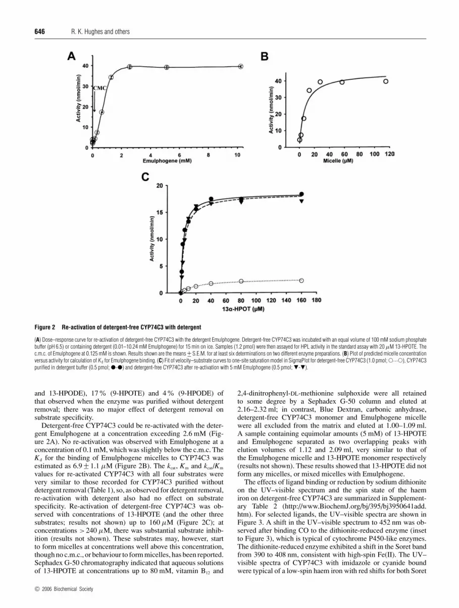
Figure 2 Re-activation of detergent-free CYP74C3 with detergent
(A) Dose–response curve for re-activation of detergent-free CYP74C3 with the detergent Emulphogene. Detergent-free CYP74C3 was incubated with an equal volume of 100 mM sodium phosphatebuffer (pH 6.5) or containing detergent (0.01–10.24 mM Emulphogene) for 15 min on ice. Samples (1.2 pmol) were then assayed for HPL activity in the standard assay with 20 µM 13-HPOTE. Thec.m.c. of Emulphogene at 0.125 mM is shown. Results shown are the means +− S.E.M. for at least six determinations on two different enzyme preparations. (B) Plot of predicted micelle concentrationversus activity for calculation of K d for Emulphogene binding. (C) Fit of velocity–substrate curves to one-site saturation model in SigmaPlot for detergent-free CYP74C3 (1.0 pmol; �···�), CYP74C3purified in detergent buffer (0.5 pmol; �-�) and detergent-free CYP74C3 after re-activation with 5 mM Emulphogene (0.5 pmol; �-�).
and 13-HPODE), 17% (9-HPOTE) and 4% (9-HPODE) ofthat observed when the enzyme was purified without detergentremoval; there was no major effect of detergent removal onsubstrate specificity.
Detergent-free CYP74C3 could be re-activated with the deter-gent Emulphogene at a concentration exceeding 2.6 mM (Fig-ure 2A). No re-activation was observed with Emulphogene at aconcentration of 0.1 mM, which was slightly below the c.m.c. TheKd for the binding of Emulphogene micelles to CYP74C3 wasestimated as 6.9 +− 1.1 µM (Figure 2B). The kcat, Km and kcat/Km
values for re-activated CYP74C3 with all four substrates werevery similar to those recorded for CYP74C3 purified withoutdetergent removal (Table 1), so, as observed for detergent removal,re-activation with detergent also had no effect on substratespecificity. Re-activation of detergent-free CYP74C3 was ob-served with concentrations of 13-HPOTE (and the other threesubstrates; results not shown) up to 160 µM (Figure 2C); atconcentrations >240 µM, there was substantial substrate inhib-ition (results not shown). These substrates may, however, startto form micelles at concentrations well above this concentration,though no c.m.c., or behaviour to form micelles, has been reported.Sephadex G-50 chromatography indicated that aqueous solutionsof 13-HPOTE at concentrations up to 80 mM, vitamin B12 and
2,4-dinitrophenyl-DL-methionine sulphoxide were all retainedto some degree by a Sephadex G-50 column and eluted at2.16–2.32 ml; in contrast, Blue Dextran, carbonic anhydrase,detergent-free CYP74C3 monomer and Emulphogene micellewere all excluded from the matrix and eluted at 1.00–1.09 ml.A sample containing equimolar amounts (5 mM) of 13-HPOTEand Emulphogene separated as two overlapping peaks withelution volumes of 1.12 and 2.09 ml, very similar to that ofthe Emulphogene micelle and 13-HPOTE monomer respectively(results not shown). These results showed that 13-HPOTE did notform any micelles, or mixed micelles with Emulphogene.
The effects of ligand binding or reduction by sodium dithioniteon the UV–visible spectrum and the spin state of the haemiron on detergent-free CYP74C3 are summarized in Supplement-ary Table 2 (http://www.BiochemJ.org/bj/395/bj3950641add.htm). For selected ligands, the UV–visible spectra are shown inFigure 3. A shift in the UV–visible spectrum to 452 nm was ob-served after binding CO to the dithionite-reduced enzyme (insetto Figure 3), which is typical of cytochrome P450-like enzymes.The dithionite-reduced enzyme exhibited a shift in the Soret bandfrom 390 to 408 nm, consistent with high-spin Fe(II). The UV–visible spectra of CYP74C3 with imidazole or cyanide boundwere typical of a low-spin haem iron with red shifts for both Soret
c© 2006 Biochemical Society

Monomer–micelle association and hydroperoxide lyase activity 647
Figure 3 Ligand-binding studies of detergent-free CYP74C3
UV–visible spectra of ligand-bound forms of oxidized native CYP74C3 (2 µM; ——); KCN(100 mM; — — —); imidazole (50 mM; - - - -); DMPhP (250 mM; — �� —). Thespectra of dithionite (50 mM) reduced CYP74C3 (2 µM) before (�����) and after(— � —) reaction with CO (∼1 mM) are also shown; inset, the difference spectrum forCO binding. The increase in absorbance below 400 nm is due to the large excess of sodiumdithionite used. All spectra were recorded at pH 6.5 in 100 mM sodium phosphate buffer (pH 6.5)at 25◦C, 1 cm path length.
and visible bands. The imidazole complex exhibited bands at 366,426, 544 and 547 nm (Figure 3) with a Kd at pH 6.5 of 362.5 +−120.4 mM. Measurements by stopped-flow absorption showedthat the kinetics of imidazole binding was biphasic (results notshown). The cyanide complex exhibited bands at 365, 434,539 and 566 nm and Kd values of 105.1 +− 24.0 and 1507.9 +−448.4 mM respectively were obtained at pH 9.0 and 8.0. Thetime course for cyanide binding monitored at 440 nm usingthe stopped-flow diode-array data was also biphasic (resultsnot shown). Ferrous low-spin haem iron spectrophotometricsignatures were also observed for O2, pyridine and BI with red-shifted Soret and visible bands (see Supplementary Table 2at http://www.BiochemJ.org/bj/395/bj3950641add.htm). Bindingof the sulphur- and phosphorous-containing ligands thiazoleand DMPhP gave low-spin haem iron bands with large Soretband shifts (see Supplementary Table 2 at http://www.BiochemJ.org/bj/395/bj3950641add.htm); the DMPhP complex has bandsat 375, 453 and 563 nm (Figure 3) with a Kd at pH 6.5 of 5.3 +−0.8 mM. Thiocyanate and azide binding resulted in a mixture ofhigh and low-spin haem iron bands (see Supplementary Table 2at http://www.BiochemJ.org/bj/395/bj3950641add.htm).
EPR spectroscopy indicated that the spectrum of the restingenzyme contained a mixture of high- and low-spin haem ironwith g factors of 8.03, 3.51, 1.68, and 2.39, 2.24, 1.93 respec-tively (values from simulation of spectra), in the ratio of 2.5:1(Figure 4a). Addition of Emulphogene at a concentration of2.4 mM, well above the c.m.c., to resting enzyme (240 µM),resulted in a change in the ratio of high to low spin haem ironto 1:5.5, but with no change in the total EPR concentration (Fig-ure 4b). The addition of 13-HPOTE at 2.4 mM to resting enzyme(240 µM) resulted in a 60% loss of EPR intensity and a ratio ofhigh to low spin haem iron of 1:2 (Figure 4c). Subsequent additionof 13-HPOTE at 2.4 mM to the enzyme sample previously re-activated with 2.4 mM Emulphogene resulted in a 50% reductionin the total concentration of the EPR signal and a ratio of high tolow spin haem iron signal of 1:6 (Figure 4d). The UV–visible
Figure 4 Effects of 13-HPOTE or detergent on the EPR spectrum ofdetergent-free CYP74C3
EPR spectra shown are: (a) 240 µM resting enzyme and (b) after reaction with 5 mMEmulphogene or (c) 2.4 mM 13-HPOTE. The sample in (b) was thawed and reacted with2.4 mM 13-HPOTE prior to immediate refreezing in liquid nitrogen and the EPR spectrum(d) was recorded again. The g values of the peaks corresponding to high- and low-spin haemiron are shown.
spectra and specific activities of the resting and detergent-re-activated enzymes after EPR were indistinguishable from thosebefore the analysis (results not shown). Similar effects were alsoseen with resting enzyme at 24 µM, which suggests that therewas no concentration dependency. At a concentration of 0.1 mM,slightly below the c.m.c. of Emulphogene, both Emulphogene and13-HPOTE had no effect on the EPR spectrum of resting enzymeat 24 µM (results not shown). Changes in the spin state of the haemiron, induced by detergent micelles, were also associated withsubtle changes in the UV–visible spectrum (results not shown).Addition of 1.56 mM Emulphogene to the detergent-free enzymeat 10.2 µM resulted in a change in the Soret region; the peak at391 nm was shifted to 393 nm and a shoulder developed at 420 nmwith an isosbestic point at 401 nm. Similar changes at longerwavelengths were observed; the features at 508 and 644 nm werereduced, the feature at 545 nm increased slightly and a newfeature appeared at 568 nm with isosbestic points at 454, 530,581 and 669 nm (results not shown). No clear changes in the UV–visible spectrum were observed during the reaction of 10.2 µMCYP74C3 in the presence and absence of 5 mM Emulphogene,with 100 µM or 2.4 mM 13-HPOTE, nor with detergent-free10.2 µM CYP74C3 and 0.08 mM Emulphogene (results notshown).
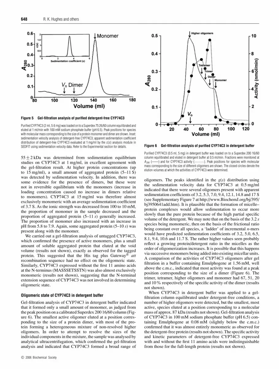
Oligomeric state of detergent-free CYP74C3
The molecular mass of His-tagged CYP74C3, based on the proteinsequence and predicted from the cDNA, was 56.8 kDa. This wasconsistent with the value determined for the native protein bygel filtration (relative to globular protein standards) of 55.2 kDa(Figure 5). These measurements, and dynamic light scattering(results not shown), indicated that detergent-free CYP74C3 wasa monomer. No dimer, which, if present, would have been cleanlyseparated from monomer, could be detected by gel filtration(Figure 5), and activity could be detected only in the monomerfraction. Under the conditions in the ultracentrifuge, analysisof the sedimentation velocity data indicated that detergent-freeCYP74C3 at 1 mg/ml was also almost exclusively a monomerwith a sedimentation coefficient of 3.5 S (inset to Figure 5).A corresponding weight-average molecular mass, Mw, of
c© 2006 Biochemical Society
648 R. K. Hughes and others
Figure 5 Gel-filtration analysis of purified detergent-free CYP74C3
Purified CYP74C3 (2 ml, 5.6 mg) was loaded on to a Superdex 75 26/60 column equilibrated andeluted at 1 ml/min with 100 mM sodium phosphate buffer (pH 6.5). Peak positions for specieswith molecular mass corresponding to the size of a protein monomer and dimer are shown. Inset:sedimentation velocity analysis of detergent-free CYP74C3; apparent sedimentation coefficientdistribution of detergent-free CYP74C3 evaluated at 1 mg/ml by the c(s) analysis module inSEDFIT using sedimentation velocity data. Refer to the Experimental section for details.
55 +− 2 kDa was determined from sedimentation equilibriumstudies on CYP74C3 at 1 mg/ml, in excellent agreement withthe gel-filtration result. At higher protein concentrations (upto 15 mg/ml), a small amount of aggregated protein (5–11 S)was detected by sedimentation velocity. In addition, there wassome evidence for the presence of dimers, but these werenot in reversible equilibrium with the monomers (increase inloading concentration caused no increase in dimers relativeto monomers). CYP74C3 at 15 mg/ml was therefore almostexclusively monomeric with an average sedimentation coefficientof 3.7 S. As the ionic strength was decreased from 100 to 10 mM,the proportion of monomer in the sample decreased and theproportion of aggregated protein (5–11 s) generally increased.The proportion of monomer also increased with an increase inpH from 5.8 to 7.9. Again, some aggregated protein (5–10 s) waspresent along with the monomer.
We carried out a gel-filtration analysis of untagged CYP74C3,which confirmed the presence of active monomers, plus a smallamount of soluble aggregated protein that eluted at the voidvolume (results not shown), exactly as observed for the taggedprotein. This suggested that the His tag plus Gateway® attrecombination sequence had no effect on the oligomeric state.Similarly, CYP74C3 expressed without the first 11 amino acidsat the N-terminus (MASSSETSSTN) was also almost exclusivelymonomeric (results not shown), suggesting that the N-terminalextension sequence of CYP74C3 was not involved in determiningoligomeric state.
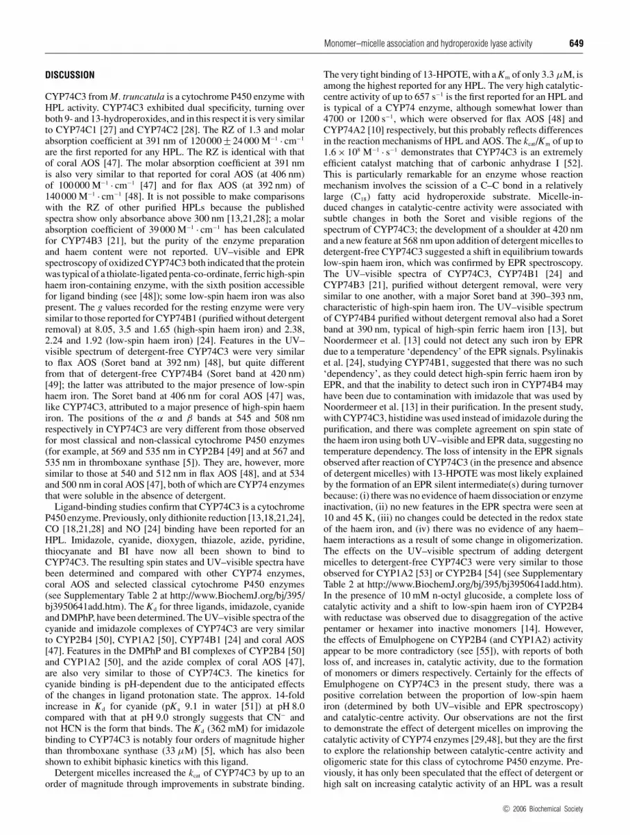
Oligomeric state of CYP74C3 in detergent buffer
Gel-filtration analysis of CYP74C3 in detergent buffer indicatedthat it formed only a small amount of monomer, as judged fromthe peak position on a calibrated Superdex 200 16/60 column (Fig-ure 6). The smallest active oligomer eluted at a position corres-ponding to the size of a protein dimer, with most of the pro-tein forming a heterogeneous mixture of non-resolved higheroligomers. In order to attempt to resolve the sizes of theindividual components in the mixture, the sample was analysed byanalytical ultracentrifugation, which confirmed the gel-filtrationanalysis and indicated that CYP74C3 formed a broad range of
Figure 6 Gel-filtration analysis of purified CYP74C3 in detergent buffer
Purified CYP74C3 (0.5 ml, 5 mg) in detergent buffer was loaded on to a Superdex 200 16/60column equilibrated and eluted in detergent buffer at 0.5 ml/min. Fractions were monitored atA 280 (——) and for CYP74C3 activity (··········). Peak positions for species with molecularmass corresponding to the size of different oligomers are shown. The closed circles denote theelution volumes at which the activities of CYP74C3 were determined.
oligomers. The peaks identified in the g(s) distribution usingthe sedimentation velocity data for CYP74C3 at 0.5 mg/mlindicated that there were several oligomers present with apparentsedimentation coefficients of 3.2, 5.3, 7.0, 9.4, 12.1, 14.8 and 17 S(see Supplementary Figure 7 at http://www.BiochemJ.org/bj/395/bj3950641add.htm). It is plausible that the formation of micelle–protein complexes would allow sedimentation to occur moreslowly than the pure protein because of the high partial specificvolume of the detergent. We may note that on the basis of the 3.2 sspecies being monomeric, then on the basis of the frictional ratiobeing constant over all species, a ‘ladder’ of incremental n-merswould have predicted sedimentation coefficients of 3.2, 5.0, 6.5,8.1, 9.4, 10.6 and 11.7 S. The rather higher values seen probablyreflect a growing protein/detergent ratio in the micelles as theorder of oligomerization increases. It is possible that this happensvia successive monomers being added into existing micellar units.A comparison of the activities of CYP74C3 oligomers after gelfiltration in a buffer containing Emulphogene at 1.56 mM, wellabove the c.m.c., indicated that most activity was found at a peakposition corresponding to the size of a dimer (Figure 6). Thetrimer, tetramer, higher oligomers and monomer had 67, 51, 20and 10% respectively of the specific activity of the dimer (resultsnot shown).
When CYP74C3 in detergent buffer was applied to a gel-filtration column equilibrated under detergent-free conditions, anumber of higher oligomers were detected, but the smallest, mostactive, species eluted at a position corresponding to a molecularmass of approx. 87 kDa (results not shown). Gel-filtration analysisof CYP74C3 in 100 mM sodium phosphate buffer (pH 6.5) con-taining Emulphogene at 0.08 mM (slightly below the c.m.c.)confirmed that it was almost entirely monomeric as observed forthe detergent-free protein (results not shown). The specific activityand kinetic parameters of detergent-free CYP74C3 expressedwith and without the first 11 amino acids were indistinguishablefrom those for the full-length protein (results not shown).
c© 2006 Biochemical Society
Monomer–micelle association and hydroperoxide lyase activity 649
DISCUSSION
CYP74C3 from M. truncatula is a cytochrome P450 enzyme withHPL activity. CYP74C3 exhibited dual specificity, turning overboth 9- and 13-hydroperoxides, and in this respect it is very similarto CYP74C1 [27] and CYP74C2 [28]. The RZ of 1.3 and molarabsorption coefficient at 391 nm of 120000 +− 24000 M−1 · cm−1
are the first reported for any HPL. The RZ is identical with thatof coral AOS [47]. The molar absorption coefficient at 391 nmis also very similar to that reported for coral AOS (at 406 nm)of 100000 M−1 · cm−1 [47] and for flax AOS (at 392 nm) of140000 M−1 · cm−1 [48]. It is not possible to make comparisonswith the RZ of other purified HPLs because the publishedspectra show only absorbance above 300 nm [13,21,28]; a molarabsorption coefficient of 39000 M−1 · cm−1 has been calculatedfor CYP74B3 [21], but the purity of the enzyme preparationand haem content were not reported. UV–visible and EPRspectroscopy of oxidized CYP74C3 both indicated that the proteinwas typical of a thiolate-ligated penta-co-ordinate, ferric high-spinhaem iron-containing enzyme, with the sixth position accessiblefor ligand binding (see [48]); some low-spin haem iron was alsopresent. The g values recorded for the resting enzyme were verysimilar to those reported for CYP74B1 (purified without detergentremoval) at 8.05, 3.5 and 1.65 (high-spin haem iron) and 2.38,2.24 and 1.92 (low-spin haem iron) [24]. Features in the UV–visible spectrum of detergent-free CYP74C3 were very similarto flax AOS (Soret band at 392 nm) [48], but quite differentfrom that of detergent-free CYP74B4 (Soret band at 420 nm)[49]; the latter was attributed to the major presence of low-spinhaem iron. The Soret band at 406 nm for coral AOS [47] was,like CYP74C3, attributed to a major presence of high-spin haemiron. The positions of the α and β bands at 545 and 508 nmrespectively in CYP74C3 are very different from those observedfor most classical and non-classical cytochrome P450 enzymes(for example, at 569 and 535 nm in CYP2B4 [49] and at 567 and535 nm in thromboxane synthase [5]). They are, however, moresimilar to those at 540 and 512 nm in flax AOS [48], and at 534and 500 nm in coral AOS [47], both of which are CYP74 enzymesthat were soluble in the absence of detergent.
Ligand-binding studies confirm that CYP74C3 is a cytochromeP450 enzyme. Previously, only dithionite reduction [13,18,21,24],CO [18,21,28] and NO [24] binding have been reported for anHPL. Imidazole, cyanide, dioxygen, thiazole, azide, pyridine,thiocyanate and BI have now all been shown to bind toCYP74C3. The resulting spin states and UV–visible spectra havebeen determined and compared with other CYP74 enzymes,coral AOS and selected classical cytochrome P450 enzymes(see Supplementary Table 2 at http://www.BiochemJ.org/bj/395/bj3950641add.htm). The Kd for three ligands, imidazole, cyanideand DMPhP, have been determined. The UV–visible spectra of thecyanide and imidazole complexes of CYP74C3 are very similarto CYP2B4 [50], CYP1A2 [50], CYP74B1 [24] and coral AOS[47]. Features in the DMPhP and BI complexes of CYP2B4 [50]and CYP1A2 [50], and the azide complex of coral AOS [47],are also very similar to those of CYP74C3. The kinetics forcyanide binding is pH-dependent due to the anticipated effectsof the changes in ligand protonation state. The approx. 14-foldincrease in Kd for cyanide (pKa 9.1 in water [51]) at pH 8.0compared with that at pH 9.0 strongly suggests that CN− andnot HCN is the form that binds. The Kd (362 mM) for imidazolebinding to CYP74C3 is notably four orders of magnitude higherthan thromboxane synthase (33 µM) [5], which has also beenshown to exhibit biphasic kinetics with this ligand.
Detergent micelles increased the kcat of CYP74C3 by up to anorder of magnitude through improvements in substrate binding.
The very tight binding of 13-HPOTE, with a Km of only 3.3 µM, isamong the highest reported for any HPL. The very high catalytic-centre activity of up to 657 s−1 is the first reported for an HPL andis typical of a CYP74 enzyme, although somewhat lower than4700 or 1200 s−1, which were observed for flax AOS [48] andCYP74A2 [10] respectively, but this probably reflects differencesin the reaction mechanisms of HPL and AOS. The kcat/Km of up to1.6 × 108 M−1 · s−1 demonstrates that CYP74C3 is an extremelyefficient catalyst matching that of carbonic anhydrase I [52].This is particularly remarkable for an enzyme whose reactionmechanism involves the scission of a C–C bond in a relativelylarge (C18) fatty acid hydroperoxide substrate. Micelle-in-duced changes in catalytic-centre activity were associated withsubtle changes in both the Soret and visible regions of thespectrum of CYP74C3; the development of a shoulder at 420 nmand a new feature at 568 nm upon addition of detergent micelles todetergent-free CYP74C3 suggested a shift in equilibrium towardslow-spin haem iron, which was confirmed by EPR spectroscopy.The UV–visible spectra of CYP74C3, CYP74B1 [24] andCYP74B3 [21], purified without detergent removal, were verysimilar to one another, with a major Soret band at 390–393 nm,characteristic of high-spin haem iron. The UV–visible spectrumof CYP74B4 purified without detergent removal also had a Soretband at 390 nm, typical of high-spin ferric haem iron [13], butNoordermeer et al. [13] could not detect any such iron by EPRdue to a temperature ‘dependency’ of the EPR signals. Psylinakiset al. [24], studying CYP74B1, suggested that there was no such‘dependency’, as they could detect high-spin ferric haem iron byEPR, and that the inability to detect such iron in CYP74B4 mayhave been due to contamination with imidazole that was used byNoordermeer et al. [13] in their purification. In the present study,with CYP74C3, histidine was used instead of imidazole during thepurification, and there was complete agreement on spin state ofthe haem iron using both UV–visible and EPR data, suggesting notemperature dependency. The loss of intensity in the EPR signalsobserved after reaction of CYP74C3 (in the presence and absenceof detergent micelles) with 13-HPOTE was most likely explainedby the formation of an EPR silent intermediate(s) during turnoverbecause: (i) there was no evidence of haem dissociation or enzymeinactivation, (ii) no new features in the EPR spectra were seen at10 and 45 K, (iii) no changes could be detected in the redox stateof the haem iron, and (iv) there was no evidence of any haem–haem interactions as a result of some change in oligomerization.The effects on the UV–visible spectrum of adding detergentmicelles to detergent-free CYP74C3 were very similar to thoseobserved for CYP1A2 [53] or CYP2B4 [54] (see SupplementaryTable 2 at http://www.BiochemJ.org/bj/395/bj3950641add.htm).In the presence of 10 mM n-octyl glucoside, a complete loss ofcatalytic activity and a shift to low-spin haem iron of CYP2B4with reductase was observed due to disaggregation of the activepentamer or hexamer into inactive monomers [14]. However,the effects of Emulphogene on CYP2B4 (and CYP1A2) activityappear to be more contradictory (see [55]), with reports of bothloss of, and increases in, catalytic activity, due to the formationof monomers or dimers respectively. Certainly for the effects ofEmulphogene on CYP74C3 in the present study, there was apositive correlation between the proportion of low-spin haemiron (determined by both UV–visible and EPR spectroscopy)and catalytic-centre activity. Our observations are not the firstto demonstrate the effect of detergent micelles on improving thecatalytic activity of CYP74 enzymes [29,48], but they are the firstto explore the relationship between catalytic-centre activity andoligomeric state for this class of cytochrome P450 enzyme. Pre-viously, it has only been speculated that the effect of detergent orhigh salt on increasing catalytic activity of an HPL was a result
c© 2006 Biochemical Society

650 R. K. Hughes and others
of some conformational change in the protein [29]. To our know-ledge, no studies in this regard have been carried out for anAOS.
CYP74C3 required extraction and purification in the presenceof detergent, but unlike other HPLs, it remained soluble and activeas a monomer when both detergent and salt were removed. Theprotein was almost entirely monomeric at concentrations from0.5 to 15 mg/ml over a wide range of pH and ionic strengths.In the presence of detergent micelles, however, the most activeCYP74C3 oligomer was of the size of a protein dimer, which isa new observation. This contrasts with other reports where HPLpurified from higher plants was either a trimer or a tetramer [15–23]. In those instances where gel filtration was carried out inthe presence of detergent and aggregation prevented, the highermolecular mass estimations may have been due to the formationof HPL complexes with the large (molecular mass 90 kDa)detergent micelles of Triton X-100 that was used at concentrationswell above the c.m.c. (0.24 mM) of this detergent [40]. TheseHPLs may also have been considerably more hydrophobic thanCYP74C3, which might explain their insolubility in the absenceof detergent. In the present study, since Emulphogene was usedat a concentration of 1.56 mM that is also well above the c.m.c.of this detergent (0.125 mM), micelles of average molecular mass62.1 kDa would have formed under the conditions used for gelfiltration. The identification of a CYP74C3 monomer and nodimer under detergent-free conditions was perhaps not surprisingbecause any protein-bound detergent remaining would have beenat concentrations well below the c.m.c., and the detergent wouldhave been entirely monomeric. Any complex formation withprotein and Emulphogene micelle would increase the molecularmass from 56.8 to 118.9 kDa, but no protein of this size oractivity at this peak position in gel filtration under detergent-freeconditions was detected. In contrast, in the presence of detergentbuffer, the molecular mass of the most active species was the sizeof a protein dimer, or of a complex between a protein monomerand an Emulphogene micelle. The conclusion of dimer formationfor CYP2B4 in the presence of detergent [55] was based on theresults of gel filtration in the presence of Emulphogene, but likeCYP74C3, may have corresponded to a protein monomer–micellecomplex, rather than a protein dimer.
Regardless of the method of purification, CYP74C3 under theconditions of the activity assay was almost entirely monomeric.Nevertheless, the kcat of the detergent-free monomer with thepreferred substrate was only 10% of the activity of the sameenzyme in detergent buffer. It may be suggested that the low levelof activity in the detergent-free preparation was due to contamin-ation with a small amount of more active higher oligomer.However, clean separation of the monomer from the dimer bygel filtration on Superdex 75, and the presence of enzyme activityin only the fraction corresponding to the size of the monomer, sug-gests that this was unlikely. Under detergent-free conditions,the 10% residual activity more likely resulted from CYP74C3monomers, which may or may not have been contaminated withdetergent monomers that coated hydrophobic patches on thesurface of the protein. We propose that detergent-free mono-mers exhibited reduced substrate binding through a conforma-tional change that was induced by release from a micellar ormembrane environment. Complete re-activation of detergent-free CYP74C3 with the detergent Emulphogene was possibleonly at concentrations of at least 26-fold higher than the c.m.c.,which suggested that detergent micelles (or the simulation ofa membranous environment) and not monomers were requiredto maintain the most active conformation of CYP74C3. Thisevidence, together with the observation that the smallest and mostactive protein species detected by gel filtration in the presence of
Emulphogene corresponded to the size of a monomer–micelle,suggested that an association between a protein monomer and adetergent micelle was the most active conformation for CYP74C3and not a protein dimer. Further evidence for the lack of pro-tein dimer formation comes from the absence, in concentrateddetergent-free CYP74C3 preparations before and after re-activ-ation with Emulphogene, of EPR signals corresponding to haem–haem interactions, and of protein sedimenting with the Mw of adimer in the analytical ultracentrifuge. The small amount of pro-tein dimer that was detected under detergent-free conditions inthe analytical ultracentrifuge was shown not to be in reversibleequilibrium with protein monomer. Increases in the activity andtightness of binding for CYP74C3 purified in detergent bufferwere unexpected because the concentration of detergent presentin the activity assay would have been well below the c.m.c. Thismust suggest that the interaction between detergent micelle andprotein monomer was stable over the very short time course of theactivity assay (20 s); the low Kd determined for micelle bindingof only 6.9 µM certainly indicated that this association was verytight; indeed, it was tighter than that for the preferred substrate(Km = 20.9 µM).
13-HPOTE is only sparingly soluble in aqueous solution so itis important to distinguish between the substrate being presentedfrom aqueous solution and from a micelle or membrane. It maybe suggested that detergent micelles, or the formation of mixedsubstrate and detergent micelles, could facilitate substrate bindingwithout any requirement for a change in protein conformation. 13-HPOTE was, however, entirely monomeric and did not form anymicelles under our experimental conditions, even in the presenceof Emulphogene micelles. This suggested that re-activation ofdetergent-free CYP74C3 (and the observed changes in spin stateof the haem iron) required either detergent micelles or sub-strate monomers, and that substrate presentation could not accountfor CYP74C3 re-activation.
We hypothesize that in E. coli, and most likely in planta,CYP74C3 is a peripheral membrane protein with small patchesof hydrophobic surface residues that promote a tight associationwith membranes, most likely as monomers. Solubilization withdetergents releases the monomers from the membranes and allowsthem to self-associate to form a mixture of monomer–micelles andhigher oligomers through hydrophobic interaction. The natureof the hydrophobic domain of CYP74C3 that is necessary formembrane association in CYP74C3 is unknown. The N-terminalsequence of cytochrome P450 enzymes has been reported to be amembrane-binding domain [12], but the N-terminal sequence ofCYP74C3 is not particularly hydrophobic and evidence from thepresent study suggests that it has no role in membrane binding.The structure of the engineered AOS domain from an AOS–LOX fusion chimaera was recently crystallized as a protein dimer[9], but this bears no similarity to CYP74C3. The present studywould suggest that the association between a protein monomerand a single detergent micelle (or perhaps a phospholipid in amembrane), and not oligomeric state, regulates the catalyticactivity of CYP74C3. This represents a new mechanism fora membrane-associated cytochrome P450 enzyme and may be adistinguishing feature of CYP74 enzymes that are distinct fromclassical cytochrome P450 enzymes that require association witha reductase in order to carry out their full range of biologicalactivities.
Unlike other plant cytochrome P450 enzymes described sofar, CYP74C3 is a natural variant that does not require proteinengineering to improve water solubility. The detailed under-standing of the physical and biochemical properties of CYP74C3described in the present study has provided valuable informationtowards our understanding of the differences in reaction
c© 2006 Biochemical Society
Monomer–micelle association and hydroperoxide lyase activity 651
mechanism of CYP74 and more typical cytochrome P450enzymes and the first crystal or NMR solution structure foran HPL or other membrane-associated plant cytochrome P450enzyme. The subsequent information that would be forthcomingon the primary determinants of substrate and product specificities,and an identification of the hydrophobic domain responsible formembrane association, are likely to be key requirements for thechallenging aim of manipulating oxylipin metabolism in planta.
We thank the Samuel Roberts Noble Foundation (Ardmore, OK, U.S.A.) for providingthe EST clone of CYP74C3, Professor Mats Hamberg for providing HPL substrates,Mr Guus van Zadelhoff and Professor Gerrit Veldink (University of Utrecht, TheNetherlands) for help with the GC-MS analysis, and Drs Mike Naldrett, Igor Galetich andMiss Karen Wilson (Department of Biological Chemistry, John Innes Centre, Norwich,U.K.) for protein sequencing. This work was funded by a European Union-funded project‘Natural Oxylipins for Defence of Ornamentals (NODO)’, project number QLK5-CT-2001-02445, and by the Biotechnology and Biological Sciences Research Council.
REFERENCES
1 Schoch, G. A., Attias, R., Belghazi, M., Dansette, P. M. and Werck-Reichhart, D. (2003)Engineering of a water-soluble plant cytochrome P450, CYP73A1, and NMR-basedorientation of natural and alternate substrates in the active site. Plant Physiol. 133,1198–1208
2 Hasemann, C. A., Kurumbail, R. G., Boddupalli, S. S., Peterson, J. A. and Desienhofer, J.(1995) Structure and function of cytochromes P450: a comparative analysis of threecrystal structures. Structure 2, 41–62
3 Williams, P. A., Cosme, J., Sridhar, V., Johnson, E. F. and McRee, D. E. (2000)Mammalian microsomal cytochrome P450 monooxygenase: structural adaptations formembrane binding and functional diversity. Mol. Cell 5, 121–131
4 Song, W.-C., Funk, C. D. and Brash, A. R. (1993) Molecular cloning of an allene oxidesynthase: a cytochrome P450 specialised for the metabolism of fatty acid hydroperoxides.Proc. Natl. Acad. Sci. U.S.A. 90, 8519–8523
5 Wang, L.-H., Tsai, A.-L. and Hsu, P.-Y. (2001) Substrate binding is the rate-limiting stepin thromboxane synthase catalysis. J. Biol. Chem. 276, 14737–14743
6 Grechkin, A. N., Mukhtarova, L. S. and Hamberg, M. (2003) Detection of an enolintermediate in the hydroperoxide lyase chain cleavage reaction. FEBS Lett. 549, 31–34
7 Casey, R. and Hughes, R. K. (2004) Recombinant lipoxygenases and oxylipin metabolismin relation to food quality. Food Biotechnol. 18, 135–170
8 Morant, M., Bak, S., Moller, B. L. and Werck-Reichhart, D. (2003) Plant cytochromesP450: tools for pharmacology, plant protection and phytoremediation.Curr. Opin. Biotechnol. 14, 151–162
9 Oldham, M. L., Brash, A. R. and Newcomer, M. E. (2005) The structure of coral alleneoxide synthase reveals a catalase adapted for metabolism of a fatty acid hydroperoxide.Proc. Natl. Acad. Sci. U.S.A. 102, 297–302
10 Pan, Z., Camara, B., Gardner, H. W. and Backhaus, R. A. (1998) Aspirin inhibition andacetylation of the plant cytochrome P450, allene oxide synthase, resembles that of animalprostaglandin endoperoxide H synthase. J. Biol. Chem. 273, 18139–18145
11 Mast, N., Andersson, U., Nakayama, K., Bjorkhem, I. and Pikulevaa, I. A. (2004)Expression of human cytochrome P450 46A1 in Escherichia coli: effects of N- andC-terminal modifications. Arch. Biochem. Biophys. 428, 99–108
12 von Wachenfeldt, C., Richardson, T. H., Cosme, J. and Johnson, E. F. (1997) MicrosomalP450 2C3 is expressed as a soluble dimer in Escherichia coli following modification of itsN-terminus. Arch. Biochem. Biophys. 339, 107–114
13 Noordermeer, M. A., van Dijken, A. J. H., Smeekens, S. C. M., Veldink, G. A. andVliegenthart, J. F. G. (2000) Characterisation of three cloned and expressed13-hydroperoxide lyase isoenzymes from alfalfa with unusual N-terminal sequences anddifferent enzyme kinetics. Eur. J. Biochem. 267, 2473–2482
14 Dean, W. L. and Gray, R. D. (1982) Relationship between state of aggregation and catalyticactivity for cytochrome P-450LM2 and NADPH-cytochrome P-450 reductase.J. Biol. Chem. 257, 14679–14685
15 Tijet, N., Waspi, U., Gaskin, D. J., Hunziker, P., Muller, B. L., Vulfson, E. N.,Slusarenko, A., Brash, A. R. and Whitehead, I. M. (2000) Purification, molecular cloning,and expression of the gene encoding fatty acid 13-hydroperoxide lyase from guava fruit(Psidium guajava). Lipids 35, 709–720
16 Shibata, Y., Matsui, K., Kajiwara, T. and Hatanaka, A. (1995) Purification and properties offatty-acid hydroperoxide lyase from green bell pepper fruits. Plant Cell Physiol. 36,147–156
17 Husson, F. and Belin, J. M. (2002) Purification of hydroperoxide lyase from green bellpepper (Capsicum annuum L.) fruits for the generation of C6-aldehydes in vitro.J. Agric. Food Chem. 50, 1991–1995
18 Itoh, A. and Vick, B. A. (1999) The purification and characterisation of fatty acidhydroperoxide lyase in sunflower. Biochim. Biophys. Acta 1436, 531–540
19 Schreier, P. and Lorenz, G. (1982) Separation, partial-purification and characterisation of afatty-acid hydroperoxide cleaving enzyme from apple and tomato fruits. Z. Naturforsch.37, 165–173
20 Fauconnier, M.-L., Perez, A. G., Sanz, C. and Marlier, M. (1997) Purification andcharacterisation of tomato leaf (Lycopersicon esculentum Mill.) hydroperoxide lyase.J. Agric. Food Chem. 45, 4232–4236
21 Matsui, K., Miyahara, C., Wilkinson, J., Hiatt, B., Knauf, V. and Kajiwara, T. (2000) Fattyacid hydroperoxide lyase in tomato fruits: cloning and properties of a recombinantenzyme expressed in Escherichia coli. Biosci. Biotechnol. Biochem. 64, 1189–1196
22 Olias, J. M., Rios, J. L., Valle, M., Zamora, R., Sartz, L. C. and Axelrod, B. (1990)Fatty-acid hydroperoxide lyase in germinating soybean seedlings. J. Agric. Food Chem.38, 624–630
23 Vick, B. A. and Zimmerman, D. C. (1976) Lipoxygenase and hydroperoxide lyase ingerminating watermelon seedlings. Plant Physiol. 57, 780–788
24 Psylinakis, E., Davoras, E. M., Ioannidis, N., Trikeriotis, M., Petrouleas, V. andGhanotakis, D. F. (2001) Isolation and spectroscopic characterisation of a recombinantbell pepper hydroperoxide lyase. Biochim. Biophys. Acta 1533, 119–127
25 Kandzia, R., Stumpe, M., Berndt, E., Szalata, M., Matsui, K. and Feussner, I. (2003) On thespecificity of lipid hydroperoxide fragmentation by fatty acid hydroperoxide lyase fromArabidopsis thaliana. J. Plant Physiol. 160, 803–809
26 Howe, G. A., Lee, G. I., Itoh, A., Li, L. and DeRocher, A. E. (2000) CytochromeP450-dependent metabolism of oxylipins in tomato. Cloning and expression of alleneoxide synthase and fatty acid hydroperoxide lyase. Plant Physiol. 123, 711–724
27 Matsui, K., Ujita, C., Fujimoto, S.-H., Wilkinson, J., Hiatt, B., Knauf, V., Kajiwara, T. andFeussner, I. (2000) Fatty acid 9- and 13-hydroperoxide lyases from cucumber. FEBS Lett.481, 183–188
28 Tijet, N., Schneider, C., Muller, B. L. and Brash, A. R. (2001) Biogenesis of volatilealdehydes from fatty acid hydroperoxides: molecular cloning of a hydroperoxide lyase(CYP74C) with specificity for both the 9- and 13-hydroperoxides of linoleic and linolenicacids. Arch. Biochem. Biophys. 386, 281–289
29 Koeduka, T., Stumpe, M., Matsui, K., Kajiwara, T. and Feussner, I. (2003) Kinetics of barleyFA hydroperoxide lyase are modulated by salts and detergents. Lipids 38, 1167–1172
30 Scott, E. E., He, Y. A., Wester, M. R., White, M. A., Chin, C. C., Halpert, J. R., Johnson,E. F. and Stout, C. D. (2003) An open conformation of mammalian cytochrome P450 2B4at 1.6-A resolution. Proc. Natl. Acad. Sci. U.S.A. 100, 13196–13201
31 Schoch, G. A., Yano, J. K., Wester, M. R., Griffin, C., Stout, E. F. and Johnson, E. F. (2004)Structure of human microsomal cytochrome P450 2C8: evidence for a peripheral fattyacid binding site. J. Biol. Chem. 279, 9497–9503
32 Williams, P. A., Cosme, J., Ward, A., Angove, H. C., Vinkovic, D. M. and Jhoti, H. (2003)Crystal structure of human cytochrome P450 2C9 with bound warfarin. Nature (London)424, 464–468
33 Yano, J. K., Wester, M. R., Schoch, G. A., Griffin, K. J., Stout, C. D. and Johnson, E. F.(2004) The structure of human microsomal cytochrome P450 3A4 determined by X-raycrystallography to 2.05-A resolution. J. Biol. Chem. 279, 38091–38094
34 Yano, J. K., Hsu, M. H., Griffin, K. J., Stout, C. D. and Johnson, E. F. (2005) Structures ofhuman microsomal cytochrome P450 2A6 complexed with coumarin and methoxsalen.Nat. Struct. Mol. Biol. 12, 822–823
35 Zhukovsky, E. A., Lee, J. O., Villegas, M., Chan, C., Chu, S. and Mroske, C. (2004) TNFligands: is TALL-1 a trimer or a virus-like cluster? Nature (London) 427, 413–414
36 Vick, B. A. (1991) A spectrophotometric assay for hydroperoxide lyase. Lipids 26,315–320
37 Aase, R. and Vanngard, T. (1975) EPR signal intensity and powder shapes –re-examination. J. Magn. Reson. 19, 308–315
38 Berry, E. A. and Trumpower, B. (1987) Simultaneous determination of haems a, b, and cfrom pyridine haemochrome spectra. Anal. Biochem. 161, 1–15
39 Bradford, M. M. (1976) A rapid and sensitive method for the quantitation of microgramquantities of protein utilizing the principle of protein–dye binding. Anal. Biochem. 72,248–254
40 Helenius, A. and Simons, K. (1975) Solubilisation of membranes by detergents.Biochim. Biophys. Acta 415, 29–79
41 Noordermeer, M. A., Veldink, G. A. and Vliegenthart, J. F. G. (1999) Alfalfa containssubstantial 9-hydroperoxide lyase activity and a 3Z:2E-enal isomerase. FEBS Lett. 443,201–204
42 Schuck, P. (2000) Size-distribution analysis of macromolecules by sedimentation velocityultracentrifugation and lamm equation modelling. Biophys. J. 78, 1606–1619
c© 2006 Biochemical Society

652 R. K. Hughes and others
43 Colfen, H. and Harding, S. E. (1997) MSTARA and MSTARI: interactive PC algorithms forsimple, model independent evaluation of sedimentation equilibrium data. Eur. Biophys.J. Biophys. Lett. 24, 333–346
44 Laue, T. M., Shah, B. D., Ridgeway, T. M. and Pelletier, S. L. (1992) Computer-aidedinterpretation of analytical sedimentation data for proteins. In AnalyticalUltracentrifugation in Biochemistry and Polymer Science (Harding, S. E.,Rowe, A. J. and Horton, J. C., eds.), pp. 90–125, Royal Society of Chemistry,Cambridge, U.K.
45 Squire, P. G. and Himmel, M. E. (1979) Hydrodynamics and protein hydration.Arch. Biochem. Biophys. 196, 165–177
46 Rahfeld, J.-U., Rucknagel, P., Stoller, G., Horne, S. M., Schierhorn, A., Young, K. D. andFischer, G. (1996) Isolation and amino acid sequence of a new 22-kDa FKBP-likepeptidyl-prolyl cis/trans-isomerase of Escherichia coli: similarity to Mip-like proteins ofpathogenic bacteria. J. Biol. Chem. 271, 22130–22138
47 Abraham, B. D., Sono, M., Boutaud, O., Shriner, A., Dawson, J. H., Brash, A. R. andGaffney, B. J. (2001) Characterisation of the coral allene oxide synthase active site withUV-visible absorption, magnetic circular dichroism, and electron paramagnetic resonancespectroscopy: evidence for tyrosinate ligation to the ferric enzyme haem iron.Biochemistry 40, 2251–2259
48 Song, W.-C. and Brash, A. R. (1991) Purification of an allene oxide synthase andidentification of the enzyme as a cytochrome P-450. Science 253, 781–784
49 Noordermeer, M. A., Veldink, G. A. and Vliegenthart, J. F. G. (2001) Spectroscopic studieson the active site of hydroperoxide lyase: the influence of detergents on its conformation.FEBS Lett. 489, 229–232
50 White, R. E. and Coon, M. J. (1982) Haem ligand replacement reactions of cytochromeP-450. Characterisation of the bonding atom of the axial ligand trans to thiolate asoxygen. J. Biol. Chem. 257, 3073–3083
51 Bordwell, F. G. (1988) Equilibrium acidities in dimethyl sulphoxide solution.Acc. Chem. Res. 21, 456–463
52 Lindskog, S., Engberg, P., Forsman, C., Ibrahim, S. A., Johnson, B. H., Simosson, I. andTibell, L. (1984) Kinetics and mechanism of carbonic anhydrase isoenzymes.Ann. N. Y. Acad. Sci. 429, 61–75
53 Haugen, D. A. and Coon, M. J. (1976) Properties of electrophoretically homogeneousphenobarbital-inducible and beta-naphthoflavone-inducible forms of liver microsomalcytochrome P-450. J. Biol. Chem. 251, 7929–7939
54 Wagner, S. L. and Gray, R. D. (1985) Effects of detergent on substrate binding and spinstate of purified liver microsomal cytochrome P-450LM2 from phenobarbital-treatedrabbits. Biochemistry 24, 3809–3814
55 Viner, R. I., Novikov, K. N., Ritov, V. B., Kagan, V. E. and Alterman, M. A. (1995) Effect ofdifferent solubilizing agents on the aggregation state and catalytic activity of twopurified rabbit cytochrome P450 isozymes, CYP1A2 (LM4) and CYP2B4 (LM2).Biochem. Biophys. Res. Commun. 217, 886–891
Received 14 October 2005/26 January 2006; accepted 2 February 2006Published as BJ Immediate Publication 2 February 2006, doi:10.1042/BJ20051667
c© 2006 Biochemical Society


![[47] Phospholipid hydroperoxide glutathione peroxidase](https://img.dokumen.tips/doc/110x75/63488ecff4145ce0ba02c82b/47-phospholipid-hydroperoxide-glutathione-peroxidase.jpg)


