Embed Size (px)
Citation preview

EUKARYOTIC CELL, Sept. 2005, p. 1550–1561 Vol. 4, No. 91535-9778/05/$08.00�0 doi:10.1128/EC.4.9.1550–1561.2005Copyright © 2005, American Society for Microbiology. All Rights Reserved.
Characterization of Farnesylated Protein Tyrosine PhosphataseTcPRL-1 from Trypanosoma cruzi†
Ileana C. Cuevas,1,2 Peter Rohloff,2 Daniel O. Sanchez,1* and Roberto Docampo2,3*Instituto de Investigaciones Biotecnologicas-Instituto Tecnologico de Chascomus, Universidad Nacional de General San Martın,
Consejo Nacional de Investigaciones Cientıficas y Tecnicas, San Martın, Provincia de Buenos Aires 1650, Argentina1;Laboratory of Molecular Parasitology, Department of Pathobiology and Center for Zoonoses Research, University
of Illinois at Urbana-Champaign, Urbana, Illinois 618022; and Department of Cellular Biology and Center forTropical and Global Emerging Diseases, The University of Georgia, Athens, Georgia 306023
Received 11 March 2005/Accepted 6 April 2005
Protein tyrosine kinases and phosphatases play important roles in the regulation of cell growth, develop-ment, and differentiation. We report here the identification in Trypanosoma cruzi of a gene (TcPRL-1) encodinga protein tyrosine phosphatase. The predicted protein (TcPRL-1) shares ca. 35% identity with the mammalianprotein tyrosine phosphatase known as phosphatase of regenerating liver 1 (PRL-1). Four copies of this proteintyrosine phosphatase are present in the T. cruzi genome, and Northern blot assays showed a transcript of �750bases. TcPRL-1 was detected by Western blot analysis only in amastigote extracts as a 21-kDa protein.TcPRL-1 was expressed in Escherichia coli, and its phosphatase activity was determined by using p-nitrophe-nylphosphate and a phosphorylated protein as substrates. In contrast to other PRLs, TcPRL-1 activity was notaffected by pentamidine, and it was inhibited by very low concentrations of o-vanadate. TcPRL-1 has aC-terminal CAAX motif (CAVM) and is farnesylated in vitro by T. cruzi epimastigote extracts and in vivoaccording to the transfection results. After transfection of T. cruzi with a vector that expresses TcPRL-1 as aC-terminal fusion to green fluorescent protein, GFP–TcPRL-1 was detected in the endocytic pathway ofepimastigotes, amastigotes, and trypomastigotes by colocalization with cruzipain and concanavalin A. Inter-estingly, a mutant form without the CAAX motif localized to the cytoplasm, in contrast to its mammaliancounterparts that localize to the nucleus. The results of these studies on TcPRL-1 reveal that, even though theanimal and parasite PRLs share similar kinetic properties, their susceptibilities to inhibitors, as well as theirlocalization, are distinct, implying that they may be involved in different cellular processes.
Phosphorylation of proteins in specific tyrosyl residues is amajor control mechanism for several processes such as normalcell growth, differentiation, metabolism, cell cycle, cell migra-tion, and gene expression, among others (16). The levels ofcellular protein phosphorylation in tyrosine residues are con-trolled by the activities of both protein tyrosine kinases andphosphatases. Although protein tyrosine phosphatases (PTPs)were initially believed to have housekeeping roles, it is nowobvious that they are highly regulated and specific enzymes(33).
PTPs form a large superfamily of enzymes with more than100 members. The hallmark that defines the PTP superfamilyis the presence of the signature motif (or active site),HCX2GX2R in the catalytic domain. PTPs can be dividedaccording to their sequence homology and substrate specificityin tyrosine-specific phosphatases and dual-specific phospha-tases. The latter class of phosphatases are the only that cleave
phosphoester bonds in proteins that contain phosphotyrosine(pTyr), as well as phosphoserine and phosphothreonine.
The phosphatase of regenerating liver (PRL) phosphatasesrepresents a new class of PTP. These phosphatases, in mam-mals, belong to a family composed by at least three members(PRL-1, -2, and -3) with an identity of �70% among them (38).It is interesting that in addition to the PTP signature motif theyshow homology, to a lesser extent, to cdc14p and PTEN/MMAC1, both dual specificity phosphatases (38). PRL phos-phatases are small enzymes with an apparent molecular massof 20 to 22 kDa.
An interesting feature of PRL phosphatases is the presenceof a C-terminal CAAX motif as the signal for protein preny-lation, where C is a cysteine, A is an aliphatic amino acid, andX is any amino acid. A protein farnesyltransferase (PFT) trans-fers farnesyl pyrophosphate (FPP) to the CAAX motif when Xis Met, Ser, or Gln, whereas protein geranylgeranyltransferaseI (PGGT-I) prefers Leu in the X position. In in vitro assays, allPRL family members are substrates of PFT (5) and PRL-1 and-2 also of PGGT-I (39). Furthermore, they are also farnesy-lated in vivo (39).
PRL-1 is able to dephosphorylate pTyr substrates, includingitself, in vitro (11). PRL-1 was originally found predominantlyin the cell nucleus (11). In CHO cells, PRLs were found asso-ciated with the plasma membrane and early endosomes whenfarnesylated and translocated to the nucleus when unlipidated(39). Recently, endogenous PRL-1 was found in the endoplas-
* Corresponding author. Mailing address for Daniel O. Sanchez:Instituto de Investigaciones Biotecnologicas, Universidad Nacional deGeneral San Martın, Avenida General Paz y Albarellos, San Martın,Provincia de Buenos Aires 1650, Argentina. Phone: (5411) 4580-7285.Fax: (5411) 4752-9639. E-mail: [email protected]. Mailingaddress for Roberto Docampo: Department of Cellular Biology, TheUniversity of Georgia, 621 Biological Sciences Building, Athens, GA30602. Phone: (706) 542-8104. Fax: (706) 542-4271. E-mail: [email protected].
† Supplemental material for this article may be found at http://ec.asm.org/.
1550
on Decem
ber 17, 2015 by guesthttp://ec.asm
.org/D
ownloaded from

mic reticulum of nonmitotic cells and associated with centro-somes and the mitotic spindle of mitotic cells (34).
PRL-1, the first PRL described, was identified as an imme-diate-early gene whose expression was induced in mitogen-stimulated cells and regenerating liver (11). In intestinal epi-thelia, PRL-1 expression is associated with cellulardifferentiation but not proliferation (12, 39). Further, PRL-1mRNA expression is elevated in several tumor cell lines butlow in proliferating, nontumorigenic cells (34). The correlationof PRL-1 expression with growth and/or differentiation sug-gests that PRL-1 may have different roles depending on thecell type. Regarding its function, several data indicate thatPRL-1 may be implicated in the control of cell growth, possiblyby modulating spindle dynamics or proper spindle function(34) or by stimulating progression from G1 to S phase (35).
The protozoan parasite Trypanosoma cruzi, the causativeagent of Chagas disease, is an important human pathogen witha digenetic life cycle involving insect and vertebrate hosts.During its complex life cycle, the parasite undergoes morpho-logical and physiological changes. These cellular events, suchas cell division, differentiation, and host cell invasion, couldinvolve kinases, as well as phosphatases. Little is known abouttyrosine phosphatases in trypanosomatids, although previousstudies have reported the presence of PTP activities in T. cruzi(1, 15), Trypanosoma brucei (1, 2), and Leishmania donovani(6). We therefore searched for molecular evidence for thepresence of these enzymes in T. cruzi.
PRLs have been found in several animal species (5, 11, 36,39). However, proteins belonging to this family have not yetbeen described in unicellular eukaryotes. We report here theisolation, expression, characterization, and subcellular localiza-tion of a PRL phosphatase from T. cruzi. The protein is far-nesylated in vitro and in vivo, and farnesylation is necessary forits novel localization in the endocytic pathway of T. cruzi.
MATERIALS AND METHODS
Cell cultures. T. cruzi CL Brener cloned stock (41) and, where indicated, theY strain were used. Different forms of the parasites were obtained as previouslydescribed (14, 15). The purity of the different forms of the parasites (epimastig-ote, tissue culture-derived trypomastigote, and amastigote) was examined byconventional microscopy and was at least 95%.
Chemicals and reagents. Fetal bovine serum, Dulbecco phosphate-bufferedsaline (PBS), glutathione-Sepharose 4B column, protein A-Sepharose, poly-lysine, thrombin, [3H]FPP (15 Ci/mmol), p-nitrophenylphosphate (p-NPP), o-vanadate, okadaic acid, pentamidine [1,5-di(4-amidinophenoxy)pentane], myelinbasic protein (MBP), recombinant human Src kinase, and protease inhibitorswere purchased from Sigma Chemical Co. Restriction enzymes, TRIzol reagent,and Geneticin (G418) were from Gibco-BRL/Life Technologies, Inc. (Gaithers-burg, MD). pGEM-T-Easy vector was from Promega. [�-32P]dCTP (3,000 Ci/mmol), pGEX2T, and the enhanced chemiluminescence detection kit were ob-tained from Amersham Biosciences. Alexa 488- and Alexa 546-labeledantibodies were from Molecular Probes, Inc. (Eugene, OR). Zeta-Probe nylonmembranes were from Bio-Rad. Keyhole limpet hemocyanin was from Pierce.[�-32P]ATP (5,000 Ci/mmol) was from New England Nuclear Life Science Prod-ucts (Boston, MA). Monoclonal antibody 212-BH6 against cruzipain (22) was agift from Julio Scharfstein (Federal University of Rio de Janeiro, Rio de Janeiro,Brazil). All other reagents were analytical grade.
Gene cloning and expression. For mapping the TcPRL-1, we carried outmini-exon-heminested reverse transcription-PCR against T. cruzi total RNA (5�g). The coding region of TcPRL-1 was retrieved by PCR on genomic T. cruziDNA using a forward primer (TcPRL-1.N, 5�-ATG GGG GCC AAC GGCACG-3�) and a reverse primer (TcPRL-1.C, 5�-CTA CAT GAC CGC ACACCC-3�). A mutant form of TcPRL-1 (TcPRL-1�CAVM) lacking the four C-terminal amino acids (173 to 176) of the CAAX domain was obtained. For this,
TcPRL-1 was amplified with the same forward primer as described above inconjunction with a reverse primer corresponding to the sequence encodingamino acids 169 to 172 (5�-CTA CCC CGC GCA ACT-3�). Catalytically inactiveTcPRL-1 (TcPRL-1C107S), in which the active site Cys residue (Cys107) wasreplaced by Ser, was obtained through PCR using forward and reverse primerscorresponding to the desired nucleotide substitution (5�-GTG CAC TCC GTTGCT GGA-3�, 5�-AGC AAC GGA GTG CAC GGC AAT-3�). The templatesused were TcPRL-1 for TcPRL-1C107S and TcPRL-1�CAVM for TcPRL-1C107S�CAVM. The PCR product was then ligated and transformed in Esche-richia coli. All PCR fragments were cloned in pGEM-T-Easy vector. For proteinexpression, wild-type, TcPRL-1�CAVM, and TcPRL-1C107S fragments weresubcloned in pGEX2T. Radioactive probes were obtained by amplification ofTcPRL-1 with TcPRL-1.N/TcPRL-1.C. Plasmid constructs were confirmed bysequencing using an ABI 377 DNA sequencer (Perkin-Elmer). All sequencecomputational analyses was done by using the Lasergene package from DNAS-TAR, Inc. The alignments were done by using on-line Workbench server fromUCSD (http://workbench.sdsc.edu).
Southern and Northern blot analysis. DNA was prepared from epimastigotesof T. cruzi using a conventional proteinase K and phenol-chloroform method(28). DNA was digested with the indicated restriction enzymes. RNA was puri-fied using TRIzol reagent according to the manufacturer’s instructions. Southernand Northern blots were performed as described previously (28). DNA and RNAwere transferred to Zeta-Probe nylon membranes and UV cross-linked. Highspecific radioactivity probes were obtained labeling with [�-32P]dCTP by PCR asdescribed previously (20). Filters were hybridized with the probe describedabove, using a hybridization solution containing 7% sodium dodecyl sulfate(SDS), 1% bovine serum albumin, 3 SSC (1 SSC is 0.15 M NaCl plus 0.015M sodium citrate), and 1 mM EDTA and washed at 63°C in 0.2 SSC–0.1%SDS.
Expression of recombinant protein in bacteria. The different TcPRL-1 (wild-type, �CAVM, and C107S) were expressed as fusions with glutathione S-trans-ferase (GST). After induction at 30°C to produce soluble protein, the differentforms were purified according to the manufacturer instructions using a glutathi-one-Sepharose 4B column. Soluble purified TcPRL-1 was obtained by in columndigestion overnight at 4°C with 50 U of thrombin prepared in PBS. After recov-ery and dialysis against 5 mM HEPES (pH 7.5), the purity of the preparation waschecked by SDS-polyacrylamide gel electrophoresis (PAGE).
Parasite extracts. PBS-washed parasites were resuspended to 109 parasites/mlin PBS containing 1% Nonidet P-40 (NP-40), 1 mM phenylmethysulfonylfluo-ride, 0.5 mM TLCK (N�-p-tosyl-L-lysine chloromethyl ketone), 5 mM EDTA,and 10 mM N-(trans-epoxysuccinyl)-L-leucine-4-guanidinobutylamide (E-64).The suspension was kept on ice for 10 min with frequent mixing and centrifugedat 3,000 g at 4°C for 10 min. The supernatant and the pellet, resuspended inthe same buffer solution, were kept and used for the SDS-PAGE experiments.
PAGE and Western blot analysis. Polyacrylamide gels were prepared accord-ing to (28). Antibodies against TcPRL-1 were obtained by using the syntheticpeptide PepTcPRL-1 (WLMRYKPRHQEGNEGSLSCC), corresponding toamino acid residues 152 to 170 of TcPRL-1. The C-terminal Cys was added forthe covalent linking of the peptide to the carrier keyhole limpet hemocyanin.Rabbit polyclonal anti-TcPRL-1 serum prepared against PepTcPRL-1 was raisedin the Immunological Resource Center, University of Illinois at Urbana-Cham-paign. Antibodies to the whole recombinant protein were obtained by immuni-zation of a rabbit with GST–TcPRL-1. Western blot analysis and detection byenhanced chemiluminescence were performed as described previously (28).Wells were loaded by using lysates from equivalent number of cells or protein asstated.
In vitro prenylation assays. Prenylation reactions were done as describedelsewhere (23) with some modifications. A T. cruzi epimastigote extract was usedas a source of prenyltransferase. The extract was prepared by sonicating 1.2 108 parasites in a buffer containing 1 mM Tris-HCl (pH 7.5), 1 mM EDTA, and1 mM dithiothreitol (DTT). The extract was then supplemented with 20 mMTris-HCl (pH 8.0), 5 mM DTT, and 5 �M ZnCl2. The lysate was clarified bycentrifugation in a microcentrifuge at 12,000 rpm for 30 min at 4°C, and thesupernatant was used in the prenylation reactions. The prenylation mixturecontained 5 mM DTT, 0.5 mM MgCl2, 20 �M ZnCl2, 30 mM potassium phos-phate (final pH 7.7), 2 �g of TcPRL-1 or TcPRL-1-�CAVM fused to GST assubstrate, and 40 �g of epimastigote extract. A total of 2 �Ci of [3H]FPP wasused as isoprenoid donors. The reaction mixture (40-�l total volume) was incu-bated at 30°C for 3 h, boiled, and resolved by SDS–10% PAGE. The gel wasincubated in En3Hance, dried, and exposed to film at 80°C for 1 week.
Phosphatase activity determination. For phosphatase assays �10 �g of re-combinant TcPRL-1 was used per reaction in a total volume of 50 �l. Assayswere performed in a reaction mixture containing 50 mM HEPES (pH 7.5), 0.1%
VOL. 4, 2005 TcPRL-1, A TYROSINE PHOSPHATASE FROM T. CRUZI 1551
on Decem
ber 17, 2015 by guesthttp://ec.asm
.org/D
ownloaded from

2-mercaptoethanol, and 10 mM p-NPP. The mixture was incubated for 1 h at37°C, and the reaction was stopped by the addition of 10 mM o-vanadate. Theresults were determined by using a spectrophotometer at 405 nm. Inhibitionstudies were done by adding increasing concentrations of sodium o-vanadate,okadaic acid, EDTA, NaF, or pentamidine from stock solutions to the activityassay. For the determination of substrate turnover, standard curves were estab-lished from serial dilutions of the corresponding unphosphorylated compound(p-NP). Reactions performed under the same conditions but in the presence ofTcPRL-1C107S were used as controls for the absence of contamination frombacteria in the enzyme preparation. The results are expressed as means � thestandard deviation values of triplicate samples.
In vitro dephosphorylation of tyrosine-phosphorylated MBP by TcPRL-1.Wild-type and TcPRL-1C107S were purified by thrombin digestion as outlinedabove. Tyrosil residues of MBP were radioactively labeled with [�-32P]ATP andrecombinant human Src kinase according to the manufacturer’s instructions.After stopping the reaction, different amounts of TcPRL-1 were incubated with�2 �g of pTyr-MBP at 37°C for 1 h in reaction buffer as described above. Thephosphatase reaction was terminated by the addition of an equal volume of 2Laemmli buffer. The samples were then boiled, separated by SDS–12% PAGE,and exposed to X-ray film. Densitometric analysis of the protein bands was doneby using the program ImageJ (http://rsb.info.nih.gov/ij/).
GFP tagging and T. cruzi transfection. Wild-type and mutant forms ofTcPRL-1 open reading frames (ORFs) were digested and subcloned into the T.cruzi expression vector pRIBOTEX (19) fused to the C terminus of the greenfluorescent protein (GFP). A total of 50 �g of QUIAGEN purified DNA wasused to transfect T. cruzi CL Brener as previously described (27). Stable trans-fectants were selected and expanded in 500 �g of G418/ml and screened byobservation under a fluorescence microscope. To confirm the correct expressionof fusion proteins, we performed immunoprecipitations of transfected parasitesusing anti-GFP serum and protein A-Sepharose. Transfections of Y strain epi-mastigotes were carried out in a 2-mm gap cuvette with a Bio-Rad Gene PulserII set at 1.5 kV and 50 �F. Four 107 parasites were harvested, washed twicewith 10 ml of HBS buffer (21 mM HEPES [pH 7.5], 137 mM NaCl, 5 mM KCl,0.7 mM Na2HPO4, 6 mM glucose) at 3,000 g for 5 min, and resuspended in 0.4ml of HBS with 50 �g of plasmid DNA (pRIBOTEX or pTcPRL-1/RIBOTEX).Parasites were recovered in 5 ml of liver infusion tryptose medium supplementedwith 10% fetal bovine serum at 28°C and, after 24 in culture, G418 was added toa final concentration of 250 �g/ml. Parasites were cloned by limiting dilution in96-well plates. Transfected epimastigotes were differentiated to mammalianforms as described previously (21). To assess the efficiency of amastigote totrypomastigote differentiation in TcPRL-transfected cells, monolayers of myo-blasts in 25-cm2 tissue culture dishes were infected for 6 h with 4 106 trypo-mastigotes. The relative percentage of amastigote and trypomastigote forms wasdetermined in culture supernatants 5 days postinfection.
Fluorescence microscopy. For cruzipain colocalization studies, parasites fixedwith 4% paraformaldehyde were allowed to adhere to poly-L-lysine-coated cov-erslips; permeabilized with 0.3% Triton X-100 for 5 min; and blocked with 3%bovine serum albumin, 1% fish gelatin, 50 mM NH4Cl, and 5% goat serum inPBS for 1 h. The cells were stained with anti-cruzipain (1:200), followed by Alexa546 goat anti-mouse antibody (1:1,000). For concanavalin A colocalization, liveparasites were incubated for 30 min in a buffer containing 116 mM NaCl, 5.4 mMKCl, 0.8 mM MgSO4, 50 mM HEPES (pH 7.4), and 5 �g of concanavalinA-TRITC/ml. Cells were then fixed and adhered to coverslips as describedabove. Confocal images were collected with a Leica laser scanning confocalmicroscope (TCS SP2) by using a 63 Plan-Apo objective lens with NA 1.32.Single optical sections were recorded with an optimal pinhole of 0.000293 ac-cording to Leica instructions. Adobe Photoshop was used for image processing.
Nucleotide sequences. The nucleotide sequence reported here has been sub-mitted to the GenBank Data Bank under accession number AY461711.
RESULTS
Cloning of T. cruzi PRL-1. Searching a database from ourlaboratory with rat PRL-1 amino acid sequence (GenBankNP_113767.1) led to the identification of a T. cruzi expressedsequence tag (EST) clone that could encode a protein homol-ogous to PRL-1. We mapped the 5� untranslated region(5�UTR) of TcPRL-1 mRNA by RT-PCR, and by PCR weobtained the coding region of TcPRL-1 for T. cruzi PRL-1.Sequencing revealed an ORF of 531 bp coding for a 176-
amino-acid protein with a predicted molecular mass of 20 kDaand an isoelectric point of 7.7.
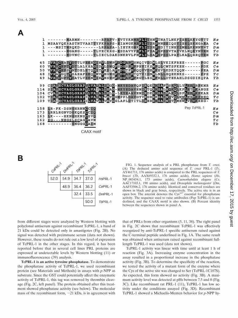
Figure 1A shows the alignment of TcPRL-1 with the ortho-logues from T. brucei (AAX69332.1), Homo sapiens(NP_003454.1), Drosophila melanogaster (AAF53506.1), andCaenorhabditis elegans (AAC17103.2). Since human, mouse,and rat PRL-1s are 100% identical, we used the human se-quence as representative of the mammalian orthologues in thealignment. All five proteins displayed in Fig. 1A show a 100%identity within the catalytic domain and a similar length.TcPRL-1 shows the consensus motif for PTP activity (boxed inFig. 1A), with the Cys107 essential for activity conserved (Fig.1A, asterisk). Other residues present in PRLs and conserved inTcPRL-1 include (i) the Asp75, which serves as a general acidin the catalysis; (ii) an Ala instead of a Ser or Thr next to thecatalytic Arg113, which explains the low catalytic efficiency ofPRLs compared to other PTPs (17); and (iii) the Cys52 thatcould form an intramolecular disulfide bond with the Cys107
and prevent its oxidative damage (17). Like all PRLs, TcPRL-1also has the CAAX motif (underlined in Fig. 1A) in the Cterminus. However, the T. cruzi CAAX motif is different fromthe ones present in other PRLs but similar to the prenylationmotif present in the T. brucei PRL and to those described inother trypanosomatids (37). As shown in Fig. 1B, the percent-age of identity between TcPRL-1 and other PRLs ranges from34 to 50%.
Genomic organization, mRNA expression, and Western blotanalysis of PRL-1 in T. cruzi. Genomic DNA from CL epimas-tigotes was digested with several restriction enzymes and hy-bridized with a labeled coding region of TcPRL-1 as a probe.Digestion with HincII, PstI, and MluI, enzymes that do not cutinside the ORF, rendered several bands in Southern blots (Fig.S1 in the supplemental material). HaeIII cut three times insidethe ORF but rendered fragments of ca. 150 to 180 bp that arenot detected in a 0.8% agarose gel. Therefore, the bands de-tected in the gel after HaeIII digestion correspond to theC-terminal part of the gene from different copies (Fig. S1 inthe supplemental material, lower panel). The same holds truefor HinfI. These results suggest that TcPRL-1 belongs to agene family composed by several members in T. cruzi. In fact,after a search of the TIGR databases (www.tigr.org) withTcPRL-1 as the query, we found four genomic sequences thatcontain paralogues to TcPRL-1. These sequences were alignedas shown in Fig. S2 in the supplemental material. The genomicsequence named chr_4784 is almost identical to TcPRL-1,while the rest of the sequences are between 90 and 97% iden-tical to TcPRL-1 (Fig. S2B in the supplemental material).
Total RNA was extracted from epimastigotes (lane E),amastigotes (lane A), and tissue culture-derived trypomastig-otes (lane T), and Northern blots of these RNAs were hybrid-ized with the same probe used in Southern blot analysis. Thesame transcript of �0.75 kb was observed in all of the life cyclestages of T. cruzi (Fig. 2A). However, if we compare the in-tensities of the bands between upper and lower panels (24SrRNA that was used to normalize the blot for equal loading),it might be inferred that in the amastigote form of the parasitethe expression was slightly higher. Since regulation of geneexpression in trypanosomatids is mainly posttranscriptional,the significance of this difference in RNA abundance is not asrelevant as at the protein level. When cell lysates prepared
1552 CUEVAS ET AL. EUKARYOT. CELL
on Decem
ber 17, 2015 by guesthttp://ec.asm
.org/D
ownloaded from
from different stages were analyzed by Western blotting withpolyclonal antiserum against recombinant TcPRL-1, a band of21 kDa could be detected only in amastigotes (Fig. 2B). Nosignal was detected with preimmune serum (data not shown).However, these results do not rule out a low level of expressionof TcPRL-1 in the other stages. In this regard, it has beenreported before that in several cell lines PRL proteins areexpressed at undetectable levels by Western blotting (11) orimmunofluorescence (39) analysis.
TcPRL-1 is an active tyrosine phosphatase. To demonstratethe phosphatase activity of TcPRL-1, we used recombinantprotein (see Materials and Methods) in assays with p-NPP assubstrate. Since the GST could potentially affect the enzymaticactivity of TcPRL-1, this tag was removed by thrombin cleav-age (Fig. 2C, left panel). The protein obtained after this treat-ment showed phosphatase activity (see below). The molecularmass of the recombinant form, �21 kDa, is in agreement with
that of PRLs from other organisms (5, 11, 38). The right panelin Fig. 2C shows that recombinant TcPRL-1 was effectivelyrecognized by anti-TcPRL-1 specific antiserum raised againstthe C-terminal peptide underlined in Fig. 1A. The same resultwas obtained when antiserum raised against recombinant full-length TcPRL-1 was used (data not shown).
TcPRL-1 activity was linear with time until at least 1 h ofreaction (Fig. 3A). Increasing enzyme concentration in theassay resulted in a proportional increase in the phosphataseactivity (Fig. 3B). To determine the specificity of the reaction,we tested the activity of a mutant form of the enzyme wherethe Cys of the active site was changed to Ser (TcPRL-1C107S).As expected, this form showed no activity (Fig. 3B). A maxi-mum activity level was detected at pHs between 7.5 and 8 (Fig.3C). Like recombinant rat PRL-1 (11), TcPRL-1 has low ac-tivity under the conditions assayed (Fig. 3D). RecombinantTcPRL-1 showed a Michaelis-Menten behavior for p-NPP hy-
FIG. 1. Sequence analysis of a PRL phosphatase from T. cruzi.(A) The deduced amino acid sequence of T. cruzi PRL-1 (Tc,AY461711, 176 amino acids) is compared to the PRL sequences of T.brucei (Tb, AAX69332.1, 176 amino acids), Homo sapiens (Hs,NP_003454.1, 173 amino acids), Caenorhabditis elegans (Ce,AAC17103.1, 190 amino acids), and Drosophila melanogaster (Dm,AAF53506.2, 176 amino acids). Identical and conserved residues areshown in black and gray boxes, respectively. The active site is in anopen box. The asterisk denotes the Cys107 essential for phosphataseactivity. The sequence used to raise antibodies (Pep TcPRL-1) is un-derlined, and the CAAX motif is also shown. (B) Percent identitybetween the sequences shown in panel A.
VOL. 4, 2005 TcPRL-1, A TYROSINE PHOSPHATASE FROM T. CRUZI 1553
on Decem
ber 17, 2015 by guesthttp://ec.asm
.org/D
ownloaded from

drolysis with a Km of 0.8 � 0.05 �M p-NPP at pH 7.5. Todetermine whether TcPRL-1, as other PRLs, is a tyrosinephosphatase, we assayed the effect of different known inhibi-tors on its intrinsic activity. TcPRL-1 phosphatase activity wasmarkedly inhibited by sodium o-vanadate in a dose-dependentmanner, showing a 50% inhibition at 75 �M o-vanadate (Fig.3E). Since PRLs show low similarity to dual specificity phos-phatases (17, 38), we assayed the activity in the presence ofseveral inhibitors to rule out a possible Ser/Thr phosphataseactivity. The Ser/Thr phosphatase inhibitor okadaic acid (up to1 mM), tested under similar conditions, showed no inhibitoryeffect against TcPRL-1 (Fig. 3F). Similar results were obtainedwith NaF (up to 5 mM) and EDTA (up to 10 mM).
A recent report (24) showed that PRLs are inhibited bypentamidine, an antiprotozoal drug, whose mechanism of ac-tion remains elusive. Pentamidine is effective against T. brucei(13) and amastigotes of L. mexicana (3). In inhibition assays,pentamidine showed little effect against recombinant TcPRL-1(Fig. 3F) even at a concentration as high as 10 �g/ml.
In cells, PRL phosphatases act on phosphorylated proteins.We therefore assayed the activity of recombinant TcPRL-1 onMBP phosphorylated on tyrosine residues (see Materials andMethods). As shown in Fig. 3G, TcPRL-1 was able to dephos-
phorylate Tyr-P-MBP, in a concentration-dependent manner.No effect on MBP dephosphorylation was observed when themutant form TcPRL-1C107S was used (Fig. 3G).
In vitro prenylation of TcPRL-1. The predicted amino acidsequence of TcPRL-1 contains the C-terminal CAAX motifwith the sequence CAVM (Fig. 1A), suggesting that this pro-tein might be posttranslationally modified with either a farne-syl or geranylgeranyl group. In T. cruzi, a PFT activity has beendescribed (37). To examine whether TcPRL-1 is a substrate fora PFT activity, we carried out a prenylation assay with recom-binant GST–TcPRL-1 fusion protein or a version lacking thelast four amino acids (GST–TcPRL-1�CAVM) as prenyl ac-ceptors and a T. cruzi epimastigote extract as a source of PFTactivity. When labeled FPP was used as the isoprenoid donor,GST–TcPRL-1 was efficiently farnesylated (Fig. 4A). However,when geranylgeranyl pyrophosphate was used as donor, wewere unable to detect any prenylation of GST–TcPRL-1, evenafter exposure of the gel for 2 weeks at 80°C (data notshown). Therefore, TcPRL-1 does not appear to be geranyl-geranylated in vitro. As expected, GST–TcPRL-1�CAVM wasnot prenylated with either isoprenoid donor, indicating thatthe last four amino acids are essential for this posttranslationalmodification (Fig. 4A and data not shown). Figure 4B shows a
FIG. 2. Expression analysis and purification of TcPRL-1. (A) Northern blot analysis of epimastigote (E), amastigote (A), and trypomastigote(T) stages of T. cruzi. Approximately 15 �g of total RNA were fractionated in a 1.5% formaldehyde-containing agarose gel, transferred to nylonmembranes, and hybridized with the 32P-labeled probe used for Southern blot analysis (Fig. S1 in the supplemental material). To control for equalloading of RNA in each lane, the membranes were stripped and reprobed with a 32P-labeled T. cruzi 24S ribosomal probe (lower panel). (B) Totalcell lysates from wild-type epimastigote (E), amastigote (A), and trypomastigote (T) stages of T. cruzi (30 �g of protein/lane) were separated bySDS-PAGE, transferred to nitrocellulose membranes, probed with anti-TcPRL-1 antibody (1:2,000) and horseradish peroxidase-conjugatedsecondary antibody (1:10,000), and visualized by chemiluminescence. Markers are in kilodaltons. (C) In the left panel, recombinant TcPRL-1 wasobtained as a fusion with GST. GST-TcPRL-1 expression was only observed after IPTG induction (compare lane U [soluble extract fromuninduced bacteria] versus lane I [soluble extract from induced bacteria]). After in-column digestion with thrombin (lane TD), a protein of 21 kDacorresponding to TcPRL-1 was obtained and, after glutathione addition, GST (27 kDa) was eluted (lane E). SDS–15% PAGE gel stained withCoomassie blue. Lanes U and I, 10 �g of protein. Lanes TD and E, 1 and 3 �g of protein, respectively. The right panel shows an SDS–15% PAGEgel loaded with one-third of the amount of protein used in the left panel, transferred to nitrocellulose, and incubated with anti-TcPRL-1 serum.Markers on the right are in kilodaltons.
1554 CUEVAS ET AL. EUKARYOT. CELL
on Decem
ber 17, 2015 by guesthttp://ec.asm
.org/D
ownloaded from

FIG. 3. Characterization of the phosphatase activity of TcPRL-1 with p-NPP or phosphorylated MBP as substrate. (A) Time-dependenthydrolysis of p-NPP assayed as described in Materials and Methods. (B) Enzyme concentration dependency using TcPRL-1 (continuous line) orTcPRL-1C107S (dashed line). (C) Effect of pH on TcPRL-1 phosphatase activity. (D) Phosphatase activity in the presence of different concen-trations of p-NPP (0–10 mM) was measured during 1 h with 140 �g of protein/ml. (E) Effect of sodium o-vanadate. The inhibition assay wasperformed by addition of different concentrations (0 to 1 mM) of sodium o-vanadate. (F) Effect of different inhibitors on TcPRL-1 activity.Abbreviations: o-Van, o-vanadate; OA, okadaic acid; PE, pentamidine. The data represent means � the standard deviations of triplicate samples.(G) Increasing amounts of TcPRL-1 (0, 0.7, 1.4, and 2.1 �g of protein) or a fixed amount of TcPRL-1C107S (2.0 �g of protein) were incubatedwith [32P]MBP for 1 h at 37°C. Samples were then boiled and loaded onto a SDS–12% PAGE gel, run, dried, and exposed to autoradiography.Densitometric analysis of the protein bands is expressed as a percentage of the control value. The experiment was repeated three times with similarresults.
VOL. 4, 2005 TcPRL-1, A TYROSINE PHOSPHATASE FROM T. CRUZI 1555
on Decem
ber 17, 2015 by guesthttp://ec.asm
.org/D
ownloaded from

Coomassie blue-stained SDS-PAGE gel loaded with the sam-ples used in the assay as a control for equal loading and thepurity of the protein preparations.
Expression of GFP-tagged TcPRL-1, TcPRL-1�CAVM, andTcPRL-1C107S�CAVM in T. cruzi. In general, the normal cel-lular levels of PRL proteins are low (11) and even undetectableby immunofluorescence analysis (39). Because of that andsince more than one PRL exists (Fig. S1 and S2 in the supple-mental material), we cloned an N-terminal epitope-tagged ver-sion of TcPRL-1 into the expression vector pRIBOTEX, andthe resultant construct was used to transfect T. cruzi epimas-tigotes (see Materials and Methods). We fused GFP to the Nterminus of TcPRL-1 in order to leave the C-terminal preny-lation motif accessible for modification by PFT. To determinethe presence and the expression level of the GFP-tagged pro-teins in the transfected parasites, we performed immunopre-cipitations with anti-GFP serum using soluble extracts of theparasites transfected with the different constructs. Figure 5Ashows that GFP, GFP–TcPRL-1, and GFP–TcPRL-1�CAVMwere immunoprecipitated by anti-GFP serum. Fusion proteinswere also detected when anti-TcPRL-1 serum was used inWestern blot assays of extracts (Fig. 5B). In Fig. 5B, we usedthree times more extract of GFP-TcPRL-1-transfected para-sites compared to those of the two other constructs to obtain aband of a similar intensity. A major protein band, with anapparent molecular mass of �47 kDa, was detected in thetransfected parasite population with anti-TcPRL-1 (Fig. 5B)and anti-GFP (Fig. 5A) sera. The lower expression levels ob-served in parasites expressing the wild-type (GFP–TcPRL-1) incomparison with the mutant constructs (GFP–TcPRL-1�CAVM or GFP–TcPRL-1C107S�CAVM) is in agreementwith the immunofluorescence results shown below. The highestexpression levels were observed with the double mutant (GFP–TcPRL-1C107S�CAVM). No growth defects were detectedupon overexpression of these constructs.
TcPRL-1 is prenylated in vivo. To know whether prenylationof TcPRL-1 occurs in vivo and to determine its importance for
the correct subcellular localization of the protein, we per-formed immunofluorescence microscopy studies with a GFP-tagged enzyme and its derivative mutants. Epimastigotes ex-pressing a GFP–TcPRL-1 fusion protein exhibited punctuatestaining near the posterior end of the cells in 93% � 1% of thecells (n � 2, 50 cells analyzed) (Fig. 6B). In many cases,fluorescence was also observed in the anterior end near theflagellar pocket (Fig. 6B and 7B). Association of prenylatedproteins with cellular membranes is usually dependent on theirprenylation status. To test whether farnesylation was requiredfor the localization of GFP–TcPRL-1 to the punctuate struc-tures, we investigated its localization in the stably expressedmutant forms lacking the prenylation signal. Parasites express-ing GFP–TcPRL-1�CAVM or GFP–TcPRL-1C107S�CAVMwere mainly labeled in the cytoplasm (Fig. 6F and H), thusestablishing that prenylation of GFP–TcPRL-1 is necessary forits association with intracellular membranes. Similar resultswere obtained when anti-GFP or anti-TcPRL-1 serum wasused in indirect immunofluorescence studies (data not shown).Epimastigotes expressing GFP alone (Fig. 6D) exhibitedstrictly cytosolic labeling. Similar results were obtained withamastigotes. Amastigotes expressing GFP–TcPRL-1 fusionprotein showed staining of a punctuate structure in their an-terior region in 75% � 5% of the cells (n � 2, 50 cells ana-lyzed) (Fig. 6J) while those expressing GFP–TcPRL-1�CAVMshowed cytosolic labeling (Fig. 6L). Analysis of individual cellsin the GFP–TcPRL-1�CAVM populations of epimastigotesand amastigotes by fluorescence microscopy revealed differentlevels of expression, but the protein was always cytosolic. Thisargues against different localization because overexpression.Taken together, these results support the prenylation ofTcPRL-1 in vivo, which is consistent with the in vitro data (Fig.4), and that prenylation plays an important role in the subcel-lular localization of TcPRL-1. Interestingly, GFP–TcPRL-1, incontrast to mammalian PRL-1 (39), does not localize to thenucleus when unprenylated.
FIG. 4. TcPRL-1 is farnesylated in vitro. Purified recombinant GST–TcPRL-1 and a mutant form without the prenylation motif (GST–TcPRL-1�CAVM) (2 �g each) were incubated with T. cruzi epimastigote extract and [3H]FPP as described in Materials and Methods. Radiolabeledproteins were analyzed by SDS-PAGE on a 15% gel, followed by fluorography. Prenylation of TcPRL-1 is shown in panel A. (B) Coomassieblue-stained SDS-PAGE of the recombinant proteins used in panel A. Markers on the right are in kilodaltons.
1556 CUEVAS ET AL. EUKARYOT. CELL
on Decem
ber 17, 2015 by guesthttp://ec.asm
.org/D
ownloaded from

Localization of TcPRL-1 in the endocytic pathway. The lo-calization of TcPRL-1 close to the flagellar pocket and inpunctuate structures in the posterior end of epimastigotes (Fig.6B and 7B) suggested a localization in the endocytic pathway.Epimastigotes possess large membrane-bound organellesfound in their posterior end that are known as reservosomes.Reservosomes are acidic organelles containing cruzipain (acysteine proteinase) and ingested proteins, and it has beenproposed that they are prelysosomal compartments (7, 10).
When epimastigotes expressing GFP–TcPRL-1 (Fig. 7A)were labeled with anti-cruzipain, they showed colocalization ofthe proteins in the posterior end of the cells, a finding consis-tent with the presence of GFP–TcPRL-1 in the reservosomes.Some GFP–TcPRL-1 labeling in the perinuclear region of theepimastigotes (GFP–TcPRL-1) might be indicative of localiza-tion in the endoplasmic reticulum due to protein overexpres-sion. Concanavalin A has previously been used to label theflagellar pocket, cytostome, and endocytic pathway of trypano-somatids (9). When GFP–TcPRL-1-expresing epimastigotes(Fig. 7B) were incubated with a concanavalin A-TRITC (tet-ramethyl rhodamine isothiocyanate) conjugate, strong labelingwas detected in the flagellar pocket region and other membra-nous structures near the anterior end of the cell as well as inthe posterior reservosomes. The anterior GFP–TcPRL-1 par-tially colocalized with the concanavalin A-TRITC labeling, andthe posterior GFP–TcPRL-1 colocalized with concanavalin A-TRITC labeling of the reservosomes.
To investigate the localization of TcPRL-1 in infectivestages, late stationary phase wild-type, GFP-, GFP–TcPRL-1-,and GFP–TcPRL-1�CAVM-expressing epimastigotes were
used to infect tissue culture cells (4), and the tissue cultureswere analyzed for the presence of trypomastigotes and amas-tigotes. When GFP–TcPRL-1 expressing amastigotes were an-alyzed by fluorescence microscopy, a small region of fluores-cence was noted (Fig. 7C) that partially colocalized withconcanavalin A-TRITC. A similar pattern of TcPRL-1 distri-bution was noted in wild-type amastigotes reacted with anti-TcPRL-1 antibody (Fig. 7D), confirming that the GFP patternwas not due to mistargeting of the overexpressed protein. Wealso show in the right part of panel D the DAPI (4�,6�-dia-midino-2-phenylindole) staining of the nucleus (arrow) andkinetoplast (arrowhead) to rule out a kinetoplast localizationof TcPRL-1. No staining was detected with preimmune serum(data not shown). Endocytosis of other markers such as trans-ferrin or bovine serum albumin was not efficient in amasti-gotes, and the number of cells available prevented subcellularfractionation techniques to confirm the localization ofTcPRL-1 in amastigotes.
The yield of trypomastigotes was low with the CL stock and,to rule out the possibility that overexpression of TcPRL-1could be inhibiting transformation of amastigotes into trypo-mastigotes, we transfected Y strain epimastigotes with eithervector alone or a plasmid expressing TcPRL-1. After differen-tiation of epimastigotes to infective forms (4, 21), we were ableto obtain trypomastigotes in similar amounts to those obtainedfrom cells transfected with vector alone (data not shown).TcPRL-1 expressing trypomastigotes also showed colocaliza-tion of the label with concanavalin A (Fig. 7E). No staining wasdetected with preimmune serum (data not shown).
FIG. 5. Immunodetection of proteins in epimastigotes stably transfected with wild-type or mutant forms of TcPRL-1. (A and B) T. cruziepimastigotes were transfected with the pRIBOTEX constructs described in Materials and Methods and selected with 500 �g of Geneticin/ml inthe growth medium. (A) Immunoprecipitation with anti-GFP-serum raised in rabbit and protein A-Sepharose. Samples were boiled and separatedby SDS-PAGE. After transfer to a nitrocellulose membrane, the Western blot was developed with GFP antiserum raised in a mouse. E, extractused in the immunoprecipitation; IP, immunoprecipitation reaction. Anti-maltose-binding protein (�-MBP) serum was used as a control forspecificity of the immunoprecipitation reaction with GFP–TcPRL-1 extract. (B) The same extracts used in panel A (10 �g) were subjected toSDS-PAGE, transferred to nitrocellulose, and incubated with anti-TcPRL-1 serum, raised against recombinant TcPRL-1. Three times more extractof TcPRL-1 (30 �g) was loaded to obtain a signal. Markers on the right are in kilodaltons.
VOL. 4, 2005 TcPRL-1, A TYROSINE PHOSPHATASE FROM T. CRUZI 1557
on Decem
ber 17, 2015 by guesthttp://ec.asm
.org/D
ownloaded from

DISCUSSION
We report here that TcPRL-1, a gene encoding a functionalPTP, is present in the T. cruzi genome. Comparison of thesequence of TcPRL-1 with those of other PTPs indicates thatit is closely related to the family of PTPs known as PRLs.Although the overall identity with other eukaryotic PRL-1s isca. 34 to 50% (Fig. 1), TcPRL-1 has several residues charac-teristic of PRLs (such as Ala114, Cys52, and Tyr56), as well asconserved residues involved in the catalytic mechanism thatare common to all tyrosine phosphatases (Cys107, Arg113, andAsp75) (20).
The low phosphatase activity exhibited by TcPRL-1, whenthe synthetic substrate p-NPP (Fig. 3) is used, is in accordanceto the nuclear magnetic resonance and kinetic study of Kozlovet al. (17). These authors proposed that the presence of an Alafollowing the active site, instead of a Ser/Thr, is responsible, inpart, for the low activity of PRLs when compared with othertyrosine phosphatases. Another important feature is the dis-tance between the Asp75 and the catalytic cysteine. Both resi-dues might get in contact only after binding the protein sub-strate, explaining the low activity with p-NPP. Other residuesmight be responsible for the higher sensitivity to o-vanadateinhibition (50% inhibitory concentration � 75 �M versus 60%inhibition at 1 mM for rat PRL-1) and the absence of penta-midine inhibition of TcPRL-1 compared to mammalianPRL-1s (24).
Like other PRLs, the protein is farnesylated in its C-terminalregion, and this modification is essential for its membranelocalization. Posttranslational modification of proteins with
farnesyl groups appears to be essential for localization of mod-ified proteins to membranes and, consequently, for their bio-logical function (40). Farnesyl groups can be transferred tocysteine residues within carboxy-terminal motifs present in sev-eral classes of proteins in a reaction catalyzed by a cytoplasmicPFT (40). Inhibition of protein farnesylation by substrate in-hibitors of farnesyl transferases or by inhibitors of the isopre-noid synthesis pathway has a profound effect on cell morphol-ogy (25), cell replication (26), and intracellular signaltransduction (18). Recent studies have indicated that proteinfarnesylation occurs in T. cruzi, since the growth of the intra-cellular forms is sensitive to protein farnesyl transferase inhib-itors (37). Interestingly, growth of amastigotes of T. cruzi, thelife stage that has higher expression of TcPRL-1 (Fig. 2B), wasinhibited by CAAX mimetics and FPP analogs, with 50% in-hibitory concentrations ranging from 10 to 50 �M. In contrast,T. cruzi epimastigotes were resistant to 50 to 100 �M PFTinhibitors (37). In addition to TcPRL-1, the only other proteinthat has been shown to be farnesylated in T. cruzi is TcRho 1,which is a small GTPase possibly involved in signal transduc-tion and metacyclogenesis (8, 23). The identification of farne-sylated proteins in T. cruzi will help to identify the mechanismof toxicity of isoprenoid synthesis and protein farnesylationinhibitors.
In epimastigotes, TcPRL-1 colocalizes in the reservosomeswith the cysteine proteinase cruzipain. It also colocalizes withconcanavalin A in the flagellar pocket, cytostome, and endo-cytic pathway of different stages. Reservosomes are large mem-brane-bound organelles found in the posterior end of T. cruzi
FIG. 6. Localization of GFP, GFP–TcPRL-1, and mutant forms by confocal fluorescence microscopy. Fluorescence images of T. cruziepimastigotes (A to H) and amastigotes (I to L) are shown. B and J, GFP–TcPRL-1; D, GFP alone; F and L, GFP–TcPRL-1�CAVM; H,GFP-TcPRL-1C107S�CAVM. Panels A, C, E, G, I, and K are phase-contrast images of the same cells. Bar, 10 �m.
1558 CUEVAS ET AL. EUKARYOT. CELL
on Decem
ber 17, 2015 by guesthttp://ec.asm
.org/D
ownloaded from

FIG. 7. Immunofluorescence microscopy showing the localization of TcPRL-1 in epimastigote (A and B), amastigote (C and D), andtrypomastigote (E) forms of T. cruzi. The figure shows the colocalization of TcPRL-1 with cruzipain in epimastigotes (A) and partial colocalizationof TcPRL-1 in epimastigotes (B), amastigotes (C and D), and trypomastigotes (E) with concanavalin A. (A) Fixed epimastigotes expressingGFP-TcPRL-1 were permeabilized and labeled with monoclonal antibody against cruzipain. Panels show (from left to right) phase-contrastmicroscopy, GFP view, cruzipain view, and overlay. (B and C) Epimastigotes (B) or amastigotes (C) expressing GFP–TcPRL-1 were incubated for30 min with 5 �g of concanavalin A-TRITC/ml and then fixed. Panels show (from left to right) phase-contrast microscopy, GFP view, concanavalinA-TRITC view, and overlay. (D) Wild-type amastigotes were incubated with concanavalin A-TRITC and then fixed and reacted with anti-TcPRL-1polyclonal antibody. Panels show (from left to right) phase-contrast microscopy, TcPRL-1 antibody view, concanavalin A-TRITC view, and overlay.DAPI staining was included in the overlay to show relationship of the staining pattern to the nucleus (arrow) and kinetoplast (arrowhead). (E) Ystrain trypomastigotes expressing TcPRL-1 were incubated for 30 min with 5 �g of concanavalin A-TRITC/ml, fixed, and reacted with anti-TcPRL-1 polyclonal antibody. Panels show (from left to right) phase-contrast microscopy, TcPRL-1 antibody view, concanavalin A-TRITC view,and overlay. Bar, 10 �m.
VOL. 4, 2005 TcPRL-1, A TYROSINE PHOSPHATASE FROM T. CRUZI 1559
on Decem
ber 17, 2015 by guesthttp://ec.asm
.org/D
ownloaded from

epimastigotes. They are acidic, contain the cysteine proteasecruzipain, and accumulate macromolecules ingested by theparasite through endocytic processes, such as albumin, perox-idase, transferrin, and low-density lipoprotein (10, 29, 30, 32).Reservosomes are considered a prelysosomal compartment(10). It has been suggested that they also contain lipids (31)and, since their number decreases during metacyclogenesis,they were postulated to have a role in the storage of nutrientsand/or proteins necessary for this differentiation step (10). Inamastigotes, TcPRL-1 localizes in a small region of the cells,which partially colocalizes with concanavalin A-TRITC stain-ing. Attempts to induce uptake by amastigotes of other endo-cytic markers such as transferrin or bovine serum albuminfailed, and the amount of amastigotes that could be obtainedprecluded the use of subcellular fractionation techniques toinvestigate this localization. Further work will be needed toinvestigate the localization of the endogenous protein in amas-tigotes.
The association of TcPRL-1 with membranes is abolished bythe removal of the CAAX motif. Under this condition,TcPRL-1 localizes in the cytoplasm. In sharp contrast, mam-malian PRLs, when unprenylated, shifted to the nucleus (39).A possible explanation for these observations is that mamma-lian PRLs have a nuclear localization signal near the C termi-nus. The C terminus of TcPRL-1 is the region of least similar-ity to other PRL-1s and does not show a signal for nuclearlocalization. The involvement of mammalian PRL-1 in cellproliferation is dependent on its nuclear localization. There-fore, the differential localization of unfarnesylated TcPRL-1 tothe cytoplasm suggests that in unicellular eukaryotes this phos-phatase must have a different role.
In conclusion, TcPRL-1 is the first PRL-type PTP charac-terized at the molecular level in unicellular eukaryotes and islocalized to endocytic membranes of T. cruzi thanks to itsC-terminal farnesyl modification.
ACKNOWLEDGMENT
We thank Ivan D’Orso for careful reading the manuscript; ShuhongLuo for transfection and cloning of Y strain epimastigotes; JulioScharfstein for the monoclonal antibody against cruzipain; and BertaFranke de Cazzulo, Liliana Sferco, and Linda Brown for technicalassistance. Preliminary genomic data obtained from http://www.tigr.org was provided by the TIGR-SBRI-KI Sequencing Consortium sup-ported by NIH grants AI45038, AI45061. and AI45039.
This study was supported in part by grants from the World Bank/United Nations Development Project/World Health OrganizationSpecial Program for Research and Training in Tropical Disease (TDRProject 970629), the Agencia Nacional de Promocion Cientifica y Tec-nologica, the Carrillo-Onativia Fellowship, the Ministerio de Salud,and the Fundacion Antorchas, Argentina (to D.O.S.) and U.S. Na-tional Institutes of Health grant AI-23259 (to R.D.). This investigationwas conducted in part in a facility constructed with support fromResearch Facilities Improvement Program Grant C06 RR 16515-01from the National Center for Research Resources, U.S. National In-stitutes of Health. I.C.C. is a fellow of the Comision de InvestigacionesCientıficas. D.O.S. is a researcher from the Consejo Nacional de In-vestigaciones Cientıficas y Tecnicas.
REFERENCES
1. Bakalara, N., A., Seyfang, T. Baltz, and C. Davis. 1995. Trypanosoma bruceiand Trypanosoma cruzi: life cycle-regulated protein tyrosine phosphataseactivity. Exp. Parasitol. 81:302–312.
2. Bakalara, N., A. Seyfang, C. Davis, and T. Baltz. 1995. Characterization of alife-cycle-stage-regulated membrane protein tyrosine phosphatase inTrypanosoma brucei. Eur. J. Biochem. 234:871–877.
3. Berman, J. D., and D. J. Wyler. 1980. An in vitro model for investigation ofchemotherapeutic agents in leishmaniasis. J. Infect. Dis. 142:83–86.
4. Caler, E. V., S. V. de Avalos, P. A. Haynes, N. W. Andrews, and B. A.Burleigh. 1998. Oligopeptidase B-dependent signaling mediates host cellinvasion by Trypanosoma cruzi. EMBO J. 17:4975–4986.
5. Cates, C. A., R. L. Michael, K. R. Stayrook, K. A. Harvey, Y. D. Burke, S. K.Randall, P. L. Crowell, and D. N. Crowell. 1996. Prenylation of oncogenichuman PTP(CAAX) protein tyrosine phosphatases. Cancer Lett. 110:49–55.
6. Cool, D. E., and J. J. Blum. 1993. Protein tyrosine phosphatase activity inLeishmania donovani. Mol. Cell Biochem. 127/128:143–149.
7. Cunha-e-Silva, N. L., G. C. Atella, I. A. Porto-Carreiro, J. A., Morgado-Diaz,M. G. Pereira, and W. de Souza. 2002. Isolation and characterization of areservosome fraction from Trypanosoma cruzi. FEMS Microbiol. Lett. 214:7–12.
8. de Melo, L. D., J. L. Nepomuceno-Silva, C. Sant’Anna, N. Eisele, R. B.Ferraro, J. R. Meyer-Fernandes, W. de Souza, N. L. Cunha-e-Silva, andU. G. Lopes. 2004. TcRho1 of Trypanosoma cruzi: role in metacyclogenesisand cellular localization. Biochem. Biophys. Res. Commun. 323:1009–1016.
9. Denny, P. W., S. Gokool, D. G. Russell, M. C. Field, and D. F. Smith. 2000.Acylation-dependent protein export in Leishmania. J. Biol. Chem.275:11017–11025.
10. de Souza, W. 2002. Basic cell biology of Trypanosoma cruzi. Curr. Pharma-ceut. Design 8:269–285.
11. Diamond, R. H., D. E. Cressman, T. M. Laz, C. S. Abrams, and R. Taub.1994. PRL-1, a unique nuclear protein tyrosine phosphatase, affects cellgrowth. Mol. Cell. Biol. 14:3752–3762.
12. Diamond, R. H., C. Peters, S. P. Jung, L. E. Greenbaum, B. A. Haber, D. G.Silberg, P. G. Traber, and R. Taub. 1996. Expression of PRL-1 nuclearPTPase is associated with proliferation in liver but with differentiation inintestine. Am. J. Physiol. 271:G121–G129.
13. Docampo, R., and S. N. J. Moreno. 2003. Current chemotherapy of humanAfrican trypanosomiasis. Parasitol. Res. 90:S10–S13.
14. Franke de Cazzulo, B. M., J. Martinez, M. J. North, G. H. Coombs, and J. J.Cazzulo. 1994. Effects of proteinase inhibitors on the growth and differen-tiation of Trypanosoma cruzi. FEMS Microbiol. Lett. 124:81–86.
15. Furuya, T., L. Zhong, J. R. Meyer-Fernandes, H.-G. Lu, S. N. J. Moreno, andR. Docampo. 1998. Ecto-protein tyrosine phosphatase activity in Trypano-soma cruzi infective stages. Mol. Biochem. Parasitol. 92:339–348.
16. Karin, M., and T. Hunter. 1995. Transcriptional control by protein phos-phorylation: signal transmission from the cell surface to the nucleus. Curr.Biol. 5:747–757.
17. Kozlov, G., J. Cheng, E. Ziomek, D. Banville, K. Gehring, and I. Ekiel. 2004.Structural insights into molecular function of the metastasis-associated phos-phatase PRL-3. J. Biol. Chem. 279:11882–11889.
18. Lerner, E. C., Y. Qian, M. A. Blaskovich, R. D. Fossum, A. Vogt, J. Sun, A. D.Cox, C. J. Der, A. D. Hamilton, and S. M. Sebti. 1995. Ras CAAX peptido-mimetic FTI-277 selectively blocks oncogenic Ras signaling by inducingcytoplasmic accumulation of inactive Ras-Raf complexes. J. Biol. Chem.270:26802–26806.
19. Martinez-Calvillo, S., I. Lopez, and R. Hernandez. 1997. pRIBOTEX ex-pression vector: a pTEX derivative for a rapid selection of Trypanosoma cruzitransfectants. Gene 199:71–76.
20. Mertz, L. M., and A. Rashtchian. 1994. Nucleotide imbalance and polymer-ase chain reaction: effects on DNA amplification and synthesis of highspecific activity radiolabeled DNA probes. Anal. Biochem. 221:160–165.
21. Montalvetti, A., P. Rohloff, and R. Docampo. 2004. A functional aquaporincolocalizes with the vacuolar proton pyrophosphatase to acidocalcisomesand the contractile vacuole complex of Trypanosoma cruzi. J. Biol. Chem.279:38673–38682.
22. Murta, A. C., P. M. Persechini, T. Souto-Padron, W. de Souza, J. A. Guima-raes, and J. Scharfstein. 1990. Structural and functional identification ofGP57/51 antigen of Trypanosoma cruzi as a cysteine proteinase. Mol. Bio-chem. Parasitol. 43:27–38.
23. Nepomuceno-Silva, J. L., K. Yokoyama, L. D. de Mello, S. M. Mendonca,J. C. Paixao, R. Baron, J. C. Faye, F. S. Buckner, W. C. Van Voorhis, M. H.Gelb, and U. G. Lopes. 2001. TcRho1, a farnesylated Rho family homologuefrom Trypanosoma cruzi: cloning, trans-splicing, and prenylation studies.J. Biol. Chem. 276:29711–29718.
24. Pathak, M. K., D. Dhawan, D. J. Lindner, E. C. Borden, C. Farver, and T. Yi.2002. Pentamidine is an inhibitor of PRL phosphatases with anticanceractivity. Mol. Cancer Ther. 1:1255–1264.
25. Prendergast, G. C., J. P. Davide, S. J. DeSolms, E. A. Giuliani, S. L. Graham,J. B. Gibbs, A. Oliff, and N. E. Kohl. 1994. Farnesyltransferase inhibitioncauses morphological reversion of ras-transformed cells by a complex mech-anism that involves regulation of the actin cytoskeleton. Mol. Cell. Biol.14:4193–4202.
26. Quesney-Huneeus, V., M. H. Wiley, and M. D. Siperstein. 1979. Essentialrole for mevalonate synthesis in DNA replication. Proc. Natl. Acad. Sci.USA 76:5056–5060.
27. Ramirez, M. I., L. M. Yamauchi, L. H. de Freitas, Jr., H. Uemura, and S.Schenkman. 2000. The use of the green fluorescent protein to monitor and
1560 CUEVAS ET AL. EUKARYOT. CELL
on Decem
ber 17, 2015 by guesthttp://ec.asm
.org/D
ownloaded from

improve transfection in Trypanosoma cruzi. Mol. Biochem. Parasitol. 111:235–240.
28. Sambrook, J., and D. W. Russell. 2001. Molecular cloning: a laboratorymanual, 3rd ed. Cold Spring Harbor Laboratory, Cold Spring Harbor, N.Y.
29. Scott, D. A., R. Docampo, J. A. Dvorak, S. Shi, and R. D. Leapman. 1997. Insitu compositional analysis of acidocalcisomes in Trypanosoma cruzi. J. Biol.Chem. 272:28020–28029.
30. Soares, M. J., and W. de Souza. 1991. Endocytosis of gold-labeled proteinsand LDL by Trypanosoma cruzi. Parasitol. Res. 77:461–468.
31. Soares, M. J., M. F. de Souza, and W. de Souza. 1987. Ultrastructuralvisualization of lipids in trypanosomatids. J. Protozool. 34:199–203.
32. Soares, M. J., T. Souto-Padron, and W. de Souza. 1992. Identification of alarge pre-lysosomal compartment in the pathogenic protozoon Trypanosomacruzi. J. Cell Sci. 102:157–167.
33. Tonks, N. K., and B. G. Neel. 2001. Combinatorial control of the specificityof protein tyrosine phosphatases. Curr. Opin. Cell Biol. 13:182–195.
34. Wang, J., C. E. Kirby, and R. Herbst. 2002. The tyrosine phosphatase PRL-1localizes to the endoplasmic reticulum and the mitotic spindle and is re-quired for normal mitosis. J. Biol. Chem. 277:46659–46668.
35. Werner, S. R., P. A. Lee, M. W. DeCamp, D. N. Crowell, S. K. Randall, andP. L. Crowell. 2003. Enhanced cell cycle progression and down regulation ofp21Cip1/Waf1 by PRL tyrosine phosphatases. Cancer Lett. 202:201–211.
36. Yarovinsky, T. O., D. W. Rickman, R. H. Diamond, R. Taub, G. S. Hageman,and C. Bowes Rickman. 2000. Expression of the protein tyrosine phospha-tase, phosphatase of regenerating liver 1, in the outer segments of primatecone photoreceptors. Brain Res. Mol. Brain Res. 77:95–103.
37. Yokoyama, K., P. Trobridge, F. S. Buckner, J. Scholten, K. D. Stuart, W. C.Van Voorhis, and M. H. Gelb. 1998. The effects of protein farnesyltrans-ferase inhibitors on trypanosomatids: inhibition of protein farnesylation andcell growth. Mol. Biochem. Parasitol. 94:87–97.
38. Zeng, Q., W. Hong, and Y. H. Tan. 1998. Mouse PRL-2 and PRL-3, twopotentially prenylated protein tyrosine phosphatases homologous to PRL-1.Biochem. Biophys. Res. Commun. 244:421–427.
39. Zeng, Q., X. Si, H. Horstmann, Y. Xu, W. Hong, and C. J. Pallen. 2000.Prenylation-dependent association of protein-tyrosine phosphatases PRL-1,-2, and -3 with the plasma membrane and the early endosome. J. Biol. Chem.275:21444–21452.
40. Zhang, F. L., and P. J. Casey. 1996. Protein Prenylation: molecular mecha-nisms and functional consequences. Annu. Rev. Biochem. 65:241–269.
41. Zingales, B., M. E. Pereira, R. P. Oliveira, K. A. Almeida, E. S. Umezawa,R. P. Souto, N. Vargas, M. I. Cano, J. F. da Silveira, N. S. Nehme, C. M.Morel, Z. Brener, and A. Macedo. 1997. Trypanosoma cruzi genome project:biological characteristics and molecular typing of clone CL Brener. ActaTrop. 68:159–173.
VOL. 4, 2005 TcPRL-1, A TYROSINE PHOSPHATASE FROM T. CRUZI 1561
on Decem
ber 17, 2015 by guesthttp://ec.asm
.org/D
ownloaded from


