Embed Size (px)
Citation preview
Chapter 6:
GASTROINTESTINAL DISORDERS Student Authors: Lehlohonolo Ntlatlapo and Savannah Verhage
Specialist Advisor: Dr Lesego Ndhlovu
This chapter covers the following topics:
● Diarrhoea
● Hepatitis
● Portal hypertension
● Gastroesophageal reflux
● Constipation
● Jaundice
● Hepatosplenomegaly
● Gastrointestinal bleeding
● Functional abdominal pain
(FAP)

DIARRHOEA Diarrhoea is defined by the WHO as ≥3 watery stools in 24 hours. Almost every child
will have diarrhoea at least once and approximately 1 in 3 hospital admissions in
South Africa are due to diarrhoea. It remains a major cause of morbidity and
mortality worldwide in children <5 years old. It is the second most common cause of
death (after HIV) in South African children of that age group.
Classification, Pathophysiology and Aetiology In diarrhoea, the excretion of water and electrolytes exceed net absorption.
Diarrhoea may be classified according to pathophysiology or duration.
Pathophysiological Classification
Diarrhoea may be classified as:
● Osmotic diarrhoea:
o It occurs when a large number of osmotically active particles are
present in the lumen, leading to the passive flow of fluid into the bowel
lumen.
o Causes include laxatives, lactulose, lactose, and other food
intolerances/allergies.
● Secretory diarrhoea:
o An activated pathway (due to the release of a toxin by a pathogen that
invaded the intestinal mucosa) or inherent abnormalities of enterocytes
cause excessive amounts of fluid to be secreted.
o Diarrhoea results when absorptive mechanisms become overwhelmed
i.e. secretion exceeds absorption.
o Infective causes of secretory diarrhoea include:
▪ Viruses e.g. rotavirus, norovirus, calicivirus, Norwalk virus
▪ Bacteria e.g. Campylobacter jejuni, Salmonella sp, E. coli, C.
difficile, Shigella sp, Yersinia enterocolitica
▪ Parasites e.g. Cryptosporidium sp, Giardia lamblia
Classification According to Duration
Diarrhoea may be considered:
● Acute:
o There is a sudden onset of increased frequency of stools which lasts
no longer than 14 days.
o It is most commonly caused by a virus and laboratory investigation is
not necessary to diagnose acute diarrhoea.
● Persistent diarrhoea:
o It is an episode of diarrhoea of presumed infectious aetiology that
begins acutely and lasts >14 days.
● Chronic diarrhoea:
o It is a diarrhoeal episode that lasts >14 days and which most commonly
has a non-infectious cause e.g. lactose intolerance, food allergies.
Clinical Features The clinical presentation and course of diarrhoea depends on the aetiology of the
diarrhoea and the host (see an image of a child threatened by severe diarrhoea
here). The patient may present with:
● Features of dehydration:
o Lethargy
o Depressed level of consciousness
o Sunken anterior fontanelle
o Dry mucous membranes and no tears
o Sunken eyes
o Reduced skin turgor
o Delayed capillary refill (>2 s)
● Complications of dehydration:
o Electrolyte disturbances (hyponatraemia, hypokalaemia and metabolic
acidosis)
o Acute renal failure
o Paralytic ileus
o Convulsions
o Cerebral damage
● FTT and malnutrition (diagnosed based on anthropometry measurements,
reduced muscle/fat mass, peripheral oedema)
● Perianal erythema
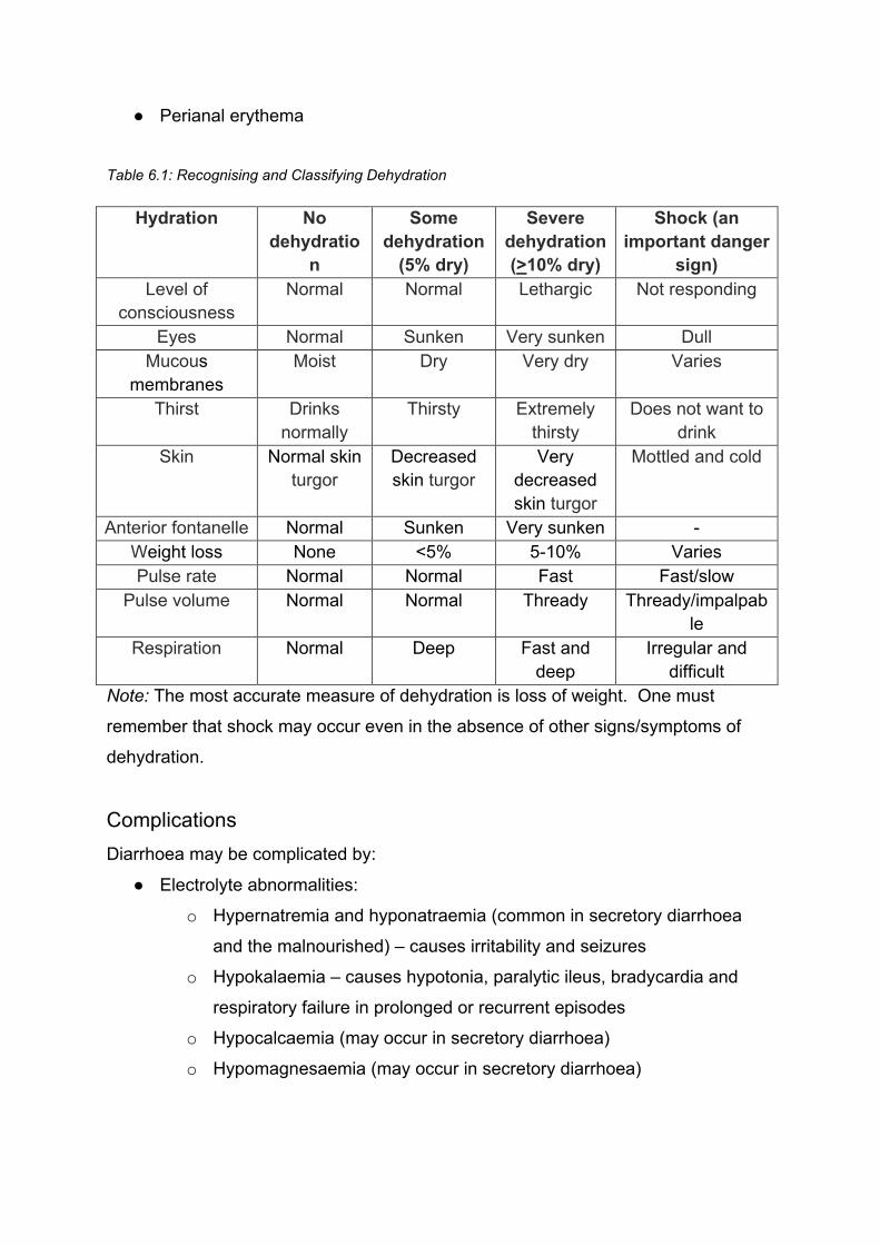
Table 6.1: Recognising and Classifying Dehydration
Hydration No dehydratio
n
Some dehydration
(5% dry)
Severe dehydration (>10% dry)
Shock (an important danger
sign) Level of
consciousness Normal Normal Lethargic Not responding
Eyes Normal Sunken Very sunken Dull Mucous
membranes Moist Dry Very dry Varies
Thirst Drinks normally
Thirsty Extremely thirsty
Does not want to drink
Skin Normal skin turgor
Decreased skin turgor
Very decreased skin turgor
Mottled and cold
Anterior fontanelle Normal Sunken Very sunken - Weight loss None <5% 5-10% Varies Pulse rate Normal Normal Fast Fast/slow
Pulse volume Normal Normal Thready Thready/impalpable
Respiration Normal Deep Fast and deep
Irregular and difficult
Note: The most accurate measure of dehydration is loss of weight. One must
remember that shock may occur even in the absence of other signs/symptoms of
dehydration.
Complications Diarrhoea may be complicated by:
● Electrolyte abnormalities:
o Hypernatremia and hyponatraemia (common in secretory diarrhoea
and the malnourished) – causes irritability and seizures
o Hypokalaemia – causes hypotonia, paralytic ileus, bradycardia and
respiratory failure in prolonged or recurrent episodes
o Hypocalcaemia (may occur in secretory diarrhoea)
o Hypomagnesaemia (may occur in secretory diarrhoea)

● Hypoglycaemia (due to catecholamine release and may develop in the
malnourished, septic or hypothermic patient)
● Metabolic acidosis (caused by loss of bases in stool, poor tissue perfusion
and decreased renal hydrogen clearance)
● Renal failure (in severe dehydration)
● Haemolytic uraemic syndrome – characterised by haemolytic anaemia, acute
kidney injury (uraemia) and thrombocytopaenia
● Rhabdomyolysis
● Shock
● Death
Investigations No routine investigations are required, as most children will recover spontaneously
within a few days, regardless of the underlying causative organism. However, a urine
dipstick and finger-prick glucose should be done on all children admitted to hospital.
Investigations may include:
● Stool culture (always done for):
o Children with bloody diarrhoea (send cultures for C. difficile,
Campylobacter sp, Shigella sp and E.coli)
● Electrolytes, RFTs and ABG (always done for):
o Severely dehydrated children
o Moderately dehydrated children with an unusual clinical picture
o Malnourished children
o Children requiring IV fluids for rehydration
o Children with complications
● ABG
Management Primary Prevention
Diarrhoea may be prevented by:
● Ensuring a clean water supply
● Encouraging good sanitation and hygiene
● Giving vitamin A prophylaxis
● Encouraging breastfeeding
● Giving supplemental zinc
● Vaccinating against rotavirus
Secondary Prevention
This involves the prevention of dehydration and its complications. Acute diarrhoea is
usually self-limiting. However, one must always assess the patient for danger signs
(not drinking, lethargy, intractable vomiting, convulsions and bloody stools) and give
supportive management.
Supportive management includes:
● Oral rehydration therapy (ORT; give as soon as possible)
● Zinc supplementation (10-20 mg daily for 10-14 days). See also the
rehydration formula recommended by UNICEF here.
Tertiary Prevention
One’s aim here is to prevent death from dehydration and complications. Thus,
tertiary prevention involves appropriately managing the complications that the child
has and appropriately managing dehydration and shock (refer to general fluid
management in the Feeds and Fluid Management chapter).
HEPATITIS Hepatitis is inflammation of the liver which can result in the damage and destruction
of hepatocytes.
Aetiology Common causes of hepatitis include viruses, autoimmune liver disease, and drugs
and toxins.
Viral Hepatitis
In viral hepatitis, the damage to the liver is not due to the viral invasion but the
immune response to the virus. In neonates the virus is usually vertically transmitted
from the mother in the perinatal period. Viral causes of hepatitis include:
● Hepatitis viruses:

o A (HAV) – transmitted via the faecal-oral transmission; the degree of
hepatic injury depends on the host’s immune response
o B (HBV) – transmitted via bodily fluids e.g. blood, semen
o C (HCV) – transmitted through contact with infected blood
o D (HDV) – not common in children as it requires HBV co-infectivity for
replication
o E (HEV) – transmitted via the faecal-oral route
● CMV
● VZV
● HSV (especially in infants)
● Enterovirus
● Rubella
● Adenovirus
● Parvovirus
Autoimmune Liver Disease
They include:
● Autoimmune hepatitis
● Sclerosing cholangitis
● Kawasaki disease
● Graft-versus-host disease
● Immunodeficiencies
Drugs and Toxins
● Medication-induced hepatitis
● Paracetamol toxicity
● Amanita phalloides (a poisonous wild mushroom)
Clinical Features They vary as they depend on the aetiology and the child’s age of the child (infant vs
child vs adolescent). Some children may be asymptomatic, while others may present
with:
● Flu-like symptoms
● Malaise
● Jaundice
● Fever
● Nausea and vomiting
● Anorexia or poor feeding
● Abdominal discomfort
● Diarrhoea
● Dark urine and clay-coloured stools
● Tender hepatomegaly
Acute HBV infection is usually symptomatic, while chronic infection is often
asymptomatic (children with the latter infection will grow and develop normally).
Chronic infection may be associated with polyarteritis nodosa and
glomerulonephritis, and can progress to cirrhosis and hepatocellular carcinoma.
Most children with chronic HCV infection are asymptomatic, but can develop
cirrhosis and hepatocellular carcinoma (especially if there is a hepatitis B co-
infection).
Some children may present in acute liver failure i.e. fulminant liver failure without
pre-existing liver disease (an uncommon medical emergency). Causes include
infection with HAV, HBV, Reye’s syndrome, drugs (e.g. paracetamol, anti-TB drugs)
and toxins (e.g. traditional medication).
Investigations One may order:
● Blood tests
o Liver enzyme levels (ALT, AST, ALP, GGT)
o LFTs (INR, albumin, etc.)
o Antibody and PCR studies for suspected viral hepatitis: these results
must be interpreted to differentiate between acute and chronic infection
o FBC
o Serum glucose
● Imaging (USS of the liver)
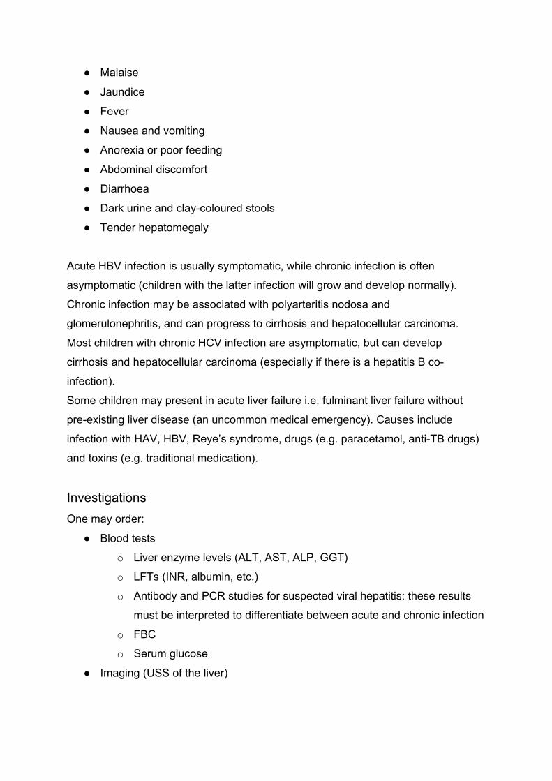
Pathophysiology, Diagnosis and Management Table 6.2: Pathophysiology, Diagnosis and Management of Hepatitis A and B
HAV HBV Pathophysiology
It is transmitted by the faecal-oral route and has an incubation period of 15-50 days. The child is most infectious 1-2 weeks before symptom onset. The child may be asymptomatic (young children) or may have a prodrome of nausea, anorexia and malaise. S/he may go on to develop jaundice, dark urine and tender hepatomegaly. Chronic infection does not develop and fulminant infection is very rare.
Children may be infected by horizontal, vertical or parenteral transmission. HBV has an incubation period of 2-6 months and a similar prodrome and clinical features to HAV (but the disease is more insidious and there is a longer prodrome). 90% of neonates and 30-40% of infected children become chronic carriers – asymptomatic in the beginning and then develop chronic hepatitis, cirrhosis and HCC.
Diagnosis Current/recent HAV infection is diagnosed if the child is HAV IgM positive (can remain positive for 4-6 months). Previous infection can be diagnosed if the child is HAV IgG positive.
Acute HBV infection is diagnosed if the child is HBV IgM, HBsAg and HBeAg positive. Chronic HBV is also diagnosed using the above tests and may be:
● High-risk (HBeAg-positive. HBeAb-positive, HBV IgG-positive)
● Low-risk (HBeAg-negative. HBeAb-positive, HBV IgG-positive).
Management The local authorities should be notified (HAV is a notifiable disease). Management includes:
● Giving supportive care (avoid liver toxic drugs and ensure adequate hydration)
Treatment of chronic HBV infection includes 𝛼𝛼-interferon, pegylated interferon and nucleotide/-sides (entecavir, tenofovir or lamivudine). Acute infection
● Following an appropriate diet (high in calories and low in protein)
● Encouraging good hand hygiene and in-hospital isolation for 1 week after the onset of jaundice (to prevent transmission)
● Managing household contacts
The child should be admitted if s/he has any danger signs (prolonged vomiting, dehydration, persistent fever, hypoglycaemia, confusion, intercurrent infection or raised INR).
is usually self-limiting and the patient should just isolate.
Prevention Two doses of the HAV vaccine (0.5 mL IMI) should be administered. The HAV vaccine can also be used as post- or pre-exposure prophylaxis. Otherwise one can give a single dose of pooled human Ig (0.04 mL/kg IM).
HBV Ig may be given to: ● Non-immune children
(2 mL IMI given 1-7 days after exposure)
● Children who have had a high-risk exposure (same as above plus a second dose 1 month later)
● Infants born to HBsAg-positive mothers (0.5 mL IMI within 12 hours of birth)
The HBV vaccine is part of the routine vaccination programme in SA.
Prevention of other causes of hepatitis:
● HCV:
o HCV is acquired during infancy and is most likely to clear
spontaneously.
o Interferon-𝛼𝛼 or ribavirin should be considered in children with chronic
infection.
● Autoimmune hepatitis (give prednisone).
Note: See the Infectious Diseases chapter for management of other viral causes.
PORTAL HYPERTENSION It is defined if the child has a portal pressure >10 mmHg or hepatic venous pressure
gradient >4 mmHg. Chronic liver disease results in increased vascular resistance or
blood volume within the portal venous system and may be complicated by portal
hypertension.
Aetiology Portal hypertension may be due to a pre-, intra- or post-hepatic cause.
● Pre-hepatic causes:
o Portal or splenic vein obstruction
o Congenital portal vein stenosis
o Extrinsic compression of the portal vein
● Intrahepatic causes:
o Biliary atresia
o Cystic fibrosis
o Autoimmune hepatitis
o Choledochal cyst
o Cirrhosis
o Schistosomiasis
o Congenital hepatic fibrosis
o Veno-occlusive disease
o Granulomatous diseases e.g. sarcoidosis, TB
● Post-hepatic causes:
o Budd-Chiari syndrome
o Inferior vena cava thrombosis
o Congenital malformation of inferior vena cava
o Constrictive pericarditis or right heart failure
Clinical Features Portal hypertension should be suspected in any child with significant GI bleeding
(acute variceal haemorrhage is the most serious complication) or unexplained
splenomegaly. The child may also present with signs of chronic liver disease such
as:
● Ascites (see also image here)
● Periumbilical vascular collaterals
● Manifestations of hypersplenism e.g. bruising from vitamin K deficiency
(leading to a prolonged INR)
● FTT
Patients with cirrhosis may present with hepatic decompensation and
encephalopathy. Pre-hepatic causes may not cause jaundice.
Investigations One must look for the underlying aetiology by performing:
1. Blood tests:
● FBC
● Liver enzyme levels
2. Imaging:
● Doppler USS e.g. portal vein and splenic vein thrombosis
● CT angiography (not routinely done but may be ordered depending on the
clinical picture)
● Liver biopsy (not routinely done but may be ordered depending on the
clinical picture)
● Endoscopy (to look for oesophageal varices)
Management The aim is to treat the underlying aetiology and complications (especially if the child
is bleeding). An early referral should be made to a hepatologist or gastroenterologist
after the patient has been stabilised.
One must avoid morbidity and mortality after a bleeding episode. Management of the
patient with an acute variceal haemorrhage may include:
● Resuscitation (ABCs):
o The patient may need to be given fluids, FFP or a RBC transfusion
(restrict fluids to 70% of maintenance). Vitamin K should also be
administered.
o Actively bleeding veins should be tamponaded with a Sengstaken-
Blakemore tube.
● Monitoring vitals, urine output, haemoglobin and level of consciousness.
● Keeping the patient nil per os and inserting an NGT (avoid if ongoing variceal
bleeding is suspected).
● Administering empiric, broad-spectrum antibiotics, octreotide and omeprazole
as needed.
● Performing surgery:
o One may perform endoscopic variceal ligation or injection
sclerotherapy (the former is the preferred method).
GASTROESOPHAGEAL REFLUX (GOR) It is the involuntary passage of gastric contents into the oesophagus and is a normal
physiological process. Episodes occur in the distal oesophagus, last <3 mins and are
asymptomatic. Secretions contain food, drink, saliva, and gastric, pancreatic and
biliary secretions. See also an image related to Reflux Gastro-Oesophagien here.
Pathophysiology GOR is usually due to transient relaxation of the lower oesophageal sphincter (LOS).
Less commonly it may be due to low LOS tone (chalasia). When the refluxed
material passes into the mouth, this is termed regurgitation. Regurgitation is common
in infancy (present in 60% of infants at 3 months old; resolved in 90% of infants by 1
year old). GOR and regurgitation are often not pathological, but complications may
arise in a few children.
Clinical Features and Complications GOR disease (GORD) is diagnosed when there are complications of GOR. There
are no clinical features that are diagnostic of GORD but the following clinical features
suggest the diagnosis:
● Oesophagitis:
o Peptic (reflux) oesophagitis causes pain, food refusal, irritability,
posturing and, less frequently, haematemesis and iron-deficiency
anaemia
● Dental erosions
● Respiratory disease/complications:
o Stridor/laryngitis
o Recurrent wheezing
o Hoarseness
o Chronic cough
o Aspiration pneumonia
o Bronchiectasis
o Asthma exacerbations
● FTT and poor weight gain
● Oesophageal strictures (in children with long-standing GORD)
● Athetoid movements and posturing:
o They are associated with GORD (Sandifer syndrome) and may be
confused with seizures, especially in children with brain damage.
o Children with cerebral palsy may have severe GORD that is resistant to
treatment and are more prone to oesophageal strictures following
erosive oesophagitis.
● Sinusitis and otitis media (this association has not been well-established)
Investigations One may order the following investigations:
● Barium swallow:
o It is performed if oesophageal, stomach or proximal bowel structural
abnormalities are suspected (e.g. malrotation, hiatal hernia,
oesophageal stricture) as these disorders may present similarly to
GORD.
● 24-hour oesophageal pH-metry:
o It is performed when the diagnosis of GOR is uncertain or to assess
the effect of therapy, as it provides a quantitative measure of acid
reflux.
● Upper GI endoscopy:
o It is performed to look for features of reflux oesophagitis and to exclude
eosinophilic oesophagitis, infectious causes and structural causes.
● Nuclear medicine “milk scan”/nuclear scintigraphy:
o It allows one to quantify the volume, frequency and height of the reflux.
● Endoscopy and biopsy:
o They are performed to identify and grade oesophagitis, and exclude
eosinophilic oesophagitis.
One must, therefore, exclude other causes of chronic respiratory disease that may
mimic GORD and assess the patient for signs of raised intracranial pressure, GI
obstruction (e.g. projectile vomiting, abdominal distension) and urinary tract infection.
Management GOR does not require treatment, however GORD does.
Management of Functional Regurgitation
It will include:
● Parental reassurance
● Advice regarding feeding technique (e.g. avoiding overfeeding, practising
burping technique) and thickening feeds (reduces frequency of vomiting, not
reflux)
● Changing feeds if milk protein sensitivity is suspected (a trial of extensively
hydrolysed feed may be done)
● Positioning the infant in a prone position (regurgitate less often):
o This should only be done in infants >1 year old who are no longer at
risk of sudden infant death syndrome (SIDS).
o Otherwise, placing the child on his/her side may provide some relief.
● Prescription of proton pump inhibitors (PPIs) e.g. omeprazole (0.7-1.4 mg/kg
in the morning 20 mins before breakfast)
The child with GORD or severe reflux may need to have a Nissen fundoplication (if
s/he does not respond to optimal medical treatment).
Management of GORD
Young infants with severe malnutrition or respiratory complications should be given =
transpyloric (NGT) feeds and PPIs for acid-related complications. H2-receptor
antagonists may also help acutely (if the patient presents with gastritis), however
they should not be chronically used. Surgery may be performed if there is no
response to optimal medical treatment
CONSTIPATION Definitions Constipation is the infrequent or irregular passage of unduly hard stools.
Faecal loading is the build-up of faeces due to ineffective or incomplete evacuation
of stool. Encopresis is an apparently wilful passage of normal consistency stool into
underclothes or other places. (Refer to child psychiatry.)
Soiling is involuntary leakage of small amounts of soft or watery stool secondary to
faecal loading and rectal dysfunction.
.
Aetiology Causes may be grouped according to the age of the child.
● Neonate:
o Intestinal obstruction e.g. atresias
o Cystic fibrosis (meconium ileus, meconium plug)
o Hirschsprung’s disease
● Child in early infancy:
o Misdiagnosis of normal, infrequent, breastfeeding stools
o Hypothyroidism
o Hirschsprung’s disease
o Dehydration
● Toddler:
o “Toilet training” constipation
o Transient constipation
o Acute constipation
o Cerebral palsy (unco-ordinated peristalsis and evacuation lead to
constipation)
● School-going child:
o Change in environment or lack of privacy
o Side effects of medication

o Abovementioned causes which have been inadequately or ineffectively
managed
o Lack of exercise or inactivity
Other causes of constipation include coeliac disease and drugs. See related image
here.
Clinical Features The child may have
● <3 bowel movements/week
● Hard, dry and difficult to pass stools
● Large stools which may obstruct the toilet
● Painful defecation (check for anal fissures, especially if there is blood in stool)
● Soiling of underwear or clothes (faecal incontinence)
● Abdominal pain
● Abdominal distension (mild)
● Weight loss
● Fever and/or vomiting
Investigations They may include:
● Growth assessment
● Abdominal examination (a faecal mass may be palpated)
● Digital rectal examination (including inspection of the peri-anal area and
rectum)
● Full neurological examination
Other tests that are not routinely done (but may be performed depending on the
suspected aetiology) include:
● Barium enema X-ray
● Thyroid function tests and serum calcium levels (only done in resistant cases)
● Rectal biopsy (if Hirschprung’s disease is suspected)
● Abdominal X-ray (not necessary to make the diagnosis)
Management Immediate Management
The parents and child should be counselled on constipation and the importance of
behavioural and dietary changes. The colon can then be cleared with repeated
phosphate-containing enemas (for disimpaction) or a balanced electrolyte
polyethylene glycol (PEG) solution.
Klean Prep® (15-25 mL/kg/hour) may also be given and an enema done (within the
first hour of starting Klean Prep®. The solution should be continued until the rectum
is clear and the abdomen is soft (~6-8 hours). However, the patient should be
observed for aspiration.
Maintenance Therapy
It may need to be continued for months or years and may include
o Macrogol e.g. Movicol®
o Osmotic laxatives e.g. lactulose, sorbitol
o Stool lubricants e.g. liquid paraffin
o Glycerine suppositories and prune juice (for children <1yrs)
o Anaesthetic cream (for anal fissures)
Prevention
Constipation may be prevented with:
o Regular physical activity
o High-fibre diet (supplement with bulk laxatives)
o Good hydration (water or fruit juice)
o Regular toilet and daily bowel routine
o Star charts or a stool diary
JAUNDICE Jaundice is the yellow discolouration of the skin and mucous membranes and is a
sign of hyperbilirubinaemia.
Pathophysiology, Classification and Aetiology Unconjugated Hyperbilirubinaemia
It may be further subclassified as:
● Neonatal jaundice (jaundice which usually develops on the second or third
day of life and which persists for <14 days from birth):
o Physiological jaundice:
▪ It usually develops on the second or third day of life.
▪ The short lifespan of foetal RBCs leads to increased haemolysis in
the neonate and, therefore, increased bilirubin production.
▪ However, the immature liver is unable to process these large
amounts of bilirubin (decreased bilirubin conjugation) resulting in
unconjugated hyperbilirubinemia.
o Breastfeeding jaundice:
▪ It occurs in the first week of life in some infants and is the result of
decreased milk intake.
▪ The resulting dehydration leads to increased enterohepatic
circulation of bilirubin.
o Haemolytic disease of the neonate
o Haemorrhage
o Polycythaemia
● Prolonged neonatal jaundice (jaundice persisting >14 days from birth):
o Breast milk jaundice:
▪ It occurs in some neonates, in the second week of life or later and
is thought to be due to a substance in breast milk that affects the
infant liver’s metabolism of bilirubin.
o Isoimmunisation
o Cephalohaematoma
o Hypothyroidism
o Sepsis
o ABO incompatibility
Conjugated Hyperbilirubinaemia
It is diagnosed if the conjugated bilirubin level is >34mmol/L or >15% of the total
bilirubin level. Causes include:
● Infection:
o Viruses – HAV, HBV, CMV, rubella, HIV, HSV (TORCH infections)
o Bacteria – syphilis, sepsis, UTI
o Protozoa – toxoplasmosis
● Biliary pathology:
o Biliary atresia
o Choledochal cyst
o Alagille’s syndrome
● Metabolic/genetic disease:
o 𝛼𝛼1-antitrypsin deficiency
o Galactosaemia
o Wilson’s disease
o Cystic fibrosis
● Drugs/toxins e.g. total parenteral nutrition (TPN)
● Autoimmune:
o Autoimmune hepatitis
o Sclerosing cholangitis
Clinical Features The child may present with:
● Yellow sclera, mucous membranes and/or skin
● Poor feeding
● Weight loss >10% in a neonate
● Lethargy
Jaundice which develops within the first 24 hours of life is likely to be pathological
and requires further investigation. Jaundice which develops after day 3 of life also
needs close monitoring and investigation.
Investigations They should include:
● Transcutaneous bilirubin (TCB) level (in neonates)
● Conjugated and total serum bilirubin (TSB) levels (in older children and/or
infants with mild jaundice)
● Other LFTs
● Maternal and neonatal blood type and rhesus (Rh) factor screen, and Coombs
test
● Haemoglobin level and reticulocyte count
● CRP
● Cholesterol level
● Thyroid function test
● Tests for viral and/or parasitic infection, as needed e.g. urine dipstick, blood
culture
Management It is important to diagnose and manage jaundice appropriately as it can result in
kernicterus if left untreated. Generally, management includes:
● Continuing breastfeeding
● Starting phototherapy and/or exchange transfusion in neonates (use
phototherapy chart for guidance in neonates)
● Treating the cause (if pathological and reversible)
Management of Early-Onset Jaundice
Jaundice which develops within the first 24 hours of life is most likely due to
haemolytic disease of the neonate (ABO or Rh incompatibility). Management,
therefore, includes:
● Checking the mother’s blood group:
o If the mother is type O then ABO incompatibility is most likely.
o Isoimmune-haemolytic disease may be treated with IV gamma globulin
(0.5 g/kg over 2 hrs) if the TSB is increasing at a fast rate despite
phototherapy or if it is <50 mmol/L below the exchange value.
o Haemolytic disease of the neonate may be prevented by administering
anti-D globulin to Rh-negative mothers within 72 hours of giving birth to
a Rh-positive child.
● Performing three-hourly TSBs
● Starting phototherapy
The following tests should also be done:
● Direct Coombs test
● Hb or packed cell volume (to diagnose anaemia)
● Peripheral blood smear
Rarely, one may need to test for glucose-6-phosphate-dehydrogenase (G6PD)
deficiency and do Hb electrophoresis.
Management of Late-Onset Jaundice
If the jaundice develops >24 hours after birth and the unconjugated bilirubin is above
the normal limit, one must:
● Check the blood groups of the mother and child (exclude blood group
incompatibility)
● Exclude sepsis or cephalohaematoma in a neonate
● Check that the neonate is sucking well and weigh baby (breastfeeding
jaundice)
● Measure packed cell volume (the child may be polycythaemic i.e. have
haematocrit (Hct) >70%)
HEPATOSPLENOMEGALY It is enlargement of both the spleen and the liver.
Aetiology Causes of hepatosplenomegaly include:
● Inflammation:
o Infection – malaria, toxoplasmosis, infectious mononucleosis, HIV,
rubella, congenital syphilis, HSV, CMV, schistosomiasis, etc. (TORCH
infections)
o Autoimmune disease
o Drugs
o Obstruction
● Infiltration:
o Disseminated TB
o Malnutrition
o Septicaemia
o Malignancy
o Sarcoidosis
o Reye’s syndrome
o Amyloidosis
● Congestion:
o Biliary atresia
o Cardiac failure (congestive or right-sided failure)
o Constrictive pericarditis
o Budd-Chiari syndrome
o Cirrhosis
● Storage disorders:
o Galactosaemia
o Glycogen storage disease
o Lipidosis
o Uncontrolled diabetes
● Space-occupying lesions:
o Abscess
o Neoplasm (benign or malignant)
● Metabolic disease:
o Wilson’s disease
o Gaucher’s disease
o Niemann-Pick disease
● Haematological disorders
o Leukaemia
o Lymphoma
o Sickle cell anaemia
o Thalassaemia
● Miscellaneous
o Juvenile rheumatoid arthritis
o Systemic lupus erythematosus (SLE)
Clinical Features The child may present with:
● Fever
● Jaundice
● FTT
● Dyspnoea
● Vomiting
● GIT bleeding
● Pallor
● Petechiae, purpura or ecchymosis
● Lymphadenopathy
● Tender hepatosplenomegaly
● Ascites
● Raised JVP (if there is a cardiac cause for the hepatosplenomegaly)
Investigations Investigations should only be performed as indicated. They may include:
● LFTs
● FBC
● Blood culture
● Mantoux test
● Imaging – USS, chest X-ray, CT scan
● Other tests e.g. 𝛼𝛼-fetoprotein, HBV antigen, PTT, INR, sweat chloride test,
ceruloplasmin
Management One must treat the cause. Thus, the patient should be referred to the relevant
paediatric specialist depending on the cause or a hepatologist if the cause is
unknown.
GASTROINTESTINAL (GI) BLEEDING
Upper GI bleeding (UGIB) is GI bleeding which is proximal to the ligament of Treitz
(the junction of the duodenum and jejunum) i.e. oesophagus, stomach or duodenum.
These patients often present with haematemesis and/or melena.
Lower GIGI bleeding (LGIB) is bleeding which is distal to the ligament of Treitz i.e.
small bowel or colon. These patients usually present with haematochezia.
Aetiology The causes of GI bleeding vary depending on the child’s age.
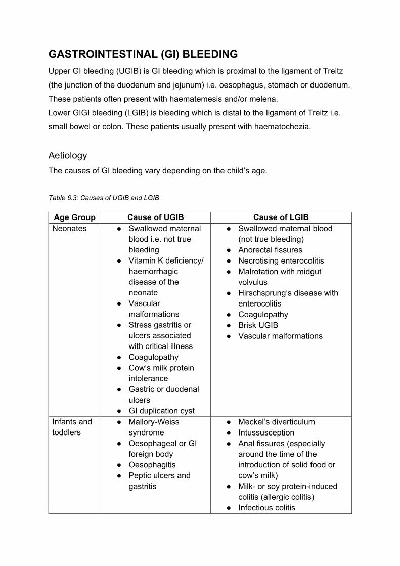
Table 6.3: Causes of UGIB and LGIB
Age Group Cause of UGIB Cause of LGIB Neonates ● Swallowed maternal
blood i.e. not true bleeding
● Vitamin K deficiency/ haemorrhagic disease of the neonate
● Vascular malformations
● Stress gastritis or ulcers associated with critical illness
● Coagulopathy ● Cow’s milk protein
intolerance ● Gastric or duodenal
ulcers ● GI duplication cyst
● Swallowed maternal blood (not true bleeding)
● Anorectal fissures ● Necrotising enterocolitis ● Malrotation with midgut
volvulus ● Hirschsprung’s disease with
enterocolitis ● Coagulopathy ● Brisk UGIB ● Vascular malformations
Infants and toddlers
● Mallory-Weiss syndrome
● Oesophageal or GI foreign body
● Oesophagitis ● Peptic ulcers and
gastritis
● Meckel’s diverticulum ● Intussusception ● Anal fissures (especially
around the time of the introduction of solid food or cow’s milk)
● Milk- or soy protein-induced colitis (allergic colitis)
● Infectious colitis

● Bleeding oesophageal varices or gastric varices
● Arterial bleeding (rare)
● Lymphonodular hyperplasia ● GI duplication cyst ● Coagulopathy ● Eosinophilic GI disease ● Infantile and very early-onset
inflammatory bowel disease (IBD)
Pre-school going age
● Anal fissures ● Intussusception ● Meckel’s diverticulum ● Other causes e.g. infectious
colitis, haemolytic uraemic syndrome, IgA vasculitis, Henoch-Schonlein purpura, juvenile polyps, very early-onset IBD, solitary ulcer syndrome
School going age
● Anal fissures ● Juvenile polyps ● Infectious colitis (Salmonella
sp, Shigella sp, Campylobacter sp, E.coli, Clostridium difficile are the most common)
● Inflammatory bowel disease ● Meckel’s diverticulum ● Solitary rectal ulcer syndrome ● IgA vasculitis (Henoch-
Schonlein purpura ● Haemorrhoids
Clinical Features The child may present with:
● Haematemesis (vomiting of bright red blood or coffee ground-like material)
● Melena (passage of black and tar-like stools with a strong odour):
o The colour and smell are due to the haemoglobin in the blood being
altered by the digestive enzymes and intestinal bacteria.
o It is important to note that black stools may also be caused by certain
medications and foods.
● Haematochezia (passage of bright red/maroon-coloured blood or fresh clots
per rectum):
o It is usually due to LGIB but can be due to UGIB in cases of short
intestinal transit time or massive UGIB.
● Occult GI bleeding (bleeding is not visible to the naked eye of the patient or
physician):
o Patients usually present with iron-deficiency anaemia or occult GI
bleeding may be identified by testing the stool for occult blood.
Approach When a patient presents with GI bleeding, one must ask the following questions
1. Is the patient haemodynamically stable or is resuscitation indicated?
2. Is it blood?
3. Is the blood from the upper GIT (dark red/black) or lower GIT (bright red)?
4. What are the most likely causes of the bleed?
The patient should then be assessed based on:
● History and examination:
o One should ask about:
▪ This episode of bleeding – chronology of the bleeding episode,
estimated blood loss, colour of blood and any associated
symptoms e.g. abdominal pain, fever, weight loss and fatigue,
recent use of NASIDs, etc.
▪ Associated symptoms (paying attention to GI symptoms) –
dyspepsia, heartburn, abdominal pain, dysphagia, and weight
loss, poor feeding or irritability (in infants), history of jaundice,
easy bruising, or change in stool colour (liver disease).
▪ Possible causes: easy bruising/bleeding, personal or family
history of liver, kidney, heart disease or coagulopathies, drug
history, travel history, diet, etc.
o On examination one should look for signs of shock and possible
causes of bleeding. Thus, a full examination (including a rectal
examination) must be done.
● Diagnostic studies; may include:

o Bloods – FBC, CRP, ESR, coagulation studies
o Stool MC&S – C. difficile, enteric pathogens, ova and parasites
o RFTs and/or LFTs (in cases where related causes are suspected)
o Plain radiographs (to identify for foreign bodies)
o Abdominal USS
o Endoscopy (if the patient has brisk or unexplained bleeding after a
thorough examination, or if s/he is in shock)
o Angiography (if the source of the bleeding could not be found on
endoscopy)
o Colonoscopy
Management Emergency Management
A gastroenterologist and general surgeon should be immediately called for any
patient with severe acute UGIB. The patient should then be resuscitated and
stabilised:
● If the child is shocked or has orthostatic hypotension (i.e. had a severe GI
bleed), s/he should be admitted to ICU for resuscitation and close
observation.
● Two large-bore IV catheters should be inserted and fluid boluses given. A
transfusion may be required (if Hb <8 g/dL).
● Surgical intervention may be required for uncontrollable bleeds.
Follow-up visits should be scheduled, especially for first-time bleeders.
Routine Management
Patients who have had a UGIB:
● The patient should be resuscitated (ABCs) and the cause treated.
● Pharmacological management includes:
o Acid suppression in clinically significant UGIB (IV PPIs or H2-receptor
antagonists).
o Temporising the difficult to control bleed (e.g. variceal bleeding) with
somatostatin and octreotide.
● Surgical management:
o Sengstaken-Blakemore tube placement.
o Endoscopic treatment (within 24-48 hours of presentation) e.g.
sclerotherapy, elastic ligature (also used for haemorrhoids),
transjugular intrahepatic portosystemic shunt (TIPS) for variceal
bleeds.
o Catheter tamponade (used if medical management fails, to stop a
continuously bleeding vessel which has been identified using a
catheter; only perform in theatre or the ICU setting).
The cause of the lower GI bleed should be identified and treated. See also a figure
on Deployed Sengstaken-Blakemore Tube in the Patient with an UGIB here.
FUNCTIONAL ABDOMINAL PAIN (FAP) These disorders are the most common causes of chronic (>2 months) abdominal
pain in children and adolescents.
Pathophysiology The pathophysiology is poorly understood but it is thought to involve an interplay
between enteric and CNS regulatory factors. These disorders may be associated
with:
● Visceral hyperalgesia:
o Eating may be associated with onset of pain and the patient is,
therefore, likely to skip meals in an attempt to avoid pain.
● Reduced pain threshold
● Referred pain following rectal distension
● Impaired gastric relaxation response to meals
Diagnosis, Classification and Clinical Features The diagnosis is made in the child with chronic abdominal pain, no danger signs,
normal examination and stool which is negative for occult blood. Certain
recognisable patterns of symptoms may be used to classify the FAP:
▪ Functional dyspepsia
▪ Irritable bowel syndrome (a disorder of large intestine) – cramping, abdominal
pain, bloating, gas, diarrhoea and/or constipation
▪ Abdominal migraine (most common in children) – abdominal pain, nausea,
vomiting, family or personal history of migraines
▪ FAP not otherwise specified
Management It should be managed in a primary care setting. The goal of treatment is a return to
normal function rather than complete elimination of pain. However, a referral may be
made if the pain cannot be managed at a primary care level.
Management is individualised and depends on the child’s and family behaviours,
triggers and symptoms. Regardless of the subtype of FAP, the management
includes:
● Assuring the patient and family that a treatment will be initiated and the
patient (and family, as they may also be affected by the condition) followed up
on a regular basis.
● Patient education:
o FAPDs are best treated using the biopsychosocial model of care.
Before starting therapy, one must define the expectation of the patient
and parents and be realistic about treatment aims.
o One must reassure the patient and family by acknowledging that the
pain is real and has affected important activities in the patient’s life.
o They must be informed that FAP is common and can be exacerbated
or made to persist by environment and psychosocial factors e.g. stress,
anxiety, social reinforcement. However, they are not life threatening.
o They must also be told that management focuses on rehabilitation
rather than treatment, and includes avoiding triggers and improving
coping skills.
● Prescribing a return to structured activities of daily living (including school):
o School absenteeism adds to family stress and can interfere with school
performance), see also an image of active children in a classroom.
o One must implement a plan ahead for pains at school, such as keeping
the first back at school short, arranging for the child to go to the nurse’s
office until the pain stops and use the bathroom whenever necessary.
A letter should be written to make the school aware of the condition
and plan.
o The family must be given guidelines for when the pain is severe
enough to warrant going home or missing school.
o School-related stressors must be identified and dealt with.
● Behaviour modification (positive, well behaviours should be reinforced, and
triggers and behaviours that cause pain should be stopped/avoided).
● Strategies to improve pain tolerance and coping:
o Psychological treatments which improve coping should form part of the
management of children and adolescents with FAP e.g. relaxation
techniques, distraction, and cognitive behavioural therapy (CBT).
o Psychological interventions appear to be more effective than
pharmacological interventions.
● Symptomatic management.