Embed Size (px)
Citation preview
Graduate School for Cellular and Biomedical Sciences
University of Bern
Exploring the pathogen-commensal continuum: Cell wall auxotrophic bacteria in gnotobiotic mice
PhD Thesis submitted by
Miguelangel Cuenca Vera
from Spain
for the degree of
PhD in Immunology
Supervisor Prof. Dr. Siegfried Hapfelmeier Institute for Infectious Diseases
Faculty of Medicine of the University of Bern Co-advisor
Dr. Emma Slack-Wetter Institute for Microbiology
ETH Zürich Original document saved on the web server of the University Library of Bern
This work is licensed under a Creative Commons Attribution-Non-Commercial-No derivative works 2.5 Switzerland
licence. To see the licence go to http://creativecommons.org/licenses/by-nc-nd/2.5/ch/ or write to Creative Commons, 171 Second Street, Suite 300, San Francisco, California 94105,
USA.


Copyright Notice This document is licensed under the Creative Commons Attribution-Non-Commercial-No derivative works 2.5 Switzerland. http://creativecommons.org/licenses/by-nc-nd/2.5/ch/
You are free:
to copy, distribute, display, and perform the work
Under the following conditions:
Attribution. You must give the original author credit.
Non-Commercial. You may not use this work for commercial purposes.
No derivative works. You may not alter, transform, or build upon this work..
For any reuse or distribution, you must take clear to others the license terms of this work.
Any of these conditions can be waived if you get permission from the copyright holder.
Nothing in this license impairs or restricts the author’s moral rights according to Swiss law.
The detailed license agreement can be found at: http://creativecommons.org/licenses/by-nc-nd/2.5/ch/legalcode.de

Accepted by the Faculty of Medicine, the Faculty of Science and the Vetsuisse
Faculty of the University of Bern at the request of the Graduate School for
Cellular and Biomedical Sciences
Bern, Dean of the Faculty of Medicine
Bern, Dean of the Faculty of Science
Bern, Dean of the Vetsuisse Faculty Bern


Acknowledgment
The present work is the compilation of four years of collaborative work, in which many people
participated.
I would like to thank my external referee Nassos Typas, for agreeing to review this
manuscript in such a short notice.
I thank my thesis committee Emma Slack and Philippe Krebs, for continuous support and
constant supply of ideas during the entire period.
I thank my supervisor Siegfried Hapfelmeier, for providing me the tools I needed for
professional work and the scientific freedom to use them.
I thank my lab-mates: Miguel, for continuous good mood and willingness to take over all the
lab-work that I failed to achieve; Nico, for supporting my bad-lab days and implementing
metal Fridays; Simi, for taking the time to teach me all the lab techniques even when I am not
the best learner; Steff, for always being willing to help despite my horrifying disorganization;
Stefi, for being my late night lab company during the long days; Tephi, for using her
knowledge to cover my immunological holes; Firuza, for being so cheerful and never
complain over taking more work and Nanda, for providing me infinite advices and guidance.
I thank all my friends, who continuously supported me during this period, went with me on all
those Aareböötle trips, heard all my complaints, cheered me up on the bad days and partied
with me on the good days.
And last I thank my family for the continuous support from afar, despite the Venezuelan
disaster always lingering there.
General index
Abstract 5 Introduction 6 Chapter 1
Author contributions 18 Abstract 19 Introduction 20 Results 21 Discussion 27 Materials and Methods 29 References 34 Supplementary Material 36
Chapter 2 Abstract 41 Introduction 42 Results and discussion 43 Materials and Methods 49 References 51 Supplementary Material 53
Chapter 3 Author contributions 56 Abstract 57 Introduction 58 Results 60 Discussion 69 Supplementary Material 72 Materials and Methods 76 References 79
General Discussion 84 Curriculum vitae 87 Declaration of originality 89

Abstract The intestinal tract of all known vertebrate animals is colonized with a high density of
bacteria, forming host-specific communities. These communities are usually composed of a
broad range of different species that have co-evolved with the host, to form very close and
beneficial. In this thesis we developed a new tool for the study of host-microbiota
interactions, based on the use of a proliferation controlled commensal E. coli strain and
germ-free mice. This strain, contained a severe cell wall synthesis defect leading to the
inability of proliferate without external supplementation. To guarantee the tightness of our
system and its similarity to the wild type strain, we tested extensively the strain properties
even under extreme cell wall starvation. This tool was further adapted to Salmonella enterica
Typhimurium allowing us to simulate artificially the first six hours of a natural Salmonella
infection, without the actual induction of disease. Our ability of simulating the early phase of
an infection led to recognition of crucial in vivo bacterial adaptations, induced by the adaptive
immunity, which led to the shift from pathogenic to commensal behavior in several
Salmonella strains. The mechanism of this behavioral shift was explored, leading to the
recognition of a Salmonella O-antigen shift, specific IgA induction and, exclusion of a
pathogenic strain combined in to protection against disease when exposed to wild type
Salmonella enterica. The additive effect of the discovered mechanisms was able to only
partly explain the observed behavior, suggesting that other mechanisms remain to be
uncovered to fully explain the behavioral shift.
5
Introduction Microbiota constitution
The mammalian microbiota begins to form at birth and continues its development until death
of the host (Zhang et al., 2014). During this period, four major groups colonize the intestinal
tract, and dominate the community: Firmicutes, Bacteroidetes, Actinobacteria and
Proteobacteria (Bäckhed et al., 2015). The Firmicutes are a group of Gram-positive
anaerobic bacteria, which include well known members as the genus Clostridium. The
Bacteroidetes are a group of Gram-negative bacteria abundant in soil, sea water and the
intestines of homoeothermic animals. The Actinobacteria comprise a phylum of Gram-
positive bacteria, containing the well-studied genus Streptomyces, and associated with the
production of several antibiotics. The last major group of gut-associated bacteria is the
Gram-negative Proteobacteria phylum, which includes the well-studied commensal E. coli, as
well as several important human pathogens including Salmonella enterica, Yersinia pestis,
Campylobacter jejunii, Helicobacter pylori and Vibrio cholerae (Khanna and Tosh, 2014;
Macpherson and McCoy, 2014).
The process of colonization of the newborn germ-free mammal in a rapid succession of
maternally and environmentally derived early colonizers and the maturation of the postnatally
acquired consortium into an adult microbiota requires a delicate interplay of all species
involved (Bäckhed et al., 2015). This ecological succession is highly influenced by dietary
factors, exposition to new immigrants and the host immune system (Koenig et al., 2010). The
mode of birth is the first major factor that influences the bacterial compositions, as it has
been shown that vaginally delivered babies have a different microbiota composition
compared to those delivered by C-section (Dominguez-Bello and Costello, 2010). After the
primary microbiota has been established, a major compositional shift happens upon
weaning, when the diet changes from milk to solid food, leading in to an adult-type microbiota
(Koenig et al., 2010).
Even established adult microbiotas show a high degree of inter- and intra-individual
(temporal) phylogenetic variability, which makes their direct comparison complex. The
colonization of germ-free mice intestinal tract with a myriad of species from a variety of
environmental sources, all lead to extremely different communities (Seedorf et al., 2014).
These different species interact between themselves in either beneficial (complementary
metabolism and degradation of toxic products) or detrimental (production of antibiotics,
competition for nutrients and direct attack) relationships that shape the final structure of the
community. The bacteria-bacteria interactions vary with the changes in host nutrition or
6
incorporation of new species, dynamically changing the community structure in a daily basis
(Bäckhed et al., 2015; Dishaw et al., 2014)
Despite the high variability of the microbiome between age-matched individuals, they at the
functional level encode a rather similar set of metabolic pathways, regardless of phylogenetic
composition (Bashan et al., 2016). The resulting microbial metabolic repertoire provides
many nutritional advantages to the host, such as synthesis of micronutrients, degradation of
toxic compounds, increase in the efficiency of nutrient absorption and protection from obesity
(Blanton et al., 2016; Bäckhed et al., 2015; Cummings and Macfarlane, 1997; Dishaw et al.,
2014; Maltby et al., 2013; McFall-Ngai and Casadevall, 2012; Ridaura et al., 2013; Tremaroli
and Bäckhed, 2012)
Advantages of the microbiota
The stability of an adult microbiota comes from a tight and intricate nutritional connection
between species, induced by an overlay of all the different metabolic pathways. This state is
achieved by a specific subset of bacteria, able to fully co-exist and resist change, referred to
as residents (Bäckhed et al., 2015). This concept separates the resident microbiota from
tourist species (organisms that only transit in the gut before being shed). The selection of
resident species and continuous exclusion of tourist (and pathogenic) species are the main
signs of a mature microbiota (Bäckhed et al., 2015; Cummings and Macfarlane, 1997;
Tremaroli and Bäckhed, 2012)
A fully developed intact microbiota provides protection against pathogens due to a
phenomenon long known as “colonization resistance”, where the resident bacteria deplete
nutrients needed for the newcomer’s proliferation while inducing an specific immune
response(Leatham et al., 2009; Maltby et al., 2013; Stecher et al., 2007a). Colonization
resistance also favors resident bacteria, as seen in E. coli, that after a period of adaptation in
the gut becomes more efficient in carbon source utilization, preventing the bloom of other E.
coli strains (Leatham et al., 2005). Since this protection is able to deter the invasion of
newcomers, many pathogens developed mechanisms to break this colonization resistance.
A well-studied mechanism for bypassing the colonization resistance is known to be crucial in
enteric infection with Salmonella enterica serovar Typhimurium (Stm) (Stecher et al., 2007b).
In this disease, Stm uses the type three secretion system (TTSS) encoded on salmonella
pathogenicity island-1 (SPI-1) to invade the intestinal mucosa (Hapfelmeier et al., 2005).
Driven by bacterial invasion and inflammasome activation, the host induces an acute
inflammatory response leading to the decrease of butyrate-producing Clostridium spp., and
hence a decrease in the Clostridium induced local anoxia (Rivera-Chávez et al., 2016).
Inflammation-related increases in the levels of reactive oxygen and nitrogen species allows
7
Stm to boost its proliferation rate and overcome colonization resistance. The increase in Stm
numbers leads to diarrhea, loss of weight, prolonged discomfort and depending on the host’s
immune competence death. This strategy has the side effect of giving rise to spontaneous
avirulent mutant, able to overgrow the virulent Stm, which can be interpreted as a population-
level regulatory mechanism leading to the clearance of the pathogen by the resident
microbiota, ensuring the survival of the host (Diard et al., 2013; Endt et al., 2010; Sturm et
al., 2011).
Methods for microbiota study
The microbiota disruption observed in Stm colitis can be artificially achieved by treating mice
with antibiotics, to lower the colonization resistance, allowing the establishment of Stm. This
antibiotic treatment is the most widely used mouse model for the study of Stm-induced colitis
(Barthel et al., 2003; Rivera-Chávez et al., 2016). This method has been very successfully
used for studying the pathogen-host interactions. However, since it leads to an artificially
disrupted microbiota it has limited use for studying the healthy host-microbiota interactions
and microbiota-pathogen-host relationship.
Depleting mice of their native microbiota by using highly concentrated antibiotic cocktails was
proposed as a method to assess the delicate microbiota-pathogen continuum an its
relationship with the host (Reikvam et al., 2011). This treatment has low reproducibility and
the microbiota recovers as soon as the antibiotic cocktail is removed (Macpherson and
McCoy, 2014). The method of choice to avoid this problem is the utilization of germ-free
animals. Germ-free mice are derived from sterile neonates that had been delivered by
aseptically performed Cesarean section, hand-reared, and then bred under sterile conditions.
Already established germ-free mouse vivaria can be efficiently extended with additional
mouse strains by axenic embryo transfer (Macpherson and McCoy, 2014).
Germ-free (sterile) and gnotobiotic (ex-germ-free mice colonized with a defined subset of
isolated species) mice provide powerful tools to study reduced-complexity and fully defined
microbial communities, as well as detailed host-microbiota interactions. In such animals it is
possible to assess which species-specific microbiota subsets are able to provide an efficient
colonization resistance, what are the long term effects of small microbiota changes and if the
microbiota is transferable between hosts (Chung et al., 2012; Lichtman et al., 2016; Ridaura
et al., 2013). But even with the use of germ-free mice researchers have been bound to
permanently expose mice to bacteria, obscuring reversible from irreversible bacterially
induced changes. To be able to transiently colonize mice, "cell wall auxotrophic" (CWA)
bacteria were developed (Hapfelmeier et al., 2010).
8
CWA bacterial strains provide a tool for limiting the host-bacterial interactions to a short
duration, effectively separating short- from long-lived responses (Hapfelmeier et al., 2010).
This tool is based on the auxotrophy for two bacteria-specific amino acids, D-alanine (D-ala)
and meso-diaminopimelic acid (m-Dap). These amino acids are main constituents of the
peptidoglycan, the essential and ubiquitous support structure in the gram-negative cell wall,
and can be substituted by other (also bacteria-specific) metabolites only under extreme
conditions (Cava et al., 2011; Lupoli et al., 2011; Xin et al., 2012). The other important factor
is that neither of these compounds are produced by mammals, leading to D-Ala/m-Dap
deprival of the CWA strains upon inoculation into germ-free animals. The D-amino acids
starvation that CWA strains undergo should induce a phenotype similar to that induced by
beta-lactam antibiotic treatment. Beta-lactam antibiotics bind to penicillin binding proteins
(PBPs) inducing an accumulation of peptidoglycan precursors in the cytosol, eventually
leading to cellular arrest or lytic death (Park and Uehara, 2008; Uehara and Park, 2008; Yao
et al., 2012).
Using a CWA E. coli K-12 strain it was possible to determine that a short lived intestinal
mucosal exposition to live bacteria was sufficient to induce long-lasting specific
immunoglobulin A (IgA), a clear sign that adaptive immune responses could be formed
independently from continuous colonization (Hapfelmeier et al., 2010). The same strain was
used for studying the role of continuous microbiota presence in the induction of
granulopoiesis, where germ-free mice had an increase on their neutrophils after being
exposed to bacteria, suggesting that live bacteria induce different responses than mere
bacterial component exposition such as LPS or dead bacterial particles (Balmer et al., 2014)
Microbiota and immunity
The immune system holds an important role in shaping and especially containing the
microbiota in the gut by several layers of redundant or complementary mechanisms. The
immune response is comprised of two main components: innate and adaptive immunity. The
function of these two components is based on the ability to recognize conserved non-
mammalian/microbial molecular patterns by germ-line encoded (hence, "innate") and
undefined non-self-antigens that are not limited to microbes by an combinatorically encoded
and evolving set of ("adaptive") immune receptors (the B and T cell receptors), respectively.
The combined effects of these two mechanisms account for most of the direct effect from the
host to manipulate the microbiota (Dishaw et al., 2014)
The intestinal innate immunity consists of a series of relatively unspecific mechanisms of
microbial containment, mostly by forming barriers and eliminating any type of microbe that
invades the tissues. The primary physical intestinal barrier is the epithelium that covers the
9
entire intestinal surface while being permeable to nutrients (Dishaw et al., 2014). This barrier
is covered by intestinal mucus, a biological gel primarily composed of mucin 2 (MUC2). The
mucus covers the entire intestinal tract and constitutes a physical barrier that separates the
microbiota from the epithelial brush border. There are two distinct mucus layers: an inner
mucus, mostly composed of tightly packed MUC2 and nearly sterile; and an outer layer,
composed of loosely packed MUC2 (Jakobsson et al., 2015). The outer mucus is
permanently colonized, and provides carbon source to a distinct microbiota that differs from
the luminal, digestive-associated microbiota (Jakobsson et al., 2015; Li et al., 2015). For the
normal formation of the mucus barrier a continuous microbial colonization for > 8 weeks is
necessary, leading to the full activation of goblet cells (the producers of the intestinal mucus)
(Jakobsson et al., 2015).
Moreover, innate immunity is the first line of pathogen-commensal differentiation in the gut.
Pro-inflammatory signaling induces a rapid release of highly fucosylated proteins, that have
been shown to promote the proliferation of commensals while downregulating the virulence
in pathogenic E. coli strains (Pickard et al., 2014). It has also been shown that the
lipopolysaccharide (LPS) of Bacteroides, a genus of known commensals, dampens the pro-
inflammatory response (Vatanen et al., 2016). Another method of limiting the epithelial
exposition to bacteria is by production of antimicrobial peptides, a group of molecules that
has been shown to be important in the maintenance of the inner mucus sterility (Meyer-
Hoffert et al., 2008). The production of antimicrobial peptides by Paneth cells is tightly
regulated by NOD2, an intracellular innate receptor for bacterial peptidoglycan
fragments/subunits, and its malfunctioning has been linked to diseases as Crohn’s ileitis
(Boneca et al., 2007; Kobayashi et al., 2005). Moreover, deficiency in innate immunity, for
example in impaired toll-like receptor (TLR) signaling leads to a hyper-induction of
compensatory adaptive immune responses, to maintain the intestinal homeostasis (Slack et
al., 2009).
The adaptive immune responses are characterized by the recognition of specific non-self-
antigen and the expansion of highly antigen-specific effector T and B cells, which also give
rise to the formation of long-lived memory cells providing long live immunological memory.
The initial recognition of relevant antigens is primarily done by dendritic cells (DCs), the main
antigen-presenting cells of the innate immune system, which process and present antigen to
T-cells. After antigen presentation has occurred, the T-cells mediate a large array of immune
mechanisms to eradicate the foreign organism, including B-cells (main mediators of
immunoglobulin responses), regulating the innate immunity and activating phagocytic cells
(Hapfelmeier and Macpherson, 2010).
10
In the gut, B-cells and T-cells are mostly organized in special structures that maximize the
interaction with DCs. These structures are classified by size or position in to: mesenteric
lymph nodes (MLNs), Peyer’s patches, or minor lymph nodes. In these structures, primed
DCs carrying relevant antigens interact with T-cells. The DC-T-cell interaction, denominated
antigen presentation, leads to the specific activation of naïve T-cells and their differentiation.
From the two main T-cell types, CD8+ (cytotoxic, mostly antiviral function) and CD4+ (helper,
Th, mostly activation of other cellular types), the helper T-cells are the ones mostly involved
in host-bacterial interactions (Perez-Lopez et al., 2016).
The helper T-cells are mainly classified in four types: Th1 (classical anti-intracellular
pathogens), Th2 (classical anti-extracellular pathogens), Th17 (common in mucosal sites and
associated to bacterial responses) and Treg (associated with anti-inflammatory responses)
(Dong, 2010).
The role of immunity in the microbiota structure
Traditionally the adaptive immune response has been attributed the role to counter and
eradicate pathogens. This role has been further supported by the observations that mice
could be vaccinated against intestinal bacterial pathogens like Salmonella enterica or Vibrio
cholerae, or that known colitogenic bacteria are stronger inducers of IgA (the main intestinal
immunoglobulin). Even though IgA was shown to be relevant for acquired immunity against
several intestinal pathogens, there are several examples for immunoglobulin-independent
responses crucial to mucosal protective immunity. Immunized mice deficient in IgA are able
to fend off enteric Stm infection, but B-cell deficient or MyD88 deficient mice are not (Ko et
al., 2009; Nanton et al., 2012). Also the presence of commensal-specific mucosal regulatory
T-cells (Treg) is crucial for the maintenance of the intestinal homeostasis, even during a
pathogen associate inflammatory response (Ivanov and Honda, 2012). Whenever the
balance between the host and the microbiota is lost, commensal-specific pro-inflammatory
T-cells are induced, the unbalanced production of which may cause pathology (Hand et al.,
2012).
The traditional pathogenesis-centered view of adaptive immune responses has recently
shifted with the description of Th17-cells, a subgroup of commensal induced T-cells that
provide extra protection against pathogens (Ivanov et al., 2009). Certain commensal species
such as segmented filamentous bacteria (SFB), Bacteroides spp. or Clostridium spp. are
known to induce Treg or Th17 responses (Geuking et al., 2011; Peterson et al., 2007; Smith
et al., 2013; van Beelen et al., 2007). These commensal-bacteria induced responses
dampen or prevent the mucosal inflammation, while also supporting the colonization of
beneficial strains. These observations led several authors to suggest that the adaptive
11
immune system primarily functions to support and maintain a rich, physiological microbiota
(Dishaw et al., 2014). There is concrete evidence supporting the idea that functional adaptive
immune system increases the predictability and stability of the microbiota. When the
microbiota of B-cell and T-cell deficient mice was studied over time, it was found that the rate
of bacterial evolution was altered, as well as the species composition (Barroso-Batista et al.,
2015; Brown et al., 2013; Zhang et al., 2014).
Aims
The study of host-induced effects on the microbiota structure and composition is the central
aspect to be covered in this thesis. For this purpose we defined three main aims which will
be further explored in each of three result chapters:
1. Design, construction and testing of a new bacterial tool that allows us to dissect short
from long lived bacteria-host interactions in mice.
2. Characterization of the early innate immune responses against a known pathogen, in
a transient infection model.
3. Study the influence of adaptive immunity in intestinal bacterial-bacterial interactions.
Original contribution
Even though the effect of the adaptive immune system on microbiota structure and
composition are well-supported, the precise mechanisms of action are poorly understood. In
this work we explored this and other questions, generating three fundamental pieces of new
evidence:
1. A new tool to study transient host-microbiota interactions, using a cell wall
auxotrophic strain of a biologically representative commensal bacterium, showed that neither
the bacterial behavior nor bacteria-host interaction are affected by the mutations. This finding
confirms that the CWA strains can be used to simulate short term host-microbiota
interactions.
2. The infection of mice with the CWA Stm strain was able to induce an increase in the
variability of inflammatory markers, without leading to inflammation. This represents an
effective separation of the primary pathogen-induced responses from the pathology it
normally causes. As a consequence, the model simulates the first 6 h of Stm induced colitis,
without inducing pathology.
3. The combination of adaptive immune response and in vivo bacterial adaptation are
crucial in the microbiota development from immigrant to resident status, by disfavoring the
colonization of bacterial newcomers. Chapter three describes novel information on the
underlying mechanism.
12
References Balmer, M.L., Schürch, C.M., Saito, Y., Geuking, M.B., Li, H., Cuenca, M., Kovtonyuk, L.V., McCoy, K.D., Hapfelmeier, S., Ochsenbein, A.F., et al. (2014). Microbiota-derived compounds drive steady-state granulopoiesis via MyD88/TICAM signaling. J. Immunol. 193, 5273-83.
Barroso-Batista, J., Demengeot, J., and Gordo, I. (2015). Adaptive immunity increases the pace and predictability of evolutionary change in commensal gut bacteria. Nature Communications 6, 8945.
Barthel, M., Hapfelmeier, S., Quintanilla-Martínez, L., Kremer, M., Rohde, M., Hogardt, M., Pfeffer, K., Rüssmann, H., and Hardt, W. (2003). Pretreatment of mice with streptomycin provides a Salmonella enterica serovar Typhimurium colitis model that allows analysis of both pathogen and host. 71, 2839-2858.
Bashan, A., Gibson, T.E., Friedman, J., Carey, V.J., Weiss, S.T., Hohmann, E.L., and Liu, Y. (2016). Universality of human microbial dynamics. Nature 534, 259-62.
Blanton, L.V., Charbonneau, M.R., Salih, T., Barratt, M.J., Venkatesh, S., Ilkaveya, O., Subramanian, S., Manary, M.J., Trehan, I., Jorgensen, J.M., et al. (2016). Gut bacteria that prevent growth impairments transmitted by microbiota from malnourished children. Science 351, aad3311.
Boneca, I.G., Dussurget, O., Cabanes, D., Nahori, M., Sousa, S., Lecuit, M., Psylinakis, E., Bouriotis, V., Hugot, J., Giovannini, M., et al. (2007). A critical role for peptidoglycan N-deacetylation in Listeria evasion from the host innate immune system. Proc. Natl. Acad. Sci. U.S.A. 104, 997-1002.
Brown, E.M., Sadarangani, M., and Finlay, B.B. (2013). The role of the immune system in governing host-microbe interactions in the intestine. Nature Immunology 14, 660.
Bäckhed, F., Roswall, J., Peng, Y., Feng, Q., Jia, H., Kovatcheva-Datchary, P., Li, Y., Xia, Y., Xie, H., Zhong, H., et al. (2015). Dynamics and Stabilization of the Human Gut Microbiome during the First Year of Life. Cell Host &Amp; Microbe 17, 690.
Cava, F., Pedro, M.A., Lam, H., Davis, B.M., Waldor, M.K., and de Pedro, M.A. (2011). Distinct pathways for modification of the bacterial cell wall by non-canonical D-amino acids. The EMBO Journal 30, 3442-3453.
Chung, H., Pamp, S., Hill, J., Surana, N., Edelman, S., Troy, E., Reading, N., Villablanca, E., Wang, S., Mora, J., et al. (2012). Gut Immune Maturation Depends on Colonization with a Host-Specific Microbiota. Cell 149, 1578-1593.
Cummings, J.H., and Macfarlane, G.T. (1997). Role of intestinal bacteria in nutrient metabolism. JPEN J Parenter Enteral Nutr 21, 357-65.
Diard, M., Garcia, V., Maier, L., Remus-Emsermann, M.N., Rol, Regoes, R., Ackermann, M., Hardt, W., and Regoes, R.R. (2013). Stabilization of cooperative virulence by the expression of an avirulent phenotype. Nature 494, 353-356.
Dishaw, L.J., Cannon, J.P., Litman, G.W., and Parker, W. (2014). Immune-directed support of rich microbial communities in the gut has ancient roots. Developmental &Amp; Comparative Immunology 47, 36.
13
Dominguez-Bello, M., and Costello, E. (2010). Delivery mode shapes the acquisition and structure of the initial microbiota across multiple body habitats in newborns. In Proceedings Of The.
Dong, C. (2010). Helper T-cell heterogeneity: a complex developmental issue in the immune system. Cellular And Molecular Immunology 7, 163.
Endt, K., Stecher, B., Chaffron, S., Slack, E., Tchitchek, N., Benecke, A., Van Maele, L., Sirard, J., Mueller, A.J., Heikenwalder, M., et al. (2010). The microbiota mediates pathogen clearance from the gut lumen after non-typhoidal Salmonella diarrhea. PLOS Pathogens 6, e1001097.
Geuking, M., Cahenzli, J., Lawson, M.E., Ng, D.K., Slack, E., Hapfelmeier, S., McCoy, K., and Macpherson, A. (2011). Intestinal Bacterial Colonization Induces Mutualistic Regulatory T Cell Responses. Immunity 34, 794-806.
Hand, T.W., Santos, L.M., Bouladoux, N., Molloy, M.J., Pagan, A.J., Pepper, M., Maynard, C.L., Elson, C.O., and Belkaid, Y. (2012). Acute Gastrointestinal Infection Induces Long-Lived Microbiota-Specific T Cell Responses. Science 337, 1553-1556.
Hapfelmeier, S., Lawson, M.A., Slack, E., Kirundi, J.K., Stoel, M., Heikenwalder, M., Cahenzli, J., Velykoredko, Y., Balmer, M.L., Endt, K., et al. (2010). Reversible Microbial Colonization of Germ-Free Mice Reveals the Dynamics of IgA Immune Responses. Science 328, 1705-1709.
Hapfelmeier, S., and Macpherson, A.J. (2010). In remembrance of commensal intestinal microbes. Communicative &Amp; Integrative Biology 3, 569.
Hapfelmeier, S., Stecher, B., Barthel, M., Kremer, M., Müller, A.J., Heikenwalder, M., Stallmach, T., Hensel, M., Pfeffer, K., Akira, S., et al. (2005). The Salmonella pathogenicity island (SPI)-2 and SPI-1 type III secretion systems allow Salmonella serovar typhimurium to trigger colitis via MyD88-dependent and MyD88-independent mechanisms. J. Immunol. 174, 1675-85.
Ivanov, I.I., Atarashi, K., Manel, N., Brodie, E.L., Shima, T., Karaoz, U., Wei, D., Goldfarb, K.C., Santee, C.A., Lynch, S.V., et al. (2009). Induction of Intestinal Th17 Cells by Segmented Filamentous Bacteria. Cell 139, 485-498.
Ivanov, I., and Honda, K. (2012). Intestinal Commensal Microbes as Immune Modulators. Cell Host &Amp; Microbe 12, 496-508.
Jakobsson, H.E., Holmén-Larsson, J., Schütte, A., Ermund, A., Rodríguez-Piñeiro, A.M., Arike, L., Wising, C., Svensson, F., Bäckhed, F., and Hansson, G.C. (2015). Normalization of Host Intestinal Mucus Layers Requires Long-Term Microbial Colonization. Cell Host &Amp; Microbe 18, 582-92.
Khanna, S., and Tosh, P.K. (2014). A clinician's primer on the role of the microbiome in human health and disease. Mayo Clin. Proc. 89, 107-14.
Ko, H.J., Yang, J.Y., Shim, D.H., Yang, H., Park, S.M., Curtiss, R., and Kweon, M.N. (2009). Innate Immunity Mediated by MyD88 Signal Is Not Essential for Induction of Lipopolysaccharide-Specific B Cell Responses but Is Indispensable for Protection against Salmonella enterica serovar Typhimurium infection. The Journal Of Immunology 182, 2305.
14
Kobayashi, K.S., Chamaillard, M., Ogura, Y., Henegariu, O., Inohara, N., Nuñez, G., and Flavell, R.A. (2005). Nod2-dependent regulation of innate and adaptive immunity in the intestinal tract. Science 307, 731-4.
Koenig, J.E., Spor, A., Scalfone, N., Fricker, A.D., Stombaugh, J., Knight, R., Angenent, L.T., and Ley, R.E. (2010). Succession of microbial consortia in the developing infant gut microbiome. Proceedings Of The National Academy Of Sciences 108, 4578.
Leatham, M.P., Banerjee, S., Autieri, S.M., Mercado-Lubo, R., Conway, T., and Cohen, P.S. (2009). Precolonized Human Commensal Escherichia coli Strains Serve as a Barrier to E. coli O157:H7 Growth in the Streptomycin-Treated Mouse Intestine. Infection And Immunity 77, 2876-2886.
Leatham, M.P., Stevenson, S.J., Gauger, E.J., Krogfelt, K.A., Lins, J.J., Haddock, T.L., Autieri, S.M., Conway, T., and Cohen, P.S. (2005). Mouse Intestine Selects Nonmotile flhDC Mutants of Escherichia coli MG1655 with Increased Colonizing Ability and Better Utilization of Carbon Sources. Infection And Immunity 73, 8039-8049.
Li, H., Limenitakis, J.P., Fuhrer, T., Geuking, M.B., Lawson, M.A., Wyss, M., Brugiroux, S., Keller, I., Macpherson, J.A., Rupp, S., et al. (2015). The outer mucus layer hosts a distinct intestinal microbial niche. Nature Communications 6, 8292.
Lichtman, J.S., Ferreyra, J.A., Ng, K.M., Smits, S.A., Sonnenburg, J.L., and Elias, J.E. (2016). Host-Microbiota Interactions in the Pathogenesis of Antibiotic-Associated Diseases. Cell Reports 14, 1049.
Lupoli, T.J., Tsukamoto, H., Doud, E.H., Wang, T.A., Walker, S., and Kahne, D. (2011). Transpeptidase-mediated incorporation of D-amino acids into bacterial peptidoglycan. J. Am. Chem. Soc. 133, 10748-51.
Macpherson, A.J., and McCoy, K.D. (2014). Standardised animal models of host microbial mutualism. Mucosal Immunology 8, 476.
Maltby, R., Leatham-Jensen, M.P., Gibson, T., Cohen, P.S., and Conway, T. (2013). Nutritional basis for colonization resistance by human commensal Escherichia coli strains HS and Nissle 1917 against E. coli O157:H7 in the mouse intestine. Plos ONE 8, e53957.
McFall-Ngai, M., and Casadevall, A. (2012). A Global Forum for Clinical Microbiologists and Immunologists. Microbe Magazine 7, 30.
Meyer-Hoffert, U., Hornef, M.W., Henriques-Normark, B., Axelsson, L., Midtvedt, T., Putsep, K., and Andersson, M. (2008). Secreted enteric antimicrobial activity localises to the mucus surface layer. Gut 57, 764.
Nanton, M.R., Way, S.S., Shlomchik, M.J., and McSorley, S.J. (2012). Cutting Edge: B Cells Are Essential for Protective Immunity against Salmonella Independent of Antibody Secretion. The Journal Of Immunology 189, 5503.
Park, J.T., and Uehara, T. (2008). How Bacteria Consume Their Own Exoskeletons (Turnover and Recycling of Cell Wall Peptidoglycan)†. Microbiology And Molecular Biology Reviews 72, 211.
15
Perez-Lopez, A., Behnsen, J., Nuccio, S., and Raffatellu, M. (2016). Mucosal immunity to pathogenic intestinal bacteria. Nature Reviews Immunology 16, 135.
Peterson, D.A., McNulty, N.P., Guruge, J.L., and Gordon, J.I. (2007). IgA Response to Symbiotic Bacteria as a Mediator of Gut Homeostasis. Cell Host &Amp; Microbe 2, 328.
Pickard, J.M., Maurice, C.F., Kinnebrew, M.A., Abt, M.C., Schenten, D., Golovkina, T.V., Bogatyrev, S.R., Ismagilov, R.F., Pamer, E.G., Turnbaugh, P.J., et al. (2014). Rapid fucosylation of intestinal epithelium sustains host-commensal symbiosis in sickness. Nature 514, 638.
Reikvam, D.H., Erofeev, A., Sandvik, A., Grcic, V., Jahnsen, F.L., Gaustad, P., McCoy, K.D., Macpherson, A.J., Meza-Zepeda, L.A., and Johansen, F. (2011). Depletion of Murine Intestinal Microbiota: Effects on Gut Mucosa and Epithelial Gene Expression. Plos ONE 6, e17996.
Ridaura, K.V., Faith, J.J., Rey, F.E., Cheng, J., Duncan, A.E., Kau, A.L., Griffin, N.W., Lombard, V., Henrissat, B., Bain, J.R., et al. (2013). Gut Microbiota from Twins Discordant for Obesity Modulate Metabolism in Mice. Science 341, 1241214-1241214.
Rivera-Chávez, F., Zhang, L.F., Faber, F., Lopez, C.A., Byndloss, M.X., Olsan, E.E., Xu, G., Velazquez, E.M., Lebrilla, C.B., Winter, S.E., et al. (2016). Depletion of Butyrate-Producing Clostridia from the Gut Microbiota Drives an Aerobic Luminal Expansion of Salmonella. Cell Host &Amp; Microbe 19, 443.
Seedorf, H., Griffin, N.W., Ridaura, V.K., Reyes, A., Cheng, J., Rey, F.E., Smith, M.I., Simon, G.M., Scheffrahn, R.H., Woebken, D., et al. (2014). Bacteria from Diverse Habitats Colonize and Compete in the Mouse Gut. Cell 159, 253.
Slack, E., Hapfelmeier, S., Stecher, B., Velykoredko, Y., Stoel, M., Lawson, M.A., Geuking, M.B., Beutler, B., Tedder, T.F., Hardt, W., et al. (2009). Innate and adaptive immunity cooperate flexibly to maintain host-microbiota mutualism. Science (New York, N.Y.) 325, 617-20.
Smith, P.M., Howitt, M.R., Panikov, N., Michaud, M., Gallini, C.A., Bohlooly-Y, M., Glickman, J.N., and Garrett, W.S. (2013). The Microbial Metabolites, Short-Chain Fatty Acids, Regulate Colonic Treg Cell Homeostasis. Science 341, 569-573.
Stecher, B., Robbiani, R., Walker, A.W., Westendorf, A.M., Barthel, M., Kremer, M., Chaffron, S., Macpherson, A.J., Buer, J., Parkhill, J., et al. (2007a). Salmonella enterica serovar typhimurium exploits inflammation to compete with the intestinal microbiota. 5, 2177-2189.
Stecher, B., Robbiani, R., Walker, A.W., Westendorf, A.M., Barthel, M., Kremer, M., Chaffron, S., Macpherson, A.J., Buer, J., Parkhill, J., et al. (2007b). Salmonella enterica Serovar Typhimurium Exploits Inflammation to Compete with the Intestinal Microbiota. Plos Biology 5, e244.
Sturm, A., Heinemann, M., Arnoldini, M., Benecke, A., Ackermann, M., Benz, M., Dormann, J., Hardt, W., Zürich, Z.S., Groningen, T.N., et al. (2011). The Cost of Virulence: Retarded Growth of Salmonella Typhimurium Cells Expressing Type III Secretion System 1. PLOS Pathogens 7, e1002143-10.
Tremaroli, V., and Bäckhed, F. (2012). Functional interactions between the gut microbiota and host metabolism. Nature 489, 242-249.
16

Uehara, T., and Park, J.T. (2008). Growth of Escherichia coli: Significance of Peptidoglycan Degradation during Elongation and Septation. Journal Of Bacteriology 190, 3914.
Vatanen, T., Kostic, A.D., d’Hennezel, E., Siljander, H., Franzosa, E.A., Yassour, M., Kolde, R., Vlamakis, H., Arthur, T.D., Hämäläinen, A., et al. (2016). Variation in Microbiome LPS Immunogenicity Contributes to Autoimmunity in Humans. Cell 165, 842.
Xin, W., Wanda, S.Y., Zhang, X., Santander, J., Scarpellini, G., Ellis, K., Alamuri, P., and Curtiss, R. (2012). The Asd+-DadB+ Dual-Plasmid System Offers a Novel Means To Deliver Multiple Protective Antigens by a Recombinant Attenuated Salmonella Vaccine. Infection And Immunity 80, 3621.
Yao, Z., Kahne, D., and Kishony, R. (2012). Distinct single-cell morphological dynamics under beta-lactam antibiotics. Molecular Cell 48, 705-12.
Zhang, H., Sparks, J.B., Karyala, S.V., Settlage, R., and Luo, X.M. (2014). Host adaptive immunity alters gut microbiota. The ISME Journal 9, 770.
van Beelen, A.J., Zelinkova, Z., Taanman-Kueter, E.W., Muller, F.J., Hommes, D.W., Zaat, S.A.J.,
Kapsenberg, M.L., and de Jong, E.C. (2007). Stimulation of the intracellular bacterial sensor NOD2
programs dendritic cells to promote interleukin-17 production in human memory T cells. Immunity
27, 660-9.
17
Chapter 1. D-alanine-controlled transient intestinal mono-
colonization with non-laboratory adapted commensal E.
coli strain HS
Author Contributions
This manuscript describes the design, construction and testing of a transient colonization
model for the study of host-microbiota interactions. This work was possible thanks to the
contribution of several authors:
Animal Experimentation: Miguelangel Cuenca, Simona Pfister and StefanieBuschor
Strain design, construction and characterization: Miguelangel Cuenca, SimonaPfister, Firuza Bayramova and Siegfried Hapfelmeier
Ex vivo IgA analysis: Simona Pfister Cell Wall probes design and construction: Erkin Kuru, Yves Brun and Michael S.
Van Nieuwenhze. Cell wall biochemical analysis: Sara B. Hernandez and Felipe Cava. 2-Photon imaging: Fernanda Coelho and Miguelangel Cuenca Statistical analysis: Miguelangel Cuenca Concept and writing the manuscript: Miguelangel Cuenca and Siegfried
Hapfelmeier
18

PLOS ONE | DOI:10.1371/journal.pone.0151872 March 22, 2016 1 / 17
OPEN ACCESS
Citation: Cuenca M, Pfister SP, Buschor S,
Bayramova F, Hernandez SB, Cava F, et al. (2016)
D-Alanine-Controlled Transient Intestinal Mono-
Colonization with Non-Laboratory-Adapted
Commensal E. coli Strain HS. PLoS ONE 11(3):
e0151872. doi:10.1371/journal.pone.0151872
Editor: Michael Hensel, University of Osnabrueck,
GERMANY
Received: September 12, 2015
Accepted: March 4, 2016
Published: March 22, 2016
Copyright: © 2016 Cuenca et al. This is an open
access article distributed under the terms of the
Creative Commons Attribution License, which permits
unrestricted use, distribution, and reproduction in any
medium, provided the original author and source are
credited.
Data Availability Statement: All relevant data are
within the paper and its Supporting Information files.
Funding: SH was supported by the Swiss National
Science Foundation (http://www.snf.ch; Grant
310030_138452) and an ERC Starting Grant from the
European Research Council (http://erc.europa.eu)
RESEARCH ARTICLE
D-Alanine-Controlled Transient Intestinal
Mono-Colonization with Non-Laboratory-
Adapted Commensal E. coli Strain HS Miguelangel Cuenca1,2☯, Simona P. Pfister1,2☯, Stefanie Buschor1,2, Firuza Bayramova1,
Sara B. Hernandez3, Felipe Cava3, Erkin Kuru4,5, Michael S. Van Nieuwenhze4, Yves
V. Brun5, Fernanda M. Coelho1, Siegfried Hapfelmeier1*
1 Institute for Infectious Diseases, University of Bern, Bern, Switzerland, 2 Graduate School GCB, University
of Bern, Bern, Switzerland, 3 Laboratory for Molecular Infection Medicine Sweden, Department of Molecular
Biology, Umeå Centre for Microbial Research, Umeå University, Umeå, Sweden, 4 Department of
Chemistry, Indiana University, Bloomington, Indiana, United States of America, 5 Department of Biology,
Indiana University, Bloomington, Indiana, United States of America
☯ These authors contributed equally to this work.
Abstract
Soon after birth the mammalian gut microbiota forms a permanent and collectively highly
resilient consortium. There is currently no robust method for re-deriving an already micro-
bially colonized individual again-germ-free. We previously developed the in vivo growth-
incompetent E. coli K-12 strain HA107 that is auxotrophic for the peptidoglycan components
D-alanine (D-Ala) and meso-diaminopimelic acid (Dap) and can be used to transiently asso-
ciate germ-free animals with live bacteria, without permanent loss of germ-free status. Here
we describe the translation of this experimental model from the laboratory-adapted E. coli
K-12 prototype to the better gut-adapted commensal strain E. coli HS. In this genetic back-
ground it was necessary to complete the D-Ala auxotrophy phenotype by additional knock-
out of the hypothetical third alanine racemase metC. Cells of the resulting fully auxotrophic
strain assembled a peptidoglycan cell wall of normal composition, as long as provided
with D-Ala and Dap in the medium, but could not proliferate a single time after D-Ala/Dap
removal. Yet, unsupplemented bacteria remained active and were able to complete their
cell cycle with fully sustained motility until immediately before autolytic death. Also in vivo,
the transiently colonizing bacteria retained their ability to stimulate a live-bacteria-specific
intestinal Immunoglobulin (Ig)A response. Full D-Ala auxotrophy enabled rapid recovery to
again-germ-free status. E. coli HS has emerged from human studies and genomic analyses
as a paradigm of benign intestinal commensal E. coli strains. Its reversibly colonizing deriv-
ative may provide a versatile research tool for mucosal bacterial conditioning or compound
delivery without permanent colonization.
under the European Union’s Seventh Framework
Programme (FP/2007-2013), ERC Grant Agreement
281904. The FC laboratory received funding support
by Molecular Infection Medicine Sweden (http://www.
mims.umu.se), Knut and Alice Wallenberg
Foundation (https://www.wallenberg.com/kaw/en;
19

PLOS ONE | DOI:10.1371/journal.pone.0151872 March 22, 2016 2 / 17
Transient Intestinal Mono-Colonization with Commensal E. coli
grant KAW 2012-0184), Kempe foundation (http://
www.kempe.com; grant JCK-1422) and the Swedish
Research Council (http://www.vr.se; grant K2014-
57X-22450-01-5). SB was supported by a Boehringer
Ingelheim Foundation (https://www.bifonds.de) PhD
scholarship. SBH was supported by an Alfonso
Martin Escudero Foundation (http://www.fundame.
org) postdoctoral scholarship. The funders had no
role in study design, data collection and analysis,
decision to publish, or preparation of the manuscript.
Competing Interests: The authors have declared
that no competing interests exist.
Introduction
The mammalian microbiota influences the biology of its host at many levels. As a consequence, a large number of human conditions are not only shaped by the host’s genetic predisposition, external environment and diet, but also the microbiota composition. However, the high micro- biota variability between individuals and between different experimental vivaria (often synony- mously referred to as “hygiene status”) generates a growing demand for new and improved animal models that provide better experimental control over microbiota composition. Numer- ous studies, spanning many decades, have utilized axenic/ germ-free animals [1] and gnotobi- otic animal models with simplified defined microbial compositions [2,3] to greatly advance our current understanding of host-microbial interactions. Comparing host phenotypes in com- plete or selective absence and presence of microbes can be highly informative. Manipulating simple microbiotas by experimentally increasing the complexity with new immigrants is gener- ally technically easier than permanently eliminating members of an established consortia. Although antibiotic treatments provide a means for the reduction of density and complexity of an already established microbiota, it is incomplete and unsustainable without continued antibi- otic administration [4] and can lead to blooms of unsusceptible or resistant microbes. Also the recovery from the antibiotic treatment back to the original state is often incomplete and irre- producible [5], potentially causing persistent dysbiosis.
We recently developed a reversible live microbial colonization model that allowed the fully transient intestinal association of germ-free animals with a live commensal bacterium, the in vivo auxotrophic commensal E. coli strain K-12 mutant HA107 (relevant genotype: Δalr ΔdadXΔasd). This mutant strain strictly depends on external supplementation with the bacte- ria-specific amino acids D-alanine (D-Ala) and meso-diaminopimelic acid (Dap) for growth. Both compounds are essential bacterial cell wall (= peptidoglycan) components without which muropeptide crosslinks between peptidoglycan polymers cannot be formed. Unless supple- mented with both compounds, these bacteria cannot synthesize a rigid cell wall and fail to pro- liferate. Unlike the standard L-amino acids, host metabolism and diet cannot supply intestinal E. coli HA107 with these these two necessary bacteria-specific amino acids, allowing the quan- titative and fully transient controlled association of germ-free animals with (in vitro-grown) live microbes followed by the rapid recovery to again-germ-free status [6]. This reversible colo- nization model has since been successfully used to study the dynamics of intestinal microbiota- induced immunity and disease [6– 9].
Although commensal E. coli represents a highly relevant early colonizer of the human gut [10] and includes strains with probiotic potential (e.g. E. coli Nissle 1920; [11]), the rather lab- adapted K-12 strain is not the most biologically representative E. coli strain. Its rough pheno- type alone (repeated in vitro passaging over decades led to loss-of-function of O-antigen bio- synthesis due to a spontaneous mutation), among numerous other mutations, have decreased its intestinal fitness [12].
To allow studies of reversible commensal E. coli colonization in a more representative bacte- rial genetic background we therefore re-constructed the genotype of K-12 strain HA107 in the well-characterized, smooth (complete LPS O-antigen structure), better colonizing, and human- trial-tested benign human commensal strain E. coli HS [13,14 ] by introducing genomic dele- tions of the genes alr, dadX and asd. Here, we describe the necessary genetic optimizations required in this bacterial genetic background and the improved phenotype of the new bacterial strain in vitro and in vivo. This improved transient E. coli colonization model may be further extended in similar form to other microbial species and utilized for probing a multitude of host responses to bacterial inoculation, or as vector for bacterial metabolite and protein delivery without permanent colonization of the host.
20
PLOS ONE | DOI:10.1371/journal.pone.0151872 March 22, 2016 3 / 17
Transient Intestinal Mono-Colonization with Commensal E. coli
Results
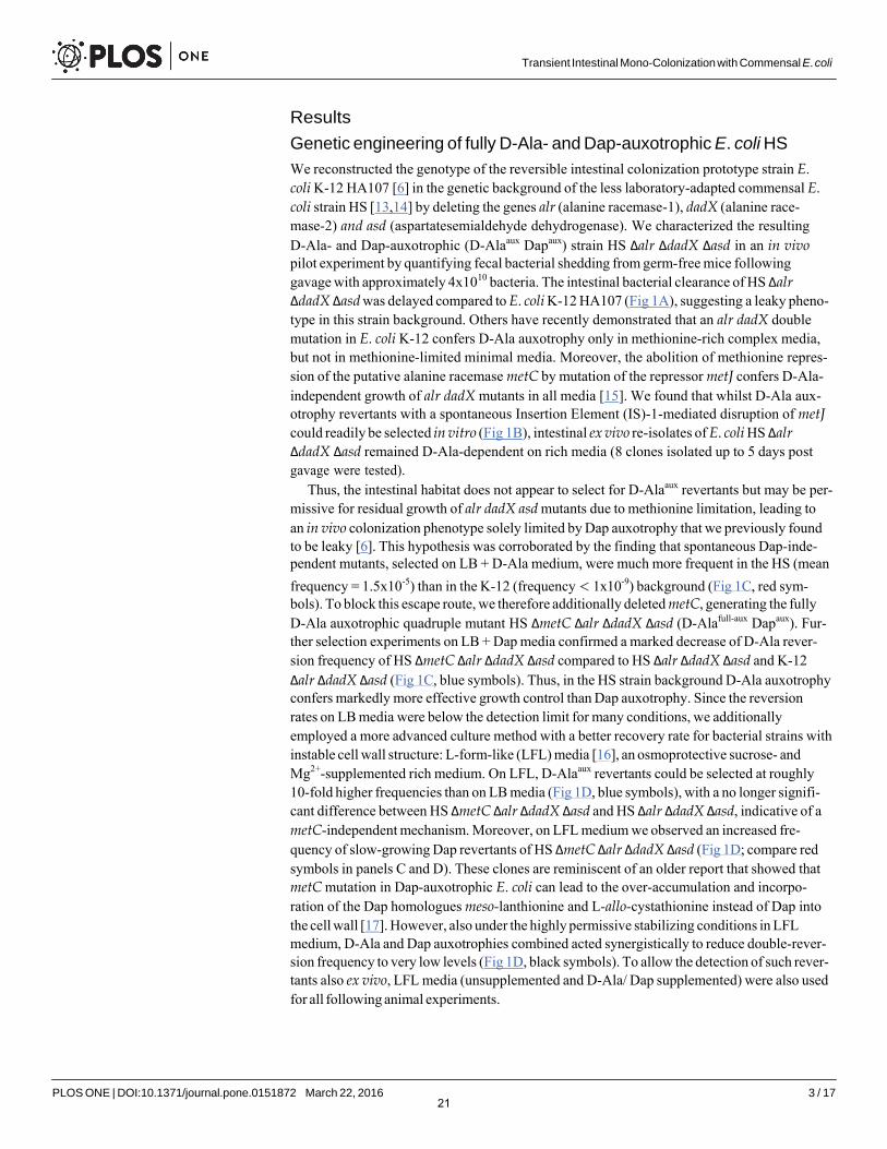
Genetic engineering of fully D-Ala- and Dap-auxotrophic E. coli HS
We reconstructed the genotype of the reversible intestinal colonization prototype strain E. coli K-12 HA107 [6 ] in the genetic background of the less laboratory-adapted commensal E. coli strain HS [13, 14 ] by deleting the genes alr (alanine racemase-1), dadX (alanine race- mase-2) and asd (aspartatesemialdehyde dehydrogenase). We characterized the resulting D-Ala- and Dap-auxotrophic (D-Alaaux Dapaux) strain HS Δalr ΔdadX Δasd in an in vivo pilot experiment by quantifying fecal bacterial shedding from germ-free mice following gavage with approximately 4x10¹⁰ bacteria. The intestinal bacterial clearance of HS ΔalrΔdadX Δasd was delayed compared to E. coli K-12 HA107 (Fig 1 A ), suggesting a leaky pheno- type in this strain background. Others have recently demonstrated that an alr dadX double mutation in E. coli K-12 confers D-Ala auxotrophy only in methionine-rich complex media, but not in methionine-limited minimal media. Moreover, the abolition of methionine repres- sion of the putative alanine racemase metC by mutation of the repressor metJ confers D-Ala- independent growth of alr dadX mutants in all media [15]. We found that whilst D-Ala aux- otrophy revertants with a spontaneous Insertion Element (IS)-1-mediated disruption of metJcould readily be selected in vitro (Fig 1 B ), intestinal ex vivo re-isolates of E. coli HS ΔalrΔdadX Δasd remained D-Ala-dependent on rich media (8 clones isolated up to 5 days post gavage were tested).
Thus, the intestinal habitat does not appear to select for D-Alaaux revertants but may be per- missive for residual growth of alr dadX asd mutants due to methionine limitation, leading to an in vivo colonization phenotype solely limited by Dap auxotrophy that we previously found to be leaky [6]. This hypothesis was corroborated by the finding that spontaneous Dap-inde- pendent mutants, selected on LB + D-Ala medium, were much more frequent in the HS (mean frequency = 1.5x10⁻⁵) than in the K-12 (frequency < 1x10⁻⁹) background (Fig 1C, red sym- bols). To block this escape route, we therefore additionally deleted metC, generating the fully D-Ala auxotrophic quadruple mutant HS ΔmetC Δalr ΔdadX Δasd (D-Alafull-aux Dapaux). Fur- ther selection experiments on LB + Dap media confirmed a marked decrease of D-Ala rever- sion frequency of HS ΔmetC Δalr ΔdadX Δasd compared to HS Δalr ΔdadX Δasd and K-12 Δalr ΔdadX Δasd (Fig 1C, blue symbols). Thus, in the HS strain background D-Ala auxotrophy confers markedly more effective growth control than Dap auxotrophy. Since the reversion rates on LB media were below the detection limit for many conditions, we additionally employed a more advanced culture method with a better recovery rate for bacterial strains with instable cell wall structure: L-form-like (LFL) media [16], an osmoprotective sucrose- and Mg2+-supplemented rich medium. On LFL, D-Alaaux revertants could be selected at roughly 10-fold higher frequencies than on LB media (Fig 1D, blue symbols), with a no longer signifi- cant difference between HS ΔmetC Δalr ΔdadX Δasd and HS Δalr ΔdadX Δasd, indicative of a metC-independent mechanism. Moreover, on LFL medium we observed an increased fre- quency of slow-growing Dap revertants of HS ΔmetC Δalr ΔdadX Δasd (Fig 1D; compare red symbols in panels C and D). These clones are reminiscent of an older report that showed that metC mutation in Dap-auxotrophic E. coli can lead to the over-accumulation and incorpo- ration of the Dap homologues meso-lanthionine and L-allo-cystathionine instead of Dap into the cell wall [17]. However, also under the highly permissive stabilizing conditions in LFL medium, D-Ala and Dap auxotrophies combined acted synergistically to reduce double-rever- sion frequency to very low levels (Fig 1D , black symbols). To allow the detection of such rever- tants also ex vivo, LFL media (unsupplemented and D-Ala/ Dap supplemented) were also used for all following animal experiments.
21
PLOS ONE | DOI:10.1371/journal.pone.0151872 March 22, 2016 4 / 17
Transient Intestinal Mono-Colonization with Commensal E. coli
Fig 1. Phenotypic characterization of auxotrophic E. coli HS mutants. (A) Fecal bacterial loads from
mice that had been gavaged with 1010 CFU of E. coli HS ΔmetC Δalr ΔdadX Δasd (filled blue triangles or E.coli K-12 Δalr ΔdadX Δasd (strain HA107; filled red circles), each symbol represents one individual. (B)Insertion sequence (IS-)1 insertion observed in HS Δalr ΔdadX Δasd after in vitro selection for D-Alaauxotrophy reversion. PCR amplification of the genomic region of metJ from E. coli HS wild type (lane 1), E.
coli HS Δalr ΔdadX Δasd original stock (lane 2), and E. coli HS Δalr ΔdadX Δasd D-Ala+ revertant selected onLB + Dap (lane 3) reveals a mobile genetic element insertion in the metJ ORF that was identified by
22
PLOS ONE | DOI:10.1371/journal.pone.0151872 March 22, 2016 5 / 17
Transient Intestinal Mono-Colonization with Commensal E. coli
sequencing as IS-1. (C, D) Frequency of auxotrophy revertants in K-12 Δalr ΔdadX Δasd, HS Δalr ΔdadX
Δasd and HS ΔmetC Δalr ΔdadX Δasd selected on LB (C) or LFL (D) containing Dap (blue triangles, D-Alaaux
revertants), D-Ala (red circles, Dapaux revertants), or no supplements (black squares; D-Alaaux Dapaux
double-revertants. (E) Bacterial growth curves of E. coli HS ΔmetC Δalr ΔdadX Δasd (solid red line) and wildtype (dotted blue line) before and after removal of D-Ala and Dap from the media (arrow indicates time point of removal).
doi:10.1371/journal.pone.0151872.g001
Normal cell wall biochemistry of in-vitro grown auxotrophs
Despite its complete dependence on externally supplied D-Ala and Dap, HS ΔmetC Δalr ΔdadX Δasd has a normal growth rate (compared to its parental wild-type strain as control) in appropriately supplemented medium (Fig 1E). As confirmation of this conditional phenotype we carried out biochemical cell wall analyses to evaluate if auxotrophic HS ΔmetC Δalr ΔdadX Δasd was able to incorporate externally acquired D-Ala and Dap into a peptidoglycan of nor- mal composition. First, we confirmed the complete absence of endogenous D-Ala racemization activity in HS ΔmetC Δalr ΔdadX Δasd. Alanine racemase activity was quantified by measuring the production of D-Ala from L-Ala in bacterial crude extracts (see material and meth ods) by two different techniques: Marfey's (FDAA) derivatization and D-amino acid oxidase (DAAO) assays (Fig 2A and 2B). No residual alanine racemase activity was detectable in HS ΔmetC Δalr ΔdadX Δasd. Second, we compared the peptidoglycan structure between laboratory-grown (D-Ala and Dap supplemented) HS ΔmetC Δalr ΔdadX Δasd and wild type. The muropeptide profile obtained by UPLC analysis showed that the peptidoglycan structures of both strains were indistinguishable (Fig 2C). Thus, in-vitro grown HS ΔmetC Δalr ΔdadX Δasd, externally supplied with D-Ala and Dap, has a cell wall of normal composition.
Sustained bacterial activity under non-permissive conditions
We have previously shown that the initial gastrointestinal passage of D-Ala/Dap auxotrophs and wild-type E. coli is similar [6] showing that the majority of bacteria survive the intestinal passage but cannot sustain colonization without reproduction in vivo. D-Ala/Dap deficiency has a highly cell cycle-dependent phenotype. Whereas non-dividing cells are stable, dividing cells at initiation of binary fission undergo a programmed autolytic cell death, an active process that is linked to the cell wall rearrangements preceding binary fission [18]. Whilst autolysis itself is an activity-dependent cellular process, little is known about the impact of D-Ala/Dap- deficiency on bacterial activity prior to autolysis. We therefore used 2-photon microscopy to dynamically track the swimming velocity of D-Ala/Dap-deprived HS ΔmetC Δalr ΔdadX Δasd (which is flagellated and motile) over time as a proxy for bacterial energy status and functional integrity of the bacterial cell envelope (into which the flagellar rotor is embedded). Tracking growth of live HS ΔmetC Δalr ΔdadX Δasd that had been cultured in medium containing D-Ala/Dap as well as the metabolic peptidoglycan label hydroxycoumarin-carbonyl-amino-D- alanine (HADA; [19]) after transfer to D-Ala/Dap-supplemented and non-supplemented medium, respectively, we could confirm that bacteria in supplemented media were able to pro- liferate with intact cellular septum formation and division (Fig 3A, bottom panels; Fig 3C top). In sharp contrast non-supplemented bacteria at this stage began to display mid-lateral bulging with cytoplasm membrane protrusion due to a breach in the cell wall rigidity and the conse- quent loss of turgency (Fig 3A, top panels; Fig 3C bottom panels), later followed by lysis leaving behind empty peptidoglycan sacculi (Fig 3A, middle panels). To study bacterial activity prior to these processes, GFP-expressing bacteria were diluted into D-Ala/ Dap-deprived and non- deprived soft agar medium (to slow down swimming for more accurate velocity measurement) and tracked by time-lapse 2-photon microscopy over an observation period between 5 and
23
PLOS ONE | DOI:10.1371/journal.pone.0151872 March 22, 2016 6 / 17
Transient Intestinal Mono-Colonization with Commensal E. coli
Fig 2. Determination of Ala-racemase activity and peptidoglycan analysis. (A, B). D-Ala production by crude extracts of HS wild-type (black line/ bar) and HS ΔmetC Δalr ΔdadX Δasd (red line/ bar) determined by (A) Marfey's derivatization-HPLC analysis and (B) D-amino acid oxidase (DAAO) assay. Heat-inactivated crude extracts of HS wild type (grey line/ bar) and HS ΔmetC Δalr ΔdadX Δasd (orange line/ bar) served as negative controls. (C) UPLC peptidoglycan analysis of HS wild type grown in LFL medium with (blue line) and without (black line) supplementation with D-Ala and Dap and HS ΔmetC Δalr ΔdadX Δasd (red line) grown in supplemented LFL. LFL: L-form-like medium. Analysis was repeated 3 times; chromatograms from one representative experiment are shown.
doi:10.1371/journal.pone.0151872.g002
24
PLOS ONE | DOI:10.1371/journal.pone.0151872 March 22, 2016 7 / 17
Transient Intestinal Mono-Colonization with Commensal E. coli
Merge GFP Peptidoglycan
Sup
plem
ente
d N
on-s
uppl
emen
ted
Div
idin
g ba
cter
ium
B
acte
rial s
accu
lus
B
ulge
d ba
cter
ium
Mea
n ve
loci
ty (μ
m/s
)
A B p<0.0001
n.s. p<0.0001
7.5
5.0
2.5
0.0 LB+D-ala/Dap LB LB
Live Live Fixed
C Merge GFP Phase contrast D
Fig 3. Bacterial activity and survival under non-permissive conditions. (A) Frame shots of a bacterium displaying cellular bulging (top), an empty bacterial sacculus after autolysis (middle), and a dividing bacterium undergoing septum formation with intact cell wall formation (bottom). Green, cytoplasmic eGFP; blue, HADA- labelled cell wall. (B-D) eGFP-expressing (green) bacteria grown in LB medium containing D-Ala and Dap were diluted in soft agar medium containing no supplements (LB, Live), D-Ala and Dap (LB + D-Ala/Dap, Live), or fixed with 4% para-formaldehyde (LB, Fixed) on a microscopy slide. Time-lapse videos were recorded using a 2-photon microscope and quantified with Volocity software. (B) Mean velocities of individual HS ΔmetC Δalr ΔdadX Δasd under the three indicated conditions. Statistical analysis: Kruskal-Wallis test withKruskalMC as post hoc. (C) Frame shots of confocal eGFP overlaid with phase contrast images of a D-Ala/ Dap-depleted bacterium displaying cellular bulging (bottom), and a D-Ala/Dap-supplemented control of normal morphology (top). (D) Frame shots of bacterium displaying cellular bulging. Top right time stamps indicate time after D-Ala/Dap depletion. (E) Track of the bacterium shown in panel C, before bulge formation (red path), after bulge formation (yellow path), after stopping and until lysis (white path).
doi:10.1371/journal.pone.0151872.g003
60 min after D-Ala/Dap depletion. We observed that HS ΔmetC Δalr ΔdadX Δasd maintained identical mean velocities in non-supplemented medium as in supplemented control medium (Fig 3B). Although in a small proportion (6%) of tracked cells the early stages of autolysis with mid-lateral outer-membrane bulge formation (as previously described for beta-lactam
5:36 6:28
5 μm 5 μm 5 μm 5 μm 5 μm
12:08 14:48
5 μm 5 μm
5 μm 5 μm 5 μm 14:52
E 5 μm
6:28
5 μm
Sup
plem
ente
d N
on-s
uppl
emen
ted
Bul
ged
bact
eriu
m
Div
idin
g ba
cter
ium
1μm 1μm
1μm 1μm 1μm
1μm 1μm 1μm
25
PLOS ONE | DOI:10.1371/journal.pone.0151872 March 22, 2016 8 / 17
Transient Intestinal Mono-Colonization with Commensal E. coli
antibiotic-induced autolysis in [18]; Fig 3D) could be observed, even bulge formation had no immediate impact on motility of the affected cells; bulged cells stopped swimming only approximately 3 min before cell death (sudden release of cytoplasmic GFP within <4 seconds; see example shown in S1 Video and Fig 3D and 3E), having little impact on mean velocity. These data collectively show that the activity and agility of D-Ala/ Dap-deprived HS ΔmetC Δalr ΔdadX Δasd remains largely unaffected until immediately before autolytic cell death, closely resembling beta-lactam antibiotic-induced cell death [18].
Transient intestinal mono-colonization
Next, we evaluated the intestinal colonization kinetics of the optimized, fully D-Ala auxotro- phic strain by gavaging germ-free mice with identical doses (4.3±1.0 x 10¹⁰ CFU; mean±SD in 200μL PBS) of the congenic mutants HS Δasd (D-Ala+ Dapaux), HS ΔmetC Δalr ΔdadX(D-Alafull-aux Dap+), HS Δalr ΔdadX Δasd (D-Alaaux Dapaux) and HS ΔmetC Δalr ΔdadX Δasd(D-Alafull-aux Dapaux), respectively (Fig 4). The oral-fecal passage and intestinal persistence of the 4 strains was compared over the course of 11 days by quantification of LFL-culturable bac- teria from fresh feces. Dapaux single-auxotroph HS Δasd showed prolonged bacterial shedding until at least day 11, indicative of residual in vivo proliferation (Fig 4A). In one cage of 4 mice inoculated with HS Δasd, spontaneous occurrence and transmission of a m-Dap auxotrophy revertant led to high-level colonization of all 4 affected individuals (Fig 4A and S1 Fig). Mice that were inoculated with either HS ΔmetC Δalr ΔdadX or HS Δalr ΔdadX Δasd returned to germ-free status within 3–6 days, but with highly variable and irregular kinetics (Fig 4B and 4C). In contrast, all animals inoculated with HS ΔmetC Δalr ΔdadX Δasd consistently returned to again-germ-free status within 3–4 days (Fig 4D). No double-revertants were recovered ex vivo on D-Ala/ Dap-free LFL medium. These data collectively show that the additional deletion of metC effectively prevented the occurrence of prolonged intestinal persistence and increased robustness of reversible colonization of germ-free animals.
The early gastrointestinal transit between 5 and 9 hours post inoculation was sampled in 1-hour intervals (S2 Fig), revealing that the peak fecal bacterial densities of HS ΔmetC Δalr ΔdadX Δasd remained within an order of magnitude as the density of the gastric inoculum (around 2x10¹⁰-2x10¹¹ CFU/g; S2 Fig), indicating that a large fraction of the inoculated bacte- ria survived the intestinal passage.
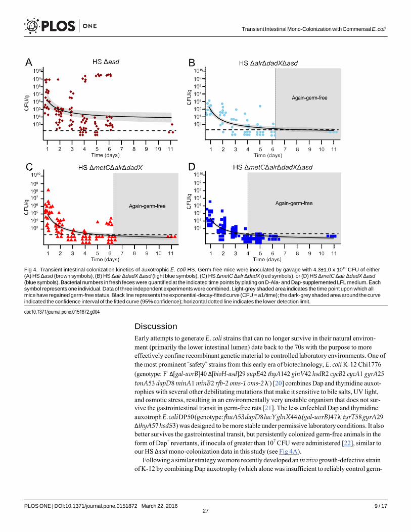
Intact IgA-stimulatory activity in vivo
Transiently colonizing D-Ala/ Dap-auxotrophic E. coli strains were originally developed to study the dynamics and dose-response relationship of commensal bacterial induction of intes- tinal immunoglobulin A (IgA) [6]. In these studies we showed that the bacterial induction of IgA strongly depended on a mucosal exposure to live E. coli, and killed bacteria were highly attenuated in their IgA stimulatory potential [6]. We therefore used the induction of live-E. coli HS-specific IgA as a sensitive readout for testing if additional mutation of metC negatively affected the IgA stimulatory activity. We compared the intestinal IgA immunogenicity of HS ΔmetC Δalr ΔdadX Δasd and its parental strain HS Δalr ΔdadX Δasd in vivo. The intestinal secretory IgA for this analysis was isolated from the germ-free mice presented in Fig 4B and 4D, 28 days after they had received equivalent doses of HS ΔmetC Δalr ΔdadX Δasd and HS Δalr ΔdadX Δasd, respectively. Quantification of the anti-E. coli HS IgA titers in a live bacterial flow cytometry assay (see Methods section and S3 Fig for details) revealed no decrease of IgA induction by metC mutation (Fig 5). Thus, the described genetic optimization in E. coli HS ΔmetC Δalr ΔdadX Δasd improved reversibility of intestinal colonization without compromis- ing its intestinal IgA stimulatory activity.
26
PLOS ONE | DOI:10.1371/journal.pone.0151872 March 22, 2016 9 / 17
Transient Intestinal Mono-Colonization with Commensal E. coli
Fig 4. Transient intestinal colonization kinetics of auxotrophic E. coli HS. Germ-free mice were inoculated by gavage with 4.3±1.0 x 10¹⁰ CFU of either(A) HS Δasd (brown symbols), (B) HS Δalr ΔdadX Δasd (light blue symbols), (C) HS ΔmetC Δalr ΔdadX (red symbols), or (D) HS ΔmetC Δalr ΔdadX Δasd
(blue symbols). Bacterial numbers in fresh feces were quantified at the indicated time points by plating on D-Ala- and Dap-supplemented LFL medium. Each symbol represents one individual. Data of three independent experiments were combined. Light-grey shaded area indicates the time point upon which all mice have regained germ-free status. Black line represents the exponential-decay-fitted curve (CFU = a1/time); the dark-grey shaded area around the curve indicated the confidence interval of the fitted curve (95% confidence); horizontal dotted line indicates the lower detection limit.
doi:10.1371/journal.pone.0151872.g004
Discussion
Early attempts to generate E. coli strains that can no longer survive in their natural environ- ment (primarily the lower intestinal lumen) date back to the 70s with the purpose to more effectively confine recombinant genetic material to controlled laboratory environments. One of the most prominent “safety” strains from this early era of biotechnology, E. coli K-12 Chi1776 (genotype: F- Δ[gal-uvrB]40 Δ[bioH-asd]29 supE42 thyA142 glnV42 hsdR2 cycB2 cycA1 gyrA25 tonA53 dapD8 minA1 minB2 rfb-2 oms-1 oms-2 λ-) [20] combines Dap and thymidine auxot- rophies with several other debilitating mutations that make it sensitive to bile salts, UV light, and osmotic stress, resulting in an environmentally very unstable organism that does not sur- vive the gastrointestinal transit in germ-free rats [21]. The less enfeebled Dap and thymidine auxotroph E. coli DP50 (genotype: fhuA53 dapD8 lacY glnX44 Δ(gal-uvrB)47 λ- tyrT58 gyrA29 ΔthyA57 hsdS3) was designed to be more stable under permissive laboratory conditions. It also better survives the gastrointestinal transit, but persistently colonized germ-free animals in the form of Dap+ revertants, if inocula of greater than 107 CFU were administered [22], similar to our HS Δasd mono-colonization data in this study (see Fig 4A).
Following a similar strategy we more recently developed an in vivo growth-defective strain of K-12 by combining Dap auxotrophy (which alone was insufficient to reliably control germ-
27
Transient Intestinal Mono-Colonization with Commensal E. coli
PLOS ONE | DOI:10.1371/journal.pone.0151872 March 22, 2016 10 / 17
Fig 5. IgA stimulatory activity of transiently colonizing E. coli HS ΔmetC Δalr ΔdadX Δasd. IgA- containing Intestinal lavages were prepared from the animals 28 days post inoculation with E. coli HS Δalr ΔdadX Δasd (red symbols) and HS Δalr ΔdadX Δasd ΔmetC (blue symbols), and germ-free control animals (black symbols). (A) IgA decoration of E. coli HS incubated with varying concentrations of intestinal secretory IgA. Geometric means of IgA-fluorescence intensities (IgA geoMFI) were plotted against IgA concentration in the assay, resulting in titration curves. (B) Anti-live-E. coli HS IgA antibody titers, expressed as -logEC50,
calculated by 4-parameter-curve-fitting of the data shown in (A). Dotted line, lower detection limit.
doi:10.1371/journal.pone.0151872.g005
free intestinal colonization) with the synergistically acting auxotrophy for D-Ala to generate a fully reversibly colonizing K-12 derivative strain HA107 that could be inoculated repeatedly in doses above 1010 CFU without permanent intestinal colonization [6]. Although HA107 does not escape from its in vivo cell wall biosynthesis deficiency, it survives the gastrointestinal tran- sit similarly well as its congenic non-auxotrophic parental strain, making it an effective tool for live bacterial conditioning of germ-free animals [6]. In the present report we further refined this approach by adapting it to the more resilient and less laboratory-adapted commensal strain E. coli HS, which required optimization of D-Ala auxotrophy.
Our data demonstrate that in E. coli HS and likely also other Enterobacteriaceae D-Ala is a more essential metabolite than Dap for peptidoglycan biosynthesis and growth. We continue to combine D-Ala auxotrophy with Dap auxotrophy as a “second hit” strategy, since it was still possible to select in vitro (but never in vivo) D-Alaaux revertants with ΔmetC Δalr ΔdadX geno- type that can grow (albeit poorly) without D-Ala supplementation by an unknown mechanism. Additional work will be required to identify (and prevent) this yet unknown escape pathway. An advantage of purely D-Ala auxotrophic strains would be the entirely cell-wall-specific phe- notype and the universal applicability of D-Ala auxotrophy since D-Ala, in contrast to Dap, is an essential metabolite of all known Eubacteria.
A main scientific application of this model is the bacterial conditioning and concomitant “normalization” of microbially shaped body functions (such as the immune system) in germ- free animals without permanent microbial colonization. Many bacterially modulated processes depend on live microbes. We therefore deliberately did not target enterobacterial colonization factors like bile acid resistance (LPS-O-antigen), adhesion (fimbriae etc.) factors or motility that may be important for productive microbe-host interaction in the intestinal mucosa. As a consequence transiently colonizing E. coli HS retains a high IgA immunogenicity and survives the intestinal transit. Also for applications using inactivated bacterial preparations or products in biological systems in vivo or in vitro the use of this model for production of such materials effectively avoids the contamination with surviving bacteria.
28
Transient Intestinal Mono-Colonization with Commensal E. coli
PLOS ONE | DOI:10.1371/journal.pone.0151872 March 22, 2016 11 / 17
In conclusion, we extended a robust transient mono-colonization model from a laboratory strain to a biologically more representative and more resilient intestinal commensal E. coli strain. This model can serve as a technology platform for numerous scientific applications. It may be used as a “sterile” biological vector for proteins, metabolites or signaling molecules that need to be delivered directly in situ or cannot be stably purified. More generally, it represents a live bacterial conditioning system for axenic animals or other sterile biological systems for the detailed study of host-microbial interactions. Many other future applications are thinkable, and the genetic approach may be extended to other microbial species.
Materials and Methods
Animal colonization experiments
Germ-free animals were re-derived from C57BL/6 mice and maintained germ-free in flexible film isolators in the Genaxen Foundation Clean Mouse Facility (CMF) of the University of Bern as described [23 ]. Experimental germ-free mice were aseptically transferred to auto- claved sealsafe-plus IVCs under positive pressure (Tecniplast, Italy) in a barrier unit of the Genaxen Clean Mouse Facility. Cage changes were carried out under strictly aseptic condi- tions. In all experiments animals were provided with sterile mouse chow (Kliba 3437; auto- claved) and autoclaved water ad libitum. All experiments were performed according to protocols approved by the Bernese Cantonal Ethical committee for animal experiments and carried out in accordance with Swiss Federal law for animal experimentation (license number BE91/14).
To generate contamination-free bacterial inoculums, D-Ala (200 μg/μ)- and Dap (50 μg/ mL)-supplemented autoclaved LB medium in sterile-filter-sealed flasks, was aseptically inocu- lated from single colonies of the test bacterium and incubated shaking at 150 rpm at 37°C for 16 hours. Bacteria were harvested by centrifugation (10 min, 4816 x g, 4°C) in a sterile aerosol- proof assembly, washed in autoclaved sterile PBS and concentrated to a density of 2 x 10¹¹
CFU/mL in sterile PBS, performed aseptically under a sterile laminar airflow. The bacterial sus- pensions were aseptically aliquoted in autoclaved plastic tubes and sealed in a sterilized second- ary containment. The sterile tubes containing the inocula and germ-free mice were aseptically imported into a sterilized laminar flow hood laid out with sterile surgical drapes, and each ani- mal inoculated with 200 μL of bacterial suspension (containing 4 x 10¹⁰ CFU in sterile PBS, at a density of 2 x 10¹¹ CFU/mL) by gavage, carried out wearing sterile surgical gowns and sterile surgical gloves. Fresh fecal pellets were collected aseptically, suspended in sterile PBS, and plated in serial dilutions on D-Ala/ Dap-supplemented or non-supplemented LFL agar and incubated aerobically at 37°C for 2: 24 hours.
Bacterial culture
LB medium (Sigma-Aldrich) was used as the standard growth media. Where required, the fol- lowing supplements were added to the media: ampicillin (Sigma, 100 μg/mL), tetracycline (Sigma, 12.5 μg/mL), kanamycin (Sigma, 50 μg/mL), meso-diaminopimelic acid (Sigma, 50 μg/mL), D-alanine (Sigma, 200 μg/mL). L-form-like media (LFL) was prepared in two parts: 75.2 g/L brain-heart infusion broth, 20 g/L agar; and separately 10 mM MgSO4, 200 g/L, sucrose, and mixed in equal parts after autoclaving. The frequencies of auxotrophy revertants were measured by plating stationary phase bacterial culture on LB or LFL agar plates contain- ing D-Ala+Dap, D-Ala only, Dap only, or no supplements and incubated at 37°C. Revertant frequencies are equivalent to the ratio revertant CFU/ total CFU.
29
Transient Intestinal Mono-Colonization with Commensal E. coli
PLOS ONE | DOI:10.1371/journal.pone.0151872 March 22, 2016 12 / 17
Bacterial genetic engineering
All bacterial strains used or generated in this study are specified in S1 Table . The E. coli HS wild type strain was kindly provided by Jim Nataro from the University of Virginia School of Medicine, Charlottesville VA, USA and is a replicate of the same bacterial stock that was fully sequenced by Rasko et al. [14] (GenBank accession no. CP000802). All deletions were carried out by Lambda Red recombineering. Mutagenesis primer sequences are specified in S2 Table . (i) Strain HA126 (Δasd::tetRA) was generated by deletion of asd using recombineering plasmid pKD46 as described [24] with minor modifications: a tetRA recombineering amplicon was amplified from genomic tetRA template DNA (isolated from a Tn10-containing bacterial strain) with primers HS-asd-mutF and HS-asd-mutR, and 2 mM L-arabinose added to express recombinase for 1 hour before the culture was stopped. (ii), Following the same protocol, alr in HA126 was deleted using recombinase plasmid pKD46 and a flp-kan-flp recombineering cas- sette amplified with primers HS-alr-mutF and HS-alr-mutR from template plasmid pKD4, fol- lowed by elimination of the kan resistance gene using FLP recombinase plasmid pCP20 as previously described [25], resulting in strain HA130 (Δalr::flp Δasd::tetRA); this procedure leaves behind one flp site). (iii), following the same procedure, dadX was deleted in HA 130, using primers HS-dadX-mut-F and HS-dadX-mutR, to generate strain HA132 (Δalr::flp ΔdadX::flp-kan-flp Δasd::tetRA). (iv), the kan resistance was removed from HA132 using pCP20, and metC was deleted using the heat-shock regulated recombinase expression plasmid pSIM6, primers HS-metC-mutF and HS-metC-mutR, and the flp-kan-flp template plasmid pKD4, following a recently published protocol [26], resulting in strain HA416 (ΔmetC::flp-kan- flp Δalr::flp ΔdadX::flp Δasd::tetRA). (v), To generate strain HA417 (ΔmetC::flp-kan-flp Δalr::flp ΔdadX::flp), the Δasd::tetRA allele in HA416 was replaced with the wild-type asd allele, using pSIM6 in combination with a recombineering amplicon produced with primers asd_F and asd_B and wild type genomic template DNA. HA417 was isolated by positive selection of recombinants on LB containing D-Ala. All deletions were verified phenotypically and by con- trol PCR (control primers specified in S2 Table ). Plasmids (see S1 Table for a complete list) were introduced by electroporation following standard protocols.
Insertion element identification
The genomic metJ region of E. coli HS was amplified by PCR using primers metJ_F and metJ_B (S2 Table ), and Sanger sequenced (Microsynth, TWON, Switzerland) using the same primers. The obtained sequences were compared using BLAST with the nr database and the best match was considered to be the correct.
Two- photon and confocal microscopy
A subculture of HS ΔmetC Δalr ΔdadX Δasd harboring eGFP expression plasmid pM979 was grown in LB containing D-Ala, Dap, ampicillin, 50 nM HADA, [27] for approximately 3 hours to reach OD600 = 0.6 and cooled down on ice. Small aliquots of this culture were sedimented by centrifugation and re-suspended 1:100 in fresh medium containing ampicillin, and either (i) no further chemicals, (ii) D-Ala and Dap, or (III) 4% para-formaldehyde (end concentration), and poured on a microscopy slide with matching media covered with low-melting-point 0.1% agarose. Time lapse videos were recorded immediately using an Olympus BX50WI fluores- cence 15 microscope (three z-stacks with 3 μm spacing, 150 μm square sections, fast mode) attached to a 2-PM scanner (TrimScope system, LaVision Biotec, Bielefeld, Germany) equipped with a 20X objective (numerical aperture = 0.95) and heated stage. Image sequences were transformed into volume-rendered four-dimensional movies using Volocity software (Improvision), which was also used for semi-automated tracking of bacteria motility. From the
30

Transient Intestinal Mono-Colonization with Commensal E. coli
PLOS ONE | DOI:10.1371/journal.pone.0151872 March 22, 2016 13 / 17
acquired videos 85 (supplemented) or 100 (non-supplemented and PFA) bacterial tracks were selected based on duration (2:10 consecutive time points, 3.3 frames/s), size (2:3 μm diameter) and the intensity of the objects (more than 1900 voxels). These tracks were imported into R and processed into plots using ggplot2 and dplyr.
For confocal microscopy, identically prepared bacterial preparations were imaged with a Zeiss LSM 710 confocal laser-scanning microscope, time-lapse movies of GFP and Phase con- trast (three z-stacks with 3 μm spacing, 150 μm square sections, fast mode channels) were recorded simultaneously using a beam splitter with a 40X oil objective (numerical aper- ture = 1.3) and heated stage. Image sequences were transformed using Imaris software.
Enzyme-linked immune sorbent assay (ELISA) for IgA
Total concentrations of IgA in mouse intestinal lavage were determined by sandwich ELISA. Coating antibodies were goat anti-mouse IgA (Southern Biotech, 1040–01); detection antibodies were horseradish peroxidase (HRP)–conjugated goat-anti-mouse IgA (A3673, Sigma). Purified monoclonal isotype control IgA (Becton Dickinson, clone M18-254, 553476) served as standard.
Live bacterial flow cytometry and IgA response quantification
Live bacterial flow cytometry quantification of E. coli HS-specific Immunoglobulin (Ig)A titers (expressed as—EC50 values) were carried out previously described [6]. Briefly, E. coli HS was grown in 0.2 μm membrane-filtered LB broth overnight at 37°C without shaking. 1 mL of cul- ture was gently pelleted for 3 min at 4816 x g in a Heraus Fresco 21 centrifuge and washed 3 times with sterile-filtered 2% BSA/ 0.005% NaN3/ PBS before re-suspending at a density of approximately 107 bacteria/mL. Intestinal IgA lavages were collected by rinsing the small intes- tine with 5 mL of 1% soybean-trypsin-inhibitor/ 0.05 M EDTA/ PBS. The intestinal washes were then centrifuged at 4816×g, 20 min; the supernatant sterile-filtered to remove bacteria- sized particles, and serially diluted in sterile-filtered 2% BSA/ 0.005% NaN3/ PBS. Serially diluted IgA-solution and bacterial suspension were mixed 1:1 and incubated at 4°C for 1 h. Bacteria were washed twice in sterile-filtered 2% BSA/ 0.005% NaN3/ PBS before re-suspending in monoclonal FITC-anti-mouse IgA (clone 10.3; Becton Dickinson). After 1 h incubation at 4°C the bacteria were washed twice with sterile-filtered 2% BSA/ 0.005% NaN3/ PBS and re- suspended in 2% para-formaldehyde/ PBS for acquisition on a FACSArray SORP flow cytome- ter (Becton Dickinson) using FSc and SSc parameters in logarithmic mode. GeoMean fluores- cence intensities were plotted against IgA concentrations analyzed using FlowJo software (Treestar, USA) and Graphpad prism software 4-parameter curve fitting to calculate—EC50 titers as previously described [6] and summarized in S3 Fig.
Isolation of peptidoglycan and UPLC analysis
To isolate murein sacculi, cultures were pelleted, resuspended in 2 mL of medium and slowly added to 2 mL of boiling 10% SDS while stirring. After boiling for 2 h, they were stirred over- night at room temperature. Cell wall material was then pelleted by ultracentrifugation (60’000 rpm, 10 min) and washed with purified water to remove SDS. Samples were digested with pro- nase E (100 μg/mL) in 10 mM Tris-HCl, pH 7.5, 1 h at 60°C to remove Braun’s lipoprotein. After addition of SDS to a final concentration of 1% (w/v), reactions were heat-inactivated and detergent was removed by washing in MQ water. Purified peptidoglycan was re-suspended in 100 μL of 50 mM NaPO4 buffer pH 4.9 and treated with 100 μg/mL muramidase (Cellosyl) for 16 h at 37°C. Muramidase digestion was stopped by boiling and coagulated proteins were removed by 10 min centrifugation at 14’000 rpm. Supernatants were reduced by adding sodium borate pH 9.5 and sodium borohydride to a final concentration of 10 mg/mL and
31
Transient Intestinal Mono-Colonization with Commensal E. coli
PLOS ONE | DOI:10.1371/journal.pone.0151872 March 22, 2016 14 / 17
incubating at RT for 30 min. Finally, samples were adjusted to pH 3.5 with phosphoric acid. Muropeptides were separated in a 20-min linear gradient of 50 mM NaPO4, pH 4.35, to 50 mM NaPO4, pH 4.95, and 15% (v/v) methanol on an AQUITY ultra-performance liquid chro- matography (UPLC) BEHC18 column (130 Å, 1.7 μm, 2.1 mm × 150 mm; Waters, USA), and detected by absorption at wavelength 204 nm.
Alanine racemase activity assay
Cleared cell lysates were prepared from cultures and assayed for alanine racemase activity. Lysates were prepared from cultures grown in LB supplemented with D-Ala and Dap (OD600 was measured for normalizing the number of cells in each sample). Cells were collected by cen- trifugation and then washed twice with ice-cold HEPES 50 mM pH 7.5. By three passages through a French press re-suspended cells were disrupted and then lysates were cleared of cell debris and membranes by centrifugation at 20’000 rpm for 30 min at 4°C. Alanine racemiza- tion assays (adapted from [28,29]) were performed in 1.5 mL Eppendorf tubes in a total volume of 200 μL by adding 160 μL of the soluble fraction of crude extract, L-Ala and pyridoxal phos- phate (PLP) at 50 mM and 20 μM respectively (final concentrations). After incubation at 37°C for 45 min the reaction was quenched by adding 40 μl of 2 M HCl.
L-FDAA (1-fluoro-2-4-dinitrophenyl-5-L-alanine amide, Marfey’s reagent, Thermo Scien- tific) was used for the derivatization of amino acids [28,30]. First quenched enzyme reactions were neutralized with 40 μL of 2 M NaOH and then a 50 μL aliquot of the sample was mixed with 100 μL Marfey's reagent (0.5% solution in acetone) and 20 μL 1 M NaHCO3. The derivati- zation mixture was incubated at 80°C for 10 min and the reaction was stopped with 2 M HCl. After cooling down to room temperature samples were diluted with 200 μL of a mixture 9:1 of buffers A (triethylamine-phosphate 50 mM pH 3) and B (triethylamine-phosphate 50 mM pH3, 40% acetonitrile). Samples were filtered and 100 μL were injected in the HPLC (high- pressure liquid chromatography). Amino acids were separated in a 45 min linear gradient of triethylamine phosphate/acetonitrile with an Aeris peptide column (250 x 4.6 mm; 3.6 μm par- ticle size, Phenomenex, USA) and detected at Abs. 340 nm. D-Ala and L-Ala were used as stan- dards of to establish retention times.
DAAO reaction coupled to peroxidase and 2,3-diaminophenazine was performed for detec- tion of the D-Ala produced by the in vitro as previously described [31]. The formation of the colorimetric product was measured at 492 nm.
Data analysis and statistical analysis
All data analysis was done using the R 3.1.1 statistical program [32] and the ggplot2 [33], dplyr [34] and pgirmess [35] packages. All scripts can be downloaded from https://github.com/ cuencam/HA41 6, and data are available on request. Statistical tests are specified in the figure legends.
Supporting Information
S1 Fig. Selection of spontaneous asd auxotrophy revertants in E. coli HS Δasd-inoculated ani- mals. 4 Germ-free mice (also depicted in Fig 4A) were inoculated by gavage with approximately 4x10¹⁰ CFU of HS Δasd. (A) CFU counts from each mouse over time, each individual highlighted in a different color. (B) PCR amplification of the genomic asd region of HS wild-type (lane 1), HS Δasd (exact genotype: Δasd::tetRA; longer PCR fragment verifies allelic exchange of asd by tetRA cassette) original stock (lane 2), and HS Δasd re-isolate from mouse 3 (verifying the correct geno- type of this revertant), verifying colonization with a revertant clone of the correct inoculated. Lane L contains molecular ladder. (C, D) Colony morphology of 4 revertant clones re-isolated
32

Transient Intestinal Mono-Colonization with Commensal E. coli
PLOS ONE | DOI:10.1371/journal.pone.0151872 March 22, 2016 15 / 17
from mouse 3 on day 2 (clone 1) and day 3 (clone 2), mouse 1 on day 3 (clone 3), and mouse 2 on day 3 (clone 4) on supplemented (C) and unsupplemented (D) LFL agar plates. (PDF)
S2 Fig. Early intestinal colonization kinetics of auxotrophic E. coli HS. Early time points of the experiment presented in main Fig 4 are shown. Germ-free mice were inoculated by gavage with around 4x10¹⁰ CFU of either (A) HS Δasd (brown symbols), (B) HS Δalr ΔdadX Δasd (light blue symbols), (C) HS ΔmetC Δalr ΔdadX (red symbols), or (D) HS ΔmetC Δalr ΔdadX Δasd (blue symbols). Each symbol represents one individual; data are combined from three independent experiments. Black line represents the exponential-decay-fitted curve (CFU = a1/ time) with the 95% confidence interval shown as dark-grey shaded area. The vertical gray line marks the time point at which all individuals have reached fecal bacterial densities 100-fold below the mean inoculum density (from top of light gray area). (PDF)
S3 Fig. Live bacterial FACS analysis and titer calculations. IgA-stained bacteria were analyzed using a BD FACSArray SORP and acquired data were exported to Treestar FlowJo. (A) Gating procedure: Single bacteria were defined as forward-scatter-width-(FSC-W)-low events. Forward scatter area (FSC-A) and Side scatter area (SSC-A) were used to eliminate electrical noise, bub- bles and debris from the analysis. Gating Red (APC channel)-low events allowed to reduce unspecific fluorescence. Three serial 3-fold dilutions of a representative positive sample are shown. (B) Three representative histograms of FITC-anti-IgA resulting from 3 serial dilutions and their overlay are shown. (C) Titration curves shown in main Fig 5A. Geometric mean fluo- rescent intensities (geoMFI; accounting for the Log Normal distribution of fluorescence data) of IgA bacterial FACS staining (y-axis) was plotted against IgA concentration in the assay (x-axis) (determined by isotype-specific sandwich ELISA). (D) 4-parameter curve fitting of the data shown panel C and main Fig 5A. Graphpad Prism 6 software was used to fit 4-parameter logistic curves to the data. Equation: Y = Bottom + (Top- Bottom)/ (1+10^((LogEC50-X)*HillSlope)). (E)–LogEC50 IgA titers. The LogEC50 values were extracted from the curve parameters, which when anti-logged corresponds to the concentration of IgA required to give half-maximum IgA binding. The—LogEC50 titer thus corresponds to the Log(1/[IgA]giving 50% binding) the dotted line to the lower detection limit. (PDF)
S1 Table. Bacterial strains and plasmids. (DOCX)
S2 Table. Primers used in this study. (DOCX)
S1 Video. Representative example of a swimming bacterium undergoing bulging and autol- ysis. The red circle highlights the bacterium of interest; white arrow indicates the moment at which the cells starts bulging. After approximately 7 min the bacterium stops active movement, followed 3 min later by autolysis, causing an instant drop of cytoplasmatic GFP signal as it is released into the extracellular medium. The video is part of the data shown in Fig 4. (MOV)
Acknowledgments
We thank all laboratory members, Emma Slack and Maria Balmer for reading and commenting on the manuscript. We thank the Clean Mouse Facility of the Department of Clinical Research,
33
Transient Intestinal Mono-Colonization with Commensal E. coli
PLOS ONE | DOI:10.1371/journal.pone.0151872 March 22, 2016 16 / 17
Kathy McCoy, and all members of the CMF Curatorium of the University of Bern for support of the gnotobiotic animal work. We thank Jens Stein and Britta Engelhardt from the Theodor Kocher Institute for the support with the two-photon microscope and Fabian Blank, the MIC and the LCI Core Facility for the support with the LSM 710 Confocal microscope at University of Bern.
Author Contributions
Conceived and designed the experiments: SH MC SPP EK MSV YVB FC FMC. Performed the experiments: MC SPP SB FB SBH FMC. Analyzed the data: SH MC SPP SB SBH FC FMC. Contributed reagents/materials/analysis tools: MSV YVB. Wrote the paper: MC SH.
References 1. Smith K, McCoy KD, Macpherson AJ. Use of axenic animals in studying the adaptation of mammals to
their commensal intestinal microbiota. Semin Immunol. 2007; 19: 59–69. doi: 10.1016/j.smim.2006.10. 002 PMID: 17118672
2. Macpherson AJ, McCoy KD. Standardised animal models of host microbial mutualism. Mucosal Immu-nol. 2014; 8: 476–486. doi: 10.1038/mi.2014.113 PMID: 25492472
3. Faith JJ, Ahern PP, Ridaura VK, Cheng J, Gordon JI. Identifying gut microbe-host phenotype relation-ships using combinatorial communities in gnotobiotic mice. Sci Transl Med. 2014; 6: 220ra11. doi: 10. 1126/scitranslmed.3008051 PMID: 24452263
4. Reikvam DH, Erofeev A, Sandvik A, Grcic V, Jahnsen FL, Gaustad P, et al. Depletion of murine intesti-nal microbiota: effects on gut mucosa and epithelial gene expression. PLoS ONE. 2011; 6: e17996.doi: 10.1371/journal.pone.0017996 PMID: 21445311
5. Dethlefsen L, Relman DA. Incomplete recovery and individualized responses of the human distal gutmicrobiota to repeated antibiotic perturbation. Proc Natl Acad Sci USA. 2011; 108 Suppl 1: 4554–4561.doi: 10.1073/pnas.1000087107 PMID: 20847294
6. Hapfelmeier S, Lawson MAE, Slack E, Kirundi JK, Stoel M, Heikenwalder M, et al. Reversible microbialcolonization of germ-free mice reveals the dynamics of IgA immune responses. Science. 2010; 328:1705–1709. doi: 10.1126/science.1188454 PMID: 20576892
7. Fritz JH, Rojas OL, Simard N, McCarthy DD, Hapfelmeier S, Rubino S, et al. Acquisition of a multifunc-tional IgA+ plasma cell phenotype in the gut. Nature. 2012; 481: 199–203. doi: 10.1038/nature10698
8. Balmer ML, Slack E, de Gottardi A, Lawson MAE, Hapfelmeier S, Miele L, et al. The liver may act as afirewall mediating mutualism between the host and its gut commensal microbiota. Sci Transl Med.2014; 6: 237ra66–237ra66. doi: 10.1126/scitranslmed.3008618 PMID: 24848256
9. Balmer ML, Schürch CM, Saito Y, Geuking MB, Li H, Cuenca M, et al. Microbiota-derived compoundsdrive steady-state granulopoiesis via MyD88/TICAM signaling. J Immunol. 2014; 193: 5273–5283. doi: 10.4049/jimmunol.1400762 PMID: 25305320
10. Rautava S, Luoto R, Salminen S, Isolauri E. Microbial contact during pregnancy, intestinal colonizationand human disease. Nat Rev Gastroenterol Hepatol. 2012; 9: 565–576. doi: 10.1038/nrgastro.2012. 144 PMID: 22890113
11. Schultz M. Clinical use of E. coli Nissle 1917 in inflammatory bowel disease. Inflamm Bowel Dis. 2008;14: 1012–1018. PMID: 18240278
12. Browning DF, Wells TJ, França FLS, Morris FC, Sevastsyanovich YR, Bryant JA, et al. Laboratoryadapted Escherichia coli K-12 becomes a pathogen of Caenorhabditis elegans upon restoration of Oantigen biosynthesis. Mol Microbiol. 2013; 87: 939–950. doi: 10.1111/mmi.12144 PMID: 23350972
13. Levine MM, Bergquist EJ, Nalin DR, Waterman DH, Hornick RB, Young CR, et al. Escherichia colistrains that cause diarrhoea but do not produce heat-labile or heat-stable enterotoxins and are non-invasive. Lancet. 1978; 1: 1119–1122. PMID: 77415
14. Rasko DA, Rosovitz MJ, Myers GSA, Mongodin EF, Fricke WF, Gajer P, et al. The pangenome struc-ture of Escherichia coli: comparative genomic analysis of E. coli commensal and pathogenic isolates. JBacteriol. 2008; 190: 6881–6893. doi: 10.1128/JB.00619-08 PMID: 18676672
15. Kang L, Shaw AC, Xu D, Xia W, Zhang J, Deng J, et al. Upregulation of MetC is essential for D-alanine-independent growth of an alr/dadX-deficient Escherichia coli strain. J Bacteriol. 2011; 193: 1098–1106.doi: 10.1128/JB.01027-10 PMID: 21193606
16. Glover WA, Yang Y, Zhang Y. Insights into the molecular basis of L-form formation and survival inEscherichia coli. PLoS ONE. 2009; 4: e7316. doi: 10.1371/journal.pone.0007316 PMID: 19806199
34
Transient Intestinal Mono-Colonization with Commensal E. coli
PLOS ONE | DOI:10.1371/journal.pone.0151872 March 22, 2016 17 / 17
17. Richaud C, Mengin-Lecreulx D, Pochet S, Johnson EJ, Cohen GN, Marlière P. Directed evolution ofbiosynthetic pathways. Recruitment of cysteine thioethers for constructing the cell wall of Escherichiacoli. J Biol Chem. 1993; 268: 26827–26835. PMID: 8262915
18. Yao Z, Kahne D, Kishony R. Distinct single-cell morphological dynamics under beta-lactam antibiotics.Mol Cell. 2012; 48: 705–712. doi: 10.1016/j.molcel.2012.09.016 PMID: 23103254
19. Kuru E, Tekkam S, Hall E, Brun YV, Van Nieuwenhze MS. Synthesis of fluorescent D-amino acids andtheir use for probing peptidoglycan synthesis and bacterial growth in situ. Nat Protoc. 2015; 10: 33–52.doi: 10.1038/nprot.2014.197 PMID: 25474031
20. Curtiss R 3rd. Modified microorganisms and method of preparing and using same. US Patent 1980;US4190495 A.
21. Wells CL, Johnson WJ, Kan CM, Balish E. Inability of debilitated Escherichia coli chi 1776 to colonisegerm-free rodents. Nature. 1978; 274: 397–398. PMID: 353563
22. Ducluzeau R, Ladiré M, Raibaud P. [Implantation of a mutant of Escherichia coli requiring diaminopime-lic acid in the digestive tract of gnotobiotic mice]. Ann Inst Pasteur Microbiol. 1986; 137A: 79–87. PMID: 3314677
23. Macpherson AJ, Geuking MB, Kirundi J, Collins S, McCoy KD. Gnotiobiotic and Axenic Animals. Ency-clopedia of Microbiology. Elsevier; 2009. pp. 237–246. doi: 10.1016/B978-012373944-5.00215-7
24. Datsenko KA, Wanner BL. One-step inactivation of chromosomal genes in Escherichia coli K-12 usingPCR products. Proc Natl Acad Sci USA. 2000; 97: 6640–6645. doi: 10.1073/pnas.120163297 PMID: 10829079
25. Cherepanov PP, Wackernagel W. Gene disruption in Escherichia coli: TcR and KmR cassettes with theoption of Flp-catalyzed excision of the antibiotic-resistance determinant. Gene. 1995; 158: 9–14. PMID: 7789817
26. Thomason L, Court DL, Bubunenko M, Costantino N, Wilson H, Datta S, et al. Recombineering: geneticengineering in bacteria using homologous recombination. Current protocols in molecular biology /edited by Ausubel FM [et al]. 2007;Chapter 1: Unit 1.16. doi: 10.1002/0471142727.mb0116s78
27. Kuru E, Hughes HV, Brown PJ, Hall E, Tekkam S, Cava F, et al. In Situ probing of newly synthesizedpeptidoglycan in live bacteria with fluorescent D-amino acids. Angew Chem Int Ed Engl. 2012; 51:12519–12523. doi: 10.1002/anie.201206749 PMID: 23055266
28. Radkov AD, Moe LA. Amino acid racemization in Pseudomonas putida KT2440. J Bacteriol. AmericanSociety for Microbiology; 2013; 195: 5016–5024. doi: 10.1128/JB.00761-13
29. Arias CA, Weisner J, Blackburn JM, Reynolds PE. Serine and alanine racemase activities of VanT: aprotein necessary for vancomycin resistance in Enterococcus gallinarum BM4174. Microbiology (Read-ing, Engl). 2000; 146 (Pt 7): 1727–1734.
30. Marfey P. Determination of D-amino acids. II. Use of a bifunctional reagent, 1,5-difluoro-2,4-dinitroben-zene. Carlsberg Research Communications. Springer-Verlag; 1984; 49: 591–596. doi: 10.1007/ BF02908688
31. Espaillat A, Carrasco-López C, Bernardo-García N, Pietrosemoli N, Otero LH, Álvarez L, et al. Struc-tural basis for the broad specificity of a new family of amino-acid racemases. Acta Crystallogr D BiolCrystallogr. 2014; 70: 79–90. doi: 10.1107/S1399004713024838 PMID: 24419381
32. R Development Core Team. R: A Language and Environment for Statistical Computing. R Foundationfor Statistical Computing 2014. http://www.R-project.org/. Accessed 01 Aug 2015.
33. Wickham H. ggplot2: Elegant Graphics for Data Analysis. Springer New York; 2009. Available: http:// had.co.nz/ggplot2/book.
34. Wickham H, Francois R. dplyr: A Grammar of Data Manipulation. In: R package version 0.4.1. 2015.Available: http://CRAN.R-project.org/package=dplyr. Accessed 17 Jul 2015.
35. Giraudoux P. pgirmess: Data Analysis in Ecology. In: R package version 1.6.2. 2015. Available: http:// CRAN.R-project.org/package=pgirmess. Accessed 17 Jul 2015.
35
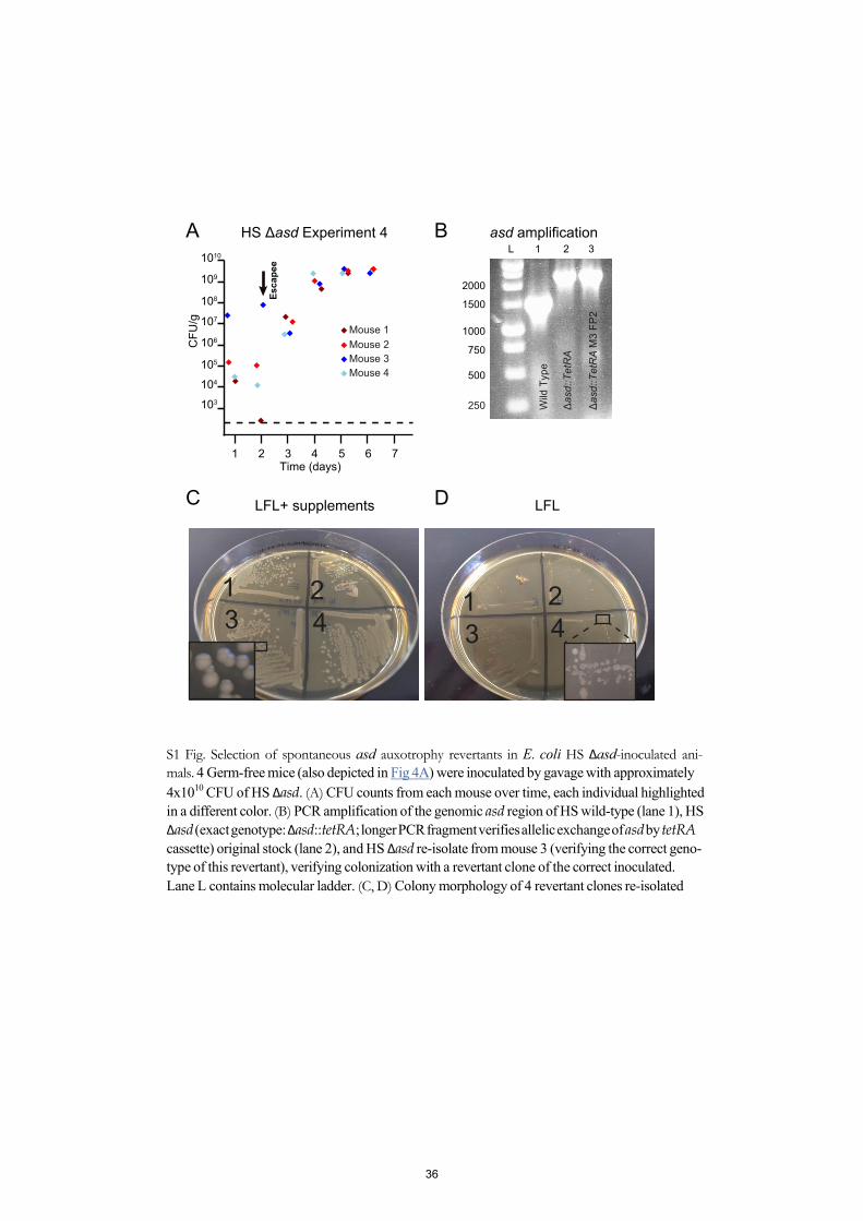
103
104
105
106
107
108
109
1010
Time (days)1 2 3 4 5 6 7
HS Δasd Experiment 4
CFU
/g
250
500
750
1000
1500
2000
L 1 2 3
Δas
d::T
etR
A
Wild
Typ
e
Δas
d::T
etR
A M
3 FP
2
Mouse 1Mouse 2Mouse 3Mouse 4
A
C
B
D
1 2432 1
3
asd amplification
Esca
pee
4
LFL+ supplements LFL
S1 Fig. Selection of spontaneous asd auxotrophy revertants in E. coli HS Δasd-inoculated ani- mals. 4 Germ-free mice (also depicted in Fig 4A) were inoculated by gavage with approximately 4x10¹⁰ CFU of HS Δasd. (A) CFU counts from each mouse over time, each individual highlightedin a different color. (B) PCR amplification of the genomic asd region of HS wild-type (lane 1), HS Δasd (exact genotype: Δasd::tetRA; longer PCR fragment verifies allelic exchange of asd by tetRA cassette) original stock (lane 2), and HS Δasd re-isolate from mouse 3 (verifying the correct geno- type of this revertant), verifying colonization with a revertant clone of the correct inoculated. Lane L contains molecular ladder. (C, D) Colony morphology of 4 revertant clones re-isolated
36
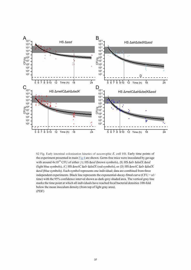
Time (h)5 6 7 8 9 10 12 18 24
103
104
105
106
107
108
109
1010
1011
1012
1013 HS ΔasdA
CF
U/g
D
5 6 7 8 9 10 12 18 24
103
104
105
106
107
108
109
1010
1011
1012
1013
HS ΔalrΔdadXΔasdB
CF
U/g
Time (h)
CF
U/g
103
104
105
106
107
108
109
1010
1011
1012
1013
5 6 7 8 9 10 12 18 24
HS ΔmetCΔalrΔdadXΔasdD
Time (h)
103
104
105
106
107
108
109
1010
1011
1012
1013
5 6 7 8 9 10 12 18 24
HS ΔmetCΔalrΔdadXC
CF
U/g
Time (h)
S2 Fig. Early intestinal colonization kinetics of auxotrophic E. coli HS. Early time points of the experiment presented in main Fig 4 are shown. Germ-free mice were inoculated by gavage with around 4x10¹⁰ CFU of either (A) HS Δasd (brown symbols), (B) HS Δalr ΔdadX Δasd (light blue symbols), (C) HS ΔmetC Δalr ΔdadX (red symbols), or (D) HS ΔmetC Δalr ΔdadX Δasd (blue symbols). Each symbol represents one individual; data are combined from three independent experiments. Black line represents the exponential-decay-fitted curve (CFU = a1/ time) with the 95% confidence interval shown as dark-grey shaded area. The vertical gray line marks the time point at which all individuals have reached fecal bacterial densities 100-fold below the mean inoculum density (from top of light gray area). (PDF)
37
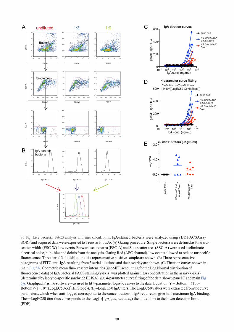
germ-free
HS ΔmetC Δalr ΔdadX ΔasdHS Δalr ΔdadX Δasd
SSC
-A
100 101 102 103 104100
101
102
103
104
63.6
100 101 102 103 104100
101
102
103
104
99.2
100 101 102 103 1040
50
100
150
200
100 101 102 103 104100
101
102
103
104
66.6
0 200 400 600 800 1000
100
101
102
103
104
57.2
100 101 102 103 104100
101
102
103
104
99.5
100 101 102 103 1040
50
100
150
200
0 200 400 600 800 1000
100
101
102
103
104
62.3
FSC-A
100 101 102 103 104100
101
102
103
104
81.9
0 200 400 600 800 1000
100
101
102
103
104
51.1
100 101 102 103 104100
101
102
103
104
99.5
100 101 102 103 1040
100
200
300
FSC-AFSC-A
FSC
-AR
ed-A
# C
ells
FSC-W FSC-WFSC-W
Yellow-A Yellow-AYellow-A
IgA - FITC IgA - FITCIgA - FITC
undiluted 1:3 1:9
100 101 102 103 1040
20
40
60
80
100
% C
ells
IgA - FITC
IgA titration curves
germ-free
A
Bacteria
Single cells
IgA-coatedbacteria
C
10-1 100 101 102 103 1040
200
400
600
IgA conc. (ng/mL)
geo
MFI
IgA
-FIT
C-lo
gEC
50
-5.0
-4.0
-3.0E. coli HS titers (-logEC50)
germ
-free
E
10-1 100 101 102 103 1040
200
400
600
4-parameter curve fittingD Y=Bottom + (Top-Bottom)/(1+10^((LogEC50-X)*HillSlope))
IgA conc. (ng/mL)
geo
MFI
IgA
-FIT
CB
HS ΔmetC Δalr ΔdadX ΔasdHS Δalr ΔdadX Δasd
HS
Δm
etC
Δal
r Δ
dadX
Δas
d
HS
Δal
r Δda
dX
Δas
d
S3 Fig. Live bacterial FACS analysis and titer calculations. IgA-stained bacteria were analyzed using a BD FACSArray SORP and acquired data were exported to Treestar FlowJo. (A) Gating procedure: Single bacteria were defined as forward-scatter-width-(FSC-W)-low events. Forward scatter area (FSC-A) and Side scatter area (SSC-A) were used to eliminate electrical noise, bub- bles and debris from the analysis. Gating Red (APC channel)-low events allowed to reduce unspecific fluorescence. Three serial 3-fold dilutions of a representative positive sample are shown. (B) Three representative histograms of FITC-anti-IgA resulting from 3 serial dilutions and their overlay are shown. (C) Titration curves shown in main Fig 5A. Geometric mean fluo- rescent intensities (geoMFI; accounting for the Log Normal distribution of fluorescence data) of IgA bacterial FACS staining (y-axis) was plotted against IgA concentration in the assay (x-axis) (determined by isotype-specific sandwich ELISA). (D) 4-parameter curve fitting of the data shown panel C and main Fig 5A. Graphpad Prism 6 software was used to fit 4-parameter logistic curves to the data. Equation: Y = Bottom + (Top- Bottom)/ (1+10^((LogEC50-X)*HillSlope)). (E)–LogEC50 IgA titers. The LogEC50 values were extracted from the curve parameters, which when anti-logged corresponds to the concentration of IgA required to give half-maximum IgA binding. The—LogEC50 titer thus corresponds to the Log(1/[IgA]giving 50% binding) the dotted line to the lower detection limit. (PDF)
38
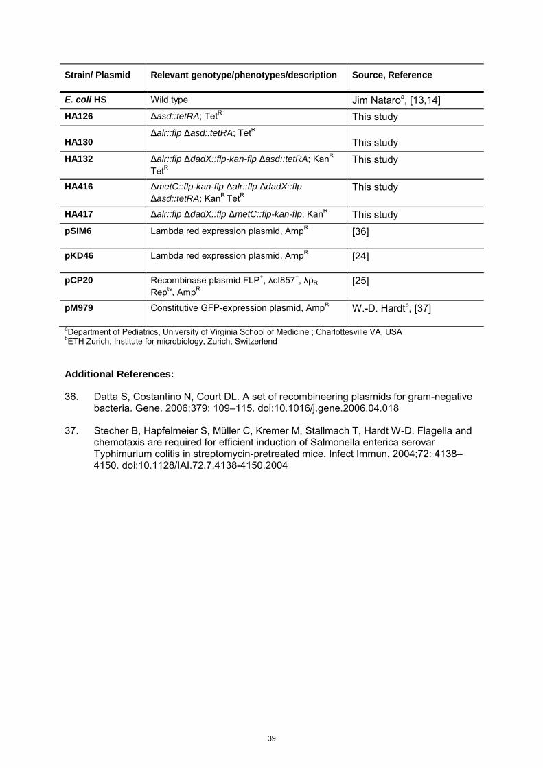
Strain/ Plasmid Relevant genotype/phenotypes/description Source, Reference
E. coli HS Wild type Jim Nataroa, [13,14] HA126 Δasd::tetRA; TetR This study
HA130Δalr::flp Δasd::tetRA; TetR
This study HA132 Δalr::flp ΔdadX::flp-kan-flp Δasd::tetRA; KanR
TetRThis study
HA416 ΔmetC::flp-kan-flp Δalr::flp ΔdadX::flp Δasd::tetRA; KanR TetR
This study
HA417 Δalr::flp ΔdadX::flp ΔmetC::flp-kan-flp; KanR This study pSIM6 Lambda red expression plasmid, AmpR [36]
pKD46 Lambda red expression plasmid, AmpR [24]
pCP20 Recombinase plasmid FLP+, λcI857+, λρR Repts, AmpR
[25]
pM979 Constitutive GFP-expression plasmid, AmpR W.-D. Hardtb, [37]
aDepartment of Pediatrics, University of Virginia School of Medicine ; Charlottesville VA, USA bETH Zurich, Institute for microbiology, Zurich, Switzerlend
Additional References:
36. Datta S, Costantino N, Court DL. A set of recombineering plasmids for gram-negativebacteria. Gene. 2006;379: 109–115. doi:10.1016/j.gene.2006.04.018
37. Stecher B, Hapfelmeier S, Müller C, Kremer M, Stallmach T, Hardt W-D. Flagella andchemotaxis are required for efficient induction of Salmonella enterica serovarTyphimurium colitis in streptomycin-pretreated mice. Infect Immun. 2004;72: 4138–4150. doi:10.1128/IAI.72.7.4138-4150.2004
39
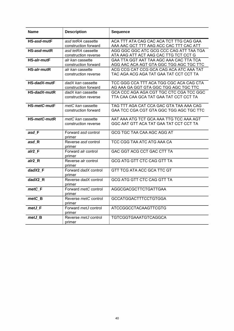
Name Description Sequence
HS-asd-mutF asd tetRA cassette construction forward
ACA TTT ATA CAG CAC ACA TCT TTG CAG GAAAAA AAC GCT TTT AAG ACC CAC TTT CAC ATT
HS-asd-mutR asd tetRA cassette construction reverse
AGG GGC GGC ATC GCG CCC CAG ATT TAA TGA ATA AAG ATT ACT AAG CAC TTG TCT CCT G
HS-alr-mutF alr kan cassette construction forward
GAA TTA GGT AAT TAA AGC AAA CAC TTA TCAAGG AAC ACA AGT GTA GGC TGG AGC TGC TTC
HS-alr-mutR alr kan cassette construction reverse
ACG CCG CAT CCG GCA CAG ACA ATC AAA TATTAC AGA ACG AGA TAT GAA TAT CCT CCT TA
HS-dadX-mutF dadX kan cassette construction forward
TCC GGG CCA TTT ACA TGG CGC ACA CAG CTA AG AAA GA GGT GTA GGC TGG AGC TGC TTC
HS-dadX-mutR dadX kan cassette construction reverse
GCA CCC AGA AGA CGT TGC CTC CGA TCC GGCTTA CAA CAA GCA TAT GAA TAT CCT CCT TA
HS-metC-mutF metC kan cassette construction forward
TAG TTT AGA CAT CCA GAC GTA TAA AAA CAG GAA TCC CGA CGT GTA GGC TGG AGC TGC TTC
HS-metC-mutR metC kan cassette construction reverse
AAT AAA ATG TCT GCA AAA TTG TCC AAA AGT GGC AAT GTT ACA TAT GAA TAT CCT CCT TA
asd_F Forward asd control primer
GCG TGC TAA CAA AGC AGG AT
asd_R Reverse asd control primer
TCC CGG TAA ATC ATG AAA CA
alr2_F Forward alr control primer
GAC GGT ACG CCT GAC CTT TA
alr2_R Reverse alr control primer
GCG ATG GTT CTC CAG GTT TA
dadX2_F Forward dadX control primer
GTT TCG ATA ACC GCA TTC GT
dadX2_R Reverse dadX control primer
GCG ATG GTT CTC CAG GTT TA
metC_F Forward metC control primer
AGGCGACGCTTCTGATTGAA
metC_B Reverse metC control primer
GCCATGGACTTTCCTGTGGA
metJ_F Forward metJ control primer
ATCCGGCCTACAAGTTCGTG
metJ_B Reverse metJ control primer
TGTCGGTGAAATGTCAGGCA
40

Chapter 2. Simulation of early intestinal infection of
Salmonella enterica Typhimurium in a disease-independent
model
Abstract
Discrimination between intestinal pathogenic and commensal bacteria is a critical function of
the mammalian immune system. The ability to recognize pathogenic bacteria and mount an
appropriate immune response is crucial for the maintenance of the homeostasis. Here, we
formulated the question if there was a different expression pattern from known mucosal
biomarkers in response to growth-proficient and in vivo growth-deficient (cell wall
auxotrophic; CWA) virulent strains of Salmonella enterica serovar Typhimurium (Stm), which
would allow us to predict possible outcome of the disease. A panel of 86 genes related to
intestinal inflammation in inflammatory bowel disease (IBD) was measured in mouse cecum
at 6 hours post inoculation with either wild type (WT) Stm, avirulent Stm, CWA Stm,
commensal E. coli HS or PBS. We built a classifier model to predict the occurrence of
inflammation, based on the assumptions that growth proficient Salmonella was the only
positive condition and all others as antigen or manipulation controls. When tested in an
independently generated dataset, the model failed to classify with enough confidence to be
used as a predictive tool. This led us to the recognition that at time point 6 h there is a high
variability in an interesting biomarker subset that is similar between WT Stm and CWA Stm,
but not present in the untreated control, avirulent Stm or E.coli HS. Based on this observation
the experiment was repeated at 9 h post inoculation and we observed that in response to WT
Stm these biomarkers had increased or maintained at the same level, whereas in CWA Stm
there was no change. This observation led to the interpretation that during the first measured
time point (6h) this increase of variability is related to the start of an immune response,
sustained until 9h in the presence of WT Stm, but is resolved after CWA Stm exposition. We
conclude that the inability of the predictive model to distinguish between the different groups
at 6 h is due to the absence of a clear difference between the groups, which is more
apparent at 9h post-gavage. These early similarities suggest that the mucosal inoculation
with CWA Stm is able to simulate the early steps of a WT Stm infection, which underscores
the useful of this experimental system for the study of immune responses, for which reason
the model for identifying differences between both responses fails to identify a differential
pattern in the biomarker set available.
41
Introduction
A critical aspect of the maintenance of intestinal homeostasis is the effective discrimination
between pathogenic and commensal bacteria. The ability to discern pathogenic bacteria
efficiently and timely is crucial, and is mediated by the interaction between innate and
adaptive immunity (Jakobsson et al., 2015). The difference between these two parts of the
immune system is based on the ability to recognize general foreign patterns of the former
and the recognition of specific non-self-antigens of the latter.
The innate immune system is composed of overlapping layers of all-purpose mechanisms to
exclude or eliminate any type of non-self-object or organisms. The first layer of protection in
the lumen is achieved by the intestinal mucus, a gel-like structure that separates the
particulate luminal contents from the epithelium while allowing the absorption of soluble
nutrients (Jakobsson et al., 2015). The mucus is also enriched with antimicrobial peptides
and other small molecules known to regulate bacterial behavior, helping in the active
maintenance of intestinal homeostasis (Jakobsson et al., 2015; Li et al., 2015; Meyer-Hoffert
et al., 2008). When these mechanisms are not sufficient for the containment of luminal
microorganisms, an inflammatory response is activated to clear the foreign particle or
organism.
The factors regulating the activation of inflammatory responses are not fully understood. It is
known that certain pathogenic microorganisms and types of antigen elicit a strong
inflammatory response, however the defining characteristics of these activators are ill-
described, since there is always an overlap between innate and adaptive responses
(Hapfelmeier et al., 2005; Sellin et al., 2014). Previously in our lab, we have shown that
transient colonization of germ-free mice with cell wall auxotrophic (CWA) Salmonella enterica
Typhimurium (Stm) induces high titers of species-specific IgA, without inducing inflammation
(Pfister et al, unpublished), effectively separating the innate inflammation from the induction
of a protective adaptive immune response. This uncoupling of adaptive immunity from
inflammation provides us with a model of pathogen exposition ideal to dissect the early
bacteria-innate immunity interaction, and further expand our understanding of inflammatory
responses.
In this section we explore the early host - CWA Stm interaction and compare it with the host-
WT Stm infection. For this approach we decided to generate a multivariate statistical model
that would be able to identify pathogen-induced innate immunity genes different between
these two strains, and separate them from general responses elicited by commensal
bacteria. Our statistical model was unable to separate the CWA Stm-induced from the WT
42
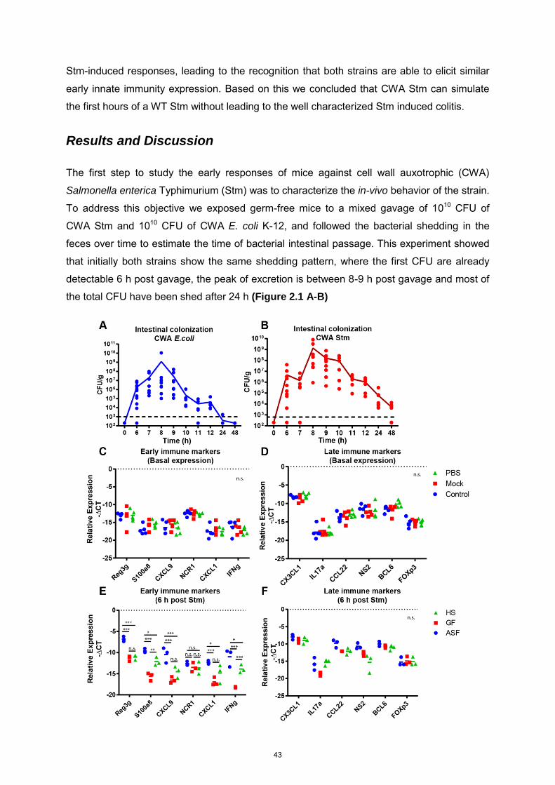
Stm-induced responses, leading to the recognition that both strains are able to elicit similar
early innate immunity expression. Based on this we concluded that CWA Stm can simulate
the first hours of a WT Stm without leading to the well characterized Stm induced colitis.
Results and Discussion
The first step to study the early responses of mice against cell wall auxotrophic (CWA)
Salmonella enterica Typhimurium (Stm) was to characterize the in-vivo behavior of the strain.
To address this objective we exposed germ-free mice to a mixed gavage of 1010 CFU of
CWA Stm and 1010 CFU of CWA E. coli K-12, and followed the bacterial shedding in the
feces over time to estimate the time of bacterial intestinal passage. This experiment showed
that initially both strains show the same shedding pattern, where the first CFU are already
detectable 6 h post gavage, the peak of excretion is between 8-9 h post gavage and most of
the total CFU have been shed after 24 h (Figure 2.1 A-B)
43
Figure 2.1. Early response of mice to cell wall auxotrophic bacteria. Germ-free mice were gavaged with a 1:1 mixture of 1010 CFU of cell wall auxotrophic (CWA) E.coli (A) and 1010 CFU of CWA Stm (B), and the luminal colonization was followed for 48 h. (C-D) Background expression of mRNA in cecum from Naïve (green triangles), mock gavaged (red squares) or PBS gavaged (blue circles). (E-F) Intestinal immune response to a 1010 CFU CWA Stm infection of HS colonized (green triangles), germ-free (red squares) or ASF colonized (blue circles) mice. All animals were sacrificed 6 h after treatment and cecal mRNA was quantified with qPCR. The expression is normalized to actin beta. Each dot represents one mouse. Anova with Tukey as posttest, ***< 0.001, ** <0.01, *<0.05, n.s = not significant.
Besides the evident similarities between the strains, CWA Stm maintained higher shedding
numbers for a longer period of time than CWA E. coli, clearly evident from residual CFU still
being shed at 48 h post gavage (figure 2.1 A-B). The bacterial shedding of CWA E. coli
corresponded well with the published colonization kinetics of this strain (Hapfelmeier et al.,
2010), suggesting that the observed longer persistence of CWA Stm might be due to better
in vivo survival than in comparable E. coli strains (Cuenca et al., 2016).
Once the average transit time was estimated, we proceeded to measure 12 immune markers
selected rationally to cover the spectrum of known intestinal responses, as a means to
establish the basal immune status in the experimental mice (Table S1). Since our main
objective was to determine the early responses that would eventually lead to inflammation,
we decided to use 6 h post gavage as the measuring time point. At this time point we
estimated that in average the bulk of bacteria was present in the cecum, inflammation would
not yet be induced. This assumption goes in line with the observations in the streptomycin-
pretreated mouse model of invasive non-typhoid salmonellosis, where the first intra-epithelial
Stm have been observed at 6 h post infection and reaching the maximum at 9 h post
infection (Sellin et al., 2014).
Since our experimental procedures induce stress and anxiety in the experimental animal by
handling and gavage, we decided to measure the expression of the 12 mRNA markers
compared to control mice that were left untreated, mock gavaged (introducing the feeding
needle into the stomach without liquid administration) or gavaged with sterile PBS (our
standard vehicle) 6 h post gavage. This basal analysis showed that the basic experimental
procedures do not cause major alterations to the gene expression of any marker measured
(figure 2.1 C-D). We could also measure that the variability of the assay is low, encouraging
its usage for measuring small but critical differences.
The results from this experiment led us to speculate, if marker expression in response to
CWA Stm infection was influenced by hygiene status/microbiota composition. To test this
hypothesis, 3 germ-free mice, 3 ASF (Altered Schaedler Flora-colonized) mice and 3 two-
week commensal E. coli HS-monocolonized mice were gavaged with CWA Stm (figure 2.1 E-F). The ASF mice showed an elevated expression pattern in nearly all the early
immune markers, but not in the late immune markers. Germ-free mice showed lower
44
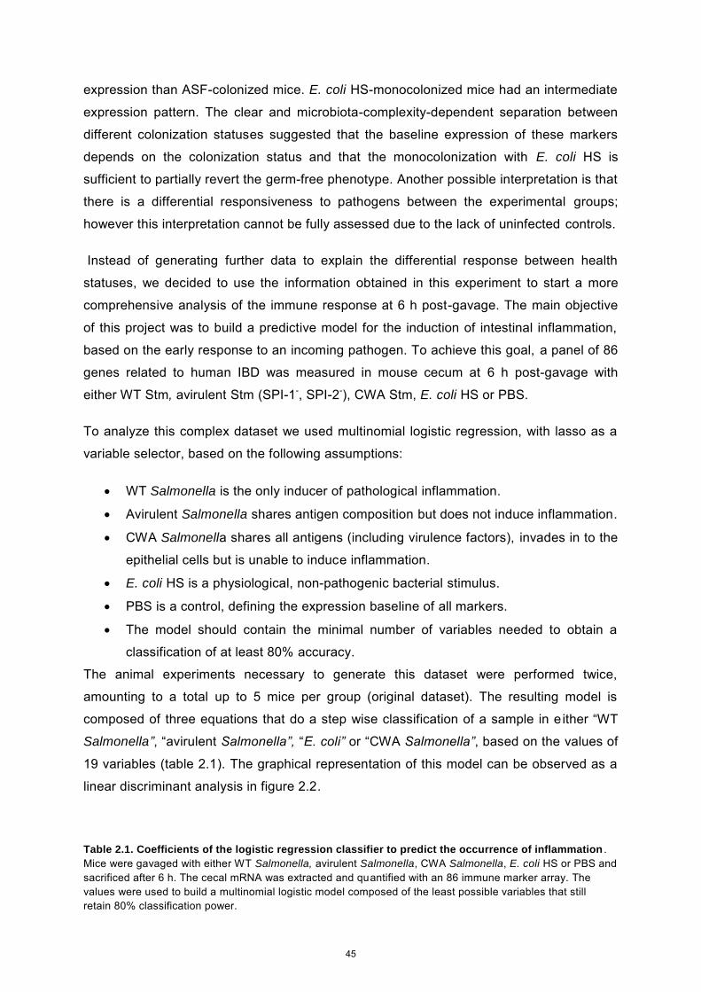
expression than ASF-colonized mice. E. coli HS-monocolonized mice had an intermediate
expression pattern. The clear and microbiota-complexity-dependent separation between
different colonization statuses suggested that the baseline expression of these markers
depends on the colonization status and that the monocolonization with E. coli HS is
sufficient to partially revert the germ-free phenotype. Another possible interpretation is that
there is a differential responsiveness to pathogens between the experimental groups;
however this interpretation cannot be fully assessed due to the lack of uninfected controls.
Instead of generating further data to explain the differential response between health
statuses, we decided to use the information obtained in this experiment to start a more
comprehensive analysis of the immune response at 6 h post-gavage. The main objective
of this project was to build a predictive model for the induction of intestinal inflammation,
based on the early response to an incoming pathogen. To achieve this goal, a panel of 86
genes related to human IBD was measured in mouse cecum at 6 h post-gavage with
either WT Stm, avirulent Stm (SPI-1-, SPI-2-), CWA Stm, E. coli HS or PBS.
To analyze this complex dataset we used multinomial logistic regression, with lasso as a
variable selector, based on the following assumptions:
WT Salmonella is the only inducer of pathological inflammation.
Avirulent Salmonella shares antigen composition but does not induce inflammation.
CWA Salmonella shares all antigens (including virulence factors), invades in to the
epithelial cells but is unable to induce inflammation.
E. coli HS is a physiological, non-pathogenic bacterial stimulus.
PBS is a control, defining the expression baseline of all markers.
The model should contain the minimal number of variables needed to obtain a
classification of at least 80% accuracy.
The animal experiments necessary to generate this dataset were performed twice,
amounting to a total up to 5 mice per group (original dataset). The resulting model is
composed of three equations that do a step wise classification of a sample in e ither “WT
Salmonella”, “avirulent Salmonella”, “E. coli” or “CWA Salmonella”, based on the values of
19 variables (table 2.1). The graphical representation of this model can be observed as a
linear discriminant analysis in figure 2.2.
Table 2.1. Coefficients of the logistic regression classifier to predict the occurrence of inflammation . Mice were gavaged with either WT Salmonella, avirulent Salmonella, CWA Salmonella, E. coli HS or PBS and sacrificed after 6 h. The cecal mRNA was extracted and quantified with an 86 immune marker array. The values were used to build a multinomial logistic model composed of the least possible variables that still retain 80% classification power.
45
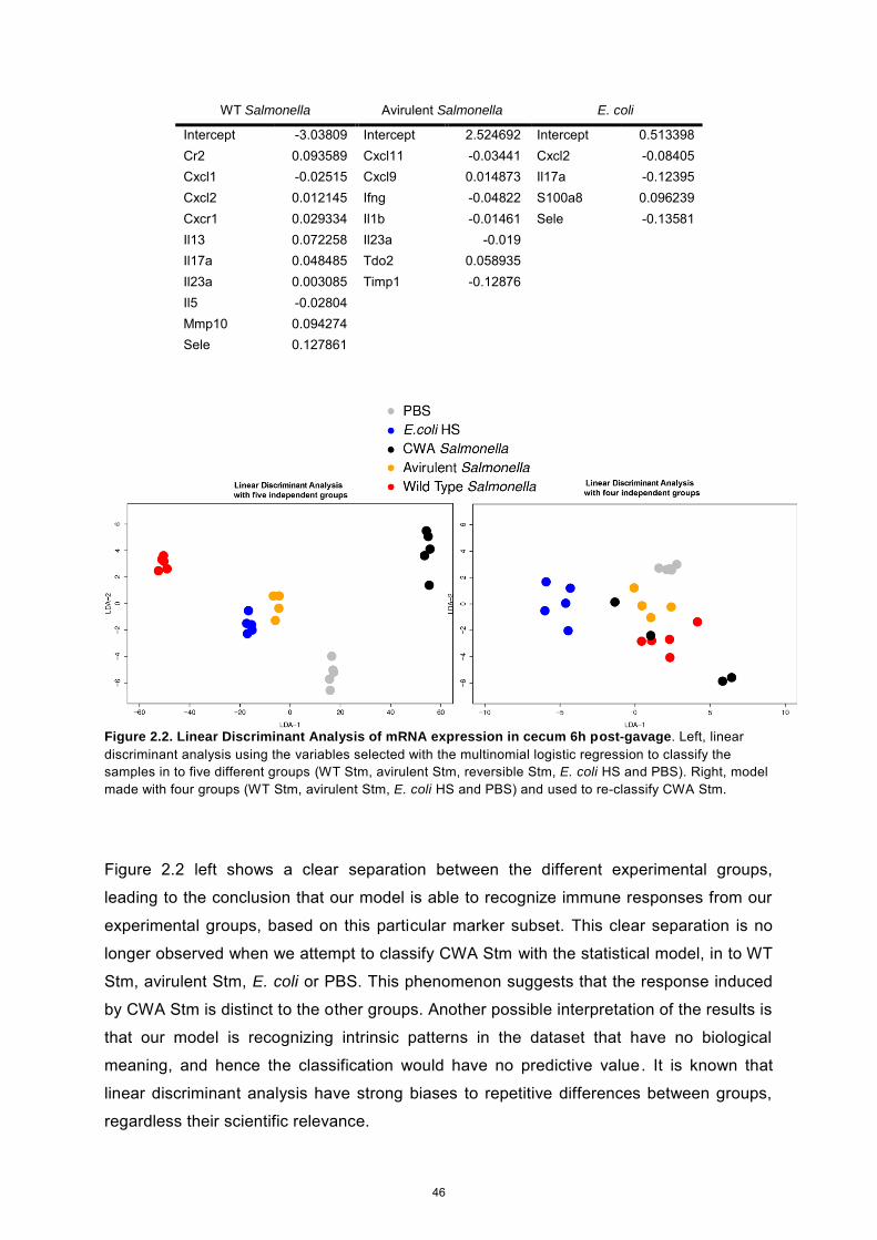
WT Salmonella Avirulent Salmonella E. coli
Intercept -3.03809 Intercept 2.524692 Intercept 0.513398 Cr2 0.093589 Cxcl11 -0.03441 Cxcl2 -0.08405 Cxcl1 -0.02515 Cxcl9 0.014873 Il17a -0.12395 Cxcl2 0.012145 Ifng -0.04822 S100a8 0.096239 Cxcr1 0.029334 Il1b -0.01461 Sele -0.13581 Il13 0.072258 Il23a -0.019 Il17a 0.048485 Tdo2 0.058935 Il23a 0.003085 Timp1 -0.12876 Il5 -0.02804 Mmp10 0.094274 Sele 0.127861
Figure 2.2. Linear Discriminant Analysis of mRNA expression in cecum 6h post-gavage. Left, linear discriminant analysis using the variables selected with the multinomial logistic regression to classify the samples in to five different groups (WT Stm, avirulent Stm, reversible Stm, E. coli HS and PBS). Right, model made with four groups (WT Stm, avirulent Stm, E. coli HS and PBS) and used to re-classify CWA Stm.
Figure 2.2 left shows a clear separation between the different experimental groups,
leading to the conclusion that our model is able to recognize immune responses from our
experimental groups, based on this particular marker subset. This clear separation is no
longer observed when we attempt to classify CWA Stm with the statistical model, in to WT
Stm, avirulent Stm, E. coli or PBS. This phenomenon suggests that the response induced
by CWA Stm is distinct to the other groups. Another possible interpretation of the results is
that our model is recognizing intrinsic patterns in the dataset that have no biological
meaning, and hence the classification would have no predictive value. It is known that
linear discriminant analysis have strong biases to repetitive differences between groups,
regardless their scientific relevance.
46
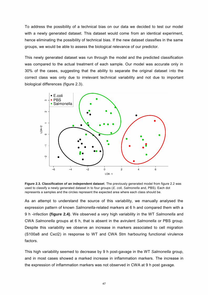
To address the possibility of a technical bias on our data we decided to test our model
with a newly generated dataset. This dataset would come from an identical experiment,
hence eliminating the possibility of technical bias. If the new dataset classifies in the same
groups, we would be able to assess the biological relevance of our predictor.
This newly generated dataset was run through the model and the predicted classification
was compared to the actual treatment of each sample. Our model was accurate only in
30% of the cases, suggesting that the ability to separate the original dataset into the
correct class was only due to irrelevant technical variability and not due to important
biological differences (figure 2.3).
Figure 2.3. Classification of an independent dataset. The previously generated model from figure 2.2 was used to classify a newly generated dataset in to four groups (E. coli, Salmonella and, PBS). Each dot represents a samples and the circles represent the expected area where each class should be.
As an attempt to understand the source of this variability, we manually analysed the
expression pattern of known Salmonella-related markers at 6 h and compared them with a
9 h -infection (figure 2.4). We observed a very high variability in the WT Salmonella and
CWA Salmonella groups at 6 h, that is absent in the avirulent Salmonella or PBS group.
Despite this variability we observe an increase in markers associated to cell migration
(S100a8 and Cxcl2) in response to WT and CWA Stm harbouring functional virulence
factors.
This high variability seemed to decrease by 9 h post-gavage in the WT Salmonella group,
and in most cases showed a marked increase in inflammation markers. The increase in
the expression of inflammation markers was not observed in CWA at 9 h post gavage.
47
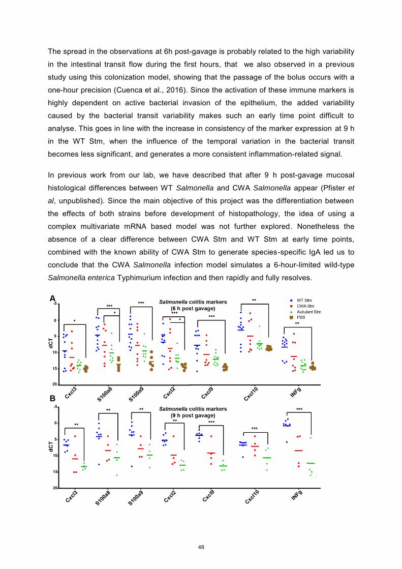
The spread in the observations at 6h post-gavage is probably related to the high variability
in the intestinal transit flow during the first hours, that we also observed in a previous
study using this colonization model, showing that the passage of the bolus occurs with a
one-hour precision (Cuenca et al., 2016). Since the activation of these immune markers is
highly dependent on active bacterial invasion of the epithelium, the added variability
caused by the bacterial transit variability makes such an early time point difficult to
analyse. This goes in line with the increase in consistency of the marker expression at 9 h
in the WT Stm, when the influence of the temporal variation in the bacterial transit
becomes less significant, and generates a more consistent inflammation-related signal.
In previous work from our lab, we have described that after 9 h post-gavage mucosal
histological differences between WT Salmonella and CWA Salmonella appear (Pfister et
al, unpublished). Since the main objective of this project was the differentiation between
the effects of both strains before development of histopathology, the idea of using a
complex multivariate mRNA based model was not further explored. Nonetheless the
absence of a clear difference between CWA Stm and WT Stm at early time points,
combined with the known ability of CWA Stm to generate species-specific IgA led us to
conclude that the CWA Salmonella infection model simulates a 6-hour-limited wild-type
Salmonella enterica Typhimurium infection and then rapidly and fully resolves.
48
Figure 2.4 Salmonella-related markers expressed at 6 or 9 h post-gavage. Mice were gavaged with either
1010 CFU WT Stm, 1010 CFU CWA Stm, 1010 CFU avirulent Stm or PBS. (A) Gene expression in mouse
cecum 6 hours post gavage (ANOVA with tukey post-test using PBS as comparison). (B) Gene expression in
mouse cecum 6 hours post gavage (ANOVA with tukey post-test using avirulent as comparison).
Table S2.1 Bacterial strains and plasmids
Strain/ Plasmid Strainname
Relevantgenotype/phenotypes/description Source, Reference
WT Stm χ4138 Wild type, UK-1 background Roy Curtiss (Dieye et al., 2009)
Avirulent Stm χ9650 gyrA1816 Δ(avrA-invH)-2::cat Δ(ssaG-ssaU)-
1::kan, NalR, CmR, KmR. χ4138 derivateRoy Curtiss (Dieye et al., 2009)
CWA Stm χ9052 Δalr-3 ΔdadB4 ΔasdA33 χ4138 derivate Roy Curtiss (Xin et al., 2012)
E. coli HS Wild type Jim Nataro (Levine et al., 1978)
CWA E.coli HA107 Δalr Δdadx Δasd Siegfried Hapfelmeier (Hapfelmeier et al., 2010)
Material and Methods
Animal colonization experiments
Germ-free animals were re-derived from C57BL/6 mice and maintained germ-free in flexible
film isolators in the Genaxen Foundation Clean Mouse Facility (CMF) of the University of
Bern as described (Macpherson and McCoy, 2014). For experiments mice were aseptically
transferred to autoclaved sealsafe-plus IVCs under positive pressure (Tecniplast, Italy) in a
barrier unit of the Genaxen Clean Mouse Facility. Cage changes were carried out under
strictly aseptic conditions. In all experiments animals were provided with sterile mouse chow
(Kliba 3437; autoclaved) and autoclaved water ad libitum. All experiments were performed
according to protocols approved by the Bernese Cantonal Ethical committee for animal
experiments and carried out in accordance with Swiss Federal law for animal
experimentation (license numbers BE94/11 and BE91/14).
To generate contamination-free Salmonella enterica Typhimurium inoculums, NaCl (0.3M),
D-Ala (200 μg/mL) and Dap (50 μg/ mL) supplemented autoclaved LB medium in sterile-filter-
sealed flasks, was aseptically inoculated from single colonies of the test bacterium and
incubated shaking at 150 rpm at 37°C for 16 hours. This subculture was then diluted 1:106 in
500mL of the same media and incubated shaking at 150 rpm at 37°C for 16 hours. Bacteria
were harvested by centrifugation (10 min, 4816 x g, 4°C) in a sterile aerosol-proof assembly,
washed in autoclaved sterile PBS and concentrated to a density of 1x10¹¹ CFU/mL in sterile
PBS, performed aseptically under a sterile laminar airflow. To generate contamination free E.
49
coli inoculums, the same procedure was followed with the exception of the media: LB
supplemented with D-Ala (200 μg/mL) and Dap (50 μg/ mL). The bacterial suspensions were
aseptically aliquoted in autoclaved plastic tubes and sealed in a sterilized secondary
containment. The sterile tubes containing the inocula and the mice were aseptically imported
into a sterilized laminar flow hood, and each animal inoculated with 200 μL of bacterial
suspension (containing 1 x 10¹⁰ CFU in sterile PBS, at a density of 1 x 10¹¹ CFU/mL) by
gavage, carried out wearing sterile surgical gowns and sterile surgical gloves. Fresh fecal
pellets were collected aseptically, suspended in sterile PBS, and plated in serial dilutions on
D-Ala/ Dap-supplemented or non-supplemented LB agar and incubated aerobically at 37°C
for 24 hours.
RT-qPCR
Cecum samples were collected 6 h or 9 h post infection with 1010 CFU of bacteria, washed in
sterile PBS and immediately preserved in RNAlater (QIAGEN) for RNA stabilization
overnight. The samples were then transferred to -80°C for long term storage. Total RNA was
extracted from approximately 15 mg of tissue using the RNeasy mini kit (Qiagen) and the
quality was assessed using the Agilent RNA 6000 Nano Kit to be higher than RIN 9. The
samples of interest were reverse transcribed with the RT2 easy first strand kit (Qiagen) and
then the cDNA libraries were analyzed using the either the 12 gene custom plate
CAPM11698 (SABiosciences ) or the mouse crohn's disease RT² profiler PCR array
(SABiosciences, PAMM-169ZE-4) with SYBR green reagents (Qiagen) in a Viia 7 Real-Time
PCR System (Thermo Scientific). For the plate PAMM-169ZE-4 five housekeeping genes
(Actb, B2m, Gapdh, Gusb and Hsp90ab1) were averaged and used for calculating the ∆CT
(CTsample - CTaverage housekeeping genes). In the plate CAPM11698 only Actb was used.
Data analysis
All multivariate data analysis was done using the R 3.1.1 statistical program and the ggplot2
(Wickham and Chang, 2015), dplyr (Wickham and Francois, 2015), glmnet (2010; 2010) and
MASS packages. The scripts used for this analysis are provided as supplementary data.
Figures 2.1 and 2.3 were done using the program Prism 6
Acknowledgments
We thank the Clean Mouse Facility for all the animal work and the Genomics Core
Facility, from the Department of Clinical Research University of Bern for the access to the
qPCR machines. This work was funded by the grants ERC 281904 and SNF
310030_138452.
50
References
(2010). Regularization Paths for Generalized Linear Models via Coordinate Descent.
Journal Of Statistical Software 33, 1--22.
Cuenca, M., Pfister, S.P., Buschor, S., Bayramova, F., Hernandez, S.B., Cava, F., Kuru,
E., Van Nieuwenhze, M.S., Brun, Y.V., Coelho, F.M., et al. (2016). D-Alanine-Controlled
Transient Intestinal Mono-Colonization with Non-Laboratory-Adapted Commensal E. coli
Strain HS. Plos ONE 11, e0151872.
Dieye, Y., Ameiss, K., Mellata, M., and Curtiss, R. (2009). The Salmonella Pathogenicity
Island (SPI) 1 contributes more than SPI2 to the colonization of the chicken by Salmonella
enterica serovar Typhimurium. BMC Microbiology 9, 3.
Hapfelmeier, S., Lawson, M.A., Slack, E., Kirundi, J.K., Stoel, M., Heikenwalder, M.,
Cahenzli, J., Velykoredko, Y., Balmer, M.L., Endt, K., et al. (2010). Reversible Microbial
Colonization of Germ-Free Mice Reveals the Dynamics of IgA Immune Responses. Science
328, 1705-1709.
Hapfelmeier, S., Stecher, B., Barthel, M., Kremer, M., Müller, A.J., Heikenwalder, M.,
Stallmach, T., Hensel, M., Pfeffer, K., Akira, S., et al. (2005). The Salmonella pathogenicity
island (SPI)-2 and SPI-1 type III secretion systems allow Salmonella serovar typhimurium to
trigger colitis via MyD88-dependent and MyD88-independent mechanisms. J. Immunol. 174,
1675-85.
Jakobsson, H.E., Holmén-Larsson, J., Schütte, A., Ermund, A., Rodríguez-Piñeiro, A.M.,
Arike, L., Wising, C., Svensson, F., Bäckhed, F., and Hansson, G.C. (2015). Normalization of
Host Intestinal Mucus Layers Requires Long-Term Microbial Colonization. Cell Host &Amp;
Microbe 18, 582-92.
51
Levine, M.M., Bergquist, E.J., Nalin, D.R., Waterman, D.H., Hornick, R.B., Young, C.R.,
and Sotman, S. (1978). Escherichia coli strains that cause diarrhoea but do not produce
heat-labile or heat-stable enterotoxins and are non-invasive. 1, 1119-1122.
Li, H., Limenitakis, J.P., Fuhrer, T., Geuking, M.B., Lawson, M.A., Wyss, M., Brugiroux,
S., Keller, I., Macpherson, J.A., Rupp, S., et al. (2015). The outer mucus layer hosts a distinct
intestinal microbial niche. Nature Communications 6, 8292.
Macpherson, A.J., and McCoy, K.D. (2014). Standardised animal models of host
microbial mutualism. Mucosal Immunology 8, 476.
Meyer-Hoffert, U., Hornef, M.W., Henriques-Normark, B., Axelsson, L., Midtvedt, T.,
Putsep, K., and Andersson, M. (2008). Secreted enteric antimicrobial activity localises to the
mucus surface layer. Gut 57, 764.
Sellin, M.E., Müller, A.A., Felmy, B., Dolowschiak, T., Diard, M., Tardivel, A., Maslowski,
K.M., and Hardt, W. (2014). Epithelium-Intrinsic NAIP/NLRC4 Inflammasome Drives Infected
Enterocyte Expulsion to Restrict Salmonella Replication in the Intestinal Mucosa. Cell Host
&Amp; Microbe 16, 237.
Wickham, H., and Chang, W. (2015). Ggplot2: An implementation of the grammar of
graphics, Version 1.0. 1.
Wickham, H., and Francois, R. (2015). dplyr: A grammar of data manipulation. R
Package Version 0.4.
Xin, W., Wanda, S.Y., Zhang, X., Santander, J., Scarpellini, G., Ellis, K., Alamuri, P., and
Curtiss, R. (2012). The Asd+-DadB+ Dual-Plasmid System Offers a Novel Means To Deliver
Multiple Protective Antigens by a Recombinant Attenuated Salmonella Vaccine. Infection
And Immunity 80, 3621.
52

Supplementary figure 2.1. Script to build a log- logistic model
Lasso.R setwd("//ifik.unibe.ch/users/Homes/cuenca/Desktop") library(glmnet) library(reshape2) #######Aquiring and shaping the training data rnadb=read.table("Data_mc14_001.csv", header=T, sep=";") class(rnadb) # The data are stored as a list names(rnadb) # The names of the list elements are x and y x <- rnadb[3:85] X<- data.matrix(x ) y<-rnadb[2] Y<-data.matrix(y) #####Fitting the model fit = glmnet(X, Y, family = "multinomial", type.multinomial = "grouped", intercept=FALSE, dfmax=20,)# fit the model fit #Plot the paths for the fit plot(fit, xvar = "lambda", label = TRUE, type.coef = "2norm")
title(main="20-variable coefficient paths",cex=0.8) #####Cross validation of the fitted model cv.fit = cv.glmnet(X, Y, family = "multinomial", type.measure="class") plot(cv.fit)# Plot the mean sq error for the cross validated fit as a function # of lambda the shrinkage parameter # First vertical line indicates minimal mse # Second vertical line is one sd from mse: indicates a smaller model # is "almost as good" as the minimal mse model tpred=predict(cv.fit,X) # Predictions on the test data mte=apply((tpred-Y)^2,2,mean) # Compute mse for the predictions points(log(fit$lambda),mte,col="blue",pch="*") # overlay the mse predictions on the plot #legend("topleft",legend=c("10 fold CV","Test"),pch="*",col=c("red","blue")) coef.exact = coef(cv.fit, s = 0.5, exact = TRUE) coef.apprx = coef(cv.fit, s = 0.5, exact = FALSE) ans<-coef(cv.fit, s=cv.fit$lambda.1se) sink("//ifik.unibe.ch/users/Homes/cuenca/Desktop/Data_wolfberg/output.txt") lapply(ans, print) sink()
53
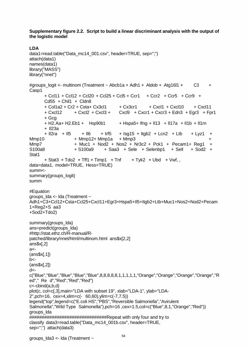
Supplementary figure 2.2. Script to build a linear discriminant analysis with the output of the logistic model LDA data1=read.table("Data_mc14_001.csv", header=TRUE, sep=";") attach(data1) names(data1) library("MASS") library("nnet")
#groups_logit <- multinom (Treatment ~ Abcb1a + Adh1 + Aldob + Atg16l1 + C3 + Casp1
+ Ccl11 + Ccl12 + Ccl20 + Ccl25 + Ccl5 + Ccr1 + Ccr2 + Ccr5 + Ccr9 + Cd55 + Chil1 + Cldn8 + Col1a2 + Cr2 + Csta + Cx3cl1 + Cx3cr1 + Cxcl1 + Cxcl10 + Cxcl11 + Cxcl12 + Cxcl2 + Cxcl3 + Cxcl9 + Cxcr1 + Cxcr3 + Edn3 + Egr3 + Fpr1 + Gcg + H2.Aa+ H2.Eb1 + Hsp90b1 + Hspa5+ Ifng + Il13 + Il17a + Il1b + Il1rn + Il23a + Il2ra + Il5 + Il6 + Irf5 + Isg15 + Itgb2 + Lcn2 + Ltb + Lyz1 +
Mmp10 + Mmp12+ Mmp1a + Mmp3 + Mmp7 + Muc1 + Nod2 + Nos2 + Nr3c2 + Pck1 + Pecam1+ Reg1 + S100a8 + S100a9 + Saa3 + Sele + Selenbp1 + Sell + Sod2 + Stat1
+ Stat3 + Tdo2 + Tff1 + Timp1 + Tnf + Tyk2 + Ubd + Vwf, , data=data1, model=TRUE, Hess=TRUE) summ<- summary(groups_logit) summ
#Equation groups_lda <- lda (Treatment ~ Adh1+C3+Ccl12+Csta+Ccl25+Cxcl11+Egr3+Hspa5+Il5+Itgb2+Ltb+Muc1+Nos2+Nod2+Pecam1+Reg2+S aa3 +Sod2+Tdo2)
summary(groups_lda) ans=predict(groups_lda) #http://stat.ethz.ch/R-manual/R- patched/library/nnet/html/multinom.html ans$x[2,2] ans$x[,2] a<- (ans$x[,1]) b<- (ans$x[,2]) d<- c("Blue","Blue","Blue","Blue","Blue",8,8,8,8,8,1,1,1,1,1,"Orange","Orange","Orange","Orange","Red"," Re d","Red","Red","Red") c<-cbind(a,b,d) plot(c, col=c[,3],main="LDA with subset 19", xlab="LDA-1", ylab="LDA-2",pch=16, cex=4,xlim=c(- 60,60),ylim=c(-7,7.5)) legend("top",legend=c("E.coli HS","PBS","Reversible Salmonella","Avirulent Salmonella","Wild Type Salmonella"),pch=16 ,cex=1.5,col=c("Blue",8,1,"Orange","Red")) groups_lda ###############################Repeat with only four and try to classify data3=read.table("Data_mc14_001b.csv", header=TRUE, sep=";") attach(data3)
groups_lda3 <- lda (Treatment ~ 54
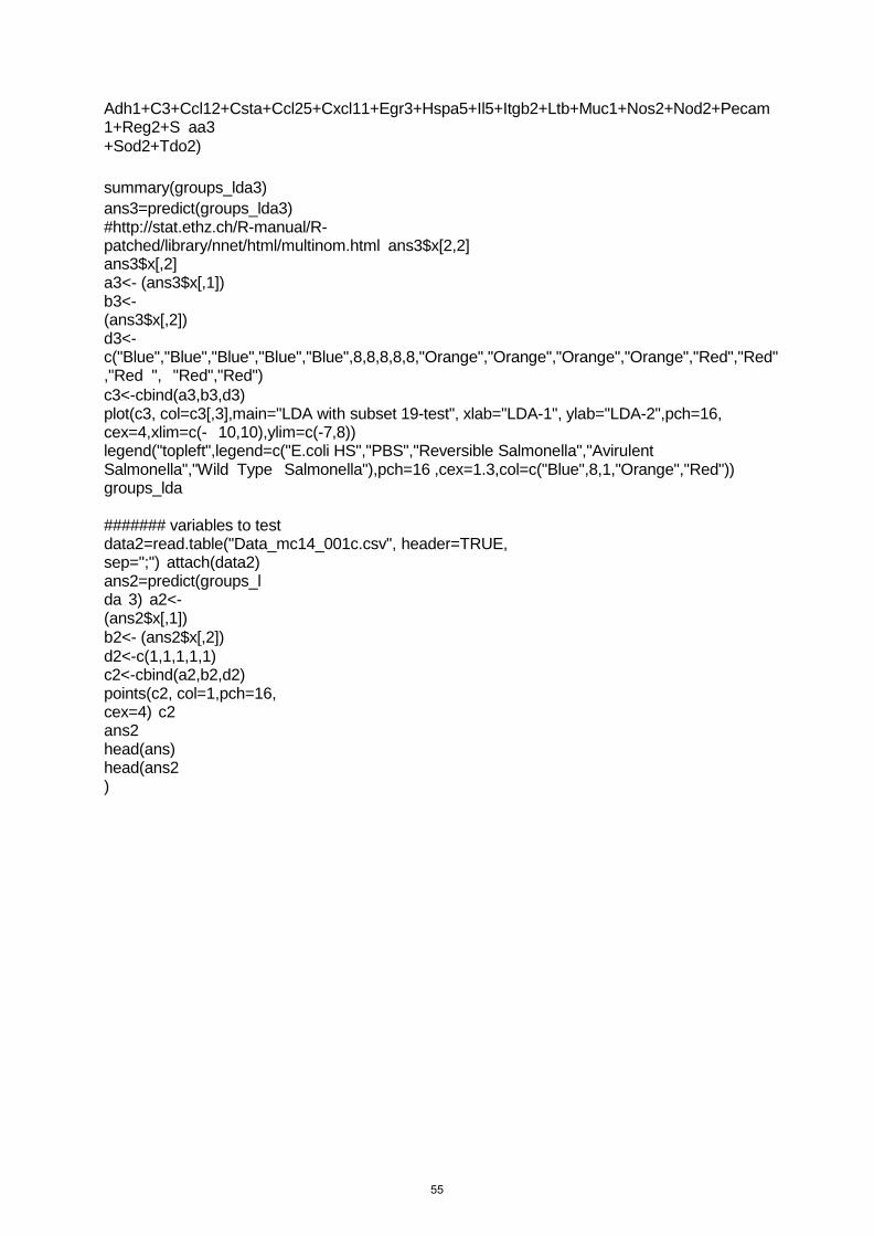
Adh1+C3+Ccl12+Csta+Ccl25+Cxcl11+Egr3+Hspa5+Il5+Itgb2+Ltb+Muc1+Nos2+Nod2+Pecam1+Reg2+S aa3 +Sod2+Tdo2)
summary(groups_lda3) ans3=predict(groups_lda3) #http://stat.ethz.ch/R-manual/R- patched/library/nnet/html/multinom.html ans3$x[2,2] ans3$x[,2] a3<- (ans3$x[,1]) b3<- (ans3$x[,2]) d3<- c("Blue","Blue","Blue","Blue","Blue",8,8,8,8,8,"Orange","Orange","Orange","Orange","Red","Red","Red ", "Red","Red") c3<-cbind(a3,b3,d3) plot(c3, col=c3[,3],main="LDA with subset 19-test", xlab="LDA-1", ylab="LDA-2",pch=16, cex=4,xlim=c(- 10,10),ylim=c(-7,8)) legend("topleft",legend=c("E.coli HS","PBS","Reversible Salmonella","Avirulent Salmonella","Wild Type Salmonella"),pch=16 ,cex=1.3,col=c("Blue",8,1,"Orange","Red")) groups_lda
####### variables to test data2=read.table("Data_mc14_001c.csv", header=TRUE, sep=";") attach(data2) ans2=predict(groups_lda 3) a2<- (ans2$x[,1]) b2<- (ans2$x[,2]) d2<-c(1,1,1,1,1) c2<-cbind(a2,b2,d2) points(c2, col=1,pch=16, cex=4) c2 ans2 head(ans) head(ans2 )
55
Chapter 3. Host-microbial IgA immune adaptation biases intestinal bacteria-bacteria competition to favor resident over immigrant strains
Author contributions This manuscript describes the interaction between the adaptive immune system and the
microbiota to outcompete an invasive pathogen. This work was possible thanks to the
contribution of several authors:
• Animal Experimentation: Miguelangel Cuenca, Stefanie Buschor, Fernanda
Coelho, Nicolas Studer and Miguel Terrazos.
• Native microbiota analysis: Miguelangel Cuenca, Nicolas Studer and Simone
Herp
• Experiment design and conceptualization: Miguelangel Cuenca, Fernanda
Coelho, Stefanie Buschor, Emma Slack and Siegfried Hapfelmeier
• LPS variation analysis: Miguelangel Cuenca and Emma Slack
• Construction of Salmonella enterica Typhimurium strains: Miguel Terrazos
56
Abstract The mammalian intestinal microbiota is a host-specific stable consortium of host-adapted
microorganisms that, amongst other beneficial functions, collectively provide protection
against pathogen invasion while improving the host nutrition. To achieve this community the
host must impose selective pressure by favoring host-compatible strains, in order to exclude
bacterial invaders that can damage the delicate host-microbiota interaction. Here we
describe the phenomenon where the combination of strain-specific IgA, a resident non-lethal
Salmonella enterica Typhimurium (Stm) strain and a third as yet unknown bacterial
adaptation, leads to the out-competition of an invading pathogenic Stm by the resident
strains. The decrease in WT Stm colonization provides a partial protection against colitis and
preserves the resident strain that in IgA absence would be lost. This approach explains how
the adaptive immunity influences the bacteria-bacteria competition, leading to the formation
of a stable and distinctive intestinal microbiota by improving the relative fitness of gut-
resident commensals.
57
Introduction The mammalian intestinal microbiota is a multi-species microbial consortium, acquired after
birth and mostly composed of bacteria that engage in a beneficial (mutualistic) or neutral
(commensal) relationship with the host organism. Mutualistic microbes have evolved to not
cause damage to a healthy and immunocompetent host, and to promote health by co-
metabolization of complex nutrients and biosynthesis of vitamins, by colonizing niches that
are restricted to the gut lumen (body exterior), and by interfering with the invasion of
microbial immigrants (that may be less adapted or pathogenic).
Any mutualistic relationship is based on bilateral actions for a common goal; therefore it
requires a healthy intestinal ecology, which can provide an optimal environment to the
resident species. The pre-colonization of the intestinal tract with certain bacterial strains has
been shown previously to be protective against the invasion by related pathogenic
immigrants due to metabolite depletion, exemplified by the inability of enterohemorragic E.
coli 0157:H7 to infect a mouse that had been pre-colonized with commensal E. coli strains
HS and Nissle 1917 (Leatham et al., 2009). Such metabolite depletion is normally induced by
a consortium of bacteria, in a process called colonization resistance (Maltby et al., 2013).
Colonization resistance of the intestinal pathogen Salmonella enteric servoar Typhimurium
(S. Typhimurium, short: Stm) has been directly linked to the luminal out-competition of
intestinal pathogens by a complex microbiota after the remission of inflammation (Endt et al.,
2010).
A colonization resistance conferring microbiota is consistently formed in all mammalian
hosts, despite of genetic, nutritional or environmental variability. This phenomenon suggests
that the host actively facilitates the establishment of a defined and controlled microbiota,
where most of the intestinal bacterial species are only transient (ecological succession and
"tourist" species that do not stably colonize) and the main residents are represented mainly
by host-adapted bacterial strains that are able to maintain a permanent colonization (Dishaw
et al., 2014). It has further been observed that despite the high phylogenic variability in the
microbiota, most of the functional categories and metabolic pathways represented in the
microbial metagenome are fairly conserved between hosts individuals (Ridaura et al., 2013).
The constitution of the intestinal microbiota has been hypothesized to require the continuous
selective pressure imposed by the adaptive immune system (Dishaw et al., 2014). Human
microbiology traditionally has focused mainly on the role of adaptive immune responses in
pathogen eradication or control. However, new approaches have led to the more general
concept that the adaptive immune system functions to “commensalize” any newcomer in the
gut, potentially pathogenic or not, in order to maintain the disease-free co-existence (Forbes
58
et al., 2008; Gauger et al., 2007; McSorley et al., 2000; Palm et al., 2014). This concept is
consistent with the fact that obligate pathogenic species, that have evolved the ability to
escape this host control, are rare, whereas opportunistic pathogenic, commensal and
mutualistic species are part of a difficult-to-categorize continuum that most hosts are
associated with.
The main host mechanisms imposing selective pressure on the gut microbial community
have been suggested to be immunoglobulin A (IgA)-, anti-microbial peptides-, and mucin 2-
(muc2) mediated (Dishaw et al., 2014; Rogier et al., 2014). The modes of action of these
different effector molecules are very broad. They can increase the distance from the
microbial community to the epithelial cells, kill bacteria in a selectively or not-selective
manner, reduce their motility (Forbes et al., 2008; Peterson et al., 2007a), inactivate
virulence factors, agglutinate bacteria (Forbes et al., 2008), provide nutrients for competing
bacterial species and also provide an anchoring site for bacterial adhesion (Rogier et al.,
2014).
The neutralization of microbial virulence factors by host-controlled mechanisms
phenotypically attenuates pathogens, which after losing their competitive edge, may be
cleared from the gut through out-competition by resident species (Endt et al., 2010).
Although all host-derived protective mechanisms are important for the control and
containment of the intestinal microbiota, only the adaptive immune system can induce
microbial species- and strain-specific immune responses, and one of the best characterized
immune mechanisms modulating the gut microbiota is intestinal IgA.
The ability of IgA to selectively affect colonization of specific members of the microbiota has
been recently shown in mice, were B. thetaiotaomicron strains can gain intestinal fitness by
capsule phase variation and loss of IgA binding (Peterson et al., 2007a). The adaptive
immune system has also been shown to alter the rate of evolution of E. coli populations, as
in RAG2-/- mice (that lack T and B cells) an adaptive immune deficiency led to a more
unpredictable fixation of adaptive E. coli mutations during intestinal colonization (Barroso-
Batista et al., 2015). This finding is corroborated by evidence for an increased gut bacterial
diversity in RAG1-/- compared with the wild type mice, an observation that can be reversed by
bone marrow transfer (Zhang et al., 2014).
Here, we show that bacteria-specific IgA can influence the bacterial-bacteria competition at
the strain level between a resident bacterial strain and an invading pathogenic variant. This
effect is generated by a tripartite interaction between bacterial serotype specific IgA, a host-
driven bacterial adaptation, and a third as yet unidentified mechanism. The host-microbiota
59
interaction described here is clear evidence for a host-immunity-driven positive selection on
host-adapted bacteria, supporting the hypothesis that the immune system protects the
resident microbiota from potentially dangerous invaders.
Results Establishment of permanent residency of STm strains with varying degrees of attenuation in non-colonization-resistant gnotobiotic mice
To study the dynamics of bacteria-host interactions following introduction of new species, we
chose to use a novel gnotobiotic mouse model containing a microbial consortium of 12
defined mouse-derived intestinal bacterial species (oligo-MM). This model has been shown
previously to be susceptible to non-typhoid invasive salmonellosis caused by Salmonella
enterica Typhimurium associated with colitis symptoms developing within 3-4 days post
infection, without the need to artificially induce infection susceptibility by antibiotic treatment
(Fransen et al., 2015; Hapfelmeier et al., 2008) (Brugiroux, et al in press).
To study the possible protective effects of immune-controlled colonization of an intestinal
pathogen a range of three isogenic mutant strains of STm with varying levels of attenuation
were used: (i) a partially attenuated sseD mutant (attenuated STm) lacking T3SS-2 function
and causing a containable, transient colitis, (ii) a cell wall auxotrophic STm mutant
(Δasd::tetRA; ΔalrP; ΔalrN, ΔmetC) harboring the complete set virulence genes but unable to
grow on nutrients provided by host or diet (CWA STm), and (iii) an avirulent invG sseD
mutant strain of STm (avirulent STm) that, lacking T3SS-1 and T3SS-2, no longer causes
measurable pathology.
Attenuated STm is a well characterized sseD mutant strain, a crucial component of the type
three secretion system (T3SS) apparatus from the Salmonella pathogenesis island 2 (SPI-2),
effectively reducing the ability of this strain to proliferate intracellularly and leading to a self-
resolving colitis (Medina et al., 1999). CWA strains show a severe colonization phenotype in
vitro and in germ-free animals since they are unable to synthetize the essential cell wall
components D-alanine (D-Ala) and meso-Diaminopimelic acid (m-Dap), and hence cannot
proliferate without external supply with D-Ala and m-Dap, two amino acids absent in diet or
the host metabolism (Cuenca et al., 2016). As a consequence CWA STm cannot proliferate
in host tissues, yet remain fully invasive as assessed with in vitro assays with limiting D-
amino acids (figure S3.1A). Avirulent STm, in addition to sseD, harbours a mutation in invG,
an indispensable component of T3SS-1, rendering it unable to actively invade cells as well
as unable to intracellularly proliferate in macrophages, resulting in complete avirulence
(Hapfelmeier et al., 2005).
60
Gnotobiotic wild-type oligo-MM mice were inoculated with 108 CFU of either attenuated,
CWA or avirulent STm, and studied for least 22 days. Although CWA Stm is entirely unable
to proliferate in vitro or in germ-free animals, this strain was able to stably colonize the
intestinal tract of oligo-MM colonized mice. We presume that our strain is able to uptake and
metabolize cell wall precursors (MurAA) released by members of the oligo-MM microbiota,
leading to colonization levels equivalent to the non-CWA strains (7.1x10⁷ ± confidence
interval 1.9x10⁷ CFU; figure 3.1A). Mice colonized with CWA Stm were able to efficiently
transmit this strain horizontally to other animals by co-habitation, ruling out this being a high
dose selection artefact (figure S3.1B).
Permanent colonization with CWA or avirulent STm was not associated with intestinal
inflammation, evidenced by baseline lipocalin 2 levels (figure 3.1B). Infection with attenuated
STm induced a transient inflammation, resulting in an increase in fecal lipocalin 2 at day 11
and 14 (figure 3.1B), which was resolved by day 22 independent of the clearance of luminal
colonization (figure 3.1A). In this model that requires no harsh antibiotic manipulation to
study intestinal STm colonization we found no evidence for the selection of spontaneous
avirulent "cheater" mutants that outcompete the more virulent strain over time (Diard et al.,
2013), as we could confirm full invasiveness of ex-vivo reisolated bacterial clones in vitro
(figure S3.1A).
The continuous colonization by both non-CWA strains was associated with live bacterial
translocation to the mesenteric lymph nodes (MLN) recoverable at day 4, similar to what has
been shown previously (Endt et al., 2010; Hapfelmeier et al., 2005). In contrast, this
phenomenon was not observed with CWA STm despite its normal-level luminal colonization
(figure 3.1C), most likely due to the short range of the bacteria-bacteria MurAA cross-feeding
required for syntrophic growth of CRW STm.
Since we added three new strains of varying virulence to the oligo-MM microbiota, we
decided to analyze the gut bacterial composition and look for compositional alterations. It has
been described previously that inflammation can alter the gut community structure (Stecher
et al., 2007), nevertheless we were unable to measure major alterations in the community
structure, even during colitis-associated colonization with attenuated STm (figure 3.1 E and F). The newly formed 13 member structure in each case remained stable for at least 22
days, fully preserving all original community members as well as the newly integrated STm
strain, regardless of STm genotype (figure 3.1 E and F).
61
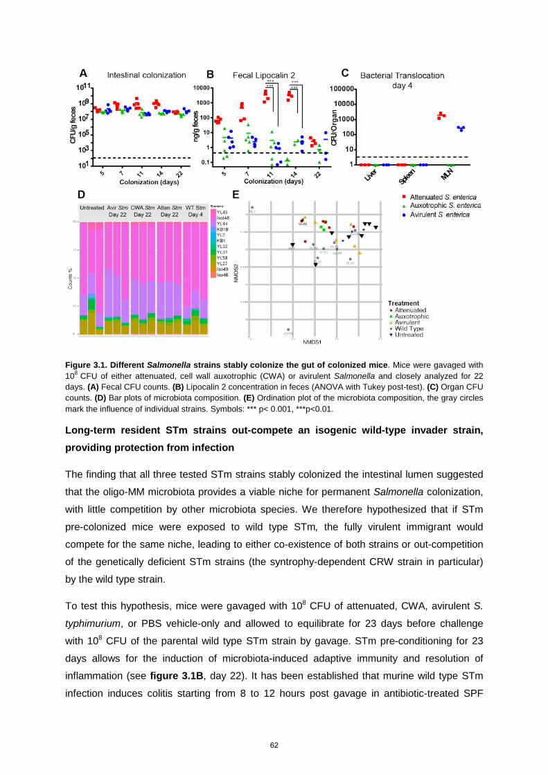
Figure 3.1. Different Salmonella strains stably colonize the gut of colonized mice. Mice were gavaged with 108 CFU of either attenuated, cell wall auxotrophic (CWA) or avirulent Salmonella and closely analyzed for 22 days. (A) Fecal CFU counts. (B) Lipocalin 2 concentration in feces (ANOVA with Tukey post-test). (C) Organ CFU counts. (D) Bar plots of microbiota composition. (E) Ordination plot of the microbiota composition, the gray circles mark the influence of individual strains. Symbols: *** p< 0.001, ***p<0.01.
Long-term resident STm strains out-compete an isogenic wild-type invader strain, providing protection from infection
The finding that all three tested STm strains stably colonized the intestinal lumen suggested
that the oligo-MM microbiota provides a viable niche for permanent Salmonella colonization,
with little competition by other microbiota species. We therefore hypothesized that if STm
pre-colonized mice were exposed to wild type STm, the fully virulent immigrant would
compete for the same niche, leading to either co-existence of both strains or out-competition
of the genetically deficient STm strains (the syntrophy-dependent CRW strain in particular)
by the wild type strain.
To test this hypothesis, mice were gavaged with 108 CFU of attenuated, CWA, avirulent S.
typhimurium, or PBS vehicle-only and allowed to equilibrate for 23 days before challenge
with 108 CFU of the parental wild type STm strain by gavage. STm pre-conditioning for 23
days allows for the induction of microbiota-induced adaptive immunity and resolution of
inflammation (see figure 3.1B, day 22). It has been established that murine wild type STm
infection induces colitis starting from 8 to 12 hours post gavage in antibiotic-treated SPF
62
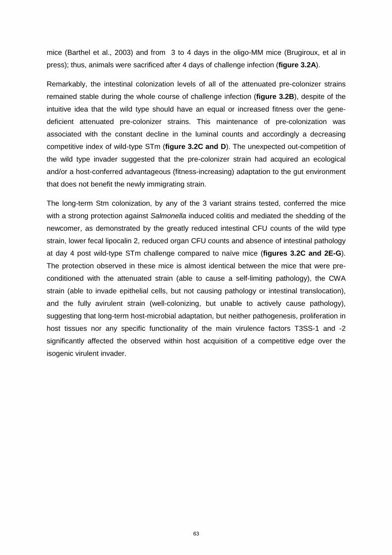
mice (Barthel et al., 2003) and from 3 to 4 days in the oligo-MM mice (Brugiroux, et al in
press); thus, animals were sacrificed after 4 days of challenge infection (figure 3.2A).
Remarkably, the intestinal colonization levels of all of the attenuated pre-colonizer strains
remained stable during the whole course of challenge infection (figure 3.2B), despite of the
intuitive idea that the wild type should have an equal or increased fitness over the gene-
deficient attenuated pre-colonizer strains. This maintenance of pre-colonization was
associated with the constant decline in the luminal counts and accordingly a decreasing
competitive index of wild-type STm (figure 3.2C and D). The unexpected out-competition of
the wild type invader suggested that the pre-colonizer strain had acquired an ecological
and/or a host-conferred advantageous (fitness-increasing) adaptation to the gut environment
that does not benefit the newly immigrating strain.
The long-term Stm colonization, by any of the 3 variant strains tested, conferred the mice
with a strong protection against Salmonella induced colitis and mediated the shedding of the
newcomer, as demonstrated by the greatly reduced intestinal CFU counts of the wild type
strain, lower fecal lipocalin 2, reduced organ CFU counts and absence of intestinal pathology
at day 4 post wild-type STm challenge compared to naïve mice (figures 3.2C and 2E-G).
The protection observed in these mice is almost identical between the mice that were pre-
conditioned with the attenuated strain (able to cause a self-limiting pathology), the CWA
strain (able to invade epithelial cells, but not causing pathology or intestinal translocation),
and the fully avirulent strain (well-colonizing, but unable to actively cause pathology),
suggesting that long-term host-microbial adaptation, but neither pathogenesis, proliferation in
host tissues nor any specific functionality of the main virulence factors T3SS-1 and -2
significantly affected the observed within host acquisition of a competitive edge over the
isogenic virulent invader.
63
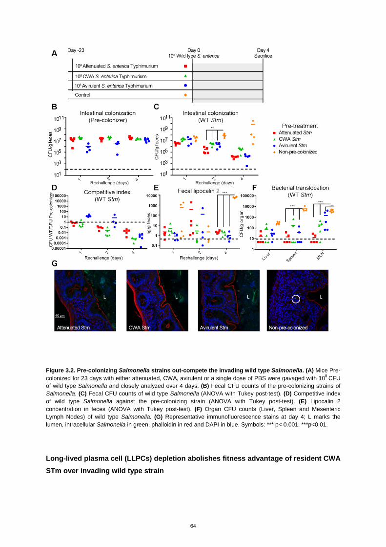
Figure 3.2. Pre-colonizing Salmonella strains out-compete the invading wild type Salmonella. (A) Mice Pre-colonized for 23 days with either attenuated, CWA, avirulent or a single dose of PBS were gavaged with 108 CFU of wild type Salmonella and closely analyzed over 4 days. (B) Fecal CFU counts of the pre-colonizing strains of Salmonella. (C) Fecal CFU counts of wild type Salmonella (ANOVA with Tukey post-test). (D) Competitive index of wild type Salmonella against the pre-colonizing strain (ANOVA with Tukey post-test). (E) Lipocalin 2 concentration in feces (ANOVA with Tukey post-test). (F) Organ CFU counts (Liver, Spleen and Mesenteric Lymph Nodes) of wild type Salmonella. (G) Representative immunofluorescence stains at day 4; L marks the lumen, intracellular Salmonella in green, phalloidin in red and DAPI in blue. Symbols: *** p< 0.001, ***p<0.01.
Long-lived plasma cell (LLPCs) depletion abolishes fitness advantage of resident CWA STm over invading wild type strain
64

To elucidate a mechanism for the observed out-competition of invading wild type STm by
resident isogenic derivatives, we decided to dissect the two possible origins of host-bacterial
co-adaptation that may evolve during intestinal long-term residency: first, the bacteria-
specific adaptive immune response, and second, fitness increasing bacterial genetic
adaptations.
In support of the first possible mechanism, it has been shown that specific IgA is able to
modulate the intestinal fitness of bacteria based on differential binding to populations
expressing variable outer membrane polysaccharide structures (Peterson et al., 2007a). To
experimentally address this possibility we depleted IgA producing plasma cells and B cells in
oligo-MM mice pre-conditioned by 23-day colonization with CWA STm one day before re-
challenge with STm wild type, by combined treatment with anti-CD20 antibody and
Bortezomib (Khodadadi et al., 2015).
Unlike in non-depleted control mice, out-competition of the wild-type invader strain by
resident CWA STm was abolished in mice depleted of IgA-producing cells. These mice
harbored a stable 1:1 ratio of both strains, despite of that fact that these mice had been co-
caged with the control group until the day of plasma cell depletion (figure 3.3A-C). The
depletion of LLPCs and B-cells did, however, not affect the organ colonization in these mice,
suggesting a certain degree of persistence in the protection against systemic infection,
possibly due to the long half-life of serum antibodies or a dominant role of T cells or other
immune cell types (figures 3.3D).
65
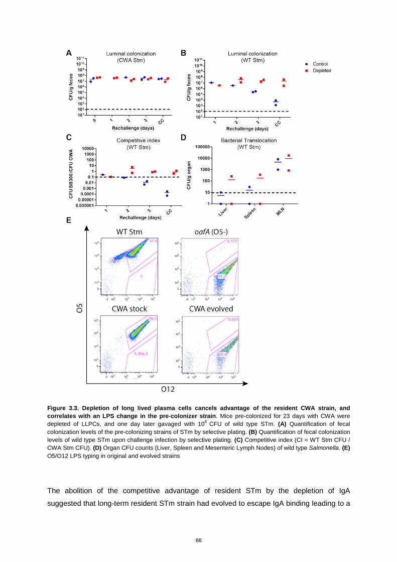
Figure 3.3. Depletion of long lived plasma cells cancels advantage of the resident CWA strain, and correlates with an LPS change in the pre-colonizer strain. Mice pre-colonized for 23 days with CWA were depleted of LLPCs, and one day later gavaged with 108 CFU of wild type STm. (A) Quantification of fecal colonization levels of the pre-colonizing strains of STm by selective plating. (B) Quantification of fecal colonization levels of wild type STm upon challenge infection by selective plating. (C) Competitive index (CI = WT Stm CFU / CWA Stm CFU). (D) Organ CFU counts (Liver, Spleen and Mesenteric Lymph Nodes) of wild type Salmonella. (E) O5/O12 LPS typing in original and evolved strains
The abolition of the competitive advantage of resident STm by the depletion of IgA
suggested that long-term resident STm strain had evolved to escape IgA binding leading to a
66
fitness gain over the non-evolved immigrant. The LPS O-antigen is the main determinant of
STm binding and protective intestinal IgA (Levinson et al., 2015; Michetti et al., 1992), and
immunity driven LPS O-antigen phase variation leading to the spontaneous loss of the
dominant O5 antigen, allows evasion of recognition by anti-O5 IgA without loss of
invasiveness (Forbes et al., 2008; Michetti et al., 1992).
To assess if this bacterial adaptation mechanism could explain the observed luminal out-
competition, we characterized the LPS composition of long term intestinal resident CWA Stm
strain re-isolated after 34 days of colonization, in comparison to the original CWA STm stock.
The standard Stm LPS is mainly composed of a mixture of O5 and O12 antigens, where the
first possess an acetylation of a side chain abequose residue that is absent in the latter (Ilg et
al., 2013).
We analyzed by flow cytometry the ability of specific anti-O5 and anti-O12 antibodies to bind
to the bacterial LPS. We found that, as previously described, our lab stock of wild-type Stm
grown in vitro is predominantly O5 and O12 antigen-double positive. Spontaneous shifts in
the abequose acetylation rate (phase variation) generate minor subpopulations of O5- only
serotype bacteria (figure 3.3E). The non-host-adapted CWA Stm lab strain was
predominantly O5-/O12-double serotype positive, with a reduced serotype O5-only
subpopulation (for unknown reasons), whereas the host-adapted long-term resident CWA
strain had lost the expression of the abequose-acetylated O5 antigen (figure 3.3E). This
observation is reminiscent of a previous report describing fitness-increasing evasion of IgA
recognition by polysaccharide phase variants of Bacteroides thetaiotaomicron (Peterson et
al., 2007a). Based on these data, we hypothesized that the ability of resident STm to
outcompete the wild type STm immigration is related to the differential selective pressure
imposed by O5-specific IgA, since most of it would bind the newcomer WT Stm and not the
evolved CWA.
Sterile induction of STm-specific IgA combined with short-term pre-colonization with host-adapted CWA STm can partially rescue the observed protection phenotype
To address this hypothesis, we performed an orthogonally designed experiment selectively
varying (i), the short-term resident strain (day-1) (original stock CWA strain versus 34-day
host-adapted CWA strain) and (ii), the anti-STm IgA immunity of the mice (Moor et al.,
2016)(orally immunized with 3 doses PAA-inactivated CWA STm O-12/5 or naïve; figure 3.4A). The main advantage of using the CWA strain over the avirulent or attenuated for this
experiment is that the strict luminal containment guarantees that all bacteria-bacteria
67
competition is limited to the lumen, and not influenced re-seeding from persistently infected
MLNs (Kaiser et al., 2013).
27 days after primary immunization with PAA-inactivated CWA STm (figure 3.4, green and
red) or kept naive (figure 3.4 blue), all mice were short-term pre-colonized by gavage with
either host adapted (figure 3.4, red and blue) or non-host-adapted CWA STm (figure 3.4,
green), followed 1 day later by a challenge with 108 CFU of wild type STm for 4 days until
sacrifice.
Pre-colonized-only mice (figure 3.4B-D, blue) showed intestinal out-competition of the CWA
pre-colonizer by the wild type strain starting on day two, and lead to the total extinction of the
evolved CWA by day 4 in almost all the mice of this group, suggesting that the difference in
fitness between the pre-colonizer strain and the wild type invader cannot only be attributed to
non-immunity-related fitness-increasing intestinal adaptations.
The mice that were immunized with PAA-inactivated and pre-colonized with non-host-
adapted CWA STm showed a clear co-existence of the pre-colonizer strain with the wild
type, that was absent in the naïve mice (figure 3.4B-C). This partial rescuing of the
phenotype observed in figure 2, suggests that the adaptive immune response plays an
important role in the preservation of the resident microbiota in combination with bacteria
adaptation to IgA recognition. None the less we were unable to observe a statistically
significant difference between the evolved and the stock CWA strains, despite the
competitive index seeming different. A possible explanation to the absence of difference
between the response between stock CWA and evolved CWA strains could be that the PAA-
treated Stm induced an altered type of anti-LPS IgA. To address this possibility we repeated
the experiment from figure 3.4 taking advantage of the transient colonization of CWA strains
in germ-free mice to induce adaptive immune response independent of colonization
(figureS3.2A). The results obtained from live bacteria IgA induction reinforce the observation
of figure 4.3, discarding the idea that the non-functionality of PAA-treated induced IgA
(figureS3.2B-E)
These results indicate that a killed-bacteria-induced adaptive immune response, in
combination with a one day pre-colonization with a CWA strain (evolved or not) was able to
partially restore the phenomena observed in a long term colonization. We conclude from this
result that a second unknown mechanism of bacterial adaptation must contribute to the
fitness increase of resident attenuated STm, in combination with the LPS O-antigen phase
variation and the IgA immunity.
68
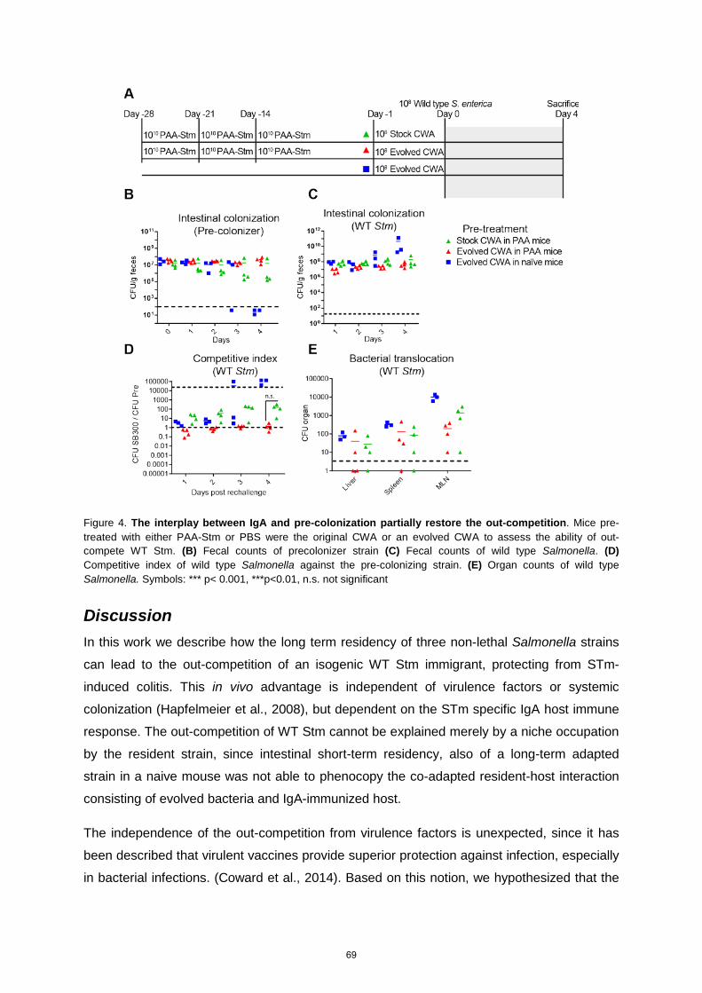
Figure 4. The interplay between IgA and pre-colonization partially restore the out-competition. Mice pre-treated with either PAA-Stm or PBS were the original CWA or an evolved CWA to assess the ability of out-compete WT Stm. (B) Fecal counts of precolonizer strain (C) Fecal counts of wild type Salmonella. (D) Competitive index of wild type Salmonella against the pre-colonizing strain. (E) Organ counts of wild type Salmonella. Symbols: *** p< 0.001, ***p<0.01, n.s. not significant
Discussion In this work we describe how the long term residency of three non-lethal Salmonella strains
can lead to the out-competition of an isogenic WT Stm immigrant, protecting from STm-
induced colitis. This in vivo advantage is independent of virulence factors or systemic
colonization (Hapfelmeier et al., 2008), but dependent on the STm specific IgA host immune
response. The out-competition of WT Stm cannot be explained merely by a niche occupation
by the resident strain, since intestinal short-term residency, also of a long-term adapted
strain in a naive mouse was not able to phenocopy the co-adapted resident-host interaction
consisting of evolved bacteria and IgA-immunized host.
The independence of the out-competition from virulence factors is unexpected, since it has
been described that virulent vaccines provide superior protection against infection, especially
in bacterial infections. (Coward et al., 2014). Based on this notion, we hypothesized that the
69

transient inflammation induced by partially attenuated STm during establishment of long-term
residency would lead to increased potency to out-compete wild-type Stm compared to the
avirulent strains tested. This hypothesis proved to be wrong since all three strains tested
(attenuated, avirulent, and CWA) were equally able to maintain their resident colonization
levels, and outcompete the wild-type Stm strain.
The hypothesis that continuous inflammation would benefit WT Stm could not be fully verified
or discarded. The high variability in lipocalin 2 at day 2 suggests that in some mice there was
a transient increase in the neutrophil mediated activity, however all the long term colonized
mice were able to out-compete wild-type Stm. The variability in inflammation correlated with
the variability in the competitive index (Figure 3.2 D and E), further supporting the idea that
the bacteria-bacteria competition is crucial for homeostasis.
It has been shown previously that IgA can modify the in vivo fitness of bacterial strains,
based on their ability to be recognized by IgA (Peterson et al., 2007b). Based on this we
explored the hypothesis that the host-imposed selective pressure was dependent on
secretory IgA, and we tested it by depletion of IgA-producing LLPCs. The depletion of
plasma cells abolished the competitive advantage of the pre-colonizer strain, leading to a
continuous co-existence of both strains. This phenomenon suggests that the continuous
selective pressure effected by specific IgA is a crucial factor in the differentiation between
resident and immigrant strains.
The idea that the main function of IgA is to increase the competitiveness of resident
microbiota against pathogens is strongly supported by current evidence. It has been shown
that colitogenic bacteria are more likely to induce IgA responses than the non-colitogenic
counterpart (Palm et al., 2014), and the excessive IgA-mediated responses against
commensals are an important factor in the development of colitis (Wenzel et al., 2014).
The precise mechanism of how IgA may alter luminal bacterial survival is not fully
understood. It has been reported that the ratio between capsule mutants and WT B.
thetaiotaomicron can be increased up to 100 fold in presence of IgA directed against the
capsule (Peterson et al., 2007a). It has also been shown that the clustering of dividing cell
populations can lead to differential exclusion of certain clones from the lumen (K. Moor, E.
Slack and coworkers, unpublished data). Besides a fitness-decreasing effect of IgA, it has
been shown that IgA can impair the function of the Salmonella T3SS, effectively neutralizing
the virulence factors (Forbes et al., 2008; Levinson et al., 2015; Michetti et al., 1992).
The continuous selective pressure imposed on the microbiota by the reactivity of IgA is
possibly the driving force to select O5 LPS negative strains of Stm. Since it is known that IgA
70
can reduce the fitness of the recognized bacteria (Peterson et al., 2007a) it is conceivable
that the ability to generate variations of LPS antigens may increase in vivo fitness. This
continuous bacteria-host interplay eventually leads to a vast array of different IgA to
accumulate over time, reducing the possibility of a newcomer non-adapted tourist to
establish.
Since IgA on its own is not able to clear Salmonella from the gut (Endt et al., 2010), we tried
to recapitulate the observed mutualistic Stm-IgA synergic interaction by sterile immunization
of the animals with PAA-inactivated Stm, followed by a one day pre-colonization (Moor et al.,
2016). This procedure was able to avoid the extinction of the CWA strain by immigration of
wild-type Stm, evidenced in a stable 1:1 colonization ratio, which was lost in the non-
immunized group that generated no Stm-binding IgA. Nonetheless, the combination of these
two factors was still unable to fully confer out-competition of immigrating wild-type Stm. One
possible explanation for this incomplete effect was that the IgA was artificially generated by
immunization with PAA-killed instead of live organisms, potentially structurally altered by the
harsh oxidation of important antigenic determinants. This hypothesis could be rejected based
on the fact that the immunization with live bacteria (Figure S3.2) generated an identical out-
competition phenotype.
Our current inability to completely re-capitulate the WT Stm out-competition by seperating
immunization from pre-colonization suggests that we could not detect all contributing
mechanisms. We therefore hypothesize that the pre-colonizer strain needs to colonize for a
longer time than one day to fully adjust to the luminal environment, since the strains were
adapted to the in vitro conditions. The mechanism of this adaptation is probably by increased
expression of scavenging proteins or a metabolic adaptation, leading to an environmental
advantage over the WT Stm strain.
In conclusion, we maintain our hypothesis that the metabolic competition by a host-adapted
resident microbial strain in combination with the ability of strain-specific IgA to decrease the
fitness of newly immigrating microbial strains, act synergistically to exclude pathogenic or
other tourist species. Our data further support the concept that the intestinal adaptive
immune system favors the species enrichment of the resident microbiota and improves
consortial stability (Dishaw et al., 2014). This mechanism sheds new light on the host-
mediated microbiota regulation, since most previous studies have only been able to describe
nutrient-related species variation.
71
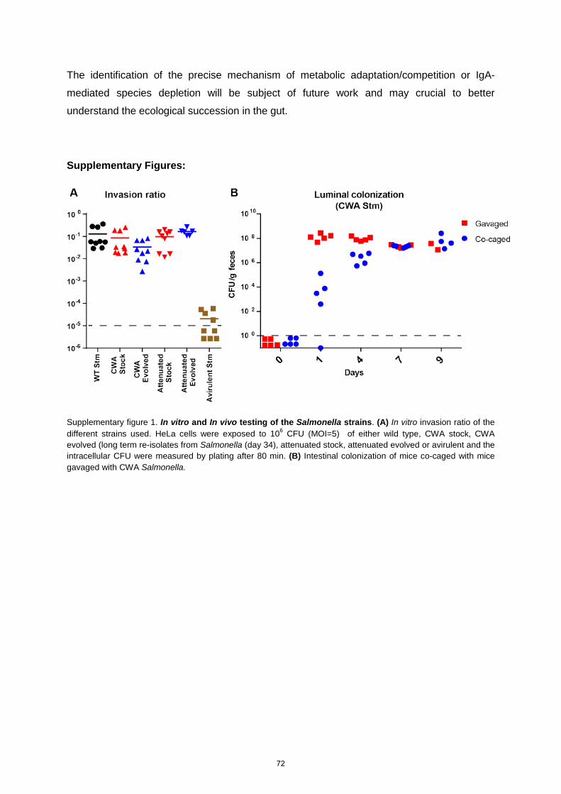
The identification of the precise mechanism of metabolic adaptation/competition or IgA-
mediated species depletion will be subject of future work and may crucial to better
understand the ecological succession in the gut.
Supplementary Figures:
Supplementary figure 1. In vitro and In vivo testing of the Salmonella strains. (A) In vitro invasion ratio of the different strains used. HeLa cells were exposed to 106 CFU (MOI=5) of either wild type, CWA stock, CWA evolved (long term re-isolates from Salmonella (day 34), attenuated stock, attenuated evolved or avirulent and the intracellular CFU were measured by plating after 80 min. (B) Intestinal colonization of mice co-caged with mice gavaged with CWA Salmonella.
72
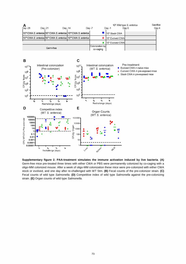
Supplementary figure 2. PAA-treatment simulates the immune activation induced by live bacteria. (A) Germ-free mice pre-treated three times with either CWA or PBS were permanently colonized by co-caging with a oligo-MM colonized mouse. After a week of oligo-MM colonization these mice were pre-colonized with either CWA stock or evolved, and one day after re-challenged with WT Stm. (B) Fecal counts of the pre-colonizer strain. (C) Fecal counts of wild type Salmonella. (D) Competitive index of wild type Salmonella against the pre-colonizing strain. (E) Organ counts of wild type Salmonella.
73
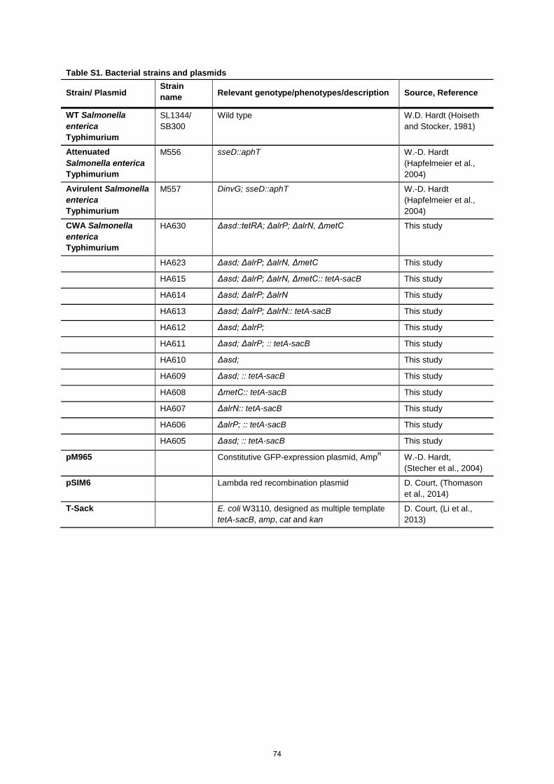
Table S1. Bacterial strains and plasmids
Strain/ Plasmid Strain name Relevant genotype/phenotypes/description Source, Reference
WT Salmonella enterica Typhimurium
SL1344/ SB300
Wild type W.D. Hardt (Hoiseth and Stocker, 1981)
Attenuated Salmonella enterica Typhimurium
M556 sseD::aphT W.-D. Hardt (Hapfelmeier et al., 2004)
Avirulent Salmonella enterica Typhimurium
M557 DinvG; sseD::aphT W.-D. Hardt (Hapfelmeier et al., 2004)
CWA Salmonella enterica Typhimurium
HA630 Δasd::tetRA; ΔalrP; ΔalrN, ΔmetC This study
HA623 Δasd; ΔalrP; ΔalrN, ΔmetC This study
HA615 Δasd; ΔalrP; ΔalrN, ΔmetC:: tetA-sacB This study
HA614 Δasd; ΔalrP; ΔalrN This study
HA613 Δasd; ΔalrP; ΔalrN:: tetA-sacB This study
HA612 Δasd; ΔalrP; This study
HA611 Δasd; ΔalrP; :: tetA-sacB This study
HA610 Δasd; This study
HA609 Δasd; :: tetA-sacB This study
HA608 ΔmetC:: tetA-sacB This study
HA607 ΔalrN:: tetA-sacB This study
HA606 ΔalrP; :: tetA-sacB This study
HA605 Δasd; :: tetA-sacB This study
pM965 Constitutive GFP-expression plasmid, AmpR W.-D. Hardt, (Stecher et al., 2004)
pSIM6 Lambda red recombination plasmid D. Court, (Thomason et al., 2014)
T-Sack E. coli W3110, designed as multiple template tetA-sacB, amp, cat and kan
D. Court, (Li et al., 2013)
74
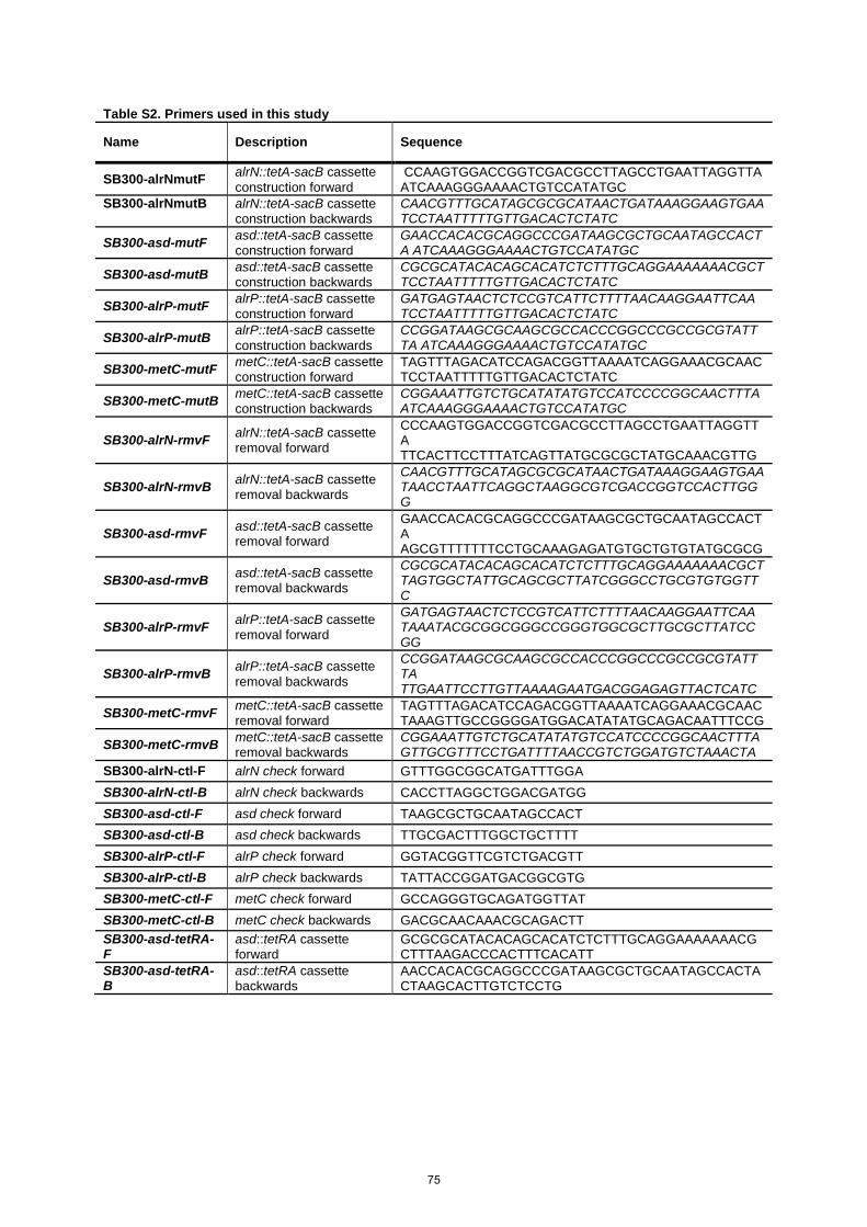
Table S2. Primers used in this study
Name Description Sequence
SB300-alrNmutF alrN::tetA-sacB cassette construction forward
CCAAGTGGACCGGTCGACGCCTTAGCCTGAATTAGGTTA ATCAAAGGGAAAACTGTCCATATGC
SB300-alrNmutB alrN::tetA-sacB cassette construction backwards
CAACGTTTGCATAGCGCGCATAACTGATAAAGGAAGTGAA TCCTAATTTTTGTTGACACTCTATC
SB300-asd-mutF asd::tetA-sacB cassette construction forward
GAACCACACGCAGGCCCGATAAGCGCTGCAATAGCCACTA ATCAAAGGGAAAACTGTCCATATGC
SB300-asd-mutB asd::tetA-sacB cassette construction backwards
CGCGCATACACAGCACATCTCTTTGCAGGAAAAAAACGCT TCCTAATTTTTGTTGACACTCTATC
SB300-alrP-mutF alrP::tetA-sacB cassette construction forward
GATGAGTAACTCTCCGTCATTCTTTTAACAAGGAATTCAA TCCTAATTTTTGTTGACACTCTATC
SB300-alrP-mutB alrP::tetA-sacB cassette construction backwards
CCGGATAAGCGCAAGCGCCACCCGGCCCGCCGCGTATTTA ATCAAAGGGAAAACTGTCCATATGC
SB300-metC-mutF metC::tetA-sacB cassette construction forward
TAGTTTAGACATCCAGACGGTTAAAATCAGGAAACGCAAC TCCTAATTTTTGTTGACACTCTATC
SB300-metC-mutB metC::tetA-sacB cassette construction backwards
CGGAAATTGTCTGCATATATGTCCATCCCCGGCAACTTTA ATCAAAGGGAAAACTGTCCATATGC
SB300-alrN-rmvF alrN::tetA-sacB cassette removal forward
CCCAAGTGGACCGGTCGACGCCTTAGCCTGAATTAGGTTA TTCACTTCCTTTATCAGTTATGCGCGCTATGCAAACGTTG
SB300-alrN-rmvB alrN::tetA-sacB cassette removal backwards
CAACGTTTGCATAGCGCGCATAACTGATAAAGGAAGTGAA TAACCTAATTCAGGCTAAGGCGTCGACCGGTCCACTTGGG
SB300-asd-rmvF asd::tetA-sacB cassette removal forward
GAACCACACGCAGGCCCGATAAGCGCTGCAATAGCCACTA AGCGTTTTTTTCCTGCAAAGAGATGTGCTGTGTATGCGCG
SB300-asd-rmvB asd::tetA-sacB cassette removal backwards
CGCGCATACACAGCACATCTCTTTGCAGGAAAAAAACGCT TAGTGGCTATTGCAGCGCTTATCGGGCCTGCGTGTGGTTC
SB300-alrP-rmvF alrP::tetA-sacB cassette removal forward
GATGAGTAACTCTCCGTCATTCTTTTAACAAGGAATTCAA TAAATACGCGGCGGGCCGGGTGGCGCTTGCGCTTATCCGG
SB300-alrP-rmvB alrP::tetA-sacB cassette removal backwards
CCGGATAAGCGCAAGCGCCACCCGGCCCGCCGCGTATTTA TTGAATTCCTTGTTAAAAGAATGACGGAGAGTTACTCATC
SB300-metC-rmvF metC::tetA-sacB cassette removal forward
TAGTTTAGACATCCAGACGGTTAAAATCAGGAAACGCAAC TAAAGTTGCCGGGGATGGACATATATGCAGACAATTTCCG
SB300-metC-rmvB metC::tetA-sacB cassette removal backwards
CGGAAATTGTCTGCATATATGTCCATCCCCGGCAACTTTA GTTGCGTTTCCTGATTTTAACCGTCTGGATGTCTAAACTA
SB300-alrN-ctl-F alrN check forward GTTTGGCGGCATGATTTGGA SB300-alrN-ctl-B alrN check backwards CACCTTAGGCTGGACGATGG SB300-asd-ctl-F asd check forward TAAGCGCTGCAATAGCCACT SB300-asd-ctl-B asd check backwards TTGCGACTTTGGCTGCTTTT SB300-alrP-ctl-F alrP check forward GGTACGGTTCGTCTGACGTT SB300-alrP-ctl-B alrP check backwards TATTACCGGATGACGGCGTG SB300-metC-ctl-F metC check forward GCCAGGGTGCAGATGGTTAT SB300-metC-ctl-B metC check backwards GACGCAACAAACGCAGACTT SB300-asd-tetRA-F
asd::tetRA cassette forward
GCGCGCATACACAGCACATCTCTTTGCAGGAAAAAAACGCTTTAAGACCCACTTTCACATT
SB300-asd-tetRA-B
asd::tetRA cassette backwards
AACCACACGCAGGCCCGATAAGCGCTGCAATAGCCACTACTAAGCACTTGTCTCCTG
75

Materials and Methods Animal colonization experiments
Germ-free animals were re-derived from C57BL/6 mice and maintained germ-free in flexible
film isolators in the Genaxen Foundation Clean Mouse Facility (CMF) of the University of
Bern as described (Macpherson and McCoy, 2014). Mice colonized with oligo-MM from birth
were also bred and maintained in the CMF. For experiments, the mice were transferred to
experimental flexible film isolators for the duration of the pre colonization or PAA-Stm
treatment. After the end of the pre-treatment the experimental mice were aseptically
transferred to autoclaved sealsafe-plus IVCs under positive pressure (Tecniplast, Italy) in a
barrier unit of the Genaxen Clean Mouse Facility. Cage changes were carried out under
strictly aseptic conditions. In all experiments animals were provided with sterile mouse chow
(Kliba 3437; autoclaved) and autoclaved water ad libitum. All experiments were performed
according to protocols approved by the Bernese Cantonal Ethical committee for animal
experiments and carried out in accordance with Swiss Federal law for animal
experimentation (license number BE91/14 and BE 36/15).
To generate contamination-free bacterial inoculums, NaCL (0.3M), D-Ala (200 μg/mL) and
Dap (50 μg/ mL) supplemented autoclaved LB medium in sterile-filter-sealed flasks, was
aseptically inoculated from single colonies of the test bacterium and incubated shaking at
150 rpm at 37°C for 16 hours. Bacteria were harvested by centrifugation (10 min, 4816 x g,
4°C) in a sterile aerosol-proof assembly, washed in autoclaved sterile PBS and concentrated
to a density of 5 108 CFU/mL in sterile PBS, performed aseptically under a sterile laminar
airflow. The bacterial suspensions were aseptically aliquoted in autoclaved plastic tubes and
sealed in a sterilized secondary containment. The sterile tubes containing the inocula and the
mice were aseptically imported into a sterilized laminar flow hood, and each animal
inoculated with 200 μL of bacterial suspension (containing 1 x 10⁸ CFU in sterile PBS, at a
density of 5 x 10⁸ CFU/mL) by gavage, carried out wearing sterile surgical gowns and sterile
surgical gloves. Fresh fecal pellets were collected aseptically, suspended in sterile PBS, and
plated in serial dilutions on D-Ala/ Dap-supplemented or non-supplemented MacConkey II
agar and incubated aerobically at 37°C for 24 hours.
Long-lived plasma cell depletion
To deplete long lived plasma cells and B-cells, mice were injected I.V under sterile conditions
with anti-CD20 (10mg/kg; 18B12 IgG2a, Biogen USA) and Bortezomib (0.75 mg/kg;
Millennium Pharmaceuticals USA) diluted in 100μL (Khodadadi et al., 2015).
76
Bacterial culture
LB medium (Sigma-Aldrich) was used as the standard growth media. MacConkey was used
as the standard media for fecal pellet counts. Where required, the following supplements
were added to the media: ampicillin (Sigma, 100 μg/mL), tetracycline (Sigma, 12.5 μg/mL),
kanamycin (Sigma, 50 μg/mL), meso-diaminopimelic acid (Sigma, 50 μg/mL), D-alanine
(Sigma, 200 μg/mL).
PAA-inactivation of STm
Wild type Salmonella enterica Typhimurium was cultured in 500 mL LB (NaCl 0.3M) shaking
at 150 rpm at 37°C for 16 h. Bacteria was harvested as described above and resuspended in
10 mL of 0.1% per-acetic acid in water for one hour. The bacterial particles were washed
three times with PBS by centrifugation and suspended at a density of 1011 particles/ mL. The
bacterial suspensions were aseptically aliquoted in autoclaved plastic tubes and sealed in a
sterilized secondary containment. The sterile tubes containing the inocula and the mice were
aseptically imported into plastic film isolators, and each animal inoculated with 200 μL of
bacterial particle suspension (containing 2 x 10¹⁰ CFU in sterile PBS, at a density of 2 x 10¹¹
CFU/mL) by gavage
Bacterial genetic engineering
All bacterial strains used or generated in this study are specified in S1 Table. The Salmonella
enterica Thyphimurium SB300, M556 and M557 were kindly provided by Wolf-Dietrich Hardt
(Hapfelmeier et al., 2004).The strain HA630 was generated by sequential P22 transduction
of the individual mutations, and using lambda Red recombineering to remove the tetA-sacB
cassettes with the appropriate “rmv” primers. All single deletions were carried out individually
in four strains (HA605-HA608) with the Court Lab Lambda Red recombineering method (Li et
al., 2013), using the pSIM6 plasmid and the tetA-sacB cassette. To generate the tetA-sacB
or tetRA cassettes PCRs were performed using the gene appropriate “mut” primer and the
T-SACK strain (tetA-sacB) or a Tn10 based cassette (tetRA) (Li et al., 2013). All deletions
were confirmed by PCR using the appropriate “ctl” primer. All primers are described in table
S2
Fluorescent imaging
The distal 3 cm of the cecum were fixed in 4% PFA in PBS for 24 h, and then transferred to a
20% Sucrose solution for 24-48 h. The fixed cecum was then embedded in OTC media, and
flash frozen with liquid nitrogen. Sections of 7 µm thickness were cut and rinsed twice in PBS
and one time in PBS/2% BSA. Sections were stained with a PBS/2% BSA solution containing
77

DAPI (Sigma-Aldrich, USA, 0.01 mg/mL final concentration) and Phalloidin-Atto 647N
(Sigma-Aldrich, USA, 0.04 nmol/mL final concentration) for 30min. Sections were rinsed
again twice in PBS and one time in PBS/2%BSA and mounted under Vectashield (Vector
laboratories, USA). Images were acquired using a Zeiss LSM710 Laser scanning microscope
with a 40X oil objective (three z-stacks with 3 μm spacing, 150 μm square sections,
independent mode channels).
Gentamicin protection assay
HeLa (Kyoto) cells were seeded into 24-well dishes and grown for 1 day to obtain 80%
confluency. The cells were cultured in Dulbecco modified Eagle medium (DMEM) containing
10% fetal bovine serum (FBS). During the whole procedure cells were incubated at 37°C
under an atmosphere containing 5% CO2. The adherent cells were washed three times with
Hanks’ buffered salt (HBSS) and covered with 500 μL HBSS, 10 min before cells were
infected with bacteria. Cultures of the appropriate strain were grown in 10 mL 0.3 M NaCl LB
for 16 h, shaking at 150 rpm, at 37°C, diluted 1:20 into 40 mL fresh 0.3M NaCl LB media,
and subcultured for 5 h under the same conditions. Bacteria were then washed once in PBS
and diluted to an OD600 of 0.6. HeLa cells were infected with 5 μL of the resulting bacterial
suspension per well and were incubated in HBSS for 50 min. To kill extracellular bacteria,
HeLa cells were washed three times with HBSS and incubated in 500 μl DMEM containing
10% FBS and 400 μg/mL gentamicin, for 30 min. This provides a total infection time of 80
min. HeLa cells were afterwards washed three times with PBS and lysed in 0.1% sodium-
deoxycholate/PBS. The CFUs of intracellular bacteria and the infectious dose were
determined by plating of appropriate dilutions on D-Ala and m-Dap supplemented LB agar.
The intracellular (= gentamicin protected) CFUs were calculated as a ratio of the whole
inoculum.
Lipocalin-2 ELISA
Fecal Lipocalin-2 was measured using the solid phase Sandwich ELISA Mouse Lipocalin-
2/NGAL DuoSet (R&D Systems, USA). The assay was performed according to the
manufacturer’s instructions, except for the horseradish peroxidase, HRP-SA which was used
from Biolegend (USA). Fecal pellets from mice were dissolved in 0.5 mL sterile PBS, and
debris were spun down (7000 rpm, 5min). The supernatant was used as a sample, and a
dilution series was made (1:3 dilution, 6 dilutions). Of the standard a dilution series was
made in duplicates (1:3 dilution, 12 dilutions). The OD405 was measured using a thermomax
microplate reader (Molecular Devices, USA) Data was analyzed using graph pad Prism
program. A four parameter dose-response curve was fitted, using equal hill slopes for all
78

samples on the same plate. Samples with low OD405 values resulting in a curve fit with R2
values below 0.95 were defined below detection limit. By comparison with the standard, the
EC50 was used to calculate the absolute amount of Lipocalin-2 per sample.
Live bacterial flow cytometry for LPS-O-antigen typing
The appropriate Salmonella strain was grown in 0.2 μm membrane-filtered LB broth
overnight at 37°C and 150 rpm. 500 μL of culture was gently pelleted for 3 min at 4816 x g in
a Heraus Fresco 21 centrifuge and suspended with 500 μL of sterile-filtered 2% BSA/
0.005% NaN3/ PBS. One microliter of this solution was transferred to a new clean 1.5 mL
tube and incubated for 5 minutes at room temperature with anti-O5 (Difco Salmonella O
antisera Factor 5 #226601) and anti-O12 antibodies (STA5 EBV-transformed B cell
supernatant). The bacteria was washed once with 500 μL of sterile-filtered 2% BSA/ 0.005%
NaN3/ PBS and pelleted for 3 min at 4816 x g in a Heraus Fresco 21. The supernatant was
discarded and the pellet was re-suspended in 200 μL of 2% BSA/ 0.005% NaN3/ PBS
containing BV421-anti-Rabbit (secondary to anti O5, Biolegend) and Ax647-anti-hIgG
(secondary to anti O12, Jackson)
Data analysis and statistical analysis
All data analysis was done using the Prism statistical programs. The appropriate statistics
are explained in the legend of each figure
Acnowledgements We thank the Clean Mouse Facility for all the animal work, Jens Stein for kindly providing the
anti-CD20 antibody, Wolf Dietrich-Hardt for providing the original WT Stm strain, Fabian
Blank, Carlos Wotzkow and the MIC for their support with the microscopes. This work was
funded by the grants ERC 281904 and SNF 310030_138452.
References Barroso-Batista, J., Demengeot, J., and Gordo, I. (2015). Adaptive immunity increases the
pace and predictability of evolutionary change in commensal gut bacteria. 6, 8945.
Barthel, M., Hapfelmeier, S., Quintanilla-Martínez, L., Kremer, M., Rohde, M., Hogardt, M.,
Pfeffer, K., Rüssmann, H., and Hardt, W. (2003). Pretreatment of mice with streptomycin
provides a Salmonella enterica serovar Typhimurium colitis model that allows analysis of
both pathogen and host. 71, 2839-2858.
79
Coward, C., Restif, O., Dybowski, R., Grant, A.J., Maskell, D.J., and Mastroeni, P. (2014).
The effects of vaccination and immunity on bacterial infection dynamics in vivo. 10,
e1004359.
Cuenca, M., Pfister, S.P., Buschor, S., Bayramova, F., Hernández, S.B., Cava, F., Kuru, E.,
Van Nieuwenhze, M.S., Brun, Y.V., Coelho, F.M., et al. (2016). D-Alanine-Controlled
Transient Intestinal Mono-Colonization with Non-Laboratory-Adapted Commensal E. coli
Strain HS. 11, e0151872.
Diard, M., Garcia, V., Maier, L., Remus-Emsermann, M.N., Rol, Regoes, R., Ackermann, M.,
Hardt, W., and Regoes, R.R. (2013). Stabilization of cooperative virulence by the expression
of an avirulent phenotype. Nature 494, 353-356.
Dishaw, L.J., Cannon, J.P., Litman, G.W., and Parker, W. (2014). Immune-directed support
of rich microbial communities in the gut has ancient roots. 47, 36-51.
Endt, K., Stecher, B., Chaffron, S., Slack, E., Tchitchek, N., Benecke, A., Van Maele, L.,
Sirard, J., Mueller, A.J., Heikenwalder, M., et al. (2010). The microbiota mediates pathogen
clearance from the gut lumen after non-typhoidal Salmonella diarrhea. PLOS Pathogens 6,
e1001097.
Forbes, S.J., Eschmann, M., and Mantis, N.J. (2008). Inhibition of Salmonella enterica
serovar typhimurium motility and entry into epithelial cells by a protective
antilipopolysaccharide monoclonal immunoglobulin A antibody. 76, 4137-4144.
Fransen, F., Zagato, E., Mazzini, E., Fosso, B., Manzari, C., Aidy, El, S., Chiavelli, A.,
D’Erchia, A.M., Sethi, M.K., Pabst, O., et al. (2015). BALB/c and C57BL/6 Mice Differ in
Polyreactive IgA Abundance, which Impacts the Generation of Antigen-Specific IgA and
Microbiota Diversity. Immunity 43, 527.
Gauger, E.J., Leatham, M.P., Mercado-Lubo, R., Laux, D.C., Conway, T., and Cohen, P.S.
(2007). Role of motility and the flhDC Operon in Escherichia coli MG1655 colonization of the
mouse intestine. 75, 3315-3324.
Hapfelmeier, S., Müller, A.J., Stecher, B., Kaiser, P., Barthel, M., Endt, K., Eberhard, M.,
Robbiani, R., Jacobi, C.A., Heikenwalder, M., et al. (2008). Microbe sampling by mucosal
dendritic cells is a discrete, MyD88-independent step in DeltainvG S. Typhimurium colitis.
The Journal Of Experimental Medicine 205, 437-50.
Hapfelmeier, S., Ehrbar, K., Stecher, B., Barthel, M., Kremer, M., and Hardt, W. (2004). Role
of the Salmonella pathogenicity island 1 effector proteins SipA, SopB, SopE, and SopE2 in
80

Salmonella enterica subspecies 1 serovar Typhimurium colitis in streptomycin-pretreated
mice. Infection And Immunity 72, 795-809.
Hapfelmeier, S., Stecher, B., Barthel, M., Kremer, M., Müller, A.J., Heikenwalder, M.,
Stallmach, T., Hensel, M., Pfeffer, K., Akira, S., et al. (2005). The Salmonella pathogenicity
island (SPI)-2 and SPI-1 type III secretion systems allow Salmonella serovar typhimurium to
trigger colitis via MyD88-dependent and MyD88-independent mechanisms. J. Immunol. 174,
1675-85.
Hoiseth, S.K., and Stocker, B.A. (1981). Aromatic-dependent Salmonella typhimurium are
non-virulent and effective as live vaccines. Nature 291, 238-9.
Ilg, K., Zandomeneghi, G., Rugarabamu, G., Meier, B.H., and Aebi, M. (2013). HR-MAS
NMR reveals a pH-dependent LPS alteration by de-O-acetylation at abequose in the O-
antigen of Salmonella enterica serovar Typhimurium. Carbohydrate Research 382, 58.
Kaiser, P., Slack, E., Grant, A.J., Hardt, W., and Regoes, R.R. (2013). Lymph Node
Colonization Dynamics after Oral Salmonella Typhimurium Infection in Mice. PLOS
Pathogens 9, e1003532.
Khodadadi, L., Cheng, Q., Alexander, T., Sercan-Alp, Ö., Klotsche, J., Radbruch, A., Hiepe,
F., Hoyer, B.F., and Taddeo, A. (2015). Bortezomib Plus Continuous B Cell Depletion
Results in Sustained Plasma Cell Depletion and Amelioration of Lupus Nephritis in NZB/W
F1 Mice. 10, e0135081.
Leatham, M.P., Banerjee, S., Autieri, S.M., Mercado-Lubo, R., Conway, T., and Cohen, P.S.
(2009). Precolonized human commensal Escherichia coli strains serve as a barrier to E. coli
O157:H7 growth in the streptomycin-treated mouse intestine. 77, 2876-2886.
Levinson, K.J., De Jesus, M., and Mantis, N.J. (2015). Rapid Effects of a Protective O-
Polysaccharide-Specific Monoclonal IgA on Vibrio cholerae Agglutination, Motility, and
Surface Morphology. Infection And Immunity 83, 1674.
Li, X., Thomason, L.C., Sawitzke, J.A., Costantino, N., and Court, D.L. (2013). Positive and
negative selection using the tetA-sacB cassette: recombineering and P1 transduction in
Escherichia coli. Nucleic Acids Research 41, e204.
Macpherson, A.J., and McCoy, K.D. (2014). Standardised animal models of host microbial
mutualism. 8, 476-486.
81
Maltby, R., Leatham-Jensen, M.P., Gibson, T., Cohen, P.S., and Conway, T. (2013).
Nutritional basis for colonization resistance by human commensal Escherichia coli strains HS
and Nissle 1917 against E. coli O157:H7 in the mouse intestine. Plos ONE 8, e53957.
McSorley, S.J., Cookson, B.T., and Jenkins, M.K. (2000). Characterization of CD4+ T cell
responses during natural infection with Salmonella typhimurium. 164, 986-993.
Medina, E., Paglia, P., Nikolaus, T., Müller, A., Hensel, M., and Guzmán, C.A. (1999).
Pathogenicity island 2 mutants of Salmonella typhimurium are efficient carriers for
heterologous antigens and enable modulation of immune responses. Infection And Immunity
67, 1093-9.
Michetti, P., Mahan, M.J., Slauch, J.M., Mekalanos, J.J., and Neutra, M.R. (1992).
Monoclonal secretory immunoglobulin A protects mice against oral challenge with the
invasive pathogen Salmonella typhimurium. Infection And Immunity 60, 1786-1792.
Moor, K., Wotzka, S.Y., Toska, A., Diard, M., Hapfelmeier, S., and Slack, E. (2016).
Peracetic Acid Treatment Generates Potent Inactivated Oral Vaccines from a Broad Range
of Culturable Bacterial Species. Frontiers In Immunology 7, 34.
Palm, N.W., de Zoete, M.R., Cullen, T.W., Barry, N.A., Stefanowski, J., Hao, L., Degnan,
P.H., Hu, J., Peter, I., Zhang, W., et al. (2014). Immunoglobulin A coating identifies
colitogenic bacteria in inflammatory bowel disease. 158, 1000-1010.
Peterson, D.A., McNulty, N.P., Guruge, J.L., and Gordon, J.I. (2007a). IgA response to
symbiotic bacteria as a mediator of gut homeostasis. 2, 328-339.
Peterson, D.A., McNulty, N.P., Guruge, J.L., and Gordon, J.I. (2007b). IgA Response to
Symbiotic Bacteria as a Mediator of Gut Homeostasis. Cell Host &Amp; Microbe 2, 328.
Ridaura, V.K., Faith, J.J., Rey, F.E., Cheng, J., Duncan, A.E., Kau, A.L., Griffin, N.W.,
Lombard, V., Henrissat, B., Bain, J.R., et al. (2013). Gut microbiota from twins discordant for
obesity modulate metabolism in mice. 341, 1241214.
Rogier, E.W., Frantz, A.L., Bruno, M.E.C., and Kaetzel, C.S. (2014). Secretory IgA is
Concentrated in the Outer Layer of Colonic Mucus along with Gut Bacteria. 3, 390-403.
Stecher, B., Hapfelmeier, S., Müller, C., Kremer, M., Stallmach, T., and Hardt, W. (2004).
Flagella and chemotaxis are required for efficient induction of Salmonella enterica serovar
Typhimurium colitis in streptomycin-pretreated mice. Infection And Immunity 72, 4138-50.
82
Stecher, B., Robbiani, R., Walker, A.W., Westendorf, A.M., Barthel, M., Kremer, M., Chaffron,
S., Macpherson, A.J., Buer, J., Parkhill, J., et al. (2007). Salmonella enterica serovar
typhimurium exploits inflammation to compete with the intestinal microbiota. 5, 2177-2189.
Thomason, L.C., Sawitzke, J.A., Li, X., Costantino, N., and Court, D.L. (2014).
Recombineering: genetic engineering in bacteria using homologous recombination. 106,
1.16.1-1.16.39.
Wenzel, U.A., Magnusson, M.K., Rydström, A., Jonstrand, C., Hengst, J., Johansson, M.E.,
Velcich, A., Öhman, L., Strid, H., Sjövall, H., et al. (2014). Spontaneous Colitis in Muc2-
Deficient Mice Reflects Clinical and Cellular Features of Active Ulcerative Colitis. Plos ONE
9, e100217.
Zhang, H., Sparks, J.B., Karyala, S.V., Settlage, R., and Luo, X.M. (2014). Host adaptive
immunity alters gut microbiota. The ISME Journal 9, 770.
83
General Discussion In this work we developed and tested a new bacterial tool for the study of the host-microbiota
interaction. This tool, referred to as cell wall auxotrophic (CWA) bacteria, was first fully
characterized for the human commensal E. coli HS. We were able to determine the extent of
CWA bacterial survival under non-permissive conditions, measure their mean half-life in
germ-free animals, and establish the similarity to the wild type bacteria-host interaction
(Cuenca et al., 2016). This characterization allowed us to simulate a short-lived colonization
with a commensal, safely return the mice to germ-free status and elicit a long-lived immune
response towards that strain. Despite this being done in the past by Hapfelmeier et al, we
offer now a more reliable system and a full characterization of all the properties, expanding
our understanding of the effects of cell wall starvation (Hapfelmeier et al., 2010).
Transient bacterial colonization using CWA bacteria provides a method to expose bacteria-
naïve germ-free mice to live antigens, but without compromising their axenic hygiene status.
The microbiota conditioning of germ-free animals may therefore be used for the immune
normalization of germ-free mice, potentially correcting several of the known alteration of their
immune system, like neutropenia, enlarged cecum size, irregular mucus or disrupted immune
responses (Balmer et al., 2014; Jakobsson et al., 2015; Macpherson and McCoy, 2014).
In line, the finding that CWA bacterial strains were able to emulate the early aspects of a
bacterial colonization led us to speculate if it would also be possible to recapitulate the early
signaling events of a pathogen infection. For this purpose we exposed germ-free mice to a
CWA strain of Salmonella enterica Typhimurium (Stm) and measured the early mucosal
transcriptional response pattern. We found that transient CWA Stm infection effectively
emulated (and was limited to) the host responses occurring during the first few hours of wild
type infection, independent of intestinal damage, invasion into deep tissues, or permanent
pathogen colonization. Our findings showed that the induction of the early intestinal innate
response by Stm is damage-independent but virulence factor-dependent. This induction is
comparable to the NLRC4 dependent epithelial cell expulsion observed by Sellin et al, where
they show that the primary line of defense starts occurring as early as 6 hours post infection
in the antibiotic treated model (Sellin et al., 2014).
The ability to emulate short-duration pathogenic or commensal bacteria-host interactions
opens up new possibilities for studying the importance of the sequential bacterial colonization
or the effects of bacterial pre-exposition in the colonization process, independently of the
permanent colonization of the gut. It also allows determining the longevity of the immune
responses after the antigen has been removed, the source of very heated discussions in the
84
mucosal immunology field (Bredholt et al., 2014; Hapfelmeier et al., 2010; Mattioli and
Tomasi, 1973).
Besides the opportunity to address basic immunology questions, the CWA system can be
used to study a complex microbiota interaction. Indeed, the process of colonization of the gut
and the consequent host-microbiota interplay can be exquisitely well studied using this
system, as we were able to establish a CWA Stm long term colonization system, which does
not lead to disease.
Utilizing the - rather unexpected - long term luminal colonization of the CWA Stm in
gnotobiotic mice harboring a reduced-complexity microbiota, as well as other modified Stm
strains, we were able to characterize the ability of the host to directly affect the bacterial
fitness in a species- and strain-specific manner. The continuous bacteria-specific selective
pressure imposed by IgA, in combination with bacterial adaptation protected the resident
strain and suppressed the establishment of a wild type Stm immigrant. This finding is
reminiscent of the advantage that certain strains can have due to surface carbohydrate
variation (Peterson et al., 2007)
Although the mechanism of how IgA alters luminal bacteria survival still remains incompletely
understood, our findings provide evidence for an active IgA-driven process to preserve
resident bacteria. This observation goes in line with the hypothesis that the adaptive immune
system promotes of a stable and beneficial resident microbiota (Dishaw et al., 2014), and the
continuous bacterial fitness-increasing adaptation (Degnan et al., 2014). An important
unexplored area of this work is the non-genetic adaptations in bacteria that might be induced
by a short stay in the gut. It has been shown in the past that once a strain manages to fully
colonize the gut, it is able to deplete nutrients and prevent the colonization of newcomers
(Maltby et al., 2013). This aspect should be further explored to assess is this is an important
factor in the out-competition of similar strains.
Future work may explore the possibilities for utilizing these interactions to change the
community structure of the microbiota therapeutically, for more effective prevention of
intestinal infection than currently achievable with traditional vaccination approaches.
References
Balmer, M.L., Schürch, C.M., Saito, Y., Geuking, M.B., Li, H., Cuenca, M., Kovtonyuk, L.V., McCoy, K.D., Hapfelmeier, S., Ochsenbein, A.F., et al. (2014). Microbiota-derived compounds drive steady-state granulopoiesis via MyD88/TICAM signaling. J. Immunol. 193, 5273-83.
85
Bredholt, G., Brokstad, K.A., Pathirana, R.D., Aarstad, H.J., Tondel, C., and Cox, R.J. (2014). Longevity of B-Cell and T-Cell Responses After Live Attenuated Influenza Vaccination in Children. Journal Of Infectious Diseases 211, 1541.
Cuenca, M., Pfister, S.P., Buschor, S., Bayramova, F., Hernandez, S.B., Cava, F., Kuru, E., Van Nieuwenhze, M.S., Brun, Y.V., Coelho, F.M., et al. (2016). D-Alanine-Controlled Transient Intestinal Mono-Colonization with Non-Laboratory-Adapted Commensal E. coli Strain HS. Plos ONE 11, e0151872.
Degnan, P.H., Barry, N.A., Mok, K.C., Taga, M.E., and Goodman, A.L. (2014). Human Gut Microbes Use Multiple Transporters to Distinguish Vitamin B12 Analogs and Compete in the Gut. Cell Host &Amp; Microbe 15, 47.
Dishaw, L.J., Cannon, J.P., Litman, G.W., and Parker, W. (2014). Immune-directed support of rich microbial communities in the gut has ancient roots. 47, 36-51.
Hapfelmeier, S., Lawson, M.A.E., Slack, E., Kirundi, J.K., Stoel, M., Heikenwalder, M., Cahenzli, J., Velykoredko, Y., Balmer, M.L., Endt, K., et al. (2010). Reversible microbial colonization of germ-free mice reveals the dynamics of IgA immune responses. 328, 1705-1709.
Jakobsson, H.E., Holmén-Larsson, J., Schütte, A., Ermund, A., Rodríguez-Piñeiro, A.M., Arike, L., Wising, C., Svensson, F., Bäckhed, F., and Hansson, G.C. (2015). Normalization of Host Intestinal Mucus Layers Requires Long-Term Microbial Colonization. Cell Host &Amp; Microbe 18, 582-92.
Macpherson, A.J., and McCoy, K.D. (2014). Standardised animal models of host microbial mutualism. Mucosal Immunology 8, 476.
Maltby, R., Leatham-Jensen, M.P., Gibson, T., Cohen, P.S., and Conway, T. (2013). Nutritional basis for colonization resistance by human commensal Escherichia coli strains HS and Nissle 1917 against E. coli O157:H7 in the mouse intestine. Plos ONE 8, e53957.
Mattioli, C.A., and Tomasi, T.B. (1973). The life span of IgA plasma cells from the mouse intestine. The Journal Of Experimental Medicine 138, 452-60.
Peterson, D.A., McNulty, N.P., Guruge, J.L., and Gordon, J.I. (2007). IgA Response to Symbiotic Bacteria as a Mediator of Gut Homeostasis. Cell Host &Amp; Microbe 2, 328.
Sellin, M.E., Müller, A.A., Felmy, B., Dolowschiak, T., Diard, M., Tardivel, A., Maslowski,
K.M., and Hardt, W. (2014). Epithelium-Intrinsic NAIP/NLRC4 Inflammasome Drives Infected
Enterocyte Expulsion to Restrict Salmonella Replication in the Intestinal Mucosa. Cell Host
&Amp; Microbe 16, 237.
86
Miguelangel Cuenca Address: Frohberweg 11 Bern 3012, Switzerland Phone: (+41) 79 646 0892 Nationality: Spanish / Venezuelan Date of birth: February 6th, 1987Email: [email protected] https://ch.linkedin.com/in/miguelangel-cuenca-a67b8517
Education
2012-2016 PhD in Immunology University of Bern, Switzerland Committee: Siegfried Hapfelmeier, Emma Slack-Wetter and, Philippe Krebs External referee: Nassos Typas
2013 Computational biology, practical course European Molecular Biology Organization
2011-2012 Master in biochemistry, molecular biology and biomedicine Universidad Complutense de Madrid, Spain Supervisor: Daniela Xavier Dias
2011 Mathematical modelling for infectious diseases dynamics, practical course Welcome Trust Genome Centre, United Kingdom
2003-2009 Bachelor degree in biology Universidad Simón Bolívar, Venezuela
Experience
2012-2016 PhD candidate, University of Bern Project: Exploring the pathogen-commensal continuum: Cell wall auxotrophic bacteria in gnotobiotic mice Design and lead two independent research projects Providing scientific support to several projects Complex data analysis and script writing using R or perl Establishment and maintenance of international collaborations Communicating results in international conferences for a technical audience Communicating results for general audience Co-supervision master and lab rotation students
2011-2012 Master Student, Universidad Complutense de Madrid Project: Genetic Scale Metabolic Reconstruction of Rhodococcus ruber Design a simple pipeline for bacterial metabolic reconstruction Perform downstream NGS analysis Translate the obtained results in to a continuation wet-lab project
2009-2011 Research assistant, Instituto De Biomedicina - Universidad Central de Venezuela Main tasks: Patient interview and selection for the research project Data analysis using multivariate statistics Laboratory analysis of patients samples
2008 Intern, Quimbiotec Main tasks: Standardization of quality control procedures for human recombinant insulin Performing laboratory techniques for product quality assessment Writing reports conforming to the European Pharmacopeia standards
87
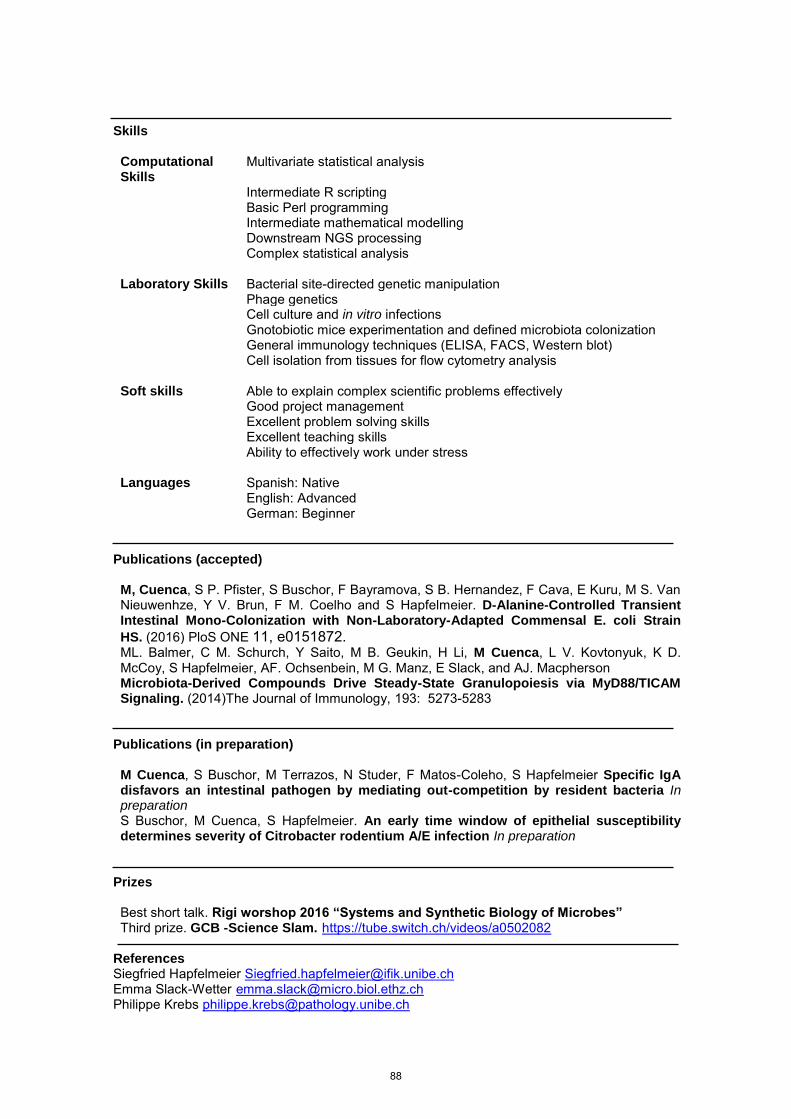
Skills
Computational Skills
Multivariate statistical analysis
Intermediate R scripting Basic Perl programming Intermediate mathematical modelling Downstream NGS processing Complex statistical analysis
Laboratory Skills Bacterial site-directed genetic manipulation Phage genetics Cell culture and in vitro infections Gnotobiotic mice experimentation and defined microbiota colonization General immunology techniques (ELISA, FACS, Western blot) Cell isolation from tissues for flow cytometry analysis
Soft skills Able to explain complex scientific problems effectively Good project management Excellent problem solving skills Excellent teaching skills Ability to effectively work under stress
Languages Spanish: Native English: Advanced German: Beginner
Publications (accepted)
M, Cuenca, S P. Pfister, S Buschor, F Bayramova, S B. Hernandez, F Cava, E Kuru, M S. Van Nieuwenhze, Y V. Brun, F M. Coelho and S Hapfelmeier. D-Alanine-Controlled Transient Intestinal Mono-Colonization with Non-Laboratory-Adapted Commensal E. coli Strain HS. (2016) PloS ONE 11, e0151872. ML. Balmer, C M. Schurch, Y Saito, M B. Geukin, H Li, M Cuenca, L V. Kovtonyuk, K D. McCoy, S Hapfelmeier, AF. Ochsenbein, M G. Manz, E Slack, and AJ. Macpherson Microbiota-Derived Compounds Drive Steady-State Granulopoiesis via MyD88/TICAM Signaling. (2014)The Journal of Immunology, 193: 5273-5283
Publications (in preparation)
M Cuenca, S Buschor, M Terrazos, N Studer, F Matos-Coleho, S Hapfelmeier Specific IgA disfavors an intestinal pathogen by mediating out-competition by resident bacteria In preparation S Buschor, M Cuenca, S Hapfelmeier. An early time window of epithelial susceptibility determines severity of Citrobacter rodentium A/E infection In preparation
Prizes
Best short talk. Rigi worshop 2016 “Systems and Synthetic Biology of Microbes” Third prize. GCB -Science Slam. https://tube.switch.ch/videos/a0502082
References Siegfried Hapfelmeier [email protected] Emma Slack-Wetter [email protected] Philippe Krebs [email protected]
88
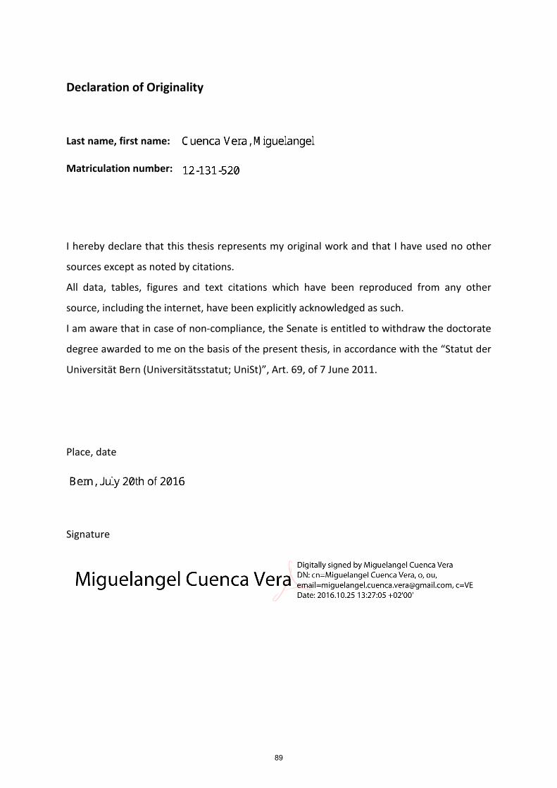
Declaration of Originality
Last name, first name:
Matriculation number:
I hereby declare that this thesis represents my original work and that I have used no other
sources except as noted by citations.
All data, tables, figures and text citations which have been reproduced from any other
source, including the internet, have been explicitly acknowledged as such.
I am aware that in case of non-compliance, the Senate is entitled to withdraw the doctorate
degree awarded to me on the basis of the present thesis, in accordance with the “Statut der
Universität Bern (Universitätsstatut; UniSt)”, Art. 69, of 7 June 2011.
Place, date
Signature
89


