Embed Size (px)
Citation preview

· ~fi.lCtrtmtnt of Jlgritu1tUrt, J):tallras .
. ' Vol. III, lJ'ullet-in JVO. 60.
eATTLE OF -SOUTHERN
LIEUT.-COL. .W. D. GUNN,
Superintendent, Indian Civil Veterinary Dept., Madras ,
1909
MADRAS;
PRINTED BY THE SUPERINTENDENT. GOVERNMENT PRESS.
[ l'tl C},;! 2 I'II/'C •• '. J [3 slrill·/lg.r. )


, I

COPYRIGHT, '1937
THE WlLLfAMS & WILKINS COMPANY
Made in the United SIllies uf America
Publi~hed l\f ay, 1937
Co"poS~D ,"In PRINtED AT THE)
WAVERLY PRESS. INC. FOR
THE WU-LIA.". & WILKINS COMPANY
B .. LTIMORE. MD. U. S A.

THE cattle ·of th'e . Madras Presidency ha:-;e long .been . famous, and of the several breeds which are to, be
'found in this· part .. 6f the country those designated the " M ysore," and the "Ongole "- . som~ti111e.s also known ,as the "N ellore," are .und0ubtedly. pre-eminent_ On account of its ~repotency tfte former is most assuredly entitled to ·first honours as, a visit to a:ll the ~arious large cattl~ fairs held in _the southern dlstricts of TrichinopoJYI
,Madura, ~nd Tlnnevelly, also in th~ more northerly di'stricts of An~ntapl:lr ~nq ' BeHary 'wiil· show how predominating is this type.
J ". The '.' Ongoles" are very . beautiful in appearance; and for their special purpose ar,e unsurpassaple. but they differ in almost every -respect from the Mysore.. They are huge in, si~e, extre.mely docile, an~ suitable fOf steady h~avy <iTaught, wher,eas the Mysore ~attIe are speCially adapted to road~ work, as they are quick, ' very ' high spirited, .and have extremely hard sound feet. _
Indian . cattle, like. those 0f ~u:rope, vary ill< most districts either as to si.ze, form, and syrrimGtry, or·~as to the growth and .Ien.gth of tneir horns, according, to tl),e varying local peculiarities of the climate, soil,. and lastly, but not least, fodder. It may be, stated 'that ' both n~tural and artificial. fodder tends to influence the form" size, and character- of the animaL Ordinarily the native wbo lives ~fl · a; ,mea)-9f t.:ice, and perhaps a few hyrbs to s~on thci - ·

CATTLE OF SOUTHERN INDIA.
sam~ with, expects that his cattle will, in like manner; pick up what they can in the way Df pasture abDut the village or. its a,djacent lands, ,so. that l),e }1e'v~~ (rDuble~,. ,himself to. gr6w green fDDd, Dr 'prepare dry fDdder fDr ,
theII). ; t~e same plant which' suppli~s him with grain feeds his-,~attle also. 'with its straw. In .mDst to~ns and villages cattle are driven Dut at all seaSDns tq. graze abroad, And 'in the 'dry seaSDn they mDr~ frequently lick the d1.1st Dn!y, and _return hDme with thei~ ~tDmachs as ,et;npty- as
: ~h:en .they started, tq rec~ive 'p~Fhaps a few handsfuJ-'of 'str.aW Dr rubbisn just sufficient .tD sust~in life. " , Madr~s is essentially a cattle-raising pr<!>viri'cej .-and
. iQ~sequently the animal wea:~th 'is enbrmDus, but, ~~ il1 I!
'<?ther ' presidencies' in India, large I:erds 0f v·ilJage 9); ~ ~(:mgrel tattle are to. be met ' with-everywhere. Many Df' ' .
phem are, worthless, being tDD weedy eveh' to. put' in~D the ;lighte~t plDugh, and they are allDw;ed to 'exist al~d .eat the rati?R Df the fi.1Dre prDfitable Dne.s: Undoubtedly reli': giDuS sentiment amDng the vast majDrity Df the peDple is c.
flv~rse to. des~r?yitig cattle, as amDng the' Hindus the bull ~~s always been consid~red ' to be sacted, and indeed is wDrshipped un~er.,the mime of Nundi, it having tDr!Ded , the vehicle Df their deity 'Shiva during hi::; peregrinatiDns. A Hindu wDuld consider it a mDst grievous sin to. kill them, ~~d otter pollutiDn, to. part,~ke Df t~ir _flesh, yet he freely pautakes, Df their milk. .
The three, great centt es 0f ca~tle raising ar-e ~hDW1J. in the aCCDmpanying map, and;frDm these they are taken by.dealers, who. fDrm a very '- large cDmmunity, to. the numerDUS annual and weekly markets ,held in this pre~i;
dency; F Dr ,guidance a list. Df the f~irs and the n:tarket~ held weekly, also. , the approximate nun)bet: of animal~ which are brD1.1ght fD~ sale, has been added at tb,e end Df ';
the paper. Frequently these dro_vers qa-ye their-,reg!ll~r

~AfTLE ·. oF' scruTHERN iNDIA.
customers, and they.receive p~yments by instalments, but , this custom is pr.incipally limited to the nor~h and west· .
..._ ern districts. 'rvt ysc>r~ has enjoyed fro.m a very early period a just renown for a superior breed of cattle. The generaiIy mild and salubrious ciimate of the plateau, .witli an e'xtensive pasture on which cultivation ha~ not 'mad~ .much inroad favour,ed cattle 'breeding, and 'a'ttracted Gollas and other nomadic trib'es from the 'north; wh.o
. brought witp_ the'm their excellent breeds which, beiqg established '"for ' generations in" the country, ~artd mixing with the indigenous population, could not fail to improve th~m. . In ~ 'cocintry in which,90 per cent. of the"'popu~ ladon subsist by agriculture, and in which cattle play ~ 'most important part, a demand for them is never wanting'. Cattle manure is used as fuel or serves to enrich the soi'l exhausted by cultivation.- The operations of ploughing, harrowing, sowing, and thinning the crop, of lift,iIJg watei' 'from ~eils for irrigation purposes are carried on almost 'entirely by bulloc'k power. The crop when cut is reo moved to the threshing fiqor, and there trodden out by the cattle, and transported by them to the market; and in fact it WQul~ be difficult for the Indian cultivator to
. get on without his c~ti:le which indeea cO'nstitute the 'life and so'ul of agriculture. The substa.nce· of the r-yot is usually estimated by the num.ber of . cattle he owns, and the numher 'of pl0ughs' he works. ' Moreover cattle are intimately associated with the demestic ,incidents of the
·peopl~. . The present of a cow with a few acres of Ian.d·tb 'the bridegroom is a noticeable part of the rriarri~ge ceremony. The present"of a cow and land is ~\so part' of"the -Brahminicaloh>sequi.esi . As a p;opitiatory.'offering j .when a relatio~ has died,-a young· bull-- i·s presented ·to-tfie·deity ',to be eventually turned loose into th~ . herd to ey~ntuall'y . become' a sin;.

eA:i:l'LE 'OF S6&rHERN INDiA.
,. , . MVS'ORE BREEDS.
All over the presidency so far as cattle breed.ing is concerned two descriptions of cattle exist side by si4e~ and' this is particularly noticeable in Mysore, and also·on the East Coast where the Qngole breed, are to. be found. The first is known as N adudana or N atudana, really viiIage cattle, wblich are by far the .most numerous, of .small ' size, compact fraI11e, and various colours; every _ .Village in the province teems with _them. They constitute the bulk of the agric\;lltl'lral stock, ~nd are the main
f •
~(;)Urc,e of the dairy supply, such as it is, The second is • ~ .' I
termed, the Doddadana meaning large cattle, and con.sistl~
of the. less nl,lmerous b1.:lt more efficient ' and valuabl~ , ki~ds, of more uniform size and colour j they are mere 9ften used in conveying the traffic of the' country than
.'in agriculture, and are largely sold in cattle markets. Doddadana and N adudana are particularly M ysore terms.
\
The_. ,DOddadana embrace the Amrat Mahal, Ha:llikar, Chitraldroog, AlumbadiJ or Mahadeswarabetta, and kindred breeds. Cattle of this description ~re only owned by well-to-do ryots and breeders. There are professional .b(eeders, but every ryot who' has a little capital adds. to his agricu1tural occupa,tion that of r,ear-ing a few head of cattle. There,are parties who keep their herds of cows
:~nd bulls' for bre~ding purposes mostly in t,he vicinity o,f -grazipg hills ~nd lowland forests. Calves .of a year old or' so ~~e bo~ght from them by the ryots, who attend
.them with much care for two or three years, and exhibit them ' for . sale to t,he best advantage at the cattle f~i~s. '
The whole breeding operations of ihis country are ~arried on by means of three descriptions of bulls;- .
\' (a) Choice specimens of the Dpd_dadana breeds, either kept in villages and homefed, which.are licensed to
• • " .1
g;cize . on I village crops or may be kept in the herds,' and freely moving with them in their jungle pastures; . these tnay be styled special superior br~eding oulls. The large ihafority of these have been dedicated to temple~, and are thus held to be sacred. I
(b) The calves of Doddadana bought when young ahd reared in villages, destined for agriculture or sale after d.stlJation, but employed as sires mean~hile. ', These m y be styled casual good breeding sires. They are
. mb'derately. good though inferior to the first named f@r br~eding, and being permitted to Gover before castratJon th~y make, it is said, less 'efficient agricultural an'd draught ~Mk. ~ .
;" ' (c) The numerous small s,ized and more or less ill ~ ~hapedryoung males of the ." Nadudan.a " ,'class herding with tWe.o.£iHage cattle, and thes~ undoubtedly lead to degenerate breeding; these may be called Nadu bulls. Nadudari,{' or village cattle are left ef!tirely to the course of nalure without any 'control, and without any of those artificial rest-rictions by which alone a breed can be sav.ed from <degeneration. Seldom is any selection made of breeding cows and bulls with reference to their fitness
I
for producing a healthy progeny_ . N or are inferior and aefective· bulls generally castrated; Cl:nd the common practice 0f driving all the village cattle" male and female, together in one herd leads to indiscriminate breeding.
, In some parts .\loweyer it is the custom for one or two villages to .club together and subscribe for a superior bull whi~h is carefully 'selected and purchased when young. 1 t' is the common p~operty of t,he v.plagers, ~nd being allowed every 1icense, everi to" the extent of grazing on private fields, keeps in excellent condition. Sucl{ bulls follow the herd during the 'day, ~nd being accus· tomed to graze on rich ' crops seldom pay h~e~ to the
6. \J
. CkI'Tt~ OF . sottTliERN iNbiA.
poor gra~ihg on the village commOR. They 'run to the field crops and gr~ze ' their fill either after the her.d re~ t.urns hOqle for the night, Qr before it is let out in th~ tl}orrVng. With the better class animals great c'are and attention is paid to the selectipn of both cows and bulls, and the conditions under which they are reared afford facilities for ,the 'regulation o'f breeding, cow~ 'of the Doddadana are kept in the ~illages; they are homefed and under sheiter, in which case only the very best bulls are secureq. for serving them .. , Each herd, . has its own. . special superior bull sometimes selected from the same perd, bU.t more often from some other herd to prevent in-and-in hreed{~g. As the bull grow.s old and deficient II in ~vigQur, a y_oung bull _is simiJarly selected and kept ip the herd to tak¢ its place . . ,The y.oung · one in many . ~ases' dnly acts the pact of "a te;;tser. No ' ~pon~t does it : apperceive symptoms of a cow bc:ing in h~at . than it appro.aches, . and .. then keeps cpnstantly ~ttending on it, lhe. skitt~sh h~b.lt of the c:ow, being tir~d of the impor; .
• j " tunity seeks: the ' proteGtiol1 of the .elder ]:mJI w'l;1ich the young one dare ~ot ,~ppr9ach and wh.kh th.en serves th~ cow. It may be of interest to know what the bree<iing experts consider '.to. be .the best points of CI: M yspre bull of ~~e best .quality;, and thes.e, th:0ugh:.th~y cannot be me~ ~ithjn a' single specimen, are more 0 less searc)1ed for in all careful selections:- . ., .
, .. ; (1') A long and' stretching frame. , - .' '.
. (2) A good"height- say 52 "inches measured, behind ~he hump, '
:. : . (3) AI fong and tapering head, with a narrow and prominent forehead. -.~ ..1 .. >.# ~.. .,. r::' ,.
(4) Small 9ut. pro~i_nent and br_igh~ ~ye~.
:.. ... ' _ ~5} Small and erect ears.
.. ... , ....... -. ,. .' " I \
, , (6) Thin, fairly long and ')graceful set of horris, the difference between their thickness at the base and ~ at" inc • 4 .. .,. ~ '", "
epd b~ing small. , '- ~ . ,.,:!
, 17) Strong' fairly.long neck witWa-sma:I1 -w:ell~~ape'd hump. ' , , ' - .', " . ,::~
, (8) ' Thin and sho~t dewlap. ' . : < : ~, ~" ,". :_,:~:t , , . (9) Broad and full c~e~t. , .. ,~ -, .: : < :.: ,:'
. " (10)' . Well formed and. strong should~rs' a,nd" hihd quarters. ' ,N, '", ;-. ,I ..
( I 1-) , Strbng and w~ll roundea ri'bs. (12) Level back and' bro'a:d loins. ' ( 13) N arrow flanks: ' (14) A level , crotip. · ,An, abtuptly falling ctoup
: being' cOl1;demne~ ('. goose (umped.". in' hOl1'~ey 'pnrase1
ology). ' ', " " " :. " : , (IS) A thin short whip like tail 'r~aching tQ O(
~ery little below the point of the hock: :, - .. _ .. (16) A wen projecting anus ring, 'so ~-hat the 'ejected
c;lung m~y f~ll clear of the bo~y. ',It should 'rtot ~e s~tu~ ated in' a niche-like hollow, as in cows· and ' old' ,ani'm;ils.- '~
(17) A ,sheath ' haying- a ' little or no , pend\::lIQu~~
growth. -, ;': ( I 8 r Legs 'of , medium ' lengtn a,nd ' wen. ' proJ;!~r
tiohed; having strong and fairly thick 'bones"and' moving with' a swin'g in perfect rhythm, and' straight :not tllrned sideways or brushing against each other. ' ., ~
(19) Short fetlocks, and 'hard and s'mall hoofs witfu ' ' equal halves having a very ~arrow cleft ' Be.tween them': A l<:mg sliank bone is considered a ·weakness. -
, (20)' The colour 'of ,the horns', ho'of~, muzzle ana skin should be black. ',. " , " 'i
: . (2 I j The skin should be thin' ap,d ,satiny: havin.g! short and soft hair. Bluish and ' iron grey co16uts. are p'teferred. ,,,,. ' ;,.' : ~' ";i
8 ' CAT'U.E·.QF- - SOUTHERN, IN-DIA. \. . ". ... '" •• _ • .... .... '":, • F \ 1 _._ . . ...
{ '; ', I (22): A ... ;·compact, .body Jr~e . fr~~ :a:11 ,pe~aulous ' • gr~)\yth~. . _ " , , .... , " , J •
, (23) .· The a:nimal sh~:)Uld be sou?d ,i,n every way; of symmetrical f¢atures" of g00d ' temper and , p{ire -}:iteed; and free from he~edit~ry dise;ses. In the selection ~f the -cow no sucn special a~tention is 'bestowed as 'in th~
• H • _ I'"t • ~ .. ... , \t-. _ ~
case of the bul), which, c'~t:tsider~ng the pumbeF of ani- " :' • . mals it is likely to ' influ<?nce.; is most carefully sel¢ct~d. ~ The' ~ain points looked for in co~s are goqd'si:ie, I'ength.
, shapely head, and~horn~, broad liips and 10ins, ~ a:ncl nic'e . c~lour. . . , '. .': > ,, ' , J~' '
~ { .t t . .
, AMRAT MAHAk EREEI? ..
1 '. Among 'the 'bre€ds' friurid 'in M ys'oFe the first pl;i.ce is;' ~. undot.i.bte~Hy ,due ' to tl:1e c Amrato~ Maha!. The. Amrat " 'Mahal, literally Milk Department,' is ' an. "' establish?le~1l f~r the preeding,Q[.a;' r7r<:;e) f cat'tle pe~.u~~,a~, to\he " c~untry of Mysbre, and tbe.'present cattle comprise 'three principal 'varieties' caned, Hallikat, ' Hagalvadi .and ChitraJdf00g . fto'In .th~ · cristr-Ids 'which,~ originally' 'p'ro:auced' thei:rl; ami ' . sO'distinct-ive is 'this . breed that th€y may readily, ,be dis '"'.'
~ tinguished from ;every, otner , bre~d in' 'I ndia. ' The'differ- ' , ent. breeds c"0l1ilposing the present Amrat Mahal c~ttle " owe. th,eir, origirt' to, the ;t~ttl,e pf the tr.ibe of Gallas and th<eir <;sub-ttibe' Of ,H aUikq_.fs who, ','wid\. t.heir superior- :
'" Cattle:'are',beIieved"to hav;e migrated iri" ari:eie'nt times .in ·' several successive waves fr'bm' the ncirth, and settI-ed'iii " different parts now comprised in -the Chitra:ldroog and Tamkur ·districts.'., - . ' "
The Karuliatti establishment of, 'th€ Vijaya~ag~r. ViCeroy (some time between 1572 and 1600) at S'e'ringa- , patam consist~d of' Hailikar c.0W;;" impotted fcrom Vizici·:-, ~gar. : ~his may he .said to 'De the nucleu~ . of the Amrat Maha~ ~at~le. · The Seringapatain\ cattle pass€G ' ioto tlle l1~J'l<;ls Qf ~he ~ W 0da yars of M ysore! so~ne .Qf w.hoJn'~

, . .


CATTLE OF SOUTHERN INDIA. . ) 9 , .'
.tlotably ·' Shall'laraj Wodayar (1617-1636), ' Kant~rava . ' N<l:r~sa Raj Wodayar (I638-~65 ?) , and , the celebrated J ~hikka Devaraj Wodayar ( I6~7,?-I704) 'made theil: oMTn '
additions to th~mJrom time to ,time, assign~lg "Kava,ls" in different parts of the ' ~ingdom. It was i,n Ghikka 'Devaraj VlodaY,:ar's time , th~t tb,e ~attle esta~lishmeiit, .obt~ine,d recognition as ol1e of , the Cheptr.tl)le~S of the . administration. I t was called "Benne' 'Ch_avad~:" or establishment of cows both a~ '.a br:eeding r stud- and ' to furIJi sh milk and butter for the pal~c,e. H~ introduce,d for the first tim~ the system of bi-andip.g iher.ri wjth his , initial -a (De). The accumulated herds , of t)le , R,aj'~s .of My~ore passed on to H yder Ali .w~en ,he ' ~~urp'e9 tlie ' throne . .' In extending his conquest, ,anq 'if.) !,e~,ucing ,the ' num~r(:' u s mlers who had held sway over ,nio're. or ' le?s exte,nsive tracts in Mysore, he acquir~d ~ls~}l;.e, ' J:terds q( ~lie ' superior cattle belong ing to ', t~e.n}:~ ,~~o_i)g , these may he mentioned the PoHegars ,of Chit~aldl~oog, ' .Tarl,-' kere, and the Raja of Nagar. B yde,r 'se:e~s_ to _h.~ye · '-,,' made extensive use of the, <iattle whicl;l : J:ld, ~<l:~ ~ppr9-priated in the movements 'oLhis, army _eq:uj'pag~, ... anet i< popularly _credited ,~ith \ h~l:ving keBt,;-~t l~a~t'·:oo,o~, ~' bullocks in , ~ifferent pa'rts of,ihe, pr<?vii_ic.~ , p~oug~.,t~,ey were not organised as carefuIty; or 'In as minut,e. aet'ails "a~
was afterwards dori~ by Tippu, on ~<:~Y~t~-rfi. .3v~~~ : ~aS~ in essential points. b~e'n adhered to ever .3in.ce'. Upon' succeeding to the throne <?f his father ' Ti~p~" ad~~d to these herds those Ot tlje PollegaF of 'HagaJ-Jadi; ' t-hiKKa Devaraj WQdayar's ana the sugge~tive' !lame of "Benne Chavad( ' was changed in 'his time i~tq t~e ' .r116re , p.~mpous one of Amrat Mahal . ffom 'Amrut'a :'';_Iiettar . . Tipf>u took great interest in 'these , cattle ' ~tid issue4 '~ " Hukumnama" or regulations for . tlie departm~nt; tae' greater part of which continued to be observed ·afte,r
2! "

10 OF SOUTHERN JNDIA.
the t'aki~g ~f ,geringap~tam, and the sa~e system ' was afterwards followed, '15)1' he Bri;tiSh officers . . Th~' J)airy , , ., '. r department seems.; to hav:e'beC:::J1 ,on 'a large sca1e~ ' 1:~e Am£t.d~w$ ,we_r;;'b('_p'ect~d to train the young)) ' ll~. ~hese ' were gi~en absolute· freedo!h and .w~re ,~lYowea· to, g 'aze
,in the 'ryots" fields ... T l;}ey ' were afterwards clftssified when they were required ~s g,UH',b';Ull(;cks, ' <,iGk , bullocks "
" ., . Il :')..... ._ ...... .. -r ..
and plough buliocks, etc~ , Tliere;'wq.s aJ1-:~.~nu~1 m':..~s.t_<; r " of the her-ds and T:iJ?Pl!.J reqJlently atterlded.1t ip; 'P<t:t:SOFl.' and' ~'istributeli ~ewards', -'" } lch was ttte C011~P/ositi.9iri: 'or_' .
" th,e ,~mrat .Ma~aJ1c~t5 l~' ~,lau~4.rClte.? bY" ~fi~ka "pef-~~'aj , " \i\T0dayar, rec0Iilsti~t~el by [I'y,.ae-r _,AIVal1d' thorough1 I . ..
< organis~'d by" Tippu SultaR., '. ' .:.~, " ',' ' '" ;f( ~ i~ "if!' A;:
r :: The attention of the:' British. was first called to t!;le ' , excellence of the breed when it enabled H yd'er Ali. '~0: ;., n;areh. roo 'miles in two~ days and a' ~~l( to,.Jhe relief ; f " " Chellumbrunl, and 'after eyef,y .. d:er~at,. 'to' draw ~Qff his :;.'~ ,~. ,gu,n,s in the _face ~~ .. th .. e~~JnY! ,and' W~1!l ' T:~pp!l , S~lt~? :''',.
was enabled to cross the Rentn'sula of Southern India ill • ~ f"' . ' ..'" -J.
one month for the recovery 0f. Bednore, ~a.nd , to mar'ch ,63 ~ .. } miles in two days before General Me.adclw? ! t : also enabled ' the ' DQke of ' Wellington, ,<p", ex~cute J1>tij.ose marches ' of unexampled rapi'dity 'whic,h i re' the' adinira~ .. . . ,.,..« . . I. ~ t . ..
t.ion of4~ilitary men, an~ the Duke brougnt it promi'- ~
nendy to ' the ~ notice of the thell' 'Cornmilnder-in-Chief Lieutenant-General Stuart. CaptaIn Davidson, ' in ' a '
..:z_ ... •
repQrt on the Aml'at Mahal cattle attached to the 'B ClI'n bay column of the English Army in Afgna:nistan in ~ I842, says :-" No ,draught cattle. in either arm)'; were 'sq
, , . :efficient as the' 230 ~ysore bullocks which acqo~lpani~d the Bombay troops to AfghaNistan. ft was entirely due to this ve);'y superior d~scription of cattle that. no' part of the. ' Bombay Park was r(}quired to be : apandoned when the troops were returnil!g to India over th,€ almost


CkFTtE oF' SOU'f,H~RN INDiA.
impracti~able ' roads through ,the Tirah Mountains. , T ,nese cattle w~re rr~q,uently upwards,~of sixtee~ liours in
Y0,ke_. ' The draught bullocks of, the, Ben_gal army w~r,e 'the 'prop<;rty of Goveril~Ti~nt, a!ld were -not in" my opin'iop . ils fine anln.1als as the M ys ore bullocks.'?... Odler metllor" able military events might also b~_ cit€d to,..th; e~edit of tneAmrat Mahal cattle. It is said that ,duringt'he. Penin- '
~ular Warthe D~ke of Wellington often re'gr~ttecl th~t he had not the' seFvices of the cattle of this breed. ,I:n 1808 the Com~issioner' of M ysore said of the'n1 :- They are active, and fiery, and walk faster than troops; in a
word the)' con~titute a distinct species, and are sald to p~sses.s the same superiority (lver other l?ullocks 'in every 'y.alu~ble quality that Arabs do over other horses. Professor Wallace remarked in 1899, that the breed as a
'whole occupies among cattle a position for form, temper and endurance strong ly analogousto that of the thorough '
~red a~long horses. _: On the fall 'of Seringapatam the whole .of the cattle
became the property of the British Government, t~e . management of the herds being allowed to remain with the Maharaja of Mysore on the condition 'of his sl!pplying a certain number of bullocks. It was , probably imagined ,that 'the same attention would ' be gi_ven to the
establishment as has been extended to it by the former <?-overnment, but Tippu Sultan had depended upon it for
,the efficiency of his army, and tl)e new Government could be actuated by no such motive. The consequence was
, 'thai tp.e establishment was left to the servants who had charge.,of !t, and by them neglected and abused; the 'British Government were disappointed in tneir expected
supplies, and th~ , GGtttle were allowed , to deg~nerate to
~uch a degree that after a per~oli of thirteen y~ars it
became nece.ss~ry' to resume cflCi:rge of it in ord~r to

12 CATTLE OF SOUTHERN INDIA.
preserve the breed from extin:<i:tion. , In '1 8 ~ 3 tn.e J\mrat . Mamal . cattle, together with the ' pasture lands were handed over to Captain ~ar:vey 'of the ~ Madras Commissariat. The herds then rapidly. improved and doubled in n':lmbers in fhe 'course' of but ten years.- Ii1 1840 the ," Maharaja's Amrat Mahal herds and grazings were ama-I .:
, -gaVlated' with thos,e of the-British Govetnn1ent, and the "whoie pl~ce'd ~nder the orders of the Mysore Commissiol).. , I,n 1860 fro~ motive!,? 9f economy Sir Charles Trevelyan ordered the establishment to be brpken up, and the herds to be sold; this appears to have been a fatal error, alike in pollcy and economy, and the results were fatal to the
' public service. The price of cattle soon beca~1e prohi- ' I bitive (Rs. 150 each) and)t was, with the cordial approval ,and assistance of the late Maharaja, re-established ) n '. I 866 by the purchase. of s~ch cows and bulls of the old ,breeds a~ were procurable in the M ysore 'coun,try ; very few were obtained owing to the Pasha of. Egypt h~ving secure'a · most of the best blood. , FortunatelY- -h:owever
. the late Maharaja was a large purchaser when the old , establishl'nent was broken up, and the Madras Government was able to obtain sufficient stock to fairly start again in 1870, the complem~nt b~ing 4,000 cows and .100 bulls. .
' .In ';883 the British GovernmelR -handed over this' valuable property to the Government of His Highness the Maharaja for' two and a q ~arter lacs. it -is n~w .. entirely under its control, and e.very effort is niade , by caref~l elimination of doubtful stock to 'restore the old breed to its former 'exc'eIIeHce.
Stud books have been opened, and the cattle are mustered by mime and brand. Births ' and deaths are , \ registered and reported in monthly returns, and frauds on the part Df subordinates h.ave bee? to a great extent


CATTLE of SOUTHERN INbIA. , 13
prevented~ The Madt:'as Go'ven,lment receive .from: the ' establishment 200 bullocks annually. Private individuals may 0btain bulls by writing t·o the military adviser to the Government of M ysore, B,a.ngalore, the prices of which are about a hun?red rupees ,each. Th.ere is frequently some delay in procuring them, as the h~rds have to be rounded up, the ' young bull selected and. secured, after wl!ich they undergo ~ process of training. , T~is is very necessary as they have been living in practically a wild state.
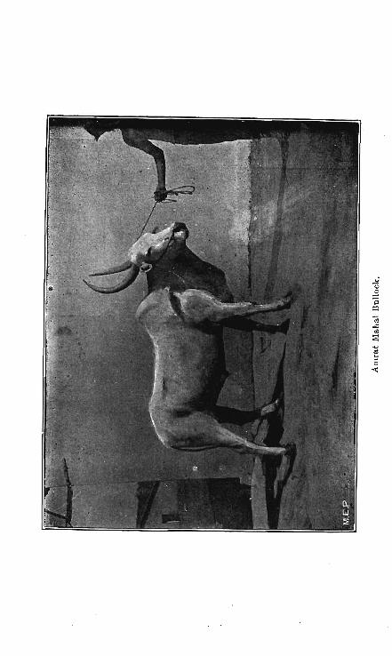
As has already been stated the cattl,e of this breed originally comprised three varieties: I (a) Hallikar, (b) Hagalvadi and (c) Chitraldroog. Prior to the abqlition
, of the department in 1860, the several herds seized by Hyder and J'ippu Sultan seem to h,ave been maintained for the most part unmixed as separate" Serwes," the distinguishing peculiarity of each breed 'being thus kept unadulterated, In 1866 when the department had to be reorganised by repurchasing the stock it was found impracticable to get back in their original purity all the cattle s,old six years before. At this juncture the men, to whom the work of collecting the cows w,:,-s entru~ted on promise of appointil~g the~l " Serwegars," freely mixed the th'ree main varieties of the old Amrat. Mahal. A large number of inferior cows of every other breed, including their own bred cattle known as " Swanta', G?su. !'
(mixed breed), and a: large number of the Mahadeswar-. betta '<;ows are also said to have been passed off for the , r-ec011struction of the Department. During recent reduc
tions and reconstitutions of " Serwes," since 1877, many . ,herds have been brok~n up and distributed, among others,
' new .her:ds 'have been form'ed out of the excess stock of the old ones, and' exchanges ' of stock ', are often being made' bet\Y'eel1 differ~rlt " Serwes " all 'tending to promote • 1
CATTLE. OF SOUTHERN lNEUA.
mixtu~e. At present the Amrat Malial breed Fannot b~ said to be as pure ':ls it, was prior to 186o, althoug:h careful selec,ii~~m -and unif0rmity' of treatment ',iti recent years seem to' have .erase? a s-ood m~ny points_of d,iffer- . ence, which lTIust have necessar:ily exi!,?ted at the time ,of recQHstitution of the herd~ in '1866. The different.' breeds of Hallikar, Hagalvadi ' and Chitralclrpog vary ' but sligh~ly, their general characteristic being the ~ame .
. Some special ' chara,cteristics developed by local peculiarities in the different herds may however be notic~d.
- The cattle reared in the " Kavals " or reserved' pastures , ,are of much larger size than those found in the n<!>rth.
Tkey are more bony, carry thicker and rather , ,Ie,ss gr~cefuIiy _set horns, hawing'comparatively thickef-,t~ils ; their hoofs are said to be not so tough as tnose of cattle in Chitraldroog from . which they differ in 'having a
, somewhat pendulQus sheath and dewlap. The cattle in the Tumkur, Hassan, and Kadur districts, though slightly smaller' in size 0 ~re ve~y much like those-'of the Amrat . Maha!' The herds of the eastern parts 'Of the Chitraldroog eonsists ' of cattle of smaller size but more ~ompact and hardly frame with a ,finer tail, thinner and , more
'_graeefully ,set h9rns, and I stronger hoofs. Th.e cattle of - the western Chitraldr.oog and Channagiri taluks . resemble the last 'nam~d variety, al'ld differing f~om the~ only, in be~ng slightly bigger in size.
o The Amrat Mahal cattle are kept in their grazing grounds which are ' cal led H Kava! ,', about '210 in number,
.and these are distribut~d , over the great~r pordon. 0~ the we~tern and c.entr§l.l parts of Mys()re, and eover an immense 'tract of country. They comprise varietie,s C?f soils, · often ~ndulatihg alid covered with scru~ jl~ll1gJe growth. The 'cattle feed- on variot.:ls 0 grasses, though
- .. (1 vunaga ' (I-leteropogo1Z ,.contortus) is . by far the ,most


Aml'at Mahal Bullock.

" , G~TTLE OF SOUTHERN INDI A:
predo;;j'nalit. _ The ir'~zing in ' tl~e Kavals ,'situaf~d i'n the _ vall~ys is mo'st nutritious. . As the ~_<?untry - ~e~onies mote elevated th~ grass becomes more scapty, and 'infeJdor ;11 quality: , The . " ' Kava~_" are divided into
. (a) hot weather, (b) wet weather antI (c) cold weather. Kavals '! according to the seasoil of the year-'af, which'
, they are ' most suitable .for grazing. ' The ' hot-weathei-" Kavals " are genera1iy", iri the beds of tanks ·· i.~ which' g rass springs up durjng the 'h:ot rr{onths, aild · where generally there -are trees capable of affol;ding 'shade to . the cattle during ' the heat of -the day. The cold .and· wet " Kavals" are those where grass dries up during the hotweat,her. The cattle are ·driven for about four ni6nths in ea~h year from May to Septen1ber to these "·maindd -k::l.vals". The herds in the west are taken to the south-' western jungles on th~ Coorg f~ontier, and those in· the north ' to the Lukkavalli and ChariJlagiri Forest" Kav'alii '" in the Kadur and Shamoga districts ; where the·~ first-'. I
showers of the advancing monsoon ensures an early and' abundant g rowth 'of grass. They the.n· return~ .1;0 ' ,t he·ir· maidan, or pla ins " Kavals '" about the heg inning 0f Sep~. tem,J:)er in each year ' when the supply of grass is·.plentiful all over M ysore.
T\:le whole of the cattle are divided iJ;to ' '.'-£erwes-" or herds, eac.h -of which, WIth attendants attached. to. it; i's kept s_eparate and distinct. The establishni€nt of .each herd is ·fixed at t~TO hundred breeding ' CQWS', one ' hundred heif~rs, ' twelve _ bulls, ' and twenty " Peshros" or' 'leaders,
• with the calves of both sexes and ~n ages, tke actua,l '~produ;ce , .of the herd the nurpber of which 'varies accord(i~g . t~ circumstances; but, which, generally speaki'ng, rai'ses ·the tGtal number of an'imals in each herd' to five of '~j'x Ihmdred.he~d. Each" Senye " orherd is placed iIi charge of a ". Serwegar" as~jsted by two ", M unda.hr" each ' or
! •• -

l · 16 CATTLE OF SOUTHERN INDIA.,
wh0FI1' is 'responsible for the proper n);nagement of the" . cattl~ under hi!;? charg'e. An establishn~erit of'gtaziel~s '
. C).nd other ' attendants ' is likewi~e attached to the herd. . The number of " Kavals" alIotted to each 'herd ;aries~'
, , '-'" I '
from three to nine according to ~he~ size of the " Kavalst:!. , " 'r ",
and the .quantity of pasturage they aff~Td, and although. the herds are not supposed t<;> be permanently attached to ·particular. " Ka·vals" still they are not removed from 'ihose wliich 'have been allotteq to them without special reasons: The whole of the herds-are divided into fourteen" Tu;kries" or ~ivisions, some composed of two and others of three herds, the" Kavals ". belonging to each herd being,' of course, ~onve_niently situated. Each Tukri i$ placed
'·1 unper the superintendence of a " Darog;;t." whose dllty it ' is to frequently inspect the herds, to muster the cattle, to check, and repor,t all irregularities on the part of the attendants in charge,of them, and also to arra~ge as far as may lay in his power any difference which may rise with the inhabitants or local authorities. At the annual inspection of the cattle which takes place in the vicinity of the grazing \ farm in the_months of July and August each ~erd is separated, carefully examined, all inferior cattle remoyed, and unmarked cattle branded. .'
';E~rly castrai'ion 'is-. the rule in the Department, andc the cal-ves are castrated when .theyare eighteen months old in the cold 'season. -, . . ') . ,
The bullocks are separated from the herds after four years of age, and those sold to the ,Madras Government are transferred to the Public Cattle' Department at H unsur and w.hen turned five years . old they are ' thoroughly t'rained to work. They are in their full vigour at seven years, and past it at twelve. They wor:k until they are fourteen or fifteen, after ' whicq,they ~apidly decline and die at about eighteen years of age. .


CA:tTL]: OF S,6UTHER~ iNDiA. , ,
to Rs .. 90, average cows Rs. 40 . to Rs. 60. A pair of fijst-~la!'is ~ulls are said to fetch ih I-iyderabad as much ,as ·Rs.: 500 and even Rs. 800. .People in the Dharwar Col-
" . iec~o;ate eagerly se~k :these c'attle and pay good priGes (or young buiIs, from two to four ·years qld even as much as' R~. · 100' tp Rs. , ISO. I t was said that a pair ~f bullocks was s'old ~here for Rs. 8~o' liaving won ' a nice in 4r~ggi~g a heavily' ladeil cart- through sandy soil.
The ' ~ Serwegars" of the Amrat Marral Department , . have'- bee~l , allowed the privilege 'of keeping their own . c~ttle with the Government herds with the consequence: that the Amr'at· Malial bulls have crossed the Nadudatia: cows and tJ1e result has been mongrel, ~nd these, from -long associ~tion, ,have t?-ken O!l all the, characteristics for the Amrat Mahal. They are now know~l by the name of Egosu or Swantagosu ,caitle. ,
. HALLIKAR BREED.
The history of this breed has already been given undeI:' the head of the Amrat Mahal cattle of which the Hallikar breed is the most i~portan~ and valuable
·,member .. " An absur~ 'legend is current among the herdsmen of the Department regarding the origin of the H~llikaf. They stite , that H,yder Ali, after Olle of his
,trips to 'the sO'uth, brought back to the Mysore country a ·number of the cows of the small Brahmini caste. These
, cows wei~ turned loose ' in ' ,a "Kaval" in the' ,Tumkur di~trict i~' which 'ther~ were" great numbers of antelope, and a ' cross :between ' the big black bu<_;k an~ the small Brahmini .co·ws gq_ve rise to the preseti.t Hallika'r breed. in support oJ this statement tl;ley point to the' small ,spot
3:below' the' inrier canthus of. the eye -which is common to the arit~lope and th~ Hal1ikar Cattl~. · It is, curious that
'wilil~ the " 11~me of Conas has disappear~d ~mong cattle
CATTLE OF S6UTI-IERN iNDIA.
that of Hallikar, ' their ·sllb~ti."ibe, has survivec,l in the' cat~le which they introduced into M;ysore. Hallikar cattle, besides chiefly comprising the Government Amrat Mahal herds, 'are to be found ip Tamkur, Hassan and Mysore districts, the cheif centres being par.ts of the Nagamangala, Kunigal and Gubbi taluk~ . . The area I
over which the breed prevails i.s not by . any means extensive, and it i's thinly scattered even within those li'mits. The reason is 'that,there are no extensive pasture lands in the habitat of tmese cattle, and the tracts being populous they are mostly homefed and are not maintained in large I~umbers except by a few breede.rs .in the .talltlks
. just named. They are freq,ue;1tly bred by small ryots who have only a few cows, and specia\ attention· is paid' to the mating of the cows and the re~ring of the young stock. .
". Mr. ,Wallace in his ' " India in 1887" gives an excei.lent description of the breed. He says :- The head is well shaped, long anp tapering towards the m~~zle which is general1y black, .the forehead bulges out sli~'ltly: and , is narrow and' furrowed in the middle. The horn~, are uJilique in shape, and differ considerably from most ' other breeds. They are usually large
" set well back 01). the
crest of the frontal bone; springing close t0gether, th.ey di·verge, i~1clining backwards each 'n a straight line for nearly half their length, and then with a gentle and graceful 'sweep bend 'fo'rwards usually lightly inclining inwards tdwards their points, which are black tipped a:~d exceedingly sha:rp. At times when the head is down, ' as when feeding, the horns can almost touch the neck in front of the hu~p. · They thickelil. gradually' as the head is approached, <'lim} are very strong near the base which seems to exte~d, apparently to give strength, down the forehead between the eye's a~ a pistinct ridge on each
• I

'CATTLE' OF SOUTHERN rNbIA.
side, thus forming a perpendicular groov:e or depression in the _centre 'of the forehead.
The colour is of a mor-e or less uniform grey, varyi\lg from light to ,a 'deep iron grey with a darker shade over the should~rs ·and hind quart~rs. Broken colours are being carefully weeded ~nd sold. ' The neck is thin, for the size 9f the cattle, but is long and sinewy. The dewlap is thin, and does not extend very far back. The ears are small and taper to a point, being 'carried 'in a horizontal position'. .
The hump is well developed in the bull. The tail is thin, and tapets like a whip. The legs are clean, strollg and sinewy, standing well apart. The hoofs ' .are small,
, well forined, black and hard, with a very close cleft between. This breed seldom attain a very large size. In shape they are remarkably neat, with muscles like whipcord. , The cows 'have a very m~sculine appearan~e <!-nd -yary
only with having a thinner hump and horns. In colour they ~re invariably of a light grey. ' They ' have a sm~ll compact udder with small and hard teats. They are poor ,milkers, though t~e milk is 'rich and sweet witli a high percentage of butter fats. They are of high plettle, and, though' mostly homebred, are not gentle
. or tractable. ' .
There is always a great demand for these cattle, and as the number annually produced is not sufficient· to meet it, high prices prevail. The average market v~lue is: Very good breeding bulls Rs. 80 to Rs. 120, average bulls Rs. 50 to Rs. 75. Very good cows Rs. 60 to Rs. 100, average cows Rs. 40 to Rs. 60. ,Bull calv~s of one year Rs. 20 to Rs. 40. "
Gujmavu is tHe f!lost valr~.Iable variety of the Hallikar . breed, and ·it is , to . be found at Kara,dahalli in .the

CATTLE OF SQUTllERN l~biA •.
Nagamangala taluk. Good Gujmavu cows of this locality ar:e little different from ~he , Amrat l\1aliaI' cows. The sba ' e 9f the ' head, face, muzzle, eyes, ears, horns',' . neck, legs , ~nd ba;rel is exa~tly of th~ same. type. The ' similarity extends even to the masculine look of the ,cow. One peculiar point wl?-ich is very highly prized in this vari~ty is the v~ry long -back which is sup'posed to give ~ them 'a glieater mechanical strength and advantage: " Th'e cattle owners of Karadahalli treat tbeir animals after' the ,fashion of the Amrat Mahal Department, they seHd their herds to distant jungles in the Heggadevankote taluk
· for the benefit of the early se~son pasture. · '. Superior bulls are kept at Karadahalli for breed·ing purposes. Cows, even from distant places, a~e taken to -
· these bulls for service upon payment ofa 'fee fr~n1 Re~ l ' ,
,to , Rs. 4 'for each service. ' I t 'is said that so h'ighly . . a,re the Gujmavu prized that ryots of .the neighbouring tal.uks of Mandaya; Seringapatam and Closep.et ad~ance to ' the ,bre,ed'ers of Karadahalli Rs. 50 to Rs. 100, for calves still in the mother's womb. If .~ cow c~lf is ' dropped th~ advance is returned, as it is not ~ustornary
. for ' the breeders' of Karadahalli to ~e'n the cows ~f the~r ' bie,ed. , If a bull calf is born it is sold according to the '
, original agreement.'. In some c~se~, s'uch" sales are subject ,' to the provision that the ca~f sh~uld be-reared f~r t~o
"years, and resold to the origina:l(owrier for its full value at th~ time of i,ts r~sale which is uSllaiIy from ' Rs. 100 to Rs. 200. This system ' of selling' and reselling is ':~ '" common custom and it affords a convenient division of faoour. , The Karadaha,lli bulls do not-attain a iarge size, the average height heing 49 inches measured behind the hump. They are very difficu,lt to tam(i! at first, but when once tamed they are far more amenable to being. handled and worked by men than the Amrat Mahal cattle which, . ' .

, CATTLE! PF SOUTHERN INDIA.
t~ the last, retain more or' less their impatie'nce' of , strangers.
CHITRALDROOG BREED.
, Th€ breed bears a close resemblance to the HaIlikar c!lttl~ diff~ring. from them only in som~. minor poi~ts. The head is sl1laller, and short'er than the Hallikar, out not stumpy like the M'ahadeswarabetta c~ttle. The 'forehead rese,mbles that of th~ Hallikar though' owing to the sh~rtne~s of the head ,it does not ~ppear to be so nart'ow, and-the furrow is absent. The horns are thinner, longer, and taper more gradually; but as they grow upwards they get fart-her apart from each other, 'and be'nd forward with a deeper curve. The ' neck, tail, and dew lap are th.inner.. \Yhite.is ' the predomil~ailt colour, and they are smaller in size thil11 the H illlikar . . _ These cattle are kept in :" Roppas':' as well as ,in
vplages, and are bred ,and treated in the san~e manner as , the, 'Mahadeswarahetta cattle. Cows of this breed ' are said t'o come t9 ' early, mat~rity and take the bull when ,
, they a~e two yea~s old, but this is really regulated 'by the ," quality and quantity of the food given to them. , When the early' rains commence 'all the 'i~rge breed~' are
,driv'en to salt l<!-nds, and are freely allowed to lick. earth salt tending thenc,eforth to in:prov~ th,eir condition i:,y grazing ·in fresh pastures. . . ~', ,
These ~attle '-a;e, tl'iough ' small, exceedingly ' ~ctive, • and .are largely ,:!sed for single bullock traffic.
, MAHADESWARABETTA' OR ALUMBADI BREED.
Th.is breed' derives its name from its chief and ~ouritai.J1 market Mahadeswarabetta in the ·' Koneg~l taluk ,of the Coim,batore district, where two large cattle fairs are held in February and ih October ' ~t which cattle exhibited are mostly of-this description: :1 t is also ' call~d

, CATTLE OF SOUTHERN INDIA.
the 'Bets·al or Cauvery breed from its hilly ·home all both sides of the Cauvery but probably a more common name is that' of Alumbadi called after a village of ~hat nanle on . the bank of the Cauvery. The ch.ief habitat of this race ~f oarde is ·in the Kankanahalli taluk of the -.l3angalore distri~t, . and the north~rn taluks of the Coimbatore and Salem districts which are diyided from Mysore by the
·ri v_er .Ca'::!very. ~ne reC!-.son for these reg ions teeriling . with great herds of cattle is the wide expanse of forest l~nd wh_ich is ·scarcely fh.or has not yet been taken up for ~ultivati'On, and with "On)y l?atches of tillage in favQured spots, and 'affording herds of cattle abundant pasture and wide a!1d unrestrained roa~ing ground . . The tracts ar.e sFony in .elev~tions and humous in the valleys. T~e forest growth being all deciduous, the pastyre lands ar~ '
thQroughly baked in the hot weather by the heat of the ~u~ so, peculiarly intense in the valleys in the low hilly ~egions. Another reason; though of secondary importan'~'e, ~s the noble str~am of the Cauvery whose waters ~re,'~ ,utilized fqr irrigation higher up in M ysore, and fertiliies some of the richest tracts in Southern India lo~'e'~ down , and which runs in these regions through s~enery of wfld grandeur on a bed too d~ep for irrigation purpos.es, and affords cattle a perennial supply of water
• ·,-1 in seasons when the country ~ecome~pal~ched up and t]1it'sty. .
Beyond these jurigle centres, but bordering on them, large herds of this .breed· are also kept in villages commanding extensive pasture. Cows and bulls of this ' ~reed, in small numbers, purchased fron1 these la,rge herds are taken away and reared in " Maidan" villag~s of ~he Kolar, B~ngC;l.lore and M ys?re districts. I t is fro~ ~hese' b'reeding-tracts that aU the caule of this kind are ~ .. <;xported to other ,districts, and to lor~jgn ,ports' such . ~s

Alnmbadi Bullock.


CA-T1ILE, QF SOUTHERN ~NDIA.
, '-
PelJang" S.ingapore, Ja,,:a and Colombe. '~ An ,ave!'age of. ni~~ thous,and large po~erful animal~. ba\Te been exported, from N egflpatam ~o Penang alO!le during toe p'ast few
, years. , -Nearly all the large -carL bullod{s used in' South· ern lrtdia are obt<l'ined from thi.s source. Th whole ' habita~ of t~e breed is iavour~ble ' to th~ devel~p~lent o'f bqne. - These c_a:ttl~ ate more ma-ssive ; ~nd are 'of,'larg-er build, than th0S~ of- the M ysore typ~ foui1d elsewhet:e, . though they a,re oftert" ~ouflg wanting ilil. their symmetry ' of f?rm; ' f.. Jo gQ~d .specim,en' ?hould have the fo1l9wing 'p-oin.ts :-, " ' - .
" , riread.-. ShQ;t and ~tQtlt,~ with thick muzzle and '" broad fo;ehead: . ," ., . :' " - .
... " t .
Horns:-r·fJh.ese are h0t so uniforlJl as tpose which are to, be ;foulld : a,fl)~mg the Amrat Mahal.and allied breea~, but are' nl0re. -}'ike _ those of the viilage 'c~ttle: They are 'P1\l<;:h shorter 'abc) stouter than. the h~~n~ o~. the Afnrat Mah~l 'cattle, an<;l, ha~e, in mQst cases; a r~ther sli,arp' tur~ backwa~ds to~ards tlfe, upper h.=!.lf of .
. their lengtli a d, at:e v~ry clo"se "tog.ether.- " ' " ' . C.olou.ir:-Frequently black: and many also may be ~
light reddis~ bt;6wn b~ut the ,!11_ajorit)' ' I , have se~n , ar~ white " , , . .'
" . .. ,.. . ~ . ~Ejles:-:Mol;~' or less ' pr'ominent, black ~nd O'ehtle'
, b . , . ~o dullriess ,vith the surrounding thick skin ovefh.a~ging .
. . ATeck.-Shoi·t and tnick. : ; ,-
.Dewlap.-rhick,· broad .and "· in sometimes-'continuin£ backwards -~o the sheath:
E;J1~s.-Long ~~d generally erect." -' I' "_
Hump.~Large and well developed .
. ' Legs.-Sho~t 'and with l?lenty of bone.
: Feet:-· Large, with the 'coronary ' band large and the, halves. usually \.mequal and 'deft wide, Conse uentl
26 " ' bATTLE OF SOUTHERN INDJ-A.
; the fee t are r~ther tender, and req~il'e slioeing~ whel~' the " cattle are used for cart work ort hard road~. .
J ...,'
.' Back.-Is very seldom ::;traight, and usually,incliRes 'from'the croup to the" Cowlick" a~d fro'~ t~ere gently rising to tl~e- hump. ' ': ; , Rz\bs.-W GIl at'ched alid -strong'. '
Sheath.-' Deeply pendulous. " RU11.'tp.-Droops from the cro~p to die tail. 1t i's
usually narrower than is consisteflt with syrilmetry. , Tail.-· Long and ' ~h.'ick at the ' root tapering
abruptly. ' -Skin.-, Thick and 100se";"gr:~e,~ally ()f, a brown '
colour.
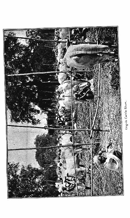
NEL:LORE BREED. . " The, Nellore br,eed pf <attle; has a wide rep'utation
.throughout India, ana even beyond its'limits, 'FOrri1erly .-the pripcipal breeding localities , .. were' situated'withln the no~rthern taluks J , of the N eHore district of the Madras Pr~sidency, but-recently these t,aluks: hcw~ ~een inclu'd~d
, in the new distt:ict of Guntur, 'so the cattJe ' should ' not ,rightly be' given the na,me Of,Nellore but ra~hei: that of Ongole, frpm which tract the best sp~cimens of the breed' are to be oBtained. -' :~i t Wil-S formerly noticeable that'''' cattle ,-bteepil}g re- '~ ceived n~~st attention in those par~s 0£ ]'t,e COulitr.y ~here c,irchlmstances of one kind or another wer,e. 'adverse to ,t}1e exte,nsive prosiClition of ag'ricllltur~ . The culti- · 'v.ators were ;epeatedly' deprived of the results 6f 'their
, labour, and were /con~equently considerably harassed ': they, therefore, as a substitute devoted. their time to raising large her.ds of cattle bf a superiot· ki~d 'which w~re then much in demand, and which they 'saved from the :grasp of the officials by moving them from place to place. 'pridet more secur~ ' Government, these cattle 'br.eeder~

CATTLE OF SOUTHERN iNDiA.
se~tled down, and being a_- fat~ly wealthy class retained their pride in t_he fine cC;l.ttle in their possession, with,the result that many beautiful , specimen-s may now , be se~n , i:n this part of the country. ;, The very best examples are to, be found -in the, villages of Karumanchi, 'Nidamanur, : PO!ldLir, J ~yavaram, Tungutur and Karavadi in the Ong-ole t~Lluk, arid in Elapalapadan, N enn~rpad aIld ·thehamlets 'al.ong the banks of the M usi in the Kandul{ur tal~k. ' Fine cattle of this" breed may also be found in
- the taluks of Vit~uKonda and _Narasaraopet in the Kistna district. I il the sQuthern part of the N ellore djstrict) ~here we.t crops are-grown, the cattle are much inferior, not,.. being so \ve)l" ~ared for or fed as in the places above named-.
The sy~tem 'of feeding observed by the ryots of the ,different parts of tli.is _country ' laturally depends ' upon the_ extent of the pasturag~. In the lowlying , parts where paddy is princip"ally grown, a certain portion ot '
_, dry laqd-is,'often kept as a pasturage for cattle. Most . of the ,cattle, however, leave the villages during Septem
ber at;d October-the southern rainy season- and are ,sent to the western taluks where there are ' extensive waste and jungle tracts. Part of the working cattle will occasionally follow tHe -otbe- ,cattle during N ovemoer - . and December, ' should i,li~ " pa~.ture land of the vil1age not be sufficient'.;. The -ry~ts often (club together and send thei.r <::;ittle"'away in~ lar e ,heFcls.' For this purpose;
• - -",0 .. ,;. ~
prior to the departure of the cattle from the village, arrangements aTe mad~ ' fQr ren.ting p~sture . blocks at a fix.ed sum for ' the season: (Oc~obe~ to February), or eng'agements: are entered tnto with: tbe holders df the
.. pasture' farms'" fol the p~sturing of th,.e whole herd for the ,season at a smaU fee per head for ear.h full grown an,i~al. Should the north-east monsoon be favourable and extend - . ' ~

CATTLE OF' sDuT'HimN INDiA.
till late in the season, ~h.e cattle are only pastured until January whe.n the paddy is harvested, after which there is very fair grazing. The whoie of the jungle grazi'ng i~ not open to the cattle at all times, for after a 'heavy burst of rain, invariably experienced about October, the best portion' of'the pasturage is preserved and kept clear of cattle for one ,or two months until the grass has grown ~lr well, when the working cattle alone are turned in and kept thereon so long as the pasturage suffices, another porti.on being similarly kept for the other cattle. Towards tihe end of January when the sorghum crop on the higher land has come to ear, the young shoots termed Zadu, which_ are not likely to mature, are removed and given to the bullocks, which are often picketed close to th!=! fields. Occasionally fields in the midst of cultivation are set apart fpr pasture, and are planted with the black and white varieties of Acacia. After being under grass for ten or twelve years, the -ground is cleared .of trees and
:-".'
broken up for cultivation., The trees shade ,the ground . . ·and favour the growth of grass, ,,,,hi 1st the pqds ' form good fodder. The pasture land held in this way is invariably distinCt for each ryot, and is generally kept exclusively for working cattle, young s,tock, and milch cows.
The country where the best cattle are raised is undulatf~g' highland interspersed wit small hills .. an<;i i~ 'mostly composed of light 'red or dark alluvial soil, where good ,so,rghum, otller millets and legumes are raised. 'The 'well-to-do ryots in these parts find their pleasure arid pride in raising fine cattle.
There is no greater truism than that which refers to the necessity of feeding young stock, and the very great care which the best breeders bestow O1i tihe young animals, 'both male and fem~Je, most certainly accoun!s for the welldeserved reputation of the Ongole > c.attle. The young

€ATTLE OF SOUTHERN INDIA.
~alves are allowed to suckle the whole of the cow's milk, and when they are three months old 'are given g,rass and a small amount of mixed grains. In every way they are
, treated and tended as the family pets, and it is only by CJoino- into the houses of the villages that the best calves 0 , 0 ,
can be seen. '
That there is considerable profit in the undertaking is 'evidenced by the high prices which the animals reali~e. Young bulls sell from Rs. 80 to Rs. 250. S;<':.~eral. fine ' animals have been exported to South America.' A good N ell ore cow is worth from Rs, 80 W Rs. ISO according to the amount of milk she yields. "
Gh~e (clarified butter) is rhad~ 'ln fair'ly larg~ qua~tities, and sold to ~ercbants Jor e~p6rt, being seldom used by, the ryots, who prefer g'ingelly oil f~r cooking, and reserve the ghee for sale. it · gohd ' Nellore cow gives from lItO 14 lb. of milk daiiy. "
. ':The breed is pl~o13ably n@t so ' hara~ a~ tbe l\fysore 'or Alumbadi, but for slo~ he·avy:.work 'i:h~y are unsurpassable, and they are dni:,crsally empleyed for drawing' very'
' !?-eavy loads in the city of 'Madra< frequently amounting to five tons. ,.'., '. " ._,
.The characteristics of the breed are-'
I-Iead. - Face moderately long; muzzle fine; fore'- ~ " head broad; eyes elliptical in shape, large and mild; skin ' round the eyes is black for about h~lf. an inch; ears long
, and drooping; horns short a~ld ~nclin~d to be stumpy; in cows the horns are longer than in bulls they are,directed outwards and slightly bad~.wards.
" Neck.-Short and thjck. .
H ·ttmp.- Well developed.
Body.-Massive, long 'and deep, but some are inclined to be flat-sided. .I n ' fin€ specimens,. the girth

,<
behi~d , the hu'mp is. about ~4 inches, a~d height ,berurid the h.ump 63 inche~. " _ .' ~ , ' " (,.
Back.-" Q( moderate lengt4, ~nd invariil!bly h-igheli at the cf9uP.
Quar~e~s.-St·rong, with a ~ons'id¢r~ble droop. . $heath:~l?endulous.: cows have also'a fold of skin
, ' . in the-positiDn qf ,the shea~h. :' l _. ~~.,. .
Ta~i~ :'_Ldng,.·fill~ and tapering:' -~ ..... , L egi.!-;-Strong, and-so~ew!Iat c~~rs·e .
. Feet.-Large apd i$ o.~wlaat s0ft , looki<Jlg. ~: ,f .'
CO~Otw.-' , Blaci{, a!1d~ wh.i~e ~ld -p ~re white, the latt er is' now most este~me#;~]5ut f ovrherlx. black' al1d white ,was
. ., ,..... ;l'.; '\
tpe predominating: coloUle .. · " . y , , " 'Ii
, T cmpcr.- . Very docile .• '. , Tbe extensive pasturage obtainable in thi'~ - part of the
. country is ~ no doubt responsible in a great measure for I . • -
tP:e~large cattle bre'eding 'industry, which Gove·rnmen~ 'has always been, .carefllt~ foster" Pre~ious, to 1 86/( !l_ poll 'tax' used,'to bi ,lev'r¢d: but ln' that year this wa~ aOQhsh ed • • - .. _' • • - • .' - I ,- ...r t
in NeUore, and a principle ',was laid down for the, fUt\1re that, ~llt of the waste of ea~h village, an area equ'al t~ 30 per cent. ~f -the area ~occupied by ~ultivati.on 'sl1oulg in
, future be reserved for common grazing to be' equally ~il- . ' joye:tl. by all villagers free' of charge '; the swrplus. wa~te; '
... if suffi~ient in extent' to make ,it w~rth ~hile to adopt'the :System: may be leased out for ol~e or tWQ years at a time ·.to the highest bidder. : - _ " . , -I Ii orde~ further to dev~lop and encourage the breeding,' of good stotk, an annual cattle show was established so far back as '1858, and cm;}tinued uninterruptedly until .187 I. During these twelve years a to~al of over RS. :I 8,000 was distributed in prizes. ' The cattle show was resuscitated' in 1904 w'ith most successful results,' and. it 'is
... ·doub~ful wh~ther -5tlch a Jq_rge collection of bulls and cows ,


~ o o '" a br. ::: o


.India. , B~ab~linf bulls .: '
. Bulls'
83
22 '>'
31
. , ' 'Bullocks, single ~ ,, ' pairs
166
13 2
Buff~lo,es, bulls
" ., bullocks 9
" cows 6
38
3 Goats, he II ~
" ' she . ; 3 - -
Total 67 0
The heifers and young bulls were an exceedinoly' crOG@ b b '
lot, and it is most sincer,~ly to be hoped that, with the encouragement , held , out by th~ Madras 'Governme!1t, the show will ·be he~d annually and equally wep ,represen,tecl. The , Cattle 'Show ,at Ongole hal' 'n:ow ' beeome a t:egular ,institudon, and, has continued ,to grow in pop,ularity. 'Ij:
would th<:;refore be a 'very g'reat · pity if ,it were al)9w~d t-0 ,drop for want of funds atld Government. silppert, 'as .this lbl:eed has become wo'rld renowned. -Dl,lring J-90q buyer.!?" came from Brazil and about 200 young stoc,k -w'ere takefl
' a.wa)~ to th is distant country whei'e thee breed ha~ betomy
:very popular. Great care is taken, in the 'selection of the village
'null, .and the collection of B1'dhmz'n£ bulls brought into . ~the show· at Ongole consIsted of forty~five handsome :upstanding animals in sph;:nqid condition. Almost every . \lillag~- has one,. -or two so-ca,ll~d B-J;a;h,nlini bulls, wh-ich
.,

GATl'L,t: OF SOUTHERN lNDIA.
are common property, having been presel~ted by the relations of a deceased villc~.ger as a memorial, or by s'om~ wealthy ryot, or having been punl:hased by public subscription. ' Such animals are always brand<;!d wi~h a ' sacred mark. '-,
Like all agriculwral clG\.sse-s the ryots of the east coast ar!=! ver.y s~perstltloUS. They are usually very llin,willing to exhibit a' favo'mite cow' owing to 'the influence of the ' Evil Eye (Drishti) : A bullocK whose -tail ' has the root -of the, tuft of the hair situated above-the 'hock is said to .
'. .. f
have E1"u~val anti to bring, ill luck. This is not, objec-- tionable in the cow. A bullock ha"ing whfte~ hair, (skin, ' horn and hoofs i ~ iconsidered' of '" eak constitutiol ' and - " .' , . should not be pU~'chased , A bl~ck 15ullock. is generally considered to ~ be ..a rogue ; if not a rogue, he is cons id,ered 'a g..reat val~e. The ,saying is :-" A black bullock ;s but tfie fourth of a bul); owt ) f he js ,guil,eless he is a
. ' bullOck and a ' quarter." A bullock . with numerous , small spots over the body "like , :deer ., 'is considered
very 'lucky. - ~
The 'form of the horns is ,supposed to itid.icate many , 'things and receives as ,Inao}' names. ,For instance. " Madakomlnf," mea!ls horns bellt backwards, and is coos'ider,ed an excellent sign in a c;ow. There is 'an old sayin~ :-" Let any. man, who does not know how to select a cow, purchase one with ho~ns bent backwards.'" Straight horns are iked, Horns pointed forwards'
, "I(opad£" indicate spirit. Irregular, ,tw.isted horns cluw~tttai a re not objected to . . Those ' which appear hollow and have l(ght ~oloured patches "I(otlikomb1t" • are considered to be very disastrous. Horns with white tops " Punkombu " are also bad. I f a cow at the time of 'p~rchasc voiQs' urIne, it is considered' a very good omeR,
, ' ,
"

UlIl;ole liull Ca.lf.


'0 .:: c: c...


CATTLE j" OF' SOUTHERN ' IND,I:A:,
, . . , 'l;>iIt if she pas'ses dung it is a bad sign. ' th~ reverse is " , tp.t:' ease with the bullock.,
. ;, f\. bullock: whis:h fails to .c,ut thC1 fqurth -pair or corner incisors" is calle,d "A:rukatti-Madu" and , is considered lucky. The, sayin~ js :-.-;:" He ''Yho pur:chases a buJIoGk
, with only six p~rl)ianent '{e~th (indsors) will become rich enQugh te purehase an elepn nt" ': 'A bullock which cuts '~nly seven perrri'aneilt front ,teeth..is unlucky to its owner; an,d is responsible ,..for th~" sayi,ng t'hat :" He whp pur • . ,
;' chases ~uc1?- a bulloc~' should ~av·e· ihe· preparat,ioris for his fun.eral made' ready'," .' ,
" ,. Certain obser~an'c~s ' ~1"e , ino~t ~crupulo\lsly . carried ' S>ut by bota purchaser., and seller at cattle sales, and, in fact, have bec~~e. unwdtten la~ ... resting for ailthortty O!l long consent: '.Disregard of these details in the _procedu~e is seriou~ly helie~ed to imperil the prosperity of
. die owner, d ie seile~, and ' the innocent animal. 'Fhe followin'g·ar~ ~he p~incipal ones :-
, . l. -(I) ' After.the 'price has been fixed the buyer ha,nds
the seller it silver coin, either a two-anna bit or ~ rupee as earnest money.. ' , ,
'( 2) T pe balance of the money may be paid at onc~ or at any stated time afterwards.
(3) . The seller has to pay the purchaser a four-anna or eigh:~-anna -bit for what is called Maralu labham of Pathi Vitbamalu (cotton seed). I t is intended that this money should be used by the buyer for fodd,er for tlle animal for" tha~ day. The purchaser is always careful to go wit~ four-anna pieces it} the event of the ~ell.er n:o . having change for a rupee.
(4) The huyer must never tie up the animal w·tp. hi own rope, and, therefore, a purc~aser never
tarries ne.

CATTLE OF SOUTHERN INDIA.
(5)' The seller ' must ' always suP.HIY the purcnaser . with a new rope, and if it i~ not available, he giv.es the' purchaser raw matei-ial which mus! be ~rajaed or twisted inte> ' a . rop~. The seller must ne,ve't~ .,give .'the · rope alr-'eady used by the animal. ' , '
(6) The 'seller ' in ' company with the' p.urchaser should 'fol' a ' short ~istance lead the animal himself with
• th~ ff'esh' rop~ and tli~n transfer the rope to the hands of the, purchaser wno then takes the ·animal home. ' This se'ttles "the sal~ contraet a:nd is never disputed. The , . conditions Of sale are never reduced to writing.
- K~NGAYAM BREED.
__ This breed ' is 'also ~nown ' by the names Kanga1zad, Kongtt and Kaniaen and derives its name from the taluk of t~e Kangayam.
These cattle are bred -in the southern and southeastern taluks of Coimbatore. There are said to be two var~eties, a large and a 'small. T?e smaller are ' found to be more ' numerous in the Kangayam, Dharapuram, Vdamalpet, Pollachi, Palladam and Erode taluks, while th~ larger variety are more prevalent in Kanlr, Aravakurichi, aqd Dindigul taluks. It is understood that the celebrated Kangayam breed are not the common cattle of Kangayam, but are the property and produce of !arge breeders such as the Pattagar of Palaiyakottai and his family, the Kadiyar Ml?nsiff Monigar, etc., who maintain herds offrom 500 to 1,000 head, anp keep large numbers
, . '
of cows and ,bulls for breedillg ,only. Many ryots, however, .own from 10 to 20 head of cattle reared for sale.. These cattle are sold, not at ordinary markets, but to deal~rs who come to the district for the purpose, or at large cattle fairs such a,s Avanashi, Mahadeshwara Hill near Kollegal, and Madura.


,
The breea 'in its purest form may be 'seen in tae herd of the Pattagar of- Palaiyakottai . who has been an ej{tens,jve breeder ,for many yeats. .. } ..
. I t is curiolIs to note that the animals seen at the . severaI' cattle fairs held in what is practically the home
1 t _.. I ..
of ,this breed are not nearly so good as animals of th~ same breed' which are sold at ' fairs in th~ sO,uthern districts. There are probably two, explanatjon~f fQr -this, ~~stly, that it is ndt profitable t,o export: any b'ut selected animals, and sec;:pl)dlyl, pur,chaser,s sele_ct the:very young' male stock aJ?Q th~se are t_aken awC!-y .to good grazing grounds where' J b.ey are sp~cially ' Jed u.p,. but the fact remains that bette~ pairs, of b}.lllo~ks _?fthjs , br~ed ,~re to be met with in the Madura and Tiflnevelly distriqs tl;tan in the' neighboiirhood. of Ka,ngayam.,' . ,. ; I n the bt_:eecl'ing of th~ ordinary cattle pi' th~ district .
tliere does not appear to be much, if any; ciire ·t_aken, in , . setting apart bulls. Every ' ryot keeps ' his cows. 'and other cattle _in his , own, fields, ' :wl]..ich are all f~nced, ~ p~actice which "scarc~ly is to ,he .seen ' b~y_ond _ the: limits of Coimbatore. As :tb.e_ yOl,lng stofko ,are ()fteQ. ' par~ed
',with 'at a very <:;arly age, ·~n9,. there -i'; }!,n e_x¢hil_hge fJom hC;lnd to hand always going on, it i impos~ibk to trace ~ut the origin of the q'laj~r:ity of tn~ · stock'.
. Regarding the ' :Pattagar's cattle however ' f~is is different. H is ' herds show a: very iffer¢nf_ syst~m of management, there is a caref~l selection of sires and da~s, the young stock are properly reared and consequently-attain a development rarely seen among cattle- " of the same breed met with in other par~s of the district. In the first place he provides a considerable area ' of perman~nt pasture land. His lalld being fenced it is f asy to separat~ dIe herds of different ages and sexes, the heifers run in one herd, and the yo_ung bulls, ,in , ,

CATTLE OF SOUTHERN ' lNtnA. , .
another, the cows in a third and.s·o on. ' He states that he keeps the preed pure by using sires 0nly from his own . herd, though the appearance' of some of the cows . and 'heifers wa.s such as to gi\re the impression that they, had a' distinct strain , of the Ongole blood in them,: and as a matter of fact he possessed several pure bred Ongole GOws. Qn'e herd bel~l1ging', to th<:( Pattagar 'consisted of about thirty-five yqung heifers about three years old ~ith a- bull running with them. ' This herd was a remarkd:bly firie ~me, the heifers being in fine condition, and were a very level lot, showing great quality. The prevailing colour was white with grey markings about the hump and quarters, though there were many fawIl, fawn white and· even light .reds. T~e latter are I?-0t thought so highly of as the whites or greys, although they are just as well made. . The ' bull was a dark gfey, verging to ' black on.heC!-d,. hump, and quarters with the chara~teristic bl"'oaq face, short, thick, but pointed horns of the breed. The pasture on which the herd was grazing, although rather bare owing.to the absence of rain, showed a strong swavd, and was evidently well able to ·carry the stock on it, and which received no additional food'.
AnotHer her~ consisted enti,rely of young bulls, all of them between·on·e and two years old. These were.a very even lot, showing however a considerable variation in colour. Ano~her heFd consisted of young bullocks which had been ca,strated at three years old,. and were .chieflY intended for sale. The Patta:gar informed me ,that he castrated his bulls at three years old, and sold nothing but bullocks.
. It fS doubtful whether there is -another landowner in India.· who pays so much attention, or carries out ' the systemic cattle. br'eeding on such good lines · as this


f " ' CA'l'T~E OF SOUTHERN iNbtA;
Pattagar , ~nd this only shows, what can he done if dt:ie attention is paid to the essential requirements. ' '. '
~?th ~arieties of this breed are strong, a~tive animal~ 'with comBact bodies, and short stout legs. In' the larger' v;ariety tlie horns are much longer, and of different shape to ,those of the small. 'They curve outwards and almost compi'ete a circle at the point where they approach the tips, and for a distance of three or four inches have 'a sharp ba~kward curve. The prevailing colour is pure white. ,'The Pauagai of Palaiyakottai is the breeder of the , s~all variety, a description of which is as follows :- '
Hea,d.-S~ort with broa:d 'level forehead, eyes dark '
and prominent, ears short and ' erect, horns spreading apart, straight, short and thick with sharp points.
N eck. - Short .and thick. , H ump,-Moderately developed. Dewlap.-' Thin and reaching to the sternum only. Body.- Compact and well ribbed l ~p.
Back.- Short, broad and level. Quarfers.-Strong, slightly drooping. Sheath.-, Not pendulous. Tai'l.-Short and thin. L egs.- Short and' of good bone. F:eel.-Small and ~hard. Colour.- The prevailing colour is white, grey and
white with grey shoulder and quarters, also red, black and broken colours are to be found. Among the ry,ots there is·no selection -of bulls, the Swami 'or Brahmini Duil is usually the village , herd sire, but tp.ere can be
.• little doubt that c~~s · are- also served by the young-, uncastliated stock. . ,. ' -
The origin , of the Bra:hmini bull is somewhat' interesting. The tradition is .that one of the early Hindu

kings -in the course ,~0f his travds. saw wnat poo"r sped .... _ c
mens the herd bull~ were, alid havirng given t}:lls matter. much thought he decreed in honour of Si7!a and of Nandi the bull, who was th~ vehicle of tp.e _God in 'his peregrinatlons- " that' 'all w@ll-to~d~ pex:~ons sho~ld,' o~ 'the death of a rd?-tive~ select the' best young bull calf they could .fino, .and present it "as -an offering to the God. These animals thus became the property of the_coinmunity:, and ' were allowed to r'oam al}d feed 'where they liked, apd
, thus bee'a.me the sire~ ,of the village ·herd. " . { . ., ,
So lqng as ,this was carried ou't in the 'spirit of the decree fine . ' animals were produ~ed, but , gr~d,ually, although' the custo~ is still kept up, p.ersons bought the" cheapest young bull they could find, as a salve to ~heir conscience, and t~~ned this loose" to eventually ' be~ome
_the Brahmini bull. And t~is will probably accou,nt, for some of tp.e wretche4 bU,lls which are to be ,seen every-when~ among the village herds. . ~ ~ '
The ' cows ot this ;breed are said to be fairly good milkers, and are pr~ferred by some people on accounf of their small well:set f~a~e. The pri~e pf 'srrta:ll bullockS' , varies' from Rs. 40 to Rs: '70 pe~ pair',~ 1)ut the larger variety command muc~ .higher prices'. , Cows when in .' full milk. may; be obta.{ned for about RS. '40.
The c,ustOm in Sout4 Coimbatore is that ' if the ryot '.possesses 'only a garden, he keeps his cattle witl1in his
garden with the exception' of the cows which aFe kept at home. If the cultivator possesses dry l ands he grazes hi,s , cattle during the day, and keeps them in th:~ house . ~ compound dhting flIe night. S0l11e 'keep their cattle
.' Flight and day o~ these dry lands. ~here seems to 'be a 'strong obje'ction to keep eattle on red 'soil 'if it can
_ 'be ~elped, as it is considered 'to be heating. ' On the (wntrat'y .black catton soil is said to be c00ling.'

Kangyam Cow.

CATTLE OF SOUTHERN INDIA. 39
_ ,It 'i~ riot customa'ry 'to afford shelter against tHe sun J or' ; ..rain, but tIie cattle' a~~ -protected agai~st the f~rious winds
which prevail' iri . Soutnern C6imbatore-: by screens of, " ~~~oo I~\ats 'and:. bra1)ches ~liich _an~ form'ed into ' a peri. T.~~$e peh s are, fuoved at intervalf of one ?: two' days,
' and in this 'way the whole field becomes , manurea. In this, wa):. also ticks, which are a great , 'pest to 'cattle, arid i'1)crease'.w'!J.ere cattle 'are confined, are pr<;;vented from fixing~6n them. , '.. " ' . . " -. . : ' '
:m'he following is the daily routine ~fcattle worked in ,., f, • ..
gardens. At '6 A.M.' ,~ ~he ryot gives his cattle water e~tli~r ~ith or .~ithout bran, .after whi€h they work at the plough ott weIls till! 2 n'oo'n; they are then tied up under the shade . of a tree, watered ,and fed. Spec~al food ~ consisting o~ bran, cotton seed, and gram ,which is soaked and~ground
.IS given in addition to ~, full ration of .straw or as, much asjt can eat. 'Ai 2 P.M., work is 'resumed and continued unt:il'~ 6 P.M. The anjmals are then tjed to pegs, in the garden ··o~ taken tos he hOllSC::, ali.d straw J~ . gi:ven· in small quant,ities at .,intervals during the night.. . The : straw ratiqn consist? of sta-Iks of 'cholum" :cun1~u, ' r~gij tenai, sam~i, ~aragu, paddy, - chola chukka (top ,oUhe cholum contaIning ears J=rfter the removal or-the grain) 'arid collu ihid (the pods ,of horsegram after '-removal 'of the :seed~ ).\ As-a rule no spcci~l.-feecl.ing ' is given to ' cows or bullocks , '11ot i~ ;~ork . . The' cattle graze on 'the · hai~ested fields, '"'and the few fields , which lie 'fallow or on any 'waste land. 'Some few :ryots in Kangayam ~reserve some Jands for
, grazing on which Kolakuttai grass is grown . . Bulls are put' ploughing at two and a half yea:r~ of
age, ,at three ~nd a,half they are yoked to carts, and t at four and a half they are trained to wor~ at water-~ifts.
Fodder ·Cfops,.grown,as such, are rare, but the-practice IS known througnout the district -aiid is ,ocGasi0nally
, if l;
(A~ '{JAT'FLE ' OF' S0PTHERN , INOM:; ,~ ~ _ ~ ~ • r
f()lIowed. In~ ~he ;: KangaY:i;1m 'qivisioo-. Qf Dharap.'liralll ., where the best cattle are' stin ' reared, there is. a,' r~gl.Jlar p;acti~e in ~~brually .an~ M;.1"ch q,f growing eith~"r ch?lu'n<l :, (Sorghum, vulgare) or him bu. (Pen,icillaria sjn1cata) , chiefly the former ' uader wel( irriga~ion, this i~ :caUed . adar (¥1e~ningt<close or. crowped), iholum frOm. its being s~wn
; ~ ', closeJy S0~ fl~;', to yi~-I9. heavily, . and of thinl1et' stalk, arid i~ grown ate such time, that f'odder .mc:ty be most nee~ea. It ',is cut down before ' earing, and affords, cQnsiderp;bk :' .provision during ,t~e hot ~e-Joather. Foclder"crops' ~re t\ot ' gr0.wn on dry lands; there is a sufficien~y ,' of. flastt,lre '
, except ' in the hot weather, 'aq,d as it is, u1!qsual to, g~t ,.rain stlffi.d~nt even for 'p;\mighing during th~ . peJ;"i()d , rec}~oning from the end of Qecember:to the 15th . April,
no,,;sHch cro'ps are pos!'?ible except on gard~n ~a:tld~. · Cholu~ -straw is a favourite fodder, and is ' eave'fglly stacked for' future use; tl):e numerous stacks t'tlat dOf th.e b.la<:k cotton soil of Udamalpet' and all_gan;lens; are, ap_' agFeeable feature in the landscape. " Paddy . ( OrY~fJ , $.at·t)va), s~mai (Panicum milt'are) and ragi (E_ leZ{-si1?e " ',cor:aca1'!a), 'sVaw are equally appnwed ,of, ctimbu' not- s9 much. Tthe ryot e,xctises himself from ~ growing fodd~r by allegiry-g, aI?-~ with some reason, that. as , his ~h91,U111
_ -fodder i? little injured by growing to maturity, he grows . ehQl_um ~s a. grain crop rather' thaI) as I::l. fodder- crop fo'r . -. the double yield>" The expens~s. of weV irrigation in.the '. hot weather ,aife eonsiaer~'ble, . and fe~ <;:aq afford , t9 lose ' the grain of the :cI1Op. ; ~eyerthel<::ss i~ is p-roQ_abl<; th~i:
- a gram crop could ,more pr,ofitably be rais~d during the . April-~'1ay rains rather' than let 'it be fed off by stQck or plol!lg;h;ed in as. gr~eFl manure. _
. " . ...., . Young" bull? are e.mascl!llated .at the ag~ qf about t;·wo
and a _half year:s by tJi~" proce$S ' of . crushing which 'is .UH..iiVeFsa.I ~in , Illdi<J. They ar<~ at~ th,!t :age aUQw.erl :,to
,; t .' .t


J cllicut Bull.


- 1 CATTLE' OF SOUTHE.RN INDIA,
cover COWS,II and heifers ' are lJUt' to the bull when their first pair' of per~a'fl~nt incisors have appeared. "
PULIKOLUM OR J ELLICUT BREED.;-... , • ~ • w 1
1t .... ' . .' ," .
Other :names ~ave been give~ .to this bfeed and ,they are _sometimes known as Kilaka,d or Kilkattu. · . Thjs i~ a very n4;merous breed, .a,nd altho.ugh a fajr- number. ar~ bred in . the .. variol1s ' :vrllag€s -in tlre 'Madur~ <;listrict by
,far tlie larg.e:St breed.ing, oper.ations 'are- concluCted i~'.the south and: s'0,uth-wes<6f 'Mad'ura; large riumber:s ar~ als0 raised' i.~ the viciniiy of ~he Cumbum valley, and the
.. , ·(l. . ~. '\. ' . . ~ \ . '! ..
Perraya: riyer where t9-er~ are 'gr~?i.ng .gt0'unds of 'vast ~xtent , Tb.xr~ ' lS ct, big breeder i iyif!g " Re~~ . Mad ur~.!lt , Ch6~aval}~~nnjyq, bY'<1!;*e ~C;1nie , ~y~nibtt~j : Ma,m,~ak,.,
",.. ,. ' ...
karar; wl},o ' o~!1s a!:>put ·.~,ooq :he~q of c~ttle;:;ag<;l ~h(i , e?,~bited, ~9m.e · at .the Madyr9- ,~attl~ Sp:o;' held ig ~2~7J , :r~e "yo~ng .';l:?!!US whi~h he , stat~d t~ ,PC: ,typiGal 'of·, tJi§! breed 'wire r,ather smaIi, bu~ o,the~wise. th~!e ~.as.; littLe
' fault to ~e f0-und with them, They were- ~ery comp?-cp; with stout legs and hard fee~" ;, The loins, shoulders, aIi'd , ne~~K~ very .po~efful, an<;i.' both, we'~e' c.apa15le of dOi'Qg, very ' " g~od work. , , He 'want'id -~s,,) oo 'e<l:ch Tol;' :the-s~- b~lIs; blllt ~his .c,lass· o~ c;attl: lfay: ~su~II~ ' be ,?~c~~~e~:. f~f. , about R!5: 35 ,~o Rs. S,c;;) each. These cattle, as"a rule, -ate c~mparativel_y small in size, but ai:,e v~ry_ ·~~.t~Xe- ; )1ci .,I • I - ..... ,)'. ... • - " -
capable of much 'ehdurance. In ,many of_!_he villages i,n ' S(!?uth, Mac\l,l:r~ ~er.ta_,~R_'O(t~e:?m~n b~ll~ 'ar~" ' R~'p~~'for th~ p'urpo§e of bull' fighting 'bi r;ath~r bull ' baitirig,: ang are .
, known as jellt'cu(whLch . ~,~ans , ap o.r}1ain~nt "i?f leaves': . from.· th~ fact -~hat' the nonl~ _ of,- tlie ~ull i:m~: usually ~ tl~corated ~ith' ~ vividly col~lired, clpth~ ,T~e ~~eihod of baiting- is as f0UO~S. A colQl\r:~d cloth~is firmfy tied 'r.ounci the horns of a 'bu.·p, and ' he _. ~s' th-~n ' set: ·fr~e: A nutttber of m~'n tnen: att~ck it atld endeav()ur to un'tie the
6 h . -,


Pulikolum Bull.

-is ' ,' L'egs.-Stroll~, short"a}!d well ,set apart, usually grey and white, but other colours prevail. ' "
"
KAP'PILIY AN BREliD. ,r .
A t'tibe of people calle? the Kappiliyans ofCu~bum i~ , the Madura district have a herd, not very nu~erous of a
somewhat distinctive breed. This tribe are· of Canarese origiri, and s~i1l speak that language. Mr. Francis in the 0~zetteer of M<I;9ura g ives a mo~t interesting account. In d~scribing them he says they are small, active, round
. barrelled aniinal~ well known for their trotting pow~rs, wh.ich t~e' people themselves declare to be desc~ndants ' of some cattle they brought with them when they first came to these parts. They are called the Devaru A vu in C~narese or in Tamil the Tambz'ran JVIadu, -both of which phrases -mean" the sacred herd."
The cows are never milked, and are only used for breeding. Members .of the herd which die are buried, and are not, as elsewhere, allowed ' to be desecrated by the chuckler's skinning k~ife. .
The leader of the herd is called "the Kiner Bull" b
palladu,Avu, and when he dies a successor is selected in .a qua-int manner, with elabora~e :lI?d expensive cere:. monial. On the .auspicious day fixed for the election, the· whole herd is assembled, and camphor, plantain, bet~l
and nut, and-s0 fO,rth are solemnly offered to it. A buncj.1e of sug!lrcane is then placed before it, and the' attendant Kappiliyans watch eage_:rly to see which of the bulls of the ~erd 'o/ill approach. an~ eat this. The animCl,I which ~rsqloes so is acclaimed as the New" King Bull" and is formally installed in his office by being daubed with saffron and kunkuman and garlanded with flowers. ' Thereafter he is treated by ~he whole caste as a God, is given the holy name. of Nandagopalaswami, and i~ allotte? to

wq_tch. :ovet ancL worship him .a . speci~l atteo.qantt -who e';Joys the i~ams w~ich stand in his ,name,~ and i~ th~ custodian of the jewels al1d !he copp~r grants' which were presepted in days 'gone by to his predecessors. " The're
f ' • . " . • are now ni~e of these grants; but they do not state the _ S~kKa year iIi which they' were drawn out, and' the· n~mes
,of the Rui~rs 'are not identifi~ble. ' " . '.', ' .. ~" :, ', The -" .Kirig ' Bull~" are' credited ~ith' ·' li~ving:'per.l , formed many miracles, many pf which ' stories are, still 6agerIy. ,related" and' their 0pini6 ti is ' still soIicit(!d t oM. . matter~ of importance: The herd is· ~oi take~ t'o ' the .h.:iJIs· for the hot .weather until its King has si:gntfied 'his approv~l by acoepting some sugar !lnd milk pl~ced near him. , His attendant always beloi1.gs to a particylar ·sub'divisi-on ,of the caste, and· when he dies hi~ successor }; ' selected in as haphazard a ' fasW n as the King " BuH 'himself: " Before the assembleti :Kappiliyans fmjll ' is offer~d to the' sacr . bull, and then a young boy, is 'sei,zed 'by divine inspiration and points out _ the man ~ho is to be the new holder of the office. . ' " . -
, , The h~~d recei,;es recr~its from outside owing' to' the , ,'Hindus: round abo~t dedicating to it all calve~ 'which ar€ born on' the first ' day of Thai, but these are n~t treated as ' b~ing quite of the elect. - The ' Kappiliyans have re;~ ', ~Ge'nt1y' r~ised RS. 11,000 by ta-xing all members of-their ea~te in the Periyakulam taluk for three ye9-rs, and have"""
, :spent this 'sum' in building roomy lna'sonry quarters 'at , -(Sumbum for the sacred breed . . Their chief grievance is . thai the same grazing fees are levied on their ar{ima:Is as on' m~re . ordinary cattle which, they urge, is equivalen~ -to treating Gods as equals of men.- ')
T AN}ORE POLLED CATTLE, ' , J
: At first siglu these ~nimals would appea'r ~o be , of a • ~ ... • t . - • •
distinct preed, and quite different from any allimals found , .

~"
. ",.,.., ~ !
,,'
" . ~ l ;'

'l'anjoro Bullock.


... o .. I -<
" CAttLE OF SOtJTHER~ rN:D~A. . 45,
in the Presideo~Y;i but this is due to' th~ dl.rTy de~t(uction' ". of their horns,-giving the head a,n appea,rance b(; ~liat of
the British polled cattle; . In' this distriq it i s., the: c~stom . t'Q p~fha ~ cattle from the cat~le dealers- who Dting ' them" cfii~fly fron: Salem and Coimbatore, but rn9-rry a[~
-,brpught ' from the breeding grQunas in,' the Cum'bum , valley in the Mad.ura di's,trid. The ' offsprin.g -whi .. c.h are calved in the Tanjore -homesteads are, 'at about rnt! age of ,~ix mOQ.ths, ' dehorned by their owners~ The procedure i~ to take some' hairs from the tail, and this is mixed
. with j.a,ggery or cqatse sugar, and the mixture is applied over. . the , 'young 'homs; ' then a heated iron- 'is . drawn ~.
'backw,ards and _- forwards .until the part is sufficiently. . ~
' burnt. The burnt part take~ t~To or th~ee ' weeks to: heaT; and prevents the growth of the horns. 1t is als'a customary to cut two or tbn~e inches off the eC!-rs, as this i's~ sl:lppo~ed to improve the appearance. of the animal:' The .destruction of the horns is supposed to increase its , ..
- strength and render it more docile, and , to a certain ~xt~nt, i~ has this effect. ,:Ihis process alters the appear.;. ;:uice=of the cattle, and most cer:tainly: g ive the iriipressio.ri that it is a special breed. I t is only in certain.-parts -of Tanj~re that· it :would be possible to breed catt~e,. 'as nearly' the -whole of it consists of delta land. EXcept m;..the appearance of the head these animals present all die chief-characteristic.:; of the Kangay:~m breed. -
.. ' . ~
.. ,
.. ··.r· .• ~ . .. . 'JJescriptidn .
. ~'. Head. - Short, forehead broad, eye$, __ briglir ' ana . prominent, horns absent, and ears shortened.
Nec(e. -. Short and thick. , 'Hump.- Moderately developed. .
.1 Dewlap;-Thin', ' and reacl)ing to .the ~ternum' only . .
. -Body.-. Compact. '. . . , .- ... -, ,_ ~ -• t

4,6 , , C ,.TtE. OF ..sOtJTli~R~ , ~ ,
-This is a very small variety ' oJ animal f~uD.d to. tl1:~ " nortl1 of the Ganjam district. They' a_;e very sym~~': ",' trical in shape a~q active, and are largely used ' for cart work in the di"strict. ' All over the taluk of Goomsur tliere is exteJ;lsive gr?-zing ground which is very suitable for
'~. . ~ c . rearirig of cattle but they never attain any size. In, 1908 a cattle show was inaui urated at Berhampur, th~ '~h~ef ,town' of the district, and handsome prizes, were 0ffer~d · :;. for ' the be~t cattle,' but it,being 'the first show~;v<j;{ heM in the distr:ict it was unfamiriar to the people. . It is, ",_ however, anticipated that in time i~ will beeo~e a ~ai,ti~ : ' able -institution, as on the first occasion it was very w~ll attend~d.. The following is a descript,ion of a typical Goomsur 'bullock :-
, , ' H ead.- Small, with a flat face, br9i d forehead, and
mild but intelligent eye!? The hotns are medium sized curving outw~rds and inwards.
'- Bodj.-Square, cOll1pact, imd well ribbed up, with good strong loins. The hump is fairly well developed:
, D ewlap.- Thin, and reaching 'to ,the sternum only. Sheath.- Adherent to the body. . Tail.-·~eac~iilg to the hocks, fine and "whippy"
with a large tuft of hair at the end. , I


;; o
U .. o o ., Ei
o


;; o o .... o o '" S " "


;: o
U

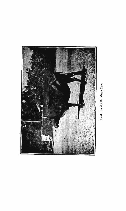


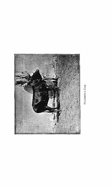
~ o
U

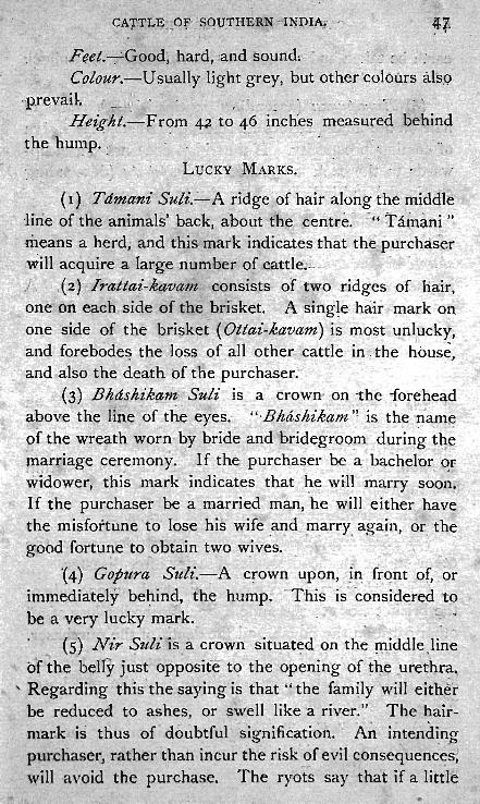
CA'r.'FE..1Lqf·.-:!?qU.THE'R~ -n~J?I~;_· '" • r.
·./f'eet.-. 'Good, harc}., and sound: : . , Colour.-Usually light grey, but other\;Q16urs als,Q
'1 ' I "prevai '. { \ I" • ~ , .. ..', - : \
... ' ~ Heigit.-·-From ~ t; 46 i~cnes' measured" behind .. .. • • ,#:>,.. .. ~ • .. w _ ... .. •
the hump . . , _ . - ," . _ . - ' . . ~ ~ -'. .. ~ _.. ~. • ... f ,I'" . .. ;,
.' ". LUCKY MARKS.. h -, .' T ...,,,- , , '.. ~ ..
.. .. ,.... ,1 ",_
(I) Tdmani ·Suli. :_'_ A ridge ' of hair along the middle_ 'line of the animals' back, ab'out tbe centre: .- " :tatnani " ,means a herg; and this mark indicates that the purchaser ,will acquire a large number of cattle.:' ... 1. .(z) . 1"rattai-kavam consists 0f ,two ridges of hair, one on each. side of the brisket. . A sing!e ha1r mark on one side of the brisket (Ottai-kavam) is most unlucky, .. . and forebode~ the loss of.all oth.er. cattle in . the house; and also the death bf the purchaser: . . (3) Bhdsitikflr..m· Suli is a crown- on -the forelieqd a:bove the line of the . eyes. " 'Bhash£kam ,; is the"name of the ~reath worn· by brid~ and bridegr~om du;ing th~ inarriage ceremony. ' If the purchaser be "a- bachelor ~; ~idower, thi~ ~ark indicates that he wi·ll 'marry so~~. If the purchaser be a married man, 'b'e will either hav~ the misfortune to lose his wife and marry again, or the good' fortu~e to ~o bta:in two w.iv~s" : . . - ,
'(4) Gopura Sulf. - . A c~own upon, j~ ~rq!lt of, . or imf!lediatelf be~inc!, the hump: his ' is' consi<;lered, t~ be a 'yery lucky m~rk. -., •
.. (5) . N1:r $uli i ~ , a crown situated <?n the ~_iddle ,line "' . . '. of the bel y just· opposit~ t9 the opening of ~"4e ure~hra,
Reg~rding thIs,' the saying is that" the family will eith.~·~ be reduced to ashes, or swell like 'a river," The hairmar.k is tlius of doubtful signification. ' An intendinO' purchaserJ rather than incur the ri k of evil consequen~es: will avoid the purchase. The ryots say that if a little
•

earth be taken and rubbed OR this hair-mark the bullock will void urine.
. (6) BurUpltra1t (ascending centipede).-A_ r'idge of hair on the hind quarters curving up to tlie back is a sign of coming prosperity. If the ridge does not 'c,urve up~ wards to the back it is called Ira1zgU,.p1Zrtin, (descending centipede) and indicates adversity to the purch?-ser. . - ..
(7) Lakshimi Suit: is a hair-mark situafed on one side 9f the neck at some distance from the dewlap. " Lak-. shumi" is the goddess of fortune . . This is considered to be the most lucky hair-mark, but is rarely met with. A pullock with such a mark is highly esteemed, and fetches a long price.
UNLUCKY MARKS.
(1) Mt"kkanti Suli or Ag1ti Suli.-Three crowns ~n the forehead arranged in form of a triangle said .to represent the three eyes of Siva of which the one in the forehead will, if opened, burn up all things witl1in the range of vision.
This mark forebodes ruin to the purchaser. , (2) Kudai-mel-kztdai. - Two crowns one over the
other on the forehead predict disaster after disaster.
(3) Ottai-kavam.- A single hair-mark 01) one side o ' th.e brisket close to the middle ]joe forebodes loss of all other cattle in the house, and the death of the purchaser.
(4) Vilattgu Suli (fetters).- Hair-marks .on the fetlocks of either pair of legs indicate that the purchaser will soon be ~n gaol.
(5) Padai Suli. - Two ridges of hail' on the back on either side of the middle line indicate_that the purchaser will soon need a coffin.

, I , CATTLE OF SOUTIiERN INDIA. 49
(6) Ira1tg:upunin.(descending centipede).-A ridge of hair" on the hind quarters not curvi'ng upwards to the back indicates adversity.
(7) NaKapp'adam.-A riq,ge of hair on the haunch spreading out ,at .one end like the hood of a cobra. If the direction of the hood is upwards it is termed Mun
nag'a11t, and if downw~rds, Pinnagam. I
(8) Tattu , Suti (obstaCle ).- A crown situated on the back' between the points of the hips indicates that any bu~iness undertaken by the purchaser will fail.
(9) T1tdaippa Suli. - A hair-IlJark on the side of the -tail near the root, sometimes extending as ridge over 'the back.

'CATTLE 'oF SOUTHERN
r
BUFFA.LOES . .
The buffalo.or Bos babulus stands next to the oX in its utility to man, and still -exists in a wild stcite in large
--numbers. There is scarcely a~ animal upon which domestici~y has made so. little impression as the buffalo, and yet it is universal, and found aIr oveJ.: the Presidency, distinguished by ' i~s large. flat horns, . spme Cl!lrved -and some long, measuring sometimes as much . • as five and six feet in length. The colour varies fmm 'a black to a light slate colour, with very scanty hairt but with tufts -of hair in ~arjous parts of the body.
These animals are very largely used whel:e wet cultivation IS 111 vogue. Althouo'h slow they are excellent workers, particularly in the pleughino' .of paddy . fields and sugar mills.
In Seuthern India ,there are probably four distinct varieties, viz. :-The Teda which are feund on the plateau of the. N ilgiri hil1s. The Parlakim di en th~ Ganjam hills en the East Coast. Malabar: the best .of which are to be found in the south-westexn portien of that district and South Canara, al1;d the small non-descript variety which may be seen everywhere.
TODA B HAL
The Toda buffale is pretty well known to the frequenters .of the N ilcriri hills where ach Teda Mund possesses a herd. They differ from the kind generally met with en the plains, and appear te be indi eneus to these hills alone. They are of exceedin ly powerful build and long in carcase, they have scarcely any hump; the chest is bread and deep; the Ie s short and sturdy; the head large and heavy, and surmeunted by horns set wide apart and curved differently t these of the animals seen


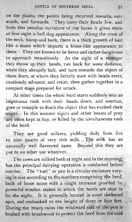
CATTtE OF -SOUTHERN INDIA. 51 , ~ ,
on the pla,ins, the point ' being recurved inwards; out~ wards, ·and forwards. They carry their he~ds low, .and _ from this pecul~ar curvature of the horns it giyes them :. at ,fi'rst ,sigp.t a ' pull dog appearance: Along the crest 'of '
~ the neck, hump and back, tbere.is a thick (Tfowth of hair , . b
,like a nlane which imparts a bison-like appearance to them. They are ~nQwn to be fierce and rather dangerous to appro~ch incautiously, At the sight of a stranger they tb,row up their ~eads, run back for some distance, when they. abruptly halt, ' and turn towards the object of these fears; at whom they fiercely st~re with heads erect, cautiously advance, and retire, then gather together in a . , ' -compact rna , s prepared for attack.
. At other times the ~hole herd starts suddenly into an , impetuous rush with their heads down, and 'Over1run; gore or trample to death the object that has excited their anger. In this manner tigers and other beasts of prey are often kept ' at bay, or killed by the simultaneous rush of the herd.
They are good milkers, yielding daily from five to nine quarts of very rich milk. The milk has an unusua~ly well flavoured taste . . Beyond this they are put to no other use whatever.
The cows are milked both at-night and in the morning, but the principal dairyillg operation is conducted before sunrise. The" tuel " or pen is a circular enclosure varyin in size accordin
b' to the numbers comprising the herd,
built of loose stone with a single entrance guarded by powerful wooden stakes in which the herds are shut in for the night. I t is generally located i,n some shelter_!!d spot, and embanked to the height of three or four feet. During the heavy, rains the windward ~ide of the pen is bushed with brushw od to protect the herd from the cold
52 CATTLE OF SOUTHERN INDIA.
and piercing wiilds. 'I~hese pens have no cove'ring whi'ltsoever, the cattle are exposed at all seasons to the rains and sun, while the floor i? covered with an accumulation of their o~ n droppino-s, The young calves ho~ever, prior to their beino- ,\ eaned, are very carefully looked after, and kept under helter at all times. DuriI;g the day the cah es either accompany their rrlOthers or ' are grazed separately in charge of an atten'dant. The 'rod a .buffalo enters very lar 'ely into the ceremonials ' of this peculiar people who interest all 'visitors to the Nilgiris. The Todas ha, e no history, no written character, and only a very elementary language. They are suppo 'ed to have lived on this plateau for up," ards of 800 years, and their onl) means of livelihood is the keeping of herds of buffaloes. Before the advent of the British Government they were considered to be " lords of the soil," but when the encroachment of the town of Ootacamund began to envelope the grazing lands of these people, the question of the title was fully gone into, and now the Toda have become tenants of the over-lord. Under the recent survey it was arranged that about fifty acres qf land, ordinarily to a considerable extent woodland, should be demarcated as the site or reserve of each . Mund or homestead; the total being about seventy. On this the Todas pay an assessment of two annas per acre, the lowest under the present assessment, and thi is the only tax they pay.
Soon after the birth of a child a youn buffalo calf is brought into the presence of the family. The father takes three measures and pours water from the third measure into the other two holdin them close t the hind quarters of the calf on the right side. The meaning of this singular rite is not clear, but it probably has reference to the future supply of milk for 'the infants' sustenance.
, CATTLE OF SOUTHERN INDIA. 53
1 Early betrothals ,are 'common among Todas, and an
interchange of buffaloes ratifies the agreement; later o~ "when the marriage is consl:lmmated, another exchange of buffaloes takes place. At the funeral a small herd of buffoloes 'is driven along with the C01:tege, and all the frien,ds of the deceased and neighbouring vill~gers to do honour to the dead. Arrived at the plac~ of burning each buffalo has a little bell hung round its neck, and they are >
then driven cl'ose to the p'ile with the words" Aran, od atu," " Go with him." Then all the mou~ners tak~ hat'ldfuls of earth and throw tbem at the buffaloes, and afterwards ask the corpse whether they ~ay kill buffaloes for it.
There is an annual cerem'onial in commemoration of all those who have died , during the year. During the first day the animals intended for sacrifice are driven in where they are safely enclosed in a kraal, two or three for each of the dead to be commemorated. The young men now throw off their blankets and rush among them, hanging on to the animals by their horns and neck, whilst a bell is hung on to the neck of each. On the seco~d day the bier is ' brought out, a hole is dug at the entrance of the kraal, and the priest " does puja." After this the " pujari ' I approaches with garlands of creep'ers which he throws at the buffalo,es. This is ~he signal for the H COtep de grace." The poor terrified creatures ,who have been half maddened by the treatment which they have already undergone from tbe young men who have spent the;! preceding horns in exciting them in every way, rush madly about, and ometlme,s leap the kraal wall, and make their escape to some di tance before they can be caught and despatched. The bodies are drao'O'ed b_ack and placed in a line with the bi r be ide them, and mourned. .
What [i 110W5 n xt is weird and cruel, and the Todas evidently fear that Government may prohibit it on the

CA').'TLE OF SOUTHERN INDIA.
score of eruelty, for they '" make a secret" of this, p~rt of , the proceedings. A buffalo, cow an~ ca~f: are prodticed" the latter is held by three men while the former 'receives' :1 blow between the horns, which ~tuns without killing her. A gash is then made under the foreleg of the poor animal, anq the Va'rzhalor inferior priest, dipping some pieces of bark into the wound, gives some of the blood t? the kinsmen, who smear !t upon the" bier" saying at th,e time, " May the sill run away," , ' . FormeHy there ,used to be, reckle~s slaughter, but GGvernment has stepped in and stopped it 011 the ground at the cruelties practised. N'one were gainers by the deaths ofthese poor animals except the I(otas who attelid Dn these occasions as musicians and they "Clajm the carcasses of all the buffaloes. ~ :nen the Todas ~re asked whyJthey give the Kotas all the carcasses, they exclaim '. ,when the.' buffaloes a re alive they are ours, when they are- ~eaq the)'4 are the Kotas,"
BUFFALOES-SOUTH CANA-RA.
: "There -is a sect of H indus known as tbe Jain Bants who oWrl a fine hardy breed of buffaloes, and orne of the best may be seen in the nei" hbourhood of Mangalore on the ~ est Coast. The nature f the cultivati n carried 011 in this part of the country nece sit,ates the u e of thes animals as the indigenous cattle are small and in signifi-. cant. Buffalo racing forms one of the great amu ements of these people, and every rich Bant keeps his" Kam bla " field sacred to "buffalo racing for the details of which I am greatly beholden to Mr. E. Thurst n. The best pairs of buffaloes used for this purpo e are valued up t Rs. 500, and are splendid animals, and, except for occasional plough drawing, are kept for racing all tbe year round


CATTLE OF SOtJTHER~ INDIA.
Each pair 6fbuffalo~s r~i11s the course q.lone a(1d is judg~d by the assembled crowd for pace, siyle, and most iJnpqrtapt of all, the height and breadtli of the sp'Ja~n which they make. ~Sometimes aqkind of gallows, pe~baps twel)ty, fe,et high, is erecte,d on the c0urse' and there is. a round -of applause if the ~splash ' r~aches up to or a'bov~ it . . Tl}~ course is generally a wet paddyJield about ISO yards 'long,
• ff full of mud, · a~d. water. The dr.iver stripping himself to the necessary minimum of garment~ mounts a Utt1e ~flat
, board about 12 by 18 inc,h:es, on which- is a,small 'elev,!tion or stool: H is,fri ends hold'on to the: buffal-oes 'for all they are' wprth, and he p'laces one foot 'oli th~ . ~tool <;lnd one on the pole attaching the so-called car to:.the 'yoke his whip is held aloffin one han,el, and onGoft1w buffaloes' tails in the other. ' He has )1.0 reilfs~ In. r'h j ~ 'w~y he com,es down' the 'collrse shoutl!ig with all his ,!nig4t wi!h the plank on which : lie stands throwing !.1p: a,.,~ott of Prince of V\ ales' feathel: ot mud and' wate,;;' .round· him, They Ha\ e frequ'ent s~ ill s, ,but the falling ",!;' q~i~~ ' sof~ and it is seldom that ,any one COI~le? t,o O'rief. 'l'y1arks are g i eJ , for t,he rac_, style" sticking-,to th~ p)a2.1~ and throwin : up the ' bi6'O'~!?t ' CiI1,q ' '' ,ides~ ?pla?h. )~.t .,~ big meeting perhaps a hundred pair will b~ ent" r-ed bro~ght from all the surrounding' cop nll')' and the "big men
"-always end th~ir buffaloes to the races, hea~ed by a local band.
P \RLAKlM EDI B f'FALOE . .
. Three distinct breeds of buffaloes are fourd in this part of the Ganjam district, namely, the ' Des~'" or , Manda," the " J erangi" and the " Peclda Kimedi" breed. The buffaloes commonly ' met with are of the U De i " or ' Manda" breed. They ,are larger than the animals found elsewhere in the ~istrict, an~ some of them

CATTLE OF OtJTHERN INDIA.
" al"e imported from the Kalahancli State.' In colo',;u' they ' are dark grey. The buffaloes of the "J ~r:angi" breea. . are smaller, ana have short horns .and short tails. They are noted [,or ,their hardiness'and sniartliless ; and'theyare somewhat darker than tbe " Desi " breed. ' Jerangi is in '
<!the ParIakinledi Maliahs, and it is Here where most of , these animals are bred .... · I , .~
The" Pe_dda Kimedi " ~uffaloes are much larger and stronger than those~ of die D ' i :and J erangi breeds.
:' They cannot:. howe,~er: ~tand tile sun so well, but are e*- ·' ceedingly useful for slow' heavy ,work. , It appears from
, such information .as is available tnat they are-' bl"ought by , the Pedda KiJuedi people from 1{alahandi and other parts
of the Samalput district of Bengal, and-are "no -:h~e'd in Pedda Kimecli. Cm,v-buffaloes of" Jerangi ' ~ ana "Pedda
' Kinledi i , bfe~d's a.re rar,ely met ~ith 'outside their nati e 'borders, as _the) do not fare-so' w~1J ~nor do they yield a' . much , milk as they O'erierally do when _found in thei; '
~ • • f .
~ nati e tract, on accouilt of change in food : These two breedS are not thel-efore' very fr ' quehtly ,met with
. outside their ' own country, and apparently. 'no attempt , is made to produce , a mixea breed ' "~, ith , ( Desi " cows and" J erangi " or i( Pedda Kimedi " bull-s.
Buffaloes are largely useo for agricultural purposes, <:and for beavy draft work which the bullock 9f th se- parts are incapable of performing . On the Parlakimedi Estate the ryots keep a far larger number f buffal es than buJiocks, as the former are the m re useful 1[1 r preparjng' land "CI.uring the rains fot' transplantation , of paddy. 0 special care is taken, nor i it nee s ary to provide pasture (! r the an; mal. , as, in addition to h grazing Jands and the cult ivated lands after he crops are cut, plenty of fodder (paddy straw) is availabl in v ry village ,throughout the year, and thi with rice bran,


CATTLE QF' SOUTHERN IND.I.&. 57 , ,.
horse ' gram, and ginge11y oil-cake, whic4 'is "'gen'erally ' given, is ~uffi~iel'lt to sustain. the animals in good conditiqn. What i~ absolutely' n'ecessary i,n' t:~gard to ' the'se anima),g i,s tftat they ~boti:]d be washe,d twice daily. Young' bulls ~ell from ' Rs., 30\0 Rs: roo.. A good cow
I is worth from Rs. 2'5- to Rs. 60 according to: the milk she . yields. , A ' cow-buffalo is generaliy considered, to 'be one S of ~he requi~ites of a family, Cl:s . she helps 'to, support .the
fam'ily by yielding plen~y of milk" and . ghee.. A ' good cow-buffaio' gives from ' I 2 to' 18 lb. of milk daily.
e - Several po?r families earn' their livelihood solely 'qy maintaining one . or two 'c<?w-buffaloes and selling milkcurd, buttermilk and ghee. The regular 'dealers in milk
.. and .ghee who a~e called" Gowdus ''' ' maintain herds of - cow-buffqloes. The herds do not contain-very ~anry fine
·specimens. _ These are always kept outside villages and towns, and live on the grazing ~hey can get, in fields and
. jungles. 'Fhey are not g.iven any rice b~an, horse-gram, oil-cake, etc., and are rather wild. _There .are weekly fairs held at Uppalada and Balada where some very fine
• animals are h.r.ought for sale. Many anim~ls . are purchased for' eXoport to other taluks in the district, and also to the adjoining districts. . . The following are some of the signs which are believed to. be good in a cowbuffalo :- .
( I) The' head ~hould be small. (2) The back should be low . . (3') Fore legs sho~ld be shorter than the ~~nd legs. (4) Horns should be fo'rmed in such a curve so as
not to allow water which may be poured over them to fall on the hoofs of the fore legs , when the animal is standing.
, The chief feature that is looked for, and to which much importance is attached in the bull-buffaloes is the
g
CATTLE OF SOUTHERN INDIA.
formation of. the· horns. I f 'the two horns are of ~qual -length bending backwards with- tips facing each other, th'is is considered · to be a good specimen. An animal that stretches out its tongue and turns it from side to side is said to b.a ve' " Pamu N aluka" {snake tongue) and 'is considered to be destructive to the owner. So also is the .animal which strikes, wi hits hoi-ns, die post to which it is tied, the action being called " Kammu Katta-dam" (hor.n. beating). .

, , .
.. ~ .. 8 ..
.,:;
9
,GA:t''J'LE; OF Spt!THERN;, .IItJ~IA.
>... .. e J:l
~
... • ::t c:>. 8 '" -E ~
8 8 an ...
, .. .. ',. 'I'd :g ~
"0 C ..
' .. '0 bI)
, c : :0 : :~
t cd d ,.t B ,5 ~ ~
"
.. '0 bI) c o .. ... :::I
p,. "
., ' ... 0 '
~ "t:I I: .. .. '0 bI) c o
59
n
, ,

60
~ I ~ t ~
~ ~ ~ ~ ~
'1 Cl
i ... / ~ 'E~T ~!
1
~, "
~
.... ·hi ~ ' .. ' ~ (51
~ ~ " ~ " ~ t l 1:1 ' ~ .::t
Ii
1 II p::
... .c
boO = 0
E .<11 ' ''d .. ~
.D
." , r
.. o
~ "' -a. .... o .. ~ z
'0 = '" .:.( .., ..2 ·s .!
~ t ,
.... o
.. ~ .. 0 0 Z 'tl "d
3
~L
~ .. . ~ := t ~
0 '0
!~ ~. :~ ='. i,. .. ~ I
" , r ,
~ ~ r
" j ••
8 til .... til b.O = til ~
.~
'0 ," 1:1
til
!,
I
8 .., B
8 '"
§
0 .g 0 '0 '0
---:::::
... 0 U
:< '"
I,g r= ~
• ."
.. ~; ,f
'f
I~ •
'0 ' ~ = til
~ . '" '0 .. r lil ~:
_ I l . '" "'.-g 8~;'-U - . ~ ~ ' ~~.' '. ~ ! _g! . c'; (l:l l ,
~ ' § ;. 'I o .. J; ...... I
o .., .
a oS . a. ' ,': i l ?
: r...
"
I'
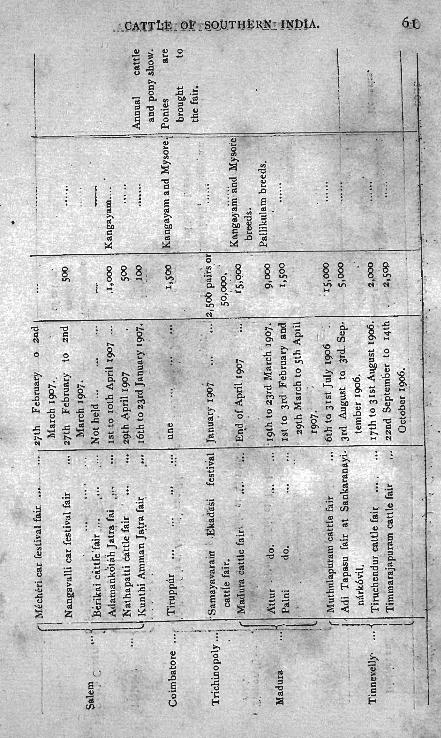
I
.1 i
, !
! i a
s"· .. c;. , (J} '
<II
~ b.O Cl <II
;.::
~ 2 <II
.J:J
.§ o
U
, -' ,
~ . . j
6.:

g, ~ .;; .. o II> <> e c.. ... o
" II>
S «
Z
tATTLt OF SOUTHkRlIl INblA.
§ Ii .. .. . II>
.J:J S ~ II> o

CA."l''l'tE ' bit -SOtJ'rHEi.tN -'IND1A. 63 '"
~ oS
1/ e .. p:;
A ~ -0 ::: -0 oS .-d .. .. boO ' A ... '" -0 -0
~ ;. g CO .. oS .. CO ~ ~ .. ::s A ::s Z ~ ,J:l
~ 1i ~ :: .a oS
~ .; ... .. -0
'" 0 -0 ,J:l .. CO ,il 'jjj II) s: -0 !'l ~ boO
-e CO .. '" 'jjj s: oS ,J:l >
1i .. CO ~ i!; .. .. ~ .£
oS :; .; '" '" ~ .. b- i!;
~ oS ..
~ ...
s: '" c:: r:! ] 0
" .. .. .. '" oS .... .. '" -0 Z -0 -0 'jjj i!;
~ ..
.~ Z co :I: ... . ... t:> ' .~ ,-------...... --------~ ;:!!1 ~ 1=1 .. ~ ...
0 0 0 0 0 0 0 0 0 0 0 0 0 .... .0 ~ 0 0 0 0 0 0 0 0 0 0 0 ::> 0 ,
" ;.-. E 0 o 0 0 0 0 ' 0 0 0 , 0 0 0 0
~ :0 .. ::s ~ \D~ ~ ' g ..;: v. v. ..:: ..;: 0 v. 0 00 ,;; .... I:! ,0.. u .. .. .. .. p.. ~ ... .:; -< C'l 0
~ E
... : 1 ~
t!
~ ~ .... .... ~
.... i:' .... .. .. " oS " ::: 2 ;:
~ .. 0': 0 .... .0 .J:J '"
..:> .. <A .. .. ----
~ ~ ::s t:.
~ " .. ;: .::: ~ .. ~
0 ~ .. ~ u ... .. f!
,J:l
0. oS 'jjj t r:! ' . .... . ' .'
.. 'jjj oS ' .c ~ 0 .J:J .... ',C .c S ..
oS .. . ~ .. oS or:! :: .. .. 'jjj p..:::: p.. I:! 6 .. .~ E ... . ~ .... boO .. .= oS 2 E .. 'jjj .. .. r:! .c bD ..
;z. -= ..:.: ._ oS
.t: ~ i '2 ::s :; .. -'" oS oS
.. I:! -0 '" cl! ..:.: I:! .; i!; ... as ::: oS r:! ..:.: .. oS 'jjj ::> 0 :a .c .c > 'jjj oS ·c
;> ;> ::s f-< ~ f-< U U ~ -< >- Z en
~ '--..--~ ~ ~ ...: ...: -s -0 .~ r:! .~
oS
-'" ::! 2! ~ ::s ~ . ... 6'D
C- .t: u ..s ~ ~ 'jjj ~ \.. ... 'C ! ... .c c:: bD 'jjj ;;: boO ... ~ .;:: oS ..,
~ '" ... boO ..
~ ,J:l i::
~ ~ .. .. ~ ::s ,;s I! 0 ::a 0 -;; :; .c !!! 0
::.d en ~ ~ ~ U ~

~ ~ .. ]; .~ '..l Q) .. :;;
~ '~ p,. oS t
fe .D "C ~ ;oa' ~ _ .
,0 oS oS , ' U ... Z ~. ], ~ ..; oS : ", . , I il: '" t>'D:: c:I ~: <- .D " CI> r: " oS oS oS
~ " '" "C 0 ~ '8. ~ t "C ... 0 "C ,l) "C c: oS " ~ .. oS oS il: .. c:I .: c... .~ "C
..c:: ' "C oS 1 ..... 'l:i .2 " oS ~
CI> -; :; " .. oS , " -; Z
~ : "C ;oa "C g:, "C !:l ~ S ~ ~ oS '" oS ..c:: til .. :E oS - " I V)
~ ~. I> .... .. oS ' ..c:: ;oa ~ ;oa '~ oS 0 .t:l "C oS U O· r , ~ - ;oa
~ ~ .ci
~ c: 0
~ ,.... . ::,E
0 ~ -~ .. ..;- .; ;:: • til
:.:: : CIS
= ''''
.;: I'"
.. t§ 0 .. ~ .. ~ ~ ... !::I 0 ~ .. L~ E 110"
oS
~ Z
"C .~ 1::1 .~ ..; d .!l ... c:I
,~ " I- O . ... .~ .;
! i r '" ,1 .. ;;; "C ~ 9 is ~ ~ III
~ 0 ~ ~ oS = ~


140 ENDOCRINES IN OBSTETRICS AND GYNECOLOGY
pared a cortical extract by means of organic solvents. This method of preparation has been improved still further so that adrenalectomized animals may be maintained in good health for years.
Recently Wintersteiner and Pfiffner (1935) and Kendall et al. (1934) have isolated a crystalline compound which maintains life in adrenalectomized animals. The substance contains only carbon, hydrogen and oxygen. Whether there is more than one substance has not been determined, nor has any substantial opinion been advanced in regard to the structural formula of the crystals.
In addition to the cortical hormone, vitamin C was isolated in crystalline form, in large amounts, from the adrenal cortex by Szent-Gyorgyi (1929). This substance was originally called ascorbic acid but tht:\ name has rccently becn changed to cevitamic acid. Its chcmical formula
o=c I'"
HO-C "" II °
HO-i/
H-C I
H-C-OH
I CH,OH
Cevitamic acid
FIG. D4
is CSHg06, and its generally accepted structural formula is shown in figure 94.
Cevitamic acid is a white or slightly yellowish-white, odorless, crystalline powder which melts between 189 and 192°C., and is freely soluble' in water, soluble in alcohol, but practically insoluble in chloroform and ether. It oxidizes on exposure to air and light. Whether vitamin C is stored or syn thesized in the cortex, is still uncertain. Its significance in the adrenal cortex is unknown.
Method of Assay. The only test for cortical hormone is biological. In common with all complicated biological tests, it is subject to wide individual variations, depending on the animals. Such factors as diet, salt and water intake, must be taken into consideration. Pfiffner, Swingle and Vors (1934) suggest the "dog unit" or that amount of hormone which will prevent an increase of 100 per cent in the blood urea of
THE ADRENAL GLANDS 141
an adrenalectomized dog in the course of from seven to ten days. This is not the most satisfactory test, the best method (and the most diffieult one) is the one by means of which the lives of adrenalectomized animals are prolonged beyond the maximum survival time of control animals.
Relation of the Adrenal Glands to Other Glands of Internal Secretion
1. To the Pituitary. The adrenotropic (interrenotropic) principle. The anterior pituitary gland stimulates the adrenal cortex. Philip
E. Smith (1930) was the first to observe that hypophysectomy in rats was followed by atrophy of the adrenal cortex, and that the normal condition was restored by intramuscular implantations of fresh rat hypophyses. Evans, Meyer and Simpson (1932) confirmed Smith's observations, and in addition produced cortical rr.pair by means of pituitary extracts. CoIIip, Anderson and Thompson (1933) found the adrenotropic substances to be closely associated with the thyrotropic, and eventually succee~ed in separating the extracts. Houssay et al. (1933) were able to stimulate the adrenal cortex of hypophysectomized dogs with pituitary extracts.
Clinically it has been well known that destruction of the hypophysis is followed by marked atrophy of the adrenal cortex (Fahr, 1918; Jaffee and Tannenberg, 1928). Pituitary tumors and acromegaly may cause hypertrophy of the adrenal cortex (\Vieth-Pedersen, 1931; Salmon, 1933; Bauer, 1933; Cushing, 1933; Berblinger, 1932); Furthermore, in anencephalic monsters having maldevelopment of the hypophysis there is usually found an atrophy and hypoplasia of the
l adrenal cortex (R. Meyer, 1912; Kohn, 1924)
The adrenotropic principle is best tested by means of the hypophysectomized rat. Anselmino, Hoffmann and HeJold (1934) suggest the use of the infantile mouse, in which adrenotropic hormone produces a hypertrophy, of the fascicular and glomerular zones. They ascribe to the adrenotropic hormone the following properties:
It is soluble in water and 50 per cent alcohol, but insoluble in 70 per cent alcohol, ether, acetone and chloroform.
It passes through a collodion membrane, and from this they deduce that it is not attached -to a protein, and that it is rather a small molecule.
It is not destroyed by dilute acid or alkali, but is destroyed by concentrated acids and alkalies, especially upon heating.
It can be heated in neutral solution up to BOac. without loss of potency.

142 ENDOCRINES IN OBSTETRICS AND GYNECOLOGY
It is not identical with the growth, gonadotropic, thyrotropic, parathyrotropic, pancreatropic, or lactation hormones.
It is conceivable that Addison's disease may be due in some cases to a failure of the adrenotropic hormone, and in other cases to (local)· disease in both adrenal glands.
There is no evidence of any atrophy of the medulla following hypophysectomy. Houssay et al. (1933) report a decrease in the epinephrine content in the medulla of dogs after the administration of pituitary extract. Anselmino, Herold and Hoffman (1934) report the finding of a separate hormone in the anterior hypophysis that acts upon the medulla. They describe histological changes in the fat staining proper: ties of the medulla and chromaffine system which are already in evidence two hours after injection, reach a maximum in nine hours and rapidly diminish after twenty-four hours. I
To the Gonads. There is a definite interrelationship between the gonads and the adrenals, but the mechanism and significance are not entirely clear. These interrelationships have been studied clinically and experimentally.
To the Ovary. Precocious puberty has been noted in the presence of adrenal tumors or hyperplasia (Thomas, 1926). Other tumors may give rise to masculinization (virilism). Addison's disease is frequently accompanied by hypoplasia or atrophy of the ovaries (Karakoscheff, 1906; Matthies, 1926; Halban, 1925). This is quite understandable if we assume the Addison's disease to be due here to a failure of the adrenotropic hormone. A definite hypertrophy in adrenal cortex occurs during pregnancy (Aschoff, 1910; Aschner, 1912). This pertains partiCUlarly to the fascicular zone, wherein there is also a definite increase in the lipoids. Menstruation is also associated with a hypertrophy of the adrenal cortex, though to a lesser extent (Stoerk and Haberer, 1908; Wehefritz, 1923). Castration is followed by hypertrophy of the cortex, especially the fascicular zone (Kohn, 1924; Anderson and Kennedy, 1932).
Pregnancy prolongs the survival of an untreated adrenalectomized dog (Rogoff and Stewart, 1927). A non-pregnant dog will survive four to nine days, and a pregnant dog thirteen to thirty-nine days. Whether this is due to foetal adrenals, or the corpus luteum of pregnancy, or the placenta is at present unknO\\1l.
The adrenal medulla dues not undergo noticeable change during pregnancy or following castration. Nor has there been any agreement as to the epinephrine content of the adrenals during these conditions.

THE ADRE~AL GLANDS 143
Blotevogel (1927) showed an increase in the chromatofillic cells in the sympa'thetic ganglion of the plexus cervicalis of the mouse, during pregnancy and following the injection of follicular hormone. The significance of this is unknown.
To the Testes. Clinically, a hypernephroma may produce precocious sexual maturity. No satisfactory explanation has been offered for this phenomenon.
Castration leads to a hypertrophy of the adrenal cortex (Schenk, 1910; Masui and Tamura, 1926). An increase in lipoids occurs. The significance of these changes is not understood. The'medulla does not seem to be involved.
Adrenalectomy frequently leads to an inhibition of spermatogenesis and development of the secondary sex characteristics (Novak, 1914; Jaffe and Marine, 1923). The simultaneous removal of the testes and adrenals produces less marked castration atrophy in the rat than the removal of the testes alone (Kishikawa, 1925). The conclusion is then drawn that the adrenal cortex sensitizes the secondary sex characteristics for the male sex hormone. Poll (1933) and Nurnberger (1932) made some interesting observations on the effect of injections of follicular and testicular hormones upon the adrenal cortex. The studies are based on certain histological pictures present in the adrenal cortex of mice. During the infantile ~tate both males and females possess a reticular zone. In the young male the reticular zone disappear~ with the first appearance of sperm in the testes, so that in the adult male the reticular zone is ahsent, and in its place a connective tissue zone develops. The adult female mouse possesses a reticular zone only during the first half of its active sex life. The zone then gradually disappears.
If male sex hormone (about one half of a yapon unit) is injected into infantile males and females, then the reticular zone rapidly disappears if. a fmy days. If an infantile male is castrated, then the reticular zone persists' and continues to develop. This process can be hastened and accentuated by the injection of estrin (16 l\lI.U. of Progynon). The process can then be reversed by the injection of onc Capon Unit of male sex hormone, i.e. the reticularis degenerates. If an adult male is castrated there is a· slight tendency for the reticularis to grow back again, but nowhere as complete as in the previous case. Castration of an infantile female leads to persistence of the reticularis, but it can be made to disappear by the injection of male sex hormone. The injection of estrin into an old mouse (200 days) causes a considerable reforma-

144- ENDOCRINES IN OBSTETRICS AND GYNECOLOGY
tion of the reticular zone. The injection of male sex hormone into an old intact male, or into a castrated young (not infantile) male leads to a reformation of the reticular zone.
BIBLIOGRAPHY
ADDISO:"<, T. 1849 A collection of the published writing of the late Thomas Addison, M.D. London: New Sydenham Society, 1868, p. 211. Quoted by Major, R. H. Classic Descriptions of Disease. Springfield, II!., 1932, p.252.
ADLER, L. 1920 Schilddriisc und Warmeregulation Untersuchungen an Winterschlafern. Arch. f. Exp. Path. 86, 159.
ALDRICH, T. B. 1901 A preliminary report on the active principle of the suprarenal gland. Am. J. Physiol. 6, 457.
ALLAN, F. N., DICKSON, B. R. AND MARKOWITZ, J. 1924 Relationship of phosphate and carbohydrate metabolism; effect of adrenalin and phlorizin on excretion of phosphate. Am. J. Physiol. 70,333.
ANDEnsoN, D. H. AND KEN:-!EDY, N. S. 1932 Studies on physiology of reproduction; changes in adrenal gland of female rat associated with oestrous cycle. J. Physiol. 76,247.
ASCI!NER, B. 1912 Uebcr dic Beziehungen zwischcn Hypophysis und Genitale. Arch. Gyniik. 97, 200.
ASCHOFF, L. 1910 Zur Morphologie der lipoiden Substanzen. Beitrag. Path. Anat. 47, 1.
BARGER, G. 1909 Methoden zur Versc!tarfung von Adrenalin und Brenzcatechinreaktionen. Bioeh. Zeit. 20, 178.
BAUER, J. ·1912 Zur FunktionsprUfung dcs vcgetativcn Nervcnsystcms. Deutsch. Arch. Klin. Med. Leipzig. 107,39.
1933 Zum hcutigcn Stand des Nebennierenproblems. Dtsch. Med. Woch. 69,565.
BAZINSKI, S. 1927 L'influence de la resection du nerf vague sur la lipaidogenese des capsules surrenales des mammiferes. Bull. d'histo!. appliquee Ed. 3, Ur. 6. S 181\-1926. Ber. Physiol. 38, 440.
BERBLINGER, W. 1930 Pathologie und pathologische Morphologie der Hypophyse des Menschen. In Hirsch, IIandbuch der Inneren Sekretion., 1,910.
BLO'£EVOGEL, W., DOHRN, 1\1. AND POLL, H. 1926 Uber den Wirkungswert weiblicher Sexualhormon. Med. Klin. 22, 1328.
1927 Sympathikus und Sexualzyklus. I. Das Ganglion Cervical Uteri des normalen Tieres. Zeitsch. f. Mikr. Anat. Forseh. 13,625.
BonEN, D. 1928 Adrenalinbestimmung im defibrinierten Blut mittels eines isolierten Darmabschnittes. Z. Exp. Med. 63, 783.
BOGER, A. VON ANn SCHROUER, H. 1934 Uber die Stillung sc!twerster Blutungen bei allen Farmen der hnmorrhagischcn Diathese und der Hamophilie durch parenternle Zufuhr yon C-Vitamin (Cehion Merck). )'1iinch. med. Woch. 81, 1335.
BOOTHBY, W. M. AND SANDIFORD, J. 1921 The relationship of the increase in blood sugar concentration to the specific dynamic action of glucose and to the specific dynamic action of adrenalin. Am. J. Physiol. 66, 293.
1923 Calorigenic effcct of adrenalin in dogs. Am. J. Physiol. 69, 463.

THE ADRENAL GLANDS 145
BRITTON, S. W. AND SILVETTE, H. '1932 Effect of cortico-adrenal extract on carbohydrate metabolism in normal animals. Am. J. Physiol. 100, 693.
CHAIKOFF, J. J. AND WEBER, J. J. 1928 Formation of sugar from fatty acids in dcpancrcatizcd dog injected with cpincphrine. J. BioI. Chern. 76, 813.
CHEN, K. K. AND SCHMInT, C. F. 1930 Ephedrine and related substances. Medicine 9, 1.
CORI, C. F. AND CORI, G. T. 1928 Relation between absorption and utilization of galactose. Proc. Soc. Exp. BioI. Med. 25, 402.
CUSHING, H. 1933 "Dyspituitarism" 20 years later, with special consideration of pituitary adenomas. Arch. Int. Med. 61, 487.
DAR!:'!, H. D. 1905 The hydrolysis of optically inactive esters by means of enzymes. Part 1. The action of lipase upon esters of. mandelic acids, the resolution of inactive mandelic acid. Part 2. The fractional hydrolysis of optically inactive esters by lipase. J. Physiol. 32, 199.
DALE, H. H. 1933 Nomenclature of fibres in the autonomic system and their effects. J. Physiol. 80, 10.
DONEGAN, J. F. 1921 Physiology of veins. J. Physiol. 55, 226. ENCLER, N. AND LILJESTRAND, G. 1927 Die Wirkung des Adrenalin auf das
Minutevolume des Herzens beim Menschen. Skand. Arch. 52, 243. FELDBERG, W., MINZ, B. AND TSUDZIMURA, H. 1934 Mechanism of nervous
discharge of adrenaline. ,J. Physiol. 81, 286. GHIRARDI, G. E. 1928 Sulle vie di eliminazione degli ormoni; linla e adrenalina.
Boll. Soc. ita!. BioI. sperim. 3, 541. GREENE, C. W. AND ATKINS, J. A. 1931 The influence of adrenalin on the coro
nary flow in the dog. Amer, J. Physiol. 97, 526, HALDAN, J. 1925 Metastatic hystero-adcnosis, preliminary rcport. Wicn. Klin.
Woch. 37, 1205. Influence of tumors on sex characters. Wien. Klin. Woch. 38, 475.
HENLE, J. 1865 Ziet. rat. Med. 3 Reihe 24, 143. HoussAY, B. A. AKD MOLINELLI, E. A. 1928 Action de la neurine et de I'ephe
drinc sur la sceretion d'adrenaline. Soc. BioI. 98, 172. JAFFE, H. L. AND MARINE, J. 1923 Influence of suprarenal cortex on gonads ~f
rabbits; effect of suprarenal injury (by removal or freezing) on interstitial cells of ovary. J. Exper. Med. 38, 93.
JUSCHTSCHENKO, A. J. 1909 Der Einfluss des 'fhyreoidins, Spermins und Adrenalins sowie der Entfernung der SchilddrUse und der Testikeln auf die Oxydationsprozcsse dcn Atmungsgasllustausch und die Giltigkeit des
, Harns bei Tieren. Bioeh. Zeitsch. 16, 365. KARAKbsCHEFF, K. L. 1906 Weitere Beitriige zur pathologisehcn Anatomic dcr
Nebennieren. Beitr. path. Anat. 39, 372. KOHN, A. 1924 Anencephalie und Nebenniere. Arch. mikroskop. Anat. u.
Entwick. 102, 113. KUNTZ, A. 1934 The Autonomic Nervous System. (Second edition enlarged
and th6roughlyrevised (with 73 engravings)). Lea lind Febiger, Philadelphia, p. 302.
KuzNETzov, A. I. 1929 Die Wirkung des Apocodeins auf die Funktion und Erregbarkeit der isolierten N ebenniere. Arch. f. Exp. Path. 144, 240.
LEW ANDOWSKY, M. 1898 Dbcr cine Wirkung des Nebcnnicrcnextraetcs auf das Augo. Zentbl. f. Physiol. 12, 599.
146 ENDOCRINES IN OBSTETRICS AND GYNECOLOGY
LlEJI, C. C. AND HYMAN, H. 1922 Study of Graves' syndrome and involuntary nervous system; vascular response of pithed cat to single intravenous injections of adrenalin. Am. J. Physio!. 63, 60.
LOEB, R. F. 1935 The adrenal cortex. J. A.l\L A. 104,2177. Other litcrature citcd in this article.
LUKSCH, F. 1913 tlber den Adrenalingehnlt der Nebennieren des Menschen bei verschieden Todesursachen. Virch. Arch. 223, 290.
MARINE, D. AND LENHART, C. H. 1920 Influence of glands of internal secretion on respimtory exchange; eITect of 8ubcutaneou.s injection of adrenalin on normal and thyroidectomized rabbits. Am. J. Physio!. 64, 248.
MARINE, E. AND BAUMANN, E..J. 1927 Duration of life after suprarenalectomy in cats and attempts to prolong it by injections of solutions containing sodium salts, glucose and glycero!. Am. J. Physio]. 81,86.
MAsur, K. AND TAMURA, Y. 1926 The eITeet of gonadectomy on the weight of the kidney, thymus, and spleen of mice. Brit. J. Exp. Bio!. 3, 207:
MARSHALL, E. K. AND DAVrS, M. E. 1916 The influence of the adrenals on the kidneys. J. Pharm. & Exp. Therap. 8: 525.
MATTHIES, T. 1926 tlber Anencephalie, mit besonderer DeriIcksichtigung des Geschlechtes der Nebennieren und del' Vererbungslehre. Zent. Gyniik. 60,3438.
MEYER, R. 1912 Ncbennieren bei Anenzephalic (Aus dem Pathologischen Institute del' Konig!. Universitiits-Frauenklinik zu Berlin). Virchow's Arch. 210, 158.
NOVAK, J. 1914 tiber den Einfluss der Nebennierenausschaltung auf das Genitale. Arch. Gyniik. 101, 36.
OLIVER, G. AKD SCHAFER, E. A. 1895 On the physiological action of extracts of pituitary body and certain other glandular organs. J. Physio!. 18, 230.
On the physiological action of extract of the sup era renal capsules. J. Physio!. Cambridge 18, 230.
POLL, H. 1933 Sexualhormone und Nebenniere. Dtsch. Med. Woch. 69,567. ROGOFF, J. M. 1935 The adrenal medulla. J. A. 1\1. A. 104, 2088. ROGOFF, J. M. AND STEWART, G. N. 1927 Studies on adrenal insufficiency;
influence of pregnancy upon survival period in adrenalectomized dogs. Am. J. Physio!. 79, 508.
SANDIFORD, I. 1920 Effect of subcutaneous injections of adrenal chlorid on heat production, blood pressure and pulse rate in man. Am. J. Physio!. 61, ·107.
SCHENK, F. 1910 Vber die Veranderungen der Nebennieren naeh Kastration. Beitr. z. klin. chir., TUbing 67, 316.
SOSKIN, S. 1927 On the "Calorigenic Action" of Epinephrin. Am. J. Physio!. 83,162.
STEWART, G. N. AND ROGOFF, J. M. 1922 Morphine hyperglycemia and ad· renals. Amcr. J. Physio!. 62, 93. _
1923 Average epinephrin output in cats and dogs. Am. J. Physio!. 66,235. 1925 Studies on adrenal insufficiency. Proc. Soc. Exp. Dio!. Med. 22, 394.
STOEIlK, O. AND HABERER, H. 1908 Beitrag zur Morphologie des Nebennierenmarkes. Arch. Micros. Anat. 72, 481.
STOLZ, F. 1905 tlber Adrcnalin und Alkylaminoacetobrenzcatechin. Ber. d. Deutsch. Chern. Gesell. 37, 4149.

THE ADRENAL GLANDS 147
TAKAMINE, J. 1902 The isolation of the active principle of the suprarenal gland. J. Physiol. 27, 29.
WEHEFRITZ, E. 1923 Systematische Gewichtsuntersuchungen an Ovarien mit Benicksichtigung anderen Drusen mit inneren Sekretion sowie iiber ihre Beziehungen zum Uterus. Z. Konstitutionslehre 9, 101.
ZONDEK, S. G. 1923 Significance of antagoDism of potassium and calcium for physiology and pathology. Klin. Woch. 2,382.
ZWEMER, R. L. AND JUNGBLUT, C. W. 193.1 Effect of various corticoadrenal extracts on diphtheria toxin in vivo and in vitro. Proc. Soc. for Exper. BioI. and Med. 32, 1583.
ZWEMER, R. L. AND WOLFRAM, J. 1935 Cortin protection against anaphylactic shock in guinea pigs. J. of Exp. Med. 61, 9.

CHAPTER X
THE THYROID GLAND AND THE THYROTROPIC HORMONE
An interrelationship exists between the thyroid gland and the adenohypophysis that is similar to that between the latter gland and the gonads. The parallelism is striking.
Rogowitsch (1888) noted an enlargement of the pituitary of dogs and rabbits following thy~idectomy. A similar enlargement was observed clinically in cases of myxoedema (MacCallum and Fabyan, 1907), cretinism (Boyce and Beadles, 1892), and cachexia thyreopriva (Wegelin, 1925). Adler (1914) destroyed the pituit~ry anlage in larval amphibia and prolonged the la::ryal state, while on the opposite extreme Gudernatsch (1912-1914) produced precocious me~amorphosis in tadpoles by thyroid feeding. In a classical experiment Philip E. Smith (1922) demonstrated that hypophysectomy in tadpoles produced atrophy of the thyroid, and that repair could be brought about by homologous transplants of the anterior pituitary or by intraperitoneal injections of bovine extracts of the hypophysis.· This work was extended by him to the rat. The atrophy of the thyroid resulting from the hypophysectomy caused a lowering of the metabolic rate by as much as 35 per cent (Foster and Smith, 1926). Meanwhile Spaul (1923, 1930) produced hypertrophy of the thyroid and acceleration of metamorphosis of tadpoles by injection of anterior pituitary extracts. Clinically, patients suffering from hypopituitarism ha\'e a subn0l1llal metabolism (Cushing, 1927), and in pituitary cachexia (Simmonds disease) atrophy of the thyroid occurs (Graubner, 1925).
Hyperplasia of the thyroid of the guinea pig was produced by Loeb (1929) and Aron (1929) by means of anterior pituitary extracts. This was also demonstrated for other species of animals, and the same phenomenon could be demonstrated in vitro (Eitel and Loeser, 1933). Homotransplants and· autotcunsplallts of thyroid tissue in animals responded by hyperplasia when the animals were injected with anterior lobe extracts (Houssay, Biasotti and Magdalena, 1932).
Aron and Klein (1930) report the finding of thyrotropic hormone in the urine. Smith and Moore (1933) were unable to confirm this. Gumbrccht (1935) reports the finding of thyrotropic hormone in the urine of patients with menopause symptoIIls, as well as during menstrua-
148

THE THY~OID GLAND 149
tion. He believes that the symptoms such as sweats, palpitation and hot flushes are due to an excess of this hormone. Hence, in order to inhibit this pituitary overactivity (in the production of thyrotropic hormone) he recommends the administration of diiodotyrosine. (One frequently sees these symptoms in patients with a low metabolic rate!)
Chemistry of Thyrotropic Hormone
The hormone has not been isolated in pure form. It is in all probability a protein molecule. It is not identical with the growth, gonadotropic, adrenotropic or lactogenic hormones. It is rapidly destroyed by heat. Peptic digestion or autolysis of the anterior lobe destroys the gonad stimulating before the thyrotropic factor (C111genot, Pouse, Vallette and Dottrens, 1934).
The Physiology of the Thyrotropic Hormone
The unit of thyrotropic hormone is as follows: Collip (1935)-the minimum amount which "when administered daily in two injections, will cause a rise of 20 per cent in the metabolism of the hypophysectomized rat by the fourth day."
Action of Thyrotropic Hormone (figs. 9M and 95B). Injections of thyrotropic hormone into normal animals produce:
a. Enlargement and hyperplasia of the thyroid (loeb, 1929; Aron, 1929).
b. An increase in the metabolic rate (Siebert and Smith, 1930). c. An increase in the heart rate (Schittenhelm and Eisler, 1932). d. A reduction of the iodine content of the thyroid gland (Schockaert
and Foster, 1932). I e. An increase in the alcohol-insoluble iodine of the blood (Closs,
Loeb and MacKay, 1932). f. A marked decrease in liver glycogen (Eitel and Loeser, 1932). g. An increase in the excretion of creatine in both the normal and the
hypophysectomized rat (Pugsley, Anderson and Collip, 1934). h. An increase in the excretion of calcium by the intestine (Pugsley
and Anderson; 1934). The symptoms of hyperthyroidism are absent when thyroidectomized
animals are ,injected ~jth thyreotropic hormone, except exophthalmus, which, ac-coidmg to Marine and Rosen (1934), is due to direct stimulation of the centers in the midbrain controlling the sympathetic innervation of the eye. Smelser (1936) showed that exophthalmos is due to an increase in orbital structures. It is readily obtained by the

FIG. 95A. Guinea pig thyroid. Normal. Epithelium low, acini filled with colloid. X520. (Courtesy of Dr. S. C. Werner.)
FIG. 95B. Guinea pig thyroid. Treated with thyrotropic hormone. Epitheliumishigh and very active. Almost complete absence of colloid. (Courtesy of Dr. S. C. Werner.)
150
THE THYROID GLAND 151
injection of beef anterior lobe extracts in thyroidectomized, but not normal, guinea pigs of both sexes.
The increase in metabolic rate which results from the injection of thyreotropic hormone into normal rats is plus 26 per cent (Collip, 1935). Larger doses do not result in higher values. On the contrary, goiterous animals show a far greater increase, from plus 95 to plus 162 per cent. Hypophysectomized rats show a decline in the metabolic rate to minus 29 per cent. Such animals are more sensitive to thyrotropic hormone than normal ones. If the injections of thyrotropic hormone into normal rats are continued for several weeks the metabolism drops to minus 29 per cent or to the level of metabolism of the hypophysectomized rat. (The explanation will be considered below.)
The iodine metabolism of the thyroid is controlled by the thyreotropic hormone. According to Oswald (1901) the iodine content of the thyroid varies in general with the amount of visible colloid. Marine et al. (1908, 1909) showed that the iodine store in general varied inversely with the degree of hyperplasia, and in extreme degrees of thyroid hyperplasia the iodine store was exhausted. The thyrotropic hormone by inducing hyperplasia thus lowers the iodine content of the gland. The normal blood iodine ranges from 10 to 12 gamma per 100 cc. The iodine occurs in two fractions, the inorganic which is alcoholic soluble, and the organic which is alcohol imloluble. Their ratio is as 1 :2. In Graves' disease the blood iodine is usually increased to from 30 to 60 gamma per 100 cc., the increase being due to a rise in the organic or alcohol insoluble fraction, which, as we have seen, results from an increase in thyrotropic hormone.
The Antithyrotropic Hormone
It might be supposed that by constantly ~njecting thyrotropic hormone it would be possible to stimulate the thyroid indefinitely. But the contrary fact is true. Korenchevsky (1930) injected pituitary extracts into rats for six to eleven weeks and noticed a decrease in weight of the thyroid, which rcached 17 per ccnt below the control weight. Similarly, the metabolic rate at firflt showed a rise which reached a peak by thc tenth day and then a return to the normal level (Siebert and Smith, 1930) (Verzar and.. Wahl, 1931). If the injections of thyrotropic hormone were carried out still longer, thcn the metabolic rate dropped below normal (Lec and Gagnon, 1930; Evans and Sarka, 1933). This phenomenon was extensively studied by CoUip and his associates (1934, 1935) and by Werner (1936).

152 ENDOCRINES IN OBSTETRICS AND GYNECOLOGY
Collip showed that continued injections of purified thyrotropic hormone produced the following effects:
a. A rise in the metabolic level to plus 28 per cent during the first week.
b. A return to the preinjectiun level during the second or third week.
c. A continued fall in the metabolic rate to minus 29 per cent by the fifth week. This corresponds to the low metabolic level of the hypophysectomized rat. Histologically, the thyroid at this stage resembles the thyroid following hypophysectomy, i.e., atrophic.
d. The pituitaries uf animals thus treated for a long time give a negative response for thyrotropic hormone and a positive response for growth hormone.
What is the nature of this inhibition? Collip found it to be a substance present in blood serum, and therefore named it the antithyrotropic hormone. "Vhen blood serllm from an animal treated for many weeks with thyrotropic hormone is injected into a hypophysectomized anj~aI (0.5 to 1.0 c9. per day for 3 days), it prevents a rise in the metabolic rate even when 200 times the required minimal effective dose of thyrotropic hormone is injected.
The antithyrotropic substance is unstable. Boiling at pH 5 for 3 minutes destroys it. Standing at room temperature destroys its potency, while standing in the refrigerator inhibits its potency. Collip (1935) reports finding this antihormone in certain patients with low metabolic rates. Its presence also explains the numerous clinical failures resulting from injections of thyrotropic hormone.
How does the antithyrotropic substance work? It does not inhibit the action of thyroxin; Collip (1935) states that when animals are treated for several weeks with thyrotropic hormone and then injected with thyroxin, a rise in metabolism occurs. The combined administration of anti thyrotropic substance and thyroxin does not prevent a rise in metabolism. Abelin (1931, 1932) has shown that diiodotyrosine inhibits the action of thyroxin, hence this effect is not similar to the action of the antithyrotropie substance.
Collip's concept of an anti thyrotropic hormone has recently been challenged by Werner (1936). The development of refractoriness has been found to depend on the type of extract in which the hormone is administered. By u,sing a flavianate preparation of thyrotropic hormone (prepared by Dr. Karl Meyer) Werner demonstrated a continued thyroid stimulation, treatment being continued as late as the 80th day.

THE THY1WID GLAND 153
A control series subjected to thyrotropic hormone made by the DykeWallen Lawrence method, showed the refractoriness as described by Collip.
When certain mixtures of thyrotropic and anti thyrotropic hormone are injected lnto a hypophysectomized rat it is possible to produce hyperplasia of the thyroid gland without raising the metabolic rate, which is about 30 per cent below normal (Collip, 1935). The nature of the hyperplasia of the thyroid is not clearly understood. Marine (1927) believes that it is due to a functional insufficiency of the gland. He gains support for this contention from the following fact. When thyrotropic hormone is injected, the first effect noted is an outpouring of organic iodine from the thyroid gland, and is coincident with the appearance of vacuoles in the colloid of the gland and a rise in the metabolic rate. A later secondary effect is the hyperplasia. Collip stresses the· fact that the metabolic changes are not directly related to the histological picture. Effects may be produced histologically with physiological change and vice versa.
The introduction of the concept of antihormone introduces new vistas into endocrine dysfunctions. An equilibrium may be assumed to exist between the hormone and its antihormone. (It has not been shown. that all hormones have antihormones-estrin has no antihormone?) Thus, hyperfunction could be due to an excess of the hormone, or lack of the antihormone. Hypofunction could be due to a lack of hormone or excess of antihormone. Returning to the thyroid, hyperthyroidism as seen in acromegaly can be due to an excess of thy~otropic hormone. In Graves' disease no histological changes in the pituitary are to be found and the disease could be due to an insufficiency of th~ antithyrotropic substance.
The Physiology and Chemistry of the-Thyroid Gland
Th~ thyroid gland occupies a central position in the endocrine system in that its single hormone-thyroxin-is linked with the oxidative processes of all the body cells.
Nerve Supply. The thyroid gland derives its nerve supply from both sympathetic and parasympathetic fibers. The former arise from the middle cervical ganglion, while the latter pass in with the superior and inferior laryngeal nerves. Kuntz (1934), in reviewing the evidence for the nervous control of the thyroid glands, points to the mass of conflicting evidence regarding this point. Variations in the blood supply to the gland bring out variations in function.

154 ENDOCIUNES IN OBSTETRICS AND GYNECOLOGY
Chemistry. The thyroid elaborates a specific hormone containing iodine. Bromine is also found in the gland, but its biological significance is unknown. The iodine in the thyroid is in organic combination with a protein, free iodine is present only in traces. This iodine compound is stored mainly in the colloid, and the iodine content of the gland corresponds to its richness in colloid.
The Iodine Compounds of the Thyroid Gland. Baumann (1896) was the first to discover the presence of iodine in the thyroid.
Three compounds containing iodine have been isolated from the thyroid gland, namely:
1. Iodothyroglobulin (Oswald, 1899). 2. Diiodotyrosine (Drechsel, 1896). 3. Thyroxine (Kendall, 1914). Iodothyroglobulin. This substance is a: globulin which contains io
dine, and exists as such in the gland. Iodothyroglobulin is as active as the powder obtained by drying the gland. By digesting the thyroglobulin with trypsin and then pepsin Harington'and Salter (1930) obtained crystalline thyroxin. Foster (1929) obtained from iodothyroglobulin 33 per cent of the iodine as diiodotyrosine and 16 per cent as thyroxin. All iodine in thyroglobulin is calorigenieally active (Lerman and Salter, 1934). The iodine content of thyroglobulin from persons with hyperthyroidism dccrea.~es as the basal metabolism increases (Cavett, Rice and McClendon, 1934). The activity of the thyroglobulin is probably that of thc thyroid itself.
Diiodotyrosine. Figure 96 shows the constitution of diiodotyrosine according to Wheeler (1930). It is a crystalline compound containing 58.7 per cent iodine. It is dextrorotary and is easily soluble in dilute ammonia, alkalies and acids. Both Harington (1929) and Foster (1929) obtained it from thyroglobulin by hydrolysis. Thompson et al. (1935) state that its calorigenic activity per millimol iA only about onc tcnthousandth that of thyroxine. As stated abovr:, Abclin belir:vcR that diiodotyrosine antagonizes thyroxine, and uses their quantitative imbalance as an explanation for various thyroid dysfunctions. It is capable of lowering the high basal metabolic rate of Graves' disease, but so is inorganic iodine. In all probability diiodotyrosine represents a step in the synthesis of thyroxin by the body ..
Thyroxine. This substance was first isolated by Kendall and Osterberg (1919). Harington (1926) established the structural formula for thyroxin, and in 1927 together with Barger synthesized this compound. Thyroxin has the structural formula given in figure N.

THE THYROID GLAND 155
Thyroxin contains 1i5 per cent of iodine. It is very sparingly soluble in water or in organic solvents, but readily soluble in alcohol after addition of alkali or an acid salt. When heated to 231-233°C. it begins to decompose, liberating free iodine. As with all biological compounds the stereochemistry is very interesting. Due to racemization during
OH
I C
/"'-. 1-C
1
IIC
-1
HC,\/CH ¥ C-CH,-C-COOH
I NH,
Diiodotyrosine
FIG. 96
OH ()I H,\ H
I o I
,/"'-.r I II
HI~ IIH ~/
CH,-CH-COOH
I NH,
Thyroxine
FIG. 97
the extraction of the compound from the gland thyroxin is optically inactive. Ha:ril}gton (I928) resolved dl-thyroxine into its optically active isomers. The laevo compound is three times as active as the dextro, and the former has been isolated as such from the thyroid by the action of proteolytic enzymes (Harington, 1930). The high potency of I-thyroxin is to be expected, for the laevo compounds are the

156 ENDOCRINES IN OBSTETRICS AND GYNECOLOGY
ones usually found in the body. According to Plummer (1921) the human thyroid secretes about 1 mgm. of thyroxine daily.
Physiology of the Thyroid Gland. 1. Effect on Metabolism. The most important physiological action of the thyroid gland is to increase the oxidative processes of the body. This is also true of thyroxin (Boothby, 1924),2.5 mgm. of thyroxin being equivalent to 1 gram dried thyroid gland. This increased oxygen utilization manifests itself also in the various isolated organs (Asher and Rohrer, 1924). The duration of motility of sperm is increased by thyroxin (Carter, 1932).
Effect on Carbohydrate Metabolism. Both thyroid extract and thyroxin cause a loss of liver glycogen. This was first observed by Cramer and Krause (1912-1913). It is also true of heart muscle but not. of skeletal muscle. The loss of glycogen is accompanied by a loss of ability to store glycogen in response to carbohydrate feeding. This is not accompanied by a lowering of the tolerance for sugar, nor is glycosuria observed. The cause for this loss of glycogen is probably the oxidation of the mobilized sugar at a high rate. The reason for this action is that thyroid hormone sensitizes the liver to stimuli which promote the discharge of glycogen. Thus thyroid feeding increases adrenalin hyperglycaemia and thyroidectomy diminishes it. When su~h a loss of liver glycogen occurs as a result of thyroid feeding a small amount of insulin will produce a fatal hypoglycaemia (Marks, 1925). This is due to the failure of the compensatory discharge of sugar.
Effect on Protein Metabolism. In myxoedema the excretion of urea is diminished, and the administration of thyroid substance increases it. This effect is less noticeable in normal animals. Creatinuria occurs in Graves' disease and is diminished when the condition is alleviated by treatment with iodine (Palmer, Carson and Sloane, 1928). In general, it may be stated that thyroid extract increases protein catabolism, affecting all the protein whether endogenous or exogenous.
Effect on Fat Metabolism. Myxoedema is accompanied by an increased fat deposit due to lessened activity. Adequate treatment with thyroid extract causes a loss of fat due to increased muscular movement. There may also be a direct effect on fat metabolism, for Abelin and Kiirsteiner (1928) have observed that thyroid feeding caused a loss of body fats within 16 hours, i.e., before the general metabolic effect of the hormone was detectable.
Effect on Water Balance. Eppinger (1917) in his monograph on oedema cites many instances of increased water and salt excretion after the administration of thyroid extract. Mahnert (1925) has noted the

THE THYROID GLAND 157
same effect in the oedema of pregnancy. This diuretic effect persists after section of the cervical cord, hence the action of the hormone must be peripheral (Fujimaki and Hildebrandt, 1924). Changes in the viscosity and surface tension of the blood, as well as changes in tissue permeability have been offered as explanations for this diuresis.
Effect on Calcium Metabolism. Hyperthyroidism shows an increase in calcium excretion (Aub, Bauer, Heath and Ropes, 1929). The extent of excretion was not in relation with the extent of increase in the metabolic rate. The increased excretion in the urine may reach more than 200 per cent above the normal. Cases of myxoedema showed an abnormally low calcium excretion, which was increased upon the administration of thyroid. The high calcium excretion of Graves' disease is decreased upon the treatment with iodine. Together with the loss o( calcium there is a loss of phosphorus suggesting its derivation from the calcium phosphate of the bones, hence the osteoporosis of Graves' disease (Hunter, 1930). There is no increase in the blood calcium in Graves' disease, in contradistinction to the rise in blood calcium as the result of parathyroid hormone where there is also an increase in calcium excretion through the urine. The reason for this heightened excretion in Graves' disease is unexplained.
Effect on Growth and Differentia.tion. Gudernatseh (1913-1914) found that the rate of metamorphosis of various species of tadpoles was markedly accelerated by thyroid feeding. When fed on ordinary meat tadpoles showed distinct hind legs 53 days, and forelegs 73 days, after the beginning of the feeding, metamorphosis being complete after 104 days. When thyroid was substituted for ordinary meat, hind legs appeared in 9 days, forelegs in 11 days, and metamorphosis was completed in 18 to 20 days. The same effect has been accomplished with 1: 1,000,000 synthetic thyroxin. General body growth was not produced in these animals, for they remained pygmies and died within a few days. In fact, growth of white rats is inhibited by thyroid or thyroxin feeding (Cameron and Carmichael, 1920, 1921). At the same time, the heart, liver, kidneys and suprarenals show~d some evidence of growth, probably as the result of extensive catabolic activity on the part of these organs. A number of workers have utilized the acceleration of the metamorphosis-of tadpoles for purposes of biological assay. If the tadpoles are fed thymus instead of thyroid they continue to grow but do not undergo metamorphosis (Gudernatseh, 1916; Romeis, 1923). Thus thyroid produces dwarf frogs and thymus, giant tadpoles. Metamorphosis of tadpoles may also be induced by diiodotyrosine (Romeis, 1923) and by parenteral feeding of inorganic iodine (Abelin, 1928).

158 ENDOCRINES IN OBSTETRICS AND GYNECOLOGY
It has been demonstrated that the ionic concentration of the milieu in which the tadpoles are kept has a marked influence on the metamorphosis induced by thyroid feeding (H. Zondek and Ucko, 1924, 1925, 1926). Calcium ions in definite, small concentration inhibit, and under given conditions, even reverse the effect of thyroid hormone, while potassium ions stimulate it. Similarly, it appears that the metamorphosis is a direct function of the hydrogen ion concentration. Thus, at a pH of 7.08 there is no effect while at 6.88 there is.a marked acceleration in the metamorphosis.
For a further consideration of the other physiological properties attributed to the thyroid gland the reader is referred to the excellent monographs of Harington (1933) and Laqueur (1934). -....
Interrelationship between the Thyroid and the Sex Glands
It has been known clinically that the thyroid gland enlarges during puberty, menstruatio-n, lactation and pregni}ncy. The nature of the interrelationship is still uncertain, but it is more than likely that it is through the mediation of the anterior pituitary gland. Thyroid feeding produces an inhibitory effect upon estrus (Abelin and Wiedner, 1932). DaCosta and Carlson (1933) showed that large doses of desiccated thyroid retarded sexual maturity of white rats of both sexes while small doses tended to accelerate it.
Tagliaferro (1933) found that thyroid enlargement occurred in rats and guinea pigs following injections of e~trogenic substance, when the injections were limited to five or ten days. But, when the injections were continued for more than twenty days involution of the thyroid occurred. Botl~pothyroidism and hyperthyroidism lead to sterility (Diiderlein, 1929). As stated previously, the effects produced are best understood when the role of the anterior hypophysis in its relation to the gonads aild thyroid, is considered. The thyrotropic aIlli gonadotropic hormones are produced by the cells of the adenohypophysis. It might well be expected that a stimulus acting upon the thyrotropic hormone would extent to the gonadotropic hormone. Thus, small doses of follicular hormone stimulate the production of gonadotropic hormone while large doses inhibit it. The same holds true for thyroid extrar.t. Tagliaferro's resl.tlts are thcn readily understandable: Small doses of estrin stimulate the production of gonadotropic hormone. The same group of adenohypophyseal cells being affected, there occurs an increased production of thyrotropic hormone, hence, the enlargement of the thyroid. Large doses of oestrogenic substance have the opposite effect lipan the pituitary, hence the opposite effect upon the thyroid gland.

THE THYR,oJD GLAND 159
BIBLIOGRAPHY
ABELIN, 1. AND KjjRSTEINER, P. 1928 tiber den Einfluss der Schilddriisensubstan zen auf den Fettstoffwechsel. Biochem. Z. 198, 19.
ADLER, L. 1914 Metamorphosestudien an Betrachierlaven: 1. Extirpation endokriner Drlisen: A. Extirpation der Hypophyse. Arch. f. EntwickIngsmechn. d. Organ. (Roux's) 39, 21.
ARON, M. 1929 Action de la prehypophyse sur la thyroide chez Ie cobaye. Compt. rend. Soc. de bioI. 102, 682.
ARON, M. AND KLEIN, M. 1930 Sur la presence dans I'urine humaine d'une substance douee de la meme action sur la thyroide que I'extrait prehypophysaire et sur I'interpretation de la reaction de diagnostic de la grossesse. Compt. rend. Soc. de bioI. lOS, 702.
AUB, .1. C., BAUER, W., HEATH, C. AND ROPES, M. 1929 Studies of calcium and phosphorus metabolism. III. The effects of the thyroid hormone and thyroid disease. J. Clin. Invest. 7, 97.
BOOTHBY, W. M. 1924 The use of iodine in exophthalmic goitre. Endo. 8, 727. BOYCE, R. AND BEADLES, C. F. 1892 Enlargement of hypophysis cerebri in
myxedema with remarks upon hypertrophy of the hypophysis associated with changes in the thyroid body. J. Path. and Bact. I, 223.
CLOSS, K., LOEB, L. A:-JD }IACKAY, E. M. 1932 The effect of an acid extract of the anterior pituitary on the iodine concentration of the blood and thyroid gland. J. BioI. Chem. 96, 585.
CRAMER, W. A. AND KRAUSE, R. A. 1912-1913 Carbohydrate metabolism in its relation to the thyroid gland. The e!'fect of thyroid feeding on the glycogen content of the liver and on the nitrogen distribution in the urine. Proc. Roy. Soc. B. 86, 550.
CUSHING, HARVEY 1927 Acromegaly from surgical standpoint. Brit. M. J. 2, 1 (July 2) an~ Brit. :VI. J. 48 (July 9).
DRECHSEL. E. 1895 Die wirksame Substanz der Schilddriise. Zbl. Physiol. 9,705.
EITEL, W. AND LOESER, A. 1932 Hypophysenvorderlappen, Schilddr(ise undl Kohlenhydratstoffwechsel der Leber. Arch. f. exper. Path. u. Pharmakol. 167,381.
FOSTER, G. L. AND SMITH, P. E. 1926 Hypollhysectomy and replacement therapy. J. A. M. A. 87, 2151.
GRAUBNER, W AJ,THER 192.1 Die hypophysare Kachexie. (~immond8sche
'l\:rankheit.) Ztschr. f. Klin. Med. 101,249. GUDERNATSCH, J. F. 1912 Feeding experiments on tadpoles: I. Influence of
specific organs given as food on growth and differentiation. A. Contribution to the knowledge of organs with internal secretion. Arch. f. Entwcklngsmechn. d. Organ (Raux's) S6, 457.
1914. Feeding~.experim_eJlts on tadpoles. II. A further contribution to the knowledge-of· organs with internal secretion. Amer. J. Anat. 16, 431.
GUIoIBRECH, P. 1935 Die Aus8cheidung thyreotrop wirksamer Substanz im Harn klimakterischer Frauen. Zent. Gynak. 59, 1331.
HARINGTON, C. R. 1926 Chemistry of Thyroxine. I. Isolation of thyroxine' from the thyroid gland. Biochem. J. 20, 293.

160 ElliDOCRINES IN OBSTETRICS AND GYNECOLOGY
1926 Chemistry of thyroxine. II. Constitution and synthesis of desiodothyroxine. Biochem. J. 20, 300.
HARINGTON, C. R. AND BARGER, G. 1927 Chemistry of thyroxine. III. ConRtitution and synthesis of thyroxine. Biochem. J. 21, 169".
HARINGTON, C. R. 1928 The resolution of dl-thyroxine. Biochem. J. 22, 1429. HARlKGTOK, C. R. AND SALTER, W. T. 1930 The isolation of I-thyroxine from
the thyroid gland by the action of proteolytic enzymes. Biochem. J. 24,456.
HARINGTOK, C. R. 1933 The Thyroid gland. London. HoussAY, B. A., BIASOTTI, A. AND MAGDALENA, A. 1932 Hipofisis y tiroides;
accion del extracto del lobulo anterior de la hipofisis sabre la histologia de la tiroides del perro. Rec. Soc. argent. de bio!. 8, 130.
HUNUII, D. 1930 The significance to clinical medicine of studies in calcium and phosphorus metabolism. Lancct 1, 947.
JUNKMA~-N, K. AND SeROEI,LER, W. 1932 Ober das thyreotrope Hormon des Hypophysenvorderbppens. Klin. Woch. 11, 1176.
KENDALL, E. C. A)lD OSTERBERG, A. E. 1919' The chemical iuentification of thyroxine. J. Bio!. Chern. 40, 265.
KENDALL, E. C. 1919 Isolation of the iodine compound which occurs in the thyroid. ,J. Bio!. Chern. 39, 125. ,
KORENCHEVSKY, v. 1930 Influence of hypophysis on metabolism, growth and sexual organs of male rats and rabbits: II. Influence of extracts of hypophysis on the body weight, weight of fat, of sexual organ3 and of endocrine organs of rats. Biochem. J. 24, 383.
LAQUEUR, FRITZ 1934 Hormone und Innerc Sekretion. Sec. ed. Dresden, 1931. "Schi1ddrilse" p. 85.
LOEB, L. AND BASSETT, R. n. 1929 Effect of hormones of anterior pituitary on thyroid gland in the guinea-pig. Proe. Soc. J<;xper. Dial. and Med. 26,860.
l\1ACCALLUM, W. G. AND FABYAN, 1\1. 1907 On the anatomy of 1\ myxedematous idiot. Blll!. .Tohns Hopkills Hosp. 18, 341.
MARINE, D. AND Wn,I,IAMS, W. W. 1908 The relation of iodine to the structure of the thyroid gland. Arch. Int. 11ed. 1, 349.
MARINE, D, AND LENHART, C. H. 1909 Further observations on the relation of iodine to the structure of the thyroid gland in the sheep, dog, hog, and ox. Arch. Int. Med. 3, 66.
MARINE, D. AND ROSE:;, S. H. 1934 The effect of the thyrotropic hormone on auto- and homeotransplants of the thyroid and its bearing on the question of secretory nerves. Amer. J. Physio!. 107,677.
MARINE, D. 1935 The physiology and principal interrelations of the thyroid. Glandular Physiology and Therapy, J. A. M. A. Chicago.
MARKS, H. P. 1925 Effect of thyroid feeding on sugar tolerancc. J. Physiol. 60,402.
PALMER, W. W., CARSON, D. A. AND SLOANE, L. W. 1928 The influence of iodine on the excretion of creatine in exophthalmic goitre. J. Clin, Invest. 6, 597.
PUGSLEY, L. 1. AND ANDERSON, E. M. 193·1 The effect of desiccat~d thyroid, irradiated ergosterol and ammonium chloride on the excretion of calcium in rats. Biochem. J. 28, 754.

THE THYROID GLAND 161
PUGSLEY, L. 1., ANDERSON, E. M. AND OOLLII', J. B. 1934 Effect of thyreotropic hormone and of desiccated thyroid on creatine and creatinine excrction. Binchem. J. 28, 113fi.
ROGOWITSCH, N. 1888 Die Veranderungen der Hypophyse nach Entfernung der Schilddriise. Beitr. f. path. Anat. u. z. aUg. Path. (Ziegler's), 4, 453.
SCIIITTENHEUI, A. AND EISLER, B. 1932 Untersuchungen der Wirkung des thyreotrnpen Hormons lIuf die T:ttigkeit der Schilddruse. Klin. Woch. 11, 1092.
SCHOCKAERT, J. A. AND FOSTER, G. L. 1932 Influence of anterior pituitary substances on total iodine content of thyroid gland in young duck. J. BioI. Chern. 96,89.
SIEBERT, W. J. AND SMI'l'H, R. S. 1930 The effect of variuus 'anterior pituitary prcparations on basal metabolism in partially thyroidectomized and in completely thyroidectomized guinea-pigs. Am .. J. PhysioL 96,394.
1930 Effect of various anterior pituitary preparations on basal metabolism in guinea-pigs. Proc. Soc. Exper. BioI. and :'led. 27, 622.
SMELSER, G. K. 1936 Experimental productiun of exophthalmos resembling that found in Graves' disease. Proc. Soc. Exper. BioI. and Med. 35, 128.
SMITH, M. G.. AND MOORE, E. 1933 Is anterior pituitary hormone demonstrable in urine of Graves' disease, in urine of guinea-pigs injected with anterior pituitary extract? Proe. Soc. ExpeL BioI. and Med. 30, 735.
SMITH, P. E'. AND SMITH, 1. P. 1922 Repair and activation of thyroid in hypophysectomized tadpole by parenteral administration of fresh anterior lobe of the bovine hypopbysis. J. M. Research 43,267.
SMITH, P. E. 1927 The disabilities caused by hypophysectomy and their repair. J. A. M. A. 88, 158.
SPAUL, E. A. 1923 Accelerated metamorphosis of frog tadpoles by injection of extract of anterior lobe of pituitary gland and the administration of iodine. J. Exper. BioI. 7, 313.
SPAUL, E. A. 1930 On the activity of the anterior lobe of the pituitary. J. Exper. BioI. 7, 49.
THOMPSON, W.O., THOMPSON, P. K., TAYLOR, S. G., NADLER, S. B. AND DICKIE, / L. F. N. 1935 The pharmacology of the thyroid in man. J. A. M. A. 104, 972.
VAN DYKE, H. B. AND WALLEN-LAWRENCE, z. 19'1.O· On the growth-promoting hormonc of the pituitary body. J. Pharm. and Exp. Ther. 40, 413,
VERZAR, F. AND V. ARVAY, A. 1931 Die Stoffwechselsteigerung durch Ovarial-ll,ormon. Biochem. Ztschr., 240, 2S. '
WEGELIN, C. 1925 Zur Kenntnis der Kachexia thyreopriva. Arch. f. klin. Med. 264, 689.
WERNER, S. C. .1936 Prolonged injcction of a thyrotropic extract without development of refractoriness. Proe. Soc. Exp. Bi~L and :\fed. 34,390.

CHAPTER XI
THE PANCREAS
One of the most brilliant discoveries in medicine is the fact that removal of the pancreas produces a fatal diabetes. This honor belongs to Oskar Minkowski (1858-1931). In a joint publication with O. V. Mering (1889-1890) he states: "After complete removal of the organ, the dogs became diabetic. It has not to do simply with a transient glycosuria, but a genuine lasting diabetes mellitus, which in every respect corresponds to the most severe form of this disease in man" (Major, 1932). The honor of discovering insulin belongs to Frederick G. Banting who was then (1922) assistant in Physiology in Macleod's laboratory in Toronto. He was assisted by C. H. Best, then a medical student. They showed that extracts of the pancreas influenced carbohydrate metabolism in a characteristic manner. It is interesting to note that J. de Meyer in 1909-1910 employed the name "insulin" for the active substance in the islet system.
Both the islets and the acinae arise from an entodermal pouch in the upper intestine. The relationship between the islet cells and the acinar cells is not entirely clear. The direct transformation of the latter into the former has frequently been suggested but not proved.
The pancreas derives its nerve supply from the coeliac plexus by way of the hepatic, superior mesenteric and splenic plexus (Kuntz, 1934). Both sympathetic and parasympathetic nerves traverse the coeliac plexus, and thus pass into the pancreas. Their anatomical separation is impossible. There is some experimental evidence to show that the right vagus is the secretory nerve to the pancreatic islets (De Corral, 1918). MacLeod (1930) advanced the opinion that the vagus center sends secretion impulses to the islet cells in response to the stimulating effect of increased concentration of glucose in the blood. The r61e of the sympathetic is as yet unknown.
The Chemistry of Insulin
Banting and Best (1922) realized the deleterious effect of trypsin upon insulin. Their first method consisted of tying off the pancreatic ducts and thus allowing the acinal to atrophy. Later they used calf embryos in whom the islet system was fully developed, and in which
162

THE PANCREAS 163
trypsin was not as yet produc~d. Together with CoHip (1922) they utilized acid alcohol extraction of fresh glands as routinely obtained from the abattoir. Insulin may be obtained directly from the islets of teleostean fishes in which the islets consist of a separate organ.
Insulin has been obtained from other organs. In all probability it is not produced there, but merely stored.
Insulin waS. first crystallized by Abel (1926, 1927). It has the following empirical formula C4.Hs9014N1l8 + 3H20. Its biological activity is given as 1 mgm. = 40 International Units. It gives positive Biuret, Pauly, Millon and ninhydrin reactions. It is an albumose, and is compo.sed of well known amino acids, namely: tyrosine, 12 per cent, cystine, 12 per cent, glutamic acid, 21 per cent, leucine, 30 per cent, arginine, 3 per cent, histidine, 8 per cent, lysine, 2 per cent. Tryptophane, aspartic acid, hydroxyglutamic acid and glycine appear to be absent. According to Wintersteiner (1928) the largest amount of the sulphur in insulin is in the disulphide form (-8-8-), about twothirds of which is accounted for by the cystine.
Crystalline insulin is irreversibly inactivated by pepsin and trypsin, for these enzymes break up the protein molecule (Charles and 8cott, 1930). High temperatures destroy insulin, especially in the presence of strong acids. Krogh and Hemmingsen determined that at 200 in a slightly acid medium insulin wiII lose only one-half of its activity in 10 years. Heating with alkalies rapidly destroys it. The carboxyl groups (-COOH) in insulin may be esterified with acid alcohol. This inactivates the hormone, but treatment with dilute alkali almost completely restores its activity (Carr et aL, 1929). Acetylation of insulin reduces its activity by four-fifths. Careful esterification almost completely r{ activates the acetyl compound (Jensen and Geiling, 1928). Reduction of crystalline insulin with cystein and glutathione results in inactivation, which is not reversed by either oxyg{n or alkali (Du Vigneaud et aL,1931). It is believed that the disulphide groups have been reduced to sulphhydryl groups (-8H). Iodine inactivates insulin immediately, probably for the same reason.
Crystalline insulin is optically active and laevorotatory, the degree of rotation depending upon the pH of the solvent. Its molecular weight determined by means _D_f the ultraccentrifuge is about 35,000.
Physiology of Insulin
1. Effect on Carbohydrate Metabolism. It might be stated at the outset that we do not know the exact mechanism of insulin action. Many of the effects obtained by the earlier investigators are now known

164 ENDOCRINES IN OBSTETRICS AND GYNECOLOGY
to be due to pharmacologically active impurities. Effects obtained by crystalline insulin alone are of significance.
The most striking effect noted after the subcutaneous injection of insulin is the hypoglycemia. Glucose disappears from the blood. What happens to this sugar? It is either broken down or it is utilized for the building up of high molecular aggregates, such as glycogen. Both phenomena occur in muscle and in the liver, in the ~ormer the breaking-down processes predominate, while in the latter the synthetic predominate. Many investigations show that the glucose enters the musculature. There sugar catabolism proceeds in two distinct phases (Embden, 1925 and Meyerhof, 1930); first, a phase of glycolysis which is anaerobic in character, and which results in the formation ,of two molecules of lactic acid from one molecule. of glucose; second, an oxidative phase involving the oxidation of the lactic acid. The energy yielded by this oxidation converts part of the lactic acid back into glycogen. The glycogen thus formed is in the liver and in the muscle. Bornstein, Guesbach and Holm (1924) noted, on the basis of studies of the respiratory exchange in animals, and the perfusion of isolated muscle, that insulin stimulates the burning up of sugar and the synthesis of glycogen. These observations we~e confirmed by Bissinger, Lesser and Zipf (1926) and Lesser and Ammon (1928). They formulated the concept that the Meyerhof process is stimulated, namely, the combination of the oxidation of glucose plus the glycogen synthesis. These phenomena take place in the muscle (Best, Holt and Marks, 1931).
2. Effect on Fat and Protein Metabolism. Our exact knowledge here is even less than that which dealt with carbohydrate metabolism. Insulin increases the alkali reserve (Klein and Holzer, 1928) and diminishes the amount of acetone bodies in the blood (Pucsko, 1928). Diabetic lipemia disappears after insulin administration (Donomae, 1928). Liver lipoids are decreased by insulin (Theis, 1928). Wiechmann (1924) and others describe a diminution of the amino acids of the blood. l\filhorat and Chambers (1928) report an increased nitrogen excretion. Goldblatt and Ellis (1931) are of the opinion that insulin does not influence protein or nitrogenous metabolism, and considering the large number of contradictory reports, their opinion is probably correct.
3. Effect on Water Metabolism. It was observed very early in the use of insulin, that there was a tendency for the occurrence of water retention and edema. Meyer-Bisch (1927), who reviewed this question, believes it to be due to three factors, namely;
a. Through the carbohydrate metabolism.

THE PANCREAS 165
b. Through the water interchange between the blood and the tissues. c. Through the nervous centers in the midbrain. Klein (1926) believes that there is an accumulation of salt in the tis
. sues. When insulin is taken for the relief of undernutrition and then discontinued, there is no increased excretion of water (Fonseca, 1926).
Insulin raises the renal th~eshold for sugar (Van Creveld and Van Dam, 1925; Sarge, 1931). This holds true only in some species of animals, and not always in man.
With regard to the ionic concentration in the blood, the following has been reported: an increase in potassium (Harrop and Benedict, 1923), a decrease in calcium (Rothschild and Jacobsohn, 1926), a decrease in phosphorus (Kerr and Blish, 1932).
Gibson (1926) lowered the hydrogen ion concentration of diabetic patients by the administration of glucose plus insulin. Kylin (1925) found that potassium ions increase, and calcium ions decrease the intensity of insulin action.
The Insulin Unit. The insulin unit is measured by means of the drop in the blood sugar of rabbits. A rabbit unit is th~t amount of insulin which will lower the blood sugar of a 2 kgm. fasting rabbit to 45 mgm. per 100 cc. of blood. Frequently hyp<?glycemic twitchings ensue at this point. A clinical unit is one-third of a rabbit unit. Well fed animals may not show this drop in blood sugar (Hoskins and Snyder, 1927). In view of the fact that reactivity of different animals varies, it is advisable to use a large number of animals when standardizing an unknown insulin solution.
Factors Controlling the Production, Excretion and Destruction I
of Insulin
La Barre and his co-workers (1931-1932) believe that the nervous system controls the production of insulin. The control reaches from a center in the thalamus through the vagus. This appears to be supported by an observation of Dworkin (1931), who noted an intensification of the insulin action after sympathectomy in the cat. On the other hand, many workers believe in the humoral regulation of insulin production and action, that is, control by the level of the blood sugar (Mason and )~I[atthe,~,_ 1925; Gayet and Guillaumie, 1933). Staub (1925) noted that a second intravenous injection of sugar does not produce a further rise in the blood sugar, and offers as an explanation for this that the further addition of sugar caused the production of a new supply of insulin and this prevented the rise in the blood sugar. The

166 ENDOCRINES IN OBSTETRICS AND GYNECOLOGY
"Staub effect" cannot be produced with lactose, galactose and maltose (Sawada, 1933). The effect of levulose varies according to different observers.
Insulin disappears rapidly from the blood stream (Heymans, 1927). Red blood corpuscles inactivate it (Walter, 1932). The addition of glucose to insulin in vitro does not inactivate it (Du Vigneaud, 1927). Sonie insulin is probably excreted through the kidneys, because following double nephrectomy a prolonged and intensified insulin effect can be dbtained (J,oeper, 1928). Whether insulin is present in urine has beelf;a disputed question.
The question whether insulin can pass through the placenta from mobiler to foetus has not been definitely settled. Pack and Barber (1929) and Corey (1932) believe it can pass the placental barrier, while Olow (1930) believes that it cannot. ArOll. (1929) reports that insulin may,pass over only in the latter part of pregnancy.
Relation to Other Glands of Internal Secretion
A. To the Pituitary. The work of Anselmino and Hoffman (1933) shows that the anterior hypophysis stimulates the production of insulin. They administered anterior pituitary extracts and obtained an increase in the size and number of islands of Langerhans in the pancreas of the rat. These led to a very slight decrease in the blood sugar of dogs and rabbits and to a disappearance of the liver glycogen in the rat. Koster (1930) observed some atrophy of the pancreas after hypophysectomy, but was unable to observe any specific changes in the islands of Langerhans. Aron (1933) reported that the intrauterine injection of thyrotropic hormone in guinea pigs 'produced an increase in the number of the islands of Langerhans in the foetal pancreas. It is quite possible that the effect may be due to the pancreatropic substance being carried along as an impurity.
It is advisable to discuss at this point two substances liberated by the anterior hypophysis, that are closely associated with carbohydrate metabolism. They are (1) the blood sugar raising principle and (2) the ketogenic principle.
I. The Blood Sugar Raising Principle. The effect of the anterior pituitary gland on the blood sugar and other carbohydrate metabolism effects is best studied on the hypophysectomized and the hypophysectomized plus pancreatectomized animal. Houssay and his coworkers have done masterful work in this field. Their work has been confirmed by Lucke, Barnes and Regan, and by CoUip and his coworkers.

THE PANCREAS 167
The cardinal fact about which,this work revolves is that the symptoms following complete pancreatectomy-glycosuria, hyperglycemia and acidosis-are markedly relieved when hypophysectomy follows the first operation.
The influence of the anterior pituitary gland on carbohydrate metabolism can best be studied if we note the effects that follow (1) hypophysectomy and (2) pancreatectomy plus hypophysectomy.
a. Effect of Complete Hypophysectomy. 1. Blood sugar-a well fed animal shows no change, while fasting often leads to a fatal hypogly'cemia unless relieved by injections of glucose (Houssay et aI., 1931, 1932). This has been confirmed by Collip (1935) on the monkey.
2. Sugar tolerance curve-Canus and Roussay (1914) and Houssay et al. (1922) believe there is no change in the dog, while Lucke and his co-workers (1933) note a decreased tolerance. These variations may be due to the nutritional condition of the animal.
3. Respiratory quotient-no 'change has been noted in the dog (Gaebler, 1929),
4. Insulin-the dogs are extremely sensitive to this hormone. Doses that are well tolerated by a normal animal will here lead to a fatal hypoglycemia (Houssay and Magenta, 1929).
5. Epinephrine-produces a hyp,erglycemia in the fed and a lesser effect in the fasting dog (Braier, 1931).
6. Glycogen content of liver-in the dog there is little change (Houssay et al., 1922).
b. Effect oj Pancreatectomy Followed by Hypophysectomy (Hou88ay Dog). Animals having undergone these operations survive from six to nine months, but their undernourishment increases and they finally die of cachexia (Houssay, 1931).
1. Blood sugar-shows marked variations ranging from 100 mgm. in the fasting to 300 mgm. per hundred {ubic centimeters in the fed dog. Glycosuria may follow feeding.
2. Insulin-produces a marked hypoglycemia (Houssay, 1933). This is why we speak of the contra-insulin hormone of the anterior hypophysis. In the intact or pancreatectomized animal the hypoglycemic effect of insulin is counterbalanced by the blood sugar raising prinr.iple of the anterior pituitary gland. This principle is lost following hypophyse-ctomy, hence the increased sensitivity of the Houssay dog to insulin.
3. Respiratory quotient-rises after the administration of glucose (Houssay and Biasotti, 1931). In pancreatic diabetes, there is an inhibition of oxidation when the blood sugar principle of the anterior

168 ENDOCRINES IN OBSTETRICS AND GYNECOLOGY
pituitary gland is present. In the absence of this principle oxidation of glucose does take place.
4. Liver glycogen-can be stored (Houssay, 1933). c. Effect of Injections of the Blood Sugar Raising Principle. 1. Into
normal animals-there is a rise in the blood sugar (Lucke, 1933). If insulin is injected the resultant hypoglycemia is decreased by simultaneous injection of the blood sugar raising principle.
2. Into pancreatectomized animals-the hypoglycemic effect of insulin is here less markedly exhibited than in the normal animal.
3. Into pancreatectomized and hypophysectomized animals,-the resistance to insulin is increased (de Benedetto, 1932) and continued injections may lead to diabetic coma (Lucke, 1933). "-
The Sile of Action of the Blood Sugar Raising Hormone. Lucke et a!. (1933) believe that the blood sugar raising principle acts upon a brain center, which in turn, acting by way of the sympathetic, stimulates the adrenals. Adrenalectomy or adrenal degeneration prevents the action of this diabetogenic hormone. On the contrary, Houssay and Biasotti (1933) find that the only essential organ is the liver for, according to these writers, complete evisceration, or adrenalectomy, or thyroidectomy, or castration fails to inhibit the action of the hormone. When the blood sugar raising hormone is inj ected, a considerable latent period elapses before the blood sugar rises (Houssay, 1933), but if the injection is given directly.into the spinal fluid, the effect is instantaneous (Lucke, 19.13). This' seems to support Lucke's contention of action by way of a brain center.
The hormone is excreted into the urine (Houssay and Biasotti, 1931) and small amount~ are found in the placenta.
Chemistry. The blood sugar raising hormone is !lot identical with any of the previously described anterior pituitary hormones. It is soluble in water and in 60 per cent alcollOl, but insoluble in absolute alcohol, benzene, ether, acetone and chloroform. It is destroyed by boiling, does not pass through ultrafilters and is easily absorbable (Rollssay and Biasotti, 19.31). It is found in thc pituitary of man.
II. The Ketogenic Principle. This substance is also known as thr, fat metabolism hormone. The first observation was made by Bnrn and Ling (1928), who observed that rats kept on-a high fat diet showed an increased excretion of acetone bodies after the injection of a dilute alkaline pituitary preparation. This observation was confirmed and extended by Hoffmann and Anselrnino (1931) and Magistris (1933).
Physiological Properties. 1. The ketone content of the blood, par-

THE "\,ANCREAS 169
ticularly beta-oxybutyric acid" is greatly increased (Hoffmann and Anselmino, 1931).
2. There is increased excretion of acetone bodies (Burn and Ling, 1928).
3. It decreases the lipoid content of the blood (Magistris, 1932). 4. Increases the glycogen content of the liver, especially so, when
given with thyroid hormone (Magistris, 1932). 5. Increases the specific dynamic action of proteins in the rat (Hoff
mann and Anselmino, 1931). 6. Depresses basal metabolism of the rat (Hoffmann and Anselmino,
1931). 7. Chronic administration decreases the blood sugar in the rat
(M agistris, 1932). Chemistry. Magistris (1932) uses aqueous extraction of acetone
stored pituitaries. His final product is ultrafiltrable and dialyzable. It is soluble in water and dilute alcohol and insoluble in absolute alcohol, ether and chloroform. The ketogenic substance is readily destroyed by heat.
Collip (1935) has definitr,ly shown that the blood sugar raising hormone and the ket;ogenic hormone are two distinct substances, not identical with any known hypophyseal hormones.
Standardization. NIagistris (1932) uses male rabbits weighing 2 kgm, that have been kept on an oat diet. They are fasted two to five hours before the test, One unit of ketogenic hormone, is that amount of this principle which doubles the total acetone bodies in the blood two hours after injection. ,
Just what relationships the diabetogenic hormones have to the sdcalled pituitary adiposities is at present unknown. There is an interesting observation by Evans and his co-workers (1933) that in one of their dogs, chronic administration of in anterior lobe extract led to marked adiposity,
C. To the Gonads. There are several observation;; noting the effect of the sex hormones on blood sugar. The results of injections of pituitary-like substances from pregnancy urine 'are not in agreement, Eidelsberg (1932) reports an increase in the blood sugar. Snoeck (1932) finds a,_decreas_e..in sugar tolerance only, without any effect on the blood sugar: Dingemanse and Kober (1933) believe that the effects are due to impurities. Barnes, Regan and Nelson (1933) reported that estrogenic hormone decreased blood sugar in pancreatectomized animals. They ascribed this effect to an inhibition of pituitary func-

170 ENDOCRINES IN OBSTETRICS AND GYNECOLOGY
tion by the follicular hormone. We have made the same observation in a case of diabetes mellitus that was resistant to insulin. Fifty thousand rat units of Progynon B definitely lowered the blood sugar. The effect lasted over several weeks.
Investigators are not unanimous as to the effect of insulin on the estrous cycle. Thus Vogt (1927) reports an inhibition of estrus in rodents by means of insulin. Del Castillo and Calatroni (1929) did not confirm this. During pregnancy or lactation there are no his tological changes in islets of the dog (Allen, '1930). An existing pregnancy does not influence experimental diabetes (Markowitz and Soskin, 1927).
Houssay and Magenta (1929) report that in their effect upon blood sugar oxytocin is a greater antagonist of insulin than vasopressin.
Clinically it has been observed that amenorrhea is frequently associated with dinbetes mellitus. This is especially true of the juvenile diabetics.
BIBLIOGRAPHY
AHEL, J. J. 1926 Crystalline insulin. Proc. Nation. Acau. Sc. 12, 132. ABEL, J. J., GElLING, E. M. K., ROUILLIER, C. A., BELL, F. K. AND WINTER
STEINER, O. 1927 Crystalline insulin. .r. Exp. Pharm. and Therap. 31,65.
ALLEN, F. M. 1920 The internal pancreatic function in relation to bouy mass und metabolism. Am. J. Physiol. 54,451.
ARON, E. 1929 Permeabilitation du placenta aux coloranto vi tau x a la fen de la gestation. Soc. BioI. 100, 844.
BANTING, G. F. AND BEST, C. R. 1922 Internal secretion of pancreas. Pancreatic extracts. J. Lab. Clin. Med. 7, 251, 464.
BANTING, F. G., BEST, C. R., COLLIP, J. B., HEPBURN, H. AND l'I'lACLEOD, J. J. R 1922 Trans. Royal Soc. Canada 16, Sec. V, 35.
BEST, HOLT AND MARKS 1931 Trans. Royal Soc. Canada 25, 93. BISSINGER, E., LESSER, E. .T. AND ZIPF, S. 1026 Carbohydrate metabolism
in mouse after injection of sugar solutions of insulin. Biochem. Z. 168, 398.
CARR, F. H., CULHA:lIE, K, FULLER, A. T. AND UNDERHILL, S. U .. F. 1929 Reversible inactivation of insulin. Biochem. J. 23, 1010.
CHARLES, C. AND SCOTT, J. 1930 Trans. Royal Soc. Canada 24, 95. COUIP, J. B. 1935 Diabetogenic, thyrotropic, adrenotropic and parathyro
tropic factors of the pituitary. J. A. M. A. 104,827. COREY, E. L. 1932 Placental Permeability to Insulin in the Albino Rat.
Physiol. Zool. 6, 36. DE CORRAL, J. 11. 1918 Dic Abhlingigkcit dcr inncren Sekretion des Pancreas
von Nervensystem. Zeit. f. BioI. 68, 395. VAN CnEvELD, S. AND VAN DAM, E. 1925 I.'Influence Exercee par des Extraits
Pancreatiques Alcoaliques sur la Fa90n Dout Les Reins se compartent Vis a Vis du Glucose. Arch. N eerl. Physiol. 10, 323.

THE PANCREAS 171
DEL CASTILLO, E. B. AND CALATRONI, C. 1929 Action de "insuline sur Ie cycle Oestrae du Rat blanc. Camp. rend. Soc. BioI. 102, 454.
DONOMAE, 1. 1928 Qber den Einfluss des Insulins auf die diabetische Lipamie. Folia endocrin., Japon, 4, 45.
DUVIGNEAUD, V. 1927 Is insulin inactivated by glucose? .J. BioI. Chern. 73, 275.
DUVIGNEAUD, V., FITCH, A., PEKAREK, E. AND LOCKWOOD, W. W. 1931 Inactivation of crystalline insulin by cysteine and glutathione. J. BioI. Chern. 94, 233.
DWORKI:'l, S. 1931 Insulin and heart ratc aftcr sympathcctomy and vagotomy. Am. J. Physiol. 96, 311.
FONSECA, F. DA 1926 Metabolisme de I'euu et Insuline. Compo rend. Soc. BioI. 95, 339.
GAYET, J. A:'lD GUILLAUMIE, A. 1933 Recherches sur les inhibitions reflexes de la secretion pancreatiquc par stimulation des nerfs sensitifs. Soc. BioI. 112, 1197, 1327.
GIBSON, R. B., GREER, L. AND BARER, A. 1926 The utilization of prot. carbohydrate and fat in hypoglycemia in diabetics requiring insulin. Am. J. Physiol. 76, 188.
GOLDBLATT, M. W. AJ.JD ELLIS, R. W. 1931 Effect of insulin on growth, nitrogen cxcretion and respiratory mctabolism. Biochcm. J. 25, 221.
HEYMANS, J. F. AND HEYMANS, C. 1927 Sur]a desparition de I'insuline du milieu sanguin apres injection intraveineuse. Soc. BioI. 96, 719.
HOSKINS, F. M. AND SNYDER, F. F. 1927 Observations on the absence of hypoglycemi!L after the intraperitoneal injection of insulin in well fed rabbits. J. BioI. Chern. 75, H7.
HOUSSAY, B. A., DI BENEDETTO, E. AND ~jAZZOCCO, P. 1933 Hypophyse et glycogene chez Ie crn.paud. Compt. rend. Soc. Riol. 113, 465.
HOUSSAY, B. A. A:-;JD DIASOl'TI, A. 1931 Pankreasdiabetes und Hypophyse beim Hund. Arch. r. d. gcs. Physiol. 227, 664.
1930 La diabetes pancreaticu de los perros hipofisoprivos. Rev. Soc. urgent. dc bioI. 6, 251. /
HOUSSAY, B. A. AND ]VIAGENTA, 1\1:. A. 1929 Action des substances retropituitaires sur la sensibilite a Finsuline des chiens prives d'hypophyse. Compt. rend. Soc. BioI. 102, 429. ./
JEJ.JSEN, H. AND GElLING, E. M. K 1928 Studies on crystalline insulin. J. Pharmacol. and Exp. Therup. 33, 511.
KERR, .T. C. AND BLISH, B. H. 1932 Effect of insulin on phosphorus compounds in the muscle. J. BioI. Chern. 97,11.
KLEIN, O. 192!l Dber Insulin und Wasserhaushalt. Klin. Woch. 5, 2364. KLEIN, O. AND HOLZER, H. 1928 Weitere Mitteilungen tiber Insulin hypo
glykiimie und Insulinshoek beim Menschen. Z. klin. Med. 107, 9·1. KYLIN, E. 1925 Influcnce of ions on action of insulin. !\ted. Klin. 21, 1262. LABARRE, J. [931 Les~Variations i~s\llino-secretoires au cours des hyper
gIyeemies intrepancreatique. Soc. BioI. 106, 1244. LESSER, E. J. AND AMMON, R. 1928 Der KohlenhydratstoffwechseI derweissen
Maus mit und olmc Insulin IV. Biochem. Z. 202, 294. LOEPER, A. AJ.JD TONNET, J. 1928 Augmentation de Faction de I'insuline dans
I'impcrmcabilite renule expcrimcntallc. Soc. BioI. 99, 19.

172 ENDOCRINES IN OBSTETRICS AND GYNECOLOGY
MACLEOD, J. J. R. 1930 Regulation of the secretion of insulin. Lancet 2, 512 MAJOR, R. H. 1932 Classic Descriptions of Disease. C. C. Thomas, Baltimore,
p.205. MARKOWITZ, J. AND SOSKIN, S. 1927 Pancreatic diabetes and pregnancy.
Am. J. Physiol. 79, 553. MASON, E. C. AND MATTHEW, C. W. 1925 Insulin action. J. Labor. Clin.
Med. 10, 703. MERING, O. V. AND MINKOWSKI, O. 1889-90 Diabetes Mellitus nach Pancreas
Extirpation. Arch. f. exper. Path. u. Pbarmakol. 26, 375. MEYER, J. DE 1910 Sur les Relations entre la Secretion Interne du Pancreas
et la Fonction Glycogenique du Foie. Arch. inter. de Physio!. 9, 1. :VIEYER, W. B., SECKEL, H. AND KALLNER, A. 1937 Insulin und Wasser-Salz
aushalt bei nichtdiabetischen Sauglingen und Erwachsenen. Klinischexperimentelle Untersuchungen bei Gesunter, Insipidus, Basedow- und Leberkranken. Z. Klin. Med. 105, 552.
MEYER-BISCH 1927 Bemerkungen zu der Arbeit VOn A. Soloman: Das verhalten des Korpergewichts und des Serumeiweissspiegels bei Tuberkulose und ihre Beeinflussung dureh das Tubcrkulin. Zeit. f. Klin. Med. 105, 291.
l\UYERHOF, O. 1930 Die Chemischen Vorgange in Muske!. Berlin. MILHORAT, A. T. AND CHAMBERS, W. H. 1928 Effect of insulin on protein
metabolism. J. BioI. Chem. 77,595. OLOW, J. 1030 tlbcr den tlbergllng von Insulin lIUS dem F6tus in die Mutter.
Biochem. Z. 217, 475. PACK, G. T. AND BARBER, D. 1929 The placental transmission of insulin from
foetus to mother. Am. J. Physio!. 90, 466. PUCSKO, O. 1928 trber den Einfluss des Insulins auf den KetQnkorpergehalt im
Blute bei Diabetes. \vieo. Arch. Inn. Med. 15, 131. ROTHSCHILD, F. AND JACOBSOIlN, M. 1926 Der Einfluss von Insulin auf die
Biutzusammensetzung beim Diabetes Mellitus. Z. Klin. Med. 104, 70. SARGE, G. 1931 L'azione dell (insulina sulle Baglie renali. Paliclinico 38,1305. SAWADA, M. 1933 Uber die Beziehungen der alimentaren Hypoglykimie sowie
des "Staub". J. Biochem. 17,91. STAUB, H. 1925 nber den Zuckerstoffwechsel des Menschen. Z. klin. Med.l04,
587. THEIS, E. R. 1928 The Lipid distribution in normal and abnormal liver
tissue. II. The effect of insulin on the lipids of rabbit liver. J. Bio!. Chem. 77, 75.
VOGT, E. 1927 trber hormonal Sterilisierung weibliche Tiere mit Insulin. Med. Klin. 23, 537.
WALTER, E. M. 1932 tlber die Verminderung der Toxicitiit des Insulins. Z. exper. Med. 85, 495.
WIECHMANN, E. 1924 tlber den Einfluss des lnsulins auf den Aminosaurengehalt von Blut und Ham beim Diabetiker. Z. Exp. Med. 44, 158.
WINTERSTEINER, 0., Du VIGNEAUD, V. AND JENSEIi', H. 1928 Studies on Crystalline Insulin. J. Pharmacol. and Exp. Ther. 32, 397.

CHAPTER XII
THE PARATHYROID GLANDS
There are usually four parathyroids in man. They are in intimate contact with the lateral lobes of the thyroid. They were first recognized as independent organs by Sandstroem (1880). Gley (1891) was the first to associate their experimental removal with tetany. MacCallum and Voegtlin (1909) showed that tetany resulting from the experimental removal of the parathyroids was associated with a fall in the calcium content of the blood to about half its normal value (from 10-11 mgm. per 100 cc. to 4-7 mgm.). Collip (1925) prepared the first concentrated active extract in 1924.
The nerve supply is derived from the sympathetic. Section of the nerve supply does not interrupt the function of the gland. The metabolic disturbances following parathyroidectomy suggest that the parathyroid hormone exerts an inhibitory influence OIl the sympathetic nervous system.
Blood Calcium
In view of the fact that the chief function of the parathyroids is to regulate calcium metabolism it is advisable to study the state of the blood calcium. Probably all of the blood calcium occurs in the plasma:! 9alcium determination is done on the blood serum but whether there is any change in the calcium distribution during clotting is unknown. All the calcium in the serum is precipitated by ammonium oxalate if sufficient time elapses and an excess of oxalate is present. The distributi\:m in the blood is as follows:
00 a Clum ... IonIze a BI d C ' I' {OrganiC (non-diffusible) Ca(50%){. . d C , Inorganic (dIffusIble) Ca (50%) non-ionized Ca
The organic calcium is probably in loose combination with protein. It is at present impossible to state in what form th~ non-ionized calcium is held. Which fraction (or possibly both) falls after parathyroidectomy is unknown.
173

174 ENDOCRINES IN OBSTETRICS AND GYNECOLOGY
Physiology
Upon the removal of the parathyroids tetany sets in. The following changes occur.
1. A drop in blood calcium. 2. A reduction of phosphorus excretion. 3. Slight increase in blood phosphate. The underlying disturbance in tetany is a shift in the ionic ratio of
the blood and tissues. Nervous irritability depends on the concentration of sodium, potassium and hydroxyl ions on one hand, and calcium and hydrogen ions on the other. Any increase in anyone of the first three, or any decrease in either of the last two, leads to tetany., A reversal of this shift removes an established tetany. The hydrogen ion concentration of the blood must be of great importance, for changes in its concentration could change the concentration of the different ions.
The injection of the active extract (parathormone) results in the following effects:
1. A rise in the blood calcium. This may reach 18 mgm. per 100 cc. serum, the peak being reached in from twelve to eighteen hours. This is followed by a slow fall. The process is hastened by intravenous injection. Which calcium fraction is raised has not been decided.
2. Repeated injections of hormone produce a marked rise in blood calcium (20 mgm. per 100 cc.). The animals have attacks of vomiting and diarrhea. Marked depression sets in. The blood calcium drops. 'The animals pass into a state of collapse and die. There is marked kidney damage as seen from the severe rise in blood urea and nonprotein nitrogen. The volume of urine is decreased (Collip, 1926). There is a decrease in the blood volume and a thickening of the blood. There is first a compensated alkalosis, then a condition of compensated acidosis, and just prior to death an uncompensated acidosis.
Chemistry
Parathormone has not been obtained in crystalline form. It is a protein, giving the protein color reactions. It is precipitated by picric and picrolonic acids. Parathyroid hormone is solu.ble in water and in 80 per cent alcohol, but insoluble in ether, acetone and pyridine. It is completely destroyed by boiling with dilute acids or alkalies, and is digested by pepsin or trypsin, hence it is ineffective when given orally. It does not dialyze through a collodion membrane.

THE PARATHYROID GLANDS 175
Method of Assay
A biological unit is equal to no of the dose necessary to produce a rise of 5 mgm. in the blood calcium of a 20 kgm. dog. The rise must be effective in five hours (CoUip). Animals show a cons'iderable variation in the response, hence the average of a fairly large number is necessary.
Relltion to Other Glands of Internal Secretion
1. To the Anterior Pituitary Gland. Hertz and Kranes (1934) and Hoffmann and Anselmino (1934) have reported that extracts of the pituitary lead to proliferation of parathyroid cells. Hertz and Albright (1934) reported the finding of a parathyrotropic substance in the urine of a patient suffering from hyperparathyroidism due to hnerplasia of the parathyroids. P. E. Smith (1927) and Koster (1930) reported a parathyroid atrophy after hypophysectomy in the rat. Simultaneous hypophysectomy and pancreatectomy lead to marked degenerative changes in the parathyroids of the dog (Coilip, 1935; Houssay and Biasotti, 1930). Hypophysectomy in young animals, in which the calcium metabolism is more easily disturbed, causes a lowering of the blood calcium (Gees ink and Koster, 1928). Gerschman (1931) did not confirm this in the adult animal. Hoffmann and Anselmino (1934) reported the preparation of a hypophyseal extract that increased the blood calcium. The extract was ineffective after parathyroidectomy. These observations support the idea of a parathyrotropic principle in the anterior hypophysis ..
2. To the Gonads. Symptoms of tetany are occasionally seen dur- , ing pregnancy (Seitz, 1909). Histological changes were not found in I the parathyroids.
Experimentally it has been demonstrated .... that a latent tetany due lo parathyroid insufficiency will become activated during pregnancy, estrus, or lactation (Chandler, 1932). Pregnant dogs die more rapidly after par'athyroidectomy than normal ones (Carlson, 1913).
The injection of ovarian and gonadotropic hormones has no influence on the blood calcium (Dixon, 1933). The appearance of tetany in animals with parathyroid hypofunction, during estrus, pregnancy and lactation can be 'pr~vellted by the administration of irradiated ergosterol. More ergosterol is needed if it is administered during pregnancy than before or after (Kozelka, 1933).
These observations seem to show that the foetal parathyroids are not able to compensate for the maternal hypoparathyroidism. The

176 ENDOCRINES IN OBSTETRICS AND GYNECOLOGY
injection of 100 biological units of parathyroid hormone into the Joetus of a dog does not produce a ri8e in the blood calcium of the mother, because the hormone cannot pass through the placenta (Hoskins and Snyder, 1933).
The removal of the ovaries in dogs does not lead to any change in tile blood calcium (CIleymol and Guinquand, 1932).
Relation of Parathyroid Hormone to Vitamin D
The relation to calciferol or vitamin D is still an open question. A deficiency of the vitamin may be associated with a low blood calciuIll, and the administration of the vitamin brings the blood calcium back to normal. On the other hand, overdosage of the vitamin leads to hypercalcaemia and to metastatic deposition of calcium salts.
Hess and his co-workers (1929) believed that the effect of vitamin D is to stimulate the parathyroids, and that in their absence the rise in blood calcium failed to occur. Other workers (Holtz et aI., 1934) could inhibit the appearance of tetany in parathyroidectomized dogs by large doses of irradiated ergosterol. The same has been accomplished clinically in post operative tetany. The present evidence seems to indicate that vitamin D controls the absorption of calcium from the intestine or depresses its excretion into the intestine. The parathyroid hormone probably controls the osteoblasts and osteoclasts and thereby calcium deposition and denudation. The hormone may be controlled by the level of the blood calcium.
BIBLIOGRAPHY
CARLSON, A. J. 1913 The parathyroids and pregnancy. Proc. Soc. Exp. BioI. and Med. 10, 183.
CHANDLER, S. B. 1932 Relation of parathyroidectomy to estrus, pregnancy and lactation in albino rat. Anat. Rec. 63, 105.
CHEYMOL, J. AND QUINQUAND, A. 1932 Sur les echan~e de calcium normaux, de chiens priv6s de leurs glandes genitales, chien emascule et chienne ovariectomis6e. L'ablation nterieure des parathyroids est sui vie, chez la chicnne de la baisse-habit.uelle de la sera calcemie. Compt. rend. Soc. BioI. 196, 287.
COLLIP, J. B. 1925 The extraction of a parathyroid hormone which will prevent Of control parathyroid tetany and which reguh,tes the level of blood calciulll. J. BioI. Chern. 63, 395.
1926 Parathyroid Glands. Medicine 6, 1. DIXON, T. F. 1933 Influence of ovarian and anterior pituitary hormones on
calcium metabolism. Biochem. J. 27,410. GERSCflMAN, n. 1931 Calcium et phosphore du plasma sanguin des chi ens
hypophysoprivfs. Compt. rend. Soc. de BioI. 108, 49·1.

THE PARATHY.ROID GLANDS 177
GLEY, E. 1891 Sur les Fonctions du, Corps Thyroide. Compt. rend. Soc. de BioI. 43, 841.
HERTZ, S. AND ALBRIGHT, F. 1934 The demonstration of a parathyreotropie substance in increased amounts in the urine of patients with hyperparathyroidism due to diffuse hyperplasia of all parathyroid glands. Proe. A. Am. Physicians. l\Iay, 1934.
HERTZ, S. AND KRANES, A. 1934 Parathyreotropic action of the anterior pituitary, Histologic evidence in the rabbit. Endocrino!' 18, 350.
HESS, A. F., WEINSTOCK, M., RIVKIN, H. 1929 Effect of thyroparathyroidectomy on the action of irradiated ergosterol. Proc. Soc. Exper. BioI. Med. 26, 555.
HOFFMA]\'N, F. AND A)lSELMI]\'O, K. J. 1934 uber die Wirkung von Hypophysenvorderlappenextrakten auf den Blutkalkspiegel. Klin. Woch. 13,44.
HOLTZ, F. 1934 A.T. 10. Berieht tiber Erfahrungen mit A.T. 10, mitgeteilt in der Sitzung der Berliner Gesellsehaft fur Psychiatrie und Nervenkrankheiten am 12, Dezember, 1933. Klin. Woeh. 13, 104.
HOSKINS, F. M. AND SNYDER, F. J( 1933 Placental transmission of parathyroid Extract. Ain. J. Physio!. 104, 530.
HoussAY, B. A. AND BIASSOTTI, A. 1930 LI1 diabetes panerel1tiea de los perros hipofisoprivos. Rev. Soc. Argent. de biD!. 6, 251.
KOSTER, S. [930 Experimentelle Untersuchungen der Hypophysenfunktion beim Hunde. Arch. f. gus. Physiol. 224, 212.
KOZELKA, F. L., HART, K B. AKD BOHSTEDT, G. 1933 Growth, reproduction and lactation in absence of parathyroid gbnd. J. Bio!. Chem. 100,715.
lVIACCALLUM, W. G. AND VOEGTLIN, C. 1909 On the relation of tetany to the pnrathyroid glands and to calcium metabolism. J. Exp. Med. 11, llS.
SANDSTROEM, J. IS80 Om en ny kartel nos l\Ienniskon oeh atski1liga daggdjur. Upsala Liikaref 1'orh. 15, 441.
SEITZ, L. 1909 Eklampsie und Parathyrcoidea. Arch. Gyniik. 89, 53. SMITH, P. E. 1927 Disabilities caused by hypophysectomy and their repair;
Tuberal (hypothalamic) syndrome in the mt. J. A. 1\1. A. 88, 158.

CHAPTER XIII
THE THYMUS
Investigators have not agreed upon the fact whether the thymus is a gland of internal secretion or a vestigial structure. It arises together with the parathyroids from the fourth branchial clefts. Microscopically it consists of a cortex and a medulla, and within the latter are the Hassall corpuscles. The nature of these bodies is in doubt.
The results of thymectomy have been variously reported by different investigators. Thus, Riddle (1931) repor~ed negative results in pigeons; :'\'[organ and Grierson (1930) in hens; Allen (1926) in rabbits; De Jongh (1931) in guinea pigs; and Anderson (1932) in rats. On the contrary, Paton (1926) noted definite deveLopmental defects in the guinea pig, and Pighini (1922) observed the same in hens and rats. Soli (1924) observed a diminution in the weight of eggs, and a softening of the shells. Messini and Cappo (1932) found retarded healing of fractures. Implantation of thymus fragments stimulates growth and activity (Demel, 1922).
The consensus of opinion is that the thymus attains its greatest growth at puberty, and thereafter diminishes in size. Thus some interrelationship between the thymus and the gonads seems to be established. The mechanism is not as yet clear. Castration inhibits the regression of the thymus, while pregnancy accelerates it (Jolly and Lieure, 1929). Castration leads to a hyperplasia of the thymus (Kiyouari, 1929; Madruzza, 1929). Thymus extracts diminish the size of the testes (Balawenetz, 1930); Da Re (1929) reports a decrease in the thymus after ovarian transplants. Loewe and Voss (1931) observed some inhibition of the estms cycle in rats after the administration of thymus extract.
The experiments with thymus extract have been very inconclusive, primarily because of the absence of a definite test object, as well as the absence of a potent extract. The effect observed could be due to impurities. Katsura (1922) observed an increased rate of growth of tissue cultures. Mino and Cerutti (1929) noted increased contractibility of the non-pregnant rabbit uterus. Daneff (1931) suggested. that the foetal thymus is a factor in the initiation of labor. (The use of thymophysin during the first stage of labor should be remembered at
178

THE ,THYMUS 179
this point!) Asher, in a series of papers, reported the results obtained with a watery extract of the thymus, called "thymocrescin." Asher and Novinsky (1930) noted a growth regulatory effect with this substance. An alcoholic extract relieved muscle fatigue (Asher, Held and Schenfinkel, 1928). The latter effect had previously been observed by MUller (1917).
Rowntree, Clark and Han~on (1934) injected rats intraperitoneally with a thymus extract prepared by Hanson. Several succeeding generations were thus injected. Each succeeding generation from the third on showed accruing precocity in growth and development. The animals matured much earlier than the controls, although the adult animals were no larger than the <:ontrols. The injected animals show small adrenal glands and lymphatic hyperplasia, but show no susceptibility to sudden death from shock. The continued injection of the extract depresses blood pressure and causes death through auriculoventricular heart hlock. Whether this action is due to the V!tgus hormone or to some impurity cannot be determined at the present state of knowledge. It does offer, though, an explanation for the sudden death in status thymolymphaticus. This conditio~ is associated with an enlarged thymlls, hyperplasia of the bone marrow, it
small heart and aorta.
BIBLIOGRAPHY
VAN ALLEN, CHESTER 1926 Thymectomy in the ~abbit. J. Exp. l\Ied. 43, 119. ANDERSON, D. H. 1932 Studies on Physiology of Reproduction; Effcct of Thy
mectomy and of Season on Age and Weight at Puberty in Female Rats. J. Physiol. 74, 49. I
ASHER, L. AND NOVINSKY, V. W. 1930 Fortgesetzte Untersuc!tunp;en zur Funk-tion der Thymus. Klin. Woch. 9, 986.
ASHER, L. AND HELD, P. 1928 Untersuchungen iiber den Einfluss del' Thymus auf die l\luskcltatigkeit. Z. Bio!. 88, 76/
RAf,AWEKETZ, S. 1930 tlher die Wirkung del' Thymokrinhitterung auf die Hoden weisser Ratten und deren Wachstum. Virch. Arch. 278, 383.
DANEFF, G. 1931 Einige Beobachtungen iiher die unteruswirkungen von Thymus extraken. Zent. Gynak. 66, 2706. ,
DE .JONGH, S. E. 1931 Dbcr den angeblichen Einfluss del' Thymus auf daB KilrperwachBtum. Acta. weerl. Phys. 1, 52.
DEMEL, E. 1922 Consequences of Hyperthymization. ]\Iitt. Grenz. Med. Chir. 34,437 ..• -
HARROP, G. A. AND -BENEDICT, E. M. 1923 The Role of Phosphate and Potassium in Carbohydrate Metabolism Following Insulin Administration. Proc. Soc. Exp. BioI. Med. 20, 430.
JOLLY, J. AND LIEURE, C. 1929 Influence de la castration sur l'Involution du Thymus. Compt. rend. Soc. BioI. 102,762.

180 ENDOCRINES IN OBSTETRICS AND GYNECOLOGY
KATSURA, H. 1922 uber den Einfluss des Thymus Resp. dessen Extraktes auf das Knochcnwachstum; studiert sowohl durch Gewebskultur als auch durch Extirpationsversuch. l\Iitt. l\Ied. Fak. Tokio 30, 177.
KlYaUARI, Y. 1929 nber den Einfluss der Geschlechtsdrusen auf das Knachenwachstum, besondcrs auf dic V crknachcrungszone und den Calcium gehalt des Knachens junger weisser Ratten und Mause. Folia endokrinol. Japan 4, 37.
LOEWE, S. AND Voss, H. F. 1931 uber Beziehungen zwischen Tqymus und Keimdriisen. Arch. Gyniik. 143, 537.
1\IADRuzZA, G. 1929 Contributo sperimentale aile correlazioni Tm timo e Genitali. Riv. ita!. Ginec. 10,641.
MORGAN, A. H. AND GRIERSON, M. 1930 The Effects of Thymectomy on Young Fowls. Anat. Rec. 47, 101.
1I1ESSINI AND CAPPO 1932 Azione combinato del timo, dell' ergosterinll irrudiata e del timodi animali ergosterinizzati suI rachitismo sperimentale. Boll. Soc. ital. BioI. spcrino. 7, 941.
Mum, P. AND CERUTTI, G. 1929 Circa l'azione di estratti di timo sulla cqntrazione uterina. Boll. Soc. ita!. BioI. sperine 4, 682.
MULLER, H. 1917 Beitriige Ztil" Physiologie der DrUsen. Z. BioI. 67, 489. PATON, D. N. 1926 Relation of Thymus and Te;tes to Growth. Edin. !lIed.
Jour. 33, 351. PIGIIINI, G. 1922 Stubi suI timo sugli effctti della timcctomia. Riv. spero
Fren. 46, 1. .DA RE, O. 1929 Involuzione timica provacata da innesti ovatici. Endokrin.
4,42. RIDDLE, O. 1924 Studies on the Physiology of Reproduction in Birds. XIX.
A hitherto Unknown Function of thc Thymus. Am. J. Physiol. 68, 557. RInDLE, O. AND KRIZENECKY,.T. 1931 Sturlies on Physiology of Reproduction
in Birds. Am. J. Physio!. 97,343. ROWNTIlEE, 1. G., CLARK, H. J. AND HA~SON, A.M. 1934 The biological effects
of thymus extract. Science 80, 274. HOWNTREE, L. G. 1935 The Thymus Gland. J. A. 1\1. A. 105, 592. ROWNTRIlE, 1. G., CLARK, .T. H., STEINDEllG, A., EINHORN, N. H. AND HANSON,
A. M. 1936 The role of the thymus and pineal glands in growth nnd development. N. Y. State J. Med. 36, 1277.
CHAPTER XIV
MENSTRUATION
Definition
By menstruation we imply regularly recurring bleeding from a premenstrual endometrium. This is to differentiate it from cyclical bleeding which implies regularly recurring bleeding from a pastmenstrual endometrium.
It is our belief that the same patient Illay on occasions vary the type of bleeding. When Lhe bleeding occurs froIll a premenstrual endometrium it implies that a corpus luteum has been formed, hence, nidation of a fertilized ovum could have occurred. Therefore, menstruation termina~es a potentially fertile cycle. On the contrary, cyclical bleeding occurs from a postmenstrual endometrium, hence in the absence of ovulation, and of a corpus luteum. Nidation is not possible, therefore, cyclical bleeding terminates a sterile cycle.
The hormones directly involved in menstruation originate in the anterior pituitary gland and the ovary. The thyroid, the adrenals and the pancreas are also closely related to the menstrual cycle, for dysfunction of any of these glands may lead to abnormalities of the cycle. Indeed, it may be stated that all glando of internal secretion may give rise, sooncr or latcr, to abnormalities of the menE'trual cycle', providing thcir dysfunction is extensive enough. Thc problem of the menstrual cycle has recently been still further complicated by the assumption of a sexual center in the mid-Ifrain. Again, not only are we aware of the number of the different hormones involved in the cycle, but in two cases (follicular and corpus llltellm hormones) we know the amount of hormone essential for mcnstTIlation (fig. 98).
It is customary to date the new menstTIlal cycle from the first day of bleeding. This is, in a way, a misnomer, for the onset of bleeding marks the terminal phase of the previous menstrual cycle. It would be more appropriate to date the new menstrual cycle from the cessation of bleeding, but that is not a clear end point, for a slight show may be present for several days. Even the onset of menstrual bleeding as noted by the patient follows the actual onset by twelve to twenty-four hours.
181
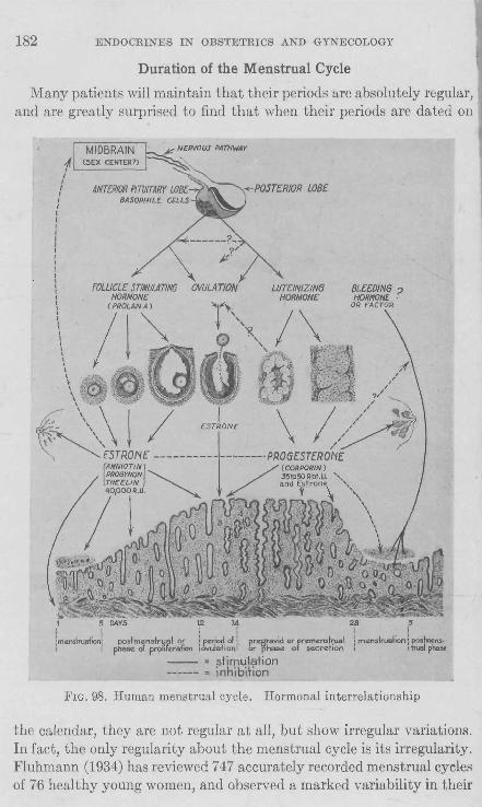
182 ENDOCRINES IN OBSTETRICS AND GYNECOLOGY
Duration of the Menstrual Cycle
Many patients will maintain that their periods are absolutely regular, and are greatly surprised to find that when their periods are dated on
{ : lmens~ruefionl ! :
'4 :2l8
postmen.truel or i period 01 i pregravid or "...me",lrual i m~"stru<lion .",!mons. pheS(t cl proliferation tovuletiOf'lI· or phase of secretion l trueJ phax
-- = stimulation ------ = inhibition
FIG. 98. Human menstrual cycle. Hormonal interrelationship
the calendar, they are not regular at all, but show irregular variations. In fact, the only regularity about the menstrual cycle is its irregularity. Fluhmann (1934) has reviewed 747 accurately recorded menstrual cycles of 76 healthy young women, and observed a marked variability in their

MENSTRUATION 18~
length. The cycles varied from 18 to 100 days, although the vast majority were between 18 and 42 days. The mean was 30.4 days, and the standard deviation 11.53. The average duration of the hemorrhage in 823 menstrual periods was 4.6 days, and no relation between the length of the menstrual cycle and the duration of the flow could be demonstrated. E. Allen (1933) noted a similar irregularity in a group of 131 women. King (1933) studied two series of cases, and concluded that 56.7 per cent of 354 periods are included in the twentysixth to the thirtieth days, while in another series, 523 periods, the twenty-fifth to the twenty-ninth days included the greatest number, 66.7 per cent. Hajek (1933) reported a series of 1,480 women and found that 82 per cent menstruated regularly, 12.9 per cent always menstruated irregularly, and 4.4 per cent bled irregularly or only occasionally. Only 56.6 per cent of the entire series had a regular 28-day cycle. These recent reports agree in the opinion that the menstrual cycle in normal women is subject to considerable variation.
The Question of Nervous Influence on the Ovary and Menstruation
The menstrual cycle is under hormonal control. Section of the spinal cord (van Wegenen, 1932) in an adult primate precipitates menstruation, but does not terminate the process, for regular cycles are resumed again. Clinically, a complete transverse myelitis of the cord does not affect menstruation. The ovary is abundantly supplied by nerve fibers arising from the ovarian plexus which in turn arises from the aortic and renal plexuses. The fibers accompany the ovarian artery. Afferent fibers are supplied by the tenth thoracic nerve. According to Kuntz (1919) the nerve supply to the ovary terminates in the blood vessels and fibromuscular tissue of the stroma, but no nerve fibers penetrate the ovarian follicles. Nerve fipers were observed in the theca folliculi and within well developed corpora lutea, but the fibers seemed merely to accompany the blood vessels. Ganglion cells were not observed within the ovary. Hence the finding of Hinsey and Markee (1934) that ovulation was induced by the injection of pregnancy urine into rabbits in the complete absence of functional nerve fibers to the ovary, was not at all unexpected. But at the same time it must not be supposed that the autonomic nervous system is without influence upon ovarian activity. Adrenalin and insulin are subject to control by the autonomic nervous system. Kraul (1927) treated both mice and rats with adrenalin and pilocarpin and obtained a loss of
184 ENDOCRINES IN OBSTETRICS AND GYNECOLOGY
estrus. Pilocarpin, which stimulates the vagus, cauoed the production of many small follicles, leading to small cystic degeneration of the ovary and an absence of luteinization. Adrenalin, which stimulates the sympathetic nervous system, inhibited follicle formation and retarded corpU8 luteum degeneration. Hinlch-Hof'fman (1934) reached similar conclusions.
The Hormonal Control of Menstruation
In considering the hormonal control of menstruation it is advisable to consider it from the viewpoint of the various tissues and hormones involved, namely:
1. The sexual center in the hypothalamlls. 2. The anterior pituitary gland. a. The follicle stimulating hormone.
b. The luteinizing ho~mone. 3. The ovary. a. Follicular hormone or estrone. b. Corpus luteum
hormone or progesterone. 4. The uterus. a. The endometrium. h. The myometrium. The Sexual Center in the Hypothalamus. Evidence for the presence'
of such a center is as yet completely laeking. The work of Hohlwcg and Junkmann suggests ,such a possibility. The location of this hypothetical center could be in the floor of the third ventricle.
The Anterior Pituitary Gland. If we consider for the moment that the presence of a sexual center has not been fully proved, then the source of the rhythmical stimuli necessary for the menstrual cyclr. must originate from the anterior pituitary gland. Histological variations in the anterior pituitary gland during the cycle have not been demonstrated, although Smith (1929) presented evidence of a correlation between the amount of gonad-stimulating hormone present in the pituitary of the guinea pig and the stage of the reproductive cycle. Both the follicle stimulating and the luteinizing hormones are essential for the stimulation of the entire cycle in the ovary. The hormones act in rotation, first the follicle stimulator and then the luteinizer, although it is also true that -the action of one overlaps the other at about the time of ovulation (Hisaw, Greep_ tnd Fevold, 1935). Not only are both hormones essential for the production of the menstrual cycle, but there must be a quantitative relatio)lship oetween them. For if the follicle stimulating hormone is present in an inadequate amount, or if its period of activity is shortened, the follicle will not develop completely, and an immature follicle is incapable of normal luteinization.

MENSTRUATION 185
But luteinization of the follicle is a function of the luteinizing hormone, hence, this hormone docs not exhibit activity until the follicle stimulating hormone has displayed its complete effect (the mature follicle). The further consequences of this lack of luteinization will1:e considered in the discussion of cystic and glandular hyperplaEia of the endometrium. Furthermore, a failure of the luteinizing hormone to act would only give rise to a proliferative endometrial phase, and the bleeding that would result from such an endometrium would be cyclical bleeding and not menstruation. A sterile cycle would be the result. An excess of the luteinizing hormone would probably give rise to a persistent corpus luteum. In the presence of such a corpus luteum menstruation would not occur and a new follicle could not form. The follicle stimulating hormone cannot exhibit its function in the presence of an excess of the other. The balance is delicately maintained.
The Ovary. The ovary, having received the hormonal impetus from the anterior pituitary gland, liberates its own hormones. The follicle stimulating hormope induces the formation of a follicle and this in turn secretes follicular hormone or estrone. The luteinizing hormone converts the ruptured follicle into a corpus luteum and the latter secretes progesterone. The ovary undergoes histological as well as hormonal changes during the cycle. The ovarian hormones are delicately balanced and disturbance of one prevents the proper exhibition of the other. The administration of too large quantities of estrin prevents the proper 'action of progestin~ as 8viaencea"b-y£llelfactionon the endometrium. The action of estrone is maintained throughout the cycle, for it is produced by the follicle in the first half of the cycle, and by the corpus luLeum during the second half (Allen, Pratt, Newell and Bland, 1930). Progesterone is the predominating hormone during the second half of the cycle. Since yrogestin has to date not been demonstrated in either blood or urine (nonpregnant), it is impossible to state whether small traces originating in the corpus luteum of the previous cycle might not be active during the first half o(the new cycle. Zondek (1930, p. 57) maintains that even in the presence of histological change in the corpus luteum (at the time of menstruation) it is not safe to draw conclusions as to its function. Such traces of corpus luteum hormone might be nec()ssary to prevent excessive proliferation of the endometrium d(iC" to c"trin.
, The amounts of estrone and progesterone required for the production of a complete cycle in a castrate, or in primary amenorrhea, have been demonstrated by I\aufmann (1932) to be 40,000 RU. of estrone

186 ENDOCRINES IN OBSTETRICS AND GYNECOLOGY
and 35-50 rabbit units of progesterone. This is a distinct step forward, for we now know not only what ovarian hormones are responsible for each phase of the menstrual cycle, but know also the quantities required. It goes without saying that this figure is not absolute, for the structure of the molecules (compare estrone with estradiol!), the nature of the solvent, the frequency of injections, the rate of excretion and utilization of the hormones, and their storage, play important roles in determining the exact dosage required. It might be asked whether 40,000 R. D. of estrin and 35-50 rabbit units of progestin represent the entire quantity of these hormones required by the organism, for their function extends definitely b~yond the local genital sphere. This question is all the more pertinent in view of the observation of Marrian and Parkes (1930) that while one mouse unit of estrin was sufficient to produce a cornified vaginal epithelium, 200 M. D. were required to duplicate all the phenomena of the estrous cycle. On that basis Parkes (1932) calculated that for the human a complete replacement therapy would involve the subcutaneous administration of about 500,000 M.D. (100,000 R.D.) of estrin. Considering the fact that the benzoic acid ester of estradiol (progynon-B) is about three times as active as estrone, Parkes' theoretical figure was therefore established as correct by the clinical tests of Kaufmann. Small amounts of estrin (2800 R.D.) are sufficient to produce growth of the endometrium and breasts in patients whose sexual organs have undergone castration atrophy (Werner and Collier, 1933). On the other hand, it is our impression that in cases of primary amenorrhea and long standing secondary amenorrhea the very large doses suggested by Kaufmann were essential to produce the full psychic and physical changes involved in the menstrual cycle.
Observations on Ovarian Ocular Implants. The possibility of actually observing an ovary during its various functional phases by transplanting it into the anterior chamber of the eye was first suggested by Schocket (1920). He noted follicle growth, rupture, and was able to demonstrate histologically an ovum in the anterior chamber. The problem was revived recently by Schocket and Markee (1928), Allen and Priest (1932) and by Podleschka and Dworzak (1933). The latter workers experimented with rabbits and reached the following conclusions. When both ovaries are transplanted follicular growth and degeneration occur. Ovulation was observed only once. After copulation corpora lutea were formed, and these functioned, for a pregravid endometrium was formed and uterine mobility was inhibited. The same phenomena were observed after injection with prolan or anterior

MENf;TRUATION 187
pituitary gland extract. The transplantation of one ovary into the eye, and the retention of the other in its normal position, changes the character of the response. Only when the ovary in situ is removed does the transplant show the usual follicle growth. The reason for the inhibition of the transplant by the ovary in situ is not known. It may be due to severance of the nerve supply when the transplantation is done. Intact nervous pathways may be essential to normal ovarian function.
R .,.8 _/
~ •••• l __ .~.~----~~ 'a
c
FIG. 99. Modified suction curettes for the removal of endometrial biopsies. (A) As modified by Kurzrok. The end of the cannula has a moderately sharp edge. It is moved along the surface of the endometrium and suction is exerted by means of the syringe. A specimen is removed from each of the uterine walls. (B) As modified by Cassidy. The end of the cannula is flexible, and it is inserted through the cervix by rotating the cannula counterclockwise until the fundus is reached. It is then removed slowly from the uterine cavity by rotating clockwise and, at the same time, suction is exerted. (C) Syringe with automatic lock and two-way valve.
The Endometrium
The methods of demonstrating endometrial change were previously carried out by an examination of curettings or by the examination of entire uteri removed at operation. Further progress was made with the suction curette (fig. 99) for with this instrument, or any of its modifications, it is possible to remove endometrial fragments repeatedly at various phases of the cycle without inconveniencing the patient to any extent. More recently Markee (1933) has studied menstruation
188 ENDOCRINES IN OBSTETlUCS AND GYNECOLOGY
in ocular endometrial implants. This method promises to add a great deal to our knowledge of this subject.
Markee used monkeys in his experiments. He used homologous endometrial implants and noted the following; The first evidence of approaching menstruation was vasoconstriction lasting six to twelve hours, and the bleeding in the eye preceded that from the cervix by several hours. Nor was the onset of bleeding in all transplants simultaneous-a fact noted in the menstruating human uterus by Bartelmez. Subepithelial hematomas formed and small papillae appeared on the epithelial surface. The papillae ruptured and bleeding continued for from twenty-five to seventy minutes. No desquamation of epith~lium occurred during the first few hours and only small bits during the first day. Regenerution occurred from outgrowths of the free extremities of the glands. Whut effect the intraocular pressure and the pEculiarities of vascularizatio!l in the eye have on this form of endometrial bleeding is still unsettled. Bleeding from a free surfuce (in the uterus) must vary from that against II resistance (in the eye), ill view of the fact that the pressure behind the endometrial bleeding cannot be very great.
Endometrial Bleeding
Endometrial bleeding may be of two types; 1. Menstrual. 2. NOli-menstrual. Menstrual bleeding here applies only to bleeding from a premenstrual
endometril1m conditioned by Ihe presence of a corpus luteum. Nonmenstrual bleeding may OCellI' every month, hence be cyclical in character. The endoIl:!l!!i.l!lll. is alwa:y~ postmenstrual in charader. Cycliclli bTeedi'Jtg implies the absence ororuiitiml.=-c=~o ~
In order to understand the problem of menstrual bleeding it is advisable to consider the factors that produce non-menstrual bleeding.
1. Oophorectomy-in the first half of the menstrual cycle (bilateral). This is common knowledge to all gynecologists. The bleeding sets in within a few days of operation. The symptoms associated with menstruation arc frequently lacking. The endometrium is po~t
menstmal in character. On the contrary, a unilateral oophorectomy containing the corpus luteum (hence done in the second half of the cycle) hastens the occurrence of the menstnml period. The bleeding is from a premenstrual endometrium and the symptoms are similar to those of a normal period.
2. The administration of follicular hormone. Allcn (1927) has shown that the administration of follicular hormone to an adult castrated

ME:-ISTUUATION 189
Macacus Rhesus Monkey until gro~th of the endometrium has been induced, followed by cessation of injections or decrease in the dosage, results in non-menstrual bleeding. Werner and Collier (1933) have obtained a similar result by injecting theelin into castrated women, even when a uniform level of daily injections has been maintained. Kurzrok, Wilson and Cassidy (1935) have observed that when cases of primary amenorrhea (due to the absence of functioning gonads) are constantly treated with large doses of follicular hormone cyclical bleeding occurs with great regularity, the intervals of bleeding approximating four weeks. There is no withdrawal or decrease in the dosage, and yet bleeding breaks in with great regularity. (In view of the complete absence of secondary sex characteristics, the presence of large quantities of follicle stimulating hormone and the absence of follicular hormone in the urine, a functioning ovary is definitely excluded in' their cases.) This leads to the probability that the stimulus to bleed is extra-gonadal in character, and in all probability emanates from the anterior pituitary gland or the uterus proper. Smith, Tyndale and Engle (1936) were able to induce bleeding in hypophysectomized monkeys by the injection of estrone. Hence neither the hypophysis nor the ovaries are essential for uterine bleeding. ' Just how this clocklike mechanism functions is at present unknown. Bleeding is an additional factotadded to the endometrium, for there are numerous cases on record of women who have never menstruated but who became pregnant without difficulty.
3. Section of the nervous connections to the ovaries. This was carried out in the Macacus Rhesus Monkey peripherally (Zuckerman, 1934), and in the spinal cord (Van Wegenen, 1932). The result is uterine bleeding within a few days. Disturbances of the autonomic nervous system in women may give rise t9 uterine hemorrhage in the absence of any local pathology, or may suddenly stop a normal menstrual, period. The modus operandi is in all probability vascular, vasoconstriction or vasodilatation.
The etiology of the bleeding designated as menstruation is unknown. An opinion, vague at the present time, is that there occurs a decrease in thc level of follicular hormone at the end of the corpus lute urn phase and that 'as i1):esult of this hormone withdrawal menstruation sets in. Our studies (Kurzrok and Creelman) of the urinary excretion of estrin fail to show any such loss of hormone at the time of menstruation. ,Figure 100 shows the excretion in one such case. The post- and premenstrual valiICs are about the same. The peak of excretion corresponds to the time of ovulation. The blood values of estrin during the cycle
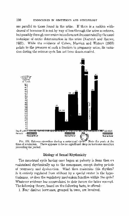
190 ENDOCRINES IN OBSTETRICS AND GYNECOLOGY
are parallel to those found in the urine. If there is a sudden withdrawal of hormone it is not by way of loss through the urine as estrone, but possiblythrOllgh conversion intoa formllot demonstrated by the usual technique of estrin determination in the urine (Kurzrok and Ratner, 1932). While the evidence of Cohen, Marrian and Watson (1935) points to the presence of such a fraction in pregnancy urine, its variation during the estrous cycle has not been demonstrated .
FIG. IOU. Estrone excretion during a menstrual time of ovulation. There appears to be no significlln preceding the period.
••
Etiology of Sexual Rhythmicity
The menstrual cycle having once begun at puberty is from then on maintained rhythmically up to the menopause, except during periods of pregnancy and dysfunction. What then maintains this rhythm? Is it entirely regulated from without by a special center in the hypothalamus, or does the regulatory mechanism function within the cycle? 'Vhatever evidence has accumulated to date favors the latter concept. The following theory, based on the following facts, is offered:
1. Four distinct hormones, grouped in twos, are involved.

MENB11lUATION 191
Follicle stimulating and follicular hormone. Luteinizing hormone and progesterone.
These hormones react functionally upon one another. 2. The gonad stimulating hormones vary at different times of the
estrous cycle (Smith, 1929). 3. Removal of the ovaries increases the amount of gonadotropic
hormone in the anterior pituitary gland (Engle, 1928). Follicular hormone therefore inhibits the gonadotropic hormones.
4. Injections of follicular hormone decrease the gonadotropic potency of the anterior pituitary gland (Moore and Price, 1932).
5. Injections of large quantities of follicular hormone induce changes in the anterior pituitary gland, resulting in luteinization of the ovaries (Hohlweg, 1934),
6. Injections of progesterone are effective in converting a castration' hypophysis into a normal one (Clauberg ami Breipohl, 1935).
7. This hormone system is in unstable equilibrium. At the onset of the post-menstrual phase the estrone concentration
is low (about the same as at the end of the pre-menstrual phase), depleted in addition by the loss via the menstrual discharges. The inhibition upon the anterior pituitary gland is removed and follicle stimulating hormone is produced in increasing amounts. This increased stimulus acts upon thc ovary, and the latter produccs increasing amounts of estrone, the prak being reached at about the timc of ovulation. The great concentration of follicular hormone in turn depresmg the follicle stimulating hormone and stimulates tbe production of the luteinizing hormone. Follicle growth is inhibited while a corpus luteum; forms. The progestin thus formed in increasing amounts (as seen from the increasing effect upon the endometrium) finally reacts upon the anterior pituitary gla~d, depressing tbe Illte_\;1izing factor and allowing the follicle stimulating hormone to renew its activity. At this point the premenstrual endometrium breaks down due to some other impetus (bleeding factor?) which arises from some source. The'rate of this "turn over" may be influenced by changes from within this system of hormones proper, or from without (sexual center or other glands of internal secretion). -Considering_ the cOIl1plexity of the factors involved it is of little wonder that aberrations frequently occur. Therapeutically we take advantage of this hormonal interplay and move one hormone against another. And, not only must the right hormone be chosen for the right plaq.e, but the amount of hormol1e exhibited is qf great importance.

192 ENDOCRINES IN OBSTETRICS AND Gl'KECOLOGY
BIBLlOGl~APHY
ALLEN, E. 1927 The menstrunl cycle of the monkey-Macnells Rhesus. Contrib. to Emb., No. 98. Carnegie Inst. Wash. PUb. 380, 19, 1.
ALLEN, E., PRATT, J. P., NEWELL, Q. U. AND BLAND, L. J. ]930 Human Tubal Ova. Contrib. to Emb. No. 414. Carnegie lnst. Wash. Pub. 22, 45.
ALLEN, E. AND PRIEST, F. O. 1932 Physiological Responses of Ectopic Ovarian and Endometrial Tissue. Surg. Gyn. and Obst. 66, 553. '
ALLEN, E. 1933 Irregularity of Menstrual Function. Am. J. Obst. and Gynec. 26,705.
CLAUBERG. C. AND BREIPOHL, W. 1935 Follikel- und LuteohofInoll in ihrer Rtickwirkung auf den Hypophysenvorder]appen. Klin. Woch. 14, 119.
COllEN, S. L" MARRIAN', G. F. AND WATSON, M. 1935 Excretion DC. estrin during pregnancy. Lancet, March 23rd, 674.
ENGLE, E. T. 1928 The role of the anterior pituitary in compensatory ovarian hypertrophy. Anat. Rec. 37, 275.
FLUHMANN, C. F. 1934 The length of the human menstrual cycle. Am. J. Obs. and Gyn. 27, 73.
HAJEK, O. 1933 trber Variabilitiit des Menstruationszyklus. Zent. f. Gyniik. 67,2.57.
HINSEY, J. C. AND MARKEE, J. K 1932 A search for neurological mechanisms ill ovulation. Proe. Sue. Exp. BioI. and Med. 30, 136.
HIRSCH-HoFFMANN, H. U. 1934 Vegetatives Nervensystem nnd Ovariulll. Arch. Gyniik. 156, 42.
HISAW, F. L., GREEP, R. O. AND FEVOLD, H. L. 1935 Experimental menstruation of Macacus Rhesus monkey. Anat. Rec. 61, 24.
HOHLWEG, W. 1934· Veriinderungen der Hypophysenvorderlappens und des Ovariums nach Behandlung mit grossen Dosen von Follikelhonnon. Klin. Woch. 13, 92.
KAUFMANN, C. 1932 Umwandlung der Uterusschlcimhaut ciner kastrierten Frau aus dem ntropischen Stadium in das sekretorisehen Funktion durch Ovarielhormon. Zent. Crnak. 56, 2058.
KING, J. L. 1933 Menstrual intervals. Am. J. Obs. & Cyn. 26, 583. KNAUS, H. 1929 trber den Zeit"unkt der Konzeptionsbiihigkeit des Weihes illl
Intcrmenstrum. Munch. Med, Woeh. 76, 1157. 1930 Zur Physiologie des Corpm Luteum. III. Mitteilung. Arch. f.
Gynak. 141, 374, 1930 Dber die Funktion des Corpus Luteum. Klin. Woch. 9, 961. 1933 Die periodische Fureht- unu Unfurchtbarkeit des Weibes. Zent. f.
Gyniik. 57, 1393. KRAUL, L. 1927 Del' Einfluss der Innervation auf den Eierstock. Arch. f.
Gyniik. 131, 600. Nervensystem und Eierstock. Arch. f. Gynak. 132, 4.
KUN1'Z, A. 1919 The innervation of the gonads in the dog. Anat. nee. 17, 203. KURZRUK, R., WILSON, L. AND CASSIDY, ~L A. 1935 Treatment of amenorrhea
with large doses of estrogenic hormone. Am. J. Ob8t. and Gynec. 29, 771.
MARKEE, J. E. 1933 Menstruation in ocular endometrial implants. Anat. Rec. 65, 66.

MJi]NSTnUATIO~ 193
MOORE, C. R. AND PRICE, D. 1932 Gonad hormone functions and the reciprocal influence between gonads and hypophysis and its bearing on the problem of sex hormone antagonism. Am. J. Anat. 50, 13.
PARKES, A. S., ROWLANDS, I. W. AND BRAMBELL, F. W. R. 1932 Effect of x-ray sterilization on oestrus in ferret. Proc. Roy. Soe. London 109, 425.
PODLESCHKA, K. AND DWORZAK, H. 1933 Vber Autotransplantationen von Ovarien .in die vordere Augenkammer des Kaninchens. Zent. f. Gyniik. 67,2114.
SCHOCKET, S. S. 1920 Physiology of Ovulation. SUrg. Gyn. Obst. 31, 48. SMITH, P. E. 1929 Evidence of a Correlation Between the Amount of Gonadal
stimulating Hormone present in the Pituitary of the. Guinea-pig and the State of the Reproductive Cycle. Anat. Rec. 42,38 .
. SMITH, P. E., Tn,DALE, H. H., AKD EKGLE, E. T. 1936 The reproduction system and its responses to ovarian hormones in hypophysectomized rhesus monkeys. Froc. Soc. Exp. BioI. and Med. 34, 245.
WEGENEN, G. VON 1932 A study of indueed menstruation in the monkey. Anat. Rec. 52,40.
WERNER, A. A. AND COLLIER, W. D. 1933 The effect of theelin injections on the castrated woman. J. A. M. A. 100, 633.
ZONDEK, B. 1930 Die Hormone des Ovariums und des Hypophysenvorderlappens. Berlin. J. Springer.
ZUCKEUMAN, S. 1934 The preganglionic pathwilys of the ovarian nerves of Rhesus Monkey. Anat. Rec. 58,43.

CHAPTER XV
OVULATION
The most important event in the menstrual or estrolls cycle is ovulatioll. The term ovulation implies the separation of the ovum from the ovary and its expulsion from the follicular cavity.
Ovulation is associated with other phenomena, namely, follicle growth and then corpus luteum formation. In view of the fact that the ovary has no inherent drive, for it is maintained in a state of function by the anterior pituitary gland, it is not surprising to find that the impetus to ovulate also depends upon the adenohypophysis. The position of ovulation in a given menstrual cycle as well as in the c cles that follow, IS 0 U mos Impor ance rom the view oint of sterility and ~y. Furthermore, I IS essential to know\vhether or not ovulation accompanies every menstrual cycle, and whether the two phenomena are dependent or independent of one another. Again, are we always dealing with spontaneous ovulation, or may it be induced out of turn.
The Follicle and the Gonadotropic Hormones
Only mature follicles can ovulate. Immature follicles must first reach a state of IIIaturity. Follicle ripening, according to Lane (1935), involves activity on the part of both gonadotropic hormones. Beginning with the primary uocyte, the fullicle stimulating hormone is apparently responsible for the formation of a granulosa about the OVUIII. The luteinizing hormone promotes the formation of an antrum folliculi, intu which the granulosa cells are made to secrete follicular hormone by the fullicle stimulating hOfmone, Of, by a cumbination uf follicle stimulating and luteinizing hUfmones.
It is a well established fact that the removal of one ovary induces hypertrophy of the remaining une. Arai (1920) has shown that this compensatory hypertrophy is not due to an increased number of ova. Hence douhling the amount of pituitary hormones for one ovary by the removal of the other, will not influence the 'number of ova in the surviving gonad. The ovogenetic rate of hypophysectomized adult rats is increased when compared with the total eggs, follicles and corpora in the control animals (Swezyand Pencharz, 1933). The role of the gonadotropic hormones begins after the formation of the oocyte.
194

OVULATION 195
The first task of the follicle stimulating hormone is to convert the pregranulosa cells into a granulosa layer and thus form the primary follicle.
The Stimulus to Ovulation
The stimulus that indu e v-ulati..QnJw; i~ origin in the anterior pltmtary gland. ml and Engle (1927) produced ovulation in im~ature mIce by successive implants of anterior pituitary glands from adult rats. Similar implants in adult animals rarely, if ever, produced ovulation. Bellerby (1929) induced ovulation in the unmated estrus rabbit by the administration of anterior pituitary extracts. This was also obtained by Friedman (1929) with urine of pregnancy. Hill and Parkes (1931) found that in the absence of the hypophysis, extracts of the anterior lobe and pregnancy urine will induce ovulation. Wolfe (1931) made some interesting observations on ovulation by means of saline extracts of sow pituitaries taken at different stages of the cycle. One milligram of pituitary tissue taken from a sow with large growing follicles (6-8 mm. in diameter) and degenerate corpora was needed to induce ovulation, but 10 mgm. were needed when the donors had larger corpora and 40 mgm. when the ovaries of the donor contained active corpora lutea plus small follicles. Wolfe and Cleveland (1931) report that it takes equal amounts of anterior pituitary tissue from either immature or adult rabbits to induce ovulation.
The Nature of the Hormones (or Hormone) that Induce Ovulation
Is ovulation due to a separate and distinct hormone or to the follicle stimulating and luteinizing hormones? To date there has been no evi-I dence produced to show that a distinct ovulating hormone exists. On the contrary, there is evidence to show tlp,t ovulation requires the presence of both the follicle stimulating and luteinizing hormones (Smith and Leonard, 1934). These workers utilized immature hypophysectomized rats in their experiments. The injection of follicle stimulating hormone from castrate urine produced large, norma! and uniform follicles. No ovulation was seen. Each follicle contained an ovum. This form of ovarian response was also noted by Kurzrok (Riley, Brickner and Kurzrok, 1933). Leonard and Smith followed their treatment 'by a single intra-peritoneal injection of pregnancy urine (antuitrin-S) and obtained ovulation. Since pregnancy urine contains large quantities of luteinizing hormone, it appears that the addition of this hormone to a properly prepared follicle induces ovulation. The steps leading up to ovulation are several, namely:
196 ENDOCRINES IN OBSTETRICS AND GYNECOLOGY
1. The conversion of a primary oocyte into a primary follicle by the follicle stimulating hormone, followed by a further growth of the granulosa due to the same hormone.
2. The formation of an antrum due to the luteinizing hormone. 3. A growth of the follicle which terminates in the mature Graafian
follicle (due to F.S.H.). 4. Ovulation upon the addition of the luteini~ing hormone. In
the human there is a sudden addition of follicle stimulating hormone at this point (Kurzrok, Kirkman, and Creelman, 1934).
It would see!ll, therefore, that ovulation is induced by a correct ad. mixture of both gonadotropic hormones acting upon a mature follicle.
The Mechanism Causing Ovulation
The exact etiological factors involved in the' expulsion of the ovum from the follicular cavity are unknown. Numerous theories have been offered in explanation. They arc as follows:
Increase in Intrafollicular Pressure. ThiR increased intrafollicular pressure has betm ascribed to increased secretion of liquor folliculi, hyperemia and blood pressure. C. Hartman (1932), in a recent review of this subject, discusses the varipus forms of evidence brought forth in support of this explanation. A plausible explanation of the sudden rise of intrafollicular pressure is the presence of smooth musculature about and within the ovary, specifically that about the Graafian follicle. Aeby (1859-1861) described smooth muscle fibers in the theca extern a of follicles, as well as heavier strands in the hilus of the mammalian ovary. He attributed the rupture of the follicle to the cQntractionH of these" muscle fibers. He was confirmed in these observations by Thomson (1919) and Winiwarter and Sainmont (190&-1909). Guttmacher and Guttmacher (1921) found nerve endings terminating in the muscle fibers of the sow ovary. They observed and recorded contractions of these muscl~ fibers, as well as the existence of sympathetic (epinephrine caused relaxation) and parasympathetic fibers (physostigmine caused contraction). The theory that ovulation is caused by a contraction of these muscle fibers, especially those about the theca externa, induced by either hormonal or nervous stimuli, is extremely attractive. The point of rupture, or stigma, becomes markedly thinned out and avascular at the time of rupture (Oertel, 1924). Schocket demonstrated proteolytic enzymes in the liquor folliculi and attributed follicular rupture to this factor. Muscular contractions of the ovary aided by proteolytic digestion of an avascul1l;_r, devitalized stigma offer
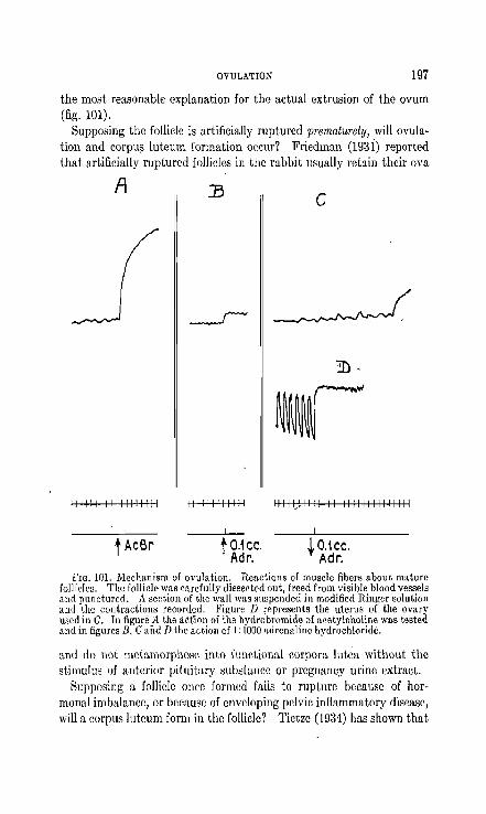
OVULATION 197
the most reasonable explanation for the actual extrusion of the ovum (fig. 101).
Supposing the follicle is artificially ruptured prematurely, will ovulation and corpu~ luteum formation occur? Friedman (193i) reported that artificially ruptured follicles in the rabbit usually retain their ova
A ]3 c
111111111111111111 111111111111 11111) 1111111111111111111111
t AcBr to.1 CC. Adr.
to.1CC . Adr.
FIG. 101. Mechanism of ovulation. Reactions of muscle fibers about mature follicles. The follicle was carefully dissected out, freed from visible blood vessels and punctured. A section of the wall ",as sllspended in modified Ringer solution and the contractions recorded. Figure D represents the uterus of the ovary used in C. In figure A the action of the hydrobromide of acetylcholine WIIS tested and in figures B, C and D the action of I : 1000 adrenaline hydrochloride.
and do not metamorpho~c into fUllctional corpora lutca without the stimulus of anterior pituitary substance or pregnaucy urine extract.
Supposing a follicle once formed fails to rupture because of hormonal imbalance, or because of enveloping pelvic inflammatory disease, will a corpus lutcum form in the folliele? Tietze (1934) has shown that

198 ENDOCRINES IN OBSTETRICS AND GYNECOLOGY
a persistent follicular cyst fails to luteinize and thereby induces cystic and glandular hyperplasia in the endometrium. Frankl (1934) maintains that the same condition occurs in pelvic inflammatory disease. Friedman (1931) occasionally encountered cystic follicles, in rabbits when he injected graded doses of pregnancy urine. Reynolds (1931) obtained the same effect, and the subsequent addition of large doses of luteinizing hormones failed to rupture these large follicles. It would seem that when the normal mechanism is disturbed, thc righting of this mechanism is a difficult task for the organism,
Mensl.rual cycle
Beginninq of menstruol cycle
~~~~~~~~~~~~~
cyde
Follicle stimul<ltin~ G hormone excretion
(Kumok)
~ Uterine motility I (Knaus)
[ A31! of human Ova (Pratt etc.)
Statistics of probdble D dale of conception
(Siegel, O~ino)
C A':le of embryo; (Gros5er)
B Endometrium (R Meyer, Schroeder)
A Corpus luteum (fraenkel) .
FIG. 102. Methods of studying human ovulation. The unshaded blocks show the limits of ovulation within the cycle; the solid black areaR show the greatest probabilities.
TIme of Human Ovulation in the Menstrual Cycle (fig. 102)
l(urzrok, Kirkman and Creelman (1934) have recently reviewed the evidence brought forth to determine the time of ovulation in the human menstrual cycle. The problem has been studicd by six different methods and to this they add a seventh. The methods are as follows:
a. The macroscopic observation of the ovary in situ. b. The microscopic examination of removed ovaries and uterine
curettings.

OVULATION 199
c. The determination of the age of early embryos, taken together with the probable time of conception.
d. The maximum probability for ovulation. e. The recovery of human ova from the uterine tubes. f. The reversal in the readion of the uterine muscle to pitui trin. g. The sudden appearance of foUide stimulating hormone in the
urine during the intermenstrum. a. Based on the macroscopic examination of the ovaries in situ
Fracnkcl and his school (Ancel and ViUemin, 1907; Tschirdewahn, 1921; Derek, 1923) place the limits of ovulation between the tenth and twenty-sixth days of the cycle, with a maximum probability at about the eighteenth day. Shaw (1925, 1932) concluded that ovulation occnrs not long before the 17th day, while Newell (1930) places the phenomenon a few days' earlier. Corner (1923), Allen (1928) and Hartman (1932) made similar observations on the ovaries of the Macacus Rhesus and placed the limits of ovulation between the 9th and 17th days of the menstrual cycle.
b. Robert Meyer, employing the histologic technic, concluded that the limits of ovulation were from the eighth to the fourteenth day of the cycle (Meyer and Ruge, 1913). Using the same technic, Marcotty (1914) placed the most probable time of ovulation on the fifteenth day, and Schroeder (1918) from the fourteenth to the sixteenth day.
c. The third method of approach to this problem was the estimation of the age of early embryos, when considered together with the history and probable date of conception. Using such evidence Triepel (1'914, 1915) placed the greatest frequency of ovulation at the eighteenth to the nineteenth day. Volkmann (~~26) based his figures on the examination of eighteen early embryos and favored an earlier ovulation time. On the basis of similar studies (Grosser, 1932) placed ovulation between the second and twenty-fourth days after the onset of the rnen:;es. He thought that the early ovulation may have been induced by coitus, drugs, violent exercise, etc., as suggested by Chazan (1911).
d. The fourth method was by means of a study of the most probable time of conception- (Siegel, 1917; Jaeger, 1917; Zangemeister, 1917). This presupposes that coitus, ovulation and conception occur within an interval of hours, and that the ovum and sperm remain fertilizable not more than thirty-six hours (Hartman, 1932, review and literature). This method places the ovulation time between the eighth and tenth days. Ogino (1932) holds the favorable period of conception to be between the tenth and the seventeenth day.

200 ENDOCRINES IN OBSTETRICS ~ND GYNECOLOGY
PROLAN A (FOLLICLE. 3TI MULATI NG HOAMONE)
PERIOD
DATE:
PROLAN A (F"OI-LICLE STIMULATING HORMONE.)
PUlIOD
DATE:
PROLAN A (fOllICL.E STIMULATIN& ~QRMONE')
PE.RIOD
DATE
PROLAN A CrOl.lleLE. STIMULATING HORr«:lN[1
PERIOD
DATE
PROLAN A (roLI..ICL(.
STIMUL.ATING HORMONE)
PERIOD
DATE
PROLAN A crOlL.ICL.E. STIM1JI.ATINO HORi"OI/£)
PE.RIOD
DATE.
I 2 3 4~0111213141516111B 1920212223:1>25262128
.. ~30 1 ~o 1 6 ~ 10" 1213 ~ 15 loI718192021222J24'~26
FIG. 103

OVULATION 201
e. It has now been definitely demonstrated by the recovery of ova from the uterine tubes that in regular cycles of average length ovulation usually occurs during the intermensirum. The estimated ovulation time for five human ova recovered from uterine tubes when possible stimuli of mating were excluded, was between the twelfth and fourteenth daytl following the first appearance of the menses (Allen, Pratt, Newell, and Bland, 1930). The probable time of ovulation of another human tubal ovum recovered by lcwis (1931) was placed on the eighteenth day following the onset of mcmtruation, after a previolls cycle of thirty-five days' duration (E. Allen, 1932).
f. Anothcr recent approach to this problem was suggested by Knaus (1930). He observed that in the rabbit prior t.o ovulat.ion, the uterus reacts to pituitrin by a contraction. Twent.y-four honrs after ovulat.ion the ut.erus no longer reacts to pituitrin, and this rcvcrsal Knaus attribut.es to the corpus luteum. Knowing the datc of reversal he places ovulation one day back (in the rabbit). Knaus found the same reversal in the human being and placed ovulation from two days back, i.e., between the fourteenth and,sixteenth days (of a twenty-eight day cycle), with a possible physiologic shift of two days to either side. Wittenbeck (1930), on the basis of experiments in vivo, agrees with Knaus, but adds that ovulation may occur on the ninth day of a twenty-eight day cycle. Knaus' method is, not a test for ovulation but is really a test for corpus luteum hormone, because a corpus lute urn may form / without ovulation (corpus llltcum atrcticum). Hermstein (1931) agrees with Knaus' findings. Kurzrok et a!. (1937) have not substantiated Knaus' findings (Chapter XVI). /'
g. Kurzrok et a!. (1934) observed that if consecutive morning specimens of urine are tested for the presence of follicle stimulatIng hormone, no hormone will be found except on one day during the intermenstrum-, ancl occasionally at the beginning and at the end of a menstrual period. Excretion of the follicle stimulating hormone occurred on the day preceding the "Mittelschmerz" and alsQ on the day following the onset of ovulation bleeding. Wil~re the excretion was followed in anyone case over several consecutive menstrual cycles, it was found that the limits
FIG. 103. Follicle stimulating hormone excretion during six months. Every overnight specimen was examined. Note th, positive F.S.H. at the time of ovulation, and on two occasions at the beginning and at the end of menstruation. During the first four months ovulation occurred successively on the 10th, 11th, 10th and 9th days. It was therefore believed that her safe period was from the 14th day on. But durin!!; the fifth month ovulation occurred on the 15th day and patient became pregnant. Note that the positive A-Z Test terminated after the curettage as a positive F.S.H.

202 ENDOCRINES IN OBSTETRICS. AND GYNECOLOGY
of hormone excretion were between the eleventh and sixteenth day (fig. 103). The limits of all cases studied were between the ninth and twenty-first days, with the greatest number occurring between the eleventh and thirteenth days. The sudden excretion of follicle stimulating horn~~ meluis sudden pituitary activity. Iu--all probability this is accompanied hy a sudden production of luteinizing hormone. No trace of this hormone was detected in the urine, possibly because a sufficient quantity did not pass over, or the method employed was not sensitive enough to detect it. Kurzrok et al., believe that oyqlation occurs on the day following the sudden' hprn1oM'~cretion (fig. 104).
- --..-.::- - - - . -Clinical Signs of Ovulation
Two clinical signs of ovulation in the human being have been recognized by gynecologists, namely, the intermenstrual pain or "Mi,ttelschmerz" and intermenstrual spotting. The former is probably due to the greatest distention of the follicle just preceding rupture .. It is not common. It may be perfectly regular, occurring on one side one month, and on the opposite side the next. Or only one side may be involved, distention of the ovary on the opposite side causing no pain. Some abnormality in innervation of the ovary is probably responsible for the phenomenon's reaching consciousness (fig. 105).
We have recently studied a number of cases of ovulation bleeding. Some were referred to us with the diagnosis of polymenorrhea, in other words, the patients were supposedly menstruating every two weeks. Careful questioning of the patients brought out the facts that even, though two consecutive bleedings lasted the same number of days, they never were of the same amount, and that one period of bleeding (ovulation) was accompanied by less discomfort than the one that followed (menstruation). By removing fragments of endometrium by means of the suction curette we were readily able to differentiate the two types of bleeding. Intermenstrual spotting occurs quite infrequently. It may last from one hour to forty-eight or more hours, and the bleeding may be very slight or moderate. Ii has also been observed in the monkey (Hartman). Schroeder (1924) studied 20 cases, and curetted 5 during this period of abnormal bleeding. He found an endometrium that was in the process of changing from a proliferative to a premenstrual type, hence corresponding to the period of ovulation.
Figure 106 represents a case of "Mittelschmerz" that was seen by Dr. Milton Robinson. The patient was a young woman, 24 years of age. Her periods were regular, every 26 to 29 days, moderate in

I/) 13 c: 12
~II c:t 10 '+- 9 06 ~. 7
.0 6
~ 5 2: 4
'" 3 b 2 I-
OVULATION !:l03
FIG. 104. Human ovulation. Results of studies bused on the excretion of follicle stimulating hormone. Each square represents the single excretion of F.S.H. during the cycle. Ovulation probably occurs about 24 hours after the excretion of the hormone. The letters represent successive months in- the same patient; the numbers, the cases studied in our first series. (Am. Jour. Obs. & Gyn., 28, 319, 193~.)
Jan.
Feb
Mar.
Apr,
May.
Jvne
July
Aug Sept.
Oct.
Nov.
-
--.~
7 8 9 10 II Days of the Menstrual Cycle
•
-
-.~ •• . ~
• -
12 13 14
FIG. 105. Case of :Vlittelschmerz. The black blocks show the date and duration of the pain. (Courtesy of Dr. Milton Robinson.)

204 ENDOCRINES IN OBSTETRICS AND GYNECOLOGY
amount and without pain. The pain lasted four hours, and moderately incapacitated the patient. The pain never began before the ninth day nor after the thirteenth. We believe that ovulatioll occurs within one day after the onset of the pain. This case shows thc limits of ovu-
I
II
ill
IT
w Y.. ...J
U >-u "2I
:.w
3ZIII
]X
INTERMENSTRUAL SPOTTING
I nte r me n-Men- strual struatio n spotting
W/%J
~
~
w;a
?
WJ
~
o 5 10 15 Day of cycle
20 25
Menstruation
I
I
I
30 35
n
40
FIG. 106. Case of intermenstrual spotting. The patient did not spot durin~ the fifth period, but did so at about the middle of the seventh. Biopsy during the spotting of the first cycle showed diapedesis through vessel walls, but no desquamation of tissue. (Courtesy of Dr. N. Kopeloff.)
lation during a period of olle year. There was absolutely no pain during February. Does this mean that ovulation did not occur in· February?
Seguy and Simonnet (1933) have described three other signs of ovulation, namely, the appearance of a glassy, translucent and extremely

OVULATION 205
fluid secretion in the cervical canal. They believe that the purpose of this secretion is to render the cervix temporarily permeable to spermatozoa.
The second sign of ovulation is the occurrence of increased sexual excitement which lasts throughout the period of glassy secretion in the cervIx.
The third sign appears a few days after the glassy cervical discharge and consists of desquamation of the vaginal epithelium. This finding is not completely in accord wi th the studies of Papanicolaou on the cyclical changes in the human vaginal epithelium. He (Papanicolaou) finds that smeam taken during the ovulative phase show a sudden increase in the number of leucocytes. This is particularly noticeable after a typical stage of leucopenia. The epithelial cells are chiefly of the intermediate or superficial type. They greatly resemble the cells of the previous phase, though their contours are not sharp. They form characteristic aggregations mixed with leucocytes. Many cells are penetrated by leucocytes. Cornification is pronounced with most of the cornified cells nucleated. Mucus is on the decrease.
The Period of Fertility in the Human
The period of fertility in the human has formed the subject of much discussion during recent years. The problem has been viewed from two angles, namely, to explain certain types of sterility, and secondly; to determine the so-called safe period for coitus during the cycle. It is with the latter topic that we will concern ourselves at this point.
The factors which determine when during a given cycle fertilization may occur, are threefold:-
1. The duration of life of spermatozoa in the female genital canal. 2. The duration of the life of the ovum after ovulation. 3. The actual time of ovulation in the cycle.
The Duration of Life of Spermatazoa in Female Genital Tract.
It. may be stated at th~~very outset that we are of opinion that only experiments'in the human are of any value in the solution of the problem. Whatever the duration of life of the rat, guinea pig or even monkey spermatozoa may be in their respective females, it can. only have a very indirect bearing on the problem as it pertains to women. Some broad principles may apply, but the specific circumstances are different in each. Another point-the fact that a sperm is motile is no proof that it has maintained its fertilizing power. Fertilization and motility Illay

206 ENDOCRINES IN OBSTETRICS AND GYNECOLOGY
be parallel throughout, but it is also conceivable that a sperm may remain motile, but may have lost its fertilizing power.
A. Duration of Life of Spermatozoa in the Vagina, and Their Transport. The duration of life of sperm in the vagina depends upon the following conditions within the vagina: l~h. The buffer action of semen and its pH.
2. The hydrogen ion concentration of the vagina. 3. The amount of seminal fluid retained within the vagina. 4. The bacterial flora of.:;the vagina. Semen is a buff1red solution with a pH ranging between 7.8 and 8.4
(Kurzrok and Miller, 1928). This buffering action is limited.' Too high concentration of either acid or alkali will overcome this buffering activity and thus become inimical to the life of the spermatozoa. Under norlllal conditions there is a constant infiltration of the seminal pool by the lactic acid from the vaginal walls. It, therefore, becomes essential for the spermatozoa to move out of the seminal pool, into the alkaline cervical mucus. This movement into the cervical canal is not hit or miss, but is a directed and oriented movement controlled by electrokinetic phenomena across phase boundaries set up by the acid vaginal secretions (lactic acid), the neutral (or slightly alkaline) semen and the alkaline cervical mucus (Miller and Kurzrok, 1932).
At the end of two hours post-coitum ninety per cent of all spermatozoa in the vagina are dead. The ,niter has never seen living sperm in the vagina later than ten hours post-coitum. Dead sperm may be found after twenty-four hours but rarely after thirty-six hours. X
Cary (Hl35) has not found sperm cell activity in the vagina later than two hours after coitus. Runge (1909) failed to find sperm in the vagina on the third day post-coitum. Haussmann (1879) found them absent at the end of the second day post-coitum. Hoehne and Behne (1914) examined the vaginal canal of a young primipara in her sixth month of pregnancy for spermatozoa at various intervals post-coitum. At the end of three quarters of an hour only an occasional sperm remained motile. Sperm removed seven hours post-coitum showed marked degenerative changes. No spermato~oa remainr,d motile at the end of an hour. The very short life of sperm in the vaginal secretions of a pregnant woman is due to the high vaginal acidity, which in a short time overcomes the buffer action of semen. Furthermore, spermatozoa mixed in vitro with the vaginal secretions of normal women, remained motile up to four hours. All evidence at hand shows that the spermatozoa that eventually reach the upper portions of the genital canal have left the seminal pool within two hours post-coitum.

OVULATION 207
The hydrogen ion concentration of the vagina is maintained in part by the activity of the Diiderlein bacilli. The glycogen of the vaginal walls is split up directly by the Diiderlein bacilli as follows (Schultheiss, 1929) :
Glycogen -> polysaccharides -> glucose -7 lactic acid
Thp, acidity of the vagina is hence a function of its lactic acid content. In alldition, there is present in the vaginal secretions a diastatic ferment that is capable of splitting glycogen into glucose (Kessler, 1935). The Diiderlein bacilli can then split the glucose further into lactic acid. When the vaginal canal becomes contaminated with other organisms the hydrogen ion concentration decreases (pH goes up). The acidity of ~he vaginal secretions, pH 4.Q--4.7, finally overcomes the buffering
. action of the semen and renders the seminal pool acid. Acidity destroys the activity of the spermatozoa.
The amount of seminal fluid retained in the vagina plays a distinct role in the viability of the spermatozoa. Usually the amount retained is about 1.0-2.00 C.c. All other factors being equal, the greater the amount of buffer the longer will it take to overcome its buffering activity. Cases of genital h~poplasia that have a shallow posterior culde-sac retain very little semen, hence motility ceases in a very short time.
Rosenthal (1931) made the interesting observation that cultures of B. coli agglutinate spermatozoa. Motility is lost within a few minutes, and the reaction is not reversible. The clumps formed could not be disintegrated. The phenomenon was entirely independent of the pH and clumping occurred with dead sperm as well. The active principle is contained within the body of the b~cteria. Attempts at agglutination with strains of streptococci, staphy10cocci, or gonococci gave negative results. Considering the fact that the B. coli commonly contaminates the vaginal canal, this agglutinating phenomenon can be very detrimental to fertilization.
B. Duration of Life of Sperm in the Cervix, and their Transport. The duration of life of spermatozoa in the cervix is longer than in the vagina. The writer P,ilS seen living sperm in cervical mucus as late as eleven hours post-coitum, whereas no living sperm were found in the vagina. Sperm may be found within the cervical mucus in contact with the seminal pool fifteen minutes post-coitum. In about one hour the spermatozoa may be found in the cervical mucus in the region of the internal os. Motion through the cervical os is fairly rapid. This is aided by two known factors, namely, the lytic action of semen upon

208 ENDOCRINES IN OBSTETRICS AND GYNECOLOGY
cervical mucus, and the increased activity of spermatozoa within the cervix. Kurzrok and Miller (1928) have described this lytic activity which appears to be due to an enzyme. Cary and Hotchkiss (1934) have recently demonstrated by means of a moving picture the marked activity of spermatozoa within the cervical mucus. The motion of the spermatozoa is much more rapid and their motility is of a different character than that seen in the seminal pool. Such an increase in activity may be expected, for it is doubtful whether the energy necessary for the sperm to travel from the seminal pool to the outer end of the Fallopian tube is all stored within the sperm head. It is more than likely that energy is obtained from the surrounding secretions. Cary (1935) has found motile sperm in the cervical mucus six to twelve hours post-coitum, and in one case sixty hours later.
Many believe that the uterus exhibits a sucking.action during orgasm which draws the semen into its cavity (like a rubber ball with aCSmall hole in it), or that by the same action the cervical plug is extruded, dipped into the seminal pool, and then sucked back into the cervix. The objections to this are many. Firstly, orgasm is not necessary for conception. Frigid women conceive as easily as those with orgasm (Dickin~on, 1927, 1931). It is debatable whether orgasm in the human is of any aid in conception. MallY patients are aware of an orgasm during bimanual or speculum examination. Motions on the part of the cervix are not felt or seen, though there may occur a slight desceneus, probably due to the increased vascularity of the uterus. There is no opening or closing of the cervical os. Fertilization may occur in the presence of an intact hymen. The ascent of sperm from the seminal pool to the outer end of the Fallopian tubes is made by the movements of the spermatozoa themselves.
C. Duration of Life of Sperm in the Uterus, and Their Transport. The rapidity of the passage of sperm through the cervix determines the time of their appearance in the uterine cavity. Schuwarski (1894) reported finding sperm in the uterine cavity one-half hour post-coitum. The further progress of the spermatozoa through. the uterine cavity is here again determined by their own motility, but in addition against an opposing ciliated stream. Both Hoehne (1911) and Roth (1904) ar~ of the opinion that the ciliated stream towards the cervix serves as an orienting mechanism to the upward motion of the spermatozoa. The rate of motion of sperm in a straight line is 3 mm. per minute, hence t.he distance of 16 em. between the internal as and ostium abdominalis tubae would take about an hour. This is on the assumption that the

OVULATION 209
sperm travelled in a straight line and without the resistance offered by an opposing ciliated stream.
Runge studied the survival of spermatozoa in the uterine cavity of 17 normal multipara, making in all 32 observations. Six hours postcoitum all uteri showed living sperm, while twelve hours post-coitum no living sperm were found in one-sixth of the uteri. At the end of 36 hours living sperm were found in only one-fourth of his cases. Beginning with the third day no living spermatozoa were found.
Haussmann (1879) made sevr:nteen observations in clevr:n women and found actively motile sperm in the uterine cavity on the third day postcoitum. He also observed some motility of weak character as late as seven days post-coitum. It is extremely doubtful whether spermatozoa whose motility was so markedly diminished were capable of fertilization.
D. Duration of Life of Sperrn in the Tubes and their Transport. There are only two observations on record describing the finding of living spermatozoa in the tubes. Zweifel (1892) describes the finding of living spermatozoa by Birch-Hirschfeld in the tubes of a prostitute who died in actu with a man (from carbon monoxide poisoning). When found the body had been dead several hours and the autopsy was performed about 14 to 16 hours later. Diihrssen (1893) describes finding living spermatozoa in the uterine ends of closed tubes three and a half weeks post-coitum. The woman had been a patient in the hospital for nine days before the operation. Diihrssen subsequently tried to demonstrate spermatozoa in the tubes of other patients but never succeeded. Niirnberger (1920) believed that sperm survived in the tubes as long as fourteen days. The writer believes that these findings are exceptional. The fact that spermatozoa have been found in tEe tubes only on such extremely rare occasions implies that the survival of sperm in the tubes is extremely short. Those in active motion pass out through the fimbriated end of the tube into the peritoneal cavity, while the sluggishly motile speon are swept back by the ciliated stream into the uterine cavity.
From the evidence at hand the writer. concludes that the fertilizing power of sperm is usually less than forty-eight hours, though motility may remain [or a somewhat longer interval. All in all, considering the importance of these topics in the problems of fertility and sterility, the inadequacy of the work done on the viability of spermatozoa in the human uterus and tubes is very startling.
Viability of the O~um. The human ovum, like the animal ovum, is

210 ENDOCRINES IN OBSTETRICS AND GYNECOLOGY
short-lived. The evidence for tho human is still meagre, for· human ova were not obtained for detailed study until recently. Allen, Pratt, Newell and Bland (1930) recovered five human tubal ova by a method of washing out the tubes in situ. Two of the ova recovered were twins, one from each ovary. They were the most nearly normal specimens obtained. One ovum was recovered on the fourteenth, three on the fifteenth, and one on the sixteenth day, after the onset of the previous menses. By correlating the stage of development of the corpora lutea with the condition of the ova, ovulation time was estimated at between the twelfth and fourtcenth daYB of the menstrual cycle. Although the ova were obtained within two, or, at thr most, three days from the date of ovulation, three of the ova showed histological signs of degeneration. This is the most direct proof as yet offered for the demonstration of the limited life of the human ovum.
Indirect evidence demonstrating the short life of the human ovum was offered by the writer in 1928. Two clinical histories were offered in support of this viC'w. Both patients wer~ young orthodox J ewesse~. They observed the Talmudicallaw of Niddah, in that they waited two weeks from the onset of the period before coitus was attempted. Because their menstrual cycles were of the three week type it was felt that by the time coitus took place the ovum of that cycle was already dead. Both patients were advised to have intercourse on the seventh day of the period and conception promptly took place. The fertility of the orthodox Jewesses was known to ancient medical writers. The reason for their fertility is that under normal circumstances coitus on the fourteenth day of the cycle is probably the most favorable time for conception.
Spontaneous and Induced Ovulation
In contradistinction to spontaneous ovulation, induced ovulation occurs in the cat and in the rabbit. The impetus to ovulate is coitus. The idea that induced ovulation may occur in women was first suggested by Chazan (1911). He offered no evidence to support his claim. The idea is interesting and is worthy of detailed consideration.
Statistical studies dealing with pregnancies resulting from but a single coitus during the entire cycle were first brought forward by Siegel (HJl7). His figures show that pregnancy can result from coitus very early or very late in the cycle. These pregnancies may be explained by assuming that ovulation lllay occur much earlier or later than is supposed or that the viability of sperm in the genital canal is

OVULATION 211
longer than we assume. The assumption of induced ovulation early in the cycle gains support from the following considerations. The rate of development of the Graafian follicles does not always seem to be a constant one. It is usually taken for granted that the follicle develops in a crescendo manner from its primordial state to maturity, the peak being reached at the time of ovulation. But it is conceivable that follicular growth may proceed to a certain stage and plateau off for a number of days. Ovulation may occur anywhere along this plateau depending upon the point where the impulse to ovulate strikes. It is not an uncommon thing for a gynecologist to find at operation, very early in the cycle (fourth to seventh day), a follicle that grossly appears ripe for rupture. Experimentally, Lane (1935) finds that in the rat the follicle increases 'abruptly in diameter soon after the first appearance of the antrUJll and then remains at a relatively constant diameter until ovulation.
The author believes that ovulation may also occur very late ill the cycle, specifically, following the so-called anovulatory or sterile cycle. In the normal menstrual cycle follicular growth is prevented during the premenstrual phase by the pre~ence of a functioning corpus luteum, and it is only when the corpus lutcum begins to degenerate that a primary follicle begins to mature. In the sterile cycle ovulation does not occur. Because of this absence of regular ovulation a corpus luteum fails to appear and the bleeding which subsequently occurs is cyclical bleeding and not true menstruation. But at the end of such a sterile cycle the inhibiting influence upon the,growth of a new follicle is absent because of the absence of a corpus luteum. Therefore, a follicle may develop here and mature, be uninHuenced by tlfe intervening cyclical bleeding, and eventually rupture in due course (or degenerate) early during th~ next menstrual cycle. Such an unusual follicle; matured just before or dllfing the bleeding of a sterile cycle, may rupture under the stimulus of coitus just before or during or immediately after the advent of the cyclical bleeding. A corpus luteum would form as a result of this induced ovulation, and then continue functioning as the corpus luteum of pregnancy. This concept would explain the undoubted occurrence of a pregnancy as the result of a single exposure very late in the cycle.
The most plausible explanation for the actual rupture of the follicle in the human is a contraction of the smooth muscle fibers in the ovary. Kurzrok et al. (1934) have demonstrated that the human ovary undergoes rhythmical contractions and that adrenal in induces a marked con-

212 ENDOCRINES IN OBSTETRICS AND GYNECOLOGY
traction followed by a considerable period of spasm. If the ovum is expelled by a contraction of the muscle fibers, then the substance actually causing the contraction of the muscle fibers may originate from some other source, such as the posterior pituitary or the medulla of the adrenal, for the adenohypophysis does not secrete a hormone capable of contracting smooth muscle. The excitement of coitus causes a rise in blood pressure, and the adrenalin produced as a result of the stimulation may be sufficient to cause the rupture of the follicle, premature as to time, but not premature as to state of development.
The So-called "Safe Period" ,
Within the past five years a large body of popular literature has grown up about this subject. The safe period is advocated by many individuals and groups, who do not always fully understand the basic phYSiology involved. As stated previously, the period of fertility depend8 on three factors, all variable, and not as yet completely determined. It may be true that a woman has a safe period every month, but how are we to determine it with our present state of knowledge? If ovulation occurs on a given day this month, will it occur on the same day next month, or the one after? What are the limit8 of ovulation within the menstrual cycle in any woman observed over a long period of time? Are the spermatozoa of an individual always endowed with the saIIle capacity to carryon in the female genital canal, or are there variations in this capacity, thus limiting or increa8ing their viability? What activating effects have the secretions of the female genital canal upon spermatozoa? That such activation occurs in the cervix is beautifully illustrated by the motion picture film of Cary and Hotchki8s. Is this activation a variable factor in the 8ame and different individuals? If in addition to the above complex factors there is added the possibility of induced ovulation, then the safe period becomes neither 8afe nor periodic.
BIBLIOGRAPHY
AEBY, C. 1859 Uber glatten 1Iuskelfasern im Ovarium und Mesovarium von Wirbclticrcn. Arch. f. Anat. Physiol. u. Wissensch. Med., 675.
1861 Die Glatten Muskelfasern in den Eierstocken der Wirbeltiere. Arch. f. Anat. Physiol. u. Wissenschr. Med., 635.
ALLEK, E. 1928 An Unfertilized Tubal Ovum frO!)l Macacus Rhesus. Anat. Rec. 37, 351.
ALLEN, E., PRATT, J. P., NEWELL, G. U. A~D BLAND, L. J. 1930 Human tubal ova; Related early corpora lutea and uterine tubes. Contr. Embryol., No. 127, 45. Carn. lnst. of Wash.

OVULATION 213
ARAI, H. 1920 On the Postnatal Development of the Ovary (Albino Rat) with Especial Reference to the Kumber of Ova. Am. J. Anat. 27, 405.
BF.LLERBY, C. W. 1929 The Relation of the Anterior Lobe of the Pituitary to Ovulation. J. PhysioL 67, Proc. 33.
CORNER, G. W. 1923 Oestrus, Ovulation and Menstruation. PhysioL Reviews 3, 457.
CARY, W. H. 1935. Personal Communication. CARY, W. H. AND HOTCHKISS, R. S. 1934 Semen AppraisaL A Differential
Stain that Advances the Study of All Morphology. J. A. 1\1. A. 102,587. DTCKINSO~, R. L. 1927 The "Safe Period" as a Birth Control Measure. Am. J.
Obs. and Gyn. 14, 718. 1931 The Control of Contraception. Williams & Wilkins, Baltimore.
DUHRSSEN, A. 1893 Vber ektopische Schwanl(erschaft. Zeit. f. Geb. u. Gyn. 27, 216.
EHRHARDT, K. 1932 .Klinische und ticrexperimentelle Untersuchungen tiber den Hormon des Hypophysenvorderlappens. Arch. Cynak. 148, 235.
ENGLE, E. T. 193( The Pituitary-Gonadal Relationship ahd the Problem of Precocious Sexual Maturity. Endoerin. 16, 405.
FRANKL, O. 1934 Vber die Schleimhautgefasse del' blutenden Gebarmutter. Wien. Klin. Woch. 1.
FRIEDMAN, M. H. 1929 The Mer.hani~m of Ovulation in the Habbit. 1. The Demonstration of a Humoral Mechanism. Am. J. PhysioL 89, 438.
FRIEDMAN, M. H. 1931 On the Mechanism of Ovulation in the Rabbit. IlL The Fate of j\lechanically Ruptured Follicles. Am. J. Physiol. 98, 209.
GRAFENBERG, E. 1918 Die zyklischen Schwankungen des Sauretiters im ,Scheidensekret. Arch. f. Gynak. 108, 628.
GUTTMACHER, ::Vi. A. AND GUTTMACHER, A. F. 1921 M~rphological and Physiological Studies on the Musculature of the Mature Graafian Follicle of the Sow. Johns Hopkins Hospital Bull. 36, 207.
HARTMAN, C. G. 1931 Phylogeny of Menstruation. Gynee. and Abd. Surg., A. M.)\.. 195.
1932 In Allen, Sex and Internal Secretions. Williams & Wilkins, Baltimore. HAUSSMAKN, D. 1879 'Gber das Verhalten del' Samerif6den in den Geschlechts
organen des Weibes. Berlin. A. IIerschwald. HILL,::Vi. AND PARKES, A. S. 1931 Studies OIl Ovulation. V. The Action of the
Ovulation Prlfdu£ing'Substance of Urine of Pregnancy on the Hypophysectomized Rabbit. J. PhysioL 71, 40.
HOEHNE, O. 1911 Vber die Flummerllng i'm weiblichen Cenitalapparat mit besonderer Berucksichtigung des Eitransportes. Vel'. d. D. Ces. f. Gyn. 14, 822.
HOEHNE, O. AND BEH)!E, K. 1914 Vber die Lebensdauer homologer und heterologer Spermatozoen im weiblichen Genitalapparat und in del' Bauchhohle. Zent. f. Gynak. 38, 5.
HOHLWEG, W. ANn DOHRK, ill. 1931 Beziehungen zwischen Hypophysenvorderlappenhormon und Keimdnisen. Wien. Arch. f. innere Med. Zl, 337.
KESSLER, R. 1935 Die Biologie def Vagina. Berichte u. d. Ges. Geb. u. Gyn. 28,433.
KNAUS, H. 1929 Vber den Zeitpunkt del' Konzeptionsbiihigkeit des Weibes im Intermenstrum. :vrtinch. Med. Woch. 76, 1157.
214 ENDOCRINES IN OBSTETRICS AND GYNECOLOGY
1030 Zur Physiologie des Corpus luteum. III. Mitteilung. Arch. f. Gyniik. 141,374.
1930 tiber die Funktion des Corpus luteum. Klin. Woch. 9, 961. 1933 Die periodische Frucht- und Lnfruchtbarkeit des Weibes. Zent. f.
Gynak. 67, 1393. KURZROK, R. 1928 The combination of a short menstrual cycle and delayed
coitus as II factor in sterility. Am. J. Obs. and Gyn. 15, 546. KURZROK, R., WJF.SBADER, H., Mur,nms, M. G. AND WATSON, B. P. 1937 The
Action of Pituitrin, Estradiol and Progesterone on the Human Uterus in Vivo. Endocrinology, in press.
KURZROK, R, KIRK1!AN, 1. A:)ID CREELMAN, M. 1934 Studies Relating to the Time of Human Ovulation. Am. J. Obs. au'd Gyn. 28,319.
LANE, C. E. 1935 The Follicular Apparatus 01 the Ovary of the Immature Rat and some of the Factors which Influence It. Anat. Ree. 61, 141.
LEONARD, S. L. AND SMITH, P. E. 1934 The Hypophyseal-like Qualities of the Gonadotropic Principle in the Urine of Certain Individuals. Am. J. Physio!. 108, 22.
l\IILLEn, E. G., JR. AND KURZROK, R. 1928 Biochemical Studies of Human Semen lind its Rel!1tion to the Mucus of the Cervix Uteri. Am. J. Obs. and Gyn. 15,56.
NDnNBERGER, L. 1920 Klinische und experimentelle Untersuchungen iiber die Lebensdauer der Spermatozoen. Monatschr. f. Geb. u. Gynak. 63, 87.
OERTEL, O. 1924 Anatomic, Histologic und Topographic des wciblichen Urogenitalapparates. Halban-Seitz. Vol. I, 339.
PAPANICOLAOU, G. N. 1933 The Sexual Cycle in the Human Female as Revealed by Vaginal Smears. Am. J. Anat. 52, 519.
REYNOLDS, S. n. M. 1931 Studies un the Uterus. V. The Influence of the Ovary on the Motility of the Non-Gravid Uterus of the Unanesthetized Rabbit. Am. J. PhysioL 97, 706.
HOSEN'rHAL, L. 1931 Spermagglutination by Bacterill .. Proc. Soc. Exp. BioI. Med. 28, 827.
ROTH, A. 190-1 Zur Kentniss der Bewegung des Spermien. Arch. f. Anat. and Phys. Phys. Ab". 1904. Cited by Keibel and ~lall, I, 19.
RUNGE, E. 1909 Etiologie und Thcrnpic der wcibliehcn Sterilitat. Arch. f. Gyniik. 87, 572.
SCHOCKET, S. S. 1916 A Suggestion as to the Process of Ovulation and Ovarian Cyst Formation. Anat. Hee. 10, 447.
SCHULTHEISS, H. 1929 Beitrage zur Biologie der Scheide. I. Untersuchungen iiber das Behalten von Scheidenflora und .Chemismus ausserhalb der Schwangerschaft und deren Abhangigkeit von dCf Ovarialfunktion. Arch. Gynilk. 136, 66.
SCHUMACHER, P. 1935 Zur Frage der periodischen Unfruchtbarkeit des Weibes. Geb. u. Gynak. 99, 476.
SCHUWARSKI 1894 Protok. d. Ges. f. Geb. u. Gyn., in Kiew, 7, Ref. Mon. f. Geb. u. Gyn. 4, 275. 1895.
SEGUY, J. AND SIMOKNET, H. 1933 Recherche de signes directs d'ovulation chez la femme. Gynee. et Obst. 28, 657.

OVULATION 215
SHAW, W. 192.5 The Relation of Ovarian Function to Menstruation. J. Physio!. 60, 193.
1932 Ovulation and the Sex Cycle. Brit. Med. J. 3710, 306. SIEGMUND, H. AND,l\fAHNERT, A. 1928 Tierexperimentelle Untersuchungen uber
die Wirkung infantilen und fetal en Hypophysenvorderlappenhormons auf infantile Keimdrusen. Munch. Med. Woeh. 1835.
SMITH, P. E. AND ENGI,E, K T. 1927 Experimental Evidence Regarding the Role of the Anterior Pituitary in the Development and Regulation of the Genital System. Am. J. Anat. 40, 159.
SMITH, P. E. 1932 The Effect on the Reproductive System of Ablations and Implantation of the Anterior Hypophysis. In Allen, Sex and Internal Secretion. Williams & Wilkins Co., Baltimore, page 734.
SWEZY, O. AND PENCHARZ, R. 1. 1933 The Ovary of the Rat after Hypophysectomy. Cal. and West., 1\led. 38, 97.
THOMSON, A. 1919 The Ripe Human Graafian Follicle together with Some Suggestions as to its Mode of Rupture. J. Anat. 54, 1.
TIETZE, K. 1934 Die Follikelpersistenz mit glandularcr Hyperplasic des Endometriums in klinischer und anatomischer Beziehung. Arch. Gyniik. 166, 525.
VON WINIWARTER, H. AND SAIN~IONT, G. 1908 Nouvelles recherches surl'ovogenese et I'organogenese de I'ovaire de'l. mammiferes (chat). Arch. de bio). 24, 1.
WOLFE, J.};L 1931 Observations on a Cyclic Variation in the Capacity of the Anterior Hypophysis to Tnduce Ovulation in the Rabbit. A. 1\1. Anat. 48,391.
WOLFE, J. M. AND CLEVELAND, R. 1931 Comparison of the Capacity of the Anterior Hypophyseal Tissue of Mature and Immature Female Rabbits to Induce Ovulation. Annt. Rec. 61, 213.
ZWEIFEL, P. 1892 Lehrbuch der Geburtshilfe III Auff. p. 34.
Addilional References
ALLEN, E. 1932 Sex and Internal Secretion. Baltimore. ANCEL, P., AND VILLEMIN, F. 1907 Cornpt. rend. des seances de la soc. de bio!.,
Paris. ASCHHEIM, S. 1933 Die Schwangcrschaftsdiagnose auS dem Harne, Berlin. BRUEHL, R. 1932 Ztschr. f. Geburtsh. u. Gyniik. 101,403. CHAZAN", S. 1911 Die Beziehung der Konzeption zur Menstruation. Zentralb!.
f. Gyniik. 36, 648. DEREK, ST. 1923 Das zeitliche Verhaltnis von Ovulation und };fenstruation,
Diss. Brcslau. FRANKEL, L. 1924" Physiologie der weiblichen Genitalorgane. Halban-Seitz,
1,517. GOECKE, II. 1930 Arch. f. Gyniik. 144, 487. GROSSER, O. 1932 Zentralbl. f. Gynitk. 66, 706. HARTMAN, C. G., FIROR, W. M., AND GEl LUNG, E. M. K. 1930 Am. J. Physiu!.
96,662. HER"STEIN, A. 1931 Zentralb!. f. Gyniik. 66,2887.

216 ENDOCRINES IN OBSTETRICS AND GYNECOLOGY
JAEGER, F. 1927 Zentralbl. f. Gyniik. 41,857. KNAUS, H. 1929 Zentralbl. f. Gyniik. 53, 2193. LEWIS, W. H. 1931 Bull. Johns Hopkins Hasp. 48, 368. MARCOTTY, A. ID14 Arch. f. Gyniik. 104, 63. MEYER, R. AND RUGE, C. II 1913 Zrntralbl. f. Gynilk. 37, 50. OGINO, L. 1932 Zentralbl. f. Gyniik. 56, 721. RILEY, H. A., BItlCKllER, n. M. AND KURZROK, It 1933 Bull. Neur. lnst. of
N. Y. 3, 53. SCHROEDER, R. 1914 Arch. f. GynUk. 101, 1.
1918 Zentralbl. f. Gyniik. 42, 633. 192.. Die Pathologie der Menstruation. Tn I Hlllblln-Seitz. Biologie und
Pathologie des Weibes. Berlin 3, 962. SIEGEL, W. P. 1917 GewolIte und ungewollte Schwankungen der weiblichcn
Fruchtbarkeit, Bedeutung de~ Cohahitatiunstermins fUr die Hiiufigkeit dcr Kno.bengehurten, Berlin.
1921 Zentralbl. f. Gymik. 46, 984. , TRIEPEL, H. 1914 Annt. Anz. 46, 385.
1915 Anat. Anz. 46, 133. TscHIImEwAIlN, F. 1921 Ztschr. f. Geburtsh. u. Gyniik. 83,80. VOLKMANN, K. 1926 :Vlonatschr. f. Geburtsh. u. Gyniik. 73, 58. WITTENBECK, F. 1930 Ovulationstcrmin und Konzcptionsfii.higkcit bei dcr
Frau. Arch. Gyniik. 142, 446. ZANGEMEISTER, W. 1917 Arch. f. Gyniik. 107,405. ZONIJEK, B. 1931 Die HorJIlone des OvariuIlls und des Hypophyscnvorder
lappens, Berlin, p. 251.

CHAPTER XVI
THE CONTROL OF THE UTERUS
Motility of the uterus depends on three factors, namely: 1. Those factors that are inherent within smooth muscle itself. 2. Nervous control through the autonomic nervous system. There
is both an extrinsic and an intrinsic innervation. 3. Control through chemical substances circulating in the blood, es
pecially hormones. These factors are not of equal importance. There may exist special
circumstances which determine the factor that is to play the most important and controlling role.
Inherent Properties of Smooth Muscle
The uterus is an involuntary, smooth, unstriated muscle. It possesses the spontaneous rhythmic activity common to all such muscle. Like all hollow viscera it adapts itself to its contents, maintaining at all times a distinct tonus. It can be distended, thus increasing the tension within the organ. The contractions resulting from the introduction of a foreign body into the uterine cavity may not be the same as the spontaneous contractions of the uterus at rest. This is of importance, especially in view of the recent studies of uterine motility by means of a hydrostatic intrauterine bag. It is questionable whether such contractions exemplify the spontaneous movements that occur in the uterus. They may merely represent expulsive efforts 011 the part of that organ to rid itself of a foreign body. There is ordinarily very little space between the uterine walls and the introduction of a bag results in the formation of a distinct cavum uteri.
Studies of uterine motility have therefore been made both in vivo and in vitro. Reynolds has confined his work chiefly to the rabbit, while Robson, Moir, Knaus, Miller, Kurzrok and Cockrill have utilized the human uterus. Kurzrok et al. (1936) have investigated the human uterine strip, and more than six hundred specimens have been set up and their reactions recorded. We believe that the reactions of the human uterine strip in vitro are similar, if not identical, to those observed in the intact human uterus in vivo. Knaus, Moir, Kurzrok et al. (1937) and others have studied the contractions of the human uterus
217
218 ENDOCRINES IN OBSTETRICS AND GYNECOLOGY
in vivo by means of an elastic intrauterine bag. The question remains open as to whether the contractions ob,erved are not abnormal, in that they represent the expulsive effortR on the part of the uterus to rid itself of a foreign body. It might be argued that the uterine strip is also not a true indicator of the contractions that go on in the human uterus in situ. The strip is severed of all nervous control. It is true that in the body the uterus is subject to both motor (sympathetic) and inhibitory (parasympathetic) nervous influences. But, at the same time, facts have accumulated showing independence of this nervous control. Thus, in the rabbit, Rein (1902) reported the spontaneous birth of young following section of all extrinsic nerves to the uterus, while. Kaminester and Reynolds (1935) demonstrated that the transplanted uterus is subject to hormonal control and the extrinsic and intrinsic innervation are nonessential. Cannon et al. (1930) reported spontaneous parturition in cats and dogs following complete extirpation of the sympathetic trunks. J. Novak (1928) in discussing the effect of lesions of the central and peripheral nervous systems quotes numerous cases from the literature showing that even very extensive lesions may have no effect on ovulation, pregnancy or labor. Parturition following complete paralysis of the lower half of the body by reason of lesions or transection of the spinal cord, proceeds very frequently with abnormal rapidity.
The Hormonal Control of the Rabbit Uterus
In order to understand the hormonal control of the human uterus it is helpful to study the same phenomenon in the rabbit. The human and the rabbit uterus may show some similar reactions, but on the other hand they Heed not necessarily do so. As we shall subsequently show, one cannot draw a conclusion from one species and then apply it, without modification, to another.
The following facts have been demonstrated for the rabbit uterus: I. Reaction to Sex Hormones. A. In Vivo. 1. Estrone is neces
sary for the initiation and maintenance of rhythmic motility of the normal uterus.
2. Progesterone exerts a vary strong inhibiting effect on such contractions.
B. In Vitro. 1. Estrin added to the perfusion bath in any concentration will not initiate spontaneous rhythmic motility in a quiescent uterus.
2. Progesterone is only very slightly soluble in water, hence the
CO~TROL OF THE UTERUS 219
. failure to inhibit contractions may be due to the low concentration of the hormone in the perfusion fluid or to the necessity of a prolonged action of progesterone on uterine muscle.
II. Reaction to Pituitrin (Oxytocin). When pituitrin is added to the perfusion fluid or is injected into the rabbit, the reaction obtained depends upon the influence of the predominating hormone.
1. When the uterus is sensitized to estrone the result is a contraction. 2. When sensitized to progesterone the effect is absent - no contrac
tion. (There is nu "reversal", that is, relaxation.) During pregnancy pituitrin produces no effect during the early stages,
but an increasingly stronger effect as the period of labor approaches. Several days fulluwing parturition the reactivity uf the uterine muscle to oxytocin decreases markedly, and is independent of whether the animals are suckling or not (Robson, 1933).
The Hormonal Control of the Human Non-Pregnant Uterus
Before we ~ubject the human uterine strip to the action of any chemical suhstance, it is essential to determine whether the strip is "alive" or not, that is, whether it shows spontaneou~ contractions. Strips that fail to show spontaneous contractions at the end of one hour in the perfusion fluid (Ringer solution) are discarded. Why some strips will not show any spontaneous contractions is, at present, unknown to us. It does not depend upon the phase of the uterine cycle. It may be due to excessive handling at time of operation or to the anesthetic (gas oxygen and ether in almost all cases). As a rule adjacent muscle strips show 11 similar lack of activity. Nor docs it depend upon the length of time the uterus has remained outside of the body, for the strips are set up within a few minutes after the hysterectomY has been performed. Nor does the failure to exhibit spontaneous contractions depend on any pathology within the uterus, for only uteri free from pathology are used, barring the presence of one or two small fibroids. Only uteri within the child-bearing age are utilized.
Spontaneous Uterine Contractions. The spontaneous rhythmic motility of the )l~erine st_rjp vaI:ies considerably. There are variations of tempo, rhythm, and extent of contractions in the different strips. Parallel strips from the same uterus behave similarly. When two strips from different uteri are set up in the same chamber each one maintains its own type of spontaneous motility independent of the other. The reasons for these variations are unknown. They are independent of the menstrual phase during which the uteri were removed. Nor are
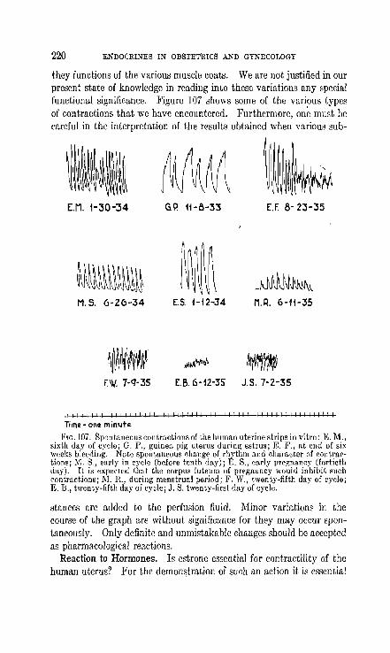
220 ENDOCRINES IN OBSTETRICS AND GYNECOLOGY
they functions of the various muscle coats. We are not justified in our present state of knowledge in reading into these variations any special functional significance. Figure 107 shows some of the various types of contractions that we have encountered. Furthermore, on~ must be careful in the interpretation of the results obtained when various sub-
[.M. 1-30-34 G.P. 11-8-33 E.f. 8- 23-:55
M.S. 6-26-34 [5. 1-12-34 M.R. 6-11-35
\~WtWI~ ~.~~ M~1W~~ f.W. 7-q-35 E.B.6-12-35 J.S. 7-2-35
111111111111 ! III! 111111111111111111111111111111111111111
Tim~ - on~ minute
Fw.107. Spontaneous contractions of the human uterine strips in vitro: E. M., sixth day uf cycle; G. P., guinea pi~ uterus durinp; estrus; E. F., at end uf six weeks bleeding. Note spontaneous change of rhythm and character of contmctions; ,,1. S., early in cycle (before tenth day); E. S., early pregnancy (fortieth day). It is expected that the corpus luteum of pregnancy would inhibit such contractions; 1\1. R., during menstrual period; F. W., twenty-fifth day of cycle; E. B., twenty-fifth day of cycle; J. S. twenty-first day of cycle.
stances are added to the perfusion fluid. Minor variations in the course of the graph are without significance for they may occur spontaneously. Only definite and unmistakahle changes should be accepted as pharmacological reactions.
Reaction to Hormones. Is estrone essential for contractility of the human uterus? For the demonstration of such an action it is essential

CONTROL OF THE UTERUS 221
to test (in vivo or in vitro) a uterus that has not been subject to recent ovarian influence, namely, one secured after a previous bilateral oophorectomy, or a uterus from a patient long after the menopause. In the latter case it is essential that the ovaries be atrophic and present no maturing follicles. The following cage is of interest. The patient was a woman aged 60. Menopause occurred twenty years previously and upon examination the entire genital tract was found senile. The contractions were similar to those seen in uteri of patients well within the menopause age. Furthermore, the strip reacted promptly and vigorously to pituitrin. Two interpretations arc possible. Either estrin is not necessary for spontaneous contractility and a positive reaction to pituitrin, or that a senile ovary produced enough estrin to sensitize the uterus. The latter explanation is the less probable, for the entire genital tract was senile, and furthermore our clinical experience with cases of primary amenorrhea has taught us that the uterus is the last organ to respond (in growth) when large doses of follicular hormone are administered. There remains one other remote possibility, and that is that while the hormone estrin was no longer present in the circulation, other estrogenic substances, such as derivatives of cholesterol or cholic acid were present in concentrations large enough to sensitize the uterus.
Change in the Reactivity of the Human Uterus. Knaus (1929 and later) stated that when the human (and also the animal) uterus is under the influence of the corpus luteum pituitrin will no longer produce an increase in tonus. In a twenty-eight day cycle this reversal first appeared on the sixteenth day and lasted to the twenty-seventh day. Since a certain stage of development of the corpus luteum was essential before enough progesterone could be produced, Knaus placed the beginning of corpus luteum formation, or ovulation, on the fourteenth day. Knaus employed first a 10 per cent iodopin and later a small intrauterine bag. He allowed the uterus to contract spontaneously for 30 minutes so as to establish a base line, and then injected 1 Vogtlein unit of pituitrin intrav~nously. Manzi and Luigi (1931) demonstrated that th~ -human uterine tube in vitro showed increased motility during the follicular phase of the cycle and diminished activity during the corpus luteum phase. The former action favored the transport of the ovum and the latter favored nidation in the endometrium. This explanation is not quite satisfactory, for during the period of greatest motility there is no ovum to transport. It is necessary to assume that this period of greatest motility of the tube extended for some time beyond the period of ovulation.

222 ENDOCRINES IN OBSTETRICS AND GYNECOLOGY
Knaus aimed to establish by his method the exact date of ovulation. This was an important observation and led to considerable literature. Schultze (1931) repeated Knaus' early experiments and concluded that early in the cycle there is little reaction to pituitrin, but the ~eaction increases as the cycle advances, the maximum effect being obtained just before menstruation. Wittenbeck (1930) also disagreed with Knaus. He checked his curves by subsequent laparotomies. He found spontaneous contractions and a rise in tonus from pituitrin in the presence of well developed corpora lutea. Hermstein (1931) 'was more in agreement with Knaus but added that inflammatory conditions in the pelvis can inhibit the motility of the uterus in the absence of a corpus luteum. Tachezy (1934) did not substantiate Knaus. The greatest increase in tonus as the result of pituitrin occurred during the second half of the cycle, namely, during the corpus luteum phase. Five cases of early pregnancy, ranging from the second to the fifth
Action of Pituitrln on the Human Uteri"c Stri, Cay of mcnstrl,lal c~cle 1 2 3 4 5 8 q 10 ft 1213 41 6 1 lq 20 2122 3 42 2 ill l.l2Q B .. ~nd 2q
1 0 o 0 o 2 4 3 4 2 1 3 5 l Z 4 4 5 3 4 41 12 2 J J 3 2 8
t 12 1 Z 1 1 1
pa r.laud 1
FIG. 108. Number of uteri examined during v."ious days of a regular menstrual cycle. None were obtained during the second, third, fourth, and fifth days of menstruation i we usually do not operate durillg menstrual flow.
month reacted to pituitrin by an increase in tonus. The last observation is interesting, for Hoehne (1925) suggested the intravenous injection of a drop of pituitrin as a test for pregnancy. An immediate contraction signified pregnancy. This is in direct opposition to the concept that the corpus luteum of pregnancy (or the placenta) protects the uterus against the effect of pituitrin. •
These observations throw considerable doubt on Knaus' dictulll that the time of uvulation can be ascertained by the Teaction of the human uterus to pituitrin.
This problem was also studied by Kurzrok, Cockrill and Miller (1936) on the human uterine strip (fig. 108). The date of the cycle on which the uperation (hysterectomy) took place was known, and in addition the endometrium was carefully studied to determine the menstrual phase. When the uvaries were removed additional information was ubtained. More than one hundred uteri have been investigated in
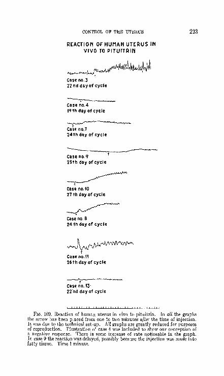
CONTROL OF THE UTERUS
REACTION OF HUMAN UTERUS IN VIVO TO PITUITRIN
~~ Case no. 3 22 nd day of cycle
~ case no.4 Iq th day of cycle
~
Ca~e no.7 24th day of cycle
Case no. q 25th day of cycle
~ t
Case no. 10 27 th day of cycle
~ Case no. 8 24th day of cycle
~ t
Case no.11 26th ffay of cycle
~ t
Case no.13· 22-nd day of cycle
223
FIG. 109. Reaction of human uterus in vivo to pituitrin. In all the graphs the arrow has been placed from one to two minutes after the time of injection. It was due to the technical set-up. All graphs are greatly reduced for purposes of reproduction. lllustration of case 4 was included to show our conception of a negative response. There is some increase of rate noticeable in the graph. In case 9 the reaction was delayed, possibly because the injection was made into fatty tissue. Time 1 minute.

224 ENDOCRINES IN OBSTETRICS AND GYNECOLOGY
REACTION OF' HUMAN UTERUS IN VIVO TO PROGESTERONE
Case no. 20 23 rd day of cycle
Case no.21 t 20th day of cycle
~ Case no. 22 24th day of cycle
Case no. 25 '1 th day of eyel c cramps
t Case no.23 32 nd day of cycle
~ Case no. 2'1 23 rd day of cycle
~ Can no. 28 t 261 h day of cycle
I I ! , " I ! I I ! I II II I 1 I ! I II , !I " " I II " , " t " 1 ! 1 ! II ! !I " 11 , ! ! ,
FIG. 110. Reaction of human uterus in vivo to progesterone. Time 1 min ute

CONTROL OF THE UTERUS 225
this manner. The uterus showed spontaneous activity during all phases of the menstrual cycle. There was no inhibition of spontaneous motility during the corpus luteum phase. The uterus reacted to pituitrin during all phases of the cycle. In nine per cent of the cases the uterus did not react to pituitrin, and it was not due to corpus luteum inhibition, for cases occurring on the eighth and ninth day of the cycle showed a typical postmenstrnal endometrium.
Still further doubt was thrown on the current concept of the action of corpus luteum on the human uterus by the recent observations of Kurzrok, Wiesbader, Mulinos and Watson (1937). They studied the
REACTION OF HUMAN UTERUS IN VIVO TO ESTRADIOL
Case no 41 5th day of cycle
t Case no. 45 6th day of cycle
t Case no. 47 13th day of cycle
t
FIG. 111. Reaction of human uterus in vivo to estradiol. Time 1 minute
reaction of the human uterus by means of the intrauterine bag. Their technique resembled that of Knaus, and in addition they studied the action of estradiol and progesterone in vivo (figs. ] 09, 110, and 11i). The results of their experiments led them to conclude, that:
1. The uterus-shows 'spontaneous contractions during all phases of the menstrual cycle.
2. The uterus reacts to pituitrill in a positive manner during all phases of the cycle. There is no corpus luteulll inhibition, hence the reaction offers no evidence as to the time of ovulation.
3. Progesterone Illay occasionally produce an increase in tonus and amplitude of contraction.

226 ENDOCRINES IN OBSTETRICS AND GYNECOLOGY
4. Progesterone produces uterine cramps during the postmenstrual phase of the cycle, but not during the premenstrual phase.
5. The cramps are not associated with significant changes in the graph nor with a previous history of dysmenorrhea.
6. Estradiol (benzoate) occasionally produces a rise iil amplitude and tonus.
7. Estradiol (benzoate) may produce cramps during the first half of the cycle, without significant changes in the tracing.
S. Oily solutions of progesterone and estradiol benzoate are rapidly absorbed from the local site of injection, the effect~ being observed within thirty minutes.
These findings throw still greater doubt on Knaus' test for ovulation. Furthermore, the .above findings show that the corpus luteum in the human does not inhibit spontaneous uterine contractions nor inhibit the reaction to pituitrin. The hormonal control of the human uterus is not identical with that of the rabbit.
The Mechanism of Parturition
The cause of the onset of lab6r has received considerable attention in the hands of various workers. Several theories have been suggested, namely,-
1. The regression of the corpus luteum. 2. The action of estrin upon the uterus, alone, and in association
with oxytocin. 3. The production of a particular substance which induces labor,
a substance similar in action but not identical with pituitrin. I. The Regression of the Corpus Luteum. In view of the fact that
the corpus luteum protects the fertilized ovum against early miscarriage the idea was extended to include the concept that labor sets in when the corpus luteum of pregnancy begins to degenerate. It was soon demonstrated however, that bilateral oophorectomy after the sixth week of pregnancy does not induce abortion. The suggestion was therefore made that the placenta takes on the fUllction of the corpus luteum and that labor sets in when the inhibitory placental influence upon the uterus is removed. In other words, as the placenta reaches term ("ages") its ability to produce inhibitory substances lessens. As long ago as 1903, Halball suggested that the placenta was a gland of internal secretion. There are no objections from a histological viewpoint to this concept. Loewe and Voss (1934) transplanted a fragment of human placenta into a mouse, and noted the appearance of rut. The

CONTROL OF THE UTERUS 227
cells of the transplant were found alive three months after operation. Wald~tein (Hl29) performed a bilateral oophorectomy on a woman on the 34th day of geRtation and noted that the blood and urine contained follicular hormone in the subsequent months of pregnancy. Thc patient went to term, and one week after term no estrone could be detected in the urine. The estrone content of the placenta was normal. That the foetus does not influence the onset of labor is evidenced by the well known dinical fact that the placenta will maintain function scveral months after the foetus dies, and that labor will set.in at about the expected time. The placenta produces prolan A and B, as well as estrin, and recently (1934) Ehrhardt found corpus luteum hormone as well. De Snoo (1922) reportcd the successful treatment of repeated abortion with placental extract. Damon (1936) and Kurzrok (1936, unpublished) have confirmed his results. Whether the inhibition of the onset of labor hy means of placental extract is due to the presence of corpus luteum hormone or some other unknown factor is still a dehatable question.
II. The Action of Estrin upon the Uterus, Alone, and in Association with Pituitrin. It was stated previously that estrin sensitizes the animal uterus. Whether this is true of the human uterus, and to what extent, is still an open question. The uterus is subjected throughout pregnancy to large concentrations of follicular hormone that are present in blood. The purpose of excess of estrin is not known. It is present in increasing concentration as pregnancy progresses and rapidly disappears from the blood stream about 10 days postpartum. The injection of estrin into rats and mice causes abortion (M. Smith, 1926; Parkes, 1930; Hain, 1935). The amount of estrin required to interrupt pregnancy rapidly increases as pregnancy advances (M. Smith). It is occasionally possible to induce abortion clinically during the second month of pregnancy. It has been our experience that when moderate doses (2,000 R.D.) fail, very large do~es (40,000 to 80,000 R.D. in one week) are also without effect. The mechanism of induction of abortion by means of follicular hormone is not clear. It may be due to hemorrhage resulting from the increased blood supply to ·the ·uterus, for it has been shown by means of ocular implants in the rabbit that follicular hormone has a vasodilator effect on the capillaries of the endometrium (Markee, 1932). The separation of the ovum from its bed causes the uterus to expel it as a foreign body.
That placental and ovarian extracts exert a synergistic action in

228 ENDOCRINES IN OBSTETRICS AND GYNECOLOGY
sensitizing the uterine muscle to the secretion of the posterior pituitary has been known for some time. Dixon (1923), and Marshall (1924) and Fenn (1924) observed that extracts of the whole ovary, but not the corpus luteum increased the uterus contracting substance of the cerebrospinal fluid. Friedman and Friedman (1933) found less oxytocin in the cerebrospinal fluid than has been claimed by Dixon and Marshall. The identity of the uterus contracting substance in the cerebrospinalliquor with oxytocin has been established 'by Dixon, and by Miura (1926). The closer the animal from which the ,ovaries were removed was to term, the greater was the stimulating effect. They concluded tl~at,
associated with the regression of the corpus luteum of pregnancy, the ovary produced increasing amounts of a substance that stimulated the posterior pituitary to increased secretion of oxytocin. Mayer noted an increased amount of oxytocin in the cerebrospinal fluid of women in labor. Siegert (1927) and Van Dyke and Kraft (1927) could not confirm Mayer's observation. Brdichka (1924) noted an increased concentration of oxytocin in the blood serum of women in labor. Thus a theory for the etiology of the onset of labor was proposed, namely, that as the corpus luteum regressed, the ovary produced increasing amounts of substances (estrone?) that stimulated the posterior pituitary. An immediate ohjection can he raised to this theory. Bilateral oophorectomy during pregnancy does not prolong pregnancy, and labor occurs in the usual manner. Furthermore, it has been demonstrated that parturition can occur in the absence of the posterior pituitary (Smith, 1932, rat; Allan and Wiles, 1932, cat). The question arises whether the placenta with its multitude of functions during pregnancy docs not also replace or supplement the oxytocic function of the posterior pituitary gland.
The role of the anterior pituitary gland in parturition has now received considerable attention. In the rat the removal of the anterior pituitary gland after mid-pregnancy causes gestation to be prolonged. The mother dies for she is unable to litter (Pencharz and Long, 1931; Selye, Collip and Thomson, 1933). Rain (1932, 1934) noted that extracts of pregnancy urine caused gestation to be prolonged. On the contrary very large doses of Antllitrin "S" administered later in pregnancy caused abortion.
The Synergistic Action between Estrin and Oxytocin during Pregnancy. Parkes (1930) demonstrated that abortion can be induced in mice by the synergistic action of estrin 'and oxytocin. Hain (1935) was unable to confirm this in the rat. The synergism in the human uterus
CONTROL OF THE UTERUS 229
is not so readily demonstrated. It has been observed clinically that the rhythmic contractions of the pregnant uterus increase both in frequency and intensity during the course of pregnancy. This is by no means universal. We have observed uteri during the latter part of pregnancy and have not noted any increased frequency of contractions. Some uteri are extremely responsive to the examining hand. On the contrary the writer has frequently done an external version during the eighth or ninth month without eliciting any, or at most very few contractions. Bourne (1933) recorded the uterine contractions at the thirty-sixth week of pregnancy and noted that they were similar to those recorded during the early stages of labor. Robson (1933) found no evidence to show that the degree of spontaneous rhythmic activity exhibited by strips of human uterine muscle removed at different stages of pregnancy and at parturition, increased progressively as term and labor approached. The experience of Miller, Kurzrok, and Cockrill has been similar (unpublished data). Robson (1933) studied the behavior of human uterine muscle to oxytocin during the various phases of gestation and noted that the minimal dose of hormone required to produce contractions decreased as term approached; the limits being more than 2 oxytocin units during the twelfth week and 0.003 unit during labor.
In view of the fact that in some animals labor may proceed in the absence of the posterior pituitary the question has arisen as to whether pituitrin is the only oxytocic substance and whether the posterior pituitary is the only source of oxytocic substances. Certainly, the hypothalamic region and the placenta'cannot be overlooked. Recently, Cockrill, Miller and Kurzrok (1934) studied the urine of patients in active labor as a source of oxytocic substances. It was previously demonstrated (Guerin-Valmale, Loriot and Verdeuil, 1931; Barjaktrovic, 1933) that labor could be induced by administering labor urine by proctoclysis. Labor could only be induced in the second half of pregnancy. A1JtQ_U:nts up tQ 4,000 gm. were necessary, and some patients were- delivered 12 hours after instillation.
Cockrill et al. (1934) collected the urine from patients during active labor and combined the specimens until a total volume of 10 or 12 liters was obtained. (Pituitrin was not given to any case.) Thirty such collections were made, and each was worked up separately. The samples were acidified with acetic acid and concentrated in vacuo at a temperature of 30° to 40°C.' The method then used to obtain the oxytocic substance was that described by Kamm and his co-workers (1928) for isolation of the active principles of the posterior lobe of the pituitary gland.

230 ENDOCRINES IN OBSTETRICS AND GYNECOLOGY
A
icc. l U 1q
B
r I:
04cc L U 25
t03 ee. '.U.21
r
C G.P.
0
~ O.2cc. o 4cc. L U. 27 'U.21
(;
II'+..U
FIG. 112. Actiun of labor urine extract on the human uterine strip. All strips were obtained from sections at term. Note the variations in the spontaneous contractions and the extent and type of response. Figure G sholVs the irregular contractions of a strip from the lower uterine segment. Figure C is the response of a guinea pig.

CONTROL OF THE UTERUS 231
The final product was a crude material of a gummy character and colored with urinary pigments, 10 liters of labor urine yielding an average of 0.5 gram. This dried material was taken up in distilled water and dialyzed over night in a refrigerator in a parchment bag. The solution from the bag was then made neutral to litmus with sodium carbonate. The solution was tested, using about 0.4 cc. from a total volume of 25 cc. representing the original 0.5 gram, on human uterine muscle obtained from Caesarian sections at term. Twenty-five strips of muscle were used. The muscle was set up according to the technique described by Kurzrok and Lieb (1930). The extract obtained from these fractions caused a prompt and sustained contraction (fig. 112). When the solutions were tested on non-pregnant human uterine muscle (55 strips, obtained at various times of the menstrual cycle), there was no effect or a slight increase of tonus causing the normal contractions to occur with slightly more vigor. Control ·experiments were made, using material prepared by the same procedure from 2 10-liter samples of urine of male patients and 3 from non-pregnant women. These control material~, tested on parallel strips of muscle from the same pregnant (25) and non-pregnant (55) uteri as were used for the labor urine extracts, gave negative results, although a few gave slightly increased tonus.
The active material was tested by Dr. Mulinos, of the Department;rof Pharmacology, for its action on the water balance of the frog, and the result indicated that the substance has a pitocin-like action. The controls when tested in the same way had no significant effect.
/"
The Action of Human Semen upon the Human Uterus
The action of human semen upon the human uterus has proven to be a striking phenomenon, one that is involved in the process of fertilization, and again in certain types of sterility. Furthermore, one of the substances, acetyl choline, which partakes in the reaction has been held by som..!)' to b~ l}_hormone. These considerations have led us to include this work in the chapter on the hormonal control of the uterus (also in Chapter XXVIII).
Kurzrok and Lieb (1930) reported that human semen caused a relaxation of the human uterine muscle. There occurred a marked loss of tonus and a loss of contractions over a period of one-half to one hour or more. In certain cases, depending apparently on differences in the semen, or in the uterus, or in both, the effect was contraction. These effects have been demonstrated both in vitro and in vivo. When a cer-
232 ENDOCRINES IN OBSTETRICS AND GYNECOLOGY
tain semen specimen relaxed most uteri, an occasional uterus would respond by contraction. On the contrary, some semen specimens (about 15 per cent) always caused contraction. Cockrill, Miller and Kurzrok (1935) studied these phenomena further and observed the following:
1. The pregnant human uterus always responded to human semen by contraction.
2. The phase of the menstrual cycle had ~o effect upon the uterine response to semen.
3. Non-human uteri (rat, rabbit, guinea pig) respond to human semen by marked contraction. (A human ut~rine strip in the same chamber relaxes.)
4. The contracting substance in semen is probably acetyl choline, or some very similar compound.
5. There is suggestive evidence that the relaxing substance is also acetyl choline, 'but in different concentration.
6. The effects of human semen upon the human uterus are independent of the presence or absence of sperm.
7. Crystalline spermine has no effect on the human uterus.
The Action of Ergot and Its Derivatives upon the Human Uterus
The fungus ergot contains a mixture of alkaloidal and non-alkaloidal substances having various pharmacological activities. Tauret (1875) isolated the first pure substance, ergotinine, but it was inert. Barger and Dale (1907) isolated a second alkaloid, ergotoxine, which possessed all the experimental pharmacological actions of ergot. Stoll (1921) extracted two further alkaloids, ergotamine and erg,otaminine. The latter is inactive. Ergotamme and ergotoxine are equivalent pharmacologically although. slightly different chemically. Recently Adair et al. (1935) have isolated a crystalline substance from ergot having the empirical formula 0l1H27NaOa, which they named ergotocin. The same substance (in all probability) has been isolated by other workers; ergometrine (Dudley and Moir, 1935), crgobasine (Stoll, 1921), and ergostetrine (Thompson, 1935). A lively discussion is at present in progress as to which name is to be adopted (Dale, 1935).
The method used for the study of ergotocin was first described by Bourne and Burn (1930) and subsequently employed also by Moir (1932). It consisted of the introduction 6f an hydrostatic bag into the uterine cavity of a woman on the fifth or sixth day post-partum. The contractions were then recorded on a suitable kymograph. A

CONTROL OF THE UTERUS 233
similar apparatus was previously used by Pierce Rucker (1930) who introduced a Voorhees bag during labor, and thus studied the reaction of the uterus subpartum to various drugs. Adair et a1. obtained the following effects with ergotocin. It is active in doses of 0.2 mg. by mou Lh. Its effect manifests itself in six to fifteen minutes and produces an increase in tonus and frequent uterine contractions. Ergotocin has a considerable margin of safety and has no apparent detrimental effect upon respiration, pulse, blood pressure and urinary output.
BIBLIOGRAPHY
ADAIR, F. L., DAVIS, E. M., KHARASCH, M. S. AND LEGAULT, R. R. 1935 A study of a new and potent ergot derivative, ergotocin. Am. J. Obs. and Gyn. 30, 466.
ALLAN, H. AND WILES, P. 1932 Rolc of Pituitary Gland in Prcgnancy and Parturition; Hypophysectomy. J. Physio!. 76,23.
ALLEN, W. M. AND REYNOLDS, S. R.:vI. 1935 Physiology of corpus luteum; eOIIlp!1rative actions of crystalline progestin and crude progestin on uterine motility in unanesthetized rabbits. Am. J. Obs. and Gyn. 30{
309. BARGER, G. AND DALE, H. H. 1907 Ergotoxinc and some other constituents of
ergot. Biochem. J. 2, 2~O. BOURNE, A. W. AND BURN", J. H. 1930 Action on hnman uterus of anesthetics
and other drugs commonly used in labour. Brit. Med. J. 2, 87. BOURNE, A. W. AND BELL, A. C. 1933 Uterine inertia. J. Obs. Gynecol. Brit.
Emp. 40, 423. BRDlCHKA, G. 1924 Dic Erregung des iiberlcbenjen Kaninchenuterus durch
Schwangeren und Kreissendenserum. Arch. f. exper. Path. 103, 188. CA:-r:srON ET AL. 1930 Quoted by Kuntz. COCKRILL, J. R., MILLER, E. G., JR. AND KURZROK, R. 1934 Presence of oxyto
cic substances in urine during labor. Proc. Soc. Exp. BioI. Med. 31, 572.
COCKRILL, J. R., MILLER, E. G., JR. AND KURZROK, R. 1935 Substance in hu-man seminal fluid affecting uterine muscle. Am. J. Physio!. 112, 557.
DALE, H. II. 1935 The new ergot alkaloid. Science 82. 99. DAMON, V. 1936 T Unpubli~hed results. . . DAVIS, E. 1'1., ADAIR, F. L., ROGERS, G., KHARASCH, M. S. AND LEGAULT, R. R.
1935 A new active principle of ergot and its effects on uterine mobility. Am. J. Obs. and Gyn. 29, 155.
DIXON, D. E. 1923 Pituitary Secretion. J. Physio!. 67, 129. DIXON, W. E. AKD MARSHALL, F. H. A. 1924 The influence of the ovary un
pituitary secretion, etc. J. Physiol. 69, 276. DUDLEY, H. W. AND MOIR, J. C. 1935 Substance responsible for traditional
clinical effect of ergot. Brit. Med. J. 1, 520. DYKE, H. B. VAN AKD KRAFT, A. 1927 The Role of the Hypophysis in the Initia
tion of Labor. Am. J. Physiol. 82, 84. EMBDEN 1925 Handbuch d. Physio!. 8, 369.

234 ENDOCRINES IN OBSTETRICS AND GYNECOLOGY
FENN, W. O. 1924 Active principles of the posterior lobe. J. Physiol. 59,395. FRIEDMAN, G. S. AND FRIEDMAN, M. H. 1933 Examination of cerebrospinal
fluid for oxytocic activity as tested by rabbit uterine fistula preparation. Am. J. Physio!. 103,244.
GENELL, S. 1934 Tests on the treatment of labor pain insufficiency with folliculin. Nord. Med. Tidskr. 6, 236.
GUERIN-VALMALE, LORIOT, C. AND VERDEUIL. 1931 Influence de I'urine de femme en travail sur Ie declenchement physiologique du travail. Bull. Soc. d'Obst. et Gyn. 20, G19.
HAIN, A. M. 1932 The physiology of pregnancy in the rat. The prolongation and interruption of pregnancy. Quart. J. Exp. Physio!. 22, 2-19.
1935 The physiology of pregnancy in the rat. Quart. J. Exp. Physio!. 25, 131.
1934 Effect upon lacta}ion of oophorectomy during pregnancy (albino rat). Quart. J. Exp. i!'.bysio!. 24, 101.
HERMSTEIN, A. 1931 Uber die Be~timmung des Ovulatiunstermine ,durch Hypophysenhi·nterlappenpriiparate. Arc)l. Gyn1lk. 144, 500.
HOEHNE, O. 1925 Klinischc Erfahrungen mit den modernon Wehenmitteln, insbesondere mit Gynergen. Arch. f. Gyniik. 126,3.16.
KAMINESTER, S. AND REYNOLDS, S. R 1935 Motility in the transplanted denervated utel'lls. Am. J. Ohs. Gyn. 30, 395.
KLM'TEN, E. AND RUFFEL, Z. 1935 Uteromuskelerregbarkeit und galvanische Erregbarkeit des Ncrvcnmuskclsystcms. Z. f. Gyn. 59, 2050.
KNAUS, H. 1931 Sterilisierung und Kon"fptionsverhiitung. Z. Gyniik. 56, 2854. 1932 Zur Bestimmung des Ovulationstermines an def menschlichen Gebiir-
mutter in situ. Z. Gynak. 66, 243. Il 1934 Periudic fertility and sterility in wum~. W. Mandrich, Vienna.
KURZROK, R., MILLER, E. G., JR. AND COCKIlILL, J. R. 1936 Hormonal control of the uterus. N. Y. State J. Mod. 36, No. 20.
KURZROK, R, WIESBADER, H., l\IULINOS, M. G. A~D WATSON, B. P. 1937 The action of Pituitrin, Estradiol and Progesterone on the Human Uterus in Vivo. Endoerin., in press.
LOEWE, S. AND Voss, H. E. 1934 Nachweis des Vorkummens vun Gelbk5rperhormon im Frauenharn. Sohweiz. ;\ied. Woeh., 1049.
MANZI, L. 1931 Azione diretta del liquido follicolare Bulla motelia tubarica. Arch. Ostetr. 18, 57.
MARKEE, J. E. 1932 Rhythmic vascular uterine changes. Am. J. Physiol. 100, 32.
1933 Menstruation in ocular endometrial implants. Anat. Rec. 56, 66. MAYER, A. 1924 nber die Wehcnerrcgonde Wirkung des Liquor eerebrospinalis
von Gebiirenden. Klin. Woch. 6, 180.5. MILLER, E. G., JR., COCKRILL, J. n. AND KURZROK, R. 1937 Reactions of human
uterine muscle in vitro to pituitrin, adrenalin and acetylcholine and their relations to the menstrual cycle. Am. J. Obs. and Gyn. 33, 154.
MIURA, Y. 1925 tiber den Gehalt der Cerebrospinallliissigkeit an Hypophysenhinterl:lppensekret. Areh. f. ges. Phys. 207, 76.
NOVAK, J. 1928 In Halban-Seitz Biologie und Pathologie des Weibes. Berlin, 5, Part 3, 1450.

CONTROL OF THE UTERUS 235
PARISOT, J. AND MATHIEU, R. 1914 Les substances extraites du lobe posterieur de l'hypophyse, etc. Compt. rend. Soc. BioI. 76, 222.
REIN, G. 1902 Quoted by Kuntz-Autonomic. nervous system. Philadelphia, 1934, page 350.
ROBSON, .1. M. 1933 Reactivity and activity of human uterus at various stages of pregnancy and lit parturition. J. Physio!. 79, 83.
1933 Effect of oestrin on reactivity and spontaneous activity of rabbit's uterus. J. Physiol. 79, 139.
1933 Reactivity and activity of rabbit's uterus during pregnancy, parturition and puerperium. .J. Physio!. 78, 309.
SCHUW'ZE, G .. K. F. 1931 Die lleaktion der nieht sehwnngeren mensehlichen Gebiirmutter auf Hypophysenhinterlappenextrakte. Z. Gyn. 55, 3042.
SELYE, H., COLLIP, J. B. AND THOMPSON, D. L. 1933 Effect of hypophysectomy upon pregnancy and lactation in mice. Proc. Soc. Exp. Bio!., N. Y., 31,82.
SIEGERT, F. 1927 Der Gehalt des Liquor Lumbalis an hypophyscnwirksamcr SuhstaRz bei schwangeren und nichtschwangeren Fmuen. Klin .. Wach. 6,1558.
SMITH, M. 1926 Interruption of pregnancy in rats by injection of ovarian follicular extract. Johns Hopkins Hospital Bull. 39, 203.
SMITH, P. E. 1932 Non-cssentiality of posterior hypophysis in parturition. Am. J. Physio!. 99, 34.5.
DE SNOO, K. 1922 Die Bedeutung der Plazenta, insbesondere des Trophoblasts filr die Schwangers.chaftsdauer und den Geburtsintritt. Mon. f. Geb. Gyn. 57, 1.
STOLL, A. 1930 The alkaloids of ergot. Lancet 2, 1148. TACHEZY, R. 1034 lJber die Reaktion des Uterus auf Pituitrin. Z. Gyniik. 58,
2663. WALDSTEDI, E. 1929 Fruhkastration in del' Schwangerschaft. Zur Genese des;
Ovarialhormons. Z. Gynfik. 63, 1305.
Additional References
BARJAKTAROVIC, S. S. 1933 Die Ursache des Geburtsbeginnes. Zb!. f. Gyn. 67,628.
KAMM, O. 1928 The active principles of the posterior lobe ofc the pituitary "land. I. The demonstration of the presence of two active principles. J r. The separation of the two principles and their concentration in the form of potent solid preparations. J. Amer. Chern. Soc. 50, 573.
KIJRZIIOK, R. AND LIER, C. C. 1930 Biochemical Studies of Human Semen. II. Action of Semen on the Human Uterus. Proc. Soc. Exp. BioI. Med. 28, 268 .. ,
MAKEPEACE, A. W., CORNER, G. W. AND ALLE", W. M. 1936 The Effect of Progestin on the In-Vitro Response of the Rabbit Uterus to Pituitrin. Am. J. Physiol. 115,376.
ilIARRIAN, G. F. AND PARKES, A. S. 1930 The Relative Amounts of Oestrin Required to Produce the Various Phenomena of Oestrus. J. Physio1. 69,372.
236 ENDOCRINES IN OBSTETRICS AND GYNECOLOGY
V. MIKULICZ-RADECKI, F. 1035 Die Behandlung dcr atonischcn Nachgeburtsblutungen. Munch. Med. Woch. 81, 1797.
PARKES, A. S. 1930 On Synergism between Oestrin and Oxytocin. J. Physio!. 69,463. .
PENCHARZ, R. 1. AND LONG, J. A. 1931 The effect of hypophysectomy on gestation in the rut. Science 74, 206.
REYNOLDS, S. M. R. AND ALLEK, W. M. 1932 Effect of Progestin-containing Extracts of Corpora Lutea on Uterine Motility in Unanesthetized Rabbit with Observations on Pseudopregnancy. Am. J. Physio!. 102, 39.
RUCKER, M. P. 1930 Action of Thymophysin on Human Pregnant Uterus in Situ. Am. J. Obs. & Gyn. 20, 79f.
TIlO~IPSON, ]\1. R. 1935 New Active Principle of Ergot. Science 81, 636. TAURET, C. 1875 Sur la presence d'un nouvel alcaloide, I'ergotinine, dans Ie
seigle ergote. Acad. d. Sc. 81,896.

CHAPTER XVII
TESTS FOR PREGNANCY
It has been the aim of gynecologists to make the diagnosis of pregnancy without the aid of a physical examination. The physicians of antiquity concerned themselves with this problem. The earliest mention of the subject is to be found in the Papyrus-Kahun. This manuscript dates from 2000 B.C. and was discovered by Sir Flenders Petrie in 1889. Therein is a consideration of the problems whether a woman can become pregnant or not, and also whether she 1:8 pregnant or not. The basis for the diagnosis of pregnancy is as follows (the text is here incomplete): the physical condition of the breast, the readiness with which vomiting can be induced, the testing of muscular irritability and the color of the face. It is rather startling to find that this papyrus reports on a method of prevention of conception, and a method of determining the sex of the foetus in utero.
The twentieth century-a space of 4000 years-finds us again deeply interested in a .biologic test for pregnancy as evidenced by the Abderhalden test, the phlorizin reaction (Kammitzer and Joseph, 1921), the adrenalin reaction (Roubitschek, 1922), the glucose tolerance test (Frank and N othmann, 1920). The last three depend on abnormalitiis of the renal threshold for glucose which occur during pregnancy (and in other related states). The tests demonstrating renal glycosuria are not reliable for pregnancy. Of recent-years the Abderhalden test has had few followers, mainly because of the difficulty in technique. Its accuracy has been disputed by many. In 1928 Professors Selmar Aschheim and Bernard Zondek, then of the University of Berlin, announced their biologic test for pregnancy. The Aschheim-Zondek Test, as it is now known, is now a standard laboratory method the world over.
The Biological Basis for the Aschheim-Zondek Test
The test is based on the following two biological principles. 1. The injections of extracts (or implants) of the anterior pituitary
gland into infantile mice (or rats) induces in these animals precocious sexual maturity. This manifests itself primarily by changes in the ovaries, and secondarily by changes in the tubes, uterus and vagina (Smith and Engle, 1927).
237
238 ENDOCRINES IN OBSTETRICS AND GYNECOLOGY
2. Substances having similar gonadotropic properties are found in the blood and urine of women who are pregnant (Aschheim and Zondek, 1928).
It is important to fE-member that there are no hormones specific for pregnancy, and that the gonadotropic hormones, liere prolan A and B may be found in other conditions. The Aschheim-Zondek Test is merely quantitative and not qualitative. This being the case, the first appearance of a positive Aschheim-Zondek Test will vary with different patients, for the rate of increase of hormone production would not be the same in all patients, and their renal thresholds will not be identical.
The Hormones Involved in the Test and Their Actions
There are two hormones involved in the Aschheim-Zondek Test, namely prolan A and B. As stated in Chapter VII the name prolan is used only when the source of the hormones is pregnancy urine. Prolan is similar but not identical with the gonadotropic hormones of the anterior pituitary gland. For prolan to be effective the anterior pituitary gland is essential, hence the test could not be carried out on a hypophysectomized mouse (Evans, Meyer and Simpson, 1932).
The action of these hormones on the immature ovary is to produce three types of response (Aschheim and Zondek, 1928).
Reaction I. Follicle maturation, with the formation of large follicles. This is due to prolan A. It is not specific for pregnancy for it may be found after castration (also spontaneous menopause) and in cases of genital carcinoma. This reaction may precede a typical AschhcimZondek Test, and may also terminate it (Kurzrok, Kirkman and Creelman, 1934) (fig. 113).
As a result of the stimulation of the ovary by means of prolan A, the follicles respond by the production of follicular hormone. A secondary reaction is thus set up, namely stimulation of the uterus so that it becomes distended with fluid, and, in addition, the vaginal orifice opens and a positive smear results. The secondary reaction has no bearing on the Aschheim-Zondek Test and is to be disregarded.
Reaction II. Hemorrhage into the follicles. This describes the "Blutpunkte" of the German writers. This reaction is pathognomonic of pregnancy and is probably due to the action of both Prolan A and B.
Reaction III. Formation of corpora lutea with .imprisonment of the ovum (corpora lutea atretica). This reaction is due to prolan A and B. The normal rhythm of follicle growth- rupture- corpus luteum forma-

--,
FIG. 1I3A. Positive follicle stimulating hormone reaction (F.S.H.). Infantile mouse. Follicles only. There are no corpora. There is beginning antrum formation in the growing follicles. This is not a positive Aschheim-Zondck test.
FIG. 1I3B. Negative Aschheim-Zondek test. Infantile mouse. A negative F.S.H. reaction would appear the same. No large follicles, or corpora lutea.
239

FIG. 114A. Positive Aschheim-Zondek test. Immature rat. B.P., blood points ("Blutpunkte") or hemorrhages into follicles. 0, ovum within a corpus luteum .
. C.L.
FIG. 114B. Positive Aschheim-Zondek test. Immature rat. C.L., corpora lutea.
240

T}JSTS FOIl PHEGNANCY 241
lion ix distnrbrd ami thr follirlr lut('iniz('~ brfore rupture occurs. The formation of corpora lutea in an illllllatur(' ovary is pathognonwnic of pregnancy. Prolan 13, alon(', dol's not stimulatr the o\'ary, IInless it has pr('yiously \wcn Htimlliated to form follides hy prolan A, for we could not haw a ('orp"s lutrlllll Ilnlrss thrre \YaS a foJlirl('.
The Source of Prolan A and B
In view of the fact that prolan A ancl B han' a Himilar action to the g-onadotropic h()rmonp~ of the ant prior pituitary g-Iand, it was as.~umed that thr origin of prolan was this g-land, This concept spemed to gain additional sUPJlort. from thp fact that the antrrior pituitary g-land cnlarg('s during pl'!'gnancy (ErrllH'im and Stllll111W, 190B), while the neurohypophysis Heems ('oll1pressrd. The ('nlargement is due to the iormat.ion of prrgnancy cells, whilr til(' basophiles and a('idophiles are definitely decrea,;p(l. But ;;p\'eml inwstigators ,oon showed that not
onE' of the threr rraction~ ('ould he prodl!('ed by implanting thr antrrior lobr from prpgnant WOlllrn (Philipp, ]\)30; Ehrhardt and i\Iaye~, 11)30). On the contrary, A~('hh('illl \YaS able to reproduce th(' rraction with transplants of human pla('enta (also decidua, ('orpu, IUt(,Ulll of pregnancy). The eyidence points to the ehorionic rpitheliulll as the somec of prolan D.I Two rpaRons could explain the absence of the hormones in the antrrior 10)(': (1) the rat!' of srcretion of the hormonc is vpry ,low; and (2) t}](' rate of rx('rpt.ion of the IOfmonp is so rapid that none is found in the implant. The first rrason <lors not s("rm plausiblr beeause of the largr amotlllt, of prolan A and n that are found in the blood and urine, Zondek fayors the latter hypotlw~is, pointing out the fact that the thyroid i~Gra\'es' diH('asc ('ontains less thyroxin than tlw normal thyroid.
The Technique of the Test
'I'll(' trehniqlw aH dps(Tihrd by A'i('hhpim ami lIolllkk ('alis for six infuntilr mirr, wrighing frolll 6-~ grams melt. A first 1Iwming 'I)('cimrn of urine is eollrcted, a('idified ~li~htly with dilutr a('ptie arid, and the same Flweiml'n is utilizrd for illjel'li()l1~ throughout the dllI'atioll of the tr.,t. Each animal i~ injected twicl' a day witll 0.25 cc. of urine suhcutaneously under the Rkin of the back, for three successive days. Thus eaeh animal will rpcpive a total of 1.5 ce. of pregnancy urine. ~o injections arr giwn on the fourth day, and the animals are sacrificed on the fifth day, The test, tIlPrrforp, lakes about 100 hours.
1 Evidence is arculllulatinl( to show that prolan A is derived from the anterior pituitary.
242 ENDOCRDIES I~ OBSTETRICS AND GYKECOLOGY
The ovaries are then examined in situ, in a good light (preferably daylight), and if necessary with the aid of a lens. If the test is positive the ovaries will show corpora lutea, or punctate hemorrhages, or both. A single corpus luteum or punctate hemorrhage in any of the twelve ovaries is sufficient to make a diagnosis of pregnancy. The number of corpora lute a or punctate hemorrhages varies from one to several per ovary. Punctate hemorrhages seem more common with some specimens than with othem. :Lhe presence of follicles is not pathognomonic of pregnancy. The conditi6n of the uterus and vagina has no bearing on the test.
The reason why infantile animals are used is that adult animals may have corpora lutea in their ovaries, and therefore confuse the test. At .the same time the animals should not be too small or too young (less than 6 grams and less than 3 weeks old) for the urine may prove lethal to them after one or more injections, and secondly, very young animals will not respond to any stimulation of the ovaries.
The reason why six animals arc used is that inexperienced technicians may kill one or more animals while doing the test, hccause of faulty technique. Even under the hest conditions some animals may die because the urine may be toxic for them. Again, not all animals react with the same intensity to a given stimulus. It is not uncommon to find one (out of five or six) animal in a given test entirely negative, so that if that animal were the only one examined the test would be read as negative, while in reality the others might show a strong positive reaction. Hcnce, a negative rcading in a single mouse is never conclusive. So irregular is the response in some animals, that the writer has seen a positive reaction in one ovary and a negative reaction in the other ovary of the same animal. Doubtful reactions should not be considered, but the test should be repeated with a fresh morning specimen of urine. When in doubt it is advisable to section and stain the ovaries and thus obtain a correct diagnosis. The choice of a morning specimen for the test is important. It is preferable that the patient should not have voided for about 6 hours previous to the collection of the specimen. It appears that the concentration of urine that occurs overnight holds also for the hormones. The writer has seen on a number of occasions a negative reaction with a casual specimen, and a positive reaction with the morning specimen of the next day.
The Accuracy of the Test
The reason why the previous tests for pregnancy were discarded was because they had an accuracy of little more than 50 per centum. In

TlflSTS FOR PREGNANCY 243
the hands of experienced workers the accuracy of the AschheimZondek Test ranges from 98 to 99 per cent. Aschheim (1933, 1935) recently reviewed his own results in 2000 cases, and obtained an accuracy of 99 per cent. Our own results are based on 1800 cases and show an accuracy of slightly less than 98 per cent. Clauberg (1933) in a recent review of the data on the test reached somewhat similar results.
The reasons for the errors in diagnosis were frequently hard to detect. Aside from conditions other than pregnancy that might give a positive test there are on record definite pregnancies that present a negative reaction. Robson (1934) mentions one such case. The writer had one case of pregnancy complicated by large fibroids wherein a negative test appeared in the fourth and fifth months of pregnancy and a positive test in the seventh month. The patient went to term and delivered a normal male child. Unfortunately the placenta was not examined. It must not be forgotten that the test represents a quantitative reaction and the amount of hormone excreted in the urine depends upon the amount produced and the level of the renal threshold. The amount produced depends upon the metabolic activity of the chorionic epithelium and the interchange of products between the syncicytium and the maternal circulation. The collection of the morning (over-night) specimen is of importance. Often it merely means that the specimen has been collected at about 7 a.m., forgettin_g or taking no account of the fact that the patient may have voided t,vo hours before. Where there are clinical reasons to suspect a pregnancy and the test is negative, then the test should be repeated with a fresh specimen of urine. It must always be ren:cmbered that the test should be used as an aid to clinical diagnosis, and not as a substitute for the physical examination.
The reactivity, the age, the size and the condition of the animals all have an important bearing on the outcome of the test, for reasons already mentioned.
The Tiine of Appear,ce and ~isappearance of the Test
Any non-clinical test for pregnancy that would be positive only in the second and third trimesters would not be of great aid in diagnosis. On the contrary one of the chief virtues of the Aschheim-Zondek Test is that by means of it a very early diagnosis of pregnancy can be made. With it a diagnosis can occasionally be made even before the patient skips a period (Aschheim, 1935; Posner, 1935). We have seen one such

244 ENDOCRINES IN OBSTETRICS AND GYNECOLOGY
case (Kurzrok, Kirkman and Creelman, 1934). Reaction I (follicles only!) may precede an early positive test by a day or two, so that the demonstration of Reaction I calls for a repetition of the test a few days later. More than two-thirds of the pregnancies will give a positive test when the patient is a day or two ovcrdue, while more than 90 per cent of pregnancies will gi,'c a positivc test when the patient is 10 days overdue.
The appearance of a positive test depends upon the establishment of a metabolic interchange between the trophoblast and the maternal circulation, in other words, when nidation of the ovum occurs. " This in turn depends upon three factors:
1. The time of ovulation in the cycle. ' 2. The time of fertilization. 3. The duration of the transport of the fcrtili7.ed ovum and nidation. These are three variable factors, hence the establishment of a meta-
bolic interchange between trophobl:1st and maternal circulation must vary. For, this reason the first appearance of a positive ,AschheimZondek Test must vary in different individuals, and in the same individual during different pregnancies. In a previous communication, the writer (1934) pointed out that the duration of the transport of the fertilized ovum from the outer end of the tube into the depths of the decidua is about 10 days. This is an average figure, for the transport may be faster or slower (ectopic?). In all probability fertilization usually (but not necessarily) occurs within twenty-four honrs of ovulation. If these two factors (fertilization and transport) remain constant, the time of nidation will depend on the time of ovulation. But ovulation may occur anywhere from the ninth to the twenty-first day of the cycle, therefore the Aschheim-Zondek Test will become positive anywhere from the twentieth day of the first cycle to the scyenth of the next, that is, if wc assume a twenty-eight day cycle. Schematically, therefore, the first appearance of the Aschheim-Zondelc Test can be calculated:
First appearance of Aschhcim-Zondek Test = Date of ovulation in the cycle + fertilization + transport.
If to the above there are added the variables of an irregular cycle, the amount of prolan produced and the height of the rellal threshold for prolan, one can appreciate the irregularity of the first appearance of a positive Aschheim-Zondek Test. But in spite of all these variables it is most remarkable that more than 90 per cent of the pregnancies are positive 10 days after the onset of the~skipped period.

TESTS FOR PREGNANCY 245
The test remains positive throughout pregnancy up to about the tenth day post partum. The writer has seen a number of positive tests two weeks post partum.but never three weeks, that is, following normal pregnancy. The reason for the persistence of the test after the expulsion qf the placenta is that patchy areas of trophoblast remain attached to the decidua, and maintain function up to the time when they undergo autolysis and destruction. It is worth remembering that Reaction I (follicles) may terminate the test.
PROLAN A (fOLLICLE STIMULATING HORMONE)
PERIOD
DATE.
PROLAN A "OLLICLI. -STIMULATING HORMONE)
PERIOD
DATE. FIG. 115. Early appearance of a positive Aschheim-Zondek test. The F.B.H.
excretion was studied every day of the cycle. A positive test was obtained on/ the eleventh day, hence ovulation occurred on the twclfth day. A positive Aschheim-Zondek test appeared two days before the expected period. Spontllneous miscarriage. At the termination of the Aschheim-Zondck test (horizontal shading) F.S.H. alone was excreted in the urin?
The Intensity of Reaction during Pregnancy A first-hand familiarity with the lest gives one the impression that a
more brilliant positive reaction is obtained early in pregnancy than late. The reason for thi~ is to be found in the observ(1tion of Aschheim and Zondek that there is a greater amollnt of hormone excreted at the beginning of pregnancy than at the end. Table Jl after Zondek (1931) shows the exc~etion 0( holh proiarl (1Ild eslrone throughout pregnancy.
PERIOD OF PREGNANCY
1- 8 weeks 3- 7 months 7-10 months
TABLE I)
ESTRONE, MOUSE Ur-,"rTS PER LITER' FUor.AN A AKD n, MOUSE UN1Ts OF um~F.
300- 600 5,000- 7,000 6,000-20,000
PER LITER OF URINE
5 ,00D-30 ,000 5 ,OUO-16 ,O()(J 4,000-20,000

246 ENDOCRINES IN OBSTETRICS AND GYNECOLOGY
The Relation of the Foetus to the Aschheim-Zondek Test
Patients frequently take it for granted that when the test is positive it means that the foetus is alive. Many physicians seem to be of the same opinion. Nothing could be further from the truth. The Aschheim-Zondek Test is a test for living chorionic epithelium. 'It merely denotes whether the trophoblast is alive or not. When the placenta dies the foetus succumbs immediately; on the contrary, when the foetus dies the placenta may go on living for months. A positive. test means that the placenta is alive, and that thc foetus may also be. A negative test (especially when repeated scveral times) means that both foetus and placenta arc dead. Furthermore, in a hydatid mole, where the test is strongly positive, there is no foetus. A pregnancy should not be terminated on the basis of a single negative test, but the test should be rcpeated. The clinical findings should not be lost sight of. Again it may be repeated, that the test supplements clinical diagnosis but does not supplant it.
The Effect of Uterine Bleeding
Uterine bleeding, per se, has no effect upon the test. In a case of threatened abortion a positive test means that a metabolic interchange is being maintained between mother and chorionic epithelium. No prognostic significance is implied by the positive test. In a threatened abortion a positive Aschheim-Zondek Test followed by several negative tests is excellent evidence that the pregnancy has terminated. Given an early pregnancy in which there is persistent bleeding, the pregnancy should not be terminated as long as a positive Aschheim-Zondek Test persists, unless special circumstances arise. It is essential to remember that the test may remain positive for a week or ten days after the gross products of conception have been expelled.
Extrauterine Pregnancy
The Aschheim-Zondek Test gives no information as to the position of the pregnancy. The test is positive wherever there is a living chorionic epithelium. When the clinical evidence is positive for ectopic and the test is positive the procedure is, of course, operation. But where there is an ectopic clinically and a negative Aschheim-Zondek Test (repeated) the treatment may be conservative. The negative reaction implies that the chdrionic epithelium is dead, the foetus is dead, and that the villi can no longer invade blood vessels. The writer has not seen a hemorrhage from a ruptured tube in the presence of a persistently nega-

TESTS FOR PREGNANCY 247
tive test. NIany clinicians are disappoil1ted in the results of the Aschheim-Zondek tests when applied to ectopic gestation. When the results are interpreted in terms of living trophoblast, then there will be no grounds for disappointment.
Hydatid Mole and Chorioepithelioma
Aschheim and Zondek first observed that in the above conditions there is an increased excretion of prolan A and B. So great is this excretion that they have adopted it as a point in the differential diagnosis between hydatid mole, chorioepithelioma and normal pregnancy. In the hydatid mole the hormone titer may reach more than 250,000 M.U. per liter (normal pregnancy 30,000 M.U.). The amount of hormone depends upon the size of the mole, the extent of its degeneration and its association with the maternal circulation. Where the interchange of metabolic products is impeded or absent then a negative test will be obtained. Values of 100,000 M.U. per liter point to the probable presence of a hydatid mole, and the diagnosis becomes certain in the presence of more than. 250,000 M.U. per liter. On the contrary, low or negative hormone findings do not exclude the presence of hydatid. Mter the expulsion of the mole a positive reaction may persist for as long as six weeks.
The intensity of prolan A and B excretion is greatest in the chorioepithelioma. We have observed one case (courtesy of Dr. Smiley) which excretei one million mouse units per liter of urine. Figures of more than 200,000 M.U. are common findings. This type of tumor frequently presents difficulties in histological diagnosis, especially when the tissue is removed by curettage and there is no history of recent pregnancy. A high hormone titer in the urine (more than 100,000 M.U.) definitely favors a diagnosis of chorioepithelioma. Since these tumors frequently follow hydatid moles, repeated Aschheim-Zondek Tests should be due after the expulsion or removal of the mole using serial dilutions of urine. The persistence of a positive test for more than six wceKs~' especially when the concentration of the hormones in the urine is high, should arouse .our suspicions to the presence of a chorioepithelioma. If after the expulsion of the mole the AschheimZondek Test becomes negative, and then after two or three weeks becomes positive again, malignant degeneration should .be suspected, and all clinical and laboratory procedure should be employed to check the diagnosis. After the removal of a chorioepithelioma the test becomes negative within a few weeks. ;Vletastases produce a recurrence

248 ENDOCRINES IN OBSTETRICS AND GYNECOLOGY
of a positive reaction, in fact, the reaction becomes positive before the clinical diagnosis of mC'tastases can be made. Chorionic epithelial tissue may be found in other tumors of both men and women, as in teratomas, especially in the genC'rative organs. A positive reaction is obtained in these cases. Heidcrieh, FelR and Matthias (1930) described a case of teratoma of thr testes containing ('horionic epithelium.
Modifications of the Test
The Aschheim-Zondek Test hafl one distinct disadvantage, namely, that it takes 100 houn; to complC'te the test. Speed in diagnosis is
FIG. 116. Positive ABchheim-Zondek test. Immature rabbit. B.P., bloou points ("Blutpunkte") or hemorrhages into follicles. The opposite ovary (above) shows less reaction.
essential in certain cases. This is accomplished by the use of the rabbit instead of mice.
A. The Friedman Test (1929). The basis for this test is that a rabbit ovulates only after copulation or after the administration of gonadotropic hormones. If the injection is madeintravenously then ovulation occurs in twenty-four hours. This occurs both in adults and in sexu-

TESTS FOR PREGNANCY 249
ally immature animals (usually a little older than 12 weeks). A positive result is read when one or both ovaries show (fig. 116):
1. One or more ovulated follicles; or 2. One or more hemorrhages in the follicles. The uterus is usually markedly enlarged and engorged, but it is not
considered in the reading of the test. Immature rabbits of about 12 weeks are obtained and separated.
The reason for this separation is that females often mount one another, thereby causing ovulation and pseudopregnancy. The animals remain separated at least two weeks before they are used, to prevent the possibility of such ovulation. Ten cubic centimeters of morning urine are slowly injected into the ear vein of the rabbit. Twenty-four to forty-eight hours after injection the rabbit is sacrificed and the results read. We always wait 48 hours. Some workers do a laparotomy under anestheRia after 24 hours, and if the reRult is doubtful reoperate twenty-four hours later. Because of the expense of the test some laboratories use the same animal repeatedly. If the test is negative the animal can be used again in two weeks; if positive in four weeks.
Our results appear less accurate when the rabbit is used as a modification, namely, 94.5 per cent. The reason is twofold. The reactivity of animals varies. Secondly, small .blood spots are occasionally found in the ovaries of normal rabbits. At the present time we usually do both tests, i.e., rabbit and mice. If the test is strongly positive we discontinue further work with the mice (and do not use them for the Aschheim-Zondek Test again). If negatIve, the injections are continued and thc mice arc then sacrificed at the usual 100 hours. To offset the factor of variability in response in rabbits some laboratories Wle two animals for every test. Greater accuracy is claimed for this procedure.
B. Use of Male Animals- Brouha and Simmonet (1930). This test d<,pends upon the gr_o_wth of the seminal vesicles after injection with gonadotropic hormone. The effect is primarily upon the testes, which in turn, secrete testicular hormone and the latter then acts upon the seminal vesicles. Morning urine is injected into six infantile male mice for 10 days. The animals are then destroyed and the vesiculae observed. If they are definitely enlarged the test is positive. We tried out this test (injections for one week) in 1929 (simultaneously with the above authors) but did not find the results reliable. The different members of our group could not agree upon the readings. The time required is too long. It is of scientific interest but of little assistance to the clinician.
250 ENDOCRINES IN OBSTETRICS AND GYNECOLOGY
C. The Growth of the Ovipositor in the Bitterling. Kanter, Bauer and Klawans (1034) have described the elongation of the ovipositor in the female bitterling as a result of injections of pregnancy urine. The accuracy of their results is somewhat less than for the Friedman test. It is too early to form a definite opinion as to the value of the test.
The Excretion of Follicular Hormone during Pregnancy
Aschheim and Zondek (1927) and Margaret Smith (1927) independently discovered that large amounts of follicular hormonr,are excreted in the urine during pregnancy. Beginning at about the middle of thc second month of pregnancy the estrin, titer constantly increases up to the end of pregnancy (from 200 to 1000 M.U. per liter). This increased excretion has been suggested as a test for pregnancy. It is not reliable for several reasons. (1) The increase may not come until the. end of the second month, hence the value of early diagnosis is lost. (2) A form of amenorrhea known as the hypcrhormonal type gives rise to increased estrin excretion, and clinically presents the same signs as an early pregnancy. 7,ondek (1031).
The source of follicular hormone during pregnancy is the placenta, i.e., the chorionic epithelium. The appearance of estrin in the urine would depend upon:
a. Whether the trophoblast is alivr, or dead. b. Whether there is an interchange of metabolic products between
the maternal and foetal tissues. c. The renal threshold for estrone. d. The form in which estrone appears. Spirlman, Goldberger and
Frank (1933) utilize the presence of estrin in the blood as an indicator whether the foetus is alive or not, obtaining a reduction of the blood estrogenic factor within 24 hours after foetal death. We have not been able to confirm this in all cases. The trophoblast is the primary factor, the foetus the secondary and wholly dependent upon the trophoblast for life. The chorionic epithelium seems to maintain its function in as far as gonadotropic and follicular hormone production is concerned, entirely independent of the foetus.
Cohen, Marrian and Watson (193.5) observcd that during the greater part of pregnancy over 99 per cent of the total estrogenic material excreted in the urine is in the "combined," ether-insoluble form, which possesses only a low physiological potency. Estrone in the urine, occurs therefore in two forms. The "free" estrone can he extracted from fresh untreated urine with ether. The "combined" estrone is that

TESTS FOR PREGNANCY 251
which can only be extracted from the urine with ether after treatment with acid. Estriol also occurs in the free and combined form. It is conceivable that the great concentration of estrone during pregnancy does no harm to the mother or foetus because the hormone exists there almost entirely in the combined form.
BIBLIOGRAPHY
ABDERHALDEN, E. 1922 Die Abderhaldensche Reaktion. J. Aul. der Abwehrfermentc. Berlin. J. Springer.
AscFflmn!, S. AND ZONDEK, B. 1928 Die Schwangerschaft diagnose aus dem Ham durch Nachweis des Hypophysenvorderlappens IIormons. Klin. Woch. 7, 1404.
ASCHHEIM, S. 1933 Scitwungefscitaft diagnose aus dem Ham. Ed. 2. Berlin. S. Karger.
1035 Pregnancy Tests. J. A. M. A. 104, 1324. BRDUHA, L. AND SIMDNNET, H. 1930 Nouvelles recherches concernant l'action
de l'urine de femme enceinte sur Ie tractus genital male. C. R. Soc. Bio!. 103, 561.
CLAUBERG, C. 1933 Die biologische Fruhdiagnose def Schwangerschaft. Ber. u. d. ges. Geb. u. Gynak. 26, 177.
COHEN, S. 1., MARllIA!', G. F. AND WATSON, M. 193.5 Excretion of oestrin during pregnancy. Lancet, March 23, 674.
EHRHARDT, K. AND l\I.UES, B. T. 1930 Beitrag zum Hormongehalt des menschlichen und tiereschen Vurderlappens. Arch. f. Gymik. 143, 446.
ERDHEIM, J. AND STUMME, E. 1909 Ubcr die Sehwangersehaftsverminderung der Hypophyse. Bcitr. z. path. Anat. u. z. allg. Pathol. 46, 1.
EVANS, H. M., MEYER, K. AND SIMPSON, 11. E. 1932 Relation of Prolan to the Anterior Hypophyseal Hormones. Am. J. Physiol., 100, 141. "
FRA:l!K, E. AND NOTHMAN, H. 1930 Dber die Verwertbarkeit der renaler{ Schwangersrhaftsglukusurie zur Ilnickdiagnose der Schwangeschaft. Munch. Med. Woch. 67, 1433.
FRIEDMAN, M. H. 1029 Mechanism of ovulatiortin the rabbit. II. Ovulation produced by the injection of urine from pregnant women. Am. J.
\ Physiol. 90, 617. HEIDEn'ICH, L., FELS, K AND MATTHIAS, E. 1930 Testikiilaris Chorionepitheliom
mit Gyniikomllstie und mit einigen Schwangerschafterscheinungen. Bcitr. z, Klin. Chir. 150, 3,19.
KA~IMITZER AND JOSEPH 1921 Zur biologisehen Diagnostik def Schwangerschaft. l'her. d. Gegen., 321 and 4.59.
KANTER, A. E., ·:QAlJER, e~ P. AND KLAWANS, A. H. 1934 A new biologic test for hOlllones ill pregnancy urine. J. A. M. A. 103, 2026.
J(URZROK, R., KIRKMAN, I. AND CilEELMAN, ~1. 1934 Studies Relating to the Time of Human Ovulation. Am. J. Obs. and Gyn. 28, 319.
PHILIPP, E. 1930 Uber den Zusammcnhang von Histologie und innersekretorischer Wirkung des Hypophysenvorderlappens. Zent. f. Gyniik. 64, 3076.
1930 Die Bildungstiitte des "Hypophysenvorderlappenshormons" in der Graviditiit. Zent. I. Gyniik. 64, 1858.

252 ENDOCRINES IN OBSTETRICS AND GYNECOLOGY
POSNER, A. C. 1935 Personal Communication. ROBSON, J. M. 1934 Recent Advances in Sex and Reproductive Physiology.
Philadelphia. P. Blakiston's p. 216. ROUBITSCHEK 1922 Die renale Schwangerschaftsglykosurie als Frilhsymptom
der Graviditat. Klin. Woch. 1,220. SPIELMAN, F., GOLDBERGER, 1\1. A. AND FRANK, H. T. 1933 Hormone Diagnosis
of the Viability of Pregnancy. J. A. l\!. A. 101,266. S~!ITH, P. E. AND l'lNGLE, E. T. 1927 Experimental Evidence Regarding the
Role of the A'~terior Pituitary in the Development and Regulation of the Genital System. Am. J. Anat. 40, 159.
ZONDEK, B. 1931 Die Hormone des Ovariums und des Hypophysenvorderlappens. J. Spring"", Herlin.
CHAPTER XVIII
MAMMARY DEVELOPMENT AND LACTATION
An examination of the processes of lactation and mammary development reveals a hormonal interrelationship involving the anterior pituitary gland, the ovary, the placenta and the mammary glands. This interrelationship is complex for it involves qualitative and quantitative factors, as well as stimulations and inhibitions on the part of the hormones.
To date thc following facts appear to bc established in mammary growth and lactation.
1. Preliminary mammary development is essential for lactation. 2. Growth of the duct system is a function of follicular hormone. 3. The mammary 'lobules in all probability arc stimulated to further
growth by corpus Iliteum hormone. 4. The immediate stimulus to lactation is provided by the lactation
hormone of the anterior pituitary. 5. This lactation hormone can only act after previous action by
the gonadal hormones (a view not entirely supported by Corner's experiments) .
6. The lactation hormone has been separated from growth, gonado-tropic and thyrotropic factors. I
7. Prolactin-as the lactation hormone is known-has been used clinically with success in women in whom lactation had failed to develop adequately by the sixth day postpartum . ./
The following discussion will elucidate these facts and attempt to show 'pow they were derived.
In view of the changes that the breast undergoes during pregnancy and lactation some coordinating mechanism between the ovaries, uterus, foetus, placenta and mammary glands has long been suspected and sought. It was supposed that the synchronizing mechanism was nervous in cnaracter.--Tn 1894 NIironoff demonstrated that a breast severed from its nervous p-onnections during prcgnancy secreted milk after parturition. ThiR observation was further supported by Goltz and Ewald (1896) who removed the lumbar cord from a bitch. The animal became pregnant, gave birth and lactated normally. Transplantation of the mammary gland was first accomplished by Ribbert
253

254 ENDOCRINES IN OBSTETRICS AND GYNECOLOGY
(1898). He transplanted the mammary gland of a guinea pig into the skin of the ear. Following pregnancy lactation occurred in the transplanted gland.
The Gonads and Mammary Growth
Clinically it is an established fact that if the gonads arc removed during adolescence, or if there is a failure of a functioning gonad to develop, the mammary glands remain infantile. The nipple is small and flat. The same facts have been demonstrated experimentally. The sudden impetus to growth which the breasts receive at puberty links the glands still further to the follicles and corpora lutea.
The Relation of Menstruation to the Mammary Glands
The mammary glands undergo cyclical changes with each menstrual period (Rosenburg, 1922). Often at a poi'nt midway between two menstrual periods, the breasts begin to show definite fullness and enlargement. This increases in intensity as the menstrual period approaches and usually ceases abruptly with the onset of the flow. On palpation the glands may give a (listinct lobular feel. Occasionally this premenstrual engorgement becomes very painful and distressing so that the patient may seek surgical aid. Histologically, the following changes are noted during the menstrual cycle, according to Rosenburg.
1. During the premenstrual phase-the glandular areas seem fairly numerous. The glands show many bud-like processes, some solid and others hollow, thus forming typical areoli. There is a distinct membrana propria about each gland.
The picture resembles that of early pregnancy. 2. During menstruation-degenerative changes begin. The nuclei
stain poorly, the glands become fewer in number, the membrana pro~ pria is less distinct so that the glands are not distinctly set off from the surrounding connective tissue.
3. During the (early) post menstrual phase the same degenerative processes continue. At the end of this phase the glands have disappeared, and only large and middle sized ducts remain. Alveoli are completely absent.
The Mammary Glands during Pregnancy
At the end of the second month of pregnancy the breasts begin to enlarge. Occasionally the patient is aware of fullness of. the breasts some time before this. The enlargement involves the fatty capsule (corpus adiposum), the connective tissu~ (corpus fibrosum) and the

MAMMARY DEVELOPMENT AND LACTATION 255
parenchyma. There occurs marked sprouting of the tubules, which become finer as they pass away from the main ducts. Each tubule finally terminates in an alveolus. The new tubules are at first solid cords, but become canalized during the sccond half of pregnancy (v. Jaschke, 1926). The connective tissue is seemingly almost obliterated by the growth of the parenchyma, 'but there is at the same time a definite increase in the elastic tissu'e fibrils surrounding the ducts. These fibrils playa definite role in the emptying of the breast during lactation.
Experimental Development of the Mammary Glands
1. Early Experiments. Lane-Claypon and Starling (1906), Foa (1908), Aschner and Grigoriu (1911) and many others prepared aqueous extracts of the fetus, placenta and ovary and injected them into female rabbits and guinea pigs. Some parenchymal growth was obtained, but their experiments are open to the objection that they failed to castrate the animals prior to the experiment. Thus the spontaneous growth of the duct system has not been ruled out. Lipoidal extracts of the ovary, corpus luteum and placenta were used by Iscovesco (1912), Fellner (1913) and Herrmann (1913, 1915) and growth of the mammary glands was obtained in both castrate and normal male and female rabbits and guinea pigs. :\,Iilk secretion did not occur. The use of a lipoidal extract, and the castrate animal were a distinct step forward.
Stimulation of Mammary Duct Growth with Estrone. The rapid growth of knowledge with regard to the source, mode of extraction and' standardization of estrone renewed our interest in the problem of mammary growth and lactation. Vintemb~er, (1925) injected liquor folliculi from cows' ovarieil into young castrated male and female rabbits and obtained growth of the ducts of the mammary glands, Allen (1927)', injected estrone into a spayed monkey and obtained marked growth of the ducts and some additional growth of the alveoli. Turner and his co-workers (1931) after an extensive experiment concluded that development of the duct system and lobule growth (depending on the species) may be~ prodllced experimentally by injections of the estrusproducing hormones (theelin and theelol) in spayed animals. Furthermore, the same stimulation can be produced in the mammary gland of the castrate male where the growth is usually very slight. In view of the fact that growth obtained by follicular hormone ·is mainly in the duct system, and because during pregnancy there occurs in addition rapid and extensive hyperplasia of the lobules, a second hormone is apparently necessary.

256 ENDOCRINES IN OBSTETRICS AND GYNECOLOGY
Stimulation of Lobule Growth with Corpus Luteum Hormone. During pregnancy or pseudo-pregnancy rapid lobule hyperplasia occurs. This is usually associated with active corpus Ilitcum fllnution. Ancel and Bouin (1911), Loeb and Hesselbcrg (1917) and many others utilized aqueous lutein extracts without notable success. Corner (1930) showed that the injcction of progestin alone into spayed rabbits did not induce lobule growth or lactation. Turner and Frank (1931) demonstrated that the injection of follicular hormonc into spayed malc rabbits followed by the injection of progestin,' failed to obtain the type of growth seen duriug pseudopregnancy or early pregnancy. On the contrary, the simultaneous injection of follicular hormone and progestin into castrated male rabbits caused the de~c]opment of both ducts and lobules in a manner strikingly similar to that seen in carly prcgnancy. On the contrary Nelson and Pfiffner (1930-1931) produced marked hypertrophy of the glands and nipples of immatufC male and female gonadectomized guinea pigs with. daily injections of a lipoid extract of sows' corpora lutea believed to be practically free of theelin. The results are different from those obtained by other workers, and that difference may be due to the possible small amounts of follicular hormone present in the lipoid extracts of corpora lutea.
Turner and Schultze (1931) implanted each day the pituitaries from castrated male and female rats into normal and castrated young female rats. When these implants were continued for nine days, growth equal to that of the mammary gland in early pregnancy was induced in the normal female. The implants were not effective in the castrates. The mammary growth produced in the normal animal is due to the luteinizing effects of the implants upon the,follicles in the ovary.
Stimulation of Mammary Growth with Anterior Pituitary Extracts. Corner (1930) injected alkaline extracts of pituitaries of sheep into ovariectomized rabbits which had never ovulated. Milk was produced and mammary proliferation resulted which- could not be distinguished from that observed at the very end of pregnancy. Since these animals had never ovulated, there could not have bren any lutein effects on the mammary glands. At the same time, it is more than probable that the glands had been subjected to the action of estrone produced by the growing gonads prior to ovulation. Corner's experiments make it seem possible that the anterior pituitary gland produces a specific hormone which is the direct cause of mammary growth during pregnancy.
Stimulation of Mammary Growth in the Human. During the treatment of primary amenorrhea associated with an absence of secondary

'MAMMARY DEVELOP~IENT AND LACTATION 257
sex characleristics by means of large doses of follicular hormone we (Kurzrok, Wilson and Cassidy, 1935) noticed definite growth of the breasts. In some cases the growth was great enough, so that after several months such breasts could not be differentiated from normal breasts, insofar as size and texture were concerned. We have not had the opportunity of examining such an artificially developed breast microscopically. During the early stages of growth the breasts had a pointed appearance, similar to that seen in young girls. This leads us to believe that the growth we obtained was limited to the duct system. Secretion was not seen in any of our cases. .
The Relation of the Anterior Pituitary Gland to Lactation
The relatiopship between the pituitary and lactation may be studied by the effect of hypophysectomy and by th~ use of extracts of the gland. Peneharz and Long (1933) and Selye, Collip and Thompson (1933) showed that mammary growth is diminished following hypophysectomy during pregnancy, and lactation fails to develop after parturition. If rats are injected with gonad-stimulating extracts mammary gland development occurs. Oophorectomy is not followed by lactation.
Stricker and G_reutcr (1929) were the first to produce lactation by means of anterior pituitary extracts. They utilized rabbits in which pseudo-pregnancy was produced through mating with vasectomized bucks. During pseudo-prr.gnancy the breasts develop in a man~er similar to normal pregnancy. They then injected the pseudo-pregnant rabbits (both intact and castrated) with alkaline extracts of the anterior pituitary. Lactation resulted. Their r(su!ts were confirmed and extended by Corner (1930), Evans and Simpson (1929), Nelson and Pfiffner (1931), Turner and Gardner (1931), Asdell (1932), Nelson (1934), and especially by Riddle and his co-workers (1932, 1933, 1934). Riddle, Bates and Dykshorn (1932, 1933) have isolated the lactogenic factor, and have shown it to be different from the growth, gonadotropic and thyreotropic factors:- They have named it prolactin. Gardner and Turner (1933) have also purified this hormone and named it galactin. Prolactin as prepared by Riddle et al. involves initial acid or alkaline. extraction, iso-electric precipitation in an aqueous medium, and repeated washing of this precipitate. As now prepared, prolactin contains some inert protein (Riddle, 1935), since about 0.1 mg. is required to give a cletectable response. There is little loss of potency upon boiling salt free preparations at pH 8.0. It is completely destroyed by tryptic digestion, and is soluble at pH 8.0. Prolactin cannot be obtained from any other gland.

258 ENDOCRINES IN OBSTETRICS AND GYNECOLOGY
Several methods for the bio-assay of prolactin have been proposed. Riddle has shown that the growth of crop-glands in pigeons is a reliable and practicable method of assay. For accurate assay five nonincu bating adult pigeons are injected once daily for four days and killed ninetysix hours after the first injection. The weight of the two crops is proportionate to the amount of lactogenic factor injected. Gardner and Turner (1933) proposp,d the lactation response in pscudo-pregnant rabbits as a rough method of bio-asBay of thc lactogenic factor.
A very interesting problem ill the physiology of lactation is what mechanism releases prolactin during the puqrperal period and what keeps it-in cheek during pregnancy. A body of evidence is accumulating which shows that the lactogcnic factor of the pituitary is suppressed during pregnancy by estrone, and that the sudden drop in estrone production during the puerperium releases prolactin from the pituitary. The evidence is as follows:
1. It is a well established clinical fact that whenever a placenta is retained within the uterine cavity and remains attached to it, lactation will not set in as long as the attachment persists (Hal ban, 1905). The same .holds true in an abdominal pregnancy wherein the placenta is attached to the abdominal contents, and is allowed to come away piecemeal.
2. Frankl (1923) implanted placental tissue into rats Dear term and observed that lactation did not occur following delivery as long as the grafts remained alive,
3. There is a constant increase of estrone in the blood and urine with the progress of pregnancy and a marked drop immediately post-partum (Aschheim and Zondek, 1927).
4. The source of estrone during pregnancy is the placenta. 5. De Jongh and Laqueur (1930), using guinea pigs as experimental
animals, injected large quantities of menformon (estrone). Upon sudden ces.sation of administration of estrone lactation set in.
6. Nelson (1934) showed that in the guinea pig th~ surgical removal of the uterus and its contents plus the ovaries resulted in lactation. Removal of the ovaries alone did not induce lactation until after the expulsion of the foeti whether abortive or at term. Lactation did not occur during the period of placental retention when the ovaries and the embryos were removed (leaving the placentae intact), but did occur following the expulsion of the placentae. Injections of estrone at parturition and during the early puc~perium inhibited lactation.
Failure to lactate following parturition may therefore be due to an excess of estrone in the system or to an insufficiency of prolactin,

MAMMARY DEVELOPMENT AND LACTATION 259
Many factors in lactation physiology are still unsolved, namely; the stimulation of lactation by continued suckling or emptying of the milk ducts; the maintenance of secretion in the breast that is being emptied but not in the otherj and the mechanism of the uterine contractions induced by suckling.
The Clinical Use of Prolactin
Kurzrok et al. (1934) reported on the clinical use of prolactin. The hormone was prepared and assayed by Bates and Riddle in sterile SOlLl
tion of pH 8.0-8.4, and was injected intramuscularly in the buttocks. 1'ho dose varied from 2.5-10.0 cc. (75-400 units). When the dose was divided the second injection followed within 12-24 hours.
The subjects were 37 women delivered of normal children at the Sloane Hospital for Women (table 10). All were afebrile except Olle who had a moderately severe sapremia. Twenty-nine cases were chosen in which lactation had failed to deyelojJ adequately by the sixth day (fifth in two cases) or later postpartulll.' These were cases which, in the clinical experience of the staff, would in almost every instance have failed further to i{nprove in milk production. In 21 of these cases there was a gain greater than 100 grams of breast milk daily when comparison is made between the day preceding prolactin dosage and the date of the patient's discharge 3 to 9 days later (good result). The amount of increase ill these 25 cases ranged between 50 and 400 grams. In four other cases (failures) there was ei ther loss or a gain of less than 50 grams; in two of these failures the dosage was purposely reduced to one-half the recommended dosage; in the other two the calise is less evident but may be due either to insufficient dosage (only 100 un'its) or to an inlwrent deficiency of mammary tisslIe. The amount of milk was determined by careful weighing of the habies before and after nursing, and the amounts recorded for caeh day. Growth curves of thc babies usually ran parallel with the increase in the amollnt of. breast milk, and as a rule there was a'dcfinitc weight increas8 beginning on the date of treatment.
1 Werner (1935) criticizes our work by stating that we used no controls, and quotes Hirst on the amount of milk to be found on the seventh day. Our control is the clinical experience of a staff dating back lllllny years, and which has as its background the very large material of the Sloane Hospital for Women. We have made the statement that if spontaneous lactation does not set in by the seventh day post-partum, the chances are against its setting in later. We have rechecked this statement during the past two years and believe it correct.
260 ENDOCRINES IN OBSTETRICS AND GYNECOLOGY
TABLE 10
Clinical Data on Sub-normal and Normal Lactation in lV omw
DOSAGE ORAMS MILK IN DAY DAYS AI~TER
C.-\.SE NUMBER ' PROLACTIN
Prolactin units On day post- Day before D~haO:g~iS- (DATE OF D18-
partum prolactin CHARGE)
1 lOO 6 ZOO 395 7 2 100 5 330 49,5 9 3 100 6 210 360 6 5 100+50+50 6 230 405 8 6 50 + 50 8 295 50 3 7 50 + 50 9 100 260 4 8 50 + 50 10 140 2ZO 3 9 50 + 50 7 225 280 7
11 75 + 3~ 8 330 170 4 12 75 + §5 8 ZOO 380 5 13 75 + 35 8 170 320 4 14 40 + 35 21 200 310 ·1 15 100 + 50 8 170 315 2 16 100 + 50 6 170 375 5 17 75 8 200 310 3 18 100 + 50 6 50 90 6 19 160 7 310 450 6 20 2,,0 10 210 360 3 21 250 7 350 470 7 22 100 + 100 9 45 280 8 23 100 + 100 8 310 460 4 24 100 + 100 8 170 570 3 25 100 + 100 0 180 260 2 26 100 + 100 7 35 225 7 27 200 + ZOO 6 310 480 9 33 200 + 200 6 40 150 3 34 100 + 100 6 210 410 8 36 100 7 120 110 5 37 100 5 20 90 6
4 100 8 420 575 5 10 75 + 35 15 520 550 4 35 100 + 100 7 380 300 5
28 200 + 200 3 220 3.'iO 8 29 200 + ZOO 2 70 190 10 30 200 3 190 400 8 31 ZOO 2 90 300 11 32 ZOO 1 70 600 11
In a small group of three cases (4, 10,-and 35; at 7 to 15 days) the amount of breast milk was probably at its maximum level, and further

MAMMARY DEVELOPMENT AND LACTATION 261
stimulation produced no consistent effect, In five presumably normal cases (28-32) prolactin was used on the first, second or third day postpartum. The data are insufficient to show whether such dosage increased the lactation beyond the normal.
In three cases of premature births (not tabulated) the dosage was also effective.
Prolactin injections produced neither loeal nor general reactions nor postpartum complications.
There were no caked breasts. On the basis of these results the clinical dose of prolactin is provision- ,
ally considered as an initial 150 units usually foliowed within 12-24 hours by 100 units.
BIBLIOGRAPHY
AI.LEN, E. 1927 The menstrual cycle of the monkey, Macacus Rhesus: observations on normal animnJs, the effects of removal of the ovaries and the effects of injections of ovarian and placental extracts into the spayed animalR. Contrih. to Emhryo!. Carn. Inst. Wash. 19, No. 98, 1.
ANCEL, P. ACoID BOUIN, P. )9.11 Recherches sur les fonctions du corps jaune gestati!. II. Sur Ie d~terminisme du developpement de la grande mammaire au cours de la gestation. J. Physio!. et de Patho!. Gen. 13, 31.
ASCHHEIM, S. AND ZONDEK, E. 1927 Hypophysenvorderlappen Hormon und Ovarialhormon im Hill'll von Schwangcrcn. Klin. Woch. 6, 1322.
ASCHNER, B. AND GRlGORIU, C. 1911 Placenta, Fotus und Keimdniae in ihrer Wirkung auf die Milchsekretion. Arch.!. Gyniik. 94, 766. I
ASDELL, S. A. 1932 The effect of the injection of hypophyseal extract in advanced lactution. Am. J. Physio!. 100, 137.
CORNER, G. W. 1930 The hormonal control of lac~ntion. Am. J. Physiol. 96, 43. DEJONGH, S. E. AND I,AQUEUII, E. 1930 Milchsekretion und Mcnformon. Klin.
Woch. 9, 2344. EVANS, 'II. M. AND SIMPSOCol, M. E. 1929 A comparison of the ovarian changes
produced in immature animals by implunts of hypophyseal tissue and hormone from the urine of pregnant women: Am. J. Physiol. 89, 381.
FELLNER, O. O. 1013 Expcrimcntcllc Untcrsuchungcn hhcr die Wirkung von Gesebsextraktcn aus del' Plazenta und den weiblichen Sexualorgancn auf das Genitale. Arch. f. Gyniik. 100, 641.
FOA, C. 1908 "Sui fattorlche determinuno l'accresimcnto e la funzione della ghiandola mammaria. Arch. di Fisiol. 6, 520.
FRA:'!KL, O. 1923 Relation between placenta and the secretion of milk. Am. J. Ohat. and Gyn. 6, 399.
GARDNER, W. U. AND TcRNER, C. W. 1933 Missouri Agriculture Experimental Station, Hesearch Dulletin 196, 1.
GOLTZ, F. R. AND EWALD, J. R. 1896 Der Huud mit verkurztem Ruckenmark. Arch. f. d. ges. Physiol. 63, 362.
262 ENDOCRINES IN OBSTETRICS AND GYNECOLOGY
HALBAN, J. 1905 Die innere Sekretion vun Ovarium und Placenta und ihre Bedeutung flir Function der Milchdrilse. Arch. f. Gynak. 12, 496.
HERRMANN, E. 1913 Zur physiologic des Corpus iuteum. Zent. f. Gynii.k. 37, 1563.
1915 tJber cine wirksame Substanz im Eierstocke und in der ,Placenta. Monutschr. f. Geb. u. Gyn;ik. 41, 1.
ISCOVESCO, H. 1912 Le lipoidc utero-stimulant de l'ovaircj proprietes physiologiques. Compt. rend. Soc. de bioI. 73, 104.
v. JASCHKB, R. T. 1926 Die Weibliche Bru~t. In Halban-Seitz Handbuch der Biologie und Pathologie des Weibes 6, 1265. .
KURZROK, It, BATES, R. W., RIDDLE, O. AND MILLER, Eo G., JR. 193-!~ The Clinical Use of Prolactin. Endocrin. 18, 18.
LANE-CLAYPOX, J. E. AND STARLlIW, E. H. 1906, An experimental inquiry into the factors which determine the growth and activity of the mammary glands. Proe. Roy. Soc. B77, 55.
LOBB, L. AND IIBSSELBERG, C. 1917 The changes in the pregnant guinea pig, the effect of lutein injections, and the correlation between the cycle of the uterus and ovaries and the cycle of the mammMY gland. J. Exper. Med. 25, 305.
l\'liRON"OFF, M. M. 189+ De l'influence du systeme nerveux des glandes mammaires. Arch. de sc. bioI., St. Peters bourg, 3, 353.
NELSON, W. O. AND PFIFFNER, J. J. 1931 Studies on the physiology of lactation. I. The relation of lacbltion to the ovarian and hypophyseal hormones. Anat. Rcc. 51, 51.
NELSON, W. O. 1934 Studies on the Physiology of Lactation. Endocrino!' 18, 33.
PENCHARZ, R. I. AN"D LONG, J. A. 1933 Hypophysectomy in the Pregnant Rat. Am. J. Anat. 53, 117.
RlBHEH'r, H. 1898 Dbcr Transplantation von Ovarium, Haden und l\itLmrna. Arch. f. Entwicklngsmchn. 7, 688. .
RIDDLE, 0., BATBS, R. W. AND DYKSHOnN, S. W. 1932 A new hormone of thc anterior pituitary. Exper. BioI. & Med. 29, 1211.
1933 The preparation, identification and assay of prolactin-a hormone of the anterior pituitary. Amer. J. Physiol.191, 105.
RIDDLE, 0., BATES, R. W. AND LAIIR, E. L. 1934 On the protein nature of prolactin and of follicle-stimulating hormones. Proc. Soc. Exp. BioI., N. Y., 31, 1223.
RIDDLE, O. 1935 The Lactogenic Factor of th~ Pituitary. J. A. M. A. 104, 636. ROSENBURG, A. 1922 Ober menstruelle duroh das Corpus luteuIIl bedingte
Mammaveriinderungen. Frank. Zt. f. Path. 27; 466. SELYE, H., COLLIP, J. B. AND TrrOl!PSON, D. L. 1933 Effectof Hypophysectomy
Upon Pregnancy and Lactation. Proc. Soc. Exp. BioI. and Med. 30, 589. STRICKER, P. AND GRUETER, F. ]929 Recherches experimentales sur les fonc
tions du lobe anterieur de l'hypophyse; influence des extraits du lobe anterieur sur l'appareil genital do In lapine et sur la montee laiteuse. Presse mod. 37, 1268.
TUR::;ER, C. W. AND FRANK, A. H. 1931 The relation between the estrus-producing hormone and a corpus luteum extract on the growth of the mam-mary gland. Sc. 73, 295. ~

MAMMARY DEVELOPMENT AND I,ACTATION 263
TURKER, C. W. AND GARD,,"ER, W. U. 1931 The relation of the anterior pituitary hormones to the dCI'elopment and secretion 01 the mammary gland. Mo. Agr. Exper. Stat. Res. Bull. 158.
TURNER, C. W. AND SCHULTZE, A. B. 1931 A study of the causes of the normal development of the mammary glands of the albino mt. Mo. Agr. Exper. Stu. Res. Bull. 157.
VINTEMBERGER, P. 1925 Action des injections de liquide lolliclllaire sur la glande mammaire. Arch. di. bioI. 35, 125.
WERNER, A. A. 1935 Experiments to Induce Lactation in Castrate WOlllen. Endocrinol. 19, 144.

CHAPTER XIX
PRIMARY AMENORRHEA
It may be stated at the onset that it is an error to class a gid of sixteen as a case of primary amenorrhea simply because she has noL JIienstruated. The mere absence of menstruation at this age is insufficient to establish the diagnosis. Other significant physical findings must be present. Some, otherwise normal girls, begin to menstruate rather late, and such factors as race, climate, familial tendency and general health should receive consideration.
It is very essential that an attempt be made to establish the etiological factor in this symptom complex. It will serve as an index of therapy and prognosis.
Primary amenorrhea can be due to anyone of three major etiological factors:
I. Failure of the anterior pituitary gland. II. Failure of the ovary.
III. Failure of the MUllerian duct syst.em. It is not uncommon to find two or all three fact om associated.
Failure of the Anterior Pituitary Gland
When primary amenorrhea is due to hypofunction of the anterior hypophysis then disturbrLllces of growth of the indi\'iduai as a whole are also present. The earlier in life the hypophyseal inadequacy begins the greater will be the abnormalities of growth. In view of the fact that the anterior pituitary gland is the driving power (motor) behind the entire genital tract there are always found inadequacies of form and function in the ovaries and MiUlerian duct system.
The following two cases are typical examples of this group.
Case R. D. P. Age 2.'i. Heferred by Dr. Louis 1\1. Wager of New York City. Chief Complaint. Never menstruated. The patient is the oldest of six children. Four younger sisters are all well, and
menstruate regularly. None is married. Patient weighed 8 pounds at birth; walked at H months. Talked when "very young." Graduated public school at Hi. Had "double pneumonia" twice, but is uncertain as to dates. Mumps and measles during childbood. Stopped growing "t-16 years. She is six inches shorter than her shortest sister. She is steadily employed as a saleslady. She "keeps compl1ny" for the past tll'O years and contemplates marriage in six months.
264

PRIMARY AMENORRHEA 265
Mother first menstruated at 16 years. The patient likes the company of boys. No definite attitude towards sex and sex life could be ascertained, but one gained the impression that she desired marriage because of the protection that is offered.
Physical Examination. The patient is a young, short and stocky Italian girl. She appears in good health. Weight: 113 pounds. Height: 55 inches. Span fiB inches; npper border of symphysis to soles uf feet: 28.5 inches; upper border of symphysis to top of head: 26.5 inches.
Head: negative. Eyes: moderate squint. Pupils react to light and accommodation. No eye
signs of thyrotoxicosis. Hair: moderate on head, but bleached. No axillary hair. Sparse pubic hair.
No excessive hair distribution. • Dentition: irregular, crowded, many cavities. Neck: no thyroid enlargement. Heart and lungs: negative. Pulse: 140-regular; of good quality. Blood
pressure: 140/80. Breasts: completely absent. Areoli: 1 cm. in diameter. Nipples: rudimen-
tary .. Abdomen: negative, stout. External genitalia: hypoplastic. No enlargement of the clitoris. Vagina: of moderate depth, narrolV, conical. Cervix: very small, movable, pin point os. Uterus: could not be palpated hy either rectal or vaginal examination; possibly
due to stoutness of the abdomen. Adnexa: not felt. X-ray of Pelvis and Long Bones. A.P. diameter: 9 cm. Transverse: 11.5 em.
The inlet is typical android in both segments. The sacrosciatic notch is the nar~ row male type. The side walls converge slightly. The subpubic arch is narrow with convex male type of pubic rami.
Type of pelvis: extreme android, characteristic of the pelvis associated with primary amenorrhea. In addition to the type of pelvis, films of the long bones show delay in epiphyseal fusion; und, apparently, this tendency of under-development is geneml throughout the skeleton. Dr. Swenson may make special comment on this point because of Dr. Kurzrok's interest. (Dr. Howard Moloy.)
General skeleton films show a diffuse decalcification. of the bones, suggesting a nutritional cl!ange. There is also a definite delay in epiphyseal fusion, as pointed out by Dr. Moloy, in all the long bones. We also note exostoses on the medial Jnarginsof the tibiae, just below the knee joints, of the type usually assoeiated with a hereditary familial chondrodystrophy. This is considered to be on an endocrine basis. The geneml osteoporosis could be due to a disturbed calcium metabolism. It might be well, also, to call attention to the fact that the first metatarsals are much shorter than usual, throwing the weight un the head of the second metatarsal. This had produced some hypertrophy of the shaft of the second metatarsal. (Dr. P. C. Swenson.)
B. M. n.: plus 28 to 30 per cent. R.epeated five times. HOfmone findings in the urine: (1) Follicular hormone-not found. (2)
Follicle stimulating hormone-present in some tests and not in others. (3) Thyrotropic hormone-present.

266 ENDOCRINES IN OBSTETRICS AND GYNECOLOGY
Diagnosis. (1) Hypopituitarism. (2) Hypogonadism. (3) Primaryamenorrhea. (4) Hyperthyroidism (1).
Progress. The patient has been treated with estradiol benzoate (progynon-B), 50,000 LU. per week for the past year. There has been definite growth of thc breasts, mainly in the subareolar area. The external genitalia have increased in size. A small nodule can now be palpated at the upper border of the broad ligaments, possibly a growing uterus. No menstruation. Pulse has sloWed down to 100, but patient would not repeat B.M.R.
Case 2. R. Z. Age 20. First seen in 19.32. Chief Complaint. Never menstruateu. Patient is the second of four children. She was in fairly good health up to the
Ilge of 12. An attack of "influenza" lelt her partially deaf. This deafness has been progressive so that at the present time it is necessary to read all questions. Similarly, speech has grown progressively poorer. She stopped growing at the age of 12, and is shorter than her sisters. Patient's mother hegan to menstruate at the age of 18, patient's 24 year old sister at 18, and her 20 year old sister at 14. She had measles, diphtheria, scarlet fever, mumps'and whooping cough in childhood, without any noticeable sequellae.
Physical Examination. The patient is a short, stocky girl in apparent goorl health. She is rather nervous and apprehensive during the entire examination.
Head: essentially negative. Eyes react to light and accommodation. No nystagmus; no exophthalmus.
Mouth: teeth crowded. Tonsils removed. Neck: no enlargement of thc thyroid, but is just palpable; no substernal
extension. Skin: fairly dry, not flushed. Hair: normal on head, somewhat scant in axillae and over pubic region. Ex
cessive distribution of hair on forearms and lower extremities. Extremities: fine digital tremor. The legs are definitely mascnline. No
convergence of the knees. Trunk: wide shoulders; inner border of scapula concave, hips narrow. Breasts: absent. Nipples and areoli markedly hypoplastic. Chest: heart and lungs negative. Pulse: 90. Blood pressure: 118/80. Abdomen: negative. External genitalia: hypoplastic, no enlargement of the clitoris. Vagina: short, shallow, 2 inches in depth. Cervix: very smull, pin point os. Uterus: small, movable, about 2 ern. in diameter, firm. Adnexa: not felt. Basal metabolic rate: from plus 21 to plus 36 per cent-repeated on numerous
occasions. Dropped to plus 13 per cent after 240,000 R.U. of Progynon-B. Hormone studies on the urine: Follicle stimulating hormone-always positive.
Estrin--4 rat units found at the first examination but repeated subsequent tests failed to find it again.
Wassermann: negative with all antigens. Blood and urine: no significant findings.

PRIMARY AMENORRHEA 267
X-ray of the sella: the sella is clearly outlined. The dorsum is heavy and inclines forward. The an tero-posterior diameter of the sella is but 7 mm. The capacity is small. The basal angle is normal, as is the development of the air cells. (Dr. C. W. Schwartz.)
X-ray of the pelvis: films of the pelvis show it be of the extreme male type in all respects. The iulet is bluut heart shape. The lower ilium is short and the sacrum flat. The notches are of the extreme male type. Tho anterior segment is narrow. The transverse of the inlet is close to the sacral promontory. The sacrum is flat, has 5 segments, is straight. The pubis is typical wedge shape male. There are prominent tuberosities as found in the male. The subpubic angle is very narrow. The rami arc straight and arise from the lower border of the symphysis in a typical male manner. (Report of Dr. Swenson.)
Psychological Studies. On the Terman Revision of the Binet-Simon Test this girl now measures 13 years, 3 months with an intelligence quotient of 83.
Sexually she appears to be very immature. She had no curiosity about sex and was told ",bout menstruation and j ntercourse by her sister two years ago. The sister has recently given her books on the sexual problems of women. She has definite erotic sensations when reading them but her day dreams are concerned with having a husband in order to be protected and taken care of after her parents are dead. She has been fond of children since she was little and would like to have them. She has gone out with hays once or twice but did not particularly enjoy it, and has no wish to go again. She would prefer to have girl friends. She is anxious to menstruate so that she may be normal like other people. Report of Dr. Florence Powdermaker.
Diagnosis. Pituitary hypofunction. Ovarian hypofunction. Hyperthyroidism?
An examination of both cases reveals the following common factors. 1. Growth-distinct retardation. Epiphysis-union is late, and
consequently the bone age of the patient is less than the actual age. Extremities-the relation of the span to the height is not out of pro
portion, so that distinct eunichoid-growth is not evident. Similarly, the relation of the lower extremities to the tnmk is not disproportionate. Pelvis-extreme male types.
2. J?entition_":crowded and irregular. 3. Sex characteristics: Both patients are genetically female. The
hormonal augmentation of sex in the same direction is absent in both cases for neither patient has developed functioning ovaries. The outcropping of characteristics of the opposite sex was not marked in case R. D. P. but distinctly·so in case R. Z. Patient R. Z. had square shoulders and narrow hips. The pelvis in both was android in type. We have found this pelvic type in all cases of primary amenorrhea associated with a cumplete absence of ovarian function. Whether the android pelvis represents a type which !!lay be modified into the gynecoid by the action of the ovarian hormones, or modified into the typical

268 ENDOCRI)!ES IN OBSTETRICS AND GYNECOLOGY
male type by the accentuation of its characteristics through the medium of the testicular hormone is at present undecided. The reasons for the ovarian failure in these cases may be twofold. One is the absence of an ovarian anlage, and the other is an absence of gonadotropic hormone during intrauterine life. The other pituitary dysfunctions in these cases make the latter explanation the more probable. When at a later date, sufficient amounts of gonadotropic hormone did become available, the ovary was far too compromised to respond adequately. It may well be that the gonads are very susceptible to gonadotropic stimulation during intrauterine life, less so during the years prec\,ding puberty, again susceptible between puberty and the menopause, and finally insusceptible after the menopause. To produce a deSired hormonal effect both the hormone and a susceptible end organ must be present.
Secondary sex characteristics were absent in both cases. This was expected for their development is entirely dependent upon both estrone and progesterone.
Miillerian Duct development-no internal genitalia could be palpated in R. D. P. when first seen, but a small nodule was originally felt in R. Z. Apparently the ovarian failure occurred very early. In fact, the lack of development of the ::\1ii,lIerian ducts leads one to suspect that in R. D. P. the ovaries llever functioned at all. On the other hand, the slight development in R. Z. offers some evidence that very inadequate ovarian fUllction was present for a short time. The significance of these points for therapy "\ViII be discussed later.
Hormonal findings were as follows: u. Persistent absence of estrone. b. Occasional presence of follicle stimulating hormone. c. High basal metabolic rate with accompanying presence of thyro
tropic hormone in the urine. The persistent absence of estrone is due to the absence of a function
ing gonad. (The morphological absence of both gonads is rare.) Repeated urine examinations on these and other patients were always llegati ve fur estrone.
The follicle stimulating hormone is occasionally found in some cases and constantly in others. It mUEt be remembered that when the ovary is absent (morphologically or physiologically) the follicle stimulating hormone appears in the urine. The sporadic appearance of follicle stimulating hormone implies either failure or irregular production uf Lhi8 hormone. This finding in some of our cases is suggestive of

PRIMARY .AMENORRHEA 269
an inadequate production of the gonadotropic hormone on the part of the anterior pituitary gland. The finding of a high basal metabolic rate in these cases, especially in the absence of clinical hyperthyroidism, is of distinct interest. In order to find some explanation for this we tested the urine of R. D. P. for the presence of thyrotropic hormone. This was done by means of a technique by Loeser (1932). We have found that the daily output of normal urine did not contain sufficient hormone to stimulate the thyroid of a guinea pig. On the contrary, one-half of the daily output of case R. D. P. was sufficient to induce definite stimulation of a guinea pig thyroid. A litter mate control was injected with the extract made from the total 24 hour urine of a patient of the same age. There was no evidence of stimulation. The possibility suggests itself that the gonadotropic and thyrotropic molecules have a common nucleus (compare the gonadal hormones) and that' this common nucleus may be utilized to form one or the other, or both.
Therapy. The usual treatment for these cases is that applied to case R. Z. This patient was treated with small doses of estrin for about 1 year without any effect. Since January, 1934, the patient has received progynon-B. The injections are given intramuscularly in the buttocks, and each dose contains 10,000 R.U. Usually, two injections are given per week. On several occasions the treatment was stopped in order to study the regressions that took place when treatment was discontinued. To date (December, 1936) the patient has received 1,290,000 R. U. of progynon-B.
The results of therapy have been as follows: Toxicity-no toxic effects of any kind were noted. During the
middle of the second year of therapy the patient complained of looie bowels for several weeks. The condition was alleviated by diet. Whether loose stools had any relation to Yle treatment is problematical. It must be remembered that occasionally normal patients suffer from ~iarrhea when menstruating and that constipatelt patientlj may have normal bowel movements during their menses.
Bleeding. Twenty cyclical bleeding~ oucurreq from January, 1934, to December, 1()36. It is understood that this is, not menstruation for no corpus luteum hormone has been giyen (except during 1 month) and we feel that the.patient is incapable of producing this hormone herself. The dates of bleeding in relation to the alllount of hormone required to produce the bleeding are given below. It required 70,000 R. U. to bring on the first period, 50,000 R. U. the second, etc.

270 ENDOCRINES IN OBSTETRICS AND GYNECOLOGY
1984 Dosag() in Rat UnitIJ Dates of B!eedmg Amount
70,000 1. Feb. 24 (first period) Scant 50,000 plus 10 rabbit units 2. J\'lar. 20-Apr. 6 Moderate
of C.L.H. 40,000 3. May 5-6 Moderate
(Breast developing) 10 ,000 4. l'Ilay 20-23 l\lodcratc 70,000 5. July 5-11 Moderate 40,000 G. Aug. 16-17 }i'air
Treatmcnt discontinucd for 5 weeks
Regression noted "
150,000 7. Nov. 20-2~ Modemtc
1935
50 ,000 8 .. Jan. 3-8 1\Iodcmtc 70,000 9. Mar. 7-11 Moderate 30,000 10. Apr. 6-0 Moderate 20,000 11. l\!ay 2-4 J\Iodemte 40,000 12. Junc 6-0 Moderate 40.000 13 .. July 10-13 1\loderate SO,OOO 14. Sept. 28 Scant
Attendance irregular
70,000 15. Nov. 14-22 Moderate 60,000 16. January 1--4 (1936) Moderate
As soon as treatment became irregular or stopped completely the cyclical hlceding ceased. The occurrence of the bleeding had a profound psychic effect upon the patients. A significant. change occurred in their dreRs and appearance. It became less infantile and very much more ladylike. (We not.iced that as a rule these patients did not use. cosmetics when they first came to the clinic.) The use of cosmetics began soon after the first. few periods were obtained. One patient with an infantile habitus, and immature sexual dcsireR and emotions, became engaged when her sexual development progressed and periods occurred. She subsequently married and is leading an apparently normal sex life. Sporadic treatment is being maintained.
The bleeding was not due to withdrawal of hormone, for it occurred during the cons1ant administration of progynon-B. It was not associated with dysmenorrhea except in one instance. It was never profuse, usually moderate and often scant. The bleeding was accompanied by the normal premenstrual molymina seen in normal women. The
PRIMARY AMENORRHEA 271
patients were aware of its impending occurrence for some days before the onset of the flow. Once a fairly regular cycle was established the patients became distinctly perturbed when an expected flow failed to appear. The generalized non-pelvic symptoms of menstruation such as increased or diminished activity, mental irritability or fatigue were also present in our cases. Subjectively it was impossible to differentiate such cyclical periods from normal menstruation.
Breast Development. Breast development has occurred in all of our cases of primary amenorrhea. It is usually the earliest response to treatment. The extent of development varies. In some cases the breasts reached a size comparable to those of normal women of the same age. Lactation was never observed. The nipples grew at a corresponding rate. Growth began in the subareolar area, so that this zone usually projected beyond the level of the breast. The breast felt smooth and even on palpation. The only expected growth was in the duct system, though we never had the opportunity to take a biopsy so as to substantiate this. When treatment stopped definite regression occurred. Whether the regression would proceed as far as complete disappearance of the breast we do not know, for the patients usually insisted that sufficient treatment be maintained so that a maximum alllount of development would be gained.
Genital Develop'Inent.· Growth of the genital tract occurred. 1. The external genitalia-showed distinct growth. Where the
labia minora were originally barely indicated, their growth progressed slowly but definitely. The amount of pubic hair increased.
2. The vagina-grew in width and depth. This was distinctly surprising to us for it involved growth of a hollow tube. Tn one case where the original depth was two inches, a depth of 3.5 inches was reached in six months. The fornices remained shallow.
/'
3. The uterub~whenever bleeding occurred it was associated with definite growth of the uterus (body and cervix). Beginning with a nodule about 2 cm. in diameter, a uterus approaching the lower limits of normality can be obtained. The uterus was always very firm at the beginning of treatment, but as growth progressed a distinct general softening was noticeable. Growth involved the entire uterus. When treatment was st<;!pped-regression occurred. The bleeding was the first to cease, the breasts receded next, and the uterus last.
The endometrium-at the beginning of treatment the endometrium was thin and showed few glands. Mitotic figures were absent. It represented what may really be called a resting state of the proliferative phase. Some thickening occurred as treatment progressed. A
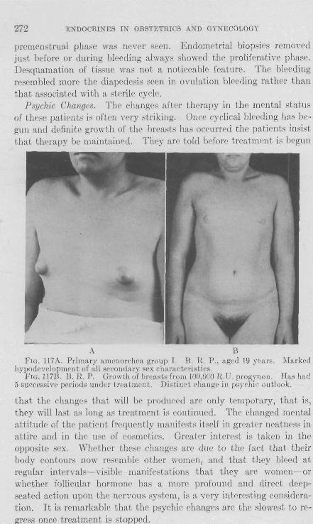
272 ENDOCRINES IN OBSTETRICS AND GYNECOLOGY
premenstrual phase was never seen. Endometrial biopsies removed just before or during bleeding always showrd thr proliferative phase. Desquamation of tislme was not a noticrable feature. The bleeding resembled more thr diapedesis sern in ovulation bleeding rather than that associatrd with a sterile cycle'.
Psychic Changes. The changefl aft!'r thrrapy in thE' mental !4tatm, of thrse patirnts is often very striking. Once cyclical blC'eding has bC'gun and definite growth of the breasts has occlIrrrd the paticntH illHiHt that thrrapy be maintain0d. Thry a1'r told bdore treatment is begun
:\. B FIG. 117A. Primary amenorrhea group 1. B. It. P., aged 19 years. i-Iarked
hypodevelopment of all s('condary sex characteristics. FIG. 117B. B. R. P. Growth of breasts from ]00,000 RU. progynon. Has had
5 successive periods undr)' treatment. Distinct change in psychic outlook.
that the changes that will be produ('ed arc only temporary, that is, they will last aH long as treatment it-) continued. The changed mental attitude of the patient frequently manifests itself in greater neatness in attire and in the UKe of cosmeticK. Grratcr interest is taken in the opposite sex. Whrther the'se changes arc due to the fact that their body contoUl'R now resemble othel' wOl1wn, and that they bleed at regular intervals- visible manifefltations that they are women- or whether follicular hormol1r has a more profound and direct deepseated action upon the nrl'VOUS sYfitem, ifi a very interesting consideration. It is remarkable that the psychic changes are the slowest to re-ress once treatment is stopped.

PRIMARY AMENORRHEA 273
Failure of the Ovary
In this group will be considered patients who show no growth disturbances, other than those due to an absence of ovarian function. These patients constitute the eunuchoid type. The growth disturbance manifests itself in:
1. Late union of the epiphyses. 2. Overgrowth of the long bones. 3. Slender build. The short and flat bones are not involved for they develop from
their osseous centers. In the hypogonad the epiphyses close much later than their normal time of union. This allows excessive growth of the long bones, providing of course that the function of the growth hormone of the anterior hypophysis remains normal. Because of this late closure of the epiphysis the span exceeds the height, and the lower measurement exceeds the' upper. The reverse is true of hyper·gonadism. In this syndrome the epiphyses close b('fore their normal tim(' of union. This results in a short individual whose height exceeds his span, and whose upper measurement exceeds the lower.
Just how the gonadal hormones influence epiphyseal closure is not clearly understood. The fact that castration in some animals leads to a delay in the epiphyseal union in the ends of the long bones was described by Sellheim (1899) and Tandler and Grosz (1910). Silberberg (1935, 1936) observed that acid extracts of anterior pituitary of cattle have a stimulating effect on the growth of cartilage and bone of growing guinea pigs. The various layers of cartilage cells become hypertrophic and hyperplastic, and are subsequently quickly calcified and replaced by bone. The epiphyseal line undergoes, thereforc, premature closure under the influence of the extract. Thyroidectomized guinea pigs respond in a Rimilar fashion to the extract.
The hypoplasia of the genitalia and the absence of secondary sex characteristics is common to both the pituitary and ovarian types of amenorrhea:
M. P. Age 20, No. 448759. First seen in April, 1935. Chief Complaint. Never menstruated, and absence of breasts. The patient is the second of four children, all girls. They are all taller than
the patient. The oldest is 24 years of age and 66 inches in height, the third is 18 and 68 inches in height, and the youngest is 11 years and 60 inches. The patient had the usual childhood diseases. She was fairly bright at school. The two older sisters are menstruating regularly having begun at 13 and 14 respectively. Her mother began to menstruate at 14.
Patient was delivered normally at full term. She thinks that she grew nor-

274 ENDOCRINES IN OBSTETRICS AND GYNECOLOGY
mally up to the age 01 thirteen, but since then her rate of growth was definitely less than her sisters! There are 110 menstrual molymina. Physical examinationthe patient is a healthy looking young woman, but appears younger than her actual age.
R eight: 57l inches. Span: 61 inches. Lower measurement: 31 inches. Head: negative. Neck: ncgative. Chest: mther heavy and seems foreshortened when compared to her legs. Heart and lungs negative. Blood pressure: 110/70. Pulse: 88. Skin: fine texture: soft. Hair: absent in pubic and axillary regions. ITair on head is thick, not coarse
(cunuchs are rarely bald). Breasts: slight elevation in nipple area. No glands palpable. Nipples and
areoli rudimentary. Abdomen: round, smooth. ~"
External genitalia: labia. very ,~budl and hypoplastic. Clitoris very small. Vagina: sound passcs in ,1 em. , Uterus and cervix: by rectal examination reveals a body about 2 em. in diam-
eter, movable, not tendCI'. Cervix e,wnot be distinguished from fundus. Adnexa: not felt. Neurologieal examination: negative. Basal metabolic rate: minus 13 and min,us 16 pcr ccnt. X-ray of the sella: a lateral film of the skull shows a rather small pituitary
lossa, with heavy posterior clinoids which bend forward at rather an acute angle. (Dr. Swenson.)
X-ray of long bones and pelvis: films of the wrists, shoulder girdles, elbows, ankles !Lnd knees show the epiphyses still un united in the wrists, !Lnkles, shoulder girdles and knecs, This shows definite retardation of development and corresponds to an epiphyseal development of approximately 17 to 18 years. (Dr. Swenson.)
Films of the pelvis show an extremely small, but very typical android type, with marked convergence of the side walls, very narrow subpubic angle with straight rami and narrow male typc of sacrosciatic notch, In fact, this pelvis reveals no female sex characteristics. (Dr. H. i'"roloy.)
Hormone studies: June 11, 1935; Follicular hormone-none. Jnne 12, 1935: Follicle stimulating hormone-negative.
No other significant findings. Wassermann: negative. Diagnosis. (1) Hypogonad. (2) Eunuch. Treatment and Results. Beginning with June 25 the patient has received a
total of 400,000 It. U. of progynon-B. It required 150,000 R.U. of progynon-B to bring on the first periud un September 20-31,1935; 70,000 RU. fur the second on November 5-9,1935; 50,000 RU. ful' the third, on December 5-7,1935; 20,000 R.U. for the fourth on Decemher 25-28. The last two periods were scantier than the first.
On December 30, 1935 thc following was noted-there is a definite growth of the
PRIMARY AMENORRHEA 275
breasts especially in the subareolar areas. Both areoli and nipples are pigmented. Pubic hair has appeared.
The cases in this group are similar to one another. They are either tall or short, but show at the ~ame time an overgrowth of the long bones. The short patients had the potentialities of becoming tall, but possibly an insufficient amount of growth hormone to accomplish this. It is noticeable that the cases responded in a similar f!lshioll, !lnd that the state of development re!lched was generally the same. Estrone did not Reem to hasten the closure of the epiphyses.
Failure of the Mullerian Duct System
The failure of development of the Miillerian duct system may be partial or complete. There may be a complete absence of the vagina, cervix and uterus, or there may be merely a marked hypoplasia of the genital tract. Various gradations between these two extremes may be found. The secondary sex r.hamclcristics are normal. We are therefore dealing with an inherent defect in the Miillerian duct system, in spite of the presence of estrone. For even if estrone is not demonstrable in the blood or urine, the presence of well developed secondary sex characteristics speak for its presence and action at some time during development. Why did not the Miillerian duct system respond? The reason is that it is not sufficient to have a stimulus to produce a certain reaction. The end organ acted upon must be responsive to the stimulus. This will be borne out. by one of the cases described below.
S. B. Age 18, No. 419042. First seen June 26,1934. Chief Complaint. Failure to menstruate. History not significant. Mother and sisters began to menstruate at about 13
years. Physical Examination. The patient is a well developed young girl with marked
scoliosis. Head:. negative. Neck: 'negative. Heart and lungs: negative. Blood pressure: 104/74. Pulse: 86. Skin: normal texture. Hair: normal distribution-no excess. Breasts: well developed, a little asymmetrical. Measurements: no disproportion. Abdomen: negative. External genitnlia, well developed. No hypertrophy of the clitoris. Vagina: cannot be felt. There is an indentation or depression below the
urethra measuring I em. in depth. Cervix and uterus: cannot be felt by rectal examination. A slight transverse
thickening may be felt high up, probably the top of the broad ligament ..

276 ENDOCRINES IN OBSTETRICS AND GYNECOLOGY
Adnexa: not felt. Neurological examination: negative. Psychological examination: The outlook of the patient is entirely feminine.
She "keeps company" and de"ires to marry. B.M.R.: plus 8 per cent. Hormone stndies on urine: Follicle stimnlating hormone-negative. Follicu
lar hormone-negative. Both studies rereated several times. X-ray of pelvis: April 29, 1935: Large anthropoid with gynecoid tendency.
Prognosis: Pelvis should offer no difficulty. If a transverse or posterior eng!Lgement occurs rotation should be ellsily brought about.
Diagnosis. Absence of MUllerian duct system. Treatment and Results. Between June 12,1934 and October 29,1935 the patient
received 580,000 R.U. of progynon-B, between January and September (l936) an additiulIILl600,OOO R.U. The results were as folluws.
1, The breasts showed no [Ldditionlll growth. 2. The external genitali[L showed no change. The indentation described above
did not grow deeper. 3. There was no definite evidence of any growth of the Miillerian duct system.
During the course of treatment a small mass about 3 cm. in diameter was occasionally felt. It did not change its size during the treatment, and from its pusition was evidently an ovary.
H. E. Age 25, No. 412358. First seen April 3, 1934. Chief Complaint. Never menstruated. Primary sterility. The patient is one of four children. One sister began to menstruate at 12 and
another at 14 years. Was seriously ill at one year, but the nature of the illness is unknown. Appendectomy at 14. Usual childhood diseases, without complications.
Patient has been married for 3t years. She comes to the clinic because she wishes to have children, but realizes that this is impossible (1) since she has never menstr.uated. No mellstruulmolymina. Leads a normal sex life and usually has orgasm. Has gained 22 pounds since IDlIrriage in spite of decreased food and increased exercise.
Physical Examination. The patient is a well developed white woman. Head: negative. Neck: negative. Heart and lungs: negative. Blood pressure: 122/84. Pulse: 78. Breasts: well developed. Nipplcs arc small. Hair: normal distribution. No hirsutism. Extremities: negative. No disproportion in length. Skin: normal texture. Abdomen: negative. Healed scar in right lower quadrant (appendectomy). External genitalia: fairly well developed. Pelvic floor: intact. Vagina: short and shallow, fornices about equal in depth and very shallow. Cervix: small, hypoplastic, "pin-point" os. Uterus: anterior, very small, movable, smuoth, fundus somewhat larger than
cervix. Adnexll: negative. Nenrologiclll examination: negative.

PRIMARY A~mNORRHF:A
Basal metabolic rate: minus 16 per cent. Follicle stimulating hormone (urine): slightly positive. Follicular hormone (urine): negative.
277
X-ray of pelvis: January 31, 1935. Lipiodol x-ray of no consequence. Repeated June 18,1936. A.P. stereos of the pelvis, with the uterine cannula in place and following injection of lipiodol show the lipiodol to be outlining the Fallopian tubes wbich do not suggest abnormality. The uterus is not filled for study. Tbe pelvimetry films taken later still show some Iipiodol in the distal ends of the tubes. Incidentally there are some asymmetrical facets at the lumbosacral junction.
N a other significant findings. Therapy and ResuU,s. The patient was given progynon-B in doses of 10,000
n.D. each, intramuscularly in the buttocks. In a<ldition t grain of thyroid extract, t. i. d.
A total of 1,300,000 RU. of progynon-B was given between April 24, 1934 and December, 193G.
On September 18, ]935 after an amenorrhea of two months the paticllt was reexamined with the question of possible pregnancy. The following are the findings:-
Vagina: very moist, slight cyanosis. Cervix: softened, slightly bluisb. Uterus: anterior, antiflexed, normal in si7,c, a little softened. Adnexa: negative. Diagnosis. Premenstrual uterus? Early pregnancy? An Asehheim-Zondek Test was negative on September 25, 1935 and another
endometrial biopsy II'as removed.
The results obtained in these two cases are of interest. (1) The secondary sex characteristics were not overstimulated in either 'patient. They had apparently reached their limits of growth. (2) The response of the genital tracts varied a great deal. Patient S. B. did not responp at all. To the best of our knowledge there was a complete failure of development of Mulleri:m ducts. The anlage may have been present, but its subsequent growth and differentiation were absent. It is probable that for the development of the Mullerian ducts a functioning embryonal ovary is necessary. If we assume that such a functioning ovary was absent during intrauterine life (or at least during the first half) but developed lat'er, then in the interim the Mullerian ducl might have lost its [lbility to respond to the gonadal hormones. They (the ducts) may have a different rate (or ability) of ['eoponse (early) in intrauterine life than later during extr;~terine exi~tence. We know that !:lecondary sex characteristics are capable of development later in life, providing an adequate supply of hormone is present. Furthermore, more estrone is required to stimulate growth of the uterus and vagina than the amount required to stimulate the gro~Lll of secondary sex characteristics.

FIG. llSA. Primary amenorrhea. Case H. E., aged 25. Endometrial biopsy on second day of first period. Followed the administration of 30,000 R.U. of progynon-B. Endometrium in general is postmenstrual in character. In several areas the glands show papillae and suggestive secretion. X75.
FIG. llSE. Primary amenorrhea. Case H. E., aged 25. Note the density of the stroma, and the pseudostratification of the glandular epithelium. X750.
278

PRIMARY AMENORRHEA
Hence the concept, that the failure of the growth of a uterus and vagina may be due to the possibility that the ovaries began their function after birth (or shortly before), acquires some validity.
Are foetal hormones similar to those of later life? The second case (H. E.) is also of considerable interest. Growth
of the uterus and vagina was secured by means of progynon-B. In fact, the growth was extensive enough to suggest the possibility of pregnancy. Cyclical bleeding occurred. The subsequent history of this patient brought out other interesting points. The patient stopped treatment for several months. When she was seen again the uterus had regressed from its maximum growth, to a size somewhat larger than that found at the beginning of treatment. If the patient had functioning ovaries, why then was the growth not maintained? When first seen by us the hormonal findings suggested ovarian hypofunction. It seems therefore that not only were we dealing with an inherent genital hypoplasia but also with an added (later) ovarian hypofunction. This brings up the question of a reciprocal stimulation between ovary and uterus.
As to the etiology of the genital hypoplasia of case H. E. the same explanation as that offered for case S. B. may be suggested. Ovarian function might have started a little later than normal, late enough for the Mullerian duct development to be so compromised as not to be able to approach normal growth and differentiation.
Prognosis of Cases of Primary Amenorrhea
The foregoing description of several typical cases suggests what can be accomplished with our present therapeutic armamentarium. Something has been achieved with these cases that was not possible a few years ago. We know that the results that we have obtained are reversible, that is, the patients revert to their original status of sexual development upon cessation of treatment. This may be unsatisfactory to some clinicians, but we feel that a beginning must be made. The results obtained point in the right direction. The next few years will see the production of more potent gonadotropic hormones, possibly in the form that will allow successful medication by mouth, or in such solvents as will permit few injections, but with prolonged and constant action. To-day we are in a position to inject 100,000 R.U. of estrin at a do"e. Ten years ago the combined laboratories in the world did not possess that amount of hormone.
In considering all our cases of primary amenorrhea as a whole, one is impressed by the following. It usually takes a large dose of estrone to

280 ENDOCRINES IN OBSTETRICS AND GYNECOLOGY
produce the first period and less as time goes on. Eventually a dose of 20,000 to 40,000 R.U. is reached that maintains fairly regular flow. nut as time progresses this maintenance dose becomes inadequate and morc and more hormone is necded to produce the desired effect. At the same time growth of all secondary sex characteristics is at first rapid but then slows up considerably. Eventually a stage is reached where oven very large doses of estrone produce only infrequent and very scanty periods. This demonstrates a resistance on the part of the patient to the medication. Is this resistance due to the development of an antibody against estrone or a modification of the molecule, or is it due to some process of inactivation?
BIBLIOGRAPHY
LOESEIl, A. 1932 Die Darstellung thyrcotrop \virksamer Extrakte aus Hypophysenvorderlappen. Arch. f. Exp. Path. u. Pharm. 166, f)93.
Sm,1,HEIM, H. 1899 Kastration und Ii:nochenwachstum. Beit. z. Geburtsh. u. Gyniik. 2, 236.
SILBERBERG, M. 1935 Effects of extracts of cattle anterior pituitary on endochondral ossification in thyroidectomized young guinea pigs. Proc. Soc. Exp. BioI. Med. 33, 554.
1935 Effects of extract of cattle anterior pituitary gland on endochondral ossification in young guinea pigs. Proc. Soc. Exp. BioI. l\Ied. 32, 1423.
SILBERBEIlG, 11'I. AND Sn,BERBERG, R. 193fi Influence of acid extmrt of cattle anterior pituitary gland on bone repair in young guinea pigs. Proc. Soc. Exp. BioI. l\Ied. 33, 177.
TANDLER, .J. AND GROSZ, S. 1913 Die biologischen Grundlagen der sekundaren Geschlcchtscharactcre. J. Springer, Berlin.

CHAPTER XX
SECONDARY AMENORRHEA
By secondary amenorrhea we mean the ces~ation of menstruation at any time after the menstrual rhythm has begun, up to the time of menopause. It is a very common gynecological symptom. Secondary amenorrhea is not a disease, but usui111y implies i1 dysfunction on the Pi1rt of the orgi1nism. The etiologici1l factors arc numcrous, and one occasioni111y sees cases in which an etiological factor cannot be determined. :Many syndromes are associated under this heading, the reason being that the cases present themselves not as syndromes hut as patients with the significant symptom, secondary amenorrhea.
Secondary amenorrhea may be divided into two types in so far as etiology is concerneci, namely:
A. Physiological amenorrhea. B. Pathologiral amenorrhea.
Physiological Amenorrhea
Physiological amenorrhea occurs during: 1. Pregnancy. 2. Lactation. Pregnancy. While by far the greatest number of patients show no
evidence of menstruation during pregnancy there are somc women who bleed to a greater or lesser extent at the expected menstrual time. Thi~ occurs only during the first four months, that is, up to the time of fusion of the decidua capsularis and parietalis. On the other hand there are patients who present their usual menstrual~olymina throughout pregnancy without any evidence of bleeding. The bleeding, when it occurs, is truly menstrual in character, for, according to our definition it occurs from a secretory endometrium. Actual desquamation of tissue is rare, the process seems to be more of a diapedesis.
What is the etiology of the amenorrhea of pregnancy? It is not due to the foetus,Jor the lImenorrhea persists after the death of the foetus. On the contrary, in ectopic gestation, the bleeding is often initiated by the death of the embryo. Its dCi1th mi1y be due to destruction of the greater part of its chorionic epithelium. The ovarian factors necessary for menstruation are present, for both follicular and corpus luteum hor-
281

282 ENDOCHINES IN OnSTETHICS A,.'1D GYNECOLOGY
mones are available. The reason for the huge estrin excretion during pregnancy is not as yet clear. Progesterone is prescn t in the corpu~ luteum verum and in the placenta (Ehrhardt and Fischer-W assels, 1936). Wilson and Kurzrok (1936) suggest that the pituitary like hormones (prolan A and B) produced during pregnancy by the placenta, inhibit the production of the bleeding factor. As to the origin of the bleeding factor Smith (Smith, Tyndale and Engle, H136) dcmonstrate~ that the pituitary is not essential for bleeding in the :Vlacacus Rhesus Monkey. Any hemorrhage is of potential danger to the foetus, hence some mechanism for the prevention of its occurrence is expected. The problem has not been settled. It is conceded by lTlany that the follicles do show some growth during pregnancy but they evidently undergo atresia. A follicle may ripen and ovulate but it must be a relatively rare occurrence. Injection of prolan A and B does not induc~ ovulation (Hamblen and Ross, 1936).
Lactation. It is well known that many women do not menstruate during lactation. Others menstruate throughout lactation, while still others do not menstruate for the first few months but do so during the latter. Furthermore, some women conceive during the lactation amenorrhea, while others, equally fertile, do not. This implies that ovulation occurs in some but not in others. What then is the status of anterior pituitary and ovarian function, ho~mone production, and the endometrial cycle during lactation? Kurzrok, Lass and Smelser (1[}37) studied a group of lactating women who were having regular cycles. They observed that between 55 and 70 per cent of the wumen showed anovulatory cycles. The bleeding, Lherefore, was HoL true menstruation [or it did not come from a premenstrual endometrium. Several cases stopped having cycles during this period of observation. There was no evidence of cyclical endometrial change during the period of amenorrhea. Hormonal studies are now being carried out on this group.
Pathological Amenorrhea
Amenorrhea is an important symptom. While it is true that it occasionally implies merely a cessation of bleeding, it more often implies a hypofunction of the gonads. Hypofunction of the ovaries leads to secondary atrophic changes in the genital tract. The utems, cervix and v!1gina must constantly be stimulated, otherwise their morphological and functional status regresses. It is well known to gynecologists of experience that during the period of amenorrhea patients cumplain of a host of symptoms which are relieved by the menstrual flow. It

SECONDARY AMENORRHEA 283
might be argued that the etiological factor causing the amenorrhea is also responsible for the other symptoms. This is often true, but not always. A flow may be established without influencing the etiological factor, and the patient experiences considerable relief. There may be more to menstruation than the mere desquamation of a pregestational endometrium. The idea of a menstrual "toxin" comes back frequently to plague the conscience of the gynecologist (Schick, 1920 ~ Macht and Lubin, 1924; IVIommsen, 1934). The importance of amenorrhea in sterility is not to he underestimated.
Pathological amenorrhea is due to numerous caus,es. Of these the following are the most important.
Disturbances of the Genital System. 1. Tho uterus-genital !lypoplasia. 2. The ovaries. Genital hypoplasw-underdevelopment of the genital tract is one of
the commonest causes of amenorrhea. As stated previously, hypofunction of the ovaries may lead to secondary atrophy of the genital tract but the cases under consideration at this point show no evidence of ovarian failure. In fact, the hypoplasia is present in spite of normal ovarian function (as evidenced by hormone titration).
Genital hypoplasia may be part of a general or universal infantilism (Aschner, 1924), or a partial infantilism whieh is limited to the genital tract. Universal infantilism forms the commonest and most important constitutional anomaly in women. It may be defined as a condition in which thr, complete development of the whole organism (or its component organ systems), insofar liS morphology and function are concerned, does not occur at all, or occurs very late. The syndrome implies imc
maturity, both somatic and psychic. Of particular interest to us is the problem of local genital infantilism or hypoplasia. Genital hypoplasia may be internal or external or both. A hypoplasia involving the external genitalia alone is rare. Usually it is associated with an internal hypoplasia and forms part of a general infantilism. 011 the contrary internal genital hypoplasia is fairly common and in the discussion below this is the form under consideration.
Physical Szgns of Genital Hypoplasia. External Genitalia: Labia majora-are small, and sparsely covered by hair,
Labia minora~smalC occasionally just barely indicated. Perineum-short, often with an indentation just above the anus.
Occasionally 11 median fold crosses the perineum from the fourchette to the anterior anal wall, representing the fusion of each lateral anlage.

284 ENDOCRINES IN OBSTETRICS AND GYNECOLOGY
Vagina-either short and shallow, or narrow, conical and long. Occasionally prominent transverse ridges may be felt, resembling the condition found in the foetal vagina. The fornices are poorly developed, are shallow, and are usually of equal depth. Occasionally there may be felt in the anterior fornix a curved linear thickening in the vaginal wall, extending forward froIll the cervix. Saenger described it as the ,"crista cervicalis mediana," and considers it as a vestige of the fused Mullerian ducts.
Cervix-small and Illay point in any direction. The os externum is usually "pinpoint." Occasionally the cervix has a lobulated appearance with a ridge-like prominence along the lowest circumference.
Uterus-is most commonly involved, and may show variations in size, length, form and posiiiO;1. The total length is shortened to a greater or lesser extent. Thr 6crvix may be as long as, or longer than the fundus. Thus there a,r'p It{vO typcs of hypoplastic uteri. One, in which the foctal form is I}\aintained, that is, the cervix is longer than the fundus. Such a utefus/may grow to normal length, but there remains always this inhibition of differentiation. The second type presents the adult differentiation, but is smaller in size than the normal uterus. The latter type occurs more frequently. There is either acute retroflexion or antiflexioll, with accompanying acute angulation of thc canal. The reason for the acute flexions is not clear. It has been suggested that the acute antiflexion is due to short utero-sacral ligaments and their insertion higher up on the fundus. On the other hand, the ligaments may be short because the uterus lies well back. There may be lateral flexions of the uterus to the right or left due to shortness of the broad ligament on the same side. The narrowing at the internal os may be due to the acute angulation or to excessively prominent folds of the arbor vitae, the latter a persistent foetal condition.
The ovaries-they may fail to descend into the pelvis and therefore remain situated above the linea tcrminalis. The pull of the abnormally situated ovarian ligaments may be sufficient to overcome the pull of the round ligaments and thus result in a retroversion or fiexion. The ovary proper lllay be small, its surface smooth due to inadequate follicle growth.
Tubes-may be tortuous. This is due to the persistence of a foetal condition, for in descending from their original position along the lumbar spine to the pelvis a certain amount of spiral twisting occurs. Normally when reaching the pelvis the tubes lose their spiral twist (Mayer, 1927).

SECONDARY AMENORRHEA 285
Etiology. The damage that eventually gives rise to genital hypo-plasia (as well as general infantilism) may occur in:
1. The germ plasm. 2. The foetus. 3. The child. 4. The adult. It is very difficult and frequently impossible to state when the dam
age occurred. It is well known that several cases may occur in one family. We have at present under our care two sisters with an identical genital hypoplasia, one suffers from amenorrhea and the other from hypermenorrhea. Aschner points out that conditions which injure germ plasm such as chronic infections (lues, tuberculosis) and intoxications (morphine, alcohol) may cause infantilism, either general, or involving one organ system. He also believes that advanced age of the parents has an unfavorable effect on the germ plasm. Similarly, malnutrition and disease may exercise the same influence. The ultimate effect of the x-rayon the germ plasm is still under discussion. In addition, Aschner adds prematurity. He points out that the mother with genital hypoplasia tends to premature delivery. She may thus transmit to the child infantilism as a dominant characteristic.
The development of the uterus during foetal life is a progressive condition. The factors that enter into play at this time arc fourfold, namely:
A. The genetic facLor of sex. B. The MUllerian duct system. C. The ovaries. D. The anterior pituitary gland. The sex of the individual is determined at the moment of fertiliza
tion. This also predetermines the character..-of the gonad. Thus we have the sex of the individual determined by genetic and hormonal factors. The MUllerian ducts develop very early in embryonic life. Their subsequ'ent growth and development depend upon the genetic impulse, and the ovarian hormones, which in turn depend upon a hormonal drive from the anteri.or pituitary gland. An inadequacy of any of these factors would lead 'to a retardation in the rate of growth of the Mullerian ducts. Thus the cl.efect might be corrected to a greater or Ic~ser extent in later life, or the defect may be permanent. The greatest impelus to growth comes during intra-uterinc life, either from more potent growth promoting factors or from a greater response of the growing tissues.
During childhood the genital canal is stimulated to further progressive growth by gonadotropic and ovarian hormones (leaving out for the

286 ENDOCRINES IN OBSTETIUCS AND GYNECOLOGY
moment the other hormones). Should the stimulus be insufficient or the genital tract non-responsive, then genital hypoplasia will result.
Furthermore, in adult life conditions causing prolonged secondary amenorrhea may lead to resultant uterine atrophy. The atrophy may lead to a small uterus, but other distinguishing features of a true genital hypoplasia are usually absent. A normal uterus undergoing secondary atrophy retains its adult proportions. It must be remembered (v. i.) that genital hypoplasia, per se, may lead to secondary amenorrhea.
Symptoms. The effects of genital hypoplasia may manifest them-selves in several ways,. namely, in;
1. Disorders of menstruation. 2. Sterility. 3. Repeated misearriag~}: 4. Difficulties of labor./ j 5. Menopause prCQox. <J
Before diseus~ing these topics it is advisable to call att~ion to the hormone findings in genital hypoplasia. Kurzrok and Ratner (1932) pointed out that many case~ show a normal estrone excretion in the urine. Other cases, fewer in number, show an absence of estrin on repeated examinations. The follicle stimulating hormone shows no abnormal variation, in that, with the original Zondek technique none is found in a morning specimen of urine. The above findings. have since been confirmed by ·us many times.
ivfenst!'uation. Genital hypoplasia may be associated with all abnormalities of menstruation. Puberty may be retarded. If the defect is ovarian in origin then the secondary sex characteristics will develop latc. The menses, having begun, may continue with greater or lesser regularity, and thcn gradually become more infrequent and scanty and finally ceasc altogether. This may occur in the twenties or curly thirties. When the periods are regular the hormonal findings may offer a due to the etiology of the hypoplasia in any parLicular case. The hormonal findings lIlay be normal. This implies a normal production of estrone on the part of the ovaries. Why then the genital hypoplasia? The conclusion seems justified that even with a normal hormonal supply the Mullerian duct system failed to respond. .An inherent defect in the gellital tract seems to be basically responsible for the condition. Again, the hormonal findings may show an insufficiency of estrin. This is most frequently seen in women who present the various types of pituitary adiposity. The associated adiposity, genital hypoplasia and' low estrin values, 8peak for a pituitary hypofunction.

SECONDARY AMENORRHEA 287
Thr. men~t,rual flow may Ahow ahnormalities ranging from hypoto hypermenorrhea. The same patient may have scanty menstruation at one time and profuse at another. This is understandable when one considers that the hypoplasia involves the myometrium, endometrium, vascular supply and probably the local nerve mechanism. Whether patients with genital hypoplasia have a greater number of anovulatory cycles than normal women is not known. A compromised myometrium may exhibit inadequate contractions for the control of menstrual bleeding.
Dysmenorrhea is very common in this syndrome. It may be due to some defect in uterine innervation. A hypoplastic uterus may also exhibit irregular uterine contractions. We feel that it is the irregularity of the contractions more than their intensity and tempo that predisposes to dysmenorrhea. It has becn our experience that the administration of estrin in large doses, especially during the first half of the cycle leads to relief of this type of dysmenorrhea. Apparently growth of the uterus Jllay~ a part ill determining the type of contractions. The acute angull1tion present in such utcri lllay offer an obstruction to blood and tissue fragments coming from the fundus. As growth of the uterus progresses the angulations become less marked.
Genital hypoplasia is olle of the commoncst inherent causes of sterility in women. There are several possibilities as to the mechanism. The anovulatory cycle may be a factor. The endometrium may be insufficiently developed to harbor a fertilized ovum. The secretions from a hypoplastic endometrium may be inimical to both sperm and fertilized ovum. Considering the importance of this condition (hypoplasia) there is woefully little exact work on the aspect that touches upon sterility.
A further difficulty is encountered cjyring pregnancy. Repeated early miscarriage .is not uncommon. Whether this is duc to the inability of the uterus to expand proportionately with the growth of the foetus, or to irregular uterine contractions, or to changes within the foctull as the result of inadequate blood supply il:i not known at present. .Mayer JJoints out that women with genital hyp'opla~ia are apt to suffer from considerable abdominal discomfort throughout pregnancy due to the iiiadequate expansion of an infantile uterus. Premature labor often occurs. When pregnancy does go to term the pains are inadequate and the labor long. Postpartum hemorrhage Illay occur.
Premature menopause is another important consequence. Why should the ovary cease to function earlier than usual? For we have

288 ENDOCRINES IN OBSTETRICS AKD GYNECOT,OGY
not only cessation of bleeding but also cessation of ovarian function. The latter is evidenced by the hormonal findings (Chapter XXIII). Two intriguing explanations present themselves. First, the ovary has a limited life and is capable of producing just so much hormone. That amount is expended uJlon a hypoplastic genital tract, which requires for its function a greater hormonal drive than the normal uterus. Second, the uterus exerts some stimulating influence upon the, ovaries. The removal of the uterus (no matter how el1rcful one is to preserve the blood supply) leads to cessation of ovarian function, mueh sooner than otherwise. An infantile uterus when it ceases to respond to ovarian hormones may also lose its stimul;tting effect upon the ovaries. The concept thl1t the stimulus, ovary to uterus, also functions in the opposite directioIl, has much in its favor, though experimental prouf is lacking.
Aschner states that the hypoplastic genital tract has a tendency to favor ectopic gestation, due to increased tortuosity of the tubes. Furthermore, pelvic infections, especially tubercular, run a more stormy and protracted course.
Treatment. Two broad methods' of treatment are potentially available, namely, to stimulate genital growth by means of gonadotropic hormones, or by means of ovarian hormones. The exhibition of gonadotropic hormones should be the method of choice. But large doses of these hormones, especially the follicle stimulating hormone, are not as yet available therapeutically. Pregnancy urine extracts containing prolan A and B do not stimulate the follicular apparatus to any extent (Hamblen and Ross, 1936). The gonadotropic effect of pregnant mares' serum on the human ovary is very marked but the use uf this material has nut as yet entered the clinical phase.
The substances available for therapy are thcrefore estrone and progesterone. Up to the present time the high cost of the latter and its scarcity have prohibited its clinical use. The main therapeutic agent· has been estrone in one of its commercial forms. We have used amniotin or theelin when smaller dosage was required and progynon-B when large dosage was essential. It might be inquired, why not use very large doses of estrone at all times, and thus secure the greatest therapeutic effect. Our results in a large series of cases of genital hypoplasia have shown us that while large dosage of progynon-B frequently brought about an immediate result, specifically where bleeding was desired, its persistent use in the more moderate cases was not necessary, and occasionally after some time, detrimental. We occasionally noted moderate but persistent headache as the result of continuous injections of progy-

SECONDARY AMENORRHEA 289
non-B. The dosage necessary to produce headache was 20,000 R.U. per week for six to eight weeks. Not all patients showed this, and what is very striking, the patients with primary amenorrhea showed it very rarely, or not at all. The headache usually cleared up within four or five days after the hormone was discontinued. The cause of the headache was probably the enlargement of the anterior hypophysis which results from the administration of large doses of estrin (Halpern and D' Amour, 1936; Kurzrok and Kirkman, unpublished results).
There is an additional theoretical objection to the use of such large dosage here. It has been demon.strated experimentally that large doses of estrin depress the production of the follicle stimulating hormone. This in turn would be followed by the ripening of fewer follicles and secondarily by diminished estrone production. Such therapy would therefore defeat our purpose, which is to exhibit estrone, and also to maintain ovarian function.
The age of the patient with genital hypoplasia 'is of considerable importance in the response to therapy. All other factors being equal, patients under twenty-five respond best, while those above thirty-five respond least. As a general rule it may be staLed that the larger the uterus at the onset of treatment the greater will be the response to therapy. At the same time there arc considerable individual variations. The first response to estrone treatment is an increased vascularity of the genital tract, This manifests itself in a distinct leucorrheal discharge, cyanosis of the vagina and cervix, and softening of the uterus. The entire picture resembles an early pregnancy, except that the total size of the uterus is smaller. Whcn a pregnancy does occur the uterus lags behind in growth when compared· with the normal f
uterus. Our impression is that the:y tend to approximate in size at about the fourth month. The enlargement rille: to incrcased vascularity is t,emporary, for when treatment is discontinucd the original size pertains.
When' actual growth occurs the result may be due to hypertrophy or hyperplasia, or to both. In mild cases hypertrophy of tissue alone would seem to be adequate, Where growth it) extensive hyperplasia and hypertrophy must occur. 'Ye have seen patients whose vagina was about 1 cm. in ·diameter-before treatment, but at the end admitted a medium speculum with ease, In such cases hypertrophy alone seems inadequate.
An extremely important question is: How permanent are the results? We are unable to judge this from ou·r present-day standpoint. Our experience is still too limited, for the treatment in vogue at the present

290 ENDOCRINES IN OBSTETRICS AN"D GYNECOLOGY
has not yet weathered the test of time. Our oldest cases do not e!{ceed five years. It is our impression that the genital hypoplasia due to pituitary (hence ovarian) insufficiency (that is, when associated with inadequate development of the secondary sex characteristics and absence of follicular hormone from the urine on repeated examinations) reverts back to the original hypoplastic condition, sooner or later, ",hen therapy is di~contin\led. On the contrary, when the hypoplasiu, is due to an inherent defect in the MUllerian duct system, associated with normal ovariu,n function at the time when the patient is first seen, then the growth which results from therapy is maintained.
The Ovary (and the Anterior Hypophysis). In view of the intimate relationship between the ovary and the anterior pituitary glaJid the/'"' discussion must include both glands. As stated previously the ovary will not function alone but is stimulated to do so by the anterior hypophysis. Hence the demonstration of one or the other of the ovarian hormones implies pituitary function.
Four hormones (at least) are involved in 'the process of menstruation, namely two ovarian, estrone and progesterone, and two gonadotropic-the follicle stimulating (F.S.H.) and luteinizing hormones (L.H.). Only estrone and F.S.H. have been demonstrated in the blood or urine in the non-pregnant state. The remaining hormones have not been found, becu,use our technique is not sufficiently fine enough to demonstrate them in minllte quantities. We will therefore consider the pituitary-ovarian relationship to secondary amenorrhea from the viewpoint of the two demonstrable hormones.
Considering, therefore, estrone u,nd F.S.H. alone, four types of excretion are present (Kurzrok, 1932), namely:
I-F.S.H. absent II-F.S.II. ",bsent
III-F.S.H. present IV-F.S.H. present
Estl'one-present Estrone-absent Estrone-absent Estrone-present
With Kurzrok and Ratner's (1932) method for estrone determination the hormone has practically always been demonstrated in the urine of normally menstruating women. F.S.H. as determined by Zondek's method is usually absent except just preceding ovulation (Kurzrok, Kirkman and Creelman, 1935). Hence the normal (F.S.H.) is always a negative test, and the presence of too much F.S.H. is abnormal. Albright, Halsted and Cloney (193.5) are in general agreement with the above findings.
Type III Excretion. E8lrone Negative; F.S.11. Positive. Given a

SECONDARY AMENORRHEA 291
case of secondary amenorrhea in which the homone excretion on repeated tests (at least two, taken a week apart) is of Type III (F.S.H. present, estrin absent) the diagnosis of cessation of ovarian function may he made. It signifies menopause (whether spontaneous, surgical or roentgen irradiation). It might he added in passing that during temporary x-ray castration the same hormonal excretion occurs, but as the effect of the rays wears off the F.S.H. becomes negative and the e~trin positive. In other words type A excretion is not always an irreversible reaction. The impor.tance of repeated hormone examination IIlust be stressed. Ovulation IIlay occur during a period of amenorrhea. Ovulation during a normal cycle would usually be represented by F.S.H. positive and estrin positive, but during amenorrhea may be represented by type III excretion. A repetition of the test one week later will determine the exact state of events. For if reaction Type III recurs then we arc dealing with a cessation of ovarian function. The sudden appearance of F.S.H. in the urine just preceding ovulation is "explosive" in character and disappears within twenty-four hours.
Case. M. D., age 20, No. 499748. (Fig. 119.) Chief Complaint. (I) Amenorl"hea-18 months. (2) Headaches, di7.7.incss and
backache-IS months. (3) Hot flushes 2 to 3 per day-IS months. (4) Gain of 1110 pounds in past two years.
Present Illness. Eighteen months OlgO without any preceding menstrual irregularity ,here was sudden onset uf amenorrhea. There Ims been no vaginal bleeding since that time. Gradual onset of headaches, dizziness and backache. Hot flushes began gradually and have increased in frequency. There has been a steady, gradual increase in weight amounting to 100 pounds. No increase in appetite or thirst. Slight increll,Se in urinary frequenry. Chronic constipation and piles. Dyspnea and palpitation on two flights since gain in weight. Has' been suffering increased "nervousness" irritability and depression for the past year.
:Menses: onset 11 years; 28 day typo; 8 days in dlJn1tion; moderate in amount and without pain.
L. M. P. {May 9, 1935. April 9. 1935.
Marital: married three YOILI·S. No children. Was oyerdue a week on two or three occasions. Took castor oil and quinine and brought period around.
Operation: Appendectomy in 1930, followed by "rectal" abscess. Sign·;ficant Physical Findin.qs. The patient is a very obese young married
_ woman of twenty· year"' ,,,hoBe fat distribution is mainly ;bout the pelvic girdle. Breasts are very pendulous and large with small nipples-gland being composed mainly of adipose tissue. Hands small, ankles rather moderate. Skin of normal texture. No abnormal distribution of hair. Abdominal stria present.
Abdomen: obese. No masses or viscera palpable. Deep suprapubic scar. No hernia.

292 ENDOCRINES IN OBSTETRICS AND GYNECOLOGY
Pelvic: External genitalia: negative, no hypertrophy of clitoris. Pelvic floor: intact. Cervix: nulliparous, small. Vagina: short, shallow, narrow. Uterus: small, anterior, movable, firm. Adnexa: not felt.
FIG. 119. Premature menopause at 19 years of age. Note trunk adiposity
Diagnosis. (1) Gcnital hypoplasia. (2) Frohlich' s syndrome- secondary. (3) Menopause precox.
Endometrial Biopsy. Endometrium very thin. Insufficient quantity removed for exact diagnosis.
B.M.R.: plus 6 per cent . Weight 105.7 kgm. Blood count: Hemoglobin: 90 per cent. R.B .C.: 4,630,000. W.B.C.: 8,600.
Polymorphonuclears: 52 per cent. X-ray of skull (Dr. Friedman): A lateral film of the skull shows no definite
evidence of enlargement of the sella turcica. There is no evidence of erosion or atrophy suggestive of intracellar tumor.
Hormone determinations- urine: estrone and follicle stimulating hormone.

SECONDARY AMENORRHEA
October 22 1936 fEstrone-ne~~tive , IF.S.H.- posltlve
October 26, 1936 {Estrone-ne~~tive F.S.H.- posltlve
November 4, 1936 {Estrone-ne~~tivel F.S.H.- posrtrve
Visual fields: normal.
293
Reaction III
Discussion. The amenorrhea, flushes, sweats and irritability are very suggestive of premature menopause. This diagnosis is confirmed by the repeated finding of large quantities of follicle stimulating hormone and an absence of estrone in the urine. It seems reasonable to believe that the primary defect resides within the ovaries. They "aged" prematurely. They can no longer respond to the gonadotropic hormones present in the system. Considering the genital hypoplasia it is doubtful whether their function was ever complete. The adiposity might have resulted from a pituitary (hypothalamic?) dysfunction secondary to the loss of estrone. In view of the extensive genital hypoplasia it is rather doubtful whether the patient was ever pregnant. The. prognosis is uncertain. as to whether ovarian function will ever return. The youth of the patient is slightly in her favor. Under similar circumstances, we have not seen a return of function when the
·patient has passed thirty. As to therapy we believe that large doses of estrone are contraindicated, for fear of further depressing the function of the anterior pituitary gland. An ovarian transplant from a young woman will be attempted.
Type IV Excretion. F.S. H. Present; Estrone Positive. This is the usual finding just preceding ovulation. It implies the pituitary stimulus and the ovarian response. It is an unusual finding during secondary amenorrhea though it is occasionally found at the beginning of menopause. The finding may signify a certain imbalance between the anterior hypophysis and the ovary. Furthermore, Type IV excretion, may be the first biological evidence of pregnancy. It may precede a positive Aschheim-Zondek Test (Kurzrok, Kirkman and Creelman, 1935). A rep~tition of the test a few days later would establish the correct diagnosis.
Type I Excretion. F.S. Jl. Absent; Estrone Present. This is the normal finding. It may also be found during secondary amenorrhea, and denotes both hypophyseal and ovarian function. Type I excretion, amenorrhea and genital hypoplasia are frequently found together.

294 ENDOCRINES IN OBSTETnICS AND GYNECOLOGY
That is, when the genital hypoplasia is accompanied by normal secondary sex characteristics. When the uterus is normal in size and there are no other significant physical findings then the question arises whether the amenorrhea in the case at point is not due to an absence of the much debated bleeding factor. It might be argued that possibly the corpus luteum phase of the cycle is incomplete, hence the patient does not menstruate. This is true when menstruation alone is considered, but cyclical bleeding may OCCllr when the estrin phase alone is present. An endometrial biopsy taken just before the expected period will immediately orient us as to the presence of a pregestational phase.
Type II Excretion (F.S. H. Absent; Estrin Absent). This is only infrequently found in the normal patient, especially on repeated examination. It is usually found associated witll secondary amenorrhea, hypo- or oligomenorrhea. It implies, in the main, pituitary hypofunc" tioIl in as far as gonadotropic hormone is concerned. For if the anterior, \ hypophysis were functioning and the ovary w,as not responsive, then we would expect to find F.S.H. in the urine.
Disturbances of the Endocrine System
The Pituitary Gland. One of the commonest conditions associated with secondary amenorrhea is obesity. One frequently encounters discussions as to which symptom leads to which. In all probability both arc due to a common endocrine disturbance, most often in the anterior pituitary gland, less oftr.n in the thyroid, and very rarely primarily in the other glands of internal secretion.
In order to understand the nature of the metabolic processes involved in obesity ami its relation to the pituitary and ovary, a short resume of lipoid metabolism will be given. For a more comprehensive survey the reader is referred to the work of Peters and Van Slyke (11l31). By lipoids we mean substances in the organism which resemble fats in· their chemical and physical properties. The lipoids may be subdivided. into the following three classes.
a. True fats, or triglycerides of fatty acids. b. Lipins, substances of fat-like nature which (on hydrolysis) yield
fatty acids or their derivatives, and in addition contain either nitrogen or phosphorus, or both. .
c. Sterols, complex chemical substances which do not resemble fats chemically, but whose importance is constantly increasing. The gonadal hormones arc stcrolH.
Fats arc triglyccrides of fatty acids, that is, esters of fatty acids and
SECONDARY· AMENORRHEA 295
glycerine. Palmitic, stearic and oleic acids are the commonest in the animal organism. The latter is unsaturated, and all contain an even number of carbon atoms. The fat in the body resembles the ingested fat, but when an animal is fcd a fat-free diet, it develops a hody fat of constant composition, which is characteristic for that animal. It is known as "physiological fat." Both types of fat arc utilized by the animal organism interchangeably. Distinction has ~lso been made between depot (storage) and tissue fat, the former implying reserve fuel and the latter is an integral part of the tissue protuplasm. Tissue fats are probably phospholipins. The fat depots are the subcutaneous tissues, mammary glands, perirenal tissues, retroperitoneal tissues, etc. The size of the fat depots depends upon hereditary, sexual, metabolic, endocrine and dietary factors.
The ingested fat is digested in the intestine by the lipase of the pancreatic juice and the succus enteric us into fatty acids and glycerine. This reaction is assisted by the bile which aids the emulsification of fat and promotes the action of the lipase. The fatty acids are converted into soaps by the alkali of the intestines and are absorbed as soaps. They are then reconverted into fatty acids (possibly by the aid of the phosphatides), absorbed by the lymphatics, reach the blood stream and then the lymphatic duct. The systemic blood fat begins to rise in from one to three houm after a fat meal, and the lipemia reaches its peak in six to seven hours, returning then to its original leveL In all probability the free fat enters the fat cells of the depots withuut undergoing any change. -In the process of catabolism the first step may be desaturation of the fatty acids in the liver. The next step is oxidation which takes place at the f3 carbon atom, so that two carbon atoms afe split off at a time. This reaction yields a fatty acid with two carbon atoms less, plus acetic acid. The process )!ontinues until a 4 carbon acid is produced, butyric acid, which in turn is oxidized to acetoacetic acid. The latter substance is a ketone and can only be oxidized in conjuriction with carbohydrate. If we begin with a fatty acid with an odd number \)f carbon atoms ketone bodies cannot be produced, for a 4 carbon acid does not result.
The excessive accumulation of fat is known as obr,sity. There are differences in the pattern of fat distribution ill both sexes. Castration and hypopituitarism in the male cause approximation to the female type. ALlip0i:iity lIlay be geuer~lizeLi ur lucalizeLi tu particular areas. Sudden increases of weight may occur in adolescence, after pregnancy, and at the menopause.

296 J<:NDOCUlNES IN OBSTETRICS AND GYNECOLOGY
Are there any differences in metabolism that differentiate the obese from the normal or the lean? To date no such differences have been found. It mllst be true that the obese ingest more food than their immediate needs, but many obese people arc not heavy caters. Nor is it a matter of muscular activity, for many stout people lead very active lives, and at the same time it is also true that a stout persqn will expend more energy than a thin one. Apparently the metabolic processes are more economical in a fat per80n than in a thin one. The basal metabolic rate is usually within llormal limits. Hagedorn, Holtoll 'and Johansen (1927) found that if obese persons were fed a high carbohydrate diet for several days and then the basal respiratory quotient taken, it was lower than that of normal people under similar condition~. They interpreted this to raean that the obese people converted their carbohydrate to fat and then burned the latter. Gardner-Hill, Smith and Jones (1925) showed that adolescents presenting pituitary adiposity and increased sugar tolerance, exhibited less than the normal rise of respiratory quotient after sugar. Adrenalin produces a rise in the basal metabolic rate and in the respiratory quotient, the elevation remaining as long as the effect of the drug persists. The ·reasons for this are probably multiple. Kranz and, Means (1927) found a diminution in the respiratory quotient in thr. obese following adrenalin, and believe that more fat than carbohydrate was utilized.
The relationship of the ovary to Lhe oxidation of ketogenic material has recently been- studied by Butts and Deuel (l933) and Butts, Cutler and Deuel (1934). They showed that when the sodium salt of acetoacetic acid (ketogenic material) was administered to fasting rats large amounts of acetone bodies were excreted in the urine (ketonuria) due to failure of complete oxidation. The surprising finding was that the urine of the females contained twice as much unoxidized material as the male. The same holds true for the human. The administration of carbohydrates results in the oxidation of these ketogenic substances, hence the conclusion is drawn that there exists a fundamental difference in carbohydrate metabolism between the sexes (since carbohydrates are required to oxidize these ketone bodies). Furthermore, when the ovaries are removed, the oxidation of the diacctic aeid is increased, so that the ketonuria is about one fourth that found in normal females and about one half that obtained with normal male rats. The injection of theelin does not significantly increase the ketone body excretion in either the castrated males or females. It wuuld seem that the ovary contains some other substance (or hormune) which influences fat me-

SECONDARY AMENORRHEA 297
tabolism and produces a ketonuria. This brings up the questionwhether we are through with the ovary when both estrone and progesterone are removed.
We have previously mentioned that extracts of the anterior lobe of the hypophysis contain a ketogenic principle, and thereby produce a ketosis. Acetone bodies are produced whenever there is a disturbance in the relative proportions of fat and carbohydrate that are being coincidentally oxidized in the tissue. If there is a decrease in thc available carbohydrate, oxidation of fat is correspondingly accelerated, the acetone bodies arising in consequence of an ihcompletc oxidation of the latter. NIirsky (1936) studied the source of these acetone bodies in the liver and found that the ketogenic principle acts directly on the liver. lIe points out that only glycogenic substances are antiketogenic and that a low liver glycogen is found in all conditions which are associated with ketonuria. Deuel (1934) noted that the ketogenic. principle caused a diminution of liver glycogen. Mirsky concludes that the deprivation of glycogen in the liver cell and the associated increased catabolism of fat leads to the formation of ketone bodies.
There are abundant clinical and pathological data which permit us to assume that the diencephalon also contains a center which exerts an important influence on the deposition, distribution and consumption of fat, especially in the subcutaneous tissue. Fulton and Bailey (1929) observed that in the "hypothalamic syndrome" produced by vcry early tumors arising in the infundibulum of the hypothalamus adiposity is a characteristic symptom. Smith (1()27) domonstrated that injury to the infundibulum of rats produced ad.iposity which was not repaired. by I
pituitary implants. Furthermore, local nerve lesions may be a basis for the "partial" or "local obesity" that one frequently encounters. lVIansfeld. and Muller (1913) observed that section of one sciatic nerve in an overfed dog produced a greater accumulation of fat in the corresponding limb. A somewhat similar finding is reported by Lostat and Vitry (1909).
Dislrophia-A.diposo-Genitalis. This condition was first described by Frohlich (1901). It may be primary or secondary, that is, it may begin before adolescence-or after growth and sex function have reached adult proportion and function. When dealing with pre- or adolescent types three cardinal signs are present, namely,
1. Adiposity. 2. Genital hypoplasia (ap.d hypofunction).

298 ENDOCRINES IN' 6BSTETRics AND GYNECOLOGY
3. Disturbances of growth. On the contrary, when the dystrophy is secondary and adult growth
has been attained, the signs arc twofold: 1. Adiposity. 2. Genital hypoplasia and hypofunction. Etiology. It has not been established whether the genesis of. dystro
phia adiposogenitalis is hypuphyseal or cerebral (diencephalon). In all probability there is an involvement uf both, and the lesions Illay be neoplastic, destructive or benign hyperplasia. Occasionally a lesion in a remote area of the brain, or a hydrocephalus may lead to adiposogenital dystrophy because of secondary pressure changes. The Frohlich Syndrome may best be considered as a hormonal (pituitary)' dysfunction plus a disturbance of a center in the floor of the third ventricle which exerts a regulatory inflil~nce on the deposition and consumption of fat. Whether the action.~{ this center is direct or by way of a hypophyseal hormone is unknown. It is of great importance to realize that when the adiposity becomes. pronounced in the adult, genital hypofunction may set in. Just how this interrelationship comes ahout is uncertain. May there exist a chemical interrelationship between the two hormones of such a character that an overproduction of one leads to an underproduction of the other? More likely, a disturbance of the function of one leads to dysfunction of the other. But the fact that the symptoms of adipusity and genital hypofunction occur very frequently to~ether makes the chemical interrelationship between the two hormone systems a distinct probability. There must exist under normal conditions a chemical balance between the pituitary hormones, so that a disturbance of one in the form of over or underproduction, will lead to an upsetting of the chemical equilibrium of the others. Whether such dissociation will lead to an excessive or diminished production of the other hypophyseal hormones will depend on factors that are as yet wholly unknown. Therefore, beginning with the symptoms of adiposity and genital hypofunction, th~ other symptoms and signs that may develop will depend upon which (and how) other hormones wiII be secondarily involved. The exact nature of the secondary symptoms wiII depend upon the type of dysfunction of the other involved hormones. For example, some cases ·of dystrophia itdiposugenitalis present symptoms and signs of hyperthyroidism, others of hypothyroidism, while many show nu thyroid symptoms at all. This is best explained by assuming that thyrotropic hormone has been involved in the first two groups, but no~ in the last. But in any
SECONDARY AMENORRHEA 299
easc, whether the thyrotropic hormone will or will not become involved is unpredictable.
Syrnptorruttolo(Jy. u. Adiposity. The sites usually involved arc thc trunk, the mammary regions and the mons veneris. The obesity most commonly involves the lower girdle and extends from the level of the navel to the lower portion of the thigh. In severe cases the fat pad over the mons may project downward in form of an apron hanging between the thighs. The breasts may become very large and pendulous. When the adiposity develops during adolescence, the increase in size is due solely to fat and not to duct or alveolar hyperplasia. If the dystrophy develops during the early pre-adolescent period the breasts may fail to grow and be indicated merely by fat pads and rudimentary nipples. Some have maintained that this adiposity is not amenable to fat cures, but our experience has been contrary to this.
b. Gonadal Hypofunction and Hypoplasia. In view of the hypofunction of the gonadotropic hormones, hypofunction of the ovaries follows. This in turn results in hypoplasia of the genital tract and in [,lo
the other secondary sex characteristics. The earlier the gonadotropic failure occurs the more profound is the underdevelopment of the" genital tract. When the dystrophy is secondary and menstruation ceases the endometrium retains its postmenstrual character and shows no cyclical change. The uterus becomes smaller than norIllal because insufficient follicular hormone is produced to maintain it in normal function. The regression in size rarely reaches the proportions of the infantile uteri seen in the pre-adolescent dystrophies.
The ovaries show no mature Graafian follicles and hence no corpora lutea. Sterility is very common. Libido is usually diminished. ThE)r menses may cease suddenly, or, what is more common, gradually diminish in amount. The intervals between periods becomes longer. After the menses have ceased, menstrual molymifl'a Illay be maintained for some time but even these 800n cease. Occasionally slight bleeding occurs every few months, but it hears no resemblance to a normal period. Patients frequently consider themselves pregnant during these periods of amenorrhea because it is associated ,~ith increasing girth.
Statural Growth. When the dystrophy occurs 'during the prc- or adolesccnt state. stunted growth is the rule. In the secondary form growth is already complete, hence no growth abnormalities are to be expected. The stunting of growth implies a hypoactivity on the part of the growth hormone. We are assuming for the moment that thyroid function (thyrotropic hormone) is normal, because when it is subnormal then the added effect of the thyroid on growth must be

300 ENDOCRINES IN OBSTETRICS AND GYKECOLOGY
considered. It is interesting to compare this with the eunuch where the skeletal growth usually exceeds the normal, because of the late' closure of the epiphyses. In the eunuch, therefore, the growth hormone maintains its function and growth ensues. This brings us to a point previously made in the discussion of the fat hormone in relation to the gonadotropic hormones. There probably exists some chemical interrelationship between the three hormones, possibly a common nucleus or origin (in the sense that the cholesterols bear to follicular, corpus luteum and testicular hormones). Much interesting speculation is possible, but the facts at hand are as yet inadequate to indicate the proper direction of thought.
The other signs and symptoms that have been observed in' the Frohlich Syndrome depend on the secondary involvement of the remaining pituitary hormones, and the hormones produced in other glands as a result of pituitary activity.
Carbohydrate Metabol£sm-may be involved, reSUlting in an increased. sugar tolerance. By this we meal] that after glucose feeding (1.75 grams per kilogram of hody weight) the blood sugar instead of rising to about 160 to 180 mgm. per cent after one hour, rises only to 130 mgm. per cent. If we consider the level of the blood sugar to depend upon (amongst other factors) insulin and the contrainsular hormone of the anterior hypophysis, then the preponderant insulin effect in dystrophia adiposogenitalia becomes more understandable. Glycosuria and hyperglycemia have been reported.
Protein Metabolism. After the ingestion of a mixed meal the heat production of the body rises. This rise in heat production which has been called "specific dynamic action" of foods, varies with the nature of the food given. It is greatest for protein. It is caused by the stimulating effect of the absorbed digestion products, or of substances formed from them, upon the cellular metabolic processes. Wang,' Strause, and Saunders (1924) have demonstrated a low specific dynamic action of protein in patients with endogenous obesity. This has not becn confirmed by others, and a suhnormal specific dynamic action has been found in Addison's disease, myxoedema, in pregnancy and in normal healthy subjects. Even if the lowered specific dynamic action held for endogenous obesity, it would be inadequate to account for the adiposity. Protein represents no more than fifteen per cent of the total calories of the food. Hence, the calories saved by this lowered oxidative stimulus from proteins would not suffice to explain the development of obesity.

SECONDARY A~1ENORRHEA 301
Nervous System. Headache is the chief nervous reaction. Whether it signifies a compensatory change ill the hypophy~i~ i~ at present undecided.
The muscular system has a low tonus. Hyperextensibility of the joints is frequently seen. The fingers are long and tapering.
Cardio-vascular System. Dyspnea, tachycardia, palpitation and syncopal attacks are frequently encountered. Whether they are due to fatty infiltration of the myocardium or an instability of the vegetative nerves is not known. These distressing cardiac symptoms are frequently brought out by small doses of thyroid extract.
The gastro-inteslinal system is often involved. Distention, constipation, nausea, vomiting, and disturbances of gastric secretion are not Ilncommon. They may be due to secondary disturbances of the posteriur pituitary or to abnormal fUllction of the vegetative nerves.
The renal symptoms may consist of polyuria, and, secundarily to that, polydipsia and polyphagia. This may be due to an involvement of the basal nuclei in the hypothalamus that control water (and ~alt) metabolism or to disturbed pituitrin secretion.
The skin may vary considerably in texture depending on the state of the thyroid and to the water retention. The hair is usually scanty. Hypertrichosis is occasionally seen and implies adrenal involvement.
Hormonal Findings. We have studied the presence of estrone and follicle stimulating hormone (F.S.H.) in the urine in a number of cases. The results were as follows:
Adolescent Group: a {Estrone-none found.
F.S.H.-none found. or
b {Estrone-nonc found. F.S.H.-present.
Type a excretion Rignifies primarily a pituitary hypofunction, and a secondary failure on the part of the ovary. Type b implies pituitary function in so far as the gonadutropic hormones are concerned but a lack of response on Lhe part uf tho ovary. It is our imprcssion that Type b is an attempt 0)1. the part of the body to correct the dystrophy.
Adult Group (secondary). Estrone-none found. F.S.H.-none found.
As abuve we believe this to lllean piLuitary-ovary hypufunction. When
302 ENDOCRINES IN OBSTETRICS AND GYNECOLOGY
estrone reappears in the urine, menstruation, as a rule, soon follows. This may be used as an index of prognosis.
Case Histories:
Case 1. J. C., age lG. Fir"t seen March 6,1936. Chief Complaint. Failure to grow. Failure to develop. Absellce of
menstruation. Adiposity. Past History. The patient is the second of two children.. The older brother is
normal. She was three weeks premature (?), though she weighed 7 pounds. Walked at One year. Dentition normal. Chicken pox, measles, and whooping cough. Tonsillectomy at 4 years. Her growth has always been slow. Normal appetite, except a great desire for ice cream and cake. Progress in school rito(lerate. Always a little overweight and short.
Physical examination, The patient is a. short, stocky, young girl. There afe definite rolls of fat about the region of the breasts, hips and mons.
Hcight: 54i inches. Weight: 119t pounds. Span: 58 inches. Lower measurements: 26 inches. Upper measurements: 28t inches. Head: negative. Mouth: dentitiunnorlllai, large incisors. Neck: negative. Skin: smooth, no pubic or [Lxilll1ry hair. No hirsutism. Chest: negfLtive. Heart: negative. Blood pressure: 130/90. Pulse: 88. Breasts: absent. No gland tissue palpable. Areoli and nipples rudimentary. Abdomen: negative. External genitalia: Labia majora: markedly hypoplastic. Labia minora:
b[Lrely indicated. Clitoris: vcry small, b[Lrcly palpable. Perineum: very short. Vagina: has [1 depth of l.fi inches, as measurcd by sound. Uterus: neither cen';x nor ulerus can be felt by rectal examination. Adnexa: not felt. Basal metabolic rate: (March 17, 1936): minus 3 per cent. Sex hormones in
urine: Follicular hormone-absent. Follicle stimulating hormone-present. X-ray (]\Iarch 10,1936): Skull: the skull is of moderate size. The bones are of
usual thickness. No intracranial calcifications. Thc sella turcica is well formed, its floor is regular, concave and shows no erosion of either clinoid. (Dr. W. T. Robinson.)
Right elbow, right wrist and pelvis March 10, 1936. (Dr. R. Golden.) A film of the right elbow in the A.P. po~ition shows an apparently normal epiphyseal closure. A film of the right wrist in the A.P. position shows the epiphysealliJles at the luwer end of both bones to be still open. At the age of 17 they should be closed as they fLre at the elbow. The epiphyses at the end of the metacarpals are still open and also those at the proximal end of the proximal phalanges. Those of
SECONDARY A~1ENORRHEA 303 the little finger curiously enough are closed. The above described findings su!';gest a rather irregular epiphyseal development.
Routine pelvimetry films show a distinctly male type of inlet with narrow, high sacrosciatic notches. The ischial spines are prominent. The subpubic arch is narrow and the side walls converge moderately. The sacrum is curved with a very mod crate inclination. The pelvis as a whole is quitc small. The centers of ossification of the 'cpiphyses of the iliac crests have not yet appeared. The books say that these should appear at the age of 17 hut r have seen a number of instances in which they have becn present at the age of 13 and 14. The epiphyses of the lower two of three vertebral bodies have apparently developed and fused normally. This exumination still further suggests a disturbance und irregularity in the epiphyseal development. Calcified nudes are present in tbe right lower quadrant.
Treatment and Results. Progynon-B, -10,000 RU. twice a week was begun on April 16, 1936. After the first three doses the patient complained of soreness in the breasts and pelvis. After 50,000 RU. of progynon-B were injected 200 R.U. of Antophysin (Pregnancy urine extract-Winthrop Chemical Co.) were substituted for one Progynon-R injection. The purpose of this was to prevent possible secondary ovarian atrophy due to the depressing action of large doses of follicular hormone on follicle stimulating hormone production. Six weeks after the onset of treatment tbe growth of breasts was definite and vaginal discharge was noted. Thyroid (B. W. and Co.) gr. I, t. i. d. was added. Vaginal bleeding on June 6 and 7 after a total of 120,000 R.U. of progynon-13 and 1,200 R. U. of Antophysin. Reexamination of the uterus on JUly 3, 1936 showed it to be the size of a small walnut. (It was not palpable at the first examination!) Vaginal bleeding (cyclical) occurred. Up to the end of December, 1936 seven periods of cyclical bleeding have occurred. It now takes 20,000 RU. of estradiol benzoate (progynon-B) to maintain rejl;ular cycles. The uterus corresponds in size to one seen in a normal girl nine years of age. The breasts resemble those of a young adolescent. A sound introduced into the vagina shows a depth of two and a half inches. There has been no growth in height.
Opinion: The results obtained in this case (and in other simila~ cases) are satisfactory in that the patient feels like any other normal woman, in that she has breasts and "mensj,.ruates." To us the treatment leaves much to be desired, first because the effects obtained are temporary, and second, the treatment is solely replacement therapy for there has been no permanent stimulation of the gonads. The general contour of the patient is now distinctly feminine. Growth has not been obtained. A really potent growth hormone for the human is not as yet available. In spite of these shortcomings the treatment is worth whil~ [i·D_Ill. every viewpoint.
Therapy. Several methods of therapy are potentially available, namely,
1. To stimulat.e the hypophYRiR. II. To substitute for the hypophysis, which is
III. To stimulate the other endocrine glands. IV. To substitute for the other endocrine glands.

304 ENDOCRINES IN OBSTETRICS AND GYNECOLOGY
The Roentgen ray has been used to stimulate the hypophysis, with the purpose of stimulating the ovaries. What, therefore, is the action of the x-rayon brain tissue in general, and on the pituitary in particular?
Scholz (1935) reported on the action of the x-ray and radium on brain tissue. His experimental work (as well as that of his collaborators) is confined to dogs. Applications ranging between 1 and 2 skin erythema doses given to young animals produced severe damage in the brain tissue. There occurred multiple circumscribed necrotic areas in the immediate vicinity of the blood vessels. Scars and Cy~t8 subsequently followed. Experiments on older dogs demonstrated that the brain tissue is not radiosensitive, but at the same time not as radioresistant as some suspect. Both immediate and remote effects were produced. The former resulted in lymphocytic infiltration of the intracerebral blood vessels with a ~nild reaction of the perivasculur glia. The conditions were fully developed after 5 weeks, but clinical signs and symptoms were absent. But after 3 to 6 months a secondary reaction set in. Multiple necroses occurred with marked degenerative reactions in the intracerebral blood vessels. It required a minimum of 8 S.E.O. to produce these effects in the blood vessels, the brain changes being considered secondary. Marked clinical changes occurred. Scholz quotes several cases from literature in which such results occurred in the human in whom thc radiation was used for therapeutic purposes. Here, as in the experimental animals the clinical picture became evident after a certain latent period.
Lacassagne (1935) reports a study of the effect of radium on the hypophysis of the rabbit. His method consisted of the introduction of radon needles into the pituitary. He was able thereby to destroy the pituitary in toto or in part. The destruction was very rapid during the first few days, but became progressively slower. He observed that: (1) the cells of the adenohypophysis were radioresistant, and (2) that all morphological types of cells showed the same radioresistance. The entire (or part of the) pituitary is eventually replaced by a network of collagen tissue.
Within a few days from the onset of the radon implantation and the resulting total destruction of the pituitary, the rabbits show evidence of heat. They accept the male but not a single one (out of 150) became pregnant. All sexual activity disappears at the end of a week and the breasts and vulva begin to atrophy. Beginning with the eighth day all cells of the mature and growing follicles undergo cytolysis and atresia. The ovary grows gradually smaller, due mainly to resorp-
SECONDARY AMENORRHEA 305
tion, and destmction of the interstitial tisslle. The primordial follicles rio not seem to be involved in the destruction. Beginning with the 12th to 15th day the uterus, tubes and vagina undergo atrophy, and this reaches a maximum at the end of It month.
[f the radon destroyed the posterior pituitary, the intermediate lobe and two-thirds of the anterior lobe, then the remaining one-third is sufficient to carryon full f~mction. The animals seem to function normally. The animals can become pregnant, go to term, deliver and rear the young. This confirms the work of P. E. Smith (1930) who did a partial hypopl:lYscctomy, lcaving only 30 per cent of the anterior lobe in situ. Lacassagne concludes that all portions of the adenohypophysis arc equally capable of producing all the hormones. Furthermore, he does not believe that therapeutic radium treatment of t.he pituitary has an effect upon the ovary and that it is impossible by this radiation to produce a so-called "hypophyseal castration."
I acassagnc then studied the effect of x-ray radiation on the hypophYl'is, He noted that the pit,uitary was resistant to the x-ray. Repeated fractional doses, totalling at the end a large amount, produced no effect on the pituitary or ovaries. The animal receiving the largest dose subsequently became pregnant. When a very large dose was given in a single sitting the animals lost weight. The hypophysis remained normal but the ovary showed some degenerative changes. He believes the changes in the ovaries to be due to inanition. Laeassaglle concludes that it is doubtful whether radiation of the hypophysis has any effect on the gonads.
Another way of potentially stimulating the hypophysis, and in! turn the ovaries, is possible. That is by small doses of estrone. In the consideration of the physiology of estrogenic substances it was stated that large doses of estrone inhibit and'small doses stimulate the adenopituitary. The administration of small doses of estrone has occasiomlly led to the clearing up of a secondary amenorrhea. IV e believe such small dosage to be inadequate for stimulating the uterus dircctly, hcnce the effect was probably by way of the anterior pituitary gland. It must be stressed that large doses of estrone depress the gonadotropic Junction~Qf the pituitary, hence th~ constant administration of large quantities of hormone defeats the purpose of the therapy. The writer has noticed the frequency of headache in patients with pituitary dystrophy in whom large amounts of hormone (greater than 100,000 RU.) were injected in a short time. Experimental evidence (on rats) has led us to believe that this headache is due to a temporary enlargement of the pituitary.

306 ENDOCRINES IN OBS'l'E'l'RICS AND GYNECOLOGY
Can we replace a pituitary in a state of partial hypofunction by means of potent pituitary extracts'! We probably could if we had extracts potent enough. The pituitary extracts that we have available. namely, antuitrin (Parke-Davis and Co.) and Anterior Pituitary Extract (Squibb) are, in our opinion, a step in the right direction. From their mode of preparations these extracts should contain:
a. Growth hormone. b. Thyrotropic hormone. c. Gonadotropic hormone. We have never succeeded in satisfying ourselves that these extracts
have clinically produced demonstrable body growth. We were 'very careful to use the extract only in cases where the epiphyses were open. Kor have we observed any noticeable thy~otropic effect. It required 1 to 2 ce. to stimulate the thyroid of a guinea pig. It is quite probable that the dosage given to our patients was inadequate. The extracts contain gonadotropic hormones, for 0.25 ce. gives a positive Friedman test. Another question is whether these anterior pituitary extracts will augment the action of pregnancy urine extr!wts (Follutein or Antuitrin-S) clinically, in the same manner that they do experimentally. Leonard (1932) demonstrated that when a pregnancy urine extract is injected concurrently with a gunadotropic hypophyseal extract containing a follicle stimulating substance in normal immature rats, the increase in the weights of the ovaries is greater than that which would be predicted by adding the increases produced by each of the extracts injected individually.
We are as yet undecided whether ouch augmentation exists clinically. Several factors are involved in this indecision. (1) Adequate follicle production has not been demonstrated for the human. Neither pregRancy urine extract nor the gonadotropic factur from the serum of the pregnant mare exhibit true follicles in the human (Kurzrok, Smith and Watson, 1937). (2) Thc clinical dosage of gonadotropic hormone has not yet been determined. (3) A given physiological response in one species is often not applicable in another.
The sole available method of therapy is substitutiun. The administration of estrone is indicated. It has been our custom to begin with 10,000 RD. every fourth day, till 40,000 RD. are given.' This is followed by a rest of two weeks and if no bleeding occurs then 40,000 RD. are again administered. If bleeding appears then the sub~ey'uent dose is now 30,000 RU. In this manner the dosage is stepped-down until the minimal dose is found that will maintain menstruation. For

SECONDARY AMENORRHEA 307
it is interesting to note that while th~ periods obtained with 40,000 R.U. are cyclical bleedings, those resulting from 10,000 R.U. or less are frequently true menstruation. The total duration of treatment varies from case to case. Many cases will resume spontaneous cycles. Others will show occasional lapses. Very few cases are wholly resistant to treatment. We have emphasized here the production of periodic bleeding, but there occurs, at the same time, a definite growth of the genital tract.
The reduction of weight is important. A diet of 900 calories is of great benefit. The greater the reduction in weight the more apt are we to obtain a satisfactory therapeutic effect. The association of weight loss and return of genital function is frequently so parallel, that the association of the hormone's involved must be more than a mere coincidencc. It speaks for a definite relationship.
Thyroid extract is of great benefit in the cases of Frohlich Syndron:e associated with hypothyroidism. Even when the basal metabolic rate is normal there is a tendency to prescribe thyroid extract, for the impression is current that thyroid extract somehow "activates" estrone given at the same time. We are not willing to subscribe to this notion, ill the cases where the metabolic rate is normal. On the contrary, 'we advise against the indiscriminate use of thyroid extract in these cases, for symptoms of thyrotoxicosis are only too frequently produced.
Until a definite method for stimulating the pituitary (and the ovaries) becomes ayailable, we must utilize those procednres which are second best.
The Adrenals / It has bren known for some'time that anatomical changes of the
adrenals arc associated with abnormalities ~ the reproductive system. Bullock and Sequeira (1905) and Gallais (1912) collected and reviewed the existing cases and as a result the adreno-genital syndrome has become a clinical entity.
The Androgenic Adrenal Tissue. It has becn recognized for some time that the adrenal cortex may be subdivided ,into two zones, each having an entirely different function. The outer zone produces cortin, while the inne!' zone.may be concerned with certain phases of the reproductive system. It is this inner zone that concerns us here. GroHman (1936) has recently reviewed the evidence in support of this inner zone and has designated the tissue as androgenic. On the other hand, androgenic tissue may be found as part of the accessory adrenal tissue.
308 ENDOCRINES IN OBSTETRICS AND GYNECOLOGY
At birth the adrenal is one-third the size of the kidney, while during embryonic life it exceeds the kidney in size. The thickness of the androgenic tissue is responsible for the large size of the adrenal. At birth the true cortex divides into the glomerular, fascicular and reticular zones. During the next two years the androgenic zone undergoE'R rapid involution. In the adult human adrenal the androgenic zone forms a thin juxta-medullary zone composed of osmophilic and pigmented cells.
Symptoms of Adreno-genital Syndrome. The symptoms depend upon the age and sex of the individual. In the main it involves a masculinization of the female, although fpminization of the male has been described (Hall, 1930; Lisser, 1936).
The cardinal symptoms are as follows: 1. Hypertrichosis of the male type. 2. Hypertrophy of the clitoris (or pruis). 3. Deepening of the voice. 4. Dysfunction of the genital tract. 5. Change in body contour. 6. Change in psychic outlook and behavior. It can be seen from the above symptomatology that we are dealing
with aberrations of the secondary sex characteristics, hence with abnormalities in the action of either estrone or testosterone. When estrone, in excess of the usual amount, acts upon the female we get sexual precocity in the young, and hyperfeminization in the adult. Pregnancy may in a sense be considered a form of hyperfeminization. Precocious sexual maturity in a girl is hyperfeminization. On the contrary, when estrone (in excess of the normal amount) acts upon the male it will stimulate in him those homologous ::;econdary sex characteristics which it stimulates normally in the female. For instance, one of the forms of feminization in the male is gynecomastia. This stigma may be present in the absence of any other feminizing characteristics. In our work with cases of primary amenorrhea (Kurzrok, Cassidy and Wilson, 1935) we have been impressed with the fact that the breast is the first tissue or organ to respond to the action of estrone. Whether it denotes a special disposition of the J)art of the breast anlage, or whether estrone is the key that particularly fits the cells composing the rudimentary breast, we do not know. For it must be remembered that there are many estrogenic molecules and one may "fit" a given organ better than another. Burrows (1935) studied the changes produced by pure estrogenic compounds in the mammae of mice. He concluded that estrone, 9: 10-dihydl'oxy-9: 10-di-n-propyl-9: 10-dihydro-

SECONDARY AMENORRHEA 309
1: 2: 5: 6-dibenzanthracene, equilenin, and estrone methyl ether, caU'Sed an extension of the mammary duct system with little development of acini. Equilin and estradiol, in the doses given, caused relatively little
FIG. 120. Case I- a boy of 11 years of age. Shows extreme hirsutism and precocious masculinity. Note the relatively long trunk due to premature epiphyseal closure of the long bones.
extension of the mammary duct system, but induced a copious development of acini. A very interesting observation was made by Nelson and Gallagher (1936), They noted that androstane-diol and andro-

310 ENDOCRINES I~ OBSTETRICS AND GYNECOLOGY
stene-dione produced remarkable proliferation of the ducts and acini. Androsterone exerted no detectable influence on the mammary glands. These observations add to our understanding of the ~yndrollle of gynecomastia.
Furthermore, the earlier the estrogenic action begins in the male, the more profound will be the induced changes.
The same reasoning is applicable to the action of testosterone on the male and female. When a young boy is involved the result is precocious masculinity [Case 1, Cahill et al. (1936)] (fig. 120).
CIUJG 6. L. S. (chart No. 379627), boy, aged 11 years, was admitted to the hospital May 29, 1933, complaining of dwarfism, excessive hairiness an~ sexual maturity. Father and mother were alive and well. There was no history of family abnormalities. Patient was the first born to his mother and considered normal up to 22 months of age. At that time he began to show evidence of structural changes. He began tu becume hairy at the age of 5 yeurs, aud at 7 or 8 developed adult genitalia. He was buckward at school; was apt to be moody and although at times was good natured, he was likely to be sensitive about his condition. He was proud of his strength and adult characteristics, and was fond of cigars. No sexual history could be obtained from him as he refused to answer nil questions.
On examination he resembled a muscular achondroplastic d\varf with profuse growth of hair on the face, chest, shoulders, arms, back and abdome:l and legs. His genitalia were adult. Red blood curpuscle count was 4,880,000; hemoglobin, 92 per cent; white blood corpuscle count was 11,500 with polymorphonuclears, 80 pcr cent; lymphocytes, 16 pcr cent, miscellaneous 4, and basal metabolism plus 10. There was no hypertension. X-ray cxaminations of his skull showed no change from normal. Repeated Wassermann tests were negative.
Hormone Studies Dale r.S.H. E8[rone
May 30,1933 Negative Negative May 31, 1933 Negative 4 RU. per liter June 1, 1933 Negative 4 RU. per liter June 2, 1933 Negative 4 R.U. per liter June 3,1933 Negative 4 H.U. per liter June 4,1933 Negative 4 RU. per liter June 5,1933 Negative 2 RU. per liter June 6, 1933 Negative No R.U. per'liter June 8, 1933 Negative 4 R.U. per liter
Analysis of his blood serum, fasting, was: Protein, 7.1; carbon dioxide, 68.2; sugar, 0.90 gram per liter; 'chlorides (as NaC!), 5.79; non-prutein nitrogen, 25.0 milligrams per liter; phosphorus, 3.4; sodium, 136.2 milli-equivalents per liter; potassium, 4.8.
X-ray examination of the ehest showed no evidence of a thymus. Air injections of the adrenals showed the left adrenal to be larger than the right but of normal shape, and not sufficiently changed so that a diagnosis of tumor could be made.
SECONDARY . .umNORRHEA 311
The patient was ttLken home by his mother and no further study was possible in this case.
The amount of estrogenic substance found in the urine is high. It may not be estrone, but some derivative of testosterone that has gained estrogenic properties due to the change in molecular structure. On the contrary it could be estrone, for whenever large amounts of male sex hormone are produced, there is an accompanying production of female sex hormone (in. the stallion). Unfortunately, when this patient was in the Hospital we were not doing male hormone determinations.
Possibly the so-called "he-man" may be a similar condition, but one which started after growth was completed. When such masculinization involves the female the result is the classical adrenogenital syndrome, virilism. Here the change is due to the action of testosterone (or related male hormone) in the female. The effect upon her will be the stimulation of these homologous secondary sex characteristics, that are ordinarily stimulated in the male by testosterone. One expects, and obtains, hirsutism uf the male type, hypertrophy of the clitoris and deepening of the voice. We have actually been able to demonstmte very large quantities of male sex hormone in our cases (2 and 3).
Male hirsutism in women is particularly important. In some it presents the sole masculinizing effect. We believe, but have not proved, that it presents the first effect of the action of testosterone (or related compound) on a woman. The hair follicles of the skin may form the most sensitive human tissue response to male sex hormone (compare estrone and the breast!) hence this symptom may be th<f first. to appear, and may be the only one,if the process of masculinization docs not go too far. Whether the hair follicle has a particular affinity for male sex hormone, or whether a-molecular variation in the male hormone is involved, we do not know. The future work with adrcnostcron (v. i.) will be watched with great interest from this angle.
That hirsutism is a local skin manifestation of specific male hormone adivity is strongly supported by the wurk uf Greenwood and Blythe (1935) on the brown leghorn capon. They showed that by means of intradermal and subcutaneous injections of relatively small amounts of estrone, theY.:' were 'able to induce a local feminizing effect in the feathers of the injected area. The effect produced was most marked in feathers near the point of injection and waned as the distance from the injected site increased. Effects other than local were not found. Darby, Miller and Kurzrok (1936, unpublished) have confirmed and extended this work in the rooster.

312 ENDOCRINES IN OBSTETHICS AND GYNECOLOGY
The syndrome of virilism has been found in the following conditions:
1. Tumors of adrenal which involve the cortex (the androgenic tissue?).
2. Tumors of the pituitary-Cushing syndrome. 3. Tumors of the ovary-arrhenoblastoma. We suggost that the masculinizing effects of the Cushing syndrome
are due to stimulation of the androgenic tissue, and that the other symptoms observed (glycosuria, obesity, etc.) are ~ue to secondary involvement of the other hypophyseal hormones. The male sex hormone that may bc active in the arrhenoblastoma can arise from the tumor c0ls or from the medulla of the ovary, for that has potential masclllinizing powers, or from the androgenic tissue. An excess of male sex hormone in arrhenoBlastomas has as yet not becn demonstrated.
( Prcgnan skeleton
FIG. 121
Chemistry. The organic chemist has again assisted the physiologists and clinicians to a better understanding of their problems. Recent work on the chemistry of substances found in the adrenal cortex has greatly helped our understanding the problcm of virilism. In attelIlpt_ing to isolate crystalline cortin Wintersteiner and Pfiffner (1935) and Reichstcin (1936) isolated a group of cry~talline substances that were inactive inasfar as any lifesaving properties of cortin were concerned. Reichstein called these substances A, B, C, and D. He believed that they belonged to thc sterols, and have as their basis the pregnant skeleton (fig. 121). Further investigation of substance A, its reaction with lead acetate in acctic acid, as well as its reaction with periodic acid, led him to believe that the side chain was glycerin. Oxidation with periodic acid led to the formation of a monoketone. Furthermore, oxidation with chromic acid yielded a diketonc. He proposed the hypothetical formulae shown in figures 122 to 124.

SECONDARY AMENORRHEA 313
Substances C and D gave the sallie diketone, thus showing their chemical similarity. The position of the third oxygen atomis unknown and has merely been indicated by a bracket. The resemblance of the mono ketone to a substituted androsteron led him to study its action on the capon. The administration of 20 gamma daily led to a positive test. Its activity is only about -fa of androsteron where 0.7 gamma
nR OR OR OR \, 3·1 1 1
/""[-C-CR-CR2
CR,] j 'd' 'd (""I/Y reno Ie ael .......
ORi '\. l 0
"'/"'- I FIG. 122 OR
OrO, (excess) lVIonoketone ill. p, ca, 236'
Substance A
/ / CrO, (excess)
,/
Diketone l\1,P, 178'
F1G, 124
FIG, 123
daily gives co~b growth, The weakening of the physiological activity may be due to··the third-oxygen atolll, or to the position of the hydroxyl group on C-atom 3 (cis or trans). Reichstein places the OR group in the trans position, similarly to trans-androsteron.
Substances C and D have marked reducing proprrtics and he suggests the constitutional formula given in figure 125.

314 ENDOCHTNES IN OnSTETRICS AND GYNECOLOGY
In addition Reichstein isolated another compound G, an unsaturated diketone. The substance did not possess estrogenic activity but was t as active as androsteron. He called this substance adrenoslcron and suggested the fo~mula shown in figure 126.
There is thus demonstrated in the adrenal gland a substance having male sex hormone properties. Its amount may be increased by a tumor or hyperplasia (of the androgenic zone?), or by some deviation in the normal metabolism of the cortex.
Substance C amI D
FiG. 125
SuustUIlCC G = adrerwsJeron
FIG. 1~6
Case 2. A. D., No. 338313, Age 32, Autopsy 12,044. (Figs. 127 A, 127B, 127C, 127D.) First admission April 2ii, ]932.
Chief Complaint. Amenorrhea, 3 years. Furunculosis, 3 years. Present Illness. About 3t years ago patient was told by local doctor that she
was pregnant. Patient does not know what he did but says he performed an operation so that she would not have a child. No anesthesia. For3 or 4 months after operation her periods were perfectly regular and normal. They then stopped suddenly and she has not menstruated since that time. No bleeding. No discharge. No pain.
For past 3 years has had "one boil after another." Went to local medical doctor who told her she had diabetic symptoms and put her on a diet. Boils have persisted. She drinks a great deal of water. _No loss of weight.

SECONDARY' AMENOHRIIEA
lVlenses: began 13-14; regular; 26 days; 4--5 days in duration. Family history: negative.
315
Physical Examination. Patient is a large stocky young colored woman. Face hairy and bloated. Hair follicles are very prominent. :Male hair distribution throughout body. Breasts well developed. Chest negative. Abdomen negative. Clitoris resembles a small penis in size and shape. External genitalia otherwise negative. Pelvic floor intact. Vagina short, narrow, shallow. Cervix: very small. Uterus very small, retroverted, retroflexed, to the left of the midline. Adnexa not felt. Weight: 170 pounds. mood pressure: 180/130.
Wassermann: negative. Basal metabolic rate: minus 18 per cent. Blood sugnr: 257 mgm. per 100 cc. Estrone (in urine): negative. Follicle stimulating hormone: negative. X-my of sella (No. 96825. Dr. Kasabach): Film of the skull in the lateral
projection shows a large sized sella turcica, the walls and the floor of which appear eroded. The dorsum sella is thinned out and lacks a normal amount of calcification. There is no definite destruction of bone comprising the sella. The picture is that of a macrosella, probably due to increase in the intrapituitary pressure from a growth, probably an adenoma.
Diagnosis. Adenoma of pituitary. Diabetes. Progress Note. An attempt was made to treat this patient with amniotin and
foUutein, and she was in addition referred to the diabetes clinic. The patient soon ceased coming to the clinic. The social service finally located her in November,1932. She appeared subsequently several times and in May, 1933 an abscess of the right thigh was incised. Blood sugar 261 mgm. The patient consented to a limitation of diet and insulin so that the blood sugar in August, 1933 was 98 mgm.
X-my of spine (June 16,1933. Dr. Dyke (No. 25326)): Anterior pituitary stereos of the entire spine show some spur formation on the lateral margins of the bodies of the midthoracic vertebrae. No evidence of atrophy of any of the bones.
X-ray treatment of the pituitary was started at the Neurological Institute, The visits again became irregular and the patient was admitted to the Pres-by
terian Hospital on l\Iay 17, 1934. She complained of poor vision, slowing of mentality, some dyspnett on climbing, and cdema of the ankles at the end of the day.
Physical Examination. _Essentially the same as above. Blood sugar: 254 mgm. Blood count: Hemoglobin: 100 per cent. R.B.C.: 5,600,000. W.B.C.: 13,300.
Polymorpholluclears: 71. Lymphocytes: 14. Monocytes 4. Eosinophiles: 1. Smear normal.
May 18,1934. X-ray of the skull (No. 96825. Dr. Swenson): Additional films of the skull snQw the signs of pituitary tumor as previously described. There is an additional interesting feature-an osteoporosis of the calvarium with a grainy appearance, much like that seen in hyperparathyroidism. This x-ray appearance has been described by Sassman and others as accompanying basophilic adenoma, probably due to a secondary stimulation of the parathyroid.
Ophthalmological examination (I\lay 22, 1934. Dr. Perera): There are several

316 EKDOCRI~ES IN OBSTETRICS AND GYNECOLOG Y
small superficial hemorrhages around both macula regions. The retinal arteries show slight sclerosis.
Visual fields show suggestive contraction of left upper temporal field and it
definite superior wedge-shaped contraction of right upper field, which does not appear to he due to the small area of old chorioretinitis beneath a vessel under the right disc. Discs normal. No atrophy or edema.
Color fields with red and blue show some contraction of the field for blue with indentatiun of both in upper temporal field. These findings are consistent with pressure all the chiasm.
Blood Ca: 11.3 mgm. per cent. Blood P: 2.4 mgm. per cent. Blood phosphatase: 3.3 units per 100 ec. serum. Blood sugar (nil fasting): 124 mgm. per cent. Basal metaholic rate (!'.by 26,1934): minus 26 per cent. Follicle stimulating hormone (May 27 and Mriy 28): negative. The patient was given 100,000 R.U. of progynon-B over a period of 5 days to
study its effect on the blood sugar. It appeared to have no effect. Her diet was 60-60-60 !Ind her urine W:J,S almost sugar-free, although fasting blood sugar was still up II bit. No insulin.
June 16, 193J: Vaginal bleeding on June 15 and 16, two weeks after the last progynon injection.
July 17,1934: Endometrial biopsy. No tissue. July 30, 1934: Endometrial biopsy. Post-menstrual. The patient received 70,000 R.U. of progynon-B between July 24 and Octuber
4,1934. She had a period in mid-August and another October 6-9. She was working at this time. Her visits again became irregular until she was admitted to the 'hospital on January 27, 1936 with a history of unconsciousness of6 hours duration. She died February 7,1036. On admission she was comatose, respirations heavy and rapid. Striking exophthalmos. Fundi showed marked retinal arteritis, discs clear, slight stiffness of neck. Lumbar puncture: yielded pink fluid containing 11,000 RB.C. and 46 W.B.C.; globulin 2+; initial pressure 400 mm. dropped to 96 on withdrawal of about 5 cc. No evidence of block. Her diabetes was partially controlled by conservative meusures, i.e., smull amuunts uf insulin to cover known amounts of carbohydrates. This wus in the main successful, though she did spill on and off. A rapidly spreading fatal lobar pneumonia developed.
Autopsy. The body is that of a well developed, obese young negrcss measuring 158 cm. The fat is fairly uniformly distributed over the hody and is not confined to the pelvic region. The legs nre large. The pigmentation of the skin is everywhere of a uniform shade and very dark. There is no abnormal pigmentation of mucous membranes or nail beds. There is rigor mortis of the jaw, neck and extremities, but no detectable livor. The calvarium is negative externally and covered with an abundance of black, kinky hair. There is a beard uf considerable size, composed of black, kinky hair over the cheeks and chin. No distinct hair is prescnt on the chest but the follicles arc prominent over tbe breasts and suprasternal region. There is a male distribution of the pubic hair. An unusual amount of hair, uniformly dark, is present over the arms, legs. The eyes are moderately prominent. The sclerae are slightly congest.pn. but there are no petechia. There

SECONDARY' AMENORRHEA 317
is no icteric tint. The pupils are round, regular and equal, mea3uring about 4mm. in [liameter. The muscles of the face are symmetrical. There is no discharge from the ears, nose or mouth. Small caries are observed in several of the front teeth. The others cannot be seen. The thyroid is palpable, but not enlarged. The tmehen is in the midline. The thorax is symmetrical. The breasts are large, equul und rounded. The nipples are brown and everted. No masses are felt. The costal angle measures about 110°. The abdomen is smoothly rounded. No stria are seen. There are no scars. The external genitalia arc ncgative except for enlnrgement of the clitoris which is approximately the sizc of a thumb. There is no pitting edema of the extremities. Venipuncture wounds afe evident in both antecubi tal fossae. The fingers are not clubbed. (Only significant findings are gi ven below.)
Pancreas: Measures 10 em. in length but is somewhat flattened. On section it, too, appears somewhat browner than usual. The lobules are clearly seen. There is no incrcase in fibrous tissue and no necrosis. The islands of Langerhans cannot be detected.
Adrenals: Arc of normal size, shape and consistency. They arc embedded in !In abund!lnce of adipose tissue, but upon removing the latter the entire adrenal is found to be present in the specimen. A series of cuts were made approximately 2 or 3 cm. apart, and no tumor was found. The cortex is of the usual depth and uniformity. It con bins an abundance of fat. The intermediate zone is everywhere dark brown and somewhat bruader than tlsual. There is no disintegration of the medulla, but only a small amount of medullary tissue appears to be present. The peri adrenal und perirenal adipose tissue are searched for additional adrenal tissue, but none was found.
Pelvic organs: The bladder is somewhat dilated and filled with black viscid fluid, apparently due to instal1!ltion of argyrol. Upon removing the latter, the mucosa is found to be irregularly roughened and red, but no areas of ulceration ,1I'e found. The vagina is rather smooth and congested. The external os' of the cervix is round and patent. A quantity of thick mucoid material is present in the cervical cunul. There ,de no erosions. The uterus is very small [or an individual of her age, but is symmetrical and no masses are present. The endometriunf is slightly congested and very thin. The Fallopian tulies appear normal. Both ovaries are small and appear to be composed largely of fibrous tissue, al though the corrugated surface seen in the senile fibrotill omry is not present here. No corpora albicantia can be seen and there are no hemorrhages or corpora lutea.
Neck organs: The thyroid is of normal size and symmetrical. On section there is less colloid than is usually present and the tissue has a somewhat fleshy appearance. There are no nodules. Only the left parathyroid, were found. Weights: LU. 42.4 mgm.; LL. 45.9 mgm. The uvula and pharyngeal mucosa are deeply congested and occasionally slightly granular, but no definite exudate is present. The trachea contains mucoid material similar to that in the larger bronchi.
Pituitary: The sellll:turcica appears slightly larger than usual, and the pituit!lry protrudes into the cranial cavity, approximately 2 mm. above the level of the clinoid processes. The posterior processes are very easily broken. The pituitary is symmetrical, but appears slightly enlarged. On section through the midline, no tumor is noted.
Brain (Dr. Wolf): Weight: 1320 grams. The cerebrum is small. The cerebral hemispheres ure symmetrical. The gyri are well rounded and the sulci of normal
318 ENDOCRINES IN OBSTE'l'RICS AND GYNECOLOGY
width. In the frontal regions the latter are filled with fluid. The pia-arachnoid is thin and translucent throughout. There is a mild pressure cone on the interior surface of the cerebellum. The stem appears normal externally. There are small suharaehnoid hemorrhages over the posterior aspects of the lateral margins of the cerebellar hemispheres. The basal vessels present no abnormalities.
On section of the cerebrum a large recent hemorrhage is found occupying the heud and body of the right caudate nucleus. and dorsal half of the right thalamus. It has ruptured into the right lateral ventricle distending and distorting it. There is hemorrhagic staining of the wall of the left ventricle (lateral) and, to a slight degree, the lining of the third ventricle. The foramcn of Monro, anterior portion of the third ventricle, and the left lateral ventricle are displaced towards the left. The lateral angle of the latter is rounded. The blood clot is browner in the posterior portion of the thalamus than it is anteriorly in the head of the caudate. Section of the cerebellum and stem reveals no abnormalitv.
Microscopic Examination. Pancreas: MUllY of the iSlets' of Langerharls show :l reduction ill the cellular content, and partial replacement by homogeneous, eosin-staining material. They are either enlarged or of the normal size. There is considerable postmortem chan~e evident in the acini, but the latter are otherwise normal. There is no fibh)~ls. The arterioles and the small and medium sized arteries are prominently_ thiekencd and a few, are hyalinized.
Elastic tissue and Van Gidsoi stains: There is'11 slight hyalinization of the small and medium sized arte~6, but no reduplication of the internal elastica. A fel\' hyalinized arterioles are seen. The hyalinized portion of the islets of Langerhans does not take the Van Gieson stain. Hib: The trabeculae are reduced in size and number. The marro\\' cells arc poorly preserved, but myelocytcs can be identified a~;t1 there is an occasional megakaryocyte, as well as the other normal elemcnts. There is no fibrosis.
Adrent1l: Fig. 127A: There is a modemte amount of protein precipitate in the peri adrenal adiposc tissue. A few small hyalinized arterioles arc present in the capsule. The cortex is broad. The cells show the usual arrangement, except for a few small intrncorticnl adenomata. There is a moderate amount of lipoid in the corticn'! cells. There is sOllie postmortem change in the intermediate zone and it is congested. A small circumscribed nodule of medullary cells is present. Fig. 127B: Is similar, except for the presence of UII extra-cortical adenoma in which the cells are large and contain very little fat.
Uterus: The endometrium is of the usual depth, and includes many glands which are straight, and are lined by high columnar epithelium. The superficial portion of the endometrium is deeply congested. The myometrium appears normal.
Ovary: There are rare Graafian follicles. The connective tissue of the cortex is highly eellular. There is only n single corpus albicnns. The blood vessels are thickened and hyalinized. L. A rare primitive Graafian follicle is present in this section, and there are several small corpora albicanta. Again the interstitial tissue is cellular. -
Thyroid: Is composed of acini of average size, containing considerable colloid. There is no increase in interstitial tissue. The acini are lined by flattened or cuboidal epithelium. There is no infiltration of lymphocytes.
Parathyroid: The two left glands are present. There is considerable infiltra-

8ECONDAII,Y AMENOllIlHEA 3Hl
tion of large fat cells throughout the gland, All types of cells are present; small basophilic cells seem to predominate.
Breast: R. The tissue is that of a normal resting gland. Only small duct-like alveolar structures are present, L. Is similar.
Brain: Basal ganglia-right: There is a large recent hemorrhage into the thalamus. In the hemorrhagic area, the parenchyma has disappeared completely, At its margins the parenchyma is somcwhat congested and markedly edematous, There is an occasional perivascular hemorrhage and some of the vessels have small numbers of lymphocytes and monocytes in their walls. Many of the nerve cells contain an excess of lipochrome and some appear to be undergoing disintegration.
Phosphotungstic acid stain: Negative, Cerebellum: There is a large recent hemorrhage into the subarachnoid space
and pia. There is some loss of Purkinje and mild edema of the granular layer in the cerebellar lobules.
Phosphotungstic stain: negative. Frontal lobe: right: The parenchyma is moderlltely edematous. There are no
other significant changes in the gray or white matter. ::\Iidbrain: The parenchyma is quite edematous. This is most marked in the
region of the substantia nigra. Many of the ganglion cells contain an excess 0 f lipoid. There are ependymal granulations of lhe lining of the aqueduct of Sylvius.
Phosphotungstic acid stain: Negative. Medulla: The findings are essentially similar to those in the midbrain. There
is some fLmorphous orange material on the ependymal surface of the floor of the fourth ventricle.
Pituitary (fig. 127C): Serial sections were made and every tenth section mounted and stained with hematoxylin and eosin. In most of the sections, no abnormality of anterior or posterior lobe is noted. In about one-third of the sections there is a distinctly outlined nodule, without encapsulation, composed of a fairly uniform type of cell. There is exceedingly little stroma, und the tissue is fragmented. A few delicate capillaries are present, The cells show no al veolar arrangement and occur in small clumps or singly. Tho type of cell cannot ge determined with this stain.
Masson's trichrome stain: Tho majority of the cells arc slightly smaller than normal basophiles. The cytoplasm is homogeneous and blue. The nucleus is round or ovoid and either solidly red or slightly vesicular.
Anatomical Diagnosis (in Part). Adenoma of pituitary: basophilic. Cortical adenoma of adrenals. Adenoma of lllodull" of adrenal. Hir"utism. Hypertrophy of clitoris. Atwpity of ovaries. Atrophy of uterus. Osteoporosis of ribs. Hyalinization of Islands of Langerhans. Diabetos mellitus (clinical).
Discussion. We are dealing here with a masculinizing syndrome in an adult woman. What is the mechanism involved? Where is the source of the~male selthormone? We can assume in general that the adenoma of the pituitary was the starting point. It stimulated some other ·gland to produce large quantities of male sex hormone, whether androsterone, testosterone or adrenosterone, or some other related compound is as yet not known. The source of the male sex hormone
320 ENDOCRINES IN OBSTETRICS AND GYNECOLOGY
FIG. 127 A. Case 2. Basophilic adenoma. Adenoma of anterior pituitary. L. P. (Courtesy of Department of Pathology.)
FIG. 127B. Case 2. Basophilic adenoma. Adenoma of anterior pituitary. H. P. (Courtesy of Department of Pathology.)
FIG. 127C. Case 2. Basophilic adenoma. Adenoma of adrenal medulla. (Courtesy of Department of Pathology.)
FIG. 127D. Case 2. Basophilic adenoma. Adenoma of adrenal cortex. (Courtesy of Department of Pathology.)

SECONDARY AMENORRHEA 321
could be the adrenal cortex or the medulla of the ovary. The nature of the hormonal stimulus from the pituitary adenoma could not be the follicle stimulating hormone, for it was not found on repeated examination. It could potentially be an excess of adrenotropic hormone, if by that we imply that the adrenal cortex responded by an increased production of adrenosterone (or similar compound). The status of adrenal adenomata is difficult to evaluate in this case. They are known to occur with relative frequency without the production of any clinical signs. The other source of the male sex hormone could be the medulla of the ovary. Against this contention may be stated that the medulla as well as the entire ovary showed no signs of being actively stimulated, on the contrary, both ovaries appeared atrophic. The ovaries did not show mature follicles, the sign of follicle stimulating hormone activity. A conceivable theory is that the pituitary adenoma produced an altered tropic hormone which acted on adrenal cortex (adenomata or androgenic cells?) with the resultant production of an excess of male sex hormone. It must not be forgotten that a cell may undergo changes in function .without showing any histological change.
In addition, the patient presented other hormonal dysfunctions, namely, diabetes, amenorrhea, hypothyroidism, and osteoporosis. These are explicable on the basis that other pituitary hormones were involved, some being produced in excess and others in insufficient amount. Just what factors govern such quantitative variation is as yet problematical.
Case 31 (Chart No. 445624.) G. C., 16 years old, student admitted to the hospital March 14, 1935, complaining of hair on face and body, a deep voice, and no menstruation. The patient's father and mother are alive and well. The father was born 39 years ago in Italy. The mother was born 33 years ago in the United States, of Italian parentage. Patient, the second oldest of the 4 children, has 3 sisters living and well. One brother died in childhood of an unknown cause. One sister died 2 years ago from a hip infection. There is no history of any family illnesses. The patient was born in the United States. She had measles and chicken pox in early childhood. At the age of 12 years she had mumps. She began her schooling at 5~ years of age and is now in her third year in high school, which is normal fer her age. She is considered bright, receives good marks, and is considered talented in ~usic. She began to menstruate at the age of 13 and had 2 periods a month apart. Her hair then was brown, her form and voice girlish. After the second period her hair became thicker, and thick dark hair developed on her face, chest, arms, body and legs. This has gradually grown heavier. She has not menstruated since the beginning of this hairy growth. Her voice has changed; has become cracked and deep and resembles a boy's "bass."
I Taken from Cahill, Loeb, Kurzrok, Stout and Smith, 1936.

322 ENDOCRINES IN OBSTETRICS AND GYNECOLOGY
She developed pimples on her face, chest and back. She has not grown in stature since the change in her hair. She has had no headaches and no disturbances of her eyes except myopia.
The patient states that she is very good in athletics and is strong as a boy, although she is slight. She has no change in her affections and is sensiti ve concerning her present condition and wishes to be like other girls as soon as possible. She shaves her face daily (fig. 128A).
Examination reveals a small, wiry girl with a very heavy black bushy mop of hair, a "deep blue" partially grown mustache and beard, even though shaven. She had hair on chest, arms, and abdomen. Her thighs, legs, pubis, and genitalia were excessively hairy. Her labia were hypertrophied. She had a long clitoris almost 2 inches in length that stood erect like a penis. The urine was negative for albumin, sugar and pathological sediment. The red blood cell count was 4,900,000; hrmoglobin, 90 per cent; white blood count, 11,480. polymorphonuclears, 73; lymphocytes, 23; eosinophilia, 3; transitionals, 1. The blood urea was 16.1; blood sugar, 105; blood pressure 138/ 76.
On March 18, 1935 her blood showed: carbon dioxide content, 70.1; Cl (as NaCI), 5.9 grams per liter; inorganic products, 3.2; protein, 7.6; sodium, 141.5 milligrams per liter; potassium, 4.7; calcium, 10.2; non-protein nitrogen,27. The pulse was 68; temperature, 97.4 degrees; weight 47.3; height, 152; body surface, l. -n; basal metabolism minus 8 per cent.
On March 20,1935, pulse was 72; temperature 97.4 degrees; weight, 47.3; height, 152; basal metabolism minus 1 per cent.
Eye examination showed a myopia of right 20/ 100; left 20/70. Her eye grounds were normal as were her perimeter fields.
On March 21,1935, pulse was 68; temperature, 97 degrees; weight, 47; height, ]52; body surface, 1.41; basal metabolism minus 5 per cent.
X-ray films taken of her chest were normal; of her abdomen showed the left kidney low, as if pushed down. Her right kidney was normal. Cystoscopy showed a normal bladder and normally functioning kidneys. Pyelograms showed kidneys to all appearances normal with the left low but not rotated. Roentgenograms taken after the injection of 250 ce. of air into the region of the left kidney showed a large tumor mass above the left kidney, rounded in shape and with a slight notch at the upper pole, and pushing the kidney downward. X-ray film taken after injection of 250 cc. of air into the region of the right kidney showed what appeared to be a normal right kidney and an apparently normal adrenal. X-ray film of the sella turcica appeared normal. X-ray film of the pelvis and extremities showed that the epiphyses had fused and no cartilage was present. I_aryngoscopic examination revealed a male voice with the thyroid cartilage more prominent and with the vocal cords longer than in a girl.
April 9, 1935: sex hormone determination of urine showed: (1) follicle stimulating hormone, negative; (2) follicular hormone 8 rat units per liter. (This is approximately normal for an adult woman.)
A glucose tolerance test was made March 15, 1935, and showed at 8 a.m. blood sugar, 95 j at 8 :30 a.m., 133; at 9 a.m., 133; at 10 a.m., 87; with the urine negative at all times.
The patient's bleeding time was 5~ minutes, and the coagulation time 4 minutes. Chemical analysis of serum on 4 day excess salt diet showed; carbon dioxide
content, 70.1; Cl (as NaCl), 5.9 grams per liter; inorganic products, 3.2; protein,

SECONDARY AMENORRHEA 32~
7.6; sodium, 141.5; potassium, 4.7; calcium, 10.2; non-protein nitrogen, 2. mgm.
Pelvic examination showed the pelvis to be android with gynecoid character; and moderate sized outlet.
April 10, 1935, a left oblique subcostal transperitoneal incision was made anc the abdomen was explored . The ovaries were very small. The uterus was small The right adrenal area was palpated but no certainty was felt concerning it, presence. There was a large round tumor mass above the left kidney and retro
FIG. 128A. Case 3. G. C., 16 years of age. Cortical adrenal tumor. Adreno· genital syndrome. Excreted 480 I.U. of male sex hormone per day.
FIG. 128B. Case 3. G. C., ten weeks after operation. Note the change it distribution of hair, mammary development and distribution of body fat.
peritoneal. Approach to this was made through the posterior parietal peritoneum and the colon was displaced downward. The kidney and the tumor were exposed The tumor was covered with large veins. These were ligated, as well as th( adrenal artery, and the tumor was then dclivered with its fascial planes, and removed. There was no injury to the adjacent structures and the tumor wa~ handled very little. The posterior peritoneum was closed without drainage aE was the incision in the anterior wall. Clysis of 2,500 cc. normal saline solutior was given afterward.

324 ENDOCRINES IN OBSTETRICS AND GYNECOLOGY
April 11, 1935, the temperature was 103; pulse, 130; blood pressure 158/28. A clysis of 3,000 cc. normal saline solution was gi ven.
April 12, the condition was excellent, she takes fluid well, her pulse is 100, and hlood pressure 140/ 70.
April 14, temperature was normal; condi tion splendid; blood pressure 120/ 60. She made an excellent r('covery and the "'ound healed by primary union.
April 27, injection of air into the right side showed aprarently no increase in size of the right adrenal as compared with the form('r x-ray films. She was discharged from the hospital. :Vlay 8, she began to menstruate, the first time in 3 years. It was quite profuse and latited 4 days. Her voice has become more soprano. lIcr hair has been coming off her body lyhm bathing. She menstruated the second time, at an interval of 29 days after the first period, which also lasted 4 days.
FIG. 128C. Case 3. Photomicrograph of tumor near the capsule. Shows al'cati somewhat resembling the reticular layer of the adrenal.
She is very happy. Tbe hair is less and she shaves now evcl'y fifth day. Shp menstruated the third time at an interval of 27 days after the preceding period. She was photographed for comparison. She is attending dances and enjoys her status (fig. 128B).
Pathological report (56596 Urol. No. 445624, J. C.). The sperimen consists of paraffin blocks from the Squier Laboratory No. 2701. :'IIirroscopic examination of sections shows a very cellular tumor which appears to be partially enrapsulated and which contain8 numerous areas of necrosis and some hemorrhage. The tumor cells are exceedingly numerous, rather oval or rounded and somewhat loosely arranged in medullary masses with delicate strands of connective tissue running through them and dividing them into small nests, and occasionally in small, cordlike formations. In other areas the cells form rather broad sheets. The cel18 are

SECONDARY AMENORRHEA 325
fairly uniform in size, shape and staining quality; have round or oval nuclei with a fine chromatin network, and show occasional nucleoli. The nuclei are located centrally within the cells. The cytoplasm is moderate in amount and rather deeply eosinophilic. No mitoses are seen. Small blood vessels are also seen coursing through the tumor. Some of these blood vessels appear to be containing collections of tumor cells. In many areas the tumor resembles the adrenal cortex, particularly the reticular zone, and to only a slight extent the glomerular zone. Owing to the cellularity, the large areas of necrosis and the presence of tumor cells in blood vessels, I believe that this tumor should be considered a carcinoma in spite of the fact that no definite capsular invasions can be made out. Sections prepared with the trichrome stain (Fuchsinophil) show the cytoplasm of the tumor cells to be bright red. The iron, mucicarmine, Fontana, and phosphotungstic acid stains reveal nothing of note .
Diagnosis. Carcinoma of suprarenal gland (cortical type) (fig. 128C). A fraction from the tissues was prepared and assayed on the adrenalectomized
dog for the cortical hormone (life maintenance factor). I was unable to demonstrate the presence of the life maintenance hormone. The quantity of tissue available in work of this character is always a limiting factor. We can conclude from our data, however, that the concentration of the cortical hormone in this adenoma was less than 20 per cent of the concentration of the hormone in the normal beef adrenal cortex. No comparison can be made with the normal adrenal since no data on this point are available. It seems to me that these observations speak against the possibility of an excessive secretion of the cortical hormonc (life maintenance factor) occurring in this case of virilism. In the course of separating the cortical hormone fraction from thc adenoma, fractions were obtained which should have contained the sex hormones if they werc prescnt. These fractions were turned over to Dr. Kurzrok. (Dr. Pfiffner.)
None of the fractions examined by me contained any estrone. Unfortunately they were not examined for male sex hormone.
The urine collected prior to operation was sent to Dr. T. F. Gallagher of the University of Chicago for determination of male sex hormone. The entire output of one week was evaporated under reduced pressure to 1.0 liter. The sample showed the presence of 3,350 International Units of male hormone, or 480 International Units per day. This is by far the greatest amount of male hormone ever found in any urine, whether male or female. The average value for normal males is 40 International Units per day; for females about 10.
Discussion. This case presents the masculinization of a young girl. It is the first recorded ease in which the adreno-genital syndrome was shown to be due to an excessive production of male sex hormone. Unfortunately the tumor was not examined for male sex hormone. Its demonstration there would have been tremendously interesting, but even if the male hormone were not found it would not disprove its production by the tumor. The symptoms and signs evidenced by this patient were the result of the action of male sex hormone on those tissues that it normally stimulates to growth in the male. Coincidental with the production of huge amounts of male hormone, a normal amount

326 ENDOCRINES IN OBSTETRICS AND GYNECOLOGY
of estrone was produced (estrone 8 R.U. per liter; F.S.H. negative). The physiological action of estrone was entirely "checked" by the high concentration of male hormone. Immediately upon the removal of the tumor- the source of the male hormone- the physiological action of estrone manifested itself. Menstruation began. The breasts grew. We do not imply that the estrone was neutralized by the male sex hormone in a chemical sense. We shall report (Darby, Miller and Kurzrok) that when two hormones, such as estrone and testosterone, are injected into an animal, then the animal will choose that hormone which is present in greatest concentration irrespective of the sex of the animal.
It is interesting to note that hirsuti~m was the first symptom to appear.
Case 4. 1. F., No. 4791,14, age 22 (figs. 129A and 129B). First seen February 13, 1936.
Chief Complaint. Hirsutism- past 101 years. Fingers cold and cyanotic. Menses irregular. Dysmenorrhea.
Present Illness. For the past 10~ years the patient has noticed an increase in amount of body hair. It has been a gradual process increasing in amount on her legs, about the umbilicus, nipples and chest, sides of face, chin, neck and upper lip. For the past five years she has shaved once a week. Her fingers have been cold and cyanotic as long as she can remember. Her feet have also been cold. She perspires considerably and her skin is constantly moist. She has always been thin, though there has been no loss of weight. No blotches or other neurocirculatory manifestations.
Menses: onset at ll! years, but very irregular from onset, about every 3 months . Periods sometimes scanty and sometimes very profuse, associated with pain, lasting from 10 to 14 days . Has had considerable endocrine therapy in past 4 or 5 years with relief only from pain. Since above treatments have been stopped the dysmenorrhea has returned.
P.M.P.: September 21- 25, 1935. L.M.P.: January 12- 18, 1936. Physical Examination. Patient is a young girl. Weight 88 pounds. Height 158 cm. Head and neck: negative . Teeth: well preserved. Chest: negative. Blood pressure ll8/60. Pulse 88. Skin: hot, moist. There is a heavy growth of black and coarse hair on chest,
abdomen and extremities. Its distribution is characteristically male. Hair on face has been removed by electrolysis .
Breasts: very slightly developed . Nipples of fair size. Voice: normal pitch and timber. Abdomen: right kidney palpable. External genitalia: slight hypertrophy of clitoris. Labia majora and minora
distinctly underdeveloped.

SECONDARY AMENORRHEA 327
Rectal examination: uterus is small, to the left of the midline, freely movable, anterior. Adnexa: negative.
Provisional Diagnosis. Cortico-adrenal tumor or hyperplasia. The patient was admitted to the Squier Urological Clinic on February 26, 1936. Discharged April 20, 1936.
Airogram (March 26,1936. Dr. Fish): The right kidney is well outlined by air and appears normal. The air has not surrounded the left kidney. The kidney shadow itself appears normal. The adrenal is not clearly outlined.
Twenty-four- and 48-hour films show the right kidney well outlined by air. It is normal in size, shape and position. The right adrenal appears normal. The
FIGS. 129A AND 129B. I.F., aged 22. Hirsutism and irregular menstruation. Patient excreted 74 I.U. of male sex hormone pCI' day.
left kidney is normal in size, shape and position. The spleen is fairly well outlined by air. The adrenal shadow seems larger than normal, but there is no definite tumor mass outlined by air on this picture.
X-ray of skull: negative . (Dr. Fish.) X-ray of pel vis (Dr. H. ¥olloy): This is a small female or gynecoid with slight
narrowing of the subpubic arch. There are a great many normal, female sex characteristics on this pelvis.
Basal metabolic rate: February 28, 1936: plus 3 per cent. March 4, 1936: minus 9 per cent.
Blood count: February 27, 1936: Hb.: 80 per cent. RB.C.: 4,380,000. W.B.C. : 19,800. (Polymorphonuclears, 85; Lymphocytes, 12; Monocytes, 2.)

328 Eli'DOCRINES IN OBSTETRICS AND GYNECOLOGY
Urine: Specific gravity: 1.012-1.025. Albumin: v. f. h. to one plus; sugar, acetone and diacetic-negative. Microscopic: occasional W.B.C. mucus threads.
Blood chemistry: April 1, 1936. Serum CO, content: 68.2 vols. per cent. Sodium HO.8 mgm. per I. Cl (as NaCI): 5.97mgm. percent. Potassium 3.3 mgm. Inorg. P.: 2.3 mgm. per cent. CalciumS.9 mgm. per cent. Protein: 7.21 mgm. per cent. Sugar 101 mgm. per cent. N.P.N.: 24 mgm. per cent. Cholesterol 227 mgm. per cent.
Hormone determinations.urine: February 28, 1936: follicular hormone-no R.U. Follicle stimulating hormone-negative.
February 29, 1936: Follicular hormone-no R.U. Follicle stimulating hormone-negative.
March 1, ]936: Follicul!1r hormone-no R.U. Follicle stimulating hormoncnegative.
Male sex hormone (Dr. T. F. Gallagher): March 2, 1936: H International Units per day.
March3, ]936: 74 International Units per day .. March 4, 1936: 74 International Units per day. March 5,1936: U International Units per day. During the next five-days (March 6-10) the patient was given Progynon, 10,000
R.U. per day, a total of 50,000 R.U. The following change in the hormone titer was noted:
I\Iarch 6, )936: 48 International Units per day. March 7,1936: 63 International Units per day. March 8, ]936: 9 International Units per day. March 9,1936: 26 International Units per day. l\Iarch 10, 1936: 32 International Units per day. The average value for normal females is 10 International Units per day. Operation. (April 1, 1936. Dr. G. F. Cahill.) The left adrenal appeared
normal and felt normal; the right was normal to palpation. The left ovary contained several firm nodul[1r are[18. The right 118 well as the left felt slightly smaller than normal.
A left oblique subcostal incision was made down to the peritoneum. The anterior peritoneum was opened and the transverse colon and splenic flexure packed medialward. The phrenico-colic ligament was divided and by blunt dissection the posterior peritoneum was then opened and the left adrenal exposed. A portion of it was excised for biopsy-.
Pathological report. (Dr. I\lelicow.) Gross: the specimen consists of two small fragments of adrenal tissue which measnre 7 x 3 x 3 and 10 x 4 x 3 mm. respecti vely.
Microscopic: The sections show tissue characteristic of the adrenal gland. The zona glomerulosa is very scanty and often not seen. The cells are small and packed in small irregular groups. The nuclei stain deeply. The cytoplasm is scanty hut compact. The zona fasciculata is the widest portion and its groups of large "foam cells" because of spongy vacuolated cytoplasm stand out from the rest. The cells are polyhedral, contain round to oval nuclei and in places their grouping and general appearance recall the microscopic picture of the so-called hypernrphroma of the kidney. In two places the cells look ragged and their nuclei stain deeply. Mitoses are rare. It is difficult to identify the reticular layer.

SECONDARy'AMENORRHEA 329
Diagnosis. Adrenal gland tissue. Subsequent history. (1) Menstrual-the periods occurred as follows: March
17-22,1936 (one week after last dose of progynon). April 4-10, 1936 (operation April 1, 1936). May 10-18, 1935. June 9-24, 1935. July 14-20, 1935. Since the opemtioll the periods IllLve become lIlore regular and ure of longer duration. (2) Hirsutism: thero has been no perceptible change to date (September, 1936).
Discussion. The report presents the first case of partial rnasculinization-hirsutisrn-in which a large amount of male sex hormone has been demonstrated, i.e., about twice the amount found in the normal male and eight times the amount found in the normal female. Other masculinizing characteristics were present to a lesser degree. Estrone was not found. It may be stated, as in the previous case, that the excess of male hormone was responsible for the masculinizing process. Again, the skin seemed to be the first involved. What is the source of the male sex hormone? We are not implying that the hormone was either testosterone, androsteron or adrenosterone, but could also be some derivative of these compounds. The source is probably the cortex of the adrenal, though the medulla of the ovary furnishes a potential possibility. If the cortex of the adrenal, then which side? Or are both sides involved'! The histologicul picture of the left adrenal is normal, but conclusions as to its function are not justified. What, then, was the nature of the stimulus acting upon the adrenals that caused this excessive production of male hormone? To these questions there is no satisfactory answer.
Estrone was absent upon repeated examination, though the infrequent periods speak for occasional follicular hormone production. It
j is conceivable that the excess of male hormone could depress the estrone production. A little light on this notion is furnished by the following observation. The male hormone content oj.. the urine from March 2 to 5 was 74 LU. per day. The administration of 50,000 RU. progynonB during the next 5 days producr,d a sharp drop in the l)1alc hormone output. This is very suggestive of a suppression of male hormone production by estrone. Is there a therapeutic advantage in the estrone administration? Further work is extremely essential before this questiull can be answered.
A very interesting therapeutic approach t? hirsutism presents itself. It is based on tlie following considerations.
1. Greenwood and Blythe (1935) have shown that the local cutaneous administration of estrin governs the type of feathers that will grow locally. At the site of injection the feathers are those of the hen, elsewhere those of the capon.

330 ENDOCRINES IN OBSTETRICS AND GYNECOLOGY
2. Our work suggests that the skin is very susceptible to the action of male hormone, and may respond by hirsutism in the absence of any other masculinizing features. Whether this is due to an inherent property of the skin or due to a special male sex hormone molecule is not known.
3. Zondek (1935) has shown that estrone is rapidly absorbed through the skin.
4. Male hormone production may be snppressed by the injection of estrone.
These considerations suggest the administration of large doses of estrone (or its derivatives) directly to the skin, both by inunction and intracutaneous injection. We are beginning such therapy in a group of cases.
The Thyroid
The relationship of the thyroid to disturbances of menstruation has been known to clinicians for a long time. Taking cases of hypothyroidism as an example, we know that some are associated with normal menstruation, some with excessive bleeding, but the majority with amenorrhea. A similar relationship is true for hyperthyroidism. This clinical interrelationship between the thyroid and the gonads was usually explained on the basis of a direct effed of the thyroid upon the metabolism of the gonads. With the extension of knowledge, the problem has become still more complicated. The anterior hypophysis with its numerous tropic hormones, .especially the thyrotropic, must be an important cog in the mechanism. The relationship between thyroid and ovary may therefore be direct, and indirect through the adenohypophysis.
The effect of estrone upon the thyroid is evidenced from the following observations. Starr and Patton (1935) studied the effect of antuitrin-S and theelin upon hyperthyroidism. They noted remission of the disease coincident with treatment. In the earliest successful cases the remission had continued for eighteen months, and in the others no rccnrrence had been observed to date. The women in whom the treatment was successful were all helow the menopause and had no history of ovarian di8ease. The failures included one boy, two women at or past the menopause, and one who had hali an ovary removed. Evidently induction of remission by pregnancy urine extract is dependent on normal ovarian function.
The writer and Dr. Alexander Goldman (1!:J35-ilti unpublished) have studied the effect of estradiol benzoate and estrone upon hyperthyroidism. They administered intramuscularly 40,000 to 80,000 R.U. within

SECONDARY ~~ENORRHEA 331
two weeks. In several of the cases a distinct drop in the basal metabolic rate was noted. There occurred a coincident remission of the disease in all its clinical manifestations. A rather startling effect was the diminution in the intensity of the exophthalmus. On the other hand, several cases failed to show any response to treatment. We are unable to prognosticate which cases will respond and which will not. One is tempted to offer a theory that' the cases which responded were those whose hypr.rthyroidism was due to an excessive amount of thyrotropic hormone. The effect of the h1rge doses of estrone was to depress the function of the anterior pituitary gland.
The following ca~e presents an interesting interrelationship between hypothyroidi~m and amenorrhea.
L. S., No. 430652, ap;e 32. July 16, 1935. Chief Complaint. (1) Amenorrhea 18 months. (2) Sluggishness 9 months. Menses began at 15 years-with 30 to 90 day intervals, 3 to 4 days in duration
with moderate pain on first day, Single, no pregnancies, Notcd increasing sluggishness, with failing memory and inability to perform
usual duties. There has been marked sensitivity to cold weather for the past two years, troublesome dryness of skin and hair for the same period. No obesity or fat pads. Best weight 150 pounus some years ago, gradual loss to 127 pounds.
Operation (1934): left iridectomy for secondary glaucoma due to post-operative iridocyclitis.
Physical Examination, Face is bloated, features immobile, skill coarse, dry and scaly, complexion waxy. Hair not particularly dry. No fat pads. No goiter. Speech quiet, deliberate and slow.
Breasts: small, hypoplastic. No hirsutism or ohesity. Abdomen: negative. External genitalia: negative. Pelvic floor: intact. Rectal: uterus at lower limits of normality, anterior, regular, movable. Adnexa: not felt. Diagnosis. Hypothyroidism. Genital hypoplasia. Secondary amenorrhea. Laboratory findings: July 16, 1935: Blood count: Hgb,: 84 per cent. R.B.C,:
3,920,000.' W.B,C.: 7,300. Polymorphonuclears: 55 per cent. July 22, 193.5: B,:\LR,: minus 40 per cent, Pulse: 64, Temperature: 97.4.
Weight': 56.1 kgm. July 21, 1935: Estrone (urine): none found. Follicle stimulating hormone
(urine): none found. Urine: negative. Wassermann: negative, all antigens. October 28, 1935 ~ Between September 9, 1935 and October 22, 1935 the patient
was given 12 doses of thyrotropic hormone, with definite clinical improvement. B,I\1,R. now: minus 31 per cent.
November 26, 1935: No further thyrotropic hormone being available patient was given 2 ee. of anterior pituitary extract (Squibb) twie~ II week without further improvement.
Progynon-ll, 50,000 I. U. per dose was started

332 ENDOCIU:-<ES IN OBSTETRICS AND GYNECOLOGY
December 19, 1935: Serum chemistry: CO, content: 68.4 vols. per cent. CI (as NaC!): 6.08 grams per liter. Inorg. P.: 3.2 mgm. per cent. Proteiu: 6.40 per Cent. N.P.N.: 28 mgm. per cent. Sodium: 141.4 mgm. per liter. Calcium: 9.8 mgm. per cent. Sugar: 0.80 gram per liter. Cholesterol: 200 mgm. per cent.
December 21, 1935: Slight staining for one day, with definite menstrual molymina.
January 7,1936: Has received 400,000 I.U. progynon-b. P.D. Thyroid extract gr. I t. i. d. started on December 10, 103.5.
Jl1nuary 18,1936: B.M.R.: minus 3 per cent. February 8, 1936: B.M.Il.: plus 15 per cent. Definite clinical improvement.
No symptoms of overdosage. March 6,1936: B.M.R.: plus I per cent. J\hrch 27, 1936: B.1"LR.: plus 2 per cent. April 2, 1936: Stained J\Iarch 27-29. '-April 16, 1936: Regular period April 12-14, profuse flow with typical menstrual
rnolymina: 650,000 LU. of progynon-B given since January 1, 1936.
It is worS:;}of note that 400,000 I.U. of progynon-B were inadequate to produce \( "menstrual" flow. Only staining was obtained. This, when the basal -metabolic rate remained lo\v. But when the basal metabolic rate reached the plus side, 650,000 LU. were sufficient to produce a profuse menstrual period. Since then constantly decreasing amounts of estrone are necessary for the production of bleeding, providing the basal metabolic rate is within normal limits. When the basal metabolic rate becomes too low it is necessary to increase the amou~ts of estrone.
Nutritional (?) Hypothyroidism and Amenorrhea. During the paRt few years the writer had the privilege of seeing about 50 cases that presented certain features common to all of them. All the patientR were young girlR in their teens. The history almost always began with the fact that the patients considered themselves too fat (120-]30 pounds) and hence dieted. The ultimate goal was to reach about 100 pounds. The diet was usually of a high protein character, and wa~ ()xtremely poor in fats and carbohydrates. When the desired loss of weight was attained the patients noted that the loss of weight continued and that they could not cat, even when they wanted to. Coincidental with this loss of weight there was a diminution in their physical energy, which manifested itself in various ways. They were either constantly tired, or appeared excessively fatigued after a moderate amount of work. Mentally they apparently were able to carryon as usual, but close questioning brought out the fact that they occasionally had to exert considerable mental drive to maintain their high grades in either high school or college. The following three cases arc representative of this group.

SECONDARY ·AMENORRHEA 333
Case 1. J. V., age 18, single. December 1936. Chie.r Complaint. Amenorrhea-14 months. Loss of weight-16 months.
Fatigue-I year. I\Tenses: 13 years; 28 ± 2; 5-6 days; moderate; without pain. In June, 1935
patient skipped a period. Was given small amounts of thyroid extract and had a normal period in August and September. L.I\I.P. October, 1935.
During the spring of 1935 patient began to diet. This consisted of leaving out all possible fats and carbohydrates. Weight when diet began: 125 pounds. Present weight: 88 pounds. At the present time she has no appetite, but when she does eat it nauseates her. No vomiting or diarrhea. No abduminal discomfort.
The physician at the college which the patient attends informs me that the patient becomes exhausted very easily. She does excellent work and is rated as one of the best students in school.
B.1I.H. studies: Novemher 2,1935: minus 11 per cent. Octoher 9,1936: minus 16 per cent. November 6,1936: minus 20 per cent.
There have been no menstrual molymina except possibly on one occasion. Hormone studies (urine): April, 1936: Estrone-negative. Follicle stimulating
hormone-present. Usual childhood diseases. No operatiun8. :;ro other significant symptoms. Physical Examinatiun. Height: 63t inches. Wcight: 88! pounds. Pulse: 88. Temperature: 98. Patient is a young girl. Very bright and intelligent. She is very thin und angular. Head: negative. Neck: thyroid not enlargetl. Skin: dry. No excessive hair. Extremities: hands cold. No tremor. Breasts: very small, hypoplastic. Heart and lungs: negative. Blood pressure: 90/68. Abdomen: scaphoid. Viscera not palpable. External genitalia: negative. Rectal examination: reveals a very small hypoplastic uterus, anterior,
movable. Adnexa: not palpable. B.I\I.R.: .Tanuary 2,1937: minus 27 per ccnt. Ho_rmone determination. F.S.H.-negative. Estrone-negative. Diagnosis. Nutritional (?) hypothyroidism and amenorrhea.
Case 2. L.M.,ageI6,No.499542. October26,1936. Chie.r Complaint: Amenorrhea since August I, 1936. Menses began at 11, and have usually been irregular, varying from two to six
weeks, lasting four days, moderate flow and without pain. Occasionally gets R.L.Q. dull achcs between periods. Hus had acne since the age of 12 years.
Has'lost 12 pounds in the past year, from 130 to 118 pounds.-

334 ENDOCRINES IN OBSTETRICS AND GYNECOLOGY
Diet was very poor in fats and starches. Physical Examination. Patient is a young healthy girl with a moderate acne. Face shows sunburn (treatment), scars, comedos. Skin: dry, moderate excessive hair distribution on arms and legs. Breasts: well developed. Heart and lungs: negative. Blood pressure: 90/M. Pulse: 72. Abdomen: negati ve. External genitalia: negative. Rectal: uterus is small, movable, anterior. Adnexa: negative. B.M.R.: November 27, 1936: minus 30 per cent. Hormone studies-urine:
November 26 1936 {Estrone-ne~~tive , F.S.H.-posltlve
December 13 1936 {Estrope-nega.tive , F.S.H.-negatlve
Blood chemistry· (December 19, 1936. Dr. Alexander Weech); IIIicro-Kjelduhl determinations on serum: N.P.N.: 28 mgm. Albumin: 6.54 grams per cent Globulin: 3.13 grams pel' cent. Total protein: 9.67·grams pel' cent. A/G ratio =
2.09. Diaqrw.is. Kutritionul (?) secondary amenorrhea. December 29, 1936 patient given thyroid extract 0.3 gram t. i. d. and a high fat
diet was lll'ged. ,January 12, 1937: Putient had regull)r period January 3-7, moderate flow.
Pulse 96 when resting. Thyroid was reduced to 0.3 gram per day.
Case 3. L. M., age 17. October 1936. Chief Complaint. Amenorrhea-IS months. l\[enses 13 years; 3 to 5 weeks; 4 to 6 days; moderate; without pain. L.IILP.-ApriI1935. No menstrual rnolyminu. Patient started dieting in April, 1935. Left out all possible fats and carbo
hydrates. Weight at onset of diet: 125 pounds. During the past winter her weight dropped to 95 pounds. Since then she has gained some weight and is now about 110 pounds. She docs not want to eat because she will get "fat." She can eat, but curbs her appetite.
B.M.R. studies: June, 1935: minus 15 per cent. December, 1935: minus 19 pel' cent.
There is an occasional tendency to futigue. Usual childhood diseases. No operations. No other complaints. Physical Examination. The patient is a young girl who does not appear ill.
She is very bright and intelligent. JTeight: 62 inches. Weight: 108 pounds. Temperature: 99. Head: neguti ve. Neck: thyroid not enlarged.

SECONDARY AMENORRlmA 335
Skin: dry, excessive hair on arms and legs. Breasts: quite large. somewhat pcndulous. Small superficial stria on surface.
(They have neithcr grown nor decreased in size during the past year, although previous to that they grew quite rapidly.)
Extremities: negative. Abdomen: viscera not pal pable. External genitalia: hypertrophy of labia minora. Rectal examination: reveals a uterus thut is allterior, nurmal ill size, shape IInu
consistency. Adnexa: not palpable. Hormone determination (urine): F.S.H.-negative. Estrone-negative. B.?1.R.: minus 13~15 per cent. Diagnosis. Nutritional (?) hypothyroidism and amenorrhea. Therapy. The patient was put I)" a high rat and carbohydrate and high caloric
diet. She would not eat all of it. The first menstrual period started 2 weeks after the increased diet was begun. A seconu period occurred one month later.
All of the cases studied so far pre~elltcd the following characteristics: 1. Loss of weight. 2. Amenorrhea. 3. Low basal metabolic rate. 4. Absence of estrone from the urine. One is impressed hy the study of these cases in that the dieting, resulting
in the loss of weight, is of major importance in this syndrome. The other signs and symptoms follow. The low basal metabolic rate and the ahRencc of estrolle H]lpak for an involvement of both the thyroid and the ovaries. The relationships of the sex hormones to the sterols, and of tryptophane to thyroxin are of interest in this connection. On the contrary, the relationship of both glands to the hypophysis, most likely brings the latter gland into the picture.
Our treatment at the present time consists of the administration of 11
high caloric diet, abundant in fats and curbohydrates. We usually add a vitamine preparation, such as a pill containing A, Band D vitamines. Where the youngster has been spoiled by dotillg parents, we occasionally hospitalize the patient. The diet (when eaten) seems to be all that is necessary to stimulate fUIlction again. The basal metabolic rate goes. up, the menses begin, and estrone is again found in the urine. The rapidity with which improvement- begins is frequently very surprising. Weare very doubtful as to efficacy of thyroid extract and estrone in these cases. Our ~arly patients were treated with these two substances alone, arid our results were invariably bad. We then used the high calorie dieL plu~ estrone and thyroid extract. The results were good, but not better than when an adequate diet alone is given.

336 ENDOCUiNES IN OBSTETRICS AND GYNECOLOGY
The Pancreas
The association of diabetes with disturbances of genital function is not uncommon. One most frequently encounters amenorrhea and sterility. The furmer is most often seen in the young diabetic. Excessive menstruation is not common. The relationship between diabetes and pregnancy and labor has been adequately discussed in the various specialized textbooks (Adair and Stieglitz, 1(34), and will not be considered in this volume. Other relationships have been discusser! in Chapter XI.
The relat.ionship of the anterior hypophysis to sugar metllbolism by virtue of the pancreotropic hormone as well as the contrainsuJar hormone, makes for interrelation with the gonads, by way uf the gunadotropic hormunes. Thus, \ve find disturbances of menstruation treated 'by insulin, such as dysmenorrhea (Altschul, 1936), as well as leukorrhea (IGaften, 1(34). We have noted temporary improvement in diabetics when given large doses of estrogenic material. The insulin requirement was lessened. There occurred a simultaneous improvement in the general well-being of the patient. The effects were fleeting lwd short-lived.
We do not as yet understand the etiology of the amenorrhea so frequently seen in young diabetics. There is a persistent absence of estrone and Follicle Stimulating Hormone from the urine.
BI13LIOG RAPHY
ADAIR, F. L. AKD STIEGLITZ, E. J. 1934 Obstetric Medicine. Lea and Febiger. Philadelphia.
ALBI\IGIIT, F., HALSTED, J. A. AKD CLONEY, E. 1935 Studies on Ovarian Dysfunction: Hormonal "Measuring Sticks" Available for Clinical Use, and Values Obtained on Normal Individuals. New England J. ]\lod. 212, 192.
ALTSCHUL, A. 1936 The Treatment of Dysmenorrhea with Insulin. J. A.M. A. 106, 1380.
ARON, lit 1933 Injections of Extract of the Anterior Hypophysis into Foetal Guinea-Pigs In Utero. Compo rend. Soc. BioI. 113, 1069.
ASCHNER, B. 1924 Die Konstitution der Frau. Munch. J. F. Bergmann, p. 254. BRA~[s, I. 193·1 Relation of Thyroid to Female Gonads. M. Times and Long
Island M. J. 62, 10. BULLOCK, W. AND SEQUEIRA, J. H. 1005 The Rdation of the Adrenals to the
Sex Organs. Trans. Puth. Soc. London 66, 189. BURROWS, H. 1936 A Comparison of the Changes Induced by Some Pure Oes
trogenic Compounds in the ]\[ammae and Testes of Mice. J. Path. and Bact. 42, 161.
BUTTS, J. S. AND DEUl>L, H. J., JR. 1933 The Sexual Variation in Carbohydrate Metabolism. II. The Metabolism of Diacetic Acid in Fasting Rats and Guinea Pigs. J. BioI. Chern. 100, 415.

SECONDARY.AMENORRHEA 337
BUTTS, J. S., CUTLER, C. H. AND DENEL, H. J., In. 1934 Sexual Variation ill Carbohydrate Metabolism; rule of anterior pituitary in metabolism of diucetic acid. J. Bial. Chern. 105, 45.
CAlIILL, G. F., LOEB, R. F., I\URZROK, R., STOUT, A. P. AND SMITH, F. M. 1936 Adrenal Cortical Tumors. Surg. Gyn. and Obs. 62, 287.
DEUEL, H . .T., JR. 1934 Relation of Anterior Pituitary Gland to Ketonuria. J. BioI. Chern. 106, 19.
EHRHARDT, C. AND F,SCHER-WASSELS, H. 1936 Untersuchungen liber den Gohalt del' menschlichen Placenta an Corpus-lutcum-Hormoll. Z. Gynak. 60, 787.
FULTON, .T. F. AND BAILEY, P. 1929 Contribution to the Study of Tumors in the Region of the Third Ventricle. J. Nerv. Ilnd Ment. Dis. 69, 1, 145, 261.
GALLAIS, A. HH2 Le Syndrome Gcnito-Surrenal. Paris. GARDliER-HILL, H., .JONES, L AND SMITH, ,J. F. 1925 Pituitary Obesity in Ado
lescence. Quart . .T. lIIed. 18, 309. GREENWOOD, A. W. AND BLYTHE, J. S. 1935 Variation in Plumage Response of
Brown Leghorn Capons to Oestrone. 1. Intramuscular Injection. II. Intradermal Injection. Proc. Royal Soc. London. Series B, 118, 97.
GHOLLMAN, A. 1936 The Adrenals. Williams & Wilkins, Baltimore. HAGEDORN, H. C., HOLTON, C. AND JOHAliSEN, A. H. 1027 The Pathology of
Metabolism of Obesity. Arch. Int. Mcd. 40, 30. HAINES, S. F. AND MUSSEY, R. D. 1935 Certain Menstrual Disturhances Asso
ciated with Low Basal Metl1bolic Rl1te without Myxedema. J. A. III. A. 105, 557.
IIALPER~, S. R. ANI! D' A~OUR, F. E. 1936 Studies on tbe Gonad-Hypophyseal Complex in Estrin-Injected Hats. Am. J. Physiol. 115,229.
HAMBLEN, 1£. C. AND Ross, R. A. 1936 A Study of Ovaries Following Preoperative Administration of an Extract of Pregnancy Urine. Am. J. Obs. and Gyn. 31, 14.
HaLL, G. 1930 Zwel miinnliche Folic von Nebennierenrindentumoren mit immcr sckretorischen Sttirungcn. Deut. Zeit. f. Chir. 226, 277.
KLAFTEli, E. 1934 Treatment of Leukorrhea 'Llld Erosions by Vaginal Applic~. tion of Insulin. Med. min. 60, .571.
KHANZ, C. 1. AND MEANS, J. H. 1927 The Effect of Epinephrine on the Partition of Food Stuffs in Obese and Normal Indiy:iduals. J. Clin. Invest. 4, 225.
l{URzROK, R. 1932 The Estimation of Estrin Ilnd the Follicle Stimulating Hormone in the Urine as an Index of Thempy in l\Ienstrual Dysfunction. Endocrin. 16, 361.
l{URZIIOK, R., LASS, P. M. AND SMELSER, J. 1937 Studies Rclating to the Time of Human Ovulation. II. During Lactation. Proc. Soc. Exp. BioI. and Med.
l{URZROK, H. AND RATNER, S. 19n The Relation of Amenorrhea Accompanied by Gepital Hyp~p'lasia to the Jiollicular Hormone in the Urine. Am. J. Obs. & Gyn. 23, 689.
l{URZROK, R., SMITH, P. E. AND WATSON, ll. P. 1937 LACASSAGNE, A. 1035 InfiuGnre of Hypophyseal Radiation Upon the Ovary.
Strahlentherapie 64, 477. LEONARD, S. 1. 1932 Difference Betwecn Human Anterior Pituitary Extract
and Prolan. Proc. Soc. Exp. BioI. and Med. 30, 1251. LISSER, H. 1936 A Case of Adrenal Cortical Tumor in an Adult Male Causing
Gynecomastia and Lactation. Endocrin. 20, 567.

338 J<JNDOCIUNES IN OBSTETRICS AND GYNECOLOGY
LOSTAT, J. L. AND VITRY, G. 1909 Adipose loclde.consecutive aux lesions experimentales du scilltique; role des gunglions lymphatiques. Rev. de. JHcd., 29,184.
MACHT, D. ANn LUBIN, D. S. 1924 Phytopharmacologic Study of Menstrual Toxin. J. Pharnl. and Exp. Therap. 22, 413.
~LAI\"SFELD, G. AND MULLER, F. 1913 Der Einfluss des Nervensystems auf die NIobilisierung von Fett. Ein Beitrag zur Physiologie der Fettwanderung. Arch. f. d. Gesam. Physio!. 112, 6.
?llARSHALL, E. K., BURGESS, W. W. AI\"D HARVEY, A. Iv!. 1933 The Site of Antidiuretic Action of Pituitary Extract. Tr. A. Am. Physic. 48, 219.
i\IAYER, A. 1927 In Veit-Stoeckel Handbuch der Gyniikologie, Third Edition, Volume 3, p. 400. l\Iunich,.r. Bergmann.
:\lIRSKY, I. A. 1936 The Source of the Blood Acetone ReSUlting from the Ad. ministration of the Ketogenic Principle of the Anterior Hypophysis.
Am. J. Physiol. 115, 424. ' MOMMSEN, H. 1934 Zur Fruge des !\lenstruationsgiftes. !\lunch. Med. Woch.
81, 1458. ' NELSON, W. O. AND GALLAGHER, T. F. 1936 Some Effects of Androgenic Sub
stances in the Rat. Science 84, 230. PETERS, .r. P. AND VAN SLYKE, D. D. 1931 QUl1n~itl1tive Clinical Chemistry.
Vol. I, Chapter III. Williams & Wilkins Co., Baltimore. QUIRBL, O. 1935 Absence of Hypophysis in 11 Case of Chondrodystrophy. Klin.
Woch. 14, 1726. RAAB, V. 1934 About .Fut Regulating Metabolism Substance of the Hypophysis
"Lipoitrin". Am. J. Obs. & Gyn. 31, 900. REICHSTEIN, T. 1930 Uber Cortin, das Hormon der Nebenniercnrinde. Uelv.
Chim. Acta 19, 29. 1936 "AndrenoRteron". Ubcr die Bestandteile dcr Nebennierenrinde II.
Hell'. Chim. Acta 19, 223. 1936 tJber die Bestandtcile der Nebennicrenrinde IV. Hell'. Chim. Acta,
19, 402. SCHICK, B. 1920 Das l\Ienstruationsgift. \Vien. Klin. Woeh. 33, 395. SCHOLZ, W. 1935 Effects uf Roentgen and Hadium Rays all Tissue of the Brain.
Deutsch. Zeit. f. N erveIlh. 136, 133. SMITH, P. E. 1927 The Experimental Feeding of Fresh Anterior Pituitary
Substance to the Hypophysectomized Rat. Am. J. Physiol. 81, 20. SMITH, P. K, TYI\"DALE, H. H. AND ENGLE, E. T. 1936 The Reproductive
System and its Hesponses to Ovarian Hormones in Hypophysectomized Hhesus Monkeys. Proe. Soc. Exp. BioI. and Me.d. 34, 245.
STARR, P. AND PATTON, H. 1935 Observations on Hemissions in Hyperthyroidism Induced by Pregnancy Urine Extract. Ann. Int.l\Ied. 8,825.
WANG, C. C., STRAUSE, S. AND SAUNDERS, A. D. 1924 Studies on the l\Ietabolism of Obesity. Specific Dynamic Action of _Food. Arch. Int. Med. 34, 373.
WINTERSTEINER, O. AND PFIFFNER, J. J. 1935 Chemical Studies on Adrenal Cortex: Isolation of Several Physiologically Inactive Crystalline Compounds from Active Extracts. J. BioI. Chern. 111, 599.
ZONDEK, B. 1935 Perkutane Follikelhormontherapie. Schweiz. l\Ied. Woch., 65, 1168.

CHAPTER XXI
CYSTIC GLANDULAR HYPERPLASIA OF THE ENDOMETRIUM
Cystic glandular hyperplasia of the endometrium is 11 functional disturbance of the endometrium, characterized by cystic ancl glandular hyperplasia of the endometrium often associated with irregular, and usually prolonged periods of bleeding.
History
The older gynecologists believed thc disease to be inflammatory in origin, in fact, attempts were made to isolate the causative microorganism. Such names as endometritis fungosa, endometritis chronica, hyperplastica (subdivided further into glandularis, interstitialis or simplex) call to mind the suppesed inflammatory origin of the disease. The histological picture which superficially resembles an inflammation is now known to be a phase of the cyclical change which the cndometrium undergoes. The newer concept of this functional disturbance arose subsequent to the classical description of the endometrial cycle by Hitschmann and Adler (1908). Cystic glandular hyperplasia is a functional hypertrophy and according to our most recent view, an exaggeration of the proliferative phase of the menstrual cycle.
With regard to the etiology of this cOlldition it is historically interesting that Bennecke in 1882 suggested that a correlation between the endometriuIll and the ovary must exist. He believed that "endometritis fungosn" was due to disturbed ovarian fUjiction. The disease was not of local origin, for local treatment had little or no effect. Furthermore, the condition of the endometrium did not resemble a true inflammatory reaction. He suggested that the disease was due to an abnormally functioning ovary, which acted upon the endometrium in a reflex manner.
Pathology
In considering The etiology of cystic glandular hyperplasia of the endometrium we shall discuss it from the viewpoints of the anterior hypophysis, the ovary and the uterus. Hence these structures have been minutely examined for pathological changes.
339

340 ENDOCUINES IN OBSTETRICS AND GYNECOLOGY
The Anterior Hypophysis. No histological change has been observed in this gland in the course of the symptom complex.
The Ovary. There arc two significant findings in the ovary, namely; 1. The pefHistence of one or more follicles. 2. The complete absence of luteinization. Both oV:.LI'ics are usually involved, and each ovary contailW usually
one, but occasionally, several cysts. They may reach a dianlPter of 3 to 5 CIIl. Each cyst represents a follicle that has not reached maturity, in which ovulation has not occurred and wherein there is hardly a trace of luteinization. Such follicles persist beyond their allotted span of life (a single menstrual cycle). Their content is a clear fluid, probably a modified liquor folliculi. The wall of the follicle is composed o( a definite granulosa layer, surrounded by a distinct theca interna. In some
follicles the walls may show patchy areas of atrophy, due in all probability to pressure within the jolliclc. Occasionally the granulosa w,all may show areas of early lutefnization. The cells increase in size, become rounded out and take a lighter Htain. This reaction may rt:pn:sent an abortive cure on thc part of the organism, an attempt to complete the life cycle of the follicle-(ovulation and) corpus luteum formation. Where the follicles are numerous the granulosa layer of some may show evidence of degeneration. Ova are occasionally demonstrateu in serial sections. Recent corpora lutea are never found, though old regressive corpora may very occasionally be seen.
The Uterus. 1. The j\Iyometrium. The myometrium is thickened, vascular and softened. The uterus is often slightly larger than usual and strongly resembles one containing an early pregnancy. Occasionally a small hard hypoplastic uterus is encountered. When bleed
ing has persisted for a long time the uterus frequently is of decided "flabby" consistency.
2. The Endometrium. The changes in the endometrium give thiS functional disturbance its name. Macroscopically the endometrium is thickened and velvety, its surface is studded with small cysts of various sizes. The surface is thrown up into folds and polypoid protrusions, hence the name "endometritis fungosa." Often various areas of the endometrium are covered by blood clots, while elsewhere the surface is distinctly necrotic.
There is no attempt at division into a decidua compacta and spongiosa.
A secretory phase is never developed. The glands arc irregular, coarse, some bcing long, while many show
cystic dilatation. Interspersed between the cystic glands are appar-
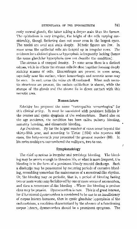
HYPERPLASIA OF THE ENDOMETRIUM 341
ently normal glands, the latter taking a deeper stain than the former. The epithelium is very irregular, the height of the cells varying considerably, though ftattening does not occur even in the largest cysts. The nuclei are oval and stain deeply. Mitotic figures are few. In some areas the epithelial cells are heaped up in irregular rows. The evidence for a distinct glandular hyperplasia is frequently lacking (hence the flume glandular hyperplasia does not (h~scribe the condition).
The stroma is of unqqual density. In some areas thcre is a distinct edema, while in others the stroma cells lie close together forming deeply staining masses of cells. Hemorrhages are present in some areas, especially near thc surface, where hemorrhagic and necrotic areas may be seen. In such areas the veins are thrombosed. When such necrotic structures arc present, the surface epithelium is absent, while the stumps of the glands and the stroma lie in direct contact with this necrotic area.
Nomenclature
~chr5dr,r ha;; proposed the name "metropathia hr,morrhagica" for this clinical entity. It must be associated with persistent follicles in the ovaries and cystic dysplasia of the endometrium. Based also on the age incidence, the condition has been called puberty bleeding, maturity bleeding, ancl climacteric bleeding.
Age 1 ncidence. By far the largest number of cases occur beyond the thirty-fifth year, and according to Tietze (1934) who reported 466 cases, the forty-seventh year presented the greatest number (60). In his series multipara outnumbered the nullipara, two to one.
Symptomatology
The chief symptom is irregular and persiste}}t bleeding. The bleeding may be severe enough to threaten life, or what is more frequent, the hleeding is in the form of a persistent bloody mucoid discharge. Such a discharge may hr, pllnr,tllated by mcurring periods of excessive bleeding, resembling ~omewhat the maintenance of a menstrual-like rhythm. Or, the hleeding may be periodic, that is, a period of bleeding lasting two or more weeks Illay be followed by one or more weeks of amenorrhea, and then a recurrence of. the bleeding .• Where the bleeding is profuse clots may be prcsellt. Dysmenorrhea is rare. This is of great interest, for if functional dysmenorrhea is considered to be due to an insufficiency of corpus luteum hormone, then in cystic glandular hyperplasia of the endometrium, a condition characterized by the absence of a functioning corpus luteum, dysmenorrhea should be a prominent symptom. The

-342 ENDOCRINES IN OBSTETRICS AND GYNECOLOGY
reverse is the case. In the juvenile form of this syndrome the abnormal bleeding may start with the first period or after a number of normal periods. As a rule, premonitory menstrual symptoms appear before the onset of the bleeding, namely, a short period of amenorrhea, or several irregular periods, irregular as to tempo, duration or intensity of bleeding.
Another interesting feature about the bleeding is the tendency to spontaneous cures and recurrences. We have observed the following types: after a period of bleeding lasting some weeks there is a short pause and the bleeding recurs, but only for a week or two, then another pause which is again followed by a period of bleeding. Thus periodic bleeding is reestablished, but each period of bleeding lasts too long or is too profuse, or both. Finally, apparently normal cycles are reestablished. After a period of regular bleeding, regular as to tempo, duration and intensity, the bleeding may begin anew. Another form frequently seen is that of a period of bleeding lasting for a variable time, followed by a period of amenorrhea ranging from 3 months to two or more years. Such periods of amenorrhea have raised the following question: What is the character of the endometrium during this period of apparent cure? Has it returned to normal cyclical variation? We, (Wilson and Kurzrok, 1936) have observed that during the period of amenorrhea or apparent cure, the endometrium retains its cystic glandular character. The morphological picture is the same, but the bleeding factor is absent. The significance of this observation will be discussed together with the etiological factors of the disease.
Cystic glandular hyperplasia of the endometrium is a benign condition. This is all the more interesting when we consider that cystic hyperplasia is brought about by an excess of follicular hormone, relative or absolute, and because of the recently demonstrated relationship between estrogenic and carcinogenic substances. The conversion of a cystic and glandular hyperplasia of the endometrium into a carcinoma of the fundus uteri has not been definitely demonstrated.
Cases of maturity bleeding frequently follow a pregnancy. No abnormality is noted before the pregnancy but a short time after its termination disturbances in the tempo, intensity or duration of bleeding begin. These are shortly followed by a period of prolonged and irregular bleeding. They do not otherwise differ from the cases of juvenile or climacteric bleeding.
The other symptoms noted in cystic and glandular hyperplasia of the endometrium are entirely due to the secondary anemia. The hemoglobin and red blood corpuscles are low. We have seen values of 15 to
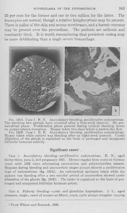
HYPERPLASIA OF THE ENDOMETRIUM 343
25 per cent for the former and one to two million for the latter. The leucocytes are normal, though a relative lymphocytosis may be present. There is pallor of the skin and mucus membranes, and a haemic murmur may be present over the precordium. The patients are asthenic and constantly tired. It is worth remembering that persistent oozing may be more debilitating than a single severe hemorrhage.
FIG. 130A. Case 1. H. N. Anovulatory bleeding, proliferative endometrium. The previous two periods have occurred after a four-week interval. No premenstrual phase. Proliferative phase present during cyclical bleeding, hence no corpus luteum formation. Biopsy taken two days before a twelve-day flow.
FIG. 130B. Case 1. H. N. Anovulatory bleeding, proliferative endometrium. Biopsy taken while patient was bleeding after an eight-week interval. Cystic dilatation of the glands is explained on the basis of prolonged and unopposed follicular hormone activity.
Significant cases1
Case 1. Anovulatory bleeding- proliferative endometrium. H. N., aged thirty-three, para ii, last pregnancy 1925. Menses regular from onset at thirteen years until 1932 when alternating amenorrhea and polymenorrhea ensued. Biopsies during bleeding and amenorrheic stages always showed a proliferative type of endometrium (fig. 130A). An endometrial specimen taken while the patient was bleeding after a two months' period of amenorrhea showed cystic dilatation of the glands (fig. 130B). The latter is explained on the basis of prolonged and unopposed follicular hormone action.
Case 2. Puberty bleeding- cystic and glandular hyperplasia. J. L., aged nineteen, single, onset of menses at fifteen years, cycle always irregular varying
1 From Wilson and Kurzrok, 1936.

344 ENDOCRINES IN OBSTETRICS AND GYNECOLOGY
from a two to six weeks' interval with a profuse flow of from four to ten days' duration. Patient bled profusely and continuously from December 5, 1932 to January 10, 1933, and the hemoglobin dropped to 40 per cent. Urine examination showed no follicular or follicle-stimulating hormones . Curettage revealed cystic and glandular hyperplasia of the endometrium (fig. 131). The bleeding stopped after intensive pregnancy urine extract therapy (Follutein-Squibb), Five blood transfusions were necessary to combat the anemia. There was no bleeding for eight months when another short episode of profuse bleeding ensued. This was readily controlled again by the gonadotropic fraction of pregnancy urine (follutein) and the treatment was continued for three months after the bleeding had stopped. There has been no bleeding since (eighteen months). A biopsy taken
FIG. 131A. Case 2. J. L. Puberty bleeding, cystic and glandular hyperplasia of the endometrium. Cystic and glandular hyperplasia of the endometrium during active bleeding.
FIG. I3lB. Case 2. J. L. Persistence of the same type of endometrium after fourteen months of amenorrhea. Treatment with large doses of follutein had stopped the flow. The bleeding has not recurred, and it is now eighteen months since the last flow.
after fourteen months of amenorrhea revealed the persistence of the cystic and glandular hyperplasia. Urine examination at the same time showed no follicular hormone but the presence of follicle-stimulating hormone. Klingler and Burch (1933) and Rock (1935) have noted that a secretory endometrium failed to develop after the bleeding was stopped by the administration of pregnancy urine extract. Thus, the cessation of the bleeding cannot be attributed to luteinization.
Case 3. Continuous bleeding for eleven years- cystic and glandular hyperplasia. C. P., aged twenty-four, menses began at thirteen years, patient bleeding continuously ever since . The bleeding varies from a profuse flow to staining but the patient is never entirely clean. Hemoglobin 65 per cent. No pregnancies

HYPERPLASIA OF THE ENDOMETRIUM 345
although married three years. Patient was curetted three times in the past four years because of the profuse bleeding. The endometrium has persistently shown marked cystic and glandular hyperplasia. The patient was given daily intravenous injections of prephysin (1 to 2 cc.) for twelve days beginning July 9, 1935. No bleeding since July 23, 1935, except for slight staining from October 12, 1935, to October 17,1935. Biopsy during the amenorrheic phase (September 17,1935) shows the persistence of the cystic and glandular hyperplasia (fig. 132). The cessation of the bleeding following intravenous anterior pituitary extract cannot, therefore, be attributed to luteinization. Incidentally, this patient had a severe and persistent facial acne which spontaneously disappeared soon after the bleeding stopped.
FIG. 132A. Case 3. C. P. Cystic and glandular hyperplasia of the endometrium. Treatment with prephysin. The histologic picture during active bleeding (typical Ii S"'iss cheese" type).
FIG. 132B. C. P. Persistence of the same morphologic characteristics during a two-month period of amenorrhea. More complete absence of premenstrual phase.
Case 4. Maturity bleeding- secretory endometrium. B. B., aged twentyeight. Hyper- and polymenorrhea past seven months. Menses previously regular since onset at fifteen years. Biopsy on the second day of a ten-day period revealed a well-developed secretory endometrium (fig. 133). Biopsy on the fourth day of the following period (eight days' duration) showed the presence of of an early proliferative endometrium. The regeneration of the endometrium thus appears to take place despite the continuation of the bleeding. Under pregnancy urine extract therapy (600 R.U. per week) there has been a gradual return to a normal cycle with a more moderate flow of five days' duration.
Case 5. Secondary amenorrhea- bleeding from a proliferative endometrium. M. S., aged thirty, amenorrhea six months' duration, menses previously regular since onset at fourteen years. No atrophy of genital organs or breasts. Biopsy

346 ENDOCRINES IN OBSTETRICS AND GYNECOLOGY
taken three days before the onset of a flow described by the patient as a typical menstrual period, revealed a proliferative endometrium of slight activity ("resting endometrium") (fig. 134).
Case 6. Secondary amenorrhea- cystic and glandular hyperplasia. C. G., aged twenty-six, onset of menses at ten years, regular until seven years ago when
FIG. 133. Case 4. B. B. Maturity bleeding; secretory endometrium. Biopsy on the second day of a ten-day menstrual flow. Prolonged and profuse bleeding from a secretory endometrium.
oligomenorrhea and obesity developed. No period for seven months. Married nine years and never pregnant. Basal metabolic rate: minus 56 per cent (repeated) but no signs of myxedema. Urine examination revealed 9 R.U. follicular hormone per liter and no follicle-stimulating hormone. Biopsy showed marked cystic and glandular hyperplasia of the endometrium (fig. 135).

FIG. 134. Case 5. M. S. Secondary amenorrhea; bleeding from proliferative endometrium. Biopsy taken three days before onset of period.
FIG. 135. Case 6. C. G. Secondary amenorrhea. Cystic and glandular hyperplasia of the endometrium. Biopsy taken after amenorrhea of seven months . No previous history of excessive bleeding, but on the contrary, oligomenorrhea.
347

348 ENDOCRINES IN OBSTETRICS AND GYNECOLOGY
Case 7. Secondary amenorrhea-secretory endometrium. S. M., aged thirtyfive, amenorrhea of two years' duration. Menses always irregular since onset at fourken years. No genital hypoplasia. Normal hormone findings in urinc (follicle-stimulating hormone, negative; 10 to 40 R.U. follicular hormone per liter). Basal metabolic rate minus 4 per cent. Biopsy revealed a secretory endometrium (fig. 136). Scanty periods resulted from the administration of large doses of progynon-B but bleeding stopped after discontinuation of treatment.
Case 8. Secondary amenorrhea (x-ray castration) experimental production of cystic and glandular hyperplasia of the endometrium. W. U., aged forty-five. diabet('s mellitus of nineteen years' duration, x-ray abortion five years ago fol-
FIG. 136. Case 7. S. l\I. Secondary amenorrhea. Secretory endometrium. Biopsy taken during amenorrhea reveals secrctory endometrium. At other "periods" has very faint staining lasting several hours, from a premenstrual endometrium.
lowed by sevcre menopausc symptoms, genital atrophy and aggravation of diabetes. During the past eighteen months the patient has received a total of 700,000 R.U. of progynon-B. This has afforded complete relief from her menopause symptoms. In addition, there has been considerable improvement in her diabetic condition. The uterus, breasts, and external genitalia have come up to full development and irregular periodic bleeding has occurred. Biopsy of the endometrium reveals cystic and glandular hyperplasia, which has been artificially produced in this case by the administration of huge doses of follicular hormone (fig. 137).
Case 9. Primary amenorrhea-experimental cystic and glandular hyperplasia. H. E., aged twellty-five, never menstruated, infantile uterus and slightly hypo-

HYPERPLASIA OF THE ENDOMETRIUM 349
plastic breasts and external genitalia. No follicular hormone in urine; folliclestimulating hormone present. These findings indicate that the amenorrhea was probably due to both partial failure of devclopment of thc Mullerian duct system and subsequent cessation of gonadal function. Bleeding was induced by the administration of progynon-B. Endometrial biopsy following 70,OCO R. U. over a period of eight weeks showed cystic and glandular hyperplasia (fig. 138).
rIG. 137. Case 8. W. U. Secondary amenorrhea (x-ray castration). Expcrimplltal production of cystic and glandular hyperplasia of the endometrium. 700,000 RU. of progynon-B given.
Case 10. Ovulation bleeding- transitional endometrium. M. S., aged twentysix, "menstruated" every two weeks during the past year, previously once in four weeks. Each profuse period is followed with considerable regularity by a scanty flow. Dysmenorrhea occurs only with the profuse period. Biopsy on the twentyfourth day of the cycle revealed a secretory endometrium (fig. 139A). The ovulation bleeding lasted from the seventeenth to the twenth-third day. The next
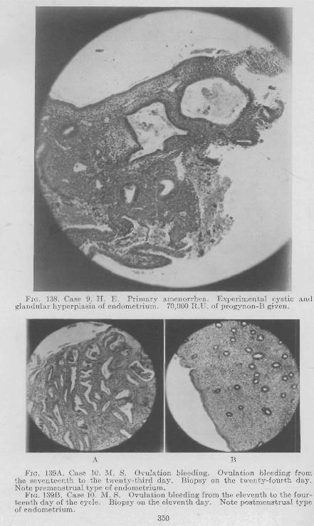
FIG. 138. Case 9. H. E. Primary amenorrhea. Experimental cystic and glandular hyperplasia of endometrium. 70,000 R.U. of progynon-B given.
A B
FIG. 139A. Case 10. M. S. Ovulation bleeding. Ovulation bleeding from the seventeenth to the twenty-third day. Biopsy on the twenty-fourth day. Note premenstrual type of endometrium.
FIG. 139B. Case 10. M. S. Ovulation bleeding from the eleventh to the fourteenth day of the cycle. Biopsy on the eleventh day. Note postmenstrual type of endometrium.
350

HYPERPLASIA OF THE ENDOMETRIUM 351
profuse period began twenty-eight days after the onset of the previous profuse flow. Another biopsy taken on the first day of the ovulation period (eleventh day of the eycle) showed a proliferative endometrium (fig. 13gB). This patient, therefore, had a normal twenty-eight-day endometrial cycle. The profuse flow repreRentpd the true menstrual period and the scanty flow thp ovulation bleeding. It will also be seen that the ovulation bleeding did not occur on the same dates on two successive months.
Diagnosis
The most important diagnostic factor is the finding of a cystic glandular hyperplasia of the endometrium. Hence, a curettage or an endometrial biopsy is e8sential. The latter procedure can be carried out by a modified Klingler and Burch (1032) suction curette (fig. (0). This method of obtaining a specimen is a simple and safe office procedure in thf' hands of a trainf'd gynpcologist. It has the grpat arivantagp that it does not require any anesthetic, it does not necessitate a stay in the hospital, and can be repeated at frequent intervals. In cases of very young girls, where neither procedure is feasible the hifltory of the cafle is of great significance. The preliminary menstrual disturbance is usually amenorrhea. Occasionally abnormalities in the rhythm of the cycle, plus subsequent persifltent irregular blccding in thc presence of a gro8sly negative pelvis, greatly favor the diagnosis of a cystic glandular hyperplasia of the endometrium. The rIoser the 8ymptoms appear to the menopaw;e the morc insistent should be the demand for a curettage.
Hormonal Findings in the Blood and Urine
In view of the fact that cystic glandular hyperplasia is due to an excess of follicular hormone, either absolute or relative, one would expect to find high estrin values in either the blood or urine. Our own studies on the hormone excretion in the urine demonstrate the following types of excretioll.
E strone
(a) Greater than 20 H.U. (b) Less than 10 R. U . (c) Negative
Follicle Slintuiatillg Hormon e
Negative Negative Positive
The least frequent form of excretion is type "a". That is, it is unusual to find large estrone values in the urine. It may signify that whatever hormone is produced is utilized mainly by the endometrium. Other tissues that estrone acts upon, such as the breasts and myometrium, do not show any excessive stimulus to grow during the course

352 ENDOCIUNES IN OnSTETHICS AND GYNECOLOGY
of this bleeding syndrome. The enlargement of the uterus that is occasionally found during the active bleeding is due to an increased vascularity of the organ, for when the bleeding stops the uterus becomes decidedly smaller. The most severe hemorrhages can be found associated with small, hard, uteri. We have never noted any undue breast enlargement during the course of the hlecdillg. This might imply several possibilities, namely: '
1. All absulute increase uf estrono rarely, if ever, ueems, fur if it did certain other tissues would show increased stimulation.
2. There is a change in the distribution of the total estrone produced, so that the endometrium takes up and utilizes more than it_s usual share. This would necessitate the concept that the first disturbance is within the endometrium proper.
3. ~ome change in the estrone molecule occurs, and thc new compound has a special "affinity" for the endometrium. The large number of modifications that the estrone molecule may undergo, and still remain estrogenic, makes this explanation possible.
The most COllllllon type of excretion is represented by type "b". The urinary estronc iF low, and the follicle stimulating hormone is absent.
When the bleeding has persisted for a considerable time we have repeatedly found in the urine an absence of estrone and the presence of the follicle stimulating hormone. This comhination is usually associated with the loss of ovarian function. It may imply an exhaustion on the part of the ovaries in spite of the fact that the stimulus (follicle stimulating hormone) is still present. In two cases we noted this finding jusL before bleeding stopped. On the other band we have not noticed this finding in other untreated cases prior to the ccssation of the bleeding.
Differential Diagnosis
The following conditions may give rise to acyclical bleeding: 1. Polyps--cervical polyps sooner or later hecome visible. Bleeding
usually occurs immediately after intercourse, or occasionally after severe bodily strain. Intrauterine polyps can ()ccasionally be diagnosed only by means of the curette.
2. Submucou8 .Myoma-the history of bleeding dates back a considerable period, The periods arc at first prolonged and intensified. Bleeding occurs soon after the period, between the periods, and finally continuous bleeding results. These symptoms correspond to increasing

HYPERP[,ASIA OF TlTFl ENDOMETRIUM 353
encroachment of the fibroid upon the cavity.of the uterus, the atrophy of the overlying mucosa and the irregularity of the uterine contractions.
3. Carcinoma of the Uterus-Cervical carcinomas become palpable and visible very early. The bleeding at the beginning is usually wholly acyclicaL Carcinoma of the fundus presents a more difficult problem. The bleeding may closely resemhle that of cystic hyperplasia. A history of recurrent attacks of bleoding, separated by regular cycles or amenorrhea greatly favors cystic hyperplasia. The curette is the final arbiter.
4. Incomplete A.bortion-The history of a period of amenorrhea preceding the bleeding may be common to both conditions. The soft and enlarged post-abortal uterus is a significant sign. A positive AschheimZondek Test is almost conclusive for pregnancy. At the same time it is to be remembered that the urine of patients with cystic hyperplasia may occasionally show a positive follicle stimulating hormone reaction. This is especially true if the bleeding has existed for a long time. The patients are usually in their generative years.
5. Endometritis-especially post-aborta!. The history is significant. The bleeding is usually associated with the periods. A temperature may be present. The patie-nts are usually young.
Etiology
Two questions are in need of further elucidation, namely, the etiologyof the entire symptom complex, and the etiology of the bleeding.
The etiology of_ the entire symptom complex may be considered from the viewpoint of:
a. The endometrium. b. The ovary. c. The anterior hypophysis. ,/ We do not believe that cystic glandular hyperplasia of the endomet
riulll arises solely from a condition inherent in the endometrium proper. The dise~se never ocellrs spontaneollsly in the absence of a functioning ovary. Neither trauma nor inflammation has been known to produce this condition. (There is the possibility that due to some change in the endometrium there occurs a change in the partition of the total amount of estrone prodnced. The endometrium takes up more than its normal requirement. This concept leads to the following idea. The endometrium, having thus altered its hormonal status, stimulates the ovary ill an abnormal way and induces thereby cystic follicles. The latter explains why a curettage occasionally cures this syndrome.) In view
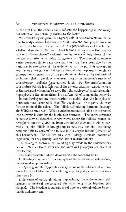
354 ENDOCRINES IN OBSTETRICS AND GYNECOLOGY
of the fact that the endometrium reflects the happenings in the ovary our attention has naturally shifted to the latter.
We consider cystic glandular hyperplasia of the endometrium to be due to a dysbalance between follicular hormone and progesterone in favor of the former. It can be due to a preponderance of the former whether absolute or relative. Cases 8 and 9 demonstrate the production of a "Swiss cheese" endometrium by means 8f large doses of thc benzoic acid ester of estradiol (progynon-B). The amount of estrone varies considerably in each case bu~ ~his IImy have been due to the variation in reactivity of Lhe endometrium. To put ~his concept in another way, we can say that cystic glandular hyperplasia is merely an extension or exaggeration of the proliferative phase of the endometrial eydie, and that it develops whenever there is an inadequate supply of progesterone. Follicles form corpora lutea. But the transformation of a mature follicle is a f6~ction of the anterior pituitary gland, hence it is the accepted viewpDinttto-day, that the etiology of cystic glandular hyperplasia of the endometrium' is a dysfunction of the adenohypophysis,
In considering normal menstmation we stated that the interplay of hormon88 mU8t occur with clock-like regularity. One pave8 the way for the action of the other. The follicle stimUlating hormone develops the follicle ~o maturity. When ovulation occurs the follicle is converted into a corpus lutcum by the luteinizing hormone. The entire sequence of events may be disturbed in two ways, either the follicles cannot be brought to maturity, and an immature follicle does not luteinize normally; or, the follicle is brought up to maturity but the luteinizing hormone fails to convert' the follicle into a corpus luteum (absence of this hormone?). The follicles may then undergo a certain amount of regression, for they usually lack the size of mature follicles.
The etiological factor of' the bleeding may reside in the endometrium per se. Neither the ovaries 1101' the anterior hypophysis are essential for bleeding.
The cases presented above demonstrate the following facts. 1. Bleeding may occur from any type of endometrium-proliferative,
transitional or premenstrual. 2. Cystic glandular hyperplasia may occur in the absence of all pre
vious history of bleeding, evclI during a prolonged period of amenorrhea (case 6).
3. In cases of cY8tic glandular hyperplasia, the endometrium still retains its identical pathological character long after bleeding has stopped. The bleeding is superimposed upon a cystic glandular hyperplastic endometrium.

HYPERPLASIA OF THE ENDOMETRIUM 355
4. Cystic glandular hyperplasia of the endometrium may be produced experimentally by means of follicular hormone in the absence of a functioning ovary.
These findings have led us to formulate the concept of a bleeding factor to explain abnormal uterine bleeding of functional origin.
Treatment of Functional Uterine Bleeding
I~xcessive functional bleeding may be controlled in any of the following ways:
1. Removal of the Bleeding snrface. Curettage affords only a temporary control of the bleeding. In pre climacteric cases it should be employed routinely as a diagnostic measure in order to definitely exclude malignancy. In puherty bleeding, curettage should be limited bceause it is usually unnecessary and often produces an unpleasant psychic effect on the patient .. Hysterectomy today has no place in the treatmcllt uf funcLional lJleeding, except perliapOi in sOllle uf the preclimacteric cases.
2. Removal of the Stimulus (Follicular Hormone) Which Produces the Bleeding Hormone. The stimulus for the secretion of the bleeding hormone may be removed by castration, either by operation or radiation. The latter is ideal for cases of pre climacteric bleeding but should never be used in younger women because it might result in permanent castration. Oophorectomy also has no place in the treatment of functional bleeding. Resectiun of a portion of each uvary does not appear to lJe a rational procedure, for a part of an ovary lllay produce cystic follicles as well as the whole one. The cystic follicles are the result of abnormal pituitary function. (Furthermore, how much ovarian tissue should be removed'!)
3. Prevention of Bleed1:ng Hormone Production. This may bc accomplished by roentgen ray irradiation of the pituitary gland but is too dangerous a method. The exact effect of a given dosage of radiation on an inclhddual patient is not accurately predictable and permanent damage may result whcn only temporary suspension of function is intended
4. Inhibition of the Activity of the Bleeding H ormonc. This offers the best method to .date of ~Qntrolling functional uterine bleeding. The activity of the bleeding hormone may be effectively checked by the administration of extracts of the corpus lllteum, pregnancy urine or the anteriur lobe of the hypophysis. Potent corpus luteulll extracts are not generally available because of their expense and limited supply. Adequate amounts of synthetic progestin should soon become available.

356 ENDOCRINES IN OBSTETRICS AND GYNECOLOGY
Practically every case of functional uterine bleeding can be controlled by pregnancy urine extract, providing thc dosage is adequate. The average daily dose required during the stage of active bleeding is from 200 to 500 RU. In very severe cases as much as 750 RU. daily, in 2 or 3 divided doses, may be necessary. The injections are best given intramuscularly in the buttocks. When the bleeding ceases, the patir:nt may be ct1rricd along on much smallrr doses (200 RU. once or twice a week) and this should be continued for several weeks or months. Following this tret1tmellt the perious IIlay occur at fairly normal intervals. At first they may be prolonged and profuse, but continued treatment both shortens and lessens the flow. This is especially likely if the previously excessive flow was periodic in occurrence. On the' 001er hand, if the bleeding was formerly more or less continuous, a prolongfd period of amenorrhea may result. In some, instances there has been
. no bleeding for well over a year. The vaille of sllch a prolonged period of amenorrhea is twofolrl. It relieve" .the overworked hematopoietic system and gives the enaocrine apparatus an opportunity to reestablish all equilibrium and lu,ter resume nomial fundion.
The exact mechanisIll by which pregnancy urine extract controls functional bleeding has not as yet been definitely established. The absence of effect on the endometrium conclusively shows that the cessation of the bleeding cannot be attributed to luteinization. We believe that pregnancy urine extract acts directly on the tissue producing the bleeding factor, possibly the endometrium.
Similar, although not as effective, results were obtained by the use of anterior pituitary extracts. They contain the gonadotropic hormones. In very severe cases, anterior pituitary pxtract (pr8physin), in dt1ily doses of 1 cc., may be injected intravenously. This usually stops the bleeding promptly but the severe reactions that occur in some patients from this intravenous administration somewhat limit its use.
Certain adjuvants to the treatment of functional uterine bleeding are important. The anemia resulting from prolonged or excessive bleeding demands careful attention. In the milder cases, iron may be given. The severe ct1ses often require one or more hlood transfusions. If a pregnant donor can be obtained, not only aTe erythrocytes and hemoglobin suppl'ied but also the anterior pituitary-l'ike hl!r17wncs. Oxytocics as pituitrin and ergot are occasionally of value especially when the bleeding is associated with uterine atony. Where the uterus is firm they are of no value.
The writer ha~ used the transfusion of human pregnancy blood on

HYPERPLASIA OF THE ENDOMETRIt:M 357
several occasions. All bleeding ceased within 48 hours. Blood was obtained from a healthy donor in her second trimester of pregnancy. The amount transfused was 3.';0 cc. It is of interest to note that neither the, blood nor thc urine of the recipient gave a positive Aschheim-Zondek Test at any time after the transfusion.
BIBLIOGRAPHY
DEKNECKE 1882 Zur Xtiologie del' "Endometritis fungosa", speziell del' "Chronischen hyp~rplasierenden Endometritis Olshausens". Arch. Gynak.20.
1885 Ein Beitrag "ur Klarung der Xtiulugie der "Endometritis fungosn,". Bed. Klin. Woch.
BURCH, J. C., PHELPS, D. AND WOLFE, J. M. 193~ Endometrial hyperplasia. Arch. Path. 17, 799.
HITSCHMANK, F. AND ADLER, L. 1908 Der Bau del' Utcrusschleimhaut des geschlechtsreifen Weihes mit hcsonderer Beriieksichtigung der Menstruation. Monat. f. Geb. u. Gyn. 27, 1.
KLINGLER, H. H. AND BURCH, .J. C. 1933 Suction in Obtaining Endometrial Biopsies. J. A. :\1. A. 99, 559.
KURZROK, R. AND RATNER, S. 1932 The Helation of Amenorrhea Accompanied by Genital Hypoplasia to the Follicular Hormone in the Urine. Am. J. Obs. & Gyn. 23, 6B9.
HOCK, J. 1935 Changes in the Endometrium after Female Sex Endocrine Therapy. A Cdn8ideration of the Effect of Antuitrin-S in Human Subjects. Endocrillol. 19, 269.
TIETZE, K. 193~ Die Follikelsper8istenz mit glandularer HyperpHisie des Endometriums in klinischer und anatomischer Beziehung. Arch. Gynak. 155, 35.
WILSON, L. A::-ID KURZROK, R. 1936 Excessive Uterine Bleeding of Functional Origin. Am. J. Ohs. and Gyn. 31, 911.

CHAPTER X XII
FUNCTIONAL DYSMENORRHEA
In the writing of this volume the author left the chapter on f(mctional dysmenorrhea for the last, with the hope that some 1V0rk would appear that would clarify this subject. No such work has appeared. In fact, the problem is more baffling than ever. Many reports on the 8ubject describe some endocrinological form of treatment. We hav!( followed the suggestions faithfully, only to meet with disappointment time and again. Here and "there an excellent result is obtained, but very frequently the repetition of the treatment in a similar case leads to complete 01" partial failure. Our difficulty lies mainly in the fact that we do not understand the problem, for here, as ,in other issues, the understanding of the problem is three-fourths of the solution.
The controlling factors of the problem are: 1. The development of the uterus. 2. ~The hormonal control of the uterus. 3. The nervous control of the uterus. Other factors also enter in, namely, (a) The constitutional type of the patient. It is well known that
the asthenic type is more prone to dysmenorrhea than the other types. The patient with general infantilism belongs to the same category.
(b) The nervous makeup-of the patient is of great importance. The highly sensitive and nervous patient suffers a great deal more than the phlegmatic type. But even the latter group can be made acutely miserable by dysmenorrhea. The psychic reaction of a woman to her sexual function is important. Undoubtedly these considerations would become clearer if we fully understood the controlling factors.
The Development of the Uterus
Thc majority of patients with functional dysmenorrhea present a hypoplasia of the uterus. It is equally true that many patients with marked infantilism of the internal genitalia have no dysmenorrhea, while others with normal uteri have. Similarly, an acute antiflexion (or retroflexion) is present in some but not in others. There is no relationship between the extent of the genital hypoplasia and the intensity of the dysmenorrhea. A number of questions vrith regard to the myo-
358

FUNCTIONAL DYSMENORRHEA 359
metrium are still awaiting a solution. What are the histological variations in thc myometrium during the cycle? Are all muscular layers of the uterus equally involved by the hypoplasia? Could a dissociation of function between the various muscular layers be responsible for the pain? Moir (1934) made the interesting suggestion that dysmenorrhea may be due to ischemia of the myometrium brought about by a uterine pressure upon the blood vessels greater than the systolic blood pressure.
The Hormonal Control of the Uterus
The human uterus undergoes contractions and rclaxations throughout the cycle. Only infrequently do we find a quiescent uterus, and thaL lIlay occur during any phase of the cycle. Our own evidence is against any corpus luteUl,u inhibition of uterine motility. One would expect that in the absence of a corpus luteum the menses should be painful. This is not the case, for patients with cystic glandular hyperplasia who bleed cyclically do not have dysmenorrhea. Similarly the anovulatory cycle is not painful. On the contrary, Kurzrok, Wic8-bader, lVIulinos and Watson (1937) have observed severe cramps in patients who were injected with 2 Rabbit Units of Progesterone during the follicular phase of the cycle, but not during the corpus luteum phase. The patients were being tested with an intrauterine bag in vivo. We have no explanation for this observation. The uterine contractions as evidenced by the kymographic tracing show no significant change. We have repeatedly examined (by means of biopsies), during the premenstrual phase, the endometrium of patients suffering from dysmenorrhea. The endometrium showed adequate premenstrual characteristics and a diagnosis of insufficient luteinization could not be made.
The Nervous Control of the Uterus
The autonomic nervous system that supplies the uterus must have some coordinating function even though uterine motility and/function (i.e. endometrial transplants in the eye) seem wholly independent of it. The internal os seems to be closely related to dysmenorrhea. It is a well known clinical· fact that in some patients the passing of a sound through the internal os, eYQI) though vQry little obstruction is encountered, leads to immediiLte severe uterine cramps. In a case recently observed by us, the gentle passage of a sound through the internal as led to severe uterine cramps plus nausea and vomiting. These same symptoms occurred at the usual menstrual periods. It i8 conceivable that the passage of tissue fragments and blood through the internal

360 ENDOCRINES IN OBST~;TmCS AND GYNECOLOGY
os has the same stimulating effect as the dilator. Whcther such observations imply a different type of innervation of the internal os in some patients is entirely undecided. In considering the question of menstrual blood passing through the internal os, the following questions present themselves: What is the character of the decomposition products (through th.e action of endometrial enzymes) of menstrual blood? What pharmacological effects could such derivatives exercise upon the uterus? Could such derivatives be modified by different nteri? Is acetylcholine (or its derivatives which act as a powcrflll oxytocic) a possible factor? Miller, Cockrill .and Kurzrok (1937) observed that acetylcholine contracted all uteri, during all phases, of the cycle, and that the magnitude of response tended to increase from about the twentieth day on.
Another interesting possibility pertains to the function of the Fraukenhauser ganglion. Branches from this ganglion pass to the sacral nerves and to the. hypogastric plexus. Blotevogel (1933) has shown that in the mou~J this ganglion undergoes morphologic change (the chromaffin cells) in direct relation to the amount of estrone and progesterone. Whether there is a similar change in this ganglion during the menstrual cycle is unknown. Variations from the normal could have interesting possibilities on the ncrVous control of the uterus.
One can readily observe from the above discussion how incomplete is our knowledge with regard to the etiology of dysmenorrhea. One thing is certain,-the etiological factors must be multiple.
Therapy
In vicw of the multiple etiological factors involved the treatment iR of necessity varied. We have recently tried out the recommended medical methods. We have frequently succeeded, and often failed. A given method may relieve the pain during one or several periods, and then subsequently fail completely. Occasionally, when one or two periods are rendered free from pain the dysmenorrhea seems to be permanently relieved. lVlany cases are permanently relieved by pregnancy, while in others there is a definite recurrence of the pain. It is our custom to start with peroral medication and when we do not suc-ceed we resort to parenteral methods. -
Medical Measures. A. The Benzyl Compounds. In view of the fact that benzyl compounds are to a certain extent antitipaslllotic we have used them extensively either alone or in conjunction with other forms of treatment. We always exhibit this form of medication first. Benzyl fumarate (in 5 grain tablets) is the drug prescribed. We recommend
FUNCTIONAl, DYSMEKORRHEA 3ul
10 grains t. i. d., p. c. for the two weeks preceding the flow, and then when the pain is expected we change to 5 grains everyone-half hour for ten successive doses. During the remaining days of the menstrual period the original dosage is again recommended. No untoward or t.oxic symptoms have been observed by us. The treatment may be repeated as often as necessary. This method affords relief in about 10 per cent of the cases.
B. Atropine. This presents our second method of therapy. It has been our custom to exhibit atropine whenever the passage of a sound through the internal os produced severe pain, especially of the type the patient experiences at her menstrual periods. It is based on the action of atropin on the sympathetic nervous system. We have used the official tincture of belladonna in doses ranging from five to ten drops three times a day. In view of the sensitivity of some patients to atropine poisoning it has been our custom to write down the toxic symptoms for the patient. They may be a dry mouth, dilated pupils, a flushed face, or a rapid pulse. The treatment is begun 3 days before the onset of the flow and continued throughout the flow. The effective dose is usually well within the limits of the recommended dose. This method affords relief in about 10 per cent of the caSCR.
C. Moderate Doses of Estrone. The administration of estrone is occasionally of value in those cases in which a genital hypoplasia is present. The milder the degree of hypoplasia the better arc the results. The purpose of the treatment is to stimulate growth of the genital tract. The patients are injected with 1000 R.U. (5,000 I.U.) of estrone twice a we~k, throughout the cycle, except during actu~l flow. This dosage does not disturb either the rhythm of the cycle or the duration of the flow. No untoward effects are noted. The number of cases benefited amounts to 1.5 to 20 per cent,. _/Reccntly we changed to peroral administration of estrone, giving 2,000 I.U. per day. Not enough cases have been studied to dale to give a definite opinion as to the value of oral medication.
D. Large Doses of Estrone. This method of treatment was first suggested by Dr. Leo Wilson (1936). It consists of the administration of 20,000 R.U. of e~tradiol benzoate (progynon-B) during the follicular phase of the cycle. He suggests the intramuscular injection of 10,000 R.U. on the seventh and eleventh day of the cycle. The administration of the same dosage during thc lattcr hnlf of the cycle is without effect. When given as suggested by Wilson, the tempo of the cycle is frequently disturbed. The period may be (about ten days) premature, or late. Often such premature or delayed periods are pain-

362 ENDOCRINES IN OBSTETRICS AND GYNECOLOGY
less. Some patients are not disturbed by double the suggested dose. The modus operandi of this large dose of estrone is not entirely clear. One would expect a disturbance of luteinization of the uterus. We have previously stated that the effect of the gonadal hormones on the endometrium is a balanced reaction, so that an excess of estrone will inhibit the development of the corpus luteum phase. Judging from our results such failure of luteinization is infrequent, though larger doses (40,000 R.U.) may lead to inadequate (or failure of) transformation of the postmenstrual endometrium into the premenstrual type. A sterile cycle may result. The relief of pain is independent of the character of the endometrium just before menstruation. In other words, the corpus luleum plays no part in the relief of symptoms. This method of therapy seems promising. It is applicable to only fifteen or twenty per cent of the cases of dysmenorrhea. We have as yet not devised a method for choosi'rig the proper cases for this form of therapy. While it is true that many cuses present a genital hypoplasia, others having a normal uterus react just as favorably.
E. Corpus Luteum Hormone. The demonstration of an inhibitory effect of corpus luteum hormone on the rabbit uterus has naturally led to its application for the same purpose on the human. From experimental evidence the treatment of dysmenorrhea with progesterone would seem justified. As stated in Chapter V our experiments with corpus iutcum ill vivo have failed to demonstrate any inhibitory action during the corpus luteum phase of the cycle. The injection of progesterone (2 mbbit units) caused no appreciable change ill the kymogmphic tracing, in fact we occasionally felt that the contractions were accentuated. We have used progesterone in the treatment of dysmenorrhea; the dosage given ranged between two and thirty Rabbit Units of hormone (proluton or progestin). Our results were poor. Only an occasional case responded to progesterone. Data are completely lacking on the character of uterine contractions during menstruation in normal women and those suffering from dysmenorrhea. A difference may be expected in some cases. The relief afforded by corpus luteum hormone in some eases may be due to its action on some other faetor than uterine motility, possibly on the blood supply or the autonomic nervous system.
F. Anterior Pituitary-like Hormones. Noval{ (1933) suggested the use of pregnancy urine extract (FoUutein or Antuitrin-S) for the relief of dysmenorrhea. Witherspoon (1935) has also reported good results. The basis of the theory is the luteinizating effect upon the ovary. Hamblen (1935, 1936) studied the effect of pregnancy urine extract on the luteinization of the human ovary. He noted the formation of

FUNCTIONAL DYSMENORRHEA 363
follicular cysts with active granulosa and fairly well preserved ova, and surrounded by a proliferated theca uterus with prominent theca lutein cells and with increased thecal vascularization. This is most marked in the younger group of patients, though no inference can be drawn as to the function of such luteal tissue. Geist (1933) also noted some increase in theca lutein cells. Assuming then that luteinization does occur when dosageS'ranging from 800 to 8,200 R.U. are given, it becomes quite questionable whether the administration of several hundred IInits just prior to and during the flow produce8 any slIch change. The absence of corpus luteum effect upon the human myometrium is again commented upon. The good results with this form of therapy must rest upon some other factor. It has becn suggested that the pain may be due to a sudden rise in the estrone titer just before menstruation. Our own experience is that the premenstrual estrone level is usually low.
Novak and Witherspoon recommend the daily intramuscular injection of 1 cc. of follutein for three to four days previous to the flow and also for the first two days of the period. Brown (1935) also has noted good results from this form of therapy. We have tried pregnancy-urine extract and Ollr good results do not exceed fifteen ]ler cent.
G. The Injection of other Substances. 1. Calcium-Boynton and Hartley (1934) report the treatment of essential dysmenorrhea with calcium, or calcium and viosterol. Two-thirds of the cases were greatly benefited. They believe that calcium is especially indicated in those cases that bnlise easily. We had had no experience with calcium as the sole remedy for dysmenorrhea.
2. Insulin has been recommended by Altschul (1936). He recommends the use of doses up to 10 Units (brand c) per day from three to seven days before or during the period. /N 0 deleterious effects have been noted. The patients showed no definite nutritional disturbances., As to the modus operandi of insulin in dysmenorrhea, Altschul cites Abel's (1931) work with insulin on animals, ,,;herein he noted a long estrus pause with a decrease in the number of follicles and an increase in the number of corpora lutea.
Surgical Measures. It has been well known that dilatation, or dilatation and curettage occasionally relieve a functional dysmenorrhea. The relief may be temporary or permanent. Stone (1935) recommends the gradual dilation of the cervix with a Hegar dilator as an office procedure.
Sympathetic neurectomy has recently come to the fore in the treatment of functional dysmenorrhea. It is needless to state that the
364 ENDOCRIKES IN OBSTETRICS AKD GYNECOLOG1
operation should only be considered when all conservative methods have failed. Wetherell (193.5) recommends the resection of the superior hypogastric plexus of the sympathetic. He notes no deleterious effect on Iibiuo or on future pregnancy. Greenhill (1!:)34), Ad8011 and Mas80n (1934), Keene (1935) anu Counseller and Craig (1934) advocate resection of the presacral sympathetic nerves. The latter writers are of the opinion that the primary etiologic factor in dYSIIlE:norrhea is dysfunction of the pelvic sympathetic nervous system. "Vhen this dysfunction is corrected by resection the benefits are permanent, while the functions of normal menstruation and childbirth are not disturbed. There is no loss of motor control of either bladder or rectum. These operations were first recommended by Cotte (1925).
BIBLIOGRAPHY
ABEL, P. 1931 ExperimentcllerUntersuchungcn uber der .I<;influss des Insulins auf die Ovarientiltigkcit.l~ Arch. l. Oyniik. 147,444.
ADSON, A. W. AND MASSON, J. C.·'·1934 Dysmenorrhea relieved by resection of presacral sympathetic nerhs. J. A. M. A. 102, 986.
ALTSCHUL, A. IU36 The tl'eatme~t of dysmenorrhea with insulin. J. A. TIl. A. 106, 1380.
BLOTEVOGEL, W. 1933 Symputhicus und Sexualzyklus. Zcitsch. f. II1ikros. Anat. Forseh. 33, 429.
BOYNTON, ll. E. A;.In HAnTLEY, E. C. 193~ Calcium in treatment of dysmenorrhea. Am. J. Qbst. and Oyn. 27, 21i3.
BROWN, W. H. 1935 Use of follutein in dysmenorrhea. Am. J. Obs. and Oyn. 29, 113.
COUNSELLER, V. S. A;.ID CRAIG, IV. 1\1. 1934 Treatment of dysmenorrhea by resection of presacral sympathetic nerves; evaluation of end-results. A. J. OLs. and GYIl. 28, 161.
COTTE, C. 1925 La sympathectomie hypogastrique a-t-8I1e sa place dans la thempeutique gyneeologique? La Pre sse Mod. 33, 98.
GEIST, S. H. 1933 Reaction of mature human ovary to Antuitrin-S. Am. J. Obs. and Gyn. 26, 588.
GREENHILL, J. P. 1934 Treatment of severe dysmenorrhea by pelvic sympathectomy. Am. Med. 40, 290.
HAMBLEN, E. C. 1935 Results of preoperative administration of extracts of pregnancy urine; studies of ovaries and of endometria in hyperplasia of endometrium following such administrations. Endocrin. 19, 165.
HAMBLEN, E. C. AND Ross, R. A. 1936 A study of ovaries following preoperative administration of an extract of pregnancy urine. Am. J. Qbs. and Gyn. 31, 14.
KEENE, F. E. 1935 Treatment of dysmenorrhea by presaeml sympathectomy. Am. J. Obs. and Oyn. 30, 534.
KURZROK, R., WIESBADER, H., MULINOS, M. G. AND WATSON, n. P. 1937 Action of Pituitrin, Estradiol and Progesterone on the Human Uterus in Vivo. Endocrinology, in press.

FUNCTIONAL DYSMENORRHEA 365
MILI,ER, E. G., COCKRILL, J. R. AND J{URZROK, R. 1937 Reactions of the human uterine muscle in vitro to pituitrin, adrenalin and acetylcholine and their relations to the menstrual cycle. Am. J. Ous. and Gyn. 33, 154.
]\loIR, C. 1934 Recording the Contr[l,ctions of the Human Pregnant and NonPregnant Uterus. Edinb. l\Ied. J. 41, 93.
NOVAK, E. AND REYKOLDS, S. R. M. 1932 The cause of primary dysmenorrhea with special reference to hormonal factors. J. A. :Vi. A. 99, 1466.
NOVAK, E. 1933 The treatment of Primary Dysmenorrhea with Especial Reference to Organotherapy. Am. J. Med. Se. 185, 237.
WILSO~, L. 1936 Unpublished results. WETHEIIELL, F. S. 1935 Intractable dysmenorrhea; relief by sympathetic
neurer.tomy. Am. J. Obs. and Gyn. 29, 331. Wl'rmmSPOoN, J. T. 1935 "Endocrinal origin of primary dysmenorrhea and its
hormonal treatment; preliminary report. Endocrin. 19, 403.

CHAPTER XXIII
THE CLIMACTERIC
The factors governing the termination of the sexual epoch a,re clo~ely associated with those fadors concerned in its inception. Hener, if we knew the exact cause of puberty we would know the reason for the mellopause. In considering the factors immediately coneerned with menstruation we discubsed the anterior hypophysis, the ovaries and the uterus. To these glands we must add the adrenals, the thyroid, the pancreas and possibly the thymus, though their contributions ar~ as yet not clearly understood. We have at least three etiological factors causing the climacteric,' depending on the organ primarily involved:
1. Anterior pituitary gland. 2. The ovaries. 3. The utcms. But in the final analysiH, the ovary is eventually involved, and about
it the syndrome centers.
Anterior Pituitary Gland
This involvement is occasionally seen in cases of secondary Frohlich Syndrome. The patient may be in her early thirties and the periods may become scantier and less frequent and finally stop completely. As time goes on one of the several symptom complexl:'s may develop. The usual vasomotor symptoms may not appear at any time, they may appear within two or three years with varying degrees of severity, or they lllay first make their appearance at the usual time, that is, in Lhe late forties. IL is (,lie first group tlmt illLerests us aL the moment. Two explanations for this absence of symptoms may be given. First, that the menopause was the result of ovarian failure, but so gradual that no noticeable symptoms occurred. Second, that the mechanisms involved in the usual form of menopause (v. i.) were absent.
The hormonal picture in these cases has not been extensively studied. The few cases that have come under our observation presented an ah-
1 We shall use the term climacteric in the larger sense, meaning all the symptoms in all organ systems arising from permanent cessation of function of the anterior pituitary-ovary-uterus complex. The term menopause is here used in the narrower sense, lneaning permanent cessation of menstruutiun.
366
THE CLIMACTERIC 367
sence of both the follicle stimulating and follicular hormones in the urine.
The Ovaries
It has previously been considered that the cessation of ovarian function was the sole cause for the climacteric. It might be stated at the outset that we are not in complete agreement with this idea. The reasons are that the ovarian failure may be secondary to that of the hypophysis, and that the symptoms of the climacteric are not solely due to the 10~R of ovarian fundion hnt to a disharmony of the other glands of internal secretion.
The Uterus
The third factor mentioned as pertaining to the menopause is the uterus. This may be encountered in cases of genital hypoplasia, wherein the periods completely stop in the twenties, or before; similarly when a hysterectomy is performed in a young woman. In either case the onset of the climacteric may vary. It may come on soon after rrssation of menstruation or be postponed until the expected onset in the late forties. In the early climacteric associated with uterine hypoplasia it is justifiable to assume that the early stopping of ovarian f unction was in some way due to the loss of uterine fUllction. We have previously (Chapter X X) stated that uterine hypoplasia is frequently due to an inherent defect within the uterus and not to ovarian hypofunction. This brings us back to the unsolved question, as to whether the uterus is merely the receptor of ovarian activity, or a contributor to it. One frequently hears the statement that when the ovaries cease to function soon aft,er a hysterectomy it is due to disruption of the ovarian circulation. That may be so, but need not always he so. We may be dealing with an internal srcretion of ..-the uterus.
\ Symptoms
The symptoms arising at the time of complete cessation of ovarian functio!]. vary tremendously with the cOl}stittitional makeup, the rapidity with w)lich the function is lost and with the psychic balance of the woman. Aschner (1924) makes the point, and very correctly so, that some women 'Si~em to be greatly benefited by the menopause. This is particularly applicable to the thin, anemic, and asthenic types, and especially so to those that suffered from profuse or prolonged menstruatioll and nervous dyspepsia. He observed that their appearance becomes so striking that they seem rejuvenated.

3GS ENDOCRINES IN OTlSTETRICS AND GYNECOLOGY
It is exceedingly difficult to state when the climacteric begins and when it ends. The mere cessation of menstruation-menopauseis not the sole criterion. The beginning of the climacteric may be years before the menopause, that is, some symptoms which are part of the climacteric syndrome begin years before the termination of the flow. Similarly, the end is not a sharp demarcation, for many women continue to show isolated symptoms into old age. The fact that a pregnancy may occur a number of years after the menopause: also points to the fact that cessation of menstruation is an important, but not the sole factor that demarcates the climacteric. Wiesel (1924) observes that the climacteric is not a limited syndrome, for many other disturbanc,es, both functional and pathological, give rise to temporary symptoms resembling those of the climacteric.
There arc no characteristic pathological (morphological) changes in the orgn.ns during the climn.cteric, except in the ovary and genital tract. Wiesel points Ollt that the \1drcnn.1 cortex may show a greater amount of lipoids, and the thyroid an excess vasculn.r sclerosis. The changes in the other orgn.ns n.re not significant, though it might be added that exhaustive pathological studies have not been made, for patients do not die of the menopause. Furthermore, our concept of the climacteric is not that of a disease based on pathological anatomy, but rather a functional disturbance of the hormonal and nervous equilibrium, in which the ovary plays the dominant or central role. The hormonal findings, so characteristic of the fully developed climacteric (v. L), do 110t limit this syndrome, for they are 110t present in many patients either at the beginning or at the end of this epoch.
It is not essential at this point to enumerate all the symptoms of the menopause. But the mechanism by which they arise is important. The hormones n.re regulators of the body fUllctions, and in addition, they regulate one another. It has definitely been established that one gland of internal secretion may influence another both in respect to morphology and to function. Furthermore, there is an intimate relationship between the hormones and the central nervous system, for instance, adrenalin stimulates the vagus and vasomotor centers, the hypoglycemia following insulin results in ncrvous_mnnifcstations, and the thyroid increases the irritability of the peripheral sympathetic nervous system. One may therefore expect a profound change in the entire regulatory mechanism when the ovary ceases to function. The gonads are an important cog in the machine. Consequently, the effects produced by this disturbance in equilibrium lllay extend as far as the regulatory mechanism controls, and that is, the entire orgn.nism. The
TIlE CLIMACTELUC 369
great number of dysfunctions that arise from this disturbed equilibrium is still further increased by the fact that the same hormone can convert a stimulating into an inhibitory reaction by a changc in its concentration (adrellalin, thyroxin). Hence, one lIlay expecl the elimacterie syndrome to inelude symptoms referable to every tissue and organ system. But whidl symploills will dcyclop depends upon the constitution (in the larger sense) of each particular patient. That some symptoms occur with greater frequency merely indicates that the mechanism causing these symptoms is closely allied to ovarian function. One frequently encounters discussions whether the menopause syndrome is due to a loss of estrone or to an excess of follicle stimulating hormone. That tries to simplify a problem that will not be so simplified. The climacteric syndrome implies a general disturbance of equilibrium and control, more profound in some systems than in oUwrs.
The effect of menstruation on the symptomatology of the climacteric is interesting. lVlany patients state that the menstrual flow results in a marked improvement of the symptoms. The rcasons for this are not quite clear. The normal curve of estrin excretion is lowest at the time of menstruatioll. Hence, if the symptoms were merely a function of the lack of estrin one would expect amarked aggravation. Two explanations are possible: one, that the estrone excretion curve reaches a peak at the timr- of menstruation instead of a diminution; two, that the symptoms are ill part independent of the estrone titer, but are due to some other undiscovered faclor-;, a point for further study.
Etiology of Cessation of Ovarian Function
Considering therefore the ovary as the hub (in rC::ipeGL to the dilllaeteric syndrome) about which the hormonal and nervous control radiates, certain qucstions become pertinent. What causes the ovary to cease functioning? Does this ccssation occur ,,(the time of the menopl1l1se, or hcfore, or after? Do bot.h oVl1ri~s stop th~ir fllndion at, t.hc same time?, Can such [unction, once stopped, be revived?
The statement has previo·usly been made that the ovary does Hot possess the inherent ability to function. The ovary lllUi:it constantly be stimulated to function by the anterior hypophysis, or to put it differently, the ovary .!leeds for its inherent metabolism a substance (gonadotropic hormone) originating in the anterior hypophysis. Without this specific substance, or when the ability to utilize this chemical compound is lost, the ovary cea:scs to function. Hence, the cessation of ovarian function could be due to a failure of the stimulator or a failure of the gonad proper. The hormonal findings at the time when

370 ENDOCRINES IN OnSTETHICS AND GYNECOLOGY
the climacterium is fully established give us some insight into this problem. They show an absence of estrone and the presence of large quantities of follicle stimulating hormone. It is believed, on good evidence, that the amount of follicle stimulating hormone produced at this time is in excess of the amollnt produced during normal ovarian function. The excess is prohably due to a loss of inhibitory control that follicular hormone normally exercises upon the anterior pituitary gland. Hence, the loss of ovarian activity is not due to the absence or insufficiency of the gonadotropic hormone necessary to stimulate the follicular apparatus of the ovary. The cessation of ovarian function is then evidently due to a change within the ovary proper, and this manifests itself in a failure on the part of the ovary to respond to ·stimuInt£on. That is, the funct1:onal 11je of the ovary is limited.
This functional limitation is first noted during early infancy. Smith and Engle (1927) studied the effect of anterior lobe implantation on immature female rats. They noted that the maturing of the reproductive system takes place rapidly, but 2 to 5 days are required when treatment is started at weaning (21·d'ays). The response becomes less rapid the earlier the injections are Wtarted. In rats fourteen days of age at the beginning of treatment, 8 to 10 days supervene before the full effect of the implants is induc1.d. A similar type of response in immature rats was observed by Saunders and Cole (1936) when using mare gonadotropic hormone. 'That these observations may apply to the young human ovary can be gathered from the following indirect evidence. Gonadotropic hormonr. has been demonstratr,d in the hlood and urine of the newborn by many observers (Neuman, 1930). Prolan occurs in large quantities in the maternal circulation from the second month of pregnancy, and yet the ovaries of the new born only rarely show signs of stimulation, in the form of follicular cysts. It might be added at this point that in the treatment of cryptorchidism we have noted that very young boys (under six years) are very refractory to treatment, and that as puberty approaches a lesser amount of prolan is necessary to secure descent of the testes.
In a recent study of the effect of the follicle stimulating hormone from mare-serum (Antex-Leo) on the human ovary by Kurzrok, Smith and Watson, (1937) the following interesting and pertinent facts were noted. When adequate quantities (3600 M.D.) were injected during the first phase of the cycle and the patients were in the third decade of life, then there resulted a marked increase in size and weight of both ovaries. N umerous large follicular cysts appeared. On the contrary, when the

THE CLIMACTERIC 371
hormone was injected into women during the fourth decade the response was only slight; an occasional small follicle appeared. When the injections were given to patients in fifth decade or beyond the ovaries did not show the slightest response, even when several times the dose (9000 ~I.U.) was injected. Even when a marked response was present in some young patients, the response could be wholly in one ovary, while the other would remain entirely refractive. In the latter cases the stimulated ovary was removed and the refractivc ovary was allowed to remain. NIcnstrnal cycles shortly reestablished themselves. These .findings indicate that the ovaries in tlte older group of patients were refractive to a dosage which was adequate for the younger group, and furthermore, that while .one 'ovary may lose its ability to respond to a given stimulus the other ovary may still retain that capacity. The reason why the refractive ovary was able to maintain function after the removal of the stimulated gonad, is that the entire amount of gonadotropic hormone liberated by the patients own pituitary was available to the single remaining ovary. It is reasonable to assume that the extent of refractiveness increases until no available amount of gonadotropic hormone SUffices. The time of onset and extent of refractiveness must vary from patient to patient. H enee the time of onset and intensity of the climacteric would vary as a result of this. The climacteric is basically due to a failure of responsiveness on the part of the·ovaries (figs. 148 and 149).
Whether we arc to assume that the functional life of one ovary is longer than the other, or that the one began to function earlier and ended sooner, is an entirely debatable question. If ovarian function ceases and results in the climacteric, can such function subsequently be revived? Under normal circumstances it cannot. Certainly the addi- J
lion of morA gonadotropic hormon~ doc~ not mvivc the ovary nor dors stimulation with the x-ray. The ovary sc~m~ to have undergonc some irreversible change. This brings up another interesting point. Supposing the ovary originating from a young girl is transplanted into a middle-aged woman during her climacteric. The menopause patient produces (and excretes) quantities of gonadotropic hormone greater than normal. 'Vill such an ovary "take," that is, function and produce demonstrable quantities of follicular hormone? The stimulus (gonadotropic hormone)"is present and a receptive ovary is provided by the donor. Maricard (1936) states that such grafts will not take. This would imply that the aging of the host is an additional factor in the function of the ovary. This subject is deserving of further lll
tensive study.

372 ENDOCRINES IN OBSTETRICS AND GYNECOLOGY
Therapy
The chid therapeutic agents for the treatment of the climacteric are estrone, calcium and Ruch therapeutic measures as each case may spBcifically require. We take cognizance of the fact that the number and intensity of symptom,; vary from patient to patient, and that women differ a great deal in temperament. Hence, there can be no cut and died method of treatment. It has been argued in many quarters that the therapeutic results obtained were merely suggestive. We do not deny that in some patients there is a distinct psychic element, but in out' experience it does not involve more than 10 or 20 per cent of the cases. It has frequently seemed to us somewhat bizarre that the loudest exponents of the "suggestive results" idea have never themselves treated a patient.
The results of therapy can be verified experimentally by the vaginal 8mcar test as demonstrated by Papanicolaou and Shorr (1936). These inveRtigators found that the vaginal smear test furnishes a simple objective guide for the treatment of menopausal syndrome with follicular hormone. With an adequate amount of estrone, there occurs a transformation of the vaginal smear from the menopausal to the leucopenic type, with the large flat cells, largely cornified, with small pyknotic nuclei. This is the type of smear which is found normally during the high follicular phase of the menstrual cycle just prior to ovulation. This change in the smear is generally associated with relief of the menopaul-lal symptoms (figs. ]40 to 147). Biopsies of the vaginal wall show a clORe correlation between the changes in the vaginal epithelium and in th(' vaginal contents. As a result of treatment, the vaginal epithelium becomes hypertrophic with the more superficial cells showing cornification and Rmall pyknotic nuclei. The outer basal zone was better differentiated and showed nuclear enlargement and mitosis.
A further ('ffect of follicular hormone in the castrate (spontaneous or induced) is to kssen the secretion and production of the follicle Rtimulating hormone. This excessive secretion is due to the abRence of the inhibiting effect that estrone exercises on the anterior hypophysis (Meyer, Leonard, HiHaw and Martin, 1930). It might be implied that because the follicle stimUlating hormone decreases at about the time when the flymptoms are alleviated, this hormone is responsible for the mcnopaURC' RymptomR. We do not favor this idea because the injection of follirlr Rtimulating hormone dorR not produce menopaURC
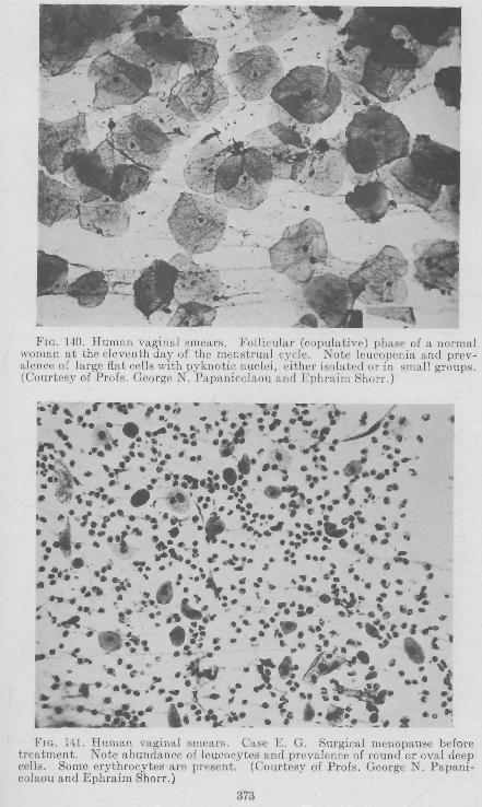
FIC. 140. Human vaginal smears. Follicular (copulative) phase of a normal woman at the eleventh day of the menstrua l cycle. Note leucopenia and preva len('e of large flat cells with pyknotic nuclei, ('ither isolated or in small groups. (Courtesy of Profs. G('orge N. Papanicolaou and Ephraim Shorr.)
;"" .r·,.·-,··if .. - ""\1 ~- .~. . , .... : .-. ';"~"·f·ii~,,,"'~i'- I~~* ~ ... ,-.,. .. ~. ~.. . .~ ..... ;;:.,' . ~. ..~ .... ~. • .. -f'" •.• _.. 41! :-:'., >
... ' . ,...J:: ..... ~ • ••• • lo. t '''".. .....,..... .... :~.. .. ') .,- - .... ~ ... • . ....: •• .: ' ~.~. . , J. • .. • . , ..... _ "'t ",'"'' -'I ~ ••••• ~ • ~ " ~.,,, " .'. -~ '.;.;_. ~·t·' --J~.~ ." e' Wi · " ' ,. • 'rl. ..... '..11 ...... ~. .. I . •• -Itt·" · . -"..- .... .-;.. i' " .. . ..... , .~... -.. ' . . .... ~.... .. • - .. ~ a ......._ . . ' ....... " ~ .... ~ '.' ..... .. _' ." ~ .. " -.. ... :.. .. •.•• ~ ...... ,,,' •••• \ _. , •• 6 ••• ~ • . •
~. _.... .,..:a -• '. ~ .. r.. ~. .. ' ~ .... ~ ., .. _' ''.. '):. '. . . ~.- .... : •• ~ • .... J'(. ... ~ ~" .•.• • : ••• ~ . .. '_ .'...,~ • .,
''''-: _ If,. , • e- .~ ....... .~ ~at. ' .. • , ...' ....... .). ... ~ . ..,.. " ~.. ~, - . .:\.$.:; ~ • ~ • co"".,· I."" (. .... ~ e: . A • ~ ••
..... ",'.C'" 8t •• ~' • ~ .: J;r ~ It ~ .. _~. +.1 .. .. .... • e .. • ..,\ , ., .. ~ '\ ••. ".. ( -. .. ,. -. ~.. . · ·r"_ ... ~ ~ .• t . ·
./1( . • ~&" -... . 'I . ' :. . h ': .......... _----... ~ _. - _'. _.-* Fro. 141. Human vaginal smears. Case E. G. Surgical menopause before
treatment. Note abundance of leucocytes and prevalence of round or oval deep cell s. Some erythrocytes are present. (Courtesy of Profs. George N . Papanicolaou and Ephraim Shorr.)
373

FIG. 142. Human vaginal smears. Case E. G. Shows disappearance of leucocytes and erythrocytes, and clearing of smear on further treatment. Deep and elongate cells prevailing. (Courtesy of Profs. George N. Papanicolaou and Ephraim Shorr.)
-~ . -...--v _ .
. . . ,
FIG. 143. Human vaginal smears. Case E. G. Typical follicular phase induced by further treatment. Note leucopenia and prevalence of flat cells with pyknotic nuclei. (Courtesy of Profs. George N. Papanicolaou and Ephraim Shorr.)
374

~IC' .. & -_. ".." • •• -, ,... _.. - ....
e' _"If.... .. • -'..... ... ." • · .. .' ... · .. .. .. " '. ,. . ~, ... ..,.... " ';... "". !. , k t .! .. ~ ••• • .• tfft • lit . _.... ., ~.... ...... .. : ' . I. ~. ~. - .". #I •• .. ••• - ... .. .. J' ... '" II' , ........ .-
, ... • . ? .. .Iit'~""" ' . '.. ••
I · ~ .. c." •• ; . • .... ~" r ~ -, .. . ,.,. \.. .• • /; ••••• ....
• III ....4O .. " • ' .. . · OS ~" 4 l' .. • .. •• "'-P . • ............ ?~. ... 'a~' . ./ .~ .. • ' *",", r .. "_"'~.j~-' ... ~ ~ ... ...... .. r .. 'i# ·~""I"· ~-~ ,... -'+~A.:') .- •.• . ~.f·
'.
.4> ..... t . • .. ~ .~."" . . .. ...... L, ..
~.c.~... ·it • • ..1..11!.. '_",. ~ _" .. ~ ... , ...... -.J .~ ,. .. -" .. . . - • ... it "'...... • . .. . t. . .... ....... ~ • ~ .... ........ .. .. ~""". c.: ... -:.. . y~ •• , ... : • .&! .• ·l~ ~ .. "';'._ ~ .. <iJ
:a'; .... ..;,~~. til ,., • ...,. .. , I.:.' ;,t-~. ~.~ ;. .. ~~ . : .. " 'Ill ...... .W'.,: ' ''I·.~''· " ,.... - ~_ ... ~'''4M· 'IJII '. ~-, ...... ~,.~ .. ,..~'" ~~.~~J;:~.~.~::.~
". ~~'. .1. ... 8 ... ", 'T:ii" - . c.o....... ,. ~ ,.. '.. . w: ._,., ....... ... • ~.i1f .~ ... ..... ".. , "" " • ••• ~ , of, -._ .. • '~' :I- '.:.-,\ _. ... ~.~ .~~J .... ~ .. ~.~.' .. :. ..... ,~~ .... ~~" - .... --:: ~ .. ..." ;:. ... .. • ,- ~..-4>'.1:'.' .. . 9111!1 .. . -If· · .../ . ~I ... 1 " A:4 . ' .• " ... 'l1t~4 \.."'t~#1;- "' • • ~."'fI .••• ~~ .. ~ ~, • • ~~, r,: ....... ~ ... -ft' • tiIlI_.. • . "" ! ~-J4"'.~.;' .. :,.. ... '-:_:." 'f.¥._. '"z ..... , 4IL , i:. .• ~ ..... W\ ." ~ •• i!.~ " • ." .......... "'" " "'_~
" ..... '1 ... ·• ... - ~~~.r~ ... !..~ f)" "_'·t 'lil' ,,:,6.~ •• ~.",-r s...;.;. :l.4t.~.. .. ~ • ~ ~!;' _ . ,'Ii. _ .~_ ,L...... .. . 11_
FIG . 144. H uman vaginal smears. Case E. G. Almost complete regression to original type after discontinuation of treatment. Note return of many leucocytes and deep cells. (Courtesy of Profs. George N. Papanicolaou and Ephraim Shorr.)
FIG, 145. H u man vaginal smears. Case E. G, Follicular phase fo llowing resumption of t reatment. (Courtesy of Profs. George N, Papanicolaou and E phraim Shorr.)

376 ENDOCRINES IN OBSTETRICS AND GYNECOLOGY
symptoms, and furthermore such symptoms may occur in the complete absence of follicle stimulating hormone from the urine.
Estrone should be given in large dose's. It is best administered hypodermically, though the oral route may be used. Oral administration required from five to twenty times the hypodermic dose. It has been our custom to begin with 50,000 I.U. (1 R.U. = 5 I.U.) of follicular hormone (amniotin or Progynon-B) twice a week. The injections are always given (with a 20 gauge needle) intramuscularly into the buttocks. Subcutaneous injection:-; produce' painful lumps. As the vaso-
FIG. 146. Human vaginal smears. Case E. G. Regression of vaginal epitheliUJn after discontinuation of treatment. Superficial cornified zone has disappearcd. Figure 1H illustrates corresponding smear. (Courtcsy of Profs. George N. Papanicolaou and Ephraim Shorr.)
motor :-;ymptom:-; dimini:-;h the number of injections is decreased until one dose every ten days is reached. Maintaining the same frequency of injection an attempt may be made to use less than 50,000 r.U. per dose. Either the patient's symptoms or the vaginal smear may be used as a guide. It is not advisable to increase the intervals between injections beyond two weeks, for there is a recurrence of symptoms. Adequate treatment results in a definite decrease in the number of sweats and flushes per day, namely, from one every half hour to one or two per day. There is a definite increase in energy and sleep becomes

THE CLIMACTERIC 377
more restful and refreshing. We must stress the importance of adequate and frequent dosage. Insufficient hormone is equivalent to none at all, for it leaves the patient discouraged with the treatment.
During the paf:lt few years we have injected in addition to estrone an alcohol-ether extract of the entire ovary. It is commercially available
FIG. 147. Human vaginal smears. Case E. G. Hypertrophy of vaginal epithelium after resumption of treatment. Note reappearance of superficial cornified zone and orderly structure. Figure 145 illustrates corresponding smear. (Courtesy of Profs. George N. Papanicolaou and Ephraim Shorr.)
in the form of Sistomensin (eiba). The basis for the use of this extract is as follows. One is frequently asked whether estrone and progesterone are the only hormones in the ovary. May there not be others whose function is not as yet clearly defined (Kaufmann, Muller and Miihlbach, 1932; Laqueur, 1933) but whose action is on the general metabolic processes, the blood vessels, blood cholesterol, fat metabolism, etc.
I

FIG. 148. Human ovary. G. P., No. 9298; aged 43. Mare serum, 3,600 M.U. Only one very young corpus luteum and numerous corpora albicanta.
r
FIG. 149. Human ovary. G. P., No . 95-16; aged 25. Treatment with mare serum, 3,000 M.U. Note the large follicular cysts which filled the entire ovary.
378

THE CLIMACTERIC 379
(F. Laqueur, 1934). It has been our impression (based on its use for more than 400 cases in 5 years) that when Sistomensin (1 cc.) is added to the injected estrone the therapeutic effect of the latter is enhanced. This applies only to the relief of the vasomotor symptoms.
The third therapeutic agent that we have routinely used is calcium. The reasons for its use are as follows: Many of the symptoms frequently encountered during the climacteric are due to disturbances of the vasomotor system-such as sweats, flushes, palpitations, etc. We believe these to be due to an increased irritability of the vasomotor system. Calcium depresses the irritability of the cell, hence its use in this connection. The salt used is the gluconate in doses ranging from 2.0 to 5.0 grams per diem.
We have occasionally used estrogenic material by mouth with excellent results. Theelol has been especially effective in cases of spontaneous menopause. The recommended dose is 150 R.D. (3 capFmles) per day.
A point to be remembered is that the climacteric may begin years before the cessation of bleeding. The symptoms are varied and occur (or are intensified) during or just preceding the menstrual flow. Premenstrual headache, lassitude and asthenia are especially troublesome. For a number of years we treated these symptoms with small or moderate doses of estrone. Our results (Kurzrok, 1932) were uniformly bad. We have recently tried large doses of estradiol (progynon-B, 5,000 to 20,000 RD.) injected intramuscularly during the ten days preceding the flow. The results are often decidedly better. The effect on the headaohe is frequently startling. One may include with this therapy premenstrual migraine (Riley, Brickner and Kurzrok, 1935). The estrone should be given several days preceding the onset of the headache. If given during the headache the thfjrapeutic effect will be nil. If the headache begins three days before the expected period, then a first dose (10,000 RD.) is given seven days before the onset of the flow and a second dose (10,000 RD.) four days before the onset. In the most successful cases the patient will be completely relieved. Some patients experience the sensation of impending headache, but it never materializes. In the smallest group no therapeutic benefit is obtained. Our impression is that the headache is the result of pituitary change (enlargement?) during the premenstrual period, for we occasionally find contractions of the visual fields or a tendency to bitemporal hemianopsia. The suggested doses of estrone apparently inhibit such changes. But it must not be forgotten that very large doses of follicular hormone may increase the size of the pituitary and

380 ENDOCRINES IN OBSTETRICS AND GYNECOLOGY
thus aggravate the headache. It is understood, of course, that all organic causes of headache have been excluded.
The question is frequently asked: How long should treatment be continued? Does treatment prolong the duration of the climacteric? Our belief is that treatment should continue as long as distressing symptoms arc present, whether it takes five weeks or five years. Patients will vnry trr:mendously in their ability to endure uncomfortable sensations. Hence the psychic make-up of the patient must serve as an additional guide in our therapy. Every effort should be made to lessen the frequency of injections, and finally to replace injections by oral medication. We are constantly being supplied with tablets having greater potcncy (1 tablet = 3,000 to 10,000 R.U.) but it must be re~ membered that it takes from 5 to 20 times as much follicular hormone to produce an effect when given by mouth as by injection. We feel that treatment neither lengthens nor shortens the duration of the climacteric. Its dmation is a function of the individual as an entity. The aim of our present treatment is to alleviate symptoms. What tomorrow will bring we cannot prophesy.
In conclusion, it may be stated that our knowledge of the climacteric is incomplete. Its mechanism IS obscure, and the treatment occasionally disappointing.
BIBLIOGRAPHY
ASCHNEll, B. 19U Die Konstitution der Frau. Munieh. J. Bergmann. leA UFMAK, MULLER AND }IUHLBACH 1932 Menstruation durch kilOstliches
Corpus luteum Hormon. l\lin. Woch. 14, 178. li:uRzRoK, R. 1032 The Estimation of Estrin and the llollicle Stimulating
Hormone in the U,·ine as an Index of Therapy in Menstrual Dysfunctions. Ilndocrin. 16, 361.
KURZROK, H., SMITH, P. K AND WATSON, B. P. 1936 The Relation of the Pituitary Gland to Menopause and Amenorrhea. Pituitary Gland. Proc. Ass. Hes. Nerv. & l\[ent. Dis., in press.
LA(,IUEUR, E, WAGNER, G. A. AND VON DER VELDEN, R. 1933 Bewcrtung der Ovariotherapy. Pp. 17. G. Thieme, Leipzig.
LAQUEUR, F. 1934 Hormone and Innere Sekretion. 2nd Edition. P. 289. (Citation of Literature.) T. Steinkopf, Dresden.
}IEYER, R. 1\., LEONARD, S. L., HISAW, F. L. AND MARTIN, S. J. 1930 Effect of Oestrin on Gonad Stimulating Power of the Hypophysis. Prou. Soc. Exp. BioI. and Med. 27, 702.
MORICARD, R. 1936 Devcloppement folliculaire pro vogue tlans l'ovaire humain en grcffe vulvairc pal' injection de mitosinc d'origine urinaire. Bull. Soc. ObsL. and Gyn., ,Junc 1 and Personal Communication.
NBUMAN", H. O. 1930 Schwangerschaftsreaktionen in neugeborencre Organismus. Sitzungst. d. Gesell. z. Beford. d. gos. Naturw. zu l\1arburg" 65, 61.

THE CLIMACTERIC 381
PAPANICOLAOU, G. N. ANn SHORR, K 1936 The action of ovarian follicular hormone in menopause, as indicated by vaginal smears. An. J. Ob8. & Gyn., 31, 806.
PFEIFFER, C. A. 1936 Sexual Differences of the Hypophyses and their Determination by the Gonads. Am. J. Anat. 68, 195.
RIL~Y, ll. A., BRICKNER, R.)'1. AND KURZROK, R. 1933 The abnurmal excretiun of theelin and prolan in patients sutTering frulll migraine. Bul!. Neur. lust. N.Y., 3,53.
SAUNDERS, F. J. AND COLE, H. H. 1936 Age and the Qualitative Ovarian Response of the Immature Rat to lVIarc Gonadotropic Hormone. Proe. Soc. Exp. Bio!. l\[ed. 33, 504.
SMITH, P. E. AND ENGLE, E T. 1927 Hastening Development of Female Genital System by Daily Homoplastic Pituitary Transplants. Am. ,J. Physio!. 80,114.
WJE;;EL, J. 1924 Innere Klinik des Klimakteriums. Halban-Seitz Biologie und Pathologie des Weibes. Vol. 3. Urban and Sch\\'urzenberg, Vienna.

CHAPTER XXIV
TOXEMIA OF PREGNANCY
It has been stated, and with a great deal of truth, that eclampsia is a disease of theories. That is not dr.nied. But from this welter of theories some have been discarded immediately, while others have led to further fruitful work and speculation, In general it may be stated that the most recent work has demonstrated that eclampsia is not a renal disease, and that the renal dal,nage, except where it was present prior to the pregnancy, is not of great significance. More recently the role of the endocrines in the causation of toxemia of pregnancy has received cOllsidera?!e and much-de~erved attention.
Hofbauer (1918) was thc first to postulate an endocrine basis for toxemia of pregnancy. HCi[lplicvcd it to be a disturbancc of the neurohypophyseal-adrenal system, to which concept he added ,later the theory of intoxication by Illeans of placental biogenous amines. I<i.istneI' (1928) involved the pituitary still 'further when he described in the blood of eclamptic patients the presence of a substance causing expansion of the melanophores in the frog. This reaction is a peripheral one (Krogh, 1926). The significance of Kustner's finding is not clear.
Cushing (1933, 1934) has contributed significant pathological data. He observed !1 marked infiltration of baHophile cells from the anterior lobe into the pars intermedia and into the posterior lobe. Furthermore, he noted within the tissue spuces of the nelJrohypophysis accumulations of hyaline bodies (Herring, 1908) in which were still present fragments of basophile cells. These hyaline bodies are considered as products of degeneration and secretion of the basophile celis, Cushing presented the general hypothesis that "The extent of basophilic invasion from the pars intermedia is a measure of posterior lohe activity and that extensive infiltration (of the posterior lobe) by these elements represents the histopathological basis of eelampsia." In addition, the number of basophiles in- the adenohypophy~is was greater than in normal pregnancy. The asoulJlpLion has been made that the bawphile cells are the source of the neurohypophyseal hormones and that normally the posterior pituitary is merely a depot for such hormones. The neurohypophysis then, according to this theory, has no internal secretory activity. Thus the total increase in
382

TOX~J~nA OF PRJ>]GNANCY 383
basophiles, the infiltration of the pars nervosa by these cells and their products of degeneration, implies an increased production of the pressor and anti-diuretic factors.
These latter concepts are not in accord with the results obtained by tissue cultures of the adult mammalian hypophysis (Geiling and Lewis, 1(135). Mouse hypophyses were used. Tho tissue cultures grew luxuriantly. The material from the cultures of the pars intermedia possessed no blood-pressure-raising property, but had marked melanophore-expanding effect when injected into frogs. Cultures from the anterior lobe were negative for both actions. Cultures from the neural lobe had both pressor and melanophore-expanding actions. Cultures of connective tissue, to which posterior pituitary solution had been added, had no pressor action, indicating that the hormone had been inactivated.
Ahlstrom (1935) studied the basophilic infiltration in the neurohypophysis on thirty-six patients without hypertension and on twenty-five patients with hypertension. He noted that eighteen of the thirtysix patients without hypertension had a basophilic cell infiltration in the neurohypophysis, ::nd that the infiltration was quite severe in eight cases. He concluded that basophilic cell invasion may exist in the absence of hypertension. Of the twenty-five patients with hypertension twenty-four had a basophilic' cell infiltration, but it could be considered rather severe in only six cases. Ahlstrom concluded that the interpretation of basophilic cell invasion is still obscure, for, although the studies on his material corroborated Cushing's claim of an increased neurohypophyseal basophilia in hypertensive conditions, they indicated also that considerable basophilic invasion may exist in ) patients without hypertension and that in patients with hypertension the basophilic invasion may be only slight. Ryssel, Evans and Crooke (1934) correlated pituitary basophilic adenoma, but not basophilia of the posterior lobc, with a raised blood pressure. Spark (1935) studied 189 pituitaries and correlated their histological structure / with their clinical features. He concluded that basophile infiltration of the neurohypophysis does not bear any relation to high blood pressure, but appears to be mainly determined by age, but men in the prime of life show the chanKe":mwe often than women. Rasmussen (1936) agrees with the above conclusions.
Anselmino and Hoffmann (J 931) again revived the concept that hypertensive toxemia is a pituitary dysfunction. They claimed to have demonstrat.ed that dllring the toxemia there occurs an increaser! procluction of posterior pituitary hormones, namely, vasopressin, as evidenced

384 ENDOCRINES IN OBSTETRICS AND GYNECOLOGY
by its anti-diuretic and pressor effects. Thry pointed out that the most important clinical manifestations of toxemia are watr:r retention and edema, an increase in blood pressure and capillary spasm, coma and convulsiolls. Identieal e1Tect~ can be produced by 1ll8an~ of large doses of pitressin. The authors pointed out that substances having these pharmacological effects can be demonstrated in the blood of toxemic patients, and that as the clinical severity of the disease increa~es a greater concentration of these substances can be demonstrated. Furthermore, a dysfunction of one gland causes a' dysorientation of the others, especially the thyroid.
The evidence of Allselmino and Hoffman has aroused contliderable discussion. Their findings have not been substantiated (Hunvit~ allll Bullock,1935). Levitt (1936) found no demonstrable effect on diuresis from the injection of the cerel~roHpinal fluid, of 25 non-hypertensive patients, or from ultrafiltrates of blood from 25 hypertensive or 15 preeclamptic:s. In 4 severe eclamptics blood nltrafiltratescallsed slight effects on water excretion without effect on chlorides. , At the same time there is much indirect evidence to support Ansclmino and Hoffman. Hofbauer (1 m3) haR pointed out that there exists a striking similarity in the hlood chemistry of eclampsia and experimental pOEterior hyperpituitarism. The administration of posterior pituitary preparations results in hyperglycemia (Geiling and Eddy, 1928), ihcreased formation of lactic acid, (lVIarenzi, 1934), considerable lowering of the CO2-combining power of the blood (Draper and Hill, 1929) and an increase in inorganic phosphates (Urechia ancl Retezeanu, 1933). In eclampsia (ancl pre-eclamptic toxemia) there occurs an increase in lactic acid (Torre-BIancho, 1936) hyperglycemia (Siegel and Wylie, 1933), a compensated acidosis (Stander and Cadden, 1934) and an increase in inorganic phosphorns. According to recent experimental studies pituitary extracts interfere in wm8 unknown manner with the oxygen utilizatioll of the tissues. 'The cells thus become anaerobic in their actiyity and go into an oxygen debt, as evidenced by the fact that arterio-venous difference is markedly reduced. 'This lowered oxygen consumption by the tissues under experimental conditions is analogous to data found in actual eclampsia.
The antidiuretic effect of the posterior pituitary hormone, oxygen lack and decreased blood flow through the kidney (Richards), as frsult of angiospalim, aecollut for Lhu diminished urinary output and for the OCCLlrrence of degenerative changes, particularly endothelial damage, in the renal parenchyma of eclamptic pULients (Hofbauer).
Hofbauer argues that in the light of the experimental evidence, pro-

TOXEMIA OF PREGNANCY 385
longed spasm of the umbilical vessel~ can be readily elicited by histamine, hence the origin of placental infacts, particularly of the hemorrhagic type frequently encountered in placentas of pre-eclamptic and eclamptic patients, is readily traceable to such substances passing through the placental barrier. In other words, placental pathology in eclampsia has the same pathogenesis as the abnormalities found in other organs and should not he considered as having causal relations to the toxemia of pregnancy.
Fauvet (1933) points out chat by administering pituitary extracts to atlimals he was able to produce all the symptoms, except eclamptic attacks. Even the pathologic-anatomic changes become manifest. Knepper (1934) believes eclampsia to be a combination of increased production of posterior pituitary hormones plus an allergic tissue reaction.
Smith and Smith (1934, 1935) approached the prohlem of toxemia of pregnancy and the endocrines from a different angle. They analyzed the urine and sera from 42 women in their last third of pregnancy, and determined the content of gonadotropic hormone (prolan) and estrone
, in these fluids. Of these women, 15 were normal, 22 had toxic ~ymptoms without convulsions and 5 had eclampsia. One of the patients with toxic symptoms was definitely ncphritic and showed normal levels of prolan and estrone. In no case diagnoscd as toxaemia or eclampsia were both estrone :md prolan within the limiw of values found in normal women at the same period of gestation. In a number of women, however, there was unquestionably a nephritic element. All but one of the toxaemic and eelamptic patients, or 96 per cent, showed excessive amounts of prolan in the urine and in the serum, and 18 of them, or 69 per cent, had subnormal values for estrone. The urines and ser"a of olle toxaeniic patient were analyzed monthly from the second month to term. During the sixth month an abnormal i;;crease in prolan and decrease in estrone appeared, which continued with fluctuations until delivery. This patient cleveloprd no toxic Ryll1ptom~ until the eighth month. From thesc rCRlllts they concluded that a quantitative imbalance of these two hormones due Lo excessive amounts of prolan and, less cOIloiHtently, to subnormal levels of estrone is typical of toxaemias of late pregnancy. .~
Smith and Smith-noted further that in late pregnancy toxemia an , excess of prolan has prohably been present for some time before the I appearance of toxic symptoms. The degree of excess of prolan does
not always run parallel with the severity of symptoms. Quantitative

386 ENDOCRINES IN OBSTETRICS ANI! GYNECOLOGY
analyses have shown that the placentas of toxaemic and eclamptic patients contain excessive amounts of prolan and tend towards low levels of estrone as compared with placentas of normal prrgnancy.
What does the increase in prolan imply? Is it the cause or the result of the toxemia'? Is it a mechanism of attack or defeI18e? What is the function of prolan during normal pregnancy,! To these questiolls we have as yet no answers.
Langrock, Fishberg, Vorzeimer and Rappaport (1936) studied ~ graUl' of patients having toxaemia of pregnancy. They noted that the large majority of this group presented a set of physical characteristics that occurred only infrequcntly in normal pregnancy. The significant char-~~~~: ,
l. Adiposity of the so-calleu pituitary Lype. 2. Android pelvis. 3. Low basal metabolic rate. 4. Appearance of being older than the chronological age. 5. Decrease in serum proteins. 6. Overweight. i. Hirsutism. The writem point out that theRe signs arc stigmata of enaoerinp. dys
function. That is, patients with snch constitutional characteristics Imve a greater tendency to develop toxaemia of pregnancy than do patients withoul such characteristics. These obsefYations have also been made by other clinicians (W. E. Caldwell, H. Aranow) though not with this systematic classification. The data presented by these obseryers are significant.
Fauvet (1936) discussed the possible relationship between the cortex of the adrenal and toxemia. He points out that there is a hypertrophy of the adrenal during pregnancy, and that this (hypertrophy) is absent during t.oxaemia, in fad., the ao.renal is smaller than norm Ill. Such hypertrophy may be produced by prolan or anterior pituitary extracts. He believes that severe toxaemia may be an expression of a hypofunction of the adrenal cortex. Fauvet states that he has found an increase in corticotropic hormone in cases of hypertension, but not in eclampsia. Cortical hormone lowers [\11 increased blood lactic add content. The blood of cclamptics contains a greater quantity of lactic acid than normal blood.
Anselmino (1936) calls attention to previous clinical observations that the symptoms of pernicioliS vomiting of pregnancy resemble those of adrenal insufficiency. Both present disturbances of carbohydrate metabolism. The hypertrophy of the adrenal cortex which is present in.normal

TOXEMIA OF PREGNANCY 387
pregnancy is absent in hyperemesis gra vidarum. Anselmino sees in this form of toxemia a hyperfunction on the part of some hormones of the anterior hypophysis. He ascribes the increase in ketone bodies to an increase in the fat metabolism hormone. The inability to store glycogen he believes to be due to a defect in the contrainsular or carbohydrateregulating hormone, Anselmino describes experiments tending to show that the blood and urine of patients with hyperemesis gravidarum contains grrater quantitirs of thrsc hormones. Hr, confirms an observation previously made by Heim that ill hyperemesis gravidarum there is an 1:ncreased excretion of prolan which he belir.v8s to he of Jlypophyseal origin.
We have recently seen another form of toxaemia of pregnancy, namely, ptyalism. This patient spit up a minimum of 1,000 ce. of saliva per day. It is interesting to note that when pituitrin is injected directly into the ventricle marked salivation occurs (Asher, 1936). The saliva contained neither prolan nor estrone.
The association of toxaemia of pregnancy with the endocrine glands opens an entirely new field. to speculation and subsequent experimentation. The following view of this relationship is in accord with some of the concepts proposed in this volume. Toxaemia of pregnancy may be considered as a response diametrically opposite to that obtained in the climacteric. In the latter we are dealing with a loss of thp, ovary from an equilibrated organism. As a resnlt of this loss the organ systems undergo rearrangement, first, because of the loss of the ovarian hormones, and second, because of the-loss of equilibrium between the other endocrine glands. Inasmuch as the hormonal pattern differs in every woman, the nature of the response to this loss of equilibrium will vary from patient to patient. On the contrary, in toxaemia of pregnancy we are dealing with the addition of a gland of internal seCl}tion-the placentato an equilibrated system. Certain changes will occur in the pregnant woman, due, first, to the addition of the placental hormones (prolan, estrone, progesterone and possibly others) and second, to the loss of equilibrium of the remaining glands of int8rnal secretion. The response of the remaining glands in the endocrine chain will in part depend upon their inherent stability. Dnstable systems, or those previo~lsly compromised. by llOrIIlonal·disturbance, will be amenable to the greatest variation 'in response. And such response may be hyperf)lnction of the posterior pituitary gland, insufficiency of the adrenal cortex, thr:ljotoxicosis, hypothyroidism, instability of the autonomic nervous system, and innumerable other dysfunctions. Each patient 'must make her own inherent response. It may be normal or abnormal
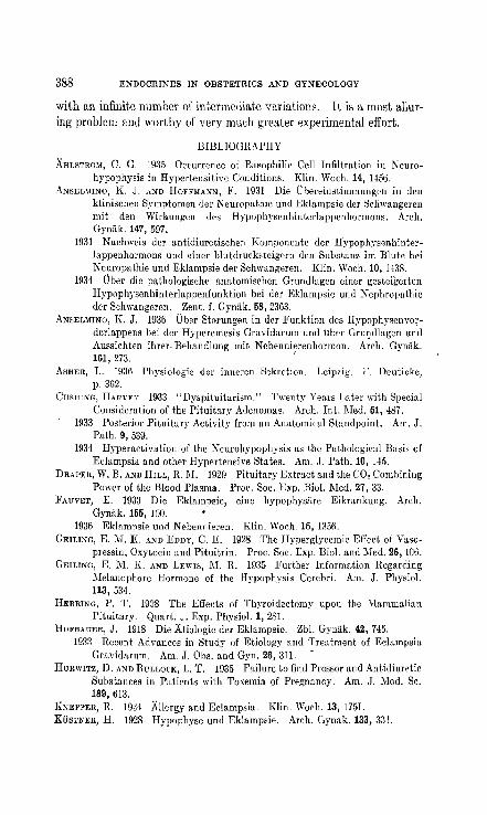
388 ENDOCRINES IN OBSTETRICS AND GY}lECOLOGY
with an infinite number of intermediate variations. It is a most alluring problem and worthy of very much greater experimental effort.
BIBLIOGHAPH Y
AHLSTROM, C. G. 1935 Occurrence of Basophilic Cell Infiltration in Neurohypophysis in Hypertensitive Conditions. Klin. Woch. 14, 1456.
ANSELMINO, K. J. A:;"D HOFFMANN, F. 1931 Die Cbereinstimmungen in den k1inischen Symptom en der Neuropathie und Eklampsie der Scliwangeren mit den Wirkungen des HypophysenhinterlappellllOrlIloll~. Areh. Gyniik. 147, 597.
1931 N "chwcis der antidiuretischen Komponellte der Hypophysenhinterlappenhormons und einer blutdrucksteigern den Substanz im Blute bei Neuropathie und Eklampsie der Schwangeren. Klin. Woch. 10,"H~R.
1934 Uber die pathologische anatomischen Grundlagen einer gesteigerten Hypophysenhinterlappcnfunktion hei der Eklampsic und Ncphropathic der Schwangeren. Zent. f. Gynlik. 58, 2363.
ANSELMINO, K. J. 1936 tIber Storungen in der Funktion des Hypophysenvo1"derlappens bei der Hyperemesis Gravidarum und liher Grlll)dlagen lind Aussiehten ihrer· Rehandlung mit Ncbennierenhormon. Arch. Gynak. w,m. •
ASHER, T.. 1936 Physiologie der inneren Sekretion. Leipzig. F. Deuticke, p.362.
CUSHING, HARVEY 1933 "Dyspiluitarism." Twenty Years Later with Special Consideration of the Pituitary Adenomas. Arch. Int. 1\Ied. 51, 487.
1933 Pusterior Pituitary Activity from an Anatomical Standpoint. Am. J. Path. 9, 539.
1934 Hypeructivation of the Neurohypophysis as the Pathological Basis of Eclampsia and other Hypertensive States. Am. J. Path. 10, 145.
DRAPER, W. B. A:;"D HILL, R.]\I. 1929 Pituitary Extract and the CO, Comhining Power of the Blood Plasma. Proc. Soc. Exp. BioI. Mcd. 27, 33.
FAUVET, E. 1933 Die Eklampsie, eine hypophysiire Eikrankung. Arch. Cynnk. 156, 100.
1936 Rklampsie und Nebennieren. Klin. Woch. 16, 1356. GEILTlW, R M. K AND EDDY, C. E. 1928 The Hyperglycemic Effect of Vaso
pressin, Oxytocin and Pituitrin. Proc. Soc. Exp. BioI. and }led. 26, 106 .. GElLING, E. M. K. AND LEWIS, 1\1. R. 1935 Further Information Regarding
Melanophore Hormone of the Hypophysis Cerebri. Am. J. Physiol. 113,534.
HERRING, P. T. 1908 The Effects of Thyroidectomy upon the }lammaliall Pituitary. Quart. J. Exp. Physiul. 1, 281.
HOFBAUER, J. 1918 Die Xtiologic der Eklampsie. Zbl. Gyniik. 42,745. 1933 Recent Advances in Study of Etiology and Treatment of Ecll1mpsia
Gravidarum. Am. J. Obs. and Gyn. 26, 311. HURWITZ, D. A~D BULLOCK, L. T. 1935 Failure to find Pressor and Antidiuretic
Substances in P[LticntR with Toxemia of Pregnancy. Am. J. Med. Sc. 189,613.
KKEPPER, R. 1934 Allergy and Eclampsia. Klin. Woeh. 13, 1751. KiisTNER, H. 1928 Hypophyse und Eklampsie. Arch. Gynitk. 133, 3:11.

TOXEMIA OF PREGNANCY 389
LEVITT, G. 1906 The Problem of an Antidiuretic Substance in the Blood of Patients with Eclampsia and other Hypertensive Diseases. J. Clin. Invest. 15, 135.
MARENZ', A. D. 1934 Action de ['extrait ante-hypophysaire sur ]'acide [actique du sang. Campt. rend. Soc. bioI. 117, 464.
1934 Acide Laetique sauguin des Chiens Hypophysoprives. Compt. rend. Soc. bioI. 117, 53.
1934 Modification du taux de I'acide lactique sanguin apres injection d'extrait retro-pituitaire. Compt rend. Soc. bioI. 117, 457.
RAS~[USSEN, A. T. 1936 The Relation of Basophilic Cells of the Human Hypophysis to Blood Pressure. Endo. 20, 673.
RUSSEL, D. S., EVA:-!S, H. AND CnOOKE, A. C. 103~ Two Cases of Basophil Adenoma of the Pituitary Cland. Lancet 2, 240.
SIEGEL, 1. A. A:-!D WYLIE, II. B. 1933 Blood Sugar Findings in Eclampsia and Preeclampsia. Am .. J. Obs. & Cyn. 26, 29.
SMITH, C. S. VAN MCD SMITH, O. W. 192.0 Excessive Gonad-Stimulating Hormone and Suhnormal Amounts of Estrin in the Toxemias of Late Pregnancy. Am. J. Physio!. 107, 128.
1935 Furtber Quantitative Determinations of Prolan and Estrin in Pregnancy. Surg Gyn. and Obs. 61, 27.
1935 Evidence for the Placentul Origin of (he Excessive ProIa" of Late Pregnancy Toxaemia and ]<;clampsia. Surg. Gyn. and Obs. 61, 1_75.
SPARK, C. 1935 Relation between Basophilic Invasion of tbe N europbyophysis and Hypertensive Disorders. Arch. Path. 19, 473.
STANDER, H. J. AND CADDEN, J. F. 1931 Blood Chemistry in Eclampsia and Preeclampsia. Am. J. Obs. and Gyn. 28, 856.
TonRFl-BLANClIO, ,J. ln~6 Die B1utmilchshnrc bei der Seh\\'ang"l'sehaftsl17.idosc. Arch. Gynak. 161, 276. .
lIRECHIA, I. C., RETEZEANU, AND GnOZE, I. 1933 Action de I'cxtrait du lobe posterieur de I'hypophyse (pitocine et pitrcssine) sur Ie calcium et la phosphor du sang. Compo rend. Soc. BioI. 103, 1363.
VORZIMER, J . • T., LA:-!GROCR, E. G., l"ISHBERG, A. ]\:f. AND l{APPAPORT, E. J\I. 1937 The endocrine basis of toxemias of pregnancy. In press.

CHAPTER XXV
PRECOCIOUS SEXUAL MATURITY
The syndrome of prec~cious sexual maturity is extremely interesting, for a complete understanding of its mechanism would enlighten us on the mechanism and etiology of normal puberty. Precocious puberty may be associated with tumors of the gonads, the pineal, the hypothalamus and midbrain, the hypophysis as well as hyperplasia and tumors of the adrenal cortex, and inflammation of the midbrain. The cases (1, 2 and 4) described below present no such evidence, and seem to be wholly on a functional basis. The remaining case (3) was complicated by the presence of a tumor of the ovary.
Case 1. J. P., No. 399227, age 4 years. First seen by Dr. Walter Timme, to whom I am indebted for this case, January 3, 1934.
Chief Complaint. Overdevelopment of pubic hair. Overdevelopment of breasts. Overdevelopment of height. Overdevelopment of weight. Three years in duration.
Family History. :Mother, 29, living and well; father 29, living and well. No exposure to tuberculosis. No familial history of carcinoma, gout, diabetes, asthma. No member of patient's family on either side has had disorder similar to patient's present illness.
Birth History. Only one pregnancy, born at term, labor not abnormal, instruments used. Child n~rmal at birth (9 pounds) except for growth on forehead and nose (subsequently sebaceous cysts). No melena or jaundice. Sat up at 8 months, walked at 12 months, sphincteric control at 18 months, single words at 12 months, talked at 18 months.
Feeding. Formula from birth. Orange juice and cod liver oil started in first month and continued under medical doctor's care. Solid food at 12 months.
Behavior. Sunny, good natured child, plays with other children, is obedient, not nervous. Seems to have plenty of pep and life. Mother has noticed nothing abnormal in child's behavior. No mental precocity.
Infections. Whooping cough at 18 months, duration 2 or 3 months. No sequelae.
Operations. Growths removed from nose at 10 months and 18 months at St. Luke's Hospital.
Prophylaxis. Small pox and diphtheria injections at 10 months. Present Illness. Patient was perfectly well during infancy and was seen by
medical doctor about once a month. When nine months old, it was first noticed by mother and medical doctor that pubic hair was developing and that her breasts were beginning to enlarge. Weight and height at this time not known but ever since then she has grown rapidly, and breasts and pubic hair have maintained a
390

PRECOCIOUS SEXUAL MATURITY 395
Blood: Rb.: 90 per cent. R.B .C.: 4,000,000. W.B.C.: 8150. Polymorphonuclears: 48. Lymphocytes: 48. Monocytes: 1. Eosinophiles: 1.
Glucose tolerance test: Control blood sugar: 101 mgm. Urine sugar: O. Glucose: 48.3 grams. Blood sugar I hour, 117; urine sugar, O. Blood sugar 2 hours, 119; urine sugar, O.
X-ray (Dr. Abbott): X-ray of knees, hands and wrists show epiphyseal development to be about as usual for this age.
X-ray of pelvis (April 11, 1936. Dr. R. C. Moloy): The inlet is well formed but definitely elongated. The side walls are straight. The sacrum has an average curvature, five segments, and the outlet is narrow with male type of pubic ramI.
FIG. 151C. Case 3. Precocious sexual maturity. Section of ovarian tumor. No evidence of follicle formation. XS60. (Courtcsy of Drs. H. B. Schoenberg and J. Felsen.)
Type of pelvis: anthropoid with gynecoid characteristics, straight side wails, masculine type of pubic rami and a long, narrow anthropoid type of sacrum.
Diagnosis. Precocious sexual maturity. Progress Note (November, lfl36). This patient has been observed at regular
intervals for the past-three years. The periods are regular, every 28 ± 5 days, and always associated with dysmenorrhea. The child is normal in all respects except for her sexual precocity.
Case 3. M. G., age 7 years . Referred by Dr. Henry Schumer. AprilI6,1934. (Figs. 151A, 15lE, and 15IC.)
396 ENDOCRINES IN OBSTETRICS AND GYNECOLOGY
Chicf Complaint. Menstruation: 2 months. Growth of breasts: past year. Family Ilistory. The patient is the second of two children. The oUu" sister
is now 12 years old and has been menstruating for the past few months. She presents no precocity. The mother began to menstruate at 11 years.
Past History. Normal spontaneous delivery. No tmuma. No previous illness except chicken pox. Growth, feeding and habits have been normal. Normal school course.
Present Illness. About a year ago it was noticed that' the patient's breasts hegan to grow. This has been especially marked in the past 6 months, when pubic hair was also noticed. First menstrual period MarcIl 5-9, ]934, second period 5 weeks later, duration 3-4 days, moderate in amount and without pain.
Physical Examination. The patient is a young well nourished girl whose general hony development corresponds to a youngster of her age, except that there is a definit~ feminine touch to her hips and wnist. She cooperates well and answers questions intelligently.
Head: negati ve. Eyes: pupils react to light and aceommodatioll. \ No eye signs. Mouth: deciduous teeth-] 0/10. Neck: negative. Thyroid not palpable Skin: texture normal, no pigmentation or excessivdmir. I,ungs: negative. Heart: no murmurs, no enlargement. nIood pressure: 128/80. Pulse: 88. Breasts: well developed, full. Gland tissue palpable. Nipples ann areoli of
adult type. Abdomen: soft, no masses. Right kidney pnlpable. l\[ons veneris: covered by hair, of adult type. Labia majom and minora: correspond to those of adolescent girl of 12 or 14
years. Clitori.: small. Vagina: patent. Uterus (rectal): corresponds in size to that of seven year old girl, cervix forms
greater part of organ, movable, firm, smooth. Adnexa: behind the uterus is a firm mass about 7-8 cm. in diameter, movable,
round, slightly sensitive, not adherent. Tubes: not palpable. Diagnosis. Granulosa cell tumor of ovary. Precocious sexual mnturity. Subsequent Course. Patient menstruated on May 1-7, 19:34. Spotted slightly
on l\Iay 13-15. Considering the phase of the cycle when the spotting occurred and the nature of this bleeding, a diagnosis of ovulation bleeding seems justifiable.
Follicular bormone (in the urinc)-4 rat units per liter} Follicle stimulating hormone (in the urine)-neglltiv,,"_
April 22, 1934.
The patient was admitted to the Bronx Hospital for operation (Dr. Herman B. Schoenberp;). Laparotomy May 21, 1934.
The right ovary was enlarged to about 8 em. in diameter. It was solid, not adherent. Surface epithelium intact throughout. Left ovary small, without the slightest tracc of follicles or corpora. Thc utcrus is small and is composed of
PHJ<JCUCIOUS Sr.:XUAL MA'l'UHl'l'Y 397
two-thirds cervix and one-third fundus. No glands palpable. Parametria negative.
Pathological Report. (Dr. J. Fclscn.) Specimen consists of a firm nodular encapsulated mass measuring 4 x 4 cm. On section mass is yellowish in color, soft.
This is a complex tumor composed of pa.le cells with central vesicular nucleus and granular cytoplasm, polymorphic, varying in shape from spindle to ovoid. The arrangement in places is suggestively perithelial and in others somewhat like glandular tissue. Many scattered areas of vacuolated und marginally spurred eosinophilic serous material arc present. No suggestive follicles containing ova can be seen. The possibility of a luteoma or adrenal ovarian rest or perithelioma or teratoblastoma can be considered (fig. 151e).
Diagnosis. Teratoblastoma ovarii with lutein stroma. Six months following operation the hormone findings were: Follicular hormone
-negative. Follicle stimulating hormone-negative. Following the opemtion thc periods stopped and the breasts regressed a little.
Early in 1936 the breasts began to swell again. Spotting on 'March 6 and 30, 1936. First regular menstrual period April 29, 1936. Sincc then the periods have occurred every 4 weeks, 3-1 days in duration, modemte in amount and without pain.
The breasts have continued their growth. Repeated rectal examinations show that the uterus corresponds in size to that of a girl of 10, but at the same time adult proportions arc now becoming manifest, in that there is noticeable gruwth of the fUlldus. The remaining uvary cannot be palpated.
April 19, 1936: Fullicular hurmune-l0 R.U. pcr liter. L M.P. March 30,1936. Fullicle stimuluting hormone-negative. April 29, 1936. June 14,1936: Follicle stimulating hormone-negative. L.)I.P. !lIay 26,1936. Follicular hormone-8 R.U. per liter. The general contour of the patient is distinctly feminine. She physically
presents a woman in miniature. Her psychic reactions seem to be those of a girl of 10 years.
X-ray of pelvis (Dr. H. C. Maloy): The inlet conforms to the anthropoid type. The sacrosciatic notch is moderately wide, but has a male character to it. The .side walls converge slightly. The subpubic arch is moderate in wirltli, possessing straight pubic rami indicative of masculine characteristics. There is a fusion anomaly of the spinous process of the first sacral segment. The epiphyseal lines of the iliac crest are fairly well formed for the age of the patient. The pubic ischial rami are also fused which, I believe, is a little unusual fur the age of the individual. While the pelvis possesses definite female characters, the outstanding findings other than this refer to the anthropoid characteristics with a few minor masculine characteristics.
X-ray of shoulders, wrists, ankles, elbows (June 18, 1936. Dr.' Kasabach): Films of the various joints, including the hips, shoulders, pelvis, elbows, wrists and ankles show scy~ml inter~sting findings in the various centers of ossification of the bones. According to ~ur anatomical chart, the epiphysis for the acromion processes and the apophyses of the os calci [Lnd the epiphyses of the upper ends of the femora usually appear and fuse [Lt a later age than 12 or 15 years. Tn this child, the epiphyses for the acromion processes of the scapulae have already appeared. The apophyses of the as calci have also appeared and have almost

398 ENDOCRINES IN OBSTETRICS AND GYNECOLOGY
completely fused. These findings suggest that some of these osseous centers have undergone a developmental stage which is somewhat more rapid than one would expect in a child of 9 years of age. On the other hand, the centers of ossification and epiphyseal changes elsewhere in the body seem to be in accordance with the patient's age.
Recent hormone determinations have yielded the following results. The positive F.S.H. on October 26, 1936 denotes ovulation.
F.H.-(October 25, 1936)-none found F.S.H.-(October 26, 1936)-positive.
F.H.-(November 1, 1936)-none found. F.S.H.-(November 2, 1936)-negative.
F.H.-(November 5, 1936)-none found. F.S.H.-(November 6, 1936)-negative.
F.H.-(November 15, 1936)-none found. F.S.H.-(November 16, 1936)-negative.
F.H.-(November 22, 1936)-none found. F.S.H.-(November 23, 1936)-negative.
F.H.-(November 29, 1936)-none found. F.S.H.-(November 30, 1936)-negative.
P.M.P.
October 8-14, 1936. November 1-6, 1936.
Case 4. D. A. M., No. 464589, age 13 (figs. 152A and 152B). September 17, 1935 ..
Chief Complaint. Persistent vaginal bleeding for past 5 weeks. Family History. Father and mother living and well. Two sisters, 19 and 16.
Show no precocity. Present Illness. Patient has menstruated regularly every month since the age
of 6 months. Secondary sex characteristics began to develop at six years, namely, breasts, pubic and axillary hair. These have progressively increased up to the the present time. Periods have always been regular, every four weeks, four to five days, moderate in amount and without pain. Patient had a regular period beginning July 20, lasting 3 days. One week later began to bleed again and has bled on and off for the past month. This has been associated with cramplike pains in the lower abdomen.
Physical Examination. Patient is a young girl with dark hair and blue eyes. She is very shy and not over-bright.
Head: negative. Eyes: react to light and accommodation. No gross disturbance in visual
fields. Mouth: hypertrophied tonsils. Teeth: some dental caries. Neck: no thyroid enlargement. Hair: pubic and axillary hair well developed. Some excessive hair on legs,
abdomen and upper lip.

PRECOCIOUS SEXUAL MATURITY
Lungs: negative. Heart: no enlargements, no murmurs. Pulse: 84.
399
Breasts: well developed especially in the subareolar areas. The breasts closely resemble those seen after treatment with large doses of follicular hormone.
FIGs. 152A AND 152B. Case 4. Precocious sexual maturity. Menses began at 6 months. Present age 13 years.
Body: is distinctly feminine, except for the legs which are very solid and muscular, and show no convergence at the knees.
Skin: of normal texture, flat pigmented areas over back. Abdomen: negative. Viscera not palpable. External genitalia: well developed, resemble those of a postadolescent girl.

400 ENDOCRINES IN OBSTETRICS AKD GYNECOLOGY
Clitoris: normal in size. Vagina: patent. Uterus (rectal): to the left of the midline, of adult proportions, and slightly
IIl1"ger than would be expected lit this age. Adnexa: to the right, in the region of the right ovary is a small ill defined cystic
mass. Impression. Precocious oexual maturity. Undetermined ovarian eyst? Basal metabolic rate: 0 per cent. ' X-my of pelvis (Dr. H. C. Molloy): The inlet is round ami wide having the
general appearance of an adult pelvis although the bones are delicate. The suture lines are more advanced than the age of the patient. The sacrosciatic notch is wide. The side walls are straight. The subpubic angle is wide.
Type of pelvis: this is a round gynecoid type Df female pelvis that demonstrates a maximum number of sex characteristics as far as the pelvic form is concerned.
X-ray of skull (Dr. Swenson): .Films of the skull show no evidence of bony or intracranial pathology. There is !1. very interesting appearance of the petrous portion of the temporal bones. The internal auditory me(1\US on the left side apPcllfs l(1rger than on the right. If an acoustic neuroma is by any chance suspected, this asymmetry might be due to such 11 process, or else we arc dealing with an incident"l asymmetry.
September 23, 1935: Folliculo.r hormonc-8 R.U. per liter. Follicle stimulating hormone-negative. L.i\1.P. Septemher 30-0ctober 3, 1935.
September 29,1935: Follicular hormone-16 R.U. per liter. Follicle stimulating hormone-negative.
Progress Nole. Early in December patient began to complain of occasional pain in the right lower quadrant. No nausea, vomiting Or temperature. Reexamination on January 30 sholl'S the right ovary to be distinctly above average size, freely movable (Professor B. P. Watson). The patient has not returned to the clinic.
It has been stressed in this volume that the establishment of sexual function requires, at least, the direct interplay of the adenohypophysisthe ovary-the genital tract and associated sex structures. This is the primary or direct meclmnisIIl. When anyone of the three is absent normal sex function is impossible. This mechanism is then influenced by other glands of internal secretion, and probably by the vitamines, in an indirect manner. It is morc than likely that this influence is exerted through the metabolism of the glands primarily concerned. This is the Recondary, or indirect mechanism. 'Ve know that at puberty a more or less sudden exacerbation of -the primary mechanism occurs. The point was previously made (Chapter XXIII) that this primary mechanil)m functions at a very low rate ill later intrauterine life and during the preadolescent period. The pituitary contains gonadotropic hormone, the ovary in turn is activated to secrete estrone, and finally the genital tract shows very slow but progressive growth.

I'HECOCIOUS SEXUAL MATURITY 401
Etiology of the Onset of Puberty
Therefore, the onset of puberty is not brought about by any new, previously non-existent mechanism, but by a sudden increase in activity of a previously functioning system. This sudden increase function may occur through severalm8chanisms, namely:
The Sudden Activation of the Anterior Pituitary Gland. The activation of the adenohypophysis could theoretically come from within or without. Up to the present time no data are available to show a sudden increase in potency of the pituitary gland. Nor do we know of any other gland that is capable of suddenly stimulating the pituitary. Rowntree et al. (1936) made the interesting ohservation that estrus hus been repeutedly demonstrated in the tcnth gcneration of 11 thymus treated strain of rats at nine to twelve d11Ys, in animuls weighing less th11n thirty grams. (Normally estrus occurs at about 60 days.) They also found mature follicles. Examination of the pituitaries of such precocious rats shows that the adult pattern (cell distribution) of the thymus injected animals is reached at an c11rlier period than in the control strain. Their findings suggest the theory that puberty is caused by the activation of the pituit11ry by thymus. Hence precocious puberty could be produced by premature activation. It must be pointed' out that cleven generations of rats were required to produce this effect, 11nd that 11 physiological response in rodents C11nnot be directly 11pplied to the human.
The Progressive Maturing of the Ovary. "INc h11ve previously pointed out (Chapter XXIII) that in the rodent the ovary will respond to stimulation only ~ftcr it has matured to a certain extent. Apparently 11t about the age of 13 years the ovary has matured sufficiently to respond fully to the pituitary stimulus. Evidence to substantiate this point is lacking for the human. On the c_ontrary, we have previously indicated (Chapter XXIII) that the hUlll11n ovary is inc11pable of response after 11 certain age. Hence the theory is suggested th11t the oV11ry occupies the dominant position in the mechanislil of puberty. When it has reached m11turity, and it must be remembered that the hypophysis is probably an important force in this attainment, it responds to stimulation by accelerating its activity. Case 3 sheds some light on thi"s'point. The right ovary was replaced by a lilass of granulosa-like cells. The left ovary ,Y118 undeveloped. There were neither follicles nor corpom I1tretira. There W11S no evidence of functional activity, either past or present. Hence the etiology of the precocity could not have been prim11rily outside of the ovary, for the stimulus

402 ENDOCRINES IN OBSTETHICS AND GYNECOLOGY
would have involved both ovaries. Furthermore when the pathologic ovary was removed the precocity subsided for about 2 years. Then menstruation and growth of the breasts began anew. The remaining ovary has now ripened, truly at a premature age. But there seems to be some family predisposition to an early puberty, for both her mother and an older sister began to menstruate at about eleven. (Weare constantly aware of the potential possibility of a metastasis from the original tumor. The patient is thoroughly examined every month, but to date, no such new growth has been found.)
As to the nature of the process by which the ovary attains maturity very little can be stated. It must reside within the fundamental metabolic processes of the cell. It is not primarily a matter of circulation, for the same arteries supply the ovary of the preadolescent as the mature individual. Nor is it a question of the number of primordial follicles, for opinion is still divided whether any such increase occurs after the ovary becomes a complete morphological entity.
The Release from the Antiho~mones. Col IIp's concept of antihormones has introduced another problem into the syndrome of precocious sexual maturity. We may assume for purposes of argument, that for every hormone there is an antihormone. l"ndcr conditions of normal fUllction only such quantities of hormone are released from the hormoneantihormone complex as are cssential for normal function. Depcnding on the titer of the two components of the complex many shades of function are possible. Thus normal puberty would result from the liberation of the hormone (gonadotropic or ovarian) from its antihormone. Precocious puberty would result from premature liberation. This theory is very interesting from a speculative point of view, but evidence to support it is lacking. No antibody has been demonstrated for estrone or progesterone. Gegerson, Clarke and Kurzrok (1936 and unpublished results) have been unable to detect gonadotropic antibodies in patients with long standing amenorrhea or during lactation. It is true that gonadotropic antibodies can be produced both ill the rabbit and the human by the parenteral introduction of large quantities of gonadotropic hormone in the form of pregnancy urine extract, anterior pituitary extract, or mare serum. But this implies the introduction of an antigen from without, a condition not- found during normal fUllction. Further development of the antihormone problem will be watched with great interest in this connection.
Evans has recently (1936) brought the topic of the gonadotropic antagonist to the fore (Chapter XXVII). This antagonist inhibits the ovary from being stimulated by the gonadotropic hormones. Pu-

PRECOCIOUS SEXUAL MATURITY 403
berty could potentially be due to a removal of this antagonist, and precocious puberty to a premature removal.
Discussion
TheRe cases of ~cxual preeoeity pre~ent many points of great interest. The preadolescent relationship between the fundus and the cervix remains in spite of regular period~. The uterus does not exceed in size that of normal· girls of the same age. Thc endometrium has matured, the myometrium has not. This observation substantiates the point made in discussing the results of treatment of primary amenorrhea, that is, the uterus is the last (slowest) to respond to the effects of estrone. Case 1 substantiates this from another angle, for even though the secondary sex charactcristics have been fully devcloped, especially the breasts, the myometrium and endometrium have retained thcir normal preadolesccnt 8tate. iVlcnstruation has not begun as yct.
Case 1 presented an additional precocity of growth. At four years. of age her height was 57.5 inches (normal 41.5 inches). This implies exccssive activity on the part of the growth hormone. The frequent association of sexual precocity and early ossification of the epiphyses is not present in this case. This is the more interesting in the light of case 3 in whom the osseous centers have undergonc a developmental stage which is somewhat more rapid than one would expect in a ehilrl of ninc.
The architecture of the pelvis presents anuther interesting proulem in cases 2 and 3. In both the diagnosis is made of anthropoid type with masculine characteristics. We believe that the terms "anthropoid" and "masculine" used in this connection are merely descriptive. These patients present the effects of premature exposure to adult quantities J
of follicular hormone. No evidence has ever been presented that they arc also producing male sex. hormone, even though we have pointed out that whenevcr large quantities of estrone are produced, there is an accompanying production of androstcrone, and v. v. It is difficult to believe that such small potential quantities of androsterone would be . ~lIfficient to modify the architecture of the pelvis in the presence of such large quantities of estrone. Case 4, on thc contrary, presents at 13 years a fully developed gynecoid pelvis.
In the description of tbese cases we have pointed out that the breast development differed from the type produced in cases of primary amenorrhea (hypogonadal) hy large doses of estradiol benzoate (progynon-B). These precocious girls presented fully developed breasts and in three gland tissue could be palpated. Since the fully developed gland in the hum all re<.ju·ire~ active curpll~ luteulIl fUIlctioIl, the ureast

404 ENDOCRINES 11\ OBSTETRICS AND GYNECOLOGY
development signifies a complete ovarian cycle, that is, follicle formation, ovulation and corpus luteum formation. Case 3 presents evidence of ovulation, a~ manifested by the sudden excretion of follicle stimulating hormone on a single day during the intermenstrual period (Kurzrok, Kirkman and Creelman, 1934).
The pigmentation of Case 4 may signify something or nothing. It is believed that pigmentation of this type may be a function of the adrenal cortex. As stated above, disturbances of the adrenal cortex may lead to precocity. This is all the more interrsting in view of Reichstcin's (1936) finding that certain fractions from the adrenal cortex (not cortin) possess estrogenic activity.
These cases of precocious sexual development are very much Blor,e than museum curiosities. A more complete understanding of the etiology behind this disturbance would go a long way towards the elucidation of some of the basic moblems of sex.function.

CHAPTER XXVI
INTERSEXUALITY
Medicine has always exhibited an intense interest in the individual who presents the characteristics of both sexes in a varying degree. An enormous literature has grown up on the subject but only recently has'a semblance of order arisen. Too much attention was paid to external morphological variations, and very little to inherent biological laws. The difficulties arose from the fact that in the hermaphrodite no correlation existed between the ma8culine or feminine characteri8tics. One could not determine the character of the gonads from a study of the habitus, the external sex characteristics or the psyche. Halban (1903) made an interesting observation that shed some light. He stated that there exist people without gonads, but who present either a masculine or feminine habitus and internal genitalia. He concluded from this that the differentiation into male or female during embryonal development is independent of the gonads. Furthermore, the sex of the fertilized ovum or zygote is therefore determined at the moment of fertilization. Further progrBs8 was maue by Hichard Goldschmidt (1931). His concept of intersexuality will be followed in this chapter.
Zygote Intersexuality
Goldschmidt chose for his experiments a butterf'J, lymantria dispar. It was chosen because the sexes are easily discernible, and by crossing a given strain with other, from a different locality he was able to produce various dr:grees of intersexuality. Ijis work led to the conclusion that in the inters exes all organs develop according to one sex, but that at a given point of development a d~fiection (or turning, reversal, Drehpunkt) occurs and development continues according to the opposite sex. Whatever different.iation has been reached (determined) along the original pattern the intersex retains, but whatever has not developed at t.he onset of this second phase, now does so but according to thr: oppo8itl!-sex. The r:arlicr this dcflr:ction (turning point) occurs the fewer organs differentiate according to the original (genr:tic) sex, and the greater is the number that differentiate according to the opposite sex. Even tlie gonads IlIay be involved in this reversal. Thus a gonad having been genetically determined as a testis may then develop
405

406 ENDOCRINES IN OBSTETRICS AND GYNECOLOGY
from the turning point on as an ovary. It therefore becomes possible for a gonad to have the morphological make up of a testis and function as an ovary, that is, prodllce female sex hormone.
Goldschmidt's experiments led to the conclusion that an intersrx anlage results from the different valencies of the sex chromosome. That is, valencies greater or less than normal result in intersexes. Moszkow-
Period oj ]}iferentiAlion
Ovum ~ ® +- (!oY
\ I MM) F,: Nonnat maJ4 @
FIG. 153. Type r
~--------L_------Z~o~
FIG. 154. Type II
ieh (1936) illustrates this in the following manner: Every living ccli is bisexual, that is, it contains within its nucleus J:llasculinizing (M) and feminizing (F) factors. In the human they exist in the sex chromosomes. The only exception are the spermatozoa, of which there are two kinds. One half of the number has a sex chromosome, while the other half has not. So that the spermatozoa may be divided equally into 1\1 and MF types, hence of all hody cells only one half of the sper-

INTERSEXUALITY 407
matozoa contain no F component. On the contrary, all ova contain buth an lVI and F factor, hence they are all lVIF. In the male M is dominant, in the female F. When fertilization occurs we have the possibilities shown in figure 153.
In this case the sperm has no F factor, hence in the zygotc the M factor dominates. Curve lVIlVI signifies constant masculine dominance
®+~ \ / MM<F' @
FIG. 155. Type III
@+~ \ / MM'>FF
@
FIG. 156. Type IV
in the developmcnt of all organs. The individual is a normal male. (Sec fig. 154.)
In this case the sperm has anlVIF factor, hence in the zygote F factor predominates. CtlfVe FF signifies constant feminine dominance in the development of all organs. The indivirllllil is a normal female. (See fig. 155.)
Let us sllppo~e that UlC ji" factor in the ovum has a feminizing va-

408 ENDOCRINES 1:"1 OBSTE~'l\ICS AND GYNECOLOGY
lency twice as great as normally. Hence in the zygote F' will predominate in spite of two MM factors. The organism will begin its development apparenlly as male, but hpcauBP of the dominance of the F' factor a reversal occurs and the organism continues its development as a female. (See fig. 156.)
Let us suppose that the lVI' factor in the sperm has a r,nasculinizing valency twice as great as nurmally. Hence in the zygote MM' will dominate over FF. The organism will begin its development as a female, but because of the dominance of the !VIM' factor a reversal occurs and the organism continues its development as a male.
Goldschmidt's experiments show that Halban was correct ·in his assumption that the organs can differcntiatr as male or female in the complete absence of gonads. Goldschmidt' calls an individual who began embryonic development as a female and finally ended as a male, a feminine intersex. The reverse is knowIl as Ii male intersex. Both types compose the zygotic'i~terscxuality. ' The gonads do not influence the development of this. type of intersex, but instead are influenced by it. Hormonal differentla~tion occurs in the human during the second embryonal month, hence zygotic differentiation occurs in the entire absence of sex hormohcs.
Hormonal Intersexuality
This form of intersexuality occurs in individuals whose sex has already been determined (zygotic). Hormonal intersexuality depends upon the action of a sex hormone upon an individual of the opposite sex, that is, female sex hormone on a male, or male sex hormone on a female l
The action is reversible. Hormonal sexuality occurs as the result of tumors or hyperplasia of the adrenal cortex, of masculinizing tumors of the ovaries, called by Robert Meyer arrhenoblastomas, and certain tumors of the anterior hypophysis (Cushing). Their effect depends upon the age of the individual, being in general more profound the less developed the individual is. There are characteristic changes in the distribution of hair and fat, in the growth of the clitoris and larynx, and in the breasts. Associated changes in the function of the genital tract occur. It is of importance to remember that such tumors (adrenal, ovarian ancl hypophyseal) may oecur without producing intcr-
I It must not be forgotten tbat even in zygotic intersexuality we are dealing with chemical complexes within the gene. The M and F factors [Irc chemical molecules thut direct development in 8. certain direction, but whether they are hormoncs or catalysts we do not know.
INTEltSEXU ALITY 409
sexuality. Furthermore, certain tumors of the adrenal produce feminization instead of the usual masculinization. This depends upon the character of the metabolism of the tumor or hyperplasia. Reichstein (1936) has shown the presence of both androgenic and estrogenic activity in the cortical fractions of the adrenal. What factors govern the productl\JIl of either of the sex hormones by these tumors? The answer may be the character of the tumor metabolism, the nature of the stimulus that produces the growth and, finally, the genetic pattern of the host. The last concept brings us back to the .zygotic intersexualityof Goldschmidt. May the M and F components of some individuals be so delicately balanced that an addition of one or the other sex hormones furnishes the impetus to precocious maturity (when young), or towards heterosexual characteristics. May not other individuals be so preponderantly M or F, that their systems appear "buffered" against the action of the opposite sex hormone. It will be of great interest to determine whether tumors of the adrenal eortex (or arrhenoblastomas) which do not produce the characteristic heterosexual changes, produce excessive sex hormone, but that the hormone fails to "make an impression" upon the individual.
lVIoszkowich proposes to differentiate the zygotic intcrsexes by the presence of prostatic tissue. The male intersex can be differentiated from the female intersex by the position of the prostate in relation to the primary urethra, that is, that portion of the urethra between the bladder and the junction with the vagina to form the urogenital sinus. When the prostate lies cephalad to this junction the individual is a male intersex, and when caudal to it a female intersex.
Case. F. P., age 36, No. 458373. Autopsy No. 11949. This case was first seen by Dr. Arthur H. Blakemore and will be published by
him in extenso. I am greatly indebted to hi~ for permission to publish the following description.
History 419950. Chief Complaint. Pain in lower abdomen and constipation for 6 weeks.
Family History. There is no familiul history of hermaphroditism, carcinoma, or syphilis.
Past History. No acute illness. He was born a hermaphrodite, always dressed as a male, took a male part. Has had no social difficulties. Developed normal sexual attracti~n to female at -12. Married 7 years Ilgo and has had normal frequent intercourse with rCllsonable plclLsurc. Had nothing to suggest menstrual periods. His wife's single pregnancy resulted in a normal boy, now 6 years old.
Present Illness. About 6 weeks ago while working as a painter, patient developed sharp pain in the right loin wbich after about one hour shifted and settled in the suprapubic region, and there was dull ache in the right lumbosacral region. The pain has occurred off and on for the past 6 weeks and has been Ilggravated by
410 ~]NDOCllIN~]S IN OBST]<]'I'RICS AND GYNECOLOGY
heavy meals. Constipation has been present since the onset of pain and has increased to the point at which daily enem:1t:1 have been necessary. Ko nausea, vomiting, or hload in the stools. The pain h:1s not been colicky in type. He has lost 14 pounds in the P:1st six weeks. During the present illness there has been slight burning on urination for which he has been taking urotropin.
Ph!Jsical Examination. Temperature 100'; pulse 100; respiration 20; blood pressure 160/105. A well develuped and well nuurished yuung adult appearing decidedly male in facial features. Female type of breast and figure. Head: eyes, ears, nose and mouth, negative. No cardiac enlargement, rough apical systolic murmur. Rhythm regular. Abdomen: prominent, particul:1rly in the lower half. On the right side arising out of the pelvis and extending almost to the umbilicus a large firm non-tender m:tss is felt. On rectal examination this mass is felt occupyin,g the pelvis.
Gcnito-U1·inary. Feminine distribution of hair. Very small penis with cxt~me hypospadias. About 1 em. behind urethral meatus is a very small opming lying between two ridges similar to the labia and containing no testes. A catheter can be passed in this opening extenrling llpward and haekward for a distance of 7 em. Fluid of a very mucoid consistency is exuded on the catheter. Reflexes normal. Extremities negative.
Laboratory Findings. mood count: W.B.C. 10,360; polymorphonuclears 77 per cent; urine negative save for a moclerate number of \V.B.C., singly aIld in clumps in the sediment. X-ray No. 135843; July 9,1935; abdomen shows no evidence of calcification in the abdominal muss. Its uutline is not discernible. The left kidney outline is obscured. Steroes and lateral films of the pelvis were taken following the injection of lipiodol iuto the perineal opening just before the glans. This outlined a smooth pocket about 5 em. long. It is wider below and narrower above as might well be seen with a pseudovagina. This points almost directly posteriorly, and there is no lipiodol entering the lumen uf possible uterus or cervix.
The pelvis is of the android type. Another x-ray of the pelvis was made with the comment that it is basically a female pelvis with masculine characteristics, and can be duplicated many times in individuals of female sex. Cystoscopy .J uly 12: third degree hypospadias. There appears to be a normal bladder except for compression from the mass outside of the bladder which forces the vault down. Veromontanum rudimentary. Aschheim-Zondek Test negative. F.S.H. determination 3 out of .. negative, with a slight stimulation on the fourth.
Course. A diagnosis of hermaphroditism with teratoma of ovary or abdominal testis was made and uperatiun performed one week after admission. A right round ligament, ovary, Fallopian tube and uterus were found. There was apparently no cervix uteri, nor were there any discernible structures on the left side. The ovary on the right waS partially repbced by a huge degenerating tumor, the pedicle of which had twisted through 180°. It was densely adherent to the rectum and sigmoid and filled the cul-de-sac and nearly the entire pelvis. There was considerable hemorrhage beneath the capsule .. There was no evidence of extension or metastasis. Tumor, uterus, right ovary and tube were removed. Diagnosis of the surgical specimen No. 58089 was malign:1nt teratoma of the ovary. No testicular tissue was found. The teratoma waS thought to be of a solid variety which is exceptionally rare and appeared to be very malignant. The patient made a good recovery from the operation and returned home on the 15th day postopemtive. He came to the hospital again two montlm later complaining of

INTERSEXUALITY 411
abdominal pain whieb was constant and dull, situated cbiefly in tbe rigbt lower quadrant and in tbe rigbt costa-vertebral angle. During tbe past five days be has been baving tenesmus and many small watery stools. He hus lost strengtb and weigbt. Pbysieal examination disclosed a tense, full, tender abdomen witb many masses palpable in tbe epigastrium, in tbe suprapubic region and in both lower qUfidmnts. Slight shifting dullness. X-ray of tbe chest showed the lung fields to be clear. Patient remained in the hospital for 2t weeks, extremely ill, and running a low grade fever. Two days before his death his urine was positive for l<'.S.H. and showed four units of oVl1rian hormone per liter.
Diagnosis. Pseudo-hermaphroditism. Teratoma of right ovary. A utopsy by Dr. Pearce, Oclober 24, 1935: (Only part of the autopsy protocol is
presented.) The body is that of a young adult indiviuual whose skeletal and muscular development is excellent. The general configuration is masculine. The calvarium is symmetrical, presents no bony abnormalities, and is covered with an abundant growth of graying black hair. The pupils are in mid-dilatation. There arc no petechiae of the conjunctivae nor is there any icterus of the sclerae. There is no discharge from the nose. The front teeth appear in good condition, but post-mortem rigor prevents examination of the inside of the mouth. The mucous membranes of the mouth are bright cherry red in color. There is fairly well marked post-mortem lividity and moderate rigor. The t"achea is in the midline. The thyroid is not palpable. The thorax is symmetrical. The breasts are moderately full and have a soft fatty consistency: The nipples l1nd areolae are female in type. A fe,,- small shotty glands are present in the neck, but there is no general adenopathy. The abdomen is distended. No masses, however, can be felt through the abdominal wall. There is a healed surgical incision measuring 18 em. in length reRching from a point just ubove the luteral to the umbilicus to the symphysis. The pubie hair has the female distribution. The penis measures 5 cm. in length (fig. 157A). The prepuce is reflected ofT the gluns and uescends laterally and posteriorly for a distance of about 4 cm. and its lower portions have the appearance of labia minora. There is n third degree hypospadias and what appears to be an urethral opening is prescnt at the most posterior point to which the prepucial folds rcach. A small amount of somcwhat watery mucoid material is present in the mcatus. There is no vaginal opening. The rectum appears normal and running from it to the urethral opening there is a raphe resembling that seen on the scrotum. No testes eaJ. be felt in the mther loose pigmented skin on each side. There is no clubbing of the fingers or toes. The nail beds are bright pink. Needle puncture wounds are present on the anterior and medi",l aspects of both thip;hs and in the left antecubital spacc. The abdominal fat is bright yellow in color. The abdominal musculature is red and well developed.
Adrenals. Are normal in size and shape. Their cortices are well filled with lipoid, and are demarcated from the n",rrow pearly gray medulla by reddish brown intermediate zone~. The rigllt adrenal tQgether with the kidney to which it is attl1ched has beeu pushed upward by the tumor in the right lumbar gutter and right'side of the abdomen and has comc to lic in a loose mass of tumor tissuc between the liver and the diaphragm on the superio-lateral aspect of the forme,'.
Pel,,;" (Jr(Jans. The hladder is liMd by wrinkled shiny white mllcosa which is slightly injected at the fundus and in the region of the trigone. In the latter situation it is also somewhat roughened. Both urethral orifices are freely patent.

412 ENDOCRINES IN OBSTETRICS AND GYNECOLOGY
The urethra is large, measuring 2 cm. in diameter. At a point 3 cm. from its opening in the bladder it is joincd by the vagina through a lumen 0.5 em. in circumference. The two structurcs then continue as a common canal to the external meatus. This portion which is lined by mucous membrane rcsembling that of the urethra is 4 cm. in length. It is surrounded by erectile tissue resembling a corpus spongiosum. The vagina begins at the described opening in the urethra and extends upward posterior to the urethra for a distance of 7 cm. At its uppermost portion there is a small structure about 2 cm. in length and 1 cm. in width resembling cervix. This contains a narrow lumen filled with brownish-red mucoid material. Its upper pole is buried in a mass of tumor tissue and in this region there are several black silk sutures. The mucosa lining the vagina is not remarkable. The wall is thick and is made up of tough fibrous tissue. The cervix and upper part of the vagina lie behind the bladder. There is a mass of firm dense tissue 0.5 cm. in width between the lower portion of the vagina and the uterus. It appears to be composed of thick fibrous tissue in which no definite structures can be made out. Between the vagina and rectum is a small amount of loose areolar tissue in which lies an extensive venous plexus. No structures resembling seminal vesicles can be found nor is there around the vagina or urethra any semblance of prostate. The rectum itself is normal. No testes or ovaries can be found on either side.
Uterus (S.P. No. 58089) (fig· i57E). The section consists of large, intertwining masses and bands of smooth muscle between which lie areas resembling endometrium. In these regions there are gland-like spaces lined by columnar epithelium. They vary greatly in size, some being extremely small, while others occupy several low power fields. They are for the most part empty, but some of the larger ones contain red blood corpuscles and lymphocytes, and an occasional polymorphonuclear. Beneath the epithelium there is a loose cellular tissue closely resembling that found in the normal endometrium. Here, too, occasional small hemorrhages have occurred. The combination of these glandular elements with the rather dense irregularly arranged bands of smooth muscle suggest an adenomyoma. ::\1asses of dark, yellowish-brown pigment often occurring in monoculear cells are found in places between the smooth muscle bundles. This resem bles hemosidetin.
Right Uterine Tube (fig. 157e). The section consists of rather loose areolar tissue among which are clumps and bands of smooth muscle, varying in size. At one side there is a cross section of a Fallopian tube which does not differ in any essential from the normal. At another point and occupying one low power field
FIG. 157A. Case 1. Intersexuality. Penis and perineal region. Note the catheter in the common urethra and vagina. (Courtesy of Department of Pathology.)
FIG. 157B. Case 1. Intersexuality. Section through endometrium. (Courtesy of Department of Pathology.) .
FIG. 157C. Case 1. IntersexualIty. Section through tube. (Courtesy of Department of Pathology.)
FIG. 157D. Case 1. Intersexuality. Adenoid struct.ure at junction of urethra and vagina. Resembles prostatic glands. (Courtesy of Department of Pathology.)
FIG. 157E. Case 1. Intersexuality. Ovary showing follicle. (Courtesy of Department of Pathology.)
INTERSEXUALITY 413
FIGs. 157A-E

<114 ENDOCRINRS IN OBSTETRICS AND GYNECOLOGY
are four gbndlike Rtructures, the lumina of which are filled with acidophilic coagulum. These are lined by closely packed columnar epithelium, and each is surrounded by a distinct broad muscular wall. At a distance from these and entirely separated from them there is a cystic structure about I t low power fields in size. It is lined in part by columnar epithelium and in part by heaped up layers of polygonal epithelial cells whose cytoplasm is in tensely basophilic. The lumen is empty. Many dilllted lympluttics are scattered through (he section.
B. This is a sccti"n taken from the left side of the uterus in the region of the site of the Fallopian tubc. This consists of bunules of smooth muscle and connective tissue among which lie seveml tubular structures lined by columnar epithelium and surrounded by II muscular 11'311 varying in thickness. One of these is much larger than the others, and appears dilated. There is likewise much less muscle in its lI'al1. It is impossible to determinc whether these structures represent a rudimentary fo'aliopiun tube or whether they [lrc merely parts of II paraovurium.
Right Ovary (jig. 157D). Muchof the section consists of normal ovarian stroma, in which there is one perfectly formed primat·y follicle containing an ovum. Tn addition there are several normal appearing corpora albicantia. J3eneath the typical ovarian stroma is a broad zone of loose fibrous tissue containing severnl small hemorrhages.
flighl Ovary and l'umor. This is II section of the"ov[lfY including II purtion of tho tumor [Lrising from it. Again there is typical o\rarian stroma and several corpora albieantia. No primary follieles arc present, however. In the loose fibrous tissue seen in the other scction there are here numerous anastomosing gland-like spaces lined by columnar epithelium which extend into the surrounding 'tissue infiltrating between the connective tissue fibers in solid cords and in adenomatous projections. There arc no base~ent membranes around the glf1nd-like spaces. The epithelial cells [Ire deeply b(lsophilie, varying considerahly in size. Theil' nuclei arc for the most part round or oval. An embryonic type of fibrous tissuc surrounds the epithelial ceJls and forms the stroma for them. This varies greatly in density, "ometimes there arc closely packed masses of nuclei with dense fibcrs between them, while in other areas there Ure fell' nuclei and few delicate fibers, The nuclei likewise Vl1ry greatly in size, some being extremely large and vesicular, while others are small. None of them contain much chromatin. A few mitotic figures can be found both in the stroma and tbe epithelium. Much hemorrhage and a considerable amount of necrosis is present.
1'urnur. Parts of this section resemble the neoplastic elements described in the preceding one. There are, however, several aduitiunal features. In many places the stroma of embryonic fibrous tissue is much denser in uppeamnce, resembling that of a fibrosarcoma. There are numerous large and small nodules of only slightly atypical cartilage. Sometimes tbe transition from the embryo!li~ stroma to the cartilage is almost indistinguishable. A few of the cartilaginous islands are surrounded by [Lcidophilic hyaline material closely resembling osteoid. Also present arc several groups of intercommunicating· large blood-filled spaces, apparently lined by epithelium and resembling a cavernous hemangioma. As in the preceding section hemorrhage and necrosis are prominent.
Pancreas. Tlle acinar cells are normal save for postmortem change. The islands of Langcrhans arc numeroUS. They vary quite markedly in size, some being very small, while others arc large, and sometimes elongated. There seems

INTERSEXUALITY 415
til be a slight increase in the amount of cOIlncetive tissue arollnd the islands and within them. Pancreatic ducts are not remarkable. ,
Adrenal. The cortical cells contain" moderate amount of lipoid. In the inner zone of the cortex at the junction of cortex and medulla the cells are rather markedly autolyzed The medulla itsel£ is normal save for an advanced degree uf shrinkage uf the cells. Chrollutiflllity, however, Cllllnot he seen.
Breast. The section consists of normal resting mammary gland. There are large dense masses of collagen in which lie groups and single g1tllld acini. The epithelium is not remarkable, although the majority of the glands are small. Some of the glands are dilated Ilnd contllin granular pink precipitate
Thyroid. The glillld is damaged by autolysis, and much of the colloid has escaped into the interacinar tissues. The acini vary slightly in size, but arc for the most part lined by low cuboidal epithelium and are well filled with colloid.
Pituitary. The gland is wide and flattened. The anterior lobe is composed for the most part of acidophilic cells. A moderate amount of colloid is present at the junction of anterior and intermediate lobes. The posterior lobe is normal.
Cervix. One end of the section is lined by squamous epithelium. In the remainder there is no mucosa, although there is a small portion somewhat suggesting endometrium. Beneath this are bundles of smooth muscle similar to those in the uterus.
Junction oj Vagina and Urethra. One side of the section is covered by squamous epithelium of the vagina, and the other by pseudostratificd colulllnar. At the tip these two meet in a series of fingm·-Iike projections covered by the latter type of epithelium, mueh of which has been torn off. Between the two surfaces arc duct-like spaces lined with heaped-up epithelium suggesting that seen in the prostatic ducts (fig. 157E).
Pineal Gland. The gland is quite cellular but comparison with others at this lind greater ageshol\'s tha t this cellularity is within the limits of normal variation.
There iii no doubt that this case presents an intersex, and probably of the male type (accepting Moszkowich's dictum of the position of the prostate). This individual began embryonic life as a male but shortly, at an early turning point (Goldschmidt's "Drehpunkt") continued di.tfcrentiation as a female. The case would belong to Type 3 of Moszkowich's classification, where lYeVI < F'/ The development as a female continued far enough, so that follicles and corpora lutea (as evidenced by the corpora albicanta) formed. Unfortunately an insufficient number of hormone analyses were made. An estrone determination done one day before exitus showed 4 R.U. per liter. Considering the histological make-up of the ovary, we would expect estrone pro-
. duction.
Case 2. 'I P". Tilis r,aRp. is one of three ~iRtr.rs whORe history and physical findings is to be pr~sented in extenso by '!\fishcll (1937). They had the following features in common. l<'eminine external genitalia, normal appearing breasts, inguinal hernias containin!!; testes including crididyrnus and rete. Internal genitalia were absent. Figures 158A and 15813 present one of the Io\onuds found

FIG. 158A. Case P. Test('s of illterHl'X tYJl<'. Tubule's resemble those of crYJltorchid tcstes. Note marked overdevelopment of interstitial cells. X160. (Courtesy of Dr. D. R. Mishcll.)
FIG. 158B. Case P. Testes of intersex type. Marked overdevelopment of interstitial tissue. X 1000. (Courtesy of Dr. D. R. MishelJ.)
416
INTERSEXUALITY 417
in case 2. There is an absence of spermatogenesis, though cells resembling spermatogonia arc present. There is a marked over development of the interstitial tissue. The following hormonal titers w('re obtained:
E strone
Before operation. . .72 R.U. per liter
After operation: July 15, 1936. None August 10,1936 .None
August 20,1936 .. . ... None September 17, 1936 . . .None September 18, 1936 . . None September 19, 1936 . . . . None
Follicle Stimulating Hormone
Not done
Not done Positive- greater than 70
M.U. per liter Positive Positive Positive Positive
The finding of estrogenic hormone before the removal of the testes and its absence after their removal indicates that the estrogenic substance was produced by the testes. It is believed that all testicular hormone is produced by the interstitial tissue, hence we believe that it was aiso the source of the estrogenic hormone. But whether this substance was actually estrone or a modified testosterone molecule we do not know. This casc leads to the following concept. According to Halban it is impossible to determine the character of the gonads from the nature of the external habitu". To this we would add that it is impossible to prognosticate in special situations (as in this case) the character of the gonadal hormone from the histologic picture of the gonads.
Hormonal Intersexes- Arrhenoblastoma
In a previous Chapter wc have discussed hormonal interscxes due to disturbances of the adrenal cortex and also the anterior hypophysis. A third type is caused by masculinizing tumors of the ovary. Considerable work tending to clarify the microscopic characteristics of these tumors has been done by Robert Meyer (1931) and Novak (1936). To date no comprehensive hormone studies are available, though in a case of arrhenoblastoma Szathmary (1934) reports an absence of estrone before operation and 130 M.U. per liter of urine after the removal of the arrhenoblastoma. One would expect the production of male sex hormones by these tumors.
Here the histologic picture ranges betwecn highly differentiated testicular adenomas, through a group showing varying numbers of tubules and acini, to a highly undifferentiated group which resembles sarcoma and may show an occasional epithelial tubule. The more undifferentiated the tumor type the greater are the masculinizing fea-

418 ENDOCRINES IN OBSTETRICS AND GYNECOLOGY
tures. About one-third of the adenomas show any degree of masculinization. The symptoms are usually amenorrhea, hirsutism, deepening of the voice, hypertrophy of the clitoris, a loss of subcutaneous fat making the figure more angular, and a decrease in the size of the breasts. Some symptoms may be more extensively developed than others. Upon the removal of the tumor the feminine characteristics return.
Phelan (1934) reported an interesting case and through his kindness I am able to reproduce a microphotograph of the tumor (fig. 159). The
FIG. 159A. Arrhenoblastoma. TIl(' tubular structure is ver)' distinct. X 160. (Courtesy of Dr. G. W. Phelan.)
patient ,\'as twenty years of age, had never menstruated and had a masculine distribution of hair. Voice coarse and distinctly masculine. Breasts flat, general figure angular. Vaginal examination revealed a well-formed introitufl, save that the labia were smaller than normal. Clitoris hypC'rtrophiC'd (2.5 cm. long and 0.5 cm. thick). Vagina readily admitted examining finger. Cervix felt as a small dimple and could only be identified through the speculum. Uterus could not be felt, but sound passC'd the cervix for 2.5 cm. The left fornix presented a small (size of tangerine) solid, slightly movable tumor. This was

INTERSEXUALITY 419
removed at operation. The uterus was found small and atrophic. Two months later she had a short period, followed by a full flow one month later. Four months after operation she became pregnant and she subsequently miscarried. Another pregnancy followed and she gave birth to a living child. There has been a gradual diminution of the hirsutism, the voice became softer. She was able to nurse after cle]iYcry. Libido and orgasm now normal. No change notrd in clitoris.
FIG. 15gB. Arrhenoblastoma. Stroma appears active. X800. (CourtC'sy of Dr. G. W. PhE'lan.)
The tumor was 2.5 x 1.5 inches, firm, with yellowish dcprf'l:)sions. The tumor is surrounded by a connectivc tiHsue capsule from which trabeculae extend into the mass proper. There are llumefOW, glandlike spaces lined by a single layer of tall columnar epithelium.
This patient was genetically a {em all' and thE'n continued development along feminine lines probably for a J111mbf'r of yrars. At Home time beforE' puberty the deflection (Drehpunkt) occurfrd and the patirnt continued development aH a male. The Hourcr of the male Kex hormone waK the arrhenoblastoma. The damage that the male hormone could do was the stimulation of thoHe secondary sex characteri"tics in the patient that male hormone normally develops in the male. We make

420 E)i[DOCRINES IN OBSTETRICS AND GYNECOLOGY
the point, that the system win utilize that hormone which is present in excess, in this case, male sex hormone. When the source of the masculinizing hormone was removed, her own female sex hormones came to the fore and she renewed her development as a female.
RIRLIOGHAPHY
GOLDSCHMIDT, R. 1931 Die Sexuellen Zwischenstufen. J. Springer,. Berlin. HALDAN, J. 1003 Die Entstehung der Geschlechts Clmraktcrc. Arch. Gynak.
70,207. :VIEYEll, R. 1931 The pathology of some special oVl1ril1n tumors and their rela
tion to sex characteristics. Am. J. Obs. & Gyn. 22, 697. l'iIISIIELL, D. R. 1937 Personl1l Communication. MOSZKOWICH, L. 1936 Die Entstehung des Hermaphroditismus. Wieil ... Klin.
Woeh.642 NOVAK, E. AND GRAY, L. A. 1936 Clinicl1ll1nd pathologicl1l differentil1tion of
certain special ovarian tumors. Granulosa cell carcinoma, arrhenoblastoma, desp;erminoma, Brenner tumor. Am. J. Obs. and Grn. 31, 213.
PHELAK, G. W. 1934 A defeminizing tumor. Am. J. Obs. and Gyn. 27, 748. HEICH~TEIN, T. 1936 trber Bestandteile der Nebennierenrinde VI. Helv.
Chim. Act~\19, 1107. SZATH1IARY, S. V. "193+ tl'bcr cine virilisierende Ovarilligeschwulst (Arrheno
blastoma ovarii). Arch. Gyn. 167, 170.

CHAPTER XXVII
HORMONAL STERILIZATION
Any contraceptive method, when successful, acts by producing temporary sterility. This temporary sterility lasts only a few hours and at best, with safest "safe period," a number of days. Unfortunately the variations in the time of spontaneous ovulation, the possibility of induced ovulation, and the variations in the length of the cycle, render the date of ovulation relatively unpredictable. When ovulation is so variable the "safe period" must fluctuate along with it. The "safe period" will only be reliable when it will be possible to prognosticate exactly the date of the next ovulation. Hence, at the present time, there is no completely satisfactory method of rendering the woman temporarily sterile for a longer period than a few hours, or at most, a few days. Hormonal sterilization deals with methods that render the woman temporarily sterile for the duration of at least one menstrual cycle.
No satisfactory method of hormonal sterilization has as yet been devised. But several methods of approach are potentially possible.
It is self-cvidcnt that when an ovum is not available for fertilization pregnancy cannot occur. The anovulatory cycle furnishes one method by which the organism limits its fertility. The character of this infertile cycle has been fully discussed in Chapter XV. It is essential to i
repeat that when ovulation is absent, a corpus luteum fails to form, ' the endometrium retains its postmenstrual characteristics, and bleeding occurs cyclically about every four weeks. Anovulatory cycles occur more frequently than was previously suspected. They form the usual type of cycle during lactation. Kurzrok, Lass and Smelser (UI::\7) have noted more than six successive sterile cycles. Fifty-five to seventy per cent of the cycles during lactation are sterile. Kurzrok and Wilson (1937) have under observation a number .of (non-lactating) patients exhibiting anovulatory cycles at irregular intervals, and several patients in whom :numerons successive cycles are sterile. The patients exhibit no other abnormality. They cannot differentiate the normal from the sterile cycle. If such cycles could be produced at will we would have available a safe contraceptive method.
The association of thc anovulatory cycle with lactation suggests a 421

422 ENDOCRIKES IN OBSTETRICS AND GY)/ECOLOGY
relationship to prolactin. It was first observed by Dresel (1935) and confirmed by Lahr and Riddle (1936) tha.t prolactin is capable of temporarily suppressing ovarian cyclic activity in both mice and rats. But even with continuous injection of prolactin cycles are again resumed. After stopping the injections there was almost immediate resumption of normal CAtl1lS in all cases. The ovaries of these anestrous rodents disclosed the retention of large corpora with large cells apparently not in sLages of degenerat,ion. Whether the anestnu; \Vas brought about by progesterone secreted by the corpora lutea is undecided. Daily dosages of 0.3 mgm. of crystalline progesterone do not inhibit estrus, but huge doses, 4.0 mgm. daily, do (Selye, Browne and Collip (1936». It is of interest to note that Kur7>rok (et al.) noted the spontancous rr,sumption of ovulatory cyclcs after several anovulatory ones, during the lactating period. It is essential to study the effect of prolactin on normal nou-lactating women, for it 111l1Y produce l1uovulatory CyeiCB, aud iu thi~ way preiient a method of hormonal sterilization.
The exact method by which the anovulatory'cycle is brought about is not knowll. Evans et a1. (1936) have published very important observations bearing on this problem. It has previously been observed (Evans and Long, 1931) that intraperitoneal injections of anterior pituitary extracts produced gigantism in rats and partial or complete inhibition of estrus. Smith (1927) observed the repair of the gonadal, thyroid and udl'enalsubnormalitieB of hypophysectomized rats by ll1ean~ of daily intnunUiicular implants of anterior lobe tissue. lIe found tlmt simultaneous intraperitoneal injection of saline extracts of bovine anterior pituitary tisstle, while not i'nterfering with thyroid and adrenal repair, prevented gonadal repair. Evans reports the separation of a gonadotropic antagonist which prevents or decreases the effect of gonadotropic hormones. The antagonist is most effective when given intraperitoneally. T t prevents the aetion of the folliele stimulating hormone, but does not interfere with luteinization. When administered in the lasL half of pregnancy, the sallle excessive luteinization is observed, pregnancy is prolonged, and normal parturition does not take place. When administered to normal adult rats, the antagonist permits extensive luteinization of ovarian follicles. EYans calls attention to the parallel effects of the administration of the antagonist and of estrone. Estrone, in addition to stimulating the production of luteinizing hormone (Hohlweg, 1934; Ylagath and Rosenfeld, 1933; HiRaw et aI., 1934), has a remarkable depressant effect on the ovarian response to follicle stimulating hormone (Fevold, Hisaw, and Greep, 1936). }<;vans et a1. suggest

HORMONAL STERILIZATION 423
that estrone stimulates the secretion of the antagonist and thereby inhibits follicular development.
The gonadotropic antagonist may be responsible for the anovulatory cycle. But we dou ht whAtl1f~r this is the whole story, because if the human follicles were to become luteinized, as do the rat ovaries, one would expect to find a premenstrual endometrium on the basis of a functioning corpus luteum. It is conceivable that luteal tissue formed within a ripening unrupturcd follicle dOCEi not pos~eEiS lutein activity. The hUlllan ovary during an anovulatory cycle has not as yet been adequately studied. The dIed of injections of the gonadotropic antagonist on ovulation in the human lllay have an important bearing on the prob-lem of hormonal sterilization. .
During the administration of large doses of estradiol benzoate (20,000 R.D. of progynon-B) for the treatment of dysmenorrhea, it was noted that the normal menstrual rhythm was frequently disturbed. As a rule the intervals between periods were lengthened, but occasionally shortencd. Weare unable to prognosticate in any given patient the effect of this high dosage of estrogenic hormone when given during the firtit two weeks of the eycle. Wilson has examined repeated biopsies during such delayed cycles and has noted that when the cycle is prolonged by four weeks, the first six weeks of this new cycle represent the proliferative phase and thc last two weeks the premenstrual phase. In other words,oyulation is delayed about four weeks. \Y r arc now studying the effect of repealrd injections of large clORe" of estradiol benzoate, for the purpose of postponing ovulation still further. The flow at the termination of slIch postponed cycles is not abnofmuJ in either quantity or duration. It is conceivable that the delay in ovulation is due to " estradiol stimulating the secretion of the gonadotropic antagonist, Hence estrone, by inhibiting ovulation, offe;;s a third possibility for hormonal sterilization.
Another method of inhibiting ovulation has been observe~d by Packes and Rowlands (1936). The rabbit bas the peculiar property of ovulating only as a result of copulation and, on the average, some ten hours after it. Parkes and Ro\\'bnd~ observed that if they injected gonadotropic anti-serum into the rabbit immediately after copUlation, ovulation would -not ,take pl~c(}. The observation was confirmed by Gegerson, 'Clarke alld 'Kurzrok, (1936). Ten cubic centimeters of a potent gonadotropic antiserum was sufficient to inhibit ovulation. The nature of such antisera is a disputed question. It is conceivable that such antiserum (shown by Gegerson et a!. (1936) to inhibit the gonadotropic
424 ENDOCTlINES IN OBSTETRICS AND GYNECOLOGY
action of pregnancy urine extract, anterior pituitary extract and pregnant mare serum) may be related to or identical with the gonadotropic antagonist. This work has not as yet been transferred to the human, although the sera of two patients injected with about 6,000 to 8,000 R.U. of Follutein over a period of seven months showed the presence uf gonadotropic antibodies. The injection of gonadotropic antisera for the prevention of ovulation in the human is a distinct possibility.
If serial sedions arc made of mouse or rat ovaries which show a positive Aschheim-Zondek Test, one frequently finds ova surrounded ("locked") by corpora lutea. Apparently luteinization has been so rapid and extensive that the follicle became luteinized before rupture. Hamblen and Ross (1936) studied the effect of pregnancy urine extract (800 to 8,200 R.U.) on the human ovary. They noted the formation of follicular cysts, with active granulosa and fairly well preserved ova, and surrounded by a proliferated theea interna with prominent theca Jutcum cells (theca h)tein CystH). Whether doses of 20,000 R.U, of pregnancy urine extract oTfli'jore would produce changes similar to those! found in the rodent is not .'di yet known.
Burdick and Pin?uje(935) and Whitney and Burdick (1936) noted that daily injectior5.si of estrone begun on the day of mating ordinarily resulted in the retention of ova in the Fallopian tube in both mice (5 R.U. per day) and rabbits (100 to 150 R.U. per day). All ova, whether they descended into the uterus or remained in the tubes, showed definite signs of degeneration by the fourth day after copulation. The prevention of pregnancy resulting from estrone injections during the early prcimplantatiun period may be due to (a) the degeneration of the ova lung before they are capable of implantation and (b) secondarily to the prevention of uterine entry of the blastocysts. These experiments raise the question whether a normally fertilized human ovum can be tube locked by an excess of estrone and secondly, whether the degeneration of the fertilized ovum can be made extensive enough to completely inhibit further development.
The potentialities of hormonal sterilization are tremendous. The problem is important enough to warrant extensive wurk Ull the human.
BIBLIOGRAPHY
BURDICK, H. O. AND PINCUS, G. 1935 The effect of estrin injections upon the developing ova of mice and rabbits. Am. J. Physiol. 111, 201.
DRESEL, 1. 1935 ElTect of prolactin on estrus cycle of non-parous mice. Science 82, 173.

HORMONAL STERILIZATION 425
EVANS, H. M. AND LONG, J. A. 1931 Effect of the anterior lobe administered intraperitoneally upon growth, maturity and oestrous cycles of the rat. Anat. Ree. 21, 62.
EVAKS, H. M., KORPI, K, PENCHARZ, R. 1. AND SmI'SON, M. E. 1936 On the separation and properties of the antagonist, a pituitary substancc inhibiting ovarian responses to gonadotropic hormones. Univ. Cal. Publ. Anat. 1,237.
FEVOT.D, H. L., RISA W, F. L. AND GREEP, R 1936 Effect of oestrin on activity of the anterior lobe of the pituitary. Am. J. Physiol. 114, 508.
GEGERSON, H. J., CLARKE, A. R. Al'm KURZROK, R. 1936 Studies on gonadotropic antihormones. Proe. Soc. exper. BioI. and lIed. 35, 193.
HAMBLEN, E. C. ,1..."10 Ross, R. A. 1936 A study of ovaries following preoperative administration of an extract of pregnancy urine. Am. J. Obs. & Gyn. 31, 14.
HISAW, F. L., F~VOLD, II. L., FOSTER, 1\1. AND BELLIlAUM, A. A. 1934 A Physiological Explanation of the Oestrous Cycle of the Rat. AMt. Ree. 60, 52.
HOHLWEG, W. 1934 Veranderungen des Rypophysenvorderlappens und des Ovariums naeh Behandlllng mit grossen Dosen von Follikelhormon. KIin. Woch. 13, 92.
KURZROK, R., LASS, P. I\I. AND SMELSER, J. 1937 Studies Relating to the Time of Human Ovulation. II. During Lactation. In press.
KURznoK, n. AND WILSON, L. 1937 The anovulatory cyele. In press. L.'HR, E. L. AND RIDDLE, O. 1936 Temporary suppression of estrous cycles
in the rat by prolactin. Proe. Soc. Exp. BioI. and 1\Ied. 34, 880. }Ll.GATH, J\I. A. AND ROSENFELD, R. l\I. 1933 Beeinflussung der Prolanwirkung
auf das inbntile Ovarium dure hgleiehzeitige ·Follikulin7.ufuhr. Klin. Woeh. 12, 1288.
PARKES,.A. S. AND !{OWL.ums, I. W. 1936 Inhihition of ovulation in the rabbit by anti-gonadotropic serum. Jour. Physio!. 87, 17.
SELYE, H., BROWNE, J. S. L. AND COLLIP, J. n. 1936 Effect of comhill~d administration of oestrone and progesterone in adult ovareetomized rats. Proc. Soc. Exp. BioI. and Mod. 34, 198.
BMITlI, P. K 1927 Disabilities caused by hypophysectomy and their repair; tubcral (hypothalamic) syndrome in rat. J. A. ]\f. A. 88, 158.
WIII1'NJolY, R. AND BUHDICK, H. O. 1936 Tube locking of ova by estrogenic substances. Endocrin. 20, 643.
WILSON, rr.'ND !\URZROK, R. 1937 The mixed endometrium. In press.
CHAPTER X XVIII
FUNCTIONAL STERILITY
The discussion will be limited here to those factors in the problem of infertility and sterility that are either solely, or in part, under control of the hormones. Hence such topics as the pelvis infections and tumors will not be discussed.
We can best understand the problem of infertility if we stop to consider what happens under normal circumstances when fertilization does occur. Superficially the problem does not appear complicated, but on close inspection we find such an involved process that one often wonders how pregnancy does manage to take place.
Let us consider the phenomena that occur immediately after coitus. The slightly alkaline semen is injected into a highly acid vaginal canal. The cervix with its alkaline wick of cervical mucus dips into this seminal pool. This cervical plug extends from surrounding margins of the external os and passes upwards beyond the internal os to fuse imperceptibly with the mucus of the utrrine cavity. The acidity of the vagina and the alkalinity of the cervix are both expressions of normal ovarian function. We believe that the former is definitely controlled by estrone and the latter probably flO. It is interesting to note that when the sudden discharge of a large quantity of thin glairy cervical mucus occurs during a singlr day in the intprmenstruum, forming thereby an external sign of ovulation, that day corresponds to the peak in estrone excretion. Millcr and Kurzrok (1932) believe that the variations in pH between the vaginal walls, the semen, and the cervical plug serve a definite purpose. This variation in hydrogen ion concentration plus the fact that- vaginal wall- semen- cervical mucus- form immiscible phase boundaries, serve to form an electrical system that orients the spermatozoa into the cervical plug. The spermatozoa must leave the seminal pool in a short time for the constant influx of lactic acid from the vaginal walls would soon overcome thc buffer action of the semen and render the sperm immobile. Hence, the migration of the sperma.tozoa into the cervix ca.nnot be left to chance. They must be oriented or directed into the cervical canal. Miller and Kurzrok believe that the current set up (and it is measurable 1) across these phase boundaries guides the negativeZy charged sperm into thc cervical plug, for under the
426
" \

FUNCTIONAL STERILITY 427
conditions normally preHent the plug forms the positive pole of the system. The ~amc explanation may be offered in anHwer to thr qUPRtionR:why do not the ~pprmatozoa leave the seminal pool and paRS outward through the vagina towardH the vllka? There i~ leHH olmtrllctioll than in the thick, glairy, sticky, cervieallJlllCllH. Under the HYHtPIll present the vaginal walls carry a negative charge, hence repel the negatiyely charged spprrn (fig. 160).
Fw. 160. Contact between semen and infected mucns. Note tbe failure of penetration and orientation.
Such a delicately balanced sYBtelll of variationH in hydrogen ion concentration acting acrORS phase boundaries can br subject to conside! able riamag8. SuppoRing thr semcn and thr cervical mucus were badly infected; tlwir pH would Hhift towardH the acid Hide and thereby deRtroy or reverHe the currentH acroti~ theHe phaKe boundarieH. The acid cervical plug would form the negative pole and then'by repel the negatively charged ~permatozoa. This is one of the reason~ why a
428 ENDOCRINES IN OBSTETRICS AND GYNECOLOGY
badly infected cervix is such a bar to fertilization. It might be asked at this point: What effect have variations in testicular function (the male sex hormones) on the pH of the semen? We do not know.
This brings up the therapeutic value of alkaline douches in the treatment of infertility. The reason usually given for their use is that they overcome the acidity of the vagina. But the very acidity of the vagina is most essential for the further progress of the spermatozoa into the cervical canal. Claims for excessive acidity in the vagina are frequently made. We know of no functional conditions that produce such excessive acidity, nor of any pH measurements made during such conditions. The only time a dilute alkaline douche is of value is when there is a very moderate endocervicitis. The dilute alkali dissolves the excessive mucus at the cervix and allows newly formed mucus to reach the cervical canal. Excessive alkalinity would temporarily lessen or overcome the acidity of the vaginal walls and thereby destroy the orienting mechanism. And even the slightly alkaline douche should be taken several hours before coitus.
Having been oriented towards the cervical plug, the spermatozoa must now pass through this plug. It is important to keep in mind the viscosity and the density of the mucous plug. The writer feels that the uterus takes no part in the passage of sperm through the canal. Its role is entirely passive, and the spermatozoa pass through it by means of their own powers of locomotion. We believe that sperm can pass through this gelatinous medium because they possess a lytic substance, probably an enzyme, which enables them to dissolve cervical mucus (Kurzrok and Miller, 1928). The lytic substance can also be found in completely aspermic semen. The clinical demonstration of the migration of spermatozoa through (normal) cervical mucus can be demonstrated by the following test. (Miller and Kurzrok.)
1. Place a small drop of semen, about 3 to 5 mm. in diameter, on the center of a clean, dry glass slide. It is essential that a goodly number of motile spermatozoa be present.
2. Remove a small fragment of cervical mucus, 3 to 5 mm. in diameter, from the cervical canal and place it on the slide about 3 mm. away from the drop of semen. This is best carried out by wiping the cervix clean, thereby removing the mucus that may have been in contact with the acid vagina for a considerable time. The cervix is then gently squeezed with a sponge forceps. The drop of mucus expressed in this manner is then used for the test.,
3. The space between the two drops (semen and mucus) must be

FUNCTIONAL STERILITY 429
completely dry and not contaminated by either semen or mucus. Drop a cover glass squarely on the drops. The weight of the cover glass will usually be sufficient to bring the margins of the drop, into contact. If only a slight (1 mm.) space intervene" between them, then very gentle pressure on the slide will bring the Rurfac('s together. If this is not sufficient then repeat the teRt bringing th(' clropR clo. ('1' together.
o
c;C o 0
o G
o
}'w.16l. Contact between scmen and normal mucus. Orientation and bcgill!ling penetration.
4. An immiscible phase boundary is thus set up betwC'en the semen and the mucus.
5. Examine the contact zone first with the low power and then with the high dry power.
6. When the test is positive, that is, when the spermatozoa penetrate the mucous plug the following phenomena may be observed. The sperm swimming close to the mucus occasionally seem to act as if they were "pulled into" it. Their heads are directed against the mucus and

430 ENDOCRINES IN OBSTETRICS AND GYNECOLOGY
by a rapid oscillation of the tail penetration of the plug begins. In a short while a whole phalanx of sperm is driving forward. Those at the tip of the phalanx frequently float back into the semen drop, and in doing so they usually swim backwards, that is, tail first. Often the phalanx divides and several channels are then begun. This phenomenon may be observed in various stages throughout numerous points in the contact zon". If one foeuseR carefully at the tip of the phalanx a
FrG. 162. Contact between semen and normal mucus. Penetration more advanced than in figure 161.
halo of dissolved cC'rvical mucus may be seen (figs. 161 and 162). ' At the end of two or three hours the entire proce.'s stops, probably because of the drying and cooling of the preparation.
7. When the tC'fJt is ncgative, that is, in the absence of penetration, the sperm swim pafJt the plug without making the slightest effort to enter it. Thifl is usually the case when the mucus is infected. Occasionally, even in the absence of any visible change in the mucus the

FUNCTIONAL STERILITY 431
spermatozoa will make no effort to penetrate it, but when the same semen specimen is tested against a plug from another patient penetration occurs (fig. 163).
8. It is our impression that the penetration occurs, in general, best at about the time of ovulation. Of all the functional states that we have had occasion to examine, the mucus from a distinctly hypoplastic uterus appears to be definitely resistant to sperm penetration. This implies that a morphologically inadequate uterus is also inadequate as to its secretions.
Va.3ina.l wal1,s - a.c:i.d.
Seminal pool - ,slight.ly alkaline
FIG. 163. Diagram showing contads between vagil).al wall, semen, and mucus
We believe that this test is of distinct clinical importance, and carry it out when the semen specimen shows numerous motile sperm. It was our impression that in two cases the penetration of the mucous drop occurred with greater facility after the male had been treated with gonadotropic pregnancy urine extract, all other factors remaining the same. Much further study is needed to completely substantiate this point.
Our therapeutic maneuvers as applied to the cervix offer an aid to this penetration mechanism. Pregnancy occasionally follows gentle dilatation of the cervix, especially when carried out late in the postmenstrual phase. The secretion of cervical mucus is constant, and adequate
432 JilNDOCnINES IN OnSTETllfCS AND GYNECOLOGY
drainage is essential. It is quite conceivable that if the drainage were inadequate the mucus plug could undergo physical and chemical change, due to such factors as dehydration, change in hydrogen ion concentration, and proteolytic digestion. Such change in the plug may be sufficient to render it partially or completely impermeable to spermatozoa. The gentle dilatation and cleansing of the cervical canal remoyes such mucus and allows fresh mucus to come into the ceryix and'thereby form a new plug.
• Another observation in this connection has been made by my former associate, Dr. Ch. Birnberg. He noted that in many cases when cervical dilatation was carried out during any day in the second week of the.menstrual cycle, follicle stimulating hormone appeared in the urine on the day following the dilatation. This means that stimuli from the cervix passed to the anterior pituitary gland and resulted in increased activity of the gland. Such increased activity could serve as an aid to ovulation and, secondly, by stimulatiI\~ the ovaries' change the character of the cervi co-uterine secretions. It i~ of interest to note in this connection that in our studies of human ovulation as evidenced by the sudden excretion of follicle stimuhitillg hormone, coitus, when accompanied by orgasm on the part of tV woman, resulted in the sudden excretion of F.S.H. ill her urine on the day following. In the absence of orgasm F.S.H. did not appear. This may have an important bearing on the problem of induced ovulation.
A definitely decreased number of spermatozoa begin to enter the uterine cavity about four hours post coitulll. It is doubtful whether more than 10 to 20 per Gent of the sperm that lVere ejaculated into the vagina ever get inLo L1w uterine eu viLy. The loss is due to if Ie necessity of surmounting the barriers of the highly acid vagina and the highly viscous cervical mucus. In addition onc is impressed, when examining a semen specimen, by the fact that not all spermatozoa seem to be endowed with the same amount of energy. Some areverysluggish from the outset. It is essential for the sperm to traverse (for them) the large expanse of uterine surface. How do they locate the tuba-uterine opening? Is it a "hit or miss" proposition, or are they oriented tuwards the tuba-uterine opening by some such mechanism as a potential gradient extending from the mucus at the external os to the tube? Do the spermatozoa enter both tubes in relatively equal numbers, or does the ovary containing the Graafian follicle exert an orienting and selective influence? What role does the ciliated current play in this upward migration of sperm? The direction of the ciliated current is outwards towards the

FUNCTIONAL STEHILITY 433
r,r-rvix and against the upward inVllRioll of the Rpr-rm. Profr-Rsor K G. Miller and the writer had calculated that the rate of flow of the ciliated stream outward was greater than rate of progress of the sperm upwards, and yet the sperm do get into the tubes. To make this possible it is essential to aSSUIlle a mechanism that either accelerates the speed of the spermatozoa, or inhibits or reverses the ciliated current. We are everywhere faced with a total lack of knowledge concerning these problems, especially in their application to the human. Is it a wonder then that we have so many unexplained cases of functional sterility?
Further problems present themselves. What is the reaction of the uterus to the semen? What controls the uterine contractions while the sperm are swimming upwards towards the tubes? What opens the tubo-uterine sphincters and allows the passage of spermatozoa into the tubes? \Ve have at least partial answers to these questions. Kurzrok and Lieb (1930) have shown that normal semen causes a profound relaxation of the uterine muscle. This relaxation lasts for about one hour or more, and is independent of the phase of the menstrual cycle. The relaxation involves all muscle coats of the uterus and during this period of relaxation the uterus is almost completely refractive to pituitrin or adrenalin. We consider this to be the normal reaction of the uterus to semen, and in all likelihood due to a specific concentration of acetyl choline (or its derivatives) in the semen. The concentration of acetyl choline is wholly independent of the presence of spermatozoa (Cockrill, Miller and Kurzrok, 1935; I(urzrok, Miller and Cockrill, 1936; iVIiller, Cockrill and ICurzrok, 1937). In our opinion this relaxation is responsible for the opening of the tubo-uterine sphincters, and allows the passage of spermatozoa into the tubes. (Sec Chapter XVI.)
On the contrary a given semen specimen may produce marked spasm of the uterus, the spasm lasting about an I11)";lr or morc. A Belllen specimen may produce relaxation in one uterus and contraction in another. Or a given uterine strip may react to one semen specimen by a relaxation and to another specimen by a contraction. These phenomena are in part due to variations in concentration of acetyl choline (or its derivatives) in the semen. The spasm involves all uterine muscle coats and sphincters. We believe that these factors may greatly influence the progress of {he spermatozoa at the internal os, through the uterus and through the tubo-uterine sphincters. Fertilization is a function of timo, and if the motion of the spermatozoa is prolonged enough their fertilizing power may be seriously impaired. This is more readily understandable when we consider the source of energy available to the ~perlIlatozua. If the sole source~ of encrJ!:.Y arc the molecules

434 ENDOCRINES IN OBSTETRICS AND GYNECOLOGY
within the sperm head, then protracting the journey towards the ovum would greatly diminish the available stores. Furthermore, if contractiuns uf the uterus were lllaintained throughout the ascent of the sperm within its cavity, motion of the' spermatozoa would be
G.P.
11'11' ':1')11111'1'1'11"1'11111 I
556
'I,
/'
; ,
B
II '"III 11II II 11111111111111'1 II1II III
c
'I" 1,1", II II' '1'1'"11111 I :1'1' ""'11111
FIG, 164. Reaction of human uterus to human semen. C shows the profound relaxation of a uterine strip produced by normal semen, B sholVS an abnormal or spastic readion. In A a human and guinea pig uterine strip have been suspended in the same chamber, hence under identical conditions. The addition of human semen (0.25 ec. in 100 ce. Ringer solution) 'causes marked spasm in the guinea pig uterus, and relaxation in the human,
hindered. The evidence i1vailable at the present time favors i1 uterin~ "pace maker" somewhere in the region of the tubo-uterine sphincters, the contraction wave passing from that point towards the cervix. But what external factors control these properties of semen? Are they

FUNCTIONAL STERILITY 435
functions of the male sex hormones? If so, which hormone? How may these factors be modified therapeutically? A great deal of work is imperative for the solution of these problems, for how can we understand and treat the abnormal if we do not understand the normal physiological processes?
The importance of uterine hypoplasia in the problem of functional sterility should not be overlooked. The problem has been discussed in part in Chapter X X. I t is reasonable to expect that morphological growth of a hypoplastic uterus as the result of the therapeutic exhibition of estrone is accompanied by an improvement in the functions and secretions. Where pregnancy is the desired effect in the treatment of genital hypoplasia we must be guided by certain principles, namely:
1. The estrogenic hormone may be administered in smaller doses throughout the cycle, or in several larger doses during the first half of the cycle. The total parenteral dosage during any cycle should not exceed 75,000 to 100,000 international units. If mouth medication is used the total unitage should be multiplied about five times.
2. When large doses are given during the postmenstrual phase the last dose should not be given later than the twelfth day of the cycle.
3. Large doses of estrogenic hormone should not be given during the secretory phase of the cycle for they may intcrfere with the development of the secretory phase of the endometrium. The result would be a "mixed endometrium" (Wilson and I\urzrok, 1937), which could be unsuitable for nidatjon of the fertilized ovum.
4. Very lq_rge doses of estrogenic hormone, 200,000 I.U. or more, frequently prolong the cycle and postpone ovulation.
The writer does at least two endometrial biopsies just precedi~g the flow, that is, one biopsy each month for two or more successive months.
The presence of 11 secretory endometriumjs evidence that ovulation had taken place during the cycle. A careful study of the endometrial fragment may give some inkling as to the extent and character of the endometrial development and transformation.
With regard to the effect of the tubal secretions on the spermatozoa, and the effect of sperm OIl the tubes, very little exact knowledge is available. Possibly our ideas concerning the duration of life and fertilizing powerof the spermatozoa would undergo considerable change if we had some inkling of tubal physiology under various conditions. To speak of therapy in this connection, at this time, is sheer nonsense.
Many observers arc of the opinion that some infertile patients actually do become pregnant but the patients miscarry a few days after the expedcd onsct of thll period. Scvcral slIch cases havc givcn a positive

436 ENDOCRINES IN OBSTETRICS AND GYNECOLOGY
Aschheim-Zondek Test on the first or second day uf the expected periud. By the time the result of the test was known bleeding had already set in. This bleeding simulated a normal menstrual period in every respect. We believe that a common cause of such early miscarriages is the inadequate secretory preparation on the part of the endometrium. As a result uf these observations we have adopted the following pulicy towards these patients. When a sterile patient with regular cycles skips her period by two days, the patient is put to bed. One half of an international unit of progesterone is injected intramuscularly every day, or one international unit every other day. When this dosage is exr.cedcd the patient frequently cumplains of a fulness in the pelvis and a sense uf approaching menstruation. Very frequcnUy bleeding does 'begin. It is our belief that the progesterone improves the endometria'! bed of the ovum, by increasing secretory activity. It is quite true that most of such skipped periods have turned out to be false alarms (as evidenced by a microscopic examination of, the passed fragments), but here and there Olle does succeed. Cjhl-~dCring the fact that therapeutic results do not exceed 10 to 15 pec[tent, when a large series of sterile cases is
1,01
considered, that is, patients infertile for two or more years, we feel justified in IIsing all ml'p1ods of therapy that arc nof.detrimentaI to the
. t I paben . {,j The reason for oui' lack of success with our best therapy is relatively
simple. lYe do not as yet understand the physiology of normal human reproduction.
BIBLIOGRAPHY
BIRNARG, C, 1936 Person"l Communication. COCKRILL, J, R, MILLER, KG" JR. AND l(URZROK, R 1935 Substance in Hu
man Seminal Fluid Affecting Uterine Muscle. Am. J, Physio!. 112, 557. KURZROK, RAND LIEB, C. C. 1930 Biochemical Studies of Human Semen,
II. Action of Semen on the Human Uterus. Proc, Soc. Exp, BioL Med. 28,268.
KURZROK, R AND MILLER, E_ G., JR, 1928 Biochemical Studies of Human Semen and its Relation to the Mucus of the Cervix Uteri, Am, J. Obs. & Gyn. 16, 56.
KUHZROK, R., MILLER, K G., JR. AND COCKRIU, J, R. 1936 Hormonal Control of the Uterus. N. Y. State Jour. Med. 36, 1554.
MILLER, E. G., JR. AND !{URznOK, It. 1932 Biochemical Studies of Human Somen. III. Factors Affecting Migrntion of Sperm through the Cervix. Am. J. Obs. & Gyn. 24, 19.
MILLER, E. G" JR" COCKRILL, .J. R. AliD KunznoK, R. 1937 Rcactions of Humnn Uterine Muscle in Vitro to Pituitrin, Adrcnalin and Acctylcholine and their Relations to the Menstrual Cycle. Am,.J. Ohs, & Gyn. 33, 154.
WrLso,,", T" AND J(unZRoK, R. 1937 Mixed enc1ometrium. In press.

CHAPTER XXIX
THE MALE SEX HORMONES
In view of tht) fact that many patients coming to the attention of gynecologists and obstetricians show definite masculinizing characteristics, it is considered advisable to include a chapter on the male sex hormones. No attempt will be made to cover the field; for such inform ation the reader is referred to the articles by Moore (1932) and Steinach (1935). We will limit ourselves to a brief discussion of the chemistry and certain physiological interrelationships.
There are two standard methods in use for determining the presence of the male sex hormones.
The Bird or Capon or Comb-growth Unit
This test is based on observations made by Pezard (1911) by means of testes transplants. He noted a direct rela~ionship between the sir.c of the comb ami the amount of testes tissue implanted. This method was extended further by Gallagher and Koch (1929), who measured the sum of the width and length, in the single-combed Leghorn capon. laqueur et al. (1930) substituted a planimetric measurement of the comb surface. Schoeller defined a capon unit as that amount of male sex hormone which injected on two successive days produces an increase of 20 per cent in comb area on the third day, in two out of three capons.
The Seminal Vesicle Regeneration Test
Steinach (1936) had observed in his early experiments with infantile castrates a growth of the secondary sex char!l~teristics as the result of testes implants. The seminal vesiules showed a macroscopic increase in width and length. Voss and Loewe (1931) called attenti~l to microscopic changes and defined their unit as the minimum amount which, when injected in three doses in thirty-six hours into a four week castrated mouse, brings about in 100 hours certain changes in:
a. The norma} h~ight of the epithelial cells. b. The presence-ofsecretion gmnules and vacuoles. c. Cell division with numerous mitoses. d. The presence of acidophilc sccretion in the lumen of the seminal
vesicle. 437

438 J:JNIJOCRINES IN OBSTETRICS AND GYNECOLOGY
The two tests are not equally sensitive to the Ramp, hormone. The isolation of the male sex hormones was a matter of the utmost
difficulty. The hormones arc present in minute amounts in male urine or in testes. One hundred liters of male urine contains 100 mgm. of hormone (androsterone) while one ton of bull testis tissue contains approximately 150 to 200 mgm. of the pure testes. hormone (testosterone) (Koch, 1936).
Chemistry
In their work on the male sex hormone Gallagher and Koch (1934) noted that the crude hormone isolated from urine was not identical with the lipoid fraction from testes. The urinary male hormdne_ concentrates were resistant to alcoholic potassium hydroxide whereas the testis tissue concentrates were very easily destroyed thereby. 8ub8c-
HO II
Androsterone
FIG. 165
quent work has demonstrated three male sex hormones; androsterone and dehyd'roandrosterone from male urine, and testosterone frdm the testis-tissue.
Androsterone. This substance was first isolated in crystalline form by Butenandt and Tscherning (1931) and to the former belongs the credit for determining its structural formula. Ruzicka (1934) was the first to synthesize the hormone from cholesterol. It is remarkahle that Butenandt established the structural formula of androsterone when he had only 25 mg. of material available.
The chemistry involved in the production and synthesis of the male sex hormones is very complicated. Only a few -pertinent facts cq,n be given in this chapter. For a complete discussion of the subject the reader is referred to the very comprehensive monograph by Fieser (1936) .
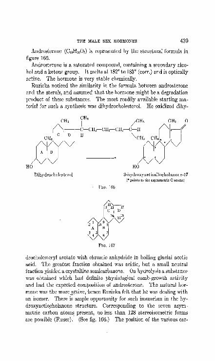
THE MALE SEX: HORMO:NES 439
Androslerone (C19Hao02) is represented by the struclural furmula ill figure 165.
Androsterone is a saturated compound, containing a secondary alcohol and a ketone group. It melts at 1820 to 1830 (corr.) and is optically active. The hormone is very stable chemically.
Ruzicka noticed the similarity in the formula between androsterone and the sterols, and assumed that the hormone might be a degradation product of these subRtanccs. The most readily available starting material for such a synthesis was dihydrocholesterol. He !Jxidized dihy-
Dihydrocholestel'Ol
FIG. 166
3-hydroxyaetioallocholunolle-17 (* poinls tu the asymmetri[l C atums)
drocholesteryl acetate with chromic anhydride in boiling glacial acetic acid. The greatest fraction obtained was acidic, but a small neutral fraction yielded a crystalline semicarbazone. On hydrolysis a substance was obtained which had definite physiological comb-growth activity and had the expected composition of androsterone. The natural hormone was the more _Q;ctivc, 'hence Ruzicka fclt that he was dealing with an isomer. There is ample opportunity for such isomerism in the hydroxyaetio.cholanone structure. Corresponding to the seven asymmetric carbon atolll~ present, no less than 128 stereoisometric forms are possible (Fieser). (See fig. 166.) The position of the various car-

440 ENDOCRINES IN OBSTETRICS AND GYNECOLOGY
bon atoms in the nucleus is shown in figure 167. The prohlem is Rimplified by the fact rings C and D have identical configuration in all the' natural sterols and bile acids. The junction C/D is known to' be trans and a similar linkage probably exists between rings B ami C. The linkages that interest us here are between rings A and B. The isomerism is dependent specifically upon the configuration at the asymmetric center C" the hydrogen atom at this point bearing either the cis or the trans relationship to the methyl group at C10 . (See fig. 168.)
CH~N~ /"-.Ifo "-./ I A B . 3 5
V rl
"f H
(Cholestane series) A Trans relationship or alia d.rrics
'l
Aill )JJ
HO H
Dihydrocholesterol (trans)
FIG. 169
FiG. 168
CH3r:1~ /"-. /"-.("-./ i
10 A Il
3 5
\/"-./ H
(CoprostlLne scri~s)
Cis or normal series
CH, Ij j //
13 5 .,/~ :
HO H
Epidihydrocholesterol (cis)
l'IG. 170
Another important type of stereoisomerism has to do with the COll
figuration of the carbon atom C3, which in all of the known sterols of natural occurrence is the point of attachment of the lone hydroxyl group. By convention the configuration of cholesterol or dihydrocholesterol is represented as in figure 169 and that of the epimeric form as in figure 170. In figure 169 the hydroxyl group bears the trans relationship to the hydrogen atom at C5 while in figure 170 the substituents are cis (Fieser).
Ruzicka prepared the four possible isomers having the structural

'THE MALE SEX HORMONES 44-1 '
formula of androsterone, but varying only in the positions of the OH and II groups on carbon atoms Ca and C, respectively. He found that the ketone obtained as an oxidation product of epidihydrocholesterol was identical in every respcct with androsteronc. It is interesting to
Dehydroandrosteronc
FIG. 171
Androstancdicil 3,17 Androstanedione 3,17
FIG. 172
CH, OH
1-1 V
Testosterone
6 4-.5 Androstcne-ol (-17) on (-3)
FIG. 173
note that the male hormone belongs to the cholestane (allo) series, while pregnanediol corresponds in configuration to coprostane. Hence in the conversion of cholesterol into androsterone it is necessary for the OH group on Ca to shift from the trans to the cis position. S~

"
Cholp.sterol
r.H! jHJ
CH,f'I-TcH OH I ) CH. fH, /CH,
I I crrl""'_ "CH, ("j/V" 1 1 (J-)-CH.rH' CH"CH, CH
HO~:V Dinydmcholestcrol ~--C/) '. H I bile acids
AJlopregnanedlOl (C. L.) Cholestenone -4 I " \ i Of' .f''' an! ('(lJlro~ I'm
RO"
, _____ J
Allopregnanoione (C. L.) Pregnanodiol (C.L.)
CH~
CH,
("li.oH
(YI/V HO/'\/"/
R<;tradjnl (11'_) ,
i :2 . 0
cHi I
I Ctr yl"" CIl3 CHi
("1/. 1 II /V
VV O/'V R
Androsterone (1\1) 'n~~t(J5teronc (M)
Estrone (F.)
I , 1 CH,
i~'I-/' /,,/
IIJ no/'\/
Equilillc (F)
Estriol (F.)
I CR,
t 11- ~O /'v'\/J I II I
noV"/' Equilenin CF)
FIG. 174. 'fhe ~terols, tljc~sex hormones and their interrellltior,siJip
:\r denotes members of the male hormone group. F denotes members of the female hormonc (theelin) group. C.L. denotes members of the corpus luteum group. '" denote6 t.he only active member of the corpus lutrum !;nHlll.

444 ENDOCRINES IN OBSTETRICS AND GYNECOLOGY
that if cholesterol is the precursor of androsterone in the organism, a biological mechanism must be present to make this rearrangement.
Dehydroanderosterone. Dehydroandrosterone is a second male sex hormone found in the urine (Butenandt and Danncnb'aum, 1934). It was also isolated from cholesterol by Ruzicka apd Wettstein (l~35). It possesses about one-third of the comb-growth activity of androsterone. (See fig. 171.)
It is interesting to note that dehyd~oandrosterone is intermediate in degree of unsaturation between androsterone and estrone, and may be a natural precursor of substances of both groups.
A number of derivatives of androsterone have been prepared by converting the keto and hydroxyl groups into two keto or two hydroxyl groups. Figure 172 gives the compounds obtained.
Their order of biological activity is as follows:
Androstanediol-3,17. .. . . . . . .. . .. . . . . . . . .. . .. Androsterone ............................. . Androsterone acetate. Annrostflnedione-3,17.
Callrm Unit Equiwlt:rtl
. ., 45-50 gamma
. .... 150-200 gamma 150-170 gamma 300 gnmma
As expected, the presence of an additional hydroxyl group (by. reduction of the ketone group) enhances the potency three times. The same effect was noted between estronc and estradiol. On thc contrary the presence of two keto groups greatly reduces the activity of the compound. The acetate docs not alter the effective dose but exhibits a marked protracted action. This may be due to the time required for hydrolysis, or to a slower process of absurption within the organism. .
Testosterone. The question arose whether the hormone in the testis was identical with the one in the urine. Or were we dealing with a condition similar to that found in the female, that is, that estrone is an excretory product, and that estradiol is the active hormone of the follicular ftuid. The previously cited work of Gallagher and Koch showed that testes extracts were inactivated by boiling with alkali while the male sex hurmone fractions frojIl urine were not. Further observations were made by LaqueUl' et al. (1935) who compared the effects of administering to castrated rats equal amounts, in terms of capon units, or urinary and testicular male hormone extracts. The animals receiving testicular extracts showed extraordinary growth of the seminal vesicles. The average weights in the case of the controls, the animals receiving urinary extracts and those receiving testicular extracts were in the ratios: '1: 14:67. This divergence of activity pointed clearly to the presence in the testes of a substance other than androsterone.

THE lIIALE SEX HonMONllS 445
David, Dingemanse, Freud and Laqueur (1935) isolated a crystalline substance from testis tissue. Their yield was 10 mg. of hormone from 100 kg. of testis tissue. They called this substance testosterone. It is about 10 times as powerful as androsterone in promoting comb growth, 10 gamma being the equivalent of one capon unit. The synthetic production of this hormone was then accomplished by Butenandt (1935) and shortly afterwards by Ruzicka (1935). Their starting
. point was dihydroandrosterone. Testosterone has the chemical structure shown in figure 173. The double bond is between carbon atoms 4 and 5, the alcoholic group (=01) on C17 and the keto group (-on) on Ca. Testosterone is optically active and melts at 1540 to 154.5 corr.
Figure 174, modified from Tscherning (1936) and Ruzicka (1936) shows the chemical relationship between the sterols and the sex hormones, and the interrelationship between the latter.
CH, OH
~I~J ~l~,,6)
o II o
CH, 0
1
~ -, CH, /"1/ /v 13
o~~/16 . 0
6-oxo-testosterone /" '-androstenc-trionc-{ -3-6-17)
FIG. 175
It is of great physiological and clinical interest to isolate compounds that are intermediate between the male and female sex hormones. Possibly such substa~ces may be produced in the various intersexes. Butenandt and Riegel (1936) have studied such a series. They in- ' creased the oxidation of androsterone by either increasing the nUlllber of double bonds (removing hydrogen) or by adding oxygen groups. They produced two such COlllPounds, namely, 6-oxo-testosterone and ~4-androstenetrione-(3-6-17) (fig. 175). Two milligrams of either substance will produce estruo in a mouse, while 4 mgm. will not produce comb growth, nor will 16 mgm. produce a change in the seminal vesicles. It is rather startling to note that two substances of such high potency as testosterone (capon unit = 10 gamma) and androstenedione (capon unit = 100 gamma) will lose their properties as male sex hormones and gain those of estrone, through the mere addition of an oxygen. The

446 ENDOCRINES IN OnSTETIUCS AND GYNI']COLOGY
relation of Butcnandt's oxidation products to Reichstein's adrenosterone (Chapter XX) will be awaited with great interest.
Further Physiological Action of Male Sex Hormones
The effect of a sex hormone on the organism of the opposite sex is of great clinical interest. Recent experimental data (l(oren'chevsky, Dennison and Simpson, 193.5) have yielded some interesting facts. These workers studied the effect of prolonged treatment of male and female rats with androsterone and its derivatives, alone or together with estrone.
Effect on Castrated Male Rats. Androstanediol and androsterone stimulated the secondary sex organs. The former was the more potent and the atrophied organs were restored to normal or more than normal weight. II
A c06pel'Utive effect on the semilll11 vesicles and prostate was seen between androsterone and estrone, though a quicker and greater effect could be obtained by increasing the dose of male hormone preparation. An antagonistic :wt.ion on the adrenals appeared to OCCllr
between the male hormones and estrone. The male sex hormones had a stimulating effect on the heart, liver,
kidneys and adrenals. There firc distinct clinical potentialities in these observatiollR.
Effect on Ovariectomised Female Rats. The injection of large doses of diol caused a partial recovery of the atrophied uterus and vagina. The effect was slight even with large doses of androsterone.
The injection of androsterone and of diol increased the rat,e of involutio)"l of the thymus, decreased the weight of the adrenals and (with large doses) of the hypophysis and slightly increased the weights of the kidneys and liver, thu~ in general producing results similar to those obtained in castmted male rats.
Simultaneous injections of estrone and large doses of androsterone or diol showed a "cooperative recovery effect" on the weights of the uterus and vagina and to a smaller degree on the thymus, while there seemed to be a slightly antagonistic effect between the actions of estrone and the male hormones Oil the adrellals.
Selye, .McCuell and Collip (1936) reported that treatment with testosterone benzoate stimulates the development and secretion of the mammary gland in normal and castrate female and male rats. Furthermore, testosterone is not estrogenic in doses sufficient to stimulate the mammary gland. Hence the effect of the male hormone on the

THE MALE SEX HORMONES 447
mammary tissue cannot be attributed to its transformation in the organism into the female sex hormone. Jung and Shafton (1935) found that proliferation of mammary tissue is normally demonstrable in boys between 12 and 17 years of age.
The effect of the male sex hormones on the female organism can be profound. The potentialities are enormous when one considers the number of variations the hormone molecules may undergo in the body chemistry, both under normal and abnormal conditions.
I am indebted to Dr. Erwin Schwenk for his advice concerning this chapter.
lllBLIOGRAPHY
BUTE1{ANDT, A. 1931 trbcr die chemische Untersuchung der Sexual hormone. Z. Angew. Chemie 44, 905.
1932 trber die Chemie der Sexualhormone. Z. Angew. Chem. 46, 655. BUTENANDT, A. AND DANNENBAUM, H. 1934 trber Androsteron; Isolierung eines
neuen, physiologisehe univirkungsnmen sterin derivntcs nus lVIiinncrhnrn, seine Verkniipfung mit Dehydro-Androsteron und Androsteron. Ein Beitrag wr Konstitution des Androsterons. Z. Physio!. Chern. 229, 192.
BUTENANDT, A. AND HANISCH 1935 trber die Umwnndlung des Dehydroandrosterons in 114-Androsten-ol-(17)-on-(3) (Testosteron) ·ein weg zur DarstelJung des Testosterons aus Cholestrin. Ber. 68, 1859.
BU'fENANDT, A. ANV RIEGEL, B. 1936 tJber 6-0xo-testosteron und verwundte Verbindungen. Ber. deutsch. Chem. Ges. 69, 1163.
DAVID, K., DINGEMANSE, E., FREUD, J. AND L.HlUEUR, E. 1935 trber krystallinischcs miinnlichcs Hormon nus Hoden (Testosteron) wirksamer als nus Ham oder aus cholestrin bereitets Androsteron. Z. Physio!. Chem. 233, 281.
DINGEMANSE, E., FREUD, J. AND LAQUEUR, E. 1935 Differences between male hormone extracts from urine and from testes. Nature 135, 184.
FIESER, L. F. 1930 The Chemistry of NaturyJ Products Related to Phenanthrene. Rheinhold Puh. Co., New York.
DE F'IIEMERY, P., :FREUD, J. AND LAQllEUR, E. 1930 Zur Messmethode von mannlichene Hormon. Arch. f. rl. ges. Physio1. 226, 740.
GALLAGHER, T. F. AND KOCH, F. C. 1020 The testicular hormone. J. Bio!. Chern. 84, 495.
1934 Effect of Alkali all the Testicular Hormone. J. BioI. Chern. 104, 611. JUNO, F. T. AND SHAFT ON, A. L. 1935 The :Vfammnry Gland in the Normal
Adolescent Male. Proc. Soc. Exp. Bio!. ]'lIed. 33,455. KOCH, F. C. 1931] The biochemistry and physiological significance of the male
sex hormones. J. Urol. 36, 382. KORENCHEVSKY, V., DENKISON, :VI. AND SIMPSON, S. 1. 1935 The prolonged
treatment of Int1le t1nd female rats with llIldro"terone and it" derivatives, alone or together with oes~rone. Biochem. J. 29, 2534.

448 ENDOCRINES IN OBSTETRICS AND GYNECOLOGY
:'v1CCUEN, C. S., SELYE, H. AND COLLIP, J. B. 1936 Effect of the testis on the mammary gland. Proc. Soc. Exp. BioI. and Med. 34, 201.
MOORE, C. R. 1932 Biolugy of the testes. In Allen, Sex and Internal Secretion. Williams & Wilkins Co., Baltimore.
PEZARD, A. 1911 Sur la determination des caracteres sexuelles secondaires chez les gallinaees. Compt. rend. de I'aead. d. S,e. 163, 1027.
REICHSTEI~, T, 1936 Hher Bestandteile der Nehenhierenrinde ;VI. HelY. Chim, Acta, 19, 1107.
RUZICKA, L., GOLDBERG, 1\1. W. AND BRtiNGGER, 1I. 1934 Zur Kenntnis der Sexualhormon. 1. Ober die Gewinnung yon 3-Chlor- und 3-0xy-iitioAlloeholanon-(17). Synthese ciner Verbindung von den Eigenschaften des Testikelhormons. Hely. Chim. Acta. 17, 1389.
RUZICKA, L. 1935 Contribution to the Knowledge of the Testicular Hormone. J. Am. Cll.em. Soc. 67, 2011.
RUZICKA, L. AND WETTSTEIN, A. 1935 Ktinstliche Herstellung des miinnlichen Sexualhormons trans-Dehydro-undrosteron und des Androsten-3, 17-dions. Helv. Chim. Acta 18, 986.
RUZICKA, L. 1936 The male sex ~09i~ones. J. Chern. Educ. 13, 3. STEINACH, E. 1936 Zur Geschi~)i.td des Mdnnlichen Sexualhormons und seiner
Wirkungcn am Slillge,(ier und beim Menschen. Wicn. Klin. \Yoch., Heft 5 und 6. to.)
TSCHERNING 1936 Hber die Chemie u. Physiologic dcr Androsterongruppe. Arg. Chern. 49, 11.
Voss, H. E. AND LOEWE, S. 1931 Maskulierung durch Androkinin. Klin. Woch. 10, 1957.

CHAPTER XXX
METHODS OF HORMONE ASSAY
The assay of the sex hormones in blood and urine has given valuable aid in the treatment and diagnosis of various obstetrical and gynecological dysfunctions. Only those methods will be given that can be carried out in a fairly well equipped hospital laboratory.
Extraction and AS3ay of Estrone
I. Urine. il. Method of Kurzrok and Ratner (1932). The twentyfour hour urine specimen is measured and 900 cc. placed in a one liter Erlenmeyer flask. This is tested and made slightly acid with dilute acetic acid. The hormone is more easily extr;1cted from an acid solution. The urine is then saturated with sodium chloride (approximately 50 grams) ,which decrea~e1i the solubility of ethyl acetate in the urine. The treated Ilrine is then covered with ethyl acetate to the top of the flask, and the flask is connected as in figure 176. The second flask (B) of. 300 cc. capacity is filled with 200 cc. ethyl acetate. The hot plate is turned to medium heat and the ethyl acetate distills over through the upper side arm, is condensed and drops to the bottom of flask (A) trom which it returns through the lower side arm to flask (B). The drops of ethyl acetate in passing upward through the urine extract the hormone. This provides continuous extraction for 48 hours. It is automatic and needs no watching. The extracted urine is then dis'carded, and the ethyl acetate extract, which contains the hormone is concentrated by vacuum distillation (fig. 177). The distilling flask (il) is filled one-third full with ethyl acetat{extract and connected with the condenser (B), a receiving flask (C), a second receiving flask (D), a trap (E) (provided with stopcock) and finally onto an aspirator type of suction pump. After the distilling flask has been heated in a steam bath, the suction is turned on. The remaining extract is allowed to drop into the flask (A) through a thistle tube which is provided with a stopcock. When. all the ethyl acetate has been added it is concentrated to as small a volume as possible and 6 ee. of propylene glycol is added through the thistle tube. The distillation is continued until all of the ethyl acetate has been removed. This last step should be done slowly to prevent bumping and loss of the hormone. The oil now COll
tains the hormone originally present in the 900 ec. of urine.
449
-,
FIG. 176. Estrone extraction appamtus. A hot plate can be used instead of the steam bath. The condenser should be long, for ethyl acet!1te is fairly volatile.
FIG. 177. The concentration apparatus. The receiving flask (D) should be 3- to 5-liter c!1pacity. It acts as a reservoir and is only emptied of ethyl acetate when full. Aside from removing and replacing the distilling fhLsk, the apparatus need not be disconnected.
150

METHODS OF .HOHMONE ASSAY 451
This 6 cc. of oil, prepared as described above, is used for the biologic assay. One group of three ovariectomized rats is used for the assay of each specimen. They arc injedcd with the oil subcutaneously in the back, at 9 a.m. and /'l p.m. of the first day and at 9 a.m. of the second clay. One rat receives 0.5 ce. oil at each injection and the other two rats receive 0.25 cc. each at each injection. Vaginal smears are taken 24 and 32 hours after the last injection. A K o. 13 dental spatula is very convenient for obtaining the vaginal secretion which is then spread in a drop of saline and examined under the microscope. A positive smear must show the cornified cells characteristic of estrus. Mucus, leucocytes and epithelial cells must be absent to establish a positive result. The hormone content of a liter of urine, expressed in terms of rat units may be calculated from the following formula.
6 1000 I . I' f' I I I - X - equa s rat umts per Iter 0 urme w len y equa s tot a amount y 900 . of oil injected into a single rat producing a positive result.
Notes: The distance between the two horizontal arms in figure 176 should be three to four inches.
The condcnsc;r should not reach the level of the upper horizontal arm. Animals that have repeatedly shown negative smears should be in
jected with potent estrogenic hormone (10 R.U.) once a month. This prevcnts cxcessive atrophy of the genital tract.
B. Bergen and Smelser Modification' of Estrone Extraction. The twenty-four-hour specimen of urine is measured and 750 cc. of it is poured into a 2-liter flask. The urine is made slightly acid with dilute acetic acid. Fifty grams of sodium chloride is added to the acidified urine and to this is added 750 cc. of ethyl acetate. This is shaken vigorously for three minutes and then poured into a separatory funnel. The urine fraction is drawn off. The ethyl,..acetate fraction is allowed to stand a few hours, then drawn off and distilled according to the method described above. The biologic assay is the same as above.
If an emulsion results during the shaking allow the ethyl acetate fraction to stand over night; then transfer to a clean flask. Centrifuge the emulsion if it is very heavy.
II. Blood. A. Method of Fluhmann (1934). The test depends on the injection, of small. amounts of untreated serum into spayed mice. A positive result is indicated by the production of a "mucification"
, Harold Bergen and Jane Smelser of the College of Physieians & Surgeons, Columbia Univcrsity. l\Iethod not previously publishcd.
452 ENDOCIUNES IN OHS'l'ETmCS AND GYNECOLOGY
of the vaginal mucosa. The method may be applied to quantitative studies, provided that a sufficient number of test animals is employed.
The blood is obtained by venipuncture and immediately placed in a tcst-tube for transmission to the laboratory. From 25 to 40 cc. are takcn each time, according to the amount required. The clear serum is secured by centrifugalization, and the cells are discarded. If it is kept sterile, the serum may be left in the ice-box for days and even weeks without losing its potency . . The animals used are adult female mice, which have been spayed 6
to 7 days prior to the day the test is begun. This is important, for if the period of castration is shorter the vapinal mucosa may show unusual changes difficult of interpretation at the end of the test, while if it is longer the mice are not as sensitive to the action of estrin. It may be possible to "sensitize" old castrates by the administration of a cornifying dose of estrin some days beforq th'ey are used, but this method has not been resorted to. The usu~tp!ocedure has been to do a bilateral oophorectomy on the mice on a Tuesday and to begin all tests on the same day of the following week.
A total of 4.5 cc. of blood serum is given to each mouse, and the test is completed in 72 hours.2 The serum is administered subcutaneously in the back, and the site of each injection is varied as much as possible to facilitate absorption. Three injections of 0.5 co. arc given daily, at 8:00 a.m., noon, and 5:00 p.m., for three consecutive days. On the f onrth morning the animal is sacrificed and the vagina is carefully dissected free. This is a simple procedure and is accomplished by making a long incision in Lhe abdominal wall and separating the symphysis pubis. As many as 20 to 30 autopsies may be done in a half hour. The vagina is fixed in formalin, mounted in paraffin, and transverse sections are made at different levels and stained with hematoxylin-eosin.
In the preliminary report of this work no differentiation was made of the intensity of the changes elicited in the mouse vagina, but this has been very unsatisfactory. It has now been found possible to recognize six reactions, as follows:
, An attempt is now being m:1de to shorten this period to 48 hours, and it has already been "hown that lllucification may be readily induced in that time. On the basis of somc unpublished observations it seems that the intensity of estrin eITects docs not depend :1lto~ether on the tobl amount of hormone injected but also on the concentration maintained in the test animal. It tllU" may he possible to intensify the reactions elicited by giving the tcst animal 6 injections of 0.75 c.c. in two days.
METHODS OF HORMONE ASSAY 453
Reaction O. Atrophy of the vagina. M UCORa shows two days of low cuboidal epithelium. Occasional leucocyte.
Reaction 1. Vaginal mucosa shows two layers, a basal of low cuboidal epithelium and a superficial of tall columnar cells. A few lcucocytes.
Reaction 2. The superficial cells are high, begin to show stratifica·tion, and secrete mucus. There is a well-marked increase in leucocytcs which may also be found in the lumen of the vagina.
Reaction 3. The epithelium of the vaginal mucosa is composed of several layers, and the cells at the surface are of the mucified variety. A characteristic feature often observed is a folding-in of the mucosa, a type of festooning, which is evidence of the rapid growth. The mucosa is invaded by large numbers of leueocytes which occur also in the lumen along with epithelial debris.
Reaction 4. The mucosa is made up of from 6 to 10 or 12 rows of cells, the lower resembling the basal eells of squamous epithelium while those at the surfaee are still of lhe tall {nucified variety. In marked cases early cornificalion IIlay be observed between the stratified epithelium and the mucified cells. Leucocytes have disappeared or are present in sman numbers.
Reaction 5. The vagina is now lined by fully developed squamous epithelium with cornified cells at the surface. There are no leucocytes.
As in all biological procedures, individual mice vary greatly in their responses to the same dosage. The accuracy of the test thus depends on using as many animals as possible for each specimen. In this work two or three mice has been the minimum employed for each test. After the response for each mouse has been estimated, the numbers are added and then divided by the number of mice used in order to give the reaction. For instance, if three mice are usecj._for a test and reactions 3, 2 and 2 are observed, the reaction elicited by that specimen would be given as 2.3.
B. Modified Method of Frank and Goldberger (1926). 1. Fifty cubic centimeters of blood is required. It is dehydrated by anhydrous sodium sulphate.
2. The.powder is twice extracted with 200 ce. of 95 per cent alcohol. 3. The alcoh.Ql fractions arc combined and ~vaporated to dryness on a
water bath. 4. The residuum is taken up in 5 cc. of olive oil and injected into
spayed mice. 5. The bioassay is according to the Allen-Doisy Method. C. Neustaedter Modification of Frank and Goldberger Method (1986).
Transfer 40 cc. of blood from a syringe to a petri dish containing 30 to 40

454 ENDOCRINES IN OBSTETRICS AND GYNECOLOGY
grams of anhydrous N1l2S04. Mix thoroughly with a glass rod. Fan until the mass has the consistency of fudge. Transfer to a mortar and grind to a very fine powder as the mixture dries. Return the powder to the petri dish for storage if the procedure must be interrupted at this point. Should the mixture not harden, add more :'h2S04. Transfer the red powder to a 250-cc. Erlenmeyer flask and add 100 cc. of ether. Stopper tightly with rubber and agitate for 20 minutes in a Holtz shaker. Rest the flask on a specially constructed board at an angle of 450 to separate the ether from the sludge by sedimentation. Decant the snpernatant fluid and <;entrifuge. Repeat the process twice (that is, use 300 cc. of ether in all). Evaporate the ccntrifugalized ,ether to dryness ill a casserole before an electric fan. Dissolve the lipoirll'psiduc
~'ih 6 cc. of benzene to which 0.6 cc. of olive oil is added. Fan off the "l;enzene. If the air is full of dust, it is preferable to cover the tuhe with
filter ]la]ler and set aside for spontalH~ous evaporation since injection of dirt leads Lo infection in the ;;limals. Sterilize the oli\'e oil which 1I0W contains Lhe lipoid extract by autocla~illg at 15 pounds for 15 minutes. This extract may be kept for several uays if rubber stoppered and stored in the dark. !
The extract is injected into mature, castrated female mice in di\~idcd doses using a sterile tuberculin syringe. On the first day 3 injections are given at 4-hour intervals. Two injections at the same interval are made on the following day. The extract is introduced into the mouse dorsally and subcutaneously. Beginning on the third morning, vaginal spreads are prepared twice daily for 4 days. The smears are made with a small bent glass tube drawn to a fine tip. By means of a rubber bulb, a drop of water is repeatedly injected and reaspirated from the vaginal canal so that a uniform and typical sample of yaginal secretion is secured. The specimen is transferred to a clelln slide, dried, fixed and immersed in aqueous thionin (1 per cent) for 1 minute; excess iitain is removed with water. Examine the vaginal smear for an estrus reaction (denoting 1 mouse unit of estrin and consid8l'cd the norm).
Method of Extraction and Assay of Follicle Stimulating Hormone
I. Urine. A. I'rfodijication of Zondek's Jlethod. Extraction: 1. Pour 4i cc. morning urine into a 2.50 cc. centrifuge tube. Test and make slightly acid with clilute acetic acid.
2. Add 200 cc. 95 per cent ethyl alcohol and allow to stand for 2 to 24 hours.
3. Centrifuge and discard the supernatant fluid.

METHODS OF HOnMONE ASSAY 455
4. Add 20 ce. ethyl ether and shake well, breaking up the precipitate. This removes the estrogenic substances.
5. Centrifuge and discard the supernatant fluid. 6. Dry the precipitate until all ether is evaporated. 7. Add 7 ee. distilled water and shake constantly for 10 minutes. 8. Centrifuge and save the supernatant fluid, which contains the
folliclc stimulating hormone. The preparation is then ready for animal assay.
Animal Assay. 1. Three immature female mice weighing from 6 to 8 grams are injected twice daily for three days. Each mouse receives 0.25 cc. at each in.iection, subcutaneously in the back. The injection is made with alec. tuberculin syringe and No. 24 hypodermic needle. The mice are not injected on the fourth day and are autopsied on the fifth day or 100 hours after the first injection.
2. A macroscopic ('xamination of the ovaries and uterus is made. A positive test shows enlarged, reddenr:d ovaries showing large follicles, and an enlarged and distendr:ciutrrus and an open vagina. A negative trst shows small white ovaries and threadlike uterus. This extract should be kept in the refrigerator. Each mouse receives the equivalent of 10.5 ce. of urine. If the te~t i8 po~itive it means that there are at leaot 1001V1.U. of Follicle Stimulating Hormone per liter. By diluting the remaining hormone solution it is possible to determine the total aIllount of hormone present.
B. kIelhod of Levin and Tyndale (1936). The chloroform-preserved urine is chilled, siphoned from any sediment and acidified to a pH 5 with acetic acid. For each liter of urine, 20 ce. of a freshly prepared aqueous 10 per cent tannic acid solution are added. An immediate precipitate forms. After thorough mixing, the pr~cipitate is allowed to settle in the cold room and is then coliected'1It the centrifuge. The bulky precipitate, which contains considerable water, is extracted once with at least 5 parts of 95 per cent ethyl alcohol followed by 3 to 4 extractions with 80 per cent alcohol. Alcohol is removed by several washings with acetone and the residue is freed of acetone by reduced pressure. In addition to removing considerable pigment and other
. inert material, the alcohol-acetone treatment removes any estrogenic substances whiCh 'may be -present.
The resultant powder (100 to 200 mgm. per liter of urine) contains 75 to 100 per cent of the original gonadotropic activity of the urine3 as
, Application of this precipitation to other types of urine (pregnancy, normal women, normal men) has yielded excellent results. Hellhuum et al., 1935 (Hell-

456 ENDOCIUNES IN OBSTETRICS AND GYNECOLOGY
indicated by parallel assays of the raw urine (or alcohol precipitates thereof). The activity of the dry tannate is quite stable, no loss being noted even 'after storage for more than a year at room temperature.
Assays are conducted by giving 20- to 22-day-old mice (8 to 12 grams body weight) subcutaneous injections once daily for 3 days, the animals being sacrificed 72 hours after the first injection. Because with minimal doses ovarian weight aloll(~ is an unreliable criterion, the mouse unit has been tentatively defined as that amount which, when administered in the above manner, will cause vaginal canalization, at least a 200 per cent increase in the weight of the uterus drained of fluid (controls 4 to 7 mgm.), and a slight increase in ovarian weight as compared to uninjectcd controls. Such a unit is very sensitive and in the absence of estrogenic substances is quitc accuratc. W c have never been able to demonstrate the prescn~e of any estrogenic activity in the alcohol washed tannates. )l '
II. Blood. Method of Fluhmam{ ,(1931). The technic of the test used in this study has been descfibld in two previous communications, and is based on the "pregnancy I test" of Aschheim and Zondek. It consi~t::i ill outaining, under slerile precaulions, froIll 15 lo 20 cc. of veIlous blood from the patient to be examined. This is then allowed to sland or is centrifuged and from 3.0 to 5.0 cc. of the clear serum are injected subcutaneously, twice daily in from 0.5 to l.0 cc. doses, into an immature female white mouse. (In view of the study by Engle and Rosaco the immaturity is determined by the age and not by the weight of the mice, and they are used only if between seventeen and twentytwo days old.) In the case of a positive result, the vaginal introitus of t,Q.e m~use is est~blished. by the f?urth or fifth da~ and the animal is t'iien kIlled and Its ovanes fi,xed 111 Zenker's solutIOn. They are embedded in paraffin and serial sections are made and studied. The conditions which indicate the presence of anterior piruitary hormone arc grouped into three categories by Aschheim and Zondek, as follows:
Anterior Pituitary Reaction One (APR I)-(The "ovulation" reaction): The ovaries show the presence of normal ripening follicles, while if autopsy is delayed normal corpora lutea are seen .and ova may be found in the Fallopian tubes.
buum, Fevold and Hisaw, 1'roe. Soc. Exp. BioI. and iVIed. 32, 1566) huve reported similar experience with pregnancy urine. Others (Thumsen and Pederson-Bjerguard, Compo rend. Soc. d. Biol. 120, 1143, 1935; Pederson-Bjergaard, Zbl. f. Gyn. SO, 372, 1936; and Friedman and Weinstein, Proe. Am. Physio!. Soc., p. 54, 1936) have applied our method to normal urines with confirmatory rcsults.

IIfETHODB OF HORMONE ABBA Y 457
APR II-(The "hemorrhagic cyst" reaction): The ovaries grossly show the presence of fine reddish dots, which correspond to hemorrhages ·into normal and abnormal follicles, and are apparently due to the intense congestion set up by the hormone. In the present study the finding of this reaction is not charted, since it is invariably an accolllpaniment of reaction III and does not seem to yield any important additional information .. In the diagnosis of pregnancy, however, it is of considerable value as it is readily perceptible grossly.
APR III -(The "luteinization" reaction): In this case the ovaries present a process of luteinization of the cells of follicles and the formation of abnormal structures resembling corpora lutea in which ovulation has not occurred and the ovum remains imprisoned. It is also worthy of note that these structures oc-cur much sooner than normal corpora lutea would be formed following ovulation and in one instance they were noted forty-eight hours following the first injection of serum from a patient with an early pregnancy.
The changes in the vaginal mucosa which accompany the various processes seen in the mouse ovaries are also of interest and have been studied in a smaller series in view of the recent observations of Wiesner. In the case of APR I, the vaginal mucosa shows the cornification characteristic of the stage of estrus, and is identical to the change elicited by the ovarian follicular hormone in spayed mice (Allen-Doisy test). On the other hand, when APR III is obtained alone the vaginal mucosa is altogether different, as it is composed of tall cylindrical cells at the surface ("pseudopregnancy" of EYans and Long; "mucification" of WiesnQr and Patel), alld is similar in appearance to the mucosa of a mouse during gestation.
Method of Assay of Luteinizing Hor~one
1. Method of Zondek (1935). a. The material is taken up in water. b. It is then injected into infantile mice (6 to 8 grams) in six divided
doses over a .period of 36-hours. All injections are subcutaneous. c. The animals are destroyed 100 hours after the first injection. d. A positive test is presence of fully developed corpora lutea. Zbndek (1935) sta£eii.that When young rats are used instead of mice
they should be destroyed 120 hours after the last injection. Corpora l(ltea develop more slowly in the rat.
2. Method of Loeser (1931). Infantile rats are used. A positive test iS'the reaction obtained in 5 out of 10 infantile rats. At least one well-formed corpus luteum must be present in each ovary.

458 ENDOCRINES IN OBSTETRICS AND GYNECOLOGY
Method of Assay of Corpus Luteum Hormone
1. Method of Corner and Allen (1929); Allen (1930). An adult doe, preferably one which had been isolated for a month or more, is mated to one or two bucks and insemination proven by discovery of spermatozoa in vaginal smears made immediately after mating. The animal is thereupon again isolated until conclusion of the experiment. About I8-hours after mating the animal is anesthetized with ether and the abdomen is opened under aseptic precautions. If ruptured follicles arc found in the ovaries as cxpcetcd, the ovaries are removed. It is our practice to clamp the ovarian pedicle tightly with a slllall artery clamp, to transfix and ligate the pedicle with blaek silk on the proxim~l side of the clamp, and to cut away the ovary along the distal side of the clamp. Bleeding points are carefully controlled by ligature with silk, avoiding ligation or kinking of the Fallopian'tube. Complete removal of the ovaries (to insure total ablation of the corpora lutea) is of course essential to accuracy of the test. A piece of the uterus about one or two centimeters long at the middle of the left c~rnu is removed beLween ligatures after the mesometrial vessels supplying the excised portion arc tied and finally the C1/t ends of the uterus are approximated by tying together the ends of the two uterine ligatures. The portion of the uterus thus removed is preserved in Bouin's fluid for use as a histological control. The incision is closed in two layers with silk. The extract is
, administered subcutaneously in the dorsal region immediately following the operation and once daily thereafter until five doses have been given. The animal is killed 5 days after the operation, one day after the final dose, and the genital tract and other tissues are removed for examination. The sites of the ovaries are carefully searched for surviving ovarian tissue and any suspicious tissues are examined under the dissecting microscope and if necessary preserved for sectioning. The right uterine cornu, the left cornu above the ligature, and the two Fallopian tubes are successively cut from the reproductive tract and washed out into watch-glasses with normal salt solution by means of a hypodermic needle and syringe, in order to obtain the embryoR. Finally the parts of the genital tract arc fixed in Bouin's fluid for sectioning. A fairly accurate provisional estimate of the result can be obtained without waiting for paraffin s('ctions, by examining with a dissecting microscope in a strong light the surfaces of razor-cuts made through the specimen after' a few hours' hardening in the fixing-fluid. The result of the test is measured by the degree of progestational proliferatiim inducerl in the uterus, according to a standard described below.
METHODS m' HORMONE ASSAY 459
The test is considered fully positive if at the time of necropsy the endometrium has attained throughout both cornua a conilition similar to that of the uterus at the 8th day of normal pregnancy (as seen in sections taken from the undilated portions of the uterus between the implantation sites).
A "rabbit unit" is the minimum dose of an extract which suffices, when divided into 5 daily doses, to alter the uterus of a doe weighing 3 to 4 kgm., under the specified experimental test, to a state equal to that uescribed in the preceding paragraph.
2. Method of Clauberg (1930). a. An immature female rabbit weighing about 800 grams is used.
b. The animal is injected witb 10 M.D. of estrone daily 8 days before the corpus lutcum injections are begun.
c. Corpus lutcuni extract is then injected for 5 days and the animal killed on the next day.
d. The uterus i8 submitted to microscopic study and the rabbit unit determined by the saDie method used by Allen and Corner.
Extraction and Assay of Male Sex Hormone From Urine
1. Method of Callow (1936). In order to obtain a result which is correct within ±20 per cent it is necessary to take a three-liter sample of an average urine for extraction so as to allow for administration of a total dose of extract equivalent to a half liter of urine per bird in a group of five capons. It is important that the urine should be kept fresh; it is preferably stored at O°C. after collection, without preservative. The proportion of concentrated hydrochloric acid required to bring the urine to pH I is determined with a IOO-m!. portion; with a fresh urine about 20 m!. per liter is necessary. Methylviolet is used as indicator, the color produced. by the urine mixed with an equal volume of the indicator solution being compared with that produced bydecinormal hydrochloric acid (the Capillator technique is convenient). The whole sample is the~ rendered strongly acid (pH below 1) hy the addition ~f sufficient hydrochloric acid to bring it to pH I, plus 20 ml. per liter in excess.
The mixture is placed in a flask fitted with a reflux condenser and a tube from the top of the condenser leading to an arrangement for ab
'sorbing f\Hues in alkali, and is then brought to the boil within! to 1 hour and boiled fur 1 l;oui·. It is then transferred to a continuous extraction apparatus and extraded "ith benzene for 20 to 24 hours. The benzene solution (500 ml.) iti freed from aeid by extracting twice with 50 Ill!. portiolls of a 2 N sodium hydroxiue, anu washeu with wu,ter.

460 ENDOcnINES IN OBSTETRICS AND GYNECOLOGY
The benzene solution is evaporated to dryness and the residue is extracted four or five times with 10-m!. portions of redistilled ether. The ethereal extracts are filtered through a coarse sintered-glass filter, and the filtrate is evaporated bit by bit in a small, weighted, £lat-bottamed tube. Sufficient arachis oil is added f-rom a syringe to the residue (100 to 300 mgm.) to make the volume up to 3 m!. and the mixture is heated find stirred until all the oil-soluble material has dissolved. '
This solution, containing the equivalent of 1 liter pf urine in 1 m!. provides for daily injections of O.lm!. per bird in a group of five capons over a period of five days in the usual method of assay, giving, with a normal urine, an easily measurable response. "
The yield of male hormone obtained in this way from pooled samples of normal female and male urine haS, amounted, respectively, to 29 and 26 international units of male hOT/heme activity per liter (equivalent to 2.9 and 2.6 mgm. of androsterone 'I~er liter).
2. Female Bitterling as a Biologic Test Animal ,for Male Hormone (Kleiner et al., 1936). Experimen~al. The female bitterling develops an ovipositor which depends from the ventral margin of the body slightly anterior to the origin of the anal fin. In the quiescent state the ovipositor is seldom visible, but in those individuals in which it is visible out of season it is very small and rarely reaches 5 mm. in length. During breeding activity the organ is prolonged until it may reach 5 cm. in length and at spawning it is inserted into the inhalant siphon of a mussel, usually Unio or Anodonta, and the ova extruded into the gill-folds. Fertilization is effected by sperm which is liberated near and drawn into the inhalant siphon of the mussel and passed over the embedded ova. Hatching occurs within the gill-folds and the fry liberate themselves, in a post-larval state, two or three weeks after oviposition.
Description of Test. To a small aquarium is added 4 liters of water, 2 liters from a stock tank and 2 liters from the tap. Two female bitterlings are placed in the tank and kept there 24 hours before introducing the material to be tested, At the end of the 24-hour period readings arc made of the size of the ovipositor. The scale used is as follows: If the ovipositor is not visible the reading is 0; if the length of the ovipositor equals the length of the first ray of the anal fin the reading is 10; un ovipositor which reaches halfway down the first ray is 5; etc. (That is, the length of the ovipositor is compared to that of the first ray of the anal fin ill equal units running from 0 to 10), No fish with ovipositors exceeding 3 on the scale, before the addition of any test material, are used.

METHODS OF HORMONE ASSAY 461
Ovipositor readings are taken at 24-, 48-, and 72-hour intervals and maximum growths under these conditions are usually observed at the second reading.
We tentatively define a bitterling unit as the amount of material, which, when added to a, tank containing 4 liters of water and 2 female bitterlings, produces an increase in the length of the ovipositor of one or both fish of 7 or more on the scale within 48 hours. For assay it is suggested that a series of dilutions may be set up and the lowest dilution giving a positive reading may be considered to contain at least one unit, providing a positive reaction is also given in the next higher dilution.
Technique for Assay of Urine. In attempting to determine the amount of male hormone in normal male urine, the following procedure was employed, using normal male medical students as subjects: For each assay, 4 aquaria were set up, containing 4 liters of water and 2 female bitterlings in the usual manner. To each tank was then added 10-, 25-, 50-, and 100-cc. portions of urine respectively. Twenty-fourhour samples of urine were used. The smallest amount causing a posi~ tive reaction was considered to contain one unit. The number of units in a 24-hour sample was then calculated. It was noted that in many instances the urine was toxic and even fatal in amounts from 50 to 100 cc. Various methods have been tried in the attempt to detoxicate the urines and finally it wasJound that dialysing is all that is necessary. By this means dialysed urines representing as much as 200 cc. of the original may be used without any ill effects whatever. Ordinarily the procedure is the following: Measured amounts of urine, usually 200 cc., are dialysed in membranes of cellophane ("plain transparent" not "moisture-proof") against running tap water for lJ to 24 hours. The volumes are then measured and amounts equivalent to 10, 25, 50, and 100 cc., respectively, of the original urine are added to 4 aquari;t, each of which contains 2 female bitterlings. The ovipositors of the fish must have been read on the preceding day and also just before addition of the dialysed urine. Subsequent readings are made in 24, 48, and 72 hours, and the number of units determined in the manner suggested above. The average excretion, of male hormone is about 35 bitterling units per day with a range of approximately 15 to 75 b.u. (sec table 11). Further work to check and enlarge this series is in progress. , Preliminary tests have indicated the unreliability of using casual
specimens for even rough quantitative work. We have noted that successive samples obtained during the day from the same individuals

462 ENDOCRINES IN OBSTETRICS AND GYNECOLOGY
have been exceedingly variable in the amounts of hormone excreted and some samples are even entirely devoid of the hormone. Apparently 24-hour samples of urine are needed in order to determine the output of this hormone with any degree of accuracy.
3. Method of Darby,4 (unpublished). Chemical Procedure. Funk et al., (1929) and Adler (1934) have shown that a substance ,which promotes comb growth in capons can be extracted from acidified urine by lipoid solvents. Extracts from urine which has not been acidified are inactive. However, as Adler has shown, the active substance can be demonstrated in such extracts after treatment with strong acid. It seems that the male hormone is ordinarily excreted in a physiologically inert form.
The first step, then, in the assay of this substance, is the acidification of the urine sample (preferably a 24-hour specimen) by the addition of 1 m!. of concentrated HCI for each 100 ml. of urine. The final pH
TABLE 11 Daily Excrel';on of Male Hormone by Normal Adult Males
NUMBER OF CUJilC CENTIMETERS OF NUMBER OF 24-HOUR URINE URINE WHICH, WHEN ADDED TO"
Sl'ECIMENS 4 LITERS OF' WATER, PRODUCE.A
POSITIve REAOl'ION
5 10- 15 15 25- 35 10 75-100
BIT'l'EnLI~O U:r..-ITS EXCRETED DAILY
75-120 24- 45 9- 22
should be between 1.0 and 2.0. Thymol blue is a convenient indicator to use for this purpose. The acidified urine is then allowed to stand for several days at room temperature, to allow the hormone to be hydrolyzed to the free state. If there is need for immediate assay hydrolysis can be speeded up by gentle heating (50°C. for four hours); but it should be remembered that heating tends to destroy some of the hormone, so should be avoided whenever possible.
The urine is then placed in a large separatory funnel, and shaken thoroughly with three-quarters of its volume of ethyl ether. The ethel' layer is separated and placed in another container, and the urine extracted twice more with fresh portions of ether (abouf one part ether to two parts urine). The urine is then discarded. The ether extracts are pooled together in the separatory funnel, and washed with two 25-
• Hugh Darby, Ph.D., Fellow, Dept. of Biochemistry, College of Physir.ians lind Surgeons, Columbill University.

METHODS OF HORMONE ASSAY 463
m!. portions of N/5 NaOH. This alkaline washing serves to remove all of the acid, practically all of the pigments, and any estrogenic substances which may be present. It is followed by several washings with 100.rn!. portions of distilled water, until the washings when tested with brom-thymol-blue have a pH of about 7.2 to 7.4. If enough time is allowed between washings for the water to settle out of the ether, three washings will suffice; but if the changes of water are added in rapid succession, more than three may be needed to reduce the pH to the desired level.
The ether now contains the hormone free from estrogenic material, but there remains a certain amount of water dissolved in it. In order to get rid of this water, as it will interfere with the ultimate dissolution of the active residue in oil, some anh¥drous sodium sulphate (about 2 grams per 100 ml. of ether) is placed in the funnel with the extract. This absorbs practically all the water present, and clings to the walls of the separatory funnel so that the ether can be decanted quantitatively. If there is enough water to dissolve any of the salt (that is, jf any droplets appear amongst the salt crystals), more sodium sulphate is added.
The dried ether extract is next placed in a large distilling flask and the ether distilled off over a water or steam bath. The distillate can be saved and used repeatedly. When the residue has been reduced to about lO-ml. volume, the distillation is stopped, and the concentrate is transferred with a capillary pipette to a 50-ml. Erlenmeyer flask. The distilling fI ask.. it rinsed with a little ether, which is added to the concentrate. The last of the ether is then evaporated from the concentrate over a water bath (BO°C.), leaving a gummy or syrupy residue. This is taken up in 10 ml. of sesame oil, with thorough mixing. It is now ready for injection.
The chemical procedure can be summarized as follows: (1) Acidification of urine specimen. (2) Hydrolysis over several days of storage. (3) Ether extraction of urine (three times). (4) Washing of ether extract ;vith alkali (twice) then with water, to
neutrality. (5) Removal of dissolved water from extract. (6) Concentration of extract by distillation; and finally, after trans
fer to small container, by evaporation. (7) Dissolution of the residue in sesame oil. Biological Procedure. At the second Conference on the Standardiza-

464 ENDOCRINES IN OBSTETRICS AND GYNECOLOGY
tion of Sex Hormones, held under the auspices of the League of Nations in 1935, the following standard was adopted for the assay of the male sex hormone: namely, the amount of growth produced in the comb of a capon by the daily injection for six days of 0.1 mgm. of androsterone. The amount of growth is expressed as the percentage increase in the area of the comb. The unit dose of androsterone (0.1 mgm. ,= 100 gamma) is physiologically equivalent, in the capon, to 15 gamma of testosterone. Hence testosterone is about seven times as active as androsterone. More will be said of the relative activities of these compounds later. Suffice it to say here that the capon test is the recog-nized method of assaying male sex hormones. "
Capons to be used for this test should be between one and two years old. Leghorns are quite commonly employed.' If possible, they should come from the same brood. Many agricultural schools will be pleased to supply capons or assist in caponizing the birds. They should be caponized veryyoung, as the operative mortality increases with age. When caponization is complete, the comb shrinks to a small white mass on the head, and the wattles below the beak become so small as to he hardly noticeable. Sometimes caponization is incomplete. In such cases, although the bird at first shows a ~hrinkage of the comb, it later grows a small but bright red comb. These incomplete capons, or slips, as they are called by poultrymen, are useless for experimentation.
The preparation under test is injected intramuscularly into the breast of the capon, care being taken not to inject into a feather follicle. A tuberculin syringe fitted with a No. 27 needle, three-quarters of an inch long, has proved satisfactory. The dose is administered daily on alternate sides of the breast for six consecutive days.
The oil used in the preparation should always be sesame oil, as the work of Miesscher et al. (1936) has shown that the solvent can markedly affect the amount of response obtained. The writer, when inj ecting known quantities of a pure preparation of testosterone into fishes, also obtained entirely different results depending on whether the material was. dissolved in propylene glycol or olive oil. As sesame oil is distinctly superior to other solvents which have been tried experimentally, it should be used as the standard carrier.
Measurement of Response. The reaction of the capon to the administration of the hormone is the commencement of growth within the first 24 hours. Thc first observable sign of respon~e is the replacement of the white color in the comb and wattles by a bright pink. This reaction does not lend itself to quantitative measurement, hence

METHODS OF HORMONE ASSAY 465
changes in the size of the comb are used as an index of response. The procedure generally recommended for measuring the comb is to make a shadow picture whose area may then be determined with the aid of a planimeter. This measurement is made at the start of the test, prior to the first injection, and is repeated daily until the comb has passed through its maximum size, and has begun to regress. In the presence of the hormone, the comb grows steadily over the injection period, and for a few days thereafter. It then starts to regress. The speed with which the comb reaches its maximum size with a given hormone dosage has been fuund to be sumewhat variable; and in the case of testosterone, at least, depends to a large extent on the nature of the acids with which
A
FIG. 178. Points of reference for comb m{asurements
it is esterified. Hence it is important tu continue the measurements uart the maximum growth has been r~ached and passed, to make sure of obserying; the complete response.
The writer has simplified the technique of estimating comb area as follows: The comb is measured along its length (distance A-B in fig. 178) with a pair of dividers, and the length recorded in centimeters. The three longest prongs of the comb (C, D and E in diagram) are measured from their tips to the base, where the comb joins the head. These heights are also expres~ed in centimeters, and are added together and divided by three to give an average prong height. This value is multiplied by the length A-B to give an arbitrary over-all area. The

466 ENDOCRINES IN OBSTETRICS AND GYNECOLOGY
measurements are repeated daily throughout the assay period. The original area (length X average height) is subtracted from the new area as obtained each day, and the percentage of growth calculated by dividing the (100 X the difference) by the original are.a. The daily percentage increases are then plotted against the time in days., A typical set of figures is given in table 12.
Estimation of Hormone Present. The dosage of test material used depends on what kind of information the assay is expected to give. If the only answer wanted is whether there is male sex hormone present or not, then the simplest procedure is to give a daily injection of 0.5 ml. of the preparation to each of three birds for six consecutive days. If the hormone is present in large quantities, the combs will show an increase well above 60 to 70 per cent by the eighth day. This method
'1;ABLE 12 ,. - . ,
LENGTH Of' I "<HEWITT OF pnONG
PERCENTAGE DAY
COMB (A-B) AREA INCREASE
C I __ D ___ E_ ~ ___ ---0 4.9 2.9, 2.7 2.4 2.67 I 13.1 1 5.1 3.0,' 2.8 2.5 277 14.1 8 2 545 3.15 3.1 2 6 295 16.1 23 3 5.6 3.2 3.1 2.7 3.00 16 8 28 4 5.7 3.2 3.1 2.8 3.03 17.3 32 5 5.8 3.2 3.105 3.0 3 12 1R 1 38 7 58 3 25 320 3.0 3.15 18.3 40 8 5.0 3.25 3.15 2.9 3 10 17 4 33
10 5.6 3 25 3.15 285 3 08 17.2 32 ---~ -
will not enable the experimenter to tell how much male sex hormone is in his preparation, but will furnish a rough idea of its activity. The reason for the inadequacy of this procedure for quantitative purposes "ill be apparent from the next paragraph.
If a careful assay is needed, the basis of the international standard must be understood. The international unit of activity is the amount of growth produced by the daily injection of 100 gamma (0.1 mgm.) of androsterone for six consecutive days: i.e., a maximum increase in the comb area of 30 per cent. This maximum i8 generally reached by the eighth day, In measuring the activity of an unknown preparation, the dosage level must be udjusted to the point where six equul daily injections produce the standard 30 per cent increase in comb area, .and no more. That is to say, a 60 per cent increase does not mean exactly twice the hormone dosage which produces a 30 per cent increase; much

METHODS OF HORMONE ASSAY 467
less does an increase of 120 per cent indicate four times the unit dosage leveL The figure of 30 per cent has been selected as the dosage level at which the bird reacts with the utmost sensitivity. Hence, to return to the practical prohlem of assaying our 10 ml. of oily material of unknown activity, the following procedure is recommended.
First inject 0.5 ml. of the preparation into one capon daily for six days. Calculate the percentage increases, and plot the figures obtained. If the maximum gocii above 60 per cent, then the remuining material (i mI.) musl be diluted with sesame oil. The physiological response indicated by 10 to 50 per cent increases in comb area is about proportional to the amount of hormone :1dministered; but above 60 per cent the response is no longer proportional to the dose, hence there can be no olankeL rule for dilution. The writer uses the following scheme. If the maximum response is 60 per cent, dilute by adding OIle part sesame oil to one part of preparation. If the response is 80 per cent, add three volumes of sesame oil to one volume of preparatioIl. Experience with this material and familiarity with the graphs obtained following excessive doses soon give an idea of what dilutions ought to be made. If enough birds are available, it is advisable to inject some standard preparation in sesame oil at several different dosage levels. The graphical records of the responses can then be kept for reference. The dilution of the unknown finally selected should be tested on three or four birds to ayerage out biological variations. If instead of a 30 per cent increase, a slightly higher percentage is obtained, let us say 45 per cent, it is permissible to estimate this as a response to 1.5 capon units of male sex hormone.
Having arrived at a dosagr, levr,1 corrcspondiJlg to approximat.ely one unit of activity, thr, next probkm io to calculate the total number of capon units in the preparation. For example, in the case already tabulated on page 181, the growth obtained (40 per cent) corresponds to 1.33 units. This was produced by a daily dose of 0.5 m!. of undiluted preparation. Hence the total unitage of the whole 10 Illl. of preparation is 1.3:~ X 2 X 10 = 26.6 units. (In caoes where dilutions have to be used to bring the· adivity_ within range of measuremenl, the dilution factor ha~ to oe included in the above calculalion.) III this particular case, two 24-hour specimens, of a total volume of 3 liters, had been extracted; hence the unitage was 1i:l.3 units per day, or 8.9 tlllits per liter. The foregoing procedure does not define the chemical nature of the active material in the urine, but it does furnish a measure of its physiological activity.

468 ENDOCRINES IN OBSTETRICS AND GYNECOLOGY
Other Methods of Assay. It must be remembered that 'the capon method of assay is the only one which has been standardized, and iA therefore of great value in enabling various workers to compare their results. It is therefore the method of choice whenever possible. However, other animals have been employed, chiefly the castrat!l rat.
In the rat method of assay, the hormone is estimated from the amount of growth induced in the seminal vesicles .of castrated animals. Young growing castrates are injected with the hormone for ten days, and killed on the eleventh day. The seminal vesicles are dissected out and weighed, and compared with the weights of the seminal vesiclf!s of uninjected castrate controls. The difficulty of this method lies in the establishment of the control value. A number of animals, castrated at the same time as the experimental group, must be carried parallel with them, aha sacrificed at the same time. All the rats must come
v from the same stock, and' be as nearly homozygous as possible, in order to keep variability at a minimum. At the start of the experiment, about 20 days after castration, the rats should weigh between 60 and 70 grams. There is reason for believing that the rat, in contrast to the capon, shows qualitatively different responses to different forms of male sex hormone; and it has been suggested that both ought to be used for testing (Korenchevsky et aL, 1935; Miesscher et aL, 1936). However, no standard procedure has yet he en agreed upon.
Another animal which has heen suggested in this connection is the female bittcrling, Rhodeus amarus. Kleiner et aL (1936) reported that the ovipositor of this fish lengthens in response to the presence of male sex hormone in the water. The possibility of utilizing the reaction for assay purposes is limited by its lack of specificity. It was originally offered as a test for female sex hormone. In our experience, the length of the ovipositor is highly variable in untreated normal bitterlings ami may be increased by a number of stimuli, such as high temperature or the addition of any foreign material to the aquarium that causes fouling of the water. According to Kleiner et aL, pure preparations of androsterone and testosterone are effective within narrowly limited ranges of concentration. The 1ITiter has attempted to repe~t these observations with crystalline compounds, but has been unable to do so. The female bitterling does not seem to offer any possibilities whatever for assaying unknown amounts of hormone in urine.
Since the whole subject of the sex hormones is in a state of rapid development, it is essential for anyone interested in clinical applications to keep in close touch with t.hp pnrrpnt. iitprotllrP

METHODS OF HORMONE ASSAY 469
BIBLIOGRAPHY
ADLER, A. A. 193-1 Active and inactive forms of the hormone promoting comb growth. Nature 133, 798.
ALLEN, W. ~L 1930 Physiology of the corpus luteum. V. The prepamtion and some chemical properties of progestin, a hormone of the corpus luteum which produces progestational proliferation. Am . .T. Physio!. 92, 174. .
CALLOW, R. K. 1936 Extraction of male hormone from urine for biological assay. Lancet, Sept. 5th, 565.
CLAUBERG, C. 1930 Das Harmon des Corpus Luteum. Zbl. f. Gynak. 64, 2757. CORNER, G. W. AND ALLEN, W._l\l. 1929 Physiology of the corpus luteum. II.
Production of a special uterine "reaction (progestational proliferation). by extracts of the corpus luteum. Am . .T. Physio!. 88, 326.
FLUHMANN, C. F. 1931 Anterior pituitary hormone in the blood of women. Endocrin. 16, 177.
1934 New Procedure for demonstration of estrin in blood of women. Endoerin. 18, 705.
FRANK, R. T. AND GOLDBERGER, M. A. 1926 VI. Demonstration of Female Sex Hormone in the Human Blood. Technic: Clinical Applicability. J. A. M. A. 87, 1719.
1935 Improvement in female sex hormone blood test in cyclical menstruating women and in pregnancy blood. Proe. Soc. Exp. BioI. and Med. 32, 1663.
FUNK, C. 1928 The Chemistry of the Estrus Producing Hormone. Proc. Soc. Exp. BioI. Med. 26, 568.
KLEINER, 1. S., WEISMAN, A. I., ~IISHKIND, D. I. AND COATES, C. W. 1936 The Female Bitterling as a Biologic Test Animal for Male Hormone. Zoologica, Vol. 21.
l{ORENCHEVSKY, V., DENNISO)!, M. AND SnIPSON, S. L. 1935 The effects of water-soluble preparations of androsterone and androsteronediol on castrated rats. Biochem .. r. 29, 21~1.
KURZROK, R. ANn RATNER, S. 1932 The Relation of Amenorrhea Accompanied by Genital Hypoplasia to the Follicular Hormone in the Urine. Am. J. Obs. & Gyn. 28, 689. ./
LEVIN, L. AND TYNDALE, H. H. 1936 Concentration and Purificatiun of the Gonadutropic Substance in Urine of Ovariectomized and Post-Menopausal Women. Proc. Soc. Exp. BioI. and Med. 34, 516.
J~OESER, A. 1931 Pharmakologische Methode zur Wertbestimmung der Hypophysenvorderlappenwirkung. Arch. f. Path. u. Phannakul. 159, 657.
lIIIESSCHER, WETTSTEIN ANn TSCHOPP 1936 Biochem . .T. 30, 1977. :-l"F.CSTAEDTER, T. 1936 A Modification of the Frank-Goldberger blood estrin
test. Endoc!"in. 20, G39. ZONDEK, B. 1931 Die Hormone des Ovariums und des Hypophysenvorder
lappens. Berlin. J. Springer, p. 115. ZmIDEK, B. 1935 Die Hormone des Ovariums und des Hypophysenvorder
lappens. Springer, Wien. Part I, p. 242. Part 2, p. 233.


AUTHOR INDEX
A
Abderhalden, 250. Abel, J. J., 121, 163, 170. Abel, P., 363, 364. Abelin, 152, 1M, 156, 157, 158, 159. Adair, 232, 233, 336. Addison, 139, 144. Adler, A. A., 462, 460. Adler, L., 6, 8, 9, 10,33, 1iO, 136, 144, 148,
159, 339, 357. Adson, 364. Aeby, 196, 212. Ahlstrom, 383, 388. Albright, 175, 177,290, 336. Aldrich, 126, 130, 144. Allan, 135, 144, 228, 233. van Allen, C., 178, 179. Allen, E., 9, 23, 50, 55, 60, 61, 62, 63,65,
83,84,93, 183, 185, 186, 188, 192, 199, 201,210,212, 215, 2M, 260.
Allen, F. 11., 170. Allen H., 233. Allen, W. i\T., 61\, 1\7, 91, 233, 235, 236,
458, 459, 469. Altschul, 336, 363, 364. Ammon, 164, 171. Ancel, 199, 215, 255, 260. Anderson, D. H., 141, 144, 178, 179. Anderson, E. 1\1., 149, 160, 161. Anselmino, HI, 142, 166, 168, 169, 175,
177, 383, 384, 386, 387, 388. Arai, 194, 213. Aranow, 386. Aristotle, I, 5, 9. Arnold, 31, 50. Aran, E., 166, 170. Aron, 1\1., 148, 149, 1,59, 336. v. Arvay, 58, 59, 161. Aschheim, 95, 98, 104, 105, 114, 120, 215,
237, 238, 239, 240, 241, 243, 244, 245, 246, 247, 248, 249, 250, 257, 260, 270, 277, 293, 353, 357.
471
Aschner, B., 142, 144,254,260,283, 285, 288, 336, 367, 380.
Aschner, H., 33, 50, 142. Aschoff, 142, 144. Asclepiades, 1. Asdell, 256, 260. Asher, 156, 179, 387, 388. Atkins, 134, 145. Aub, 157, 159. Avicenna, 5.
B
v. Baer, 4, 9. Bailey, 297, 337. Balawenetz, 178, 179. Balint, 114, 115, 118. 13aniecki, 57, 59. Banting, 162, 170. Barber, 166, 172. Barer, 171. Barger, 131, 144, 154, 160,232,.'233. Barjuktarovie, 229, 235. Barnes, 166, 169. llartelmez, 38, 50, 188. Bascom, 18, 20, 21, 22. Bash, 106, 117: Bassett, 160. Bates, 256, 258, 261. Bauer, C. P., 249, 251. Bauer, J., 136, 141, 144. Bauer, W., 157, 159. Baumann, 128, 146, 1M. Bazinski, 137, IH. Beadles, 148, 159. Behne, 206, 213. Bell, A. C., 233. Bell, F.1\., 170. llellerby, 195, 213. Benedict, 165, 179. Benetts, 106, 117. Bennecke, 339, 357. Benoit, 21. Bergen, 451.
472 AUTHOR INDEX
Berhlinger, 46, 50, 95, 104, 108, 117, 120, 141, 145.
Bernard, 2, 9. Berthold, 2, 6, 7, 9. Best, 162, 164, 170. Biasotti, 148, 160, 168,171, 175, 177. Bichat,2. Birch-Hirshfeld, 209. Birnherg, 432, 436. Bischoff, 5, 9. Bissinger, 164, 170. B"'ir, 56, 59. filakemore, 409. Bland, 185, 192, 201, 210, 212. Blish, 165, 171. Blotevogel, 59, H3, B-1, 360, 364. Blum, 135. Blythe, ~29, 3~7. Bogen, 136, 144. Bohstedt, 177. Bonham, 56, 60. Buuthby, 135, 136, 144, 156, 159. Borden, 2. Borst, 107, 117. Bouin, 255, 260. Bourne, 57, 60, 229, 232, 23~. Boyce, 148, 159. Boyer, 132. Boynton, 363, 364. Braicr, 167. Bmnbell, 193. Brams, 336. Brdichka, 228, 233. Breipohl, 191, 192. Brickner, 195, 216, 379, 381. Britton, 138, 145. Brosins, 111, 117. Browne, 73, 02, 422, -125. Brouha, 56, 57, 59, 91, 95, 249, 250. Brown-Scquard, 2, 3, 9. Brown, 363, 364. Bruehl, 215. Briingger, 448. Bugbee, 126. Bullock, L. T., 384, 388. Bullock, W., 307, 336. Hurch, 344, 351, 357. Burdick, 424, 425.
Burges., 338. Burn, 57, 60, 169. Burns, E. L., 93. Burns, R. K., 20, 21. Burrows, 308, 336. Butenandt, 61, 62, 66, 71, 72, n, 75, 76,
80, 84, 85, 86, 89, 91, 92, 438,. 444, 445, 446, 447.
Butts, 296, 335, 337.
C
Cadden, 38t. Cahill, 310, 321, 328, 337. Calatroni, 170, 17l. Caldwell" 386. Callow, 459, 469. Cameron, 157. Camus, 167. Canavo, 107; 120. Cannon, 134, 218, 233. Cappo, 178, 180. Carlson, lii8, 175, 176. Carmichael, 157. Carr, 163, 170. Carson, 156, 160. Carter, 156. Cary, 206, 208, 212, 213. Cassidy, 55, 187, 189, 192, 256, 308. Cavett, 154. Cerutti, 178, 180. Chaikoff, 135, 145. Chambers, 164, 172. Chandler, 175, 176. CharieR, 163, 170. Cha7.an, 199, 210, 215. Chen, 131, 145. Cheymol, 17G. Chiappony, 108, 119. Clarke, A. R., lIS, 402, 423, 425. Clark, G. A., 125. Clark, H. J., 179, 180. Clauberg, 61, 62; 63, 66, 191, 192, 243,
250, 459, 469. Cleveland, 195, 215. Cloney, 290, 336. Closs, 149, 159. Coates, 469. Cobler, 91.

AUTHOR INDEX 473
Cockrill, 122, 123, 124, 126, 135, 217, 222, 229, 232, 233, 234, 360, 365, 433, 436.
Cohen, 190, 192, 250, 251. Cole, 115, 117, 370, 381. Collier, 186, 189, 193. Collip, 73,92,99, 112, 113, 117, 119, 141,
149, 151, 152, 153, 161, 163, 166, 167, 169, 170, 173, 174, 175, 176, 228, 235, 261,402,422,425,447,448.
Cook, 76, 78, SO, 81, 83, 92. Corey, 166, 170. Cori, C. F., 135, 145. Cori, G. T., 135, 145. Corner, 56, 59, 61, 62, 63, 65, 66, 199, 213,
235, 252, 255, 256, 260, 458, 459, 469. Cotte, 364. Counsellor, 364. Craig, 364. Cramer, 156, 159. Creelman, 55, 119, 189, 196, ,198, 214,
238, 244, 251, 290, 293, 404. Crooke, 383, 389. Culhane, 170. Curatullo, 7, 9. Cushing, 110, 120, 141, 145, 148, 159,
312, 382, 383, 388, 408. Cushny, 64, 66. Cutler, 296, 337.
Da Costa, 158. Dakin, 130, 145.
D
Dale, 64, 66, 122, 125, 131, 133, 145, 232, 233.
Damon, 227, 233. D'Amour, 289, 337. Daneff, 178, 179. Dannenbaum, 444, 447. Darby, 411, 326, 462. Da Re, 178, 180. David, 445, 447. Davis, 138, 146, 233. De Benedetto, 168. De Corral, 162, 170. De Fremery, 447. De Graaf, 4, 9. De Jongh, 92, "178, 179, 257, 260. Del Castillo, 170, 171.
Demel, 178, 179. De Meyer, 162, 172. Dennison, 446, 447, 469. Dcmnkowa, 114, 119. De Snoo, 47, 51, 227, 23.5. Derek, 199, 215. Deuel, 296, 297, 336, 337. di Benetto, 171. Dickens, 114, 117. Dickie, 161. Dickinson, 208, 213. Dickson, 144. Dietel, 121, 126. Dingemanse, 56, 92, 169, 445, 447. Dixon, T. F., 175, 176. Dixon, W. E., 228, 233. Dodds, 68, 76, 78, 80, 8.1, 92. DOderlcin, 158. Dohrn, 57, 60, 92, 108, 118, 144,213. Doisy, .52, 59, 69, 72, 92, 114, 119. Domm,21. Donegan, 134, 145. Donomae, 164, 171. Donn, 93. Dottrens, 149. Draper, 384, 388. Drechsel, 154, 159. Dresel, 422, 424. Druckrey, 83, 93. Dudley, 232, 233. Dilhrssen, Q()9, 213. Du Vigneaud, 163, 166, 171. Dworkin, )65, 171. Dworzak, 186, 193. Dyke, 315. Dykshorn, 256, 261.
E
Eddy, 384, 388. Ehrhardt, C., 282, 337. Ehrhardt, K., 61, 62, 66, 98, 105, 106,
117,213, 227, 241, 251. Eidelsberg, 169. Einhorn, ISO. Eisler, 149, Hi1. Eitel, 148, 149, 159. Ellis, 164, 171. Emery, 106, 117.
474 AUTHOR INDEX
Embden, 164, 233. Encler, 134, 145. Engel, 83, 107, 117. Engclmann, 6, 10. Engle, 95, 96, 97, 98, 100, 101, 102, 103,
1O~, 111, 117, 119, 120, 189, 191, 192, 193, 195, 213, 215, 237, 251, 282, 338, 370,381.
Eppinger, 156. Erdheim, 241, 251. Evans, 9, 10, 23, 50, 98, 101, 104, 108,
111, 112, 114, 117, 118, 119, 141, 151, 169, 238, 251, 2.16, 260, 383, 389, 402, 422, 425.
Ewald, 7, 9, 252, 260.
Fabyan, 148, 160. Fahr, 141.
F
Falloppio, 3. Fasbender, 3, 4, 9. Fauvet, 383, 386, 388. 1"ee, 50, 58, 60. Fekete, 121, 126. Feldberg, 133, 145. Felix, 12, 13, 15, 16, 17,21. Fellncr, 8, 9, 55, 61, 66, 254, 260. Fels, 62, 66, 8~, 93, 99, 118, 119,247,251. Felsen, 395, 397. Fenn, 228, 234. Fevold, G3, G6, 67, 111, 115, 118, 184, 192,
422, 425, 456. Fichera, 108, 118. Fipser, 80, 92, 438, 439, 440, 447. Firor, 215. Fish,327. Fischer-Wassels, 282, 337. Fishberg, 386, 389. Fitch, 171. Fluhmann, 182, 192, 451, 456, 469. Faa, 254, 260. Fonseca, 165, 171. Fontes, 121, 126. Foster, G. L., 116, 148, 149,154,159,161. Foster, ~I., 425. Fraenkel, 8, 9, 34, 35, 50, 57, 59, 61, 64,
67, 198, 199, 215. Frank, A. R., 255, 261.
Frank, E., 237, 250, 251. Frank, R. T., 55, 56, 59, 60, 105, liS,
453, 469. Frankl, 198, 213, 257, 260. Freud, H5, ·1047. Fridenson, 92. Friedman, G. S., 228, 234. Friedman, M. II., 56, 60, 64, 67, 121,
195, 197, 198, 213', 228, 234, 247, 249, 251.
Frohlich, 297, 298,300,307. Fujimaki, 157. Fuller, 170. Fulton, 297, 337. Funk, 462, 469.
Gagnon, 151. Gaillard, 5. Galen, 3,9. Gallais, 307, 337.
G
Gallagher, 309, 325, 328, 338, 437, 438, 444, 447.
Gardner-Hill, 296, 337. Gardner, 256, 257, 260, 262. Gayet, 165, 171. Geesink, 175. Gegerson, 118, 402, 423, 425. Geiling, 124, 163, 170, 171,215,383,384,
388. Geist, 363, 364. Genell, 234. Gerschman, 175, 176. Ghirardi, 136, 145. Gibson, 165, 171. Girard, 73,92. Glaevecke, 7, 9. Gley, 173, 177. Goehel, 54, 59. Goecke, 215. Goldberg, 448. Goldberger, 105, 118; 250, 251, 453, 469. Goldblatt, 164, 171. Golden, 302. Goldman, 330. Gulusclunidt, 405, 406, 408, 409, 415, 420. Goltz, 7, 8, 9, 252, 260. Goodman, 118.

AUTHOR INDEX 475
Grafenberg, 213. Grammatikatis, 7, 10. Graubner, 148,159. Gray, 420. Greene, 134, 145. Greenhill, 364. Greenwood, 76, 78, 92, 329, 337. Greep, 184, 192, 422, 425. Greer, 171. Greuter, 256, 261. Greving, 44, 50. Grierson, 178, ISO. Grigoriu, 254, 260. GroHman, 124, 307, 337. Grosser, 198, 199, 215. Grosz, 273, 280. Grote, 121, 126. Gruze, 389. Gudernatsch, 148, 157, 159. Gucrin-Valmale, 229, 234. Guesbach, 164. Guillaumie, 165, 171. Guinquand, 176. Gumbrech, 159. Gustavsan, 56, 60, 64, 67. Guttmacher, A. F., 196, 213. Guttmacher, i\I. A., 196, 213.
H
Haberer, 142, 146. Haberlandt, 65. Hagedorn, 116, 296, 337. Haggstrom, 0., 50. Hiiggstrom, P., 33, 50. Hain, 227, 228, 234. Haines, 337. Hajek, 183, 192. Halban, 65, 142, 145, 226, 257, 261, 405,
408, 420. Halpern, 289, 337. Halstead, 290, 336. Halter, 125. Hamblen, 282, 288, 3.17, 362, 364, 424,
425. Hammond, 30. Hanisch, 92, 447. Hansoll, 179, ISO. Harington, 154, 155, 158, 159, 160.
Harris, 65, 67. Harrop, 137, 165, 179. Hart, E. B., 177. Hart, G. H., 115, 117. HllI'tley, 363, 364. Hartman, 139, 196, 199, 203, 213, 215. Hartoch, 107. Harvey, 2. Harvey, A. M., 338. IIaterius, 65, 67. Haurowitz, 114, 115, 1I8. Haussmann, 206, 209, 213. Heath, 157, 159. Hegar, 7, 10. Heidenhain, 2. Heiderich, 99, 119, 247. Heim, 386. Held, 179. Hellbaum, I IS, 425, 455, 456. Hemmingsen, 163. Henlc, 2, 132, 145. Hepburn, 170. Hermstein, 123, 126, 201, 215, 222, 234. Herold, HI, 142. Herophil us, 3. Herring, 382, 388. Herrmann, 10, 55, 61, 67, 254, 26!. Hertz, R., US. Hertz, S., 175, 177. Hess, 176, 177. Hessel berg, 255, 261. Hewett, 76, SO, 92. Hewitt, 1I4, 118. Heymans, C., 17!. Heymans, J. F., 166, 17!. Hiegel', 81, 92. Hildebrandt, 68, 71, 93, 1.57. Hill, i\I., 98, 114, 118, 195, 213. Hill, R. M., 384, 388. Hinsey, 183, 192. Hirsch-Hoffman, 184, 192. Hirst, 258. Hisaw, 61, 62, 63, 65, 66, 67, Ill, U4,
118, 184, 192, 372, 3SO, 422, 425, 456. Hitschmann, 6, 10, 33, 50, 339, 357. Hochwald, 83, 93. Hoehne, 206, 208, 213, 222, 234. Hofbauer, 48, 107, 1I8, 382, 384, 388.
476 AUTHOR INDEX
Hoffmann, 141, 142, 166, 168, 160, 175, 177, 383, 384, 388.
Hohlweg, 55, 57, 59, 60, 92, 93, 108, 109, UO, U8, 184, 191, 192, 213, 422, 425.
Holl, 308, 337. Holm, 164. Holt, 164, 170. Holton, 296, 337. Holter, 126. Holtz, 176, 177. Holzer, 164, 171. Horne,S. Hoskins, 134, 165, 171, 176, 177. Hotchkiss, 208, 212, 213. Houssay, 136, 141, 142, 145, 148, 160,
166, 167, 168, 170, 171, 175, 177. Hsing, 124, 126. Humphrey, 19, 20, 21. Hunter, 157, 160. Hurwitz, 384, 388. Hyman, 134, 146.
Ichikawa, 81, 94. Illingworth, 64, 67. Iscovesco, 10, 55, 254, 261.
J
Jacobi, 60, 91. Jacobsohn, 165, 172. Jaeger, 199, 216. Jaffe, 143, 145. Jaffee, 141. v. Jascbke, 254, 261. Jayle, 7, 10. Jensen, 117, 163, 171. Johansen, 296, 337. Jolly, 178, 179. Jones, 296, 337. Joseph 237, 251. Jung, 447. Jungblut, 147. J unkman, 109, 118, 160, 184. Juschtschenko, 136, 145.
Kallas, 108, 119. Kallner, 172.
K
Kaminester, 218, 234. Kamm, 121, 126, 229, 235. Kammitzer, 237, 251. Kanter, 249, 251. Karakoscheff, 142, 145. Kasabach, 315, 3ril. Katsura, '178, 180. ICatzman, 114, 119. Kaufmann, 63, 67, 68, 185, 186, 192, 377,
380. Keene, 364. Kehrer, 7, 10. Keller, 19, 21. Kendall, MO, 154, 160. Kennaway, 81,92. Kennedy, 142, 144. Kerckring, 4, 5. Kerr, 165, 171. Kerschbaum, 125. Kervily, 48, 50. Kessler, 207, 213. I(harasdi, 233. King, SO, 183, 192. Kirkman, 55, 119, 196, 198, 214, 238,
244, 251, 289, 290, 293, 404. Kishikawa, 143. Kiwisch, 5. ](iyouari, 178, 180. Klaften, 234, 336, 337. Klawans, 249, 251. Klein, 111., 148, 159. Klein, 0., 164, 165, 171. Kleiner, 460, 468, 469. Klingler, 344, 351, 357. I(nauer, 7, 10. Knaus, 57, 60, 63, 64, 67, 122, 123, 192,
198, 201, 213, 216, 217, 221, 222, 225, 226, 234.
Knepper, 385, 388. Kober, 92, 169. Koch, 56, 59, 437; 438, 444, 447. Kohler, 47, 50. Kohn, 141, 142, 145. Kolliker, 2. Kopcloff, 20,1. Korenchevsky, 151, 160, 446, 447, 468,
469.
Korpi, 118, 425.

AUTHOR INDEX 477
Koster, 166, 175, 177. Kozelka, 175, 177. Kraft, 228, 233. Kranes; 175, 177. Kranz, 296, 337. Kraut, 101, 106, 119, 183, 192. Krause, 156, 159. Krizenecky, 180. Krogh, 163,382. Kulka, 120, 126. i{un, 56, 59. Kunrad, 6, 10. Kuntz, 137, ]<15, 153, 162, 183, 192. Kursteiner, 156, 159. Kurzrok, 55, 84, 106, 115, 118, 119, 122,
123, 124, 125, 135, 187, 189, 190, 192, 195, 196, 198, 201, 203, 206, 208, 211, 214, 216, 217, 222, 225, 227, 229, 231, 232, 233, 234, 235, 238, 2H, 251, 256, 258, 261, 264, 282, 286, 289, 290, 293, 306, 308, 311, 321, 325, :].26, .337, 342, 343, 3.,7, 3.,9, 360, 364, 365, 370, 379, 380, 402, 404, 421, 422, 423, 425, 426, 428, 433, 435, 436, 449, 469.
Kuschinsky, 106, 119. KUstncr, 382, 388. Kuznetzov, 136, 14.5. Kylin, 165, 171.
L
La Barre, J., 165, 171. La B[I,rre, S., 124, 126. Lacassagne, 83, 93, 304, 337. Lahr, 261, 422. 425. Landau, 3, 7, 10. Lane, 110, 111, 119, 194,211,214. Lane-Claypon, 254, 261. Langrock, 386, 389. Lass, 282, 337, 421, 425. Laqueur, E., 56, 60, 69, 92,158,160,257,
260, 377, ~BO, 437, 444, 445; 447. Laqueur, F., 377, 379, 380. Lawson, 76, 80, 92. Lee, 151. Legault, 233. Lehman, 57, 60, 108, 1I!l. Lenhart, 136, H6, 160.
Leonard, 63, 67, Ill, 112, 114, 118, 119, 195,214,306,337,372,380.
Lerman, 154. Lesser, 164, 170, 171. Levin, 455, 469. Levitt, 384, 389. Lcwandowsky, 145. Lewis, :VI. R., 383, 388. Lewis, W. Il., 106, 117, 201, 216. Lieb, ]34, 146, 231, 2~5, H~. 4~o. Lieure, 178, 179. Liljestrand, 134, 145. Lillie, 19, 20, 21, 22. Ling, 169. Lipschutz, W., 12~, 126. Lipschutz, A., 1l0, 119. Lisser, 308, 337. Lockwood, 171. Loeb, L., 83, 93, 148, 149, \59, 160, 255,
261. Loeb, Il. F., 137, 146, 321, 337. Loeper, 166, 171. Loeser, 148, 149, 159, 268, 280, 457, 469. Loewe, 18, 22, 178, 180,226,234,437,448. Long, 9, 10, 23, 50, 228, 236, 256, 261,
422, 425. Loriot, 229, 234. Lostat, 297, 338. Lubin, 283, 338. Lucke, 16~ 167, 168. tuigi, 221. Luksch, 136, 146.
l\I
MacCallum, 148, 160, 173, 177. Macht, 283, 33S. MacLeod, 162, 170, 172. i\IacKay, 149, 159. Madruzza, 178, 180. Mugath, 422, 425. Magdalena, 148, 160. Magenta, 167, 170, 171. Magistris, i68, 169. Mahnert, 156, 215. Major, 162, 172. l\Jllkepeache, 235. i\lllmo1i, 92.
478 AUTHOR INDEX
l\Iansfeld, 297, 338. Manzi, 221, 234. l\Iarcotty, 199, 216. lIIarenzi, 384, 389. Marine, D., 136, 146, 149, 151, 153, 160. Marine, E., 128, 146. l\Iarine, J. 143, 145-Markee, 183, 186, 187, 188, 192,227,234. Markowitz, 144,170,172. Marks, 156, 160, 164, 170. Marrian, 50, 53, 55, 58, 59, 60, 68, 72,
75, SO, 93, 186, 100, 192, 235, 250, 251. Marshall, E. K., W\, 338. Marshall, F. H. A., 228, 23.3. Marshall, P. G., 30, 114, 120, 138.: Martin, 372, 380. Mason, 165, 172. Masson, 364. l\Iasui, 143, 146. Mathieu, 235. Matthew, 16.5, 172. Matthias, 99, 119, 247, 251. Mauriceau, 4. Matthies, 142, 146. l\IlIurizio, 93. Mayer, 228, 234, 284, 287, 338. Mayes, 241, 2.j1.
Mayneord, 81, 92. Mazzocco, 171. :\IcClendon. 154. II'IcCorquodale, 52, 59. McCuen, 446, 4·18. Means, 296, 337. l\Ielicol\', 328. Merine, 162, 172. Messini, 178, ISO. Meyer-Bisch, 164, 172. Meyer, K., 101, 115, 117, 119, 152, 238,
251. Meyer, R., 32, 50, 141, 146, 198, 199,216,
408,417,420. Meyer, R. K., 65, 66, 67, 372, 380. Meyer, W. B., 172. l\Ieyerhof, 164, 172. Miesse her, -164, 468, 469. v. Mikulica-Rl1decki, 236. i\Iilhorat, 164, 172. "liller, E. G., Jr., 122, 123, 124, 126,
135, 206, 208, 214, 216, 222, 229, 232, 233, 234, 261, 311, 326, 360, 365, '126, 428, 430, 433.
Minkowski, 162, 172. Mino, 178, 180. _\ilinz, 133, 145. Mironoff, 252, 261. Mirsky, 297, 338. Mishell, ·115, '!l6, '120. Mishkind, 469. Modrokowski, 125, 126. Moinelli, 136. Moir, 122, 124, 126, 216, 232, 233, 359,
365. Moliere, 5. Molitor, 125, 126. Molinelli, 145. i\[oloy, 2M, 274, 327, 395, 397, 400.
iVIommscn, 283, 338. i\IOlld, 7, 10. l\[ulltpellier, 108, 119. Moore, C. R., 93, 108, 120, 101, 193,
437,4-18. Moore, E., 148. 1\[oore, R. A., 57. l\Iorp;an, 178, 180. l\loric:ll'd, 371, 380. l\lorris, 7. l\loskop, 93. Moszkowieh, 406, 409, 415. 420. 1\!iihlbach, 377, 380. l\Iuira, 228, 23-1. Mulino8, 122, 126, 214, 225, 231, 234,
359, 364. l\IUller, F., 297, 338, 377, 380. Miiller, R., 179, ISO. Milller, J., 2. 1\lussey, 337. i'lIyddleton, 114, 120.
N
Nadler, 161. Nelson, 65, 67, 169, 255, 256, 257, 261,
309, 338. Neuman, 370, 380. Neustaedter, 4IH, 469. Newell, 185, 192, 199,201,210,212. N othmfln, 237, 251.

AUTHOR DIDEX 470
Novak, E., 34, 50, 362, 363, 365,417,420. Novak, J., 143, 146, 218, 234. Nuvinsky, 179. Nurnberger, 143, 209, 214.
o Oertel, 196, 214. Oesterreicher', 106, 107, 1 HI. Ogino, 198, 199, 216. O'Leary, 40, 50.
Oliver, 130, 146. 01011', 166, 172. Osterberg, 154, 160. Oswald, 151, 154. Overholster, 83, 93. Overkamp, 4.
Pack, !GG, 172. Paladino, 4, 10. Palmer 156, 160. Palz, 119.
P
Papanicolaou, 9, 10,23,38, iiI, 205, 214, 372, 373, 374, 375, 376, 377, 381.
Pari sot, 235. Parkes, 50, 53, 55, 58, 59, 60, 68, 93, 98,
114, 118, 186, 193, 195, 213, 227, 228, 235, 236, ·123, ·125.
Patel, 457. Paton, 178, 180. Patton, 330, 338. Pearce, 411. Pederson-Bjer!(urtrd, 456. Pekarek, 171. Peneharz, 101, 118, 119, 194, 215, 228,
236, 256, 261, 425. Perera, 31.';. Perry, 83, 93. Peters, 294, 338. Petrie, 237. Pezf1rd, 437, 4-18. Pick, 125. Pfeiffer, 99, 381. Pfiffner, 65, 67, 139, 140, 255, 256, 261,
312, 32+, 338. Pfluger, 5, 10. Phelan, ·118, ·119, ·120. Phelps, 357.
Philipp, 99, 105, 119,241, 251.· Pick, 126. Pighini, 178, 180. Pincus, 424. Plummer, 156. Podleschka, 186, 193. Poehl, 3. Poll, 143, 144, 146. Posner, 243, 251. Po use, 149. PUII'derrnakcr, 266. Pratt, 61,65,67,185,192,198,201,210. Prenant, 10. Price, 191, 193. Priest, 186, 192. Pucsko, 164, 172. Pugsley, 149, 160, 161.
Qugenot, J.l9. Quirrl,.338.
Q
i{
R!1ab, 125, 126, 338. nabl, 10. HapPlLpurt, 386, 389. Rasmussen, 46, 51, 383, 389. R!1tner, 190,286, 290, 337, 357, ~ 19, 469. Regan, 166, 169. Hi!gis, 7, 10. . l~eichert, !QI, mi. lteichstein, 312, 313, 314, 338, 40,[, 409,
,[20, 4,[6, 4,[8. Reiss, 83, 93, 107, 114, 115, ll8, 120. Rein, 218, 235. Retezeanu, 384, 389. Reynolds, 56, 60, 6,[, 67, 122, 198, 214,
218, 233, 234, 236, 365. Ribbel't, 252, 261. Rice, 154. Richards, 384. Richter, 48, 51. Riddle, 11, 22, !O2, 119, 178, 180, 21;6,
257, 258, 261, 422, 425. Riegel, 445, ,[,[7. Riley, 195,216, 379, 381. Ritchie, 5. Hivkin, 177.

480 AUTHOR INDEX
Honinson, M. IL, g, 10. Robinson, 'Yl., 202, 203. Robinsun, W. T., 302. Robson, 64, 67, 122, 123, 124, 126, 217,
219, 229, 235, 2·13, 251. Rock, 344, 357. Rogers, 233. Rogoff, 128, 134, 136, 137, 139, J42, 14f1. Rogowitsch, 148, 161. Rohrer, 156. Romeis, 157. Ropes, 157, 159. lIosen, 149, 160. Rosenbloom, 55, 59. Itosenburg, A., 252, 261. Rosenburg, R., 50, 51. Rosenfeld, 422, 425. Rosenheim, 79. llosenthal, 207, 214. Ross, 282, 288, 337, 364, 424, 425. 110th, 208, 214. llothschild, 165, 172. Roubitschek, 237, 251. Rouillier, 170. Roussay, 167. Routh, 8, 10. Rowe, 121, 1~6. Itowlllnds, 193,423,425. lIolI"ntree, 179, 180, 401. Rucker, ~L P., 236. Rucker, P., 233. Iluffel, 23-1. Rufus,3. Ruge, 199, 216. Ruhl, 105, ]]7. Hunge, 2IJ6, 209, 214. Huschig, 62, 66, 84, 93. Russel, 383, 389. Rutgers, 92. Ruzicka, 438, 439, 440, 4H, 445, 448.
S
Sainmont, 196, 215. Salmon, 141. Salter, 154, 160. Sandstroem, 173, 177. Sandiford, I., 135, 136, H6. Sandiford, J., 135, 136, 144.
Snndlliesco, 92. -Surge, 165, 172. Sarka, 151. Saunders, 300, 338, 370, 381. Sawada, 166, 172. Schaefer, 111, 117. Schafer 130, 146. Schenfinkel, 179. Schenk, 143, 146. Schick, 283, 338. Schickele, 8, 10. Schittenhelm, 149, 161. Schleidt, 93, 108. Schmidt, A. A., 119. Schmidt, C. F., 114, 131, 145. Schmidt, J., 85, 91. Schocknert, 149, WI. Schocket, 186, 193, 196, 214. Schoellcr,.M, 59,71,76,93, 160, 4~7. Schoenberg, 395, 396. Scholz, 304, 338. Schottliinder, 31, 51. Schroder, 35,51,144,341. Schroeder, 34, 35, 198, 199,202, 216. Schultheiss, 207, 214. Schultze, G. K. F., 123, 126, 222, 235. Schultze, A. B., 255, 262. Schumacher, 214. Schumer, R., 394, 395. SChUWllfSki, 208, 214. Schwamm,2. Schwartz, 266, 391, 393. Schwenk, 68, 71, 76, 93, 447. Sciensinski, 93, 107, 120. Scott, 6, 163, 170. Seastune, ]]9.
Seckel, 172. Se"uy, 204, 214.-Seitz, 33, 51, 175, 177. Sellheim, 273, 280. Selye, Ill, 112, 113, 119. 228, 235, 256,
261,422,425, 446, 448. Seglleira, 307, 336. Sevay, 119. Severinghalls, 46, 51. 119. Shaftun, 447. Shaw, 199, 215. Shorr, 372, 373, 374, 375, 376, 377, 381.

AUTHOR INDEX 481
Siebert, 149, 151, 161. Siegel, 1. A., 384, 389. Siegel, W. P., 198, 199, 210, 216. Siegert, 228, 235. Siegmund, 98, 120, 215. Silberberg, M., 273, 280. Silberberg, R., 280. Silvette, 138, 145. Simkin~, 12, 22. Simon, 120, 126. Simonnet, 56, 57, 59, 91, 95, 204, 214,
249, 250. Simpson, M. E., 98, 101, 104, 108, 114,
117, 118, 119, 141, 238, 251, 260, 425. Simpson, S. L., 446, 447, 469. Sintemma, 10. Sloane, 156, 160. Slotta, 62, 66, 84, 93. Smelser, G. Ie, 149, 161. Smelser, .r., 282, ~37, 421, 425, 451. Smiley, 247. Smith, F. M., 321, 337. Smith G. S., 385, 389. Smith, 1. P., 161. Smith, J. F., 296, 337. Smith, I\L, 227, 2~.5.
Smith, Margaret, 249. Smith, ]\f. G., 148, 161. Smith, O. W., 385, 389. Smith, P. E., 95, 96, 97, 98, 99, 100, 101,
102, 103, 104, Ill, 112, 115, 119, 120, 121, 126, 141, H8, 149, 161, 175, 177, 184, 189, 191, 193, 195, 214, 228, 235, 237, 251, 282, 297, 305, 306, 337, 338, 370, 380, 381, 422, 425.
Smith, R. S., 151, 161. Bmitzeff, 93. Snoeek, 160. Snyder, 165, 171, 176, 177. Soli, 178. Soskin, 135, 136, 146, 170, 172. Sossmrlll, 315. Spark, 383, 389. Spaul, 114, 120, 148, 161. Spielman, 105, 118, 250, 251. Spiriti, 48, 51. Stander, 384, 389. Starling, 125, 127, 254, 261.
Starr, 330, 338. Staub, 165, 166, 172. Stehle, 12l. Steinach, 18, 22, 56, 50, 437, H8. Steinberg, ]80. Steno,4. Stewart, 128, 134, 136, 137, 139, 142, 146. Stieglitz, 336. Stieve, 12, 22. Stockard, 9, 10, 23, 51. Stoerk, 142, 146. Stoll, 232, 235. Stol7., 130, 147. Stone, 363. Stormer, 73, 80, 91. Stout, 321, 337. Strassmann, 5, 10. Strause, 300, 338. Stricker, 256, 261. Stumme, 241, 251. Swenson, 264, 266, 274, 315, 400. Swezy, 113, 194, 215. Swingle, 125, 127, 139, 140. Sylvius, 1. Szathmary, 417, ·120. S7.p.nt-Gyorgyi, 140.
T
Tnchczy, 123, 127,222,235. Tagliaferro, 158. Takarninc, 130, 147. Tamura, 143, 146. Tandler, 19, 21, 273, 280. Tannenberg, 141. Tarulli, 7, 9. Tauret, 232, 236. Taylor, 161. Teel, llO, 120. Tells, '14, 51. Thayer, 52, 59, 92. Theis, 164, 172. Thomas, 142. Thompson, M. Ie, 236. Thompson, D. L., 73, 92, 113, 119, 141, .
232, 235, 256, 261. Thompson, P. K., 161. Thompson, W.O., 1M, 161.
Thomson, 196, 215, 228.

482 AUTHOR INDEX
Thomsen, 456. Tietze, 1!l7, 215, 341, 3.';7.
Tilney, 46, 51. Timme, 390. Tonnet, 171. Tarre-Blancha, 38~, 389. Trcndelenburg, 136. Triepcl, 109, 216. Tscherning, 438, H5, 448. TscbirdewlIhn, 199, 216. Tschopp, 469. Tsudzimura, 133, 145. Turner, 254, 255, 256, 257, 260, 261, 262. Tyndale, 189, 193, 282, 338, 453, 469.
Ucko, 158. Underhill, 170. Urechia, 384, 389.
Vallette, 149.
U
V
Van Crcvold, 165, 170. Van Dam, 165, 170. Van Dyke, 64, 67, 153,161,228,233,294. Van Horne, 4. Van Slyke, 338. Van Wegenen, 183, 189, 193. Velcr,92. Venette,4. Verdeuil, 229, 2~4. Verney, 127. Verzar, 58, 59, 151, 161. Villemin, 199, 215. Vintemberger, 55, 59, 254, 262. Vitry, 297, 338. Voegtlin, 173, 177. Vagt, 170, 172. Volkmann, 199,216. Von Weering, 83, 9-1. Von Winiwarter, 215. VOl'S, 140. Vorzimer, 386, 389. Voss, 18, 22, 178, 180, 226, 234, 437, ,148, Vulpian, 131,132.
Wager, 263. WfLgnet·, 380.
W
Wahl, 151. Waldeyer, 4, 10. Waldstein, 227, 235, WaHen-Lawrence, Ill, 120, 153. Wallart, 33, 51. Walter, 166, 172. Wang, 300, 338. Warren, 76, 93. Wiesbader, 122, 126, 214, 225, 234, 359,
364, Watson, B, p" 115, 122, 126, 100, 192,
214, 225, 2~, 306, 337, 3.59, 364, 370,· 380, 391, 400.
Watson', M., 250, 251. Weber, 135, 145. Wegelin, 148, 161. Webefritz, 142, 147, Walther, 148, 159. Weichert, 65, 67. Weinstock, 177. Weisman, 469. Werner, A, A., 186, 189, 193, 258, 262. Wemer, S, C" 150, 151, 152, 161. Westphal, 66, SO, 91. Wetherell, 36-1, 365, Wettstein, 444, 448, 469. Wheeler, 154.
Whitney, 424, 425. Wiechmann, 1M, 172. Wiedner, 158. Wieland, 80. Wiesel, 368, 381. Wiesner, 114,120,457. Wieth-Pederson, HI. Wiles, 228, 233. Williams, 160. Willier, 12, 19, 22. Willis, 1. Wilson, 39, 55, 189, 192, 256, 282, 308,
342, ~43, ?!i7, 361, ~65, 421, .123, ,125, 435, 436.
Windaus, 80. Winiwnrter, 196. IVintersteiner, 62, 67, 84, 91, 140, 163,
170, 172, 312, 338. Wirz, 105, 120. Witherspoon, 362, 363, 365. Witschi, A., 21, 22. Witschi, E., 21, 22.

AUTHOR INDEX 483
Wittenbeck, 123, 127, 201, 216, 222. Wodon, 124, 126. Wolf, 317. Wolfe, 195,215,357. Wolfram, 147. Womack, 56, 59. Wonuer, U8. Wylie, 384, 389.
y
Yamagiwa, 81, 91.
Z
Zangemeister, 199, 216.
Zip!, 164, 170.
Zondck, n., 9, 10, 33, 51, 54, 56, 58, 59,
60, 71, 94, 95, 98, 99, 104, 105, 106, 107, 108, J 14, 120, 12.5, 185, 193, 216, 236, 238, 239, 2-ta, 241, 243, 244, 245, 246, 247, 248, 249, 250, 251, 257, 260, 270, 277, 286, 293, 330, 338, 353, 357, 454, 457, 469.
Zondck, R, 158. Zondck, S. G., 134, 147.
Zuckerman, 68, 93, 189, 193. Zweifel, 209, 215.
Zwcmer, 139, 147.


SUBJECT INDEX
A
Abortion, early prevention of, 64. Adenohypophysis, H. Adrenal glands, 128.
and amenorrhea, 307. extirpation of, 128. physiology, 133. relationships, 141.
Adrenalectomy, cffect of, 138. Adrenalin, 130.
effect on metabolism, 13:\. Adreno-genital syndrome, 308. Amenorrhea, primary, 264.
prognosis, 279. Amenorrhea, secondary, 281.
chemistry, 312. pathology, 282. physiology, 281. therapy, 303.
Anterior pituitary gland, %. and menstruation, 184. failure of, 264. in climacteric, 366. relation to lactation, 257.
Antihormones, release from the, ,102. Antithyrotropic hormone, 151. Arrhenoblastoma, 4li. Ascheim-Zondek test, 237. Assay, Darby method, 462.
methods of hormone, 449. Atresia, follicle, 33.
13
Bitterling test, 460. Blood, use of, in assay, 4,j6. Blood calcium, 173. Blood sugar raising pl'inciple, 166. ilreast, 48.
C
Carbohydrate metabolism In amenorrhea, 300.
485
Carcinogenesis, 81. Cardio-vascular system in amenor
rhea, 301. Chromosomal factors of sex, 11. Climacteric, 366.
symptoms, 367. therapy, 372.
Comb-growth unit, 437. Corpus Juteum, and mammary glands,
255. development of, 112. formation, 31. regression of, 226.
CorpuA luteum hormone, assay of, 4.'i8. distribution of, 61. nomenclature, 62. physiology, 61, 63.
Cortex, 137. Cortical hormone, chemistry of, 139.
D
Darby method of assay, 462. Dysmenorrhea, functional, ~iiS.
therapy, 360.
E
Embryology, 11. Endocrine factors of sex, 11. Endocrine gfands and toxemia of preg-
nancy, 385. Endocrine system, disturbances of, 29:1. Endometrial bleeding, 188. Endometrium, 'historical, 6.
hyperplasia of, 339. in menstruation, 187.
Epinephrine, 131. effect on metabolism, 135. effect on temperature, 136. production and destruction of, J36.
E.rgot, action on uterlls, 232. Estradiol, 70. Estrin, 56.
and oxytocin during pregnancy, 228. ...::::-:::!!:

486 SUBJECT IXDEX
Estriol, cheinistry of, 72. Estrogellesis, 81. Estrogenic compounds, synthetic, 76. Estrogenic hormones, 52. Estrogenic substances, 73.
distribution of, 53. unit of, .52.
Estrone, action of, 55. assay of, 449. chemistry, 00. fate of, in the organism, 58. physiology, 52.
Estrous cycle in rodent, 23. Estrous phenomenon in rabbit, 30.
F Fertility, period of, 205. Follicle atresia, 33. Follicle-stimulating hormone, assa),
of,454.
G
Gastro-intestinal system in (lmenor-rheu, 30J.
Genital hypoplnsill, 283. Genital system, disturbances of, 283. Glllnd e"trncts, early studies of, 8. Gonadal hormones, 79. Gonadottopic hormones, 95.
and follicle, 19{. chemistry, 114. effects, !l3. evidence for duality, Ill. production and excretion, 104. source of, 99.
Gonads, and mammary growth, 254. function of, 6.
Graafian follicle, development of, J 12.
Hirsutism, 321. Histology, 23.
H
Hormonal intersc}(es, 417. Hormonal intersexuality, 408. Hormonal steriliz!ltion, 421. Hormone asst1y, methods of, 149. Hormones, involved in pregnancy
tests, 238.
role in sex-differentiation, 19.
Hyperplasia of the endometrium, 339. diagnosis, 351. differential diagnosis, 352. ~tiology, 3.53. hormonal findings, 3.:;]. pathology, 339. symptom~tology, 341.
Hypophysis, male and female difference, 99.
Hypothyroidism, and amenorrhea, 332.
Ilisulin, chemistry, 162. effects, 163. I
factors controlling, 165. physiology, 163.
Intermedin, 125. Intersexuality, 405. Interstitial tissue, formation of, 33. Iodo-prolan, preparation and proper-
ties, llii.
K
Ketogenic principle, 168.
L
Lactation, 233. Luteinizing hormone, assay of, 457.
i\'l
?lIale sex hormones, 437. assay of, {59. chemistry, 438. physio]()gicrtl action, 446.
;\lammilfY development and lactation, 252.
::\lummary glands, during pregnancy, 254.
experimental development of, 23.j. relation to menstruation, 254.
::'Ilelanophore effect, 125. Menstruation, 181.
duration of cycle, 182. historical, 4. hormonal control of, 18{. nervous influence on, 183. relation to mammary glands, 234.

SUBJECT INDEX 487
]lliillerian duct system, 15. failure of, 275.
)1uscular system, in amenorrhea, 30!.
N
Nervous system in amenorrhea, 301. Neurohypophysis, -14.
o Ovarian implants, 186. Ovary, 185.
development of, 11. failure of, 273. historical, 1. human, 30. in climacteric, 367. nervous influence on, 183. progressive maturing of, 401.
Ovulation, 194. clinical signs of, 202. historical, 4. mechanism uf) 190. spuntaneous and induced, 210. stimulus to, 195. time of, 198.
Oxytocin, and estrin during pregnancy, 228.
p
PaIlcreas, 162. and amenorrhea, 336. relationships of, 166.
Pl1rathormone, chemistry, 174. Parathyroid glands, 173.
hlood calcium, 173. method of "ssrty, 175. physiology of, 174. relationships, 175.
Parturition, mechanism of, 226. Pituitary extracts and mammary
glands, 255. Pituitary gland, 44.
activation of, 401. and amenorrhea, 294. middle lobe, 46. relation to adrenals, 141. relation to pancreas, 166.
Pituitrin and control of uterus, 219.
Placen ta, 47. Placental hormones, 112. Posterior pituitary gland, 121.
chemistry of lobe hormones, 121. physiology of lobe hormones, 122.
Precocious sexual maturity, 390. Pregnancy, estrin und oxytocin in, 228.
tests for, 237. toxemia of, 382.
Prugesterone, 61. chemistry, 84. derivations of, 88.
Prolactin, clinical use of, 259. Prulan, 99, 241. Puberty, etiology of onset uf, 401.
R
ltabbit, estrous phenomenon in, 30. uterus of, hormonal control, 218.
Rodent, effect of m!Lle sex hormone on, 4'J6.
estrous cycle in, 23. gonadal response in, 98.
S
"Safe-period," 212. Secundary sex characteristics, 17. Semen, action on uterus, 231. Seminal vesicle test, 437. Serum, p'I'egnancy mare, 115. Sex-center, in hypothalamus, 184.
in midbrain, J!O. Sex characteristics, secondary, 17. Sex-differentiatiun, morphological, 18.
role of hormones in, 19.
Sex hormones, chemistry, 68. interrelation of, 107. relationships, 75.
Sex rhythm, 190. Spermatozoa, duration in female gen
ital tract, 205. Sterility, functiunal, 426.
temporary, 421. Sterilization, hormonal, ,121. Sterols, relationship to hormones, 79.
T
Test, Bitteriing, 460. Test, seminal vesicle, 437.

488 SUBJECT INDEX
Tests for pregnancy, 237. Thymus gland, 178. Thyroid gland, 148.
and amenorrhel1, 330. chemistry and phY8iology, 153. effects of, 156. interrelationship with sex glands, 158
Thyrotropic hormone, 148. action, 149. chemistry, 149. physiology, 149.
Toxemia of pregnancy, 382.
u Urine, usc of, in assay, 4Mi Uterus, 33.
actiun of ergot on, 232. action of estrin and oxytocin on, 227. action of semen on, 231.
basal layer of, 34. control of, in dysmenorrhea, 359. development of, in functional dys-
menorrhea, 358. function ai, 6. hormonal control of, 219. in climacteric, 367. menstrual cycle, 39. menstrual phase 01, 35. motility of, 217. uterine bleeding, 355.
v
Vitamin-D, rebtion to parathyroids, 176.
z Zygote intersexuality, 405.


![भैमीव्याख्या [ चतुर्थ भाग ] - AVG-Sanskrit](https://img.dokumen.tips/doc/110x75/6334b929a6138719eb0b320b/-avg-sanskrit.jpg)


![Gaudapada-Karika [Sanskrit-English] - Wikimedia Commons](https://img.dokumen.tips/doc/110x75/631d4c3f76d2a4450503ef37/gaudapada-karika-sanskrit-english-wikimedia-commons.jpg)