Embed Size (px)
Citation preview
Developmental Cell, Vol. 9, 639–650, November, 2005, Copyright ©2005 by Elsevier Inc. DOI 10.1016/j.devcel.2005.09.011
Canonical Wnt Signaling and Its AntagonistRegulate Anterior-Posterior Axis Polarization byGuiding Cell Migration in Mouse Visceral Endoderm
Chiharu Kimura-Yoshida,1,5 Hiroshi Nakano,1,5
Daiji Okamura,6 Kazuki Nakao,3
Shigenobu Yonemura,4 Jose A. Belo,7,8
Shinichi Aizawa,2,3 Yasuhisa Matsui,6
and Isao Matsuo1,5,*1Head Organizer Project, Vertebrate Body Plan Group2Vertebrate Body Plan Group3Laboratory for Animal Resources and Genetics
Engineering Team4Laboratory for Cellular MorphogenesisRIKEN Center for Developmental Biology (CDB)2-2-3 Minatojima-minami-choChuou-ku KobeHyogo 650-0047Japan5Department of Molecular EmbryologyOsaka Medical Center and Research Institute
for Maternal and Child Health840 Murodo-choIzumiOsaka 594-1101Japan6Cell Resource Center for Biomedical ResearchInstitute of Development, Aging, and CancerTohoku UniversitySeiryo-machi 4-1SendaiMiyagi 980-8575Japan7 Instituto Gulbenkian de CienciaRua da Quinta Grande6. Apartado 142781-901 OeirasPortugal8Centro de Biomedicina Molecular e EstruturalUniversidade do AlgarveCampus de Gambelas8005-139 FaroPortugal
Summary
The mouse embryonic axis is initially formed with aproximal-distal orientation followed by subsequentconversion to a prospective anterior-posterior (A-P) po-larity with directional migration of visceral endodermcells. Importantly, Otx2, a homeobox gene, is essentialto this developmental process. However, the geneticregulatory mechanism governing axis conversion ispoorly understood. Here, defective axis conversion dueto Otx2 deficiency can be rescued by expression ofDkk1, a Wnt antagonist, or following removal of onecopy of the b-catenin gene. Misexpression of a canon-ical Wnt ligand can also inhibit correct A-P axis rota-tion. Moreover, asymmetrical distribution of �-cateninlocalization is impaired in the Otx2-deficient and Wnt-
*Correspondence: [email protected]
misexpressing visceral endoderm. Concurrently, ca-nonical Wnt and Dkk1 function as repulsive andattractive guidance cues, respectively, in the migra-tion of visceral endoderm cells. We propose that Wnt/�-catenin signaling mediates A-P axis polarization byguiding cell migration toward the prospective anteriorin the pregastrula mouse embryo.
Introduction
By embryonic day 5.5 (E5.5), the mouse embryonic axisis initially generated in a proximal-distal (P-D) orienta-tion; subsequently, prior to gastrulation, this axis isconverted to the anterior-posterior (A-P) direction (Bed-dington and Robertson, 1999). During the “axis rota-tion” process, a distinct population of visceral endo-derm cells marked by Hex expression is located at thedistal tip of the egg cylinder; these cells migrate proxi-mally to the prospective anterior side of the embryo(Srinivas et al., 2004; Thomas et al., 1998). Coinciden-tally, in the proximal ectoderm region, several genes,Cripto, Nodal, and Wnt(s), are expressed; furthermore,expression shifts to the posterior side, where the pros-pective primitive streak forms (Kimura et al., 2000,2001; Thomas et al., 1998). Axis conversion involves thecoordination of both anterior migration of distal visceralendoderm (DVE) cells and the posterior shift of proxi-mal markers, which transforms the P-D orientation tothe definitive A-P polarity.
A-P axis formation has been shown to be regulatedby the TGF-β/Nodal signaling pathway. Notably, TGF-β/Nodal signaling promotes DVE formation; Nodal in theepiblast induces Nodal in the visceral endoderm as wellas other target genes such as Otx2 and Nodal antago-nists, e.g., Cerl and Lefty1 (Brennan et al., 2001). Addi-tionally, the Nodal antagonists Cerl and Lefty1 also par-ticipate in the formation of anterior visceral endoderm(AVE) by controlling cell proliferation in the visceral en-doderm layer (Yamamoto et al., 2004).
Moreover, the axis conversion process requires thefunction of Otx2, a paired-type homeobox gene. Thenull mutation of the Otx2 gene demonstrated axis rota-tion failure, resulting in a headless phenotype in themouse embryo (Kimura et al., 2000; Perea-Gomez etal., 2001). Although Nodal antagonists Cerl and Lefty1were present in DVE of Otx2−/− embryos (Kimura et al.,2000; Perea-Gomez et al., 2001), dickkopf1 (Dkk1), aWnt antagonist, was absent (Kimura et al., 2001; Perea-Gomez et al., 2001; Zakin et al., 2000). These findingssuggest that Wnt or other signaling pathways may beinvolved in axis rotation, in addition to Nodal signaling.However, the genetic mechanism by which Otx2 con-trols A-P axis specification in terms of Wnt signalingremains unknown.
During axis specification in Xenopus embryos, ca-nonical Wnt signaling is mediated by β-catenin (Loganand Nusse, 2004). In the absence of the Wnt ligand,β-catenin is constituitively phosphorylated by the ser-ine threonine kinase, GSK3; subsequently, phosphory-

Developmental Cell640
lated β-catenin is thought to undergo degradation. Uponinhibition of GSK3 activity by Wnt signaling, dephos-phorylated β-catenin translocates into the nucleus asan active form, and, consequently, β-catenin activatesexpression of specific target genes. A secreted Wnt an-tagonist, Dkk1, can bind Wnt coreceptors LRP5/6 anddegrade β-catenin indirectly (Glinka et al., 1998; Mao etal., 2002).
The current investigation provided evidence corre-sponding to a link between Otx2 and Wnt/β-catenin sig-naling with respect to A-P axis polarization. Data sug-gested that Dkk1, which is a downstream target of Otx2,functions as an attractive guidance cue controlling di-rectional migration of visceral endoderm cells and caninhibit the nuclear localization of β-catenin in visceralendoderm. Moreover, this study also demonstrated thatdefective DVE migration consequent to Otx2 deficiencycan be rescued by expression of Dkk1 alone or hetero-zygosity of the b-catenin mutation. In conclusion, wepropose that Otx2 attenuates β-catenin activity in thevisceral endoderm via Wnt antagonists including Dkk1,and that this serves as a mechanism to control Dkk1/Wnt-mediated guidance of DVE cell migration towardthe future anterior side during A-P axis polarization.
Results
Expression of the Dkk1 Gene during DVE MigrationWe previously demonstrated that Otx2−/− embryos failto form the A-P axis correctly (Kimura et al., 2000). Pre-cise expression analysis of molecular markers duringDVE migration involving whole-mount in situ hybridiza-tion revealed apparent normal formation of DVE inOtx2−/− embryos at E5.5, although migration to one sideproximally was not possible (Figures 1A, 1B, 1A#, and1B#). Expression of the DVE markers Hex, Cerl, andLefty1 was detected in the distal portion of visceral en-doderm at E5.5 in Otx2−/− embryos, and this expressionwas similar to that of wild-type (Figures 1A, 1B, 1A#,and 1B# and data not shown). Moreover, Nodal expres-sion was normal in the epiblast of mutant embryos (Fig-ures 1C and 1C#). At E6.5, expression of these markersremained in the distal portion of visceral endoderm andthe proximal epiblast (Kimura et al., 2001, 2000; Perea-Gomez et al., 2001). These data clearly demonstratedthat Otx2−/− embryos can form DVE normally aroundE5.5, whereas mutant DVE fails to migrate anteriorlyeven at E6.5.
In order to elucidate the role of Dkk1 during DVE mi-gration, Dkk1 expression was examined in detail (Fig-ures 1E–1K). Around E5.5, prior to migration, Dkk1 ex-pression was initially detected in the proximal portionof DVE in a circular pattern (Figures 1D–1F and 1H–1K).Importantly, Dkk1 expression was negative in the mostdistal tip of the visceral endoderm, which is evidencedby the thickened morphology (Kimura et al., 2000); in con-trast, Hex and Cerl expression was positive throughoutthe entire DVE (Figures 1A and 1B). Dkk1 expressionin Otx2−/− mutants at E5.5 was assessed to determinewhether Otx2 is necessary for induction of Dkk1 ex-pression from the initial phase or simply for mainte-nance at subsequent stages. Consequently, Dkk1 ex-pression was not observed in mutant embryos, which
Fd
(DC((c(TaheCdS
igure 1. Marker Analysis of Wild-Type and Otx2 Mutant Embryosuring A-P Axis Development
A–D#) Whole-mount in situ hybridization in (A–D) wild-type and (A#–#) Otx2−/− embryos at E5.5. (A and A#) Hex, (B and B#) Cerl, (C and#) Nodal, and (D and D#) Dkk1.
E–G) Dkk1 expression at (E) E5.25, (F) E5.5, and (G) E5.75.H–K) Dkk1 expression at E5.25 in cross-sections at the level indi-ated in (H). Arrows indicate Dkk1 expression.L–O) Comparative expression analysis of Cerl and Dkk1 at E5.75.he GFP signal of the Cerl-GFP transgenic embryo was recorded,nd, subsequently, Dkk1 mRNA expression was examined by in situybridization. (L) Bright and (M) dark field views of the Cerl-GFPmbryo. (N) Lateral and (O) frontal views of Dkk1 expression of theerl-GFP embryo. (M and N) Dotted lines indicate the Cerl-positiveomain. (N) Arrowheads indicate the Dkk1-positive domain.cale bars indicate 50 �m.

Wnt/β-Catenin Signaling in Mouse Axis Polarization641
indicated that Otx2 is essential for induction of Dkk1expression (Figure 1D#).
When DVE cells began to migrate to the anterior sideby E5.75, Dkk1 expression was observed in the futureanterior side of the visceral endoderm; however, Dkk1expression was not evident in the posterior side (Figure1G). Additional comparative expression analysis withCerl and Dkk1 revealed that Dkk1-positive visceral en-doderm cells are apparently located in the foremost as-pect of Cerl-positive migrating cells (Figures 1L–1O).These data suggest the possibility that asymmetricalDkk1 expression in the visceral endoderm might play arole in directional migration of DVE cells.
Dkk1 Expression Rescues Defective AxisConversion Caused by Otx2 DeficiencyIn order to determine whether Dkk1 participates in axisrotation as a downstream target of Otx2, mutant micewere generated in which the Dkk1 cDNA was insertedinto the Otx2 locus (Otx2dkk/+) (Figure 2). In the targetingvector, the Otx2 gene was disrupted via insertion ofDkk1 cDNA and the PGKneo cassette flanked by twoloxP sites (Figure 2A; Figure S1; see the Supplemen-tal Data available with this article online). Chimeras(Otx2dkk-neo/+) were mated with b-actin cre transgenicmice in order to remove the PGKneo cassette. Nor-mally, Dkk1 is expressed in AVE at the gastrulationstage and in the presumptive diencephalic region at thesubsequent head fold stage (Figures 2B and 2C) (Glinkaet al., 1998). However, in Otx2dkk/+ embryos, in additionto endogenous Dkk1 expression domains, Dkk1 ex-pression was detected in the epiblast at E6.5 and in theentire rostral brain region at E7.8 (Figures 2D and 2E).Further RT-PCR analysis confirmed the absence of
Figure 2. Dkk1 Expression Rescues Axis Rotation Defects Caused by Otx2 Mutation
(A) Generation of the Dkk1 knockin mutation in the Otx2 locus. Diagrammatic representations of the wild-type Otx2 allele, knockin vector,recombinant allele, and excised allele. Probes A and B are those employed for Southern blotting to identify the knockin mutation.(B–E) Whole-mount in situ hybridization with the Dkk1 probe in (B and C) wild-type and (D and E) Otx2dkk/+ embryos. (B and D) E6.5 and (Cand E) E7.8.(F–X) Whole-mount in situ hybridization in (F, I, L, O, R, U, and W) wild-type, (G, J, M, P, and S) Otx2−/−, and (H, K, N, Q, T, V, and X)Otx2dkk/dkk embryos. (F–H and O–Q) Cerl, (I–K) T, (L–N) Cripto, (R–T) Foxa2, (U and V) Six3, and (W and X) Rpx. (F–N) E6.5, (O–T) E7.8, and(U–X) E8.0.AVE, anterior visceral endoderm; di, diencephalic region; ps, primitive streak.
Otx2 mRNA in Otx2dkk/dkk embryos (Figure S1). Thesefindings clearly indicated that the knockin Dkk1 cDNAis expressed correctly in lieu of the Otx2 gene.
To determine whether axis rotation defects causedby the Otx2 mutation can be restored upon replace-ment of Dkk1 cDNA, several molecular markers wereanalyzed in Otx2dkk/dkk embryos (Figures 2F–2X). InOtx2−/− embryos, expression of the AVE markers Cerl,Hex, and Lefty1 occur in the distal tip even at E6.5 (Fig-ures 1A#, 1B#, and 2G; data not shown). Coincidentally,T expression is present in the proximal side, and Criptoexpression is apparent in the entire epiblast of Otx2−/−
embryos (Figures 2J and 2M). Importantly, in Otx2dkk/dkk
embryos, expression of AVE markers Cerl and Hex wasdetected in AVE in a manner similar to that of wild-typeembryos (Figure 2H; data not shown). Concurrently, Tand Cripto expression were restricted normally to theposterior side of Otx2dkk/dkk embryos (Figures 2K and2N). Restoration of axis conversion was observed fre-quently in Otx2dkk/dkk embryos (n = 20/50). These find-ings demonstrate that axis rotation defects due to theOtx2 mutation can be partially rescued by Dkk1 expres-sion alone. In addition, Otx2 regulates Dkk1 expressionin the AVE directly through Otx2 binding sites in theDkk1 promoter (Figures S2 and S3). As a result, theaforementioned data suggest that Dkk1 can mediateaxis conversion from the P-D to the A-P orientation asa crucial downstream target of Otx2.
The anterior neuroectoderm markers Rpx/Hesx1 andSix3 were examined to determine whether later fore-brain abnormalities of Otx2−/− mutant embryos couldbe rescued by Dkk1 cDNA (Figures 2U–2X). However,neither marker was induced in Otx2dkk/dkk embryos atE8.0 (Figures 2V and 2X; Rpx/Hesx1, n = 0/5; Six3, n =

Developmental Cell642
0/17). In order to assess whether failure in forebrain in-duction of Otx2dkk/dkk embryos is a consequence ofanterior mesendoderm defects, expression of the mo-lecular markers Cerl and Foxa2 were analyzed (Figures2O–2T). As expected, expression of these markers wasseverely affected in Otx2dkk/dkk embryos (Figures 2Qand 2T). This finding indicates that Otx2dkk/dkk embryosare unable to form the anterior mesendoderm properly.These results in concert suggest that failure of AVE mi-gration may be primarily attributable to the absence ofthe Wnt antagonist, Dkk1, in Otx2−/− embryos; more-over, additional signaling molecules may be necessaryfor subsequent forebrain induction as previously pro-posed (Stern, 2001).
Rescue of Axis Rotation Defects in Otx2−/− EmbryosLacking One Copy ofDkk1 inhibits canonical Wnt signaling at the extracellu-lar level via direct interaction with LRP5/6 and Kremen,which ultimately downregulates β-catenin activity in thenucleus (Mao et al., 2002). In order to examine geneticinteraction between Otx2 and Wnt/β-catenin signaling,Otx2+/− mutants were mated with mice carrying theb-catenin null allele; subsequently, double-mutant phe-notypes were examined (Brault et al., 2001) (Figure 3).Otx2+/−;b-catenin+/− mutants were viable and fertile(data not shown); as a result, Otx2−/−;b-catenin+/− micewere obtained by crossing Otx2+/−;b-catenin+/− mutantswith Otx2+/− mutants. Molecular marker analysis wasperformed at E6.5 to analyze axis development inOtx2−/−;b-catenin+/− embryos. Cerl, an AVE marker, ex-pression in the DVE of Otx2−/− embryos was restricted
tnni(o4ca(lpatasttetessTsu
rv4rtt
Figure 3. Rescue of A-P Axis Patterning Defects in Otx2−/−;b-catenin+/− Embryos
(A–E$) Whole-mount in situ hybridization analysis of (A, A#, and A$)Cerl, (B, B#, and B$) Hex, (C, C#, and C$) Dkk1, (D, D#, and D$)Cripto, and (E, E#, and E$) T expression at E6.5; (A–E) wild-type,(A#–E#) Otx2−/−, and (A$–E$) Otx2−/−;b-catenin+/−. AVE, anterior vis-ceral endoderm; ps, primitive streak.
tdpsOTfc
depeb
etbTacTOa
MPtTta
o one side of the Otx2−/−;b-catenin+/− visceral endo-erm (Figures 3A, 3A#, and 3A$; n = 3/10). Similarly,roper expression of another AVE marker, Hex, was ob-erved in the anterior side of the visceral endoderm intx2−/−;b-catenin+/− embryos (Figure 3B$; n = 2/6).hese findings indicate that the anterior migration de-ect of DVE cells in Otx2 null mutants is partially res-ued after removal of one copy of the b-catenin gene.To establish whether restoration of Otx2 null mutant
efects is a consequence of ectopic induction of Dkk1xpression due to heterozygosity of b-catenin, Dkk1 ex-ression was examined in these mutant embryos. How-ver, Dkk1 expression was never induced in Otx2−/−;-catenin+/− embryos (n = 0/6; Figure 3C$). Moreover,xpression of posterior markers Cripto and T appearedo be restricted to one side of Otx2−/−;b-catenin+/− em-ryos (n = 2/7, 2/4; Figures 4D$ and 4E$, respectively).hese results, in concert, demonstrate that defectivexis rotation in Otx2−/− embryos could be partially res-ued after removal of one copy of the b-catenin gene.he aforementioned genetic evidence clearly supportstx2 participation in specification of A-P polarity viattenuation of Wnt/β-catenin signaling.
isexpression of Wnt8, a Canonical Wnt Ligand,revents Axis Conversion from the P-D
o the A-P Orientationhe aforementioned findings suggest the possibilityhat the attenuation of Wnt/β-catenin signaling can initi-te A-P axis conversion in the mouse embryo. In ordero determine whether attenuation of canonical Wnt sig-aling is essential for correct A-P axis formation duringormal development, a transgenic mouse misexpress-
ng the canonical Wnt ligand, mouse Wnt8A genemWnt8A), was generated; subsequently, phenotypesf these animals were analyzed (Figure S4 and Figure). mWnt8A is a mouse cognate of Xenopus Wnt8 andhicken Wnt8c, both of which possess strong doublexis-inducing activity in frog and mouse, respectivelyPopperl et al., 1997; Logan and Nusse, 2004). The Cre/oxP system was employed in order to examine precisehenotypes of transgenic embryos reliably during A-Pxis development (Figure S4). The lacZ gene flanked bywo loxP sites was inserted between the CAG promoternd mWnt8A cDNA. To remove the blockade of the lacZtop codon, chimeras were mated with b-actin-creransgenic mice, which ubiquitously express Cre pro-ein (Lewandoski et al., 1997). mWnt8A expression wasvaluated in the resultant Tg(CAG-mWnt8A) embryoso identify mWnt8A misexpression. Normally, mWnt8Axpression occurs in the proximal epiblast prior to E6.0;ubsequently, by E6.5, it is restricted to the posterioride (Figure S4 and Figure 4A). In contrast, hemizygousg(CAG-mWnt8A) embryos exhibited ectopic expres-ion of the mWnt8A gene throughout the epiblast (Fig-re 4A#).Morphological analysis of Tg(CAG-mWnt8A) embryos
evealed failure to specify the embryonic region of theisceral endoderm appropriately at E6.5 (Figures 4B–E and 4B#–4E#). Wild-type embryos lost their symmet-ically cylindrical shape; additionally, distinct curva-ures were apparent on the opposite side of the primi-ive steak at this stage (Figure 4B). In contrast, the

Wnt/β-Catenin Signaling in Mouse Axis Polarization643
Figure 4. Morphological and MolecularMarker Analyses of Tg(CAG-mWnt8A) Em-bryos
(A–J#) Whole-mount in situ hybridization in (Aand F–J) wild-type and (A# and F#–J#) Tg(CAG-mWnt8A) embryos; (A and A#) mWnt8A, (F,F#, J, and J#) Cerl, (G, G#, I, and I#) Hex, and(H and H#) Dkk1 at (I, I#, J, and J#) E5.5 and(A, A#, F–H, F#–H#) E6.5. Morphological an-alysis of (B–E) wild-type and (B#–E#) trans-genic embryos at E6.5. Fine structure of em-bryos by (C and C#) semithin sections andcorresponding (D, D#, D, and E#) electron mi-crographs. Scale bars are 50 �m in (A)–(C),(A#)–(C#), (F)–(J), and (F#)–(J#) and are 10 �min (D), (D#), (E), and (E#). epi, epiblast; mv,microvilli.
external morphology of Tg(CAG-mWnt8A) embryos wasnearly symmetrical, and curvature was not evident atE6.5 (Figure 4B#). The size of Tg(CAG-mWnt8A) em-bryos was nearly identical to that of wild-type embryos;consequently, morphological abnormalities are not at-tributable to developmental retardation.
Subsequently, fine structure was examined via utilityof semithin and ultrathin sections to assess the visceralendoderm structure more precisely. These experimentsrevealed that embryonic visceral endoderm is improp-erly regionalized in these embryos (Figures 4C–4E and4C#–4E#). In wild-type embryos, visceral endodermcells of the extraembryonic region displayed a tall, co-lumnar appearance and a high degree of vacuolizationin association with dense microvilli, whereas the em-bryonic region consisted of squamous cells with lowernumbers of microvilli (Figures 4D and 4E) (Batten andHaar, 1979). DVE cells of Tg(CAG-mWnt8A) embryospossessed a tall, columnar shape, but not a squamousshape, and many microvilli, characteristics identical tothose of visceral endoderm cells at the level of the ex-traembryonic region (Figures 4D# and 4E#). However,morphological features of epiblast cells were un-changed in the Tg(CAG-mWnt8A) embryos (Figures 4Dand 4D#). These findings suggest that misexpressionof mWnt8A leads to developmental failure of visceralendoderm, but not of epiblasts.
To further assess A-P axis phenotypes in transgenicembryos, several marker genes were examined at thegastrulation stage (Figures 4F–4J and 4F#–4J#). Expres-
sion of AVE markers Cerl, Hex, and Lhx1 was observedin the DVE of Tg(CAG-mWnt8A) embryos; expressionwas not shifted to one side at E6.5 (Figures 4F# and4G#; data not shown). Circular, symmetrical Dkk1 ex-pression remained in the proximal portion of DVE; how-ever, it was not restricted to the anterior side (Figure4H#). Notably, expression of Cerl and Hex occurred nor-mally in DVE of the transgenic embryos at E5.5, sug-gesting that DVE is formed appropriately in Tg(CAG-mWnt8A) embryos (Figures 4I# and 4J#). Concurrentwith these findings, in terms of posterior markers, prox-imal T expression remained, and Cripto and Nodal ex-pressions were apparent throughout the entire epiblastof Tg(CAG-mWnt8A) embryos at E6.5 (Figure S5). Theaforementioned data demonstrate that misexpressionof mWnt8A leads to failure of axis conversion with re-spect to the P-D to the A-P orientation.
Asymmetrical Distribution of �-Catenin Localizationin the Visceral Endoderm Is Impaired in Otx2-Deficient and Tg(CAG-mWnt8A) EmbryosThese lines of evidence suggest the possibility that,during normal A-P axis development, β-catenin activityis attenuated in AVE. In order to test this point directly,expression and cellular localization of the active form ofβ-catenin, which is dephosphorylated at residues Ser37and Thr41 (Staal et al., 2002), was analyzed via confocalimmunofluorescence. Findings indicate that nuclearand cytoplasmic β-catenin is specifically diminished inAVE of wild-type embryos (Figures 5A and 5B).

Developmental Cell644
Figure 5. Expression and Cellular Localization of the Active Form of the β-Catenin Protein during A-P Axis Specification
(A–D) Fluorescent images of (A and B) wild-type, (C) Otx2−/−, and (D) Tg(CAG-mWnt8A) embryos at (A) E5.5 and (B–D) E6.5. (A–D) Anti-active-β-catenin (green), TOTO-3 (nuclei, blue), E-cadherin (adherens junctions, red), and the merged image. The areas shown in the magnified viewsare marked in the upper merged images (white squares). (A) Active-β-catenin protein is strongly detected in the cytoplasm of the entirevisceral endoderm as well as in the nucleus, where the TOTO-3 image is merged (white arrows). (B) Nuclear and cytoplasmic β-cateninexpression is upregulated in the posterior visceral endoderm (white arrows). (C) Localization of nuclear β-catenin is evident in both proximalaspects of the visceral endoderm (white arrows). (D) Nuclear β-catenin localization is observed in the proximal visceral endoderm (whitearrows). epi, epiblast.
At E5.5, β-catenin is substantially localized in the cy-toplasm of the entire visceral endoderm at the level ofboth embryonic and extraembryonic regions (Figure5A). Additionally, β-catenin was frequently localized inthe nucleus of the proximal and distal aspects of the em-bryonic visceral endoderm region. However, in the epi-blast, β-catenin is uniformly present at the cell surface,although it is never observed in the cytoplasm and nu-cleus. These findings afford evidence that β-catenin isuniformly distributed in the embryonic visceral endodermregion at E5.5; moreover, Wnt/β-catenin signaling may beactivated more strongly in the visceral endoderm than inthe epiblast.
At E6.5, cytoplasmic and nuclear β-catenin were dis-tributed in the visceral endoderm layer, but not in theepiblast, a situation similar to that at E5.5 (Figure 5B).
eprADTβee
teaβbo
However, cytoplasmic and nuclear β-catenin was mark-
dly diminished in AVE; in contrast, it was elevated inosterior visceral endoderm (Figure 5B). This asymmet-ical distribution of β-catenin in conjunction with the-P axis could be observed initially around E5.75, whenVE cells migrate to the anterior side (data not shown).hese data demonstrate that nuclear and cytoplasmic-catenin are asymmetrically distributed in the visceralndoderm layer along with the A-P axis in the mousembryo.In order to address whether asymmetrical distribu-
ion of β-catenin is impaired in Otx2−/− mutant embryos,xpression and cellular localization of β-catenin werenalyzed at E6.5 (Figure 5C). Cytoplasmic and nuclear-catenin, which are elevated throughout the entire em-ryonic visceral endoderm region, did not exhibit obvi-us asymmetrical distribution in Otx2−/− embryos. How-
ever, in mutant epiblasts, β-catenin was uniformly
Wnt/β-Catenin Signaling in Mouse Axis Polarization645
localized exclusively at the cell surface in a mannersimilar to that in wild-type embryos. These findings pro-vide direct evidence that asymmetrical distribution ofcytoplasmic and nuclear β-catenin cannot occur in theOtx2−/− visceral endoderm layer; moreover, these dataindicate that Otx2 is required to reduce cytoplasmic andnuclear β-catenin expression in AVE.
To ascertain whether the reduction of cytoplasmicand nuclear β-catenin expression in AVE is necessaryfor correct A-P axis development, β-catenin expressionwas examined in Tg(CAG-mWnt8A) embryos, which ex-hibit migratory defects of DVE cells as described (Fig-ure 4). Consequently, reduction of cytoplasmic andnuclear β-catenin expression in visceral endoderm didnot occur in the transgenic embryos (Figure 5D). InTg(CAG-mWnt8A) embryos, β-catenin expression wasstrongly upregulated in the cytoplasm as well as in thenucleus of the visceral endoderm layer. Unexpectedlyin the epiblast, β-catenin expression was unchanged; itwas detected exclusively at the cell surface in a manneridentical to that of the wild-type. Therefore, these find-ings suggest that reduction of β-catenin expression inAVE may be crucial for correct A-P axis rotation; more-over, failure of AVE migration observed in Tg(CAG-mWnt8A) embryos may be due primarily to the visceralendoderm, but not to the epiblast.
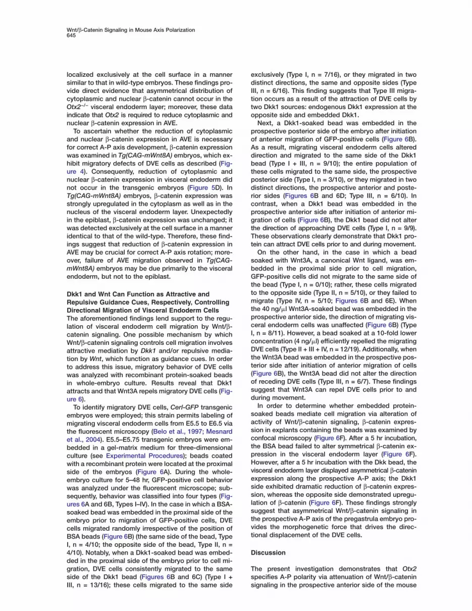
Dkk1 and Wnt Can Function as Attractive andRepulsive Guidance Cues, Respectively, ControllingDirectional Migration of Visceral Endoderm CellsThe aforementioned findings lend support to the regu-lation of visceral endoderm cell migration by Wnt/β-catenin signaling. One possible mechanism by whichWnt/β-catenin signaling controls cell migration involvesattractive mediation by Dkk1 and/or repulsive media-tion by Wnt, which function as guidance cues. In orderto address this issue, migratory behavior of DVE cellswas analyzed with recombinant protein-soaked beadsin whole-embryo culture. Results reveal that Dkk1attracts and that Wnt3A repels migratory DVE cells (Fig-ure 6).
To identify migratory DVE cells, Cerl-GFP transgenicembryos were employed; this strain permits labeling ofmigrating visceral endoderm cells from E5.5 to E6.5 viathe fluorescent microscopy (Belo et al., 1997; Mesnardet al., 2004). E5.5–E5.75 transgenic embryos were em-bedded in a gel-matrix medium for three-dimensionalculture (see Experimental Procedures); beads coatedwith a recombinant protein were located at the proximalside of the embryos (Figure 6A). During the whole-embryo culture for 5–48 hr, GFP-positive cell behaviorwas analyzed under the fluorescent microscope; sub-sequently, behavior was classified into four types (Fig-ures 6A and 6B, Types I–IV). In the case in which a BSA-soaked bead was embedded in the proximal side of theembryo prior to migration of GFP-positive cells, DVEcells migrated randomly irrespective of the position ofBSA beads (Figure 6B) (the same side of the bead, TypeI, n = 4/10; the opposite side of the bead, Type II, n =4/10). Notably, when a Dkk1-soaked bead was embed-ded in the proximal side of the embryo prior to cell mi-gration, DVE cells consistently migrated to the sameside of the Dkk1 bead (Figures 6B and 6C) (Type I +
III, n = 13/16); these cells migrated to the same sideexclusively (Type I, n = 7/16), or they migrated in twodistinct directions, the same and opposite sides (TypeIII, n = 6/16). This finding suggests that Type III migra-tion occurs as a result of the attraction of DVE cells bytwo Dkk1 sources: endogenous Dkk1 expression at theopposite side and embedded Dkk1.
Next, a Dkk1-soaked bead was embedded in theprospective posterior side of the embryo after initiationof anterior migration of GFP-positive cells (Figure 6B).As a result, migrating visceral endoderm cells altereddirection and migrated to the same side of the Dkk1bead (Type I + III, n = 9/10); the entire population ofthese cells migrated to the same side, the prospectiveposterior side (Type I, n = 3/10), or they migrated in twodistinct directions, the prospective anterior and poste-rior sides (Figures 6B and 6D; Type III, n = 6/10). Incontrast, when a Dkk1 bead was embedded in theprospective anterior side after initiation of anterior mi-gration of cells (Figure 6B), the Dkk1 bead did not alterthe direction of approaching DVE cells (Type I, n = 9/9).These observations clearly demonstrate that Dkk1 pro-tein can attract DVE cells prior to and during movement.
On the other hand, in the case in which a beadsoaked with Wnt3A, a canonical Wnt ligand, was em-bedded in the proximal side prior to cell migration,GFP-positive cells did not migrate to the same side ofthe bead (Type I, n = 0/10); rather, these cells migratedto the opposite side (Type II, n = 5/10), or they failed tomigrate (Type IV, n = 5/10; Figures 6B and 6E). Whenthe 40 ng/�l Wnt3A-soaked bead was embedded in theprospective anterior side, the direction of migrating vis-ceral endoderm cells was unaffected (Figure 6B) (TypeI, n = 8/11). However, a bead soaked at a 10-fold lowerconcentration (4 ng/�l) efficiently repelled the migratingDVE cells (Type II + III + IV, n = 12/19). Additionally, whenthe Wnt3A bead was embedded in the prospective pos-terior side after initiation of anterior migration of cells(Figure 6B), the Wnt3A bead did not alter the directionof receding DVE cells (Type III, n = 6/7). These findingssuggest that Wnt3A can repel DVE cells prior to andduring movement.
In order to determine whether embedded protein-soaked beads mediate cell migration via alteration ofactivity of Wnt/β-catenin signaling, β-catenin expres-sion in explants containing the beads was examined byconfocal microscopy (Figure 6F). After a 5 hr incubation,the BSA bead failed to alter symmetrical β-catenin ex-pression in the visceral endoderm layer (Figure 6F).However, after a 5 hr incubation with the Dkk bead, thevisceral endoderm layer displayed asymmetrical β-cateninexpression along the prospective A-P axis; the Dkk1side exhibited dramatic reduction of β-catenin expres-sion, whereas the opposite side demonstrated upregu-lation of β-catenin (Figure 6F). These findings stronglysuggest that asymmetrical Wnt/β-catenin signaling inthe prospective A-P axis of the pregastrula embryo pro-vides the morphogenetic force that drives the direc-tional displacement of the DVE cells.
Discussion
The present investigation demonstrates that Otx2specifies A-P polarity via attenuation of Wnt/β-catenin
signaling in the prospective anterior side of the mouse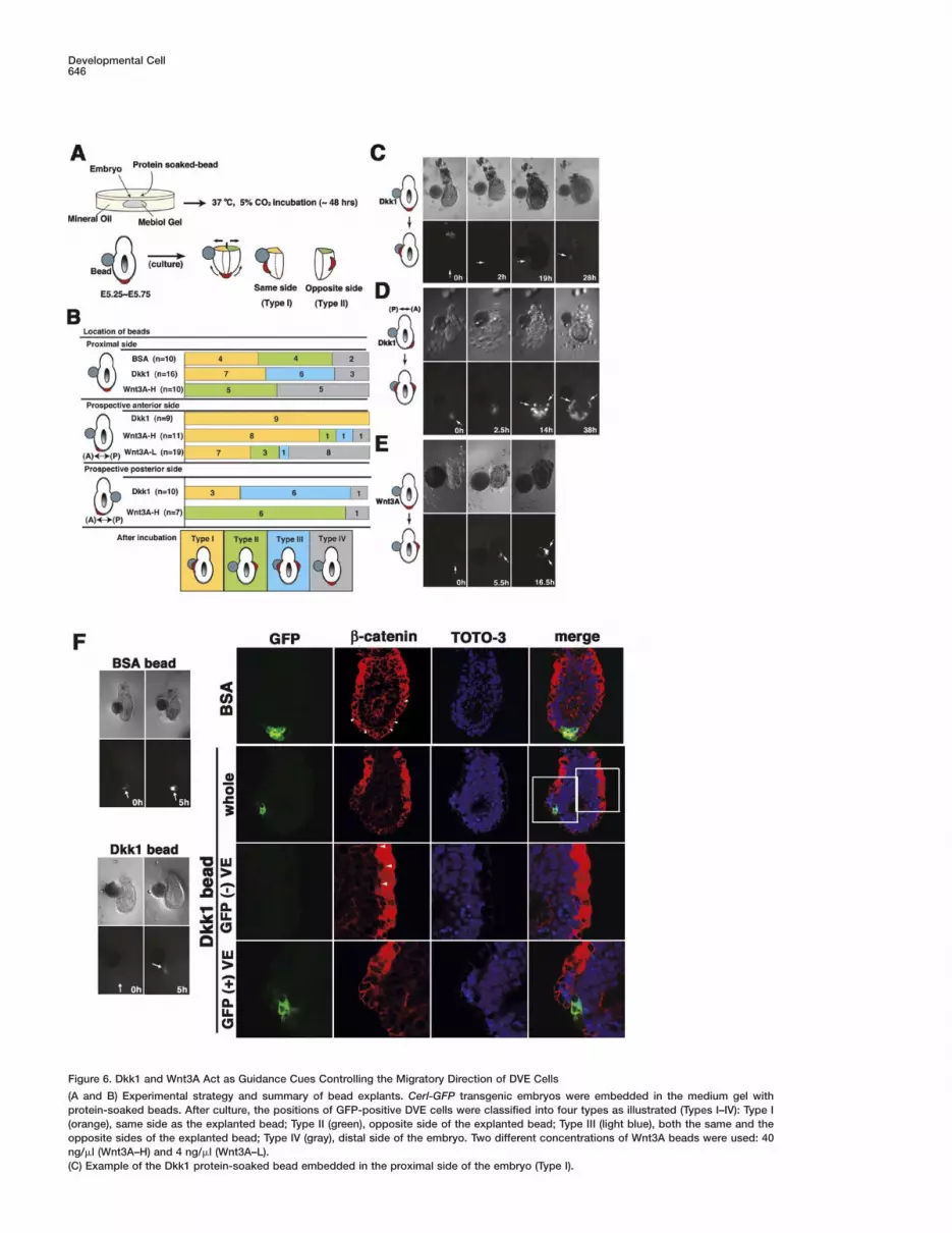
Developmental Cell646
Figure 6. Dkk1 and Wnt3A Act as Guidance Cues Controlling the Migratory Direction of DVE Cells
(A and B) Experimental strategy and summary of bead explants. Cerl-GFP transgenic embryos were embedded in the medium gel withprotein-soaked beads. After culture, the positions of GFP-positive DVE cells were classified into four types as illustrated (Types I–IV): Type I(orange), same side as the explanted bead; Type II (green), opposite side of the explanted bead; Type III (light blue), both the same and theopposite sides of the explanted bead; Type IV (gray), distal side of the embryo. Two different concentrations of Wnt3A beads were used: 40ng/�l (Wnt3A–H) and 4 ng/�l (Wnt3A–L).(C) Example of the Dkk1 protein-soaked bead embedded in the proximal side of the embryo (Type I).

Wnt/β-Catenin Signaling in Mouse Axis Polarization647
ment of the mouse embryo (Figure 7). Normally, prior to
(D) Example of the Dkk1 protein-soaked bead embedded in the prospective posterior side of the embryo (Type III).(E) Example of the Wnt3A-H protein-soaked bead embedded in the proximal side of the embryo (Type II). GFP-positive cells are indicated bywhite arrows in (C)–(E).(F) The BSA or Dkk1 protein-soaked beads embedded in the proximal side of the embryo for β-catenin expression analysis. After a 5 hrincubation, Cerl-positive cells do not migrate to one side in the BSA-embedded explant, while these cells start to migrate to one side in theDkk1-embedded explant. Cerl-GFP (green), anti-active-β-catenin (red), TOTO-3 (nuclei, blue), and the merged image. The areas shown in themagnified views are marked in the upper merged images (white squares).
of β-catenin or to insufficient levels of Dkk1 expression.
embryo. The Wnt antagonist Dkk1, which is a crucialdownstream target of Otx2, acts as an attractive guid-ance cue controlling the directional migration of vis-ceral endoderm cells. Moreover, to our knowledge, thisgenetic evidence provides novel insights into evolution-arily conserved mechanisms governing primary bodyaxis formation in metazoans.
Wnt/�-Catenin Signaling RegulatesA-P Axis DevelopmentOur findings led to the proposal of a model describingthe role of Wnt/β-catenin signaling in A-P axis develop-
Figure 7. Schematic Model of the Mecha-nism Governing A-P Axis Development viaWnt/β-Catenin Signaling in the Mouse Embryo
(A) Schematic representation of wild-typemouse embryos between E5.5 and E6.0. AtE5.5, DVE cells are thought to possess theability to migrate in the proximal directionmarked by the Dkk1-positive domain (dottedarrows). At E5.75, Wnt/β-catenin signaling isattenuated by anterior restriction of Dkk1 ex-pression. Direction of DVE cell migration isindicated by an arrow. By E6.0, Dkk1 expres-sion is detected in the most anterior portionof AVE; furthermore, axis conversion is com-pleted in the A-P direction.(B and C) Schematic diagrams regarding ex-pression of active-β-catenin in the wild-typeat E5.5 and E6.0 and in the Otx2−/− andTg(CAG-mWnt8A) embryos at E6.0. (B) AtE5.5, cytoplasmic and nuclear β-catenin islocalized in the visceral endoderm layer, butnot in the epiblast layer; at subsequent E6.0,cytoplasmic and nuclear β-catenin expres-sion is reduced anteriorly and upregulatedposteriorly in the wild-type. (C) In the mi-gration-defective embryos, cytoplasmic andnuclear β-catenin expression is symmetri-cally distributed in the entire visceral endo-derm even at E6.0. A, anterior; AVE, anteriorvisceral endoderm; D, distal; epi, epiblast; P,posterior; Pr, proximal; VE, visceral endo-derm; xe, extraembryonic ectoderm.
AVE migration, around E5.5, a canonical Wnt antago-nist, Dkk1, is expressed in the proximal portion of DVEcells in a circular, symmetrical pattern (Figures 7A). Onthe other hand, canonical Wnt ligands, e.g., mWnt8Aand Wnt3, are expressed in the proximal epiblast (Fig-ure 7A). At this stage, any proximal direction may becompetent with respect to migration of DVE cells, assuggested by symmetrical Dkk1 expression. However,β-catenin expression does not appear to be downregu-lated in these Dkk1-positive domains. This delayed at-tenuation of β-catenin expression is probably attribut-able to the time lag associated with protein degradation

Developmental Cell648
By E5.75, attenuation of Wnt/β-catenin signaling in theAVE is closely linked with the axis conversion processfrom the P-D to the A-P orientation. Axis rotation mayoccur primarily as a result of asymmetrical expressionof Wnt antagonists, including Dkk1. Concurrently, Dkk1expression is downregulated in the prospective poste-rior side and upregulated in the anterior side, whereasβ-catenin expression displays the opposite profile (Fig-ures 7A and 7B).
Wnt signaling has been classified into two pathways:the canonical and noncanonical pathways, or b-catenin-dependent and -independent pathways, respectively(Logan and Nusse, 2004). The current study demon-strates that the canonical Wnt antagonist, Dkk1, and itsnuclear target, β-catenin, participate in DVE migration.Additionally, misexpression of a canonical Wnt ligand,mWnt8A, prevented correct A-P axis conversion (Figure4). Coincidentally, asymmetrical distribution of β-cateninexpression was not observed in the AVE of Otx2-defi-cient and Tg(CAG-mWnt8) embryos (Figures 5 and 7C).These results, in concert, strongly support the crucialrole of asymmetrical β-catenin localization in A-P axisconversion. However, Dkk1 homozygous mutant em-bryos appeared to undergo normal A-P axis develop-ment (Mukhopadhyay et al., 2001). To account for thisdisparity, other inhibitory molecules of Wnt/β-cateninsignaling, which complement the Dkk1 function in vis-ceral endoderm at different levels of the signaling path-way, may be present, albeit at reduced levels in Otx2-deficient embryos, e.g., Cerl, Axin, TCF3, Wise, andICAT (Logan and Nusse, 2004).
Wnt/�-Catenin Signaling Guides DirectionalMigration of Visceral Endoderm CellsThis investigation provides for the evidence that the ca-nonical Wnt ligand and its antagonist function as guid-ance cues in the DVE cell migration. Migrating visceralendoderm cells possess unique morphology distinctfrom that of adjacent endoderm cells, as well as activemigratory character; these cells continuously changeshape and project filopodial processes (Kimura et al.,2000; Srinivas et al., 2004). Comparative expressionanalysis of Dkk1 with Cerl suggests that Dkk1-positivecells are located at the forefront of migratory DVE cells(Figure 1). These findings suggest that Dkk1 may deter-mine the migratory direction of DVE cells. Moreover, ap-parent normal expression of Nodal and its antagonistsin Otx2-deficient embryos (Figure 1) might afford evi-dence in support of the previously described observa-tion that Otx2−/− DVE cells display no significant changewith respect to cell proliferation; rather, they exhibit adefect associated with directional migration (Perea-Gomez et al., 2001). Further BrdU incorporation experi-ments with Wnt8A-misexpressing embryos and Dkk1/Wnt3A-embedded explants revealed that Wnt ligandsand Dkk1 failed to mediate asymmetrical cell prolifera-tion along with the A-P axis (C.K.-Y., D.O., Y.M., andI.M., unpublished data). In a manner consistent withthese notions, Dkk1 can attract Cerl-positive DVE cellsand a canonical Wnt ligand, Wnt3A, can repel them asa migratory guidance cue (Figure 6). Furthermore, at-tenuation of β-catenin expression in the cytoplasm andthe nucleus during DVE cell migration appears to be
lbttWa
OowTpdtnidprvtdetpfStTdsd
pnpneeit
ieaafIaa2mnsiTdm
E
CIa
inked to asymmetrical expression of Dkk1 and the em-edded Dkk1 bead (Figures 5 and 6). In conjunction,hese findings directly support mediation of the direc-ional migration of DVE cells by the combination of
nt-mediated repulsion and its antagonist-mediatedttraction.
tx2 May Regulate Asymmetrical Distributionf b-Catenin Activity in Conjunctionith the Primary Body Axishis study indicates that localization of the dephos-horylated form of β-catenin is dynamically regulateduring A-P axis specification (Figures 5, 7B, and 7C). Inhe wild-type visceral endoderm layer, cytoplasmic anduclear β-catenin expression are specifically reduced
n the prospective anterior side. Notably, in both Otx2-eficient and Tg(CAG-mWnt8A) embryos, which dis-lay failure of axis rotation, the expression is not down-
egulated; rather, it is upregulated throughout the entireisceral endoderm layer (Figures 5 and 7). Although fur-her molecular analysis is necessary in order to eluci-ate the precise molecular mechanism by which Dkk1xpression is initially induced in the most proximal por-ion of DVE and subsequently downregulated in therospective posterior side, Otx2 expression is crucial
or Dkk1 expression in the visceral endoderm (Figures2 and S3). In addition, Dkk1 alone can rescue axis ro-
ation failure attributable to Otx2 deficiency (Figure 2).hese findings suggest that Otx2 specifies A-P axisevelopment primarily via regulation of Wnt/β-cateninignaling pathways, including Dkk1, in the visceral en-oderm.Surprisingly, mWnt8A transcripts driven by the CAG
romoter are upregulated primarily in the epiblast, butot in the visceral endoderm (Figure 4A#), whereas ex-ression of the dephosphorylated form of β-catenin isot elevated in the epiblast layer of Tg(CAG-mWnt8A)mbryos. This finding suggests the involvement of un-xpected molecular mechanisms via which Wnt signal-
ng can be transmitted to β-catenin activity mainly inhe visceral endoderm, but not in the epiblast layer.
To our knowledge, this genetic evidence affords novelnsights into evolutionarily conserved mechanisms gov-rning primary body axis formation across the metazo-ns. The asymmetrical distribution of β-catenin activitylong with the A-P axis plays a pivotal role in the speci-ication of A-P polarity throughout metazoan embryos.n amphibians, fish, ascidians, sea urchins, and cnid-rians, β-catenin is localized to cell nuclei preferentiallyt one pole of the cleavage-stage embryo (Imai et al.,000; Logan et al., 1999; Schneider et al., 1996; Wikra-anayake et al., 2003). In these various organisms,
uclear activity of b-catenin is required for early axispecification and the subsequent establishment of crit-cal signaling centers, “organizers”, in the early embryo.he present investigation suggests that asymmetricalistribution of β-catenin expression serves as a primaryediator of axis specification in the mammalian embryo.
xperimental Procedures
onstruction of Targeting Vector for Dkk1 Knockin Micen the targeting construct, Dkk1 cDNA with polyadenylation signalsnd the PGKneo cassette were inserted into the SmaI and the

Wnt/β-Catenin Signaling in Mouse Axis Polarization649
EcoRI sites, located 220 bp upstream from the translation initiationsite and in the first intron, respectively. Homologous recombinantTT2 ES cells and chimeric mice were obtained as described (Mat-suo et al., 1995).
Generation of CAG-mWnt8A Transgenic MiceMouse Wnt8A cDNAs were isolated from a mouse cDNA library. Inbrief, mWnt8A cDNA fused with a CAG promoter (Niwa et al., 1991)was ligated to the lacZ gene flanked by two loxP sites. Subse-quently, a CAG-lacZ-mWnt8A transgene was constructed via liga-tion of the aforementioned resultant vectors with the neo genedriven by the PGK1 promoter with a polyadenylation signal. TT2ES cells were cultured, electroporated with linearized transgenicvectors, and selected in G418 (Figure S4).
GenotypingTransgenic and knockin founders and their progenitors were rou-tinely determined by PCR. Primers and lengths of the products inthe PCR analyses were as follows: in the transgenic mice, CAG-lacZ-mWnt8A was identified with primers CAG-pro (5#-TAGAGCCTCTGCTAACCATGTTCATGCCTT-3#) and CAG-lacZ (5#-AGTGTCCCAGCCTGTTTATCTACGGCTTAA-3#), yielding 270 bp. The CAG-mWnt8 allele excised by Cre was determined with primers CAG-pro and CAG-mWnt8A (5#-GATGGCAGCAGAGCGGATGGCATGAATGAA-3#), yielding 500 bp. Dkk1 knockin mice were identified withthe following primers: wild-type Otx2 allele with primers Forward1(5#-GTATTTTCCTTGCTACCAAACTGCCGAGTG-3#) and Reverse1(5#-CTGGAGGGAAGCCACACCTCTAAGGATTAA-3#), yielding 400bp, and Dkk1 knockin allele with primers Reverse1 and SVDK-2 (5#-ACAGCAGAAACATACAAGCTGTCAGCTTTG-3#), yielding 200 bp.b-catenin mutant mice were obtained from the Jackson Laboratoryand genotyped as described (Brault et al., 2001).
Expression Analysis and Semi- and Ultrathin SectionsIn situ hybridization was performed as described (Wilkinson, 1998).For morphological analysis, embryos were fixed in 2% paraformal-dehyde plus 2.5% glutaraldehyde in 0.1 M sodium cacodylatebuffer (pH 7.3); subsequently, tissues were postfixed for 2 hr in 1%OsO4 and were embedded in Poly Bed 812. Semithin sections (0.55�m thickness) were produced with a glass knife and stained with0.5% toluidine blue. Ultrathin sections (70 nm thickness) were ob-tained with a diamond knife and examined after staining with uranylacetate and lead citrate.
Whole-Embryo Culture with Recombinant Protein BeadsFemale ICR mice were sacrificed between 10:00 and 17:00 hr onthe fifth day of pregnancy. Embryos were dissected from decidualtissue in M2 medium lacking Phenol red. The GFP-positive em-bryos, which were collected under a fluorescent microscope(Leica), were cultured in the Mebiol Gel matrix (Mebiol, Inc.) con-taining 50% DMEM and 50% rat serum in 5% CO2 at 37°C for5–48 hr for three-dimensional culture. Images of migratory behaviorof visceral endoderm cells were captured and recorded with a Ha-mamatsu chilled CCD camera (C5985). Beads (Affigel Bluo Gel,BioRad) were rinsed in PBS several times and soaked in 200 ng/�lmouse Dkk1 recombinant protein (R&D system), 40 and 4 ng/�lmouse Wnt3A recombinant protein (R&D System), or 50 mg/mlBSA-PBS solution overnight at 4°C or for 1 hr at 37°C. Preparedbeads were transplanted to the proximal side of the embeddedtransgenic embryo.
ImmunohistochemistryWild-type, Otx2−/−, Tg(CAG-mWnt8A), and Cerl-GFP embryos werefixed as described (Ciruna and Rossant, 2001). Primary antibodieswere applied at the following concentrations: 10 mg/ml for rat anti-E-cadherin (ECCD-2) (Takara Shuzo) and at a 1:300 dilution foranti-active-β-catenin (anti-ABC) (8E7) (Upstate). Appropriate spe-cies-specific, fluorophore-labeled secondary antibodies (MolecularProbes) were applied at 1:200 dilution. Nuclei were stained withTOTO-3 iodide (Molecular Probes) at a 1:500 dilution in the pres-ence of 100 �g/ml RNase A. Staining was examined with a confocalmicroscope (Leica).
Supplemental DataSupplemental Data including five figures are available at http://www.developmentalcell.com/cgi/content/full/9/5/639/DC1/.
Acknowledgments
We are grateful to Drs. E. De Robertis, R. Grosschedl, B.G. Herr-mann, B. Hogan, G. Martin, R. Nusse, M.M. Shen, and K. Yamamurafor in situ probes. We also wish to thank Dr. C. Niehrs for Dkk1cDNA plasmid; the Animal Resources and Genetic EngineeringTeam, RIKEN Center for Developmental Biology for housing themice; and Ms. Naoko Inoue, Mr. Hiroshi Kiyonari, Ms. Rika Naka-yama, Ms. Ayako Nagao, and Ms. Kuniko Kitajima for their assis-tance. This work was supported in part by grants-in-aid for Scien-tific Research on Priority Areas and Young Scientists (B) from theMinistry of Education, Culture, Sports Science and Technology,Japan.
Received: September 24, 2004Revised: April 28, 2005Accepted: September 22, 2005Published: October 31, 2005
References
Batten, B.E., and Haar, J.L. (1979). Fine structural differentiation ofgerm layers in the mouse at the time of mesoderm formation. Anat.Rec. 194, 125–141.
Beddington, R.S., and Robertson, E.J. (1999). Axis developmentand early asymmetry in mammals. Cell 96, 195–209.
Belo, J.A., Bouwmeester, T., Leyns, L., Kertesz, N., Gallo, M., Follet-tie, M., and De Robertis, E.M. (1997). Cerberus-like is a secretedfactor with neutralizing activity expressed in the anterior primitiveendoderm of the mouse gastrula. Mech. Dev. 68, 45–57.
Brault, V., Moore, R., Kutsch, S., Ishibashi, M., Rowitch, D.H.,McMahon, A.P., Sommer, L., Boussadia, O., and Kemler, R. (2001).Inactivation of the β-catenin gene by Wnt1-Cre-mediated deletionresults in dramatic brain malformation and failure of craniofacialdevelopment. Development 128, 1253–1264.
Brennan, J., Lu, C.C., Norris, D.P., Rodriguez, T.A., Beddington,R.S., and Robertson, E.J. (2001). Nodal signalling in the epiblastpatterns the early mouse embryo. Nature 411, 965–969.
Ciruna, B., and Rossant, J. (2001). FGF signaling regulates meso-derm cell fate specification and morphogenetic movement at theprimitive streak. Dev. Cell 1, 37–49.
Glinka, A., Wu, W., Delius, H., Monaghan, A.P., Blumenstock, C.,and Niehrs, C. (1998). Dickkopf-1 is a member of a new family ofsecreted proteins and functions in head induction. Nature 391,357–362.
Imai, K., Takada, N., Satoh, N., and Satou, Y. (2000). (β)-cateninmediates the specification of endoderm cells in ascidian embryos.Development 127, 3009–3020.
Kimura, C., Yoshinaga, K., Tian, E., Suzuki, M., Aizawa, S., and Mat-suo, I. (2000). Visceral endoderm mediates forebrain developmentby suppressing posteriorizing signals. Dev. Biol. 225, 304–321.
Kimura, C., Shen, M.M., Takeda, N., Aizawa, S., and Matsuo, I.(2001). Complementary functions of Otx2 and Cripto in initial pat-terning of mouse epiblast. Dev. Biol. 235, 12–32.
Lewandoski, M., Meyers, E.N., and Martin, G.R. (1997). Analysis ofFgf8 gene function in vertebrate development. Cold Spring Harb.Symp. Quant. Biol. 62, 159–168.
Logan, C.Y., and Nusse, R. (2004). The Wnt signaling pathway indevelopment and disease. Annu. Rev. Cell Dev. Biol. 20, 781–810.
Logan, C.Y., Miller, J.R., Ferkowicz, M.J., and McClay, D.R. (1999).Nuclear β-catenin is required to specify vegetal cell fates in the seaurchin embryo. Development 126, 345–357.
Mao, B., Wu, W., Davidson, G., Marhold, J., Li, M., Mechler, B.M.,Delius, H., Hoppe, D., Stannek, P., Walter, C., et al. (2002). Kremenproteins are Dickkopf receptors that regulate Wnt/β-catenin signal-ling. Nature 417, 664–667.

Developmental Cell650
Matsuo, I., Kuratani, S., Kimura, C., Takeda, N., and Aizawa, S.(1995). Mouse Otx2 functions in the formation and patterning ofrostral head. Genes Dev. 9, 2646–2658.
Mesnard, D., Filipe, M., Belo, J.A., and Zernicka-Goetz, M. (2004).The anterior-posterior axis emerges respecting the morphology ofthe mouse embryo that changes and aligns with the uterus beforegastrulation. Curr. Biol. 14, 184–196.
Mukhopadhyay, M., Shtrom, S., Rodriguez-Esteban, C., Chen, L.,Tsukui, T., Gomer, L., Dorward, D.W., Glinka, A., Grinberg, A., Hu-ang, S.P., et al. (2001). Dickkopf1 is required for embryonic headinduction and limb morphogenesis in the mouse. Dev. Cell 1, 423–434.
Niwa, H., Yamamura, K., and Miyazaki, J. (1991). Efficient selectionfor high-expression transfectants with a novel eukaryotic vector.Gene 108, 193–199.
Perea-Gomez, A., Lawson, K.A., Rhinn, M., Zakin, L., Brulet, P., Ma-zan, S., and Ang, S.L. (2001). Otx2 is required for visceral endodermmovement and for the restriction of posterior signals in the epiblastof the mouse embryo. Development 128, 753–765.
Popperl, H., Schmidt, C., Wilson, V., Hume, C.R., Dodd, J., Krum-lauf, R., and Beddington, R.S. (1997). Misexpression of Cwnt8C inthe mouse induces an ectopic embryonic axis and causes a trunca-tion of the anterior neuroectoderm. Development 124, 2997–3005.
Schneider, S., Steinbeisser, H., Warga, R.M., and Hausen, P. (1996).β-catenin translocation into nuclei demarcates the dorsalizing cen-ters in frog and fish embryos. Mech. Dev. 57, 191–198.
Srinivas, S., Rodriguez, T., Clements, M., Smith, J.C., and Bedding-ton, R.S. (2004). Active cell migration drives the unilateral move-ments of the anterior visceral endoderm. Development 131, 1157–1164.
Staal, F.J.T., van Noort, M., Strous, G.S., and Clevers, H.C. (2002).Wnt signals are transmitted through N-terminally dephosphorylatedβ-catenin. EMBO Rep. 3, 63–68.
Stern, C.D. (2001). Initial patterning of the central nervous system:how many organizers? Nat. Rev. Neurosci. 2, 92–98.
Thomas, P.Q., Brown, A., and Beddington, R.S. (1998). Hex: a ho-meobox gene revealing peri-implantation asymmetry in the mouseembryo and an early transient marker of endothelial cell precursors.Development 125, 85–94.
Wikramanayake, A.H., Hong, M., Lee, P.N., Pang, K., Byrum, C.A.,Bince, J.M., Xu, R., and Martindale, M.Q. (2003). An ancient role fornuclear beta-catenin in the evolution of axial polarity and germlayer segregation. Nature 426, 446–450.
Wilkinson, D.G. (1998). In Situ Hybridization: A Practical Approach,Second Edition (Oxford/New York: Oxford University Press).
Yamamoto, M., Saijoh, Y., Perea-Gomez, A., Shawlot, W., Behringer,R.R., Ang, S.L., Hamada, H., and Meno, C. (2004). Nodal antago-nists regulate formation of the anteroposterior axis of the mouseembryo. Nature 428, 387–392.
Zakin, L., Reversade, B., Virlon, B., Rusniok, C., Glaser, P., Elalouf,J.M., and Brulet, P. (2000). Gene expression profiles in normal andOtx2−/− early gastrulating mouse embryos. Proc. Natl. Acad. Sci.USA 97, 14388–14393.


