Embed Size (px)
Citation preview

C
LM
a
ARRAA
KDOOOC
1
iclamoitt
shaOcrp
h0
Cell Calcium 55 (2014) 191–199
Contents lists available at ScienceDirect
Cell Calcium
jou rn al hom epage: www.elsev ier .com/ locate /ceca
almodulin modulates insect odorant receptor function
atha Mukunda, Fabio Miazzi, Sabine Kaltofen, Bill S. Hansson, Dieter Wicher ∗
ax Planck Institute for Chemical Ecology, Department Evolutionary Neuroethology, Hans-Knöll-St. 8, D-07745 Jena, Germany
r t i c l e i n f o
rticle history:eceived 14 November 2013eceived in revised form 14 February 2014ccepted 14 February 2014vailable online 23 February 2014
eywords:rosophiladorant receptorrco
a b s t r a c t
Insect odorant receptors (ORs) are heteromeric complexes of an odor-specific receptor protein (OrX) and aubiquitous co-receptor protein (Orco). The ORs operate as non-selective cation channels, also conductingCa2+ ions. The Orco protein contains a conserved putative calmodulin (CaM)-binding motif indicating arole of CaM in its function. Using Ca2+ imaging to monitor OR activity we investigated the effect of CaMinhibition on the function of OR proteins. Ca2+ responses elicited in Drosophila olfactory sensory neuronsby stimulation with the synthetic OR agonist VUAA1 were reduced and prolonged by CaM inhibition withthe potent antagonist W7 but not with the weak antagonist W5. A similar effect was observed for Orcoproteins heterologously expressed in CHO cells when CaM was inhibited with W7, trifluoperazine orchlorpromazine, or upon overexpression of CaM-EF-hand mutants. With the Orco CaM mutant bearing
2+
lfactory sensory neuronalcium imaginga point mutation in the putative CaM site (K339N) the Ca responses were akin to those obtained forwild type Orco in the presence of W7. There was no uniform effect of W7 on Ca2+ responses in CHO cellsexpressing complete ORs (Or22a/Orco, Or47a/Orco, Or33a/Orco, Or56a/Orco). For Or33a and Or47a weobserved no significant effect of W7, while it caused a reduced response in cells expressing Or22a and ashortened response for Or56a.
© 2014 Elsevier Ltd. All rights reserved.
. Introduction
Functional properties of sensory receptors, such as sensitiv-ty and response duration, have to be regulated according to theharacteristics of the external signal transduced into intracellu-ar information. To optimize temporal resolution the response to
given stimulus has to be terminated. The most straightforwardechanism of response cessation is a negative feedback control
f receptor activation. Such mechanisms, as e.g. the Ca2+-inducednhibition of cyclic nucleotide-gated channels in vertebrate olfac-ory sensory neurons (OSNs), inhibit further entry and terminatehe odor signal [1].
Insect odorant receptors (ORs) represent ligand-gated non-elective cation channels that conduct also Ca2+ [2,3]. They areeterodimers composed of an odorant-specific OR protein (OrX)nd an ubiquitous co-receptor protein (Orco) [4]. Besides therX/Orco heteromers also Orco homomers form Ca2+ conductingation channels [3]. Activation of Orco channels plays an important
ole in the regulation of OR sensitivity to odorants [5]. Orco mostrobably contributes to the pore of the heteromeric OR channels∗ Corresponding author. Tel.: +49 3641 571415.E-mail address: [email protected] (D. Wicher).
ttp://dx.doi.org/10.1016/j.ceca.2014.02.013143-4160/© 2014 Elsevier Ltd. All rights reserved.
[6]. While the heteromers are activated by odorants, both kinds ofchannels open upon binding of the synthetic ligand VUAA1 [7].
The Ca2+ import into cells through ion channels such as voltage-gated Ca2+ channels or cyclic nucleotide gated channels, and inconsequence the free Ca2+ concentration, has to be tightly reg-ulated [8–12]. Thus, termination of receptor response not onlyrestores the capability to sense further stimuli but also protectsthe cell from Ca2+ overload that may damage or even kill it. Oneof the most important proteins that link Ca2+ concentration to reg-ulation of Ca2+ influx or extrusion is calmodulin (CaM) [13]. CaMcloses, for example, voltage-gated Ca2+ channels [14] and accel-erates Ca2+ extrusion by the plasma membrane Ca2+ pump whenactivated by increased free Ca2+ level. In mammalian odorant recep-tors response termination includes CaM-mediated inactivation ofCNG channels [15] and down regulation of intracellular free Ca2+
[16].As insect OR activation is accompanied by Ca2+ influx into
sensory cells, there is an obvious question whether CaM mightplay a role in regulating the function of insect ORs. To monitorOR activity, we observed the rise in intracellular Ca2+ concentra-tion [Ca2+]i as previously performed in vertebrate and invertebrate
preparations [17,18]. We tested the effect of CaM inhibition on theagonist-induced rise in [Ca2+]i in Drosophila OSNs and observeda reduced response. To specify the role of OR proteins in thisregulation we proceeded to investigate the role of CaM in the
1 Calciu
cc
2
2
Gaais5F2
uHCpTieGiStOue1o
2
mdtf1nob
2
mbmoEtasf
wcfca
t
92 L. Mukunda et al. / Cell
ontrol of heterologously expressed Orco proteins and OrX/Orcoombinations.
. Materials and methods
.1. Fly preparation and OR expression in cultured cells
Drosophila melanogaster flies with genotype w;UAS-CaMP3.0;Orco-Gal4 were maintained on conventional cornmealgar medium under a 12 h light: 12 h dark cycle at 25 ◦C. Flies werenesthetized in ice; after decapitation antennae were excised, fixedn vertical position on a glass coverslip using a two componentilicon and immersed in Drosophila Ringer solution (in mM HEPES,; NaCl, 130; KCl, 5; MgCl2, 2; CaCl2, 2; and sucrose, 36; pH = 7.3).lagelli were cut below half of their length and incubated for0 min to remove air bubbles.
The open reading frame of Drosophila Orco was PCR-amplifiedsing gene specific primers with restriction sites for XhoI andindIII and cloned into the pcrII TA-cloning vector (Invitrogen,arlsbad, CA, USA). The identity of the insert was subcloned into thecDNA3.1(−) expression vector via the integrated restriction sites.he sequences were confirmed by double strand DNA sequenc-ng (Eurofins MWG Operon, Ebersberg, Germany). CHO cells stablyxpressing Orco were purchased from cytobox UG (Konstanz,ermany) and grown in cytoboxTM CHO select medium contain-
ng puromycin. The cells were grown on poly-l-lysine (0.01%,igma–Aldrich, Steinheim, Germany) coated coverslips and cul-ured at a density of ∼1 × 10⁶ per 35 mm dish. The CaM mutants andrs 22a, 47a, 33a and 56a were transfected with 0.3–0.5 �g/wellsing Rotifect transfection kit (Roth, Karlsruhe, Germany). Forxperiments cells were exposed to bath solution (in mM: NaCl,35; KCl, 5; MgCl2, 1; CaCl2, 1; HEPES, 10; d-glucose, 10; pH = 7.4;smolarity 295 mOsmol/l).
.2. Site directed mutagenesis
Orco CaM K339N mutation was performed using site directedutagenesis. Two overlapping mutagenic oligonucleotides were
esigned to introduce point mutation in position (Lysine) K 339o Aspargine (N) residue. The PCR products were then used to run aull length PCR using Orco (full length) primers. The final product of.56 kb band was T:A cloned into Topo vector (invitrogen life tech-ologies) and subsequently sub-cloned into XhoI and HindIII sitesf pcDNA 3.1(−) expression vectors. The sequences were confirmedy double strand DNA sequencing (Eurofins MWG operon).
.3. Calcium imaging in flies and cells
Excitation of cells and OSNs was performed with a monochro-ator (Polychrome V, TILL Photonics, Gräfelfing, Germany) coupled
y means of an epifluorescence condenser into an Axioskop FSicroscope (Carl Zeiss, Jena, Germany) with a water immersion
bjective (LUMPFL 40xW/IR/0.8; Olympus, Hamburg, Germany).mitted light was separated by a 400-nm dichroic mirror and fil-ered with a 420-nm long-pass filter. Fluorescence images werecquired using a cooled CCD camera controlled by TILLVision 4.0oftware (TILL Photonics). The resolution was 640 × 480 pixel in arame of 175 �m × 130 �m.
GCaMP3.0 was excited with 475 nm light at a 0.2 Hz frequencyith an exposition time of 50 ms. The response magnitude was
alculated as the average �F/F, expressed in percentage, for eachrame, where F was estimated as the mean fluorescence level cal-
ulated for each neuronal body cell on 10 frames before eachpplication of DMSO (control), W7 or W5 solution.CHO cells were loaded with fura-2 by incubation in bath solu-ion containing 5 �M fura-2/acetomethylester (Molecular Probes,
m 55 (2014) 191–199
Invitrogen) for 30 min. Free intracellular Ca2+ concentration([Ca2+]i) was determined with the fluorescence ratio method andcalculated according to [Ca2+]i = Keff(R − Rmin)/(Rmax − R). Imagepairs were obtained by excitation for 150 ms at 340 nm and 380 nm(ratio R); background fluorescence was subtracted. Keff, Rmin, andRmax were determined using permeabilized cells (Ca2+ free; 5 mMCa2+; 500 nM Ca2+). The values of Keff, Rmin, and Rmax were 1.95 �M,0.2, and 5.3, respectively.
OSNs and CHO cells were stimulated using VUAA1 (applicationof 100 �l of 100 �M solution via pipette) after incubation in thepresence of W7, W5 (application of 100 �l of 10 �M solution viapipette), or in the equivalent amount of DMSO (in which W7 or W5were dissolved) as a control, for 50 s.
2.4. Chemicals
VUAA1 (N-(4-ethylphenyl)-2-((4-ethyl-5-(3-pyridinyl)-4H-1,2,4-triazol-3-yl)thio)acetamide) and VU0183254 (10-(((4-ethyl-5-(furyl)-4H-1,2,4-triazol-3-yl)thio)acetyl)-10H-phenothiazine)were synthesized by the group “Mass Spectrometry/Proteomics”of the Max-Planck Institute for Chemical Ecology (Jena, Germany).W-7 and W-5 hydrochloride were purchased from Tocris bio-science (Wiesbaden-Nordenstadt, Germany), and chlorpromazine(CPZ), trifluoperazine (TFP) and Ruthenium red (RR) from SigmaAldrich (Steinheim, Germany).
2.5. Data analysis
The transmembrane domain prediction was performed byTTHMM server v.2.0. CaM motif prediction was done using CaM tar-get database (http://calcium.uhnres.utoronto.ca/ctdb/ctdb/). TheOrco sequences of various insect species were aligned using MUS-CLE alignment tool (Geneios, Auckland, New Zealand). Statisticalanalysis was performed in Prism 4 software (GraphPad Software,Inc., La Jolla, CA, USA). Data were analyzed using paired or unpairedt-tests with Welch’s correction in case heteroscedasticity of dataoccurred. Data are given as mean ± SEM (standard error of mean).
3. Results
As Orco is a ubiquitous constituent of insect ORs, a universalregulation of ORs via CaM would be expected to rely on the controlof Orco function. Consequently, we first performed a screening forputative CaM binding sites in Drosophila Orco. We identified thecandidate amino acid motif 336SAIKYWVER344 within the secondintracellular loop of the Orco protein (Fig. 1A). This motif is highlyconserved in Orco proteins from other insect species, indicating animportant functional role. For test purposes we produced an Orcomutant bearing a point mutation (K339N) in the putative CaM site.
We developed a fly antenna preparation expressing GCamp3 toperform Ca2+ imaging in olfactory sensory neurons (OSNs) underin situ conditions (Fig. 1). Removal of the proximal part of the thirdantenna segment allowed observation of OSN cell bodies local-ized within the antenna (Fig. 1). This preparation was then used totest a possible role of CaM in controlling OR function in OSNs. Weasked whether inhibition of CaM had an effect on OR response uponstimulation with the agonist VUAA1. As ORs are Ca2+-permeablechannels the change in [Ca2+]i could be used as a reporter for ORfunction. Application of VUAA1 caused a steep, transient increasein GCaMP3 fluorescence and thus in [Ca2+]i (Fig. 2A and C). Thepeak of the response was reached within 10–15 s after stimulus
onset, followed by a decay of fluorescence, the time course of whichwas described by an exponential function. The fluorescence levelafter response was lower than before indicating either bleaching orenhanced Ca2+ extrusion and/or buffering.
L. Mukunda et al. / Cell Calcium 55 (2014) 191–199 193
Fig. 1. Conservation of putative calmodulin (CaM) binding motifs in insect odorant coreceptor Orco and scheme of Drosophila antenna preparation. (A) Alignment of aminoacids at the indicated position in the second intracellular loop of Orco. The amino acids 336SAIKYWVER344 showing maximum score in CaM binding site search are highlyconserved among various insect species (MUSCLE alignment, geneious software). Asterisk, position of the point mutation (K339N) introduced in the Orco CaM mutant.The NCBI accession number for Orco protein sequences are: Drosophila melanogaster NP 524235; Drosophila mojavensis BAJ23263.1; Drosophila ananassae XP 001959817.1;Drosophila erecta XP 001978924.1; Drosophila yakuba XP 002096053.1; Drosophila sechellia XP 002041712.1; Aedes aegypti AAT01220.1; Drosophila simulans XP 002080251.1;Anopheles gambiae AAR14939.1; Manduca sexta ACM18060.1; Bombyx mori NP 001037060.1; Epiphyas postvittana ACJ12928.1; Ceratosolen cornutus ACU31808.1; Heliothisvirescens CAD31851.1; Ostrinia nubilalis BAJ23263.1; Bactrocera cucurbitae ADK97803.1; Ceratitis capitata NP 001266301.1. (B) Drosophila antenna preparation used for Ca2+
imaging experiments. Isolated antennae were embedded in a two-component silicon and rinsed with Drosophila ringer solution. The flagellum was cut below half of its lengthin order to get access to the olfactory sensory neurons (OSNs). The left insert shows a sensillum housing the OSN dendrites equipped with odorant receptors (ORs). ORs arecomposed of an odorant-specific receptor protein (OrX) and the Orco co-receptor. (B) Top view of the preparation in transmission light. Note the arista (asterisk) and thesensilla around the border of the cut (arrow). (C) Preparation as in (B) with GCaMP excitation by 475 nm light. Single neuronal cell bodies (asterisks) and processes towardthe sensilla (arrows) become visible.
194 L. Mukunda et al. / Cell Calciu
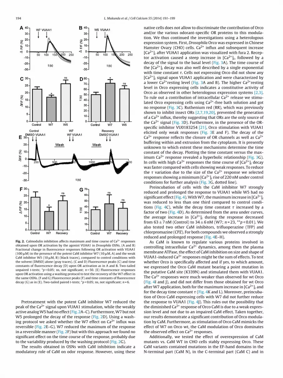
Fig. 2. Calmodulin inhibition affects maximum and time course of Ca2+ responsesobtained upon OR activation by the agonist VUAA1 in Drosophila OSNs. (A and B)Fractional change in fluorescence responses following OR activation with VUAA1(100 �M) in the presence of the potent CaM inhibitor W7 (10 �M, A) and the weakCaM inhibitor W5 (10 �M, B) (black traces), compared to control conditions withthe solvent (DMSO) alone (gray traces). (C and D) Fluorescence peaks (C) and timeconstants of fluorescence decay (D) upon OR activation as in A and B. Two-tailedunpaired t-tests; *p < 0.05; ns, not significant; n = 50. (E) Fluorescence responsesupon OR activation using a washing protocol to test the recovery of the W7 effect inthe same OSNs. (F and G) Fluorescence peaks (F) and time constants of fluorescenced
paWirist
m
Additionally, we tested the effect of overexpression of CaM
ecay (G) as in (E). Two-tailed paired t-tests; *p < 0.05; ns, not significant; n = 6.
Pretreatment with the potent CaM inhibitor W7 reduced theeak of the Ca2+ signal upon VUAA1 stimulation, while the weaklyctive analog W5 had no effect (Fig. 2A–C). Furthermore, W7 but not5 prolonged the decay of the response (Fig. 2D). Using a wash-
ng protocol we asked whether the W7 effect on Ca2+ influx waseversible (Fig. 2E–G). W7 reduced the maximum of the responsen a reversible manner (Fig. 2F) but with this approach we found noignificant effect on the time course of the response, probably due
o the variability produced by the washing protocol (Fig. 2G).The results obtained in OSNs with CaM inhibition indicate aodulatory role of CaM on odor response. However, using these
m 55 (2014) 191–199
native cells does not allow to discriminate the contribution of Orcoand/or the various odorant-specific OR proteins to this modula-tion. We thus continued the investigations using a heterologousexpression system. First, Drosophila Orco was expressed in ChineseHamster Ovary (CHO) cells. Ca2+ influx and subsequent increase[Ca2+]i after VUAA1 application was visualized with fura 2. Recep-tor activation caused a steep increase in [Ca2+]i, followed by adecay of the signal to the basal level (Fig. 3A). The time course ofthe [Ca2+]i decay was also well described by a single exponentialwith time constant �. Cells not expressing Orco did not show any[Ca2+]i signal upon VUAA1 application and were characterized bya lower Ca2+resting level (Fig. 3A and B). The higher Ca2+restinglevel in Orco expressing cells indicates a constitutive activity ofOrco as observed in other heterologous expression systems [2,3].To rule out a contribution of intracellular Ca2+ release we stimu-lated Orco expressing cells using Ca2+-free bath solution and gotno response (Fig. 3C). Ruthenium red (RR), which was previouslyshown to inhibit insect ORs [2,7,19,20], prevented the generationof a Ca2+ influx, thereby suggesting that ORs are the only source ofthe Ca2+ signal (Fig. 3D). Furthermore, in the presence of the OR-specific inhibitor VU0183254 [21], Orco stimulation with VUAA1elicited only weak responses (Fig. 3E and F). The decay of theCa2+ response reflects the closure of OR channels as well as Ca2+
buffering within and extrusion from the cytoplasm. It is presentlyunknown to which extent these mechanisms determine the timeconstant of the decay. Plotting the time constant versus the max-imum Ca2+ response revealed a hyperbolic relationship (Fig. 3G).In cells with high Ca2+ responses the time course of [Ca2+]i decaywas faster compared with cells showing weak responses. To reducethe � variation due to the size of the Ca2+ response we selectedresponses showing a minimum [Ca2+]i rise of 220 nM under controlconditions for further analysis (Fig. 3G, dotted line).
Preincubation of cells with the CaM inhibitor W7 stronglyreduced and prolonged the response to VUAA1 while W5 had nosignificant effect (Fig. 4). With W7, the maximum increase in [Ca2+]iwas reduced to less than one third compared to control condi-tions (Fig. 4C), while the decay time constant � increased by afactor of two (Fig. 4D). As determined from the area under curves,the average increase in [Ca2+]i during the response decreasedfrom 63 ± 7 nM (Control) to 34 ± 6 nM (W7; n = 23, **p < 0.01). Wealso tested two other CaM inhibitors, trifluoperazine (TFP) andchlorpromazine (CPZ). For both compounds we observed a stronglyreduced and prolonged response (Fig. 4E–H).
As CaM is known to regulate various proteins involved incontrolling intracellular Ca2+ dynamics, among them the plasmamembrane ATPase, the effect of CaM inhibition on size and shape ofVUAA1-induced Ca2+ responses might be the sum of effects. To testwhether Orco is specifically affected and if yes, to which amount,we expressed the Orco CaM mutant bearing a point mutation inthe putative CaM site (K339N) and stimulated them with VUAA1.The Ca2+ responses were much weaker than observed for wt Orco(Fig. 4I and J), and did not differ from those obtained for wt Orcoafter W7 application, both for the maximum increase in [Ca2+]i andfor the decay time constant � (Fig. 4K and L). Moreover, preincuba-tion of Orco CaM expressing cells with W7 did not further reducethe response to VUAA1 (Fig. 4J). This rules out the possibility thatthe diminished Ca2+ response of Orco CaM is due to a weak expres-sion level and not due to an impaired CaM effect. Taken together,our results demonstrate a significant contribution of Orco modula-tion by CaM. Furthermore, as stimulation of Orco CaM mimicks theeffect of W7 on Orco wt, the CaM modulation of Orco dominatesthe observed effect on Ca2+ responses.
mutants vs. CaM WT in CHO cells stably expressing Orco. TheseCaM variants contained mutations in the EF-hand domains in theN-terminal part (CaM N), in the C-terminal part (CaM C) and in

L. Mukunda et al. / Cell Calcium 55 (2014) 191–199 195
Fig. 3. Ca2+ responses in Orco-expressing CHO cells obtained with VUAA1. (A) Free calcium concentration [Ca2+]i in response to VUAA1 application (100 �M) in Orcoexpressing (n = 24) and native CHO cells (n = 24). (B) Resting Ca2+ level as in (A). (C–E) [Ca2+]i upon VUAA1 application (100 �M) with Ca2+ free bath solution (C, n = 26), inthe presence of the cation channel blocker ruthenium red (RR, 100 �M)(D, n = 26), and with the OR antagonist VU0183254 (100 �M)(E, n = 19). (F) Maximum [Ca2+]i as in (E).( 2+ 1 stim 2 2+
u
botCCftlsomn
rOei
G) Relationship between maximum [Ca ]i and time constants of decay upon VUAAsed for further analysis. Unpaired t-test ***p < 0.001.
oth (CaM NC) [22]. Given that the N- and C-terminal EF-handsf CaM differ in their affinity to Ca2+, one would expect differen-ial effects of the different mutations on size and/or duration ofa2+ signals elicited upon Orco stimulation. However, all mutantaM forms modified the Ca2+ signals similar to W7 with no dif-erence in the phenotype of calcium responses between N and Cerminal mutations (Fig. 5). The maximum of [Ca2+]i increase wasargely attenuated (Fig. 5E), while in parallel the decay time con-tant � became larger (Fig. 5F). In contrast to conditions withoutverexpression, the average [Ca2+]i rise was not changed for theutant CaM forms (34 ± 3 nM, CaM wt, n = 30; 33 ± 4 nM, CaM N,
= 29; 27 ± 2 nM, CaM C, n = 43; 32 ± 5 nM; CaM NC; n = 45).We next asked how CaM inhibition would affect the Ca2+
esponse upon OR stimulation in cells expressing heteromericRs. When Or22a was coexpressed with Orco, the Ca2+ signalslicited after VUAA1 stimulation appeared considerably prolongedn comparison with those obtained from solely Orco expressing
ulation. Hyperbola, regression curve, R = 0.5; dotted line, minimum Ca response
cells (Fig. 6A). Heterologously expressed Orco and Or22a mayform homomers (Orco/Orco, Or22a/Or22a) as well as heteromers(Or22a/Orco) [23]. In order to test whether there are different popu-lations of Ca2+ responses in terms of decay kinetics we plottedthe number of cells characterized by a given decay time constant(Fig. 6B). In cells co-expressing Or22a with Orco we found a largevariation of �, while in cells solely expressing Orco there was arather narrow � distribution. There was, however, no consider-able overlap in the � distributions as expected for a significantcontribution of Orco monomers in the Or22a and Orco expressingcells (Fig. 6B). In Orco expressing cells we found a correla-tion between slow decay and weak Ca2+ responses (Fig. 3D).There was a similar relationship in cells coexpressing Or22a,
yet with generally larger time constants (Fig. 6C). Takentogether, a slow decay of Ca2+ responses in cells expressingOr22a/Orco might reflect an inherent property of heteromeric ORcomplexes.
196 L. Mukunda et al. / Cell Calcium 55 (2014) 191–199
Fig. 4. CaM inhibition affects maximum and time course of Ca2+ responses obtainedwith VUAA1 in Orco-expressing CHO cells. (A and B) Effect of the potent CaMinhibitor W7 (10 �M, A, n = 23) and the weak CaM inhibitor W5 (10 �M, B, n = 8)on mean [Ca2+]i upon application of VUAA1 (100 �M). (C, D) [Ca2+]i maxima (C) andtime constants of [Ca2+]i decay (D) as in A and B. (E and F) Effect of the CaM inhibitorstrifluoperazine (TFP, 10 �M)(E, n = 22) and chlorpromazine (CPZ, 25 �M)(F, n = 21)on responses to VUAA1. (G and H) [Ca2+]i maxima (G) and time constants of [Ca2+]i
decay (H) as in E and F. (I–L) Effect of the point mutation K339N in Orco CaM onCa2+ responses. Mean [Ca2+]i upon application of VUAA1 (100 �M)(I, n = 12). [Ca2+]i
maxima for wt Orco (n = 24), Orco CaM without (n = 12) and with W7 preincuba-tion (n = 17) (J); wt Orco in the presence of W7 (n = 23) and Orco CaM (n = 12)(K). (L)Time constants of [Ca2+]i decay for wt Orco with W7 and Orco CaM. Unpaired t-test;*
rao(
trg
Fig. 5. Overexpression of CaM mutants affects maximum and time course of Ca2+
responses obtained with VUAA1 in Orco-expressing CHO cells. (A–D) Mean [Ca2+]i
upon application of VUAA1 (100 �M) in cells overexpressing CaM WT (A, n = 30),N-terminal (B, n = 29), C-terminal (C, n = 43) and N- plus C-terminal (NC, n = 45) CaM-EF-hand mutants. (E and F) [Ca2+]i maxima (E) and time constants of [Ca2+]i decay (F)as in (A–D). Non-parametric Kruskal–Wallis test with Dunn’s multiple comparison;mutants versus control; *p < 0.05; **p < 0.01; ***p < 0.001.
p < 0.05; ***p < 0.001; ns, not significant.
Pre-incubation of Or22a expressing cells with W7 stronglyeduced the maximum in [Ca2+]i rise (Fig. 6D and E) but it did notffect the decay kinetics of the Ca2+ signal (Fig. 6F). Overexpressionf CaM mutants affected the Ca2+ responses in a similar way as W7Fig. 6G–K).
We also co-expressed a couple of other odorant-specific recep-or proteins with Orco, namely Or33a, Or47a and Or56a. Or47aepresents, like Or22a, a food odor-detecting type. Or56a, theeosmin receptor [24], represents a receptor for a danger signal.
Or33a is expressed in the same sensillum as Or56a and detectsother odors of negative valence. The Ca2+ responses elicited byVUAA1 stimulation varied considerably in decay kinetics (Fig. 7A).While for Or33a the decay was as slow as for Or22a, the Ca2+ signalsobtained with Or47a and Or56a appeared to be even more long last-ing. In contrast to Or22a and Orco or solely Orco expressing cells,preincubation with W7 did not significantly affect the maximumin [Ca2+]i rise after VUAA1 stimulation (Fig. 7B and C). In addition,for Or33a and Or47a, W7 did not change the decay kinetics of theCa2+ signal (Fig. 7B and D). For Or56a, W7 accelerated the decaykinetics (Fig. 7B and D). The average rise in [Ca2+]i determined fromthe area under curve became reduced from 73 ± 22 nM (Control) to23 ± 2 nM (W7; n = 19, *p < 0.05).
Taken together, inhibition of CaM functions was found to reducethe Ca2+ response of Orco channels upon agonist stimulation. ForOrX/Orco heteromers, there was no consistent effect of impairedCaM activity on Ca2+ responses. For Or22a containing heteromersthe Ca2+ responses appeared to be reduced while they becameshortened for Or56a. For ORs comprising Or33a or Or47a CaM inhi-
bition had no effect.
L. Mukunda et al. / Cell Calcium 55 (2014) 191–199 197
Fig. 6. CaM inhibition affects maximum but not the time course of Ca2+ responses obtained with VUAA1 in Orco- and Or22a-expressing CHO cells. (A) Mean [Ca2+]i uponapplication of VUAA1 (100 �M). n = 19. (B) Distribution of cells (number n) with time constants of [Ca2+]i decay in cells expressing Orco and Or22a/Orco indicated. Curves, fittedGaussian distribution. (C) Relationship between maximum [Ca2+]i and time constants of decay upon VUAA1 stimulation for cells expressing Orco and Or22a/Orco. Hyperbolas,regression curves, R2 = 0.5 (Orco), 0.2 (Or22a/Orco); dashed line, minimum Ca2+ response for Or22a/Orco used for further analysis. (D) Mean [Ca2+]i upon application of VUAA1(100 �M) in the presence of W7 (10 �M) (n = 27). (E and F) [Ca2+]i maxima (E) and time constants of [Ca2+]i decay (F) as in A and D. (G–I) Effect of overexpression of CaMmutants on Ca2+ responses upon VUAA1 in Orco and Or22a expressing cells (N-terminal, G, n = 27; C-terminal, H, n = 37; NC terminal, I, n = 27). (J and K) [Ca2+] maxima (J)a ; ns, n
4
iFaCotaot[o
nd time constants of [Ca2+]i decay (K) as in A and G–I. Unpaired t-test; ***p < 0.001
. Discussion
In the present study we investigated if and how CaM activ-ty affects the Ca2+ response of insect ORs to agonist stimulation.or this purpose we developed a Drosophila antenna preparationllowing us to observe these signals under in situ conditions.aM inhibition initially reduced the maximum of the Ca2+ signalbtained upon agonist application, but also prolonged this signal inhe OSN cell bodies. This dual effect is similar to what was observedfter CaM inhibition in the heterologous system with expression of
nly the Orco subunit of ORs (Fig. 4). This phenotype may reflecthe predominant expression of Orco in the OSN cell body membrane25]. Although our antenna preparation in principle also allows tobserve dendritic regions of the OSNs (Fig. 1D), we failed in gettingi
ot significant.
sufficiently resolved and mechanically stable fluorescence recor-dings at this level.
In a previous study we could show that repeated subthresholdodor stimulation of Drosophila OSNs led to OR sensitization [5].This sensitization required Orco activation as a first step. It couldbe mimicked by processes activating Orco before odor stimulationand it could be suppressed by Orco inhibition. For example, fliesexpressing an Orco mutant with disrupted PKC phosphorylationthat is insensitive to cAMP activation [26] did not sensitize [5]. Themechanism by which Orco activation leads to OR sensitization is
presently unknown. Given the fact that Orco channels conduct Ca2+,a role of CaM in mediating sensitization could not be excluded. Ourresults obtained after co-expression of OrX proteins and Orco show,however, no consistent effect of CaM on the various OR constructs,
198 L. Mukunda et al. / Cell Calcium 55 (2014) 191–199
Fig. 7. CaM inhibition affects Ca2+ responses obtained with VUAA1 in Orco and various Or protein-expressing CHO cells differentially. (A and B) Mean [Ca2+]i upon applicationof VUAA1 (100 �M) in control condition (A, n = 13 (Or33a), 17 (Or47a), 19 (Or56a)) and in the presence of W7 (B, n = 15 (Or33a), 27 (Or47a), 19 (Or56a)) in cells expressingt 2+ 2+
(D) ass
wtutObti
sAwdAot
tOqbmtt
he indicated ORs. (C and D) [Ca ]i maxima (C) and time constants of [Ca ]i decayignificant.
hich was a prerequisite to provide a global mechanism of sensi-ization. The lack of a uniform modification of the Ca2+ responsepon CaM inhibition in OrX/Orco heteromers may indicate thathe regulation of this heteromeric receptor current is performed byrco homomers. Their presence in the OSN membrane is indicatedy recent observations [25,23]. Another explanation may be thathe heterologous expression system misses some required playern the regulatory pathway.
The modulation of the Orco channel response by CaM inhibitionuggests a stimulatory role of CaM activity on Orco function (Fig. 4).
tiny amount of Ca2+ influx obtained by weak Orco stimulationould thus be amplified. It remains to be shown which furtherownstream processes might lead to subsequent OR sensitization.
direct pacemaker activity of Orco can be excluded as we havebserved that Orco activation via 8-bromo-cAMP did not acceleratehe discharge rate of OSNs [5].
The present study provides evidence that CaM activity affectshe function of Orco channels and may have specific effects onrX/Orco couples. These results form the basis to answer furtheruestions such as for the determinants of CaM action on Orco
y mutation of candidate amino acids in the putative recognitionotif. Other investigations should clarify whether there is an addi-ional interaction between plasma membrane Ca2+ pump, CaM andhe ORs.
in A and B. Unpaired t-test and Mann Whitney test; *p < 0.05; ***p < 0.001; ns, not
Conflict of interest statement
All authors declare that they have no conflicts of interest.
Acknowledgements
The authors thank S. Neumann for orienting experiments withthe fly antenna preparation, Drs. R. Schönherr and S.H. Heinemannfor providing the CaM-EF-hand mutants, and Dr. Heinemann forhelpful discussion on the manuscript. The study was supported bythe Max Planck Society (LM, SK, BSH, DW) and the InternationalMax Planck Research School (FM).
References
[1] F. Zufall, G.M. Shepherd, S. Firestein, Inhibition of the olfactory cyclicnucleotide gated ion channel by intracellular calcium, Proc. Biol. Sci. 246 (1991)225–230.
[2] K. Sato, M. Pellegrino, T. Nakagawa, T. Nakagawa, L.B. Vosshall, K. Touhara,Insect olfactory receptors are heteromeric ligand-gated ion channels, Nature
452 (2008) 1002–1006.[3] D. Wicher, R. Schäfer, R. Bauernfeind, M.C. Stensmyr, R. Heller, S.H. Heine-mann, B.S. Hansson, Drosophila odorant receptors are both ligand-gatedand cyclic-nucleotide-activated cation channels, Nature 452 (2008) 1007–1011.
Calciu
[
[
[[
[
[
[
[
[
[
[
[
[
[
[
[and heteromeric function of Drosophila odorant receptors in vivo, PLoS Biol. 4(2006) e20.
L. Mukunda et al. / Cell
[4] M.C. Larsson, A.I. Domingos, W.D. Jones, M.E. Chiappe, H. Amrein, L.B. Vosshall,Or83b encodes a broadly expressed odorant receptor essential for Drosophilaolfaction, Neuron 43 (2004) 703–714.
[5] M.N. Getahun, S.B. Olsson, S. Lavista-Llanos, B.S. Hansson, D. Wicher, Insectodorant response sensitivity is tuned by metabotropically autoregulated olfac-tory receptors, PLoS ONE 8 (2013) e58889.
[6] T. Nakagawa, M. Pellegrino, K. Sato, L.B. Vosshall, K. Touhara, Amino acidresidues contributing to function of the heteromeric insect olfactory receptorcomplex, PLoS ONE 7 (2012) e32372.
[7] P.L. Jones, G.M. Pask, D.C. Rinker, L.J. Zwiebel, Functional agonism of insect odor-ant receptor ion channels, Proc. Natl. Acad. Sci. U. S. A. 108 (2011) 8821–8825.
[8] M.J. Berridge, P. Lipp, M.D. Bootman, The versatility and universality of calciumsignalling, Nat. Rev. Mol. Cell Biol. 1 (2000) 11–21.
[9] T. Budde, S. Meuth, H.C. Pape, Calcium-dependent inactivation of neuronalcalcium channels, Nat. Rev. Neurosci. 3 (2002) 873–883.
10] T.Y. Chen, K.W. Yau, Direct modulation by Ca2+–calmodulin of cyclic nucleotide-activated channel of rat olfactory receptor neurons, Nature 368 (1994)545–548.
11] R. Rizzuto, T. Pozzan, Microdomains of intracellular Ca2+: molecular determi-nants and functional consequences, Physiol. Rev. 86 (2006) 369–408.
12] D.E. Clapham, Calcium signaling, Cell 131 (2007) 1047–1058.13] G.C. Faas, S. Raghavachari, J.E. Lisman, I. Mody, Calmodulin as a direct detector
of Ca2+ signals, Nat. Neurosci. 14 (2011) 301–304.14] B.Z. Peterson, C.D. DeMaria, J.P. Adelman, D.T. Yue, Calmodulin is the Ca2+ sensor
for Ca2+-dependent inactivation of L-type calcium channels, Neuron 22 (1999)549–558.
15] Y. Song, K.D. Cygnar, B. Sagdullaev, M. Valley, S. Hirsh, A. Stephan, J. Reisert,H. Zhao, Olfactory CNG channel desensitization by Ca2+/CaM via the B1b sub-
unit affects response termination but not sensitivity to recurring stimulation,Neuron 58 (2008) 374–386.16] S. Antolin, J. Reisert, H.R. Matthews, Olfactory response termination involvesCa2+-ATPase in vertebrate olfactory receptor neuron cilia, J. Gen. Physiol. 135(2010) 367–378.
[
m 55 (2014) 191–199 199
17] B. Malnic, J. Hirono, T. Sato, L.B. Buck, Combinatorial receptor codes for odors,Cell 96 (1999) 713–723.
18] K. Ukhanov, Y. Bobkov, B.W. Ache, Imaging ensemble activity inarthropod olfactory receptor neurons in situ, Cell Calcium 49 (2011)100–107.
19] T. Nakagawa, T. Sakurai, T. Nishioka, K. Touhara, Insect sex-pheromone signalsmediated by specific combinations of olfactory receptors, Science 307 (2005)1638–1642.
20] A.S. Nichols, S. Chen, C.W. Luetje, Subunit contributions to insect olfactoryreceptor function: channel block and odorant recognition, Chem. Senses 36(2011) 781–790.
21] P.L. Jones, G.M. Pask, I.M. Romaine, R.W. Taylor, P.R. Reid, A.G. Waterson, G.A.Sulikowski, L.J. Zwiebel, Allosteric antagonism of insect odorant receptor ionchannels, PLoS ONE 7 (2012) e30304.
22] U. Ziechner, R. Schönherr, A.K. Born, O. Gavrilova-Ruch, R.W. Glaser, M. Malese-vic, G. Kullertz, S.H. Heinemann, Inhibition of human ether a go–go potassiumchannels by Ca2+/calmodulin binding to the cytosolic N- and C-termini, FEBS J.273 (2006) 1074–1086.
23] P.F. German, S. van der Poel, C. Carraher, A.V. Kralicek, R.D. Newcomb, Insightsinto subunit interactions within the insect olfactory receptor complex usingFRET, Insect Biochem. Mol. Biol. 43 (2013) 138–145.
24] M.C. Stensmyr, H.K. Dweck, A. Farhan, I. Ibba, A. Strutz, L. Mukunda, J. Linz, V.Grabe, K. Steck, S. Lavista-Llanos, D. Wicher, S. Sachse, M. Knaden, P.G. Becher, Y.Seki, B.S. Hansson, A conserved dedicated olfactory circuit for detecting harmfulmicrobes in Drosophila, Cell 151 (2012) 1345–1357.
25] R. Benton, S. Sachse, S.W. Michnick, L.B. Vosshall, Atypical membrane topology
26] V. Sargsyan, M.N. Getahun, S. Lavista Llanos, S.B. Olsson, B.S. Hansson, D. Wicher,Phosphorylation via PKC regulates the function of the Drosophila odorant core-ceptor, Front. Cell. Neurosci. 5 (2011) 5.


