Embed Size (px)
Citation preview
© 2013 Trabelsi et al. This work is published by Dove Medical Press Ltd, and licensed under Creative Commons Attribution – Non Commercial (unported, v3.0) License. The full terms of the License are available at http://creativecommons.org/licenses/by-nc/3.0/. Non-commercial uses of the work are permitted without any further
permission from Dove Medical Press Ltd, provided the work is properly attributed. Permissions beyond the scope of the License are administered by Dove Medical Press Ltd. Information on how to request permission may be found at: http://www.dovepress.com/permissions.php
International Journal of Nanomedicine 2013:8 3447–3453
International Journal of Nanomedicine Dovepress
submit your manuscript | www.dovepress.com
Dovepress 3447
O r I g I N a l r e s e a r c h
open access to scientific and medical research
Open access Full Text article
http://dx.doi.org/10.2147/IJN.S49323
Nanotoxicological evaluation of oxidative responses in rat nephrocytes induced by cadmium
hamdi TrabelsiInès azzouzsoumaya FerchichiOlfa TebourbiMohsen saklyhafedh abdelmeleklaboratory of Integrative Physiology, Faculty of sciences of Bizerte, carthage University, Jarzouna, Tunisia
correspondence: hamdi Trabelsi laboratoire de Physiologie Intégrée, Faculté des sciences de Bizerte, 7021 Jarzouna, Tunisia Tel +216 23 532 901 Fax +216 72 590 566 email [email protected]
Abstract: The aim of this study was to investigate the interaction of cadmium chloride with
mineral elements in rat nephrocytes in terms of the biosynthesis of nanocomplexes. The results
show that selenium supplementation enhanced cadmium accumulation in kidneys. Analysis of
the fluorescence revealed an increase in red fluorescence in the kidneys of rats co-exposed to
cadmium and selenium. Interestingly, X-ray diffraction measurements carried out on kidney
fractions of co-exposed rats point to the biosynthesis of cadmium selenide and/or sulfide
nanoparticles (about 62 nm in size). Oxidative stress assays showed the ability of selenium to
reduce lipid peroxidation and to restore glutathione peroxidase and superoxide dismutase activity
in kidneys. Hence, cadmium complexation with selenium and sulfur at a nanoscale level could
reduce oxidative stress induced by cadmium in kidneys.
Keywords: nanoparticles, detoxification, oxidative stress, X-ray diffraction, fluorescence
microscopy, kidneys
IntroductionCadmium (Cd) is a well-recognized environmental pollutant with numerous adverse
health effects. Sources of exposure to this metal include food, water, and alcoholic
beverages.1–3 Many studies revealed that Cd accumulates preferentially in hepatic and
renal tissues.3–5 Previous studies point out that metallothionein (MT) biosynthesis – a
family of cysteine-rich low molecular weight proteins – sequestrates the metal.3,6,7
In fact, lesions of the proximal tubule in the kidney cortex were observed after Cd
exposure.8 Various studies point out that Cd toxicity is related to oxidative stress, since
this metal can alter the antioxidant defense system in several animal tissues. Studies
on mammals have shown that Cd stimulated formation of reactive oxygen species.2,3
Moreover, Cd has been suggested to practice some of its toxic effects by disturbing the
metabolism of essential metals, such as zinc (Zn)9–11 and selenium (Se).12,13 Treatment
with Se during Cd exposure has been demonstrated to have beneficial effects on Cd
toxicity.12,14–16 However, based on the available published literature, the effect of Se
on Cd toxicity is not yet well studied.
In fact, the main target organs for Cd accumulation are the liver, kidneys, and
other tissues.2,3 However, Trabelsi et al showed that the subacute toxicity of Cd may
be related to its eventual potential to generate Cd sulfide (CdS) and/or Cd selenide
(CdSe) nanomaterials at the cellular level.17 In addition, to the authors’ knowledge,
there are no studies investigating whether the protective effect of Se is related to its
eventual potential to bind Cd, leading to nanocomplexes in living systems.
International Journal of Nanomedicine 2013:8submit your manuscript | www.dovepress.com
Dovepress
Dovepress
3448
Trabelsi et al
The aim of this investigation was to study the reduction
of Cd-induced nephrotoxicity in terms of the biosynthesis
of nanocomplexes.
Material and methodschemicalsCd chloride (CdCl
2) and sodium selenite (Na
2SeO
3) were
purchased from Sigma-Aldrich (St Louis, MO, USA). All
other chemicals were of analytical grade and were purchased
from standard commercial suppliers.
animalsAdult Wistar male rats (SIPHAT, Bin Arous, Tunisia) weighing
200–220 g at the time of the experiments were randomly divided
into the following groups: control rats (n = 6), Cd-exposed
rats (n = 6), and rats co-exposed to cadmium and selenium
(Cd + Se) (n = 6). Animals were housed at 25°C, under a
12-hour/12-hour light/dark cycle, with free access to water and
commercial mash. Animals were cared for under the Tunisian
code of practice for the Care and Use of Animals for Scientific
Purposes. The experimental protocols were approved by the
Faculty Ethics Committee (Faculté des Sciences de Bizerte,
Jarzouna, Tunisia).
animal treatmentThe control group was intraperitoneally injected with
0.10 mL of 0.90% saline solution for 14 consecutive days.
The Cd-treated group was intraperitoneally injected with a
sublethal dose of Cd (1.50 mg Cd/kg of body weight) for
14 days.3 The co-exposed rats were treated with Cd (1.50 mg
Cd/kg of body weight) and Se (0.20 mg/L per os [by mouth])
for 14 days.13
cd determinationRenal slices for Cd analyses were oven dried (60°C) to
a constant weight. The dried tissues (100 mg from each
sample) were digested with 3 mL trace pure nitric acid at
120°C. The volume was then adjusted to 10 mL with deion-
ized water.18
Cd concentrations in the acid solutions were measured by
atomic absorption spectrophotometry using a PerkinElmer
306 spectrometer equipped with a PerkinElmer Intensitron®
lamp (Waltham, MA, USA). Cd concentration is expressed
in µg/g dry tissue weight.19
Fluorescence microscopyKidney fractions were fixed with 10% formaldehyde and were
evaluated by fluorescence microscopy using a DM-IRBE
microscope (Leica Microsystems, Wetzlar, Germany) coupled
with a digital charge-coupled device camera (CoolSNAP™
FX; Princeton Instruments, Trenton, NJ, USA).
Powder samples preparation and X-ray diffraction (XrD) measurementsFourteen days after intraperitoneal injection, the control
and treated groups were sacrificed and their kidneys were
harvested. The tissues were weighed, rinsed with ice-cold
deionized water, and dried with filter paper. Kidney frac-
tions were dried for 5 days at 50°C. Fractions were mixed
and sieved in order to obtain powder. XRD measurements
were carried out on a Bruker D8 ADVANCE powder X-ray
diffractometer (Bruker Corporation, Billerica, MA, USA),
using copper (Cu) Kα (λ = 1:5402 Å) incident radiation with
a scan range of 20, 2θ ,70.17
Tissue preparationThe control and treated groups were sacrificed and their kid-
neys were immediately harvested. The tissues were weighed,
rinsed with ice-cold deionized water, and dried with filter
paper. Fractions of tissues (500 mg) were homogenized
in buffer (tris[hydroxymethyl]aminomethane 10 mmol/L,
ethylenediaminetetraacetic acid 1 mmol/L, phenylmeth-
ylsulfonyl fluoride 1 mmol/L; pH 7.4). The homogenates
were centrifuged at 600 g for 10 minutes and centrifuged
again at 13,000 g for 20 minutes at 4°C to obtain a post-
nuclear homogenate and postmitochondrial supernatant
fractions.20
antioxidant enzyme assaysLipid peroxidation in tissues was measured by thiobarbitu-
ric acid reactive substances and was expressed in terms of
malondialdehyde (MDA) content.21 Catalase (CAT) activity
was assayed by ultraviolet spectrophotometry.22 Glutathione
peroxidase (GPx) activity was measured according to Gunzler
et al.23 Superoxide dismutase (SOD) activity was determined
by measuring the inhibition of the auto-oxidant of pyrogallol
by spectrophotometry (Jenway 6505 UV/ Visible; Bibby
Scientific Limited, Stone, UK) at 420 nm, using Marklund
and Marklund’s modified method.24
Data presentation and statistical analysisA one-way analysis of variance followed by Tukey’s multiple
comparisons test was performed using GraphPad Prism®
version 6.00 for Windows (GraphPad Software, Inc, La Jolla,
CA, USA). Data are reported as the mean ± standard devia-
tion. The level of significance was set at P , 0.05.
International Journal of Nanomedicine 2013:8 submit your manuscript | www.dovepress.com
Dovepress
Dovepress
3449
cadmium-induced oxidative responses in rat nephrocytes
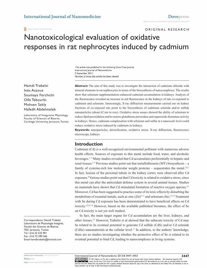
Resultscd accumulation in kidneysFigure 1 shows an accumulation of Cd in rat kidneys in com-
parison to the control group. Moreover, Se supplementation
(0.20 mg/L per os) facilitates Cd accumulation compared
to values found with Cd-exposed rats (1827 ± 172.72 µg/g
versus 1065 ± 217.80 µg/g; P , 0.01).
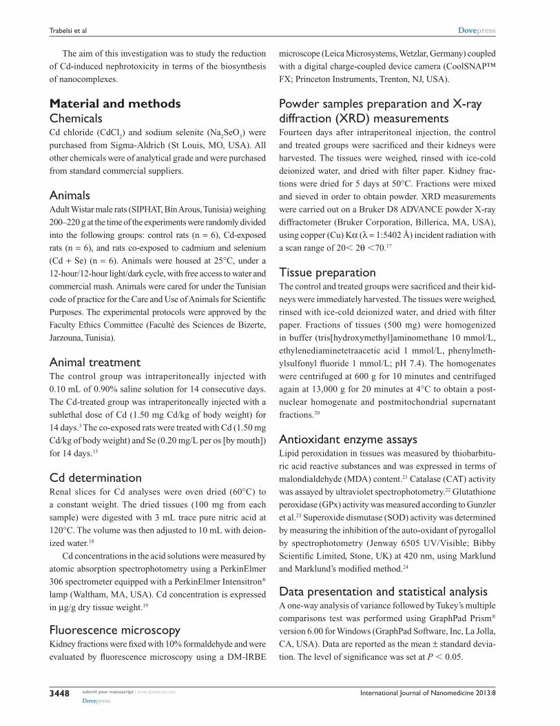
Fluorescence microscopyFluorescence microscopy images show no fluorescence
signals in the control kidneys (Figure 2A–B). However, red
fluorescence was detected in the glomeruli and renal tubu-
lar of Cd-treated kidneys (Figure 2C–D). Interestingly, the
intensity of the fluorescence signal was higher after exposure
to Cd and Se supplementation in comparison with the Cd-
treated group (Figure 2E–F).
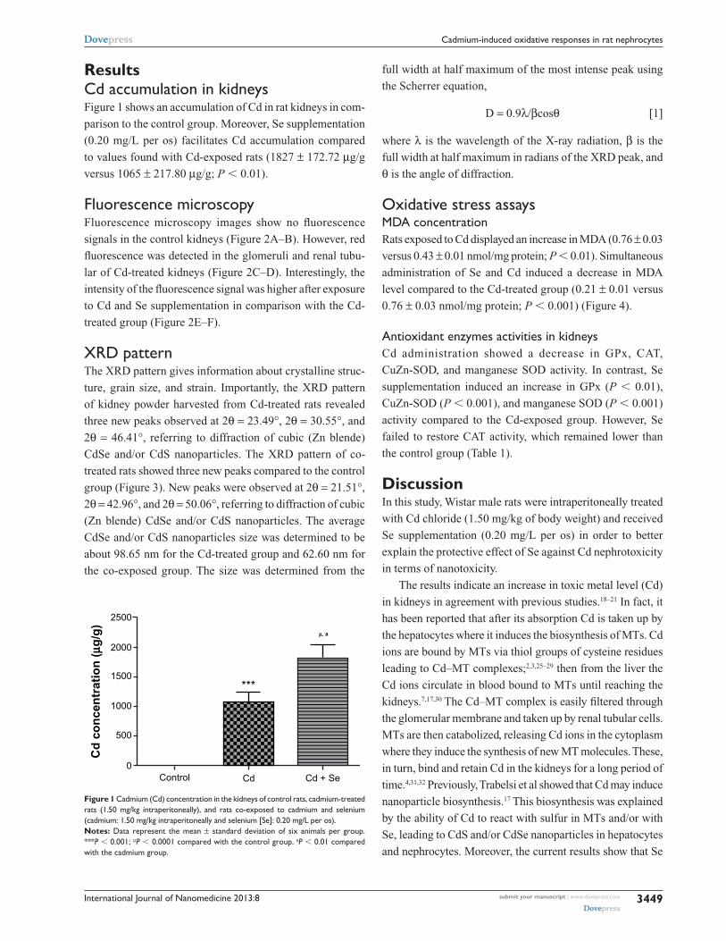
XrD patternThe XRD pattern gives information about crystalline struc-
ture, grain size, and strain. Importantly, the XRD pattern
of kidney powder harvested from Cd-treated rats revealed
three new peaks observed at 2θ = 23.49°, 2θ = 30.55°, and
2θ = 46.41°, referring to diffraction of cubic (Zn blende)
CdSe and/or CdS nanoparticles. The XRD pattern of co-
treated rats showed three new peaks compared to the control
group (Figure 3). New peaks were observed at 2θ = 21.51°,
2θ = 42.96°, and 2θ = 50.06°, referring to diffraction of cubic
(Zn blende) CdSe and/or CdS nanoparticles. The average
CdSe and/or CdS nanoparticles size was determined to be
about 98.65 nm for the Cd-treated group and 62.60 nm for
the co-exposed group. The size was determined from the
full width at half maximum of the most intense peak using
the Scherrer equation,
D = 0.9λ/βcosθ [1]
where λ is the wavelength of the X-ray radiation, β is the
full width at half maximum in radians of the XRD peak, and
θ is the angle of diffraction.
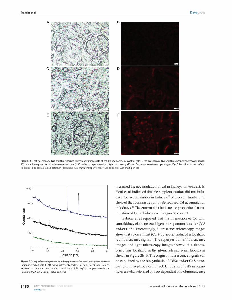
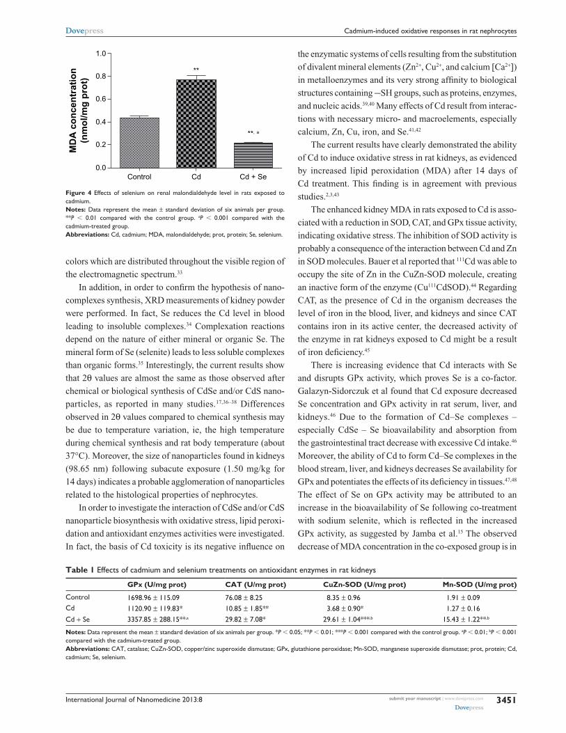
Oxidative stress assaysMDa concentrationRats exposed to Cd displayed an increase in MDA (0.76 ± 0.03
versus 0.43 ± 0.01 nmol/mg protein; P , 0.01). Simultaneous
administration of Se and Cd induced a decrease in MDA
level compared to the Cd-treated group (0.21 ± 0.01 versus
0.76 ± 0.03 nmol/mg protein; P , 0.001) (Figure 4).
antioxidant enzymes activities in kidneysCd administration showed a decrease in GPx, CAT,
CuZn-SOD, and manganese SOD activity. In contrast, Se
supplementation induced an increase in GPx (P , 0.01),
CuZn-SOD (P , 0.001), and manganese SOD (P , 0.001)
activity compared to the Cd-exposed group. However, Se
failed to restore CAT activity, which remained lower than
the control group (Table 1).
DiscussionIn this study, Wistar male rats were intraperitoneally treated
with Cd chloride (1.50 mg/kg of body weight) and received
Se supplementation (0.20 mg/L per os) in order to better
explain the protective effect of Se against Cd nephrotoxicity
in terms of nanotoxicity.
The results indicate an increase in toxic metal level (Cd)
in kidneys in agreement with previous studies.18–21 In fact, it
has been reported that after its absorption Cd is taken up by
the hepatocytes where it induces the biosynthesis of MTs. Cd
ions are bound by MTs via thiol groups of cysteine residues
leading to Cd–MT complexes;2,3,25–29 then from the liver the
Cd ions circulate in blood bound to MTs until reaching the
kidneys.7,17,30 The Cd–MT complex is easily filtered through
the glomerular membrane and taken up by renal tubular cells.
MTs are then catabolized, releasing Cd ions in the cytoplasm
where they induce the synthesis of new MT molecules. These,
in turn, bind and retain Cd in the kidneys for a long period of
time.4,31,32 Previously, Trabelsi et al showed that Cd may induce
nanoparticle biosynthesis.17 This biosynthesis was explained
by the ability of Cd to react with sulfur in MTs and/or with
Se, leading to CdS and/or CdSe nanoparticles in hepatocytes
and nephrocytes. Moreover, the current results show that Se
2500
2000
1500
1000
500
0Control
***
Cd
co
nce
ntr
atio
n (
µg/g
)
µ, a
Cd Cd + Se
Figure 1 cadmium (cd) concentration in the kidneys of control rats, cadmium-treated rats (1.50 mg/kg intraperitoneally), and rats co-exposed to cadmium and selenium (cadmium: 1.50 mg/kg intraperitoneally and selenium [se]: 0.20 mg/l per os). Notes: Data represent the mean ± standard deviation of six animals per group. ***P , 0.001; µP , 0.0001 compared with the control group. aP , 0.01 compared with the cadmium group.
International Journal of Nanomedicine 2013:8submit your manuscript | www.dovepress.com
Dovepress
Dovepress
3450
Trabelsi et al
A B
20 µm
20 µm
20 µm20 µm
20 µm
20 µm
C D
E F
Figure 2 light microscopy (A) and fluorescence microscopy images (B) of the kidney cortex of control rats. light microscopy (C) and fluorescence microscopy images (D) of the kidney cortex of cadmium-treated rats (1.50 mg/kg intraperitoneally). light microscopy (E) and fluorescence microscopy images (F) of the kidney cortex of rats co-exposed to cadmium and selenium (cadmium: 1.50 mg/kg intraperitoneally and selenium: 0.20 mg/l per os).
20
0
100
400
Co
un
ts (
au)
900
1600
30 40
Position [°2θ]50 60 70
Figure 3 X-ray diffraction pattern of kidney powder of control rats (green pattern), cadmium-treated rats (1.50 mg/kg intraperitoneally) (black pattern), and rats co-exposed to cadmium and selenium (cadmium: 1.50 mg/kg intraperitoneally and selenium: 0.20 mg/l per os) (blue pattern).
increased the accumulation of Cd in kidneys. In contrast, El
Heni et al indicated that Se supplementation did not influ-
ence Cd accumulation in kidneys.25 Moreover, Jamba et al
showed that administration of Se reduced Cd accumulation
in kidneys.27 The current data indicate the proportional accu-
mulation of Cd in kidneys with organ Se content.
Trabelsi et al reported that the interaction of Cd with
some kidney elements could generate quantum dots like CdS
and/or CdSe. Interestingly, fluorescence microscopy images
show that co-treatment (Cd + Se group) induced a localized
red fluorescence signal.17 The superposition of fluorescence
images and light microscopy images showed that fluores-
cence was localized in the glomeruli and renal tubules as
shown in Figure 2E–F. The origin of fluorescence signals can
be explained by the biosynthesis of CdSe and/or CdS nano-
particles in nephrocytes. In fact, CdSe and/or CdS nanopar-
ticles are characterized by size-dependent photoluminescence
International Journal of Nanomedicine 2013:8 submit your manuscript | www.dovepress.com
Dovepress
Dovepress
3451
cadmium-induced oxidative responses in rat nephrocytes
colors which are distributed throughout the visible region of
the electromagnetic spectrum.33
In addition, in order to confirm the hypothesis of nano-
complexes synthesis, XRD measurements of kidney powder
were performed. In fact, Se reduces the Cd level in blood
leading to insoluble complexes.34 Complexation reactions
depend on the nature of either mineral or organic Se. The
mineral form of Se (selenite) leads to less soluble complexes
than organic forms.35 Interestingly, the current results show
that 2θ values are almost the same as those observed after
chemical or biological synthesis of CdSe and/or CdS nano-
particles, as reported in many studies.17,36–38 Differences
observed in 2θ values compared to chemical synthesis may
be due to temperature variation, ie, the high temperature
during chemical synthesis and rat body temperature (about
37°C). Moreover, the size of nanoparticles found in kidneys
(98.65 nm) following subacute exposure (1.50 mg/kg for
14 days) indicates a probable agglomeration of nanoparticles
related to the histological properties of nephrocytes.
In order to investigate the interaction of CdSe and/or CdS
nanoparticle biosynthesis with oxidative stress, lipid peroxi-
dation and antioxidant enzymes activities were investigated.
In fact, the basis of Cd toxicity is its negative influence on
the enzymatic systems of cells resulting from the substitution
of divalent mineral elements (Zn2+, Cu2+, and calcium [Ca2+])
in metalloenzymes and its very strong affinity to biological
structures containing -SH groups, such as proteins, enzymes,
and nucleic acids.39,40 Many effects of Cd result from interac-
tions with necessary micro- and macroelements, especially
calcium, Zn, Cu, iron, and Se.41,42
The current results have clearly demonstrated the ability
of Cd to induce oxidative stress in rat kidneys, as evidenced
by increased lipid peroxidation (MDA) after 14 days of
Cd treatment. This finding is in agreement with previous
studies.2,3,43
The enhanced kidney MDA in rats exposed to Cd is asso-
ciated with a reduction in SOD, CAT, and GPx tissue activity,
indicating oxidative stress. The inhibition of SOD activity is
probably a consequence of the interaction between Cd and Zn
in SOD molecules. Bauer et al reported that 111Cd was able to
occupy the site of Zn in the CuZn-SOD molecule, creating
an inactive form of the enzyme (Cu111CdSOD).44 Regarding
CAT, as the presence of Cd in the organism decreases the
level of iron in the blood, liver, and kidneys and since CAT
contains iron in its active center, the decreased activity of
the enzyme in rat kidneys exposed to Cd might be a result
of iron deficiency.45
There is increasing evidence that Cd interacts with Se
and disrupts GPx activity, which proves Se is a co-factor.
Galazyn-Sidorczuk et al found that Cd exposure decreased
Se concentration and GPx activity in rat serum, liver, and
kidneys.46 Due to the formation of Cd–Se complexes –
especially CdSe – Se bioavailability and absorption from
the gastrointestinal tract decrease with excessive Cd intake.46
Moreover, the ability of Cd to form Cd–Se complexes in the
blood stream, liver, and kidneys decreases Se availability for
GPx and potentiates the effects of its deficiency in tissues.47,48
The effect of Se on GPx activity may be attributed to an
increase in the bioavailability of Se following co-treatment
with sodium selenite, which is reflected in the increased
GPx activity, as suggested by Jamba et al.15 The observed
decrease of MDA concentration in the co-exposed group is in
Table 1 effects of cadmium and selenium treatments on antioxidant enzymes in rat kidneys
GPx (U/mg prot) CAT (U/mg prot) CuZn-SOD (U/mg prot) Mn-SOD (U/mg prot)
control 1698.96 ± 115.09 76.08 ± 8.25 8.35 ± 0.96 1.91 ± 0.09cd 1120.90 ± 119.83* 10.85 ± 1.85** 3.68 ± 0.90* 1.27 ± 0.16cd + se 3357.85 ± 288.15**,a 29.82 ± 7.08* 29.61 ± 1.04***,b 15.43 ± 1.22**,b
Notes: Data represent the mean ± standard deviation of six animals per group. *P , 0.05; **P , 0.01; ***P , 0.001 compared with the control group. aP , 0.01; bP , 0.001 compared with the cadmium-treated group.Abbreviations: caT, catalase; cuZn-sOD, copper/zinc superoxide dismutase; gPx, glutathione peroxidase; Mn-sOD, manganese superoxide dismutase; prot, protein; cd, cadmium; se, selenium.
Control
MD
A c
on
cen
trat
ion
(nm
ol/m
g p
rot)
0.0
0.2
0.4
0.6
0.8
1.0
**
**, a
Cd Cd + Se
Figure 4 effects of selenium on renal malondialdehyde level in rats exposed to cadmium.Notes: Data represent the mean ± standard deviation of six animals per group. **P , 0.01 compared with the control group. aP , 0.001 compared with the cadmium-treated group. Abbreviations: cd, cadmium; MDa, malondialdehyde; prot, protein; se, selenium.
International Journal of Nanomedicine 2013:8submit your manuscript | www.dovepress.com
Dovepress
Dovepress
3452
Trabelsi et al
agreement with previous investigations and can be explained
by the recovery of biological activity of SOD and GPx.49,50
The results suggest that Cd induced oxidative stress in
kidneys. This oxidative stress may be associated with the
biosynthesis of nanocomplexes (CdSe and/or CdS). The data
report for the first time, to the authors’ knowledge, that Se
decreases the oxidative response by enhancing the biosynthe-
sis of nanocomplexes (CdSe and/or CdS) in rat nephrocytes.
This biosynthesis may represent a detoxification pathway
after Cd treatment. Hence, as CdSe and/or CdS nanoparticle
biosynthesis increases in nephrocytes, the bioavailability
of Cd decreases, leading to weak oxidative responses. This
finding can be explained by the following ratio:
Number of (CdX) nanoparticles
Pro-oxidant marker (MDA level),
where (X) refers to sulfur and/or selenium. The ratio
increase is correlated to the number of biosynthesized
nanoparticles associated with a concomitant decrease in Cd
bioavailability and MDA level in kidneys. The nanocom-
plexes could be evaluated by imagery methods based on the
evaluation of red fluorescence intensity, which could be used
as a health marker.
ConclusionTo the authors’ knowledge, this is the first study that inves-
tigates interactions of Cd with sulfur and Se in nephrocytes
in terms of CdSe and/or CdS nanoparticle biosynthesis. The
results show that Cd may induce the biosynthesis of red
fluorescent CdSe and/or CdS nanoparticles in kidneys. This
study also shows that Cd induced oxidative stress by disturbing
antioxidant enzymes activities. Se supplementation reduced
Cd-induced toxicity in rat kidneys, probably due to its ability to
bind Cd in nanosized insoluble and fluorescent complexes.
AcknowledgmentsThe authors would like to thank Dr Ahmed Rejeb (Depart-
ment of Anatomic Pathology, Ecole Nationale de Médecine
Vétérinaire, Sidi Thabet, Tunisia) and Mr Hazem Ben
Mabrouk (Institut Pasteur de Tunis, Belvedere, Tunisia) for
their help in the fluorescence microscopy work.
DisclosureThe authors report no conflicts of interest in this work.
References1. Jarup L, Berglund M, Elinder CG, Nordberg G, Vahter M. Health effects
of cadmium exposure: a review of the literature and a risk estimate. Scand J Work Environ Health. 1998;24(Suppl 1):1–51.
2. Amara S, Abdelmelek H, Garrel C, et al. Influence of static magnetic field on cadmium toxicity: study of oxidative stress and DNA damage in rat tissues. J Trace Elem Med Biol. 2006;20(4):263–269.
3. Chater S, Douki T, Garrel C, Favier A, Sakly M, Abdelmelek H. Cadmium-induced oxidative stress and DNA damage in kidney of pregnant female rats. C R Biol. 2008;331(6):426–432.
4. Nordberg M, Nordberg GF. Distribution of metallothionein-bound cadmium and cadmium chloride in mice: preliminary studies. Environ Health Perspect. 1975;12:103–108.
5. Yoshikawa H. [Accumulation of cadmium in organs of mice by a long-term injection of cadmium and interactions of cadmium with copper, manganese and zinc already present in the animals.] Sangyo Igaku. 1979;21(2):171–177. Japanese.
6. Shaikh ZA, Smith JC. The biosynthesis of metallothionein in rat liver and kidney after administration of cadmium. Chem Biol Interact. 1976;15(4):327–336.
7. Nordberg GF, Nordberg M. Different binding forms of cadmium: implications for distribution and toxicity. J UOEH. 1987;Suppl 9: 153–164.
8. Jarup L. Cadmium overload and toxicity. Nephrol Dial Transplant. 2002;17(Suppl 2):35–39.
9. Petering HG, Choudhury H, Stemmer KL. Some effects of oral inges-tion of cadmium on zinc, copper, and iron metabolism. Environ Health Perspect. 1979;28:97–106.
10. Bonner FW, King LJ, Parke DV. The effect of dietary cadmium on zinc, copper and iron levels in the bone of rats. Toxicol Lett. 1980;5(2): 105–108.
11. Amara S, Abdelmelek H, Garrel C, et al. Preventive effect of zinc against cadmium-induced oxidative stress in the rat testis. J Reprod Dev. 2008;54(2):129–134.
12. Chen RW, Whanger PD, Weswig PH. Selenium-induced redistribution of cadmium binding to tissue proteins: a possible mechanism of protection against cadmium toxicity. Bioinorg Chem. 1975(2);4: 125–133.
13. Ghodbane S, Amara S, Garrel C, et al. Selenium supplementation ameliorates static magnetic field-induced disorders in antioxidant status in rat tissues. Environ Toxicol Pharmacol. 2011;31(1):100–106.
14. Whanger PD, Ridlington JW, Holcomb CL. Interactions of zinc and selenium on the binding of cadmium to rat tissue proteins. Ann N Y Acad Sci. 1980;355:333–346.
15. Jamba L, Nehru B, Bansal MP. Effect of selenium supplementation on the influence of cadmium on glutathione and glutathione peroxidase system in mouse liver. The Journal of Trace Elements in Experimental Medicine. 2000;13(3):299–304.
16. El-Sharaky AS, Newairy AA, Badreldeen MM, Eweda SM, Sheweita SA. Protective role of selenium against renal toxicity induced by cadmium in rats. Toxicology. 2007;235(3):185–193.
17. Trabelsi H, Azzouz I, Sakly M, Abdelemelek H. Subacute toxicity of cadmium on hepatocytes and nephrocytes in the rat could be considered as a green biosynthesis of nanoparticles. Int J Nanomedicine. 2013;8:1121–1128.
18. Takashima M, Nishino K, Itokawa Y. Effect of cadmium administration on growth, excretion, and tissue accumulation of cadmium and histo-logical alterations in calcium-sufficient and -deficient rats: an equalized feeding study. Toxicol Appl Pharmacol. 1978;45(2):591–598.
19. Congui L, Chicca M, Pilastro A, Turchetto M, Tallandini L. Effects of chronic dietary cadmium on hepatic glutathione levels and glutathione peroxidase activity in starlings (Sturnus vulgaris). Arch Environ Contam Toxicol. 2000;38(3):357–361.
20. Beytut E, Aksakal M. The effect of long-term supplemental dietary cadmium on lipid peroxidation and the antioxidant system in the liver and kidneys of rabbits. Turkish Journal of Veterinary and Animal Sciences. 2002;26(5):1055–1060.
21. Placer ZA, Cushman LL, Johnson BC. Estimation of product of lipid peroxidation (malonyl dialdehyde) in biochemical systems. Anal Biochem. 1966;16(2):359–364.
International Journal of Nanomedicine
Publish your work in this journal
Submit your manuscript here: http://www.dovepress.com/international-journal-of-nanomedicine-journal
The International Journal of Nanomedicine is an international, peer-reviewed journal focusing on the application of nanotechnology in diagnostics, therapeutics, and drug delivery systems throughout the biomedical field. This journal is indexed on PubMed Central, MedLine, CAS, SciSearch®, Current Contents®/Clinical Medicine,
Journal Citation Reports/Science Edition, EMBase, Scopus and the Elsevier Bibliographic databases. The manuscript management system is completely online and includes a very quick and fair peer-review system, which is all easy to use. Visit http://www.dovepress.com/ testimonials.php to read real quotes from published authors.
International Journal of Nanomedicine 2013:8 submit your manuscript | www.dovepress.com
Dovepress
Dovepress
Dovepress
3453
cadmium-induced oxidative responses in rat nephrocytes
22. Beers RF Jr, Sizer IW. A spectrophotometric method for measuring the breakdown of hydrogen peroxide by catalase. J Biol Chem. 1952;195(1): 133–140.
23. Gunzler WA, Kremers H, Flohe L. An improved coupled test procedure for glutathione peroxidase (EC 1-11-1-9-) in blood. Z Klin Chem Klin Biochem. 1974;12(10):444–448.
24. Marklund S, Marklund G. Involvement of the superoxide anion radical in the autooxidation of pyrogallol and a convenient assay for superoxide dismutase. Eur J Biochem. 1974;47(3):469–474.
25. El Heni J, Messaoudi I, Hamouda F, Kerkeni A. Protective effects of selenium (Se) and zinc (Zn) on cadmium (Cd) toxicity in the liver and kidney of the rat: histology and Cd accumulation. Food Chem Toxicol. 2008;46(11):3522–3527.
26. Aughey E, Fell GS, Scott R, Black M. Histopathology of early effects of oral cadmium in the rat kidney. Environ Health Perspect. 1984;54:153–161.
27. Jamba L, Nehru B, Bansal MP. Redox modulation of selenium binding proteins by cadmium exposures in mice. Mol Cell Biochem. 1997;177(1–2):169–175.
28. Mitsumori K, Shibutani M, Sato S, et al. Relationship between the development of hepato–renal toxicity and cadmium accumulation in rats given minimum to large amounts of cadmium chloride in the long term: preliminary study. Arch Toxicol. 1998;72(9):545–552.
29. Rodilla V, Miles AT, Jenner W, Hawksworth GM. Exposure of cultured human proximal tubular cells to cadmium, mercury, zinc and bismuth: toxicity and metallothionein induction. Chem Biol Interact. 1998;115(1):71–83.
30. Klaassen CD, Liu J, Choudhuri S. Metallothionein: an intracellular protein to protect against cadmium toxicity. Annu Rev Pharmacol Toxicol. 1999;39:267–294.
31. Nordberg M. General aspects of cadmium: transport, uptake and metabolism by the kidney. Environ Health Perspect. 1984;54:13–20.
32. Nordberg GF, Jin T, Nordberg M. Subcellular targets of cadmium nephrotoxicity: cadmium binding to renal membrane proteins in animals with or without protective metallothionein synthesis. Environ Health Perspect. 1994;102(Suppl 3):191–194.
33. Murray CB, Norris DJ, Bawendi MG. Synthesis and characterization of nearly monodisperse CdE (E = sulfur, selenium, tellurium) semiconduc-tor nanocrystallites. J Am Chem Soc. 1993;115(19):8706–8715.
34. Seppanen K, Laatikainen R, Salonen JT, et al. Mercury-binding capacity of organic and inorganic selenium in rat blood and liver. Biol Trace Elem Res. 1998;65(3):197–210.
35. Feroci G, Badiello R, Fini A. Interactions between different selenium compounds and zinc, cadmium and mercury. J Trace Elem Med Biol. 2005;18(3):227–234.
36. Chang W, Shen Y, Xie A, Zhan H, Wang J, Lu W. Controlled synthesis of CdSe and CdSe/CdS core/shell nanoparticles using Gemini surfactant Py-16-10-16 and their bioconjugates with BSA. J Colloid Interface Sci. 2009;335(2):257–263.
37. Zeng R, Zhang T, Liu J, et al. Aqueous synthesis of type-II CdTe/CdSe core–shell quantum dots for fluorescent probe labeling tumor cells. Nanotechnology. 2009;20(9):095102.
38. Huang F, Lin X, Cheng C, Chen P. Fabrication of chitosan–CdSe/CdS/ZnS multilayer films by electrostatic self-assembly method. Appl Surf Sci. 2012;258(19):7359–7364.
39. Jacobson KB, Turner JE. The interaction of cadmium and cer-tain other metal ions with proteins and nucleic acids. Toxicology. 1980;16(1):1–37.
40. Stohs SJ, Bagchi D. Oxidative mechanisms in the toxicity of metal ions. Free Radic Biol Med. 1995;18(2):321–336.
41. Waalkes MP, Coogan TP, Barter RA. Toxicological principles of metal carcinogenesis with special emphasis on cadmium. Crit Rev Toxicol. 1992;22(3–4):175–201.
42. Beyersmann D. Interactions in metal carcinogenicity. Toxicol Lett. 1994;72(1–3):333–338.
43. Hussain T, Shukla GS, Chandra SV. Effects of cadmium on superoxide dismutase and lipid peroxidation in liver and kidney of growing rats: in vivo and in vitro studies. Pharmacol Toxicol. 1987;60(5):355–358.
44. Bauer R, Demeter I, Hasemann V, Johansen JT. Structural properties of the zinc site in Cu, Zn-superoxide dismutase; perturbed angular correlation of gamma ray spectroscopy on the Cu, 111Cd superoxide dismutase derivative. Biochem Biophys Res Commun. 1980;94(4): 1296–1302.
45. Jurczuk M, Brzoska MM, Moniuszko-Jakoniuk J, Gaazyn-Sidorczuk M, Kulikowska-Karpinska E. Antioxidant enzymes activity and lipid per-oxidation in liver and kidney of rats exposed to cadmium and ethanol. Food Chem Toxicol. 2004;42(3):429–438.
46. Galazyn-Sidorczuk M, Brzoska MM, Rogalska J, Roszczenko A, Jurczuk M. Effect of zinc supplementation on glutathione peroxidase activity and selenium concentration in the serum, liver and kidney of rats chronically exposed to cadmium. J Trace Elem Med Biol. 2012;26(1): 46–52.
47. Gambhir J, Nath R. Effect of cadmium on tissue glutathione and glutathione peroxidase in rats: influence of selenium supplementation. Indian J Exp Biol. 1992;30(7):597–601.
48. Sasakura C, Suzuki KT. Biological interaction between transition metals (Ag, Cd and Hg), selenide/sulfide and selenoprotein P. J Inorg Biochem. 1998;71(3–4):159–162.
49. Ognjanovic BI, Markovic SD, Pavlovic SZ, Zikic RV, Stajn AS, Saicic ZS. Effect of chronic cadmium exposure on antioxidant defense system in some tissues of rats: protective effect of selenium. Physiol Res. 2008;57(3):403–411.
50. Klotz LO, Kroncke KD, Buchczyk DP, Sies H. Role of copper, zinc, selenium and tellurium in the cellular defense against oxidative and nitrosative stress. J Nutr. 2003;133(5 Suppl 1):1448S–1451S.


