Embed Size (px)
Citation preview
b u r n s 3 5 ( 2 0 0 9 ) 4 – 1 4
avai lable at www.sc iencedi rec t .com
journal homepage: www.e lsevier .com/ locate /burns
Review
Burn resuscitation
Ricardo Alvarado a,b, Kevin K. Chung b, Leopoldo C. Cancio a,b, Steven E. Wolf a,b,*aDepartment of Surgery, University of Texas Health Science Center-San Antonio, United StatesbBurn Centre, United States Army Institute of Surgical Research, United States
a r t i c l e i n f o
Article history:
Accepted 6 March 2008
Keywords:
Burn resuscitation
Burns
Burn resuscitation evolution
a b s t r a c t
Current guidelines outlining the resuscitation of severely burned patients, in the United
States, were developed over 30 years ago. Unfortunately, clinical burn resuscitation has not
advanced significantly since that time despite ongoing research efforts. Many formulas exist
and have been developed with the intention of providing appropriate, more precise fluid
resuscitation with decreased morbidity as compared to the current standards, such as the
Parkland and modified Brooke formulas. The aim of this review was to outline the evolution
of burn resuscitation, while closely analyzing current worldwide guidelines, adjuncts to
as addressing future goals.
# 2008 Elsevier Ltd and ISBI. All rights reserved.
Future consideration in burnresuscitation
resuscitation, as well
Contents
1. Introduction. . . . . . . . . . . . . . . . . . . . . . . . . . . . . . . . . . . . . . . . . . . . . . . . . . . . . . . . . . . . . . . . . . . . . . . . . . . . . . . . . . . . 4
2. History . . . . . . . . . . . . . . . . . . . . . . . . . . . . . . . . . . . . . . . . . . . . . . . . . . . . . . . . . . . . . . . . . . . . . . . . . . . . . . . . . . . . . . . . 5
3. The Parkland formula . . . . . . . . . . . . . . . . . . . . . . . . . . . . . . . . . . . . . . . . . . . . . . . . . . . . . . . . . . . . . . . . . . . . . . . . . . . . 7
4. The modified Brooke formula . . . . . . . . . . . . . . . . . . . . . . . . . . . . . . . . . . . . . . . . . . . . . . . . . . . . . . . . . . . . . . . . . . . . . . 8
5. Muir–Barclay formula . . . . . . . . . . . . . . . . . . . . . . . . . . . . . . . . . . . . . . . . . . . . . . . . . . . . . . . . . . . . . . . . . . . . . . . . . . . . 9
6. Other considerations for effective resuscitation . . . . . . . . . . . . . . . . . . . . . . . . . . . . . . . . . . . . . . . . . . . . . . . . . . . . . . . 10
7. Current effectiveness of accepted guidelines . . . . . . . . . . . . . . . . . . . . . . . . . . . . . . . . . . . . . . . . . . . . . . . . . . . . . . . . . 11
8. Future considerations . . . . . . . . . . . . . . . . . . . . . . . . . . . . . . . . . . . . . . . . . . . . . . . . . . . . . . . . . . . . . . . . . . . . . . . . . . . 12
9. Summation . . . . . . . . . . . . . . . . . . . . . . . . . . . . . . . . . . . . . . . . . . . . . . . . . . . . . . . . . . . . . . . . . . . . . . . . . . . . . . . . . . . 13
References . . . . . . . . . . . . . . . . . . . . . . . . . . . . . . . . . . . . . . . . . . . . . . . . . . . . . . . . . . . . . . . . . . . . . . . . . . . . . . . . . . . . 13
1. Introduction
Resuscitation after severe burn, specifically in the first 24 h
after injury, has been and remains a taxing assignment for all
burn care providers, regardless of level of training. Accepted
guidelines (Parkland and modified Brooke formulas) provide a
foundation for focused resuscitation boundaries, and remain
* Corresponding author at: US Army Institute of Surgical Research, 3406315, United States. Tel.: +1 210 916 3301; fax: +1 210 271 0830.
E-mail address: [email protected] (S.E. Wolf).
0305-4179/$34.00 # 2008 Elsevier Ltd and ISBI. All rights reserved.doi:10.1016/j.burns.2008.03.008
the mainstay of what is taught about initial resuscitation
around the world, from first responders to intensivists and
trauma surgeons. The large difference in recommended total
fluid between these accepted formulas of resuscitation,
exemplifies the ongoing controversies that exist in applying
appropriate therapy [1–3]. Many studies exist that examine
alterations or adjustments in resuscitation protocols that may
0 Rawley E Chambers, Building 3611, Fort Sam Houston, TX 78234-
b u r n s 3 5 ( 2 0 0 9 ) 4 – 1 4 5
lead to improved outcomes, however, none are definitive nor
have replaced the tried and true standards. The fact remains
that these guidelines, even if followed closely, do not always
insure a smooth resuscitation, and under-resuscitation and
over-resuscitation after severe burn and associated morbidity
continue to plague providers and patients despite any
advances in therapy [4–8]. This can be related, to some extent,
to the difficulty in implementing the Parkland or Brooke
formulas during signs of physiologic decompensation such as
hypotension or systemic acidosis. Often, this leads to high
infusion rates in an attempt to augment cardiac preload that
may or may not be effective, or in fact may be harmful. Also,
this notion leads to a high rate of non-compliance with these
formulas by many inexperienced providers.
Many very important advances in burn resuscitation were
made over the last 60 years, although very little of significance
has developed since the 1960s and 1970s when Baxter and
Pruitt focused research efforts in burn resuscitation and
proposed the Parkland and modified Brooke formulas,
respectively [9]. Many questions remain unanswered and
future considerations are plentiful in this difficult arena. The
goal of this article is to review burn resuscitation evolution,
understand how we have arrived at today’s guidelines, and
reiterate the questions that continue to befuddle and should
be addressed in future studies.
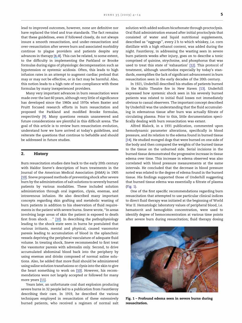
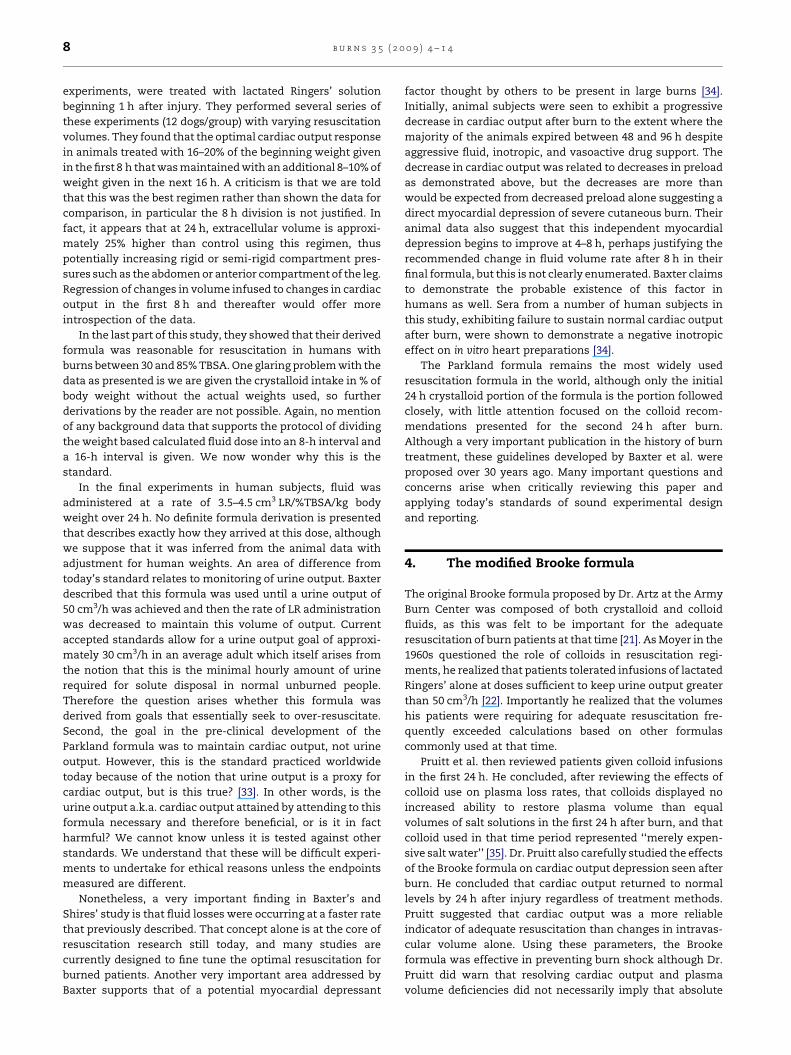
Fig. 1 – Profound edema seen in severe burns during
resuscitation.
2. History
Burn resuscitation studies date back to the early 20th century
with Haldor Sneve’s description of burn treatments in the
Journal of the American Medical Association (JAMA) in 1905
[10]. Sneve proposed methods of preventing shock after severe
burn by the administration of salt solutions to severely burned
patients by various modalities. These included solution
administration through oral ingestion, clysis, enemas, and
intravenous infusion. He also described many important
concepts regarding skin grafting and metabolic wasting of
burn patients in addition to his observation of fluid require-
ments in the patient with severe burns. Sneve wrote, ‘‘In areas
involving large areas of skin the patient is exposed to death
first from shock . . .’’ [10]. In describing the pathophysiology
leading to the shock state seen in burns he postulated that
various irritants, mental and physical, caused vasomotor
paresis leading to accumulation of blood in the splanchnic
vessels depriving the peripheral vasculature of adequate fluid
volume. In treating shock, Sneve recommended to first treat
the vasomotor paresis with adrenalin only. Second, to drive
accumulated abdominal blood back into the periphery by
using enemas and drinks composed of normal saline solu-
tions. Also, he added that more fluid should be administered
using saline solution transfusions or clysis into the skin to give
the heart something to work on [10]. However, his recom-
mendations were not largely accepted or followed for many
more years [11].
Years later, an unfortunate coal dust explosion producing
severe burns in 32 people led to a publication from Fauntleroy
describing their care. In 1919, he described the various
techniques employed in resuscitation of these extensively
burned patients, who received a regimen of normal salt
solution with added sodium bicarbonate through proctoclysis.
Oral fluid administration ensued after initial proctoclysis that
consisted of water and liquid nutritional supplements,
described as ‘‘eggnogs’’, every 2 h to which whiskey, i.e. corn
distillate with a high ethanol content, was added during the
night. Fauntleroy, in addressing the wasting seen in severe
burn patients weeks after injury, goes on to describe a tonic
comprised of quinine, strychnine, and phosphorus that was
used to treat this state of ‘exhaustion’ [12]. This protocol of
treatment, although unorthodox especially by today’s stan-
dards, exemplifies the lack of significant advancement in burn
resuscitation seen in the early decades of the 20th century.
In 1921, Underhill described his studies of patients burned
in the Rialto Theatre fire in New Haven [13]. Underhill
expressed how systemic shock seen in his severely burned
patients was related to initial fluid losses not immediately
obvious to casual observers. The important concept described
by Underhill was the understanding that the fluid accumulat-
ing in edematous tissue after burn was actually filtrate of
circulating plasma. Prior to this, little documentation speci-
fically dealing with burn resuscitation was extant.
Alfred Blalock, in a 1931 publication, commented upon
hemodynamic parameter alterations, specifically in blood
pressure, and its relation to the edema found in burned tissue
[14]. He studied mongrel dogs that were burned on one side of
the body and then compared the weights of the burned tissue
to the tissue on the unburned side. Serial incisions in the
burned tissue demonstrated the progressive increase in tissue
edema over time. This increase in edema observed was also
correlated with blood pressure measurements at the same
intervals. He concluded that the decrease in blood pressure
noted was related to the degree of edema found in the burned
tissue. His findings supported those of Underhill suggesting
that burned tissue edema was essentially a filtrate of plasma
(Fig. 1).
One of the first specific recommendations regarding burn
resuscitation that attempted to use particular clinical indices
to direct fluid therapy was initiated at the beginning of World
War II. Hematologic laboratory values of peripheral blood, i.e.
hematocrit and hemoglobin concentration, were used to
identify degree of hemoconcentration at various time points
after severe burn during resuscitation; fluid therapy dosing
b u r n s 3 5 ( 2 0 0 9 ) 4 – 1 46
recommendations were then based on degree of hemocon-
centration [15–17]. This therapy fell out of favour with
recognition that these recommendations failed to account
for ongoing losses that occur after identifying the degree of
hemoconcentration, essentially a snapshot in time. In
essence, patients treated in this manner were under-resusci-
tated and it was acknowledged that a more standardized
formula of resuscitation was warranted to improve patient
outcomes.
January 1942 was a pivotal time in burn history as the
National Research Council, under Chairman Isadore S. Ravdin,
agreed that a new standardized approach to burn resuscita-
tion based on surface area of burn should be recommended to
the military. Dr. H. Harkins accepted the challenge, and using
his observations from studies in canine burn models,
proposed the first well-known burn resuscitation formula
based on extent of surface area burned as a key guide dictating
fluid therapy. His proposal stated that all patients with greater
than or equal to 10% body surface area burn should be
resuscitated with 1000 cm3 of plasma for each 10% surface
area burned, to be administered over the first 24 h after injury
[17]. Interestingly, at that conference, the Board recommended
the use of a crystalloid solution (5% dextrose in physiologic
saline solution) administered at a rate of 20–40 drops per
minute, in addition to the plasma, but only after sufficient
plasma had been given to restore proper circulating volume
[18] (Table 1).
Cope and Moore published their experience with burns
related to the Coconut Grove Disaster of 1942, where they
suggested a relationship between systemic shock after burn
and generalized edema development. This paper, considered a
landmark publication by many, was recognized as the first to
suggest that resuscitation should be formulated from the
patient’s body weight and extent of burn [19]. They acknowl-
edged the shortcomings of a formula based on surface area
burned alone, and described their findings related to inter-
stitial space expansion and its important relationship to renal
function and/or failure. Cope and Moore also emphasized the
concept of increased fluid needs of patients with pulmonary
injury due to smoke. They proposed a resuscitation formula
called the ‘Body-Weight Burn Budget,’ using colloid and
Table 1 – Historical overview of various burn resuscitation for
1942 Harkins formula Any patient with at
surface area burn o
1947 Body-Weight Burn Budget First 24 h: 1–4 LLR +
24 h: same formulat
1952 Evan’s formula First 24 h: NS at 1 m
water. Second 24 h:
amount of glucose i
1953 Brooke formula First 24 h: LR at 1.5
switch to D5W 2000
1974 Formula First 24 h: LR at 4 m
Second 24 h: colloid
urinary output
1979 Modified Brooke First 24 h: LR at 2 m
remaining 16 h. Sec
maintain urine outp
1984 Monafo formula First 24 h: saline wi
adjusted per urine o
electrolyte solutions, based on anticipated interstitial space
expansion according to body weight given over the first 48 h
after burn. Evans followed years later with the ‘Evan’s formula’
that was the first to calculate fluid requirements based on body
weight and total body surface area (TBSA) burn [20]. This
formula, which included colloid consisting of albumin instead
of plasma because plasma fell out of favour for risk of blood-
borne disease transmission. This formula was the basis of
fluid resuscitation for many years.
C.P. Artz from the Army Burn Center in San Antonio
followed with a modified version of Evan’s formula which
placed more emphasis on crystalloid resuscitation and a
decreased dose of colloid for burn resuscitation, especially in
the first 24 h after injury. The basis for this difference lies in Dr.
Artz’s description of the changes in plasma composition
immediately after severe burn. He states that water and
electrolytes were lost into the burn wound at a greater rate
than protein losses. Artz referred to the earlier work of Fox
describing the dramatic sodium shifts into the cells and
interstitium seen in burns, leading to an intravascular hypo-
osmolar state. Addressing the replacement of sodium losses
due to electrolyte shifts through the use of more crystalloid
and less colloid within the first 24 h after severe burn became
the key factor in Dr. Artz’s proposed resuscitation guidelines.
This formula came to be known as the Brooke formula [21].
These formulas were the basis for burn resuscitation for years
until further controversy arose over the role of colloids in burn
resuscitation formulas.
Moyer and others, in 1965, described their studies of burn
shock in relation to extravascular sodium concentrations [22].
Their general focus was on the distribution of sodium and
other oncolytes during resuscitation. This led to the develop-
ment of formulas composed only of crystalloid. This change in
approach set the stage for a boom in research in this area with
focus placed on burn resuscitation strategy. In the late 1960s
through the 1970s, investigators at the Institute of Surgical
Research (ISR) in San Antonio and at Parkland Hospital in
Dallas emerged as the dominant figures in burn resuscitation
research from which the widely accepted Parkland formula
was proposed [23]. Pruitt et al. from the ISR promptly followed
with a recommendation calculating fluid requirements as in
mulas
least a 10% burn: administer 1000 cm3 plasma for each 10% total
ver first 24 h
1200 ml 0.5 NS + 7.5% body weight colloid + 1.5–5 l D5W. For second
ion except change colloid to 2.5% body weight
l/kg/% burn + colloids at 1 ml/kg/% burn + plus 2000 ml glucose in
one-half the first 24 h crystalloid and colloid req. + the same
n water as in the first 24 h
ml/kg/% TBSA burn + colloid at 0.5 ml/kg/% TBSA burn. Second 24 h:
ml
l/kg/% TBSA; give half in first 8 h and the remaining over next 16 h.
at 20–60% of calculated plasma volume to maintain adequate
l/kg/% TBSA burn, one half in the first 8 h and half in the
ond 24 h: colloid at 0.3–0.5 ml/kg/% TBSA burn + D5W to
ut
th 250 mequiv. Na + 150 mequiv. lactate + 100 mequiv. Cl. Rate
utput. Second 24 h: one third of isotonic salt administered orally
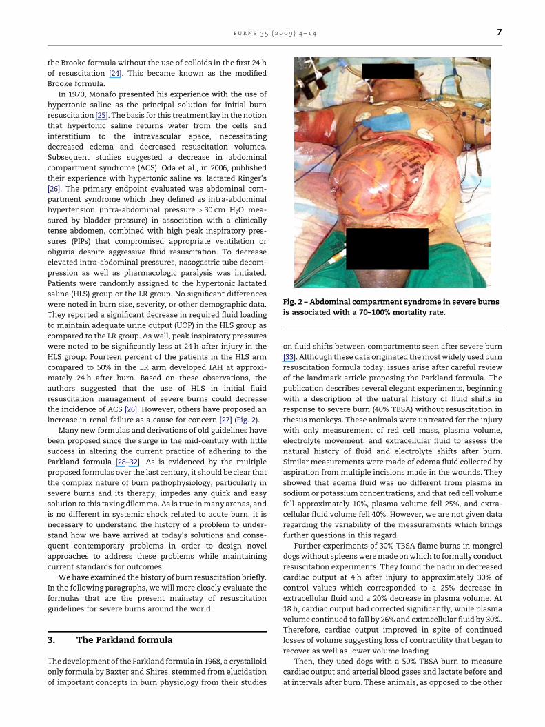
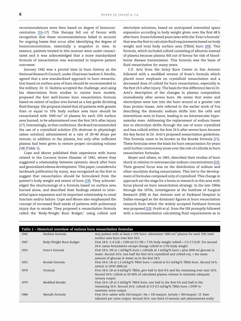
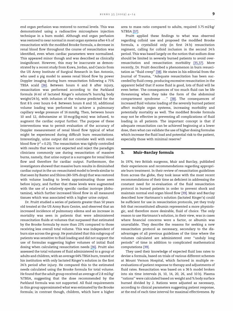
Fig. 2 – Abdominal compartment syndrome in severe burns
is associated with a 70–100% mortality rate.
b u r n s 3 5 ( 2 0 0 9 ) 4 – 1 4 7
the Brooke formula without the use of colloids in the first 24 h
of resuscitation [24]. This became known as the modified
Brooke formula.
In 1970, Monafo presented his experience with the use of
hypertonic saline as the principal solution for initial burn
resuscitation [25]. The basis for this treatment lay in the notion
that hypertonic saline returns water from the cells and
interstitium to the intravascular space, necessitating
decreased edema and decreased resuscitation volumes.
Subsequent studies suggested a decrease in abdominal
compartment syndrome (ACS). Oda et al., in 2006, published
their experience with hypertonic saline vs. lactated Ringer’s
[26]. The primary endpoint evaluated was abdominal com-
partment syndrome which they defined as intra-abdominal
hypertension (intra-abdominal pressure > 30 cm H2O mea-
sured by bladder pressure) in association with a clinically
tense abdomen, combined with high peak inspiratory pres-
sures (PIPs) that compromised appropriate ventilation or
oliguria despite aggressive fluid resuscitation. To decrease
elevated intra-abdominal pressures, nasogastric tube decom-
pression as well as pharmacologic paralysis was initiated.
Patients were randomly assigned to the hypertonic lactated
saline (HLS) group or the LR group. No significant differences
were noted in burn size, severity, or other demographic data.
They reported a significant decrease in required fluid loading
to maintain adequate urine output (UOP) in the HLS group as
compared to the LR group. As well, peak inspiratory pressures
were noted to be significantly less at 24 h after injury in the
HLS group. Fourteen percent of the patients in the HLS arm
compared to 50% in the LR arm developed IAH at approxi-
mately 24 h after burn. Based on these observations, the
authors suggested that the use of HLS in initial fluid
resuscitation management of severe burns could decrease
the incidence of ACS [26]. However, others have proposed an
increase in renal failure as a cause for concern [27] (Fig. 2).
Many new formulas and derivations of old guidelines have
been proposed since the surge in the mid-century with little
success in altering the current practice of adhering to the
Parkland formula [28–32]. As is evidenced by the multiple
proposed formulas over the last century, it should be clear that
the complex nature of burn pathophysiology, particularly in
severe burns and its therapy, impedes any quick and easy
solution to this taxing dilemma. As is true in many arenas, and
is no different in systemic shock related to acute burn, it is
necessary to understand the history of a problem to under-
stand how we have arrived at today’s solutions and conse-
quent contemporary problems in order to design novel
approaches to address these problems while maintaining
current standards for outcomes.
We have examined the history of burn resuscitation briefly.
In the following paragraphs, we will more closely evaluate the
formulas that are the present mainstay of resuscitation
guidelines for severe burns around the world.
3. The Parkland formula
The development of the Parkland formula in 1968, a crystalloid
only formula by Baxter and Shires, stemmed from elucidation
of important concepts in burn physiology from their studies
on fluid shifts between compartments seen after severe burn
[33]. Although these data originated the most widely used burn
resuscitation formula today, issues arise after careful review
of the landmark article proposing the Parkland formula. The
publication describes several elegant experiments, beginning
with a description of the natural history of fluid shifts in
response to severe burn (40% TBSA) without resuscitation in
rhesus monkeys. These animals were untreated for the injury
with only measurement of red cell mass, plasma volume,
electrolyte movement, and extracellular fluid to assess the
natural history of fluid and electrolyte shifts after burn.
Similar measurements were made of edema fluid collected by
aspiration from multiple incisions made in the wounds. They
showed that edema fluid was no different from plasma in
sodium or potassium concentrations, and that red cell volume
fell approximately 10%, plasma volume fell 25%, and extra-
cellular fluid volume fell 40%. However, we are not given data
regarding the variability of the measurements which brings
further questions in this regard.
Further experiments of 30% TBSA flame burns in mongrel
dogs without spleens were made on which to formally conduct
resuscitation experiments. They found the nadir in decreased
cardiac output at 4 h after injury to approximately 30% of
control values which corresponded to a 25% decrease in
extracellular fluid and a 20% decrease in plasma volume. At
18 h, cardiac output had corrected significantly, while plasma
volume continued to fall by 26% and extracellular fluid by 30%.
Therefore, cardiac output improved in spite of continued
losses of volume suggesting loss of contractility that began to
recover as well as lower volume loading.
Then, they used dogs with a 50% TBSA burn to measure
cardiac output and arterial blood gases and lactate before and
at intervals after burn. These animals, as opposed to the other
b u r n s 3 5 ( 2 0 0 9 ) 4 – 1 48
experiments, were treated with lactated Ringers’ solution
beginning 1 h after injury. They performed several series of
these experiments (12 dogs/group) with varying resuscitation
volumes. They found that the optimal cardiac output response
in animals treated with 16–20% of the beginning weight given
in the first 8 h that was maintained with an additional 8–10% of
weight given in the next 16 h. A criticism is that we are told
that this was the best regimen rather than shown the data for
comparison, in particular the 8 h division is not justified. In
fact, it appears that at 24 h, extracellular volume is approxi-
mately 25% higher than control using this regimen, thus
potentially increasing rigid or semi-rigid compartment pres-
sures such as the abdomen or anterior compartment of the leg.
Regression of changes in volume infused to changes in cardiac
output in the first 8 h and thereafter would offer more
introspection of the data.
In the last part of this study, they showed that their derived
formula was reasonable for resuscitation in humans with
burns between 30 and 85% TBSA. One glaring problem with the
data as presented is we are given the crystalloid intake in % of
body weight without the actual weights used, so further
derivations by the reader are not possible. Again, no mention
of any background data that supports the protocol of dividing
the weight based calculated fluid dose into an 8-h interval and
a 16-h interval is given. We now wonder why this is the
standard.
In the final experiments in human subjects, fluid was
administered at a rate of 3.5–4.5 cm3 LR/%TBSA/kg body
weight over 24 h. No definite formula derivation is presented
that describes exactly how they arrived at this dose, although
we suppose that it was inferred from the animal data with
adjustment for human weights. An area of difference from
today’s standard relates to monitoring of urine output. Baxter
described that this formula was used until a urine output of
50 cm3/h was achieved and then the rate of LR administration
was decreased to maintain this volume of output. Current
accepted standards allow for a urine output goal of approxi-
mately 30 cm3/h in an average adult which itself arises from
the notion that this is the minimal hourly amount of urine
required for solute disposal in normal unburned people.
Therefore the question arises whether this formula was
derived from goals that essentially seek to over-resuscitate.
Second, the goal in the pre-clinical development of the
Parkland formula was to maintain cardiac output, not urine
output. However, this is the standard practiced worldwide
today because of the notion that urine output is a proxy for
cardiac output, but is this true? [33]. In other words, is the
urine output a.k.a. cardiac output attained by attending to this
formula necessary and therefore beneficial, or is it in fact
harmful? We cannot know unless it is tested against other
standards. We understand that these will be difficult experi-
ments to undertake for ethical reasons unless the endpoints
measured are different.
Nonetheless, a very important finding in Baxter’s and
Shires’ study is that fluid losses were occurring at a faster rate
that previously described. That concept alone is at the core of
resuscitation research still today, and many studies are
currently designed to fine tune the optimal resuscitation for
burned patients. Another very important area addressed by
Baxter supports that of a potential myocardial depressant
factor thought by others to be present in large burns [34].
Initially, animal subjects were seen to exhibit a progressive
decrease in cardiac output after burn to the extent where the
majority of the animals expired between 48 and 96 h despite
aggressive fluid, inotropic, and vasoactive drug support. The
decrease in cardiac output was related to decreases in preload
as demonstrated above, but the decreases are more than
would be expected from decreased preload alone suggesting a
direct myocardial depression of severe cutaneous burn. Their
animal data also suggest that this independent myocardial
depression begins to improve at 4–8 h, perhaps justifying the
recommended change in fluid volume rate after 8 h in their
final formula, but this is not clearly enumerated. Baxter claims
to demonstrate the probable existence of this factor in
humans as well. Sera from a number of human subjects in
this study, exhibiting failure to sustain normal cardiac output
after burn, were shown to demonstrate a negative inotropic
effect on in vitro heart preparations [34].
The Parkland formula remains the most widely used
resuscitation formula in the world, although only the initial
24 h crystalloid portion of the formula is the portion followed
closely, with little attention focused on the colloid recom-
mendations presented for the second 24 h after burn.
Although a very important publication in the history of burn
treatment, these guidelines developed by Baxter et al. were
proposed over 30 years ago. Many important questions and
concerns arise when critically reviewing this paper and
applying today’s standards of sound experimental design
and reporting.
4. The modified Brooke formula
The original Brooke formula proposed by Dr. Artz at the Army
Burn Center was composed of both crystalloid and colloid
fluids, as this was felt to be important for the adequate
resuscitation of burn patients at that time [21]. As Moyer in the
1960s questioned the role of colloids in resuscitation regi-
ments, he realized that patients tolerated infusions of lactated
Ringers’ alone at doses sufficient to keep urine output greater
than 50 cm3/h [22]. Importantly he realized that the volumes
his patients were requiring for adequate resuscitation fre-
quently exceeded calculations based on other formulas
commonly used at that time.
Pruitt et al. then reviewed patients given colloid infusions
in the first 24 h. He concluded, after reviewing the effects of
colloid use on plasma loss rates, that colloids displayed no
increased ability to restore plasma volume than equal
volumes of salt solutions in the first 24 h after burn, and that
colloid used in that time period represented ‘‘merely expen-
sive salt water’’ [35]. Dr. Pruitt also carefully studied the effects
of the Brooke formula on cardiac output depression seen after
burn. He concluded that cardiac output returned to normal
levels by 24 h after injury regardless of treatment methods.
Pruitt suggested that cardiac output was a more reliable
indicator of adequate resuscitation than changes in intravas-
cular volume alone. Using these parameters, the Brooke
formula was effective in preventing burn shock although Dr.
Pruitt did warn that resolving cardiac output and plasma
volume deficiencies did not necessarily imply that absolute
b u r n s 3 5 ( 2 0 0 9 ) 4 – 1 4 9
end organ perfusion was restored to normal levels. This was
demonstrated using a radioactive microsphere injection
technique in a burn model. Although end organ perfusion
was restored to near normal in most organ systems after 4 h of
resuscitation with the modified Brooke formula, a decrease in
renal blood flow throughout the course of resuscitation was
identified, even when cardiac parameters were normalized.
This appeared minor though and was described as clinically
insignificant. However, this may be inaccurate as demon-
strated by a recent study from Kuwa, Jordan, and Cancio from
the US Army Institute of Surgical Research in San Antonio,
who used a pig model to assess renal blood flow by power
Doppler imaging during burn resuscitation following a 75%
TBSA scald [36]. Between hours 6 and 8 after injury,
resuscitation was performed according to the Parkland
formula (4 ml of lactated Ringer’s solution/% burn/kg body
weight/24 h), with infusion of the volume predicted for the
first 8 h over hours 6–8. Between hours 8 and 10, additional
volume loading was performed to achieve a pulmonary
capillary wedge pressure of 16 mmHg. Then, between hours
10 and 12, dobutamine at 10 mcg/(kg min) was infused, to
augment the cardiac output further. The purpose of these
interventions was to permit evaluation of the accuracy of
Doppler measurement of renal blood flow typical of what
might be experienced during difficult burn resuscitations.
Interestingly, urine output did not correlate well with renal
blood flow (r2 = 0.25). The resuscitation was tightly controlled
with results that were not expected and reject the paradigm
clinicians commonly use during resuscitation of massive
burns, namely, that urine output is a surrogate for renal blood
flow and therefore for cardiac output. Furthermore, the
investigators showed that massive burn results in diminished
cardiac output in the un-resuscitated model to levels similar to
that seen by Baxter and Shires (40–50% drop) that was restored
with volume loading to levels approximating those seen
before injury, and further that these levels were augmented
with the use of a relatively specific cardiac inotrope (dobu-
tamine), which further increased blood flow in all measured
tissues which was associated with a higher urine output.
Dr. Pruitt studied a series of patients greater than 50 years
old treated at the US Army Burn Centre, and observed that an
increased incidence of pulmonary edema and an increase in
mortality was seen in patients that were administered
resuscitation fluids at volumes that surpassed that estimated
by the Brooke formula by more than 23% compared to those
receiving less overall total volume. This was independent of
burn size across the group. He postulated that this subgroup of
patients was sensitive to fluid loading and did not support the
use of formulas suggesting higher volumes of initial fluid
dosing when calculating resuscitation needs [36]. Pruitt also
assessed the total volumes of fluid administered to a group of
adults and children, with an average 64% TBSA burn, treated at
his institution with only lactated Ringer’s solution in the first
24 h period after injury. He compared this to the estimated
needs calculated using the Brooke formula for total volume.
He found that the adult group received an average of 2.8 ml/kg/
%TBSA, suggesting that the dose recommended by the
Parkland formula was not supported. All fluid requirements
in this group approximated what was estimated by the Brooke
formula; in contrast though, children, which have a higher
area to mass ratio compared to adults, required 3.75 ml/kg/
%TBSA [37].
Pruitt applied these findings to what was observed
regarding colloid use and proposed the modified Brooke
formula, a crystalloid only (in first 24 h) resuscitation
regiment, calling for colloid inclusion in the second 24 h
period. This was based largely on the notion that sodium dose
should be limited in severely burned patients to avoid over-
resuscitation and resuscitation morbidity [35,37]. More
recently, Dr. Pruitt described a phenomenon in burn resusci-
tation as ‘‘fluid creep’’ [38]. He states in his editorial from the
Journal of Trauma, ‘‘Adequate resuscitation has been suc-
ceeded by fluid creep, producing excessive resuscitation in the
apparent belief that if some fluid is good, lots of fluid will be
even better. The consequences of too much fluid can be life
threatening when they take the form of the abdominal
compartment syndrome . . .’’ [38]. The consequences of
increased fluid volume loading of the severely burned patient
affect multiple organ systems, increasing morbidity and
potentially mortality as well. The modified Brooke formula
may not be effective in preventing all complications of fluid
loading in all patients. The important concept is that if
adequate resuscitation can be achieved beginning at a lower
dose, then what can validate the use of higher dosing formulas
which increase the fluid load and potential risk to the patient,
especially those with minimal reserve?
5. Muir–Barclay formula
In 1974, two British surgeons, Muir and Barclay, published
their experiences and recommendations regarding appropri-
ate burn treatment. In their review of resuscitation guidelines
from across the globe, they took issue with the most recent
recommendations of their time as deficient in addressing the
constant need for re-evaluation of the fluid resuscitation
protocol in burned patients in order to prevent shock and
maintain normal end organ blood flow [39]. Muir and Barclay
described how Hartmann’s solution (lactated Ringer’s) could
be sufficient for use in resuscitation protocols, yet they truly
felt that reconstituted albumin represented a more physiolo-
gic, and therefore more desirable, fluid of choice. The only
reason to use Hartmann’s solution, in their view, was in cases
where financial concerns were a factor, or albumin was
unavailable. They describe the reasons for seeking a new
resuscitation protocol as necessary, secondary to the dis-
advantages of all previous guidelines of the time where the
volumes calculated are administered over ‘‘unduly long
periods’’ of time in addition to complicated mathematical
computations [39].
They used their knowledge of expected fluid loss rates to
devise a formula, based on trials of various different schemes
at Mount Vernon Hospital, which factored in multiple re-
evaluations of patient response to therapy and adjustments of
fluid rates. Resuscitation was based on a 36 h model broken
into six time intervals (4, 10, 14, 20, 26, and 10 h). Plasma
‘‘rations’’ were calculated based on weight and % body surface
burned divided by 2. Rations were adjusted as necessary,
according to clinical parameters suggesting patient response,
at the end of each time interval for maximal effectiveness. By
b u r n s 3 5 ( 2 0 0 9 ) 4 – 1 410
their description, volumes were rarely excessive and adjust-
ments made in the rations were usually increases in volume,
although no experimental data is presented in their text to
support their claims or allow for validation of their data. The
Muir/Barclay formula is still commonly used in Great Britain
today. No sound evidence of benefit over today’s other
standards has been shown. The concern arises with this
formula in that even with colloid infusions being used for the
first 24 h, in stark contrast to what has been shown as
ineffective by Baxter and Pruitt studies, the overall volume of
fluid needs may tend be underestimated.
6. Other considerations for effectiveresuscitation
As is seen, absolute consensus on resuscitation formulae has
not been reached. The inherent challenges faced by providers
caring for severely burned casualties during the initial
resuscitation period have been described previously. In
November 2005, the USAISR implemented a military-wide
burn resuscitation guideline that was developed along with a
burn flow sheet, which required the documentation of the
initial 24 h resuscitation for all severely burned casualties [40].
We found that a lower initial resuscitation volume (2 cm3/kg/
%TBSA burn vs. 4 cm3/kg/% TBSA burn) was associated with
lower total infused volume even when age, injury, and other
clinical factors were accounted. However, we also did not find
any difference in clinical outcomes in this small study. In a
separate study, Cancio et al. described factors that help predict
which patients might require increased fluid volumes to
achieve adequate resuscitation over what was calculated by
the modified Brooke formula [41]. He concluded that burn size
and weight (negatively) were associated with greater volume
requirements. In a time where decreasing the sodium load
given to patients is critical, as discussed by Pruitt, it is evident
by this data that the Parkland formula only leads to increased
sodium loading of the patient with no increased benefit or
difference in outcomes. This should be considered in
prescribing volumes for initial resuscitation of the severely
burned.
Abdominal compartment syndrome is of great concern to
burn care providers and must be addressed in any context
commenting on resuscitation volumes. ACS is associated with
significant detrimental effects on multiple organ systems to
include decreased pulmonary compliance, cardiac dysfunc-
tion, malperfusion to the bowel, hepatic, and renal systems.
Large fluid volumes during burn resuscitation are considered a
high risk for the development of ACS. Ivy et al. described their
experience with intra-abdominal pressures and the relation-
ship to fluid volumes infused in major thermal injury. They
determined that an infusion approaching or surpassing 0.25 l/
kg during the acute burn resuscitation greatly increases the
risk of developing intra-abdominal hypertension and/or ACS
[42]. Based upon this data, it is imperative that methods be
delineated for decreasing overall fluid volumes dosed during
initial burn resuscitations in order to prevent such a
complication. This has proven to be a difficult task.
The use of colloid has been examined in decreasing overall
fluid volumes and improving outcomes. In 1983, Goodwin et al.
from the ISR described a randomized trial comparing the
efficacy of crystalloid and colloid solutions used in burn
resuscitation [43]. They concluded that the use of colloid did
not provide any long lasting benefit and may even be
deleterious to the pulmonary system as they showed an
increase in the accumulation of lung water in those that
received colloid solution, even when no inhalation injury was
present. The interesting finding in the paper is that the colloid
group required less fluid volume for successful resuscitation
when compared to the crystalloid group. Specifically, during
the 12–24 h period, clinical hemodynamic parameters were
improved in the colloid group over the crystalloid group. To
some, this suggests the theory that albumin leakage into the
wound is over after the first 12 h. In relation to ACS, this raises
the question to whether immediate use of colloid could reduce
fluid volumes and subsequently ACS risk. If so, would it be at
the expense of the pulmonary system? On that same note,
another important question has arisen. Is plasma resuscita-
tion better than standard colloid resuscitation in reducing
fluid load and incidence intra-abdominal hypertension and
ACS when compared to crystalloid in burn patients? If so,
when? O’Mara et al. published their data addressing just that
issue [7]. Patients were randomized to either a plasma
resuscitation group or a crystalloid only group and measure-
ments of intra-abdominal pressures were recorded and
analyzed. The Parkland formula was used to calculate initial
fluid requirements. They showed that the plasma group
required overall less fluid loading and sustained significantly
lower intra-abdominal pressures below the threshold for
complications of intra-abdominal hypertension when com-
pared to the crystalloid resuscitation arm. Although, they did
also conclude that resuscitation with plasma did not improve
overall outcomes as has previously been seen in colloid
resuscitation studies.
One other form of resuscitation not addressed thus far
regards using oral solutions for burned patients. Many of the
first methods of burn resuscitation involved the use of oral
solutions. In mass casualty situations or where IV fluids are
not readily available, oral fluids could be easily produced at
low cost. Many of these ideas stem from the treatment of
severe diarrhea [44,45]. In 2003, the US Army Institute of
Surgical Research collaborated with the American Burn
Association in a survey of the burn bed availability in the
United States should there be a national disaster [46]. The
results were concerning, to say the least. It was determined
that there would not be sufficient burn bed availability to
handle the patient load of burns from a single mass casualty
situation with over 500 severe burns. Therefore, needs for
alternative resuscitation modes were deemed imperative for
adequate preparedness. In 2006, investigators from San
Antonio and Galveston published a historical overview of
experiences with oral resuscitation therapies and gave sample
recipes for homemade oral resuscitation therapies [47]. Cited
were multiple investigators describing their experiences with
oral resuscitation therapies throughout the 1940s–1990s using
various electrolyte-based solutions. The effect of severe burn
on gastrointestinal physiology, mainly ileus, poses a true
roadblock to effective oral resuscitation and therefore cur-
rently supports the current standard of care, IV fluid
resuscitation. A recent publication from the UTMB Galveston
b u r n s 3 5 ( 2 0 0 9 ) 4 – 1 4 11
Shriner’s Burn Hospital group addressed the need for oral
resuscitation fluid use in burns in a swine study comparing the
World Health Organization Oral Resuscitation Solution, a
citrate-buffered glucose electrolyte solution, to IV lactated
Ringers per the Parkland formula guidelines [48]. Intestinal
absorption rates were delineated and clinical parameters were
compared between the groups. They concluded that enteral
fluid resuscitation is feasible without detriment in a severe
burn up to 40% TBSA. Any burn more severe than this could
encounter maximal intestinal absorption limitations and the
effectiveness of oral solutions could not be determined by
their study. Lack of data to support oral solution guidelines
regarding type of to be used and timing of administration after
burn exist. Despite this it is concluded that oral resuscitation is
feasible for use in burn shock, especially in mass casualty
situations. Nonetheless, more research is imperative in order
to better define the appropriateness of such guidelines.
The use of proctoclysis as a means of appropriate fluid
resuscitation also has been discussed. One such protocol, the
Murphy’s drip, was described in 1913 by John Murphy from
Chicago as an adequate means of providing appropriate fluid
therapy [49]. A warmed, saline-based solution was delivered at
a rate of 1.5–2 pints every 2 h. This has not caught up to the
mainstream as its methods and effectiveness still require
sound experimental investigation.
While choosing the appropriate resuscitation regiment to
implement in a severely burned patient is difficult enough; the
art of monitoring and determining if what you have prescribed
is actually effective, can be just as taxing. Urine output, on an
hourly basis, seems to be the most widely accepted clinical
parameter to follow for evaluation of adequacy of therapy.
This has relatively recently, though, been challenged [50–52].
As in earlier protocols, UOP goals were to achieve 50 cm3/h for
an average size adult. Now goals are reduced to roughly 30–
35 cm3/h for an average sized adult. Children UOP goals
approximate double that of the adults, per kilogram body
weight, for adequate resuscitation. Other invasive parameters
have been used and challenged as well, such as central venous
pressure and pulmonary artery catheters [53,54]. Invasive
thermodilution monitoring is yet another form of advanced
monitoring of fluid therapy with unsubstantiated results in
recent trials [54]. Many other techniques have been used as
well without overwhelming evidence to support their use as
the standard of care. Needless to say, more studies are needed
to help define better parameters for resuscitation endpoints.
For example, maintaining cardiac output was the goal used in
the studies leading to the Parkland formula, yet most, if any,
do not use this parameter as the chief endpoint of resuscita-
tion. Despite the fact that we have the ability to measure
cardiac output, should we be focusing on it? For the meantime,
then, urine output remains the standard for monitoring
adequacy of resuscitation after severe burn unless a fruitful
alternative comes to bear.
One more very important question regarding resuscitation
has arisen regarding the immunomodulatory effects of
lactated Ringers’. Some suggest that use of this particular
fluid may be of actual harm. Rhee et al. presented such a case
at the 1997 Annual Meeting of the Western Trauma Associa-
tion [55]. There, they discussed the results of a study in swine
which they concluded that LR was responsible for increased
neutrophil activation after resuscitation for hemorrhage or
after infusion without hemorrhage. LR used in the majority of
hospitals contains a racemic mixture of the D-lactate and L-
lactate isomers. L-Lactate is the more natural form normally
found in mammalian metabolism. A more recent study of the
separate effects of each isomer on human leukocytes revealed
that the D-lactate isomer was actually responsible for an
observed increased production of reactive oxygen species and
effects on gene expression leading to increased apoptosis seen
after exposure to the racemic formulation of LR [56].
Neurocardiac toxicity with serious consequences has also
been described in animal models by infusion of D-lactate LR
compared to no toxic signs seen when given only L-lactate LR
[57]. Some manufactures are now producing LR containing
only the L-lactate isomer. As long as crystalloid and therefore
LR remains the standard of care for initial fluid resuscitation,
the growing body of evidence regarding using racemic
formulations of LR is seriously called into question and should
have no role in any resuscitation.
7. Current effectiveness of acceptedguidelines
In most places, two formulas are accepted as guidelines for the
resuscitation of severely burned patients, the Parkland and
modified Brooke formulas. Burn care providers of all special-
ties are trained in the use of these formulas in order to help
prevent the onset of burn shock prior to the patient’s arrival in
a burn centre with experienced burn staff. Unfortunately, in
the ‘‘heat of the moment’’, inexperienced first responders as
well as many higher level emergency care physicians fail to
have recall of the appropriate calculations for adequate fluid
needs of the patient. Burn size, which is used for fluid volume
calculations, is commonly grossly under- or overestimated by
inexperienced providers [58]. This is possibly due to anxiety
induced by the situation in concert with the multiple formulas
with multiple variables to remember, making it difficult to
correctly implement. This leads to a high rate of non-
compliance seen in following these resuscitation guidelines
often leading to over- or under-resuscitation. In 2004, Bhat
et al. published the results of a two-phase survey study in
which they polled physicians and students on their recall and
use of resuscitation formulas [59]. One-hundred and ninety-
eight Emergency Medicine physicians in the US and United
Kingdom were asked for their knowledge on accurately
recalling any resuscitation formula and specifics relating to
any correct formula recalled. The second phase of the study
tested the ability of 22 medical students, given the correct
formula and case studies, to correctly compute the appro-
priate fluid therapy for the patients described in each case
study. Results of the first phase showed that 4% of those in the
United Kingdom and 33% of those in the US could accurately
recall a standard resuscitation formula. In the students, even
after being supplied with the correct formulas, large mis-
calculations occurred. This can be deadly for patients,
especially those with deficient reserve. The authors of the
study argue that a simplified initial resuscitation protocol is
necessary. At the USAISR we are currently implementing just
that, a simplified resuscitation formula named ‘The Rule of
b u r n s 3 5 ( 2 0 0 9 ) 4 – 1 412
Ten’ (ROT), which falls within ABA acceptable guidelines for
fluid therapy [60]. ‘The Rule of Ten’, which consists of two
steps, eliminates the confusion of recalling a difficult formula
and its accurate implementation. First, the estimated burn
size in %TBSA is multiplied by 10 to derive the initial fluid rate
in ml/h. For every 10 kg above 80 kg, 100 ml is added to this
rate. When compared directly to the Parkland and modified
Brooke formulas, the fluid rate would be overestimated for
patients under 40 kg, while underestimated for patients over
140 kg. The ‘Rule of Ten’ is currently implemented as protocol
at our institution and can easily be used to rapidly estimate the
initial fluid rate for the majority of adult patients. Simple and
rapid calculation of the initial fluid rate will allow pre-hospital
and other emergency care providers to focus their attention on
the process of resuscitation. Further adjustments to fluid rate
can be made based on clinical observations of effect.
8. Future considerations
The reality of burn resuscitation evolution is that our guide-
lines have been in place for 40 years with no significant
changes despite the findings of continued complications
during resuscitation [9]. Complications remain possibly as
result of overzealous over-resuscitation, irresponsible under-
resuscitation, or simply the lack of recall for complex
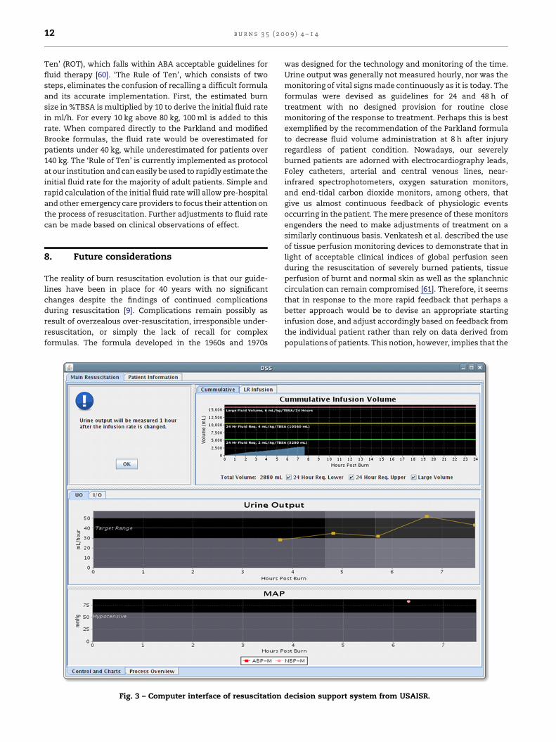
formulas. The formula developed in the 1960s and 1970s
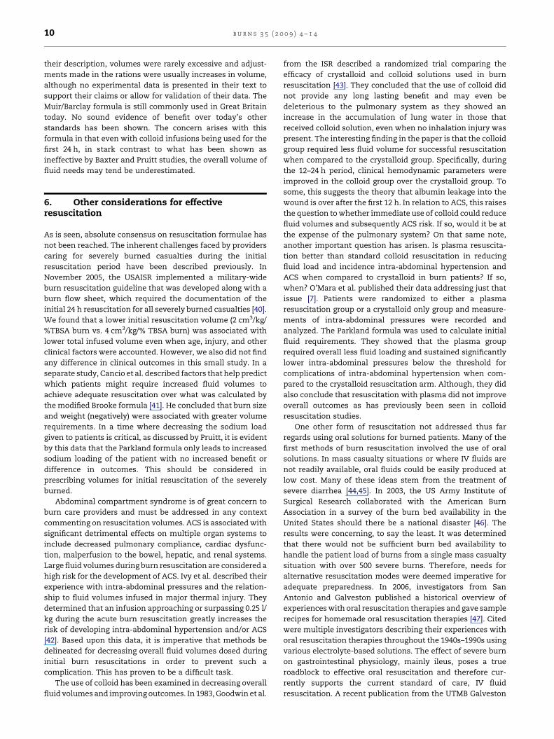
Fig. 3 – Computer interface of resuscitation
was designed for the technology and monitoring of the time.
Urine output was generally not measured hourly, nor was the
monitoring of vital signs made continuously as it is today. The
formulas were devised as guidelines for 24 and 48 h of
treatment with no designed provision for routine close
monitoring of the response to treatment. Perhaps this is best
exemplified by the recommendation of the Parkland formula
to decrease fluid volume administration at 8 h after injury
regardless of patient condition. Nowadays, our severely
burned patients are adorned with electrocardiography leads,
Foley catheters, arterial and central venous lines, near-
infrared spectrophotometers, oxygen saturation monitors,
and end-tidal carbon dioxide monitors, among others, that
give us almost continuous feedback of physiologic events
occurring in the patient. The mere presence of these monitors
engenders the need to make adjustments of treatment on a
similarly continuous basis. Venkatesh et al. described the use
of tissue perfusion monitoring devices to demonstrate that in
light of acceptable clinical indices of global perfusion seen
during the resuscitation of severely burned patients, tissue
perfusion of burnt and normal skin as well as the splanchnic
circulation can remain compromised [61]. Therefore, it seems
that in response to the more rapid feedback that perhaps a
better approach would be to devise an appropriate starting
infusion dose, and adjust accordingly based on feedback from
the individual patient rather than rely on data derived from
populations of patients. This notion, however, implies that the
decision support system from USAISR.
b u r n s 3 5 ( 2 0 0 9 ) 4 – 1 4 13
feedback received from our monitors actually reflects phy-
siologic condition. With this assumption in mind, then
perhaps automated decision support systems that take
physiologic feedback into account and make recommenda-
tions for changes in treatment in a timely fashion would be
more appropriate [62]. At the United States Army Institute of
Surgical Research in collaboration with investigators in
Galveston, we have embarked on such a program with initial
fluid administration rates determined by the ‘Rule of Ten’ [60].
From this point, adjustments are made hourly based on
measured urine output entered into a decision support
algorithm derived from close monitoring of 40 severely burned
patients (Fig. 3). We are closely monitoring the adequacy of
resuscitating patients in this fashion, and will subsequently
assess the role of colloid in resuscitation, specifically the
utility of plasma.
Another key consideration now coming to the forefront of
therapy is that of high volume hemofiltration for those
patients that develop shock or have difficult resuscitations
[63–65]. The current belief is that capillary leakage that leads to
fluid losses and needs for large amounts of fluid therapy are
mediated by pro- and anti-inflammatory mediators. The exact
mechanism of how these mediators act in concert to produce
such profound capillary integrity disruption is not known. The
use of hemofiltration devices to remove such mediators has
shown encouraging results. The USAISR has presented data on
the use of high volume hemofiltration in burn patients
developing septic shock and has shown and decreased in
hospital mortality in the study [66]. More patients across
multiple centres are needed to verify these findings and their
application to burn shock as well. The exact pathophysiology
of severe burn and the mediators involved need to be
elucidated before any great advances can potentially be made.
Hemofiltration devices may just help unearth the mystery.
9. Summation
The resuscitation of severely burned patients has clearly
evolved over the last century, with a lull in significant
progress since the 1970s, at the expense of the patient in our
opinion. The guidelines used today were developed 40 years
ago, yet remain the mainstay of current initial fluid therapy
despite ongoing research. Preventing complications of over-
or under-resuscitation still confounds burn providers as no
recent advances have been made in this arena as well. As we
continue to investigate new ways to address current fluid
therapy needs, monitor end points of resuscitation, and
prevent the negative effects of our current therapies; it is of
absolute importance that we head in the new century with a
strong focus on elucidating the mysteries of burn pathophy-
siology. It is at that time only that we can provide targeted
fluid resuscitation therapy that will change the way we
practice from today’s standard of care to the benefit of our
patients.
Conflict of interest
None.
r e f e r e n c e s
[1] Pruitt Jr BA. Protection from excessive resuscitation:pushing the pendulum back. J Trauma 2000;49:387–91.
[2] Demling RH. Fluid replacement in burned patients. SurgClin North Am 1987;67:15–30.
[3] Cartotto RC, Innes M, Musgrave MA, et al. How well doesthe Parkland formula estimate actual fluid resuscitationvolumes. J Burn Care Rehabil 2002;23:258–65.
[4] Demling RH. The burn edema process: current concepts. JBurn Care Rehabil 2005;26:207–27.
[5] Du G, Slater H, Goldfarb IW. Influences of differentresuscitation regimens on acute early weight gain inextensively burned patients. Burns 1991;17:147–50.
[6] Demling RH, Kramer GC, Gunther R, Nerlich M. Effect ofnon-protein colloid on post-burn edema formation in softtissues and lung. Surgery 1984;95:593–602.
[7] O’Mara MS, Slater H, Goldfarb IW, Saushaj PF. Aprospective, randomized evaluation of intra-abdominalpressures with crystalloid and colloid resuscitation in burnpatients. J Trauma 2005;58:1011–8.
[8] Sullivan SR, Friedrich JB, Engrav LH, et al. ‘‘Opiod creep’’ isreal and may be the cause of ‘‘fluid creep’’. Burns2004;30:583–90.
[9] Greenhalgh DG. Burn resuscitation. J Burn Care Res2007;28:555–65.
[10] Sneve H. The treatment of burns and skin grafting. JAMA1905;45:1–8.
[11] Pruitt Jr BA. Centennial changes in surgical care andresearch. Ann Surg 2000;232:287–301.
[12] Fauntleroy AM. The surgical lessons of the European war.Ann Surg 1916;64:136–50.
[13] Underhill FP. The significance of anhydremia in extensivesurface burn. JAMA 1930;95:852–7.
[14] Blalock A. Experimental shock. The importance of the localloss of fluid in the production of the low blood pressureafter burn. Arch Surg 1931;22:610–6.
[15] Black DAK. Treatment of burn shock with plasma andserum. Br Med J 1940;2:633.
[16] Elkinton JR, Wolff WA, Lee WE. Plasma transfusion in thetreatment of the fluid shift in severe burns. Ann Surg1940;10:112.
[17] Harkins HN, Lam CR, Romence H. Plasma therapy in severeburns. Surg Gynecol Obstet 1942;75:410.
[18] National Research Council (US) Committee on Surgery.Burns, shock, wound healing and vascular injuries/prepared under the auspices of the Committee on Surgeryof the Division of Medical Sciences of the National ResearchCouncil. Philadelphia/London: W.B. Saunders Co.; 1943.
[19] Cope O, Moore FD. The redistribution of body water and thefluid therapy of the burned patient. Ann Surg1947;126:1010–45.
[20] Evans EI, Purnell OJ, Robinett PW, Batchelor A, Martin M.Fluid and electrolyte requirements in severe burns. AnnSurg 1952;135:804.
[21] Artz CP, Moncrief JA. The burn problem. In: Artz CP,Moncrief JA, editors. The treatment of burns. Philadelphia:W.B. Saunders Co.; 1969. p. 1–22.
[22] Moyer CA, Margraf HW, Monafo WW. Burn shock andextravascular sodium: treatment with Ringer’s solutionwith lactate. Arch Surg 1965;90:799–811.
[23] Baxter CR. Fluid volume and electrolyte changes in theearly post-burn period. Clin Plast Surg 1974;1:693–703.
[24] Pruitt Jr BA, Mason Jr AD, Moncrief JA. Hemodynamicchanges in the early postburn patient: the influence of fluidadministration and of a vasodilator (hydralazine). J Trauma1971;11:36–46.
b u r n s 3 5 ( 2 0 0 9 ) 4 – 1 414
[25] Monafo WW. The treatment of burn shock by theintravenous and oral administration of hypertonic lactatedsaline solution. J Trauma 1970;10:575–86.
[26] Oda J, Ueyama M, Yamashita K, et al. Hypertonic lactatedsaline resuscitation reduces the risk of abdominalcompartment syndrome in severely burned patients. JTrauma 2006;60:64–71.
[27] Huang PP, Stucky FS, Dimick AR, et al. Hypertonic sodiumresuscitation is associated with renal failure and death.Ann Surg 1995;221:543–57.
[28] Onarheim H, Missavage AE, Kramer GC, Gunther RA.Effectiveness of hypertonic saline-dextran 70 for initialfluid resuscitation of major burns. J Trauma 1990;30:597–603.
[29] Cooper AB, Cohn SM, Zhang HS, et al. Five percent albuminfor adult burn shock resuscitation: lack of effect on dailymultiple organ dysfunction score. Transfusion 2006;46:=80–9.
[30] Milner SM, Kinsky MP, Guha SC, et al. A comparison of twodifferent 2400 mOsm solutions for resuscitation of majorburns. J Burn Care Rehabil 1997;18:109–15.
[31] Berger MM, Pictet A, Revelly JP, Frascarolo P, Chiolero RL.Impact of a bicarbonated saline solution on earlyresuscitation after major burns. Intensive Care Med2000;26:1382–5.
[32] Warden GD. Burn shock resuscitation. World J Surg1992;16:16–23.
[33] Baxter CR, Shires GT. Physiological response to crystalloidresuscitation of severe burns. Ann NY Acad Sci1968;150:874–94.
[34] Baxter CR, Cook WA, Shires GT. Serum myocardialdepressant factor of burn shock. Surg Forum 1966;17:1–2.
[35] Pruitt Jr BA. The burn patient. I. Initial care. Curr Probl Surg1979;16:1–55.
[36] Kuwa T, Jordan BS, Cancio LC. Use of power Dopplerultrasound to monitor renal perfusion during burn shock.Burns 2006;32(6):706–13.
[37] Pruitt Jr BA, Goodwin Jr CW. Current treatment of theextensively burned patient. Surg Annu 1983;15:331–64.
[38] Pruitt Jr BA. Protection from excessive resuscitation:‘‘Pushing the Pendulum Back’’. J Trauma 2000;49:567–8.
[39] Muir IA, Barclay TL. Burns and their treatment. Chicago:Year Book Medical Publishers; 1974.
[40] Alvarado RA, Chung KK, Renz EM, Cancio LC, Ennis J, BarilloDJ, et al. Burn resuscitation of severely burned militarycasualties: fluid begets more fluid; in press.
[41] Cancio LC, Chavez S, Alvarado-Ortega M, Barillo DJ, WalkerSC, McManus AT, et al. Predicting increased fluidrequirements during the resuscitation of thermally injuredpatients. J Trauma Injury Infect Crit Care 2004;56(2):404–13.discussion 413–4.
[42] Ivy ME, Atweh NA, Palmer J, Possenti PP, Pineau M, D’AiutoM. Intra-abdominal hypertension and abdominalcompartment syndrome in burn patients. J Trauma InjuryInfect Crit Care 2000;49(3):387–91.
[43] Goodwin CW, Dorethy J, Lam V, Pruitt Jr BA. Randomizedtrial of efficacy of crystalloid and colloid resuscitation onhemodynamic response and lung water following thermalinjury. Ann Surg 1983;197(5):520–31.
[44] Avery ME, Snyder JD. Oral therapy for acute diarrhoea: theunderused simple solution. N Engl J Med 1990;323:891–4.
[45] Victora CG, Bryce J, Fontaine O, Monasch R. Reducingdeaths from diarrhoea through oral rehydration therapy.Bull World Health Organ 2000;78:1246–55.
[46] Barillo DJ, Jordan MH, Jocz RJ, et al. Tracking the dailyavailability of burn beds for national emergencies. J BurnCare Rehabil 2005;26:174–82.
[47] Cancio LC, Kramer GC, Hoskins SL. Gastrointestinal fluidresuscitation of thermally injured patients. J Burn Care Res2006;27:561–9.
[48] Michell MW, Oliveira HM, Kinsky MP, Vaid SU, Herndon DN,Kramer GC. Enteral resuscitation of burn shock usingWorld Health Organization oral rehydration solution: apotential solution for mass casualty care. J Burn Care Res2006;27(6):819–25.
[49] Murphy JB. Recurrent appendicitis-retrocecal appendix,with description of Dr. Murphy’s proctoclysis. The surgicalclinics of John B Murphy MD at Mercy Hospital Chicago,vol. 2. Philadelphia: W.B. Saunders Co.; 1913. p.345–52.
[50] Dries DJ, Waxman K. Adequate resuscitation of burnpatients may not be measured by urine output and vitalsigns. Crit Care Med 1991;19:327–9.
[51] Engrav LH, Colescott PL, Kemalyan N, et al. A biopsy of theuse of the Baxter formula resuscitate burns or do we do itlike Charlie did it? J Burn Care Rehabil 2000;21:91–5.
[52] Friedrich JB, Sullivan SR, Engrav LH, et al. Is supra-Baxterresuscitation in burns patients a new phenomenon? Burns2004;30:464–6.
[53] Shah MR, Hasselblad V, Stevenson LW, et al. Impact of thepulmonary artery catheter in critically ill patients:metaanalysis of randomized clinical trials. JAMA2005;294:1664–70.
[54] Holm C, Mayr M, Tegeler J, et al. A clinical randomizedstudy on the effects of invasive monitoring on burn shockresuscitation. Burns 2004;30:798–807.
[55] Rhee P, Burris D, Kaufmann C, Pikoulis M, Austin B, Ling G,et al. Lactated Ringer’s solution resuscitation causesneutrophil activation after hemorrhagic shock. J Trauma1998;44:313–9.
[56] Koustova E, Stanton K, Gushchin V, Alam HB, Stegalkina S,Rhee PM. Effects of lactated Ringer’s solutions on humanleukocytes. J Trauma 2002;52:872–8.
[57] Chan L, Slater J, Hasbargen J, Herndon DN, Veech RL, WolfS. Neurocardiac toxicity of racemic D,L-lactate fluids. IntegrPhysiol Behav Sci 1994;29:383–94.
[58] Freiburg C, Igneri P, Sartorelli K, Rogers F. Effects ofdifferences in percent total body surface area estimation onfluid resuscitation of transferred burn patients. J Burn CareRes 2007;28(January/February (1)):42–8.
[59] Bhat S, Humphries YM, Gulati S, Rylah B, Olson WE,Twomey J, et al. The problems of burn resuscitationformulae; a need for a simplified guideline. J Burns Wounds2004;3:7.
[60] Chung KK, Alvarado RA, Blackbourne LH, Renz EM,Chisholm GB, Zarzabal LA, et al. The Rule of Ten: asimplified approach to initial burn resuscitation in adults;in press.
[61] Venkatesh B, Meacher R, Muller MJ, Morgan TJ, Fraser J.Monitoring tissue oxygenation during resuscitation ofmajor burns. J Trauma Injury Infect Crit Care2001;50(3):485–94.
[62] Hoskins SL, Elgjo GI, Lu J, et al. Closed-loop resuscitation ofburn shock. J Burn Care Res 2006;27:377–85.
[63] Kellum JA, Bellomo R, Mehta R, Ronco C. Blood purificationin non-renal critical illness. Blood Purif 2003;21:6–13.
[64] Honore PM, Joannes-Boyau O. High volume hemofiltrationin sepsis: a comprehensive review of rationale, clinicalapplicability, potential indications and recommendationsfor future research. Int J Artif Organs 2004;27:1077–82.
[65] Ratanarat R, Brendolan A, Piccinni P, Dan M, Salvatori G,Ricci Z, et al. Pulse high-volume haemofiltration fortreatment of severe sepsis: effects on hemodynamics andsurvival. Crit Care 2005;9:R294–302.
[66] Chung K, Juncos L, Wolf S, et al. Continuous renalreplacement therapy improves survival in severelyburned military casualties with renal failure. J Trauma;in press.


