Embed Size (px)
Citation preview
Page 1/54
Syllabus B. Sc. Radio Imaging Technology
(Three Years Program)
Edition 2020-21
Page 2/54
Notice
1. Amendments made by the Board of Management of the University in Rules/ Regulations of Graduate Medical Courses shall automatically apply to the Rules/ Regulations of the Mahatma Gandhi University of Medical Sciences & Technology.
2. The University reserves the right to make changes in the syllabus/ books/ guidelines, fee-structure or any other information at any time without prior notice. The decision of the University shall be binding on all.
3. The jurisdiction of all court cases shall be Jaipur Bench of Hon’ble Rajasthan High Court only.
Page 3/54
RULES & REGULATIONS OF B.Sc. MEDICAL TECHNOLOGY COURSES
(3 Years Degree Course)
DURATION OF COURSE: The course shall be of 3 years duration from the date of commencement of academic session MEDIUM OF INSTRUCTION English shall be the medium of instruction. OBJECTIVES: At the end of the course, the learner should be able to: (1) Gain comprehensive theoretical and practical knowledge required to work as a
radiological assistant in the field of Radiodiagnosis and Radiotherapy. (2) Independently conduct and interpret all, routine and special radiological and imaging
investigations. (3) Provide radiological services in acute emergency and trauma including its medico legal
aspects. ELIGIBILITY FOR ADMISSION: For admission a candidate should have passed the 10+2 (Senior Secondary) Examination
or its equivalent Examination Science stream i.e. Physics, Chemistry and Biology Subjects with 50% marks in the aggregate from any recognized Board.
Candidate should have completed the minimum age of 17 years as on 31st December of the year of admission to B. Sc. Radio Imaging Technology Course.
SELECTION OF CANDIDATES: Selection for B.Sc. Radio Imaging Technology Courses shall be done by an Admission Board strictly on merit judged on the basis of University Entrance Examination conducted in the month of July / August every year.
COMMENCEMENT OF THE COURSE The Course shall commence from the 1st August of every Academic year. RESERVATION: Reservation of seats shall be applicable in accordance with Rajasthan State Government reservation policy. ATTENDANCE: 75% in theory and 75% in practical/clinical in each year. Any one failing to achieve this, shall not be allowed to appear in the University examination.
ENROLMENT: Every candidate who is admitted to B.Sc. Medical Technology Courses in Mahatma Gandhi Medical College & Hospital shall be required to get himself/herself enrolled with the Mahatma Gandhi University of Medical Sciences & Technology after paying the prescribed eligibility/enrolment fees.
The candidate shall have to submit the application form duly filled in and forwarded to the University through Principal of the College for the enrolment/eligibility along with the
Page 4/54
original documents with the prescribed fees (upto November 30 of the year of admission without late fees and upto December 31 of the year of admission with late fees)
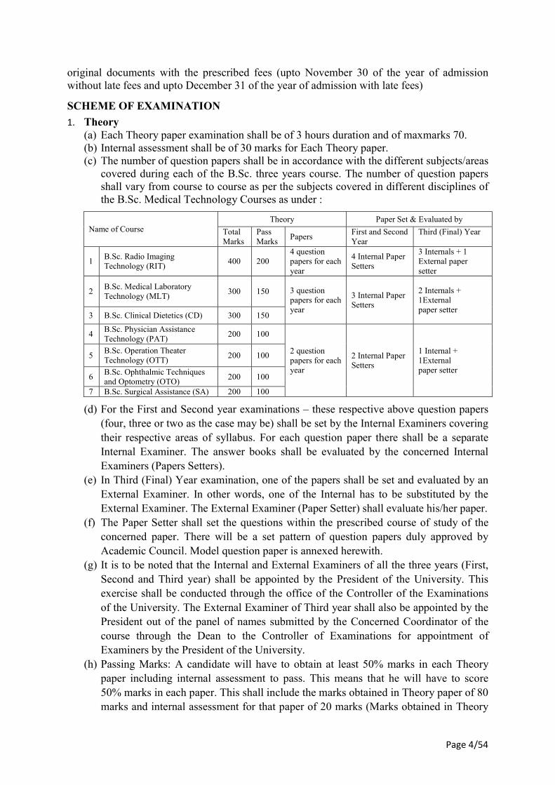
SCHEME OF EXAMINATION
1. Theory (a) Each Theory paper examination shall be of 3 hours duration and of maxmarks 70. (b) Internal assessment shall be of 30 marks for Each Theory paper. (c) The number of question papers shall be in accordance with the different subjects/areas
covered during each of the B.Sc. three years course. The number of question papers shall vary from course to course as per the subjects covered in different disciplines of the B.Sc. Medical Technology Courses as under :
Name of Course Theory Paper Set & Evaluated by
Total Marks
Pass Marks
Papers First and Second Year
Third (Final) Year
1 B.Sc. Radio Imaging Technology (RIT)
400 200 4 question papers for each year
4 Internal Paper Setters
3 Internals + 1 External paper setter
2 B.Sc. Medical Laboratory Technology (MLT)
300 150 3 question papers for each year
3 Internal Paper Setters
2 Internals + 1External paper setter
3 B.Sc. Clinical Dietetics (CD) 300 150
4 B.Sc. Physician Assistance Technology (PAT)
200 100
2 question papers for each year
2 Internal Paper Setters
1 Internal + 1External paper setter
5 B.Sc. Operation Theater Technology (OTT)
200 100
6 B.Sc. Ophthalmic Techniques and Optometry (OTO)
200 100
7 B.Sc. Surgical Assistance (SA) 200 100
(d) For the First and Second year examinations – these respective above question papers (four, three or two as the case may be) shall be set by the Internal Examiners covering their respective areas of syllabus. For each question paper there shall be a separate Internal Examiner. The answer books shall be evaluated by the concerned Internal Examiners (Papers Setters).
(e) In Third (Final) Year examination, one of the papers shall be set and evaluated by an External Examiner. In other words, one of the Internal has to be substituted by the External Examiner. The External Examiner (Paper Setter) shall evaluate his/her paper.
(f) The Paper Setter shall set the questions within the prescribed course of study of the concerned paper. There will be a set pattern of question papers duly approved by Academic Council. Model question paper is annexed herewith.
(g) It is to be noted that the Internal and External Examiners of all the three years (First, Second and Third year) shall be appointed by the President of the University. This exercise shall be conducted through the office of the Controller of the Examinations of the University. The External Examiner of Third year shall also be appointed by the President out of the panel of names submitted by the Concerned Coordinator of the course through the Dean to the Controller of Examinations for appointment of Examiners by the President of the University.
(h) Passing Marks: A candidate will have to obtain at least 50% marks in each Theory paper including internal assessment to pass. This means that he will have to score 50% marks in each paper. This shall include the marks obtained in Theory paper of 80 marks and internal assessment for that paper of 20 marks (Marks obtained in Theory
Page 5/54
paper + Marks obtained in internal assessment = the Total Marks obtained in respect of each paper).
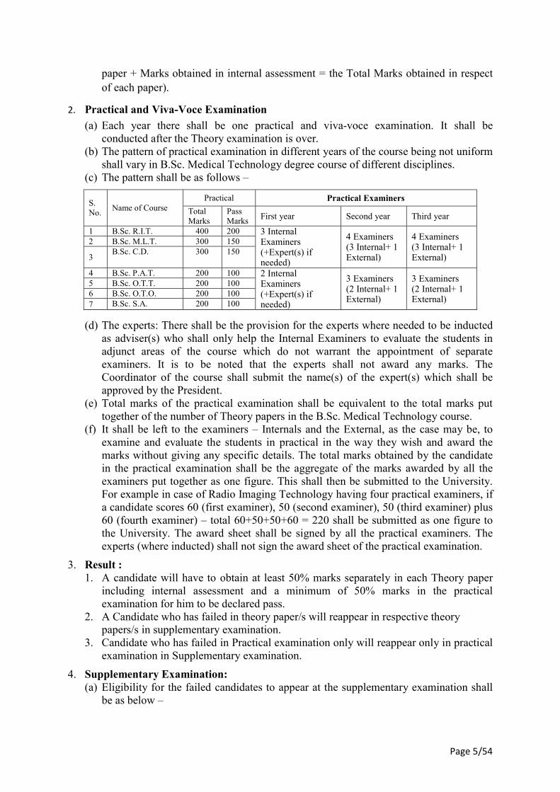
2. Practical and Viva-Voce Examination
(a) Each year there shall be one practical and viva-voce examination. It shall be conducted after the Theory examination is over.
(b) The pattern of practical examination in different years of the course being not uniform shall vary in B.Sc. Medical Technology degree course of different disciplines.
(c) The pattern shall be as follows –
S. No.
Name of Course Practical Practical Examiners
Total Marks
Pass Marks First year Second year Third year
1 B.Sc. R.I.T. 400 200 3 Internal Examiners (+Expert(s) if needed)
4 Examiners (3 Internal+ 1 External)
4 Examiners (3 Internal+ 1 External)
2 B.Sc. M.L.T. 300 150
3 B.Sc. C.D. 300 150
4 B.Sc. P.A.T. 200 100 2 Internal Examiners (+Expert(s) if needed)
3 Examiners (2 Internal+ 1 External)
3 Examiners (2 Internal+ 1 External)
5 B.Sc. O.T.T. 200 100 6 B.Sc. O.T.O. 200 100 7 B.Sc. S.A. 200 100
(d) The experts: There shall be the provision for the experts where needed to be inducted as adviser(s) who shall only help the Internal Examiners to evaluate the students in adjunct areas of the course which do not warrant the appointment of separate examiners. It is to be noted that the experts shall not award any marks. The Coordinator of the course shall submit the name(s) of the expert(s) which shall be approved by the President.
(e) Total marks of the practical examination shall be equivalent to the total marks put together of the number of Theory papers in the B.Sc. Medical Technology course.
(f) It shall be left to the examiners – Internals and the External, as the case may be, to examine and evaluate the students in practical in the way they wish and award the marks without giving any specific details. The total marks obtained by the candidate in the practical examination shall be the aggregate of the marks awarded by all the examiners put together as one figure. This shall then be submitted to the University. For example in case of Radio Imaging Technology having four practical examiners, if a candidate scores 60 (first examiner), 50 (second examiner), 50 (third examiner) plus 60 (fourth examiner) – total 60+50+50+60 = 220 shall be submitted as one figure to the University. The award sheet shall be signed by all the practical examiners. The experts (where inducted) shall not sign the award sheet of the practical examination.
3. Result : 1. A candidate will have to obtain at least 50% marks separately in each Theory paper
including internal assessment and a minimum of 50% marks in the practical examination for him to be declared pass.
2. A Candidate who has failed in theory paper/s will reappear in respective theory papers/s in supplementary examination.
3. Candidate who has failed in Practical examination only will reappear only in practical examination in Supplementary examination.
4. Supplementary Examination: (a) Eligibility for the failed candidates to appear at the supplementary examination shall
be as below –
Page 6/54
i. Failed in Theory Paper(s) and failed in Practical – shall reappear in the respective failed Theory paper(s) and Practical examination.
ii. Failed in Theory paper/papers and passed in Practical examination – shall reappear only in the concerned failed Theory paper(s).
iii. Passed Theory papers but failed in Practical – shall reappear only in the Practical Examination. (b) There shall be a supplementary examination within two months of the declaration of
the result of the main examination. Internal assessment marks obtained in main examination in the concerned failed paper/papers shall be carried forward for working out the result of supplementary Theory paper(s) examination. Such candidate who has secured less than 50% marks in the internal assessment will be allowed to improve his internal assessment marks in the repeat supplementary internal assessment examination.
(c) Marks secured by the candidate in passed main examination/supplementary examination Theory paper(s) and/or practicals, as the case may be, will be carried forward for working out his result.
(d) Result: i. A candidate obtaining at least 50% marks in the supplementary Theory paper(s)
and 50% marks in the supplementary practical examination, as the case may be, shall be declared successful.
ii. A candidate who has failed in supplementary theory paper(s) examination shall have to reappear only in the failed theory paper(s) at the subsequent examination.
iii. A candidate who has failed in supplementary practical examination shall have to reappear both in theory (all papers) and practical at the next main examination.
5. Promotion to Second/Third Year A candidate failed in theory paper(s) /Practical examination only shall be promoted to
next year. (b) A candidate will be allowed to appear for the Final (3rd)year examination only when
the backlog of all papers (theory and practical) of 1st and 2nd year Exams is cleared (c) The student is required to complete the course within 6 years from the joining of the
course
6. Result - Division: Successful candidates will be categorized as under-
1. Those, securing 50% and above but less than 60% in the aggregate marks of First, Second & Third year taken together
Pass
2 Those, securing 60% and above but less than 75% in the aggregate marks of First, Second & Third year taken together
Pass with I Division
3 Those, securing 75% and above in the aggregate marks of First, Second & Third year taken together
Pass with Honors
PAPER SETTER/EXAMINER
1. All the examiners, paper setters, theory examination answer books evaluators, Internal and External Examiners for Practical examinations shall be appointed by the President of the University.
Page 7/54
2. Qualification of the Paper setter / Examiner: Senior Demonstrator and above. 3. Paper setter can be an examiner REVALUATION / SCRUTINY Re-evaluation of answer book(s) of the B. Sc. Medical Technology courses may be permissible in not more than 25% of the theory papers within 15 days from the date of declaration of examination result on submission of his/her application on the prescribed form along with the requisite fees. Such answer book(s) shall be re-evaluated as per University rules. Reevaluation of answer book(s) shall not be permitted for second attempt in any paper. Scrutiny (re-totaling) of answer book(s) of the B. Sc. Medical Technology courses may be permissible within 15 days from the date of declaration of examination result on submission of his/her application on the prescribed form along with the requisite fees as per University Rules. GRACE MARKS 1. A student who appears in the whole examination in first attempt and obtains the required
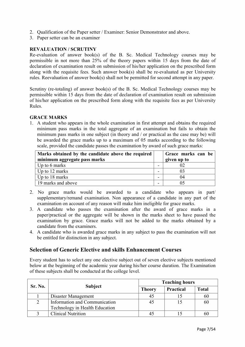
minimum pass marks in the total aggregate of an examination but fails to obtain the minimum pass marks in one subject (in theory and / or practical as the case may be) will be awarded the grace marks up to a maximum of 05 marks according to the following scale, provided the candidate passes the examination by award of such grace marks:
Marks obtained by the candidate above the required minimum aggregate pass marks
Grace marks can be given up to
Up to 6 marks - 02 Up to 12 marks - 03 Up to 18 marks - 04 19 marks and above - 05
2. No grace marks would be awarded to a candidate who appears in part/ supplementary/remand examination. Non appearance of a candidate in any part of the examination on account of any reason will make him ineligible for grace marks.
3. A candidate who passes the examination after the award of grace marks in a paper/practical or the aggregate will be shown in the marks sheet to have passed the examination by grace. Grace marks will not be added to the marks obtained by a candidate from the examiners.
4. A candidate who is awarded grace marks in any subject to pass the examination will not be entitled for distinction in any subject.
Selection of Generic Elective and skills Enhancement Courses
Every student has to select any one elective subject out of seven elective subjects mentioned below at the beginning of the academic year during his/her course duration. The Examination of these subjects shall be conducted at the college level.
Sr. No. Subject Teaching hours
Theory Practical Total
1 Disaster Management 45 15 60 2 Information and Communication
Technology in Health Education 45 15 60
3 Clinical Nutrition 45 15 60
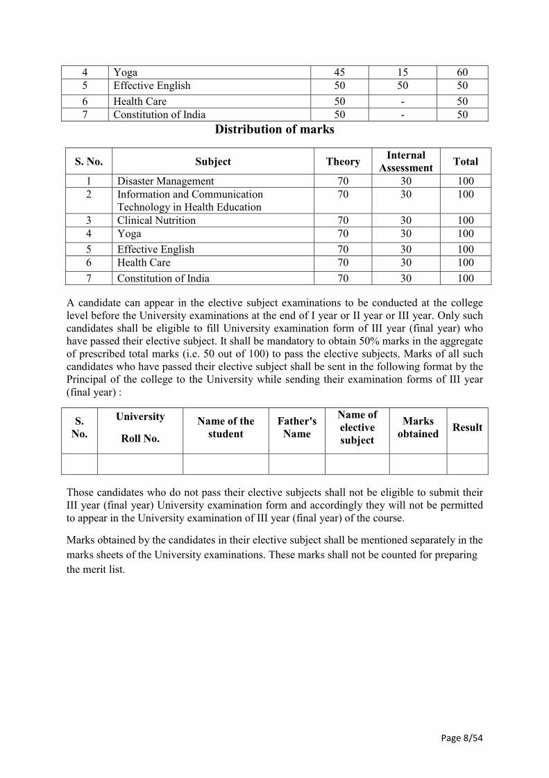
Page 8/54
4 Yoga 45 15 60 5 Effective English 50 50 50
6 Health Care 50 - 50 7 Constitution of India 50 - 50
Distribution of marks
S. No. Subject Theory Internal
Assessment Total
1 Disaster Management 70 30 100 2 Information and Communication
Technology in Health Education 70 30 100
3 Clinical Nutrition 70 30 100 4 Yoga 70 30 100
5 Effective English 70 30 100 6 Health Care 70 30 100
7 Constitution of India 70 30 100
A candidate can appear in the elective subject examinations to be conducted at the college level before the University examinations at the end of I year or II year or III year. Only such candidates shall be eligible to fill University examination form of III year (final year) who have passed their elective subject. It shall be mandatory to obtain 50% marks in the aggregate of prescribed total marks (i.e. 50 out of 100) to pass the elective subjects. Marks of all such candidates who have passed their elective subject shall be sent in the following format by the Principal of the college to the University while sending their examination forms of III year (final year) :
S. No.
University
Roll No.
Name of the student
Father's Name
Name of elective subject
Marks obtained
Result
Those candidates who do not pass their elective subjects shall not be eligible to submit their III year (final year) University examination form and accordingly they will not be permitted to appear in the University examination of III year (final year) of the course.
Marks obtained by the candidates in their elective subject shall be mentioned separately in the marks sheets of the University examinations. These marks shall not be counted for preparing the merit list.
Page 9/54
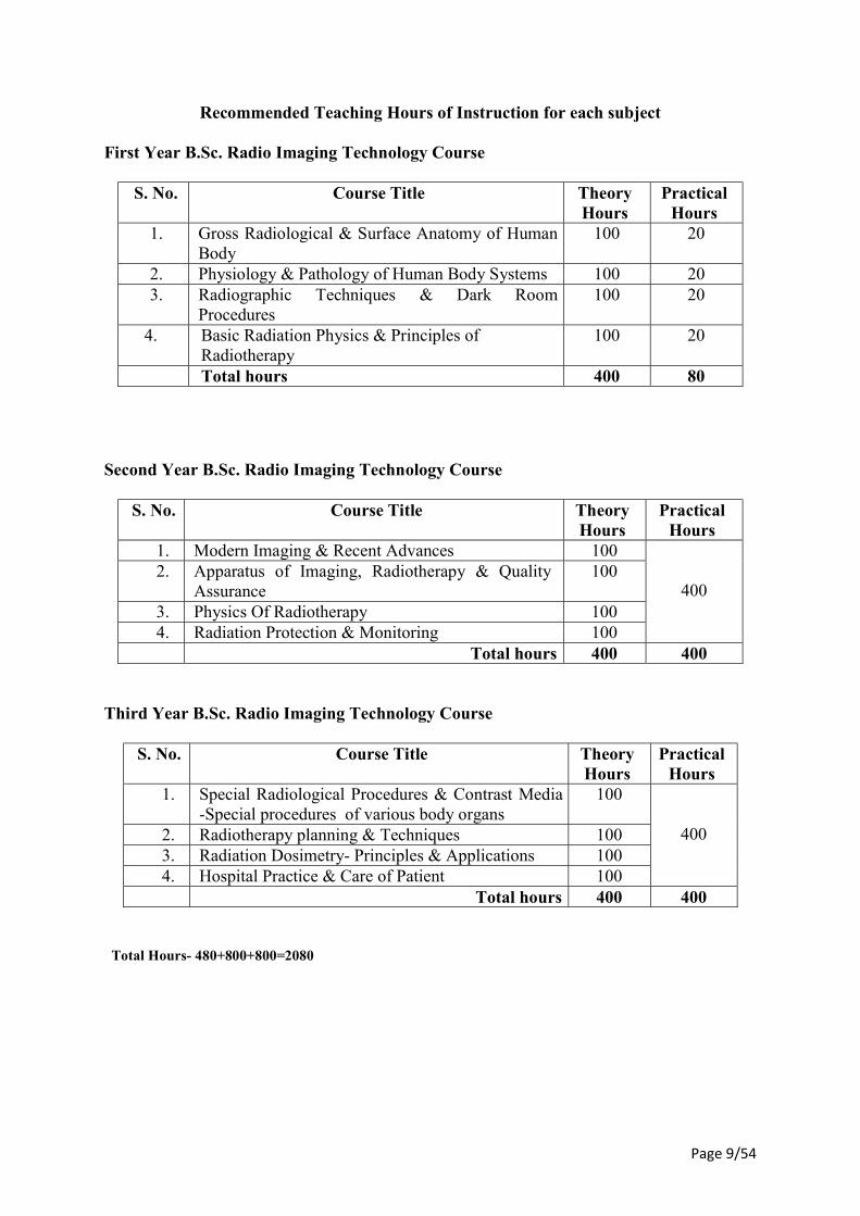
Recommended Teaching Hours of Instruction for each subject First Year B.Sc. Radio Imaging Technology Course
S. No. Course Title Theory Hours
Practical Hours
1. Gross Radiological & Surface Anatomy of Human Body
100 20
2. Physiology & Pathology of Human Body Systems 100 20 3. Radiographic Techniques & Dark Room
Procedures 100 20
4. Basic Radiation Physics & Principles of Radiotherapy
100 20
Total hours 400 80
Second Year B.Sc. Radio Imaging Technology Course
S. No. Course Title Theory Hours
Practical Hours
1. Modern Imaging & Recent Advances 100
400 2. Apparatus of Imaging, Radiotherapy & Quality
Assurance 100
3. Physics Of Radiotherapy 100 4. Radiation Protection & Monitoring 100
Total hours 400 400
Third Year B.Sc. Radio Imaging Technology Course
S. No. Course Title Theory Hours
Practical Hours
1. Special Radiological Procedures & Contrast Media -Special procedures of various body organs
100
400 2. Radiotherapy planning & Techniques 100 3. Radiation Dosimetry- Principles & Applications 100 4. Hospital Practice & Care of Patient 100
Total hours 400 400
Total Hours- 480+800+800=2080
Page 10/54
Marks Distribution
First Year B.Sc. Radio Imaging Technology
Code No
Subject Written Practical Theory I.A.
Theory Total Theory
Practical + Oral
I.A. Practical
Total Practical
7261 Gross Radiological & Surface Anatomy of Human Body
70 30 100
7262 Physiology & Pathology of Human Body Systems
70 30 100
7263 Radiographic Techniques & Dark Room Procedures
70 30 100
7264
Basic Radiation Physics & Principles of Radiotherapy
70 30 100
7265 Practical - - - 280 120 400 Total 280 120 400 280 120 400
Second Year B.Sc. Radio Imaging Technology
Code No
Subject Written Practical Theory I.A.
Theory Total Theory
Practical + Oral
I.A. Practical
Total Practical
7266 Modern Imaging & Recent Advances
70 30 100
7267 Apparatus of Imaging, Radiotherapy & Quality Assurance
70 30 100
7268 Physics of Radiotherapy 70 30 100 7269 Radiation Protection &
Monitoring 70 30 100
7270
Practical - - - 280 120 400
Total 280 120 400 280 120 400
Page 11/54
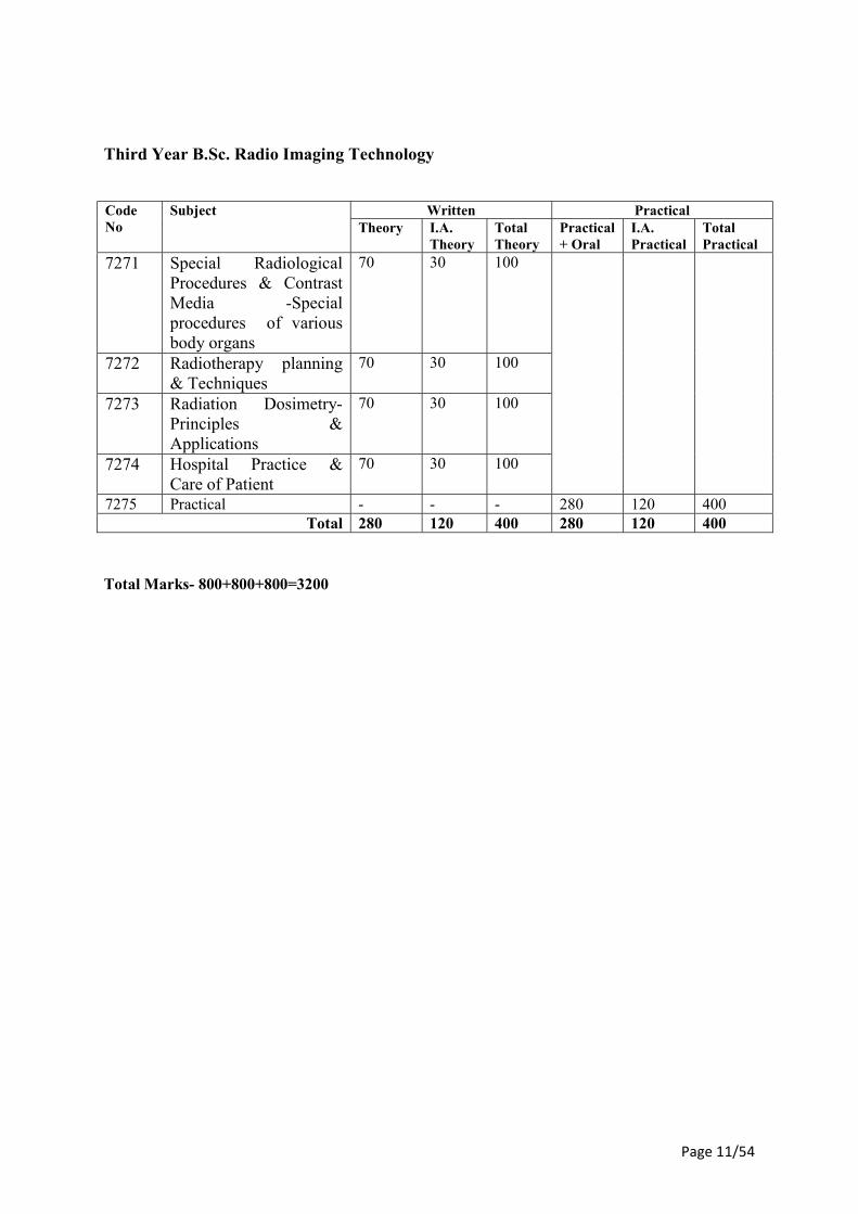
Third Year B.Sc. Radio Imaging Technology
Code No
Subject Written Practical Theory I.A.
Theory Total Theory
Practical + Oral
I.A. Practical
Total Practical
7271 Special Radiological Procedures & Contrast Media -Special procedures of various body organs
70 30 100
7272 Radiotherapy planning & Techniques
70 30 100
7273 Radiation Dosimetry- Principles & Applications
70 30 100
7274 Hospital Practice & Care of Patient
70 30 100
7275 Practical - - - 280 120 400 Total 280 120 400 280 120 400
Total Marks- 800+800+800=3200
Page 12/54
First Year B.Sc. Radio Imaging Technology
(1 Year Duration)
Paper-I
Gross Radiological & Surface Anatomy of Human Body
Theory Hours: 100 Total: 100
Introduction to Anatomy as a whole, Skeleton-bones & joints, formation of bones, structure of bones, classification of bones according to shape, Developmental classification, Regional classification, structural classification & growth of skeleton. Centre of ossification, type of bone, type of joints. Gross structure of human long bone, parts of young bone. Medico-legal & anthropological aspects of skeletal system, Estimation of age, sex, stature (height) and race. Classification & characters of joints, structural, functional & regional. Applied anatomy of joints, dislocation of joints. embryology, cell division, fertilization, development of embryo, gamete formation, menstrual cycle, formation of germ layers, development of embryonic disc, Placenta, formation of tissues, organs & systems of human body, congenital malformations.
Page 13/54
Paper II
Physiology & Pathology of Human Body Systems
Theory Hours: 100 Total: 100
The Respiratory System (1) Position and structure, Nose and nasal cavities, Functions: respiratory, Olfactory,
Pharynx, Larynx-Functions: respiratory, vocal Trachea, Bronchi, lungs: lobes lobules, pleura:
(a) Types of cells, tissues, bones and joints. Introduction to system and cavities of the body.
(b) Heart and blood vessels (Circulatory system) Blood vessels: arteries, veins, capillaries, sinusoids, structure and functions. Heart: Position, structure and functions. Circulation of blood: Pulmonary, systemic, portal, main blood vessels, their origins
and distribution. Blood: Composition & functions. Anaemia, Leukaemia, Thrombocytopenias. The lymphatic system (1) Parts of the lymphatic system. Lymph channels: Capillaries, vessels ducts structure and
functions. (2) Lymph nodes: position structure and functions. (3) Lymphatic tissues: tonsils, adenoids, intestinal nodules (4) Spleen: position, structure and functions, diseases and conditions of the system. The digestive system (1) Alimentary tract structure: (a) Mouth, pharynx, salivary glands, esophagus, stomach, liver, gall bladder, small
intestine, large intestine: Position, structure and functions of these organs. The Urinary System (1) Parts of urinary system. Position, structure and functions. (2) Kidneys, ureters, urinary bladder and urethra. Formation and composition of urine. Water
and electrolyte balance. The reproductive system (1) The Female reproductive system (a) External genitalia: position, structures and functions. Perineum. Internal organs:
positions and structures. Vagina, uterus, Fallopian tubes, ovaries. Breasts (Mammary glands)
(2) The Male reproductive system (a) Scrotum, testis, epididymis: position, structure and functions. Spermatic cords,
seminal vesicles, Ejaculatory ducts: position, structure and functions. (b) Prostate gland: Position Urethra and penis: position, structure and functions The Endocrine system (1) Endocrine glands:
Page 14/54
(a) Pituitary and hypothalamus: Position and structure Thyroid gland, parathyroid glands Adrenal (Supra renal) glands, Pancreas: Position, types of cells
The organs of sense (1) Hearing and the ear External, middle and inner ear. Physiology of hearing and diseases of
ear. (2) Sight and the eye: Position, structure, sclera, cornea, choroids, ciliary body, Iris, lens,
retina, optic nerves. (3) Sense of smell Olfactory nerves, origins, distribution. (4) Sense of taste. The Nervous system (1) Neurons: Structure, types and properties. (2) Central nervous system: Neurons, neurolgia meninges. Ventricles of brain, C.S.F. Brain,
spinal cord: Structures, functions peripheral nervous system. (3) Spinal and cranial nerves: origin distribution and functions. (4) Automatic nervous system: Sympathetic and Para-sympathetic: origin distribution and
functions. The Skin (1) Structure of skin Epidermis, dermis Functions of skin (2) Cell injury, immune system-components & disease, oedema& its types, (3) Haemodynamic disorders, imbalance of electrolytes, Hyperaemia, congestion,
Haemorrhage, Thrombosis, Embolism, Ischaemia. Infarction, Inflammation causes, types of inflammation, pathogenesis & inflammatory cells of inflammation, sepsis, asepsis, abnormalities of tissue ulceration.
Page 15/54
Paper III
Radiographic Techniques & Dark Room Procedures
Theory Hours: 100 Total: 100
Radiographic Techniques of whole body (1) Individual bones of skeletal system of human body. Special projection whenever
required and indicated as in skull & neck including petrous, temporal, mastoids, nasal sinuses, foramina and mandible, TM joint, open mouth & close mouth, optic foramina, sellaturcica, internal auditory canal, sphenoid bone, soft tissue neck, nasopharynx, larynx, teeth intra-oral and extra-oral projections, occlusal view.
(2) Chest & Thorax Bones: Chest-PA, lordotic view (Apicogarm), oblique lateral, thoracic inlet view, decubitus view
(3) Abdomen: general preparation of patient, plain film exam, Flat plate Abdomen, Upright and KUB X Ray.
(4) Upper limb: fingers, hands, carpal-tunnel view, wrist-projections, Projections for scaphoid, forearm, elbow, humerus, shoulder joints, acromio-clavicular joint, sterno-clavicular joint, clavicle & scapula.
(5) Lower limb: toes, feet, calcaneum, ankle joint, leg bones, different views of knee patella, inter condyler notch, and femur.
(6) Vertebral Column: Atlanto occipital joint, odontoid, cervical spine, cervico-thoracic spine, dorsal spine, thoraco lumbar spine, lumbosacral spine, sacrum, coccyx, scoliosis, kyphosis, flexion extension, and both oblique views of spines.
(7) Hips & Pelvis: Pelvis with both hip joints in different positions, internal and external rotation, frog position, SI Joint.
(8) Ward mobile radiography: electrical supply, radiation protection, instruction to be followed for portable radiography.
(9) Operation Theatre technique: General precautions. Asepsis in techniques. Exposure risks, radiation protection.
(10) Others: Dental radiography, macro & micro radiography, Cine radiography, localization of foreign body, battery operated units (conducer), mass miniature radiography, other emergency radiography.
Dark Room Procedures (1) The photographic process: Introduction, visible light, images produced by radiation,
light sensitive photographic materials. (2) Image characteristic: Real and mental images, reflected, transmitted and emitted light
images Photographic emulsions. The photographic latent image. Positive process (3) Construction of x-ray film & its cross over effect. (4) Sensitometry: Photographic density, characteristic curves, (5) The storage of film materials and radiograph; (6) Intensifying screens and cassettes. Luminescence: fluorescence and phosphorescence.
Construction of an intensifying screen. (7) Fluorescent materials. Types of intensifying screens (8) types of cassettes (9) Film processing: Development. The nature of development-manual or automatic. The
PH scale.
Page 16/54
(10) The constitution of developing solutions both in manual and automatic processing and properties of developing chemicals.
(11) Film processing: Fixing and role of a fixing solution. Constitution of the fixing solutions and properties of the constituents. Factors affecting the quality of fixer.
(12) Development procedure, laser & bright procedure. (13) Processing equipment: Materials for processing equipment, processors for manual
operation, hangers, control of chemicals temperature by heating and thermostat, immersion heaters as well as cooling methods.
(14) Dark Room: Layout and planning. (15) Type of entry, door design. Dark room illuminations - white light and safe lighting
Page 17/54
Paper IV
Basic Radiation Physics & Principles of Radiotherapy
Theory Hours: 100 Total: 100
(1) SI Units, Force, mass, momentum, work, energy, power, density, pressure, heat, sound, wave and oscillations. Atomic structure: Atom, nucleus, Bohr theory of hydrogen atom, atomic mass and energy units, distribution of orbital electrons atomic energy levels, nuclear forces, nuclear energy levels, particle radiations, electromagnetic radiations, electricity and magnetism. Nuclear Transformations: Radioactivity, decay constant, activity half life, mean life, radioactive series, radioactive equilibrium, modes of decay: Alpha decay, Beta decay, electron capture, internal conversion & isomeric transition.
(2) Nuclear reactions: (α, ρ reaction, (α, n reaction), proton bombardment, deuteron bombardment, neutron bombardment, photodisintegration, fission, fusion, activation of nuclides, nuclear reactors Interaction of radiation with matter: ionization and excitation, various types of interaction processes (photoelectric effect, Compton scattering, pair production etc.) Interaction of charged particles and neutrons with matter. Comparative beam characteristics.
(3) Production of X-rays: X-ray tube, anode, cathode construction and working principles of transformers and autotransformers used in x-ray circuits, voltage rectification and measurements in x-ray circuits. Physics of x-ray production (Bremsstrahlung and Characteristic x-rays). Properties of matter, heat, light, magnetism, electricity and electromagnetism. Principles and working of x-ray tube. Measuring instruments voltage or KV meters. Measurement of tube current Principles of thermionic emission and rectification in x-ray technology. High voltage circuits in x-ray Units. Electrical hazards and safety. Tube rating in imaging and therapy xray tube and thermal safety. Intensity of radiation and its variation with distance, KV, MA. Introduction to electro-magnetic spectrum, definition of wave length and its quantum relationship with peak kilovoltage. Physical principles of radiation. Exponential and trignometric functions used in radiological calculations.
(4) Introduction to: Malignant and non-malignant tumours treated by radiotherapy, Radioactivity and ionizing radiations used in treatment of malignancy, sources and techniques, Tissue tolerance, tumour lethal dose, therapeutic ratio and radiosensitivity, Units of exposure and radiation, prescription of radiation treatment, Radiation reactions and normal tissue tolerance.
(5) Definitions and basics of teletherapy techniques: (a) Orthovoltage and megavoltage machines. (b) Teletherapy machines – cobalt and linear accelerator. (c) Basic principles and clinical applications of beam direction and modification devices. (d) Clinical application of mould room techniques (6) Principles of basic radiobiology: (a) Cell cycle. (b) Cell survival curve. (c) LET, RBE and OER. (d) Time dose and fractionation. (e) Acute and chronic radiation effects.
Page 18/54
(7) Basics of Brachy therapy: (a) Definition and basic principles. (b) Radium and its substitutes used. (c) Interstitial implantation. (d) Intracavitary and intraluminal brachytherapy. (e) Surface Moulds. (8) Demonstration (a) Cobalt machine – parts and functioning. (b) Linear accelerator – parts and functioning. (c) Beam direction devices. (d) Beam modification devices. (e) Lead bench and radium safe. (f) Mould room techniques and cast making. (g) Interstitial implantation – HDR microselectron. (h) Intracavitary application – MDR & HDR selectron machine. (i) Surface mould. (j) Radiation safety devices.
Page 19/54
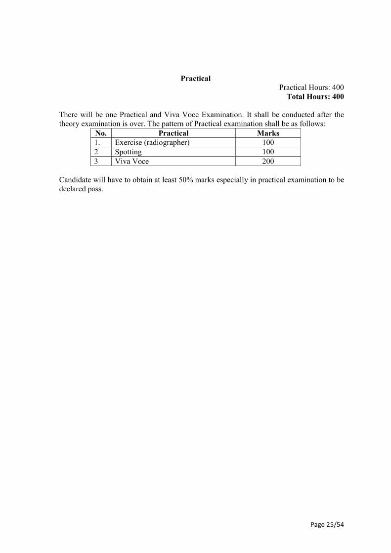
Practical
Practical Hours: 400 Total Hours: 400
There will be one Practical and Viva Voce Examination. It shall be conducted after the
theory examination is over. The pattern of Practical examination shall be as follows: No. Practical Marks
1. Exercise (radiographer) 100 2 Spotting 100 3 Viva Voce 200
Candidate will have to obtain at least 50% marks especially in practical examination to be
declared pass.
Page 20/54
Second Year B.Sc. Radio Imaging Technology
(1 Year Duration)
Paper-I
Modern Imaging & Recent Advances Theory Hours: 100
Total: 100
(1) Recent advances in imaging technology: Detailed knowledge of ultrasound, colour Doppler, different types of transducers, their principles, applications & role in medicine & cross sectional anatomy.
(2) CT scan, conventional, spiral (helical), Multislice: Historical development, its principle and applications, various generations & definition of terms and cross sectional anatomy& use of diagnostic methods.
(3) Magnetic Resonance Imaging (MRI): Principle, application, its advantage over computed tomography or ultra-sonography. Its limitations, uses & cross sectional anatomy.
(4) Spectroscopy: Principle, application and uses. (5) Computerised Radiography: Principle, application, advantage & technique. (6) Digital Radiography: Principle, scanned projection radiography, digital subtraction
angiography application, definition, advantages & techniques. (7) DSA: Uses, application, techniques & principle (8) Picture Archiving Communication System (PACS): Basic knowledge of PACS,
application, principle & image transmission. (9) Mammography: Principle, application, advantage in soft tissue radiography, physics,
filtration, QA & QC. (10) Orthopantamogram: Application, principle technique and uses & advantages. (11) Positron Emission Tomography (PET): Basic principle, clinical application &
advantages. (12) Different types of cameras e.g. laser, photography: principle, processing &
applications. (13) Radio isotopes: Principles of Scanner, Rectilinear scanner, gamma camera. (14) DEXA: Principles, applications and instrumentation. (15) Fundamentals: Applications of computers in Radiology.
Page 21/54
Paper II
Apparatus of Imaging, Radiotherapy & Quality Assurance
Theory Hours: 100 Total: 100
Electrical system and Mains supply : The electrical system, generation of electricity, distribution of electric energy, use of electric energy. High Tension Generators - Rectifications - Types of rectifier – valve and solid state. Self rectified high tension circuit. Half wave, four valve full wave, three phase, full wave rectified circuit, voltage wave forms in high tension generators. Constant potential circuit programmed generators and modular generators The X-ray Tube; Historical developments including General features of the X-ray tube. The fixed anode, rotating anode x-ray tube. Rating of X-ray tubes, focal spot sizes. Methods of heat dissipation in x-ray tubes, common tube faults. Developments in the rotating anode tube. Tube stands and ceiling tube supports. Mammography tubes and equipment, accessories. Different types of tubes and choice of an xray tube. Components and controls in the X-ray circuits: The high tension transformer, the rectification of high tension. The control of kilovoltage (KV), kilovoltage indication. The filament circuit and control of tube current. Milliamperes (MA) indications. Main voltage compensation. Mains supply and the x-ray set. Exposure Switches and Exposure Timers: Switching systems timing system, exposure switching and its radiographic applications The control of scattered Radiation: Significance of scatter. Beam limiting devices-cones, diaphragm (collimeters). Beam centring devices. Grid: its types, components of grid, grid movements. The assessment of grid functions, grid-errors, other scatter reduction methods - air gap technique. Portable and mobile X-ray Units. Fluoroscopic Equipment: Structure of a fluorescent screen. The fluoroscopic image. The fluoroscopic table, spot film devices and explorators. Protective measures and physiology of vision, image quality. Image Intensifiers (I.I.T.V. system): An Image intensifier tube, its design, its application. angiographic tables, contrast medium injection device. Linear accelerator: Block diagram and design of LINAC, power supply, modulator, electron gun, magnetron/kylstron, wave guide system, accelerator tube (traveling wave and standing wave type), flattening filters, scattering foil, complete QA procedures of the LINAC. Heavy charged particle beam generators. Simulator: Design, construction types and uses of Simulator in Radiotherapy. Quality Assurance Tests for Simulator Quality assurance in medical care and Radiodiagnosis: (1) Aim, Regulations and Accreditation. Purchasing Equipment, Identification of imaging
requirements, Developments of equipment specifications, Selection of equipment,
Page 22/54
(2) Installations & Acceptance testing of equipment, Continuing education, Monitoring Equipment Performance, Routine checks of all radiological and imaging equipment including CT & MRI, Routine checks of film processing systems, Processor Monitoring, External beam evaluation, Routine checks of Diagnostic radiographic system like - focal spot size determination, half value layer, collimator, check, central ray and Bucky tray accuracy, Distance and centering indicators accuracy, Angulator or protector accuracy, KV accuracy, MA accuracy, exposure timer accuracy, resolution, exposure reproducibility.
(3) QA tests and procedures in manual brachytherapy. (4) QA tests and procedures of HDR and PDR units. (5) QA tests and procedures of TPS and Gamma knife and X knife units.
Page 23/54
Paper III
Physics of Radiotherapy
Theory Hours: 100 Total: 100
Radiation Units: Activity, Becquerel (Bq), exposure, Roentgen, absorbed dose, rad, Gray, dose-equivalent, rem, Sievert, KERMA. Relation between absorbed dose, exposure and KERMA. Calculation of absorbed dosenfrom exposure, Absorbed dose to air, Absorbed dose to any medium, Bragg-Gray theory. Stopping power. Transfer of absorbed dose from one medium to another of photons, electrons. Exposure from radioactive sources, exposure rate constant. Dose distribution and scattering in medium: Properties of phantom materials and various types of phantoms, depth dose distribution, dose build-up, percentage depth dose and its influencing factors. Back scatter factor, tissue air- ratio and influencing factors. Relation between TAR and PDD. Scatter-air-ratio. Dose calculation of irregular fields using Clarkson’s method. Dosimetric calculations: Dose calculation parameters, collimator scatter factor (Sc), phantom scatter factor (Sp), Tissue phantom ratio (TPR), tissue maximum ratio (TMR), and their influencing factors. Relationship between TMR and PDD. Scatter maximum ratio (SMR). Dose calculations for linear accelerator and Co-60 unit using Sc, Sp factors for SSD and SAD methods, irregular fields, asymmetric fields etc. Isodose distribution of phantom beam: Isodosecharts, measurement of isodose curves, parameters of isodose curves: beam quality, source size, SSD and SDD – penumbra effect, collimation and flattening filter, field size, Wedge filters: wedge angle, wedge transmission factor, wedge systems, effect of beam quality, design of wedge filters. Bolus, tissue compensators, shielding blocks. Electron beam therapy: Electron interactions, rate of energy less, collisional losses (ionization and excitation) radiation losses (bremsstrahlung), polarization, stopping power, absorbed dose, electron scattering, most probable energy, mean energy, energy at depth. Determination of absorbed dose, output calibration, phantom, reference depth and field size, absorbed dose calculation, depth dose distribution, central axis depth dose curves, isodose curves for different electron energies. Field flatness and symmetry, beam collimation, field size dependence, electron source, x-ray contamination.
Page 24/54
Paper IV
Radiation Protection & Monitoring
Theory Hours: 100 Total: 100
Radiation protection quantities and units: Effective dose equivalent (HE), Equivalent dose ((HTR), effective dose (E). Sources of radiation exposure: Natural sources and human made sources. Standards and regulations, philosophies of exposure limit, occupational limits, non-occupational limits. Biological effects of radiation: (1) Direct and indirect action of radiation, cell cycle effect, somatic and genetic effects. (2) Effects on tissues and organs: Stochastic and nonstochastic (deterministic) effects, acute
effects, late effects (3) Effects of radiation on Embryo & fetus: lethal effects, organ malformation, growth
impairment, mental retardation, cancer induction, genetic effects, (4) Late (delayed) effects: cataract formation, organ function, cancer induction. Personal dosimetry devices: (1) Film badges, TLD badges, pocket ion chambers, electronic devices, Cr-39 foils, bubble,
counting statistics, distributions, standard deviation. Standard error, confidence internal. (2) Detection and measurement of Ionizing/radiation: Field survey instrument, GM survey
instruments, personnel Monitoring devices, film badge, TLD, pocket dosimeter, pulsed optically stimulated Luminescence dosimeter (POSL) etc. Radiation Protection Procedures for Patients and Personnel Advisory Groups & Regulatory Agencies - ICRP, NCRP, UNSCEAR, AERB. Limiting exposure to ionizing radiation - Dose limits, ICRP recommendations ALARA principle. Protection of Personnel, Principles of personnel exposure reduction - Time, distance, shielding, protective barriers, protective devices. Protection of the patient, Beam limitation, technique selection, general shielding, grids, image receptors, projection, repeat radiography etc. Radiation exposure and pregnancy - ALARA and Pregnancy, the pregnant radiation worker, patient and radiation exposure standards
Page 25/54
Practical Practical Hours: 400
Total Hours: 400
There will be one Practical and Viva Voce Examination. It shall be conducted after the theory examination is over. The pattern of Practical examination shall be as follows:
No. Practical Marks 1. Exercise (radiographer) 100 2 Spotting 100 3 Viva Voce 200
Candidate will have to obtain at least 50% marks especially in practical examination to be
declared pass.
Page 26/54
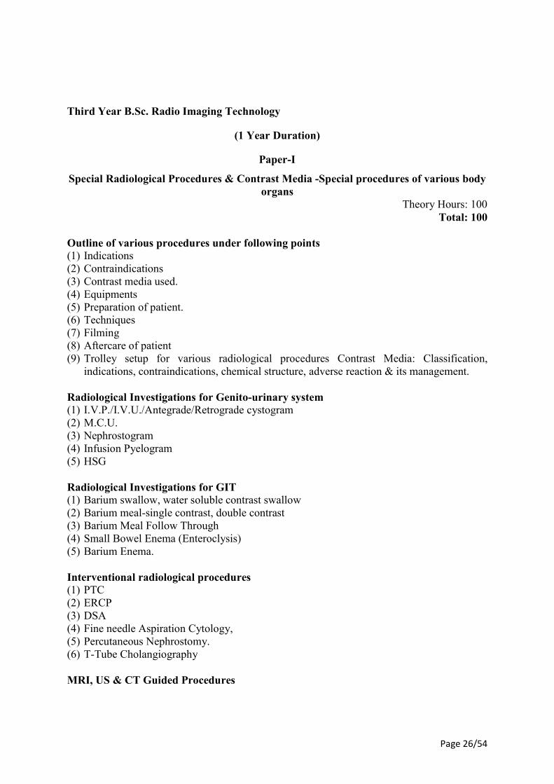
Third Year B.Sc. Radio Imaging Technology
(1 Year Duration)
Paper-I
Special Radiological Procedures & Contrast Media -Special procedures of various body organs
Theory Hours: 100 Total: 100
Outline of various procedures under following points (1) Indications (2) Contraindications (3) Contrast media used. (4) Equipments (5) Preparation of patient. (6) Techniques (7) Filming (8) Aftercare of patient (9) Trolley setup for various radiological procedures Contrast Media: Classification,
indications, contraindications, chemical structure, adverse reaction & its management. Radiological Investigations for Genito-urinary system (1) I.V.P./I.V.U./Antegrade/Retrograde cystogram (2) M.C.U. (3) Nephrostogram (4) Infusion Pyelogram (5) HSG Radiological Investigations for GIT (1) Barium swallow, water soluble contrast swallow (2) Barium meal-single contrast, double contrast (3) Barium Meal Follow Through (4) Small Bowel Enema (Enteroclysis) (5) Barium Enema. Interventional radiological procedures (1) PTC (2) ERCP (3) DSA (4) Fine needle Aspiration Cytology, (5) Percutaneous Nephrostomy. (6) T-Tube Cholangiography MRI, US & CT Guided Procedures
Page 27/54
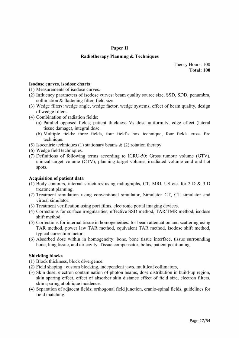
Paper II
Radiotherapy Planning & Techniques
Theory Hours: 100 Total: 100
Isodose curves, isodose charts (1) Measurements of isodose curves. (2) Influency parameters of isodose curves: beam quality source size, SSD, SDD, penumbra,
collimation & flattening filter, field size. (3) Wedge filters: wedge angle, wedge factor, wedge systems, effect of beam quality, design
of wedge filters. (4) Combination of radiation fields: (a) Parallel opposed fields; patient thickness Vs dose uniformity, edge effect (lateral
tissue damage), integral dose. (b) Multiple fields: three fields, four field’s box technique, four fields cross fire
technique. (5) Isocentric techniques (1) stationary beams & (2) rotation therapy. (6) Wedge field techniques. (7) Definitions of following terms according to ICRU-50: Gross tumour volume (GTV),
clinical target volume (CTV), planning target volume, irradiated volume cold and hot spots.
Acquisition of patient data (1) Body contours, internal structures using radiographs, CT, MRI, US etc. for 2-D & 3-D
treatment planning. (2) Treatment simulation using conventional simulator, Simulator CT, CT simulator and
virtual simulator. (3) Treatment verification using port films, electronic portal imaging devices. (4) Corrections for surface irregularities; effective SSD method, TAR/TMR method, isodose
shift method. (5) Corrections for internal tissue in homogeneities: for beam attenuation and scattering using
TAR method, power law TAR method, equivalent TAR method, isodose shift method, typical correction factor.
(6) Absorbed dose within in homogeneity: bone, bone tissue interface, tissue surrounding bone, lung tissue, and air cavity. Tissue compensator, bolus, patient positioning.
Shielding blocks (1) Block thickness, block divergence. (2) Field shaping : custom blocking, independent jaws, multileaf collimators, (3) Skin dose; electron contamination of photon beams, dose distribution in build-up region,
skin sparing effect, effect of absorber skin distance effect of field size, electron filters, skin sparing at oblique incidence.
(4) Separation of adjacent fields; orthogonal field junction, cranio-spinal fields, guidelines for field matching.
Page 28/54
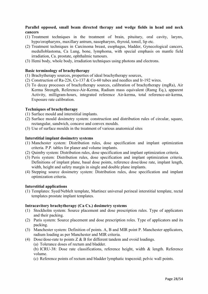
Parallel opposed, small beam directed therapy and wedge fields in head and neck cancers (1) Treatment techniques in the treatment of brain, pituitary, oral cavity, larynx,
hypo/oropharynx, maxillary antrum, nasopharynx, thyroid, tonsil, lip etc. (2) Treatment techniques in Carcinoma breast, esophagus, bladder, Gynecological cancers,
medulloblastoma, Ca Lung, bone, lymphoma, with special emphasis on mantle field irradiation, Ca. prostate, ophthalmic tumours.
(3) Hemi body, whole body, irradiation techniques using photons and electrons. Basic terminology of brachytherapy (1) Brachytherapy sources, properties of ideal brachytherapy sources, (2) Construction of Ra-226, Cs-137 & Co-60 tubes and needles and Ir-192 wires. (3) To decay processes of brachytherapy sources, calibration of brachytherapy (mgRa), Air
Kerma Strength, Reference-Air-Kerma, Radium mass equivalent (Ramg Eq.), apparent Activity, milligram-hours, integrated reference Air-kerma, total reference-air-kerma, Exposure rate calibration.
Techniques of brachytherapy (1) Surface mould and interstitial implants. (2) Surface mould dosimetry system: construction and distribution rules of circular, square,
rectangular, sandwich, concave and convex moulds. (3) Use of surface moulds in the treatment of various anatomical sites Interstitial implant dosimetry systems (1) Manchester system: Distribution rules, dose specification and implant optimization
criteria. P.P. tables for planer and volume implants. (2) Quimby system: Distribution rules, dose specification and implant optimization criteria. (3) Peris system: Distribution rules, dose specification and implant optimization criteria.
Definitions of implant plane, basal dose points, reference dose/dose rate, implant length, width, height and safety margin in single and double plane implants.
(4) Stepping source dosimetry system: Distribution rules, dose specification and implant optimization criteria.
Interstitial applications (1) Templates: Syed/Neblelt template, Martinez universal perineal interstitial template, rectal
templates prostate implant templates. Intracavitory brachytherapy (Ca Cx.) dosimetry systems (1) Stockholm system: Source placement and dose prescription rules. Type of applicators
and their packing. (2) Paris system: Source placement and dose prescription roles. Type of applicators and its
packing. (3) Manchester system: Definition of points. A, B and MIR point P. Manchester applicators,
radium loading as per Manchester and MIR criteria. (4) Dose/dose-rate to points Z & B for different tandem and ovoid loadings. (a) Tolerance doses of rectum and bladder. (b) ICRU-38: Dose rate classifications, reference height, width & length. Reference
volume. (c) Reference points of rectum and bladder lymphatic trapezoid; pelvic wall points.
Page 29/54
(d) Concept of 60 Gy. Applicators of Ca Cx: Pre-loaded applicators (Stockholm, Paris etc.), Fletcher suit applicators.
(e) Henschke applicators, ring applicators, vaginal applicators. (f) Different tools, catheters and other necessary items required for interstitial implant. (5) Dose calculations for brachytherapy sources: Exposure rate constant, exposure rate and
effect of inverse square law, sievert integral to calculate exposure rate from a line source and point source, TG-43 dose calculations methods for brachytherapy sources. Dose calculations of surface mould, interstitial implants, intra-cavitary applications using orthogonal radiographs. 2-D and 3-D planning for LDR and HDR units using orthogonal radiographs, CT Scans.
(6) Gamma Knife, construction, design and working principles. QA procedures and different clinical applications of gamma knife. Dose prescription criteria in the treatment of gamma knife.
(7) X-knife, modification of LINAC, necessary accessories required for X-knife, energy choice of x-ray photons in X-knife, QA procedures and application and techniques in the treatment using circular cones and their planning.
(8) Cyber Knife: Principles and applications. (9) Principles and working of asymmetric jaws in radiotherapy. Techniques in which
asymmetric jaws are used. Uses of of asymmetric jaw. (10) Tomotherapy : Principles and applications. Design and working of MLC and MMLC. (11) QA procedures of MLC and MMLC. Conformal radiotherapy (CRT) and intensity
modulated radiotherapy (IMRT). (12) Use of MMLC in stereotactic radiotherapy and IMRT. (13) Inverse planning system. (14) Intra- operative Radiotherapy (IORT). (15) Uses of PDR unit in brachytherapy. (16) Radiobiological explanation of PDR treatment techniques. Advantage and disadvantage
of PDR brachytherapy. (17) QA procedures.
Page 30/54
Paper III
Radiation Dosimetry-Principle & Applications
Theory Hours: 100 Total: 100
Principles of gas field detectors (1) Characteristic curve of gas filled detectors. Regions of the characteristic curve: ionization
region, proportional region, GM region. (2) Construction of gas filled detectors and their working. (3) GM counters resolving time, true count rate. Scintillation counters, semiconductor
detectors, alpha particle monitoring, gamma & x-ray monitoring, neutron monitoring devices.
Measurement of ionizing radiation (1) Exposure, roentzen, free air ionization chamber, thimble chambers, chamber wall,
effective atomic number, chamber calibration: (a) Condenser chambers, chamber sensitivity, stem effect, farmer chamber electrometers:
string electrometer, other electrometers special chambers. 1. Extrapolation chamber (b) Parallel plate chambers. Ion collection, chamber polarity effect. Environmental
conditions, measurement of exposure. (2) Principle of Bragg-Gray theory. Stopping power, chamber volume. Effective
measurement points. (a) plane parallel chambers (b) cylindrical chambers. (3) Construction and working of plane parallel and cylindrical chambers and their use in
dosimetry of photon and electron beam calibration. (4) Calibration protocols for megavoltage beams. Cavity gas calibration factor, Bragg-Gray
cavity for photon beams and electron beams. Dose calibration parameters. Transfer of absorbed dose from one medium to another.
(5) Measurement of absorbed dose using calorimetry, chemical dosimetry solid state methods; TLD film dosimetry.
Page 31/54
Paper IV
Hospital Practice & Care of Patient
Theory Hours: 100 Total: 100
(1) Hospital staffing and administration, records, professional ethics, cooperation with other staff and departments, departmental organisation.
(2) Handling of the patients while doing radiography of following patients i.e. seriously ill, traumatized patients, visually impaired, speech and hearing impaired, mentally impaired, drug addicts and non-english speaking patients, patients on oxygen therapy.
(3) Understanding patient needs - patient dignity of inpatient and out patients. (4) Interaction with the patients relatives and visitors. (5) Methods of effective communication - verbal skills, body language, professional
appearance, visual contact etc. (6) Elementary personal and departmental hygiene, dealing with receptacles, bed pans and
urinal etc. General preliminaries to the exam. (7) Moving chair and stretcher patient. (8) Unconscious patient, general comfort and reassurance for the patient. (9) Vital signs and oxygen - patient’s homeostasis status. (10) Body temp, respiratory rate, pulse, blood pressure, oxygen therapy, oxygen devices,
chest tubes and lines. (11) First aid - shock, electrical shock, hemorrhage, burns, Asphyxia, fractures, loss of
consciousness. (12) Emergency treatment to the collapsed patient. (13) Artificial respiration and resuscitation. (14) Preparation of patient for general and special radiological examinations. (15) Supervision of patients undergoing special examination. (16) Administration of drugs and contrast media. Aseptic and sterile procedures. (17) Handling of infections patients in the department or in the ward. (18) Regulation of dangerous drugs. (19) Trolley set up for special x-ray examinations, Radiation hazards and protective
measures.
Page 32/54
Practical Practical Hours: 400
Total: 400
There will be one Practical and Viva Voce Examination. It shall be conducted after the
theory examination is over. The pattern of Practical examination shall be as follows: No. Practical Marks 1. Exercise (radiographer) 100 2 Spotting 100 3 Viva Voce 200
Candidate will have to obtain at least 50% marks especially in practical examination to be
declared pass.
Page 33/54
B.Sc. RIT-I GRSAHB.-I 7261
B.Sc Radio Imaging Technology Part-I (Main) Examination Month Year
Paper I Gross Radiological and surface anatomy of Human Body
Time: Three Hours Maximum Marks: 70
Attempt all questions
All the parts of one question should be answered at one place in sequential order Student shall be allowed to take only one supplementary copy along with one main answer
book. Q.1 Describe Structure of Bones with diagram. 15
OR Describe classification of joints with suitable examples. Q.2 Describe Types of cartilage and their function in the body. 15
OR Describe formation of bone. Q.3 Write short notes on:- (attempt any five out of Seven) 40 (a) Anatomy of knee joint. (b) Anatomy of wrist joint. (c) Surface Anatomy of Lung. (d) Ball & socket joint. (e) Neurons. (f) Arch of Aorta and branches. (g) Cell division.
Page 34/54
B.Sc. RIT-I PPHBS.-II 7262
B.Sc Radio Imaging Technology Part-I (Main) Examination Month Year
Paper II Physiology and Pathology of Human Body Systems
Time: Three Hours Maximum Marks: 70
Attempt all questions
All the parts of one question should be answered at one place in sequential order Student shall be allowed to take only one supplementary copy along with one main answer
book. Q.1 Discus the physiology of circulatory system. 15
OR Describe the position, structure and functions of kidneys with the note on water and
electrolyte balance. Q.2 Describe the function of pancreatic juice. 15
OR Describe the constituents of blood. Q.3 Write short notes on: - (attempt any five out of Seven) 40 (a) Function of pituitary gland. (b) Rickets. (c) Diabetes Mellitus. (d) Function of Liver. (e) Pleural effusion. (f) Scurvy. (g) Spermatogenesis.
Page 35/54
B.Sc. RIT-I RTDRP.-III 7263
B.Sc Radio Imaging Technology Part-I (Main) Examination Month Year
Paper III Radiographic Techniques and Dark Room Procedures
Time: Three Hours Maximum Marks: 70
Attempt all questions
All the parts of one question should be answered at one place in sequential order Student shall be allowed to take only one supplementary copy along with one main answer
book. Q.1 Describe in details of film processing in Dark Room. 15
OR Describe all Radiographic Techniques of Chest. Q.2 Describe about Dark Room layout and Planning. 15
OR Describe ward mobile Radiography. Q.3 Write short notes on: (attempt any five out of Seven) 40 (a) Intensifying screen. (b) Cassettes. (c) Developer. (d) Skyline view of knee. (e) Submento-vertex of Skull. (f) Radiographic Technique of T.M. Joint. (g) Latent Image.
Page 36/54
B.Sc. RIT-I BRP&PR. - IV 7264
B.Sc Radio Imaging Technology Part-I (Main) Examination Month Year
Paper IV Basic Radiation Physics and Principles of Radiotherapy
Time: Three Hours Maximum Marks: 70
Attempt all questions
All the parts of one question should be answered at one place in sequential order Student shall be allowed to take only one supplementary copy along with one main answer
book. Q.1 Explain the construction of X-ray tube and physics of X-ray production 15
OR Explain about how the photons, charged particles and neutrons interact with the
matter. Q.2 Enumerate the development of therapy machines and explain elaborately about Co-60
and Linear Accelerator. 15 OR
Explain the basic principles and clinical applications of beam direction and modification devices
Q.3 Write short notes on: (attempt any five out of Seven) 40 (a) What is radioactive equilibrium? What are their types? (b) What is Bremsstralung and how is it different from characteristic radiation with
detail explanation (c) What is radioactivity? Derive its equation of decay constant and half life. (d) Explain in detail about different modes of decay (e) Explain about tube rating charts. (f) What are the fundamental and derived quantities and mention its units. (g) Define LET, RBE and OER. Give its relation.
Page 37/54
B.Sc. RIT-II MI&RA. - I 7266
B.Sc Radio Imaging Technology Part-II (Main) Examination Month Year
Paper I Modern Imaging and Recent Advances
Time: Three Hours Maximum Marks: 70
Attempt all questions
All the parts of one question should be answered at one place in sequential order Student shall be allowed to take only one supplementary copy along with one main answer
book. Q.1 Describe basic principle of ultrasound & types of transducers. 15
OR Describe basic principle of CT scan & enumerate various generation of CT scan. Q.2 Describe basic principle of MRI & its clinical applications. 15
OR Describe basic principle of DEXA & its clinical applications. Q.3 Write short notes on: (attempt any five out of Seven) 40 (a) MR Spectroscopy. (b) Computerized Radiography. (c) Mammography Tube. (d) Radioisotopes in thyroid disease. (e) Computers in Radiology. (f) Piezo-electric effect. (g) Advantage of MR over CT in Brain.
Page 38/54
B.Sc. RIT-II AIRQA. - II 7267
B.Sc Radio Imaging Technology Part-II (Main) Examination Month Year
Paper II Apparatus of Imaging, Radiotherapy and Quality Assurance
Time: Three Hours Maximum Marks: 70
Attempt all questions
All the parts of one question should be answered at one place in sequential order Student shall be allowed to take only one supplementary copy along with one main answer
book. Q.1 Describe basic X-ray circuit and Voltage Rectification. 15
OR Describe Linear Accelerator principle and construction with Magnetron and Klystron. Q.2 Describe various types of Grids and its uses 15
OR Describe various QA test and procedures for Linac and HDR Brachy therapy units. Q.3 Write short notes on: (attempt any five out of Seven) 40 (a) Flattening filters and scattering foil. (b) Image intensifier tube (c) Differentiate stationary and rotating anodes in X-ray tubes (d) What are the regulations and accreditation to procure X-ray generating
equipment? (e) How can you measure KV, mAs, Timer Linearity and contrast resolution? (f) Uses of simulator in Radiotherapy. (g) Explain about neutron Generators.
Page 39/54
B.Sc. RIT-II POR.-III 7268
B.Sc Radio Imaging Technology Part-II (Main) Examination Month Year
Paper III Physics of Radiotherapy
Time: Three Hours Maximum Marks: 70
Attempt all questions
All the parts of one question should be answered at one place in sequential order Student shall be allowed to take only one supplementary copy along with one main answer
book. Q.1 Describe physics of electron beam and its application in therapy. 15
OR Explain any four beam parameters which are used in dosimetric calculations. Q.2 Describe various radiation quantities and its units. 15
OR Describe isodose distribution of 6MV energy and write down various parameters of
isodose curves. Q.3 Write short notes : (attempt any five out of Seven) 40 (a) Electron interaction. (b) Beam modifying devices. (c) Define PDD and Explain Influencing parameters. (d) Explain relation between TAR and PDD. (e) Clarkson’s method. (f) Explain collimator and phantom scatter factor (g) Differentiate SSD and SAD technique in treatment time calculations.
Page 40/54
B.Sc. RIT-II RP&M. - IV 7269
B.Sc Radio Imaging Technology Part-II (Main) Examination Month Year
Paper IV Radiation Protection and Monitoring
Time: Three Hours Maximum Marks: 70
Attempt all questions
All the parts of one question should be answered at one place in sequential order Student shall be allowed to take only one supplementary copy along with one main answer
book. Q.1 Briefly explain 15 i) Exposure ii) Absorbed dose iii) KERMA iv) Dose Equivalent v) Committed dose equivalent vi) Effective dose equivalent vii) Equivalent dose and viii) Effective dose.
OR Describe and explain i) Direct and Indirect actions ii) somatic and genetic effect iii)
stochastic and deterministic effects. Q.2 Explain in detail about film badges and TLD badges and write down the advantages
and disadvantages. 15 OR
Explain about radiation protection survey meter and area zone monitor. Q.3 Write short notes on: (attempt any five out of Seven) 40 (a) Optically stimulated Luminescence dosimetry. (b) Regulatory bodies for radiation protection. (c) Explain the basic principle of radiation protection. (d) Define ALARA principle and briefly explain dose limits for public, occupational
worker and pregnant. (e) Explain the effects of radiation on Embryo and fetus. (f) Describe various types of exposures. (g) Pocket Dosimeter.
Page 41/54
B.Sc. RIT-III SRP&CM-SPVBO.-I 7271
B.Sc Radio Imaging Technology Part-III (Main) Examination Month Year
Paper I Special Radiological Procedures &
Contrast Media -Special procedures of various body organs
Time: Three Hours Maximum Marks: 70
Attempt all questions
All the parts of one question should be answered at one place in sequential order Student shall be allowed to take only one supplementary copy along with one main answer
book. Q.1 Classify contrast media & describe the various adverse effects of contrast media &
their management. 15 OR
Enumerate the investigation of Digestive system & describe in detail the barium meal examination procedure.
Q.2 Enumerate Radiological Investigations of genitourinary tract: Describe IVP in brief. 15
OR Describe in detail the procedure, indications and contraindications of
hysterosalpingography. Q.3 Write short notes on: (attempt any five out of Seven) 40 (a) T-Tube cholangiography. (b) Sialography. (c) Enteroclysis . (d) M.C.U (e) ERCP (f) PTC (g) Barium Swallow
Page 42/54
B.Sc. RIT-III RTP&T.-II 7272
B.Sc Radio Imaging Technology Part-III (Main) Examination Month Year
Paper II Radiotherapy Planning and Techniques
Time: Three Hours Maximum Marks: 70
Attempt all questions
All the parts of one question should be answered at one place in sequential order Student shall be allowed to take only one supplementary copy along with one main answer
book. Q.1 Describe different methods of correction for contour irregularities. 15
OR Describe different methods of correction for tissue inhomogeneities. Q.2 Describe construction, design and working principle of Gamma knife and QA
procedures. 15 OR
Describe about various advance treatment modalities i) Conformal ii) IMRT iii) IGRT and iv) Design of MLC and MMLC. Q.3 Write short notes on: (attempt any five out of Seven) 40 (a) Stereotactic Radio-surgery and radiotherapy. (b) Manchester, Quimby and Paris systems. (c) Define point A, Point B, Bladder and rectum points. (d) Intracavity brachytherapy systems. (e) Explain various types of applicators and catheters. (f) Write down the advantages and disadvantages of PDR brachytherapy (g) Interstitial applications templates.
Page 43/54
B.Sc. RIT-III RD-P&A. - III 7273
B. Sc Radio Imaging Technology Part-III (Main) Examination Month Year
Paper III Radiation Dosimetry- Principles and Applications
Time: Three Hours Maximum Marks: 70
Attempt all questions
All the parts of one question should be answered at one place in sequential order Student shall be allowed to take only one supplementary copy along with one main answer
book. Q.1 Describe construction and working principle of Gas Filled Detectors. 15
OR Describe the measurement of absorbed dose by using calorimetry, chemical, film &
TLD dosimetry methods. Q.2 Explain about Bragg-gray cavity theory and spencer attixcavity theory. 15
OR Enumerate the calibration protocol for Megavoltage Photon beam by using TRS-398. Q.3 Write short notes on: (attempt any five out of Seven) 40 (a) Write a short note on free air ionization chamber. (b) Write short note on effective point of measurement of Parallel plate and
cylindrical chambers. (c) Briefly explain the different correction factors which influencing the measurement
of absorbed dose. (d) Write a short on scintillator and semiconductor detectors. (e) Write short on neutron monitoring devices. (f) Explain briefly the regions of characteristic curve. (g) Briefly explain about the ionization thimble chamber.c
Page 44/54
B.Sc. RIT-III BRP&PR. - IV 7274
B.Sc Radio Imaging Technology Part-III (Main) Examination Month Year
Paper IV Hospital Practice and Care of Patient
Time: Three Hours Maximum Marks: 70
Attempt all questions
All the parts of one question should be answered at one place in sequential order Student shall be allowed to take only one supplementary copy along with one main answer
book. Q.1 Describe in brief the handling of patients while doing radiography of following
patients : 15 (a) Spinal Injury patterns. (b) Patient on Oxygen Therapy.
OR Describe the methods of hygiene methods of hygiene personal & departmental. Q.2 Describe the Trolley set-up for following special Radiological Investigations. 15 (a) Sono guided aspiration of liver abscess. (b) HSG.
OR Describe the methods of effective communication with patient. Q.3 Write short notes on: (attempt any five out of Seven) 40 (a) Rule of ten. (b) Vital signs. (c) Importance of records. (d) First aid in patient with electric shock. (e) Preparation of patients for IVP. (f) Emergency drugs. (g) Radiography of pregnant patient.
Page 45/54
Elective Paper- Non – University Examination DISASTER MANAGEMENT
Theory Hours: 45 Practical Hours: 15
Total Hours: 60 Introduction to Disasters
a. Concepts, and definitions (Disaster, Hazard, Vulnerability, Resilience, Risks) b. Disasters c. Classification Causes, Impacts (including social, economic, political, environmental,
health, psychosocial, etc.) d. Differential impacts- in terms of caste, class, gender, age, location, disability Global
trends in disasters. urban disasters, pandemics, complex emergencies, Climate Change Approaches to Disaster Risk reduction
a. Disaster cycle - its analysis, Phases, Culture of safety, prevention, mitigation and preparedness community based DRR, Structural- non structural ensures, roles and responsibilities of- community, Panchayati Raj Institutions/Urban Local Bodies (PRIs/ULBs), states, Centre, and other stake- holders.
Inter-relationship between Disasters and Development
a. Factors affecting Vulnerabilities, differential impacts, impact of Development projects such as dams, embankments, changes in Land-use etc. Climate Change Adaptation. Relevance of indigenous knowledge, appropriate technology and local resources
Disaster Risk Management in India
a. Hazard and Vulnerability profile of India Components of Disaster Relief: Water, Food, Sanitation, Shelter, Health, Waste Management institutional Arrangements (Mitigation, Response and Preparedness, DM Act and Policy, Other related policies, plans, programmes and legislation).
Project Work: (Field Work, Case Studies)
a. The project /fieldwork is meant for students to understand vulnerabilities and to work on reducing disaster risks and to build a culture of safety. Projects must be conceived creatively based on the geographic location and hazard profile of the region where the college is located
Suggested Reading list:
Alexander David, Introduction in 'Confronting Catastrophe', Oxford University Press, 2000
Andharia J. Vulnerability in Disaster Discourse, JTCDM, Tata Institute of Social Sciences Working Paper no. 8, 2008
Blaikie, P, Cannon T, Davis I, Wisner B 1997. At Risk Natural Hazards, Peoples' Vulnerability and Disasters, Routledge.
Coppola P Damon, 2007. Introduction to International Disaster Management, Cuny, F. 1983. Development and Disasters, Oxford University Press.
Page 46/54
INFORMATION AND COMMUNICATION TECHNOLOGY IN HEALTH EDUCATION
Theory Hours: 45
Practical Hours: 15 Total Hours: 60
Learning objectives Upon successful completion of this subject, students should
1. To obtain the basic knowledge on computer, devices used in computers. 2. To know the uses of computers like MS office, Power point Presentations, Excel
documents. 3. To know about uses of internet, its advantages in regular updating the knowledge in
Occupational therapy profession. SYLLABUS Introduction
1. Introduction to computers-History of Computer, Generation of Computer, Classification of Computers, Input Devices, Output Devices, Central Processing Unit, Components of CPU, Memory Unit, Peripheral Devices
2. Introduction to M.S. Windows 3. Internet and its applications 4. MGUMST web forum & portal 5. Google Applications 6. Introduction to M.S. Office - Word, Power Point, Excel, 7. Publisher
The Digital Age Computer and communications, the five operations of a computer-and communication system- input, processing, output, storage and communications as well as the corresponding categories of hardware, five major categories of computers, development I communication Technology. Applications Software Applications and systems software, ethics of copying software, four types of applications software, entertainment education and reference, productivity and business and specialized, key functions of word processors, spreadsheets, database managers, graphics programs and suites, group-ware, and internet web browsers. Storage Devices Units of storage capacity, primary and secondary storage, data compression, data storage on diskette, hard disks, optical disks, and magnetic tape and describe the purposes of storage media. Communications Usage of communications technology, telephone-related services, online information services, the internet
Page 47/54
Multimedia What is multimedia – Multimedia PC– Multimedia Hardware - Central processor – color display, Multimedia accessories – CD ROM – Digital Audio – Audio speakers – Digital video– MIDI – deodisc Read/write storage device- Multimedia software Radio propagation: Use of computers in physical therapy – Application Packages used in statistical analysis. Recommended books
1. Free T. Hotstetter, ―Multimedia Literacy‖ M<egraw Hill, 2. Simon J. Gibbs, Dinoysios C. Tsichritziz, ―Multimedia programming‖, Addison
Wesley 3. John F.Koefgel Buford, ―Multimedia Systems‖, Addison Wesley 4. John Vince, ―Virtual Reality Systems‖ Addison Wesley. 5. AndressF.Molisch, ―Wideband Wireless digital communication‖ Pear Education
Asia
Page 48/54
CLINICAL NUTRITION Theory Hours: 45
Practical Hours: 15 Total Hours: 60
COURSE OBJECTIVE: The objective of this course is that after 30 hours of L, D, P the student shall be able to understand the basic knowledge about Diet, balanced diet, metabolism, malnutrition, under nutrition, over nutrition, deficiency disease. COURSE OUTCOME:
1. Become familiar about the nutritive values of food. 2. Explain about the food sources from which we obtain vitamins. 3. Become familiar with various compositions of food. 4. Well versed with digestion at each stages of digestive system. 5. Become familiar with different cooking methodologies. 6. Know and explain about food preparations by food manufacturer. 7. Explain thoroughly about the advantages and disadvantages of various convenience
foods. UNIT ISOURCES OF FOOD
1. Nutritive value of foods, 2. Food Sources from which key vitamins are derived
UNIT II DIGESTIVE SYSTEM
1. Digestion and absorption –Digestion at each stage of the digestive system 2. Dietary guidelines- Factors affecting food requirements. Planning and serving of
family meals. Meals for all ages and occupations. UNIT III COMPOSITION OF FOOD Composition and value of the main foods in the diet - Milk, meat, fish, cheese, eggs, margarine and butter cereals (wheat, rice, maize, millets, oats) fruits and vegetables UNIT IV PROCESSING OF FOOD
1. Cooking of food -Transfer of heat by conduction, convection and radiation. 2. Principles involved in the different methods of cooking – boiling, stewing, grilling,
baking, roasting, frying, steaming, pressure cooking, cooking in a microwave oven. FOOD PREPARATION
1. Convenience foods- Foods partly or totally prepared by a food manufacturer – dehydrated, tinned, frozen, ready to eat. Intelligent use of these foods.
Page 49/54
2. Advantages and disadvantages Text Book:
1. Agarwal, Textbook of human nutrition, JP, 1 Ed, 2014 Reference:
1. Kenneth F. Kiple, KriemhildConeè Ornelas, The Cambridge world history of food, Cambridge University Press,Ist ed,2000
Page 50/54
YOGA Theory Hours: 45
Practical Hours: 15 Total Hours: 60
COURSE OBJECTIVE: The objective of this course is that after 30 hours of lectures & demonstrations, the student will be able to understand the basic concepts about Asanas and its effects, therapeutics effects of Yoga COURSE OUTCOME:
1. Demonstrate the introduction and principles of yoga. 2. Knowledge of history of yoga and yoga in modern India. 3. Outline of yoga background and importance of yoga in modern world. 4. Learning the types and forms of Asanas and description of physiological effect of
yoga. 5. Understanding the role of yoga in Occupational Therapy
UNIT-I Introduction to Yoga
1. Introduction to Yoga 2. Principles of Yoga
UNIT- II Patanjali
1. History of Yoga 2. Yoga in Ancient and Modern India
UNIT- III Folds of Yoga
1. Types & Forms of Yoga 2. Asanas & its physiological effects
UNIT- IV Yogic Science
1. Scientific background of Yoga 2. Yoga in modern world
UNIT -V Advantages of Yoga
1. Physiological Effects of Yoga 2. Therapeutic Uses of Yoga
Textbook:
1. BKS Iyengar, Light of Yoga, JP, 1st Ed, 2012. Reference:
1. PayalGidwaniTiwari, Body Gaurders, CBS, 2nd Ed, 2009
Page 51/54
EFFECTIVE ENGLISH Theory Hours: 60 Total Hours: 60
Course Objective: The objectives of this course is that after 40 hours of lectures, demonstrations and practicals the student will be able to Speak fluently, intelligibly and appropriately to teachers, Colleagues, Doctors, Patients and friends at the college, Hospital and hostel etc. about academic or (occupational) areas of interest. Course Outcome:
1. Students can gain knowledge about the various traditions writer and followed in English
2. Individuals can gain self – confidence in their own voice and speak out their opinions with confidence
3. Students will gain the ability to become a accomplished active readers 4. Helps to build the knowledge and understanding simultaneously through listening and
give their point of view 5. Students will be able to write effectively in variety of professional and social setting 6. Acquire the ability to read and understand the literature and have the ability to
identify the topics and formulate questions 7. Good communication skills which helps in easy rapport between the patient and
therapist 8. Gain the fluency in speaking which helps in easy teaching method and presentation
UNIT – I INTRODUCTION 1. History of the language 2. Regional distribution 3. Variation in dialect and accent
UNIT – II PHONOLOGY
1. Consonants and vowels 2. Phontactics 3. Stress, rhythm and intonation 4. Regional variation
UNIT – III GRAMMER
1. Noun, Pronoun 2. Verb, Tense 3. Adjuncts 4. Adjectives
UNIT – IV SYNTAX
1. Clause syntax 2. Auxillary verbs 3. Vocabulary 4. Word formation 5. Pronounciation
Page 52/54
UNIT – V PRESENTATION 1. Oral presentation & Panel discussion 2. Interview preparation 3. Clarity and specificity
Text Book:
1. O’ Connor, I.D., Better English Pronunciation - Cambridge, Cambridge University.2009
Reference:
1. Water F.V.A , Proficiency Course in English – Hodder and Stronghton, London.1994 2. Tone Daniel, I.M. , English Pronouncing Dictionary –Dent and sons Ltd.
London.2004
Page 53/54
HEALTH CARE Theory Hours: 50 Total Hours: 50
Introduction to Health
1. Definition of Health, Determinants of Health, Health Indicators of India, Health Team Concept.
2. National Health Policy 3. National Health Programmes (Briefly Objectives and scope) Population of India and
Family welfare programme in India Introduction to Nursing
1. What is Nursing? Nursing principles. Inter-Personnel relationships. Bandaging: Basic turns; Bandaging extremities; Triangular Bandages and their application.
2. Nursing Position, Bed making, prone, lateral, dorsal, dorsal re-cumbent, Fowler's positions, comfort measures, Aids and rest and sleep.
3. Lifting and Transporting Patients: Lifting patients up in the bed. Transferring from bed to wheel chair. Transferring from bed to stretcher.
4. Bed Side Management: Giving and taking Bed pan, Urinal: Observation of stools, urine. Observation of sputum, understand use and care of catheters, enema giving.
5. Methods of Giving Nourishment: Feeding, Tube feeding, drips, transfusion Care of Rubber Goods
6. Recording of body temperature, respiration and pulse, Simple aseptic technique, sterilization and disinfection. Surgical Dressing: Observation of dressing procedures
First Aid:
1. Syllabus as for Certificate Course of Red Cross Society of St. John's Ambulance Brigade.
Reference Books:
1. Preventive and Social Medicine by J.Park 2. Text Book of P & SM by Park and Park 3. Counseling& Communicate skills for medical and health, Bayne- Orient Longman
Pvt. Ltd.
Page 54/54
Constitution of India Theory Hours: 50 Total Hours: 50
Unit-I: Meaning of the term ‘Constitution’. Making of the Indian Constitution 1946- 1950. Unit-II: The democratic institutions created by the constitution Bicameral system of Legislature at the Centre and in the States. Unit-III: Fundamental Rights and Duties their content and significance. Unit – IV: Directive Principles of States Policies the need to balance Fundamental Rights with Directive Principles. Unit – V: Special Rights created in the Constitution for: Dalits, Backwards, Women and Children and the Religious and Linguistic Minorities. Unit-VI: Doctrine of Separation of Powers legislative, Executive and Judicial and their functioning in India. Unit – VII: The Election Commission and State Public Service commissions. Unit – VIII: Method of amending the Constitution. Unit – IX: Enforcing rights through Writs: Unit – X: Constitution and Sustainable Development in India. Reference Books:
1. J. C. Johari: The Constitution of India- A Politico-Legal Study-Sterling Publication, Pvt. Ltd. New Delhi.
2. J. N. Pandey: Constitution Law of India, Allahbad, Central Law Agency, 1998. 3. Granville Austin: The Indian Constitution – Corner Stone of a Nation-Oxford, New
Delhi, 2000.


