Embed Size (px)
Citation preview

© 2014 Boos et al. This work is published by Dove Medical Press Limited, and licensed under Creative Commons Attribution – Non Commercial (unported, v3.0) License. The full terms of the License are available at http://creativecommons.org/licenses/by-nc/3.0/. Non-commercial uses of the work are permitted without any further
permission from Dove Medical Press Limited, provided the work is properly attributed. Permissions beyond the scope of the License are administered by Dove Medical Press Limited. Information on how to request permission may be found at: http://www.dovepress.com/permissions.php
International Journal of Nanomedicine 2014:9 5317–5339
International Journal of Nanomedicine Dovepress
submit your manuscript | www.dovepress.com
Dovepress 5317
O r I g I N a l r e s e a r c h
open access to scientific and medical research
Open access Full Text article
http://dx.doi.org/10.2147/IJN.S66867
autologous serum improves bone formation in a primary stable silica-embedded nanohydroxyapatite bone substitute in combination with mesenchymal stem cells and rhBMP-2 in the sheep model
anja M Boos1,*annika Weigand1,*gloria Deschler1
Thomas gerber2
andreas arkudas1
Ulrich Kneser1
raymund e horch1
Justus P Beier1
1Department of Plastic and hand surgery, University hospital of erlangen, Friedrich-alexander-University of erlangen-Nürnberg FaU, erlangen, 2Institute of Physics, University of rostock, rostock, germany
*These authors contributed equally to this work
Abstract: New therapeutic strategies are required for critical size bone defects, because the
gold standard of transplanting autologous bone from an unharmed area of the body often leads
to several severe side effects and disadvantages for the patient. For years, tissue engineering
approaches have been seeking a stable, axially vascularized transplantable bone replacement
suitable for transplantation into the recipient bed with pre-existing insufficient conditions. For
this reason, the arteriovenous loop model was developed and various bone substitutes have been
vascularized. However, it has not been possible thus far to engineer a primary stable and axially
vascularized transplantable bone substitute. For that purpose, a primary stable silica-embedded
nanohydroxyapatite (HA) bone substitute in combination with blood, bone marrow, expanded,
or directly retransplanted mesenchymal stem cells, recombinant human bone morphogenetic
protein 2 (rhBMP-2), and different carrier materials (fibrin, cell culture medium, autologous
serum) was tested subcutaneously for 4 or 12 weeks in the sheep model. Autologous serum
lead to an early matrix change during degradation of the bone substitute and formation of new
bone tissue. The best results were achieved in the group combining mesenchymal stem cells
expanded with 60 μg/mL rhBMP-2 in autologous serum. Better ingrowth of fibrovascular tissue
could be detected in the autologous serum group compared with the control (fibrin). Osteoclastic
activity indicating an active bone remodeling process was observed after 4 weeks, particularly
in the group with autologous serum and after 12 weeks in every experimental group. This study
clearly demonstrates the positive effects of autologous serum in combination with mesenchymal
stem cells and rhBMP-2 on bone formation in a primary stable silica-embedded nano-HA bone
grafting material in the sheep model. In further experiments, the results will be transferred to
the sheep arteriovenous loop model in order to engineer an axially vascularized primary stable
bone replacement in clinically relevant size for free transplantation.
Keywords: nanostructured bone substitute, bone tissue engineering, autologous serum, mesen-
chymal stem cells, recombinant human bone morphogenetic protein 2, sheep model
IntroductionCritical size bone defects are still an unsolved problem in orthopedic and reconstruc-
tive surgery. Autologous bone grafts are the most common treatment used for defects
resulting from tumor excision, debridement after osteomyelitis, or crush injuries, and
are the gold standard of current therapeutic strategies. Donor site morbidities are a well
known but unsolved problem after autologous bone graft harvesting. Tissue engineering
came into the focus of researchers many years ago for the improvement of treatment
concepts and also for studying cellular interactions.1,2 In the interdisciplinary field of
correspondence: anja M BoosDepartment of Plastic and hand surgery, University hospital of erlangen, Friedrich-alexander-University of erlangen-Nürnberg Krankenhausstrasse 12, D-91054 erlangen, germanyTel +49 913 1853 3277Fax +49 913 1853 9327email [email protected]
Journal name: International Journal of NanomedicineArticle Designation: Original ResearchYear: 2014Volume: 9Running head verso: Boos et alRunning head recto: Bone tissue engineering using a nanostructured bone substituteDOI: http://dx.doi.org/10.2147/IJN.S66867
International Journal of Nanomedicine 2014:9submit your manuscript | www.dovepress.com
Dovepress
Dovepress
5318
Boos et al
bone tissue engineering, a large number of studies3,4 were
published on restoring a defect with the aid of biomaterials,
cells, and signal molecules.
Optimal bone scaffolds possess osteogenic potential,
provide shape for a certain period, and subsequently degrade
easily within the biological environment.5 Within the broad
range of biomaterials, calcium phosphate-based bioceram-
ics, especially hydroxyapatite (HA), β-tricalcium phosphate,
bioglasses, and mixtures thereof, are most widely used and
considered to be biocompatible, non-immunogenic, and
osteoconductive.6,7
Several studies were able to demonstrate the benefit of
early bone ingrowth and repair through the incorporation of
silicon into porous HA or calcium silicate ceramics.8,9,13,31
Others investigated the influence of silicon on cell prolifera-
tion and osteogenic signaling in human mesenchymal stem
cells (MSC) and could show transient osteogenic signals in
MSC.10,11
A silica gel-embedded, non-sintered nanocrystalline
HA bone substitute (NanoBone®; Artoss GmbH, Rostock,
Germany) with interconnecting pores from nanometer to
millimeter range was recently developed and approved.12
It was evaluated in several experimental studies as demon-
strating faster bone formation, remodeling, and resorption
rates after implantation, compared with other commercially
available HA, tricalcium phosphate, or gelatin sponge
materials.13–15
For further osteoinductive effects, the utilization of osteo-
genic cells, such as mature osteoblasts or multipotent MSC
isolated from bone marrow, trabecular bone, adipose tissue,
synovial membrane, or other tissue types, is an established
strategy in bone tissue engineering.16–19 The application of
MSC was investigated for bone tissue engineering purposes
and appeared to be superior to other cell types in terms
of osteogenic differentiation capacity and osteoinductive
properties.5
One possibility for improving the osteoinductivity of
scaffolds is the application of biologically active molecules.
Growth factors that occur within a healthy bone matrix
or are expressed during fracture healing are, for example,
transforming growth factor β, insulin-like growth factor I and
II, platelet-derived growth factor, fibroblast growth factor,
and various types of bone morphogenetic proteins (BMPs).
BMPs are members of the transforming growth factor β
superfamily and are well known to be osteoconductive and
chondroinductive.20,21 However, the clinical application of
BMPs is restricted to limited indications, for example, in
non-unions and spinal fusion.22
The sheep arteriovenous loop model was established in
recent years to engineer vascularized transplantable tissues
in clinically relevant size. Bone matrices were vascularized
and ectopic bone parts were engineered. However, a bone
block that was stable enough for transplantation in a critical
size bone defect has thus far not been generated. The present
study aimed to evaluate a primary stable nanocrystalline HA
bone substitute material and a suitable cell source in com-
bination with recombinant human (rh)BMP-2 and different
carrier materials to engineer a bone block with sufficient
primary stability.
After engineering a stable bone block in the subcutaneous
model, the results should be evaluated in the arteriovenous
loop model with the view to axially vascularized tissue
engineering and free transplantable primary stable bone
replacement.
Materials and methodssheep model and operative techniqueIn total, eleven female merino land sheep with a body
weight of 25–35 kg, aged 4–5 months, were operated on.
German regulations for the care and use of laboratory
animals were observed at all times. The Animal Care
Committee of the University of Erlangen-Nürnberg and
the government of Mittelfranken, Germany, approved all
experiments (Az 54.2531.31-23/06/Az 54-2532.1-44/11).
The animals were housed in the veterinary care facility
under standardized conditions of 55% air humidity and
18°C room temperature with a 12-hour light/dark rhythm
as described previously.23 The sheep were fed once a day
with a standard sheep diet (ssniff-Spezialdiäten GmbH,
Soest, Germany) and hay and water ad libitum. The animals
were deprived of food for 24 hours prior to surgery to limit
regurgitation.
Sedation and analgesia of the sheep were induced by
administration of midazolam (Midazolam-ratiopharm®;
ratiopharm GmbH, Ulm, Germany) 0.5–1 mg/kg intramus-
cularly and ketamine 5–10 mg/kg intramuscularly (Ketavet®;
Pharmacia GmbH, Berlin, Germany). Subsequently, oro-
tracheal intubation (7.5 Charrière) was performed under
laryngoscopic control. The respirator (Dräger, Lübeck,
Germany) was adjusted to controlled artificial respiration
using intermittent positive pressure ventilation with weight-
adapted breath volume. Anesthesia was maintained by
inhalation of a gas mixture of 1%–2% isoflurane (Forene®;
Abbott GmbH & Co., KG, Wiesbaden, Germany) with air/
oxygen. To adjust intraoperative fluid volume loss, the
animals received weight-adapted crystalloids (Fresenius

International Journal of Nanomedicine 2014:9 submit your manuscript | www.dovepress.com
Dovepress
Dovepress
5319
Bone tissue engineering using a nanostructured bone substitute
Kabi AG, Bad Homburg, Germany) during the operation.
Perioperative antibiotic therapy (1–1.25 mg/kg cefquinome;
Cobactan®; Intervet Deutschland GmbH, Unterschleißheim,
Germany) was administered intramuscularly, followed by a
postoperative administration. Carprofen 4 mg/kg was given
subcutaneously (Rimadyl®; Pfizer, Berlin, Germany) as
postoperative analgesia.
The surgical site was shaved, prepped, and draped for
sterility. Subcutaneous pockets were prepared on the back
of the sheep. Two nano-HA bone substitute blocks were
sutured together with a non-absorbable 3-0 suture, resulting
in a diameter of 15×10×10 mm and a volume of 1.5 cm³ per
construct. Scaffolds were implanted into the subcutaneous
pockets, which were closed with non-absorbable 2-0 inter-
rupted sutures.
In the first part of the study, bone substitute blocks (Artoss
GmbH) were soaked with different carrier materials (fibrin
fibrinogen-thrombin-matrix TISSEEL - Fibrin Sealant, Bax-
ter Healthcare SA, Zurich, Switzerland], Dulbecco’s Modi-
fied Eagle’s Medium [Gibco/Life Technologies, Carlsbad,
CA, USA], autologous serum) in combination with expanded
and directly retransplanted MSC for an implantation period
of 4 weeks (n=2). The MSC were loaded by a vacuum pro-
cedure into the scaffolds.
Fibrin was prepared according to the manufacturer’s
protocol. In brief, sealer protein concentrate was dissolved in
aprotinin solution (3,000 KIU/mL). Fibrinogen was diluted
to a concentration of 20 mg/mL by addition of fibrinogen
buffer. Next, 5 mL of 40 mM CaCl2
solution was added
to thrombin for a final concentration of 500 IU/mL. Blood
was collected by puncture of the jugular vein using serum
S-Monovette® needles (Sarstedt AG & Co, Nümbrecht,
Germany). After clotting, serum was obtained by centrifuga-
tion (10 minutes, 3,400 g).
In the second part of the study, bone substitute block was
soaked with 2 mL blood, 2 mL bone marrow, or expanded
or directly retransplanted MSC, and 60 μg/mL rhBMP-2
(InductOs®; Wyeth Pharmaceuticals, Pfizer Inc., New York
City, NY, USA) dissolved in 1.5 mL of the carrier material,
depending on the experimental group. The constructs were
implanted for 12 weeks (n=5; Figure 5A and B).
In the third part of the study, the best groups in parts 1
and 2 were combined and implanted for 12 weeks (n=5).
Bone substitute blocks with fibrin (1.5 mL) were used as
the control. An overview of the groups is given in Table 1.
All constructs were distributed over different animals. In the
second and third parts of the study, it was determined that the
control group fibrin was implanted in each sheep.
For explanting of constructs, the sheep were anesthetized
as described above. Euthanasia was performed using T61®
indepth anesthesia (5–10 mL/50 kg intravenously; Intervet
International GmbH).
Table 1 Overview of the different experimental groups
Implantation time period (weeks)
Bone substitute Cell carrier material
Mesenchymal stem cells
rhBMP-2
Study part one 4 NanoBone® n=2 Fibrin Directly retransplanted –
4 Fibrin expanded –4 cell culture medium Directly retransplanted –4 cell culture medium expanded –4 autologous serum Directly retransplanted –4 autologous serum expanded –Study part two 12 NanoBone® n=5 Fibrin – –
12 Bone marrow – –12 Blood – –12 Fibrin Directly retransplanted –12 Fibrin expanded –12 Fibrin – 60 μg/ml Study part three12 NanoBone® n=5 Fibrin expanded 60 μg/ml 12 autologous serum expanded 60 μg/ml
Notes: For evaluation of optimal carrier materials, the bone substitute block was soaked with fibrin, cell culture medium, or autologous serum, and implanted subcutaneously in sheep for 4 weeks (part 1 of the study). additional implantation of different cells and the bone growth factor rhBMP-2 were evaluated in a 12-week group (part 2 of the study). In part 3 of the study, the best experimental groups from the first two parts were combined.Abbreviation: rhBMP-2, recombinant human bone morphogenetic protein 2.
International Journal of Nanomedicine 2014:9submit your manuscript | www.dovepress.com
Dovepress
Dovepress
5320
Boos et al
Bone marrow harvesting, and isolation and cultivation of MscAfter sedation and analgesia of the sheep and local
anesthesia (prilocaine hydrochloride, Xylonest® 1%; Astra-
Zeneca, London, UK), a small incision was made above
the spina iliaca dorsalis cranialis (human anatomical cor-
relate: posterior superior iliac spine) of the iliac crest. An
11 gauge needle was used to puncture the iliac crest and
about 20 mL of bone marrow were aspirated into heparin-
coated syringes (6,000 IE/10 mL). The bone marrow was
diluted with phosphate-buffered saline 1:4 (Dulbecco 1×;
Biochrom AG, Berlin, Germany) and bone marrow stem
cells were harvested by Ficoll gradient centrifugation as
described previously.23
MSC were immediately retransplanted without any
interim expansion or storage procedure, or expanded in
Dulbecco’s Modified Eagle’s Medium with 1% penicillin-
streptomycin and 10% fetal calf serum (all from Biochrom
AG) to a final cell number of 6 million per implant. Before
implantation, the cells were labeled using dioctadecyl-
3,3,3′3′-tetramethylindocarbocyanine perchlorate; Cell-
Tracker™ CM-DiI, (Invitrogen, Carlsbad, CA, USA)
according to the manufacturer’s protocol.
histomorphometric analysesFor histomorphometric analyses, the cross-sectional areas
of the specimens, degraded bone substitute, area with
tissue ingrowth, and newly formed bone were measured
and calculated as follows. After explantation, constructs
were formalin-fixed, decalcified by EDTA (ethylenedi-
aminetetraacetic acid) solution (Fluka; Sigma-Aldrich
Corporation, St Louis, MO, USA) in an ultrasonic bath
(Sonocool® 255; Bandelin GmbH & Co., KG, Berlin,
Germany), and paraffin-embedded. Next, 3 μm cross-
sections were obtained from two standardized planes
using a rotary microtome (Microm International GmbH,
Walldorf, Germany). Paraffin sections were stained with
hematoxylin-eosin and photomicrographs were taken using
a Leica microscope and digital camera (Leica Microsys-
tems, Wetzlar, Germany).
For histomorphometric analyses, two sections per
specimen were completely scanned at 25× magnification and
merged into one image (Adobe Photoshop CS3 extended;
Adobe Systems Inc., San Jose, CA, USA). This image was
analyzed semiautomatically in the Leica Application Suite
V3 according to the user’s manual. One blinded observer
detected typical coloring in the hematoxylin-eosin stain-
ing for measuring bone surface, cross-sectional and bone
substitute degradation area, or in the periodic acid Schiff
(PAS) staining for calculation of tissue ingrowth area. Images
were further processed by generating binary images. These
program settings were used for all images. Areas were cal-
culated automatically by the program. Mean values for the
results of both planes were used for statistical analyses.
For calculating absorption of the whole specimen, the
cross-sectional area was measured and correlated with the
cross-sectional area of a bone substitute block before implan-
tation (bone substitute native). Degraded bone substitute was
calculated using the bone substitute and pore surface of a
native bone substitute block.
In the second and third parts of the study, the impact of
the different bone formation potential of sheep was reduced
by back calculating the percentage of newly formed bone
area in the experimental group to the percentage of newly
formed bone area in the control group (bone substitute
with fibrin).
For the detection of bone remodeling processes, his-
tochemical tartrate-resistant acidic phosphatase (TRAP)
staining was used to identify TRAP-positive, osteoclast-like
cells. After deparaffinization and rehydration, the slides
were incubated in TRAP buffer (pH 5) containing 3.28 g/L
sodium acetate (Merck KGaA, Darmstadt, Germany) and
46.01 g/L sodium tartrate dehydrate (Carl Roth GmbH &
Co., KG, Karlsruhe, Germany) for 10 minutes. Immediately
afterwards, the sections were placed in freshly prepared
TRAP staining solution consisting of 10 mg naphthol AS-MX
phosphate and 60 mg Fast Red Violet LB Salt (both Sigma-
Aldrich Chemie GmbH, Steinheim, Germany), 1,000 μL
N-N-dimethylformamide (Merck KGaA), 500 μL Triton
X-100 (Carl Roth GmbH & Co. KG), and 50 mL TRAP
buffer. After an incubation period of about 10–60 minutes at
37°C, this reaction was stopped by a washing step in distilled
water. TRAP-positive cells developed red staining. Finally,
the slides were counterstained with Mayer’s hemalaun solu-
tion (Merck KGaA).
To determine the extent of osteoclastic activity, four
regions of interest were examined at 200× magnification
in two planes for each specimen. Regions of interest were
chosen at each of the center points of the four outer sides,
always ensuring that regions of interest were located in
an area of tissue ingrowth. To distinguish TRAP-positive
osteoclasts from other cells, such as tissue macrophages and
foreign body giant cells, osteoclasts had to meet the following
criteria: proximity to bone/bone substitute, more than two
nuclei, ruffled border, and granular cytoplasm. For calcula-
tion of the osteoclast density per micrometer bone substitute,
International Journal of Nanomedicine 2014:9 submit your manuscript | www.dovepress.com
Dovepress
Dovepress
5321
Bone tissue engineering using a nanostructured bone substitute
TRAP-positive cells and length of bone substitute per region
of interest were measured (using the interactive measurement
of the Leica Application Suite, Leica Microsystems).
Immunohistochemical stainingFor identification of endothelial cells, immunohistochemistry
with a von Willebrand factor antibody (polyclonal rabbit
anti-human/mouse/rat; Biocare Medical, Concord, NH,
USA) and a secondary biotinylated antibody (anti-rabbit IgG
[H+L]; Vector Laboratories Inc., Burlingame, CA, USA) was
performed by means of the ABC reagent (Vectastain® Elite
ABC Reagent RTU; Vector Laboratories Inc).
Endothelial cells were also detected by CD31 immuno-
fluorescence staining with a primary monoclonal mouse anti-
ovine CD31 antibody (Anti-CD31/PECAM-1; MorphoSys
AG, Martinsried/Planegg, Germany) using a biotin-free
tyramide signal amplification system (CSA II system; Dako
Denmark A/S, Glostrup, Denmark).
Both endothelial cell stainings were performed, because
of the better histological overview in the von Willebrand
factor staining and the increased possibility of locating
DiI-labeled MSC in relation to the blood endothelial cells in
the CD31 staining. Using the same system, Ki67 (mouse anti-
human Ki67 clone MIB-1; DakoCytomation A/S, Glostrup,
Denmark) immunofluorescence staining was performed to
determine cell proliferation.
For detection of apoptotic cells, a terminal deoxynucleo-
tidyl transferase dUTP nick end labeling (TUNEL) assay
(fluorescein FragEL™ DNA fragmentation detection kit;
Calbiochem/Merck KGaA) was used.
Collagen type I immunohistochemical staining with a
primary antibody against collagen type I (polyclonal rabbit
anti-bovine antibody; Biologo, Kronshagen, Germany) and
a biotinylated secondary antibody (anti-rabbit IgG [H+L];
Vector Laboratories Inc) was performed using the ABC
reagent for detection of bone area.
Osteoblasts were visualized with an alkaline phosphatase
(not tissue-specific) polyclonal rabbit anti-human antibody
(GeneTex Inc., Irvine, CA, USA) using the DakoCytoma-
tion EnVision® + Dual Link System-HRP (DAB+; Dako
Denmark A/S).
For visualization of the matrix change of the bone substi-
tute from an inorganic into an organic matrix, PAS staining
was performed. Slides were incubated for 5 minutes in peri-
odic acid which oxidizes glycols into aldehydes. Thereafter,
staining was performed with Schiff reagent for 15 minutes
under formation of a purple dye (PAS staining kit, Carl Roth
GmbH & Co., KG).
An antibody against alpha smooth muscle cells (αSMA)
was used for determination of vessel maturation (clone 1A4).
Using a ZytoChemPlus (AP) polymer bulk kit (both from
Zytomed Systems GmbH, Berlin, Germany), αSMA-positive
cells were stained in red color.
Counterstaining was always performed with Mayer’s
hemalaun solution (Merck KGaA) or, in the case of immu-
nofluorescence staining, with 4′,6-diamidino-2′-phenylindole
dihydiochloride (DAPI; Roche Applied Science/Roche,
Basel, Switzerland).
scanning electron microscopy and energy dispersive x-ray spectroscopyFor characterization of the material architecture, composition,
and matrix change of the bone substitute block, scanning elec-
tron microscopy and energy dispersive x-ray spectroscopy
analyses were performed for a range of decalcified slides in
each experimental group (DSM 960 scanning electron micro-
scope with a silicon detector; Carl Zeiss AG, Oberkochen,
Germany, using software by SAMx, Lavardens, France).
Quantitative real-time Pcr For RNA isolation, one part of each construct was dissected
and immediately frozen in liquid nitrogen. Constructs were
powdered using a Mixer Mill MM200 (Retsch GmbH, Haan,
Germany). TRIzol® reagent (Ambion, Life Technologies),
chloroform (Th Geyer GmbH & Co., KG, Renningen,
Germany) and ethanol (Merck KGaA) were added to the
powder to isolate RNA according to the phenol chloro-
form extraction protocol. In addition, RNA purification
was performed using a RNeasy mini kit (Qiagen, Hilden,
Germany). The probes were reverse-transcribed into comple-
mentary DNA using an Omniscript® reverse transcription kit,
oligo-dT primers, and RNase inhibitor (Qiagen). Quantitative
real-time polymerase chain reaction (PCR) was carried out
using ABsolute™ qPCR SYBR® Green Mix (Thermo Fisher
Scientific, Waltham, MA, USA) and a Light Cycler (iCycler
iQ5; Bio-Rad Laboratories Inc., Hercules, CA, USA). All
kits were used according to the manufacturers’ protocols.
Samples were tested as triplicates, and only variations of less
than 1.5 threshold cycles were tolerated. Threshold cycles
after cycle 35 were defined as invalid. Data evaluation was
performed using the ΔΔCT-method.
Real-time PCR was performed to assess the upregulation of
genes important for osteogenesis, such as collagen I (forward
primer ′AAGACATCCCACCAGTCACC′; reverse primer
′TAAGTTCGTCGCAGATCACG′), osteocalcin (forward
primer ′TGAGCTCAACCCTGACTGTG′; reverse primer

International Journal of Nanomedicine 2014:9submit your manuscript | www.dovepress.com
Dovepress
Dovepress
5322
Boos et al
′GTCCTGGAGAGAAGCCAGAG′), osteonectin (forward
primer ′ACGGGTACCTGTCTCACACC′; reverse primer
´GTCCAGGGCGATGTACTTGT´) osteopontin (forward
primer ′TCCCACTGACATTCCAACAA′; reverse primer
′CTGTGGCATCTGGACTCTCA′) and RUNX2 (forward
primer ′CGCATTCCTCATCCCAGTAT′; reverse primer
′GCCTGGGGTCTGTAATCTGA′) within the constructs.
Actin (forward primer ′GTCCACCTTCCAGCAGATGT′; reverse primer ′ATCTCGTTTTCTGCGCAAGT′) was used
as the housekeeping gene.
statistical analysisThe data are expressed as the mean ± standard deviation. The
statistical analysis was performed using Statistical Package
for the Social Sciences version 18.0 for Windows (IBM
Corporation, Armonk, NY, USA). Results of these histomor-
phometric and molecular biological analyses were interpreted
statistically by one-way analysis of variance and Tukey’s
honest significant difference test as a post hoc test. Due to the
small group size, a normal distribution was confirmed using
the Shapiro–Wilk test. The assumption of sphericity was tested
by Mauchly’s test of sphericity. However, also in the case of
non-violation, Greenhouse-Geisser correctional adjustment
was applied due to the small group size. In the event of a
non-normal distribution, the non-parametric Kruskal–Wallis
test was used together with the Mann–Whitney U-test with
a Bonferroni-corrected significance level (α’=α/m, where α
is the level of significance and m is the number of compari-
sons). For comparing two samples, the Student’s t-test for
independent samples was used. Homogeneity of variance
was checked using Levene’s test. Non-normally distributed
samples were tested by the non-parametric Mann–Whitney
U-test. The level of statistical significance was set to P0.05.
A P-value 0.01 was considered to be highly statistically
significant. In part 1 of the study, no statistical tests were
used due to the small sample size.
Results Part 1 of the study was carried out for evaluation of the best
carrier material (fibrin, cell culture medium, autologous
serum) for cells or growth factors to be used in implanta-
tion of the nanostructured bone substitute block. In part 2,
the scaffold was implanted with different cells (bone mar-
row, blood, expanded and directly retransplanted MSC) and
with the growth factor rhBMP-2. In part 3 of the study, the
most promising groups (autologous serum, expanded MSC,
rhBMP-2) were combined in two booster groups (expanded
MSC and rhBMP-2 in autologous serum and additionally in
fibrin; the experimental groups are shown in Table 1).
Biocompatibility of nanostructured bone substitute materialIn total, 14 separate experimental groups were evaluated in
the sheep model. Postoperative seroma at the implantation
site developed in a few of the sheep. One of the sheep in the
fibrin group developed an infection. Infection did not occur in
the blood or bone marrow group (data not shown). This sheep
with infected implants was excluded from the study and the
groups were repeated in another sheep. None of the other sheep
showed any signs of infection or intolerance of the implants. No
invasion of inflammatory cells as a sign of immune response
was obvious in the explants (Figures 1A and 6A).
evaluation of different carrier materials To enhance perfusion of the bone substitute with cells
and growth factors and to support early cell ingrowth and
invasion of vasculature, we decided to compare different
carrier materials. Cell culture medium and autologous serum
were used instead of fibrin and compared with respect to
Fibrin Medium Serum
MSC
retr
ansp
lM
SC e
xp
100 μm
HE
Col
lage
n ty
pe I
Alk
alin
e ph
osph
atas
e
MSC
retr
ansp
lM
SC e
xpM
SC re
tran
spl
MSC
exp
A
C
B
**
� �
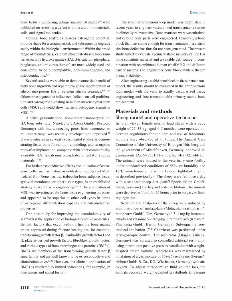
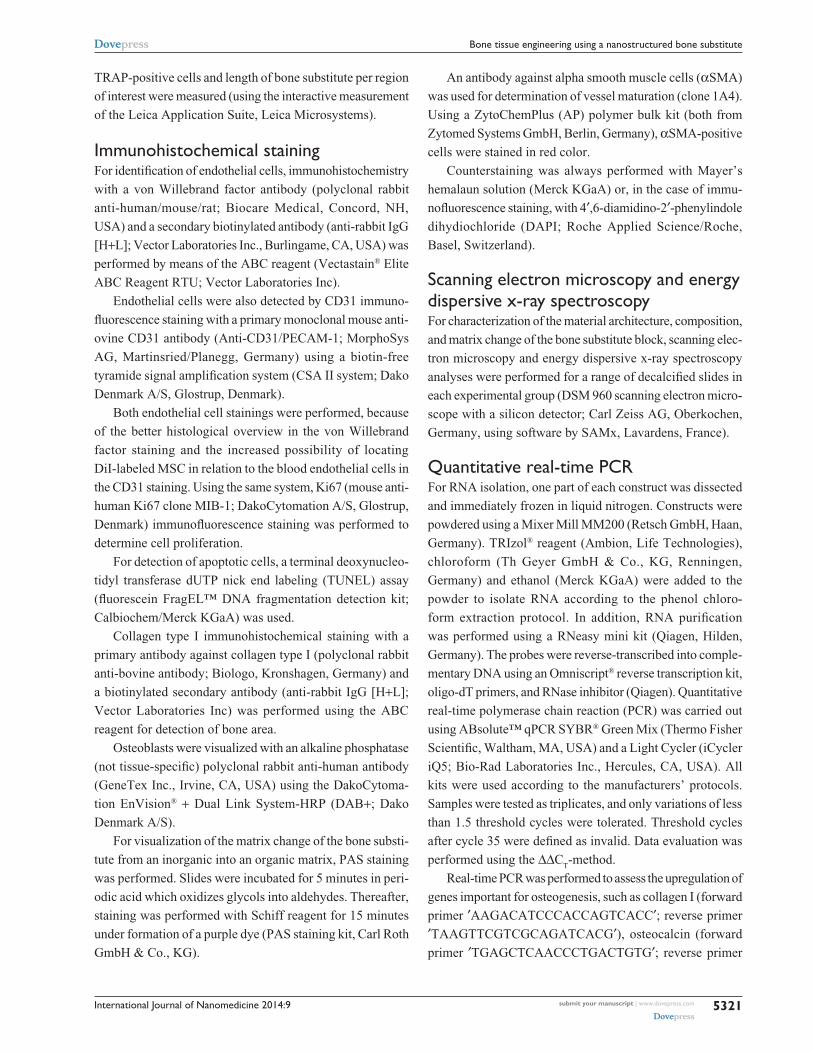
Figure 1 hematoxylin-eosin (A), collagen type I (B), and alkaline phosphatase (C) immunohistochemical staining in the 4-week group. Bone formation was only seen in the groups with Msc expanded in autologous serum or cell culture medium (*).Abbreviations: exp, expanded; retranspl, retransplanted; he, hematoxylin-eosin; Msc, mesenchymal stem cells.
International Journal of Nanomedicine 2014:9 submit your manuscript | www.dovepress.com
Dovepress
Dovepress
5323
Bone tissue engineering using a nanostructured bone substitute
handling, tissue ingrowth, cross-sectional areas of the whole
construct, degraded bone substitute area, vascularization,
bone formation, and cell behavior. Groups were implanted,
comparing all three carrier materials with directly retrans-
planted and expanded MSC.
Hematoxylin-eosin and immunohistochemical staining
against collagen type I and alkaline phosphatase demon-
strated bone formation after 4 weeks only in the experimen-
tal groups with expanded MSC in cell culture medium or
autologous serum (Figure 1A–C).
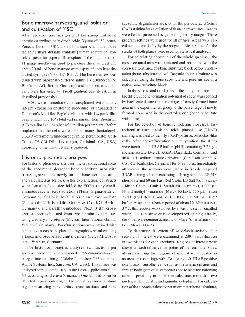
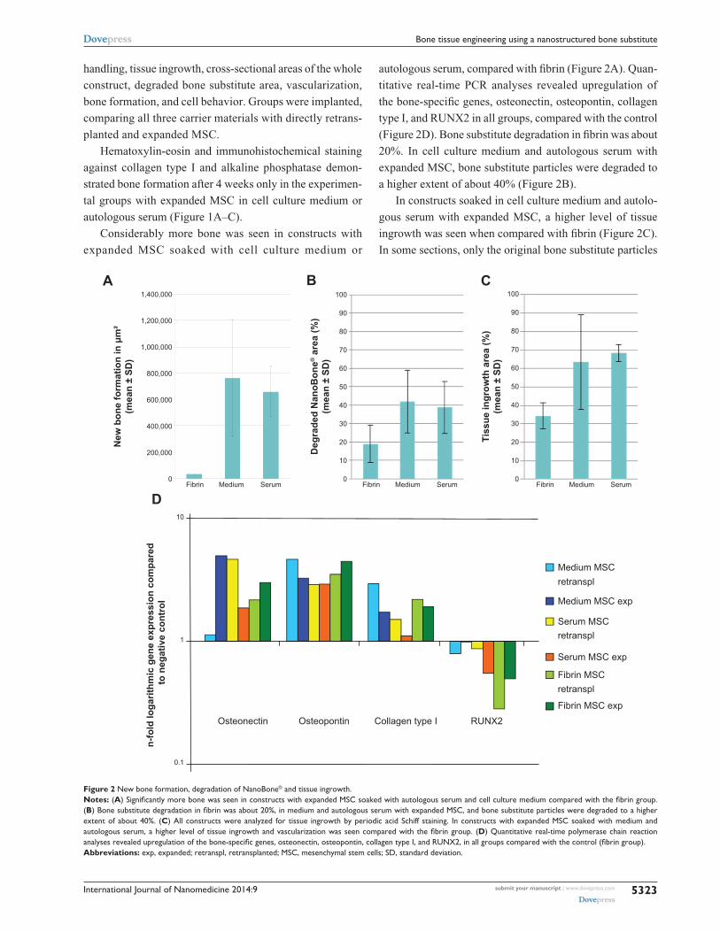
Considerably more bone was seen in constructs with
expanded MSC soaked with cell culture medium or
autologous serum, compared with fibrin (Figure 2A). Quan-
titative real-time PCR analyses revealed upregulation of
the bone-specific genes, osteonectin, osteopontin, collagen
type I, and RUNX2 in all groups, compared with the control
(Figure 2D). Bone substitute degradation in fibrin was about
20%. In cell culture medium and autologous serum with
expanded MSC, bone substitute particles were degraded to
a higher extent of about 40% (Figure 2B).
In constructs soaked in cell culture medium and autolo-
gous serum with expanded MSC, a higher level of tissue
ingrowth was seen when compared with fibrin (Figure 2C).
In some sections, only the original bone substitute particles
Figure 2 New bone formation, degradation of NanoBone® and tissue ingrowth.Notes: (A) Significantly more bone was seen in constructs with expanded MSC soaked with autologous serum and cell culture medium compared with the fibrin group. (B) Bone substitute degradation in fibrin was about 20%, in medium and autologous serum with expanded MSC, and bone substitute particles were degraded to a higher extent of about 40%. (C) all constructs were analyzed for tissue ingrowth by periodic acid schiff staining. In constructs with expanded Msc soaked with medium and autologous serum, a higher level of tissue ingrowth and vascularization was seen compared with the fibrin group. (D) Quantitative real-time polymerase chain reaction analyses revealed upregulation of the bone-specific genes, osteonectin, osteopontin, collagen type I, and RUNX2, in all groups compared with the control (fibrin group).Abbreviations: exp, expanded; retranspl, retransplanted; Msc, mesenchymal stem cells; sD, standard deviation.
New
bon
e fo
rmat
ion
in μ
m²
(mea
n ±
SD)
0 0
10
20
30
40
50
60
70
80
90
100
0
10
20
30
40
50
60
70
80
90
100
200,000
400,000
600,000
800,000
1,000,000
1,200,000
1,400,000
Fibrin Medium Serum Fibrin Medium Serum Fibrin Medium Serum
0.1
1
10
Medium MSC retranspl
Medium MSC exp
Serum MSC retranspl
Serum MSC exp
Fibrin MSC retranspl
Fibrin MSC exp
n-fo
ld lo
garit
hmic
gen
e ex
pres
sion
com
pare
d
to n
egat
ive
cont
rol
A
DTi
ssue
ingr
owth
are
a (%
)(m
ean
± SD
)
Deg
rade
d N
anoB
one®
are
a (%
)(m
ean
± SD
)
B C
Osteonectin Osteopontin Collagen type I RUNX2
International Journal of Nanomedicine 2014:9submit your manuscript | www.dovepress.com
Dovepress
Dovepress
5324
Boos et al
without any surrounding tissue could be detected. Less tis-
sue ingrowth was shown in the 4-week compared with the
12-week group (data not shown).
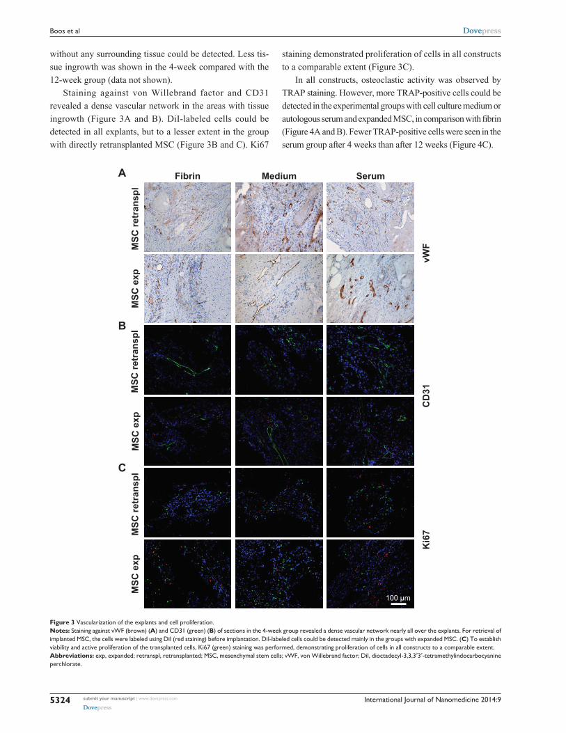
Staining against von Willebrand factor and CD31
revealed a dense vascular network in the areas with tissue
ingrowth (Figure 3A and B). DiI-labeled cells could be
detected in all explants, but to a lesser extent in the group
with directly retransplanted MSC (Figure 3B and C). Ki67
staining demonstrated proliferation of cells in all constructs
to a comparable extent (Figure 3C).
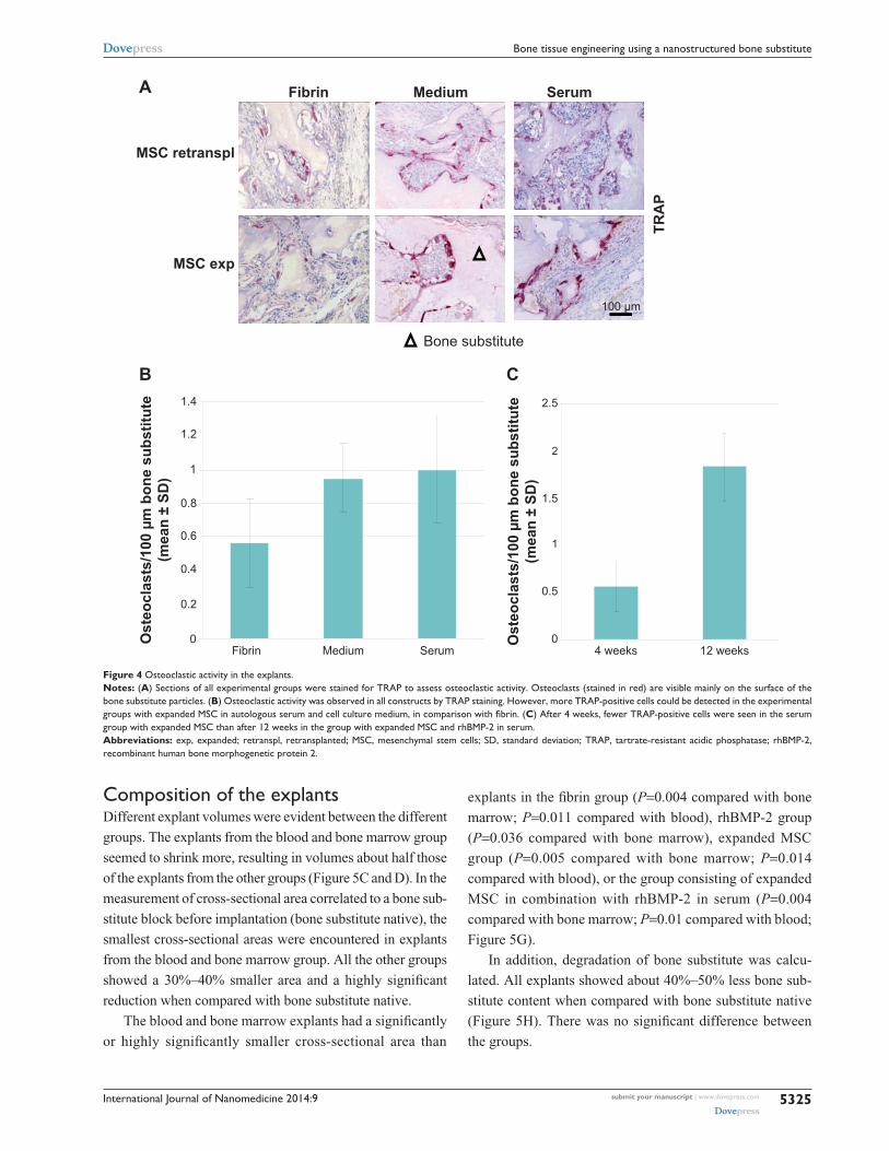
In all constructs, osteoclastic activity was observed by
TRAP staining. However, more TRAP-positive cells could be
detected in the experimental groups with cell culture medium or
autologous serum and expanded MSC, in comparison with fibrin
(Figure 4A and B). Fewer TRAP-positive cells were seen in the
serum group after 4 weeks than after 12 weeks (Figure 4C).
Figure 3 Vascularization of the explants and cell proliferation.Notes: staining against vWF (brown) (A) and cD31 (green) (B) of sections in the 4-week group revealed a dense vascular network nearly all over the explants. For retrieval of implanted Msc, the cells were labeled using DiI (red staining) before implantation. DiI-labeled cells could be detected mainly in the groups with expanded Msc. (C) To establish viability and active proliferation of the transplanted cells, Ki67 (green) staining was performed, demonstrating proliferation of cells in all constructs to a comparable extent.Abbreviations: exp, expanded; retranspl, retransplanted; Msc, mesenchymal stem cells; vWF, von Willebrand factor; DiI, dioctadecyl-3,3,3′3′-tetramethylindocarbocyanine perchlorate.
vWF
Ki6
7C
D31
Fibrin Medium Serum
MSC
retr
ansp
lM
SC e
xpM
SC re
tran
spl
MSC
exp
MSC
retr
ansp
lM
SC e
xp
A
C
B
100 μm
International Journal of Nanomedicine 2014:9 submit your manuscript | www.dovepress.com
Dovepress
Dovepress
5325
Bone tissue engineering using a nanostructured bone substitute
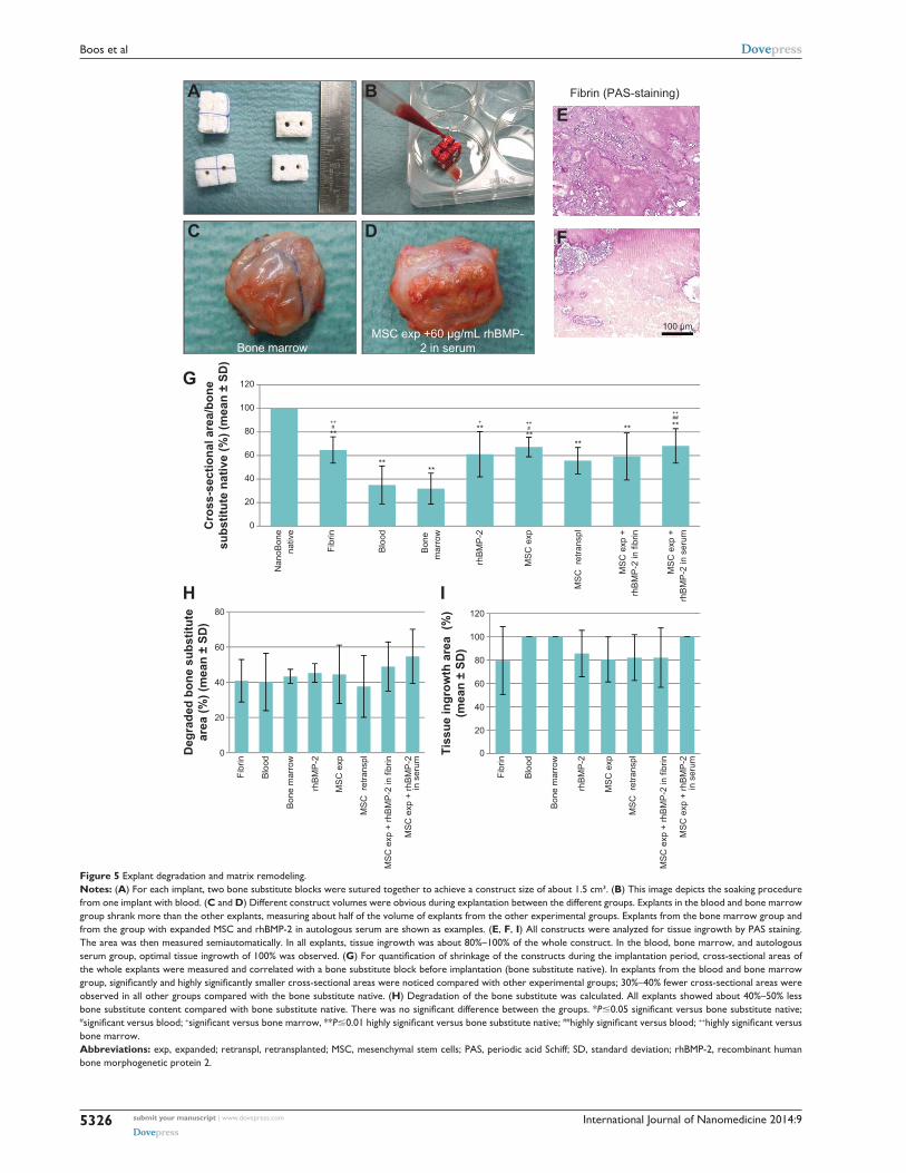
composition of the explantsDifferent explant volumes were evident between the different
groups. The explants from the blood and bone marrow group
seemed to shrink more, resulting in volumes about half those
of the explants from the other groups (Figure 5C and D). In the
measurement of cross-sectional area correlated to a bone sub-
stitute block before implantation (bone substitute native), the
smallest cross-sectional areas were encountered in explants
from the blood and bone marrow group. All the other groups
showed a 30%–40% smaller area and a highly significant
reduction when compared with bone substitute native.
The blood and bone marrow explants had a significantly
or highly significantly smaller cross-sectional area than
explants in the fibrin group (P=0.004 compared with bone
marrow; P=0.011 compared with blood), rhBMP-2 group
(P=0.036 compared with bone marrow), expanded MSC
group (P=0.005 compared with bone marrow; P=0.014
compared with blood), or the group consisting of expanded
MSC in combination with rhBMP-2 in serum (P=0.004
compared with bone marrow; P=0.01 compared with blood;
Figure 5G).
In addition, degradation of bone substitute was calcu-
lated. All explants showed about 40%–50% less bone sub-
stitute content when compared with bone substitute native
(Figure 5H). There was no significant difference between
the groups.
Figure 4 Osteoclastic activity in the explants.Notes: (A) sections of all experimental groups were stained for TraP to assess osteoclastic activity. Osteoclasts (stained in red) are visible mainly on the surface of the bone substitute particles. (B) Osteoclastic activity was observed in all constructs by TraP staining. however, more TraP-positive cells could be detected in the experimental groups with expanded MSC in autologous serum and cell culture medium, in comparison with fibrin. (C) after 4 weeks, fewer TraP-positive cells were seen in the serum group with expanded Msc than after 12 weeks in the group with expanded Msc and rhBMP-2 in serum.Abbreviations: exp, expanded; retranspl, retransplanted; Msc, mesenchymal stem cells; sD, standard deviation; TraP, tartrate-resistant acidic phosphatase; rhBMP-2, recombinant human bone morphogenetic protein 2.
Bone substitute
0
0.5
1
1.5
2
2.5
Ost
eocl
asts
/100
µm
bon
e su
bstit
ute
(mea
n ±
SD)
MSC retranspl
MSC exp
Fibrin Medium Serum
Fibrin Medium Serum 4 weeks 12 weeks0
0.2
0.4
0.6
0.8
1
1.2
1.4
Ost
eocl
asts
/100
µm
bon
e su
bstit
ute
(mea
n ±
SD)
A
CB
TRA
P
100 μm
International Journal of Nanomedicine 2014:9submit your manuscript | www.dovepress.com
Dovepress
Dovepress
5326
Boos et al
Figure 5 explant degradation and matrix remodeling.Notes: (A) For each implant, two bone substitute blocks were sutured together to achieve a construct size of about 1.5 cm³. (B) This image depicts the soaking procedure from one implant with blood. (C and D) Different construct volumes were obvious during explantation between the different groups. explants in the blood and bone marrow group shrank more than the other explants, measuring about half of the volume of explants from the other experimental groups. explants from the bone marrow group and from the group with expanded Msc and rhBMP-2 in autologous serum are shown as examples. (E, F, I) all constructs were analyzed for tissue ingrowth by Pas staining. The area was then measured semiautomatically. In all explants, tissue ingrowth was about 80%–100% of the whole construct. In the blood, bone marrow, and autologous serum group, optimal tissue ingrowth of 100% was observed. (G) For quantification of shrinkage of the constructs during the implantation period, cross-sectional areas of the whole explants were measured and correlated with a bone substitute block before implantation (bone substitute native). In explants from the blood and bone marrow group, significantly and highly significantly smaller cross-sectional areas were noticed compared with other experimental groups; 30%–40% fewer cross-sectional areas were observed in all other groups compared with the bone substitute native. (H) Degradation of the bone substitute was calculated. All explants showed about 40%–50% less bone substitute content compared with bone substitute native. There was no significant difference between the groups. *P0.05 significant versus bone substitute native; #significant versus blood; +significant versus bone marrow, **P0.01 highly significant versus bone substitute native; ##highly significant versus blood; ++highly significant versus bone marrow.Abbreviations: exp, expanded; retranspl, retransplanted; Msc, mesenchymal stem cells; Pas, periodic acid schiff; sD, standard deviation; rhBMP-2, recombinant human bone morphogenetic protein 2.
Bone marrowMSC exp +60 μg/mL rhBMP-
2 in serum
BA
DC
20
0
40
60
80
100
120
0
20
40
60
80
0
20
40
60
80
100
120
**
****
****
**
** **#++ + ++
#
##++
G
H
Fibrin (PAS-staining)
E
F
100 µm
Tiss
ue in
grow
th a
rea
(%)
(mea
n ±
SD)
Deg
rade
d bo
ne s
ubst
itute
area
(%) (
mea
n ±
SD)
Cro
ss-s
ectio
nal a
rea/
bone
subs
titut
e na
tive
(%) (
mea
n ±
SD)
Nan
oBon
ena
tive
Fibr
in
Blo
od
Bon
e m
arro
w
rhB
MP
-2
MS
C e
xp
MS
C r
etra
nspl
MS
C e
xp +
rhB
MP
-2 in
fibr
in
MS
C e
xp +
rhB
MP
-2 in
ser
um
Fibr
in
Blo
od
Bon
e m
arro
w
rhB
MP
-2
MS
C e
xp
MS
C r
etra
nspl
MS
C e
xp +
rhB
MP
-2 in
fibr
in
MS
C e
xp +
rhB
MP
-2in
ser
um
Fibr
in
Blo
od
Bon
e m
arro
w
rhB
MP
-2
MS
C e
xp
MS
C r
etra
nspl
MS
C e
xp +
rhB
MP
-2 in
fibr
in
MS
C e
xp +
rhB
MP
-2in
ser
um
I

International Journal of Nanomedicine 2014:9 submit your manuscript | www.dovepress.com
Dovepress
Dovepress
5327
Bone tissue engineering using a nanostructured bone substitute
Tissue ingrowth Slides were stained for PAS (Figure 5E and F) and the area
of tissue ingrowth was measured semiautomatically. The
shift from an inorganic to an organic matrix could be dem-
onstrated in all areas where tissue had grown. The surfaces
of the bone substitute particles, which were surrounded by
newly formed tissue, were purple in color, indicating a high
content of polysaccharides, neutral mucopolysaccharides,
mucoglycoproteins, and glycoproteins, as well as glycolip-
ids and phospholipids (Figure 5E). In contrast, areas with
no tissue ingrowth showed weaker staining and could be
distinguished very well (Figure 5F).
All explanted constructs showed tissue ingrowth areas
of about 80%–100%. Especially in the border parts of the
explants, nano-HA bone substitute particles were surrounded
by connective tissue and blood vessels. In the center of some
constructs, there was partly no tissue ingrowth on the over-
view pictures. The explants from the blood, bone marrow,
and autologous serum group seemed to have the best tissue
ingrowth of 100%. There were no statistically significant
differences between the groups (Figure 5I).
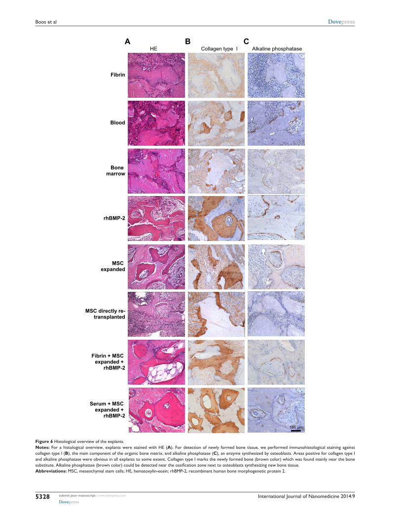
Bone formationFor detection of newly formed bone tissue, slides of all
experimental groups were stained against collagen type I,
the main component of the organic bone matrix, and alkaline
phosphatase, an enzyme synthesized by osteoblasts. Areas
positive for collagen type I and alkaline phosphatase could
be detected above all in the groups with expanded MSC
and rh-BMP-2 in combination with fibrin and serum, as
well as in the rhBMP-2 group. Newly formed bone parts
could be seen particularly on the surfaces of bone substitute
particles. Alkaline phosphatase could be detected near the
ossification zone next to osteoblasts synthesizing new bone
tissue (Figure 6B and C).
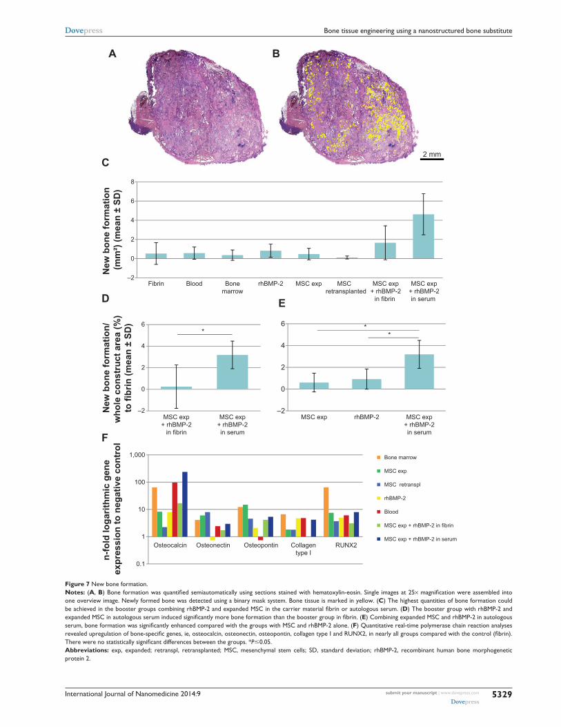
New bone formation was quantified semiautomatically
as mentioned in the Material and methods section (Figure 7A
and B). In the booster groups, combining rhBMP-2 and
expanded MSC in carrier medium fibrin or autologous serum,
the highest quantities of bone formation could be achieved
(Figure 7C). Subcutaneous bone substitute implants with two
different concentrations of rhBMP-2 (12 and 60 μg/mL) have
been evaluated in previous experiments, and shown the 60 μg/
mL concentration to be superior (data not shown). Comparing
the two booster groups, in the group combining rhBMP-2 and
expanded MSC in autologous serum, significantly more bone
formation could be achieved (P=0.025; Figure 7D). Expanded
MSC and rhBMP-2 alone could enhance bone formation, but,
in combination with autologous serum, bone formation was
significantly higher (P=0.016; Figure 7E). Quantitative real-
time PCR analyses revealed upregulation of the bone-specific
markers, osteocalcin, osteonectin, osteopontin, collagen type I
and RUNX2, in nearly all groups when compared with the
control (fibrin; Figure 7F). There were no statistically sig-
nificant differences between the groups.
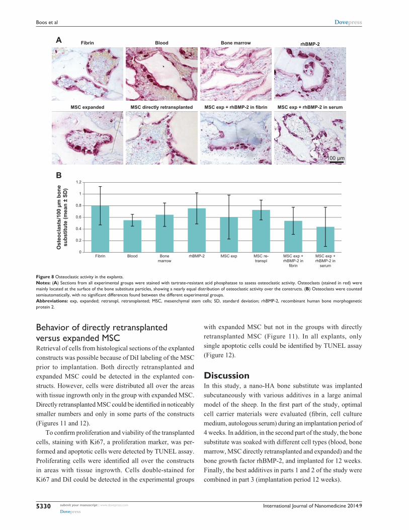
Osteoclastic activity Slides of all experimental groups were TRAP-stained for
quantitative evaluation of osteoclastic activity. Osteoclasts
were mainly located at the surface of the bone substitute
particles, showing a nearly equal distribution of osteoclas-
tic activity over the constructs. Osteoclasts were counted
semiautomatically as mentioned in the Material and methods
section. There were no significant differences between the
experimental groups (Figure 8A and B).
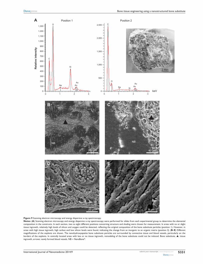
Matrix changeScanning electron microscopy and energy dispersive x-ray
spectroscopy analyses were performed from slides of each
experimental group to confirm the element composition in the
constructs. For these measurements, two to eight different posi-
tions were chosen per section concerning structure and shad-
ing. In areas with no or slight tissue ingrowth, relatively high
levels of silicon and oxygen could be detected, reflecting the
original composition of the bone substitute particles (Figure 9A,
position 1). However, in areas with high tissue ingrowth, high
carbon and low silicon levels were found, indicating the change
from an inorganic to an organic matrix (Figure 9A, position 2).
In Figure 9B–E, different magnifications of the histological
sections are shown. The matrix change could be observed
particularly in the border parts of the constructs, in contrast
with the centrally located areas with no tissue ingrowth.
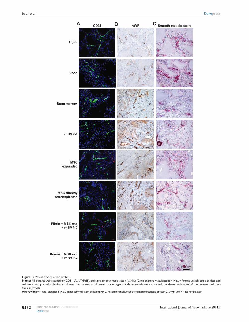
Vascularization of constructs To examine vascularization, sections from each experimental
group were stained against CD31, von Willebrand factor,
and αSMA. Newly formed vessels could be detected and
were nearly equally distributed all over the constructs in
areas with tissue ingrowth. In general, the endothelial cell
layer of the vessels could be nicely stained for CD31 and
von Willebrand factor, showing different sizes of vessels
in the construct. In addition, maturation of larger vessels
by coverage of smooth muscle cells was shown by αSMA
staining. Remodeling of the vascularization of the constructs
in a hierarchically organized system could be demonstrated
by the staining (Figure 10A–C).
International Journal of Nanomedicine 2014:9submit your manuscript | www.dovepress.com
Dovepress
Dovepress
5328
Boos et al
MSC directly re-transplanted
MSC expanded
Fibrin
rhBMP-2
Fibrin + MSC expanded +
rhBMP-2
Serum + MSC expanded +
rhBMP-2
Blood
Bone marrow
HE Collagen type I Alkaline phosphataseA CB
100 µm
Figure 6 histological overview of the explants.Notes: For a histological overview, explants were stained with he (A). For detection of newly formed bone tissue, we performed immunohistological staining against collagen type I (B), the main component of the organic bone matrix, and alkaline phosphatase (C), an enzyme synthesized by osteoblasts. areas positive for collagen type I and alkaline phosphatase were obvious in all explants to some extent. collagen type I marks the newly formed bone (brown color) which was found mainly near the bone substitute. Alkaline phosphatase (brown color) could be detected near the ossification zone next to osteoblasts synthesizing new bone tissue.Abbreviations: Msc, mesenchymal stem cells; he, hematoxylin-eosin; rhBMP-2, recombinant human bone morphogenetic protein 2.
International Journal of Nanomedicine 2014:9 submit your manuscript | www.dovepress.com
Dovepress
Dovepress
5329
Bone tissue engineering using a nanostructured bone substitute
BA
2 mm
–2
0
2
4
6
8
–2
0
2
4
6
New
bon
e fo
rmat
ion/
who
le c
onst
ruct
are
a (%
) to
fibr
in (m
ean
± SD
)
–2
0
2
4
6* *
*
C
ED
0.1
1
10
100
1,000
n-fo
ld lo
garit
hmic
gen
e ex
pres
sion
to n
egat
ive
cont
rolF
Fibrin Blood Bonemarrow
rhBMP-2 MSC exp MSC retransplanted
MSC exp+ rhBMP-2
in fibrin
MSC exp+ rhBMP-2in serum
New
bon
e fo
rmat
ion
(mm
²) (m
ean
± SD
)
rhBMP-2MSC exp MSC exp+ rhBMP-2in serum
MSC exp+ rhBMP-2
in fibrin
MSC exp+ rhBMP-2in serum
Blood
Bone marrow
rhBMP-2
MSC exp
MSC retranspl
MSC exp + rhBMP-2 in fibrin
MSC exp + rhBMP-2 in serumOsteocalcin Osteonectin Osteopontin Collagen
type IRUNX2
Figure 7 New bone formation.Notes: (A, B) Bone formation was quantified semiautomatically using sections stained with hematoxylin-eosin. Single images at 25× magnification were assembled into one overview image. Newly formed bone was detected using a binary mask system. Bone tissue is marked in yellow. (C) The highest quantities of bone formation could be achieved in the booster groups combining rhBMP-2 and expanded MSC in the carrier material fibrin or autologous serum. (D) The booster group with rhBMP-2 and expanded MSC in autologous serum induced significantly more bone formation than the booster group in fibrin. (E) combining expanded Msc and rhBMP-2 in autologous serum, bone formation was significantly enhanced compared with the groups with MSC and rhBMP-2 alone. (F) Quantitative real-time polymerase chain reaction analyses revealed upregulation of bone-specific genes, ie, osteocalcin, osteonectin, osteopontin, collagen type I and RUNX2, in nearly all groups compared with the control (fibrin). There were no statistically significant differences between the groups. *P0.05.Abbreviations: exp, expanded; retranspl, retransplanted; Msc, mesenchymal stem cells; sD, standard deviation; rhBMP-2, recombinant human bone morphogenetic protein 2.
International Journal of Nanomedicine 2014:9submit your manuscript | www.dovepress.com
Dovepress
Dovepress
5330
Boos et al
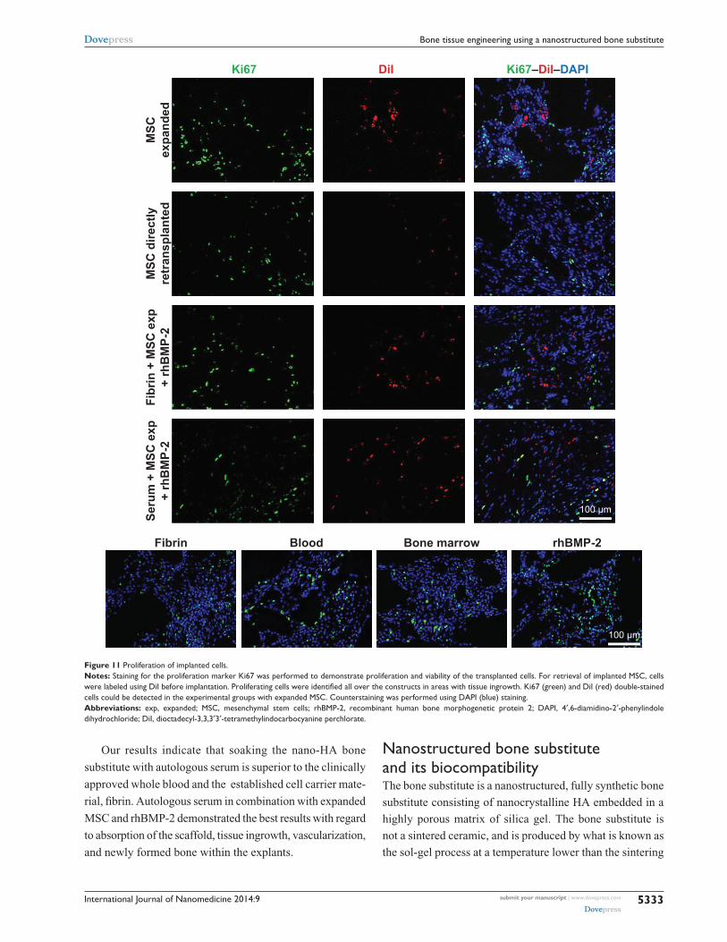
Behavior of directly retransplanted versus expanded MscRetrieval of cells from histological sections of the explanted
constructs was possible because of DiI labeling of the MSC
prior to implantation. Both directly retransplanted and
expanded MSC could be detected in the explanted con-
structs. However, cells were distributed all over the areas
with tissue ingrowth only in the group with expanded MSC.
Directly retransplanted MSC could be identified in noticeably
smaller numbers and only in some parts of the constructs
(Figures 11 and 12).
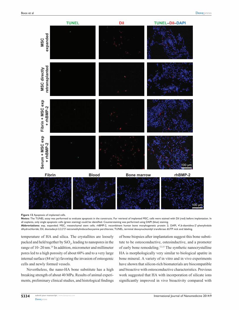
To confirm proliferation and viability of the transplanted
cells, staining with Ki67, a proliferation marker, was per-
formed and apoptotic cells were detected by TUNEL assay.
Proliferating cells were identified all over the constructs
in areas with tissue ingrowth. Cells double-stained for
Ki67 and DiI could be detected in the experimental groups
Figure 8 Osteoclastic activity in the explants.Notes: (A) sections from all experimental groups were stained with tartrate-resistant acid phosphatase to assess osteoclastic activity. Osteoclasts (stained in red) were mainly located at the surface of the bone substitute particles, showing a nearly equal distribution of osteoclastic activity over the constructs. (B) Osteoclasts were counted semiautomatically, with no significant differences found between the different experimental groups.Abbreviations: exp, expanded; retranspl, retransplanted; Msc, mesenchymal stem cells; sD, standard deviation; rhBMP-2, recombinant human bone morphogenetic protein 2.
MSC directly retransplantedMSC expanded
Fibrin Blood Bone marrow rhBMP-2
MSC exp + rhBMP-2 in serumMSC exp + rhBMP-2 in fibrin
100 µm
0
0.2
0.4
0.6
0.8
1
1.2
A
B
Fibrin Blood Bonemarrow
rhBMP-2 MSC exp MSC re-transpl
MSC exp +rhBMP-2 in
fibrin
MSC exp +rhBMP-2 in
serum
Ost
eocl
asts
/100
µm
bon
esu
bstit
ute
(mea
n ±
SD)
with expanded MSC but not in the groups with directly
retransplanted MSC (Figure 11). In all explants, only
single apoptotic cells could be identified by TUNEL assay
(Figure 12).
DiscussionIn this study, a nano-HA bone substitute was implanted
subcutaneously with various additives in a large animal
model of the sheep. In the first part of the study, optimal
cell carrier materials were evaluated (fibrin, cell culture
medium, autologous serum) during an implantation period of
4 weeks. In addition, in the second part of the study, the bone
substitute was soaked with different cell types (blood, bone
marrow, MSC directly retransplanted and expanded) and the
bone growth factor rhBMP-2, and implanted for 12 weeks.
Finally, the best additives in parts 1 and 2 of the study were
combined in part 3 (implantation period 12 weeks).
International Journal of Nanomedicine 2014:9 submit your manuscript | www.dovepress.com
Dovepress
Dovepress
5331
Bone tissue engineering using a nanostructured bone substitute
Figure 9 scanning electron microscopy and energy dispersive x-ray spectroscopy.Notes: (A) scanning electron microscopy and energy dispersive x-ray spectroscopy were performed for slides from each experimental group to determine the elemental composition in the constructs. In each section, two to eight different positions concerning structure and shading were chosen for measurement. In areas with no or slight tissue ingrowth, relatively high levels of silicon and oxygen could be detected, reflecting the original composition of the bone substitute particles (position 1). However, in areas with high tissue ingrowth, high carbon and low silicon levels were found, indicating the change from an inorganic to an organic matrix (position 2). (B–E) Different magnifications of the explants are shown. The nanohydroxyapatite bone substitute particles are surrounded by connective tissue and blood vessels, particularly on the borders of the explants. In centrally located areas with less or no tissue ingrowth, remodeling of the bone substitute could not be induced. Bone substitute, : tissue ingrowth, arrows: newly formed blood vessels. NB = NanoBone®.
200 µm 40 µm
20 µm 9 µm
ED
CB
NB NB
NB
→
▲▲
▲
keV
Rel
ativ
e in
tens
ity
2 mm
P2P1
A Position 1
1,300
1,200
1,100
1,000
900
800
700
600
500
400
300
200
100
0 0
500
1,000
1,500
2,000
2,500
0
C
CO
O
NaNa
Si
SiAu
Au
AuAu
1 2 3 0 1 2 3
Position 2
International Journal of Nanomedicine 2014:9submit your manuscript | www.dovepress.com
Dovepress
Dovepress
5332
Boos et al
Figure 10 Vascularization of the explants.Notes: all explants were stained for cD31 (A), vWF (B), and alpha smooth muscle actin (αsMa) (C) to examine vascularization. Newly formed vessels could be detected and were nearly equally distributed all over the constructs. however, some regions with no vessels were observed, consistent with areas of the construct with no tissue ingrowth.Abbreviations: exp, expanded; Msc, mesenchymal stem cells; rhBMP-2, recombinant human bone morphogenetic protein 2; vWF, von Willebrand factor.
MSC directlyretransplanted
MSCexpanded
Fibrin
rhBMP-2
Fibrin + MSC exp+ rhBMP-2
Serum + MSC exp+ rhBMP-2
Blood
Bone marrow
CD31 vWF Smooth muscle actinA CB
100 µm
International Journal of Nanomedicine 2014:9 submit your manuscript | www.dovepress.com
Dovepress
Dovepress
5333
Bone tissue engineering using a nanostructured bone substitute
Our results indicate that soaking the nano-HA bone
substitute with autologous serum is superior to the clinically
approved whole blood and the established cell carrier mate-
rial, fibrin. Autologous serum in combination with expanded
MSC and rhBMP-2 demonstrated the best results with regard
to absorption of the scaffold, tissue ingrowth, vascularization,
and newly formed bone within the explants.
Nanostructured bone substitute and its biocompatibilityThe bone substitute is a nanostructured, fully synthetic bone
substitute consisting of nanocrystalline HA embedded in a
highly porous matrix of silica gel. The bone substitute is
not a sintered ceramic, and is produced by what is known as
the sol-gel process at a temperature lower than the sintering
Ki67 DiI Ki67–DiI–DAPI
MSC
dire
ctly
retr
ansp
lant
edM
SCex
pand
ed
Fibrin rhBMP-2
Fibr
in +
MSC
exp
+ rh
BM
P-2
Seru
m +
MSC
exp
+ rh
BM
P-2
Blood Bone marrow
100 µm
100 µm
Figure 11 Proliferation of implanted cells.Notes: staining for the proliferation marker Ki67 was performed to demonstrate proliferation and viability of the transplanted cells. For retrieval of implanted Msc, cells were labeled using DiI before implantation. Proliferating cells were identified all over the constructs in areas with tissue ingrowth. Ki67 (green) and DiI (red) double-stained cells could be detected in the experimental groups with expanded Msc. counterstaining was performed using DaPI (blue) staining.Abbreviations: exp, expanded; Msc, mesenchymal stem cells; rhBMP-2, recombinant human bone morphogenetic protein 2; DaPI, 4′,6-diamidino-2′-phenylindole dihydrochloride; DiI, dioctadecyl-3,3,3′3′-tetramethylindocarbocyanine perchlorate.
International Journal of Nanomedicine 2014:9submit your manuscript | www.dovepress.com
Dovepress
Dovepress
5334
Boos et al
temperature of HA and silica. The crystallites are loosely
packed and held together by SiO2, leading to nanopores in the
range of 10–20 nm.24 In addition, micrometer and millimeter
pores led to a high porosity of about 60% and to a very large
internal surface (84 m2/g) favoring the invasion of osteogenic
cells and newly formed vessels.
Nevertheless, the nano-HA bone substitute has a high
breaking strength of about 40 MPa. Results of animal experi-
ments, preliminary clinical studies, and histological findings
of bone biopsies after implantation suggest this bone substi-
tute to be osteoconductive, osteoinductive, and a promoter
of early bone remodeling.13,15 The synthetic nanocrystalline
HA is morphologically very similar to biological apatite in
bone mineral. A variety of in vitro and in vivo experiments
have shown that silicon-rich biomaterials are biocompatible
and bioactive with osteoconductive characteristics. Previous
work suggested that HA with incorporation of silicate ions
significantly improved in vivo bioactivity compared with
Figure 12 apoptosis of implanted cells.Notes: The TUNel assay was performed to evaluate apoptosis in the constructs. For retrieval of implanted Msc, cells were stained with DiI (red) before implantation. In all explants, only single apoptotic cells (green staining) could be identified. Counterstaining was performed using DAPI (blue) staining.Abbreviations: exp, expanded; Msc, mesenchymal stem cells; rhBMP-2, recombinant human bone morphogenetic protein 2; DaPI, 4′,6-diamidino-2′-phenylindole dihydrochloride; DiI, dioctadecyl-3,3,3′3′-tetramethylindocarbocyanine perchlorate; TUNel, terminal deoxynucleotidyl transferase dUTP nick end labeling.
Fibrin rhBMP-2Bone marrowBlood
TUNEL DiI TUNEL–DiI–DAPI
MSC
dire
ctly
retr
ansp
lant
edM
SCex
pand
edFi
brin
+ M
SC e
xp+
rhB
MP-
2Se
rum
+ M
SC e
xp+
rhB
MP-
2
100 µm
100 µm
International Journal of Nanomedicine 2014:9 submit your manuscript | www.dovepress.com
Dovepress
Dovepress
5335
Bone tissue engineering using a nanostructured bone substitute
conventional HA materials.25 Another study demonstrated
that a bone substitute comprised of nanocrystalline HA
embedded in a silica matrix was successful in oral and maxil-
lofacial surgery.14 A previous study showed that the silica
gel in the bone substitute is replaced by bone matrix glyco-
proteins with known functions in attraction, adhesion, and
differentiation of bone cells as osteoblasts and osteoclasts.25
Silica has an important impact on regulation of generation,
mobilization, differentiation, and activation of osteoclast
precursors. It has been established that a high silica content
promotes rapid bone mineralization.26 These findings indicate
that this bone substitute could be an ideal material for the
treatment of critical size defects.
The implants were well tolerated overall by the sheep.
Only one sheep developed an infection, which is within the
common range for this animal model. The good biocompat-
ibility has been shown previously in in vivo studies and in
clinical application.24,27
Optimal cell carrier materialFor evaluation of an optimal cell carrier material, the bone
substitute was implanted in combination with expanded or
directly retransplanted MSC and fibrin, cell culture medium,
or autologous serum. An important aspect of this study was the
bone formation capacity of the different groups. Hardly any
bone formation was found in explants with directly retrans-
planted MSC. Therefore, analysis of the first part of the study
was reduced to experimental groups with expanded MSC. Cell
culture medium and autologous serum proved to be superior to
fibrin with regard to bone formation capacity, tissue ingrowth,
and degradation of the bone substitute. In constructs with cell
culture medium and autologous serum, more TRAP-positive
cells could be identified when compared with fibrin. This may
be in concordance with reduced vascularization and absorp-
tion of the bone substitute in the fibrin group. The expression
of osteogenic genes did not demonstrate a significant differ-
ence between the experimental groups.
All these findings can probably be explained by the higher
viscosity of fibrin, which is not able to penetrate the block
material as good as the more liquid autologous serum or cell
culture medium. Irregular soaking of the fibrin sealant may
be the reason for the inconsistent bone formation and high
standard deviation in the fibrin groups. Positive proliferative
and angiogenic effects have been described by other groups,
and should have made fibrin an ideal carrier medium for
MSC for bone tissue engineering purposes. However, in this
study, autologous serum and cell culture medium seemed to
be more promising.28,29 An advantage of autologous serum is
that it contains a broad range of growth factors and cytokines.
It has been shown that autologous serum has a positive influ-
ence on MSC behavior.30 For positively charged molecules
in particular, it is possible to stick to the negatively charged
surface by SiO groups of the bone substitute and therefore
promote the remodeling process.31 Fibrin could cover the
bone substitute surface and therefore mask the SiO groups,
so that the matrix change and induction of bone formation
could not take place. In addition, use of autologous serum is
not associated with as many regulatory issues as using serum
or fibrin of foreign species.32 Due to the range of positive
aspects of autologous serum and the similar results with cell
culture medium, we finally decided to use autologous serum
in the following part of the study.
Degradation, absorption, and matrix change of bone substituteAs cell sources, blood, bone marrow, directly retransplanted
and expanded MSC were implanted in combination with the
bone substitute. Expanded MSC proved to be the optimal cell
source with regard to adequate absorption and degradation
of the bone substitute, good tissue ingrowth, bone formation
capacity, and cell proliferation.
A crucial aspect of bone regeneration using bone substi-
tutes is absorption of the bone substitute and simultaneous
formation of new bone.33 In this study, shrinkage of the
whole constructs, degradation of the scaffold, and the tissue
ingrowth area were analyzed. The largest tissue ingrowth
areas and complete vascularization could be achieved in
groups with blood and bone marrow, and finally in the
“booster group” combining expanded MSC and rhBMP-2
in serum.
Tissue ingrowth areas in the other experimental groups
covered about 80% after 12 weeks. However, highly signifi-
cant shrinkage of the experimental groups with blood and
bone marrow was obvious when compared with other cell
sources or combination with rhBMP-2, as shown in Figure 5G.
Other researchers working with nano-HA granules or blocks
soaked in blood have observed absorption of the bone sub-
stitute without adequate new bone formation.25,34
In the 12-week group, about 50% of the bone substitute
particles were degraded. Other groups observed degrada-
tion and complete absorption under the same circumstances
because of the high porosity and loosely packed HA structure
of the bone substitute.12,35 Compared with other bone substi-
tutes, NanoBone® was degraded in a faster manner, which
could avoid the risk of residual bone substitute material and
an inflammatory response.27,36

International Journal of Nanomedicine 2014:9submit your manuscript | www.dovepress.com
Dovepress
Dovepress
5336
Boos et al
TRAP-positive cells could always be detected without
significant differences between the groups. After 12 weeks,
significantly more TRAP-positive cells were counted than
after 4 weeks. The appearance of TRAP-positive cells can be
interpreted as a sign of early bone formation, as hypothesized
by others.25 However, the coexistence of TRAP-positive
and osteoblastic cells and the absence of further inflam-
matory cells suggests the start of remodeling of the bone
substitute rather than an inflammatory process. Götz et al
explain the existence of TRAP-positive cells by the high
content of osteocalcin and osteopontin on the surface of the
bone substitute, stimulating osteoclast adhesion and differ-
entiation.13 More TRAP-positive cells were counted in the
groups with less bone formation, possibly due to less free
nano-HA bone substitute surface available for osteoclastic
activity because of more newly formed bone near the bone
substitute surface.
Representative sections were examined by electron
microscopy to analyze the matrix change from an inorganic
composition with high levels of silicon and oxygen to an
organic composition with high levels of carbon. In some
areas without any tissue ingrowth, relatively high amounts
of inorganic matrix could be detected, representing the origi-
nal bone substitute matrix. In contrast, in well vascularized
areas, high amounts of carbon and low levels of silicon could
be demonstrated. As described in previous studies,13,15,23,31
a matrix change could be detected particularly near the
surface of the bone substitute and only in well vascularized
areas.25 In our constructs, a matrix change was visible in all
experimental groups and therefore paves the way for further
degradation of the nano-HA bone substitute by osteoclasts
and new bone formation.
New bone formation depending on implanted cells and carrier materialAreas positive for collagen type I and alkaline phosphatase
could be detected in all constructs to varying degrees, demon-
strating successful bone formation using the bone substitute
in combination with different additives.
The nano-HA bone substitute is clinically approved,
soaked with whole blood. There are several animal and clini-
cal studies demonstrating that this strategy leads to a matrix
change in the bone substitute and finally to formation of new
bone.27,34 Taking whole blood is a simple and rapid method
for clinical application. In our study constructs soaked with
whole blood, high interindividual variance concerning bone
formation was obvious. In addition, bone formation remained
very low compared with previous bone substitute studies.
However, it has to be kept in mind that the present experi-
ment involves induction of ectopic (that means in abnormal
sites) bone formation in subcutis. Dietze et al emphasize
that contact between the nano-HA bone substitute and well
vascularized bone is essential for successful therapy.37 For
this reason, ectopic bone formation, needed for our tissue
engineering purposes, is not possible using the bone substi-
tute solely soaked with whole blood.
Differing interindividual results were observed in the
group with bone marrow. Some sheep had high yields of
bone formation, whereas some showed only early bone
formation. This could be due to the heterogeneous cell
population and often low numbers of MSC located in the
bone marrow.38 In addition, growth factor and cytokine con-
centrations are known to be very variable.39,40 The expected
benefit of endothelial progenitor cells and angiogenic
growth factors, as described by other groups,41–46 could not
be observed.47
One strategy to improve these results is to concentrate
the bone marrow cell fraction, which in the present study
involved directly retransplanted MSC. However, even when
working with concentrated cell suspensions, varying cell
numbers are yielded, which can make even robust results
questionable.48 Expanded MSC have been used in a wide
range of studies, both for bone tissue purposes and in other
fields of regenerative medicine.49,50 MSC are expected to be
the ideal cell population because of their good differentiation
capacity as well as their secretion of a wide range of regula-
tory cytokines and their good influence as anti-inflammatory
and immunosuppressive agents.51 After 12 weeks, expanded
MSC did not induce significantly more bone formation
than directly retransplanted MSC. However, after 4 weeks,
expanded MSC clearly showed an earlier and faster potential
for new bone formation. Moreover, in constructs with directly
retransplanted MSC, less labeled MSC could be found than in
constructs with expanded MSC. This could possibly be due
to the higher cell numbers achieved after expansion and the
variable and low numbers of stem cells in the concentrated
directly retransplanted MSC, which could not be determined
before implantation.
RhBMP-2 was used as a bone growth factor, inducing
a high rate of bone formation in the constructs alone and in
combination with expanded MSC. Because of its well known
osteogenic capacity, BMP-2 has been approved for clinical
application and a huge range of experimental studies have
made use of it.52 In a preliminary study, two concentrations
of rhBMP-2 were tested in combination with the bone sub-
stitute, showing an advantage of 60 μg/mL when compared
International Journal of Nanomedicine 2014:9 submit your manuscript | www.dovepress.com
Dovepress
Dovepress
5337
Bone tissue engineering using a nanostructured bone substitute
with 12.5 μg/mL (data not shown). The BMP concentration
needed for healing of bone defects is unknown so far.53 The
concentration depends on the species, the carrier material,
and the defect itself.52,54 Side effects of high concentrations
could possibly lead to ectopic bone formation and an increase
in osteoclastic activity. In our study, osteoclastic activity
was comparable in all constructs from the 12-week group,
and bone formation in the tissue around the implants could
not be observed.
BMP-2 should be used in a carrier medium that ensures
its slow release over time. Fibrin was used for that purpose
in several previous studies, and seems to be ideal for slow
growth factor release. In addition, it is absorbable and sup-
ports angiogenic ingrowth.55,56 In this study, soaking the scaf-
folds with fibrin resulted in inferior results when compared
with autologous serum and cell culture medium, possibly due
to the special nanostructured architecture. Comparison of the
two combination groups with expanded MSC and rhBMP-2
in fibrin and serum demonstrated that fibrin is not crucial
for the delivery of the growth factor BMP-2 over time. We
could clearly show that addition of BMP-2 to serum mark-
edly increases the capacity for bone formation while always
using the same batch of serum, so at least intratest validity
and reproducibility were ensured in this study.
HA is a calcium phosphate (Ca5(PO
4)
3OH), which repre-
sents 55% of the weight of bone in vertebrates. Silicon, an
essential trace element, plays an active role in human bone
mineralization and calcification, and was demonstrated to
be pivotal in formation of connective tissue and stimulation
of osteoblast proliferation.25,26 The bone substitute used in
this study has been made of both HA and silica, which is
achieved through specific sol-gel techniques. After het-
erotopic transplantation of this bone substitute, there is a
change of the inorganic silica gel matrix into organic host
compounds.25 By soaking the bone substitute with growth
factors, the protein is incorporated into the scaffold prior to
the matrix change. The kinetics of the initial matrix change
of the implanted biomaterial are enhanced by this procedure.
The protein is released into the extracellular environment
during the following matrix change and degradation of the
bone substitute, supporting bone growth.
Quantitative real-time PCR analyses revealed upregula-
tion of osteocalcin, osteonectin, osteopontin, collagen type I,
and RUNX2 in nearly all groups when compared with the
control (bone substitute with fibrin). No statistically sig-
nificant differences between the experimental groups could
be determined. However, high standard deviations were
observed. The upregulation of osteogenic markers in all
the bone substitute groups indicates good osteogenic and
osteoinductive properties of the nano-HA bone substitute,
as described previously.13 It should be noted that the areas in
which bone formation and remodeling of the bone substitute
take place were not equally distributed over the constructs.
Only a small part of the explants was analyzed for molecu-
lar biology, probably leading to use of non-representative
samples from the experimental groups and resulting in high
standard deviations.
ConclusionA nanostructured clinically approved bone substitute mate-
rial was evaluated in this study, soaked with different car-
rier materials, and implanted in a large animal model in
combination with different cells and the bone growth factor
rhBMP-2. Autologous serum improves tissue ingrowth of
the nano-HA bone substitute and leads to earlier and more
rapid bone formation compared with fibrin. Because of its
low viscosity, autologous serum is ideal for penetration of the
nanostructured bone substitute and can induce early matrix
change by delivery of a wide range of autologous growth
factors and cytokines. Osteogenic cells and rhBMP-2 could
therefore evenly distribute into the bone substitute, with
degradation and simultaneous bone formation within the
nano-HA bone substitute being stimulated.
Combining expanded MSC with rhBMP-2 in autologous
serum leads to a high yield of bone formation and is supe-
rior to the clinically approved procedure where the bone
substitute is soaking in whole blood. In the next step, these
results should be transferred to the sheep arteriovenous loop
model to engineer axially vascularized primary stable bone
replacement of a clinically relevant size.
AcknowledgmentsWe would like to thank Stefan Fleischer, Marina Milde, and
Ilse Arnold-Herberth for their excellent technical support.
This work contains parts of the doctoral theses of A Weigand
and G Deschler. This study was funded by the ELAN Fonds
für Forschung und Lehre, University of Erlangen-Nürnberg,
Erlangen, Germany; the Baxter Innovations GmbH, Vienna,
Austria; the Staedtler Foundation, University of Erlangen-
Nürnberg, Erlangen, Germany; the Forschungsstiftung
Medizin Erlangen, University of Erlangen-Nürnberg,
Erlangen, Germany; and the Xue Hong and Hans Georg
Geis Foundation. We acknowledge support by Deutsche
Forschungsgemeinschaft and Friedrich-Alexander-Univer-
sität Erlangen-Nürnberg as part of the Open Access Publish-
ing funding program.
International Journal of Nanomedicine 2014:9submit your manuscript | www.dovepress.com
Dovepress
Dovepress
5338
Boos et al
DisclosureOne of the authors (TG) was involved in the development
of the bone substitute material. He is executive director
of Artoss GmbH, the distributor of NanoBone®. He was
involved in the design of the study. All other authors confirm
that they have no conflicts of interest in this work.
References 1. Horch RE, Kneser U, Polykandriotis E, Schmidt VJ, Sun J, Arkudas
A. Tissue engineering and regenerative medicine – where do we stand? J Cell Mol Med. 2012;16(6):1157–1165.
2. Horch RE, Boos AM, Quan Y, et al. Cancer research by means of tissue engineering – is there a rationale? J Cell Mol Med. 2013;17(10):1197–1206.
3. Ma J1, Both SK, Yang F, Cui FZ, Pan J, Meijer GJ, Jansen JA, van den Beucken JJ. Concise review: cell-based strategies in bone tissue engineering and regenerative medicine. Stem Cells Transl Med. 2014 Jan;3(1):98–107. doi: 10.5966/sctm.2013-0126. Epub 2013 Dec 3.
4. Dawson JI1, Kanczler J, Tare R, Kassem M, Oreffo RO. Concise review: bridging the gap: bone regeneration using skeletal stem cell-based strategies - where are we now? Stem Cells. 2014;32(1):35–44. doi: 10.1002/stem.1559.
5. Arvidson K, Abdallah BM, Applegate LA, et al. Bone regeneration and stem cells. J Cell Mol Med. 2011;15(4):718–746.
6. Barrere F, van Blitterswijk CA, de Groot K. Bone regeneration: molecular and cellular interactions with calcium phosphate ceramics. Int J Nanomedicine. 2006;1(3):317–332.
7. Hing KA, Revell PA, Smith N, Buckland T. Effect of silicon level on rate, quality and progression of bone healing within silicate-substituted porous hydroxyapatite scaffolds. Biomaterials. 2006;27(29):5014–5026.
8. Xu S, Lin K, Wang Z, et al. Reconstruction of calvarial defect of rab-bits using porous calcium silicate bioactive ceramics. Biomaterials. 2008;29(17):2588–2596.
9. Patel N, Brooks RA, Clarke MT, et al. In vivo assessment of hydroxy-apatite and silicate-substituted hydroxyapatite granules using an ovine defect model. J Mater Sci Mater Med. 2005;16(5):429–440.
10. Huang DM, Chung TH, Hung Y, et al. Internalization of mesoporous silica nanoparticles induces transient but not sufficient osteogenic signals in human mesenchymal stem cells. Toxicol Appl Pharmacol. 2008;231(2):208–215.
11. Xu JL, Khor KA. Chemical analysis of silica doped hydroxyapatite biomaterials consolidated by a spark plasma sintering method. J Inorg Biochem. 2007;101(2):187–195.
12. Gerike W, Bienengraber V, Henkel KO, et al. The manufacture of synthetic non-sintered and degradable bone grafting substitutes. Folia Morphol (Warsz). 2006;65(1):54–55.
13. Gotz W, Gerber T, Michel B, Lossdorfer S, Henkel KO, Heinemann F. Immunohistochemical characterization of nanocrystalline hydroxyapa-tite silica gel (NanoBone(r)) osteogenesis: a study on biopsies from human jaws. Clin Oral Implants Res. 2008;19(10):1016–1026.
14. Heinemann F, Mundt T, Biffar R, Gedrange T, Goetz W. A 3-year clinical and radiographic study of implants placed simultaneously with maxillary sinus floor augmentations using a new nanocrystalline hydroxyapatite. J Physiol Pharmacol. 2009;60 Suppl 8:91–97.
15. Henkel KO, Gerber T, Lenz S, Gundlach KK, Bienengraber V. Macroscopical, histological, and morphometric studies of porous bone-replacement materials in minipigs 8 months after implantation. Oral Surg Oral Med Oral Pathol Oral Radiol Endod. 2006;102(5):606–613.
16. Rath SN, Strobel LA, Arkudas A, et al. Osteoinduction and survival of osteoblasts and bone-marrow stromal cells in 3D biphasic calcium phosphate scaffolds under static and dynamic culture conditions. J Cell Mol Med. 2012;16(10):2350–2361.
17. Jones E, Yang X. Mesenchymal stem cells and bone regeneration: cur-rent status. Injury. 2011;42(6):562–568.
18. Cancedda R, Bianchi G, Derubeis A, Quarto R. Cell therapy for bone disease: a review of current status. Stem Cells. 2003;21(5):610–619.
19. Chen HT, Lee MJ, Chen CH, et al. Proliferation and differentiation potential of human adipose-derived mesenchymal stem cells isolated from elderly patients with osteoporotic fractures. J Cell Mol Med. 2012;16(3):582–593.
20. Garrison KR, Shemilt I, Donell S, et al. Bone morphogenetic protein (BMP) for fracture healing in adults. Cochrane Database Syst Rev. 2010;6:CD006950.
21. Pneumaticos SG, Triantafyllopoulos GK, Basdra EK, Papavassiliou AG. Segmental bone defects: from cellular and molecular pathways to the development of novel biological treatments. J Cell Mol Med. 2010;14(11):2561–2569.
22. Govender S, Csimma C, Genant HK, et al. Recombinant human bone morphogenetic protein-2 for treatment of open tibial fractures: a pro-spective, controlled, randomized study of four hundred and fifty patients. J Bone Joint Surg Am. 2002;84A(12):2123–2134.
23. Boos AM, Loew JS, Weigand A, et al. Engineering axially vascularized bone in the sheep arteriovenous-loop model. J Tissue Eng Regen Med. 2013;7(8):654–664.
24. Abshagen K, Schrodi I, Gerber T, Vollmar B. In vivo analysis of biocompatibility and vascularization of the synthetic bone grafting substitute NanoBone. J Biomed Mater Res A. 2009;91(2):557–566.
25. Xu W, Holzhuter G, Sorg H, et al. Early matrix change of a nanostruc-tured bone grafting substitute in the rat. J Biomed Mater Res B Appl Biomater. 2009;91(2):692–699.
26. Jugdaohsingh R. Silicon and bone health. J Nutr Health Aging. 2007;11(2):99–110.
27. Bienengraber V, Gerber T, Henkel KO, Bayerlein T, Proff P, Gedrange T. The clinical application of a new synthetic bone grafting material in oral and maxillofacial surgery. Folia Morphol (Warsz). 2006;65(1):84–88.
28. Lee OK. Fibrin glue as a vehicle for mesenchymal stem cell delivery in bone regeneration. J Chin Med Assoc. 2008;71(2):59–61.
29. Rajangam T, An SS. Fibrinogen and fibrin based micro and nano scaf-folds incorporated with drugs, proteins, cells and genes for therapeutic biomedical applications. Int J Nanomedicine. 2013;8:3641–3662.
30. Kobayashi T, Watanabe H, Yanagawa T, et al. Motility and growth of human bone-marrow mesenchymal stem cells during ex vivo expansion in autologous serum. J Bone Joint Surg Br. 2005;87(10):1426–1433.
31. Gerber T, Holzhüter G, Götz W, Bienengräber V, Henkel K-O, Rumpel E. Nanostructuring of biomaterials – a pathway to bone grafting substitute. Eur J Trauma. 2006;32(2):132–140.
32. Choi J, Chung JH, Kwon GY, Kim KW, Kim S, Chang H. Effec-tiveness of autologous serum as an alternative to fetal bovine serum in adipose-derived stem cell engineering. Cell Tissue Bank. 2013;14(3):413–422.
33. Kolk A, Handschel J, Drescher W, et al. Current trends and future per-spectives of bone substitute materials – from space holders to innovative biomaterials. J Craniomaxillofac Surg. 2012;40(8):706–718.
34. Kirchhoff M, Lenz S, Henkel KO, et al. Lateral augmentation of the mandible in minipigs with a synthetic nanostructured hydroxy-apatite block. J Biomed Mater Res B Appl Biomater. 2011;96(2): 342–350.
35. Rumpel E, Wolf E, Kauschke E, et al. The biodegradation of hydroxyapatite bone graft substitutes in vivo. Folia Morphol (Warsz). 2006;65(1):43–48.
36. Liu Y, Wang G, Cai Y, et al. In vitro effects of nanophase hydroxy-apatite particles on proliferation and osteogenic differentiation of bone marrow-derived mesenchymal stem cells. J Biomed Mater Res A. 2009;90(4):1083–1091.
37. Dietze S, Bayerlein T, Proff P, Hoffmann A, Gedrange T. The ultra-structure and processing properties of Straumann bone ceramic and nanobone. Folia Morphol (Warsz). 2006;65(1):63–65.
International Journal of Nanomedicine
Publish your work in this journal
Submit your manuscript here: http://www.dovepress.com/international-journal-of-nanomedicine-journal
The International Journal of Nanomedicine is an international, peer-reviewed journal focusing on the application of nanotechnology in diagnostics, therapeutics, and drug delivery systems throughout the biomedical field. This journal is indexed on PubMed Central, MedLine, CAS, SciSearch®, Current Contents®/Clinical Medicine,
Journal Citation Reports/Science Edition, EMBase, Scopus and the Elsevier Bibliographic databases. The manuscript management system is completely online and includes a very quick and fair peer-review system, which is all easy to use. Visit http://www.dovepress.com/testimonials.php to read real quotes from published authors.
International Journal of Nanomedicine 2014:9 submit your manuscript | www.dovepress.com
Dovepress
Dovepress
Dovepress
5339
Bone tissue engineering using a nanostructured bone substitute
38. Filip S, Mokry J, Vavrova J, et al. The peripheral chimerism of bone marrow-derived stem cells after transplantation: regeneration of gastrointestinal tissues in lethally irradiated mice. J Cell Mol Med. 2014;18(5):832–843.
39. Meyer U, Wiesmann HP, Berr K, Kubler NR, Handschel J. Cell-based bone reconstruction therapies – principles of clinical approaches. Int J Oral Maxillofac Implants. 2006;21(6):899–906.
40. Hernigou P, Homma Y. Tissue bioengineering in orthopedics. Clin Cases Miner Bone Metab. 2012;9(1):21–23.
41. Masuda H, Asahara T. Post-natal endothelial progenitor cells for neo-vascularization in tissue regeneration. Cardiovasc Res. 2003;58(2): 390–398.
42. Geuze RE, et al. Influence of endothelial progenitor cells and platelet gel on tissueengineered bone ectopically in goats. Tissue Eng Part A. 2009;15(11):3669–3677.
43. Seebach C, et al. Endothelial progenitor cells improve directly and indirectly early vascularization of mesenchymal stem cell-driven bone regeneration in a critical bone defect in rats. Cell Transplant. 2012;21(8):1667–1677.
44. Zigdon-Giladi H, et al. Mesenchymal Stem Cells and Endothelial Progenitor Cells Stimulate Bone Regeneration and Mineral Density. J Periodontol, 2013.
45. Aguirre A, Planell JA, Engel E. Dynamics of bone marrow-derived endothelial progenitor cell/mesenchymal stem cell interaction in co-culture and its implications in angiogenesis. Biochem Biophys Res Commun. 2010;400(2):284–291.
46. Henrich D, et al, Simultaneous cultivation of human endothelial-like differentiated precursor cells and human marrow stromal cells on beta-tricalcium phosphate. Tissue Eng Part C Methods. 2009;15(4):551–560.
47. Tateishi-Yuyama E, Matsubara H, Murohara T, et al. Therapeutic angio-genesis for patients with limb ischaemia by autologous transplantation of bone-marrow cells: a pilot study and a randomised controlled trial. Lancet. 2002;360(9331):427–435.
48. Zhong W, Sumita Y, Ohba S, et al. In vivo comparison of the bone regeneration capability of human bone marrow concentrates vs platelet-rich plasma. PLoS One. 2012;7(7):e40833.
49. Choudhery MS, Khan M, Mahmood R, et al. Mesenchymal stem cells conditioned with glucose depletion augments their ability to repair-infarcted myocardium. J Cell Mol Med. 2012;16(10):2518–2529.
50. Song L, Xu J, Qu J, et al. A therapeutic role for mesenchymal stem cells in acute lung injury independent of hypoxia-induced mitogenic factor. J Cell Mol Med. 2012;16(2):376–385.
51. Porada CD, Almeida-Porada G. Mesenchymal stem cells as thera-peutics and vehicles for gene and drug delivery. Adv Drug Deliv Rev. 2010;62(12):1156–1166.
52. Lo KW, Ulery BD, Ashe KM, Laurencin CT. Studies of bone morphogenetic protein-based surgical repair. Adv Drug Deliv Rev. 2012;64(12):1277–1291.
53. Salgado AJ, Coutinho OP, Reis RL. Bone tissue engineering: state of the art and future trends. Macromol Biosci. 2004;4(8):743–765.
54. Cancedda R, Giannoni P, Mastrogiacomo M. A tissue engineering approach to bone repair in large animal models and in clinical practice. Biomaterials. 2007;28(29):4240–4250.
55. Schutzenberger S, Schultz A, Hausner T, et al. The optimal carrier for BMP-2: a comparison of collagen versus fibrin matrix. Arch Orthop Trauma Surg. 2012;132(9):1363–1370.
56. Arkudas A, Pryymachuk G, Hoereth T, et al. Composition of fibrin glues significantly influences axial vascularization and degradation in isolation chamber model. Blood Coagul Fibrinolysis. 2012;23(5):419–427.


