Embed Size (px)
Citation preview

Cellular Signalling 23 (2011) 609–620
Contents lists available at ScienceDirect
Cellular Signalling
j ourna l homepage: www.e lsev ie r.com/ locate /ce l l s ig
Review
Bone Morphogenetic Proteins: A critical review
Beth Bragdon a,b, Oleksandra Moseychuk b, Sven Saldanha c, Daniel King b, Joanne Julian b, Anja Nohe b,⁎a Department of Molecular and Biomedical Sciences, University of Maine, Orono, Me, 04469, USAb Department of Biological Sciences University of Delaware, Newark, DE, 19716, USAc Department of Chemical Engineering University of Delaware, Newark, DE, 19716, USA
⁎ Corresponding author. Department of Biology, Unive19716, USA. Tel.: +1 302 831 2959; fax: +1 302 831 2
E-mail address: [email protected] (A. Nohe).
0898-6568/$ – see front matter © 2010 Elsevier Inc. Aldoi:10.1016/j.cellsig.2010.10.003
a b s t r a c t
a r t i c l e i n f oArticle history:Received 18 July 2010Received in revised form 14 September 2010Accepted 1 October 2010Available online 16 October 2010
Keywords:Bone Morphogenetic ProteinsBMP receptorsExtracellular matrix
BoneMorphogenetic Proteins (BMPs) are potent growth factors belonging to the Transforming Growth FactorBeta superfamily. To date over 20 members have been identified in humans with varying functions duringprocesses such as embryogenesis, skeletal formation, hematopoesis and neurogenesis. Though their functionshave been identified, less is known regarding levels of regulation at the extracellular matrix, membranesurface, and receptor activation. Further, current models of activation lack the integration of these regulatorymechanisms. This review focuses on the different levels of regulation, ranging from the release of BMPs intothe extracellular components to receptor activation for different BMPs. It also highlights areas in research thatis lacking or contradictory.
rsity of Delaware, Newark, DE,281.
l rights reserved.
© 2010 Elsevier Inc. All rights reserved.
Contents
1. Introduction . . . . . . . . . . . . . . . . . . . . . . . . . . . . . . . . . . . . . . . . . . . . . . . . . . . . . . . . . . . . . . 6092. Bone Morphogenetic Proteins (BMPs) . . . . . . . . . . . . . . . . . . . . . . . . . . . . . . . . . . . . . . . . . . . . . . . . . . 6093. Extracellular regulation . . . . . . . . . . . . . . . . . . . . . . . . . . . . . . . . . . . . . . . . . . . . . . . . . . . . . . . . . 6114. Regulation at the cell surface . . . . . . . . . . . . . . . . . . . . . . . . . . . . . . . . . . . . . . . . . . . . . . . . . . . . . . 614
4.1. BMP receptors: ligand binding . . . . . . . . . . . . . . . . . . . . . . . . . . . . . . . . . . . . . . . . . . . . . . . . . . 6144.2. BMP receptors: ligand dependent assembly . . . . . . . . . . . . . . . . . . . . . . . . . . . . . . . . . . . . . . . . . . . . 6164.3. BMP receptors: receptor activation . . . . . . . . . . . . . . . . . . . . . . . . . . . . . . . . . . . . . . . . . . . . . . . . 617
5. Signaling . . . . . . . . . . . . . . . . . . . . . . . . . . . . . . . . . . . . . . . . . . . . . . . . . . . . . . . . . . . . . . . 6185.1. Smad-dependent pathway . . . . . . . . . . . . . . . . . . . . . . . . . . . . . . . . . . . . . . . . . . . . . . . . . . . . 6185.2. Smad-independent pathways . . . . . . . . . . . . . . . . . . . . . . . . . . . . . . . . . . . . . . . . . . . . . . . . . . 618
6. Conclusion. . . . . . . . . . . . . . . . . . . . . . . . . . . . . . . . . . . . . . . . . . . . . . . . . . . . . . . . . . . . . . . 618References . . . . . . . . . . . . . . . . . . . . . . . . . . . . . . . . . . . . . . . . . . . . . . . . . . . . . . . . . . . . . . . . . 619
1. Introduction
Bone Morphogenetic Proteins (BMPs) comprise an extensivegroup of phylogenetically conserved growth factors of which over20 members have been identified to date and constitute the largestsubgroup of the Transforming Growth Factor Beta (TGFβ) superfamily[1,2]. First detected in extracts of bone, their pleiomorphic functionsduring development were initially appreciated in invertebrates [3–6].Although they share some fundamental similarities with othermembers of the TGFβ superfamily, the complexity of levels at whichtheir signaling function is both determined and modulated farexceeds those imposed on the other members of the superfamily. As
the number of BMPs increased to accommodate the increasingcomplexity of the organisms whose formation they directed,functional constraints increased in parallel. Current models of BMPsignaling inadequately convey the diversity intrinsic to the group as awhole. The purpose of this review is to provide a comprehensivecatalogue of the diverse modes by which BMP signaling function iscontrolled in vertebrates with an emphasis on those aspects whichrequire further research to promote a more complete understandingof the processes involved.
2. Bone Morphogenetic Proteins (BMPs)
BMPs constitute the largest subfamily of the TGFβ superfamily. Thecatalogue of BMPs presented in Table 1 serves to introduce themembers of the BMP family and the diversity of processes theyfunctionally support. Although initially detected by their ability to

Table 1Known BMPs. BMPs, their expression, chromosome, known functions, mouse gene and mutations.
BMP Human expression Humanchromosome
Functions inhumans
Mouse gene Mutations in mice Ref
BMP-1 Thymus, bone marrow,spleen, brain, spinal cord,heart, skeletal muscle,kidney, lung, liver,pancreas, prostate
8p21.3 Metalloprotease that cleavesCOOH–propeptides of procollagensI, II, and III/induces cartilageformation/cleaves BMPantagonist chordin
Chromosome1432.5 cM
Heterozygous: reduced ossificationof the skull, persistent herniationof the gut, abnormal collagen fibrilsin the amnion, death at birth
[121,122]
BMP-2 Spleen, kidney, lung,pancreas
20p12 Skeletal repair and regeneration/heart formation
Chromosome276.1 cM
Heterozygous null: die atembryonic day 7.5–9 with failureof the proamniotic canal toclose and abnormal developmentof the heart in theexocoelomin cavity
[10,123]
BMP-3 (osteogenin) Thymus, bone marrow,spleen, brain, heart,skeletal muscle, pancreas,prostate
4q21.21 Negative regulator of bonemorphogenesis
Chromosome555.9 cM
Homozygous: increased bonedensity
[124,125]
BMP-3b (GDF10) Brain, spinal cord, skeletalmuscle, pancreas, prostate
10q11.22 Cell differentiation regulation/skeletal morphogenesis
Chromosome1413.0 cM
Homozygous: normal phenotype [126,127]
BMP-4 (BMP-2b) Thymus, bone marrow,spleen, brain, spinal cord,heart, skeletal muscle,kidney, lung, liver,pancreas, prostate
14q22–q23 Skeletal repair and regeneration/kidney formation
Chromosome1415 cM
Heterozygous null: abnormalitiesof the kidney and urinary tract.Targeted mutants: embryoniclethality, aberrant mesodermdifferentiation, developmentalretardation, disorganized posteriorstructures, failure of the lensinduction and lack primordialgerm cells
[11,123,128–130]
BMP-5 Thymus, bone marrow,spleen, brain, spinal cord,heart, skeletal muscle,kidney, lung, pancreas,prostate
6p12.1 Limb development/bone andcartilage morphogenesis/connecting soft tissues
Chromosome942.0 cM
Homozygous recessive: shortened,slightly ruffled external ears due toa defective cartilage frameworkaffecting the whole skeleton. A seriesof genomic deletions of the regioncause embryonic lethality
[131–133]
BMP-6 (Vrg1, Dvr6) Thymus, bone marrow,spleen, brain, spinal cord,heart, skeletal muscle,kidney, lung, liver,pancreas, prostate
6p24–p23 Cartilage hypertrophy/bonemorphogenesis/nervous systemdevelopment
Chromosome1320.0 cM
Homozygous: delayed ossificationin the developing sternum,females smaller in size
[133–135]
BMP-7 (OP1) Thymus, bone marrow,spleen, brain, spinal cord,heart, skeletal muscle,kidney, lung, liver,pancreas, prostate
20q13 Skeletal repair and regeneration/kidney and eye formation/nervoussystem development
Chromosome2102.0 cM
Homozygous: postnatal lethality,a wide range of skeletal and cartilageabnormalities, renal dysplasia andpolycystic kidney, and eye defects
[123,128,133,134,136–138]
BMP-8a (OP2) Thymus, bone marrow,spleen, brain, spinal cord,heart, kidney, lung,pancreas, prostate
1p34.3 Bone morphogenesis/spermatogenesis
Chromosome457.4 cM
Homozygous targeted:spermatogenesisdefects and germ celldegeneration
[12,139]
BMP-8b Bone marrow, spleen,brain, spinal cord, heart,skeletal muscle, kidney,liver, pancreas
1p35–p32 Spermatogenesis Chromosome457.4 cM
Homozygous: incidents of lethality.Heterozygous and survivinghomozygous males: various degreesof germ cell deficiency and infertility
[12]
BMP-9 (GDF2) Liver 10q11.22 Bone morphogenesis/developmentof cholinergic neurons/glucosemetabolism/anti-angiogenesis
Chromosome14Syntenic
Data not found [102,140]
BMP-10 Thymus, bone marrow,spleen, brain, spinal cord,heart, skeletal muscle,lung, liver, pancreas,prostate
2p13.3 Heart morphogenesis Chromosome6CytobandD2
Homozygous null: embryoniclethality with cardiac dysgenesis
[102,141]
BMP-11(GDF11)
Thymus, bone marrow,spleen, brain, spinal cord,pancreas
12q13.2 Pattering mesodermal andneural tissues, dentinformation
Chromosome10CytobandD3
Targeted KO: lethality, defects inurinary, renal, nervous system,vision and eye development,skeleton and respiratory
[142,143]
BMP-12(GDF7/CDMP2)
Data not found 2p24.1 Ligament and tendondevelopment/sensory neurondevelopment
Chromosome12Syntenic
Targeted KO: defects in nervoussystem, embryogenesis, shorterlife span
[144,145]
BMP-13(GDF6/CDMP2)
Data not found 8q22.1 Normal formation of bones andjoins/skeletal morphogenesis/chindrogenesis
Chromosome4Syntenic
Homozygous null: multiplejoint and skeletal patterningdefects affecting theextremities, inner ear,and skull
[145–147]
BMP-14(GDF5/CDMP1)
Bone marrow, heart,kidney, liver
20q11.2 Skeletal repair and regeneration Chromosome290 cM
Homozygous null: slightly shorterbones of the limbs, and drasticallyshorter bones of the feet, withsome complete or partial fusions
[123,145]
610 B. Bragdon et al. / Cellular Signalling 23 (2011) 609–620

Table 1 (continued)
BMP Human expression Humanchromosome
Functions inhumans
Mouse gene Mutations in mice Ref
BMP-15 (GDF9b) None Xp11.2 Oocyte and folliculardevelopment
ChromosomeX0.5 cM
Homozygous: female infertility orreduced female fertility andsmaller litter size, abnormalities infolliculogenesis, ovulation, andoocyte morthology
[148]
BMP-16 Data not found Data notfound
Skeletal repair andregeneration
Chromosome13
Data not found. [149]
BMP-17 Data not found Data notfound
Data not found Data notfound
Data not found [150]
BMP-18 Data not found Data notfound
Data not found Data notfound
Data not found [150]
Note. Some information was obtained from the Jackson Laboratory [http://www.informatics.jax.org/] and GeneCards Database [http://www.genecards.org/], where the cutoff for theexpression was at 10 of Normalizes intensity.
611B. Bragdon et al. / Cellular Signalling 23 (2011) 609–620
direct ectopic bone formation, hence the name, BMPs are now knownto be involved in so many developmental processes that severalinvestigators have suggested to change the name to Body Morpho-genetic Proteins [6,7]. Based on their sequence similarity and knownfunctions, BMPs are typically divided into at least four subgroups:BMP2/ 4, BMP5/ 6/ 7/ 8a/ 8b, BMP9/ 10, and BMP12/ 13/ 14 [8,9].Genetic manipulation allowed investigators to determine functions ofindividual BMPs. Those involved (BMPs 2, 4, 8b, 10) at the earlieststages of development were embryonic lethal in homozygous nullswith the embryonic stage of lethality indicating involvement inpreceding processes [10–13]. Complicating interpretation of pheno-types are compensatory functional overlaps so that in some cases dualor multiple deletions were necessary [14–16]. Embryonic lethalityalso precluded establishing function in postnatal development andadult tissue homeostasis, requiring conditional knockouts [17,18].
Detection of expression, whether mRNA or protein, proved to bean insufficient indication that the BMP was functional in the nearbyenvironment. The most striking example is BMP4, which is ubiqui-tously expressed even at the blastocyst stage, although the level ofexpression is lower in trophoblast [19,20]. Although expression levelsare fundamental to function, as implied by the severity of theheterozygote null phenotype for BMP4 [21], the level of a particularBMP available to a particular cell (bioavailability) is imperative toBMP functions. The bioavailability is modulated within the extracel-lular environment (Fig. 1). The issue of bioavailability is most evidentwith the role of BMP4 in establishing the embryonic dorso/ventralaxis; although the expression levels of BMP4 vary in both extraem-bryonic and embryonic cells, it is the bioavailability that is vital forembryonic patterning [19]. It is beyond the scope of this review tosurvey transcriptional and translational regulation of BMP expression.In this review we will discuss aspects of BMP structure, post-translational modifications, and metabolic processing that relate tobioavailability.
BMPs are synthesized (synthesis and secretion summarized inFig. 1A) as large precursors of about 400–500 amino acids consistingof an N-terminal signal peptide directing secretion, a prodomain forproper folding, and a C-terminal mature peptide [22,23]. Carboxy-terminal mature proteins are known to be proteolytically cleavedupon dimerization from the prodomain at an Arg-X-X-Arg sequenceby serine endoproteases (in the case of BMP4 by Furin, PC6 and PC7[24]). Active BMPs are composed of 50–100 amino acids with sevencysteines, six of them forming three intramolecular disulfide bonds,known as cysteine knots. The seventh cysteine is used for dimeriza-tion with another monomer by forming a covalent disulfide bond,thus forming the biologically active signaling molecule [25]. With theexceptions of BMP3, GDF9, and BMP15, which lack the seventhcysteine, but appear to be biologically active as monomers, all BMPsform either homodimers or heterodimers. Heterodimerization ofBMP2/ 5, BMP2/ 6, BMP2/ 7, and BMP4/ 7 have been observed both in
vivo and in vitro, and appear to be more effective activators of thesignaling pathways than their respective homodimers [23,26,27].
3. Extracellular regulation
Following cleavage at the primary site, the prodomain remainsnon-covalently associated with the mature active BMP dimer(Fig. 1A). This was initially misinterpreted as lack of prodomaincleavage or assumed to require a proteolytic cleavage within theextracellular environment to release the active mature protein as isthe case for TGFβ and GDF8. Recent studies have established thatminimally BMPs 4,7,9,10,11 and GDFs 5,8 form complexes with theirprodomains following cleavage [28–30]. Only the BMP2 prodomainand the short form of the BMP4 prodomain fail to complex with theirrespective growth factors, although BMP2 may initially be released asa complex which is unstable and rapidly dissociates [28]. Thesignificance of prodomain association lies in the observation that,following secretion, the complex is directly targeted to elements ofthe extracellular matrix (Fig. 1B), specifically microfibrils, where theprodomain mediates binding to fibrillins (except for the prodomain ofGDF8; the prodomains of BMPs 9 and 11 have not been tested forfibrillin binding) [28,31]. Thus, BMPs would be released as solubleactive mature forms capable of diffusion away from the cell of originwhile those stably complexed with the prodomain would be tetheredand concentrated within the ECM, if the ECM contained microfibrilsand BMPs or their prodomain do not contain other crucial bindingsites. Several questions regarding the association of the mature activeBMP dimer with its prodomain remain unanswered. For those growthfactors that do complex with their prodomain, are all released as acomplex or is a portion of each released as an uncomplexed solubleform? BMP4 undergoes a tissue specific secondary cleavagewithin theprodomain to produce a long and a short prodomain which appear todifferentially dictate whether the mature growth factor is released asa soluble (short) or tethered (long) complexed form with subsequenteffects on signaling range [24,32–35]. BMP2 is the only other BMPfamily member possessing a potential secondary cleavage site [34]. Iscomplex formation a paradigm for those family members who havenot been examined in this respect? And for those familymembers thatdo complex but are not targeted to fibrillins are they targeted to someother component of ECM? When a growth factor that is normallyreleased as a soluble homodimer, such as BMP2, forms a heterodimerwith a factor which does form a complex with its prodomain, does theheterodimer form a complex with the prodomains (the prodomain ofBMP2 is capable of binding to fibrillins)? And finally there is thequestion of latency: does association with the ECM through prodomainassociation require proteolytic cleavage to release the active dimer? Theprodomain/dimer complex proved to be equally bioactive compared tothe uncomplexed dimer when presented to cells in a soluble form,indicating that the presence of the prodomain in a soluble context had

Fig. 1. BMP bioavailability and regulation by extracellular environment. Summary of the secretion and bioavailability of BMPs and the extracellular regulations. (A) Process leading tothe secretion of BMPs. (1) Transcription of BMPs is in the nucleus. (2) This is followed by translation in the ER. (3) In the Golgi are post-translational modifications. (4) Next is theproteolytic cleavage of the prodomain and dimerization of monomers. (5) Then secretion of BMPs from the releasing cell in dimeric formwith orwithout the prodomain, or packagedinto vesicles. (B) Fate of BMP molecules after secretion from the releasing cell. (1) Secretion of dimerized BMPs linked with the prodomain or not, and their interactions with theirown ECM (fibrillin). (2) BMPs may be secreted in matrix vesicles. (3) BMPs are secreted into the bloodstream. (4) Secreted BMPs interact with neighboring cells ECM. (5) BMPdimers (associated and non-associated with prodomain) bind to receptors and initiate signaling, while (6) antagonists to BMPs can either interact directly with BMP or its receptorproducing no signal.
612 B. Bragdon et al. / Cellular Signalling 23 (2011) 609–620
no effect and type II receptors were able to competitively dissociate theprodomain from the complex as demonstrated for BMP7 [30]. However,when the complex was immobilized, Type II receptor binding wassignificantly diminished. Binding to Type I receptors is unaffected by thepresence of prodomain since the prodomain obstructs only the epitopesresponsible for Type II receptor recognition [30]. In a tissue context,where both receptor and growth factor complexwouldbeanchored, thereceptor by the membrane and the BMP dimer/prodomain complex toECM, it seems that affinity for the Type II receptorwould be reduced andpotentially affect the ability of the complex to promote assembly of theheteromeric receptor complex necessary for receptor activation. Thus,latency does not appear to be imposed by the presence of theprodomain, requiring proteolytic cleavage from the dimer in order tobe active as is the case for TGFβ and GDF8 [31], but the complex mayrequire release from ECM for full bioactivity. This release could beaffected by either dissociation of the prodomain/fibrillin binding ordissociation of the prodomain/BMP dimer complex.
Another aspect of BMP biology may contribute to bioavailability.While the discussion above relates to BMPs released via theconventional secretory pathway, BMPs 1–7 have been detected ascomponents of matrix vesicles deposited by growth plate chondro-cytes [36]. Matrix vesicles serve as centers of mineralization and are asubtype of shedding vesicles originating from the plasma membrane[37–40]. Neither the nature of this association nor the BMP form
(complexed with prodomain or not) was determined. If locatedwithin the interior in a soluble uncomplexed form, they wouldbecome available only when the vesicle ruptured during the processof mineralization and would remain protected and concentrateduntil that occurred. If associated with some component of the exteriorsurface of the vesicular membrane, it is possible that they would bebioactive prior to vesicular rupture (Fig. 1). Microvesicles, bothexosomes and shedding vesicles, are released by a variety of cell types,including embryonic stem cells, and are utilized by cells tocommunicate and coordinate with cells both nearby and distant[41]. Components of this vesicular traffic vary by cell type and stage ofdifferentiation. Furthermore, they are found in virtually everybodily fluid, including serum (Fig. 1B). BMP2 and 4 are known to beconstituents of serum and are generally assumed to be the solublemature active form which may not be the case [42–44]. It would be ofinterest to determine whether vesicular traffic released by other celltypes carry BMPs and, if so, how and in what form the BMPs areassociated with the vesicles.
The bioavailability of soluble mature active BMP forms released bythe cell is further constrained by the presence of one or more of theantagonists listed in Table 2. Currently over 15 known antagonists areclassified into three subgroups based on size of the cysteine knot:differential screening-selected gene abbreviative in Neublastoma(Dan) family with an eight-member ring, twisted gastrulation (Tsg)

Table 2Known BMP antagonists. Antagonists, known BMPs, their functions, expression, mutations in mice, and size.
Antagonist BMPs Functions in humans Human expression Mutations in mice Size (kDa) Ref
Dan familyDan BMP2 BMP4
BMP7BMP14
Tumor suppressant/proliferation/dorsalization
Embryonic tissues,Spemann organizer
Targeted KO: defects in skeletonand behavior. No abnormalphenotype associated withhomozygous null
19 [151,152]
PRDC(Grem2)
BMP2 BMP4BMP2/4BMP6 BMP7
Regulation of BMP signalingin ovary, brain, and otheradult tissues.
Ovary, brain, spleen,osteoblasts,pharyngeal, opticcup, omits, arches
No data found 24 [153,154]
Gremlin BMP2 BMP4BMP2/4BMP7
Kidney and limb development.Blocks osteoblast differentiationand function
Kidney, limb, brain,testis, mesenchymaltissues
Homozygous null: neonatal lethalitywith bilateral agenesis of the kidneysand ureters, oligodactyly, limb skeletalmalformations, cyanosis, dyspnea,and abnormal lung morphology
28 [155–160]
Cerberus BMP2 BMP4BMP7
Neural tissue formation/headorganizer. Blocks Nodal, BMP,and Wnt signaling
Neural tissues Homozygous: appear normal.One allele displays behavioralabnormalities and a mild increasein body weight with age
31 [161]
Coco BMP4 Neural inducer/ectopic headin embryos/left-right axisformation. Blocks BMP/TGF-βand Wnt signaling
Embryonic tissues,ectoderm,mesoderm
Homozygous null: partial neonatallethalitywith left pulmonary isomerism,abnormal heart looping, atrial andventricular septal defects and thoracicsitus inversus. Surviving pups displaypartial premature deathwith abdominalorgan position abnormalities
25 [162–164]
Caronte Data not found Data not found Data not found Data not found Data notfound
[165]
USAG-1(Ectodin/Wise)
BMP2 BMP4BMP6BMP7
Kidney disease progression/tooth development
Kidney, teeth, uterus Targeted KO: craniofacial,abnormalities in nervous system.Spontaneous: craniofacial,abnormalities in skeleton, anddigestive system
28 [166–171]
Sclerostin*(SOST)
BMP4 BMP5BMP6
Negative regulator of boneformation
Arteries, brain,kidney, liver,duodenum, stromalcells, osteocytes,hypertrophicchondrocytes
Homozygous: sclerostosis,prolongation of active bone formingphase of osteoblasts, withcharacteristic of excessive bone growth
24 [58,172–177]
Dante (Dte) Data not found Data not found Data not found Data not found Data notfound
[163,171]
Twisted gastrulationTsg BMP2 BMP4 As agonist-enhances cleavage
of BMP/chordin complex byBMP1/tolloid (releasing free BMP)As antagonist-binds BMPs, silencingsignalingRequired to specify thedorsal-most structures in embryo.
Osteoblasts Homozygous: healthy at birth, but failto thrive and exhibit dwarfism withdelayed endochondral lymphopenia,ossification. Premature death.
23.5 [57,178–182]
Chordin FamilyChordin BMP2 BMP4
BMP7Neural induction/mesodermdorsalization
Chondrocytes,osteoblasts,Spemann organizer
Homozygous for a targeted null: somelethality prior to embryonic day 8.5,but most die perinatally withabnormalities of the skull,malfunctions of cervical and thoracicvertebrae, cardiovascular defects, andabsence of parathyroid and thymus
120 [183–186]
Ventroptin(Chordin-like-1/Neuralin 1)
BMP4 BMP5BMP6
Retinal patterning Ventral retina,forebrain,diencephalon, limbbuds
No data found 51 [187,188]
Chordin-like-2 BMP2 BMP4 BMP5BMP6 BMP7 GDF5
No data found No data found No data found No datafound
[186,189,190]
Kielin No data found No data found No data found No data found No datafound
[191]
Nel No data found No data found No data found No data found No datafound
[192]
Crossveinless2(BMPER)
No data found No data found No data found No data found No datafound
[186,193,194]
Brorin BMP2 BMP6 Neurogenesis Selectively in neuraltissue
No data found No datafound
[195]
Brorin-like BMP2 BMP6 Neurogenesis Selectively in neuraltissue
No data found No datafound
[196]
(continued on next page)
613B. Bragdon et al. / Cellular Signalling 23 (2011) 609–620

Table 2 (continued)
Antagonist BMPs Functions in humans Human expression Mutations in mice Size (kDa) Ref
Noggin BMP2 BMP4 BMP5BMP6 BMP7 BMP13BMP14
Bone formation/apoptosis/important in neural tissuesformation/dorsal ventral axisformation
Neural tissues,Spemann organizer,chondrocytes
Homozygous: joint lesions, axialskeleton abnormalities and lethality.Chordin/Noggin double mutants lackpart of forebrain, eyes, have disruptedmesoderm formation and abnormalleft-to-right patterning
64 [185,197–201]
OthersFollistatin BMP2 BMP4 BMP6
BMP7 BMP11Neural induction/growth andweight/whisker, teeth, and skeletaldevelopment/musculoskeletalsystem. Inhibits Activin A and BMPsfunction
During gastrulationin the blastophore,proliferatingchondrocytes andosteoblasts,Spemann organizer
Null mice: insufficiencies in skeleton.Homozygous null: retarded growth,reduced diaphragm and intercoastalmuscle mass that lead to neonatalrespiratory failure, shiny tight skin,defects of the hard palate andthirteenth ribs, and abnormal whiskersand teeth
35 [202,203]
FLRG BMP2 BMP11 Neural induction/modulatedosteoclast differentiation. InhibitsActivin A and BMPs function
Heart, lung, kidney,testis, bone
Targeted KO: defects in cardiovascular,homeostasis, liver, adipose, endocrine,and digestive systems
36 [204,205]
Note. Information about mutations in mice was obtained from the Jackson Laboratory [http://www.informatics.jax.org/] and some information was obtained from GeneCardsDatabase [http://www.genecards.org/], where the cutoff for the expression was at 10 of Normalizes intensity.
614 B. Bragdon et al. / Cellular Signalling 23 (2011) 609–620
with a nine-member ring, and chordin (Chd), noggin (Nog),ventroptin (Chrdl1), follistatin (Fst) and FLRG-follistatin-relatedgene (Fstl3) with ten-member rings. The Dan family is divided intofour subfamilies: Dan (NO3/Nbl1 – neublastoma, suppression oftumoregenesity1); PRDC – protein related to Dan and Cerberus(Grem2), gremlin (DRM – downregulated by v-mos/IHG-2 – inducedin high glucose-2); Cerberus (Cer1) and coco (Dand5); and USAG-1 –
uterine sensitization-associated gene-1 (Sostdc1-sclerostin domaincontaining 1) and sclerostin [45,46]. In addition to those listed inTable 2, a subgroup of the TGFβ superfamily that also exertsantagonism of BMP activity via direct interaction with BMPs includesXnr3, Lefty, BMP3 and BMP15 [47,48]. The structural basis for theirinhibitory activity has been suggested to lie in the absence of theseventh cysteine residue necessary for dimerization but precisely howthey interact with BMPs to effect their antagonism is unclear [49].Structural studies of a limited number of the antagonists listed inTable 2 provide the basis for our current understanding of BMP–antagonist molecular interactions, the simplest interpretation beingthat they either block BMP–receptor interactions by either maskingepitopes on BMPs that are responsible for interaction with both type Iand type II receptors (Figs. 1B and 2A), or such as Inhibin and BMP3which competes with BMPs for receptor binding [50]. Structuralmodels for the masking of epitopes on BMPs are composed of a BMPdimer associated with an antagonist dimer. Since dimerization ofthe antagonist is N-terminus/N-terminus, cleavage of the antagonistprodomain must occur for dimerization to take place [49,51]. Thesame is not true for the BMPs, where dimerization precedes cleavage,and both events occur intracellularly [52]. Furthermore, if the BMPdimer is complexed with its prodomain, can the antagonist associatewith the complex or does it displace the prodomain in the processof associating with the BMP dimer? This may depend on whetherthe antagonist encounters the BMP dimer/prodomain complex in asoluble or tethered context.
The antagonists listed in Table 2 are a disparate group, varying insize, sequence and mode of interaction with BMPs (a more detailedprofile of individual antagonists, their roles in development andpathophysiology may be found in ref [49,53–55]). Their antagonisticfunction is not limited to BMPs; some interact with greater affinity withothermembers of theTGFβ superfamily. Somehave additional functionsunrelated to their interactions with BMPs [49,56]. Nor are they all pureantagonists; some, such as tsg, function as agonists aswell, while others,such as noggin and sclerostin, can interact with each other to cancel outtheir respective antagonism [49,57,58]. As is evident in Table 2, there is adegree of promiscuity in the interaction of antagonists with BMPs,
presumably accompanied by varying affinities for the BMPs with whichthey each interact. However, relative affinities have not been deter-mined for mixed contexts where one or more BMPs encounter one ormore antagonists, a situation that is likely to occur in vivo. Clearly,antagonist function is dependent on its expression, its bioavailability,and its affinity for the BMPswithwhich it interacts.Whether affinity fora particular BMP is affected by anchorage of the antagonist itself and/oranchorage of the BMP remains to be determined. BMP2has the ability todirectly associate with heparan sulfate proteoglycans [59,60]. Nogginand chordin family members also interact with heparan sulfateproteoglycans, an ability which is thought to promote bioavailabilitythrough retention at the cell surface [61,62]. But it is unclear whetherthe heparin sulfate proteoglycans are anchored in the membrane(glypicans, for instance) or components of the ECM, or both, dependingon cell context. And it is also unclear whether affinity of the antagonistfor the respective BMP is affected by these associations. Another aspectof antagonist/BMP interactionwhichhas not receiveddue considerationis the fact that the association is non-covalent and therefore presumablyreversible. If BMPs are retained at the cell surface in association with anantagonist bound to a membrane-anchored proteoglycan, and thatanchorage was in amembrane domain also occupied by BMP receptors,dissociationofBMPdimer fromtheantagonist could conceivably bring itin proximity to the receptor ectodomain. In contrast, BMPs associatedwith elements of ECM either directly, or indirectly through theirassociated prodomain or an antagonist would be less likely to be closeenough to result in ligand/receptor association rather than reassociationto antagonist or ECM.
All of the above serve to titer the effective concentration of BMPsavailable for interaction with receptors regardless whether the BMPsare endogenously produced or from a distant site. This modulationmay occur within the extracellular space in a soluble context oranchored context to ECM or a constituent of the plasma membrane.
4. Regulation at the cell surface
It is at the cell surface that the BMP ligand associates with theextracellular domains of BMP receptors to produce a signaling assembly(Fig. 2).
4.1. BMP receptors: ligand binding
BMP receptors (BMPRs) are serine/threonine kinase receptors,composed of a short extracellular domainwith 10–12 cysteine residues,a single transmembrane domain, and the intracellular serine/threonine

Fig. 2. Cellular membrane regulation of BMP signaling by receptor complexes. BMP receptors and antagonists present at the cell membrane surface before (upper images) and after(lower images) BMP stimulation. (A) Receptor complexes and antagonist for general BMP signaling. (1) Monomeric ligands (BMP3 and BMP15) bind to receptors and act asantagonist against dimeric BMPs. (2) Besides co-receptors, there are pseudo BMP receptors such as BAMBI. It binds BMP as a type I receptor and interacts with type II receptors, yetthe kinase domain is lacking and unable to initiate signaling. (3) Proteins that are antagonist against BMPs either interact with receptors (A) or BMPs (B) to block signaling. (4) BMPbinding and activating BMP receptor complexes of type I and type II receptors initiate downstream BMP signaling. (5) Co-receptors for BMP receptors are present at the cell surface.Shown here is the GPI linked co-receptor, DRAGON. It interacts with BMP and stabilizes the active receptor complex therefore enhancing the signal. Though there are other co-receptors that inhibit signaling. (B) Receptor complexes discovered specifically for BMP2 signaling. The stimulation of BMP2 initiates receptor re-shuttling resulting in receptorclustering and BMP2 downstream signaling pathways. (1) The constitutively active BRIa is able to signal without BMP2 binding or re-shuttling. Though shown with BMP2, otherconstitutively active type I receptors should be able to initiate a signal. Shuttling is unknown for other BMPs and type I receptors. (2) Non-interacting BMP receptors, type I and typeII, are located at the cell. BMP2 binds to the type I receptor, in turn recruits the type II receptor and results in an active BMP receptor complex able to initiate signaling. (3) Preformedreceptor complexes of BRIa and BRII also exist at the cell surface. BMP2 binds to these complexes and the receptors re-shuttle to initiate signaling. It is not known if other BMPs bindto these complexes. (4) Expression of the constitutively active BRIa with BRII enhances BMP2 signaling in the absence of BMP2 due to the re-shuttling of BRIa. (5) CK2 interacts withBRIa. When the interaction is blocked, Smad signaling is initiated.
615B. Bragdon et al. / Cellular Signalling 23 (2011) 609–620
kinase domain. A limited number of receptors serve the multitude ofligands described above. There are five known BMP type I receptors(BRI): ALK1 (Acvrl1), ALK2 (ActRI), ALK3 (BRIa), ALK4 (ActRIb) andALK6 (BRIb); and three type II receptors: BRII, ActRIIa, and ActRIIb [25].In addition, an alternative splice variant of BRII which lacks most of theC-terminal tail (short form of BRII) has been identified [63]. Some ofthese are sharedwith othermembers of theTGFβ superfamily. Althoughthe fundamental structure of TGFβ superfamily ligands is similar,distinctions in the binding interfaces and their relative locationsproduce different binding modes for BMPs and other family membersin their interactionswith their respective receptor ectodomains. In bothcases the initial interaction is with the high affinity anchoring receptor.For BMPs this is the Type I, while for other members of the superfamily,the Type II receptor is the high affinity anchoring receptor [64,65].Whether this occurs with a monomer or dimer of the respectiveanchoring receptor appears to depend on the effective ligandconcentration, with monomeric Type I requiring higher BMP concen-
trations [66]. For BMPs the subsequent recruitment of additionalreceptors of the same type or alternate type while sequential, is notcooperative and there is no direct contact between respectiveectodomains. This is in contrast to TGFβ interaction with its receptorswhere the ligand/Type II receptor interaction form the interactiveinterface for docking of the Type I receptor, so that binding of the tworeceptor subtypes is both sequential and cooperative with contactbetween the respective receptor subtypes [65]. Which Type I receptorinteracts with a particular BMP is specified by structural elements andresidues exposed at the binding interface of both ligand and receptor. Insome cases, these are modulated by post translational modifications asseen forN-glycosylation of BMP6which is necessary for interactionwithActR-I [67]. BMPs bind with different affinity to specific receptors(Table 3), but relative affinities have not been determined for all BMPs,thus Table 3 indicates potential interaction which would be dependenton receptor expression and effective ligand concentration. At loweffective concentrations, BMPs would bind dimeric Type I receptor for

616 B. Bragdon et al. / Cellular Signalling 23 (2011) 609–620
which it had higher relative affinity before binding tomonomeric Type Ireceptor [66]. In some cases, there is no clear preference in affinity foreither Type I or Type II. BMP7 has variously been reported to have ahigher affinity for Type II [66,68–70] or equal affinity for both Type I andType II [66]. Further complicating this picture is the prodomainassociation as described in the previous section. Clearly, the context inwhich the relative affinities are determined is important. And eventhosedetermined in a twodimensional context (whole cell, in vitro)willhave to be cautiously interpreted when generalizing to a threedimensional tissue context.
4.2. BMP receptors: ligand dependent assembly
For TGFβ receptors, homodimerization of receptors is known tooccur in the endoplasmic reticulum so that the final assembly is likelyto consist of a heterohexameric complex of a dimeric ligand and ahomodimer of each receptor subtype since ligand binding does notseem to affect dimeric associations [71]. Additionally, the TGFβreceptor subtypes have ligand independent affinity for each other sothat some ligand independent assembly occurs [64]. In vitro studiesusing the extracellular domain showed that BMP2 has one BRIa andtwo BRII binding sites [66]. However, in vivo studies illustrated apresence of homodimers (30%) [72]. These studies suggest that thereare intracellular interactions between receptors. For signaling tooccur, the final assembly has to contain a minimum of one Type I witha functional kinase domain and one Type II with a functional kinasedomain (activated receptor complexes are summarized in Fig. 2). Thefinal assembly has yet to be demonstrated definitively for any of theBMP ligand/receptor combinations. Only mutations that render theType I kinase domain constitutively active would no longer requirethe presence of a Type II receptor or ligand to initiate a signal, thoughthe presence of BRII does enhance signaling [73], due to BRII induced
Table 3BMP receptors. BMP receptors with their known ligands, expression, chromosome location
Name BMP Expression Chromosome
ALK1 (Acvrl1) BMP9 BMP10 Thymus, bone marrow, spleen,brain, spinal cord, heart, skeletalmuscle, kidney, lung, liver,pancreas, prostate
12q11–q14
ALK2 (ActRI) BMP2 BMP4 BMP6BMP7 BMP9
Thymus, bone marrow, spleen,brain, spinal cord, heart, skeletalmuscle, kidney, lung, liver,pancreas, prostate
2q23–q24
ALK3 (BRIa) BMP2 BMP4 BMP6BMP7 BMP10 BMP12BMP13 BMP14
Thymus, bone marrow, brain,spinal cord, heart, skeletalmuscle, kidney, lung, liver,pancreas, prostate
10q22.3
ALK4 (ActRIb) BMP3 BMP11 Thymus, bone marrow, spleen,brain, spinal cord, heart, skeletalmuscle, kidney, lung, liver,pancreas, prostate
12q13
ALK6 (BRIb) BMP2 BMP4 BMP6BMP7 BMP10 BMP12BMP13 BMP14
Thymus, bone marrow, spleen,brain, spinal cord, heart, skeletalmuscle, kidney, lung, liver,pancreas, prostate
4q22–q24
BRII BMP2 BMP4 BMP6BMP7 BMP9 BMP10BMP12 BMP13BMP15
Thymus, bone marrow, spleen,brain, spinal cord, heart, skeletalmuscle, kidney, lung, liver,pancreas, prostate
2q33–q34
ActRIIa BMP2 BMP3 BMP4BMP6 BMP7 BMP10BMP11 BMP12BMP13 BMP14
Thymus, bone marrow, spleen,brain, spinal cord, heart, skeletalmuscle, kidney, lung, liver,pancreas, prostate
2q22.3
ActRIIb BMP2 BMP6 BMP7BMP9 BMP11 BMP14BMP16
Thymus, bone morrow, spleen,brain, spinal cord, heart, skeletalmuscle, kidney, lung, liver,pancreas, prostate
2p22
Note. Some information was obtained from the Jackson Laboratory [http://www.informatics.expression was at 10 of Normalizes intensity.
cell surface shuttling of receptors. Then what is the basis forpreformed BMP receptor assemblies if there is no contact betweenectodomains of the receptor subtypes except through ligand assem-bly, since ligand association is still necessary for receptor kinaseactivation of the putative preformed assemblies? How would theyremain associated in the absence of ligand or assemble in the firstplace? They would have to associate through either the transmem-brane domain or cytoplasmic tail domains if there is no connection ofextracellular domains in the absence of ligand. This does not precludeco-localization of receptor subtypes without assembly. Theoretically,based on a dimeric ligand, the most complex final assembly could becompletely heteromeric: a heterodimeric ligand, two different Type Ireceptor monomers and two different Type II receptor monomers, oran even higher order of oligomerization as has been suggested [66]. Intheory, a monomeric ligand such as BMP3 or BMP15 should be able toassemble aminimal signaling assembly. Yet they act as antagonists fordimeric ligands as described above. Agents which interfere withligand/receptor interaction in a competitivemanner, by competing forligand binding sites on the receptor interface, are another form ofantagonist. Inhibin acts in this fashion [74]. Co-receptors (Table 4)either promote or inhibit assembly. The Dragon family of co-receptorsis structurally related to Type I receptors in their ectodomains but areGPI anchored. They each specifically interact with subsets of BMPs andfunction to expand utilization or recruitment of Type II receptors thatwould not ordinarily be utilized by a particular BMP, thus intensifyingsignaling where the preferred Type II receptor is present or enablingsignaling where there is no other choice of Type II receptor [75]. Itseems possible that they may act by directing a Type I monomer tomembrane domains containing the Type II receptor by virtue of theirGPI anchor, stabilizing the intermediate complex as a 1:2 interactionduring transit. Transmembrane protein BAMBI (BMP and Activinmembrane-bound inhibitor) acts as a pseudoreceptor at the cell
s, and mutations in mice.
Mutations in mice Ref
Targeted KO: lethality, serious defects incardiovascular, including fusion of major arteriesand veins, abnormalities in embryogenesis
[29,102,206–208]
Targeted KO: lethality, defects in cardiovascular,embryogenesis, nervous system, skeleton, growthand size
[96,128,134,139,206,209,210]
Homozygous null: die by embryonic day 9.5,smaller, and form no mesoderm. Conditional KO:gross malformations of the limbs with completeagenesis of the hindlimb
[8,63,96,102,128,209–211]
Targeted KO: lethality, abnormalities inembryogenesis, nervous and respiratory systems
[124,207,210,212]
Affect shape of the distal limb skeleton resulting inbrachydactyly or failure to generate digit cartilage.Female sterility
[8,63,96,102,128,207,209–211]
Homozygous null: arrest at the egg cylinder stageand die before embryonic day 9.5 with failure toform organized structure and lacking mesoderm
[8,63,96,102,206,209,210,213]
Targeted KO: skeletal and facial abnormalities,defect in reproduction in adults
[8,96,102,207,209,210,214]
Targeted KO: abnormal left–right axisdevelopment, atrial and ventricular septal defects,right-sided morphology of the left atrium and leftlung, spleen hypoplasia
[206,207,209,210,215,216]
jax.org/] and GeneCards Database [http://www.genecards.org/], where the cutoff for the

Table 4Co-receptors.
Positive References
Dragon familyRGMa [75,217]RGMc [75,217]RGMb [75,217]
Receptor tyrosine kinase (RTK)c-Kit [218]
TGF beta type III receptorsEndoglin [219]Betaglycan [220]
NegativeBAMBI [76,221]RTKs [103,222]
Fig. 3. Smad-dependent signaling by BMP2. BMP2 can bind to preformed complexes(PFCs) located either in caveolae (1A) or CCPs (1B). Once bound, CK2 and FKBP12 arereleased from type I, and type II receptor phosphorylates and activates the type I receptorat the GS box (2A and 2B). The activated type I receptor phosphorylates R-Smads (3Aand 3B). In order for genetic Smad signaling to occur, endocytosis must occur (4A/5A and4B/5B). Further for R-Smads to be phosphorylated at the cell surface, the receptors mustmove to the free plasmamembrane (4A followed by 3C and 5C) since BRII is not present atthe cell surface prior to BMP2 stimulation.
617B. Bragdon et al. / Cellular Signalling 23 (2011) 609–620
surface by forming stable complexes with the Type II receptors, butlacks the intracellular serine/threonine kinase domain thus inhibitssignaling by titrating available Type II receptors [76].
4.3. BMP receptors: receptor activation
Ligand engagement and assembly is followed by activation of theType I kinase by phosphorylation within a juxtamembrane region of itscytoplasmic domain rich in glycine and serine residues, the GS box, bythe constitutively active Type II kinase. Activation is presumed to occurthrough a conformational alteration induced by ligand engagement.Since recruitment and phosphorylation of Smads is dependent onactivation of the Type I kinase, it is disturbing that it receives so littleattention as another level at which signaling is potentially regulated.Although the Type I BMP receptor kinasedomain is generally consideredinactive, a mechanism must be in place to maintain that state so thatligand engagement and assembly with the Type II receptor arenecessary for activation, in other words, positive control of the inactivestate. As with other levels of regulation, regulation of Type I BMPreceptor kinase activation state may have both similarities anddifferences from that of Type I TGFβ receptor kinase. Type I TGFβreceptors bind FKBP12 at a leucine/proline motif within the GS boxstabilizing the inactive conformation which is imposed by the unpho-sphorylated GS box. This motif is conserved in all Type I receptors andalthough binding of FKBP12 has not been rigorously demonstrated forall Type I receptors utilized by BMPs, it is assumed that it performs asimilar function when associated [77]. Recently Casein Kinase 2 (CK2)was identified to interact with BRIa and to be released upon BMP2stimulation. Peptides to block this interaction induced signaling,suggesting CK2 is involved with receptor activation [78].
At the cell surface, little is known about the placement of BMPRs andfurther research is greatly needed. There is however, some informationregarding specifically BMP2 and type I receptors, BRIa and BRIb, andBRII, therefore wewill discuss these receptors. BMPRs, BRIa and BRII aresegregated in Clathrin Coated Pits (CCPs) and detergent resistantmembranes (DRMs). DRMs can be further identified as either caveolaeor non-caveolae fractions. Caveolae are flask shape invaginations onthe cell membrane that are involved in cellular processes such assignaling and endocytosis [79–81]. It is thought the signaling cascade ofBMP2 is dependent on this segregation, Smad-dependent signalingresults from CCPs and Smad-independent pathways are initiated inDRMs, including caveolae. Yet this mechanism is unclear due toconflicting reports and more importantly lack of research. We mostlyfocus on the activation of the Smad-dependent pathway, since Smad-independent pathways are less studied.
The current model for BMP2 signaling (Fig. 3) includes regulatorySmads (Smad 1, 5, and 8) being phosphorylated at the cell surfaceand CCP-mediated endocytosis is then needed for Smad translocation tothe nucleus for a genetic response. Activation of the Smad-independentpathway is via DRMs, including caveolae, and ALP production is
dependent on both the Smad-independent and -dependent pathways[23,82]. This is basedon reports that 1)disruptionof lipid rafts, caveolae,and CCPs does not alter Smad phosphorylation, 2) blocking endocytosisdoes not change Smad phosphorylation, 3) chemical disruption of CCPsinhibits Smad-dependent signaling andALPproduction, and 4) lipid raftdisruption decreased ALP activity and production [82]. Further Smad1 isrecruited to early endosomes and enhances phosphorylation andtranslocation to the nucleus by Endofin [83].
There have been contradictory reports and gaps in researchpertaining to caveolae and CCPs' role in BMP2 signaling. First is BRIaand BRII localization. There is about 30% of BRIa is present at the freemembrane but BRII is not. BRII is localized to caveolae (20%) and CCPs(80%) in A431 cells [80,84]. In order for Smad phosphorylation tooccur at the cell surface, the receptorsmust change localization, whichis not included as part of the BMP signaling model. Second, there aredifferent reports for CCPs role on Smad signaling. CCP disruption byother modes, both chemically and mechanically resulted in a tentimes faster BMP2 dependent nuclear translocation of Smad1 and theSmad1 dependent expression of the JunB gene was increased by afactor of three to four. The Smad1 dependent levels of ALP andosteopontin expression were three and half to five times higher [85].Further a more specific disruption of CCPs by a mutant of EPS15,stimulated the Smad-dependent pathway [84]. Moreover down-regulation of Endofin decreased Smad phosphorylation by 20%.Taken together it suggests only a portion of the Smad signal (~20%)is dependent on Clathrin mediated endocytosis, while the majority is
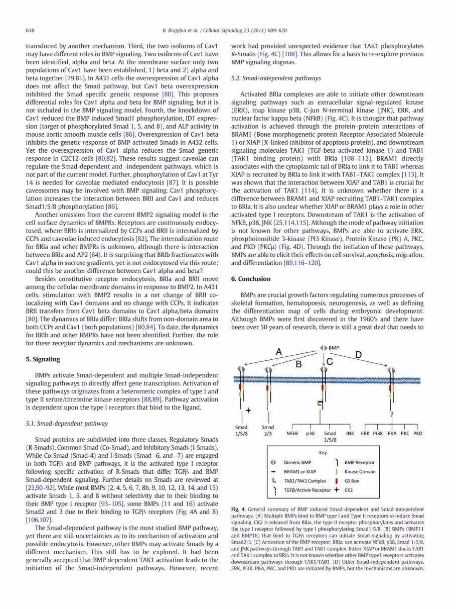
Fig. 4. General summary of BMP induced Smad-dependent and Smad-independentpathways. (A) Multiple BMPs bind to BMP type I and Type II receptors to induce Smadsignaling. CK2 is released from BRIa, the type II receptor phosphorylates and activatesthe type I receptor followed by type I phosphorylating Smad1/5/8. (B) BMPs (BMP11and BMP16) that bind to TGFβ receptors can initiate Smad signaling by activatingSmad2/3. (C) Activation of the BMP receptor, BRIa, can activate NFkB, p38, Smad 1/5/8,and JNK pathways through TAB1 and TAK1 complex. Either XIAP or BRAM1 docks TAB1and TAK1 complex to BRIa. It is not knownwhether other BMP type I receptors activatesdownstream pathways through TAK1/TAB1. (D) Other Smad-independent pathways,ERK, PI3K, PKA, PKC, and PKD are initiated by BMPs, but the mechanisms are unknown.
618 B. Bragdon et al. / Cellular Signalling 23 (2011) 609–620
transduced by another mechanism. Third, the two isoforms of Cav1may have different roles in BMP signaling. Two isoforms of Cav1 havebeen identified, alpha and beta. At the membrane surface only twopopulations of Cav1 have been established, 1) beta and 2) alpha andbeta together [79,81]. In A431 cells the overexpression of Cav1 alphadoes not affect the Smad pathway, but Cav1 beta overexpressioninhibited the Smad specific genetic response [80]. This proposesdifferential roles for Cav1 alpha and beta for BMP signaling, but it isnot included in the BMP signaling model. Fourth, the knockdown ofCav1 reduced the BMP induced Smad1 phosphorylation, ID1 expres-sion (target of phosphorylated Smad 1, 5, and 8), and ALP activity inmouse aortic smooth muscle cells [86]. Overexpression of Cav1 betainhibits the genetic response of BMP activated Smads in A432 cells.Yet the overexpression of Cav1 alpha reduces the Smad geneticresponse in C2C12 cells [80,82]. These results suggest caveolae canregulate the Smad-dependent and -independent pathways, which isnot part of the current model. Further, phosphorylation of Cav1 at Tyr14 is needed for caveolae mediated endocytosis [87]. It is possiblecaveosomes may be involved with BMP signaling. Cav1 phosphory-lation increases the interaction between BRII and Cav1 and reducesSmad1/5/8 phosphorylation [86].
Another omission from the current BMP2 signaling model is thecell surface dynamics of BMPRs. Receptors are continuously endocy-tosed, where BRIb is internalized by CCPs and BRII is internalized byCCPs and caveolae induced endocytosis [82]. The internalization routefor BRIa and other BMPRs is unknown, although there is interactionbetween BRIa and AP2 [84]. It is surprising that BRIb fractionates withCav1 alpha in sucrose gradients, yet is not endocytosed via this route;could this be another difference between Cav1 alpha and beta?
Besides constitutive receptor endocytosis, BRIa and BRII moveamong the cellular membrane domains in response to BMP2. In A431cells, stimulation with BMP2 results in a net change of BRII co-localizing with Cav1 domains and no change with CCPs. It indicatesBRII transfers from Cav1 beta domains to Cav1 alpha/beta domains[80]. The dynamics of BRIa differ; BRIa shifts from non-domain area toboth CCPs and Cav1 (both populations) [80,84]. To date, the dynamicsfor BRIb and other BMPRs have not been identified. Further, the rolefor these receptor dynamics and mechanisms are unknown.
5. Signaling
BMPs activate Smad-dependent and multiple Smad-independentsignaling pathways to directly affect gene transcription. Activation ofthese pathways originates from a heteromeric complex of type I andtype II serine/threonine kinase receptors [88,89]. Pathway activationis dependent upon the type I receptors that bind to the ligand.
5.1. Smad-dependent pathway
Smad proteins are subdivided into three classes, Regulatory Smads(R-Smads), Common Smad (Co-Smad), and Inhibitory Smads (I-Smads).While Co-Smad (Smad-4) and I-Smads (Smad -6, and -7) are engagedin both TGFβ and BMP pathways, it is the activated type I receptorfollowing specific activation of R-Smads that differ TGFβ and BMPSmad-dependent signaling. Further details on Smads are reviewed at[23,90–92]. While most BMPs (2, 4, 5, 6, 7, 8b, 9, 10, 12, 13, 14, and 15)activate Smads 1, 5, and 8 without selectivity due to their binding totheir BMP type I receptor [93–105], some BMPs (11 and 16) activateSmad2 and 3 due to their binding to TGFβ receptors (Fig. 4A and B)[106,107].
The Smad-dependent pathway is the most studied BMP pathway,yet there are still uncertainties as to its mechanism of activation andpossible endocytosis. However, other BMPs may activate Smads by adifferent mechanism. This still has to be explored. It had beengenerally accepted that BMP dependent TAK1 activation leads to theinitiation of the Smad-independent pathways. However, recent
work had provided unexpected evidence that TAK1 phosphorylatesR-Smads (Fig. 4C) [108]. This allows for a basis to re-explore previousBMP signaling dogmas.
5.2. Smad-independent pathways
Activated BRIa complexes are able to initiate other downstreamsignaling pathways such as extracellular signal-regulated kinase(ERK), map kinase p38, C-jun N-terminal kinase (JNK), ERK, andnuclear factor kappa beta (NFkB) (Fig. 4C). It is thought that pathwayactivation is achieved through the protein–protein interactions ofBRAM1 (Bone morphogenetic protein Receptor Associated Molecule1) or XIAP (X-linked inhibitor of apoptosis protein), and downstreamsignaling molecules TAK1 (TGF-beta activated kinase 1) and TAB1(TAK1 binding protein) with BRIa [108–112]. BRAM1 directlyassociates with the cytoplasmic tail of BRIa to link it to TAB1 whereasXIAP is recruited by BRIa to link it with TAB1–TAK1 complex [113]. Itwas shown that the interaction between XIAP and TAB1 is crucial forthe activation of TAK1 [114]. It is unknown whether there is adifference between BRAM1 and XIAP recruiting TAB1–TAK1 complexto BRIa. It is also unclear whether XIAP or BRAM1 plays a role in otheractivated type I receptors. Downstream of TAK1 is the activation ofNFkB, p38, JNK [25,114,115]. Although the mode of pathway initiationis not known for other pathways, BMPs are able to activate ERK,phosphoinositide 3-kinase (PI3 Kinase), Protein Kinase (PK) A, PKC,and PKD (PKCμ) (Fig. 4D). Through the initiation of these pathways,BMPs are able to elicit their effects on cell survival, apoptosis, migration,and differentiation [89,116–120].
6. Conclusion
BMPs are crucial growth factors regulating numerous processes ofskeletal formation, hematopoesis, neurogenesis, as well as definingthe differentiation map of cells during embryonic development.Although BMPs were first discovered in the 1960's and there havebeen over 50 years of research, there is still a great deal that needs to

619B. Bragdon et al. / Cellular Signalling 23 (2011) 609–620
be understood [6]. Its known signaling is initiated by BMP binding toits BRI and type II receptors. Its genetic control is mediated bymultiplesignaling pathways that can be divided into two groups, Smad-independent and Smad-dependent pathways. This review hashighlighted some of these areas in BMP research: secretion, proteinsin the signaling receptor complexes, regulation of receptor activation,receptor localization and effect on signaling. An understanding in BMPsignaling may allow for novel clinical applications. Hopefully in thenext 50 years these questions will be answered.
References
[1] K. Lavery, et al., J. Biol. Chem. 283 (30) (2008) 20948.[2] M. Kawabata, T. Imamura, K. Miyazono, Cytokine Growth Factor Rev. 9 (1)
(1998) 49.[3] K.V. Anderson, et al., Cold Spring Harb. Symp. Quant. Biol. 57 (1992) 409.[4] E.L. Ferguson, K.V. Anderson, Development 114 (3) (1992) 583.[5] R.P. Ray, et al., Development 113 (1) (1991) 35.[6] M.R. Urist, Science 150 (698) (1965) 893.[7] WagnerD.O. , et al., Sci. Signal. 3 (107) (2010) mr1.[8] S. Mazerbourg, A.J. Hsueh, Hum. Reprod. Update 12 (4) (2006) 373.[9] A. von Bubnoff, K.W. Cho, Dev. Biol. 239 (1) (2001) 1.
[10] H. Zhang, A. Bradley, Development 122 (10) (1996) 2977.[11] G. Winnier, et al., Genes Dev. 9 (17) (1995) 2105.[12] G.Q. Zhao, B.L. Hogan, Mech. Dev. 57 (2) (1996) 159.[13] H. Chen, et al., Development 131 (9) (2004) 2219.[14] A. Bandyopadhyay, et al., PLoS Genet. 2 (12) (2006) e216.[15] R.Y. Kim, E.J. Robertson, M.J. Solloway, Dev. Biol. 235 (2) (2001) 449.[16] M.J. Solloway, E.J. Robertson, Development 126 (8) (1999) 1753.[17] D. Guo, et al., J. Bone Miner. Res. 19 (2004) S14.[18] K. Cox, et al., J. Bone Miner. Res. 19 (2004) S11.[19] K.A. Lawson, et al., Genes Dev. 13 (4) (1999) 424.[20] G.Q. Zhao, Genesis 35 (1) (2003) 43.[21] P. Ducy, G. Karsenty, Kidney Int. 57 (6) (2000) 2207.[22] Y.T. Xiao, L.X. Xiang, J.Z. Shao, Biochem. Biophys. Res. Commun. 362 (3) (2007)
550.[23] C. Sieber, et al., Cytokine Growth Factor Rev. 20 (5–6) (2009) 343.[24] S.M. Nelsen, J.L. Christian, J. Biol. Chem. 284 (40) (2009) 27157.[25] A. Nohe, et al., Cell. Signal. 16 (3) (2004) 291.[26] S.C. Little, M.C. Mullins, Nat. Cell Biol. 11 (5) (2009) 637.[27] D.I. Israel, et al., Growth Factors 13 (3–4) (1996) 291.[28] G. Sengle, et al., J. Biol. Chem. 283 (20) (2008) 13874.[29] M.A. Brown, et al., J. Biol. Chem. 280 (26) (2005) 25111.[30] G. Sengle, et al., J. Mol. Biol. 381 (4) (2008) 1025.[31] F. Ramirez, D.B. Rifkin, Curr. Opin. Cell Biol. 21 (5) (2009) 616.[32] S. Sopory, et al., J. Biol. Chem. 281 (45) (2006) 34021.[33] C. Degnin, et al., Mol. Biol. Cell 15 (11) (2004) 5012.[34] Y. Cui, et al., Genes Dev. 15 (21) (2001) 2797.[35] D.C. Goldman, et al., Development 133 (10) (2006) 1933.[36] N.N. Nahar, et al., J. Bone Miner. Metab. 26 (5) (2008) 514.[37] L.N. Wu, et al., J. Biol. Chem. 268 (33) (1993) 25084.[38] H.C. Anderson, J. Cell Biol. 41 (1) (1969) 59.[39] E. Bonucci, J. Ultrastruct. Res. 20 (1) (1967) 33.[40] Z. Xiao, et al., J. Proteomics 72 (1) (2009) 34.[41] E. Cocucci, G. Racchetti, J. Meldolesi, Trends Cell Biol. 19 (2) (2009) 43.[42] K. Kodaira, et al., Biochem. Biophys. Res. Commun. 345 (3) (2006) 1224.[43] B. Herrera, G.J. Inman, BMC Cell Biol. 10 (2009) 20.[44] ElstnerA. , et al., J. Neurooncol. (2010).[45] O. Avsian-Kretchmer, A.J. Hsueh, Mol. Endocrinol. 18 (1) (2004) 1.[46] U.A. Vitt, S.Y. Hsu, A.J. Hsueh, Mol. Endocrinol. 15 (5) (2001) 681.[47] E. Di Pasquale, A.H. Brivanlou, J. Biol. Chem. 284 (38) (2009) 26127.[48] L.W. Gamer, et al., Dev. Biol. 285 (1) (2005) 156.[49] WalshD.W. , et al., Trends Cell Biol. (2010).[50] V. Rosen, Ann. NY Acad. Sci. 1068 (2006) 19.[51] J. Groppe, et al., Nature 420 (6916) (2002) 636.[52] M. Yanagita, Biofactors 35 (2) (2009) 113.[53] E. Gazzerro, E. Canalis, Rev. Endocr. Metab. Disord. 7 (1–2) (2006) 51.[54] M. Yanagita, Cytokine Growth Factor Rev. 16 (3) (2005) 309.[55] W. Balemans, W. Van Hul, Dev. Biol. 250 (2) (2002) 231.[56] T.B. Thompson, et al., Dev. Cell 9 (4) (2005) 535.[57] M. Ikeya, et al., Dev. Biol. 337 (2) (2010) 405.[58] D.G. Winkler, et al., J. Biol. Chem. 279 (35) (2004) 36293.[59] R. Ruppert, E. Hoffmann, W. Sebald, Eur. J. Biochem. 237 (1) (1996) 295.[60] X. Jiao, et al., J. Biol. Chem. 282 (2) (2007) 1080.[61] C.C. Rider, Biochem. Soc. Trans. 34 (Pt 3) (2006) 458.[62] K.K. Wurzler, et al., Mund Kiefer Gesichtschir. 8 (2) (2004) 83.[63] F. Liu, et al., Mol. Cell. Biol. 15 (7) (1995) 3479.[64] Y. Shi, J. Massague, Cell 113 (6) (2003) 685.[65] J. Groppe, et al., Mol. Cell 29 (2) (2008) 157.[66] K. Heinecke, et al., BMC Biol. 7 (2009) 59.[67] S. Saremba, et al., FEBS J. 275 (1) (2008) 172.[68] B.B. Koenig, et al., Mol. Cell. Biol. 14 (9) (1994) 5961.[69] P. Knaus, W. Sebald, Biol. Chem. 382 (8) (2001) 1189.
[70] J. Greenwald, et al., Mol. Cell 11 (3) (2003) 605.[71] L. Gilboa, et al., J. Cell Biol. 140 (4) (1998) 767.[72] L. Gilboa, et al., Mol. Biol. Cell 11 (3) (2000) 1023.[73] A. Nohe, et al., J. Cell Sci. 116 (Pt 16) (2003) 3277.[74] NickelJ. , et al., J. Bone Joint Surg. Am. 83-A Suppl 1 (Pt 1) (2001) S7-14.[75] E. Corradini, J.L. Babitt, H.Y. Lin, Cytokine Growth Factor Rev. 20 (5–6) (2009)
389.[76] D. Onichtchouk, et al., Nature 401 (6752) (1999) 480.[77] F. Kugimiya, et al., Biochem. Biophys. Res. Commun. 338 (2) (2005) 872.[78] B. Bragdon, et al., Biophys. J. 99 (3) (2010) 897.[79] NoheA. , et al., Faraday Discuss. 126 (2004) 1858 discussion 245-54.[80] A. Nohe, et al., J. Cell Sci. 118 (Pt 3) (2005) 643.[81] B. Razani, S.E. Woodman, M.P. Lisanti, Pharmacol. Rev. 54 (3) (2002) 431.[82] A. Hartung, et al., Mol. Cell. Biol. 26 (20) (2006) 7791.[83] W. Shi, et al., J. Cell Sci. 120 (Pt 7) (2007) 1216.[84] B. Bragdon, et al., Biophys. J. 97 (5) (2009) 1428.[85] C. Rauch, et al., Am. J. Physiol. Cell Physiol. 283 (1) (2002) C235.[86] J.W. Wertz, P.M. Bauer, Biochem. Biophys. Res. Commun. 375 (4) (2008) 557.[87] E.M. Khan, et al., J. Biol. Chem. 281 (20) (2006) 14486.[88] A. Nohe, et al., J. Biol. Chem. 277 (7) (2002) 5330.[89] Q. Zhou, et al., Cardiovasc. Res. 76 (3) (2007) 390.[90] B. Song, K.D. Estrada, K.M. Lyons, Cytokine Growth Factor Rev. 20 (5–6) (2009)
379.[91] C.S. Hill, Cell Res. 19 (1) (2009) 36.[92] S. Itoh, P. ten Dijke, Curr. Opin. Cell Biol. 19 (2) (2007) 176.[93] A.S. Pachori, et al., J. Mol. Cell. Cardiol. 48 (6) (2010) 1255.[94] K. Miyazono, S. Maeda, T. Imamura, Cytokine Growth Factor Rev. 16 (3) (2005)
251.[95] S. Mazerbourg, et al., J. Biol. Chem. 280 (37) (2005) 32122.[96] T. Ebisawa, et al., J. Cell Sci. 112 (Pt 20) (1999) 3519.[97] C. Kersten, et al., BMC Immunol. 6 (1) (2005) 9.[98] V. Zuzarte-Luis, et al., Dev. Biol. 272 (1) (2004) 39.[99] R. Motazed, et al., Pharm. Res. 25 (10) (2008) 2440.
[100] T.W. Axelrad, T.A. Einhorn, Cytokine Growth Factor Rev. 20 (5–6) (2009) 481.[101] P.D. Upton, et al., J. Biol. Chem. 284 (23) (2009) 15794.[102] L. David, et al., Blood 109 (5) (2007) 1953.[103] M. Sammar, et al., Genes Cells 9 (12) (2004) 1227.[104] A.M. Sullivan, G.W. O'Keeffe, J. Anat. 207 (3) (2005) 219.[105] R.K. Moore, F. Otsuka, S. Shimasaki, J. Biol. Chem. 278 (1) (2003) 304.[106] S.P. Oh, et al., Genes Dev. 16 (21) (2002) 2749.[107] A.E. Drummond, Cell Tissue Res. 322 (1) (2005) 107.[108] J.H. Shim, et al., EMBO J. 28 (14) (2009) 2028.[109] K.M. Wu, et al., J. Biomed. Sci. 13 (3) (2006) 345.[110] H. Shibuya, et al., EMBO J. 17 (4) (1998) 1019.[111] K. Yamaguchi, et al., EMBO J. 18 (1) (1999) 179.[112] K. Kurozumi, et al., Genes Cells 3 (4) (1998) 257.[113] P.J. Chung, et al., J. Biol. Chem. 277 (42) (2002) 39850.[114] M. Lu, et al., Mol. Cell 26 (5) (2007) 689.[115] J. Guicheux, et al., J. Bone Miner. Res. 18 (11) (2003) 2060.[116] K. Sugimori, et al., J. Bone Miner. Metab. 23 (6) (2005) 411.[117] Y.S. Lee, C.M. Chuong, J. Cell. Physiol. 170 (2) (1997) 153.[118] J. Lemonnier, et al., J. Biol. Chem. 279 (1) (2004) 259.[119] G.C. Reilly, et al., Cell Commun. Signal 3 (1) (2005) 3.[120] C. Gamell, et al., J. Cell Sci. 121 (Pt 23) (2008) 3960.[121] E. Kessler, et al., Science 271 (5247) (1996) 360.[122] N. Suzuki, et al., Development 122 (11) (1996) 3587.[123] T. Nakase, H. Yoshikawa, J. Bone Miner. Metab. 24 (6) (2006) 425.[124] BahamondeM.E. , LyonsK.M. , J. Bone Joint Surg. Am. 83-A Suppl 1 (Pt 1) (2001)
S56.[125] A. Daluiski, et al., Nat. Genet. 27 (1) (2001) 84.[126] N.S. Cunningham, et al., Growth Factors 12 (2) (1995) 99.[127] J. Hino, et al., Biochem. Biophys. Res. Commun. 223 (2) (1996) 304.[128] P. ten Dijke, et al., J. Biol. Chem. 269 (25) (1994) 16985.[129] P.X. Xu, et al., Development 130 (14) (2003) 3085.[130] S. Oida, et al., DNA Seq. 5 (5) (1995) 273.[131] J.A. King, et al., Dev. Biol. 166 (1) (1994) 112.[132] T.B. Ro, et al., Oncogene 23 (17) (2004) 3024.[133] G.V. Hahn, et al., Genomics 14 (3) (1992) 759.[134] D. Nonner, et al., J. Neurochem. 77 (2) (2001) 691.[135] M.J. Solloway, et al., Dev. Genet. 22 (4) (1998) 321.[136] Y. Zhang, G. Ge, D.S. Greenspan, J. Biol. Chem. 281 (51) (2006) 39096.[137] A.T. Dudley, K.M. Lyons, E.J. Robertson, Genes Dev. 9 (22) (1995) 2795.[138] G. Luo, et al., Genes Dev. 9 (22) (1995) 2808.[139] G.Q. Zhao, et al., Genes Dev. 10 (13) (1996) 1657.[140] L. David, et al., Circ. Res. 102 (8) (2008) 914.[141] L.W. Hillier, et al., Nature 434 (7034) (2005) 724.[142] M. Nakashima, et al., Hum. Gene Ther. 14 (6) (2003) 591.[143] L.W. Gamer, et al., Dev. Biol. 208 (1) (1999) 222.[144] K. Furuya, et al., J. Cell. Biochem. 72 (2) (1999) 177.[145] N.M. Wolfman, et al., J. Clin. Invest. 100 (2) (1997) 321.[146] H. Nochi, et al., J. Bone Miner. Res. 19 (1) (2004) 111.[147] B. Shen, et al., Int. J. Biol. Sci. 5 (2) (2009) 192.[148] E. Di Pasquale, et al., J. Clin. Endocrinol. Metab. 91 (5) (2006) 1976.[149] N. Feiner, et al., BMC Evol. Biol. 9 (2009) 277.[150] CelesteA.J. , MurrayB.L. , Bone Morphogenetic Protein (BMP)-17 and BMP-18
Compositions, Genetics Institute, LLc, US, 2003.

620 B. Bragdon et al. / Cellular Signalling 23 (2011) 609–620
[151] H. Enomoto, et al., Oncogene 9 (10) (1994) 2785.[152] M.S. Dionne, W.C. Skarnes, R.M. Harland, Mol. Cell. Biol. 21 (2) (2001) 636.[153] H. Ideno, et al., Exp. Cell Res. 315 (3) (2009) 474.[154] S. Sudo, et al., J. Biol. Chem. 279 (22) (2004) 23134.[155] L.Z. Topol, et al., J. Biol. Chem. 275 (12) (2000) 8785.[156] R. McMahon, et al., J. Biol. Chem. 275 (14) (2000) 9901.[157] R.C. Pereira, A.N. Economides, E. Canalis, Endocrinology 141 (12) (2000) 4558.[158] O. Michos, et al., Development 131 (14) (2004) 3401.[159] M.K. Khokha, et al., Nat. Genet. 34 (3) (2003) 303.[160] A. Zuniga, et al., Nature 401 (6753) (1999) 598.[161] S. Piccolo, et al., Nature 397 (6721) (1999) 707.[162] E. Bell, et al., Development 130 (7) (2003) 1381.[163] S. Marques, et al., Genes Dev. 18 (19) (2004) 2342.[164] J.J. Pearce, G. Penny, J. Rossant, Dev. Biol. 209 (1) (1999) 98.[165] Y. Yokouchi, et al., Cell 98 (5) (1999) 573.[166] M. Yanagita, et al., Biochem. Biophys. Res. Commun. 316 (2) (2004) 490.[167] D.G. Simmons, T.G. Kennedy, Biol. Reprod. 67 (5) (2002) 1638.[168] Y. Kassai, et al., Science 309 (5743) (2005) 2067.[169] A. Murashima-Suginami, et al., Biochem. Biophys. Res. Commun. 359 (3) (2007)
549.[170] M. Tanaka, et al., Kidney Int. 73 (2) (2008) 181.[171] N. Itasaki, et al., Development 130 (18) (2003) 4295.[172] R.L. van Bezooijen, et al., J. Exp. Med. 199 (6) (2004) 805.[173] M.E. Brunkow, et al., Am. J. Hum. Genet. 68 (3) (2001) 577.[174] N. Kusu, et al., J. Biol. Chem. 278 (26) (2003) 24113.[175] D.G. Winkler, et al., EMBO J. 22 (23) (2003) 6267.[176] X. Li, et al., J. Bone Miner. Res. 23 (6) (2008) 860.[177] R.L. van Bezooijen, et al., J. Bone Miner. Res. 22 (1) (2007) 19.[178] M. Ikeya, et al., Mech. Dev. 125 (9–10) (2008) 832.[179] M. Melnick, et al., Arch. Oral Biol. 51 (5) (2006) 433.[180] T. Nosaka, et al., Mol. Cell. Biol. 23 (8) (2003) 2969.[181] M. Oelgeschlager, et al., Nature 405 (6788) (2000) 757.[182] F.C. Wardle, J.V. Welch, L. Dale, Mech. Dev. 86 (1–2) (1999) 75.[183] Y. Sasai, et al., Cell 79 (5) (1994) 779.[184] D. Zhang, et al., J. Bone Miner. Res. 17 (2) (2002) 293.[185] D. Bachiller, et al., Nature 403 (6770) (2000) 658.[186] J.L. Zhang, et al., J. Biol. Chem. 282 (27) (2007) 20002.
[187] H. Sakuta, et al., Science 293 (5527) (2001) 111.[188] N. Nakayama, et al., Dev. Biol. 232 (2) (2001) 372.[189] OrenA. , et al., Gene 331 (2004) 17.[190] N. Nakayama, et al., Development 131 (1) (2004) 229.[191] J. Lin, et al., Nat. Med. 11 (4) (2005) 387.[192] Y. Jiang, et al., Mol. Cell. Neurosci. 41 (2) (2009) 113.[193] M.E. Binnerts, et al., Biochem. Biophys. Res. Commun. 315 (2) (2004) 272.[194] A.L. Ambrosio, et al., Dev. Cell 15 (2) (2008) 248.[195] N. Koike, et al., J. Biol. Chem. 282 (21) (2007) 15843.[196] H. Miwa, et al., FEBS Lett. 583 (22) (2009) 3643.[197] W.C. Smith, R.M. Harland, Cell 70 (5) (1992) 829.[198] L.J. Brunet, et al., Science 280 (5368) (1998) 1455.[199] J.A. McMahon, et al., Genes Dev. 12 (10) (1998) 1438.[200] E. Gazzerro, V. Gangji, E. Canalis, J. Clin. Invest. 102 (12) (1998) 2106.[201] J. Marcelino, et al., Proc. Natl Acad. Sci. USA 98 (20) (2001) 11353.[202] S.J. Lin, et al., Reproduction 132 (2) (2006) 179.[203] M.M. Matzuk, et al., Nature 374 (6520) (1995) 360.[204] K. Tsuchida, et al., J. Biol. Chem. 275 (52) (2000) 40788.[205] L. Bartholin, et al., Biol. Cell 97 (7) (2005) 577.[206] M. Scharpfenecker, et al., J. Cell Sci. 120 (Pt 6) (2007) 964.[207] O. Shmueli, et al., C.R. Biol. 326 (10–11) (2003) 1067.[208] D.W. Johnson, et al., Nat. Genet. 13 (2) (1996) 189.[209] R. Derynck, X.H. Feng, Biochim. Biophys. Acta 1333 (2) (1997) F105.[210] I. Yanai, et al., Bioinformatics 21 (5) (2005) 650.[211] H. Ide, et al., Cytogenet. Cell Genet. 81 (3–4) (1998) 285.[212] P. ten Dijke, et al., Oncogene 8 (10) (1993) 2879.[213] B.L. Rosenzweig, et al., Proc. Natl Acad. Sci. USA 92 (17) (1995) 7632.[214] C.J. Donaldson, L.S. Mathews, W.W. Vale, Biochem. Biophys. Res. Commun. 184
(1) (1992) 310.[215] R. Kosaki, et al., Am. J. Med. Genet. 82 (1) (1999) 70.[216] S. Ishikawa, et al., J. Hum. Genet. 43 (2) (1998) 132.[217] P.J. Halbrooks, et al., J. Mol. Signal. 2 (2007) 4.[218] S. Hassel, et al., J. Cell. Physiol. 206 (2) (2006) 457.[219] N.P. Barbara, J.L. Wrana, M. Letarte, J. Biol. Chem. 274 (2) (1999) 584.[220] K.C. Kirkbride, et al., J. Biol. Chem. 283 (12) (2008) 7628.[221] WordingerR.J. , ClarkA.F. , Exp. Biol. Med. (Maywood) 232 (8) (2007) 979.[222] W. Jin, et al., Cancer Res. 67 (20) (2007) 9869.


