Embed Size (px)
Citation preview
Biosonar and Neural Computation in Bats
Bats extract remarkably detailed information about their surroundings from biosonar signals. Neurons in their auditory
systems are highly specialized for performing this task
It used to be a common misconception that bats' use of sound pulses to navigate and locate prey is a
crude system, the acoustic equivalent of feeling one's way in the dark with a cane. But biosonar has since been shown to be anything but crude: an echolocating bat can pursue and capture a fleeing moth with a facility and success rate that would be the envy of any military aerospace engineer.
In addition to providing information about how far away a target is, bat sonar can relay some remarkable details. Doppler shifts-changes in the frequency of the echo relative to the original signal-convey information not only about the relative velocity of a flying insect but also about its wingbeat. The amplitude of the echo, combined with the delay, indicates the size of the target. The amplitudes of the component frequencies correspond to the size of various features of the target. Differences between the ears in intensity and arrival time of sound give the azimuth of the target, whereas the interference pattern of sound waves reflected within the structure of the outer ear gives the elevation.
The complex neural computations needed to extract this information occur within a brain the size of a large pearl. For the past 27 years my colleagues and I have been exploring the neural mechanisms that underlie the
NORUO SUGA has been professor of biology at Washington University in Saint Louis, Mo. , since 1976. Suga was born in Japan and attended the Tokyo Metropolitan UniverSity, where he received his B.A. in 1958 and his Ph.D. in biology in 1963. He then went to Harvard University as a research associate, where he first studied the auditory system of bats with Donald R. Griffin.
60 SCIENTIFIC AMERICAN June 1990
by Nobuo Suga
echolocating abilities of bats. The well-defined characteristics of a bat's auditory world make the animal ideal for elucidating the information proceSSing that goes on in its auditory system. Similar mechanisms are undoubtedly shared by other animals.
There are some 800 species of Microchiropteran bats in the world today, all of which are presumed
to echo locate. These species live in diverse habitats and vary greatly in behavior and physical characteristics. Their biosonar pulses also differ, even among species within the same genus. Nevertheless, these pulses can be classified into three types: constant frequency (CF), frequency modulated (FM) and combined CF-FM. CF pulses consist of a single frequency, or tone. FM pulses sweep downward and sound like chirps. Combined CF -FM pulses consist of a long, constant tone followed by a downward chirp, iiiiiiu. In many bats the tones are not pure but rather consist of a fundamental, or first, harmonic and several higher harmonics (multiples of the fundamental frequency).
Most bat species emit only one type of pulse. The little broWn bat, Myotis lucifuqus, is an "FM" bat; it emits FM pulses lasting between .5 and three milliseconds and sweeping downward by about one octave. The mustached bat, Pteronotus parnellii, is a "CF-FM" bat; it emits long CF pulses lasting between five and 30 milliseconds followed by a short FM sweep lasting between two and four milliseconds. Several species change their pulses, depending on the situation. The fishcatching bat, Noctilio leporinus, for example, emits CF and CF -FM pulses while cruising in flight but emits FM pulses while hunting prey.
A long CF pulse is excellent for de-
tecting targets larger than the wavelength of the signal, because the reflected sound energy is highly concentrated at a particular frequency. It is also ideal for measuring Doppler shifts. The CF pulse is not appropriate, however, for locating a target precisely or discerning its details. A larger number of frequencies is needed to obtain more information about target features. Bats broaden their frequency bandwidth by producing harmonics and by emitting FM bursts that sweep over a wide frequency range. FM pulses also contain more information about time and so are used to compute echo delays and thereby determine the distance to a target.
Certain bat species control the energy in each harmonic depending on the distance to a target. If the target is far away, they amplify the lower harmonics, which are less attenuated by the air. But if the target is nearby, they enhance the higher harmonics to obtain finer details of the target. When closing in on prey, Microchiropterans shorten the duration of pulses and increase the rate of pulse emission, up to 200 per second in FM bats and up to 100 per second in CF-FM bats. This adjustment occurs not only because bats need to characterize the prey in greater detail but also because when the distance between a bat and its prey is small, the angular position of the prey changes more rapidly, and so the bat needs to emit more signals to track the prey accurately.
The hunting strategies and behavior of a bat species are directly related to the characteristics of its biosonar. The
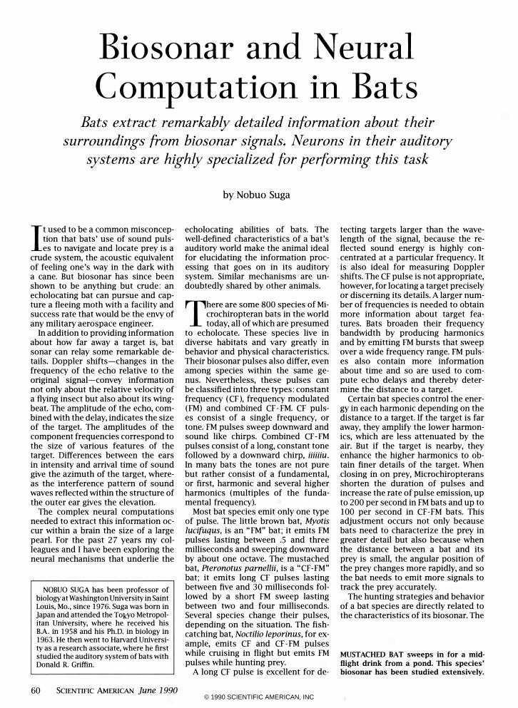
MUSTACHED BAT sweeps in for a midflight drink from a pond. This species' biosonar has been studied extensively.
© 1990 SCIENTIFIC AMERICAN, INC
key elements of the biosonar, in turn, are reflected in the functional organization of its auditory system. Since bat biosonar was established by Donald R. Griffin and Robert Galambos four decades ago, neuroethologists have studied the auditory system in several bat species but most of all in the little brown bat, the mustached bat and the horseshoe bat, Rhinolophus ferrumequinum. Each of these bats produces distinctive biosonar pulses.
The auditory mechanisms of the mustached bat have been the most thoroughly examined. The biosonar and peripheral auditory system of this bat were first described in 1964 and 1972, respectively, by Alvin Novick and his co-workers at Yale University. In 1972 I began research on the peripheral auditory system of the mustached bat with James A. Simmons, now at Brown University, and on the central auditory system with Philip H.-S. Jen, now at the University of Missouri at Columbia. Subsequently, Toshiki Manabe and Kazuro Kujirai, now at Yokohama City University, William E . O'Neill, now at the University of Rochester, and nearly two dozen others have joined in studying this bat's central auditory system and its neural
mechanisms for processing biosonar information. This article will mainly describe findings for this species. �ying mustached bat detects the
relative velocity of objects by the Doppler shift in the echoes.
When a bat flies toward a stationary object, the pulses that strike and are reflected by it become compressed, or Doppler-shifted. The echo received by the bat is therefore uniformly higher in frequency than the emitted pulse. When the animal flies toward a flying insect, the insect's beating wings introduce oscillating frequency shifts, which are superposed on the overall Doppler shift, like small surface ripples on an ocean wave.
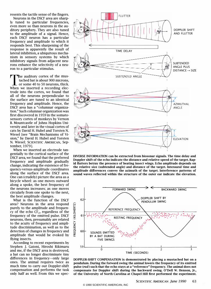
Certain bats, such as the mustached bat and horseshoe bat, can detect ripples from insect wings against the echoes associated with stationary objects, such as walls and vegetation. How do they do it? Part of the answer lies in a trick called Doppler-shift compensation, first seen by Hans-Ulrich Schnitzler of the University of Tiibingen. A mustached bat at rest emits a fundamental tone of around 30.5 kilohertz, along with three higher harmonics; the "resting" frequency of
the second harmonic (CF2) is around 61 kilohertz. If the bat detects a Doppler-shifted echo at 63 kilohertz from a stationary object, it reduces the frequency of emitted pulses by about 1.8 kilohertz, so that subsequent echoes are stabilized at a "reference" frequency of around 61.2 kilohertz.
These bats turn out to be specialized to analyze tiny differences in frequencies near the reference frequency. Hence, Doppler-shift compensation brings the echo CF2 into the range at which the bat can most easily detect ripples from beating insect wings.
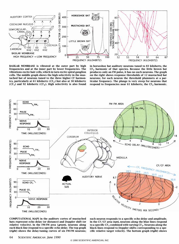
This specialization begins in the bat's ear. Of particular interest is the inner ear, or cochlea, which contains the basilar membrane, a thin, elongated sheet curled up like a snail. When sound waves vibrate the eardrum, this vibration is conducted to the basilar membrane, stimulating tiny hair cells on the membrane. The excitation is transmitted, via the spiral ganglion cells, along the auditory nerve fibers to the brain.
The neural signal produced at the cochlea must contain all the information vital to the bat. The physical properties of an acoustic signal-amplitude, time and frequency-must in
© 1990 SCIENTIFIC AMERICAN, INC
MUSTACHED BAT lITILE BROWN BAT
N N 120 CF. \ � �\ \\ I- 120 FM.\ � c:r: UJ UJ J: CF, \ �\ 0 90 FM,\ � :;z CF, \ �\ >- 60 FM, \ u z CF, UJ
� �� => 30 I FM,\ 0 UJ PULSE ECHO c:r: LL. 0
0 20 40 60 TIME (MILLISECONDS)
HUNTS IN VEGETATION
\\ \\ J: 90 0 ,u�' � :;z
\\,' 60 >-u z
\ \ � UJ => 30 0 UJ c:r: I LL. 0
80 100 0
.\ \ .. \ ECHO
,'. , ' . i
20 40 60 80 TIME (MILLISECONDS)
". \� , .. \', 100 120
" --,,,X - .... \ ""tt!f? ',j :
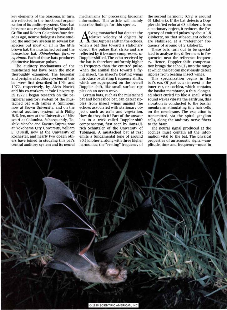
BIOSONAR PULSE of the mustached bat consists of a long, constant-frequency ( CF ) component followed by a short, frequency-modulated (FM) component. Each pulse contains four harmonics (indicated by subscripts). When closing in on a
target, the bat emits shorter pulses at a higher rate, while keeping the same tones. The little brown bat emits only FM chirps. When nearing a target, it emits shorter, lower chirps at a faster rate. Each species emits pulses suited to its behavior.
turn be translated into neural activity. Amplitude is expressed by the rate at which the auditory nerve fibers discharge impulses: the greater the amplitude, the higher the discharge rate. The duration of signals and the intervals between them are mimicked by the pattern of the nerve impulses. The frequency of the signal is expressed by location on the basilar membrane: high frequencies vibrate the portion nearest the eardrum, whereas lower ones stimulate portions farther in.
A certain portion of the mustached bat's basilar membrane is unusually thick. This thickness is related to extreme sensitivity to frequencies of between 61.0 and 61.5 kilohertz (the CF2 of the Doppler shift-compensated echoes) as well as insensitivity to frequencies of around 59.5 kilohertz (the CF2 of the Doppler shift-compensating pulses). In other words, the membrane is strongly stimulated by the echoes but poorly stimulated by the animal's own vocalizations.
The frequency selectivity of the spiral ganglion cells is extremely high within the key range of 61.0 to 61.5 kilohertz. They are tuned to single frequencies. That is, each neuron has a "best" frequency (the frequency that evokes the largest response), which differs slightly from that of its neighbors. Indeed, these neurons are so sharply tuned to their best frequencies that they can detect shifts as small as .01 percent. Flying insects can easily evoke frequency shifts an order of magnitude greater. The auditory periphery is also highly tuned to analyze frequency shifts near CF1 (30-kilo-
62 SCIENTIFIC AMERICAN June 1990
hertz) and CF3 (92-kilohertz) signals. In the mustached bat the great sen
sitivity and sharp tuning of the auditory periphery to the CF 2 frequency are combined with Doppler-shift compensation to proffer three advantages. First, the auditory periphery is exquisitely sensitive to the CF 2 echo (near 61 kilohertz) but is insensitive to the bat's emitted CF2 pulse (near 59 kilohertz) during Doppler-shift compensation; hence, masking of the echo by the emitted pulse is minimal. Second, the sharply tuned neurons are well able to detect the signal even if it is embedded in background noise. Third, the array of sharply tuned neurons has a high likelihood of picking up the echo from the beating wings of a flying insect as the echo sweeps up and down in frequency.
These advantages enable the mustached bat to hunt insects successfully, even in dense vegetation. Doppler-shift compensation and frequency tuning do not exist in FM bats, such as the little brown bat, which hunt in open air.
O nce the auditory signal has been coded into nerve Signals, it must be further analyzed to
extract such information as the velocity or distance of prey. This process occurs in the central auditory system. From the cochlea, signals are processed sequentially, beginning at the cochlear nucleus and proceeding to the lateral lemniscus, inferior colliculus, medial geniculate body and finally to the auditory cortex.
Using slender electrodes to record
nerve impulses from single neurons, my colleagues and I studied the responses of neurons in the mustached bat's auditory system as we stimulated the animal with biosonar signals. What we discovered was an extraordinarily developed system for processing that information. In particular, we found that different processing tasks are parceled out among several anatomically distinct areas of the auditory cortex. One region contains neurons that respond only to certain frequencies and amplitudes of echoes. A second region responds only to frequency differences between pulses and echoes. A third region is sensitive to the time interval between pulses and echoes.
By far the largest of the specialized regions in the mustached bat's auditory cortex is the one that processes Doppler-shifted CF 2 signals. This region, called the DSCF area, represents only a narrow sliver of the frequency range, between 60.6 and 62.3 kilohertz (when the bat's resting frequency is 61.00 kilohertz). Yet it occupies 30 percent of the primary auditory cortex. The exact frequencies overrepresented differ among individual bats according to their resting frequencies. In other words, each bat's auditory system is personalized.
Similar overrepresentation is found in the brain wherever the signal being processed is critical to an animal's behavior. For example, in cats and monkeys the visual cortex overrepresents the fovea, the area of the retina where visual acuity is highest. the primate somatosensory cortex overrep-
© 1990 SCIENTIFIC AMERICAN, INC
resents the tactile sense of the fingers. Neurons in the DSCF area are sharp
ly tuned to particular frequencies, even more so than neurons in the auditory periphery. They are also tuned to the amplitude of a signal. Hence, each DSCF neuron has a particular frequency and amplitude to which it responds best. This sharpening of the response is apparently the result of lateral inhibition, a ubiquitous mechanism in sensory systems by which inhibitory signals from adjacent neurons enhance the selectivity of a neuron to a particular stimulus.
The auditory cortex of the mustached bat is about 900 microns, or some 40 to 50 neurons, thick.
When we inserted a recording electrode into the cortex, we found that all of the neurons perpendicular to the surface are tuned to an identical frequency and amplitude. Hence, the DSCF area has a "columnar organization." Such columnar organization was first discovered in 1959 in the somatosensory cortex of monkeys by Vernon B. Mountcastle of Johns Hopkins University and later in the visual cortex of cats by David H. Hubel and Torsten N. Wiesel [see "Brain Mechanisms of Vision," by David H. Hubel and Torsten N. Wiesel; SCIENTIFIC AMERICAN, September, 19791.
When we inserted an electrode tangentially to the cortical surface of the DSCF area, we found that the preferred frequency and amplitude gradually change, indicating the existence of frequency-versus-amplitude coordinates along the surface of the DSCF area. One can (crudely) picture the area as a bicycle wheel: as one moves outward along a spoke, the best frequency of the neurons increases; as one moves circularly from one spoke to the next, the best amplitude changes.
What is the function of the DSCF area? Neurons in the area respond purely to the amplitude and frequency of the echo CF2 , regardless of the frequency of the emitted pulse. DSCF neurons, then, presumably are related to the acuity of frequency and amplitude discrimination, as well as to the detection of changes in frequency and amplitude that would be evoked by flying insects.
According to recent experiments by Stephen J. Gaioni, Hiroshi Rikimaru and me, if the DSCF area is destroyed, a bat can no longer discriminate tiny differences in frequency-only large ones. The animal requires twice as much time to carry out Doppler-shift compensation and performs the task only half as well. From this we spec-
TIME DELAY , , I ,
DOPPLER SHIFT AND FLUTTER
- -- : " SUBTENDED
�' � ,� DISTANCE�SI" SUBTENDED ANGLE I :
,
ANGLE PLUS
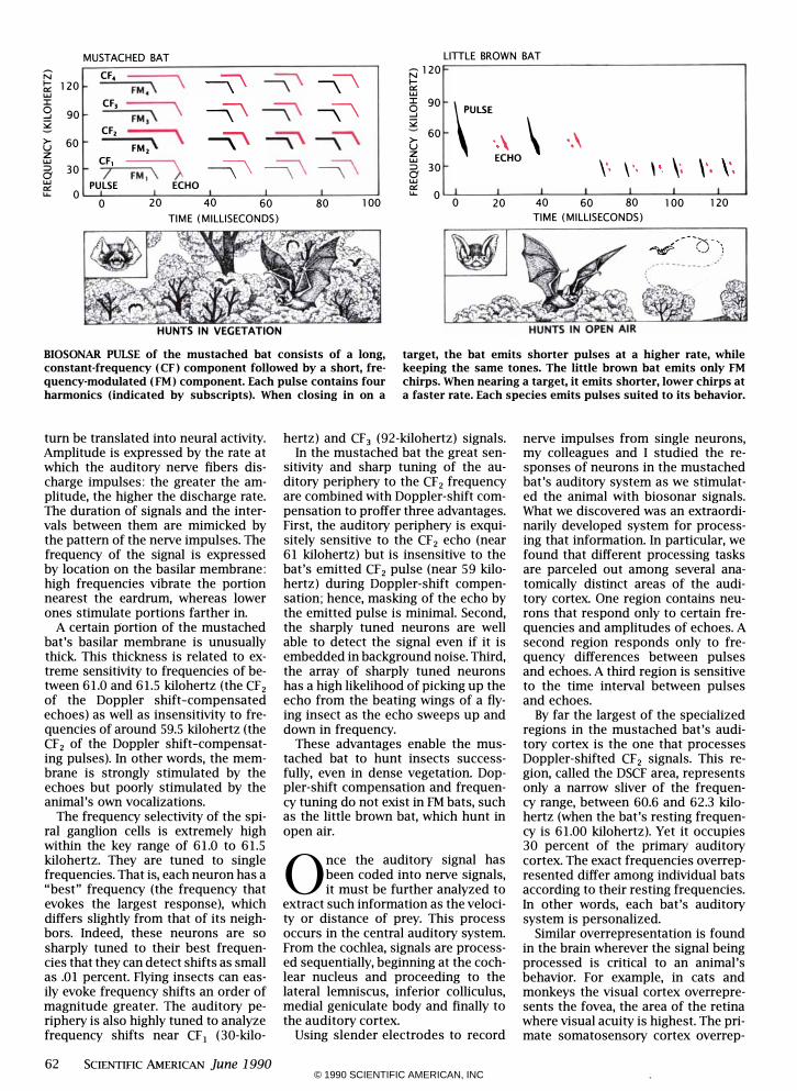
DIVERSE INFORMATION can be extracted from biosonar signals. The time delay and Doppler shift of the echo indicate the distance and relative speed of the target. Rapid flutters betray the presence of beating insect wings. Echo amplitude depends on the relative size (subtended angle) and distance of the target. lnteraural time and amplitude differences convey the azimuth of the target. Interference patterns of sound waves reflected within the structure of the outer ear indicate the elevation.
� UJ :I: o -' 52 >u z UJ ::> 8 c:r: u..
----�FO�R-W�A�R�D�SW�IN�G�--�> �<�--�B�A�C�K�W�A�R�D�S�W�IN�G�----
62 DOPPLER SHIFT BY ./ PENDULUM SWING
6 1 : •• :. : .:::.:: � ..... ------------------------ - .-':;.-1: :�.::.-: .. ::. :.: e •• ! • • • •• •• • ••• RESTING FREQUENCY .... •• • . .. • • • • . . , -. . . . . . . :- :. .. . : :. .. ::\ .. �. /. .. ... ;�::. 60 SOUNDS EMITTED �-:...... :.�:.
BY A BAT DURING ./ :6·.;::···· FIVE SWINGS
. -. . . . ...... ::::
59�------�0
--------------1L----- -------�
2-------------
3�
TIME (SECONDS)
DOPPLER-SlDFT COMPENSATION is demonstrated by placing a mustached bat on a pendulum. During the forward swing the animal lowers the frequency of its emitted pulse (red) such that the echo stays at a "reference" frequency. The animal does not compensate for Doppler shift during the backward swing. O'Dell W. Henson, Jr., of the University of North Carolina at Chapel Hill first performed the experiment.
SCIENTIFIC AMERICAN June 1990 63 © 1990 SCIENTIFIC AMERICAN, INC
Vl HORSESHOE BAT id C1 ...J UJ 400 ' ! co 0 ...J::J UJ Cl �� 0 300 MUSTACHED BAT _UJ U...J � ��60 �::::> 0:: ClVl 0 ...JVl I- 200 oUJ U I� <{ Vl u.. UJCl O::Z 40 I::::> 100 1-0 Vl
BASILAR MEMBRANE 0
HIGH FREQUENCY -> LOW FREQUENCY 0 25 SO 75 100 6 7 FREQUENCY (KILOHERTZ)
BASIlAR MEMBRANE is vibrated at the outer part by high frequencies and at the inner part by lower frequencies. The vibrations excite hair cells, which in turn excite spiral ganglion cells. The middle graph shows the high selectivity in the mustached bat of neurons tuned to the three higher CF harmonics, particularly at 6 1 kilohertz ( CF2) but also at 30 kilohertz ( CF 1) and 92 kilohertz ( CF 3)' High selectivity is also found
N ECHO I-0:: 90 �FM, UJ I 0 60 ...J :;;2 PULSE 30 �FM, >-
u Z 0 UJ NERVE
� ::::> 0 RESPONSE UJ 0:: u..
0 10 20 30 TIME (MILLISECONDS)
N I- 90 0:: UJ ECHO I 0 60 \ FM, ...J PULSE :;;2 30 �FM, >-u Z 0 UJ NERVE � ::::> RESPONSE 0 UJ 0:: u.. 0 10 20 30
TIME (MILLISECONDS)
in horseshoe bat auditory neurons tuned to 83 kilohertz, the CF2 harmonic of that species. Because the little brown bat produces only an FM pulse, it has no such neurons. The graph on the right shows response thresholds of 12 mustached bat neurons; for each neuron the threshold plummets at a particular frequency. The plunge is very steep for neurons that respond to frequencies near 6 1 kilohertz, the CF2 harmonic.
FM-FM AREA ---................
1 1 8 L<,:
f�&><s � 4 6 � «'� ECHO DELAY (MILL 8 1 0 1 4
� ISECONDS)
MEDULLA
�C CF/CF AREA F, FREQUENCY "'-"7l��2� 9 .:2'0 29.5 (KILOHERrz)
• AUDITORY NERVE 8. 7 30.0 30.5 �
N I- 90 0:: UJ I 60 0 ...J :;;2 30 >-u 0 Z UJ ::::> 0 UJ 0:: u..
ECHO CF, \.
PULSE CF, �
....KRVE RESPONSE
0 10 20 30 TIME (MILLISECONDS)
ACTUAL SIZE
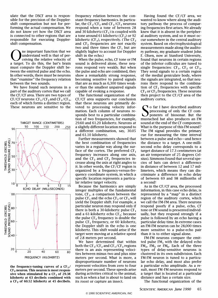
COMPUTATIONAL MAPS in the auditory cortex of mustached bats represent echo delay (or distance) and Doppler shift (or relative velocity). In the FM-FM area (green), neurons along each black line respond to a specific echo delay. The top graph (right) shows the delay-tuning curves of six FM-FM neurons;
64 SCIENTIFIC AMERICAN June 1990
each neuron responds to a specific echo delay and amplitude. In the CF / CF area (tan), neurons along the blue lines respond to a specific CF 1 combined with varying CF 2' Neurons along the black lines respond to Doppler shifts corresponding to a specific relative target velocity. The bottom graph (right) shows
-2
© 1990 SCIENTIFIC AMERICAN, INC
ulate that the DSCF area is responsible for the precision of the Dopplershift compensation but not for performing the actual compensation. We do not know yet how the DSCF area is connected to other regions that are responsible for executing Dopplershift compensation.
One important function that we understand well is that of perceiving the relative velocity of
a target. To do this, the bat's brain must compute the Doppler shift between the emitted pulse and the echo. In other words, there must be neurons that "examine" the frequency relation between the two sounds.
We have found such neurons in a part of the auditory cortex that we call the CF /CF area. There are two types of CF /CF neurons: CF,/CF2 and CF,/CF3 , each of which forms a distinct region. These neurons are sensitive to the
TARGET RANGE (CENTIMETERS) 50 100 150 200 250 90 '---�---T��r-��--�
:::; u:j � 70 COw O....J Ww 0 0:: �::> g � 50 o � ::co.. lflo o:: z � 6 30
Vl
10 0 3 6 9 12 15 ECHO DELAY (MILLISECONDS)
1oo r-------�!\�------�r_--_.
...J3 � �80 O....J C5� �::> g tf]60 0 0:: ::co.. lflo o:: z ::c::> I- Sl40
200� --�2� 0� --�4� 0 -----62 0
-----8�0
FREQUENCY (KILOHERTZ)
the frequency-tuning curves of a CF Ii CF z neuron. This neuron is most responsive when stimulated by a CF, of 29.38 kilohertz at 63 decibels combined with a CFz of 60.52 kilohertz at 45 decibels.
frequency relation between the constant-frequency harmonics. In particular, the CF,/CF2 and CFI/CF3 neurons respond when a tone of between 28 and 30 kilohertz (CF,) is coupled with a tone around 61 kilohertz (CF 2) or 92 kilohertz (CF 3)
' respectively. (The CF 2
and CF 3 frequencies are not exactly two and three times the CF I but are slightly higher to account for Doppler shift in the echo.)
When the pulse, echo, CF tone or FM sound is delivered alone, these neurons respond very weakly. But when the pulse and echo are combined, they show a remarkably strong response, becoming sensitive to paired signals that are as much as 6,300 times weaker than the smallest unpaired signals capable of evoking a response.
The functional organization of the CF /CF area supports our conclusion that these neurons are primarily devoted to processing velocity information. Each column of neurons responds best to a particular combination of two frequencies, for example, 29.60 and 61.20 kilohertz. Neurons at a slightly different location respond to a different combination, say, 30.05 and 61.10 kilohertz.
Further measurements showed that the best combination of frequencies varies in a regular way along the surface of the cortex. The preferred CF I frequency increases along one axis, and the CF2 and CF3 frequencies increase along the axis at right angles to it. In other words, the CF /CF region is organized by a frequency-versus-frequency coordinate system, in which a specific location represents a particular relative target velocity.
Because the harmonics are simply integer multiples of the fundamental tone, CF I, a comparison between the pulse CF, and the echo CF 2 or CF 3 will yield the Doppler shift. For example, a particular neuron may respond only if there is both a 30-kilohertz pulse CFI and a 61-kilohertz echo CF2; because the pulse CF 2 frequency is double the pulse CFI frequency, or 60 kilohertz, the Doppler shift in the echo is one kilohertz. This shift would arise if the target were moving at a relative speed of 2.8 meters per second.
We have determined that within both the CFI/CF2 and CFI/CF3 regions there is an axis representing velocities between minus two and plus nine meters per second. What is more, a disproportionate number of neurons represent velocities from zero to four meters per second. These speeds arise during activities critical to the animal, such as when a bat is about to land on its roost or capture an insect.
Having found the CF /CF area, we wanted to know where along the auditory pathway the process of comparing frequencies first arises. We already knew that it is absent in the peripheral auditory system, and so it must occur somewhere in the central auditory system. Based on electrophysiological measurements made along the auditory pathway, my graduate student John F. Olsen, now at Stanford University, found that neurons in certain regions of the inferior colliculus are tuned to single frequencies: CFI, CF2 or CF3• They send signals to a certain region of the medial geniculate body, where the signals are integrated, so that neurons there respond to the combination of CF I frequencies with specific CF2 or CF3 frequencies. These neurons then project to the CF /CF area of the auditory cortex. s o far I have described auditory
processing of only the CF components of biosonar. But the
mustached bat also produces an FM sound at the end of the CF component. What is the purpose of this FM sound? The FM Signal provides the primary cue for measuring the time interval between a pulse and echo-and hence the distance to a target. A one-millisecond echo delay corresponds to a target distance of 17.3 centimeters (at an air temperature of 25 degrees Celsius). Simmons found that several species of bats can detect a difference in distance of between 12 and 17 millimeters, which means they can discriminate a difference in echo delay of between 69 and 98 millionths of a second!
As in the CF /CF area, the processed information, in this case echo delay, is represented by a "map" in a distinct region of the auditory cortex, which we call the FM-FM area. There neurons respond poorly if a pulse, echo, CF tone or FM sound is presented individually, but they respond strongly if a pulse is followed by an echo having a particular delay time. In extreme cases an FM-FM neuron can be 28,000 times more sensitive to a pulse-echo pair than it is to either signal alone.
FM-FM neurons compare the emitted pulse FMI with the delayed echo FM2, FM3 or FM4• Each of the three types of delay-sensitive neurons is clustered in its own subdivision. Each FM-FM neuron is tuned to a particular echo delay, and most also prefer a particular echo amplitude. As a result, most FM -FM neurons respond to a target that is located at a particular distance and has a certain size.
The functional organization of the
SCIENTIFIC AMERICAN June 1990 65 © 1990 SCIENTIFIC AMERICAN, INC
FM -FM area supports our conclusion that this area is primarily devoted to processing distance information. Each column of neurons responds to a particular echo delay, and the columns are arranged so that the preferred delay increases along one axis. This axis represents delays from .4 to 18 milliseconds, or target ranges of from seven to 310 centimeters. The resolving power of this neuron array is presumably such that an animal can detect a difference in target distance of about 10 millimeters. And indeed, studies of bat behavior by Simmons bear out this presumption.
How do pathways in the auditory system give rise to neurons that are sensitive to pulse-echo
delays? As with neurons that respond to combinations of frequencies, those that respond to pulse-echo delays are first found in the medial geniculate body. To find out how these neurons are connected to the rest of the auditory pathway, Olsen injected them with horseradish perOxidase, which diffuses along nerve pathways. The substance made its way to two distinct groups of cells in the inferior collicuIus as well as to the FM-FM area of the auditory cortex.
Clearly, the two groups of collicular neurons, one group tuned to the pulse FMj and the other to higher harmonics in the echo FM, converge on a sin-
gle group of neurons in the medial geniculate to create neurons sensitive to combinations of FM components. My graduate student John A. Butman has shown that this combination sensitivity is mediated by the receptor for N-methyl-D-aspartate (NMDA) to a large extent. The receptor's biophysical properties cause the neuron's response to be amplified when neural inputs coincide. Hence, the receptor performs the logical AND operation (as in "IF A AND B, TIIEN en).
How do these neurons become sensitive to echo delays? Olsen has found that FM -FM neurons in the medial geniculate body do not respond to echo delays as strongly as do the FM -FM neurons in the auditory cortex and that they respond somewhat to unpaired pulses and echoes. Significantly, these neurons always take longer to respond to the pulse FMj than they do to the echo FM2 , FM3 or FM4• What is more, the difference in response latency is the same as the preferred pulseecho delay for each neuron.
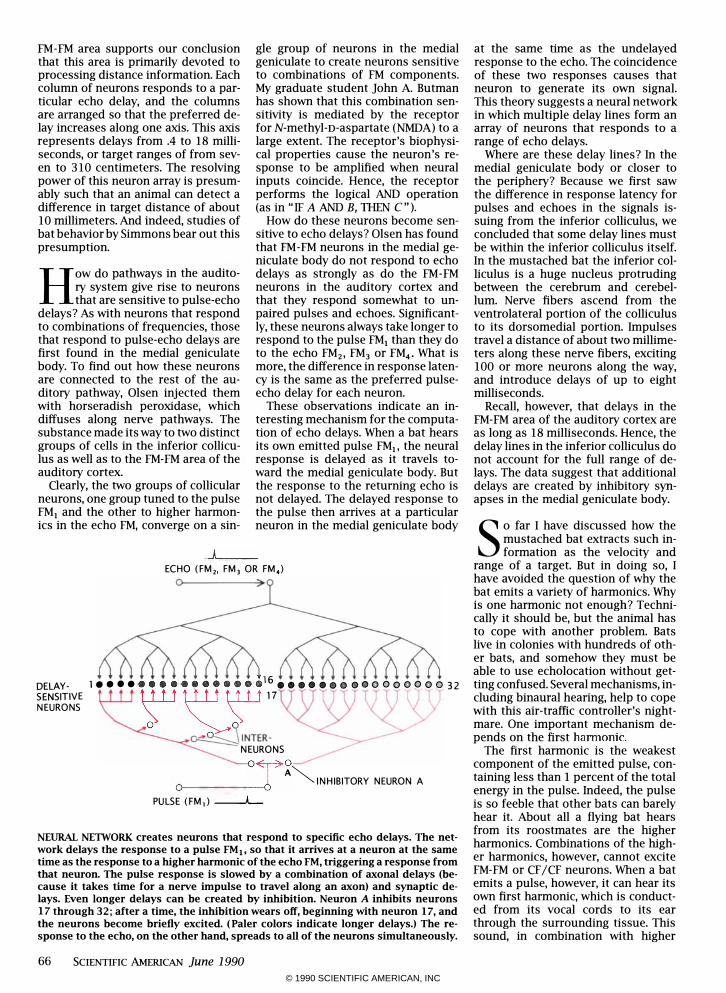
These observations indicate an interesting mechanism for the computation of echo delays. When a bat hears its own emitted pulse FMj, the neural response is delayed as it travels toward the medial geniculate body. But the response to the returning echo is not delayed. The delayed response to the pulse then arrives at a particular neuron in the medial geniculate body
.J� __
ECHO (FM" FM, OR FM.) ° :>
rl�;'r'VE I .... ill! � � i j' �7" .... • •• 0 000000 31
NEURONS � ° � INTER-
NEURONS oro 0----- A � INHIBITORY NEURON A
PULSE (FM,) --A...-
NEURAL NElWORK creates neurons that respond to specific echo delays. The network delays the response to a pulse FMjo so that it arrives at a neuron at the same time as the response to a higher harmonic of the echo FM, triggering a response from that neuron. The pulse response is slowed by a combination of axonal delays (because it takes time for a nerve impulse to travel along an axon) and synaptic delays. Even longer delays can be created by inhibition. Neuron A inhibits neurons 17 through 32; after a time, the inhibition wears off, beginning with neuron 17, and the neurons become briefly excited. (Paler colors indicate longer delays.) The response to the echo, on the other hand, spreads to all of the neurons simultaneously.
66 SCIENTIFIC AMERICAN June 1990
at the same time as the undelayed response to the echo. The coincidence of these two responses causes that neuron to generate its own signal. This theory suggests a neural network in which multiple delay lines form an array of neurons that responds to a range of echo delays.
Where are these delay lines? In the medial geniculate body or closer to the periphery? Because we first saw the difference in response latency for pulses and echoes in the signals issuing from the inferior colliculus, we concluded that some delay lines must be within the inferior colliculus itself. In the mustached bat the inferior colliculus is a huge nucleus protruding between the cerebrum and cerebellum. Nerve fibers ascend from the ventrolateral portion of the colliculus to its dorsomedial portion. Impulses travel a distance of about two millimeters along these nerve fibers, exciting 100 or more neurons along the way, and introduce delays of up to eight milliseconds.
Recall, however, that delays in the FM-FM area of the auditory cortex are as long as 18 milliseconds. Hence, the delay lines in the inferior colliculus do not account for the full range of delays. The data suggest that additional delays are created by inhibitory synapses in the medial geniculate body. s o far I have discussed how the
mustached bat extracts such information as the velocity and
range of a target. But in doing so, I have avoided the question of why the bat emits a variety of harmonics. Why is one harmonic not enough? Technically it should be, but the animal has to cope with another problem. Bats live in colonies with hundreds of other bats, and somehow they must be able to use echolocation without getting confused. Several mechanisms, including binaural hearing, help to cope with this air-traffic controller's nightmare. One important mechanism depends on the first harmonic�
The first harmonic is the weakest component of the emitted pulse, containing less than 1 percent of the total energy in the pulse. lode ed, the pulse is so feeble that other bats can barely hear it. About all a flying bat hears from its roostmates are the higher harmonics. Combinations of the higher harmOnics, however, cannot excite FM-FM or CF /CF neurons. When a bat emits a pulse, however, it can hear its own first harmonic, which is conducted from its vocal cords to its ear through the surrounding tissue. This sound, in combination with higher
© 1990 SCIENTIFIC AMERICAN, INC
CF SELECTIVITY
iii
RANGE
DELAY LINES
I FM SELECTIVITY
i iii
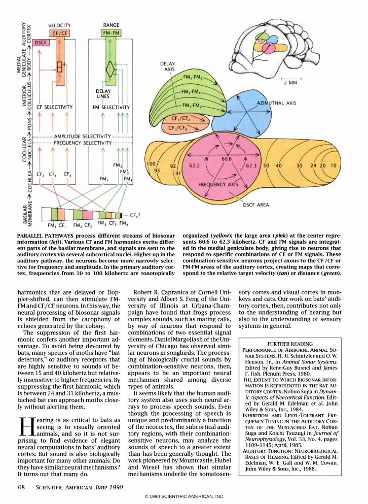
PARALLEL PATIIWAYS process different streams of biosonar information (left). Various CF and FM harmonics excite different parts of the basilar membrane, and signals are sent to the auditory cortex via several subcortical nuclei. Higher up in the auditory pathway, the neurons become more narrowly selective for frequency and amplitude. In the primary auditory cortex, frequencies from 10 to 100 kilohertz are tonotopically
organized (yellow); the large area (pink) at the center represents 60.6 to 62.3 kilohertz. CF and FM signals are integrated in the medial geniculate body, giving rise to neurons that respond to specific combinations of CF or FM signals. These combination-sensitive neurons project axons to the CF jCF or FM-FM areas of the auditory cortex, creating maps that correspond to the relative target velocity (tan) or distance (green).
harmonics that are delayed or Doppler-shifted, can then stimulate FMFM and CF / CF neurons. In this way, the neural processing of biosonar signals is shielded from the cacophony of echoes generated by the colony.
The suppression of the first harmonic confers another important advantage. To avoid being devoured by bats, many species of moths have "bat detectors," or auditory receptors that are highly sensitive to sounds of between 15 and 40 kilohertz but relatively insensitive to higher frequencies. By suppressing the first harmOniC, which is between 24 and 31 kilohertz, a mustached bat can approach moths closely without alerting them.
Hearing is as critical to bats as seeing is to visually oriented animals, and so it is not sur
pnsmg to find evidence of elegant neural computations in bats' auditory cortex. But sound is also biologically important for many other animals. Do they have similar neural mechanisms? It turns out that many do.
68 SCIENTIFIC AMERICAN June 1990
Robert R. Capranica of Cornell University and Albert S. Feng of the University of Illinois at Urbana-Champaign have found that frogs process complex sounds, such as mating calls, by way of neurons that respond to combinations of two essential signal elements. Daniel Margoliash of the University of Chicago has observed similar neurons in songbirds. The processing of biologically crucial sounds by combination-sensitive neurons, then, appears to be an important neural mechanism shared among diverse types of animals.
It seems likely that the human auditory system also uses such neural arrays to process speech sounds. Even though the processing of speech is unique and predominantly a function of the neocortex, the subcortical auditory regions, with their combinationsensitive neurons, may analyze the sounds of speech to a greater extent than has been generally thought. The work pioneered by Mountcastle, Hubel and Wiesel has shown that similar mechanisms underlie the somatosen-
sory cortex and visual cortex in monkeys and cats. Our work on bats' auditory cortex, then, contributes not only to the understanding of hearing but also to the understanding of sensory systems in general.
RJRlliER RFADING PERFORMANCE OF AIRBORNE ANIMAL So
NAR SYSTEMS. H. -U. Schnitzler and O. W. Henson, Jr. , in Animal Sonar Systems. Edited by Rene-Guy Busnel and James F. Fish. Plenum Press, 1980.
THE ExTENT TO WHICH BIOSONAR iNFOR
MATION Is REPRESENTED IN THE BAT Au
DITORY CORTEX. Nobuo Suga in Dynamic Aspects of Neocortical Function. Edited by Gerald M. Edelman et al. John Wiley & Sons, Inc., 1984.
iNHIBmoN AND LEvEL-TOLERANT FRE
QUENCY TuNING IN THE AUDITORY COR
TEX OF THE MUSTACHED BAT. Nobuo Suga and Koichi Tsuzuki in Journal of Neurophysiology, Vol. 53, No. 4, pages 1109-1145; April, 1985.
AUDITORY FUNCTION: NEUROBIOLOGICAL
BASES OF HEARING. Edited by Gerald M.
Edelman, W. E. Gall and W. M. Cowan. John Wiley & Sons, Inc. , 1988.
© 1990 SCIENTIFIC AMERICAN, INC


