Embed Size (px)
Citation preview

BIOLOGICAL ASSESSMENT AND REMEDIATION
OF CONTAMINATED SEDIMENTS
SAÏD EL FANTROUSSI & SPIROS N. AGATHOS*Unit of Bioengineering, Faculty of Bioengineering,
Agronomy & Environment, Catholic University of Louvain,
Place Croix du Sud 2/19,B-1348 Louvain-la-Neuve, Belgium
DIETMAR H. PIEPER*, ROBERT WITZIG, BEATRIZ CÁMARA, LOTTE GABRIEL-JÜRGENS & HOWARD JUNCAGBF German Research Centre for Biotechnology,
Mascheroder Weg 1 38124 Braunschweig, Germany
GIULIO ZANAROLI & FABIO FAVA* DICASM, Faculty of Engineering, Alma Mater Studiorum-
University of Bologna, viale Risorgimento 2, 40126 Bologna,
Italy
JOSÉ R. PÉREZ-JIMÉNEZ & LILY Y. YOUNG Department of Environmental Sciences and Biotechnology
Center for Agriculture and the Environment, Cook College,
Rutgers, The State University of New Jersey, New Brunswick,
New Jersey 08901, USA
KELLY HAMONTS, RICHARD LOOKMAN, MIRANDA MAESEN, LUDO DIELS, AND WINNIE DEJONGHE*
Flemish Institute for Technological Research, Geel, Belgium
JOHN DIJK AND DIRK SPRINGAELCatholic University of Leuven, Heverlee, Belgium
* to whom correspondence should be addressed
Abstract- Various approaches to clean contaminated aquatic environments have been proposed. In recent years, natural attenuation has received increasing attention and it is generally accepted that microorganisms are the principal mediators of the natural attenuation of many pollutants. However, the complexity of environmental systems such as sediments requires a multifaceted approach to understand microbial processes and their potential. This is even more so under in situ conditions, where the activity of pollutant degrading microorganisms is generally slow, partial and
179
, 179–238. D. Reible and T. Lanczos (eds.), Assessment and Remediation of Contaminated Sediments© 2006 Springer.

180 S.E. FANTROUSSI ET AL.
constrained spatially and/or temporally. Recent developments in molecular biology and genomics are offering tools to explore microbial processes at a level that encompasses the genetic characteristics of the local microbial players, culturable or not, as well as their organization into complex communities and their interactions both with each other and with the target chemicals. It is now possible to study microbes directly in their environments at the population level as well as at the single cell level and to link biology to geochemistry. Integrative knowledge from culture independent studies based on functional characters and assessment of the diversity and quantity of catabolic genes in response to pollution, will allow a deeper understanding of and a rational intervention in environmental processes. Moreover, the use of genomic libraries to retrieve genes from natural bacterial communities without cultivation will allow a breakthrough in accessing new microbial capabilities. In this chapter, the main features, advantages and limitations of these innovative approaches to the biomonitoring and analysis of intrinsic bioremediation potential of polluted environments and sediments are critically reviewed. Then, the potential of the same strategies in the integrated chemical, physical and biological monitoring and characterization of polluted sediments subjected to natural decontamination is shown through the description of the main results of case studies performed on a) polychlorinated biphenyl (PCB)-contaminated marine sediments of the Porto Marghera area of Venice Lagoon (Italy) in which the occurrence of PCB-reductive dechlorination processes has been demonstrated for the first time in the literature, b) sediments contaminated by chlorinated aliphatic hydrocarbons (CAHs) collected from different positions of the eutrophic river Zenne (Vilvoorde, Belgium), where they have been found to act as a natural biobarrier for the CAHs occurring in the groundwater that is passing through the sediment zone, hereby reducing the risk of surface water contamination, and c) other environmental contaminated systems subjected to ex-situ and in situ active bioremediation, where these processes are described on the basis of the experience accumulated in pilot and real-life systems.
Keywords: polychlorinated biphenyls (PCBs), chlorinated aliphatic hydrocarbons, bioaugmentation, biostimulation, reductive dechlorination, biosensors, PCR, DGGE, T-RFLP, metagenomics, sulfate-reducing bacteria, fingerprinting methods, exsitu treatment, in situ treatment, engineered bioremediation systems.

BIOLOGICAL ASSESSMENT 181
1. The detection and characterization of microbial processes in sediments
Microbes critically impact and mediate nearly every major biogeochemical cycle on Earth. Microbial interactions that occur on the scale of microns can regulate global processes on planet-wide scales. As an example, the decomposition and transformation of both organic and inorganic compounds in sediments is almost completely carried out by microbes. The crucial role of microbes in the environment had been underestimated since the first discovery of these microscopic living forms. The main reason for this was the difficulty to access the astonishing diversity of microbes since the classical approaches based on their cultivation in the laboratory are biased and inadequate. There is a wide consensus among microbiologists that more than 90% of microbes are refractory to the classical approaches of cultivation1. Recent advances in understanding the diversity and importance of microbes, using culture-independent approaches, has led without any ambiguity to the conclusions that microbes are by far the most dominant living forms in terms of number of species, number of individual cells, total biomass, diversity of habitats, and diversity of cellular chemistries. Thus the environmental impact of microbes is being taken seriously and stands in the forefront of studies aiming at understanding the global environmental changes. Microbes dominate animal life in sediments and aquifers, and they are responsible for major fluxes of organic and inorganic nutrients. They are involved in processes as different as anaerobic methane oxidation2,uranium accumulation3, pesticide degradation4, and polychlorinated biphenyl degradation5.
1.1. HOW TO MASTER INCREASING COMPLEXITY
The complexity of microbial life in both natural and man-impacted habitats requires a multidisciplinary approach. In addition to their large diversity, microbes live in interaction with the different components of their environments. Figure 1.1 shows schematically the inherent complexity of a polluted sediment undergoing bioremediation. Several factors affect the process of biodegradation (bioavailability, physico-chemical conditions, composition of microbial communities, etc.). The characterization of such a complex biotope is now feasible using tools offered by such disciplines as ecology, biogeochemistry, physiology, biochemistry, genetics, process engineering, molecular biology, and metagenomics. However the relevance

182 S.E. FANTROUSSI ET AL.
and efficiency of each discipline depends on the scale of study (pure culture, laboratory bioreactors, or field studies). Figure 1.2 shows the relevance of each discipline to the field studies (adapted from E.L.Madsen6). The most striking discipline that has recently emerged is metagenomics, defined as the culture-independent genomic analysis of entire microbial communities. Metagenomics provides access to the pool of genomes of a given environment using a comprehensive survey of nucleotide sequence, structure, regulation, and function. Furthermore, direct genomic cloning offers the possibility to retrieve unknown sequences or functions, whereas methods relying on PCR amplification require prior knowledge of the sequence of genes for the design of primers7,8.
bioremediation. The anaerobic dechlorination of tetrachloroethene (PCE) is used as anexample of productive microbial metabolism leading to site cleanup.
Figure 1.1. Schematic representation of the complexity of polluted sediments undergoing

BIOLOGICAL ASSESSMENT 183
relevance to different scales of studies.
1.2. EXPERIMENTAL APPROACHES
Studying microbes in sediments involves several approaches that can be summarised by the following flowchart.
Phase 1: Environmental sampling: physiological studies (adapted from E. L. Madsen9)
1. Sediment, soil, water or sludge in field site. 2. Aseptically remove, contain, transport to laboratory. 3. Divide into replicate live and abiotic treatments. 4. If appropriate, add radiolabelled or unlabelled organic compound of
interest.5. Use analytical chemistry or physiology tools to periodically measure
consumption or production. 6. Compare time courses of live and abiotic treatments. 7. Interpret and consider genetic studies in Phases 2 and 3.
Phase 2: Specialized microbiological studies using culture-based methods
1. Isolate pure culture expressing activity found in Phase 1. 2. Characterize growth, cell yield, sequential induction, and other
characteristics during pollutant metabolism.
Figure 1.2. Disciplines involved in understanding microbial processes in sediments and their

184 S.E. FANTROUSSI ET AL.
3. Extract and identify metabolites, enzymes and cofactors linked to pollutant metabolism.
4. Study metabolites, enzymes and cofactors in cell-free system. 5. Determine portion of genomic or plasmid DNA coding for pollutant
metabolism by screening a cloned DNA library, by transposon mutagenesis or by other procedure.
6. Do hybridization, restriction mapping and sequencing DNA analyses seeking ORFs, phylogenetic relationships to similar genes, etc.
7. Elucidate details of gene expression and regulation via various genetic & molecular techniques (transposon mutagenesis, expression clones, insertional inactivation, inducer/reporter experiments).
Phase 3: Specialised microbiological studies using culture-independent approaches 1. Using a few grams of sediments, extract the whole genomic material
(DNA and/or RNA). 2. Clean up the nucleic acids. 3. Proceed through PCR or in situ hybridization to study species or
consortia of interest. 4. Use fingerprinting methods (DGGE, TRFLP, etc.) and clone libraries
to study microbial diversity. 5. For genomic and metagenomic studies shear the nucleic acid fragments
and clone them in vectors such as fosmids and BACs. 6. Use the function-driven approach or the sequence-driven approach to
explore the phylogenetic and physiological diversity of the sediment biotope.
1.3. MOLECULAR TOOLS TO DETECT AND MONITOR MICROBES
The development of molecular tools has allowed more precise and sensitive techniques to be applied for microbial detection in sediments compared to classical microbiological methods. The most popular and widely used molecular techniques are those based on polymerase chain reaction (PCR). PCR techniques targeting specific portions of genes have been used to detect microbes and to explore microbial diversity in sediments, aquifers and soil samples. Several studies have shown the advantage of using PCR to detect and quantify the presence of microbes. As an example El Fantroussi et al10 have used 16S rRNA-based techniques to study the biodegradation of chloroaromatic compounds in a simulated aquifer. The utilisation of PCR in parallel with biodegradation in microcosms showed an unambiguous role of bioaugmentation as a strategy for the bioremediation

BIOLOGICAL ASSESSMENT 185
of polluted sediments under constraints of fluid flow patterns, depth of matrix and presence of competing indigenous microbial communities.
Hendrickson et al.11 examined the environmental distribution of organisms belonging to the Dehalococcoides group, known to be major players in the anaerobic transformation of chlorinated compounds, and their association with chloroethene-contaminated sites. Samples from 24 chloroethene-dechlorinating sites scattered throughout North America and Europe were tested for the presence of members of the Dehalococcoides
group by using a PCR assay developed to detect Dehalococcoides 16S rRNA gene (rDNA) sequences. Sequences identified by homology to the Dehalococcoides group were detected at 21 sites. Full dechlorination of chloroethenes to ethene occurred at these sites. Dehalococcoides sequences were not detected in samples from three sites at which partial dechlorinationof chloroethenes occurred, where dechlorination appeared to stop at 1,2-cis-dichloroethene. Phylogenetic analysis of the 16S rDNA amplicons confirmed that Dehalococcoides sequences formed a unique 16S rDNA group11.
Other studies have demonstrated the power of combining molecular techniques with classical approaches to understand microbial processes in sediments. De Lipthay et al.12 evaluated how the in situ exposure of a subsurface aquifer to phenoxy acid herbicides at low concentrations (<40 µg l-1) changes the local microbial community composition. Sediment and groundwater samples collected from a herbicide-exposed area and unexposed controls were analyzed for the presence of general microbial populations, Pseudomonas bacteria, and specific phenoxy acid degraders. Both culture-dependent and culture-independent methods were applied. Theabundance of microbial phenoxy acid degraders (100 to 104 per gram ofsediment) was determined by most probable number (MPN) assays, andtheir presence was only detected in herbicide-exposed sediments. Similarly,PCR analysis showed that the 2,4-dichlorophenoxyacetic acid degradation pathway genes tfdA and tfdB were only detected in sediments from contaminated areas of the aquifer. Different populations of tfd genes have been found, suggesting that the in situ herbicide degradation was caused by the activity of a heterogeneous population of phenoxy acid degraders. The number of Pseudomonas bacteria was higher in sediments subjected to high levels of phenoxy acid. Furthermore, high numbers of CFU compared to direct counting of 4',6-diamidino-2-phenylindole-stained cells under the microscope suggested an increased culturability of the indigenous microbial communities from acclimated sediments. The findings of this study demonstrate that continuous exposure to low herbicide concentrations can markedly change the bacterial community composition of a subsurface aquifer.

186 S.E. FANTROUSSI ET AL.
Anaerobic methane oxidation is a process that effectively controls emission of methane from many anaerobic environments into theatmosphere and thus plays an important role in the global methane budget2.Microbially mediated oxidation of methane in anoxic marine systems is a globally significant process, with up to 90% of the oceanic methaneproduction recycled in anaerobic marine sediments13. Other studies have shown that microorganisms living in anoxic marine sediments consume more than 80% of the methane produced in the world's oceans. In addition to single-species aggregates, consortia of metabolically interdependent bacteria and archaea were found in methane-rich sediments. A combination of fluorescence in situ hybridization and secondary ion mass spectrometry showed that cells belonging to one specific archaeal group associated with the Methanosarcinales were all highly depleted in C-13. This depletion indicates assimilation of isotopically light methane into specific archaeal cells2. Anaerobic methane oxidation was also investigated in marine sediment from Aarhus Bay, Denmark14. Measured concentration profiles for methane and sulfate, as well as in situ rates determined with isotope tracers, indicated that there was a narrow zone of anaerobic methane oxidationabout 150 cm below the sediment surface. Methane could account for 52% of the electron donor requirement for the peak sulfate reduction rate detected in the sulfate-methane transition zone. Molecular signatures of organisms present in the transition zone were detected by using selective PCR primers for sulfate-reducing bacteria and for Archaea. One primer pairamplified the dissimilatory sulfite reductase (DSR) gene of sulfate-reducingbacteria, whereas another primer (ANME) was designed to amplify archaealsequences found in another study of sediments from the Eel River Basin, as these bacteria have been suggested to be anaerobic methane oxidizers15.This study is a nice illustration of combining chemistry to molecular biology to bring into stark evidence globally significant biogeochemical processes that are mediated by diverse coexisting and interacting microbial populations14.
1.4. SYNTROPHY
Anaerobic oxidation of methane, also called reverse methanogenesis, in sediments illustrates once again the importance of syntrophy in the microbial world in general and in anaerobic processes in particular. As in the process of methane production, methane oxidation occurs in ecological niches where archaea, responsible for methane oxidation; live in association with sulfate reducers. Syntrophy was first discovered in 1967 as a synergistic relationship between an ethanol-oxidizing bacterium and a methanogen. In the late 1970s, the importance of syntrophic relationships

BIOLOGICAL ASSESSMENT 187
became established through culture-based studies of methanogens interacting with bacteria that oxidized lactate, butyrate, or propionate. Biochemically, these bacteria dispose of reducing equivalents by reducing protons or bicarbonate to H2 and formate, respectively. Both products serve as electron carriers in interspecies H2 transfer and interspecies formate transfer between syntrophic acetate-producing bacteria and methanogens. Since their original discovery, many other syntrophic systems have been identified, and the central role of syntrophic interactions in environmental samples and engineered anaerobic bioreactors is now widely recognized. Syntrophy (or syntrophism) can be defined as the interaction of two or more populations that satisfy each other’s nutritional needs16. This strategy of “feeding together” is involved in the anaerobic breakdown of various sugars, amino acids, and aromatic compounds. El Fantroussi et al.10 used Desulfomonile tiedjei for bioaugmentation of a simulated aquifer polluted with 3-chlorobenzoate. This dechlorinating bacterium was isolated from a syntrophic association with a benzoate oxidizer and a H2-consuming methanogen (Figure 1.3).
Figure 1.3. Example of a bacterial syntrophic association.
D. tiedjei is a scavenger which relies on electron donors and vitamins from other organisms and uses a broad range of electron acceptors including 3-chlorobenzote17. The establishment of this strain in the simulated aquifer was evaluated with physiological microcosms and PCR
D. tiedjei
BZ-2
Methanospirillum sp.
CH4
vitamins
3-chlorobenzoate
benzoate
Cl-
Vitaminsacetate
+
acetate
H2 + CO2

188 S.E. FANTROUSSI ET AL.
detection of its 16S rRNA signatures. There was a close relationship between 3-chlorobenzoate dechlorination, methane production and PCR detection10.
1.5. ENVIRONMENTAL MONITORING – BIOSENSORS
To fully understand microbial processes in sediments there is a need for new, fast, specific, accurate, precise and cost effective methods for field assays to monitor both chemical species and ‘effect related’ processes (e.g. xenoestrogens). Several techniques have been developed for environmental monitoring of chemical species. These methods include ultra-sensitive techniques based on immunoassays as well as whole-cell (mainly by using genetically engineered microorganisms “GEM”) biosensors to assess toxicity / bioavailability that can be applied to both metals and organics. However there are still some technical barriers related to sample treatment and cleanup prior to instrumental analysis. These procedures are also time-consuming and often not suited for in situ field monitoring, e.g. sediments. To achieve this goal Acha et al.18 have developed a novel attenuated total reflection-Fourier transform infrared (ATR-FTIR) sensor and applied it to the continuous on-line monitoring of a dechlorination process. This optical sensor was developed to measure noninvasively part-per-million (ppm) concentrations of trichloroethylene (TCE), tetrachloroethylene (PCE), and carbon tetrachloride (CT) in the aqueous effluent of a fixed-bed dechlorinating bioreactor, without any prior sample preparation (Figure 1.4).
Figure 1.4. ATR-FTIR sensor (shown inside the circle) coupled to an anaerobic dechlorinating bioreactor for continuous on-line monitoring of chlorinated organic compounds.
recirculation of mixed liquor
FTIR spectrometerFTIR spectrometer
bioreactorbioreactor
sensorsensor

BIOLOGICAL ASSESSMENT 189
The sensor was based on an ATR internal reflection element (IRE) coated with an extracting hydrophobic polymer, which prevented water molecules from interacting with the infrared (IR) radiation. The selective diffusion of chlorinated compound molecules from aqueous solution into the polymer made possible their detection by the IR beam. With the exclusion of water the detection limits were lowered, and measurements down to 2, 3, and 2.5 mg/L (ppm) for TCE, PCE, and CT, respectively, became possible. Before coupling the ATR-FTIR sensor to the dechlorinating bioreactor, preliminary spectra of the chlorinated compounds were acquired on a laboratory scale configuration in stop-flow and flow-through closed-loop modes, in order to study the direct response of the sensor to any arbitrary concentration change of the analytes. Subsequently, the bioreactor (a fixed bed which could also simulate a sediment matrix) was monitored with the infrared sensor coupled permanently to it. The sensor tracked the progression of the analytes' spectra over time without perturbing the dechlorinating process. The accuracy of this ATR-FTIR sensor was validated against gas chromatography (GC) measurements of the chlorocompounds17.
1.6. CONCLUSION
Microbial processes play crucial roles in the transformation of organic and inorganic compounds in sediments. The recent developments in molecular biology allowed a better understanding of the distribution, diversity, and functions of microbes in sediments. Novel key processes have been attributed to microbes thanks to molecular biology such us anaerobic methane oxidation. Coupling these molecular techniques with geochemistry involving sophisticated tools such as chemical and biochemical sensors opens up interesting horizons for comprehensive studies of microbial processes in sediments.
supported by grants CHLOREM (BIO 4-CT 1998-0303) and MADOX (QLK3-CT-2001-00345) of the European Commission.
1.7. ACKNOWLEDGEMENTS
The work of the authors S. E Fantroussi and Spiros N. Agathos was .

190 S.E. FANTROUSSI ET AL.
2. Accumulating basic knowledge on biodegradation processes and use of this knowledge for analyzing catabolic potential and metabolic networks in situ by culture-independent approaches
Various approaches to clean contaminated environments have been proposed including chemical, physical or biological treatments. Among them, the biological treatment is considered an efficient and cost saving way to achieve remediation of contaminated sites19-21. During the previous years, natural attenuation, “naturally occurring processes in soil and groundwater that act without human intervention to reduce the mass, toxicity, mobility, volume or concentration of contaminants in those media”22 has received increasing attention. It is generally accepted that microorganisms are the principal mediators of the natural attenuation of many pollutants. They transform or mineralize pollutants, thereby usually decreasing their masses and toxicities, in contrast to most other processes contributing to natural attenuation. However, in some cases pollutants may be transformed into more toxic products, as reported for the anaerobic transformation of trichloroethylene. The use of natural attenuation thus requires a detailed monitoring to determine how effective natural attenuation is for attaining site remediation goals. In order to properly evaluate natural attenuation at a site, it is necessary to know the location and concentration of the contaminants, how the contaminants move in the environment, and how their concentration changes over time. Reliance on biologically meditated natural attenuation thus requires information on whether it can occur, whether it is actually occurring at a significant rate, which mechanisms and pathways are involved and, specifically, how it will behave in the future. Active in situ remediation involves the stimulation of the indigenous microbial activity by the addition of nutrients and/or electron acceptors. As in the case for natural attenuation, an understanding of the response of the indigenous microbial community is necessary for successful stimulation. Even more unpredictable are bioaugmenation approaches, as the added microorganisms, which are supposed to increase the degradative performance on site, not only have to express these capabilities under in situ conditions, but also have to compete with the complex microflora inhabiting the site under study. It is thus evident that only a detailed understanding of the functioning and interactions in microbial communities will allow their rational manipulation, and the overcoming of factors limiting efficient bioremediation.

BIOLOGICAL ASSESSMENT 191
2.1. BACTERIAL DEGRADATION OF AROMATIC POLLUTANTS
Aromatic compounds have been discharged into the environment during the last years in increasing amounts. They have significant impact on natural microbial communities, and thus, on the global element mass fluxes. The functional diversity in nature shows that many microorganisms have the potential to degrade and recycle aromatic compounds23. This potential can be used for the bioremediation processes mentioned above.
Many microorganisms have evolved biodegradative pathways to use aromatic compounds as a sole carbon and energy source24-26. Even though the first observations were made using aerobic microorganisms27,28, it is well established that aromatics can be degraded under nitrate-, iron-, or sulfate-reducing and even under methanogenic conditions29,30.
2.1.1. Rieske non-heme iron oxygenases
Aerobic microorganisms usually initiate degradation by activation of the aromatic nucleus through oxygenation reactions. The introduction of hydroxyl-groups, usually in ortho-position to one another, results in a few central intermediates such as catechols, protocatechuate, gentisate and hydroxyhydroquinones. These intermediates are subject to oxygenolytic ring cleavage followed by channeling of the ring-cleavage products into the central metabolism31.
The so called Rieske non-heme iron oxygenases are one of the key families of enzymes important for aerobic degradation of aromatics. These enzymes usually catalyze the incorporation of two oxygen atoms into the aromatic ring to form arene-cis-dihydrodiols32, a reaction which is followed by a dehydrogenation catalyzed by cis-dihydrodiol dioxygenases to give catechols or substituted catechols. The oxygenases, such as benzoate, naphthalene, biphenyl, or toluene dioxygenases are multicomponent enzyme complexes, composed of a terminal oxygenase component (iron-sulfur protein) and different electron transport proteins (a ferredoxin and a reductase or a combined ferredoxin-NADH-reductase). The catalytic iron-sulfur proteins are heteromultimers, comprising a large ( ) and a small ( )subunit with the former containing a Rieske-type [2Fe-2S] cluster, a mononuclear non-heme iron and a substrate binding site (Fig. 2.1).

192 S.E. FANTROUSSI ET AL.
Figure 2.1. Biochemical and genetic organization of toluene 2,3-dioxygenases as an example of Rieske non heme iron oxygenases. Two electrons are transferred from NADH via the electron transfer chain, consisting of reductase and ferredoxin (Fd) to the terminal oxygenase component, comprising the small -subunit and the large -subunit. The incorporation of two oxygen atoms into the substrate results in the formation of a cis-dihydrodiol. Regions of the gene encoding residues of the -subunit responsible for substrate specificity are indicated in white.
Comparison of the amino acid sequences of the terminal oxygenase -subunits revealed that they form a family of diverse but evolutionarily related sequences (Figure 2.2). Although none of the enzymes is completely specific, a broad correlation between the grouping in toluene/biphenyl, naphthalene, benzoate or phthalate families and the native substrates oxidized by the family members can be observed.
Figure 2.2. Dendrogram showing the relatedness of members of different families of Rieske non heme iron oxygenases ( -subunits).

BIOLOGICAL ASSESSMENT 193
Enzyme engineering studies on biphenyl, benzene, chlorobenzene, and naphthalene dioxygenases showed that the -subunit of the terminal oxygenase determines substrate specificity and that only slight sequence differences in the amino acid sequence can be associated with dramatic changes in substrate specificity or regiospecificity33-36. As an example a single amino acid difference in toluene dioxygenase results in transformation of tetrachlorobenzene33 or in a completely new regioselectivity of naphthalene dioxygenase36. Regioselectivity also determines whether or not a given substrate can subsequently be subject to mineralization. As an example, dibenzofuran can be mineralized after a so called angular attack at the oxygen bridge37 and an adjacent carbon atom, whereas lateral attack results in the formation of dead-end products38.Consequently, dioxygenases crucially determine the range of substrates that are accessible to microbial degradation and the metabolic net in microbial communities. Crystal structures are available for some members of Rieske non-heme iron oxygenases39,40. Those allow the modeling of active site structures of newly discovered derivative enzymes and at least a partial explanation of enzyme performance, even though amino acid residues, which have no contacts with substrates also strongly changed enzyme performance41,42. Nevertheless, Rieske-type non-heme iron oxygenases are an enzyme family in which significant advances have been made to relate sequence information to function.
2.1.2. Toluene monooxygenases
Two groups of enzymes have been reported to attack the non-activated benzene nucleus by monooxygenation, the multicomponent aromatic monooxygenases and some members of the multicomponent phenol hydroxylases43, both groups belonging to the family of soluble diiron monooxygenases. These monooxygenases can perform regio- and stereospecific hydroxylations (Fig. 2.3), and in the case of transformation of toluene, 2-methylphenol44, 3-methylphenol45 as well as 4-methylphenol46,were reported to be formed, however, recent results showed that 4-methylphenol but not 3-methylphenol is formed by the enzyme termed toluene 3-monooxygenase47. Further oxygenation of the intermediate cresols by phenol hydroxylase results in the formation of 3-methyl- or 4-methylcatechol, respectively.
Alternatively, 4-methylphenol can be subject to oxygenation of the methylsubstituent resulting finally in protocatechuate (Figure 2.3) as ring-cleavage substrate48. Evidently, again, substrate specificity will significantly determine the metabolic net responsible for degradation.

194 S.E. FANTROUSSI ET AL.
Figure 2.3. Transformation of toluene by two successive monooxygenations results in the formation of methylcatechols which are usually mineralized via extradiol cleavage.
Moreover, dependent on the regioselectivity, xylenes will be transformed to dead-end products. The aromatic monooxygenases consist of an electron transport system comprising a reductase and a ferredoxin, a catalytic effector protein which is assumed to play a role in assembly of an active oxygenase and a terminal hydroxylase with a ( )2 quaternary structure and a diiron center contained in each -subunit43. Even though the effector protein is necessary not only for effective coupling, but also for a high regioselectivity49, the -subunit was majorly responsible for substrate specificity and regioselectivity43. Various mutagenesis and directed evolution studies have actually identified residues which control regioselectivity and activity50,51. The structure of toluene monooxygenase from P. stutzeri OX1 has recently been elucidated52 which offers the opportunity to understand how the enzyme adjusts the active site pocket to control product regioselectivity.
2.1.3. Catechol dioxygenases
Whereas a relatively broad diversity of activation mechanisms is possible in aromatic degradation, these pathways usually converge in the formation of (substituted) catechols as central intermediates53. Whereas chlorinated aromatics are predominantly degraded via intradiol cleavage54
methylsubstituted aromatics such as toluene or xylenes are usually degraded via the respective catechols and extradiol cleavage (Fig. 2.3). Extradiol dioxygenases are thus key enzymes in the degradation of aromatic compounds and many of such proteins and their coding sequences have been described, purified and characterized in the last decades. Examination

BIOLOGICAL ASSESSMENT 195
of their evolutionary relationships55 showed that the majority of extradiol dioxygenases with preferences for monocyclic substrates were, at the protein level, phylogenetically closely related. However, members of this enzyme subfamily were reported to differ significantly in their substrate specificity42,56 and, thus, the metabolic network in environmental communities will be significantly interconnected with the C23O diversity. Catechol transforming activities are specifically important, as catechols are highly toxic for microorganisms57 and even low accumulation will have severe impact in community functioning58.
2.1.4. Anaerobic degradation of aromatics
In recent years our knowledge on anaerobic degradation of aromatics has been significantly enhanced and genetic determinants for various key steps in anaerobic aromatic degradation have been elucidated. Under anaerobic conditions, aromatic hydrocarbons are initially attacked by novel reactions and in most cases oxidized further to benzoyl-CoA, a common intermediate in anaerobic catabolic pathways29. Toluene degradation is initiated by an unusual addition reaction of the toluene methyl group to the double bond of fumarate to form benzylsuccinate59 catalyzed by benzylsuccinate synthase, a unique type of glycyl-radical enzyme60. However, not only toluene degradation is initiated by fumarate addition. Similar reactions have been shown to be involved in the anaerobic activation of m-xylene, m- and p-cresol, n-alkanes and 2-methylnaphthalene61,62. Even though the metabolism of toluene has mainly been elucidated in denitrifying Thauera
and Azoarcus it became evident that benzylsuccinate forming activities are found across a wide range of phylogenetically and physiologically diverse bacteria, and have also been found in the iron-reducing Geobacter
metallireducens strains63. In contrast to the information on degradation of toluene and xylenes, information on anaerobic degradation of naphthalene and benzene is scarce64,65.
2.1.5. Reductive dehalogenation
It is known since more than one decade ago that chloroaromatics can function as an alternative electron acceptor in a type of anaerobic respiration66. Several anaerobic bacteria have been identified as being able to reductively dehalogenate chlorinated aliphatics as well as chlorinated aromatics and to couple this reaction to the synthesis of ATP via a chemiosmotic mechanism. Bacterial dehalorespiration is currently considered to be the most important process for the detoxification of organohalogens under anaerobic conditions. The dehalorespiring bacteria known to belong to the low GC content Gram-positive bacteria (Desulfitobacterium and Dehalobacter), the Proteobacteria (Desulfomonile,

196 S.E. FANTROUSSI ET AL.
Desulfuromonas and Dehalospririllum, now transferred to the genus Sulfurospirillum), and the genus Dehalococcoides67. Although dehalorespiring bacteria are widespread, Dehalococcoides species in particular seem to be of major environmental importance. It has been shown that members of this group can grow on vinyl chloride as electron acceptor68, a process previously assumed to be co-metabolic (i.e. without deriving energy or carbon from the transformed substrate). In addition, Dehalococcoides strains are, thus far, the only microorganisms known to be capable of growing anaerobically on chlorobenzenes69, polychlorinated biphenyls and even chlorinated dioxins70. Reductive dehalogenases involved in tetrachloroethene or chlorophenol respiration have been intensively studied (Figure 2.4). This novel subclass of reductases shares some common features at the biochemical and genetic level. Except for the chlorobenzoate reductive dehalogenase of Desulfomonile tiedjei that contains a heme cofactor and that is produced as a heterodimer71, reductive dehalogenases contain two iron–sulfur clusters and a corrinoid67.
Figure 2.4. Reductive dehalogenation of TCE (trichloroethene) to cis-1,2-dichloroethene.
They are produced as preproteins with a twin-arginine signal peptide recognized by the export system specific for the export of periplasmic proteins containing cofactors72. The protein sequence also revealed consensus sequences characteristic for the binding of two iron–sulfur clusters. Also vinyl chloride reductive dehalogenase was recently identified as a novel member of the family of corrinoid/iron-sulfur cluster containing reductive dehalogenases73.
2.2. MOLECULAR TOOLS FOR ASSESSING BIODEGRADATION POTENTIAL
It has been shown that the in situ catabolic potential of microbial communities depends on the history of the site under study, the site characteristics (geochemistry and redox conditions) and, in cases where the contaminated soil is covered by vegetation, interactions between the

BIOLOGICAL ASSESSMENT 197
microbial community and plant species, which are up to now poorly defined. Moreover, it is well known that many environmental microorganisms may not be cultivated under laboratory conditions. Culturing-independent molecular techniques are, currently, rapidly increasing our understanding of microbial community structure and activity in the subsurface. Polymerase Chain Reaction (PCR) amplification of nucleic acids extracted from environmental samples is at present the most powerful cultivation-independent technique. PCR facilitates the sensitive and fast detection of low amounts of specific gene fragments. This is of importance for monitoring purposes, as subsurface environments are, in general, oligotrophic, hence characterized by low biomass from which low amounts of nucleic acids can be extracted.
Molecular microbial ecology has significantly increased our knowledge on the diversity and dynamics of microbial communities in nature. By different PCR approaches, predominantly using the 16S rRNA gene or the inter-spacer region of 16-23S rRNA genes, specific taxonomic groups responsible for degradation, and specific species, can be characterized, quantified and their spread followed over time74. However, as only in a subset of scenarios, a degradation capability is directly related to a specific taxonomic group, research in recent years has focused on the adaptation of molecular ecology methods for assessing community composition to characterize the makeup of catabolic genes, as the abundance of degradation genes can be assumed to indicate the biodegradation potential in contaminated soils.
2.2.1. Analysis of catabolic genes
Catalytic enzymes involved in aromatic degradation are encoded by genes, which have evolved to produce proteins performing specific actions. Members of the different gene families share a certain degree of similarity represented by conserved sequence regions. With only two short sequences conserved along two fairly divergent gene family members, it is possible to detect, by amplification, regions between the conserved parts, in DNA from isolates or from environmental samples. The initial step in this process is to identify the crucial gene and protein regions. Subsequently, conserved regions are identified within the different groups of given gene families and evaluated for primer design.
Thus far, analysis of catabolic genes has focused mainly on the characterization of the presence or abundance of a family of catabolic genes73,75,76 without assessing the finer levels of sequence diversity. However, the diversity of catabolic gene sequences as described above, often reflects differences in substrate specificity or affinity. Single amino acid differences have been reported to drastically change substrate

198 S.E. FANTROUSSI ET AL.
specificity of catabolic enzymes (see above). A more detailed picture of the catabolic gene structure and sequence diversity in environmental samples will, thus, significantly increase our knowledge of the functional potential of microbial communities. Moreover, shifts in catabolic gene structure will allow the deduction of the evolutionary fitness of catabolic genes, operons and their respective hosts. A variety of molecular fingerprinting approaches previously developed to assess community structure, via the analysis of 16S rDNA or rRNA diversity, may be applied to define functional gene structure and we succeeded recently77 to adapt fingerprinting methods to elucidate the diversity of various key families of catabolic genes. Together with promising methods to characterize functional gene expression in environmental samples, and an improved knowledge on structure/function relationships in catabolic genes high throughput fingerprinting methods would help future trends to identify catabolic gene diversity and structure, as well as active operons and pathways under changing environmental conditions.
2.2.2. Fingerprinting of catabolic genes
A promising fingerprinting technique that is able to resolve fragments of identical length but different sequence composition is SSCP (single strand chain polymorphism). This technique (Figure 2.5) takes advantage of the property of the sequence-dependent conformation acquired by single-strand DNA molecules separated in gels under non-denaturing conditions. The application was originally conceived to compare amplifications from single templates to rapidly identify single nucleotide polymorphisms in the spanned sequence fragment78. For each single amplification (of a dsDNA PCR product) two single strands are produced generating two different conformations. The SSCP method was optimized to discriminate sequence diversity of environmental 16S rRNA genes by amplification with one 5’end phosphorylated primer, allowing the degradation of the phosphorylated strands of each PCR amplicon by lambda exonuclease digestion. This simplifies and improves the resolution of the method for complex amplicon mixtures79. The SSCP-approach has been used in various studies identifying changes in predominant members and bacterial taxonomical composition80.
Because of their central role in aromatic catabolism, the catechol 2,3- dioxygenases (C23O) subfamily I.2.A genes have been analysed in studies of diverse environments and their presence has been observed in various contaminated sites75,81.

BIOLOGICAL ASSESSMENT 199
Figure 2.5. Schematic representation of the PCR-SSCP fingerprinting technique, which can be used to analyze complex amplicons from environmental DNA.
Genes encoding catechol 2,3-dioxygenases (C23O) were used as functional targets to assess the catabolic gene diversity in differentially BTEX (benzene, toluene, ethylbenzene, xylene) contaminated environments by polymerase chain reaction-single-strand conformation polymorphism (PCR-SSCP)77. Site specific PCR-SSCP fingerprints were obtained, showing that gene diversity experienced shifts correlated to temporal changes and levels of contamination. Overall, the PCR-SSCP technique was shown to be a powerful tool for assessing the diversity of functional genes and the identification of predominant gene polymorphs in environmental samples as a prerequisite to understand the functioning of microbial communities. PCR-SSCP profiling of a defined catabolic gene family with limited diversity lower the possibility of different single strands having identical mobility, in contrast to the use of PCR-SSCP for detection of highly diverse 16S rRNA gene sequences where bands were found to contain a multitude of different sequence types80.
As described above, Rieske non-heme iron oxygenases are a key family defining aerobic degradation networks. The same contaminated site and various isolates were used to develop a SSCP fingerprinting method covering regions defining substrate specificity and gene variants with new active site structure could be observed to be predominant in contaminated sites and obviously selected for by benzene as contaminant (Witzig, unpublished).

200 S.E. FANTROUSSI ET AL.
2.3. NEW METABOLIC ROUTES OBSERVED IN ISOLATES
The above described methods rely on nucleic acid probes and PCR primers designed on the base of information retrieved from isolates and can thus only cover a subset of the activities assumed to be present in environmental samples and will not cover new genes or gene products. However, even isolate-based approaches still recover a broad set of new metabolic diversity previously undescribed.
One example is a new pathway for aerobic aromatic metabolism initially observed in Azoarcus evansii82,83. It could be shown that in various bacteria benzoate is first converted to its coenzyme A thioester, benzoyl-CoA, which is subsequently attacked by an oxygenase, followed by a non-oxygenolytic ring-fission. Respective genes were also observed during sequencing of the genome of the biphenyl degrading organism Burkholderia xenovorans LB400 and recently it could be shown that benzoate, when produced during biphenyl metabolism by this strain, is actually degraded via the benzoyl-CoA pathway84.
Also new routes for the degradation of chlorocatechols as central intermediates in aerobic chloroaromatic degradation could be elucidated, which ressemble variations of or patchworks between archaetype catechol degradation pathways and chlorocatechol pathways. In Rhodococcus
opacus 1CP, the dehalogenation reaction during 3-chlorocatechol degradation is not catalyzed during cycloisomerization, as described for chlorocatechol pathways, but by a specialized muconolactone isomerase similar to enzymes commonly involved in catechol degradation85. 4-Chlorocatechol degradation by Pseudomonas sp. MT1 is initiated by enzymes resembling those of the archaetype catechol degradation pathway, however, dehalogenation is catalyzed by a new type of hydrolase acting on an unstable pathway intermediate86. Recent sequencing of the gene revealed that the protein does not show significant sequence similarity to any gene of described function available in public databases (Cámara, unpublished).
2.4. METAGENOMICS
One approach that relies neither on conserved nucleotide sequences nor on previous knowledge on isolates is the use of genomic libraries to retrieve genes from natural bacterial communities without cultivation. The field of metagenomics is currently rapidly developing. Genomic DNA is extracted and inserted into vectors, such as plasmids, cosmids or bacterial artificial chromosomes, which can maintain inserts of around 100 kb. These can then be propagated in appropriate bacterial strains, usually E. coli and be screened for expressed catabolic activities. Interestingly, the activity-based screening of metagenome libraries usually resulted in the discovery of gene

BIOLOGICAL ASSESSMENT 201
products, which were only distantly related to previously described ones87,88. However, there are two significant drawbacks of the current procedures: (a) the necessity to screen an immense set of clones, only a few of which encode the desired activity, especially if the screening system is time consuming, and (b) the poor expression in E. coli of many genes present in metagenome libraries. In an effort to quantify the accessibility of the metagenome, it was shown that only 40% of the genes present in the genome of 32 prokaryotes could be easily detected in E. coli89. However, new Streptomyces and Pseudomonas strains that are optimized to express environmental gene libraries have recently been constructed90. One approach to overcome time-consuming screening assays is a procedure termed substrate induced gene expression screening91 which is based on the knowledge that catabolic gene expression is generally induced by substrates and metabolites of catabolic enzymes and, in many cases, controlled by regulatory elements situated proximate to catabolic genes. A metagenomic library was constructed in a green fluorescent protein (GFP)-expression vector, and clones expressing GFP in the presence of a target substrate were selected by cell sorting. By this procedure, novel catabolic genes could successfully be isolated from a metagenome library.
Considering the well-known limitation to describe microbial diversity by culture-dependent approaches, the analysis of environmental metagenomes will probably reveal that actually only a fraction of the metabolic capacity of microorganisms is thus far documented. This also holds for the metabolic capacity to degrade aromatic pollutants. Specifically the mechanisms of anaerobic biodegradation need to be characterized and exploited to their finer details, as it has been done in the studies for aerobic degradation pathways, where more detailed biochemical and genetic information has been obtained thus far.
The work of the authors D. H. Pieper, R. Witzig, B. Camara, L. Gabriel-Jürgens & H. Junca was supported by grants ACCESS (EVK1-CT-1999-00023), AMICO (QLK3-2000-00731) and BIOTOOL (GOCE 003998) of the European Community and by the DFG-European Graduate College 653.
3. Detection and characterization of microbial processes associated to PCB degradation in marine contaminated sediments
PCBs are poorly biodegradable and highly toxic contaminants. Due to their high hydrophobicity, PCBs released into aquatic systems tend to strongly accumulate in anoxic freshwater, estuarine and marine subsurface sediments92, where half-lives of several months up to many years have been
2.5. ACKNOWLEDGEMENTS

202 S.E. FANTROUSSI ET AL.
estimated. Several studies have documented the occurrence of reductive dechlorination processes towards PCBs in anaerobic freshwater sediments, where highly chlorinated meta and para-substituted PCBs can be generally bioconverted, mainly under methanogenic conditions, into low-chlorinated ortho-substituted congeners93,94. On the contrary, little is known about the occurrence of reductive dehalogenation processes towards weathered PCBs in marine sediments95,96, where in general sulfidogenic conditions prevail over methanogenesis. To our knowledge, the reductive dechlorination of pre-existing PCBs was documented only once in sediments of New BedfordHarbor under methanogenic conditions95,97. Some other studies have documented the reductive dechlorination of PCBs in sediment slurries developed with salt rich media where, however, spiked PCBs and/or synthetic media were employed98,99,100. Thus, more information on the potential fate of aged PCBs in marine contaminated sediments is required. In particular, we need studies performed on real contaminated sediments suspended in their own real water under laboratory conditions that closely mimic those occurring in situ93,94 .This might allow us to collect information of some relevance for predicting, when combined to lines of biogeochemical evidence101, the potential of biological processes in the final in situ restoration of contaminated sediments. In this paper, the main results are reviewed of two studies in which the occurrence of microbially mediated reductive dechlorination processes towards aged and spiked PCBs has been detected and characterized in distinct sediments of the Porto Marghera area (Venice lagoon, Italy) suspended in water from the site. Preliminary data on the dechlorination activity and the microbial composition of a consortium enriched from the most active microcosms are also presented.
3.1. FIRST DETECTION AND CHARACTERIZATION OF REDUCTIVE DECHLORINATION PROCESSES IN THE SEDIMENTS OF THE VENICE LAGOON
3.1.1. Experimental approach
A series of slurry-phase anaerobic microcosms consisting of a PCB contaminated sediment of the industrialized area Porto Marghera (Brentella Canal, Venice lagoon, Italy) suspended at 25% (v/v) in the water collected from the same contaminated area were developed. Replicate microcosms were set up also under different conditions in order to select for various indigenous microbial populations and to indirectly determine their potential involvement in PCB biotransformation. Pasteurized microcosms, as well as molybdate-amended and 2-bromoethanesulfonate (BES)-amended micro-cosms, were established in order to select for spore-forming bacteria, to inhibit sulfate-reducing bacteria and to inhibit methanogenic bacteria,

BIOLOGICAL ASSESSMENT 203
respectively. In addition, sterile microcosms were set up under each condition. Finally, a parallel set of identical microcosms was also spiked with 2,3,4,5,6-pentachlorobiphenyl (20 mg/kg dry wt sediment) in order to study the possibility of “priming” the dechlorination of the sediment aged PCBs and the mechanism through which they were dechlorinated under each of the experimental conditions. Microcosms were set up in 30 ml serum bottles starting from a preliminary sediment slurry (25% v/v) that was prepared under strictly anaerobic conditions as described by Fava et al.102. BES and molybdate were added as stock solutions in sterile lagoon water to the final concentration of 30.0 mM and 20.0 mM, respectively. Six and a half l of an acetone stock solution (70 mM) of 2,3,4,5,6-pentachlorobiphenyl were added to each spiked microcosm under sterile conditions to yield a final concentration of about 20 mg (kg dry wt sediment)-1, whereas the same volume of fresh acetone was added to the non-spiked microcosms. Pasteurization and sterilization were performed before adding the exogenous PCB. Pasteurized microcosms were obtained with a 15 minutes treatment at 90°C in a water bath, whereas sterile microcosms underwent autoclaving treatment at 121°C for 1 h for 3 consecutive days with incubation at 28°C between each autoclaving treatment102. Under each culture condition, a set of triplicate biologically active and sterilized microcosms was prepared. All the developed microcosms were then incubated stationary at 25 1 C in the dark for 20 weeks. During this period they were periodically sampled and analyzed to determine the volume and the composition of the headspace gas, as well as the concentration of PCBs and inorganic anions (i.e., SO4
= and Br-)according to Fava et al.102.
At the end of the experiment the Terminal-Restriction Fragment Length Polymorphism (T-RFLP) technique was used to investigate the complexity of the microbial population in the most active microcosms as compared to the original sediment. The DNA was extracted from 0.6 grams of original sediment (conserved at 4°C in the dark under strict anoxic conditions) and from the pellet of 1.5 ml of slurry according to Scala and Kerkhof103. DNA was purified by CsCl gradient and 16S rDNA was amplified using universal bacterial [6]-carboxyfluorescein-labelled 27F (5’-AGAGTTTGATCM-TGGCTCAG-3’) and 1525R (5'-AAGGAGGTGWTCCAR-3’) primers by Polymerase Chain Reaction (PCR). Five l of the PCR products were then digested at 37°C for 2 hours with MnlI, RsaI and HhaI in three independent 20 l reactions, precipitated and resuspended in 14.75 l of deionized formamide and 0.25 l of TAMRA 500 (Perkin-Elmer, Boston, MA, USA). Fluorescently labelled fragments were then separated and detected with an ABI Prism 310 capillary sequencer (PE Applied Biosystem, Foster City, CA, USA) using the GeneScan mode. T-RFLP profiles were compared by Sorensen’s similarity analysis.

204 S.E. FANTROUSSI ET AL.
3.1.2. Results and discussion
A total PCB concentration of 0.784 0.351 mol (kg dry wt sediment)-1
was detected in the biologically-active and sterile microcosms at the 7th day of incubation. Significant changes of the initial PCB distribution profile were unequivocally observed in the untreated microcosms at the 20th week, where an extensive depletion of highly chlorinated biphenyls together with a stoichiometric accumulation of tri- and di-chlorinated, ortho-substitutedbiphenyls were observed (Fig. 3.1A).
A less extensive but significant transformation of endogenous PCBs was also observed in the pasteurized microcosms, where the accumulation of 2-chlorobiphenyl was also observed (Fig. 3.1B). On the contrary, the sediment PCBs were only poorly transformed in the microcosms supplemented with BES or molybdate (Figs. 3.1C and 3.1D, respectively). Comparable changes in the endogenous PCBs distribution profiles were observed in the 2,3,4,5,6-pentachlorobiphenyl-spiked microcosms102.
Figure 3.1. Bioconversion of sediment-carried PCBs after 20 weeks of incubation. White bar: sterile microcosms; Black bar: biologically active microcosms. A: untreated microcosms; B: pasteurized microcosms; C: BES amended microcosms; D: Molybdate amended microcosms. (1):2-CB; (2):4-CB; (3):2,6-/2,2'-CB; (4):2,4-/2,5-CB; (5):2,4'-/2,3-CB; (6):2,4,6-CB; (7):2,2',5-/2,2',4-/4,4'-CB; (8):2,3,6-/2,3',6-CB; (9):2,3,3'-/2',3,4-/2,2',5,6'-CB; (10):2,2',4,6'-/2,3,4'-CB; (11):2,2',5,5'-CB; (12):3,3',4-CB; (13):2,2',3,5-CB; (14):3,4,4'-/2,3,3',6-/2,2',3,4'-CB; (15):2,2',3,4-/2,3,4',6-CB; (16):2,3',4',5-CB; (17):2,3',4,4'-/2,2',3,5',6-CB; (18):2,2',3,4',5-/2,2',4,5,5'-CB; (19):2',3,4,4',5-/2,2',3,4',5',6-/2,3',4,4',5-CB; (20):2,2',-3,4',5,5'-CB;(21):2,2',3,3',4,6'-/2,2',4,4',5,5'-/2,3,3',4,4'-CB;(22):2,3,3',4,5,6-/2,2',3,4,4',5/-2,3,3'4,4',6-CB; (23):2,2',3,3',4,5,6-/2,3,3',4,4',5'-/2,2',3,3',4,5',6,6'-CB; (24):-2,2',3,4,4',5,5'-CB. CB: chlorobiphenyl.
Concentration (
mol/kg dry sedim
ent)
0
0.1
0.2
0.3
0.4
0
0.1
0.2
0.3
0.4
0
0.1
0.2
0.3
0.4
0
0.1
0.2
0.3
0.4
1 2 3 4 5 6 7 8 9 10 11 12 13 14 15 16 17 18 19 20 21 22 23 24
A
D
C
B

BIOLOGICAL ASSESSMENT 205
A detectable production of gas was observed in all the biologically active microcosms during the 20 weeks of incubation (Table 3.1). In the molybdate-amended microcosms a significant methane production and no consumption of the initial 2.1 0.1 g l-1 of SO4
= were observed (Table 3.1). On the contrary, a consumption of about 30-50 % of the initial SO4
= was observed in the untreated, pasteurized and BES-amended microcosms, where no methane production was detected. In addition, in the BES-amended microcosms a release of about 6 mg l-1 of Br- was measured (Table 3.1). The same changes were observed in the PCB-spiked set of microcosms, except for sulfate consumption in the untreated microcosms, where it was slightly faster and more extensive than in the corresponding non-spiked microcosms (data not shown)102.
Table 3.1. Microbial activities detected in the microcosms after 20 weeks of incubation.
Microcosms Sulfatedepletion (%)
Produced Gas (ml)
Produced CH4
(ml)ReleasedBr- (mg/l)
Sterile microcosms -19.3 9.3 0 0 0 S + W** 29.9 7.8* 1.25 0.21 0 0 S + W, Pasteurized 41.2 14.2* 0.65 0.21 0 0 S + W + Molybdate -12.4 2.7* 2.40 1.00 0.17 0.01 0 S + W + BES 47.3 35.0* 1.55 0.64 0 6.01 0.88
*: values corrected for the percentage sulfate consumption in the sterile microcosms.
**: S + W = sediment + water
Taken together, these results indicate that indigenous sulfate-reducing bacteria were responsible for the detected PCB-reductive dechlorination. A low dechlorination activity was detected in the microcosms supplemented with BES, where a marked consumption of SO4
= was observed. A significant release of Br- was also observed in these microcosms; this suggests that BES acted as the preferential electron acceptor for the dehalogenating bacteria, thus inhibiting PCB dechlorination. The detection of both reductive dechlorination activity and sulfidogenic activity in the pasteurized microcosms indicates that spore-forming, sulfate-reducing bacteria were involved in the process. A similar hypothesis has already been proposed in the literature93 but it is the first time that it is formulated for the reductive dechlorination of PCBs pre-existing in a marine sediment resuspended in the site water. The exogenous 2,3,4,5,6-pentachlorobiphenyl was markedly bioconverted into its tetra-, tri- and di- ortho-chlorinateddaughter products in the spiked untreated microcosms, showing that the process had a higher selectivity towards meta- and para- positions. A less extensive but significant 2,3,4,5,6-pentachlorobiphenyl transformation also

206 S.E. FANTROUSSI ET AL.
occurred in the pasteurized microcosms, where 2-chlorobiphenyl was accumulated under these culture conditions, as also observed in the corresponding non-spiked microcosms102. Finally, the dechlorination of the spiked PCB did not significantly affect the onset of the pre-existing PCB dechlorination, probably because of the low concentration ( 1 mg/kg) of the weathered PCBs in the sediment and/or the inhibition by high concentrations of SO4
= and salt occurring in these microcosms. At the end of the 20 weeks incubation, the spiked microcosms that
exhibited the higher PCB dechlorination activity, i.e. the untreated and the pasteurized ones, were chosen for the analysis of the indigenous microbial population considering that the exogenous PCB may have favored the growth of PCB-dechlorinating microorganisms. For comparison, the analysis was also performed on the original sediment. Since different microbial taxons can be distinguished by the sequence of the 16S rRNA genes, we analyzed the polymorphisms of these genes using the T-RFLP technique. Three different restriction enzymes were chosen (MnlI, RsaI andHhaI) in order to better resolve the complexity of the microbial populations. Very different T-RFLP profiles were generated by the microcosms and the sediment (Fig. 3.2 for MnlI digestion), indicating that large modifications in the microbial community structure occurred in both the microcosms as compared to the original sediment. Sorensen’s similarity analysis showed that the microbial population occurring in the untreated microcosms at the end of the experiment was identical to that detected in the original sediment only by the 26%, 29% and 32% when restriction digestion was performed with MnlI, RsaI and HhaI, respectively. A lower similarity was found between the microbial population detected in the pasteurized microcosm and in the sediment (8%, 10% and 25%, respectively). This low similarity is due to the higher complexity of the microbial population occurring in the microcosms, as revealed by the larger number of terminal fragments detected in these samples. The analysis of the relative abundance of each terminal fragment also showed that three major fragments were generated from the sediment DNA with each restriction enzyme. These were not detected or were detected at much lower level in the microcosms as compared to the sediment (Fig. 3.2 for MnlI digestion). In addition, many of the major peaks detected in the microcosms were not found or were poorly represented in the original sediment.

BIOLOGICAL ASSESSMENT 207
Figure 3.2. T-RFLP profile resulted from MnlI digestion: percentage relative abundance of each terminal fragment calculated as peak area/total area ratio.
These data indicate that a selective enrichment of some microbial populations potentially involved in PCBs dechlorination occurred in both microcosms, probably as a consequence of the ongoing dechlorinating process towards PCBs. We cannot exclude the possibility that the observed effects could be also partly due to other factors, such as the incubation temperature, that could have favored indigenous mesophilic bacteria, or the sediment slurry mixing provided during the microcosms setup and sampling, that probably increased the bioavailability of organic substrates and sulfates. In addition, some components of the microbial population not detected in original sediment could have been derived from the site water used to prepare the slurry.
In conclusion, an extensive reductive dechlorination process towards pre-existing and spiked PCBs was observed in both untreated and pasteurized microcosms of sediment and site-water of the Venice lagoon. PCB dechlorination was selectively directed to meta- and para- positions and seemed to be mediated by sulfate-reducing and spore-forming indigenous bacteria. Finally, T-RFLP analysis showed that some microbial populations originally occurring in the sediment significantly enriched in the PCB dechlorinating microcosms. This could be the consequence of the specific environmental conditions established in the microcosms (e.g., an incubation temperature higher than that occurring at the site from which the sediment was collected, a good slurry homogeneity, that might have increased the bioavailability of sediment nutrients, etc.) but also to the PCB-reductive dechlorination processes that might have favored the specialized bacteria over the other indigenous ones.
44.70 37.71
0
5
10
15
20
25
53 60 63 64 111
120
130
133
134
144
153
155
198
200
206
208
210
212
213
216
217
235
240
242
245
247
248
253
268
273
281
282
283
285
286
287
288
295
297
299
307
328
TF (bp)
Rel
ativ
e ab
unda
nce
(%).
Sediment Untreated microcosm Pasteurized microcosm

208 S.E. FANTROUSSI ET AL.
3.2. REDUCTIVE DECHLORINATION PROCESSES OF COPLANAR PCBs IN ANOTHER SEDIMENT OF THE VENICE LAGOON AND PRELIMINARY CHARACTERIZATION OF ITS MICROBIAL POPULATION
3.2.1. Experimental approach
The sediment employed in this second work was also collected from the Brentella Canal. It was black, silty mud and contained approximately 1.6 mg/kg (on dry wt basis) of a mixture of PCBs which could be partially ascribed to PCBs of Aroclor 1242 and Aroclor 1254. A set of 8 slurry-phase anaerobic microcosms consisting of the sediment suspended at 25% (v/v) in water of the same contaminated area was developed under N2:CO2
(70:30) atmosphere according to Fava et al.96. Four of them (2 biologically active and 2 autoclave-sterilized) were used to investigate the occurrence of reductive dechlorination processes towards sediment-carried PCBs whereas the other 4 microcosms (2 biologically active and 2 autoclave-sterilized) were spiked with 3,3’,4,4’-tetrachlorobiphenyl, 3,3’,4,4’,5-pentachloro-biphenyl, 2,3’,4,4’,5-pentachlorobiphenyl, 3,3’,4,4’,5,5’-hexachlorobi-phenyl and 2,3,3’,4,4’,5-hexachlorobiphenyl (all from Ultra Scientific, Rhode Island, USA) in order to investigate: a) the possibility of “priming” (i.e. stimulating) the dechlorination of PCBs pre-existing in the sediment, and b) the potential biological fate of target coplanar dioxin-like PCBs under the geochemical conditions created in the microcosms. Each exogenous PCB was individually added (as solutions at 10,000 mg/l prepared in acetone) to a final concentration of 100 mg/kg of dry sediment; a volume of pure acetone identical of that employed to provide PCBs in the spiked microcosms was added to the parallel non spiked microcosms. Sterile microcosms were prepared through autoclave-sterilization performed at 121°C for 1 h in three consecutive days; in the case of spiked control microcosms, exogenous PCBs were added after autoclave sterilization.
The developed microcosms were then incubated stationary at 25 1 Cin the dark for 16 months, during which they were periodically sampled and analyzed to determine the volume and the composition of the head-space gas, as well as the concentration of PCBs and of SO4
= according to Fava et al.96.
3.2.2. Results and discussion
A total amount of sediment carried PCBs corresponding to 1.60 ± 0.13 mg/kg of dry sediment was found to occur in the non-spiked sterile and biologically active microcosms after 7 days of microcosm incubation. No transformation of such PCBs was detected in the sterile microcosms until

BIOLOGICAL ASSESSMENT 209
the end of the experiment. On the contrary, after a 5 months lag phase a significant dechlorination activity towards pre-existing PCBs was detected in the corresponding non spiked biologically active microcosms. The reductive dechlorination process was directed towards several hexa-, penta- and tetra-chlorinated congeners, that were significantly depleted and stoichiometrically bioconverted into less chlorinated congeners at the end of the 16 months experiment. 2,2’,5/2,2’,4/4,4’-chlorobiphenyl and 2,4/2,5-chlorobiphenyl, as well as 4-monochlorobiphenyl, were the dechlorination products more significantly accumulated in the biologically active microcosms at the end of the experiment (Fig. 3.3).
Figure 3.3. Changes in the PCB concentration in the biologically active non spiked microcosms (estimated vs. the sterile ones) at the end of the 16th month of incubation.
A detectable sulfate-reduction activity started to take place in the active microcosms since the first month of incubation. Sulfate was then quickly depleted by about 92% after 2 months of incubation and by 100% at the end of the 3rd month of experiment (Fig. 3.4). No significant biogas production was observed in the active microcosms until sulfate was completely depleted. A large amount of biogas (27.5 ± 16.5 ml) consisting of more than 47% of methane was detected in the same microcosms between the 3rd and the 5th month of incubation, i.e. immediately after complete sulfate depletion and before PCB dechlorination started. Methane production was detected at a lower rate over the whole experiment (up to the 16th month)(Fig. 3.4).
0.0
0.5
1.0
1.5
2.0
2.5
3.0
2 , 2 ' , 3 , 4 , 4 ' , 5 ' - C B
2 , 2 ' , 3 , 3 ' , 4 , 6 ' - 2 , 3 , 3 ' , 4 , 4 ' - C B
2 , 3 , 3 ' , 4 ' , 5 - C B
3 , 3 ' , 4 , 4 ' - 2 , 3 , 3 ' , 4 ' , 6 - C B
2 , 2 ' , 3 , 4 , 5 ' - C B
2 , 2 ' , 3 ' , 4 , 5 - C B
2 , 2 ' , 4 , 4 ' , 5 - C B
2 , 2 ' , 3 , 4 ' , 5 - 2 , 2 ' , 4 , 5 , 5 ' - C B
2 , 3 , 4 , 4 ' - 2 , 3 , 3 ' , 4 ' - C B
2 , 2 ' , 3 , 4 ' , 6 - C B
2 , 3 ' , 4 , 4 ' - 2 , 2 ' , 3 , 5 ' , 6 - C B
2 , 3 ' , 4 ' , 5 - C B
2 , 4 , 4 ' , 5 - C B
2 , 3 , 4 ' , 5 - C B
2 , 2 ' , 3 , 4 - 2 , 3 , 4 ' , 6 - C B
3 , 4 , 4 ' - 2 , 3 , 3 ' , 6 - 2 , 2 ' , 3 , 4 ' - C B
2 , 2 ' , 3 , 5 ' - C B
2 , 2 ' , 4 , 4 ' - 2 , 4 , 4 ' , 6 - C B
2 , 2 ' , 4 , 5 ' - C B
2 , 2 ' , 5 , 5 ' - C B
2 , 2 ' , 3 , 6 ' - C B
2 , 2 ' , 3 , 6 - C B
2 , 4 ' , 5 - 2 , 4 , 4 ' - C B
2 , 3 ' , 4 - C B
2 , 3 ' , 5 - C B
2 , 2 ' , 3 - 2 , 4 ' , 6 - C B
2 , 2 ' , 5 - 2 , 2 ' , 4 - 4 , 4 ' - C B
2 , 2 ' , 6 - C B
2 , 4 ' - 2 , 3 - C B
2 , 3 ' - C B
2 , 4 - 2 , 5 - C B
2 , 6 - 2 , 2 ' - C B
4 - C B
2 - C B
PC
B c
ong
eners
m o l / k g d r y s e d i m e n t

210 S.E. FANTROUSSI ET AL.
Figure 3.4. Sulfate consumption (solid line, empty symbols), biogas (solid line, solid symbols) and methane production (dashed line, solid symbols) in the biologically active non spiked (squares) and spiked (triangles) microcosms during the 16 months of incubation (standard deviation as error bars).
Very similar trends, both in terms of sulfate consumption and methane production, were observed in the parallel biologically active spiked microcosms, where the overall amount of produced methane was about 60% of that detected in the non spiked ones (Fig. 3.4). The biotransformation of several sediment-carried PCBs could not be quantified in the spiked microcosms, as some of them were produced from the spiked congeners dechlorination. However, the fate of about 30% of the GC-ECD peaks ascribed to pre-existing PCBs could be monitored and compared with that observed in the non spiked microcosms (Table 3.2). A significant transformation of such pre-existing PCBs and of the exogenous PCBs was detected starting from the 5th month of incubation. However, the dechlorination of the exogenous PCBs only slightly influenced the bioconversion extent and pattern of pre-existing PCBs (Table 3.2), suggesting that the dechlorination of the latter was not significantly primed by the addition of the 5 exogenous coplanar congeners.
-0.5
-0.4
-0.3
-0.2
-0.1
0.0
0.1
2,2',3,4,4',5
'-CB
2,2',3,4,5'-
CB
2,2',3',4,5-
CB
2,2',3,4',6-
CB
2,3',4,4'-
2,2',3,5',6-
2,3,4',5-CB
2,2',3,4-
2,3,4',6-CB
2,2',3,6'-CB
2,2',3-
2,4',6-CB
2,2',5-
2,2',4-4,4'-
2,4'-2,3-CB
2,6-2,2'-CB
PCB congeners

BIOLOGICAL ASSESSMENT 211
Table 3.2. Changes in the PCB concentration ( moles/kg of dry sediment) in the biologically active non spiked and spiked microcosms (all estimated vs. the sterile ones) at the end of the 16th month of incubation.
The exogenous PCBs were markedly bioconverted into less chlorinated congeners, such as 3,3’,5,5’-/2,3’,4,4’-, 2,3’,4’,5- and 2,4,4’,5-tetrachlorobiphenyl, 2,4,4’-, 2,3’,4- and 2,3’,5-trichlorobiphenyl and 3,4- and 3,4’-dichlorobiphenyl between the 5th and the 8th month of incubation, and were found to be depleted by more than 80% at the end of the experiment (data not shown). This finding is of great relevance, as it indicates that indigenous microbial consortia selected in the spiked primary microcosms were able to rapidly and extensively dechlorinate some dioxin-like coplanar PCBs that are regarded as some of the most toxic PCBs reported in the literature104. The extensive dechlorination of the spiked PCBs only slightly intensified the rate and extent of the dechlorination of the PCBs pre-existing in the sediment. The lack of significant priming effects in aged PCB contaminated marine sediments has also been observed in a different contaminated sediment of the Venice Lagoon spiked with 2,3,4,5,6-pentachlorobiphenyl102 and in a sediment of the LCP Chemicals Superfund site in coastal Georgia (USA)97.
Taken together, these data suggest that PCB pre-existing in the contaminated sediment of Porto Marghera employed in this study can undergo microbial reductive dechlorination. This finding supports previous observations on the occurrence of dechlorination processes in contaminated sediments of the Venice Lagoon96,102. As all these laboratory studies were performed under geochemical conditions that mimick those occurring in
Changes in PCB concentrations – active vs. sterile microcosms ( moles/kg of
dry sediment)
PCB congeners Non spiked microcosms Spiked microcosms
2,6-2,2'-CB -0,129 ± 0,072 -0,108 ± 0,062
2,4'-2,3-CB -0,021 ± 0,020 0,034 ± 0,032
2,2',5-2,2',4-4,4'-CB 0,021 ± 0,015 -0,052 ± 0,009
2,2',3-2,4',6-CB 0,007 ± 0,002 -0,016 ± 0,002
2,2',3,6'-CB -0,010 ± 0,006 -0,020 ± 0,001
2,2',3,4-2,3,4',6-CB -0,018 ± 0,008 -0,037 ± 0,003
2,3,4',5-CB -0,004 ± 0,007 -0,024 ± 0,002
2,3',4,4'-2,2',3,5',6-CB -0,181 ± 0,069 -0,239 ± 0,041
2,2',3,4',6-CB -0,003 ± 0,002 -0,006 ± 0,002
2,2',3',4,5-CB -0,013 ± 0,007 -0,024 ± 0,001
2,2',3,4,5'-CB -0,001 ± 0,005 -0,017 ± 0,004
2,2',3,4,4',5'-CB -0,261 ± 0,138 -0,289 ± 0,144

212 S.E. FANTROUSSI ET AL.
situ, it is possible to speculate that such processes might be also in progress in situ in the lagoon of Venice.
Both sulfate-reduction and methane production occurred sequentially inthe biologically active microcosms; PCB reductive dechlorination became detectable when sulfate was completely depleted and methanogenesis started to be significant. Thus, sulfate-reducing bacteria capable of dechlorinating PCBs in the absence of sulfate or methanogenic bacteria were probably responsible for the detected PCB biodegradation processes. The first hypothesis is supported by the work of Zwiernik et al.105 and by the results of our recent work carried out on another contaminated sediment of Venice Lagoon102, whereas the second one is consistent with the findings reported by Alder et al.95 on the aged PCB-contaminated sediment of the New Bedford Harbor. However, it cannot be excluded that either sulfate reducing bacteria and methanogenic bacteria along with fermentative bacteria, that generally strictly interact with methanogenic and sulfate-reducing bacteria in anaerobic environments106, played a direct role in the PCB dechlorination, as already proposed by other authors94,107.
To gain deeper insights on the microorganisms potentially involved in the process, a culture enrichment program was started by developing new microcosm sets where the same sediment, spiked with the 5 coplanar PCBs, was progressively applied at lower percentages. Spiked PCB-dechlorination rate increased by about 70% by moving from the primary to the secondary microcosm (developed with sediment in site water at 25% v/v) and by over 100% to the tertiary microcosms (developed with 12% or 6% v/v of sediment). A remarkable increase in sulfate-reduction rates and a progressive decrease in the methanogenic activity were also detected during the serial transfers of the culture, suggesting that an enrichment of sulfate-reducing bacteria likely occurred and supporting their possible involvement in the dechlorination process. Molecular analyses of the microbial communities occurring in these enriched microcosms are currently in progress.
3.3. CONCLUSIONS
The occurrence of microbially-mediated, reductive dechlorination processes towards weathered PCBs and spiked high chlorinated and co-planar PCBs has been demonstrated in 2 contaminated sediment of the Brentelle Canal of the Porto Marghera area (Venice lagoon, Italy). The detected processes exhibited meta- and para-specificity (apparently they proceed through dechlorination pattern H’ and M) and were not significantly “primed” by the dechlorination of exogenous PCBs, that were rapidly and extensively dechlorinated. PCB dechlorination seemed to be mediated by sulfate-reducing bacteria, that probably started to use PCBs as electron acceptors

BIOLOGICAL ASSESSMENT 213
during sulfate reduction but in particular when their native electron acceptor was completely depleted.
Such activities were detected under geochemical conditions that closely mimic those occurring in situ, and this allows to speculate that similar processes are also in progress in situ. However, the detected PCB dechlorination processes were slow and partial. In general, microbial processes occurring in situ are very constrained temporally and spatially. Further, natural sediment systems are complex, heterogeneous, and subjected to (bio)turbation phenomena and low and variable temperatures. Thus, the findings described above do not necessarily prove that biodegradation will provide sufficient natural in situ decontamination of the site but for sure, if properly combined with other lines of microbial and biogeochemical evidence coming from the same site101, a strong indication of biodegradation in situ, and will justify further investigations.
The work of the authors G. Zanaroli, F. Fava, J.R. Pérez-Jiménez & L.Y. Young was supported by grants from the University of Bologna, the Italian MIUR (PRIN 1999) and the Rutgers University. G. Zanaroli was recipient of a fellowship provided by the EC-US Exchange Scientists program in Environmental Biotechnology (EC Accompanying Measure QLK3-CT-2002-30292)
4. Sediments as a biobarrier for CAH-polluted groundwater
4.1. DEGRADATION OF CONTAMINANTS IN THE INTERFACE ZONE OF THE TRANSFER OF GROUNDWATER TO SURFACE WATER
Contaminated groundwater reaching surface waters such as rivers and lakes is considered to be an important source of continuous pollution of these surface water bodies. This is especially the case in industrial, urban and agricultural areas that are often located near surface water bodies. However, the fate of the infiltrating groundwater pollutants might be influenced by the sediment zone in eutrophic water bodies since such sediments possess characteristic biological and physico-chemical degradation properties108-111.Considering the generally high abundance of bacteria in sediments and the high bacterial activity therein, these microorganisms could play a major role in the degradation of groundwater pollutants infiltrating the sediment zone. Conant et al.112 studied a tetrachloroethene (PCE) groundwater plume discharging into a river and showed that the near-river zone strongly modified the distribution, concentration and composition of the plume prior to discharging into the surface water. Of the various factors affecting the plume, the extensive anaerobic biodegradation that occurred in the shallow streambed deposits had the greatest effect because it altered the composition and potentially changed the overall toxicity of the plume112.

214 S.E. FANTROUSSI ET AL.
Knowledge on natural attenuation of passing pollutants and the potential to stimulate and sustain occurring degradation processes in the sediment zone are however scarce. This is especially due to the lack of appropriate monitoring devices and tools to measure in situ mass balances of pollutants and other reactants/products.
4.2. STUDY OF THE SEDIMENTS OF THE RIVER ZENNE (BELGIUM) AS A BIOBARRIER AGAINST THE DISCHARGE OF CAH-CONTAMINATED GROUNDWATER INTO A EUTROPHIC RIVER
We evaluated the intrinsic capacity of eutrophic river sediment microbial communities to degrade Chlorinated Aliphatic Hydrocarbons (CAHs). The Belgian river Zenne at Vilvoorde was selected for this study. In this area, the Zenne drains groundwater polluted with vinyl chloride (VC) and cis-1,2-dichloroethene (cis-DCE). In a first instance, the concentrations of CAHs discharging into the Zenne were determined by the sampling of groundwater in monitoring wells and Geoprobe direct push sampling techniques (screenpoints) followed by a piston drill sampling of the interstitial water in the river bed sediment. In addition, anaerobic microcosms were constructed using sediment material obtained from different positions in the interface of the CAH-polluted part of the river Zenne. These microcosms, and molecular biological techniques were used to study the CAH-degradation potential of the microbial population of the eutrophic river sediments. The information obtained will indicate if the sediments can be used as a natural biobarrier against the infiltration of the CAH pollution into the surface water.
4.2.1. Determination of the CAH-influx zones in the zenne riverbed
In Vilvoorde (Belgium), a 1,2 km-wide CAH groundwater plume, originating from former industrial activities, extends downgradient of multiple source zones to the river Zenne. Previous research indicated that PCE, that was spilled at the sources, is successively converted to TCE, DCE and mainly VC in the aquifer while the groundwater is flowing towards the Zenne.
To determine the zones where the highest CAH concentrations are reaching the river, a screen point measurement campaign was organized. During this campaign, temporary monitoring wells (screen points) were installed along the side of the Zenne at a distance of approximately 5 m from the river and 125 m from each other. Water samples were taken at a depth of 7 and 10 m-bgs at these screen points and in selected monitoring wells. The obtained water samples were analyzed, determining the concentration of CAH, ethene and ethane in the laboratory by, respectively, GC-MS and GC-FID analyses.

BIOLOGICAL ASSESSMENT 215
Figure 4.1 represents the positions of the screen points and monitoring wells along the Zenne that were sampled. Fig. 4.2 indicates the measured concentration of CAH and ethene in the obtained water samples.
Figure 4.1. Site map showing CAH groundwater plume that is discharging into the river Zenne.
The results indicate that at a depth of 7 m-bgs, mainly VC and to a lesser extent DCE are flowing into the Zenne (Fig. 4.2). The highest concentrations of VC were measured in screen point n°4 (154 µg/L), screen point n°5 (677 µg/L) and 6 (127 µg/L). Samples obtained at a depth of 10 m-bgs contained next to VC also a high concentration of DCE (Fig. 4.2). These compounds were again mainly found in screen points n°5 (VC: 2212 µg/L; DCE: 150 µg/L) and 6 (VC: 743 µg/L; DCE: 0 µg/L). From these results we concluded that the VC-plume is mainly flowing into the Zenne in the area between screen point n°5 and 6. To narrow the area of investigation, three new screen points were drilled (screen points 5.1, 5.2 and 5.3, approximately 40 m apart and located between screenpoints n°5 and 6). Based on the analysis results we selected a 50 m long test area in the
and ethane (data not shown) were detected. This indicates that complete degradation of the groundwater pollutants VC and cis-DCE to their non-toxic end products ethene and ethane is occurring. However, at some locations, this microbial reductive dechlorination rate clearly does not suffice to prevent the groundwater pollutants from infiltrating into the river.
river Zenne (containing screenpoints 5 and 5.1. See rectangle on Fig. 4.2) at
Next to VC and DCE, also the degradation products ethene (Fig. 4.2) which all further measurements and sampling were conducted.
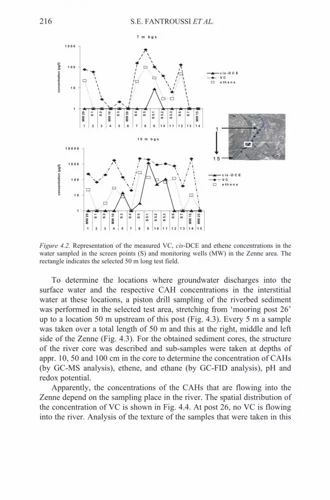
216 S.E. FANTROUSSI ET AL.
7 m b g s
1
1 0
1 0 0
1 0 0 0
MW
29
S 1
S 2
MW
16
S 3
MW
28
S 4
S 5
S 5
.1
S 5
.2
S 5
.3
S 6
S 7
MW
15
1 2 3 4 5 6 7 8 9 1 0 1 1 1 2 1 3 1 4
co
nc
en
tra
tio
n (
µg
/l)
c i s -D C E
V C
e t h e n e
1 0 m b g s
1
1 0
1 0 0
1 0 0 0
1 0 0 0 0
MW
29
S 1
S 2
MW
16
S 3
S 4
S 5
S 5
.1
S 5
.2
S 5
.3
S 6
S 7
MW
15
MW
25
1 2 3 4 5 7 8 9 1 0 1 1 1 2 1 3 1 4 1 5
co
nc
en
tra
tio
n (
µg
/l)
c is - D C E
V C
e t h e n e
1
1 5
1
1 5
Figure 4.2. Representation of the measured VC, cis-DCE and ethene concentrations in the water sampled in the screen points (S) and monitoring wells (MW) in the Zenne area. The rectangle indicates the selected 50 m long test field.
To determine the locations where groundwater discharges into the surface water and the respective CAH concentrations in the interstitial water at these locations, a piston drill sampling of the riverbed sediment was performed in the selected test area, stretching from ‘mooring post 26’ up to a location 50 m upstream of this post (Fig. 4.3). Every 5 m a sample was taken over a total length of 50 m and this at the right, middle and left side of the Zenne (Fig. 4.3). For the obtained sediment cores, the structure of the river core was described and sub-samples were taken at depths of appr. 10, 50 and 100 cm in the core to determine the concentration of CAHs (by GC-MS analysis), ethene, and ethane (by GC-FID analysis), pH and redox potential.
Apparently, the concentrations of the CAHs that are flowing into the Zenne depend on the sampling place in the river. The spatial distribution of the concentration of VC is shown in Fig. 4.4. At post 26, no VC is flowing into the river. Analysis of the texture of the samples that were taken in this

BIOLOGICAL ASSESSMENT 217
Mid-river
Right side
Left sideMid-river
Right side
Left side
Non-polluted groundwater
Polluted groundwater
Flow direction Zenne
Post 26Screenpoint 5
5 m
Screenpoint 5.1
45 m 0 m25 m
Left side
Middle side
Right side
Non-polluted groundwater
Polluted groundwater
Flow direction Zenne
Post 26Screenpoint 5
5 m
Screenpoint 5.1
45 m 0 m25 m
Non-polluted groundwater
Polluted groundwater
Flow direction Zenne
Post 26Screenpoint 5
5 m
Screenpoint 5.1
Non-polluted groundwater
Polluted groundwater
Flow direction Zenne
Post 26Screenpoint 5
5 m
Screenpoint 5.1
45 m 0 m25 m45 m 0 m25 m
Left side
Middle side
Right side
Figure 4.3. Piston drill sampling at multiple locations in the streambed of the river Zenne.
last area indicated that they contain layers with very fine sand that could prevent the infiltration of VC into the river. In the area from 15 m to 45 m upstream of post 26, VC is flowing into the Zenne and the highest concentrations are detected between 25 and 30 m upstream of this post (> 1000 µg/L). While VC is detected throughout the studied Zenne area, cis-DCE is only present between 30 m and 40 m upstream of post 26 at a concentration of 27 to 300 µg/L (data not shown).
VClog (µg/L)
Depth in sediment :
-5 cm
-50 cm
-100 cm
0.0
0.5
1.0
1.5
2.0
2.5
3.0
3.5
4.0
VClog (µg/L)
Depth in sediment :
-5 cm
-50 cm
-100 cm
0.0
0.5
1.0
1.5
2.0
2.5
3.0
3.5
4.0
Figure 4.4. Measured interpolated VC and cis-DCE concentrations in the pore water of the sampled sediment in the river Zenne at the three sampling lines (right, middle and left) and depths (10, 50 and 100 cm).

218 S.E. FANTROUSSI ET AL.
Depending on the position in the Zenne, the VC-concentration at the top of the piston drill samples (approximately 10 cm beneath the surface water) is lower, equal or higher than in the middle and the bottom of the cores (Fig. 4.4). However, at one location a VC concentration of 1139 µg/kg d.m. is detected in the top of the sediment layer. This indicates that VC is discharging into the surface water of the river Zenne. At other locations, e.g. closer to post 26, much lower VC concentrations are present. The concentrations of ethene and ethane that are detected (data not shown) indicate that full dechlorination of VC to ethene and ethane is occurring, but apparently not efficiently enough to prevent VC from flowing into the river.
4.2.2. Determination of the degradation potential of the eutrophic river
sediment microbial community by molecular analyses and anaerobic
degradation tests
To determine the degradation potential of the eutrophic river sediment microbial community, anaerobic microcosms and molecular biological techniques were applied on sediment material obtained from different positions in the interface of the CAH-polluted test area of the river Zenne.
The presence of dehalogenating bacteria and chloroethene dehalogenase genes was investigated in surface water, groundwater and sediment slices obtained from macro-core sediment samples (Fig. 4.5) collected at the right, middle and left side of the Zenne located around ‘mooring post 26’. The undisturbed sediment cores were frozen on dry ice immediately after sampling and divided into slices of approximately 0,5 cm in the laboratory, using an electrical saw. The presence of CAH degrading bacteria (Dehalococcoides and Desulfuromonas species) and genes involved in the first steps (pceA and tceA) and the last step (tceA, vcrA and bvcA) of the reductive dehalogenation of PCE to ethene was investigated by PCR113-116.
It can be concluded that the groundwater discharge spatial distribution is highly heterogeneous, mostly depending on local sediment permeability. At some locations (e.g. at post 26) full dechlorination was observed, while VC and cis-DCE reach the surface water and discharge into the river at spots with a high-velocity groundwater influx.

BIOLOGICAL ASSESSMENT 219
Figure 4.5. Macro-core sampling at three different places in the river bed and the obtained undisturbed sediment sample.
Only the PCR results of a sediment core obtained at the right side of the river are shown (Fig. 4.6). Dehalococcoides species, capable of degrading PCE completely to ethene, were present in the investigated groundwater and in all the sediment slices of the cores obtained from the right, middle and left side of the Zenne riverbed, but not in the surface water. Desulfuromonas species, capable of degrading PCE to cis-DCE, were also found in almost all sediment slices of the different cores and in the surface water of the river Zenne, but not in the groundwater. The pceA(1) gene of Dehalobacter restrictus, Desulfitobacterium hafniense, and Desulfitobacterium sp. PCE1, and the pceA(2) gene of Sulfurospirillum
multivorans were only detected in some of the sediment slices throughout the river bed. The pceA(2) gene was also detected in surface water of the river Zenne. The tceA gene of Dehalococcoides sp. strain FL-2 and D.
ethenogenes strain 195 was present in the groundwater, surface water and some or all sediment slices from the cores collected at the three different sampling places. The encoded TCE reductive dehalogenase can transform TCE to non-toxic ethene, but the reduction of VC to ethene is co-metabolic. The VC reductive dehalogenase genes of Dehalococcoides sp. strain VS and BAV1, vcrA and bvcA, were only detected in the sediment core collected from the right side and not in the middle or the left side of the Zenne. Since these two Dehalococcoides species can derive energy for growth from the degradation of cis-DCE and VC to non-toxic ethene as opposed to D. ethenogenes strain 195 and FL-2, their presence indicates

220 S.E. FANTROUSSI ET AL.
that a large microbial degradation potential is present in the river sediment where polluted groundwater is passing the sediment zone.
The actual anaerobic degradation of VC and cis-DCE by the eutrophic river sediment microbial community was studied in anaerobic microcosms constructed with fresh Zenne sediment obtained from the top (5-10 cm) or the bottom (10-20 cm) of undisturbed sediment cores that were taken in the test area. A mixture of sediment material (37,5 g) obtained from the left and right side of the Zenne was suspended in 70 ml of groundwater that was collected directly upstream from the river using a Geoprobe direct-push system. The degradation of VC and cis-DCE was followed in function of time by headspace GC-FID analysis. The results of these batch degradation
indicate that both the in situ concentrations of VC (around 300 µg/L) and cis-DCE (around 150 µg/L) were rapidly reduced to non-toxic ethene and ethane both in the top and the bottom river sediment. When 2000 µg/L cis-DCE was re-added to the microcosms, it was again completely reduced to ethene within 1 month.
Dehalococcoides Desulfuromonas pce A(1)pce A(2)
tce A vcr AB bvc A
2019181716151413121110987654321
groundwater 7m bgs 10m bgs
surface water
black
gray
Figure 4.6. PCR results of the detection of dehalogenating bacteria and chloroethene dehalogenase genes in a sediment core obtained at the right side of the river Zenne. Boxes represent surface water, groundwater or the sediment slices that were obtained from the frozen sediment core. Empty boxes indicate that no PCR product was obtained, shaded boxes indicate the presence of the bacterium or catabolic gene.
tests (Fig. 4.7 representing results 10-20 cm depth sediment material)

BIOLOGICAL ASSESSMENT 221
1
10
100
1000
10000
0 50 100 150
time (days)
co
ncen
trati
on
(µ
g/l)
VC
cis-DCE
ethene
ethane
Dead control Living
Spike cis-DCE Spike cis-DCE
1
10
100
1000
10000
0 50 100 150
time (days)
VC
cis-DCE
ethene
ethane
1
10
100
1000
10000
0 50 100 150
time (days)
co
ncen
trati
on
(µ
g/l)
VC
cis-DCE
ethene
ethane
Dead control Living
Spike cis-DCE Spike cis-DCE
1
10
100
1000
10000
0 50 100 150
time (days)
VC
cis-DCE
ethene
ethane
Figure 4.7. Anaerobic degradation of VC and cis-DCE in batch degradation tests containing (10-20 cm depth) river sediment material and groundwater from the CAH-polluted test area of the river Zenne.
4.3. CONCLUSIONS
A high microbial CAH degradation potential was detected in the river sediment of the Zenne in Belgium. The detected Dehalococcoides sp. strain VS and BAV1, which can actually grow on VC and cis-DCE as sole energy source, are probably responsible for the relatively fast and complete reductive dehalogenation of these groundwater pollutants that is observed in the microcosms. By degrading VC and cis-DCE, the sediments act as a natural biobarrier for the CAHs present in the groundwater that is passing through the sediment zone, hereby reducing the risk of surface water contamination.
However, some locations in the studied test area have high groundwater influx rates. At these locations VC and cis-DCE most likely reach the surface water and discharge into the river. At these locations, the observed microbial degradation potential of the river sediment apparently is inadequate to prevent the CAHs from reaching the surface water. Future research will therefore focus on how to stimulate the degradation of the CAHs, both in the river sediment and in the aquifer material upstream of the river Zenne.
5. Ex-situ and in-situ biotreatment of contaminated sediments
5.1. INTRODUCTION
Pollution of soils, sediments and groundwaters is a major problem worldwide. The total world hazardous waste remediation market is

222 S.E. FANTROUSSI ET AL.
estimated at about US $16 billion per year. There are at least 350 000 contaminated sites in Western Europe alone and it may cost as much as US $ 400 billion to clean the most dangerous of these sites over the next 20-25 years117. Sediments provide essential habitat for many freshwater, estuarine, and marine organisms. In aquatic systems, most anthropogenic chemicals and waste materials, particularly persistent organic and inorganic chemicals, may accumulate in sediments. These sediments become repositories for many of the more toxic chemicals that are introduced into surface waters. These pollutants can attach to suspended particulates in the water, and subsequently settle out to the bottom. Through complex chemical, physical and biological interactions, these contaminants may be further transformed and transported to other parts of the aquatic system. At elevated concentrations, contaminated sediments lead to many problems in lakes, rivers and harbors, including fish advisories, habitat impairments and restrictions on dredging. Contaminated sediments may also pose an unacceptable risk to aquatic organisms, aquatic-dependent wildlife and humans. Contaminants that build up in the food chain are of particular concern, especially mercury, polychlorinated biphenyls (PCBs), dioxins and organochlorine pesticides. It has become clear that sediment cleanup is an important task and several remediation strategies are designed and implemented to eliminate toxic pollutants from these matrices. Physical and chemical treatments have been used to clean up sediments but they are usually very costly and they are not applicable to all types of environments. For instance, air or steam stripping is practiced primarily for the cleanup of unsaturated soils, whereas “pump and treat” technologies target mainly the groundwater (and sometimes saturated soil and sediment zones), involving pumping and capturing of contaminants (e.g. on a sorbent or by skimming, flotation, etc.) from the water, which is returned to the aquifer. This physical flushing of the pollutants from large volumes of groundwater or saturated sediment is extremely slow and costly. Washing of the solid matrix is also practiced, but is mainly of use on unsaturated soils and less so on saturated soils and sediments. Finally, incineration is a drastic measure, addressing soils and sediments that are contaminated with weathered, often intimately encrusted recalcitrant pollutants. Its cost is high and the environmental impacts can be severe.
Bioremediation strategies are less expensive and friendlier to the environment than physical and chemical treatments of contaminated soils and, to a lesser extent, sediments. This set of technologies uses biological, primarily microbial, activities and can be classified into three major techniques:
BIOLOGICAL ASSESSMENT 223
1. In situ: No excavation, the solids remain in place. This class of techniques can be subdivided into engineered in situ bioremediation and intrinsic bioremediation (or natural attenuation). Examples include bioventing, biosparging, etc.
2. Ex situ: Sediment is excavated and transferred to be treated in specialized installations far from the site. It is always an engineered solution. Examples: land farming, slurry reactors. A sub-category of ex situ bioremediation, occasionally considered as a separate class of techniques, is on site bioremediation, which implies treatment of a soil or sediment at the surface of the site (e.g., biopiling).The applicability of different biological treatments depends on the
susceptibility of the pollutants to biodegradation (Table 5.1) and on the type of environment targeted for cleanup. A number of prior steps are needed before deciding on the bioremediation strategy, including a thorough characterization of the site in terms of hydrology, extent and type of contamination, intrinsic microbial activity, and supply rates of key materials, such as electron acceptors or donors118.
Table 5.1. Contaminant susceptibility to bioremediation.
Pollutant Frequency of
occurrence
Status of bio-
remediation
Limitations
Polycyclic
aromatic
hydrocarbons
Common Emerging Sorb strongly to
subsurface
Solids
Chlorinated
aliphatics
Very frequent Emerging Nonaqueous
phase liquids
(NAPL)
Chlorinated
aromatics
Common Emerging NAPL and
solids
Polychlorinated
biphenyls
Infrequent Emerging Sorb strongly to
subsurface
Solids
Heavy metals &
metalloids
Common Possible to
immobilize
Availability
highly variable
Gasoline,
fuel oil
Very frequent Established NAPL

224 S.E. FANTROUSSI ET AL.
5.2. IN SITU BIOREMEDIATION
In situ bioremediation encourages growth and reproduction of indigenous microorganisms to enhance biodegradation of organic constituents in the saturated zone. In situ groundwater bioremediation can effectively degrade organic constituents which are dissolved and adsorbed onto the aquifer matrix. In an analogous manner, a sediment contaminated with organic pollutants can be cleaned up. In situ bioremediation can be effective for the
situ bioremediation is more effective when combined with other saturated zone remedial technologies (e.g., air sparging) and, for the case of contaminated soils, with vadose zone remedial operations (e.g., soil vapor extraction, bioventing). Air sparging involves the injection of compressed air or oxygen directly into the contaminated subsurface. Injected air traverses horizontally and vertically in channels through the sediment column, creating an underground stripper that removes volatile and semivolatile organic contaminants by volatilization and entrainment in the gas stream. The injected air helps to flush the contaminants into the unsaturated zone. Soil Vapor Extraction (SVE) usually is implemented in conjunction with air sparging to remove the generated vapor-phase contamination from the vadose zone. Oxygen added to the contaminated groundwater and vadose-zone soils also can enhance biodegradation of contaminants below and above the water table by stimulating the local microbial activity. Bioventing is an in situ remediation technology that combines soil vapor extraction methods with bioremediation. It uses vapor extraction wells that induce air flow in the subsurface through the use of a vacuum. Bioventing can be effective in remediating emissions of petroleum products, such as gasoline, jet fuels, kerosene, and diesel fuel but is mostly applicable to unsaturated matrices. Since the contaminated sediment represents a partly or totally saturated zone, a water circulation strategy for the supply of metabolism stimulating materials can often be applied in situ,
e.g. for the engineered bioremediation of a nonaqueous phase liquid such as petroleum. In this case, hydrogen peroxide can be used as a dissolved source of oxygen, via vertical wells and horizontal infiltration galleries118,119
(see below).
5.2.1. Biostimulation and bioaugmentation
In situ bioremediation can be implemented in a number of treatment modes, including: Aerobic (oxygen respiration); anoxic (nitrate respiration); anaerobic (non-oxygen respiration); and co-metabolic. The aerobic mode has been proven most effective in reducing contaminant levels of aliphatic (e.g., hexane) and aromatic petroleum hydrocarbons (e.g., benzene,
range of xenobiotic compounds indicated in Table 5.2. In certain cases in

BIOLOGICAL ASSESSMENT 225
naphthalene) typically present in gasoline and diesel fuel. In the aerobic treatment mode, the sediment and aquifer area is oxygenated by one of three methods: Direct sparging of air or oxygen through an injection well; saturation of water with air or oxygen prior to re-injection; or addition of hydrogen peroxide directly into an injection well or into reinjected water. Irrespective of the method of oxygenation, it is important to ensure that oxygen is being distributed throughout the area of contamination. Anoxic, anaerobic, and co-metabolic modes are sometimes used for remediation of other compounds, such as chlorinated solvents, but are generally slower than aerobic respiration in breaking down xenobiotics. The key parameters that determine the effectiveness of in situ bioremediation are: (a) hydraulic conductivity of the aquifer, which controls the distribution of electron acceptors and nutrients in the subsurface; (b) biodegradability of the xenobiotic constituents, which determines both the rate and degree to which constituents will be degraded by microorganisms; and (c) location of the xenobiotic contamination in the subsurface. In general, the sediment/aquifer system will determine hydraulic conductivity. Fine-grained media (e.g.,clays, silts) have lower intrinsic permeability than coarse-grained media (e.g., sands, gravels). Bioremediation is generally effective in permeable media. However, depending on the extent of contamination, bioremediation also can be effective in less permeable silty or clayey media, which represent a considerable proportion of sediments. In general, a medium of lower permeability will require a longer period of time to clean up than a more permeable medium3. The biodegradability of a xenobiotic constituent is a measure of its ability to be metabolized (or co-metabolized) by microorganisms. The chemical characteristics of the contaminants will dictate their biodegradability. For example, heavy metals are not degraded by bioremediation but they can be sequestered as insoluble precipitates under specific redox conditions. The biodegradability of organic constituents depends on their chemical structures and physical/chemical properties (e.g., water solubility, water/octanol partition coefficient). Highly soluble organic compounds with low molecular weights will tend to be more rapidly degraded than slightly soluble compounds with high molecular weights. The low water solubilities of the more complex compounds render them less bioavailable to microorganisms. Consequently, the larger, more complex chemical compounds may be slow to degrade or may even be recalcitrant to biological degradation118,119.
When oxygen is introduced to the subsurface as a terminal electron acceptor, it can react with dissolved iron [Fe(II)] to form an insoluble iron precipitate, ferric oxide. This precipitate can be deposited in infiltration trenches or channels, reducing permeability. The effects of iron precipitation tend to be most noticeable around injection wells, where

226 S.E. FANTROUSSI ET AL.
oxygen concentration in groundwater is highest and can render injection wells inoperable.
Extreme pH values (i.e., less than 5 or greater than 10) are generally unfavorable for microbial activity. Typically, optimal microbial activity occurs under neutral pH conditions (i.e., in the range of 6-8). The optimal pH is site specific. For example, aggressive microbial activity has been observed at lower pH conditions outside of this range (e.g., 4.5 to 5) in natural systems. Because indigenous microorganisms have adapted to the natural conditions where they are found, pH adjustment, even toward neutral, can inhibit microbial activity. If man-made conditions (e.g.,releases of petroleum) have altered the pH outside the neutral range, pH adjustment may be needed. If the pH of the groundwater is too acidic, lime or sodium hydroxide can be added to increase the pH. If the pH is too alkaline, then a suitable acid (e.g., hydrochloric) can be added to reduce the pH. Changes to pH should be closely monitored because rapid changes of more than 1 or 2 units can inhibit microbial activity and may require an extended acclimation period before the microbes resume their activity.
As a rule, microorganisms require carbon/energy sources (electron donors) and terminal electron acceptors (TEA) to enzymatically transform the target contaminants. Carbon source(s) and TEA are introduced in in situ
engineered bioremediation systems via injection wells or infiltration galleries. Extraction wells ensure that the pollutant plume does not spread contaminants into clean areas or accelerate the movement toward receptors. Placement of extraction wells is critical, especially in systems that also use nutrient injection wells or infiltration galleries. These additional sources of water can alter the natural groundwater flow patterns which can cause the contaminant plume to move in an unintended direction or rate. Without adequate hydraulic control, this situation can lead to worsening of the original condition and complicate the cleanup or extend it.
as N and P, to sustain microorganism growth and biodegradation. Nutrients may be available in sufficient quantities in the sediment/aquifer system (intrinsic in situ bioremediation, in which no external intervention is taking place other than a careful monitoring of the natural attenuation of the target contaminants) but, more frequently, nutrients need to be added to maintain adequate bacterial populations (biostimulation)118,119. Addition of N, P and
established for in situ bioremediation of subsurface contaminated by hydrocarbons. Supply of electron donors (e.g. sugar or methane) or TEA (e.g. oxygen or hydrogen peroxide) in case of such limitations, or addition
Nutrient injection systems may be unnecessary if the sediment/
other inorganic nutrients to optimize microbial growth is becoming
groundwater system contains adequate amounts of nutrients, such

BIOLOGICAL ASSESSMENT 227
of detergents in case of low bioavailability of the pollutants, are instances of biostimulation119.
Sometimes the microbial community of a polluted site may not display the metabolic potential for degradation and complete mineralization of the target pollutant. This could be due to very low numbers of a single microorganism possessing the entire pathway for its eventual breakdown to harmless end products. More likely, the pollutant may be a complex molecule or a mixture of compounds which could be broken down only by a specific combination of microorganisms (‘consortium’) and pathways. In such cases, successful bioremediation may call for bioaugmentation, i.e. the inoculation of the polluted sediment with specific populations of microorganisms120. Bioaugmentation is not yet a mature technology for in situ bioremediation of soils and groundwater and, even less so, of sediments. With the exception of a few field applications most bioaugmentation reports concern laboratory-scale studies. It is then no surprise that bioaugmentation is mired in controversy, given the excessive claims of success, esp. by commercial companies, and the documented failures of large-scale inoculation campaigns119,120. Typical bioaugmentation options include: the addition of a pre-adapted pure bacterial strain (e.g. inoculating with chloroaromatic degraders); the addition of pre-adapted consortia (e.g. PCB-degrading enrichment cultures to contaminated sediments); addition of genetically modified bacteria for simultaneous mineralization of chloro- and methylaromatics; and transfer of biodegradation-relevant genes by conjugation into microorganisms present in the biotope to help remove PCBs or pesticides120. Ideally, the applicability and the limits of bioaugmentation should be tested in a situation comparable to the eventual intervention, i.e., at a mesocosm or pilot scale, after initial microcosm tests. In the first study of bioaugmentation of a realistic 500-liter pilot-scale reactor simulating an anaerobic aquifer/sediment system, we tested the capacity of an inoculum of the halorespiring anaerobe Desulfomonile tiedjei to dechlorinate 3-chlorobenzoate (3-CB) continuously fed into the saturated zone together with acetate+formate as cosubstrate121. Heterogeneous distribution of 3-CB dechlorination activity correlated with the presence of the inoculated bacterium which was detected by its 16S rRNA gene signature using PCR. Denaturing gradient gel electrophoresis (DGGE) of PCR amplicons from the total DNA of different compartments in the reactor demonstrated distinct community fingerprints as a function of location and in response to process modifications121Reductive dehalogenation of organic pollutants in groundwater following biostimulation of native microbial communities is often seen in the field, where chlorinated ethenes (PCE, TCE) are completely transformed to ethene122. According to industrial practitioners of

228 S.E. FANTROUSSI ET AL.
in situ bioremediation in the field, bioaugmentation with specific halorespiring anaerobes of the genus Dehalococcoides, which degrades chloroethenes to ethene without stopping at cis-dichloroethene (cDCE) or at vinyl chloride (VC), may shorten the lag phase close to the injection wells, but is not required to assure complete in situ dechlorination. Moreover, because it is not necessary to constrain hydrogen levels for the benefit of inoculated Dehalococcoides sp., it is also not necessary to limit rates of electron donor (cosubstrate) consumption by using commercial “designer” slow-release cosubstrates122. In a pilot investigation consistent with this standpoint, we examined on a demonstration (pilot) scale the dynamics of in situ bioremediation of a sediment co-contaminated with trichloroethene (TCE) and nickel123,124 in preparation for the field scale in situ interventionat the former metallurgical industrial site of Bunnik near Utrecht (The Netherlands), where the contaminated anaerobic sediment was located 20-30 m below ground. The 680-liter “sandbox-type” reactor filled with the contaminated sediment was fed under strict anaerobic conditions a 10 mg/l TCE-containing stream simulating the plume together with a methanol+lactate cosubstrate mixture. The cosubstrate chemical oxygen demand (COD) was progressively diminished from 1000 mg/l down to 100 mg/l as the redox potential reached levels consistent with sulfate reduction and methanogenesis while the feed COD/SO4
- - ratio was fixed at 100/0.6 (w/w)123. In previous small-scale fed-batch sediment column tests, this cosubstrate mixture had been shown to be a good electron donor to sustain complete TCE dechlorination, and a level of sulfate up to 10 mM not only did not interfere with the complete transformation of TCE to ethane, but also contributed to Ni sequestration in the form of NiS124. Results of the pilot reactor showed that TCE was dechlorinated to harmless ethene via cDCE and VC by indigenous microorganisms in the sediment, whereas concurrent stimulation of sulfate-reducing anaerobic bacteria with the fed sulfate led to the generation of sulphide which was efficient in precipitating nickel within the sediment123. Remarkably, probing with 16s rDNA –based PCR gave no indication of the presence of dechlorinating bacteria of the Dehalococcoides group, which indicates that the complete dechlorination was brought about either by as yet unknown halorespirers or cometabolically by local anaerobic trophic groups including sulphate reducers124. The dynamics of the process is shown in Fig. 5.1.

BIOLOGICAL ASSESSMENT 229
Figure 5.1. Time course of the concentrations of pollutant and products in the demonstration-scale sediment bioreactor. Chloroethene concentrations at the reactor outlet in the gas phase (upper panel) and in the liquid phase (lower panel).
5.3. EX SITU BIOREMEDIATION
Above ground treatment of contaminated sediments is an option in the case of limited of quantities of heavily contaminated sediments, and whenever regulatory compliance or other constraints impose a rapid cleanup118,119.This set of technologies, which includes slurry bioreactor treatment, land farming and composting, should only be practiced when the usually high costs of excavation and exposure to workers and residents can be more than compensated by a major risk reduction stemming from the removal of the pollutant source.
TC
E, cD
CE
(mg/L
)T
CE
, cD
CE
(mg/L
)
VC
,E
thene
(mg/L
)V
C,E
thene
(mg/L
)

230 S.E. FANTROUSSI ET AL.
Slurry bioreactor treatment involves vigorous agitation of the sediment which may constitute 5-20% of the solids in a two- or three-phase configuration (depending on whether there is aeration or not) in the presence of native or, occasionally, externally introduced competent microorganisms. The degree of control and optimization that can be exercised on this system is considerable: in addition to vigorous inocula, nutrients, TEA and other additives can be introduced as needed. Therefore, the endpoints of this treatment are also far more predictable than in in situ
bioremediation. Because of the high degree of convective mass transfer in this setup, the rates of biotransformation and mineralization of the target contaminants are several-fold higher than what is observed in situ. In addition, mass transfer into the aqueous phase and, hence, overall rates of biodegradation in the case of less bioavailable, poorly water soluble compounds (such as PCB, PAH etc.), can be enhanced with the addition of solubilizing agents such as surfactants117,118.
Land farming involves the mixing of contaminated sediment into the surface layer of topsoil, to which there is an occasional addition of nutrients and moisture, the aim being an aerobic cometabolic degradation of the target pollutants by the soil microorganisms118. This empirical, generally uncontrolled, “low” technology can be upgraded by mixing the contaminated solids with fresh organic residues, resulting in composting117.The addition of air by appropriate channeling or turning as well as the regulation of moisture (and nutrient or cosubstrate input) can enhance the rates of pollutant degradation and also increase the overall degree of control and predictability of the treatment of sediments. Nonetheless, both these techniques can be cumbersome, time-consuming and expensive: in addition to the costs of excavation, transportation and mechanical intervention (e.g. turning, tilling, etc.), they do require ample land areas. Moreover, certain very recalcitrant contaminants, even if immobilized or rendered non-bioavailable by sorption and/or covalent binding to the humic acids of the compost or to other components of the soil matrix may present a risk of eventually leaching out under extreme local conditions.
The work of the authors S. E Fantroussi and Spiros N. Agathos was supported by grants CHLOREM (BIO 4-CT 1998-0303) and MADOX (QLK3-CT-2001-00345) of the European Commission.
Acknowledgements
.

BIOLOGICAL ASSESSMENT 231
References
1. N.R. Pace, A molecular view of microbial diversity and the biosphere, Science 276, 734–740 (1997).
2. V.J. Orphan, C.H. House, K.U. Hinrichs, K.D. McKeegan, and E.F. DeLong, Methane-consuming archaea revealed by directly coupled isotopic and phylogenetic analysis, Science 293, 484–487 (2001).
3. K.P. Nevin, K.T. Finneran, and D.R. Lovley, Microorganisms associated with uranium bioremediation in a high-salinity subsurface sediment, Appl. Environ. Microbiol. 69, 3672–3675 (2003).
4. S. El Fantroussi, W. Verstraete, and E.M. Top, Enrichment and molecular characterization of a bacterial culture that degrades methoxy-methyl urea herbicides and their aniline derivatives, Appl. Environ. Microbiol.66, 5110–5115 (2000).
5. M.R. Harkness, J.B. McDermott, D.A. Abramowicz, J.J. Salvo, W.P. Flanagan, M.L. Stephens, F.J. Mondello, R.J. May, J.H. Lobos, K.M. Carroll, et al., In situ stimulation of aerobic PCB biodegradation in Hudson river sediments, Science 259, 503–507 (1993).
6. E.L. Madsen, Epistemology of environmental microbiology, Environ. Sci. Technol. 32, 429–439 (1998).
7. L. Eyers, I. George, L. Schuler, S.N. Agathos, and S. El Fantroussi, Environmental metagenomics: exploring the unmined richness of microbes to degrade xenobiotics. Appl. Microbiol.Biotechnol. 66, 123–130 (2004).
8. P.D. Schloss, and J. Handelsman, Biotechnological prospects from metagenomics, Curr. Opin. Microbiol. 14, 303–310 (2003).
9. E.L. Madsen, Methods for determining biodegradability. In: C.J. Hurst, R.L. Crawford, G.R. Knudsen, M.J. McInerney and L.D. Stetzenbach (Eds) Manual of Environmental Microbiology, 2nd Edition, ASM Press, Washington DC. (2002).
10. S. El Fantroussi, M. Belkacemi, E.M. Top, J. Mahillon, H. Naveau, and S.N. Agathos, Bioaugmentation of a soil bioreactor designed for pilot scale anaerobic bioremediation
11. E.R. Hendrickson, J.A. Payne, R.M. Young, M.G. Starr, M.P. Perry, S. Fahnestock, D.E. Ellis, and R.C. Ebersole, Molecular analysis of Dehalococcoides 16S ribosomal DNA from chloroethene-contaminated sites throughout North America and Europe, Appl. Environ. Microbiol. 68, 485–495 (2002).
12. J.R. de Lipthay, N. Tuxen, K. Johnsen, L.H. Hansen, H.J. Albrechtsen, P.L. Bjerg and J. Aamand, In situ exposure to low herbicide concentrations affects microbial population composition and catabolic gene frequency in an aerobic shallow aquifer, Appl. Environ. Microbiol. 69, 461–7 (2003).
13. W.S. Reeburgh, S.C. Whalen, and M.J. Alperin, The role of methylotrophy in the global methane budget, p. 1–14. In J.C. Murrell, and D.P. Kelly (ed.), Microbial growth on C1 compounds. Proceedings of the 7th International Symposium. American Society for Microbiology, Washington, D.C. (1993).
14. T.R. Thomsen, K. Finster, and N.B. Ramsing, Biogeochemical and molecular signatures of anaerobic methane oxidation in a marine sediment, Appl. Environ. Microbiol. 67, 1646–1656 (2001).
15. K.U. Hinrichs, J.M. Hayes, S.P. Sylva, P.G. Brewer, and E.F. DeLong, Methane-consuming archaebacteria in marine sediments, Nature 398, 802–805 (1999).
16. R.M. Atlas and R. Bartha, Microbial Ecology: Fundamentals and Applications. Menlo Park, CA: Benjamin/Cummings (1998).
studies, Environ. Sci. & Technol. 33, 2992–3001 (1999).

232 S.E. FANTROUSSI ET AL.
17. W.W. Mohn and J.M. Tiedje, Microbial reductive dehalogenation, Microbiol. Rev. 56, 482–507. (1992).
18. V. Achá, M. Meurens, H. Naveau, and S.N. Agathos, ATR-FTIR sensor development for continuous on-line monitoring of chlorinated aliphatic hydrocarbons in a fixed-bed bioreactor, Biotechnol. Bioeng. 68, 473–487 (2000).
19. R.M. Atlas, Microbial hydrocarbon degradation - bioremediation of oil spills, J. Chem.
Technol. Biotechnol. 52, 149–156 (1991). 20. F.H. Chapelle, Bioremediation of petroleum hydrocarbon-contaminated ground water:
The perspectives of history and hydrology, Ground Water 37, 122–132 (1999). 21. D.C. Dobbins, C.M. Aelion, and F. Pfaender, Subsurface, terrestrial microbial ecology
and biodegradation of organic chemicals: A review, Crit. Rev. Environ. Ctrl. 22, 67–136 (1992).
22. D.F. Pope, and J.N. Jones, 1999, Monitored natural attenuation of chlorinated solvents, edited. (EPA, Washington, 1999), pp. 1–3.
23. K. Watanabe, H. Futamata, and S. Harayama, Understanding the diversity in catabolic potential of microorganisms for the development of bioremediation strategies, Antonie
Van Leeuwenhoek 81, 655–663 (2002). 24. D.H. Pieper, V. dos Santos, and P.N. Golyshin, Genomic and mechanistic insights into
the biodegradation of organic pollutants, Curr. Opin. Biotechnol. 15, 215–224 (2004). 25. K. Watanabe, Microorganisms relevant to bioremediation, Curr. Opin. Biotechnol. 12,
237–241 (2001). 26. C.S. Harwood, and R.E. Parales, The beta-ketoadipate pathway and the biology of
self-identity, Annu. Rev. Microbiol. 50, 553–590 (1996). 27. N. Söhngen, Benzin, Petroleum, Paraffinöl und Paraffin als Kohlenstoff-und
Energiequelle für Mikroben, Zentr. Bacteriol. Parasitenk. Abt II 37, 595–609 (1913). 28. R.Y. Stanier, The oxidation of aromatic compounds by fluorescent Pseudomonas, J.
Bacteriol. 55, 477–494 (1947). 29. M. Boll, G. Fuchs, and J. Heider, Anaerobic oxidation of aromatic compounds and
hydrocarbons, Curr. Opin. Chem. Biol. 6, 604–611 (2002). 30. J. Heider, A.M. Spormann, H.R. Beller, and F. Widdel, Anaerobic bacterial
metabolism of hydrocarbons, FEMS Microbiol. Rev. 22, 459–473 (1998). 31. M.R. Smith, The biodegradation of aromatic hydrocarbons by bacteria,
Biodegradation 1, 191–206 (1990). 32. D.T. Gibson, and R.E. Parales, Aromatic hydrocarbon dioxygenases in environmental
biotechnology, Curr. Opin. Biotechnol. 11, 236–243 (2000). 33. S. Beil, J.R. Mason, K.N. Timmis, and D.H. Pieper, Identification of chlorobenzene
dioxygenase sequence elements involved in dechlorination of 1,2,4,5-tetrachlorobenzene, J. Bacteriol. 180, 5520–5528 (1998).
34. K. Furukawa, H. Suenaga, and M. Goto, Biphenyl dioxygenases: functional versatilities and directed evolution, J. Bacteriol. 186, 5189–5196 (2004).
35. M. Zielinski, S. Backhaus, and B. Hofer, The principal determinants for the structure of the substrate- binding pocket are located within a central core of a biphenyl dioxygenase alpha subunit, Microbiology 148, 2439–2448 (2002).
36. R.E. Parales, S.M. Resnick, C.L. Yu, D.R. Boyd, N.D. Sharma, and D.T. Gibson, Regioselectivity and enantioselectivity of naphthalene dioxygenase during arene cis-dihydroxylation: Control by phenylalanine 352 in the alpha subunit, J. Bacteriol. 182, 5495–5504 (2000).
37. J. Armengaud, B. Happe, and K.N. Timmis, Genetic analysis of dioxin dioxygenase of Sphingomonas sp. strain RW1: Catabolic genes dispersed on the genome, J. Bacteriol.
180, 3954–3966 (1998).

BIOLOGICAL ASSESSMENT 233
38. D.T. Gibson, R.L. Roberts, M.C. Wells, and V.M. Kobal, Oxidation of biphenyl by a Beijerinckia species, Biochem. Biophys. Res. Comm. 50, 211–215 (1973).
39. Y. Furusawa, V. Nagarajan, M. Tanokura, E. Masai, M. Fukuda, and T. Senda, Crystal structure of the terminal oxygenase component of biphenyl dioxygenase derived fromRhodococcus sp strain RHA1, J. Mol. Biol. 342, 1041–1052 (2004).
40. X.S. Dong, S. Fushinobu, E. Fukuda, T. Terada, S. Nakamura, K. Shimizu, H. Nojiri, T. Omori, H. Shoun, and T. Wakagi, Crystal structure of the terminal oxygenase component of cumene dioxygenase from Pseudomonas fluorescens IP01, J. Bacteriol.
187, 2483–2490 (2005). 41. M. Zielinski, S. Kahl, H.J. Hecht, and B. Hofer, Pinpointing biphenyl dioxygenase
residues that are crucial for substrate interaction, J. Bacteriol. 185, 6976–6980 (2003). 42. H. Junca, I. Plumeier, H. Hecht, and D. Pieper, Difference in kinetic behaviour of
catechol 2,3-dioxygenase variants from a polluted environment, Microbiology 150, 4181–4187 (2004).
43. J.G. Leahy, P.J. Batchelor, and S.M. Morcomb, Evolution of the soluble diiron monooxygenases, FEMS Microbiol. Rev. 27, 449–479 (2003).
44. M.S. Shields, S.O. Montgomery, P.J. Chapman, S.M. Cuskey, and P.H. Pritchard, Novel pathway of toluene catabolism in the trichloroethylene-degrading bacterium G4, Appl. Environ. Microbiol. 55, 1624–1629 (1989).
pathway cloned from Pseudomonas pickettii PKO1, J. Bacteriol. 176, 3749–3756 (1994).
46. G.M. Whited, and D.T. Gibson, Toluene-4-monooxygenase, a three-component enzyme system that catalyzes the oxidation of toluene to p-cresol in Pseudomonas
mendocina KR1, J. Bacteriol. 173, 3010–3016 (1991). 47. A. Fishman, Y. Tao, and T.K. Wood, Toluene 3-monooxygenase of Ralstonia pickettii
PKO1 is a para-hydroxylating enzyme, J. Bacteriol. 186, 3117–3123 (2004). 48. G.M. Whited, and D.T. Gibson, Separation and partial characterization of the enzymes
of the toluene-4-monooxygenase catabolic pathway in Pseudomonas mendocina KR1, J. Bacteriol. 173, 3017–3020 (1991).
49. K.H. Mitchell, J.M. Studts, and B.G. Fox, Combined participation of hydroxylase active site residues and effector protein binding in a para to ortho modulation of toluene 4-monooxygenase regiospecificity, Biochemistry 41, 3176–3188 (2002).
50. L. Rui, K.F. Reardon, and T.K. Wood, Protein engineering of toluene ortho-monooxygenase of Burkholderia cepacia G4 for regiospecific hydroxylation of indole to form various indigoid compounds, Appl. Microbiol. Biotechnol. 66, 422–429 (2005).
51. K.A. Canada, S. Iwashita, H. Shim, and T.K. Wood, Directed evolution of toluene ortho-monooxygenase for enhanced 1-naphthol synthesis and chlorinated ethene degradation, J. Bacteriol. 184, 344–349 (2002).
52. M.H. Sazinsky, J. Bard, A. Di Donato, and S.J. Lippard, Crystal structure of the toluene/o-xylene monooxygenase hydroxylase from Pseudomonas stutzeri OX1 - Insight into the substrate specificity, substrate channeling, and active site tuning of multicomponent monooxygenases, J. Biol. Chem. 279, 30600–30610 (2004).
53. W. Reineke, Development of hybrid strains for the mineralization of chloroaromatics by patchwork assembly, Annu. Rev. Microbiol. 52, 287–331 (1998).
54. D. Pieper, and W. Reineke, 2004, Degradation of chloroaromatics by Pseudomona(d)s, in: The Pseudomonads Vol III. Biosynthesis of Macromolecules and Molecular metabolism, edited by J.L. Ramos, (Kluwer Academic/Plenum Publishers, New York, 2004), pp. 509–574.
45. R.H. Olsen, J.J. Kukor, and B. Kaphammer, A novel toluene-3-monooxygenase

234 S.E. FANTROUSSI ET AL.
55. L.D. Eltis, and J.T. Bolin, Evolutionary relationships among extradiol dioxygenases, J.
Bacteriol. 178, 5930–5937 (1996). 56. A. Kitayama, T. Achioku, T. Yanagawa, K. Kanou, M. Kikuchi, H. Ueda, E. Suzuki,
H. Nishimura, T. Nagamune, and Y. Kawakami, Cloning and characterization of extradiol aromatic ring-cleavage dioxygenases of Pseudomonas aeruginosa JI104, J.
Ferm. Bioeng. 82, 217–223 (1996). 57. N. Schweigert, A.J.B. Zehnder, and R.I.L. Eggen, Chemical properties of catechols
and their molecular modes of toxic action in cells, from microorganisms to mammals, Environ. Microbiol. 3, 81–91 (2001).
58. D. Perez-Pantoja, T. Ledger, D.H. Pieper, and B. Gonzalez, Efficient turnover of chlorocatechols is essential for growth of Ralstonia eutropha JMP134(pJP4) in 3-chlorobenzoic acid, J. Bacteriol. 185, 1534–1542 (2003).
59. H.R. Beller, and A.M. Spormann, Anaerobic activation of toluene and o-xylene by addition to fumarate in denitrifying strain T, J. Bacteriol. 179, 670–676 (1997).
60. B. Leuthner, C. Leutwein, H. Schulz, P. Horth, W. Haehnel, E. Schiltz, H. Schagger, and J. Heider, Biochemical and genetic characterization of benzylsuccinate synthase from Thauera aromatica: a new glycyl radical enzyme catalysing the first step in anaerobic toluene metabolism, Mol. Microbiol. 28, 615–628 (1998).
61. F. Widdel, and R. Rabus, Anaerobic biodegradation of saturated and aromatic hydrocarbons, Curr. Opin. Biotechnol. 12, 259–276 (2001).
62. E. Annweiler, A. Materna, M. Safinowski, A. Kappler, H.H. Richnow, W. Michaelis, and R.U. Meckenstock, Anaerobic degradation of 2-methylnaphthalene by a sulfate-reducing enrichment culture. Appl. Environ. Microbiol. 66, 5329–5333 (2000).
63. S.R. Kane, H.R. Beller, T.C. Legler, and R.T. Anderson, Biochemical and genetic evidence of benzylsuccinate synthase in toluene-degrading, ferric iron-reducing Geobacter metallireducens, Biodegradation 13, 149–154 (2002).
64. C.D. Phelps, J. Battistelli, and L.Y. Young, Metabolic biomarkers for monitoring anaerobic naphthalene biodegradation in situ, Environ. Microbiol. 4, 532–537 (2002).
65. J.D. Coates, R. Chakraborty, and M.J. McInerney, Anaerobic benzene biodegradation - a new era, Res. Microbiol. 153, 621–628 (2002).
66. W.W. Mohn, and J.M. Tiedje, Evidence for chemiosmotic coupling of reductive dechlorination and ATP synthesis in Desulfomonile tiedjei, Arch. Microbiol. 157, 1–6 (1991).
67. C. Holliger, C. Regeard, and G. Diekert, 2003, Dehalogenation by anaerobic bacteria, in: Dehalogenation: Microbial Processes and Environmental Applications, edited by M.M. Häggblom, and I.D. Bossert. (Kluwer Academic Publishing, Boston, USA, 2003), pp. 115– 157.
68. J.Z. He, K.M. Ritalahti, K.L. Yang, S.S. Koenigsberg, and F.E. Loffler, Detoxification of vinyl chloride to ethene coupled to growth of an anaerobic bacterium, Nature 424, 62–65 (2003).
69. L. Adrian, U. Szewzyk, J. Wecke, and H. Gorisch, Bacterial dehalorespiration with chlorinated benzenes, Nature 408, 580–583 (2000).
70. M. Bunge, L. Adrian, A. Kraus, M. Opel, W.G. Lorenz, J.R. Andreesen, H. Gorisch, and U. Lechner, Reductive dehalogenation of chlorinated dioxins by an anaerobic bacterium, Nature 421, 357–360 (2003).
71. S. Ni, J.K. Fredrickson, and L. Xun, Purification and characterization of a novel 3-chlorobenzoate-reductive dehalogenase from the cytoplasmic membrane of Desulfomonile tiedjei DCB-1, J. Bacteriol. 177, 5135–5139 (1995).

BIOLOGICAL ASSESSMENT 235
72. B.C. Berks, F. Sargent, E. De Leeuw, A.P. Hinsley, N.R. Stanley, R.L. Jack, G. Buchanan, and T. Palmer, A novel protein transport system involved in the biogenesis of bacterial electron transfer chains, Biochim. Biophys. Acta 1459, 325–330 (2000).
73. J.A. Muller, B.M. Rosner, G. von Abendroth, G. Meshulam-Simon, P.L. McCarty, and A.M. Spormann, Molecular identification of the catabolic vinyl chloride reductase from Dehalococcoides sp strain VS and its environmental distribution, Appl. Environ.
Microbiol. 70, 4880–4888 (2004). 74. V. Torsvik, and L. Ovreas, Microbial diversity and function in soil: from genes to
ecosystems, Curr. Opin. Microbiol. 5, 240–245 (2002). 75. P. Wikstrom, A. Wiklund, A.C. Andersson, and M. Forsman, DNA recovery and PCR
quantification of catechol 2,3-dioxygenase genes from different soil types, J.
Biotechnol. 52, 107–120 (1996). 76. H.R. Beller, S.R. Kane, T.C. Legler, and P.J. Alvarez, A real-time polymerase chain
reaction method for monitoring anaerobic, hydrocarbon-degrading bacteria based on a catabolic gene, Environ. Sci. Technol. 36, 3977–3984 (2002).
77. H. Junca, and D.H. Pieper, Functional gene diversity analysis in BTEX contaminated soils by means of PCR-SSCP DNA fingerprinting: comparative diversity assessment against bacterial isolates and PCR-DNA clone libraries, Environ. Microbiol. 6, 95–110 (2004).
78. M. Orita, H. Iwahana, H. Kanazawa, K. Hayashi, and T. Sekiya, Detection of polymorphisms of human DNA by gel electrophoresis as single-strand conformation polymorphisms, Proc. Natl. Acad. Sci. U.S.A. 86, 2766–2770 (1989).
79. F. Schwieger, and C.C. Tebbe, A new approach to utilize PCR-single-strand-conformation polymorphism for 16s rRNA gene-based microbial community analysis, Appl. Environ. Microbiol. 64, 4870–4876 (1998).
80. A. Schmalenberger, and C.C. Tebbe, Bacterial diversity in maize rhizospheres: conclusions on the use of genetic profiles based on PCR-amplified partial small subunit rRNA genes in ecological studies, Mol. Ecol. 12, 251–261 (2003).
81. A. Okuta, K. Ohnishi, and S. Harayama, PCR isolation of catechol 2,3-dioxygenase gene fragments from environmental samples and their assembly into functional genes, Gene 212, 221–228 (1998).
82. J. Gescher, A. Zaar, M. Mohamed, H. Schagger, and G. Fuchs, Genes coding for a new pathway of aerobic benzoate metabolism in Azoarcus evansii, J. Bacteriol. 184, 6301–6315 (2002).
83. A. Zaar, J. Gescher, W. Eisenreich, A. Bacher, and G. Fuchs, New enzymes involved in aerobic benzoate metabolism in Azoarcus evansii, Mol. Microbiol. 54, 223–238 (2004).
84. V.J. Denef, J. Park, T.V. Tsoi, J.M. Rouillard, H. Zhang, J.A. Wibbenmeyer, W. Verstraete, E. Gulari, S.A. Hashsham, and J.M. Tiedje, Biphenyl and benzoate metabolism in a genomic context: outlining genome-wide metabolic networks in Burkholderia xenovorans LB400, Appl. Environ. Microbiol. 70, 4961–4970 (2004).
85. O.V. Moiseeva, I.P. Solyanikova, S.R. Kaschabek, J. Groning, M. Thiel, L.A. Golovleva, and M. Schlomann, A new modified ortho cleavage pathway of 3-chlorocatechol degradation by Rhodococcus opacus 1CP: Genetic and biochemical evidence, J. Bacteriol. 184, 5282–5292 (2002).
86. P. Nikodem, V. Hecht, M. Schlomann, and D.H. Pieper, New bacterial pathway for 4-and 5-chlorosalicylate degradation via 4-chlorocatechol and maleylacetate in Pseudomonas sp strain MT1, J. Bacteriol. 185, 6790–6800 (2003).

236 S.E. FANTROUSSI ET AL.
87. J. Yun, S. Kang, S. Park, H. Yoon, M. Kim, S. Heu, and S. Ryu, Characterization of a novel amylolytic enzyme encoded by a gene from a soil-derived metagenomic library, Appl. Environ. Microbiol. 70, 7229–7235 (2004).
88. S. Lee, K. Won, H. Lim, J. Kim, G. Choi, and K. Cho, Screening for novel lipolytic enzymes from uncultured soil microorganisms, Appl. Microbiol. Biotechnol. 65, 720–726 (2004).
89. E.M. Gabor, W.B.L. Alkema, and D.B. Janssen, Quantifying the accessibility of the metagenome by random expression cloning techniques, Environ. Microbiol. 6, 879–886 (2004).
90. A. Martinez, S.J. Kolvek, C.L.T. Yip, J. Hopke, K.A. Brown, I.A. MacNeil, and M.S. Osburne, Genetically modified bacterial strains and novel bacterial artificial chromosome shuttle vectors for constructing environmental libraries and detecting heterologous natural products in multiple expression hosts, Appl. Environ. Microbiol.
70, 2452–2463 (2004). 91. T. Uchiyama, T. Abe, T. Ikemura, and K. Watanabe, Substrate-induced gene-
expression screening of environmental metagenome libraries for isolation of catabolic genes, Nat. Biotechnol. 23, 88–93 (2005).
92. J.F. Jr. Brown and R.E. Wagner, PCB movement, dechlorination and detoxification in the Acushnet estuarine, Environ. Toxicol. Chem. 9, 1215–1233 (1990).
93. D.L. Bedard and J.F. Quensen III, Microbial reductive dechlorination of polychlorinated biphenyls, in: Microbial Transformation and Degradation of Toxic
Organic Chemicals, edited by L.Y. Young and C.E. Cerniglia (Wiley-Liss Division, John Wiley and Sons, New York, 1995), pp.127–216.
94. J. Wiegel and Q. Wu, Microbial reductive dehalogenation of polychlorinated biphenyls, FEMS Microbiol. Ecol., 32, 1–15 (2000).
95. A.C. Alder, M.M. Häggblom, S. Oppenheimer and L.Y. Young, Reductive dechlorination of polychlorinated biphenyls in anaerobic sediments, Environ. Sci.
Technol. 27, 530–538 (1993). 96. F. Fava, S. Gentilucci, and G. Zanaroli, Anaerobic biodegradation of weathered
polychlorinated biphenyls (PCBs) in contaminated sediments of Porto Marghera (Venice Lagoon, Italy), Chemosphere 53, 101–109 (2003).
97. L.D. Palekar, K.A. Maruya, J.E. Kostka and J. Wiegel, Dehalogenation of 2,6-dibromobiphenyl and 2,3,4,5,6-pentachlorobiphenyl in contaminated estuarine sediment, Chemosphere 53, 593–600 (2003).
98. J.L. Lake, R.J. Pruell and F.A. Osterman, An examination of dechlorination process and pathways in new Bedford Harbor sediments, Mar. Environ. Res. 33, 31–47 (1992).
99. G.D. Øfjord, J.A. Puhakka, and J.F. Ferguson, Reductive dechlorination of Aroclor 1254 by marine sediment cultures, Environ. Sci. Technol. 28, 2286–2294 (1994).
100. M. Berkaw, K.R. Sowers and H.D. May, Anaerobic ortho dechlorination of PCBs by estuarine sediments from Baltimore Harbor, Appl. Environ. Microbiol. 62, 2534–2539 (1996).
101. S.E. Apitz, B.P. Ayers and V.J. Kirtay, Use of data on contaminant/sediment interactions to streamline sediment assessment and management. http://www.spawar.navy.mil/sti/publications/pubs/tr/1918/tr1918cond.pdf (2004).
102. F. Fava, G. Zanaroli and L.Y. Young, Microbial reductive dechlorination of pre-existing PCBs and spiked 2,3,4,5,6-pentachlorobiphenyl in anaerobic slurries of a contaminated sediment of Venice Lagoon (Italy), FEMS Microbiol. Ecol. 44, 309–318 (2003).

BIOLOGICAL ASSESSMENT 237
103. D.J. Scala and L.J. Kerkhof, Nitrous oxide reductase (nosZ) gene-specific PCR primers for detection of denitrifiers and three nosZ genes from marine sediments”, FEMS Microbiol. Lett. 162, 61–68 (1999).
104. R.D. Kimbrough, Polychlorinated biphenyls and human health: an update, Crit. Rev.
Toxicol. 25, 133–163 (1995). 105. M.J. Zwiernik, J.F. III Quensen and S.A. Boyd, FeSO4 amendments stimulate
extensive anaerobic PCB dechlorination, Environ. Sci. Technol. 32, 3360–3365 (1998).
106. D.M. Ward and M.R. Wrinfey, Interactions between methanogenic and sulfate-reducing bacteria in sediments, Adv. Microbiol. Ecol. 3, 141–175 (1985).
107. J. Kim and G.-Y. Rhee, Reductive dechlorination of polychlorinated biphenyls: interactions of dechlorinating microorganisms with methanogens and sulfate reducers, Environ. Toxicol. Chem. 18, 2696–2702 (1999).
108. G. Bretschko, and H. Moser, Transport and Retention of Matter in Riparian Ecotones, Hydrobiologica 251(1–3): 95–102 (1993).
109. B.G. Fraser, and D.D. Williams, Seasonal Boundary Dynamics of a Groundwater/Surface Water Ecotone, Ecology 79(6): 2019–2031 (1998).
110. M.W. Naegeli., and U. Uehlinger, Contribution of the Hyporheic Zone to Ecosystem Metabolism in a Prealpine Gravel-Bed River, Journal of the North American
Benthological Society 16(4): 794–804 (1997).111. M. Push, D. Fiebig, I. Brettar, H. Eisenmann, B.K. Ellis, L.A. Kaplan, M.A. Lock,
M.W. Naegeli, and W. Traunspurger, The Role of Micro-Organisms in the Ecological Connectivity of Running Waters, Freshwater Biology 40(3): 453–495 (1998).
112. B. Jr. Conant, J.A. Cherry, and R.W. Gillham, A PCE Groundwater Plume Discharging into a River: Influence of the Streambed and Near-River Zone on Contaminant Distributions, Journal of Contaminant Hydrology 73(1–4): 249–279 (2004).
113. R. Krajmalnik-Brown, T. Hölscher, I.N. Thomson, F.M. Saunders, K.M. Ritalahti, and F.E. Löffler, Genetic Identification of a Putative Vinyl Chloride Reductase in Dehalococcoides sp. Strain BAV1, Applied and Environmental Microbiology 70(10):6347–6351 (2004).
114. F.E. Löffler, Q. Sun, J. Li, and J.M. Tiedje, 16S rRNA Gene-Based Detection of Tetrachloroethene-Dechlorinating Desulfuromonas and Dehalococcoides Species,Applied and Environmental Microbiology 66(4): 1369–1374 (2000).
115. J.A. Müller., B.M. Rosner, G. von Abendroth, G. Meshulam-Simon, P.L. McCarty, and A.M. Spormann, Molecular Identification of the Catabolic Vinyl Chloride Reductase from Dehalococcoides sp. Strain VS and Its Environmental Distribution, Applied and Environmental Microbiology 70(8):4880–4888 (2004).
116. C. Regeard, J. Maillard, and C. Holliger, Development of Degenerate and Specific PCR Primers for the Detection and Isolation of Known and Putative Chloroethene Reductive Dehalogenase Genes, Journal of Microbiological Methods 56(1): 107–118 (2004).
117. P. Vandevivere, and W. Verstraete, Environmental applications, p. 531–557. In C. Ratledge, and B. Kristiansen (ed.), Basic Biotechnology, 2nd Edition. Cambridge University Press, Cambridge, U.K. (2001).
118. B.E. Rittmann, and P.L. McCarty, Environmental Biotechnology: Principles and Applications, Chapter 15, p. 695–728. McGraw-Hill, New York (2001).
119. J.C. Philp, and R.M. Atlas, Bioremediation of Contaminated Soils and Aquifers, p. 139–236. In R.M. Atlas, and J.C. Philp (ed.) Bioremediation. American Society for Microbiology Press, Washington, DC (2005).

238 S.E. FANTROUSSI ET AL.
120. S. El Fantroussi, and S.N. Agathos, Is bioaugmentation a feasible strategy for pollutant removal and site remediation? Curr. Opin. Microbiol. 8, 268–275 (2005).
121. S. El Fantroussi, M. Belkacemi, E.M. Top, J. Mahillon, H. Naveau, and S.N. Agathos, Bioaugmentation of a soil bioreactor designed for pilot-scale anaerobic bioaremediation studies, Environ. Sci. Technol. 33, 2992–3001 (1999).
122. E.K. Nyer, F. Payne, and S. Sutherson, Environment vs. bacteria or let’s play ‘name that bacteria’, Ground Wat. Monitor. Remed. 23, 36–45 (2003).
123. R. El Mamouni, R. Jacquet, P. Gerin, and S.N. Agathos, Influence of electron donors and acceptors on the bioremediation of soil contaminated with trichloroethene and nickel: Laboratory and pilot-scale study. Wat. Sci. Technol. 45, 49–54 (2002).
124. O. Drzyzga, R. El Mamouni, S.N. Agathos and J.C. Gottschal, Dehalogenation of chlorinated ethenes and immobilization of nickel in anaerobic sediment columns under sulfidogenic conditions. Environ. Sci. Technol. 36, 2630–2635 (2002).


